Ester in Ester Rabba: Een rabbijns-joodse vrouw van bijbelse komaf
Journal of Inorganic Biochemistry 96 (2003) 493–502www.elsevier.com/ locate/ jinorgbio
P latinum complexes of diaminocarboxylic acids and their ethyl esterderivatives: the effect of the chelate ring size on antitumor activity and
qinteractions with GMP and DNAa b b c b´Silvia Moradell , Julia Lorenzo , Ana Rovira , Marc S. Robillard , Francesc X. Aviles ,
d e a , c a*Virtudes Moreno , Rafael de Llorens , M. Angeles Martinez , Jan Reedijk , Antoni Llobeta `´ ´ ` `Departament de Quımica, Area de Quımica Inorganica, Facultat de Ciencies, Universitat de Girona, Av. Montilivi s/n, 17071Girona, Spain
b ´ `Institut de Biotecnologia i Biomedicina, Departament de Bioquımica i de Biologia Molecular, Universitat Autonoma de Barcelona,08193 Bellaterra, Spain
cLeiden Institute of Chemistry, Gorlaeus Laboratories, Leiden University, PO Box9502, 2300RA Leiden, The Netherlandsd ´ `Departament de Quımica Inorganica, Universitat de Barcelona, Diagonal 647, 08028Barcelona, Spain
e ´ `Unitat de Bioquımica, Departament de Biologia, Facultat de Ciencies, Universitat de Girona, Av. Montilivi s/n, 17071Girona, Spain
Received 18 March 2003; received in revised form 6 June 2003; accepted 9 June 2003
Abstract
A number of new Pt(II) complexes is described having the general formula PtCl (LL), where LL is a chelating diamine ligand. Ligands2
LL were chosen asD,L-2,3-diaminopropionic acid and its ethyl ester, andD,L-2,4-diaminobutyric acid and its ethyl ester. The compoundswere characterized using analytical and spectroscopic methods. The influence of the size of the chelate ring and its functionalization onthe biological properties was studied. It was demonstrated by circular dichroism (CD) that the effects on the secondary structure of DNAinduced by the four complexes are different. The interaction takes place at the N7 position of the purine bases, as shown by NMR studies.The platinum complexes of 2,3-diaminopropionic acid and 2,4-diaminobutyric acid are able to form intrastrand adducts with DNA and todistort the double helix by changing the base stacking. The ethyl ester derivatives uncoil the DNA from the B form to the C form. Theinteractions with 59-GMP and DNA were compared with their antitumor activity. The platinum complexes of diaminocarboxylic acidsexhibit cytotoxic activity in the A431, HeLa, and HL-60 cell lines in a dose- and time-dependent manner. 2003 Elsevier Inc. All rights reserved.
Keywords: Cisplatin; Anticancer drugs; Chelating ligands; GMP interactions; DNA interactions
1 . Introduction new drugs with improved properties[5]. In the mostsuccessful second-generation cisplatin analogs (i.e. carbop-
The observation that neutral platinum coordination latin), the chloride ligands have been replaced by acompounds inhibit division and cell growth has generated carboxylate. This structural variation in carboplatin seemsmuch interest in the potential value of inorganic drugs in to be responsible for the reduced toxicity of this compoundthe field of cancer chemotherapy[1–6]. Cisplatin has been [14,17]. Furthermore, platinum(II) complexes containingshown to have potent antitumor activity and is nowadays ethylene diamine or functionalized ethylene diamines haveroutinely employed in the treatment of several cancers also been tested for in vitro and in vivo activity[12,18–[7–16]. However, because of its severe side-effects a need20]. The activity was found to depend on the aminefor new platinum complexes has arisen, and several new substituents, as well as on the labile ligands coordinated toderivatives have been synthesized and tested against Pt(II). A variety of new generation drugs has recently beenvarious tumor model systems, with the hope of discovering prepared and tested in search for compounds retaining
biological activity with strongly reduced toxic side-effects[1,17,21].qSupplementary data associated with this article can be found at
Several studies on structure–activity relationships con-doi:10.1016/S0162-0134(03)00252-6cern [PtAm X ] species where Am5amine and X5halide*Corresponding author. Fax:134-972-418-150. n 2
E-mail address: [email protected](M.A. Martinez). [13–15].The nature of the amine side chain and its length
0162-0134/03/$ – see front matter 2003 Elsevier Inc. All rights reserved.doi:10.1016/S0162-0134(03)00252-6
494 S. Moradell et al. / Journal of Inorganic Biochemistry 96 (2003) 493–502
Erba 1108 microanalyzer. Mass spectra were run on aNavigator Thermo Quest Finigan using MeOH/H O as2
solvent under electrospray conditions (electrospray ioniza-tion mass spectrometry, ESI-MS). pH measurements werecarried out at 298 K with a Radiometer PHM 80 pH meter,using a Hamilton Combination glass electrode.
2 .2. Materials and reagents
The platinum complexes were prepared using a method´adapted from Inagaki et al.[18] and Gonzalez et al.[31].
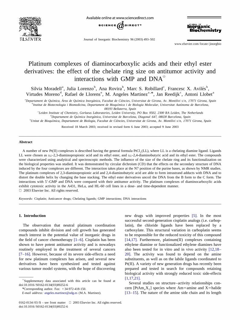
The solvents used were purchased from SDS (Barcelona,Fig. 1. Structures of the diamine dichloroplatinum complexes used in this Spain). The disodium salt of 59-rGMP was purchased fromstudy. 1a5PtCl (dap), 1b5PtCl (Etdap), 2a5PtCl (dab), 2b52 2 2 Sigma and potassium dihydrogenphosphate (KH PO ) was2 4PtCl (Etdab).2
purchased from J.T. Baker Chemicals (Deventer, theNetherlands). Calf Thymus DNA was from Sigma (Mad-
play an important role in the hydrolytic behaviour of therid, Spain).
complexes, influencing their solubility and thus affectingthe membrane permeability and consequently influencingthe platinum concentration inside the cell. 2 .3. Preparation of platinum complexes
It is known that the ultimate target of platinum com-pounds is DNA and that the major binding site is at the N7
2 .3.1. cis-Dichloro-(1-(carboxylic acid)-1,2-atoms of the purine bases. This binding takes place after
diaminoethane)platinum(II) [PtCl (dap)] ?H O2 2hydrolysis of the chloride ions inside the cell[6,16,22–26].2,3-Diaminopropionic acid monohydrochloride (dap?
From studies on oligonucleotides and DNA the GG andHCl) (0.288 g, 2.007 mmol) was neutralized with a
AG sequences within double-stranded DNA are known toNaHCO solution (0.9 g, 3.57 mmol) and then added to a3be the major sites of platinum binding. However, a varietysolution of K PtCl (0.9 g, 2.169 mmol). The mixture was2 4of binding modes is possible, each mode resulting in astirred at room temperature for 24 h. The dark solution
specific distortion of the DNA and in theory giving rise toformed was acidified to pH 2 with 3 M HCl. The resulting
a specific pharmacological effect[27]. Elucidating theprecipitate was washed in acetone and ethyl ether, and
mechanism of interaction of the platinum complexes withdried under vacuum for at least 24 h. Yield: 35%. IR (KBr)
nucleotides would be helpful in understanding their modes 21n (cm ): 3432 (st. OH), 3237–3201 (st. NH), 3125–2942
of action [28–30].(st. CH), 1716 (st. C=O), 1585 (d NH), 1247–1192 (st.
The present paper deals with two series ofcis-platinum 1NC, st. CO). H-NMR (200 MHz, D O)d (ppm): 2.87 (m,2complexes (Fig. 1) varying in the length of the chelatingCH ), 3.64 (m, CH). [PtClC H N O ?H O]: 7.22% N,2 2 3 8 2 2 2diamine ligand and the substituent in the diamine back-9.28% C, 2.60% H, 18.27% Cl. Found: 7.28% N, 9.87% C,
bone. These complexes were designed, synthesized and 1 22.54% H, 18.06% Cl. [M2H ] m /z 369.2.further investigated to establish how the substituents andthe size of the diamine chelate ring affect the binding toGMP and DNA, and the in vitro antiproliferative activity. 2 .3.2. cis-Dichloro-(1-(carboxylic acid)-1,3-
diaminopropane)platinum(II) [PtCl (dab)] ?1/4HCl2
2,4-Diaminobutyric acid dihydrochloride (dab?2HCl)2 . Experimental (0.309 g, 1.584 mmol) was neutralized with a NaHCO3
solution (0.945 g, 3.75 mmol) and then added to a solution2 .1. Analytical instruments of K PtCl (0.945 g, 2.277 mmol). The mixture was2 4
stirred at room temperature for 24 h. The dark solutionIR spectra were recorded in the solid state (KBr pellets) formed was acidified to pH 2 with 3 M HCl. The resulting
21in the range 4000–400 cm using a FT-IR Mattson precipitate was washed in acetone and ethyl ether, and1 13Satellite spectrometer. H- and C-NMR spectra were dried under vacuum for at least 24 h. Yield: 30%. IR (KBr)
21recorded on a Bruker DPX 200 spectrometer using n (cm ): 3447 (st. OH), 3239–3199 (st. NH), 3125–2940deuterium oxide (D O) and dimethyl sulfoxide-d (st. CH), 1717 (st. C=O), 1585 (d NH), 1215–1082 (st.2 6
1 1(DMSO-d ) as solvents. H-NMR spectra for GMP interac- NC, st. CO). H-NMR (200 MHz, D O)d (ppm): 2.41 (m,6 2
tions were recorded on a Bruker DPX 300 spectrometer CH ), 2.81 (m, NCH ), 4.09 (m, CH). [PtCl C H N O?2 2 2 4 10 2 2
with a 5 mm multi-nucleus probe, using D O as solvent 1/4HCl]: 7.12% N, 12.22% C, 2.63% H, 20.28% Cl.2
with tetramethylsilane (TMS) as an external reference at Found: 7.26% N, 12.42% C, 3.21% H, 20.82% Cl. [M21 2
d 50 ppm. Elemental analyses were carried out on a Carlo H ]m /z 383.2.
S. Moradell et al. / Journal of Inorganic Biochemistry 96 (2003) 493–502 495
2 .3.3. cis-Dichloro-(1-(carboxylic acid ethyl ester)-1,2- for 24 h. Formation of the products was quantified usingdiaminoethane)platinum(II) [PtCl (Etdap)] ?H O the relative H8 signal intensities of 59-GMP.2 2
A NaHCO solution (0.125 g, 1.48 mmol) was added to3
a solution of K PtCl (0.301 g, 0.725 mmol). The mixture 2 .5. Formation of drug–DNA complexes2 4
was stirred at room temperature and a solution of 2,3-diaminopropionate ethyl ester dihydrochloride (Etdap? The ligands and complexes were dissolved in an aque-2HCl) (0.151 g, 0.736 mmol), prepared as previously ous solution of 2% DMSO in all cases. The solutions weredescribed[31], was added after a few minutes. The final freshly prepared before use. Aliquots of these solutionsmixture was stirred at room temperature for 4 days. The were added to the Calf Thymus DNA in a TE (Tris–resulting precipitate was washed with acetone, and ethyl EDTA) buffer solution (50 mM NaCl, 10 mM Tris–HCl,ether, and dried under vacuum for at least 24 h. Yield: 0.1 mM EDTA, pH 7.4). The amount of complex added to
2178%. IR (KBr)n (cm ): 3249–3191 (st. NH), 3108–2989 the DNA solution was designatedr (the input molar ratioi
(st. CH), 1740 (st. C=O), 1571 (d NH), 1223–1089 (st. of Pt to nucleotide). The mixture was incubated at 378C1NC, st. CO). H-NMR (200 MHz, DMSO-d ) d (ppm): for 48 h.6
1.33 (t, CH , J 5 3 Hz), 2.80 (m, NCH ), 3.70 (m, CH),3 213 2 .6. Circular dichroism spectroscopy4.30 (m, CH ), 5.50–6.55 (m, 4 NH). C-NMR (50 MHz,2
DMSO-d ) d (ppm): 13.90 (CH ), 48.34 (NCH ), 60.206 3 2
The CD spectra of the drug–DNA complexes (DNA(CH), 61.60 (CH ), 168 (C=O). [PtCl C H N O ?H O]:2 2 5 12 2 2 2
concentration 20 mg/ml, molar ratiosr 5 0.10, 0.30 and6.73% N, 14.43% C, 3.39% H, 17.04% Cl. Found: 6.77% i
0.50) were recorded at room temperature on a JASCON, 14.80% C, 3.12% H, 16.70% Cl.J720 spectropolarimeter with a 450 W xenon lamp. Eachsample was scanned twice in a range of wavelengthsbetween 220 and 330 nm. The CD spectra shown are the2 .3.4. cis-Dichloro-(1-(carboxylic acid ethyl ester)-1,3-averages of three independent scans. The data are ex-diaminopropane)platinum(II) [PtCl (Etdab)]2
pressed as averaged residue molecular ellipticity inA NaHCO solution (0.080 g, 0.952 mmol) was added32 21degrees?cm ?dmol .to a solution of K PtCl (0.200 g, 0.481 mmol). The2 4
mixture was stirred at room temperature and a solution of2 .7. Incubation experiments and apoptosis evaluation.2,4-diaminobutyrate ethyl ester dihydrochloride (Etdab?
Cell lines and growth conditions2HCl) (0.101 g, 0.456 mmol), prepared as previouslydescribed[31], was added after a few minutes. The final
A431 human squamous carcinoma cells and HeLamixture was stirred at room temperature for 4 days. Thehuman cervix carcinoma cells were cultured in Dulbecco’sresulting precipitate was washed with acetone, and ethylmodified Eagle’s medium (DMEM) supplemented with 2ether, and dried under vacuum for at least 24 h. Yield:
21 mM L-glutamine and 10% fetal bovine serum (FBS, Life30%. IR (KBr)n (cm ): 3230–3188 (st. NH), 3119–2952Technologies) at 378C in a humidified 10% CO in-(st. CH), 1740 (st. C=O), 1586 (d NH), 1224–1096 (st. 2
1 cubator. HL-60 human promyelocitic leukemia cells wereNC, st. CO). H-NMR (200 MHz, DMSO-d ) d (ppm):6
cultured in RPMI-1640 supplemented with 2 mML-1.30 (t, CH ,J 5 3 Hz), 2.00 (m, CH ), 3.50 (m, NCH ),3 2 2
glutamine and 10% FBS in a humidified atmosphere at 5%3.44–3.50 (m, CH, under the HDO signal from theCO . All cell lines were obtained from the American Typesolvent), 4.20 (q, CH ,J 5 3 Hz), 5.20–5.40 (m, 4NH). 22
13 Culture Collection (ATCC, Rockville, MD, USA).C-NMR (50 MHz, DMSO-d ) d (ppm): 14 (CH ), 29.706 3
(CH ), 53.80 (CH), 61.70 (CH ), 170 (C=O).2 2
2 .8. Cytotoxicity assays[PtCl C H N O ]: 6.80% N, 17.48% C, 3.42% H,2 6 14 2 2
17.20% Cl. Found: 6.45% N, 17.46% C, 3.71% H, 17.18%HL-60, HeLa and A431 cellular survival was evaluatedCl.
using the MTT method ([3-(4,5-dimethylthiazol-2-yl)-2,5-diphenyltetrazolium bromide]), which quantifies the num-
2 .4. Reaction of the platinum complexes with the ber of surviving cells at a given time after exposure tomononucleotide 59-GMP cytotoxic drugs. Cells were plated onto 96-well sterile
3plates in 100ml of medium at a density of 5310 cells perAll reactions were carried out in NMR tubes (D O as well and incubated for 24 h. Compounds, dissolved in2
solvent). 59-GMP was incubated with each of the complex- H O, were added in final concentrations ranging from 0 to2
es in a ratio of 4:1 in D O at pH 7.0 (50 mM KD PO ) 500mM to a volume of 100ml /well. 24, 48 and 72 h later,2 2 4
and 378C. The absolute concentrations of the investigated 20ml of MTT solution was added to each well and thecomplexes depended on the solubility and varied from 3.4 plate was incubated for 2–3 h at 378C in a humidifiedto 5.0 mM. The reactions were monitored by automatically 10% CO atmosphere. Cell viability was determined by2
1recording the H-NMR spectra at time intervals of 30 min measuring the absorbance at 495 nm using a Microplate
496 S. Moradell et al. / Journal of Inorganic Biochemistry 96 (2003) 493–502
Reader. All cytotoxicity experiments were performed three
times in quadruplicate. The 50% inhibitory dose (IC )50
was defined as the drug concentration that reduced thenumber of living cells to 50%.
2 .9. Analysis of DNA fragmentation
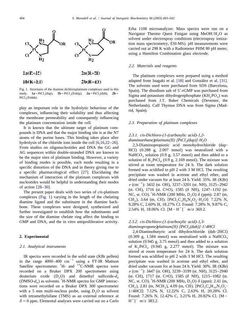
For DNA ladder formation analysis, characteristic ofScheme 1.5apoptotic cell death, 5?10 HL-60 cells /ml were plated
and exposed to 15mM cisplatin, 150 or 300mM carbop- 22Using this method, the (dap) ligand reacts with PtCl as4latin and the new platinum compounds for 72 h. Theshown in Scheme 1.drug-treated cells were centrifuged and washed twice with 1The platinum complexes were characterized by IR, H-PBS (phosphate-buffered saline). The DNA was extractedNMR, elemental analysis and ESI-MS. The IR spectrawith an Apoptotic DNA-Ladder kit (Roche). After electro-
21(cm ) of all platinum complexes show a doublen bandasphoresis of the total DNA from each sample on a 1.5%21at 3200–3300 cm for –NH and the expected absorption2agarose gel, DNA was visualized by ethidium bromide
21for n(C=O) stretching appears at 1716 and 1717 cm forstaining and photographed under UV illumination.PtCl (dap) and PtCl (dab), respectively, and at 1740 for2 2
the ethyl ester derivatives. For the ester derivatives, the12 .10. Quantitative evaluation of apoptosis by Annexin V- H-NMR spectra show a signal at 4.3 ppm corresponding
FITC to the ethyl ester CH and a triplet at 1.3 ppm corre-2
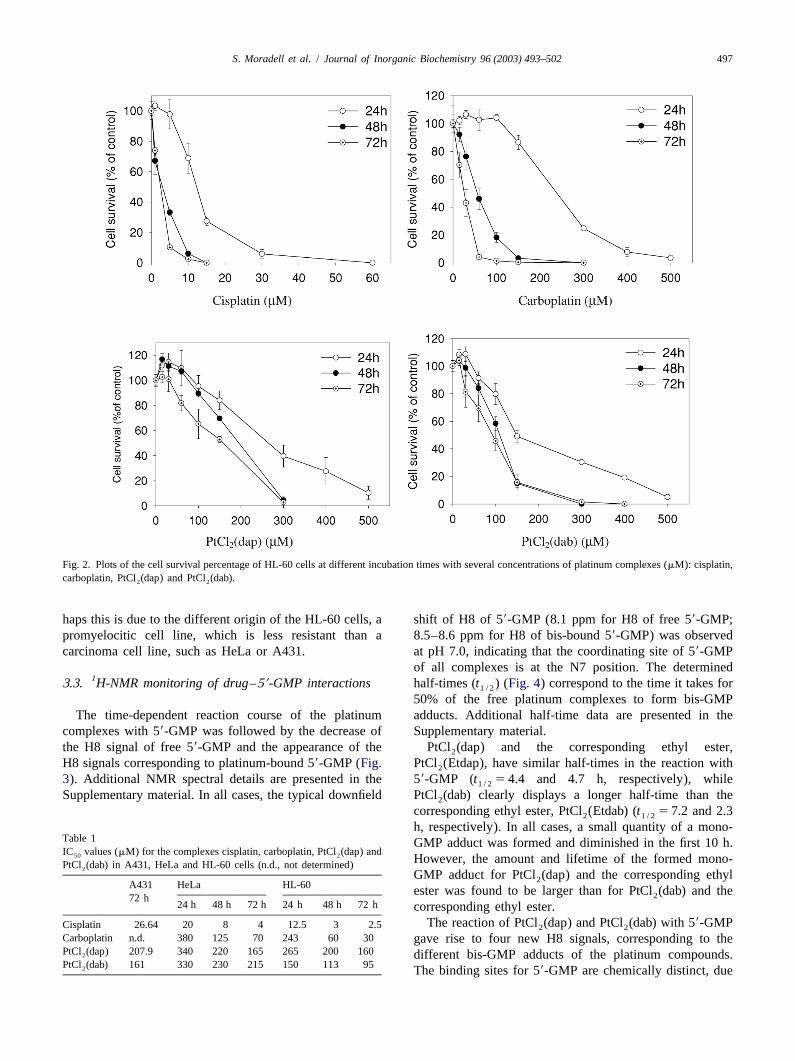
sponding to the ethyl ester CH . NH signals do not appear3HL-60 tumor cells were seeded on six-well tissue when D O is used, but in DMSO these signals appear at25culture plates at a density of 5310 cells /ml. Compounds 5.2–5.5 ppm. The ESI-MS spectra of PtCl (dap) and2were added at a final concentration of 5 or 15mM for PtCl (dab) present the signal of the anion corresponding to2cisplatin and 75 or 150mM for the other complexes and the loss of the acid proton atm /z 369.2 and 383.2,incubated for 24 or 48 h. After the desired period of respectively. The ESI-MS spectra are presented in theincubation, cells were centrifuged and washed twice with Supplementary material.cold PBS. The supernatant was removed and cells wereresuspended in Annexin V buffer provided with the 3 .2. Drug cytotoxicityAnnexin V-FITC kit (anexin V labeled with fluoresceinisothiocyanate) (Roche). Then 2ml of Annexin V-FITC The growth-inhibitory effect of PtCl (dap) and2(An) and 2ml of propidium iodide (PI) were added and PtCl (dab) complexes was examined in A431, HL-60 and2samples were incubated for 15 min at room temperature in HeLa cells using cisplatin and carboplatin as positivethe dark, prior to analysis using a FACScalibur Becton controls. In all cases, drug treatment in the concentrationDickinson flow cytometer. FITC fluorescence was mea- range between 0 and 500mM resulted in a dose- andsured at 530–545 nm and the fluorescence of DNA–PI time-dependent inhibition of cell survival (Fig. 2). Table 1complexes at 575–606 nm. Cell debris was excluded from shows the IC values of the compounds for the cell lines.50analysis by an appropriate forward light scatter threshold In the HeLa cell line, the dap and dab complexes have asetting. Ten thousand cells were analyzed in each con-similar effect as carboplatin, reducing cell survival in adition. Four quadrants of the cytograms were set using more progressive way than cisplatin. However, at shorternegative controls. The proportions of cells in each quad- times, PtCl (dap) and PtCl (dab) were more effective than2 2rant were expressed as the percentage of the total popula-carboplatin. The cytotoxic effects of both new compoundstion. on HeLa cells were similar at the times tested.
In A431 cells and HL-60 cells a progressive decrease ofcell survival was observed for PtCl (dap) and PtCl (dab)2 2
3 . Results and discussion when the drug concentration increased. In both cell lines,the complex PtCl (dab) was more effective than2
3 .1. Synthesis and chemical characterization PtCl (dap). In HL-60 cells at a short incubation time (242
h), PtCl (dab) was more active than carboplatin. However,2
The platinum complexes were synthesized using both complexes were found to be less active than carbop-equimolar ratios of K PtCl and diamine chelating ligands latin after 48 and 72 h incubation, suggesting that the2 4
following a method adapted from Inagaki et al.[18] and binding to nuclear DNA is less than that of cisplatin or´Gonzalez et al.[31]. Free diamine chelating ligands are carboplatin.
present with hydrochloride molecules which have to be In all experiments, the HL-60 cell line appeared to beneutralized with NaHCO before binding to platinum. more sensitive to treatment by platinum complexes. Per-3
S. Moradell et al. / Journal of Inorganic Biochemistry 96 (2003) 493–502 497
Fig. 2. Plots of the cell survival percentage of HL-60 cells at different incubation times with several concentrations of platinum complexes (mM): cisplatin,carboplatin, PtCl (dap) and PtCl (dab).2 2
haps this is due to the different origin of the HL-60 cells, a shift of H8 of 59-GMP (8.1 ppm for H8 of free 59-GMP;promyelocitic cell line, which is less resistant than a 8.5–8.6 ppm for H8 of bis-bound 59-GMP) was observedcarcinoma cell line, such as HeLa or A431. at pH 7.0, indicating that the coordinating site of 59-GMP
of all complexes is at the N7 position. The determined13 .3. H-NMR monitoring of drug–59-GMP interactions half-times (t ) (Fig. 4) correspond to the time it takes for1 / 2
50% of the free platinum complexes to form bis-GMPThe time-dependent reaction course of the platinum adducts. Additional half-time data are presented in the
complexes with 59-GMP was followed by the decrease of Supplementary material.the H8 signal of free 59-GMP and the appearance of the PtCl (dap) and the corresponding ethyl ester,2
H8 signals corresponding to platinum-bound 59-GMP (Fig. PtCl (Etdap), have similar half-times in the reaction with2
3). Additional NMR spectral details are presented in the 59-GMP (t 54.4 and 4.7 h, respectively), while1 / 2
Supplementary material. In all cases, the typical downfield PtCl (dab) clearly displays a longer half-time than the2
corresponding ethyl ester, PtCl (Etdab) (t 5 7.2 and 2.32 1 / 2
h, respectively). In all cases, a small quantity of a mono-T able 1 GMP adduct was formed and diminished in the first 10 h.IC values (mM) for the complexes cisplatin, carboplatin, PtCl (dap) and50 2 However, the amount and lifetime of the formed mono-PtCl (dab) in A431, HeLa and HL-60 cells (n.d., not determined)2
GMP adduct for PtCl (dap) and the corresponding ethyl2A431 HeLa HL-60 ester was found to be larger than for PtCl (dab) and the272 h
24 h 48 h 72 h 24 h 48 h 72 h corresponding ethyl ester.The reaction of PtCl (dap) and PtCl (dab) with 59-GMPCisplatin 26.64 20 8 4 12.5 3 2.5 2 2
Carboplatin n.d. 380 125 70 243 60 30 gave rise to four new H8 signals, corresponding to thePtCl (dap) 207.9 340 220 165 265 200 1602 different bis-GMP adducts of the platinum compounds.PtCl (dab) 161 330 230 215 150 113 952 The binding sites for 59-GMP are chemically distinct, due

498 S. Moradell et al. / Journal of Inorganic Biochemistry 96 (2003) 493–502
teriomers, leading to two H8 signals for each binding site,making four in total. In the case of PtCl (dab), some of the2
four signals overlap slightly.When the reaction of the ethyl esters PtCl (Etdap) and2
PtCl (Etdab) with 59-GMP was followed, one set of four2
new H8 signals appeared initially. However, after severalhours a second set of four H8 signals emerged. In the caseof PtCl (Etdab) the eight new H8 signals overlapped2
slightly. Raising the temperature to 548C did not alter thewidth and number of H8 signals, indicating that the bis-59-GMP adducts rotate freely about the Pt–N7 bond. Conse-quently, the appearance of eight new H8 signals instead offour due to the presence of rotamers can possibly beexcluded. At the same time and rate as the appearance ofthe second set of four H8 signals, a second tripletcorresponding to the CH from the ethyl ester appeared3
upfield from the original signal. The fact that the tripletshift is upfield and the rate of its appearance is muchslower than the rate of 59-GMP adduct formation stronglysuggest that the ethyl esters are hydrolyzed during thereaction. In order to substantiate this, solutions of theplatinum complexes PtCl (Etdap) and PtCl (Etdab) (pH2 2
17.0, 37 8C) without 59-GMP were studied with H-NMRfor 24 h. Again, an upfield shift of the triplet corre-sponding to CH was observed. Furthermore, without the3
overlap caused by 59-GMP, a quartet signal originating1 from the CH group of the ethyl esters was observed, asFig. 3. H-NMR spectra obtained from study of the interaction between 2
well as its upfield shift upon hydrolysis.PtCl (dap) and 59-GMP for 24 h. Spectra were recorded in D O with2 2
TMS as reference at 378C and pH 7.0.
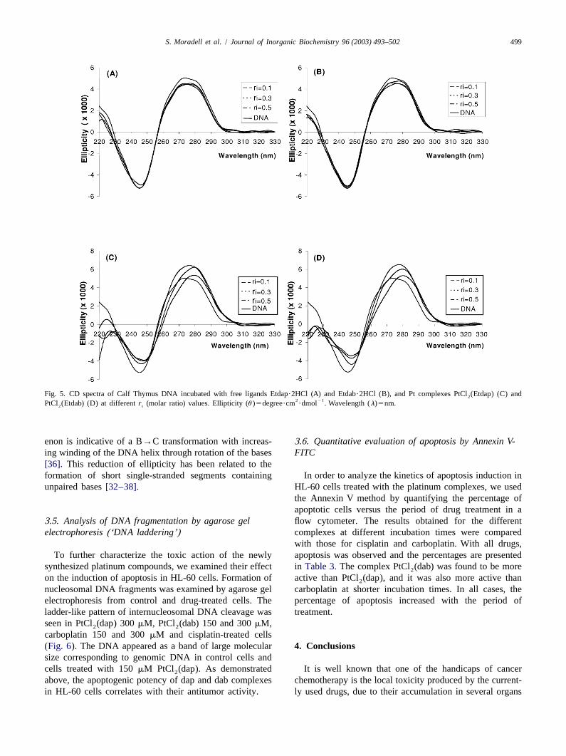
3 .4. Circular dichroism spectroscopyto the non-symmetric nature of the platinum complexes,and would lead to two distinct H8 signals in the NMR In order to show the drug interaction with DNA and thespectrum. However, adduct formation between 59-GMP modification of its secondary structure, the CD spectra ofand the racemic platinum complexes results in two dias- Calf Thymus DNA incubated with free ligands (dap?HCl,
dab?2HCl, Etdap?2HCl and Etdab?2HCl) and the corre-sponding platinum complexes at several molar ratios (r 5i
0.1, 0.3 and 0.5) were recorded (Fig. 5). Spectra of the free ligands were obtained first to
determine whether they cause any modification of the helixstructure and to compare them with the results for thecomplexes. Free ligands did not show an effect on the CDspectrum of Calf Thymus DNA, as significant changes inwavelength and ellipticity values of the DNA bands werenot observed (Table 2). In contrast, the changes inellipticity and wavelength caused by the platinum com-plexes are significant. The behavior of PtCl (dap) and2
PtCl (dab) is similar to cisplatin[32]. In both cases, the2
ellipticity of the positive band increases with increasingvalues of r . An upshift in the l and l is alsoi max min
observed. These modifications are probably due to openingof the helix upon the formation of DNA intrastrandadducts [11,33–35]. However, PtCl (Etdap) and2
PtCl (Etdab) induce different modifications in the sec-2Fig. 4. Time-dependent reactions of complex PtCl (dap) with 59-GMP2ondary structure of DNA. In that case, the two complexesclearly shows monofunctional intermediates. Half-life times (t ) of the1 / 2
formation of the bis-adducts were determined graphically. reduce the ellipticity of the positive band. This phenom-
S. Moradell et al. / Journal of Inorganic Biochemistry 96 (2003) 493–502 499
Fig. 5. CD spectra of Calf Thymus DNA incubated with free ligands Etdap?2HCl (A) and Etdab?2HCl (B), and Pt complexes PtCl (Etdap) (C) and22 21PtCl (Etdab) (D) at differentr (molar ratio) values. Ellipticity (u )5degree?cm ?dmol . Wavelength (l)5nm.2 i
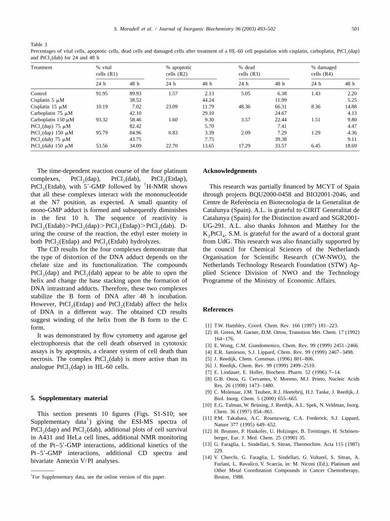
enon is indicative of a B→C transformation with increas- 3 .6. Quantitative evaluation of apoptosis by Annexin V-ing winding of the DNA helix through rotation of the bases FITC[36]. This reduction of ellipticity has been related to theformation of short single-stranded segments containing In order to analyze the kinetics of apoptosis induction inunpaired bases[32–38]. HL-60 cells treated with the platinum complexes, we used
the Annexin V method by quantifying the percentage ofapoptotic cells versus the period of drug treatment in a
3 .5. Analysis of DNA fragmentation by agarose gel flow cytometer. The results obtained for the differentelectrophoresis (‘ DNA laddering’) complexes at different incubation times were compared
with those for cisplatin and carboplatin. With all drugs,To further characterize the toxic action of the newly apoptosis was observed and the percentages are presented
synthesized platinum compounds, we examined their effect inTable 3.The complex PtCl (dab) was found to be more2
on the induction of apoptosis in HL-60 cells. Formation of active than PtCl (dap), and it was also more active than2
nucleosomal DNA fragments was examined by agarose gel carboplatin at shorter incubation times. In all cases, theelectrophoresis from control and drug-treated cells. The percentage of apoptosis increased with the period ofladder-like pattern of internucleosomal DNA cleavage was treatment.seen in PtCl (dap) 300mM, PtCl (dab) 150 and 300mM,2 2
carboplatin 150 and 300mM and cisplatin-treated cells(Fig. 6). The DNA appeared as a band of large molecular 4 . Conclusionssize corresponding to genomic DNA in control cells andcells treated with 150mM PtCl (dap). As demonstrated It is well known that one of the handicaps of cancer2
above, the apoptogenic potency of dap and dab complexes chemotherapy is the local toxicity produced by the current-in HL-60 cells correlates with their antitumor activity. ly used drugs, due to their accumulation in several organs

500 S. Moradell et al. / Journal of Inorganic Biochemistry 96 (2003) 493–502
T able 2CD spectral data for DNA for differentr (molar ratio)i
r l u l ui max max min min2 21 2 21(nm) (deg?cm ?dmol ) (nm) (deg?cm ?dmol )
3 3DNA 273.6 5.03?10 245.2 25.25?103 3dap?HCl 0.1 276.2 5.19?10 243.8 26.18?103 30.3 276.6 4.76?10 245.6 25.99?103 30.5 274.6 4.74?10 245.6 25.73?103 3dab?2HCl 0.1 276.4 4.63?10 245.6 25.89?103 30.3 279.6 4.47?10 245.6 26.00?103 30.5 275.6 4.70?10 246 25.73?103 3Etdap?2HCl 0.1 273.4 4.51?10 245.4 25.23?103 30.3 275.6 4.42?10 245.4 25.23?103 30.5 277.0 4.53?10 246.2 24.93?103 3Etdab?2HCl 0.1 277.8 4.75?10 245.6 25.08?103 30.3 273.2 4.60?10 245.4 25.03?103 30.5 276.8 4.53?10 245.4 25.20?103 3PtCl (dap) 0.1 276.8 6.05?10 246.8 25.21?1023 30.3 277.6 6.98?10 247.4 24.99?103 30.5 279.6 6.97?10 247.8 24.66?103 3PtCl (dab) 0.1 276.8 6.00?10 247.2 25.52?1023 30.3 278.2 6.74?10 247.4 24.95?103 30.5 277.8 6.71?10 248.2 24.67?103 3PtCl (Etdap) 0.1 276.6 6.39?10 246.6 24.29?1023 30.3 279.8 6.20?10 247.6 23.98?103 30.5 279.6 5.33?10 248.2 23.90?103 3PtCl (Etdab) 0.1 277.6 6.52?10 247.8 24.40?1023 30.3 279.8 6.01?10 247.6 23.74?103 30.5 280.2 5.32?10 247.2 23.46?10
and tissues at therapeutic doses. Apoptosis is considered to be a cleaner system of cell death than necrosis, because
lysis of the necrotic cells leads to the production of localside-effects due to the release of toxic substances frominside the cell to the extracellular matrix. Thus, efforts arebeing directed towards the synthesis of novel compoundswith antineoplastic activity resulting from apoptosis induc-tion. The four complexes discussed above were studied todetermine how the size of the chelate ring and its func-tionalisation influences the properties (e.g. biological ac-tivity) of the platinum drugs.
Cell-survival assays indicate that PtCl (dap) and2
PtCl (dab) show, at high concentrations, cytotoxic activity2
against tumor cells in a dose- and time-dependent way,similar to that known for carboplatin.
The cytotoxic activity of the dap and dab complexes isnot the same in the different cell lines. At 72 h, PtCl (dab)2
is more active than PtCl (dab) in A431, but in HeLa it is2
more active at 24 and 48 h, but not after 72 h incubation.Fig. 6. Agarose gel electrophoresis of DNA extracted from humanmyelogenous leukemic HL-60 cells. Line 1 shows untreated HL-60 cells PtCl (dab) appears to cross the cell membrane more2growing exponentially (control). Lines 2, 3 and 4 show DNA extracted quickly than PtCl (dap), probably because its larger che-2from HL-60 cells treated for 72 h with 150mM PtCl (dap), PtCl (dab)2 2 late backbone makes the complex more lipophilic, there-and carboplatin, respectively. Line 5 shows DNA extracted from HL-60
fore its transport trough the membrane is likely to becells treated for 72 h with 15mM cisplatin. Lines 6, 7 and 8 show DNAeasier. This explains why this complex is more active thanextracted from HL-60 cells treated for 72 h with 300mM PtCl (dap),2
PtCl (dab) and carboplatin, respectively. PtCl (dap) at shorter incubation times.2 2
S. Moradell et al. / Journal of Inorganic Biochemistry 96 (2003) 493–502 501
T able 3Percentages of vital cells, apoptotic cells, dead cells and damaged cells after treatment of a HL-60 cell population with cisplatin, carboplatin, PtCl (dap)2
and PtCl (dab) for 24 and 48 h2
Treatment % vital % apoptotic % dead % damagedcells (R1) cells (R2) cells (R3) cells (R4)
24 h 48 h 24 h 48 h 24 h 48 h 24 h 48 h
Control 91.95 89.93 1.57 2.13 5.05 6.38 1.43 2.20Cisplatin 5mM 38.52 44.24 11.99 5.25Cisplatin 15mM 10.19 7.02 23.09 11.79 48.36 66.31 8.36 14.88Carboplatin 75mM 42.10 29.10 24.67 4.13Carboplatin 150mM 93.32 58.46 1.60 9.30 3.57 22.44 1.51 9.80PtCl (dap) 75mM 82.42 5.70 7.41 4.472
PtCl (dap) 150mM 95.79 84.96 0.83 3.39 2.09 7.29 1.29 4.362
PtCl (dab) 75mM 43.75 7.75 39.38 9.112
PtCl (dab) 150mM 53.56 34.09 22.70 13.65 17.29 33.57 6.45 18.692
The time-dependent reaction course of the four platinum A cknowledgementscomplexes, PtCl (dap), PtCl (dab), PtCl (Etdap),2 2 2
1PtCl (Etdab), with 59-GMP followed by H-NMR shows This research was partially financed by MCYT of Spain2
that all these complexes interact with the mononucleotide through projects BQU2000-0458 and BIO2001-2046, and`at the N7 position, as expected. A small quantity of Centre de Referencia en Biotecnologia de la Generalitat de
mono-GMP adduct is formed and subsequently diminishes Catalunya (Spain). A.L. is grateful to CIRIT Generalitat dein the first 10 h. The sequence of reactivity is Catalunya (Spain) for the Distinction award and SGR2001-PtCl (Etdab).PtCl (dap).PtCl (Etdap).PtCl (dab). D- UG-291. A.L. also thanks Johnson and Matthey for the2 2 2 2
uring the course of the reaction, the ethyl ester moiety in K PtCl . S.M. is grateful for the award of a doctoral grant2 4
both PtCl (Etdap) and PtCl (Etdab) hydrolyzes. from UdG. This research was also financially supported by2 2
The CD results for the four complexes demonstrate that the council for Chemical Sciences of the Netherlandsthe type of distortion of the DNA adduct depends on the Organisation for Scientific Research (CW-NWO), thechelate size and its functionalization. The compounds Netherlands Technology Research Foundation (STW) Ap-PtCl (dap) and PtCl (dab) appear to be able to open the plied Science Division of NWO and the Technology2 2
helix and change the base stacking upon the formation of Programme of the Ministry of Economic Affairs.DNA intrastrand adducts. Therefore, these two complexesstabilize the B form of DNA after 48 h incubation.However, PtCl (Etdap) and PtCl (Etdab) affect the helix2 2 R eferencesof DNA in a different way. The obtained CD resultssuggest winding of the helix from the B form to the C
[1] T .W. Hambley, Coord. Chem. Rev. 166 (1997) 181–223.form.[2] H . Green, M. Garner, D.M. Orton, Transition Met. Chem. 17 (1992)It was demonstrated by flow cytometry and agarose gel
164–176.electrophoresis that the cell death observed in cytotoxic [3] E . Wong, C.M. Giandomenico, Chem. Rev. 99 (1999) 2451–2466.assays is by apoptosis, a cleaner system of cell death than[4] E .R. Jamieson, S.J. Lippard, Chem. Rev. 99 (1999) 2467–3498.
[5] J . Reedijk, Chem. Commun. (1996) 801–806.necrosis. The complex PtCl (dab) is more active than its2[6] J . Reedijk, Chem. Rev. 99 (1999) 2499–2510.analogue PtCl (dap) in HL-60 cells.2[7] E . Lindauer, E. Holler, Biochem. Pharm. 52 (1996) 7–14.[8] G .B. Onoa, G. Cervantes, V. Moreno, M.J. Prieto, Nucleic Acids
Res. 26 (1998) 1473–1480.[9] C . Molenaar, J.M. Teuben, R.J. Heetebrij, H.J. Tanke, J. Reedijk, J.
5 . Supplementary material Biol. Inorg. Chem. 5 (2000) 655–665.¨[10] E .G. Talman, W. Bruning, J. Reedijk, A.L. Spek, N. Veldman, Inorg.
Chem. 36 (1997) 854–861.This section presents 10 figures (Figs. S1-S10; see[11] P .M. Takahara, A.C. Rosenzweig, C.A. Frederick, S.J. Lippard,1Supplementary data ) giving the ESI-MS spectra of Nature 377 (1995) 649–652.
PtCl (dap) and PtCl (dab), additional plots of cell survival ¨[12] H . Brunner, P. Hankofer, U. Holzinger, B. Treittinger, H. Schonen-2 2
in A431 and HeLa cell lines, additional NMR monitoring berger, Eur. J. Med. Chem. 25 (1990) 35.[13] G . Faraglia, L. Sindellari, S. Sitran, Thermochim. Acta 115 (1987)of the Pt–59-GMP interactions, additional kinetics of the
229.Pt–59-GMP interactions, additional CD spectra and[14] V . Cherchi, G. Faraglia, L. Sindellari, G. Voltarel, S. Sitran, A.
bivariate Annexin V/PI analyses. Furlani, L. Ravalico, V. Scarcia, in: M. Niconi (Ed.), Platinum andOther Metal Coordination Compounds in Cancer Chemotherapy,
1For Supplementary data, see the online version of this paper. Boston, 1988.
502 S. Moradell et al. / Journal of Inorganic Biochemistry 96 (2003) 493–502
[15] M .S. Robillard, M. Galanski, W. Zimmermann, B.K. Keppler, J. [28] Y . Qu, J.A. Fitzgerald, H. Rauter, N. Farrell, Inorg. Chem. 40 (2001)Reedijk, J. Inorg. Biochem. 88 (2002) 254–259. 6324–6327.
[16] M . Meroueh, J. Kjellstrom, K.S.M. Martensson, S.K.C. Elmroth, [29] A .T.M. Marcellis, C.G. van Kralingen, J. Reedijk, J. Inorg. Bio-C.S. Chow, Inorg. Chim. Acta 297 (2000) 145–155. chem. 13 (1980) 213–222.
[17] D . Lebwohl, R. Canetta, Eur. J. Cancer 34 (1998) 1522. [30] E .L.M. Lempers, M.J. Bloemink, J. Brouwer, Y. Kidani, J. Reedijk,[18] K . Inagaki, Y. Kidani, K. Suzuki, T. Tazuko, Chem. Pharm. Bull. 28 J. Inorg. Biochem. 40 (1990) 23–35.
´ ´ ´(1980) 2286–2291. [31] M .L. Gonzalez, J.M. Tercero, A. Matilla, J. Niclos-Gutierrez, M.T.´ ´ ´[19] J . Lean, P.G. Parsons, A.B. Awaluddin, J.J. Jacobs, D.J. Maddalena, Fernandez, M.C. Lopez, C. Alonso, S. Gonzalez, Inorg. Chem. 36
J.G. Wilson, S.K. Khoo, C. Ward, Cancer Chemother. Pharmacol. 26 (1997) 1806–1812.(1990) 42–46. [32] J .P. Macquet, J.L. Butour, Biochimie 60 (1978) 901.
¨[20] Y . Kageyama, Y. Yamazaki, H. Okuno, J. Inorg. Biochem. 70 (1998) [33] V . Brabec,V. Kleinwachter, J. Butour, M.P. Johnson, Biophys. Chem.25–32. 35 (1990) 129.
´ ´ ´ ´ ´[21] J . Reedijk, Inorg. Chim. Acta 198–200 (1992) 873–878. [34] R . Zaludova, A. Zakouska, J. Kasparova, Z. Balcarova, V.¨ ´[22] C .J. van Garderen, M.J. Bloemink, E. Richardson, J. Reedijk, J. Kleinwachter, O. Vrana, N. Farrell, V. Brabec, Eur. J. Biochem. 246
Inorg. Biochem. 42 (1991) 199–205. (1997) 508–517.[23] S .U. Dunham, S.U. Dunham, C.J. Turner, S.J. Lippard, J. Am. [35] D .Z. Yang, S.S.G.E. van Boom, J. Reedijk, J.H. van Boom, A.H.J.
Chem. Soc. 120 (1998) 5395–5406. Wang, Biochemistry 34 (1995) 12912–12920.[24] L .G. Marzilli, S.O. Ano, F.P. Intini, G. Natile, J. Am. Chem. Soc. [36] V . Marchan, V. Moreno, E. Pedroso, A. Grandas, Chem. Eur. J. 7
121 (1999) 9133–9142. (2001) 808–815.´[25] E .C.H. Ling, G.W. Allen, K.Vickery, T.W. Hambley, J. Inorg. Chem. [37] X . Riera, A. Caubet, C. Lopez, V. Moreno, Polyhedron 18 (1999)
78 (2000) 55–62. 2549–2555.´[26] T .W. Hambley, E.C.H. Ling, V.P. Munk, M.S. Davies, J. Biol. Inorg. [38] V . Moreno, G. Cervantes, G.B. Onoa, F. Sampedro, P. Santalo,
Chem. 6 (2001) 534–542. Polyhedron 16 (1997) 4297–4303.[27] N .G. Di Msi, F.P. Intini, C. Pacifico, L. Maresca, G. Natile, Inorg.
Chim. Acta 310 (2000) 27–33.
Copyright © 2022 FDOKUMEN