Histopathology of epi-LASIK in eyes with virgin corneas and eyes with previously altered corneas
Upload
khangminh22Category
view
0download
0
Pigmentary disorders of the eyes and skin
Syril Keena T. Que, MD a, Gillian Weston, BA a,Jeanine Suchecki, MD b, Janelle Ricketts, MBA, MD a,⁎
aDepartment of Dermatology, University of Connecticut Health Center, 21 South Road, Farmington, CT 06032bDepartment of Ophthalmology, University of Connecticut Health Center, 21 South Road, Farmington, CT 06032
Abstract Oculocutaneous albinism, Menkes syndrome, tuberous sclerosis, neurofibromatosis type 1,
dyskeratosis congenita, lentiginosis profusa syndrome, incontinentia pigmenti, andWaardenburg syndrome
all are genodermatoses that have well established gene mutations affecting multiple biological pathways,
including melanin synthesis, copper transport, cellular proliferation, telomerase function, apoptosis, and
melanocyte biology. Onchocerciasis results from a systemic inflammatory response to a nematode infection.
Hypomelanosis of Ito is caused by chromosomal mosaicism, which underlies its phenotypic heterogeneity.
Incomplete migration of melanocytes to the epidermis and other organs is the underlying feature of nevus of
Ota. Vogt-Koyangi-Harada and vitiligo have an autoimmune etiology; the former is associated with
considerable multiorgan involvement, while the latter is predominantly skin-limited.
© 2015 Elsevier Inc. All rights reserved.
Oculocutaneous albinism
Background
Oculocutaneous albinism (OCA) is a condition that
involves mutations in the genes mediating melanin synthesis
(Table 1).1,2 This results in the hypopigmentation of the
skin, hair, and eyes; an increased risk of skin cancer; and an
increased susceptibility for development of photophobia,
refractive errors, and other visual anomalies. OCA1 A is the
most severe form, with complete absence of melanin.
Individuals with OCA1 A are born with white hair at birth,
while those with the other types are born with pigmented hair
and can accumulate more pigment over time.
Ocular manifestations
The human fovea is responsible for much of our visual
function, including color vision and highly specialized central
fine visual acuity.3 In OCA, reduced visual acuity can result
from foveal hypoplasia.4 The absence of melanin also leads to
optic nerve fibermisrouting,whichmay contribute to strabismus
and reduced stereoscopic vision (depth perception).5,6
The development of nystagmus can occur between six to
eight weeks of age and results from poor vision. It is initially
slow with a large amplitude, but the amplitude often decreases
within the first year of life.7 Poor fixation related to nystagmus
and the development of strabismus and high refractive errors
can lead to ambylopia, defined as reduction of vision due to the
lack of development of the visual pathway during childhood.
Patients with OCA can also develop varying degrees of iris
hypopigmentation and transillumination as well as reduced
pigmentation of the retinal pigment epithelium. The amount of⁎ Corresponding author. Tel.: 860-679-4600; fax: 860-679-7534.
E-mail address: [email protected] (J. Ricketts).
http://dx.doi.org/10.1016/j.clindermatol.2014.10.007
0738-081X/© 2015 Elsevier Inc. All rights reserved.
Clinics in Dermatology (2015) 33, 147–158
hypopigmentation varies with the type of albinism, as with the
skin. All of these findings can contribute to reduced visual
acuity, color vision impairment, and photophobia.5
Diagnostic tests for ocular manifestations include the
detection of iris transillumination on slit lamp examination,
assessment for macula transparency and foveal hypoplasia on
retinal examination, and the detection of abnormal optic nerve
fiber crossing usingmonocular visual evoked potential.8 Prism
and alternate cover testing often discloses strabismus.7
Spectral domain optical coherence tomography (OCT) is the
best way to evaluate the retina for the characteristic findings of
ocular albinism, including the absence of or reduced foveal
depression, absence of specialized foveal photoreceptors, and
continuation of the inner retinal layers through the fovea.4,9–11
Teller acuity cards12 can be used to measure visual acuity
and shows improvement in acuity upon vertical presentation
compared with horizontal presentation of the cards. This is
likely due to the horizontal nature of the nystagmus in
oculocutaneous albinism.7
Treatment of the ocular manifestations in oculocutaneous
albinism for themost part involves corrective lenses. Eyeglasses
can correct visual acuity from 20/107 to 20/80.913 and also
improve the patient’s strabismus. In response to nystagmus,
patients will often develop a compensatory head posture.
Depending on the extent of this adjustment, extraocular
surgery can be considered to shift this position closer to the
primary gaze.14,15
Menkes syndrome/disease
Background
Menkes syndrome, also known as kinky hair disease, is an
X-linked recessive neurodegenerative disorder caused by
missense mutations in the ATP7 A gene (copper transport
gene on chromosome Xq21.1), which causes impaired
intestinal copper absorption,16 reduced activity of copper-
dependent enzymes,17,18 progressive hypotonia, seizures,
and failure to thrive.19 Skin and hair abnormalities (including
hypopigmentation of hair, twisted hairs or pili torti, and
pale and lax skin) result from decreased keratin fiber strength,
tyrosinase activity, and melanin synthesis.20 Treatment includes
infusions with copper salts.
Ocular manifestations
Patients with Menkes’ syndrome present with various
ocular findings, including poor visual acuity, myopia,
strabismus, blue irides, and iris stromal hypoplasia,21 which
result from deficient cytochrome c oxidase and superoxide
dismutase, copper dependent enzymes.
Menkes disease also involves visual changes secondary to
retinopathy. Menkes-related retinopathy has two main
causes: (1) the overall systemic copper deficiency and
(2) the loss of retinal Menkes protein, which normally helps
to regulate the copper levels of overlying photoreceptors.22
Tuberous sclerosis
Background
Tuberous sclerosis complex (TSC) is an autosomal
dominant neurocutaneous syndrome characterized by the
development of multiple hamartomas in several organs. The
tumor suppressor genes responsible for TSC include the TSC1
gene on chromosome 9 and the TSC2 gene on chromosome
16. These genes directly target RAS, which inactivates
mammalian target of rapamycin (mTOR), the key regulator
of cell proliferation and organ size.23
Cutaneous manifestations associated with TSC include
hypomelanotic macules/ash leaf spots (present in 97.2%),
facial angiofibromas (74.5%) (Figure 1), Shagreen patches
(48.1%) (which are connective tissue nevi sometimes
referred to as collagenomas), molluscum pendulum
(22.6%), (which is a skin polyp with papillomatosis of the
epidermis and dilated blood vessels), forehead fibrous
plaques (18.9%), periungual fibromas (15.1%), and “con-
fettilike” macules (2.8%) (which are multiple 1-2 mm white
Table 1 Oculocutaneous albinism gene mutations
Oculocutaneous
albinism (OCA)
Responsible
gene
Gene function Gene
localization
Prevalence
OCA1 Tyrosinase Rate limiting enzyme
for melanin production
11 q14-q21 1:40,000
OCA2 P gene Exact function unknown,
involved in transport into and out of melanosomes
and regulation of melanosome pH
15 q 1:36,000 (white Europeans)
1:3900-10,000 (Africans)
OCA3 TYRP1 Involved in stabilization of tyrosinase 9 q23 Rare (white Europeans, Asians)
1:8,500 (Africans)
OCA4 SCL45 A2 May transport substances required
for melanin biosynthesis
5 p Rare (white Europeans)
1:8,500 (Japanese)
148 S.K.T. Que et al.
spots symmetrically distributed over the extremities).24
Systemic manifestations include seizures (which occur in
90% to 96% of TSC patients and are the leading cause of
morbidity in TSC),25 renal angiomyolipomas,26 and cardiac
rhabdomyomas.27
Themanagement of TSC is, for themost part, symptomatic;
however, rapamycin and its analogues hold some promise as a
potential therapy. Rapamycin can normalize the dysregulated
mTOR pathway in TSC. It has shown some success in
inducing the regression of brain astrocytomas28 and renal
angiomyolipomas29 associated with TSC.
Ocular manifestations
The most common ocular finding in TSC is a retinal
hamartoma, which can be found in 40% to 50% of TSC
patients.30 The retinal hamartoma in TSC can present as a
unilateral, transparent, noncalcified lesion or as bilateral
calcified tumors, and is often benign with no treatment
required. Rarely, the tumor requires treatment in the presence
of growth, inflammation, and related ocular complications.
In the past, enucleation was the treatment of choice. New
potential therapeutic modalities have been described in case
reports. Recently, brachytherapy was successfully used in
the treatment of patients with exudates and vitritis related
to an aggressive hamartoma.31 There has also been a
report of a patient with tuberous sclerosis, bilateral retinal
hamartomas, and macular edema, which responded well
to treatment with bevacizumab and intravitreal triamcin-
olone acetonide.32
Other ocular complications of the tuberous sclerosis
complex include retinal pigment epithelial depigmented
lesions,33 refractive errors, and strabismus.34
Neurofibromatosis
Background
Type I neurofibromatosis (NF1) is an autosomal dominant
geno-oculo-dermatosis caused by deletions or mutations in
the neurofibromin gene (NF1) on chromosome 17 p11.2. The
NF1 gene encodes neurofibromin, which negatively regu-
lates RAS protein,35 leading to excess cell growth and an
increased potential for malignant transformation. An indi-
vidual can be diagnosed with NF1 if he or she has two or
more of the following:36
1. Six or more café au lait spots (N0.5 cm diameter in
prepubertal children, N1.5 cm after puberty) (Figure 2)
2. Axillary or inguinal freckling
3. Cutaneous neurofibromas or one plexiform neurofibroma
4. Two or more Lisch nodules
5. Optic pathway gliomas
6. Specific bony lesions (sphenoid wing dysplasia,
pseudoarthrosis of the tibia)
7. A first-degree relative with NF1
The diagnosis can be difficult to make in young children,
because many of the manifestations are age-dependent and
many phenotypic variations exist.37,38
Ocular manifestations
The optic pathway includes the optic nerve, chiasm,
tracts, and radiations. The optic pathway gliomas may
result in mild proptosis or decreased visual acuity due to
compression of the optic nerve along the pathway. Optic
nerve pallor or optociliary shunt vessels may be present on
examination. There is a suspected correlation between optic
nerve gliomas and the risk of cerebral arteriopathy. Due to
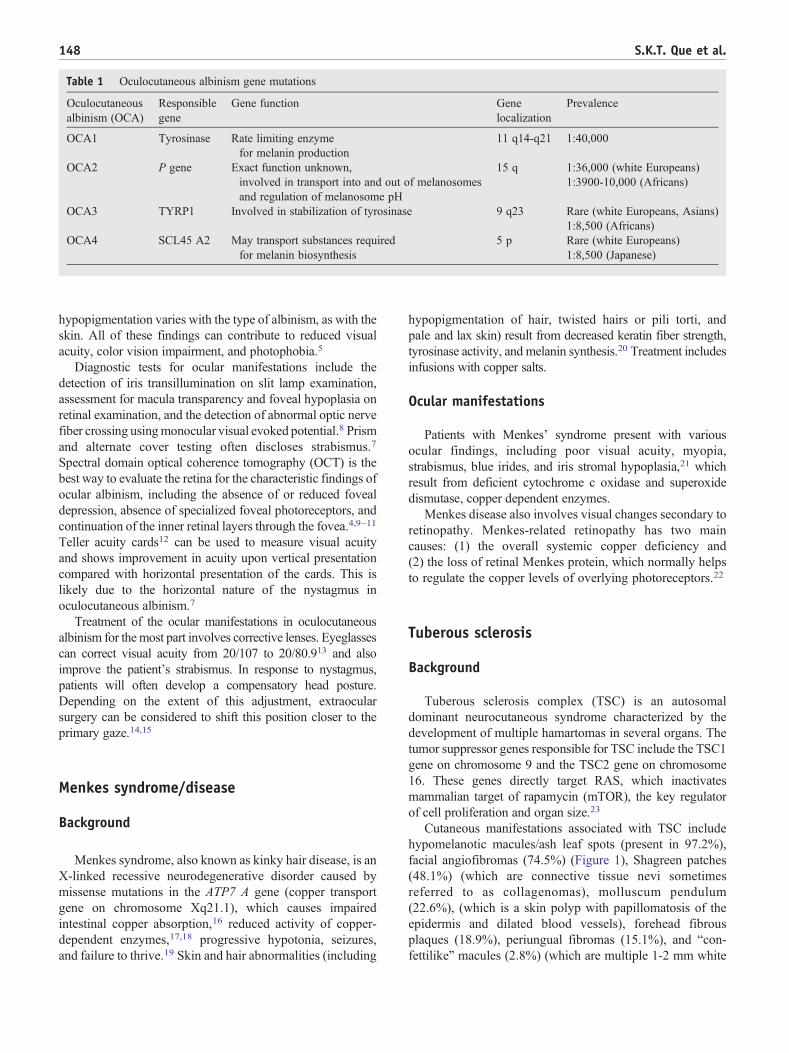
Fig. 1 Multiple facial angiofibromas in a patient with tuberous
sclerosis.
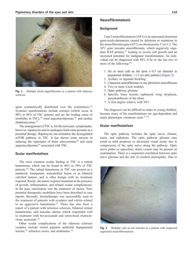
Fig. 2 Multiple café au lait macules in a patient with suspected
segmental neurofibromatosis.
149Pigmentary disorders of the eyes and skin
this correlation, it has been suggested that individuals
with NF1, especially those with optic nerve gliomas, undergo
vascular imaging by MRA or other brain imaging studies.38
The management of optic pathway gliomas remains
controversial. They may remain stable or slowly enlarge in
childhood or adulthood. Visual deficits in NF1 patients with
optic nerve gliomas can include decreased visual acuity, loss
of color vision, visual field defects, and loss of contrast
sensitivity. Associated findings also include strabismus,
nystagmus, proptosis, afferent pupillary defects, and optic
disc pallor. Studies have shown that retinal nerve fiber layer
thickness inversely correlates with optic pathway gliomas
and decreased vision.37 The location of optic nerve gliomas
in the noncortical visual pathway, either in the optic nerves,
chiasm, tracts, and radiations, makes biopsy or surgical
resection difficult due to the risk of visual loss. Chemother-
apy with carboplatin and vincristine can be used to manage
rapidly enlarging optic nerve gliomas. Radiation therapy is
no longer used since the risk of adversely impacting vision
and the risk of developing secondary malignancies such as a
malignant peripheral nerve sheath tumors.39,40
Lisch nodules are also present in NF1 and are one of the
characteristic findings. These are benign iris hamartomas that
do not impair vision. They are usually bilateral and found on
the iris surface or deep within the stroma. They are present in
more than 95% of carriers of the NF1 gene mutation and can
be useful as a diagnostic marker for neurofibromatosis.38
Other findings associated with NF1 are ciliary body cysts
and retinal pigmentary abnormalities.
Dyskeratosis congenita
Background
Dyskeratosis congenita is a rare, hereditary disorder
involving defective telomere biology and dyskerin gene
mutations and is clinically characterized by a triad of
reticular cutaneous hyperpigmentation, nail dystrophy, and
oral leukoplakia.41–43 Dyskeratosis congenita is a fatal
condition where patients usually develop aplastic anemia
and malignant transformation of the oral leukoplakia. Bone
marrow failure can also lead to opportunistic infections or
hemorrhage.43 Treatment of this condition involves short-
term treatments, such as anabolic steroids (which temporarily
increase blood counts), granulocyte macrophage colony-
stimulating factor, granulocyte colony-stimulating factor,
and erythropoietin44; however, the only long-term treatment
is hematopoietic stem cell transplant, which does not
necessarily prolong survival.45
Ocular manifestations
Ophthalmic manifestations have been observed in 40% of
patients with dyskeratosis congenita.41 The most common
findings are blepharitis, cicatricial entropion, trichiasis, loss
of cilia, nasolacrimal duct obstruction, and optic atro-
phy.41,46 Trichiasis is defined as the abnormal positioning
of eyelashes that grow inward towards the eye, potentially
abrading the ocular surface and causing corneal damage and
subsequent decrease in vision. Most of the anterior segment
findings of dyskeratosis congenita are likely related to
epithelial abnormalities in the ocular skin and mucous
membranes.46 Severe ocular surface disease can occur from
these epithelial abnormalities and include chronic cicatricial
keratoconjunctivitis (which may lead to significant symp-
tomatology and visual impairment).47
Only a small proportion of patients with dyskeratosis
congenita had retinal vascular changes; however, early
recognition and treatment of these changes can prevent
retinal neovascularization, exudative retinopathy, and retinal
detachment and, therefore, the related vision loss.48,49
In addition to the syndromic manifestations, treatment of
dyskeratosis congenitia with radiation or the use of steroids
before stem cell transplantation will increase the risk of
glaucoma and cataract formation. A baseline ophthalmologic
exam and periodic eye exams are therefore recommended for
every patient with dyskeratosis congenita.
Lentiginosis profusa/LEOPARD syndrome
Background
LEOPARD syndrome is an autosomal dominant condi-
tion and an acronym that stand for the cardinal features of
this condition, including lentigines (Figure 3), ECG
conduction abnormalities, ocular hypertelorism, pulmonic
stenosis, abnormal genitalia, retardation of growth, and
sensorineural deafness. While it is not part of the acronym,
hypertrophic cardiomyopathy is also commonly present. The
syndrome was first described by Zeisle and Becker in 1936,
but the term “LEOPARD syndrome” was not coined by
Gorlin until 1969.50 Other names for this condition are
lentiginosis profusa, multiple lentigines syndrome, cardio-
cutaneous syndrome, Moynahan syndrome, and progressive
cardiomyopathic lentiginosis. Lentigines do appear in 100%
of cases, usually after age 4 or 5, and thousands of lentigines
can be present by puberty. Genes known to be associated
with Leopard syndrome are PTPN11, RAF1, and BRAF.51
Ocular manifestations
Ocular associations with LEOPARD syndrome include
hypertelorism, which is defined as an abnormally wide
interorbital distance, and mild palpebral ptosis, a term that
describes drooping of the eyelid. Lentigines can sometimes
also appear in the sclera,51 which is usually of no
consequence to the vision. One paper has reported the
presence of a coloboma (a missing area of tissue or gap) in
150 S.K.T. Que et al.
the iris, retina, and choroid in a child with LEOPARD
syndrome,52 but this finding is rarely seen. Colobomas are
usually formed when a gap called the choroid fissure, which
is present during early development, fails to close before a
child is born.53
It was shown in one study that a significant number of
patients had a high percentage of abnormalities of visual
function in general and of visuo-motor integration. Ocular
movements were abnormal half of the time, and stereopsis
was abnormal in at least half the of patients.54 Because these
eye findings are common, baseline and follow-up ophthalmologic
exams are recommended.
Onchocerciasis
Background
Onchocerciasis, also known as river blindness, is caused
by the nematode Onchocerca volvulus. The blackfly vector
Simulium, transmits the infective larvae to humans. The
classic clinical finding in Onchocerciasis is the onchocer-
coma. These present as firm subcutaneous nodules usually
located on the head and torso, and lower extremities.
Calcification of the lesions occurs when the Onchocerca
die. Skin findings in infected patients can also include
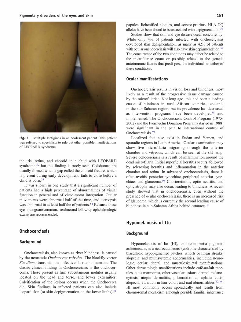
leopard skin (or skin depigmentation on the lower limbs),55
papules, lichenified plaques, and severe pruritus. HLA-DQ
alleles have been found to be associated with depigmentation.56
Studies show that skin and eye disease occur concurrently.
While only 4% of patients infected with onchocerciasis
developed skin depigmentation, as many as 42% of patients
with ocular onchocerciasiswill also have skin depigmentation.57
The concurrence of the two conditions may either be related to
the microfilariae count or possibly related to the genetic
autoimmune factors that predispose the individuals to either of
these conditions.
Ocular manifestations
Onchocerciasis results in vision loss and blindness, most
likely as a result of the progressive tissue damage caused
by the microfiliariae. Not long ago, this had been a leading
cause of blindness in rural African countries, endemic
in the sub-Saharan region, but its prevalence has decreased
as intervention programs have been developed58 and
implemented. The Onchocerciasis Control Program (1975-
2002) and the Ivermectin Donation Program (started in 1988)
were significant in the path to international control of
Onchocerciasis.59
Localized foci also exist in Sudan and Yemen, and
sporadic regions in Latin America. Ocular examination may
show live microfilaria migrating through the anterior
chamber and vitreous, which can be seen at the slit lamp.
Severe ochocerciasis is a result of inflammation around the
dead microfilaria. Initial superficial keratitis occurs, followed
by sclerosing keratitis and inflammation in the anterior
chamber and retina. In advanced onchocerciasis, there is
often uveitis, posterior synechiae, peripheral anterior syne-
chiae, and glaucoma.60 Chorioretinitis, optic neuritis, and
optic atrophy may also occur, leading to blindness. A recent
study showed that in onchocerciasis, even without the
presence of ocular onchocerciasis, there is an increased risk
of glaucoma, which is currently the second leading cause of
blindness in sub-Saharan Africa behind cataracts.61
Hypomelanosis of Ito
Background
Hypomelanosis of Ito (HI), or Incontinentia pigmenti
achromicans, is a neurocutaneous syndrome characterized by
blaschkoid hypopigmented patches, whorls or linear streaks;
alopecia; and multisystemic abnormalities, including neuro-
logic, ocular, dental, and musculoskeletal manifestations.
Other dermatologic manifestations include café-au-lait mac-
ules, cutis marmorata, other vascular lesions, dermal melano-
cytosis, atopic dermatitis, pilomatrixoma, aplasia cutis,
alopecia, variation in hair color, and nail abnormalities.62–64
HI most commonly occurs sporadically and results from
chromosomal mosaicism although possible familial inheritance
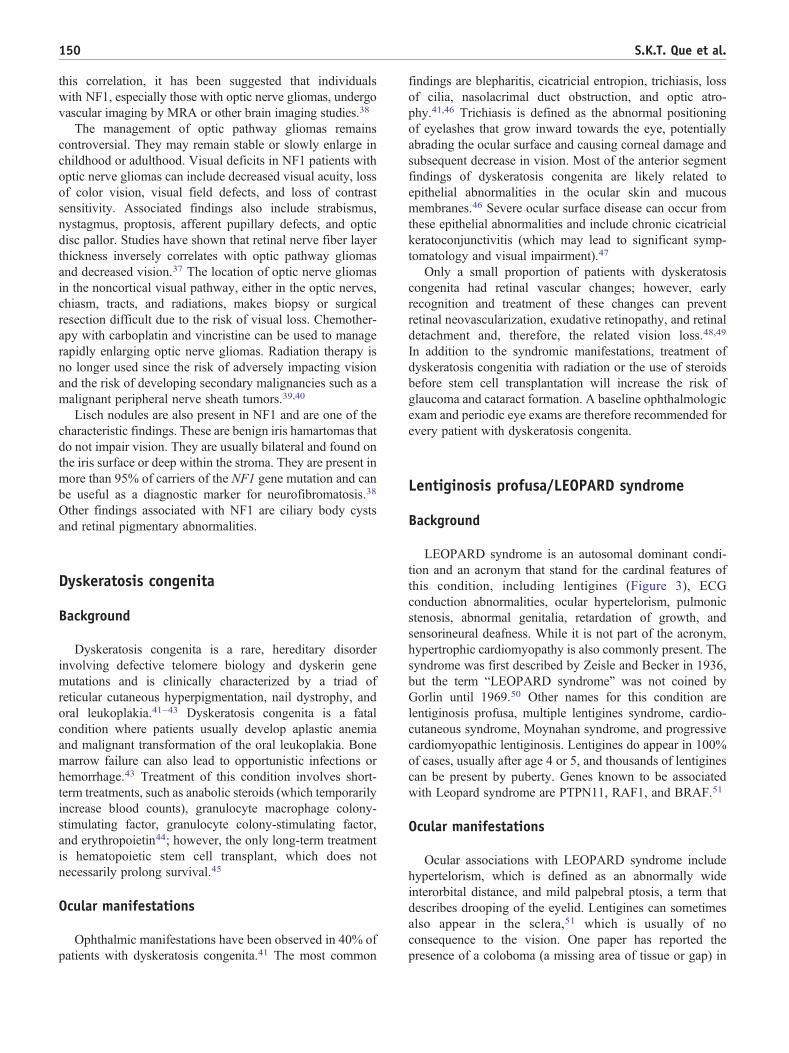
Fig. 3 Multiple lentigines in an adolescent patient. This patient
was referred to specialists to rule out other possible manifestations
of LEOPARD syndrome.
151Pigmentary disorders of the eyes and skin

patterns have been reported.65–74 HI presents usually within the
first year of life with the characteristic skin lesions and the
neurologic and other manifestations become readily apparent
in infancy or early childhood.62 The prevalence of HI remains
around 1 in 700 new pediatric neurology referrals62 and
occurs with equal incidence in both sexes.63 Clinical
monitoring includes regular follow-up with the pediatrician
and other specialists.
Ocular manifestations
The ocular manifestations, seen in about 25% of patients
with HI, are varied and nonspecific. Reported ocular
abnormalities have included anterior segment changes,
such as subtle iris depigmentation, corneal asymmetry, and
more serious diseases including corneal opacity, micro- and
macro-ophthalmia, and cataracts. Posterior segment findings
have also been reported and include choroidal hypopigmen-
tation and atrophy, retinal detachment, optic nerve hypopla-
sia and optic atrophy. Other ophthalmic findings that have
been present in patients with HI include refractive errors,
strabismus, nystagmus,75,76 scleral melanosis, hypertelorism,
and eyelid anomalies.62–64,77 In addition, a case of retinoblas-
toma in a 16month old with HI was reported in 2011.78Due to
the wide variability in ocular presentation, patients with HI
should be evaluated as early as possible with a comprehensive
ophthalmologic examination.
Incontinentia Pigmenti
Background
Incontinentia Pigmenti (IP) (also known asBloch-Sulzberger
syndrome, OMIM 308300) is a multisystem disease originally
described by Bloch and then Sulzberger in the 1920s. The
disease is characterized by four cutaneous stages. The first stage
presents in the neonatal period with vesiculobullous skin lesions
that follow the lines of Blaschko.79 The second stage is marked
by hyperkeratotic and verrucous lesions appearing between 2
and 6 months of age. In the third stage the lesions evolve into
hyperpigmented patches, usually, by the end of infancy.80
The fourth and final cutaneous stage is characterized by
hypopigmentation, atrophy, and alopecia of the affected skin
by early adulthood.81 The dermatologicmanifestations of IP can
also include nail dystrophy. Potential systemic manifesta-
tions include neurologic, ophthalmologic, skeletal and
dental abnormalities. IP is an X-linked dominant genetic
disorder involving a mutation in the Nuclear Factor-Kappa
B Essential Modulator (NEMO), (also known as the
IKBKG, inhibitor of kB kinase gamma) gene. A normal
copy of the NEMO gene is necessary for regulated NF-
kB activation to prevent inappropriate apoptosis under
TNFα signaling.82,83
Updates on ocular manifestations
The ophthalmologic manifestations of IP are varied and can
be among the most severe and debilitating complications of the
disease. It is estimated that between 16% and 66% of patients
with IP have eye abnormalities,82–85 typically in the form of
proliferative retinal vasculopathy. Eye findingsmay also include
strabismus, nystagmus, microphthalmia, cataracts, glaucoma,
optic atrophy, and retinal detachment,84 most of which are
secondary to advanced retinopathy. Of these abnormalities,
retinal detachment is considered a severe end stage complication
of retinopathy in IP, as it can result in blindness. Retinal
complications arise as a result of a vascular ischemia caused by
occlusion of the retinal arterial vasculature.79 At the junction of
the avascular and vascular retina, abnormal arteriovenous
malformations occur along with microvascular abnormalities
and neovascularization. This inner retinal ischemic atrophy that
causes proliferative vitreoretinopathy can progress to vitreous
hemorrhage and retinal detachment79 and severe loss of vision.
In some cases, regression of retinopathy may occur spontane-
ously. Often treatment must be performed with laser or
cryoablation of affected retinal areas and can slow the
progression of these potential complications82,86; however,
early detection is one of the most important aspects of effective
treatment. Intravenous fluorescein angiography with a
handheld retinal camera is typically performed under general
anesthesia in babies with IP. Oral fluorescein may be used in
place of intravenous injection.87 Early, frequent ophthal-
mologic screenings of infants with IP are recommend
to maximize chances of preserving vision.88,89 Several
recent studies show the considerable variation of ocular
findings (Table 2).82,84,85,90–95
Nevus of Ota
Background
In oculodermal melanocytosis, or nevus of Ota, a
hamartoma of spindle-shaped melanocytes can be found in
the skin, eyes, and central nervous system. In the skin, the
lesion presents as a bluish, black patch most commonly in the
areas of the dermis innervated by the first and second
divisions of the trigeminal nerve. Pigmentation of the sclera
and other ocular tissues is readily apparent upon clinical
examination. Ocular or oculodermal melanocytosis occurs in
all races, but has a higher incidence in people with darker
skin types and is more common in females. In Caucasians,
the prevalence of ocular melanocytosis was found to be
around .038% of the population.96 The incidence in Asian
populations generally may be higher, although a Chinese
study cited a similar prevalence of oculodermal melanocy-
tosis in 0.034% of the population.97 Most cases present in
infancy or childhood, but there are also reports of adult-onset
nevus of Ota.
152 S.K.T. Que et al.
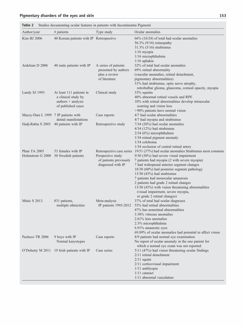
Table 2 Studies documenting ocular features in patients with Incontinentia Pigmenti
Author/year # patients Type study Ocular anomalies
Kim BJ 2006 40 Korean patients with IP Retrospective 66% (16/24) of total had ocular anomalies
56.3% (9/16) retinopathy
31.3% (5/16) strabismus
1/16 myopia
1/16 microphthalmia
1/16 aphakia
Ardelean D 2006 40 male patients with IP A series of patients
presented by authors
plus a review
of literature
32% of total had ocular anomalies
69% retinal abnormality
(vascular anomalies, retinal detachment,
pigmentary abnormalities)
31% had strabismus, optic nerve atrophy,
retrobulbar glioma, glaucoma, corneal opacity, myopia
Landy SJ 1993 At least 111 patients in
a clinical study by
authors + analysis
of published cases
Clinical study 33% squints
40% abnormal retinal vessels and RPE
10% with retinal abnormalities develop intraocular
scarring and vision loss
N90% patients have normal vision
Macey-Dare L 1999 7 IP patients with
dental manifestations
Case reports 4/7 had ocular abnormalities
4/7 had myopia and strabismus
Hadj-Rabia S 2003 40 patients with IP Retrospective study 7/34 (20%) had ocular anomalies
4/34 (12%) had strabismus
2/34 (6%) microphthalmia
1/34 retinal pigment anomaly
1/34 coloboma
1/34 occlusion of central retinal artery
Phan TA 2005 53 females with IP Retrospective case series 19/51 (37%) had ocular anomalies Strabismus most common
Holmstrom G 2000 30 Swedish patients Prospective study
of patients previously
diagnosed with IP
9/30 (30%) had severe visual impairment
7 patients had myopia (2 with severe myopia)
7 had widespread anterior segment changes
18/30 (60%) had posterior segment pathology
13/30 (43%) had strabismus
7 patients had monocular amaurosis
2 patients had grade 2 retinal changes
13/30 (43%) with vision threatening abnormalities
(visual impairment, severe myopia,
or grade 2 retinal changes)
Minic S 2013. 831 patients,
multiple ethnicities
Meta-analysis
IP patients 1993-2012
37% of total had ocular diagnoses
53% had retinal abnormalities
47% has nonretinal abnormalities
3.38% vitreous anomalies
2.61% lens anomalies
2.3% microphthalmia
6.91% amaurotic eyes
69.89% of ocular anomalies had potential to affect vision
Pacheco TR 2006 9 boys with IP
Normal karyotypes
Case reports 8/9 patients had normal eye examination
No report of ocular anomaly in the one patient for
which a normal eye exam was not reported
O’Doherty M 2011 19 Irish patients with IP Case series 5/11 (47%) had vision threatening ocular findings
2/11 retinal detachment
2/11 squint
2/11 corticovisual impairment
1/11 amblyopia
1/11 cataract
1/11 abnormal vasculature
153Pigmentary disorders of the eyes and skin
Ocular manifestations
Oculodermal melanocytosis is usually a benign condi-
tion but patients have a 10% chance of developing
glaucoma98 and possibly an increased risk of associated
melanoma,99 which has been documented to occur in the
skin100,101; the uveal tract (which includes the iris, ciliary
body, and choroid)102–107; the central nervous sys-
tem108,109; and the orbit.110 The clinical presentation of
cutaneous melanoma developing within Nevus of Ota can
be subtle. Case reports displayed that the melanomas do not
show typical clinical features of cutaneous melanoma and
may mimic features of a benign subcutaneous cyst or
nodule.100 Treatment with lasers can result in reasonable
clearance of pigmented skin lesions111; however, it does not
eliminate the risks of glaucoma or melanoma in either the
skin or ocular tissues.
Ocular manifestations occur in two thirds of patients
with cutaneous lesions. In the eye, melanocytosis has been
shown to be present in the episclera and choroid in 100%
of patients; 10% have conjunctival involvement; and 18%
have retinal involvement, depending on the study.112,113
When examining patients, ultrasound biomicroscopy can
be used in conjunction with a slit lamp examination to
evaluate the iris and ciliary body. The choroid is evaluated
by fundus examination and B-Scan ultrasound. Recently,
using ultrasound biomicroscopy, the mean ciliary body
thickness was found to be statistically significantly greater
in the affected than in contralateral, unaffected, eyes. The
affected eyes also consistently showed hyperreflectiv-
ity.114 Ciliary body melanomas, by contrast, would show
evidence of a mass and lower reflectivity.114 This
technique for assessing the ciliary body may now provide
additional important information when evaluating nevus
of Ota.
Malignant melanoma in association with Nevus of Ota,
although rare, is often difficult to treat and can result in
a local spread to orbital tissues, nerves, and bone. Early
treatment can allow for preservation of orbital tissues and
vision110,115; therefore, patients with nevus of Ota may
benefit from regular skin and ophthalmologic examina-
tions to facilitate earlier diagnosis and treatment.
Since as many as 10% of patients with nevus of Ota
will develop glaucoma,98 it is important to monitor for
any changes in intraocular pressure and pigmentation.116
Treatment of cutaneous lesions can be performed
with Q-switched Alexandrite and Q-Switched (QS)
Nd-YAG lasers.111 Ocular lesions are not amenable to
laser treatment. It should be noted that Q-switched
lasers are among the most damaging to the eye and
proper eye protection for both the physician and the patient
is important.
Lastly, a recent retrospective review showed that surgical
reduction of conjunctival and scleral pigmentation for cosmetic
improvement of pigmentation within the eye itself can be
performed as well.117
Vogt-Koyanagi-Harada
Background
Patients with Vogt-Koyanagi-Harada (VKH) Syndrome,
of which vitiligo is a characteristic feature, are at risk of
serious visual loss if treatment is delayed.
VKH, named after the three physicians who initially
described the condition, is considered a multisystemic,
autoimmune inflammatory disorder118,119 that affects
the skin, eyes, ears, and meninges. VKH likely results
from the complex interplay of genetic and environmen-
tal factors.120–128 There are several criteria required
for diagnosis.129
Ocular manifestations
Ocular manifestations of VKH include a bilateral chronic
diffuse granulomatous uveitis. Uveitis is the most common
ocular complication of VKH and, if untreated, can result in
blindness. Among patients referred to uveitis clinics around
the world, VKH is identified in as many as 1% to 4% of
patients.130–133 In Japan, as many as 8% of uveitis cases
result from VKH.133 Inflammation targets melanocytes
located in the uveal tract. A recent study of VKH in Indian
children showed evidence of vision loss, which was
attributed most commonly to the development of cataracts
and the presence of active inflammation, but complications
due to chronic uveitis, such as subretinal fibrosis, retinal
pigment epithelium changes, and cystoid macular edema
contributed to vision loss in a subset of patients as well.134
Ocular anterior segment findings may include mild to severe
bilateral inflammation with seclusion of the pupil and
posterior synechiae. Keratic precipitates, which can be
small or large, are granulomatous. Perilimbal vitiligo may
be present. Bilateral panuveitis, optic disc hyperemia,
vitreous opacities, edematous exudative choroiditis with
nodular yellow lesions and serous retinal detachment can
develop. Late in the disease neovascularization of the optic
nerve and retina can occur, leading to vitreous hemorrhage.
When evaluating patients with VKH, fluorescein angiogra-
phy can be performed to show characteristic retinal findings.
Choroidal thickening can be seen on B-scan ultrasound.
Another potential test to differentiate VKH is ocular
coherence tomography.135 Ocular manifestations also
include development of cataracts (40%), glaucoma (38%
that require intervention), retinal fibrosis, and neovascu-
larization, all of which can increase a patient’s risk of
developing blindness. Although more studies regarding the
factors affecting visual outcomes are needed, early,
aggressive treatment in the acute phase and prompt
treatment of any recurrent inflammation may help to
delay disease progression.134 Treatment includes systemic,
local and periocular corticosteroids, steroid sparing agents,
or both.
154 S.K.T. Que et al.
Vitiligo
Background
In vitiligo, autoimmune destruction of melanocytes in the skin
results in cutaneous depigmentation. Risk factors include a family
history of vitiligo, medical history of autoimmunity, trauma, and
the presence of several identified gene loci.136 Vitiligo occurs
in people of all ethnicities and disease onset usually occurs in
childhood or adolescence. Other associated conditions include
autoimmune thyroid disorders, diabetes, and alopecia areata.
Ocular manifestations
There are very few studies documenting the ocular
findings in patients with vitiligo. It was shown that patients
with vitiligo have retinal pigment epithelial (RPE) atrophy or
hypopigmentation ,137 pigment hypertrophy, choroidal
nevi,138 peripapillary atrophy around the optic nerve,139
and choroidal hypopigmentation.140,141
Finally, uveitis may also occur with greater frequency in
patients with vitiligo,142,143 but this is not shown in all
studies.139,144–148 Because eye disease has been documented
in patients with vitiligo, patients should receive routine
ophthalmologic exams.
Waardenburg syndrome
Background
Waardenburg syndrome is a genetic syndrome consisting
primarily of anomalies of the skin, hair, eyes, and ears and
affects 1 in 40,000 according to population studies. The
musculoskeletal system and gastrointestinal tract can also
be affected in certain subtypes. As established at the
Waardenburg consortium, diagnosis requires fulfillment of
either two major criteria or one major and two minor
criteria.149 Major criteria include the characteristic white
forelock (hair depigmentation), pigmentary anomalies of the
iris, congenital sensorineural deafness, dystopia canthorum,
or an affected first degree relative. Minor criteria are
depigmented macules or patches, synophrys, broad nasal
root, nose hypoplasia, or early graying of the hair by age 35.
There are four major types of Waardenburg syndrome,
generally involving the PAX3 (Paired box 3), MITF
(microophthalmia-associated transcription factor), SOX10
(Sry box 10), EDN3 (endothelin 3), and EDNRB (endothelin
receptor type B) genes.150,151
Ocular manifestations
Recently, in patients with Waardenburg syndrome, the
presence of both choroidal and iris hypopigmentation was
confirmed. Fortunately, visual acuity was generally not
affected.152 Reduced visual acuity, when present, can result from
foveal hypoplasia, amblyopia, and/or vitreous hemorrhaging.153
Other scattered case reports of possible ocular associations
include bilateral congenital cataracts,154 diabetic retinopathy,155
retinoblastoma,156 congenital eyelid ptosis,157 branch retinal
vein occlusion,158 and strabismus.159 Japanese patientswithWS
type IIwere also found to have hypopigmentation of both the iris
and retina.160
Although reduced visual acuity is not typically an issue in
Waardenburg syndrome, there are cases of loss of vision.
Due to this and the reported variations in ophthalmologic
findings, comprehensive eye examinations should be
performed routinely.
References
1. Cheng T, Orlow SJ, Manga P. Loss of OCA2 disrupts the unfolded
protein response and increases resistance to endoplasmic reticulum
stress in melanocytes. Pigment Cell Melanoma Res. 2013;26:826-834.
2. Manga P, Orlow SJ. The pink-eyed dilution gene and the molecular
pathogenesis of tyrosinase-positive albinism (OCA2). J Dermatol.
1999;26:738-747.
3. Hendrickson A. Organization of the adult primate fovea. In: Penfold
PL, Provis JM, eds. Macular Degeneration. Heidelberg: Springer-
Verlag; 2005. p. 1-20.
4. Harvey PS, King RA, Summers CG. Spectrum of foveal development in
albinism detected with optical coherence tomography. J AAPOS.
2006;10:237-242.
5. Grønskov K, Ek J, Brondum-Nielsen K. Oculocutaneous albinism.
Orphanet J Rare Dis. 2007;2:1-8.
6. Schmitz B, Krick C, Käsmann-Kellner B. Morphology of the optic
chiasm in albinism. Ophthalmologe. 2007;104:662-665.
7. Summers CG. Albinism: Classification, clinical characteristics, and
recent findings. Optom Vis Sci. 2009;86:659-662.
8. Bouzas EA, Caruso RC, Drews-Bankiewicz MA, Kaiser-Kupfer MI.
Evoked potential analysis of visual pathways in human albinism.
Ophthalmology. 1994;101:309-314.
9. Seo JH, Yu YS, Kim JH, et al. Correlation of visual acuity with foveal
hypoplasia grading by optical coherence tomography in albinism.
Ophthalmology. 2007;114:1547-1551.
10. Izquierdo NJ, Emanuelli A, Izquierdo JC, et al. Foveal thickness and
macular volume in patients with oculocutaneous albinism. Retina.
2007;27:1227-1230.
11. Mohammad S, Gottlob I. The functional significance of foveal
abnormalities in albinism measured using spectral-domain optical
coherence tomography. Ophthalomology. 2011;118:1645-1652.
12. Preston KL, McDonald M, Sebris SL, Dobson V, Teller DY.
Validation of the acuity card procedure for assessment of infants
with ocular disorders. Ophthalomology. 1987;94:644-653.
13. Anderson J, Lavoie J, Merrill K, King RA, Summers CG. Efficacy of
spectacles in persons with albinism. J AAPOS. 2004;8:515-520.
14. Kestenbaum A. New operation for nystagmus. Bull Soc Ophtalmol Fr.
1953;6:599-602.
15. Anderson JR. Causes and treatment of congenital eccentric nystagmus.
Br J Ophthalmol. 1953;37:267-281.
16. Danks DM, Cartwright E, Stevens BJ, Townley RR. Menkes’ kinky
hair disease further definition of the defect in copper transport.
Science. 1973;179:1140-1142.
17. Royce PM, Camakaris J, Danks DM. Reduced lysyl oxidase activity in
skin fibroblasts from patients with Menkes’ syndrome. Biochem J.
1980;192:579-586.
155Pigmentary disorders of the eyes and skin
18. Maehara M, Ogasawara N, Mizutani N, Watanabe K, Suzuki S.
Cytochrome c oxidase deficiency in Menkes kinky hair disease. Brain
Dev. 1983;5:533-540.
19. Kim YH, Lee R, Yoo HW, et al. Identification of a novel mutation in
the ATP7 A gene in a Korean patient with Menkes disease. J Korean
Med Sci. 2011;26:951-953.
20. Finner AM. Nutrition and hair: Deficiencies and supplements.
Dermatol Clin. 2013;31:167-172.
21. GaschAT, Caruso RC, Kaler SG, Kasier-KupferM.Menkes' syndrome:
Ophthalmic findings. Ophthalmology. 2002;109:1477-1483.
22. Krajacic P, Qian Y, Hahn P, et al. Retinal localization and copper-
dependent relocalization of the Wilson and Menkes disease proteins.
Invest Ophthalmol Vis Sci. 2006;47:3129-3134.
23. Tee AR, Manning BD, Roux PP, Cantley LC, Blenis J. Tuberous
sclerosis complex gene products, tuberin and hamartin, control mTOR
signaling by acting as a GTPase-activating protein complex toward
Rheb. Curr Biol. 2003;13:1259-1268.
24. Schwartz RA, Fernández G, Kotulska K, Jóźwiak S. Tuberous
sclerosis complex: Advances in diagnosis, genetics, and management.
J Am Acad Dermatol. 2007;57:189-202.
25. Curatolo P, Verdecchia M, Bombardieri R. Tuberous sclerosis
complex: A review of neurological aspects. Eur J Paediatr Neurol.
2002;6:15-23.
26. O’Callaghan FJ, Noakes MJ, Martyn CN, Osborne JP. An
epidemiological study of renal pathology in tuberous sclerosis
complex. BJU Int. 2004;94:853-857.
27. Webb DW, Thoma RD, Osborne JP. Cardiac rhabdomyomas and their
association with tuberous sclerosis. Arch Dis Child. 1993;68:367-370.
28. Franz DN, Leonard J, Tudor C, et al. Rapamycin causes regression of
astrocytomas in tuberous sclerosis complex. Ann Neurol. 2006;59:
490-498.
29. Wienecke R, Fackler I, Lisenmaier U, et al. Antitumoral activity of
rapamycin in renal angiomyolipoma associated with tuberous sclerosis
complex. Am J Kidney Dis. 2006;48:e27-e29.
30. Rowley SA, O’Callaghan FJ, Osborne JP. Ophthalmic manifestations
of tuberous sclerosis: A population based study. Br J Ophthalmol.
2001;85:420-423.
31. Drummond SR. Retinal astrocytoma managed by brachytherapy.
Ophthalmology. 2009;116:597.
32. Lonngi M, Gold AS, Murray TG. Combined bevacizumab and
triamcinolone acetonide injections for macular edema in a patient with
astrocytic hamartomas and tuberous sclerosis. Ophthalmic Surg
Lasers Imaging. 2013;44:85-90.
33. Shields CL, Reichstein DA, Bianciotto C, Shields JA. Retinal pigment
epithelial depigmented lesions associated with tuberous sclerosis
complex. Arch Ophthalmol. 2012;130:387-390.
34. Akinci A, Oner O, Guven A, Degerliyurt A, Munir K. Refractive
errors and strabismus in children with tuberous sclerosis: A controlled
study. J Pediatr Ophthalmol Strabismus. 2009;46:345-348.
35. Jett K, FriedmanMJ. Clinical and genetic aspects of neurofibromatosis
1. Genet Med. 2010;12:1-11.
36. Neurofibromatotis. Conference statement. National Institutes of
Health Consensus Development Conference. Arch Neurol. 1988;45:
575-578.
37. Avery RA, Fisher MJ, Liu GT. Optic pathway gliomas. J
Neuroophthalmol. 2011;31:269-278.
38. Schnur RE. Type I, neurofibromatosis: A geno-oculo-dermatologic
update. Curr Opin Ophthalmol. 2012;23:364-372.
39. Listernick R, Ferner RE, Liu GT, Gutmann DH. Optic pathway
gliomas in neurofibromatosis-1: Controversies and recommendations.
Ann Neurol. 2007;61:189-198.
40. Segal L, Darvish-Zargar M, Dilenge ME, Ortenberg J, Polomeno RC.
Optic pathway gliomas in patients with neurofibromatosis type 1:
Follow-up of 44 patients. J AAPOS. 2010;14:155-158.
41. Drachtman RA, Alter BP. Dyskeratosis congenita: Clinical and
genetic heterogeneity. Report of a new case and review of the
literature. Am J Pediatr Hematol Oncol. 1992;14:297-304.
42. Ogden GR, Connor E, Chisholm DM. Dyskeratosis congenita: Report
of a case and review of literature. Oral Surg Oral Med Oral Pathol.
1988;5:58-91.
43. Bessler M, Wilson DB, Mason PJ. Dyskeratosis congenita and
telomerase. Curr Opin Pediatr. 2004;16:23-28.
44. Erduran E, Hacisalihoglu S, Ozoran Y. Treatment of dyskeratosis
congenita with granulocyte-macrophage colony-stimulating factor and
erythropoietin. J Pediatr Hematol Oncol. 2003;25:333-335.
45. Gadalla SM, Sales-Bonfim C, Carreras J, et al. Outcomes of allogeneic
hematopoietic cell transplantation in patients with dyskeratosis
congenita. Biol Blood Marrow Transplant. 2013;19:1238-1243.
46. Tsilou ET, Giri N, Weinstein S, et al. Ocular and orbital manifestations
of the inherited bone marrow failure syndromes: Fanconi anemia and
dyskeratosis congenita. Ophthalmology. 2010;117:615-622.
47. Merchant A, Zhao TZ, Foster CS. Chronic keratoconjunctivitis
associated with congenital dyskeratosis and erythrokeratoderma
variablis. Two rare genodermatoses. Ophthalmology. 1998;105(7):
1286-1291.
48. Teixeira LF, Shields CL, Marr B, Horgan N, Shields JA. Bilateral
retinal vasculopathy in a patient with dyskeratosis congenita. Arch
Ophthalmol. 2008;126:134-135.
49. Vaz-Pereira S, Pacheco PA, Gandhi S, et al. Bilateral retinal
vasculopathy associated with autosomal dominant dyskeratosis
congenita. Eur J Ophthalmol. 2013;23:772-775.
50. Gorlin RJ, Anderson RC, Blaw M. Multiple lentigines syndrome. Am
J Dis Child. 1969;117:652-662.
51. Martínez-Quintana E, Rodríguez-González F. LEOPARD syn-
drome: clinical features and gene mutations. Mol Syndromol.
2012;3:145-157.
52. Rudolph G, Haritoglou C, Kalpadakis P, Boergen KP, Meitinger T.
LEOPARD syndrome with iris-retina-choroid coloboma. Discordant
findings in monozygotic twins. Ophthalmologe. 2001;98:1101-1103.
53. Hero I, Farjah M, Scholtz CL. The prenatal development of the optic
fissure in colobomatous microphthalmia. Invest Ophthalmol Vis Sci.
1991;32:2622-2635.
54. Alfieri P, Cesarini L, Zampino G, et al. Visual function in Noonan and
LEOPARD syndrome. Neuropediatrics. 2008;39:335-340.
55. Coffeng LE, Fobi G, Ozoh G, et al. Concurrence of dermatological and
ophthalmological morbidity in onchocerciasis. Trans R Soc Trop Med
Hyg. 2012;106:243-251.
56. Murdoch ME, Payton A, Abiose A, et al. HLA-DQ alleles associate
with cutaneous features of onchocerciasis. The Kaduna-London-
Manchester Collaboration for Research on Onchocerciasis. Human
Immunol. 1997;55:46-52.
57. Browne SG. Onchocercal depigmentation. Trans R Soc Trop Med
Hyg. 1960;54:325-334.
58. Budenz DL. Blindness and visual impairment in an urban West
African population: The Tema Eye Survey. Ophthalmology.
2012;119:1744-1753.
59. Richards Jr FO, Boatin BA. Control of onchocerciasis. Adv Parasitol.
2006;61:349-394.
60. Newland HS, White AT, Greene BM, Murphy RP, Taylor HR. Ocular
manifestations of onchocerciasis in a rain forest area of West Africa.
Br J Ophthalmol. 1991;75:163-169.
61. Egbert PR, Jacobson DW, Fiadoyor S, Dadzie P, Ellingson KD.
Onchocerciasis: A potential risk factor for glaucoma. Br J Ophthalmol.
2005;89:796-798.
62. Pascual-Castroviejo I, Roche C, Martinez-Bermejo A, et al. Hypo-
melanosis of Ito: A study of 76 infantile cases. Brain Dev. 1998;20:
36-43.
63. Ruiz-Maldonado R, Toussaint S, Tamayo L, Laterza A, del Castillo V.
Hypomelanosis of Ito: Diagnostic criteria and report of 41 cases.
Pediatr Dermatol. 1992;9:1-10.
64. Pascual-Castroviejo I, López-Rodriguez L, de la Cruz Medina M,
Salamanca-Maesso C, Roche Herrero C. Hypomelanosis of Ito.
neurological complications in 34 cases. Can J Neurol Sci. 1988;15:
124-129.
156 S.K.T. Que et al.
65. Rubin MR. Incontinentia Pigmenti achromians, multiple cases within
a family. Arch Dermatol. 1972;105:424-425.
66. Lungarotti MS, Martello C, Calabro A, Baldari F, Mariotti G.
Hypomelanosis of Ito associated with chromosomal translocation
involving Xp11. Am J Med Genet. 1991;40:447-448.
67. Steichen-Gersdorf E, Trawöger R, Duba HC, et al. Hypomelanosis of
Ito in a girl with plexus papilloma and translocation (X;17). Hum
Genet. 1993;90:611-613.
68. Koiffmann CP, de Souza DH, Diament A, et al. Incontinentia Pigmenti
achromians (hypomelanosis of ITO, MIM 146150): Further evidence
of localization at Xp11. Am J Med Genet. 1993;46:529-533.
69. Bitoun P, Philippe C, Cherif M, Mulcahy MT, Gilgenkrantz S.
Incontinentia Pigmenti (type 1) and X;5 translocation. Ann Genet.
1992;35:51-54.
70. Pinheiro A, Mathew MC, Thomas M, et al. The clinical profile of
children in India with pigmentary anomalies along the lines of
Blaschko and central nervous system manifestations. Pediatr
Dermatol. 2007;24:11-17.
71. Ritter CL, Steele MW, Wenger SL, Cohen BA. Chromosome
mosaicism in hypomelanosis of Ito. Am J Med Genet. 1990;35:14-17.
72. Chitayat D, Friedman JM, Johnston MM. Hypomelanosis of Ito—a
nonspecific marker of somatic mosaicism: Report of case with trisomy
18 mosaicism. Am J Med Genet. 1990;35:422-424.
73. Donnai D, Read AP. Hypomelanosis of Ito. Lancet. 1992;339:819-820.
74. Pellegrino JE, Schnur RE, Kline R, Zackai EH, Spinner NB. Mosaic
loss of 15 q11 q13 in a patient with hypomelanosis of Ito: Is there a
role for the P gene? Hum Genet. 1995;96:485-489.
75. Weaver Jr RG, Martin T, Zanolli MD. The ocular changes of
Incontinentia Pigmenti achromians (hypomelanosis of Ito). J Pediatr
Ophthalmol Strabismus. 1991;28:160-163.
76. Amon M, Menapace R, Kirnbauer R. Ocular symptomatology in
familial hypmelanosis Ito. Incontinentia Pigmenti achromians.
Ophthalmologica. 1990;200:1-6.
77. Ruggieri M, Pavone L. Topical review: Hypomelanosis of Ito: Clinical
syndrome or just phenotype? J Child Neurol. 2000;15:635-644.
78. El-Sawy T, Abramson D. Retinoblastoma presenting in a child with
hypomelanosis of Ito. Open Ophthalmol J. 2011;5:55-58.
79. Goldberg M. The skin is not the predominant problem in Incontinentia
Pigmenti. Arch Dermatol. 2004;140(6):748-750.
80. Bodak N, Hadj-Rabia S, Hamel-Teillac D, de Prost Y, Bodemer C.
Late recurrence of inflammatory first-stage lesions in Incontinentia
Pigmenti. Arch Dermatol. 2003;139:201-204.
81. Minić S, Trpinac D, Obradović M, Incontinentia Pigmenti diagnostic
criteria update. Clin Genet. 2014;85:536-542. doi: http://dx.doi.org/
10.1111/cge.12223. Published online June 26, 2013.
82. O'Doherty M, McCreery K, Green AJ. Incontinentia Pigmenti-
ophthalmological observation of a series of cases and review of the
literature. Br J Ophthalmol. 2011;95:11-16.
83. Al-Zuhaibi S, Ganesh A, Al-Waili A. A female child with skin lesions
seizures: Case report of Incontinentia Pigmenti. Sultan Qaboos Univ
Med J. 2009;9:157-161.
84. Minić S, Trpinac D, Obradović M, Systematic review of central
nervous system anomalies in incontinentia pigmenti. Orphanet J Rare
Dis. 2013. doi: http://dx.doi.org/10.1186/1750-1172-8-25. Published
online August 25, 2013.
85. Kim BJ, Shin HS, Won CH, et al. Incontinentia Pigmenti: Clinical
observation of 40 Korean cases. J Korean Med Sci. 2006;21:474-477.
86. Rahi J, Hungerford J. Early diagnosis of the retinopathy of
Incontinentia Pigmenti: Successful treatment by cryotherapy. Br J
Ophthalmol. 1990;74:377-379.
87. Patel CK, Fung THM, Muqit MMK. Non-contact ultra-widefield
retinal imaging and fundus fluorescein angiography of an infact with
Incontinentia Pigmenti without sedation in an ophthalmic office
setting. J AAPOS. 2013;17:309-311.
88. Berlin AL, Paller AS, Chan LS. Incontinentia Pigmenti: A review and
update on the molecular basis of pathophysiology. J Am Acad
Dermatol. 2002;47(2):169-187.
89. Meuwissen MEC, Mancini GMS. Neurological findings in Incon-
tinentia Pigmenti; a review. Eur J Med Genet. 2012;55:323-331.
90. Hadj-Rabia S, Froidevaux D, Bodak N, et al. Clinical study of 40 cases
of Incontinentia Pigmenti. Arch Dermatol. 2003;139:1163-1170.
91. Ardelean D, Pope E. Incontinentia Pigmenti in boys: A series and
review of the literature. Pediatr Dermatol. 2003;23(6):523-527.
92. Phan TA, Wargon O, Turner AM. Incontinentia Pigmenti case series:
Clinical spectrum of Incontinentia Pigmenti in 53 female patients and
their relatives. Clin Exp Dermatol. 2005;30:474-480.
93. Pacheco TR, LevyM, Collyer JC, et al. Incontinentia Pigmenti in male
patients. J Am Acad Dermatol. 2006;55:251-255.
94. Landy SJ, Donnai D. Incontinentia Pigmenti (Bloch-Sulzberger
syndrome). J Med Genet. 1993;30:53-59.
95. Macey-Dare LV, Goodman JR. Incontinentia Pigmenti: Seven cases
with dental manifestations. Int J Paediatr Dent. 1999;9:293-297.
96. Gonder JR, Ezell PC, Shields JA, Augsburger JJ. Ocular melanocy-
tosis: A study to determine the prevalence rate of ocular melanocy-
tosis. Ophthalmology. 1982;89:950-952.
97. Leung AK, Kao CP, Cho HY, et al. Scleral melanocytosis and
oculodermal melanocytosis (nevus of Ota) in Chinese children. J
Pediatr. 2000;137:581-584.
98. Teekhasaenee C, Ritch R, Rutnin U, Leelawongs N. Glaucoma in
oculodermal melanocytosis. Ophthalmology. 1990;97:562-570.
99. Dutton JJ, Anderson RL, Schelper RL, Purcell JJ, Tse DT. Orbital
malignant melanoma and oculodermal melanocytosis: Report of two
cases and review of the literature. Ophthalmology. 1984;91:497-507.
100. Patel BC, Egan CA, Lucius RW, et al. Cutaneous malignant melanoma
and oculodermal melanocytosis (nevus of Ota): Report of a case and
review of the literature. J Am Acad Dermatol. 1998;38:862-865.
101. Lindsey SF, Sanchez MI, Elgart GW, et al. Malignant melanoma from
a nevus of Ota in a pediatric patient with fatal outcome. J Am Acad
Dermatol. 2013;69:e195-197. doi: http://dx.doi.org/10.1016/j.jaad.
2013.05.009. Published, online October 2013.
102. Chen YC, Chang CH, Hsu SL, Hsu MW, Lee CL. Malignant
melanoma of the choroid in the eye with oculodermal melanocytosis of
a Chinese woman. Kaohsiung J Med Sci. 2010;26:673-678.
103. Sharan S, Grigg JR, Billson FA. Bilateral naevus of Ota with choroidal
melanoma and diffuse retinal pigmentation in a dark skinned person.
Br J Ophthalmol. 2005;89:1529-1545.
104. Singh M, Kaur B, Annuar NM. Malignant melanoma of the choroid in
a naevus of Ota. Br J Ophthalmol. 1988;72:131-133.
105. Carreño E, Saornil MA, Garcia-Alvarez C, et al. Prevalence of ocular
and oculodermal melanocytosis in Spanish population with uveal
melanoma. Eye (Lond). 2012;26:159-162.
106. Qian Y, Zakov ZN, Schoenfield L, Singh AD. Iris melanoma arising in
iris nevus in oculo (dermal) melanocytosis. Surv Ophthalmol.
2008;53:411-415.
107. Hudson HL, Valluri S, Rao NA. Choroidal melanomas in Hispanic
patients. Am J Ophthalmol. 1994;118:57-62.
108. Rivers JK. Dural melanoma associated with OCD and blue nevi. J
Cutan Med Surg. 2001;5:381-385.
109. Wang J, Guo ZZ, Zhang SG, et al. Microsurgical treatment of
meningeal malignant melanoma accompanied by nevus of Ota: Two
case reports and a literature review.Melanoma Res. 2013;23:502-504.
110. John H, Britto JA. Nonchoroidal intraorbital malignant melanoma
arising from naevus of Ota. J Plastic Reconstruc and Aesthet Surg.
2010;63:e387-389. doi: http://dx.doi.org/10.1016/j.bjps.2009.09.014.
Published online on October 21, 2009.
111. Chan HH, Leung RS, Ying SY, et al. A retrospective analysis of
complications in the treatment of nevus of Ota with the Q-switched
alexandrite and Q-switch Nd:YAG lasers. Dermatol Surg. 2000;26:
1000-1006.
112. Kim TI, Yoon J, Choi J, Tchah H. Flipped scleral flap surgery for
reduction of ocular pigmentation in oculodermal melanosis. Cornea.
2005;24:482-485.
113. Teekhasaenee C, Ritch R, Rutnin U, Leelawongs N. Ocular findings in
oculodermal melanocytosis. Arch Ophthalmol. 1990;108:1114-1120.
157Pigmentary disorders of the eyes and skin
114. Velazquez-Martin JP, Krema H, Fulda E, et al. Ultrasound biomicro-
scopy of the ciliary body in ocular/oculodermal melanocytosis. Am J
Ophthalmol. 2013;155:681-687.
115. Van Raamsdonk CD, Bezrookove V, Green G, et al. Frequent somatic
mutations of GNAQ in uveal melanoma and blue nevi. Nature.
2009;457:599-602.
116. Magaresevic L, Abazi Z. Unilateral open-angle glaucoma associated
with the ipsilateral nevus of Ota. Case reports Ophthalmol Med. 2013
doi: http://dx.doi.org/10.1155/2013/924937. Published, online April
9, 2013.
117. Kim JY, Hong JT, Lee SH, et al. Surgical Reduction of ocular
pigmentation in patients with oculodermal melanocytosis. Cornea.
2012;31:520-524.
118. Chi W, Yang P, Li B, et al. IL-23 promotes CD4+ T cells to produce
IL-17 in Vogt-Koyanagi-Harada disease. J Allergy Clin Immunol.
2007;119:1218-1224.
119. Rathinam SR, Namperumalsamy P, Nozik RA, Cunningham Jr ET.
Vogt-Koyanagi-Harada syndrome after cutaneous injury. Ophthal-
mology. 1999;106:635-638.
120. Ishikawa A, Shiono T, Uchida S. Vogt-Koyanagi-Harada disease in
identical twins. Retina. 1994;14:435-437.
121. Rutzen AR, Ortega-Larrocea G, Schwab IR, Rao NA. Simultaneous
onset of Vogt-Koyanagi-Harada syndrome in monozygotic twins. Am
J Ophthalmol. 1995;119:239-240.
122. Shindo Y, Ohno S, Nakamura S, Onoé K, Inoko H. A significant
association of HLA-DPB1*0501 with Vogt-Koyanagi-Harada’s
disease results from a linkage disequilibrium with the primarily
associated allele, DRB1*0405. Tissue Antigens. 1996;47:344-345.
123. Zhao M, Jiang Y, Abrahams IW. Association of HLA antigens with
Vogt-Koyanagi-Harada syndrome in a Han Chinese population. Arch
Ophthalmol. 1991;109:368-370.
124. Weisz JM, Holland GN, Roer LN, et al. Association between Vogt-
Koyanagi-Harada syndrome and HLA-DR1 and -DR4 in Hispanic
patients living in southern California. Ophthalmology. 1995;102:
1012-1015.
125. Hu K, Yang P, Jiang Z, et al. STAT4 polymorphism in a Chinese Han
population with Vogt-Koyanagi-Harada syndrome and Behcet’s
disease. Hum Immunol. 2010;71:723-726.
126. Jiang Z, Yang P, Hou S, Li F, Zhou H. Polymorphisms of IL23 R and
Vogt-Koyanagi-Harada syndrome in a Chinese Han population. Hum
Immunol. 2010;71:414-417.
127. Shu Q, Yang P, Hou S, et al. Interleukin-17 gene polymorphism is
associated with Vogt-Koyanagi-Harada syndrome but not with Behcet’s
disease in a Chinese Han population. Hum Immunol. 2010;71:988-991.
128. Hu K, Hou S, Li F, et al. JAK1, but not JAK2 and STAT3, confers
susceptibility to Vogt-Koyanagi-Harada (VKH) syndrome in a Han
Chinese population. Invest Ophthalmol Vis Sci. 2013;54:3360-3365.
129. Read RW, Holland GN, Rao NA, et al. Revised diagnostic criteria for
Vogt-Koyanagi-Harada disease: Report of an international comittee on
nomenclature. Am J Ophthalmol. 2001;131:647-652.
130. Mondkar SV, Biswas J, Ganesh SK. Analysis of 87 cases with Vogt-
Koyanagi-Harada disease. Jpn J Ophthalmol. 2000;44:296-301.
131. RathinamSR,Namperumalsamy P.Global variation and pattern changes
in epidemiology of uveitis. Indian J Ophthalmol. 2007;55:173-183.
132. Singh R, Gupta V, Gupta A. Pattern of uveitis in a referral eye clinic in
North India. Indian J Ophthalmol. 2004;52:121-125.
133. Damico FM, Kiss S, Young LH. Vogt-Koyanagi-Harada disease.
Semin Ophthalmol. 2005;20:183-190.
134. Martin TD, Rathinam SR, Cunningham ET. Prevalence, clinical
characteristics, and causes of vision loss in children with Vogt-
Koyanagi-Harada disease in South India. Retina. 2010;30:1113-1121.
135. Ishihara K, Hangai M, Kita M, Yoshimura N. Acute VKH disease in
enhanced sptectral domain optical coherence tomography. Ophthal-
mology. 2009;116:1799-1807.
136. Jin Y, Birlea SA, Fain PR, et al. Genome-wide association analyses
identify 13 new susceptibility loci for generalized vitiligo. Nat Genet.
2012;44:676-680.
137. Albert DM, Wagoner MD, Pruett RC, Nordlund JJ, Lerner AB.
Vitiligo and disorders of the retinal pigment epithelium. Br J
Ophthalmol. 1983;67:153-156.
138. Cowan CL, Halder RM, Grimes PE, Chakrabarti SG, Kenney Jr JA.
Ocular disturbances in vitiligo. J Am Acad Dermatol. 1986;15:17-24.
139. Baskan EB, Baykara M, Ercan I, Tunali S, Yucel A. Vitiligo and
ocular findings: A study on possible associations. J Eur Acad
Dermatol Venereol. 2006;20:829-833.
140. Vingerling JR, Owens S, van der Meijden WI, Hoyng CB, Bird AC.
Cutaneous vitiligo associated with choroidal hypopigmentation. Eye.
2004;18:939-940.
141. Ciardella AP, Horsely MB, Brown DM. Hypopigmentary
fundus changes seen with cutaneous vitiligo. Arch Ophthalmol.
2007;125:576.
142. Biswas G, Barbhuiya JN, Biswas MC, Islam MN, Dutta S. Clinical
pattern of ocular manifestations in vitiligo. J Indian Med Assoc.
2003;101:478-480.
143. Wagoner MD, Albert DM, Lerber AB, et al. New observations on
vitiligo and ocular disease. Am J Ophthalmol. 1983;96:16-26.
144. Shankar DS, Shashikala K, Madala R. Clinical patterns of vitiligo and
its associated comorbidities: A prospective controlled cross-sectional
study in South India. Indian Dermatol Online J. 2012;3:114-118.
145. Narita T, Oiso N, Fukai K, et al. Generalized vitiligo and associated
autoimmune diseases in Japanese patients and their families. Allergol
Int. 2011;60:505-508.
146. Handa S, Dogra S. Epidemiology of childhood vitiligo: A study of 625
patients from north India. Pediatr Dermatol. 2003;20:207-210.
147. Agarwal S, Gupta S, Ojha A, Sinha R. Childhood vitiligo:
Clinicoepidemiologic profile of 268 children from the Kumaun
Region of Uttarakhand. Indian Pediatr Dermatol. 2013;30:348-353.
148. Ayotunde A, Olakunle G. Ophthalmic assessment in black patients
with vitiligo. J Natl Med Assoc. 2005;97:286-287.
149. Farrer LA, Grundfast KM, Amos J, et al. Waardenburg syndrome
(WS) type I is caused by defects at multiple loci, one of which is near
ALPP on chromosome 2: First report of the WS consortium. Am J
Hum Genet. 1992;50:902-913.
150. Pingault V, EnteD,Dastot-LeMoal F, et al. Review and update ofmutations
causing Waardenburg syndrome.Hum Mutat. 2010;31:391-406.
151. Bansal Y, Jain P, Goyal G, Singh M, Mishra C. Waardenburg
syndrome—A case report. Cont Lens Anterior Eye. 2013;36:49-51.
152. Shields CL, Nickerson SJ, Al-Dahmash S, Shields JA. Waardenburg
Syndrome: Iris and choroidal hypopigmentation findings on
anterior and posterior segment imaging. JAMA Ophthalmol.
2013;131:1167-1173.
153. Ang CS. Recurrent vitreous haemorrhage in a patient withWaardenburg
syndrome. Asian J Ophthalmol. 2002;4:13-14.
154. Vichare N, Bhargava N. Waardenburg syndrome: A rare case with
bilateral congenital cataract: An unusual entity. Med J Armed Forces
India. 2013;69:172-174.
155. Kashima T, Akiyama H, Kishi S. Asymmetric severity of diabetic
retinopathy in Waardenburg syndrome. Clin Ophthalmol. 2011;5:
1717-1720.
156. Moshfeghi DM, Wilson MW, Haik BG, et al. Retinoblastoma
metastatic to the ovary in a patient with Waardenburg syndrome.
Am J Ophthalmol. 2002;133:716-718.
157. Chen H, Jiang L, Xie Z, et al. Novel mutations of PAX3, MITF, and
SOX10 genes in Chinese patients with type I or type II Waardenburg
syndrome. Biochem Biophys Res Commun. 2010;397:70-74.
158. Kadoi C, Hayasaka S, Yamamoto S. Branch retinal vein occlusion in a
patient with Waardenburg syndrome. Ophthalmologica. 1996;210:
354-357.
159. Delleman JW, Hageman MJ. Ophthalmological findings in patients
with Waardenburg syndrome. J Ped Ophthalmol and Strabismus.
1978;15:341-345.
160. Ohno N, Kiyosawa M, Mori H. Clinical findings in Japanese patients
with Waardenburg syndrome type 2. Jpn J Ophthalmol. 2003;47:
77-84.
158 S.K.T. Que et al.
Copyright © 2022 FDOKUMEN