Phylogenetic relationships in the genus Psorophora Robineau ...
29
Available online at www.easletters.com ISSN No: 2349-2864 Entomology and Applied Science Letters, 2014, 1, 4:22-50 22 http://www.easletters.com/issues.html Phylogenetic relationships in the genus Psorophora Robineau-Desvoidy (Diptera: Culicidae), based on morphological characters Jonathan Liria 1 and Juan-Carlos Navarro 2 1 Laboratorio Museo de Zoología, Facultad Experimental de Ciencias y Tecnología, Universidad de Carabobo, Valencia, Venezuela 2 Laboratorio de Biología de Vectores, Instituto de Zoología y Ecología Tropical. Universidad Central de Venezuela. Caracas DC, Venezuela Correspondence: [email protected] (Received: 14/8/14 ) (Accepted:5/9/14) _____________________________________________________________________________________________ ABSTRACT Phylogenetic relationships in the genus Psorophora were examined based on a cladistic analysis of 66 morphological characters (fourth instar larvae, pupae, males and females) of 29 species available from the 45 species reported in the genus, representing the three subgenera. The ingroup species were: five Psorophora (10 spp total, 50%), seven Grabhamia (15 spp total, 47%) and twelve Janthinosoma (20 spp total, 60%). Two Culicini genera (Culex and Toxorhynchites), two Aedini genera (Aedes and Haemagogus), and one Mansoniini (Mansonia) were used as outgroups and sister groups respectively. A parsimony analysis using TNT resulted in 11 trees, each with 164 steps (CI = 0.66 and RI = 0.83). The analysis indicated that Aedini (Haemagogus, Aedes and Psorophora), is monophyletic group that includes Mansonia titillans (Mansoniini), suggesting natural inclusion of the Mansoniini tribe into Aedini. The genus Psorophora is monophyletic, supported by 3 synapomorphies: larvae with presence of trident-like scales in segment VIII; female with tergo and sternum VIII with rod-like structure and male genitalia with few teeth on the sternite process on segment X. The current subgeneric classification was validated by our results. The clades representing the subgenera Psorophora, Grabhamia and Janthinosoma were supported by four, three and two synapomorphies respectively. However, within Grabhamia and Janthinosoma, internal polytomies were observed leaving the internal evolutionary relationships unresolved. Key words: Aedini, cladistics, mosquitoes, morphology, VEEV, vectors _____________________________________________________________________________________________ INTRODUCTION Mosquitoes (Diptera: Culicidae) play an important role as vectors of different pathogens, like arboviruses that cause dengue, yellow fever, equine encephalitis, Mayaro and other emerging diseases. Venezuelan equine encephalitis virus (VEEV) has caused several epizoodemics in Southern North America and South America [1-4]. The main epizootic vectors of VEEV include a wide range of mosquitoes that feed on mammals: Aedes, Mansonia, and Psorophora. Species of Culex (Melanoconion) spp. are reported as enzootic vectors [5]. Later, Psorophora confinnis (Lynch Arribálzaga) and Aedes taeniorhynchus (Wiedemann) have been reported infected and also as epizootic VEEV vectors [6], while Ae. taeniorhynchus has been involved in natural and experimental epizoodemic transmission [1, 7-9]. Others Psorophora species are probably also involved in epizootic transmission [5,10], but also several species of Psorophora has been implicated as vectors of Wyeomyia virus, La Crosse virus and other arboviruses [11]. The genus Psorophora is comprised by 45 species, subdivided in the subgenera Grabhamia (15 spp.), Janthinosoma
-
Upload
khangminh22 -
Category
Documents
-
view
0 -
download
0
Transcript of Phylogenetic relationships in the genus Psorophora Robineau ...

Available online at www.easletters.com
ISSN No: 2349-2864
Entomology and Applied Science Letters, 2014, 1, 4:22-50
22 http://www.easletters.com/issues.html
Phylogenetic relationships in the genus Psorophora Robineau-Desvoidy (Diptera: Culicidae), based on morphological characters
Jonathan Liria1 and Juan-Carlos Navarro2
1Laboratorio Museo de Zoología, Facultad Experimental de Ciencias y Tecnología,
Universidad de Carabobo, Valencia, Venezuela
2Laboratorio de Biología de Vectores, Instituto de Zoología y Ecología Tropical. Universidad Central de Venezuela. Caracas DC, Venezuela
Correspondence: [email protected] (Received: 14/8/14 ) (Accepted:5/9/14)
_____________________________________________________________________________________________
ABSTRACT Phylogenetic relationships in the genus Psorophora were examined based on a cladistic analysis of 66 morphological characters (fourth instar larvae, pupae, males and females) of 29 species available from the 45 species reported in the genus, representing the three subgenera. The ingroup species were: five Psorophora (10 spp total, 50%), seven Grabhamia (15 spp total, 47%) and twelve Janthinosoma (20 spp total, 60%). Two Culicini genera (Culex and Toxorhynchites), two Aedini genera (Aedes and Haemagogus), and one Mansoniini (Mansonia) were used as outgroups and sister groups respectively. A parsimony analysis using TNT resulted in 11 trees, each with 164 steps (CI = 0.66 and RI = 0.83). The analysis indicated that Aedini (Haemagogus, Aedes and Psorophora), is monophyletic group that includes Mansonia titillans (Mansoniini), suggesting natural inclusion of the Mansoniini tribe into Aedini. The genus Psorophora is monophyletic, supported by 3 synapomorphies: larvae with presence of trident-like scales in segment VIII; female with tergo and sternum VIII with rod-like structure and male genitalia with few teeth on the sternite process on segment X. The current subgeneric classification was validated by our results. The clades representing the subgenera Psorophora, Grabhamia and Janthinosoma were supported by four, three and two synapomorphies respectively. However, within Grabhamia and Janthinosoma, internal polytomies were observed leaving the internal evolutionary relationships unresolved. Key words: Aedini, cladistics, mosquitoes, morphology, VEEV, vectors _____________________________________________________________________________________________
INTRODUCTION
Mosquitoes (Diptera: Culicidae) play an important role as vectors of different pathogens, like arboviruses that cause dengue, yellow fever, equine encephalitis, Mayaro and other emerging diseases. Venezuelan equine encephalitis virus (VEEV) has caused several epizoodemics in Southern North America and South America [1-4]. The main epizootic vectors of VEEV include a wide range of mosquitoes that feed on mammals: Aedes, Mansonia, and Psorophora. Species of Culex (Melanoconion) spp. are reported as enzootic vectors [5]. Later, Psorophora confinnis (Lynch Arribálzaga) and Aedes taeniorhynchus (Wiedemann) have been reported infected and also as epizootic VEEV vectors [6], while Ae. taeniorhynchus has been involved in natural and experimental epizoodemic transmission [1, 7-9]. Others Psorophora species are probably also involved in epizootic transmission [5,10], but also several species of Psorophora has been implicated as vectors of Wyeomyia virus, La Crosse virus and other arboviruses [11]. The genus Psorophora is comprised by 45 species, subdivided in the subgenera Grabhamia (15 spp.), Janthinosoma

Jonathan Liria and Juan-Carlos Navarro Entomol. Appl. Sci. Lett., 2014, 1 (4):22-50 ______________________________________________________________________________
23 http://www.easletters.com/issues.html
(20 spp.) and Psorophora (10 spp.), distributed from the south of Canada to Argentina. [12-14]. John Belkin and others [15; p. 116] on revision of the Culicidae of Jamaica, stated that “We have encountered more problems in studying Psorophora that with any other genus primarily because of the very confused taxonomy of the group and the paucity of material, particularly in the subgenus Janthinosoma. It appears that undue reliance has been placed on similarity in male genitalia which has resulted in unwarranted extensive synonymy of species frequently well characterized in the larvae or pupae or even in adult ornamentation. Distinctive features in the immature stages are not always easily found because of very pronounced individual variation in chaetotaxy. The male genitalia appear to be extremely similar in many forms in the subgenera Janthinosoma and Grabhamia.” Recently, some papers focused on the misguided use of autapomorphic characters to define or create taxonomic hierarchies in the past. The consequence of this practice and the use of non-explicit methods is the creation of non-natural (paraphyletic) classifications that complicates the handling of identification keys and hides the probable evolutionary relationships of the groups (e.g recently [16]). Such use has been demonstrated and also, new taxa and hierarchic arrangements have been proposed by the correct use of cladistic methods in Sabethini [17,18], Culicini [19], and Anophelinae [20] using cladistic analysis, by the contrast others have been creating confusing new taxa using an incorrect interpretation of this explicit method [21]. Phylogenetic Systematics in Culicidae: The phylogenetic relationships in Culicidae have not been examined rigorously, due, among other things, to the high diversity with more than 3000 species worldwide, including 969 Neotropical species [12, 22]. However Faran [23] and Faran & Linthicum [24] approached mosquito phylogeny, particularly on the subgenus Nyssorhynchus of Anopheles, but without use cladistic methods. Later, cladistic methods were used with chromosomes on Anopheles (Cellia) [25], Aedini tribe (Haemagogus, Aedes and Psorophora) using rDNA-ITS [26], for Anopheles gambiae Giles [26], the Pipiens complex of Culex L., both with rDNA (ITS 1&2) [28], also using the white gene for some genera of Culicidae [29], with the Australian Anopheles (mtDNA) [30], and [31] with infragroups of Culex (Melanoconion) with rDNA (ITS-2). On the other hand, using cladistics and morphological evidence, [17] proposed evolutionary trends of the Sabethini. Then, [32] showed the first approach for the family Culicidae. Later [33], focusing on the Series Pyretophorus of Anopheles using pupal and adult characters (particularly the female cibarial armature). Recently, Navarro & Liria [19] focused on 18 Neotropical Culicini species using larval mouthpart characters, and demonstrated the monophyly of the genus Culex, and reduce the genus Deinocerites subgenus of Culex. Sallum et al. [20] focused on 64 Anophelinae species and reduced the Anopheles subgenera to 3 taxa. Finally, Harbach & Peyton [18] created the genus Onirion (Sabethini) without using the characters of the masculine genitalia and with the information presented in previous work [32]. Later, Reinert et al. [21] to increase the Aedini genera, however that proposal have been not accepted by many authors and editors because that lacks of cladistic support. Recently, [34] in a review of Culicidae taxonomy, classification and phylogeny, concluded (p.629) “that the application of explicit methods of phylogenetic analysis is revealing weaknesses in the traditional classification of mosquitoes, but there is strength in intuitive interpretation because the explicit methodology often confirms the monophyly of mosquito taxonomic groups that are diagnosed by unique combinations of characters”. Furthermore [35], using characters from all life stages coded for 270 Aedini species, including non-aedine genera as outgroup. However, those authors underweight high homoplasic characters to produce phylogenetic hypothesis, and culminate discussing groups (generic-level) validation based on clades that are supported (Jacknifing frequencies) by values ≥ 40, or slightly lower values in a few cases. The internal relationships in Psorophora have only been studied in the context of phenetics or global similarity by Hendrickson & Sokal [36], including 29 species of Culicidae: 15 of Psorophora (subgenera Psorophora, Grabhamia and Janthinosoma) and 14 of Aedes. Using 158 adult characters, [36] consider (p.390) “that the 3 subgenera of Psorophora are lumped in a single genus simply by a historical accident. The 3 subgenera seem quite far from one another phenetically and are as close or closer to various members of the genus Aedes than to each other.” Furthermore Wesson et al. [26] using ITS-rDNA with Aedes (Stegomyia), Ae. (Protomacleaya), Ae. (Aedimorphus), Haemagogus and Psorophora, showed a neighbor-joining tree where Psorophora ferox is placed in the same group with Ae. (Pro.) triseriatus (Say) and separate from Hg. mesodentatus Komp & Kumm. Wesson et al. stated that (p.266) “Both evolutionary analysis methods grouped the three New World species separated from the other four Aedes species (representing the Stegomyia and Aedimorphus subgenera), thus distinguishing Ae. triseriatus from the others in the genus. The similarities among these three species suggest a common evolutionary

Jonathan Liria and Juan-Carlos Navarro Entomol. Appl. Sci. Lett., 2014, 1 (4):22-50 ______________________________________________________________________________
24 http://www.easletters.com/issues.html
origin” . However, Harbach & Kitching [32] consider Ae. triseriatus and other species to be possibly more closely related to Haemagogus than to any other genus, and they also pointed out a simplification due to so many uncertainties in the taxonomic status into Aedes, and the few number of species used in the analysis from Wesson et al.. Harbach & Kitching [32] did not make any inference regarding the relationships among Psorophora and the rest of the Aedini species: Armigeres, Udaya, Eretmapodites, Heizmannia, Opifex and Zeugnomyia, all of the Old World, or the worldwide distributed Aedes or the New World Haemagogus. Psorophora is placed in an unresolved node with Aedes and Opifex. Later on, the clade of Psorophora + (Coquilletidia + Mansonia = Mansoniini) is supported by two synapomorphies, and these authors state that is regrettable that taxonomic studies of Psorophora are not available, suggesting that an exhaustive revision of this genus. Considering the limitations of phenetics in evolutionarily informative classifications [37], our aim was to test the Hendricks and Sokal proposal [36] using a cladistic methods and to infer the internal relationships within the genus Psorophora, using all morphological evidence available and including new characters, to propose an evolutionary hypothesis of the genus.
MATERIALS AND METHODS Source of specimens and taxa: twenty-nine species were examined. These belong to the three subgenera of the genus Psorophora (Psorophora, Grabhamia and Janthinosoma), two additional species of Aedini: Ae. serratus (Theobald), Haemagogus celeste Dyar & Nuñez-Tovar, one species of Mansoniini: Mansonia titillans (Walker), and two species of Culicini: Cx. coronator Dyar & Knab and Toxorhynchites theobaldi (Dyar & Knab). The species used are listed in Table 1. We used specimens from collections of National Museum of Natural History - Smithsonian Institution, Laboratorio de Morfología de Insectos (DERM) and Laboratorio de Biología de Vectores (see [12,22]). Selection of characters and cladistic analysis: Sixty-six characters from three life stages were included in the analysis (Table 2): 21 from fourth-instar larvae, two from pupae and 43 from adults (including 12 of male genitalia and three of female). The genera Culex and Toxorhynchites were chosen as outgroup to root the trees supported by the sister relationships of Psorophora reported by [32]. Ae. serratus, Hg. celeste, and Ma. titillans were used as ingroup (sister) in agreement with the currently accepted classifications. The 66 characters were codified 44 as binary and 22 as multistate. All characters were initially treated as equally weight and unordered. Characters not determined in one or more taxa were scored as missing (?). Characters were scored based on recent nomenclature [38-40]. We followed the subgeneric nomenclature of Reinert [41], but not the proposed generic classification of Aedini [21] because we do not agree with their cladistic interpretation of the classification due the weakness that explained in the introduction. The unweighted character data set (Table 2) was analyzed under the parsimony criterion using the TNT program [42] to search for the most parsimonious cladograms, using the heuristic strategy [37,43]: Wagner trees with 100 random taxa sequence additions (mult*100), and Tree Bisection and Reconnection (TBR) at each group of trees (25 for each replication or hold/25) and all the most parsimonious trees were retained in memory. These trees were displayed (using WINCLADA [44]) with synapomorphies, autapomorphies and homoplasies; subsequently the consistency and retention indices were calculated for each character and tree. Support for individual and derived branches was evaluated by Jackknifing and Bootstrapping [37,45,46] calculated by 1,000 pseudoreplications, and heuristic searches employed with TBR branch-swapping and 25 random-addition replications per bootstrap/jackknife pseudoreplicate, after deleting autapomorphic characters [47].
RESULTS AND DISCUSSION Morphological characters: Larval Characters (Fourth Instar, L4) 1. Antenna: Shorter than head, without surpassing their anterior border (0); shorter than head, surpassing their anterior border (1); longer than head (2). In [48-50], the most widespread condition within Culicidae are antennas thin and shorter than head surpassing the

Jonathan Liria and Juan-Carlos Navarro Entomol. Appl. Sci. Lett., 2014, 1 (4):22-50 ______________________________________________________________________________
25 http://www.easletters.com/issues.html
anterior border (e.g. Aedes serratus and Haemagogus celeste), representing an exception the genera Mansonia, Coquillettidia and Aedomyia which the antenna is thicker and longer than cephalic capsule, and in species of Culex (Melanoconion) which the antennas are thin and longer than head. In the larval cephalic tagma of strict predators species: Culex (Lutzia) halifaxii, Aedes (Mucidus) painei, Toxorhynchites brevipalpis, Tx. splendens and Tx. inornatus, all these taxa the antenna are shorter without surpassing their anterior border of head [49]. The last character state also occurs in some Neotropical species, e.g., Tx. portoricensis, Tx. haemorrhoidalis superbus, Tx. guadeloupensis, Tx. theobaldi, Cx. (Lut.) bigoti, Trichoprosopon digitatum, Runchomyia frontosa, and Johnbelkinia longipes [15,48]. 2. Antenna: Thin (0); thick (1). 3. Mandible: Posterior dorsal tooth (PDT) absent (0); with two equal teeth (1); with several accessory teeth (2). In his study, [51] did not show the posterior dorsal tooth in his figures of the genera of Culicidae. However, he points out that the posterior dorsal tooth can be toothed or modified. Subsequently, [38] showed the PDT in Cx. (Cux.) pipiens, Ae. (Och.) taeniorhynchus and An. (Ano.) crucians, without any comparative discussion of the structure. In a recent work [52,53] and [19] report this structure for 18 species of Culicini (genus Culex including Deinocerites), highlighting the phylogenetic importance of this character. And [54] show the mandible and PDT for 12 Psorophora species, concluding that several accessory teeth occur in subgenera Janthinosoma and Grabhamia, while in subgenus Psorophora the posterior dorsal tooth is absent. 4. Mandible: Mandibular comb (MnC) absent (0); with short filaments (1); with long filaments (2). In all genera of Culicidae [51], except for Uranotaenia and predatory species (e.g., Psorophora s. str., Culex subgenus Lutzia, and the genus Toxorhynchites), have a mandibular comb. Later, [19] and [53] found great variation of phylogenetic value in the development of the mandibular comb in the Culex subgenera Culex, Melanoconion, Carrollia, Microculex, Phenacomyia, and Anoedioporpa. In the present study, the mandibular comb of species of Janthinosoma and Grabhamia has long and thin filaments (similar to those of Mansonia titillans), while it has long and thick filaments in Ae. serratus and Hg. celeste. According to [51], the MnC in Aedini of the genera Armigeres, Eretmapodites, Opifex, Udaya, and Zeugnomyia is comprised of long and thick filaments. 5. Mandible: MnC absent (0); formed by thin filaments (1); formed by thick filaments (2). 6. Mandible: Mandibular rake blade (MRB) absent (0); present (1). The mandibular rake blade it is extremely variable in size [51], and usually serrate and larger than ventral tooth. In predator species e.g. Cx. (Lutzia) bigoti and Ae. (Mucidus) alternans the MRB is reduced, while in Tx. theobaldi and subgenus Psorophora is absent. This structure occurs in Hg. celeste, Ae. serratus, Cx. coronator, Ma. titillans, Ps. (Jan.) sp. and Ps. (Gra.) sp. [19,51]. Then [54] also report the MRB for the species of Psorophora, showing differences between Janthinosoma (single) and Grabhamia (double). The double MRB has been showed for the subgenera Deinocerites, Melanoconion, Anoedioporpa and Microculex of Culex [19]. 7. Mandible: Mandibular Rake (MnR) absent (0); present (1). The MnR is not presented in predator species e.g. Tx. theobaldi and Psorophora (Psorophora) [except for Ae. (Muc.) sp. and Cx. (Lut.) sp.], while the rest of the studied Culicidae [Ma. titillans, Hg. celeste, Cx. coronator, Ae. serratus, Ps. (Jan.) sp. and Ps. (Gra.) sp.] it is very variable in size and shape [19,51,53,55]. 8. Mandible: Mandibular lobe (MnL) absent (0); without spicules and not sclerotized (1); with spicules and strongly sclerotized (2). Within subgenera of Anopheles, [56] indicate differences among Kerteszia, Anopheles and Nyssorhynchus, later on Culex, a large shape variety in this structure is reported [19], e.g., Culex (Deinocerites) sp. it is very lengthened, in the predator species Cx. (Lutzia) sp. is reduced, and in the Toxorhynchites species is absent. Into the predators species of Psorophora (subgenus Psorophora), the MnL is reduced, sclerotized and with spicules. This observation was corroborated by [54] for Ps. howardii and [55] for Ps. ciliata. 9. Maxilla: Galeastipital stem (GSS) absent (0); present (1). 10. Maxilla: 4-Mx shorter than MxBo (0); longer than MxBo (1). In a Culicidae maxillae descriptions, [57] report the 4-Mx very long (almost the length of the maxillary body:

Jonathan Liria and Juan-Carlos Navarro Entomol. Appl. Sci. Lett., 2014, 1 (4):22-50 ______________________________________________________________________________
26 http://www.easletters.com/issues.html
MxBo) and sclerotized for: Aedomyia, Hodgesia, Mimomyia, Mansonia, Coquillettidia, Phoniomyia and Sabethes. In the remaining taxa (including Psorophora), the 4-Mx is variable in size (but never long than above genera) being an exceptional taxa Anopheles, Bironella and Chagasia, which it is reduced. 11. Maxilla: 4-Mx thin (0); thick (1). 12. Maxilla: 1-Mx a single sensor (0); double sensors (1). A single sensor (or filament) occurs in: Psorophora, Orthopodomyia and Phoniomyia. In species of Ae (Mucidus.), Opifex and Limatus, the double of sensors are fused [57]. In Anopheles, [56] shown two sensors for the subgenera. In addition, the last authors reports differences in the length of 1-Mx among Kerteszia, Anopheles and Nyssorhynchus. The same variations was stated by [53] within Carrollia subgenus of Culex. We have observed two sensors are present for Ps. (Jan.) cyanescens and the species of Ps. (Psorophora). In the reference taxa, Ma. titillans, Hg. celeste, Ae. serratus and Tx. theobaldi, the setae 1-Mx is comprised by double sensors. 13. Maxilla: Maxillary body (MxBo) as long as wide (0); longer than wide (1). In a Culicidae maxillae descriptions, [57] report it as long as wide in most of the taxa, being an exception the genera Aedomyia, Hodgesia, Mimomyia, Mansonia and Coquillettidia where is longer than wide. Later, [19] find that this character is shared in the subgenera of Culex: Anoedioporpa, Microculex, Melanoconion and Deinocerites, being the maxillary body more than twice the width of its base. 14. Maxilla: Maxillar spiculose area (MSpA) absent (0); present (1). According to [57] in the Culicidae genera, Chagasia, Bironella, Anopheles, Aedes, Udaya, Culex, Topomyia, Malaya and Maorigoeldia, these spicules occurs in the border of the maxillary body. Within different subgenera in Culex the spicules number represent numerous homoplasies [19]. In Psorophora, the spicules are present in some species of the subgenus Janthinosoma: Ps. albipes, Ps. ferox, Ps. cyanescens, Ps. lutzii, Ps. melanota and Ps. discrucians, and Grabhamia: Ps. columbiae, Ps. confinnis, Ps. jamaicensis and Ps. pygmaea. In all the species of Psorophora s. str. are absent. Finally, the MSpA occurs in Ae. serratus and Cx. coronator while in Tx. theobaldi and Ma. titillans is absent. 15. Dorsomental teeth (DmT): With four teeth (0); with 7-8 teeth (1); with more than 11 teeth (2). The dorsomental teeth (DmT), is shared in larvae of Diptera-Nematocera, with a serrate border in dorsomentum, some authors [15,38,49,50,58] showed the dorsomentum (and DmT) in different taxa. This character seems to be variable in number of teeth in the genera Aedes, Culex, Uranotaenia, Sabethini as Wyeomyia (New World), Tripteroides and Maorigoeldia (Old World), and few ones in Anopheles, Mansonia, Coquilletidia, Mimomyia, and Aedomyia. 16. Labiohypopharynx: Labial palpal sensors (LPS3 and LPS4) long (0); short (1). 17. Labiohypopharynx: Lateral premental teeth (LPT) developed (0); not developed (1). 18. Labiohypopharynx: Superlingua (SI) present (0); absent (1). 19. Labiohypopharynx: Premental sensory (PS) not developed (0); developed (1). 20. Labiohypopharynx: Ventral premental processes (VPP) absent (0); abundant, with scales shape (1); not very abundant, with teeth shape (2). The VPP are absent in Heizmania and Toxorhynchites, in some Aedini: Opifex, Eretmapodites, Zeugnomyia, Udaya, Armigeres, Aedes and Haemagogus, are very abundant with scales shape. In the on study Psorophora and Mansonia are not very abundant. In Ae. serratus and Hg. celeste a typical VVP showed scales shape, nevertheless in Psorophora, Cx. coronator and Ma. titillans was tooth type and not very numerous. 21. VIII abdominal segment: Combs without three-dentate scales, comprised by a single and central spine (0); with three-dentate scales (1). The three-dentate scales in the comb of VIII is wide reported in [15,48,49,54], for mosquitoes of North American, Neotropical, South Pacific and Jamaica respectively in comparison with the other genera of Culicidae [being the

Jonathan Liria and Juan-Carlos Navarro Entomol. Appl. Sci. Lett., 2014, 1 (4):22-50 ______________________________________________________________________________
27 http://www.easletters.com/issues.html
only exception Ae. (Stegomyia) aegypti where the comb scales are lightly similar]. On our study, all the species of Psorophora shown three-dentate scales in contrast with Ma. titillans, Tx. theobaldi, Ae. serratus, Hg. celeste and Cx. coronator. 22. X-segment, abdominal: Precratal setae absent (0); present (1). In their revision [15], considered this character as diagnostic for Psorophora. None of the other genera and in the literature was found that character is shared by another taxon. Nevertheless, in the genera Mansonia and Coquillettidia some precratal setae are present, although there are not numerous as in Psorophora. 23. X-segment, abdominal: Saddle complete and forming a ring (0); with incomplete ring (1). The sclerotized saddle of the tenth abdominal segment, it has been used in many taxonomic keys to differentiate Aedes from Psorophora. Recently, under a cladistic view [32] consider this character (p. 335) “A saddle is almost always complete in Orthopodomyia and Culiseta, and usually complete in Culex, Psorophora and Uranotaenia. It is usually incomplete in Aedes and Mimomyia subgenus Ingramia.” The complete saddle, forming a ring around segment X (ch. 38. [32]) is a synapomorphy for Psorophora + Mansoniini (Coquillettidia + Mansonia), with a reversal of the state (saddle complete). Nevertheless, [15] showed some species of Psorophora without the saddle of the segment X forming a complete ring. In addition, the key of [59], Psorophora appears separated twice, because some Colombian species can to present this complete saddle. In our case, Ps. (Gra.) pygmaea and Hg. celeste have an incomplete ring, contrary to the other taxa with the saddle forming a ring around the tenth segment. 24. Breathing tube: Without modification to attach the aquatic plants (0); with this modification (1). Pupal Characters 25. Ventral lobes: not developed (0); well developed (1). In their studies about Psorophora pupae [15] and [60], considered the ventral lobes developed a diagnostic character for Psorophora (Psorophora) and Janthinosoma, while in Grabhamia there are not developed. We agree with these authors based on our analysis. The sister and outgroups analyzed (Ae. serratus, Hg. celeste, Ma. titillans, Tx. theobaldi and Cx. coronator) these lobes were not developed. 26. Abdominal segments: Without spicules in all segments (0); present in III-V (3); presents in IV only (1); presents in III-IV (2). The most widespread condition in Culicidae is the absence of spicules in the pupal abdomen. However, [61] shows the presence of spicules on pupal segments IV for Ps. forceps and IV-VI for Ps. discrucians. Later, [60] to affirm that the spicules on the IV segment are characteristic of some species of the subgenera Janthinosoma of Psorophora. Later [62] in the pupa of Ae. serratus description shows the presence of spicules in the segments III to V. Subsequently, [15] indicate that these spicules are diagnostics for the Serratus group (Aedes subgenus Ochlerotatus; genus sensu [63]). Nevertheless, in Psorophora three patterns occurs: The absence of spicules in Ps. cyanescens, Ps. totonaci (Janthinosoma), species of Grabhamia and Psorophora s. str.; the presence in the segments III-IV in Ps. (Jan.) discrucians and Ps. (Jan.) albigenu; and the presence on IV for Janthinosoma species Ps. ferox, Ps. champerico, Ps. lutzii, Ps. forceps, Ps. longipalpus, Ps. melanota and Ps. pseudomelanota. Adult Characters 27. Head (female Cibarium): With four palatal papillae (pp) (0); with six pp (1). The palatal papillae (pp: Lee 1974) are four in most of the Culicidae species, except for Psorophora (Janthinosoma), Culiseta and Aedes (Ochlerotatus) dorsalis where six papillae are observable [64,65]. Particularly, in Janthinosoma the pp are different in size, being the anterior ones smaller, while the posterior couples are large [66]. 28. Head (female Cibarium): short pp (0); long pp (1). In the subgenus Grabhamia, the palatal papillae are long [66], in opposition of the other studied taxa and the literature reports [64,65,67]. 29. Head (female Cibarium): Without cibarial teeth (0); with cibarial teeth (1). The cibarial armature or cibarial teeth (CT: [68]) are common in species of Culicini (Culex) and Anophelinae (Anopheles subgenera Cellia, Nyssorhynchus and Kerteszia). One Aedini (Opifex fuscus) and one of Sabethini (Wyeomyia smithii) has been reported the presence of cibarial teeth [15,64,65, 68-71]. Interestingly, in Psorophora this structure is also occurs [66]. It was observed in Grabhamia species only was located in the posterior-ventral

Jonathan Liria and Juan-Carlos Navarro Entomol. Appl. Sci. Lett., 2014, 1 (4):22-50 ______________________________________________________________________________
28 http://www.easletters.com/issues.html
wall with a series of specialized teeth-like structures. 30. Head (female Cibarium): With 5-8 trichoidea sensilla (ts) (0); with 14-15 ts (1). The trichoidea sensilla (ts: [67]) in most of the Culicidae there have 5-8, being an exceptional case in Toxorhynchites (Tx. rutilus septentrionalis) and members of Psorophora (subgenus Psorophora) where there are around 1 -15 sensilla. [64,65,66] 31. Head: Maxillary palpomere (females) with three segments (0); with four segments (1). 32. Head: Maxillary palpomeres (female) 3rd similar to the others (0); 3rd longer than the others (1). 33. Head: Occiput with white scales and a conspicuous spot of violets scales (0); only with golden scales (1); white and a small spot of dark scales (2); white spread only (3); blue (4); green (5); yellow and white (6). In the subgenus Janthinosoma species three patterns were observed: The occiput is white and with a conspicuous spot (large or small) of violet scales in Ps. albigenu, Ps. albipes, Ps. varipes, Ps. lutzii, Ps. longipalpus and Ps. horrida, also previously reported by [72] and [73]. Nevertheless, in Ps. varipes, the spot violet rather seems something dispersed in opposition to the other taxa with a rounded shape. A golden pattern occurs in Ps. ferox, Ps. cyanescens, Ps. discrucians and Ps. champerico. The third pattern comprises dispersed scales in Ps. johnstonii. In the Grabhamia species the occiput is white with a small spot of dark scales, with the exception of Ps. discolor. 34. Thorax: Dorsocentral setae absent (0); dorsocentral setae present (1). 35. Thorax: Paratergite nude (0); with scales or setae (1). 36. Thorax: Acrostical setae absent (0); acrostical setae present (1). 37. Thorax: Preespiracular setae absent (0); setae present (1). 38. Thorax: Postspiracular setae absent (0); setae present (1). 39. Thorax: Lower mesepimeronn without setae (0); with setae (1). 40. Thorax: Mesoscutum scales copper, green on center and lateral (0); scale in longitudinal bands only (1); green or blue metallic scales (2); mixed yellow and white (3); yellow on both the sides and dark in the center (4); white on both the sides and dark in the center (5); brown on both sides and white in the center (6); mixed dark and white (7). In the Lane’s [48] revision, the descriptions are presented for the species of Psorophora, and in these patterns shown the mesoscutum scales. In species Ps. (Janthinosoma) albipes and Ps. (Jan.) lutzii can be appreciated lateral yellow bands and a dark wide band, on the contrary in Ps. (Jan.) ferox and Ps. (Jan.) cyanescens the mesoscutum is place setting for yellow scales that can be blended with white (i.e. Ps. ferox). In the subgenera Psorophora and Grabhamia, the first one the mesoscutum does not possess scales and just they occurs in bands, on the contrary in the second the scales varied patterns form from three white lines (Ps. confinnis) until borders of copper scales with shadowy mixtures of white (i.e. Ps. cingulata). In the genus Aedes varied patterns of scales are presented, being this character used thoroughly in the classification of some subgenera: Ochlerotatus, Stegomyia, Howardina and Finlaya (15,48,74). On the other hand in Haemagogus, the mesoscutum is covered with scales of metallic colors (mainly blue and green); this next to the development of the antepronote is the main characters diagnoses for such taxon (48,50). Concerning Culex patterns are presented varied in the subgenera: Lutzia, Melanoconion, Aedinus, Anoedioporpa, Culex, Tinolestes and Microculex [48,71,75]. Subsequently Tx. theobaldi also shows variations in the scales, although in general they are of metallic colors (mainly blue or green). Finally, Ma. titillans showed dispersed scales with no pattern. [48] 41. Legs: Femur III, dark (0); blue dark complete (1); dark with white marks (2); yellow with basal dark band and white on border (3); yellowish with dark erect scales on apex (4). 42. Legs: Tibia, dark (0); dark with white spots forming conspicuous white circles (1); dark with irregular white

Jonathan Liria and Juan-Carlos Navarro Entomol. Appl. Sci. Lett., 2014, 1 (4):22-50 ______________________________________________________________________________
29 http://www.easletters.com/issues.html
spots (2); blue dark without spots (3); white with a few dark spots (4); dark peppered with white scales (5). In his revision, Dyar [76], was the first to use the presence of conspicuous white spots in the tibia in the key of Ps. (Grabhamia) spp. Later, [15] report the tibia spots patterns to differentiate some Grabhamia species. In Janthinosoma, Psorophora s. str., Tx. theobaldi, Cx. coronator, Hg. celeste, Ae. serratus and Ma. titillans, the patterns and colors in the tibia were different from Grabhamia species reported. 43. Legs: Fore tarsomere 5th (Ta-III5) dark (0); white marked (1); half white, half dark (2). Lanes’s study [48], uses this character (in the step 8 of the Psorophora key), to differentiate species of Janthinosoma: e.g., Ps. varipes, Ps. johnstonii, Ps. discrucians, Ps. ferox, Ps. melanota, Ps. circunflava, Ps. champerico, Ps. albipes, Ps. lutzii and Ps. forceps presents white tarsomere, while Ps. lanei and Ps. cyanescens have it dark. Subsequently, [77] modify the Lane’s key: hind 5th tarsomere with dark marking variable, and the alternative option 5th hind tarsomere white. This way differentiates two blocks of species: Ps. albigenu, Ps. johnstonii, Ps. discrucians and Ps. fiebrigi in the first block; Ps. circunflava, Ps. lutzii, Ps. forceps, Ps. champerico, Ps. albipes, Ps. melanota, Ps. ferox, Ps. amazonica and Ps. albigenu in the second one. Grabhamia species presents the 5th tarsomere half scaled with white and black, while in Psorophora species the 5th tarsomere it is dark. 44. Legs: Fore tarsomere 4th (Ta-III4) dark (0); marked with white (1); half white and dark half (2). In Psorophora, the tarsomere III-4 is white in Ps. albipes, Ps. albigenu, Ps. champerico, Ps. ferox, Ps. discrucians, Ps. lutzii, Ps. longipalpus, Ps. horrida, Ps. melanota and Ps. varipes, while it is half white in Ps. (Jan.) johnstonii and the species of Ps. (Grabhamia). Finally, Ta-III4 is dark in Ps. (Jan.) cyanescens and the other studied taxa (Tx. theobaldi, Cx. coronator, Ae. serratus, Hg. celeste and Ma. titillans). 45. Head: Female Proboscis, with no white ring (0); with a white ring (1). The presence of the white ring in the proboscis seems to be unique for the Grabhamia species, although this character is also presented in Ma. titillans. Differences in the white ring length occurs in Grabhamia subgenus, e.g., in Ps. cingulata and Ps. infinis is small, while in Ps. confinnis, Ps. columbiae, Ps. jamaicensis and Ps. discolor is larger. The white ring is absent in the subgenera Janthinosoma and Psorophora; it also lack in Tx. theobaldi, Cx. coronator, Hg. celeste and Ae. serratus. 47. Legs: Erect scales absent in fore leg (0); present (1). Lane´s [48] did not report this character for Psorophora. However, in our studied species it is well conspicuous for Ps. (Pso.) cilipes, Ps. (Pso.) ciliata and Ps. (Jan.) ferox, while in the other species (including the sister taxa) there are absent. 48. Legs: Simple claw (0); double (1). Lane [48] uses this character for the subgeneric distinction between Grabhamia and Janthinosoma, nevertheless in Psorophora s. str. and the genus Aedes (particularly subgenera Ochlerotatus and Finlaya); the double claws are present and also in the Old Word Aedes 49. Wing: scales with uniform color (0); with pale and dark scales (1). Some authors [48,54], uses this character to differentiate the Confinnis group species [15] form other Grabhamia species. Nevertheless, in Mansoniini, dispersed scales of pale and dark colors are also presents (but thicker scales). While in the other taxa these white and dark scales are absent. However, Anopheles, Aedes (Finlaya) Kochi Group [12 species], Orthopodomyia [nine species], Aedomyia [five species], Culex (Culex) [six species], Cx. (Lutzia) [two species], Uranotaenia [three species] and Ps. (Gra.) signipennis, patterns of dark and white scales present in the costal vein, already used as characters diagnoses and for the definition of infrageneric groups [78]. In the species Ps. (Gra.) cingulata, Ps. infinis and members of the subgenera Janthinosoma and Psorophora (except for Ps. pallescens) the wings present this pattern scales. Our specimens of Ps. (Gra.) columbiae, Ps. confinnis, Ps. jamaicensis, Ps. pygmaea and Ps. discolor (Confinnis group) the wings show pale and dark scales. The last one shown the patterns are present in the veins R5, M1, M2, M3+4, CuA, and 1A. 50. Wing: With thin scales (0); with wide scales (1). 51. Abdomen (female): with short cerci (0); with long cerci (1). 52. White scales on torus (females): absent (0); present and large (1); present and small (2). Cerqueira report this character in Psorophora (Janthinosoma) [79], into the key for Ps. lutzii, Ps. albipes and Ps. forceps. In Ps. albipes and Ps. forceps presents a patch of white scales in the internal face of the torus, while Ps.

Jonathan Liria and Juan-Carlos Navarro Entomol. Appl. Sci. Lett., 2014, 1 (4):22-50 ______________________________________________________________________________
30 http://www.easletters.com/issues.html
lutzii is unscale. Subsequently, [80] in description of Ps. lanei, consider that the female torus is ocher without indicating the presence or absence of internal scales. Then Roth [72], indicates that the torus is spherical and with a patch of white scales in Ps. horrida and Ps. longipalpus. Later on, [81] report dark brown torus with some white scales in the internal side for Ps. amazonica. Subsequently, [77] reports for Ps. albigenu and Ps. albipes the presence of white scales in the torus. Recently, [82] it indicates for Ps. pseudoalbipes (male) and Ps. pilosus (male) a group of white scales in the internal side of the torus. Then [83], in Ps. pseudomelanota the torus is brown dark and it does not present scales. Lastly, in Ps. mathesoni, the torus presents in its expensive mesal a big patch of white scales [84]. On the other hand in Ps. (Grabhamia), the torus shows in its internal face some thin scales with white reflection to silver. In the mosquitoes of Jamaica revision Belkin et al. [15] indicate the presence of scales or white setae in the mesal face in Ps. (Gra.) infinis, Ps. insularia and Ps. pygmaea. Finally, in Ps. (Psorophora) and the other studied taxa these scales (thin or thick) they are not presented. 53. Female genitalia: Insula short (0); insula long, tongue-like (1). In Reinert’s comprehensives work [40,63,85,86] for Aedini, reports that the insula very long tongue-like with setae in Psorophora is unusual for the tribe, nevertheless certain similarities occur in the development of the insula for the genera Ayurakitia Thurman and Haemagogus [50]. This character occurs in Hg. celeste, suggesting that is not unusual as reported by Reinert. 54. Female genitalia: VIII sternite with no sclerotized rods (0); with sclerotized rods (1). 55. Male genitalia: Gonostylar claw (GC) long (0); short (1). 56. Male genitalia: Gonostylar claw blunt (1); sharp (1). In Psorophora spp. and Ma. titillans, the gonostylar claw is short and sharp, while long and blunt in Ae. serratus, Hg. celeste and Tx theobaldi; in Cx. coronator short and blunt. 57. Male genitalia: Gonostyle wide and with a digit form projection (0); thin at all extension (1); thin on the base and apex, wide in the middle (2); thin almost all extension but wide on apex (3); thin at base and well and hyper-developed in apex (4). In Ma. titillans and other species of the genus the gonostyle (Gs: [38]) is wide and with a projection digit form [15,48,87]. On the other hand, in Tx. theobaldi, Cx. coronator, Ae. serratus and Hg. celeste the gonostyle are thin. Then in Psorophora, the widespread pattern is thin and wide Gs in the rest, alone attenuating in its apex. In some cases, like Janthinosoma and Psorophora s. str. the gonostyle can only get wider in the apex until arriving to a great development [15,48,54,73, among others]. 58. Male genitalia: Gonostylar reticulation absent (0); reticulation present (1). 59. Male genitalia: Gonostyle without accessory setae (0); short (1); long (2). The accessory setae of the gonostyle are absent in Cx. coronator, Ae. serratus, Hg. celeste and Ma. titillans. In Tx. theobaldi and subgenera the subgenera Grabhamia and Janthinosoma (except for Ps. melanota, long and thin), the setae are shorts and thin. In Psorophora s. str. appear some variations: Ps. ciliata, Ps. cilipes and Ps. saeva have short and thick setae, Ps. pallescens short and thin while in Ps. lineata is absent. In the description for Ps. (Jan.) pseudomelanota [83] indicates the presence of long setae in the gonostyle, that could be suggest it relationship with Ps. (Jan.) melanota. 60. Male genitalia: Gonocoxite (Gc) long (0); short (1). In Psorophora, the gonocoxite is long for Ps. (Jan.) champerico and Ps. longipalpus, while in the other species it is short. 61. Male genitalia: Gonocoxite without internal setae (0); internal setae very long (2); modified in scales (3); modified in hooks (2); short (1). 62. Male genitalia: Gonocoxite with apicodorsal lobule absent (0); present (1).

Jonathan Liria and Juan-Carlos Navarro Entomol. Appl. Sci. Lett., 2014, 1 (4):22-50 ______________________________________________________________________________
31 http://www.easletters.com/issues.html
63. Male genitalia: with claspette stem absent (0); fused to the gonocoxite (1); separated from the gonocoxite (2). In Ma. titillans, Ae. serratus, Hg. celeste, species of Ps. (Psorophora) and in most of Ps. (Janthinosoma) the claspette stem is separated from gonocoxite. In Ps. (Jan.) cyanescens and Ps. (Grabhamia) spp, the claspette is fused with gonocoxite. In the remaining taxa (Tx. theobaldi and Cx. coronator) the claspette is absent. 64. Male genitalia: Claspette without foliated setae (0); single setae (1); one seta with fingernail shape (2); present but accompanied by others (3); present (4). The claspette filament (CF; [38]) it is a specialized setae, usually simple or filiform apical. In Aedini, some Aedes species and Haemagogus, the claspette presents a single filament in its apex, and on the other hand in some Aedes subgenera, and the genera Armigeres and Opifex, the claspette shows several setae [15,48,49,50,58,63]. In Psorophora, the species of Grabhamia present several simple setae in the claspette, while in most of the Janthinosoma the foliated setae is accompanied by other; a particular case happens in Ps. (Jan.) cyanescens in that the claspette presents several simple setae. On the other hand, in Psorophora s. str. the claspette presents a foliated setae accompanied by other simple ones. 65. Male genitalia: Aedeagus without serrate border (0); with serrate border (1). 66. Male genitalia: X sternite process (PSX) without apical teeth (0); with few teeth (1); with a crown of teeth (2). The PSX (sensu [39]) present some teeth in the apical region (genus Psorophora), a crown of teeth or spines is occurs in Culex coronator, and is absent in remain taxa analyzed. Other taxa e.g., Hodgesia solomonis, Ficalbia solomonis, Mimomyia gurneyi, Coquilletidia (5 spp.), Mansonioides melanesiensis, Tripteroides (9 spp.), presents conspicuous teeth in the process of the sternite X [15,48,49,50,54,58]. Cladistic analysis Eleven equally parsimonious trees with 164 steps each were detected with a consistency index (CI) of 0.66 (66%) and a retention index (RI) of 0.83 (83%). Permutation of suboptimal trees and random addition did not identify additional cladograms [37,43]. One of the most parsimonious trees (with with synapomorphies and homoplasies) and the strict consensus cladogram (with Bootstrapping and Jackknife values are shown in the Fig. 1 and 2 respectively. Node Aedini+(Mansonia+Psorophora), Fig. 1: This node is supported by three synapomorphies (characters 15, 38 and 63): In the larvae, the dorsomentum teeth are 11 to 15 (ch.15: 0->2; Fig. 4); adult with postspiracular setae (ch.38: 0->1); and the male genitalia, the stem the claspette is separated from the gonocoxite (ch.63: 0->2, with independent occurrence in all species of the Janthinosoma subgenus, except Ps. cyanescens). Several authors, including [49] and [15], showed the Culex subgeneric variability in the teeth number from the dorsomenton. In Cx. (Culex) iyengari and Cx. quinquefasciatus, among others is about 12 teeth, in Cx. (Cux.) albinervis, Cx. squamosus, Cx. bitaeniorhynchus and Cx. starckeae is countless, in Cx. (Culiciomyia) papuensis, Cx. fragilis and Cx. pullus is from 13 to 17. For the previously exposed thing, this synapomorphy should be taken carefully. According to [32] the postspiracular setae are present, in Aedes (except the Ayurakitia and Kompia subgenera), Heizmannia, Armigeres (Armigeres), Psorophora, Haemagogus, Eretmapodites, Opifex, Udaya and in Mansoniini: Coquillettidia (Rhynchotaenia) and Mansonia, and in general it is absent in Culex, Hodgesia, Zeugnomyia, Toxorhynchites and Sabethini. In Aedini, the claspette and the mesal lobes are homologies structures that can be identified as such by their basomesal connection, more or less thin. Certain authors examined comparatively this structure in Culicidae, observing that in species of Aedes subgenus Geoskusea has gotten lost, while on the other hand in the subgenus Howardina the claspette has been developed as a spicule, other times in a lobe lengthened with a usually expanded portion. In fact, in two species of this taxon the claspette has decreased to an aedeagal guide. This particularity it also happen in species of the subgenus Kenknightia of Aedes [85]. In the revision of the Jamaican species, [15] stated for Mansonia titillans: Claspette: Greater part sessile on sidepiece, including a sternal sclerotization meeting its mate on midline, a tergal sclerotization extending towards mesal basal angles of sidepiece, and a broader caudal sclerotization extending in basal mesal membrane of sidepiece. On the other hand, for the Aedini species in the Neotropics is characterized in the larval phase, for the following character combinations [15]: Scales of the comb present, normal siphon with comb with at least a couple of teeth, and with a subventral setae near to its base, ventral setae with several couples of setae. The adults of Aedes and Psorophora posses postspiracular setae and the females have the abdomen finishing in tip; nevertheless

Jonathan Liria and Juan-Carlos Navarro Entomol. Appl. Sci. Lett., 2014, 1 (4):22-50 ______________________________________________________________________________
32 http://www.easletters.com/issues.html
although the genus Haemagogus resembles each other to the sabethines (tribe Sabethini), they can differ because the base of the third coxa is clearly ventral to the meron and the postnotum without setae. The pupal lacks tracheoid in the trumpet and they are but similar to Culisetini and Orthopodomyiini. Recently, [32] in a family analysis show that the node Aedini+Mansoniini is supported by two synapomorphies: in the adult, dorsocentral setae absent and paratergite with scales. Subsequently, they point out that these characters revert in the node 71 (Aedes+Eretmapodites+Opifex+Psorophora +Mansoniini) and in fact the dorsocentral seta have the same transformation (present to absent) in Toxorhynchites and with partial reversion until being made polymorphic in Tripteroides. Then the paratergite appear independently in Chagasia, Aedomyia and Culiseta. In this work, the character related with the absence or presence scales/setae on the paratergite, it showed to not be informative or autapomorphic. Only an Aedini (Haemagogus celeste) it showed paratergite scales, while in Ae. serratus, this sclerite was nude. Even when in the genus Aedes both conditions can be presented [32]. Nevertheless, in the Aedini definition sensu [49] that suggest Aedes as a natural group, the author refers that certain members of the tribe shared affinities with higher taxa, except for Anophelinae. This tribe includes more than 1100 species in nine genera that are extremely varied, and many of them difficult to identify at generic level due to the shared characters. In fact several overlapped combinations of characters are necessary for the definition of genera and several subgenera and included species. The general characters of the tribe Aedini includes in the adult the presence of tarsal claws and the female abdomen in sharp form. Even when the tarsal claws are not represented in all species, they are not in any other tribe of Culicinae [32]. Node Aedini (Aedes+Haemagogus), Fig. 1: This node is supported (bootstrap: 56%/jackknife: 70%) by four synapomorphies in the larva: MnC (ch.5: 0->2) with thick filaments, with galeastipal steam present (ch.9: 0->1; Fig. 5A), and ventral premental with numerous spicules like scales (ch.20: 0->1; Fig. 5B). The galeastipal steam is presented in aedines like Aedes, Haemagogus, Opifex, Udaya and Zeugnomyia, and it is absent in Eretmapodites, Armigeres, Psorophora, Ae. (Muscidus) and Heizmannia. Then, in the labiohypopharynx the numerous premental spicules and with form of scales are appreciated in Aedes, Haemagogus, Opifex, Eretmapodites, Zeugnomyia, Udaya, Armigeres, and it is different in Psorophora and Heizmannia. In the Haemagogus revision, Arnell [50] suggested that Haemagogus is one of the derived genus in the tribe (in the sense of [15]), originated in South America or close to the Caribbean (where their occurs in a diverse way) and undoubtedly splitting from the genus Aedes, even when there are not elements of the present fauna in the Neotropical aedines that show obvious likeness with Haemagogus. More recently, [26] using the rDNA (region ITS) and [29] using the white gene, obtained trees where the species of Aedes forming a paraphyletic group, locate in a clade to Ps. ferox and Ae. (Pro.) triseriatus, and other with Hg. equinus and Ae. triseriatus. Subsequently, their suggest that the position of Haemagogus as generic range is questioned, and propose the separation of Aedes in two genera, one contain to Haemagogus, Psorophora and species related to Ae. triseriatus. Later, [32] consider that although Ae. triseriatus and other species can be more related to Haemagogus that to other species of Aedes, the proposal would be a simplification due to many questions without answer on the taxonomic status of many species and bigger groups. Recently, Reinert [63] using diagnostic characters (particularly in the female genitalia) and without a cladistic treatment, split the Aedes in two genera: Aedes s. str and Ochlerotatus Lynch Arribálzaga. This author considers that the creation of more natural groups and defined better genera are valid reasons to propose this change and overweight the initial unsuitability of the generic change of the affected species. In fact the author seems that it tried to look for characters to build the perfect key instead of studying the genus evolution and phylogeny, and additionally the its creation of new taxon using autapomorphic characters, represents inappropriate practice to the definition of natural groups. It is not in doubt the difficulties in Aedes and very probably, as the molecular evidence it points out it, be a paraphyletic group; however the unique phylogenetic treatment of Aedini in [21] shows Ochlerotatus as polyphyletic group and others taxa into Aedes with very low support to a stable creation of new genera. Node Mansonia+Psorophora, Fig. 1: This node is supported (bootstrap: 56%/jackknife: 63%) by four synapomorphies: In the larva 4-Mx setae are thick (ch.11: 0->1) and precratal setae are present (ch.22: 0->1; Fig. 6A), the adult legs are dark with white marks (ch.41: 0->2; this character is missed in the consensus tree) and the male genitalia presents a gonostylar claw short and sharp (ch.56: 0->1; Fig. 6B). The precratal setae in the tenth abdominal segment of the larvae are characteristics of Psorophora and Mansonia. In the first one, they are multiple setae around the border of the segment forming a

Jonathan Liria and Juan-Carlos Navarro Entomol. Appl. Sci. Lett., 2014, 1 (4):22-50 ______________________________________________________________________________
33 http://www.easletters.com/issues.html
continuous array, and in Mansonia they are few simple or double setae (not more than four). On the other hand, in Coquillettidia [15,49] precratal setae does not occur in the X segment. The gonostylar claw shows differences in Aedini, this way in most the classic condition of the long GC is presented and blunt, in Opifex and species of Ae. (Levua) it is short and sharp, and in Ae. (Verralina) is absent [49]. Recently, [32] consider that the clade formed by Psorophora+(Coquillettidia+Mansonia) node 48 it is supported by two synapomorphies: complete saddle, forming a ring around the segment X (ch.23: 1->2; interocular space constrained, without scales or setae extending to the postfrontal sutures (ch.38: 1->0, highly homoplastic). Later on, those authors indicate that of both characters, just the saddle (reverted in the node 75) provides enough supports for the clade (obviously discarding the homoplastic characters). The above-mentioned is interesting, because in our work the saddle of the segment X form a complete ring in all the studied species (including Psorophora) with two exceptions, Ps. (Gra.) pygmaea, and another in Hg. celeste. By the other hand, the genus Mansonia sensu Belkin [49] and Belkin et al. [15] included to Coquillettidia as subgenus that later on was elevated to generic level [87,88]. Belkin points out for Mansoniini that the adults form a heterogeneous group linked Culex and Aedes, similar to this last one, but without the tarsal claws and the female truncated abdomen, reason because shows off the possible affinities of the tribe, and more toward the genus Ficalbia (Old World). Nevertheless, the larvae of Mansonia and Coquillettidia are clearly different to Culex and Aedes and due to this a tribe was created separated for these taxa. In his review [88] studied Mansonia and Coquillettidia characters of immature and mature, as well their biology, and its consider that [49] did not made the Mansoniini diagnosis; then these authors defined the tribe by: With acrostical and dorsocentral setae in a row across the previous region to the pre-scutelar area; development of supraalar and pronotal setae; prespiracular setae absent; postspiracular setae absent or present; high mesokatepisternal setae present; mesepimeral lower and high present; 1st tarsomere short than tibia; wing with wide scales; female abdomen with blunt apex; gonostylus with basal lobule and a terminal claw at the apex. Larvae with respiratory breathing tube transformed in to piercing organ adapted for the fixation of aquatic plants; antennas with almost so long filament that the rest of the antenna; X segment twice long as wide; pupae with respiratory trumpet adapted for the fixation of aquatic plants. Some of these characters was used in the present analysis of Psorophora: Dorsocentral setae (ch.34) present in all species with exception of Toxorhynchites theobaldi and Haemagogus celeste; acrostical setae (ch.36) present in all species with exception of Tx theobaldi and Hg celeste; prespiracular setae (ch.37) absent in Ma titillans, Cx coronator, Ae serratus and Hg. celeste; lower mesepimeron with setae in all species with exception of Hg. celeste; gonostylar claw blunt (ch.56) in Psorophora spp. and Ma titillans. Our results shows that Aedini is a monophyletic group including Mansoniini: then, two actions could be suggested: To consider Mansonia like a member more than the Aedini or to consider separated to Mansonia and Psorophora like a group, however by mean of a carried out cladistic studies including more number of Mansoniini species will can clarify the evolutionary relationships. Recently Reinert et al. [21] pointed out in the discussion of the all Aedini data set “…However, the recovered relationships are still unsatisfactory. Mansonia titillans is once again included in Aedini, this time as the unambiguous sister-group of Psorophora (although it is worth noting that this clade is present in the preferred hypothesis of generic relationships recovered by Harbach & Kitching, 1998).” Those authors also included just one taxon of Mansoniini (Ma titillans) and this taxon was placed basal to Psorophora but also with other Aedini as Ochlerotatus. Node Psorophora s. str.+(Grabhamia+Janthinosoma), Fig. 1: The genus Psorophora is supported (bootstrap: 66%, jackknife: 69%) by three synapomorphies: the larvae for presence of trident-like scales in the VIII abdominal segment (ch.21: 0->1; Fig. 7A), in the adult (females) with tergum and sternum VIII (ch.54: 0->1; it Fig. 7B) with structure rod-like, and in the male genitalia the sternite process of the X (ch.66: 0->1; Fig. 7C) it possesses few teeth. The scales of the VIII segment abdominal trident-like, that is with a long central thorn and two or three a little shorter, it is a character that defines to the genus Psorophora very well. Nevertheless, Belkin [49] for the species of the subgenus Stegomyia of Aedes, reports in Ae. aegypti similar scales without the typical form of Psorophora. Later Reinert [40] show for the female genitalia that the lateral bars (rods) strongly sclerotized and pigmented, of the tergum and sternum VIII seem to be a stable surface that gives form and it forces to the primary membranous sclerites. In some species of the subgenus Ochlerotatus of Aedes: Ae. mitchellae, Ae. nigromaculis and Ae. sollicitans are present similar structures, even when these they differ significantly in form.

Jonathan Liria and Juan-Carlos Navarro Entomol. Appl. Sci. Lett., 2014, 1 (4):22-50 ______________________________________________________________________________
34 http://www.easletters.com/issues.html
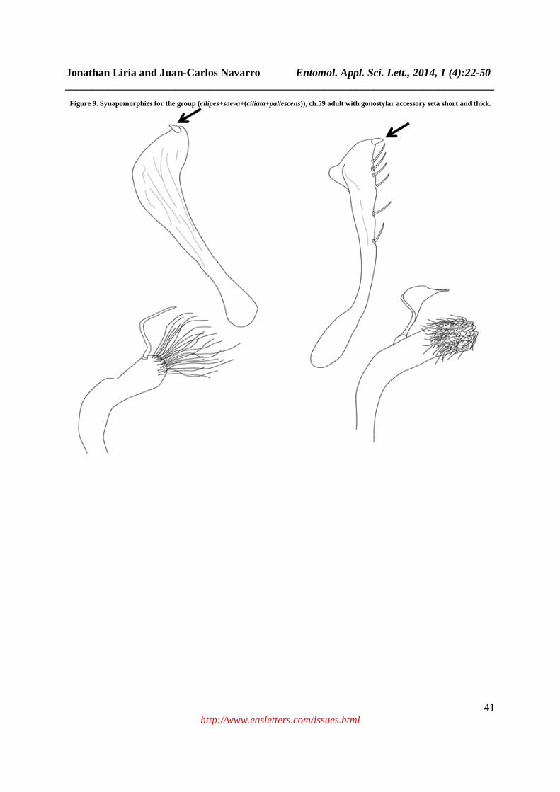
Node Psorophora s. str., Fig. 1: The node is supported (bootstrap: 100%, jackknife: 99%) by four synapomorphies: the larval mandibular lobe presents spicules and very sclerotized (ch.8: 0->1; Fig. 8A), labiohypopharynx with developed premental sensory (ch.19: 0->1; Fig. 8B), in the adult with mesoscutum with naked longitudinal areas (ch.40: 0->3; Fig. 8C), femur III yellowish with with dark erect scales on apex (ch.41: 0->4) and aedeagus with sawed border (ch.65: 0->1; Fig. 8D). Subsequently, Ps. lineata (supported by autamorphic ch.61) is more related and basal to the node that group to cilipes+(saeva+(ciliata+pallescens)). This node is supported by a synapomorphy: in the adult with gonostylar accessory seta short and thick (ch.59: 0->1 Fig. 9A), in Ps. pallescens is short and thin). Finally, Ps. ciliata and Ps pallescens are contained based on a homoplasy (ch.11). In a revision of the Argentinean species [89], consider that the subgenus Psorophora diagnosis is: big mosquitoes, legs with erect scales especially in the apex of the femurs, tibia and first segments of the posterior tarsus, female palpus biggest that the first five segment of flagellum antennal, palpus of the male exceeding the long of the proboscis for the last two segments that are enlarged and hairy, mesonotum with naked longitudinal areas, spiracular setae generally numerous and long, female tarsal claws and the male toothed. Clip (gonostylus) thin, with the expanded apex or bilobulated, some short and strong thorns in their internal face. Later on, [14] and [90] showed for Ps. saeva their likeness with Ps. cilipes, on the base of the male genitalia, where the gonocoxite is distal conical with setae covering around the third distal of this, and a modified setae (foliform) located in the base of the hairy area. The gonostylus has a thin apex, the angle subapical it is big and in lateral and near position to the apex; the gonostylar claw is long and with several strong setae on conspicuous tubers in the internal margin of the half apical. Presently study, the larval synapomorphies increases the characters support to define the subgenus, and those presented in the genitalia they support the hypothesis of the relationships of Ps. saeva with Ps. cilipes and other near species. Node Grabhamia and Janthinosoma, Fig. 1: This node is supported (bootstrap: 58%, jackknife: 60%) by one adult synapomorphic character, gonostylus reticulated (ch.58: 0->1; Fig. 10). Hendrickson and Sokal [36] in their phenetic proposal, indicates “that the three subgenera was placed in an only genus by a historical accident”; according to these authors the three taxa is far away (in a phenetics sense) to each other, and surely nearer to some members of Aedes. Equally, they point out that Grabhamia and Psorophora are very distinct, and related with Ae. (Stegomyia) aegypti. Our results, shows the three subgenera into a monophyletic group, with Psorophora as basal taxon and sister of Grabhamia + Janthinosoma as more related taxa and derivate groups. However [21], shows an inverse relationship among these taxa, with Grabhamia + Janthinosoma as basal clade and Psorophora as derivate. Then, the internal relationships are equal in both hypothesis, the basal and derivate group can be forced by the rooted taxa. Node Grabhamia, Fig. 1: This node is supported (bootstrap/jackknife: 99%) by three synapomorphies in the adults: female Cibarium with developed palatal papillae (ch.28: 0->1; Fig. 11A), tarsomere III5 (ch.43: 0->2; Fig. 11B) half white and dark half and white scales in small torus (ch.52: 0->2). Inside this node it is appreciated Ps. discolor in more basal position to the group ((cingulata+infinis)+(columbiae+jamaicensis+(confinnis+pygmaea))). This last one supported by one adult synapomorphy, occiput with white scales and a small stain of dark scales (ch. 33: 3->2). The other nodes are supported by homoplasies, except for the group confinnis+pygmaea supported by a synapomorphy: fore leg (femur) with white scales that form irregular bands (ch.42: 2->3 Fig. 11C). According to [15] Grabhamia is differentiated at least of the other two subgenera by the presence of clear basal marks in the basal segments of all the tarsi and the absence of extensive areas without scales between the acrostical and dorsocentral areas. These authors in Jamaica, consider two different groups exist: (1) cingulata group, represented by Ps. infinis where the wing has dark scales, and (2) confinnis group, represented by Ps. jamaicensis, Ps. pygmaea and Ps. insularia (including Ps. columbiae that is present in Cuba and Great Cayman) whose the wings have white and dark scales. In our cladistic analysis there are not evidence to accept the groups proposed by [15]; in fact, the character of the scales in the wings was used in the study (ch.49: 0->1, with independent occurrence in Ps. pallescens), being homoplastic. Additionally, the topology of the cladogram where the arrangements type is appreciated the cingulata and confinnis groups, they are spurious because they are supported by homoplasies. On the other hand, the node that contains Ps. confinnis and Ps. pygmaea it is supported by a synapomorphies (ch.42: 2->3), femur dark with irregular white stains.

Jonathan Liria and Juan-Carlos Navarro Entomol. Appl. Sci. Lett., 2014, 1 (4):22-50 ______________________________________________________________________________
35 http://www.easletters.com/issues.html
Figure 1. One of the 11 most parsimonious trees for the data matrix found with TNT, presented to show character mapping, non homoplasious (dark circles) and homoplasious (white circles).
Another interesting aspect inside Grabhamia clade, is the basal position of Ps. discolor, because the larval morphology (particularly the siphon and antennae) is distinctive within the genus. Recently, material of the Psorophora confinnis Complex was studied (Ps. confinnis and Ps. columbiae sensu [15] and [91] from Greg Lanzaro’s collections (UC Davis) from the United States (Maryland, California, Arkansas and Florida), Colombia (Tolima and Bogotá), and Mexico (Oaxaca) and also using our specimens from Venezuela. The cladistic analysis of
Aedes+Haemagogus
Tx. theobaldi
Cx. coronator
Ae. serratus
Hg. celeste
Ma. titillans
Ps. cingulata
Ps. columbiae
Ps. confinnis
Ps. discolor
Ps. infinis
Ps. jaimaicensis
Ps. pygmaea
Ps. albigenu
Ps. albipes
Ps. champerico
Ps. cyanescens
Ps. discrucians
Ps. ferox
Ps. johnstonii
Ps. lutzii
Ps. longipalpus
Ps. horrida
Ps. melanota
Ps. varipes
Ps. ciliata
Ps. cilipes
Ps. lineata
Ps. pallescens
Ps. saeva
6629181413
6251482614
615341393635342318
575049464542402417151310321
13
4623
26
26
5233
5726
47
44
52
64
5940
5730
6157
595749
42
14
46
3311
52464544432928
605732
14
43
4033
5214
43
6444261
412714
595857
11
6459
6548474032301916865431
6654535137217
56412211
2095
633815
Aedini+Mansonia
Mansonia+Psorophora
Psorophora
Grabhamia
Psorophora s. str.
Grabhamia+Janthinosoma Janthinosoma

Jonathan Liria and Juan-Carlos Navarro Entomol. Appl. Sci. Lett., 2014, 1 (4):22-50 ______________________________________________________________________________
36 http://www.easletters.com/issues.html
these data (unpublished) using morphometric and qualitative characters was conducted and several more parsimonious solutions were obtained that demonstrate the difficulty of the group, as well as the necessity of including new evidence (morphological and molecular) in the future studies.
Figure 2. Strict consensus tree of the 11 most parsimonious tress obtained with TNT (L=166 steps, consistency index = 0.66, retention index = 0.82). Number above each branch indicates the percentage of the Bootstrap (below Jackknife percentage) in which that node is
supported.
Tx. theobaldi
Cx. coronator
Ae. serratus
Hg. celeste
Ma. titillans
Ps. cingulata
Ps. columbiae
Ps. confinnis
Ps. discolor
Ps. infinis
Ps. jaimaicensis
Ps. pygmaea
Ps. albigenu
Ps. albipes
Ps. champerico
Ps. cyanescens
Ps. discrucians
Ps. ferox
Ps. johnstonii
Ps. lutzii
Ps. longipalpus
Ps. horrida
Ps. melanota
Ps. varipes
Ps. ciliata
Ps. cilipes
Ps. lineata
Ps. pallescens
Ps. saeva
56 70
56 63
100 99
66 69
58 60
99 99
51
69 64
51 55

Jonathan Liria and Juan-Carlos Navarro Entomol. Appl. Sci. Lett., 2014, 1 (4):22-50 ______________________________________________________________________________
37 http://www.easletters.com/issues.html
Figure 4. Synapomorphy for Aedini (including Mansonia and Psopohora), ch.15 Dorsomentum teeth are 11 to 15.
Figure 5. Synapomorphies for Aedini (Aedes+Haemagogus), ch.9 galeastipal steam present.

Jonathan Liria and Juan-Carlos Navarro Entomol. Appl. Sci. Lett., 2014, 1 (4):22-50 ______________________________________________________________________________
38 http://www.easletters.com/issues.html
Figure 6. Synapomorphies for Mansonia+Psorophora, ch.22 precratal setae present (A) and ch.56 male genitalia presents a gonostylar claw short and sharp (B).

Jonathan Liria and Juan-Carlos Navarro Entomol. Appl. Sci. Lett., 2014, 1 (4):22-50 ______________________________________________________________________________
39 http://www.easletters.com/issues.html
Figure 7. Synapomorphies for Psorophora s. str.+(Grabhamia+Janthinosoma), ch.21 larvae with trident-like scales in the VIII abdominal segment (A), ch.54 adult (females) with tergo and sternum VIII with structure rod-like (B), and ch.66 male genitalia the sternite process
of the X it possesses few teeth (C).

Jonathan Liria and Juan-Carlos Navarro Entomol. Appl. Sci. Lett., 2014, 1 (4):22-50 ______________________________________________________________________________
40 http://www.easletters.com/issues.html
Figure 8. Synapomorphies for Psorophora s. str., ch.8 the larval mandibular lobe presents spicules and very sclerotized (A), ch.19 labiohypopharynx with developed premental sensory (B), ch.40 adult with mesoscutum with naked longitudinal areas (C) and ch.65
aedeagus with sawed border (D).
Jonathan Liria and Juan-Carlos Navarro Entomol. Appl. Sci. Lett., 2014, 1 (4):22-50 ______________________________________________________________________________
41 http://www.easletters.com/issues.html
Figure 9. Synapomorphies for the group (cilipes+saeva+(ciliata+pallescens)), ch.59 adult with gonostylar accessory seta short and thick.

Jonathan Liria and Juan-Carlos Navarro Entomol. Appl. Sci. Lett., 2014, 1 (4):22-50 ______________________________________________________________________________
42 http://www.easletters.com/issues.html
Figure 10. Synapomorphies for Grabhamia and Janthinosoma, ch.58 gonostylus reticulated.

Jonathan Liria and Juan-Carlos Navarro Entomol. Appl. Sci. Lett., 2014, 1 (4):22-50 ______________________________________________________________________________
43 http://www.easletters.com/issues.html
Figure 11. Synapomorphy for Grabhamia, ch.28 female cibarium with developed palatal papillae (A) and ch.43 tarsomere III5 half white and dark half (B).

Jonathan Liria and Juan-Carlos Navarro Entomol. Appl. Sci. Lett., 2014, 1 (4):22-50 ______________________________________________________________________________
44 http://www.easletters.com/issues.html
Figure 12. Synapomorphy for Janthinosoma, ch.27 female cibarium with six palatal papillae.

Jonathan Liria and Juan-Carlos Navarro Entomol. Appl. Sci. Lett., 2014, 1 (4):22-50 ______________________________________________________________________________
45 http://www.easletters.com/issues.html
Figure 13. Synapomorphy for Janthinosomna (excluding Ps. cyanescens), ch.26 pupae with spicules of the abdomen present in the IV segment (A) and ch.44 adult with tarsomere III4 white (B).

Jonathan Liria and Juan-Carlos Navarro Entomol. Appl. Sci. Lett., 2014, 1 (4):22-50 ______________________________________________________________________________
46 http://www.easletters.com/issues.html
Table 1. List and geographical distribution of the species included in the cladistic analysis
Tribe Genus Subgenus Species Distribution Culicini Culex Culex Cx. coronator Dyar and Knab Neotropical Toxorhynchites Lynchiella Tx. theobaldi (Dyar and Knab) Neotropical Mansoniini Mansonia Mansonia Ma. titillans Walker Neartic/Neotropical Aedini Aedes Ochlerotatus Ae. serratus (Theobald) Neotropical Haemagogus Haemagogus Hg. celeste Dyar and Nuñez-Tovar Neotropical Psorophora Psorophora Ps. lineata Humboldt Neotropical Ps. saeva Dyar & Knab Neotropical Ps. cilipes Fabricius Neartic/Neotropical Ps. ciliata Fabricius Neartic/Neotropical Ps. pallescens Edwards Neotropical Grabhamia Ps. columbiae (Dyar and Knab) Neartic Ps. cingulata (Fabricius) Neotropical Ps. confinnis (Lynch Arribalzaga) Neartic/Neotropical Ps. discolor (Coquillett) Neartic Ps. infinis (Dyar and Knab) Antillan Ps. jamaicensis (Theobald) Antillan Ps. pygmaea (Theobald) Antillan Janthinosoma Ps. albigenu Lutz Neotropical Ps. albipes Theobald Neotropical Ps. champerico Dyar & Knab Neartic/Neotropical Ps. cyanescens Coquillet Neartic/Neotropical Ps. discrucians Walker Neotropical Ps. ferox Humboldt Neartic/Neotropical Ps. johnstoni (Grabham) Neartic Ps. lutzii Theobald Neotropical Ps. longipalpus Randolph & O’Neil Neartic Ps. horrida (Dyar & Knab) Neartic Ps. melanota Cerqueira Neotropical Ps. varipes (Coquillett) Neartic
Table 2. Data matrix of 66 characters and 29 taxa used in the cladistic analysis (?, Missing data; -, inapplicable characters)
1 5 10 15 20 25 30 35 40 45 50 55 60 65 | | | | | | | | | | | | | | Tx. theobaldi 000000000001001100000000000001105000101000000000000000011031100000 Cx. coronator 102111110001111001020000000010006101001301000000000000111001400002 Ae. serratus 10222111100101200001000003000000-101011611000001001000011001012400 Hg. celeste 102221111001002001010010000000004010010201000000000010011001302400 Ma. titillans 21121111011110001002010100000000-101011725001200110000100001002200 Ps. cingulata 102211010000002000021100000110002101111322221100001111102131001101 Ps. columbiae 102211010000012000021100000110002101111322221200101111102131001101 Ps. confinnis 102211010000012000021100000110002101111323221200101111102131001101 Ps. discolor 102211010010102000021100000110001101111324221200101111102131001101 Ps. infinis 10221101000000200002110000????002101111322221100001111102131001101 Ps. jaimaicensis 102211010000012000021100000110002101111322221200101111102131001101 Ps. pygmaea 102211010000012000021110000110002101111323221100101111102131001101 Ps. albigenu 20221101001000200002110012????000101111531010000001211102131002301 Ps. albipes 202211010010012000021100121000000101111531110000001211102131002301 Ps. champerico ------------------------1110000111011115311100000010??103130002301 Ps. cyanescens 102211010011012000021100101000001101111331000000001011102131001101 Ps. discrucians 20221101001001200002110012????001101111331010000001011104131002301 Ps. ferox 202211010010012000021100111000001101111331110010001011102131002301 Ps. johnstonii 20221101001000200002110011????001101111331020000001211102131002301 Ps. lutzii 20221101001001200002110011????000101111531110000001011102131002301 Ps. longipalpus 202211010010002000021100111000010101111531110000001211103130002001 Ps. horrida 2022110100100020000211001?????000101111531110000001211102131002301 Ps. melanota 20221101001001200002110011????00?1011112311100000010??102121002301 Ps. varipes 202211010010002000021100111001000101111531010000001211104131002301 Ps. ciliata 000000020001002100121100100001013101111141000011001011101011002311 Ps. cilipes 000000020011002100121100100001013101111141000011001011101011002311 Ps. lineata 000000020011002100121100100001013101111141000011001011103001202111 Ps. pallescens 000000020001002100121100100001013101111141000011101011102031002311 Ps. saeva 000000020011002100121100100001013101111141000011001011101011002311

Jonathan Liria and Juan-Carlos Navarro Entomol. Appl. Sci. Lett., 2014, 1 (4):22-50 ______________________________________________________________________________
47 http://www.easletters.com/issues.html
Node Janthinosoma, Fig. 1: This node is supported (bootstrap: 69%/jackknife: 64%) by two synapomorphies in the adult: female cibarium with six palatal papillae (ch.27: 0->1; Fig. 12) and femur III yellow with dark base and white basal border (ch.41: 1->3). According to [15] and [92], the metallic violet scales of the legs, labium, and abdominal tergites can identify this subgenus. Most of the species have conspicuous white marks on hind tarsomeres 4 and/or 5 and sometimes on 3. These authors consider the taxonomy of the subgenus to be very confused, due to the lack of males and associated immature stages; the larvae have been described for about half of the species. However [92] emphasizes the urgent need for more intense morphological studies of the subgenus Janthinosoma, and the importance of many new diagnostic morphological characters. In studies of the morphology of the cibarium in Culicidae [65], show the presence of six palatal papillae in Ps. (Janthinosoma) ferox and Ps. (Jan.) varipes. This character is also observed in Culiseta alaskaensis, Cu. inornata, Cu. morsitans, Aedes dorsalis and Coquillettidia perturbans and could possibly be involved in food palatability. The character should be more thoroughly studied, using electron microscopy and even physiological response to determine possible differences or similarities between these taxa and Janthinosoma. The most basal taxon of the subgenus is Ps. cyanescens, which is separated from the next node by two synapomorphies: pupae with spicules on abdomen segment IV (ch.26: 0->2; Fig. 13A) and adult with hind tarsomere 4 white (ch.44: 0->1; Fig. 13B). The claspette of the male genitalia of Ps. cyanescens is fused to the gonocoxite, as it is in species of Grabhamia; this is interesting, particularly because of the location of the species in the tree topology where Ps cyanescens seems to be a link species between both subgenera. On the other hand, it is also the only species (together with Ps. lanei, not included in the analysis) that presents the tarsomere III4,5 dark. In the only subdivision of this subgenus [79,81] proposed the Lutzii Series as a natural group that comprise Ps. lutzii, Ps. albipes, Ps. forceps and Ps. amazonica. Our results do not allow supporting the validity of the series, because the nodes are supported by homoplasies and the material of Ps. amazonica was not revised and immature of Ps. forceps are unknown.
CONCLUSION In this work we found morphological diagnostic characters for the genus Psorophora, in the larva, abundant precratal setae in the anal segment (or tenth) and scale of the eighth segment (or comb of the VIII) trident-like. As for the adults, there were no unique characters for this taxon, nor any reason for the continued separation of Aedes for the combination of conventional characters. At the subgeneric level Psorophora s. str. was the evolutionary basal and most distinctive taxon, due to the presence in the larvae antennas shorter than the head without surpassing the previous border, mandibular lobes with spicules and sclerotized, labiohypopharynx with premental sensor developed; in the adult, mesoscutum with longitudinal areas without scales, fore legs with erect scales, female cibarium with 14-15 trichoid sensilla and aedeagus with sawed intern border. In Grabhamia and Janthinosoma, diagnostic characters were found in adults. In Grabhamia: the presence of long palatal papillae in the feminine cibarium, fore femur with a tarsomere half white and dark half, and small white scales in the internal face of the torus. Janthinosoma is characterized by the presence of six palatal papillae in the cibarium and a yellow fore femur with dark basal band and white border. The genera Aedes and Haemagogus showed characters shared in the larva by the presence in the maxilla of the galeastipal stem and labiohypopharynx with numerous ventral premental processes with scales-like form. Mansonia, presents ten diagnostic characters: In the larva, the antenna are wide and longer than the head, the jaw has the latter dorsal tooth formed by two teeth, labiohypopharinx with lateral premental teeth inconspicuous, and breathing siphon with a modification to adhere to aquatic plants. In the adult, the wings have white and dark wide scales, and in the male genitalia the gonostylus is wide and with a digitiform projection. The genus Psorophora is clearly a monophyletic taxon, but Aedini (Haemagogus, Aedes and Psorophora), using the data produced to date, seems to be a paraphyletic group that should contain the members of Mansoniini to form a more natural classification. This proposal is supported by the results obtained in a cladistic analysis of the family Culicidae [32]. Our results plus the latter authors’ hypothesis, show the Mansoniini tribe (Mansonia and Coquillettidia) placed along Aedini. However, the addition of more Mansoniini taxa and with the use of molecular data may support or to reject the hypothesis of a Mansoniini +Aedini clade as natural group. The infrageneric classification of Psorophora shows that the 3 subgenera form monophyletic groups, although internal polytomies were observed in Grabhamia and Janthinosoma because the relationships among some species could not be established.

Jonathan Liria and Juan-Carlos Navarro Entomol. Appl. Sci. Lett., 2014, 1 (4):22-50 ______________________________________________________________________________
48 http://www.easletters.com/issues.html
Acknowledgments We thank Pablo Goloboff (INSUE-UNT) for the suggestions about the earlier cladistics methods on the MSc thesis of JL. Thanks especially to Rick Wilkerson for the invitation (JL) to WRBU, and Jim Pecor and Thomas Gaffigan (WRBU-MSC) by technical invaluable assistance in the Smithsonian Collections. To Scott C Weaver and Tom Zavortink for review and suggestions. This research was funded in part by NIH-NASA, FOGARTY-NIH, A139800, AI-48807, TW005919 (SC Weaver, and JCN in Venezuela) FONACIT S1-20010000939 (JCN) and FONACIT, Misión Ciencia 2008000911-4 to JCN.
REFERENCES
[1] Sellers, R.F., Bergold, G.H., Suárez, O.M. Morales, A. American Journal of Tropical Medicine and Hygiene. 1965. 14,460-469. [2] Weaver, S.C., Pfeffer, M., Marriot, K., Kang, W. Kinney, R.M. American Journal of Tropical Medicine and Hygiene. 1999. 60,441-448. [3] Weaver, S.C., Salas, R., Rico-Hesse, R., Ludwig, G.V., Oberste, M.V., Boshell, J. Tesh, R. Lancet. 1996. 348,436-440. [4] Wang, E., Barrera, R., Boshell, J., Ferro, C., Navarro, J.C., Salas, R., Weaver, S.C. Journal of Virology. 1999. 73,4266-4271. [5] Weaver, S.C. Recurrent emergence of Venezuelan equine encephalomyelitis. In: Emergence infections I. WM Sheld, D Amstrong and JM Hughes (Eds.). Chapter 3. ASM Press, Washington, DC. 1998. Pp. 27-42. [6] Sudia, D. Arthropod vectors of epidemic Venezuelan equine encephalitis. In. Proc. Workshop-Symposium on Venezuelan Encephalitis Virus. Pan American Health Organization, Washington, D. C. 1972. Pp. 157-161. [7] Turell, M.J. Journal Medical Entomology. 1999. 36,407-409. [8] Turell, M.J., Barth, J. Coleman, R.E. Journal of the American Mosquito Control Association. 1999. 15,295-298. [9] Brault, A.C., Powers, A.M., Ortiz, D., Estrada-Franco, J.G., Navarro-Lopez, R. Weaver, S.C. Proceedings of the National Academic of Science. 2004. 31, 11344-11349. [10] Weaver, S.C., Ferro, C., Barrera, R., Boshell, J. Navarro, J.C. Annual Review Entomology. 2004. 49,141-74. [11] Karabastos, N. International Catalogue of Arboviruses (including other viruses of vertebrates). 3th Ed. Americam Tropical Medicine and Hygiene. San Antonio Texas. 1985. 674-4. [12] Guimarães, J.H. Systematic Database of Diptera of the Americas South of the United States, Family Culicidae. Fundação de Amparo a Pesquisa do Estado de São Paulo/Ed. Pleide. São Paulo. 1997. Pp. 286. [13] Liria, J. Barrera, R. Navarro, J.C. Nuevos Entomotropica. 2001. 16,197-198. [14] Liria, J, Navarro, J.C. Entomotropica. 2003a. 18,113-119. [15] Belkin, J.N. Heinemann, S.J. Page, W.A. Contributions of the American Entomological Institute. 1970. 6,1-458. [16] Tanaka, K. Japanese Journal of Systematic Entomology. 2003. 9,159-169. [17] Judd, D.D. Systematic Entomology. 1996. 21,129-150. [18] Harbach, R.E. Peyton, E.L. Bulletin of Natural History Museum of London (Entomology). 2000. 69,115-169. [19] Navarro, J.C. Liria, J. Journal of the American Mosquito Control Association. 2000. 16,75-85. [20] Sallum, M.A.M., Shultz, T.R. Wilkerson, R.C. Annals of the Entomological Society of America. 2000. 93,745-775. [21] Reinert, J.F., Harbach, R.E. Kitching, I. Zoological Journal of the Linnean Society. 2004. 142,289-368. [22] Knight, K.L. Stone, A. Thomas Say Found Entomolical Society American. 1977. 6,1-611. [23] Faran, M.E. Contributions of the American Entomological Institute. 1980. 15,1-215. [24] Faran, M.E. Linthicum, K.J. Mosquito Systematics. 1981. 13,1-81. [25] Pape, T. Mosquito Systematics. 1992. 24,1-11. [26] Wesson, D.M., Porter, C.H. Collins, F.H. Molecular Phylogenetics and Evolution. 1992. 1,253-269. [27] Besansky, N.J., Powell, J.R., Caccone, A., Hamm, D.M., Scott, J.A. Collins, F. H. Proccedings of the National Academic of Science. 1994. 91,6885-88. [28] Miller, B.R., Crabtree, M.B. Savage, H.M. Insect Molecular Biology. 1996. 5,93-107. [29] Besansky, N.J. Fahey, T. Utility of the white gene in estimating phylogenetic relationships among mosquitoes (Diptera: Culicidae). Molecular Biology and Evolution. 1997. 14,442-454. [30] Foley, D., Bryan, J., Yeates, D., Saul, A. Molecular Phylogenetics and Evolution. 1998. 9,262-275. [31] Navarro, J.C. Weaver, S.C. Journal Medical Entomology. 2004. 41,475-581. [32] Harbach, R.E. Kitching, I. Systematic Entomology. 1998. 23,327-370. [33] Thomas, G.A., Harbach, R.E. Kitching, I.J. Systematic Entomology. 1999. 24,193-205. [34] Harbach, R.E. Zootaxa. 2007. 1668, 591-688.

Jonathan Liria and Juan-Carlos Navarro Entomol. Appl. Sci. Lett., 2014, 1 (4):22-50 ______________________________________________________________________________
49 http://www.easletters.com/issues.html
[35] Reinert, J.F., Harbach, R.E. Kitching, I. Zoological Journal of the Linnean Society. 2009. 157,700-794. [36] Hendrickson, J.A. Sokal, R. R. Annals of the Entomological Society of America. 1968. 61,385-392. [37] Goloboff, P.A. Principios Básicos de Cladística. Sociedad Argentina de Botánica, Buenos Aires – Argentina. 1998. Pp. 81. [38] Harbach, R.E. Knight, K.L. Taxonomists’ Glossary of Mosquito Anatomy. Plexus Publishing, Inc. 1980. Pp. 415. [39] McAlpine, J.F., Peterson, B.V., Shewell, G.E., Teskey, H.J., Wockeroth, J.R. Wood, D.M. Manual of Neartic Diptera. Volume 1. Research Branch Agriculture Canada. Monograph No. 27. 1981. Pp. 674. [40] Reinert, J.F. Comparative anatomy of female genitalia of genera and subgenera on tribe aedini (Diptera: Culicidae). Part I. Introduction, preparation techniques, and anatomical terminology. Part II. Genus Psorophora Robineau-Desvoidy. Part II. Genus Udaya Thurman. Pat IV. Genus Zeugnomyia Leicester. Contributions of the American Entomological Institute. 2000a. 32,1-52. [41] Reinert, J.F. Mosquito Systematics. 1975. 7,105-110. [42] Goloboff, P.A. Farris, J. Nixon, K. Cladistics. 2008. 24, 774-786. [43] Goloboff, P.A. Cladistics. 1996. 12,199-220. [44] Nixon, K.C. Winclada (BETA) ver. 0.9.99m24 publicado por el autor, Ithaca, NY. 1999, Beta version at http://www.cladistics.com. [45] Farris, J.A., Albert, V.A., Källersjö, M., Lipscomb, D. Kluge, A.G. Cladistics. 1996. 12,99-124. [46] Kitching, I., Forey, P., Humphries, C. Williams, D. Cladistics: The Theory and Practice of the Parsimony Analysis. Oxford University Press. 1998. Pp. 228. [47] Carpenter, J.M. Cladistics. 1996, 12,177-181. [48] Lane, J. Neotropical Culicidae. Vol I. University of São Paulo, São Paulo. Brazil. 1953. Pp. 1111. [49] Belkin, J.N. The mosquitoes of the south Pacific (Diptera: Culicidae) Volumes 1 and 2. Los Angeles CA, University of California Press. 1962. [50] Arnell, J.H. Mosquito studies (Diptera: Culicidae) XXXII. A revision of the genus Haemagogus. Contributions of the American Entomological Institute. 1973. 10,1-174. [51] Knight, K.L. Journal Medical Entomology. 1971. 8,189-205. [52] Liria, J. Navarro, J.C. Boletin de Entomología Venezolana. 1999. 14,53-62. [53] Liria, J, Navarro, J.C. Zootaxa. 2003b. 186,1-8. [54] Yamatugi, S. LaCasse, W.J. Mosquito fauna of North america Part III - Genera Orthopodomyia, Mansonia and Psorophora (with 38 original plates). Office of the Surgeon, Headquarters Japan Logistics Command, APO 343. 1951. Pp. 265. [55] Shalaby, A.M. Bulletin of the Society Entomological of Egypt. 1957. 41,429-455. [56] Pérez, B.Y. Navarro, J.C. Acta Zoologica Mexicana. 1996. 68,13-26. [57] Harbach, R.E. Knight, K.L. Mosquito Systematics. 1977. 9,455-477. [58] Arnell, J.H. Mosquito studies (Diptera: Culicidae) XXXIII. A revision of the Scapularis Group of Aedes (Ochlerotatus). Contributions of the American Entomological Institute. 1976. 13,1-144. [59] González, R. Darsie, R.F (Jr.). Clave ilustrada para la determinación genérica de larvas de Culicidae de Colombia y el nuevo mundo. Boletin del Museo Entomológico de la Universidad del Valle. 1996. 4,21-37. [60] Barr, R.A. Barr, S. Mosquito studies (Diptera: Culicidae). XIII. Pupae of the genus Psorophora in North America and Puerto Rico. Contributions of the American Entomological Institute. 1969. 4,1-20. [61] Xavier, S.H. Revista Brasileira de Biologia. 1964. 24,235-238. [62] Xavier, S.H., Silva-Mattos, S. Revista Brasileira do Malariologia e Docenças Tropicais. 1967. 18,616-624. [63] Reinert, J.F. Journal of the American Mosquito Control Association. 2000c. 16,175-188. [64] Lee, R.M. Davies, D.M. (1978) International Journal of Insect Morphology and Embryology. 1978. 7,189-194. [65] Lee, R.M. Craig, D.A. Canadian Journal of Zoology. 1983. 61,633-646. [66] Liria, J. Navarro, J.C. Neotropical Entomology. 2007. 36,902-909. [67] Lee, R.M. Quaestiones Entomologicae. 1974. 10,187-217. [68] Sirivanakarn, S. Mosquito Systematics. 1978. 10,474-492. [69] Christophers, S.R. The fauna of the British India, including Ceylon and Burma. Vol. IV. Family Culicidae. Tribe Anophelini. Taylor and Francis, London. 1933. Pp. 371. [70] Sinton, J.A. Covell, G. Indian Journal of Medical Research. 1927. 15,301-308. [71] Sallum, M.A.M. Forattini, O.P. Journal of the American Mosquito Control Association. 1996. 12,517-600. [72] Roth, L.M. The male and larva of Psorophora (Janthinosoma) horrida (Dyar and Knab) and a new species of Psorophora from the United States (Diptera: Culicidae). Proccedings of the Entomological Society of Washington. 1945. 47,1-23.

Jonathan Liria and Juan-Carlos Navarro Entomol. Appl. Sci. Lett., 2014, 1 (4):22-50 ______________________________________________________________________________
50 http://www.easletters.com/issues.html
[73] Guedes, A.S., Souza, M.A., Maciel, C.S. Xavier, S.H. Revista Brasileira do Malariologia e Docenças Tropicais. 1964. 16,2-14. [74] Zavortink, T.J. Mosquito studies (Diptera: Culicidae) XXVIII. The New World species formerly placed in Aedes (Finlaya). Contributions of the American Entomological Institute. 1972. 8,1-206. [75] Berlin, O.G.W. Belkin, J.N. Mosquito studies (Diptera: Culicidae) XXXVI. Subgenera Aedinus, Tinolestes, Anoedioporpa of Culex. Contributions of the American Entomological Institute. 1980. 17,1-104. [76] Dyar, H.G. The mosquitoes of the Americas. Publ. 387. Carnegie Institute Washington, Washington DC. 1928. Pp. 616. [77] Guedes, A.S. Souza, M.A. Sobre Psorophora (Janthinosoma) albigenu Lutz, 1908 e Psorophora (Janthinosoma) albipes (Theobald, 1907) (Diptera: Culicidae). Revista Brasileira do Malariologia e Docenças Tropicais. 1964. 16,471-486. [78] Wilkerson, R.C. Peyton, E.L. Journal Medical Entomology. 1990. 27,207-224. [79] Cerqueira, N.L. Revista de Entomologia. 1939. 10,78-85. [80] Shannon, R.C. Cerqueira, N.L. Revista de Entomologia. 1943. 14,135-137. [81] Cerqueira, N.L. Uma n. sp. Boletin do Museo Parana Emilio Goeldi. 1960. 26,1-10. [82] Duret, P. Neotropica. 1971. 17,15-28. [83] Barata, J.M.S. Cotrim, M.D. Uma nova espécie de Psorophora (Diptera, Culicidae). Revista Brasileira de Entomologia. 1971. 15,35-40. [84] Belkin, J.N. Heinemann, S.J. Psorophora (Janthinosoma) mathesoni, sp. nov. for “varipes” of the southeastern U.S.A. Mosquito Systematics. 1975. 7,363-366. [85] Reinert, J.F. Comparative anatomy of female genitalia of genera and subgenera on tribe aedini (Diptera: Culicidae). Part V. Genus Aedes Meigen. Contributions of the American Entomological Institute. 2000b. 32, 1-102. [86] Reinert, J.F. Comparative anatomy of the female genitalia of genera and subgenera in tribe Aedini (Diptera: Culicidae). Part XI. Genus Haemagogus Willinston. Contributions of the American Entomological Institute. 2002. 32,1-46. [87] Ronderos, R.A., Bachmann, A.O. Mansoniini Revista de la Sociedad Entomologica Argentina. 1963b. 26,57-65. [88] Ronderos, R.A. Bachmann AO. Revista de la Sociedad Entomologica Argentina. 1963a. 25,43-51. [89] Del Ponte, E. Castro, M. Las Revista de la Sociedad Entomologica Argentina. 1952. 15,211-230. [90] Stone, Revista Brasileira de Entomologia. 1967. 12,59-65. [91] Bickley, W.E. Mosquito Systematics. 1984. 16,162-167. [92] Harrison, B.A. Varnado, W. Whitt, P.B. Goddard, J. Journal of Vector Ecology. 2008. 33,232-237.