Phenyl- and benzylurea cytokinins as competitive inhibitors of cytokinin oxidase/dehydrogenase: A...
11
Research paper Phenyl- and benzylurea cytokinins as competitive inhibitors of cytokinin oxidase/dehydrogenase: A structural study David Kope cný a, b, * , Pierre Briozzo c , Hana Popelková d , Marek Sebela b , Radka Kon citíková b , Luká s Spíchal e , Jaroslav Nisler e , Catherine Madzak f , Ivo Frébort b , Michel Laloue a , Nicole Houba-Hérin a a Laboratoire de Biologie Cellulaire, INRA, Route de Saint-Cyr, F-78026 Versailles Cedex, France b Department of Biochemistry, Faculty of Science, Palacký University, Slechtitel u 11, CZ-78371 Olomouc, Czech Republic c Institut Jean-Pierre Bourgin, UMR INRA-AgroParisTech 1318 Dynamic and Structure of Lipid Bodies, F-78850 Thiverval-Grignon, France d Department of Molecular, Cellular and Developmental Biology, University of Michigan, Ann Arbor, MI 48109-1048, USA e Laboratory of Growth Regulators, Faculty of Science, Palacký University and Institute of Experimental Botany, ASCR, Slechtitel u 11, CZ-783 71 Olomouc, Czech Republic f UMR1319 Micalis, INRA-AgroParisTech, CBAI, F-78850 Thiverval-Grignon, France article info Article history: Received 15 November 2009 Accepted 7 May 2010 Available online 15 May 2010 Keywords: Benzylurea Crystal structure Cytokinin oxidase/dehydrogenase Inhibitor Phenylurea abstract Cytokinin oxidase/dehydrogenase (CKO) is a flavoenzyme, which irreversibly degrades the plant hormones cytokinins and thereby participates in their homeostasis. Several synthetic cytokinins including urea derivatives are known CKO inhibitors but structural data explaining enzymeeinhibitor interactions are lacking. Thus, an inhibitory study with numerous urea derivatives was undertaken using the maize enzyme (ZmCKO1) and the crystal structure of ZmCKO1 in a complex with N-(2-chloro-pyridin- 4-yl)-N 0 -phenylurea (CPPU) was solved. CPPU binds in a planar conformation and competes for the same binding site with natural substrates like N 6 -(2-isopentenyl)adenine (iP) and zeatin (Z). Nitrogens at the urea backbone are hydrogen bonded to the putative active site base Asp169. Subsequently, site-directed mutagenesis of L492 and E381 residues involved in the inhibitor binding was performed. The crystal structures of L492A mutant in a complex with CPPU and N-(2-chloro-pyridin-4-yl)-N 0 -benzylurea (CPBU) were solved and confirm the importance of a stacking interaction between the 2-chloro-4-pyridinyl ring of the inhibitor and the isoalloxazine ring of the FAD cofactor. Amino derivatives like N-(2-amino-pyridin- 4-yl)-N 0 -phenylurea (APPU) inhibited ZmCKO1 more efficiently than CPPU, as opposed to the inhibition of E381A/S mutants, emphasizing the importance of this residue for inhibitor binding. As highly specific CKO inhibitors without undesired side effects are of major interest for physiological studies, all studied compounds were further analyzed for cytokinin activity in the Amaranthus bioassay and for binding to the Arabidopsis cytokinin receptors AHK3 and AHK4. By contrast to CPPU itself, APPU and several benzylureas bind only negligibly to the receptors and exhibit weak cytokinin activity. Ó 2010 Elsevier Masson SAS. All rights reserved. 1. Introduction Cytokinins are plant hormones that regulate a large number of developmental and physiological events [1]. Naturally occurring cytokinins consist of an adenine/adenosine moiety carrying an N 6 - isoprenoid or N 6 -aromatic side chain. Cytokinin-related processes are triggered by the activation of a phosphorelay cascade starting from sensor histidine kinases. Three cytokinin receptors, AHK2, Abbreviations: ACPBU, N-(2-amino-6-chloro-pyridin-4-yl)-N 0 -benzylurea; ACPPU, N-(2-amino-6-chloro-pyridin-4-yl)-N 0 -phenylurea; AHK, Arabidopsis histidine kinase; APPU, N-(2-amino-pyridin-4-yl)-N 0 -phenylurea; CKO/CKX, cytokinin oxidase/dehydrogenase; CPPU, N-(2-chloro-pyridin-4-yl)-N 0 -phenylurea; CPBU, N-(2-chloro-pyridin-4- yl)-N 0 -benzylurea; DCPIP, 2,6-dichlorophenol indophenol; DCPPOU, N-(2,6-dichloro-pyridin-4-yl)-N 0 -phenoxyurea; DCPPU, N-(2,6-dichloro-pyridin-4-yl)-N 0 -phenylurea; DCPBC, (2,6-dichloro-pyridin-4-yl)-carbamic acid benzyl ester; DCPBMU, N-(2,6-dichloro-pyridin-4-yl)-N 0 -benzyl-N 0 -methylurea; DCPBU, N-(2,6-dichloro-pyridin-4-yl)-N 0 - benzylurea; iP, N 6 -(2-isopentenyl)adenine; iPR, N 6 -(2-isopentenyl)adenosine; MOCPBU, N-(2-chloro-6-methoxy-pyridin-4-yl)-N 0 -benzylurea; PPU, N-(pyridin-4-yl)-N 0 - phenylurea; PU48, N-(imidazo[1,2-a]pyridin-4-yl)-N 0 -phenylurea; PU60, N-(2-chloro-1-oxy-pyridin-4-yl)-N 0 -phenylurea; PU71, (2-chloro-pyridin-4-yl)-carbamic acid 4- chloro-phenyl ester; TDZ, N-(1,2,3-thidiazol-5-yl)-N 0 -phenylurea i.e. thidiazuron; Z, N 6 -(trans-4-hydroxy-3-methyl-2-buten-1-yl)adenine i.e., zeatin; ZmCKO/ZmCKX, cytokinin oxidase/dehydrogenase from Zea mays. * Corresponding author. Department of Biochemistry, Faculty of Science, Palacký University, Slechtitel u 11, CZ-78371 Olomouc, Czech Republic. Tel.: þ420 585634927; fax: þ420 585634933. E-mail address: [email protected] (D. Kope cný). Contents lists available at ScienceDirect Biochimie journal homepage: www.elsevier.com/locate/biochi 0300-9084/$ e see front matter Ó 2010 Elsevier Masson SAS. All rights reserved. doi:10.1016/j.biochi.2010.05.006 Biochimie 92 (2010) 1052e1062
Transcript of Phenyl- and benzylurea cytokinins as competitive inhibitors of cytokinin oxidase/dehydrogenase: A...

lable at ScienceDirect
Biochimie 92 (2010) 1052e1062
Contents lists avai
Biochimie
journal homepage: www.elsevier .com/locate/b iochi
Research paper
Phenyl- and benzylurea cytokinins as competitive inhibitors of cytokininoxidase/dehydrogenase: A structural study
David Kope�cný a,b,*, Pierre Briozzo c, Hana Popelková d, Marek �Sebela b, Radka Kon�citíková b,Luká�s Spíchal e, Jaroslav Nisler e, Catherine Madzak f, Ivo Frébort b, Michel Laloue a, Nicole Houba-Hérin a
a Laboratoire de Biologie Cellulaire, INRA, Route de Saint-Cyr, F-78026 Versailles Cedex, FrancebDepartment of Biochemistry, Faculty of Science, Palacký University, �Slechtitel�u 11, CZ-78371 Olomouc, Czech Republicc Institut Jean-Pierre Bourgin, UMR INRA-AgroParisTech 1318 Dynamic and Structure of Lipid Bodies, F-78850 Thiverval-Grignon, FrancedDepartment of Molecular, Cellular and Developmental Biology, University of Michigan, Ann Arbor, MI 48109-1048, USAe Laboratory of Growth Regulators, Faculty of Science, Palacký University and Institute of Experimental Botany, ASCR, �Slechtitel�u 11, CZ-783 71 Olomouc, Czech RepublicfUMR1319 Micalis, INRA-AgroParisTech, CBAI, F-78850 Thiverval-Grignon, France
a r t i c l e i n f o
Article history:Received 15 November 2009Accepted 7 May 2010Available online 15 May 2010
Keywords:BenzylureaCrystal structureCytokinin oxidase/dehydrogenaseInhibitorPhenylurea
Abbreviations: ACPBU, N-(2-amino-6-chloro-pyridAPPU, N-(2-amino-pyridin-4-yl)-N0-phenylurea; CKO/yl)-N0-benzylurea; DCPIP, 2,6-dichlorophenol indophDCPBC, (2,6-dichloro-pyridin-4-yl)-carbamic acid benbenzylurea; iP, N6-(2-isopentenyl)adenine; iPR, N6-phenylurea; PU48, N-(imidazo[1,2-a]pyridin-4-yl)-N0
chloro-phenyl ester; TDZ, N-(1,2,3-thidiazol-5-yl)-Ncytokinin oxidase/dehydrogenase from Zea mays.* Corresponding author. Department of Biochemist
fax: þ420 585634933.E-mail address: [email protected] (D. K
0300-9084/$ e see front matter � 2010 Elsevier Masdoi:10.1016/j.biochi.2010.05.006
a b s t r a c t
Cytokinin oxidase/dehydrogenase (CKO) is a flavoenzyme, which irreversibly degrades the planthormones cytokinins and thereby participates in their homeostasis. Several synthetic cytokininsincluding urea derivatives are known CKO inhibitors but structural data explaining enzymeeinhibitorinteractions are lacking. Thus, an inhibitory study with numerous urea derivatives was undertaken usingthemaize enzyme (ZmCKO1) and the crystal structure of ZmCKO1 in a complex with N-(2-chloro-pyridin-4-yl)-N0-phenylurea (CPPU) was solved. CPPU binds in a planar conformation and competes for the samebinding site with natural substrates like N6-(2-isopentenyl)adenine (iP) and zeatin (Z). Nitrogens at theurea backbone are hydrogen bonded to the putative active site base Asp169. Subsequently, site-directedmutagenesis of L492 and E381 residues involved in the inhibitor binding was performed. The crystalstructures of L492A mutant in a complex with CPPU and N-(2-chloro-pyridin-4-yl)-N0-benzylurea (CPBU)were solved and confirm the importance of a stacking interaction between the 2-chloro-4-pyridinyl ringof the inhibitor and the isoalloxazine ring of the FAD cofactor. Amino derivatives like N-(2-amino-pyridin-4-yl)-N0-phenylurea (APPU) inhibited ZmCKO1 more efficiently than CPPU, as opposed to the inhibition ofE381A/S mutants, emphasizing the importance of this residue for inhibitor binding. As highly specific CKOinhibitors without undesired side effects are of major interest for physiological studies, all studiedcompounds were further analyzed for cytokinin activity in the Amaranthus bioassay and for bindingto the Arabidopsis cytokinin receptors AHK3 and AHK4. By contrast to CPPU itself, APPU and severalbenzylureas bind only negligibly to the receptors and exhibit weak cytokinin activity.
� 2010 Elsevier Masson SAS. All rights reserved.
1. Introduction
Cytokinins are plant hormones that regulate a large number ofdevelopmental and physiological events [1]. Naturally occurring
in-4-yl)-N0-benzylurea; ACPPU, N-CKX, cytokinin oxidase/dehydrogenenol; DCPPOU, N-(2,6-dichloro-pyzyl ester; DCPBMU, N-(2,6-dichlor(2-isopentenyl)adenosine; MOCPB-phenylurea; PU60, N-(2-chloro-10-phenylurea i.e. thidiazuron; Z, N
ry, Faculty of Science, Palacký Uni
ope�cný).
son SAS. All rights reserved.
cytokinins consist of an adenine/adenosine moiety carrying an N6-isoprenoid or N6-aromatic side chain. Cytokinin-related processesare triggered by the activation of a phosphorelay cascade startingfrom sensor histidine kinases. Three cytokinin receptors, AHK2,
(2-amino-6-chloro-pyridin-4-yl)-N0-phenylurea; AHK, Arabidopsis histidine kinase;ase; CPPU, N-(2-chloro-pyridin-4-yl)-N0-phenylurea; CPBU, N-(2-chloro-pyridin-4-ridin-4-yl)-N0-phenoxyurea; DCPPU, N-(2,6-dichloro-pyridin-4-yl)-N0-phenylurea;o-pyridin-4-yl)-N0-benzyl-N0-methylurea; DCPBU, N-(2,6-dichloro-pyridin-4-yl)-N0-U, N-(2-chloro-6-methoxy-pyridin-4-yl)-N0-benzylurea; PPU, N-(pyridin-4-yl)-N0--oxy-pyridin-4-yl)-N0-phenylurea; PU71, (2-chloro-pyridin-4-yl)-carbamic acid 4-6-(trans-4-hydroxy-3-methyl-2-buten-1-yl)adenine i.e., zeatin; ZmCKO/ZmCKX,
versity, �Slechtitel�u 11, CZ-78371 Olomouc, Czech Republic. Tel.: þ420 585634927;
D. Kope�cný et al. / Biochimie 92 (2010) 1052e1062 1053
AHK3 and AHK4 have been described in Arabidopsis [2e4]. Cyto-kinin biosynthesis is catalyzed by isopentenyltransferases [5,6],while inactivation occurs either via N- or O-conjugation or viairreversible oxidation by cytokinin oxidases/dehydrogenases (CKO/CKX, EC 1.5.99.12) [1].
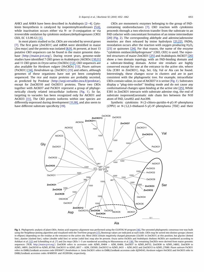
In most plants studied so far, CKOs are encoded by several genes[7]. The first gene (ZmCKO1) and mRNA were identified in maize(Zea mays) and the protein was isolated [8,9]. At present, at least 13putative CKO sequences can be found in the maize genomic data-base (http://maize.jcvi.org/). During recent years, genome-widestudies have identified 7 CKO genes in Arabidopsis (AtCKOs) [10,11]and 11 CKO genes in Oryza sativa (OsCKOs) [12]. CKO sequences arealso available for Hordeum vulgare (HvCKOs) [13], Pisum sativum(PsCKO1) [14], Dendrobium sp. (DsCKO1) [15] and others, althoughgenomes of these organisms have not yet been completelysequenced. The rice and maize proteins are probably secreted,as predicted by Predotar (http://urgi.versailles.inra.fr/predotar),except for ZmCKO10 and OsCKO11 proteins. These two CKOstogether with AtCKO7 and PsCKO1 represent a group of phyloge-netically closely related intracellular isoforms (Fig. 1). So far,targeting to vacuoles has been recognized only for AtCKO1 andAtCKO3 [11]. The CKO protein isoforms within one species aredifferently expressed during development [11,18], and also seem tohave different substrate specificity [19].
sO4OKCmZ
6OKCmZ
CsO31OKCmZ
11OKCmZ
1OKCsD
3OKCtA
2OKCtA
4OKCtA
1OKCmZ
1OKCsO
5OKCmZ
2OKCsO
8OKCmZ
9OKCmZ
6OKCSO
7OKCsO
21OKCmZ
01OKCsO
ylG
psA
0001
0001
29
0001
0001
0001
0001
169
186
959
0001
0001
0001
0001
0001
989
0001
275878
1
Fig. 1. Phylogenetic analysis of plant CKOs. Amino acid sequence alignment was performed uusing the Neighbour-Joining algorithm and visualized with the TreeView program [17]. Bootsin ellipses) depending on the residue at the entrance to the active site. Most CKOs contain nline), alanine (dashed line), valine (double solid line) or serine (solid line) may also be preAshikari et al. [12] and Schmülling et al. [7] and Zea mays CKOs 1e5 are numbered accordinsequences (TIGR, http://maize.jcvi.org/): ZmCKO6 refers to accession code AZM5_10404AZM5_14891, ZmCKO10 to AZM5_85766, ZmCKO11 to AZM5_6817 þ AZM_150369, ZmCKOrefers to EMBL/GenBank accession code EF030477, Dendrobium cv. Sonia DsCKO1 refers to EMEMBL/GenBank accession codes AF490591 and AY209184, respectively.
CKOs are monomeric enzymes belonging to the group of FAD-containing oxidoreductases [7]. CKO reaction with cytokininsproceeds through a two-electron transfer from the substrate to anFAD cofactor with concomitant formation of an imine intermediate[20] (Fig. 2). The corresponding aldehyde and adenine/adenosinemoieties are then released by imine hydrolysis [21,22]. FADH2reoxidation occurs after the reaction with oxygen producing H2O2[23] or quinones [24]. For that reason, the name of the enzyme“cytokinin oxidase/dehydrogenase” (CKO, CKX) is used. The repor-ted structures of maize ZmCKO1 [25] and Arabidopsis AtCKO7 [26]show a two domain topology, with an FAD-binding domain anda substrate-binding domain. Active site residues are highlyconserved except for one at the entrance to the active site, whereGlu (E381 in ZmCKO1), Asp, Ser, Gly, Val or Ala can be found.Interestingly, these changes occur in clusters and are in partconsistent with the phylogenetic tree. For example, intracellularCKOs contain valine, in case of AtCKO7 it is serine (Fig.1). Substratesdisplay a “plug-into-socket” binding mode and do not cause anyconformational changes upon binding at the active site [25]. WhileE381 in ZmCKO1 interacts with substrate adenine ring, the end ofsubstrate isoprenoid/aromatic side chain lies between the N10atom of FAD, Leu492 and Asn399.
Synthetic cytokinins N-(2-chloro-pyridin-4-yl)-N0-phenylurea(CPPU) or N-(1,2,3-thidiazol-5-yl)-N0-phenylurea (TDZ) and their
1.0
5OKCtA5OKC
7OKCtA
1OKCsP
01OKCmZ
11OKCsO
7OKCmZ
3OKCsO8OK
laV
alA
reS
3OKCvH
2OKCvH
9OKCsO
4OKCsO
3OKCmZ
2OKCmZ
6OKCtA
1OKCtA
0001
0001
0001
0001
0001
768
0001
0001
0001
0001
0001
0001
6
000
sing the CLUSTALW program [16]. The unrooted phylogenetic consensus tree was builttrap values are indicated at each node. CKOs may be sorted into distinct groups (shownegatively charged glutamate (Glu381 in ZmCKO1) at this position, but glycine (dotted
sent. Oryza sativa OsCKOs and Arabidopsis thaliana AtCKOs are numbered according tog to Massonneau et al. [18]. The remaining ZmCKOs were derived from maize genomicþ AZM_10406, ZmCKO7 to AZM5_84752, ZmCKO8 to AZM5_14892, ZmCKO9 to
12 to AZM5_6431 þ AZM_6432 and ZmCKO13 to AZM5_27680. Pisum sativum PsCKO1BL/GenBank accession code AJ294543, Hordeum vulgare HvCKO2 and HvCKO3 refer to

NN
NH
N
O
O
RHN
N
N
NH
N
OO
OO
NH
NN
N
N
ONH
O
HNNR
NH
OO
OHO
HN 2
N
NHN
N
O
O
OO
O
O
OHO
HO
786
5
4a4
9
01
12 3
501H
H-
O2H2O2 /
961D961D
183E 183E
/
HDAF-E 2 lanetub-2-lyhtem-3+eninedAenimi-Pi
Qemyzneoc/negyxo 0
e2 -
H+
1
2 3 45
6 789
501H
PiDAF-E
+ +
+
Fig. 2. Scheme of the CKO reaction mechanism with N6-isopentenyladenine as a substrate. During the reductive half reaction, iP is oxidized to an imine intermediate with theconcomitant reduction of FAD to FADH2. Then the imine undergoes hydrolysis to form the corresponding aldehyde and adenine. The reduced FAD cofactor is reoxidized either in theoxidase mode by oxygen with subsequent release of H2O2 or in the dehydrogenase mode through various electron acceptors (coenzyme Q0 is shown here as an example). Asp169(ZmCKO1 numbering) fulfills the role of a hydrogen bond acceptor for the secondary amino group of iP. Glu381 forms a hydrogen bond interactionwith the N9 atom of the substrate.
D. Kope�cný et al. / Biochimie 92 (2010) 1052e10621054
derivatives are known as CKO inhibitors [27e29]. It is worth tomention that a tritiated N-(2-azido-6-chloro-pyridin-4-yl)-N0-phenylurea (azido-CPPU) was used as a probe to photolabelZmCKO1 frommaize kernels and in consequence the first completecDNA coding for a CKO was obtained [8]. So far, the ability of CPPU,TDZ and related derivatives to substitute for active adenine-typecytokinins has been demonstrated in various callus culture bioas-says [30e33]. It has been suggested for a long time that theircytokinin activity is caused by CKO inhibition leading to anincreased endogenous level of cytokinins. However, it was recentlyshown that plant cytokinin receptors also recognize syntheticcytokinins like TDZ [4,34].
To shed light on the action of urea-derived cytokinins, we per-formed kinetic, site-directed mutagenesis and X-ray crystallo-graphic studies. First, an inhibitory study with 15 different ureaderivatives was performed. The compounds were tested with wildtype (WT) ZmCKO1 and mutants of two residues, namely E381 andL492, involved in substrate-binding. E381A/S and L492A mutantswere designed to obtain an enzyme with enlarged entrance to theactive site and with larger internal active site cavity, respectively.WT and L492A mutant were crystallized and the crystals weresoaked with a phenylurea derivative - CPPU and with a benzylureaderivative - N-(2-chloro-pyridin-4-yl)-N0-benzylurea (CPBU). Thecrystal structures were solved and binding modes analyzed. Theurea compounds were further tested for binding to the Arabidopsishistidine kinase receptors and for their cytokinin activity in theAmaranthus bioassay. The purpose was to find out whether thestudied compounds show only an inhibitory effect on CKO or if theyalso have innate cytokinin activity.
2. Materials and methods
2.1. Chemicals and enzymes
N-(1,2,3-thidiazol-5-yl)-N0-phenyl-urea (TDZ), CPPU, 2,6-dichlorophenolindophenol (DCPIP), N6-(2-isopentenyl)adenine(iP), trans-zeatin (tZ) and bicinchoninic acideprotein assay kitwerepurchased from SigmaeAldrich Chemie (Steinheim, Germany). Theother urea derivatives [35,36] shown in Table 1 were kindlyprovided by Prof. René Mornet, University of Angers, France. Theywere characterized by mass spectrometry on a Q-Tof micro instru-ment (Waters-Micromass, Manchester, UK) and kept as 20 mM
stock solutions in DMSO at �20 �C. The corresponding chemicalformulas are presented in Table 1.
2.2. Activity and protein assays
CKO activity in the dehydrogenase mode was assayed by theDCPIP method [10]. Protein content was estimated using a colori-metric assay with bicinchoninic acid (SigmaeAldrich Chemie) andusing BSA as a standard. A molar absorption coefficient of12,000 M�1 cm�1 of FAD at 445 nm [23] was used to estimate theconcentration of purified ZmCKO1 solutions.
2.3. Enzyme expression, crystallization, data collectionand processing
Recombinant CKOs were expressed in Yarrowia lipolytica andpurified according to a published protocol [23]. The E381A, E381Sand L492A mutants were prepared by site-directed mutagenesisusing the QuickChange II kit (Stratagene). This was performed onpBluescript KS� vector carrying ZmCKO1 cDNA as a 1875 bp longBglII-KpnI fragment subcloned from pINA6703 plasmid [23] usingthe following primer pairs: 50-GTGCACGGCGCGGAGGTGGCG-30
and 50-CGCCACCTCCGCGCCGTGCAC-30 for E381A, 50-GGTGCACGGCTCGGAGGTGGCG-30 and 50-CGCCACCTCCGAGCCGTGCACC-30 forE381S, and 50-AGTACAAGACCTACGCGGCGCGGCACACG-30 and 50-CGTGTGCCGCGCCGCGTAGGTCTTGTACT-30 for L492A. Then, E381A,E381S and L492A were cloned as SfiI-KpnI fragments into SfiI-KpnIdigested pINA1267 yeast shuttle vector and transformed intorecipient Y. lipolytica Po1g strain [37].
Crystals of WT and L492A mutant were grown by the hanging-drop vapor-diffusion method at 20 �C for several weeks [38].Crystals of WT ZmCKO1 were soaked with CPPU at 10 mM finalconcentration for 1 h. Crystals of L492A mutant were soaked withCPPU and CPBU to obtain a final ligand concentration of 3 mM and2 mM, respectively. The crystals were then transferred into a cryo-protectant solution (the reservoir solution supplemented with 20%glycerol) for 3 min and frozen at 100 K. Diffraction data werecollected at the European Synchrotron Radiation Facility (ESRF,Grenoble, France) on the BM-30A (WT-CPPU complex), ID14-2(L492A-CPPU complex) and ID14-1 (L492A-CPBU complex) beam-lines using a CCD detector. Data processing and scaling was per-formed using the DENZO and SCALEPACK programs [39] for the

Table 1IC50 values of urea-type inhibitors for the wild-type ZmCKO1 and three mutants Inhibition was measured by the initial rate DCPIP method [10] with 10 mM iP in 75 mMpotassium phosphate buffer, pH 7.0, containing 0.1mgml�1 BSA. Identity of the synthetic inhibitors was checked bymass spectrometry including accuratemass determination.
Compound Structure [M þ H]þ IC50 (mM)
Wild type L492A E381A E381S
TDZ
S
NN
NH
NH
O
e 50 5 15 17
CPPU NNH
NH
O Cl
e 18 1.5 3 4
APPU NNH
NH
O NH2
229.1144 2 0.04 32 35
ACPPUNN
HNH
O NH2
Cl263.0787 1.8 0.04 0.6 0.7
DCPPUNN
HNH
O
Cl
Cl
282.0292 4.5 0.08 0.4 0.5
PU60 N+
NH
NH
O Cl
O 264.0541 130 5 15 17
PU71 NNH
O
O Cl
Cl 283.0079 39 0.2 6 6
PU48 NH
N
N
NH
O
253.1121 6 0.1 0.4 1
CPBUNH NN
H
O Cl
262.0766 42 35 45 55
DCPBUNH NN
H
O
Cl
Cl
296.0377 9 2 3 4
DCPBMUN NN
H
O
Cl
Cl
CH3
310.0522 2 1.5 1.1 2
DCPBCO NN
H
O
Cl
Cl
297.0203 9 7 4 7
(continued on next page)
D. Kope�cný et al. / Biochimie 92 (2010) 1052e1062 1055

Table 1 (continued )
Compound Structure [M þ H]þ IC50 (mM)
Wild type L492A E381A E381S
DCPPOU ONH NN
H
O
Cl
Cl
298.0150 20 5 5 8
ACPBUNH NN
H
O
Cl
NH2
277.0855 22 6 24 32
MOCPBUNH NN
H
O
Cl
O CH3
292.0858 22 6 15 20
Table 2Data collection and refinement statistics.
Data set of: Wild type L492A mutant L492A mutant
Ligand: CPPU CPPU CPBU
Space group C2 C2 C2Unit cell (Å,�)a 249.5 249.0 248.5b 50.5 50.5 49.7c 51.3 50.8 50.5a ¼ g 90.0 90.0 90.0b 94.0 93.7 93.7
Resolution (Å) 30e2.15 50e1.90 25e1.85Observed reflections 329936a 158992b 210667b
Unique reflections 32292 49559 50659Completeness (%) 92.1 (86.9)c 99.0 (97.1) 97.6 (98.5)I/s (I) 21.1 (3.7) 16.3 (2.6) 18.3 (4.1)Rsym (%)d 4.8 (26.6) 4.6 (41.5) 5.3 (37.4)Rcryst (%)e 19.1 21.7 22.1Rfree (%)f 23.1 25.5 24.6RMSD bond lengths (Å) 0.006 0.007 0.006RMSD bond angles (�) 1.30 0.99 1.35Mean B value (Å2)protein 32.5 32.6 30.4ligand 22.4 22.5 33.5
Ramachandran statistics (%)g
most favorable regions 89.0 89.5 89.5additional allowed regions 10.7 10.1 10.3
Solvent molecules 295 315 338
a As indicated by SCALEPACK [39].b As indicated by XDS [40].c Numbers in parentheses represent values in the highest resolution shell:
2.15e2.20 Å for WT with CPPU, 1.9e2.0 Å for L492A with CPPU and 1.85e1.90 Å forL492A with CPBU, last of 10 shells.
d Rsym ¼ P
h
P
ijIðh; iÞ � hIðhÞij=P
h
P
iIðh; iÞwhere I(h,i) is the intensity value of the
ith measurement of h and hIðhÞi is the corresponding mean value of I(h) for all Imeasurements.
e Rcryst ¼P
(jFobsj � jFcalcj)/PjFobsj, where jFobsj and jFcalcj are the observed and
calculated structure factor amplitudes respectively.f Rfree is the same as Rcryst but calculated with a 5 or 10% subset of all reflections
that was never used in crystallographic refinement.g As evaluated by PROCHECK [41].
D. Kope�cný et al. / Biochimie 92 (2010) 1052e10621056
WT-CPPU complex and the XDS software [40] for the L492Amutantin complex with CPPU or CPBU. Crystals were monoclinic, spacegroup C2, with one molecule per asymmetric unit and similar unitcell dimensions. Data collection and processing statistics are givenin Table 2.
More trials with for example TDZ and N-imidazo[1,2-a]pyridin-4-yl-N0-phenylurea (PU48) were performed. However, the soakedcrystals did not provide usable X-ray diffraction data. Similarly,cocrystallization attempts were not successful. The ureacompounds are not readily soluble in water at higher concentra-tions, so their stock solutions had to be made in DMSO or 70%ethanol. This caused precipitation of the inhibitors during infiltra-tion, which negatively affected diffraction properties of manycrystals.
2.4. Structure solution and refinement
Crystal structures were solved by molecular replacement at3.5 Å resolution using the program MOLREP [42] and the PDBcoordinate file 1W1O of native ZmCKO1 [25] as amodel. The crystalstructures of WT-CPPU (2.15 Å) and L492A-CPBU (1.85 Å)complexes were refined using CNS 1.1 [43]. L492A-CPPU complexwas refined up to a 1.9 Å resolution using REFMAC [44]. Electrondensity maps were evaluated using COOT [45]. The final modelswere evaluated by PROCHECK [41] (Table 2) and contain onepolypeptide chain (the zone between amino acids 274 and 279being disordered), one FAD molecule and one inhibitor molecule.Five Asn residues are glycosylated, namely Asn63, Asn134, Asn294,Asn323 and Asn338, with one or two modeled N-acetyl-D-glucos-amines. The average root mean square deviation (RMSD) betweenall three structures is 0.3e0.4 Å for all Ca atoms. Molecular graphicsimages were generated using PYMOL [46].
2.5. Bacterial cytokinin binding assay and Amaranthus bioassay
E. coli strains KMI001 harboring the plasmid pSTV28-AHK3 orpIN-III-AHK4 for heterologous expression of Arabidopsis thalianacytokinin receptors CRE1/AHK4 and AHK3 were used for thebinding assay. The assay is based on activation of the b-galactosi-dase reporter gene [3,4]. Induced and non-induced (control) strainswere cultured in M9 medium containing 0.1% casamino acids at25 �C for 16 h [34]. After centrifugation, the supernatant was used
for a spectrofluorimetric activity assay. Cytokinin activity was alsodetermined using a standard bioassay based on the induction ofbetacyanin biosynthesis in the cotyledons of Amaranthus tricolorseedlings grown in the dark [47]. The experiments were conductedover a wide concentration range from 10�8 M to 10�4 M.

D. Kope�cný et al. / Biochimie 92 (2010) 1052e1062 1057
2.6. Accession numbers
The atomic coordinates and structure factors have been depos-ited in the Protein Data Bank (www.rcsb.org) under accession codes2QKN for WT-CPPU, 3KJM for L492A-CPPU and 2QPM for L492A-CPBU.
3. Results and discussion
3.1. Kinetic analysis of ZmCKO1 inhibitors
Aromatic N,N0-urea derivatives, namely CPPU and TDZ, havebeen reported as noncompetitive or uncompetitive inhibitors ofCKO in the oxidase mode [27e29]. However, in the dehydrogenasemode with 2,6-dichlorophenol indophenol (DCPIP) as an electronacceptor, the CKO inhibition by urea derivatives appears to becompetitive towards substrate [10] and uncompetitive towardsthe electron acceptor [24]. Our results show that the mostfrequently studied urea derivatives, namely CPPU, CPBU and TDZ,inhibit WT-ZmCKO1 competitively in the presence of DCPIP. Withzeatin as a substrate, their Ki values are 1.6, 3 and 4 mM, respectively(Fig. 3AeC) in accordance with previously published data [10].
The studied urea-type inhibitors are shown in Table 1 with theirrespective IC50 values (concentration at which the enzyme activityis inhibited by 50%) determined using 10 mM iP as a substrate. Sucha concentration of iP was chosen based on the saturation curve ofWT (Fig. 3D) and with respect to Km values of the studied mutants,which will be discussed later in the text. All studied compoundsbehave as inhibitors and their addition to the enzyme does notcause bleaching of FAD absorption spectrum, which is a character-istic property of CKO substrates.
Among studied phenylureas, N-(2-amino-pyridin-4-yl)-N0-phenylurea (APPU) and N-(2-amino-6-chloro-pyridin-4-yl)-N0-
Fig. 3. Double-reciprocal plots of competitive inhibition of ZmCKO1 by urea derivatives. EnLineweavereBurk plots were constructed using zeatin as a substrate in the concentration raand 3 mM (:). The insets show secondary plots of slopes against the inhibitor concentratiopresence of 50 mM DCPIP.
phenylurea (ACPPU) are the most potent inhibitors of ZmCKO1.They are about 10 times stronger (IC50 ¼ 2 mM) than the referenceinhibitor CPPU (IC50 ¼ 18 mM) and 25 times stronger than the otherinhibitor TDZ (IC50 ¼ 50 mM). The dichloro derivative N-(2,6-dichloro-pyridin-4-yl)-N0-phenylurea (DCPPU) and PU48 are bothstronger inhibitors by a factor of 4 and 3 than CPPU. The presence ofan oxygen atom bound to the pyridinyl nitrogen in N-(2-chloro-1-oxy-pyridin-4-yl)-N0-phenylurea (PU60) leads to a 7-fold decreasein the inhibitory strength with respect to CPPU.
The benzylurea derivatives CPBU and N-(2,6-dichloro-pyridin-4-yl)-N0-benzylurea (DCPBU) are slightly weaker inhibitors of WTenzyme (IC50 42 and 9 mM) than their phenylurea analogs CPPUand DCPPU. A methylurea derivative N-(2,6-4-dichloro-pyridin-4-yl)-N0-benzyl-N0-methylurea (DCPBMU) is a stronger inhibitorthan DCPBU itself. Other double substituted benzylureas namelyN-(2-amino-6-chloro-pyridin-4-yl)-N0-benzylurea (ACPBU), N-(2-chloro-6-methoxy-pyridin-4-yl)-N0-benzylurea (MOCPBU) and N-(2,6-dichloro-pyridin-4-yl)-N0-phenoxyurea (DCPPOU) are strongerthan CPBU but weaker than DCPBU.
3.2. Crystal structure of WT ZmCKO1 in complex with CPPU
To analyze the binding mode of urea-type cytokinins, the crystalstructure of WT enzyme in a complex with CPPU was solved.A summary of the refinement results together with model statisticsis given in Table 2. The CKO molecule (Fig. 4A) is composed of anFAD-binding domain (residues 33e244 and 492e534) anda substrate-binding domain (residues 245e491). The FAD cofactoris attached to His105 through the 8-methyl group of its isoalloxa-zine ring. The electron density map of CPPU is nearly planar witha small torsion between both aromatic rings (Fig. 4B and C). Thus,the inhibitor molecule occupies the same place as CKO substratespreviously reported by Malito et al. [25] (Fig. 4D). The first ring is
zyme activity was measured in the presence of CPPU (A), TDZ (B) and CPBU (C). Thenge of 2.5e10 mM. The respective inhibitor concentrations were 0 mM (,), 1.5 mM (-)n, which were used for Ki determination. (D) A saturation curve of WT-ZmCKO1 in the

Fig. 4. Binding of the phenylurea inhibitor CPPU at the active site of ZmCKO1. (A) Ribbon representation of ZmCKO1 molecule with CPPU inside the active site. The FAD-bindingdomain (residues 33e244 and 492e534) and the substrate-binding domain (residues 245e491) are in grey and brown color respectively. FAD cofactor (in yellow) and inhibitormolecule (in black) are shown in spheres. (B, C) CPPU (in Fo-Fc omit maps contoured at 3s) bound in its inside and outside orientation at the active site of ZmCKO1. Neighboringamino acid residues are labeled. CPPU carbons are depicted in black/violet and the chlorine atom is in green. (D) Superposition of CPPU in the predominant orientation (in black)with the substrate iP (in light blue) [25] over the isoalloxazine ring of the FAD cofactor. (E) Bidentate hydrogen bonding interaction between CPPU urea nitrogens and the oxygenatom of Asp169 in an orthogonal view to (B).
D. Kope�cný et al. / Biochimie 92 (2010) 1052e10621058
positioned at a distance of 3.4 Å from the C4a atom of the isoal-loxazine ring of FAD while the second ring points towards theprotein surface. The urea nitrogens are at a distance of 2.8 Å and3.2 Å from a side chain oxygen atom of Asp169, allowing hydrogenbond interactions (Fig. 4E). The carbonyl oxygen of the urea back-bone is 3.5 Å far from the N5 atom of the cofactor isoalloxazine andit is surrounded by the edge of Trp397 ring and the side chain ofLeu458.
The density of the chlorine atom is less resolved. According toomit maps, the 2-chloro-4-pyridinyl ring of CPPU may be posi-tioned either towards the reactive locus of the isoalloxazine ring ofFAD (“inside orientation”, Fig. 4B) or towards the protein surface(“outside orientation”, Fig. 4C). However, the inside orientation ispredominant. When refined at a half occupancy, B-factor value ofthe chlorine atom of CPPU in the inside orientation is lower (18 Å2)than that in the outside orientation (29 Å2). In the first case, thechlorine lies in a hydrophobic pocket surrounded by the N10 atomof FAD, Leu492 and Asn399. The asparagine amino group interacts
with the pyridinyl nitrogen of CPPU. In the latter case, the chlorineatom points to Leu390 and the pyridinyl nitrogen interacts withGlu381, a hydrogen bond acceptor. The density of Glu381 is not wellresolved indicating that it may adopt various conformationsaccording to the CPPU position.
The presence of a substituent on the heterocyclic ring of N-pyridinyl-N0-phenyl-ureas appears essential for their inhibitoryproperties towards CKO: compared to CPPU, PPU is a weakerinhibitor [27]. However, the fact that CPPU with an additionalfluorine atom on the phenyl ring (N-(2-chloro-pyridin-4-yl)-N0-5-fluoro-phenylurea) is 30 times stronger inhibitor than CPPU itself[10] suggests that in this case both interactions, i.e. with Glu381 onone side and with Asn399, Leu492 and the N10 atom of FAD on theother side, become simultaneously possible. Dichloro-substitutedurea derivatives (DCPPU and DCPBU) are generally stronger inhib-itors than monochloro-substituted ones (CPPU and CPBU). In thatcase, the second chlorine atom would appear in a distance ofapproximately 3.5 Å from Tyr491, Ile184 and the C2 atom of FAD.

D. Kope�cný et al. / Biochimie 92 (2010) 1052e1062 1059
Consequently, van der Waals interactions with these residueswould further stabilize and strengthen the binding of chlorinatedligand.
3.3. Kinetic and structural analysis of CKO mutants
To reveal important interactions involved in the inhibitorbinding, residues E381 and L492 were found suitable for site-directed mutagenesis. E381A, E381S and L492A mutants weredesigned to disturb interactions found significant in the crystalstructure of WT-CPPU complex. Moreover, E381A, E381S
Fig. 5. Binding of CPPU and CPBU inhibitors at the active site of L492A mutant. (A) CPPUorientation at the active site of L492A mutant. (B) The shift of CPPU position over the FAD cogrey) at the active site of L492A mutant is shown in Fo-Fc omit map contoured at 3s. (D) SL492A. (E, F) A difference between substrate internal cavity of WT (grey) and L492A mutant (an internal substrate cavity (2). The internal cavity is connected by a very narrow pore with ais much more enlarged so that both cavities merge in one and the ligand can bury deeper. Inprobe of 1.1 Å.
mutations mimic the active site composition of naturally occurringZmCKO4/6 and ZmCKO2/3. The Michaelis constants of the mutantsfor the best substrate iP (3.0 mM for E381A, 3.2 mM for E381S and5.9 mM for L492A) do not differ so much from that of WT (1.1 mM).
Both E381 mutants are alike as regards to their kinetic behaviorwith tested urea inhibitors. As opposed to the results obtained withthe wild-type enzyme, the two amino phenylureas APPU andACPPU are weaker inhibitors of both E381A and E381S than thecorresponding chloro phenylureas. Thus, an interaction of theinhibitor molecule with Glu381 appears very important for thebinding at the active site. The finding that APPU and ACPPU surpass
(in Fo-Fc omit map contoured at 3s) bound in its inside (blue) and outside (green)factor in WT (black colored) and L492A (blue colored) enzymes. (C) Binding of CPBU (inuperposition of CPPU (in blue) and CPBU molecules (in grey) over the FAD cofactor inblue) with bound CPPU. The active site is composed of a funnel-shaped region (1) and ofcavity (3) situated above the ribityl part of the FAD cofactor. In L492A mutant, this poreterior surface was calculated using Hollow [48] with a grid spacing of 0.8 Å and interior

0
2
4
6
8
50
75
100
β)
%(
ytivitca
esadisotcalag-
10 μM 20 μM 10 μM
1 μM
AHK4
0
20
40
60
80
100
120
1 μM 10 μM
AHK3
OS
MD
UPPC
06UP
UPPCD
UPPCA
UPPA
84UP
17UP
UBPC
UBPCD
UMBPCD
CBPCD
UOPPCD
UBPCA
UBPCO
M
ZDT
0.0
0.1
0.2
0.3
0.4)mn
026-735(.sba.ffiD
1 μM 10 μM
0.1 μM
0.01 μM
Amaranthus
Fig. 6. Activity of the urea cytokinins in bioassays. The compounds were tested atseveral concentrations for binding to the Arabidopsis AHK3 and AHK4 receptors (E. colibioassay) and for cytokinin activity (Amaranthus bioassay). Incubation time was 16 and42 h for the receptor assays and Amaranthus bioassay, respectively. b-Galactosidaseactivity refers to the induction of reporter gene expression and differential absorptionrefers to the induction of betacyanin synthesis. The values shown in the AHK graphsare means of three replicates and those in the Amaranthus graph are means of fivereplicates. Dashed columns indicate the lower concentration range used for TDZ.
D. Kope�cný et al. / Biochimie 92 (2010) 1052e10621060
the inhibition strength of CPPU in WT results from the stronghydrogen bond interaction between the inhibitor amino group andGlu381. Such an interaction cannot occur with CPPU or in E381mutants.
The L492Amutant retains an unchanged pattern of selectivity tovariously substituted ureas but it is much more susceptible to theinhibition by phenylureas as IC50 values are 10e50 times lowerthan those for WT enzyme. The IC50 values for APPU and ACPPUdrop to 0.04 mM and represent the lowest measured values. Thevalues for benzylureas are also lower than those for WT enzyme.To understand these findings, L492A mutant was crystallized andits crystals were soaked with CPPU and CPBU (Table 2).
The L492A mutation leads to an enlargement of the substrateinternal cavity over the isoalloxazine ring of FAD. The electrondensity map of CPPU at the active site of L492A is alike to thatobserved in WT with a clear density of the chlorine atom. CPPU isagain found in both “inside” and “outside” orientations (Fig. 5A)and in a plane nearly parallel to that of the isoalloxazine ring. Theinside orientation is again predominant (B-factor values of chlorineatoms with 0.5 occupancy refined to 17 Å2 and 34 Å2 respectively).The urea nitrogens are at a distance of 3.5 Å and 2.8 Å from Asp169side chain. The 2-chloro-4-pyridinyl ring of CPPU is buried deeperby 1.3 Å than inWT structure so that it overlaps the pyrimidine ringof the isoalloxazine plane and the chlorine atom is moved towardsthe cavity that appeared in consequence of L492 mutation (Fig. 5B).Interestingly, a glycerol molecule originating from the cryoprotec-tant solution fills the extra space between the deeper buried CPPUmolecule and Glu381 residue.
The electron density map of CPBU shows only one “inside”orientation (Fig. 5C). The 2-chloro-4-pyridinyl ring of CPBU is asdeep as that of CPPU at the active site of the L492Amutant (Fig. 5D),i.e. about 1.1 Å deeper than CPPU in WT structure. The urea nitro-gens are at a distance of 3.3 Å from Asp169, allowing hydrogenbond formation. The urea backbone oxygen points towards theedge of Trp397 as in the case of CPPU. Conformation of CPBU is notplanar as the benzyl ring points slightly upwards with respect tothe plane formed by the pyridinyl ring and urea backbone.
The crystal structures of WT and L492A, when superposed,differ in the dimensions of internal substrate cavity (Fig. 5E and F).This allows the inhibitor to penetrate slightly more deeply into theactive site of L492A and therefore to bind more tightly as a strongerstacking interaction might appear between pyridinyl ring of theinhibitor and the isoalloxazine ring of FAD. From the inhibition datait is evident that this interaction is intensified by the planarconformation of phenylureas in contrast to benzylureas containinga very flexible benzyl ring.
3.4. Cytokinin bioassays
The influence of various substituents mainly at pyridinyl ring ofphenyl- and benzylureas on their cytokinin properties was investi-gated using bioassays (Fig. 6). In the bacterial receptor binding assay,the results are similar for both AHK3 and AHK4 receptors. Generally,TDZ is the best ligand followed by CPPU. In the case of the AHK4receptor, TDZ reaches the CPPU binding level already at concentra-tions ten times lower. Binding of the tested CPPU derivativesdecreases in the following order: CPPU > DCPPU > ACPPU > APPU,where APPU is almost inactive and close to the negative control.PU48 and PU71 show very weak ligand properties. In general, phe-nylureas are better ligands for the AHK cytokinin receptors thanbenzylureas and related derivatives, which are weak ligands. Thisimplies that both shorter structure andpossibly planar conformationfavor the binding of phenylureas.
Results obtained in the Amaranthus bioassay are very similar tothose from receptor binding assay. In comparison with the activity
of TDZ, the studied phenylureas are much less active, but moreefficient than benzylureas. APPU is the only inactive phenylureaderivative. The benzylureas DCPBC, ACPBU andMOCPBU are almostinactive over the selected concentration range. Our results are inagreement with the previous data obtained in a tobacco calluscytokinin bioassay. In those experiments, the introduction of anelectronegative substituent, such as chlorine, at the a-position ofthe pyridinyl ring of PPU increased its cytokinin activity [49] whilea primary amino group had the opposite effect [32,50]. In thisbioassay, CPBU has been reported to be 1000-fold less active thanCPPU [32].
The affinity of phenylureas to the AHK receptors decreases in theorder CPPU > DCPPU > ACPPU > APPU being opposite to the orderfor ZmCKO1 inhibition. Interestingly, the oxygen atom in PU60 doesnot interfere with binding to AHKs but hinders binding of PU60making it weak inhibitor of ZmCKO1. The different binding prop-erties of APPU and CPPU with respect to ZmCKO1 and the AHKreceptors could be explained in terms of chemical interactionsincluding electrostatic attraction or repulsion and the possibility ofhydrogen bonding. In the case of ZmCKO1, the importance of

D. Kope�cný et al. / Biochimie 92 (2010) 1052e1062 1061
Glu381 was discussed above. Conversely, charge repulsion bya basic amino acid in AHK3 or AHK4 receptors could be responsiblefor their reduced binding to APPU compared to CPPU. According torecent structural studies on the AHK4 receptor [51,52], ligand-interacting residues such as Arg305, His311 or Arg258 (corre-sponding to Pro285, Arg291, Gln238 in AHK3) could be responsiblefor the reduced binding of APPU compared to CPPU.
4. Conclusions
While rational, structure-based design of specific inhibitorsrequires a detailed description of the native enzyme, it alsodepends on the information available from the structures ofinhibitor complexes which demonstrate the different binding sitesin the enzyme [53]. Urea-type cytokinins are competitive inhibitorsof CKO. The most frequently used inhibitors CPPU and CPBU pref-erentially bind in the orientation in which their 2-chloro-4-pyr-idinyl ring stacks against isoalloxazine plane of FAD cofactor andurea backbone nitrogens are hydrogen bonded to the side chain ofAsp169, the residue involved in the catalytic mechanism. Amongthe tested urea derivatives, we have identified several candidateswith very low cytokinin activity, which could find interesting bio-logical applications for example in experiments relying on blockingcytokinin degradation without triggering cytokinin signaling.Compared to urea derivatives, the suicide substrates N6-(buta-2,3-dienyl)adenine and N6-(penta-2,3-dienyl)adenine [38] are moreefficient CKO inhibitors but they bind also to AHK receptors andshow significant cytokinin activity.
Understanding the binding mode of urea cytokinins in ZmCKO1may help to improve properties of suicide substrates or otherinhibitors like 6-anilinopurine derivatives [54] in order to increasetheir inhibitory strength and diminish the undesirable agonistcytokinin activity. For example, two 6-anilinopurine derivatives,namely 2-chloro- and 2-fluoro-6-(3-methoxyphenyl)aminopurine,were found to be very potent CKO inhibitors and the presence ofa halogen group at the C2 atom of the purine ringwas recognized asessential for the inhibitory strength [54]. Interestingly, when thehalogen group at the C2 atom was replaced by an amino group,activation of cytokinin receptors decreased similarly like it wasobserved in this study for APPU. The structural analysis of ZmCKO1with a 6-(3-methoxyphenyl)aminopurine representative (Kope�cnýD., Briozzo P., Spíchal L., unpublished results) shows that theseinhibitors bind in a fashion similar to substrates and that thehalogen group would overlap with the position of chlorine of CPPUin outside orientation (Fig. 4C). The presence of an additionalhalogen group on the 3-methoxyphenyl ring in a position corre-sponding to the chlorine atomof CPPU in inside orientation (Fig. 4B)should strengthen the inhibitor binding. In accordance with allthese observations, suicide substrates bearing either halogen oramino group at the C2 atomof the purine ring could exhibit strongeraffinity to CKO (in the case of the halogen group) or lower affinity tocytokinin receptors (in the case of the amino group).
Acknowledgments
Drs. Franck Borel (beamline BM-30A) and Joanna Timmins(ID14-3) from ESRF are thanked for their attentive assistance andDr. René Lenobel and Dr. Ond�rej Novák for performing ESI-MSanalyses. Prof. David Morris from the University of Southampton isthanked for his help with manuscript preparation. This work wassupported by grants MSM 6198959215 from the Ministry ofEducation, Youth and Sports of the Czech Republic, 522/08/P113and 522/08/0555 from the Czech Science Foundation.
References
[1] D.W.S. Mok, M.C. Mok, Cytokinin metabolism and action. Annu. Rev. PlantPhysiol. Plant Mol. Biol. 52 (2001) 89e118.
[2] T. Inoue, M. Higushi, Y. Hashimoto, et al., Identification of CRE1 as a cytokininreceptor from Arabidopsis. Nature 409 (2001) 1060e1063.
[3] T. Suzuki, K. Miwa, K. Ishikawa, H. Yamada, H. Aiba, T. Mizuno, The Arabidopsissensor His-kinase, AHK4, can respond to cytokinins. Plant Cell Physiol. 42(2001) 107e113.
[4] H. Yamada, T. Suzuki, K. Terada, et al., The Arabidopsis AHK4 histidine kinase isa cytokinin-binding receptor that transduces cytokinin signals across themembrane. Plant Cell Physiol. 42 (2001) 1017e1023.
[5] T. Kakimoto, Identification of plant cytokinin biosynthetic enzymes as dime-thylallyl diphosphate: ATP/ADP isopentenyltransferases. Plant Cell Physiol. 42(2001) 677e685.
[6] K. Takei, H. Sakakibara, T. Sugiyama, Identification of genes encoding adeny-late isopentenyltransferase, a cytokinin biosynthesis enzyme, in Arabidopsisthaliana. J. Biol. Chem. 276 (2001) 26405e26410.
[7] T. Schmülling, T. Werner, M. Riefler, E. Krupková, I. Bartrina y Manns, Struc-ture and function of cytokinin oxidase/dehydrogenase genes of maize, rice,Arabidopsis and other species. J. Plant Res. 116 (2003) 241e252.
[8] N. Houba-Hérin, C. Pethe, J. d’Alayer, M. Laloue, Cytokinin oxidase from Zeamays: purification, cDNA cloning and expression in moss protoplasts. Plant J.17 (1999) 615e626.
[9] R.O. Morris, K.D. Bilyeu, J.G. Laskey, N.N. Cheikh, Isolation of a gene encodinga glycosylated cytokinin oxidase frommaize. Biochem. Biophys. Res. Commun.255 (1999) 328e333.
[10] K.D. Bilyeu, J.L. Cole, J.G. Laskey, et al., Molecular and biochemical charac-terization of a cytokinin oxidase from maize. Plant Physiol. 125 (2001)378e386.
[11] T. Werner, V. Motyka, V. Laucou, R. Smets, H. Van Onckelen, T. Schmülling,Cytokinin-deficient transgenic Arabidopsis plants show multiple develop-mental alterations indicating opposite functions of cytokinins in theregulation of shoot and root meristem activity. Plant Cell 15 (2003)2532e2550.
[12] M. Ashikari, H. Sakakibara, S. Lin, et al., Cytokinin oxidase regulates rice grainproduction. Science 309 (2005) 741e745.
[13] P. Galuszka, J. Frébortová, T. Werner, et al., Cytokinin oxidase/dehydrogenasegenes in barley and wheat: cloning and heterologous expression. Eur. J. Bio-chem. 271 (2004) 3990e4002.
[14] M. Held, A.N. Pepper, J. Bozdarov, M.D. Smith, R.J.N. Emery, F.C. Guinel, Thepea nodulation mutant R50 (sym16) displays altered activity and expressionprofiles for cytokinin dehydrogenase. J. Plant Growth Regul. 27 (2008)170e180.
[15] S.H. Yang, H. Yu, C.J. Goh, Functional characterisation of a cytokinin oxidasegene DsCKX1 in Dendrobium orchid. Plant Mol. Biol. 51 (2003) 237e248.
[16] J.D. Thompson, D.G. Higgins, T.J. Gibson, CLUSTAL W: improving the sensi-tivity of progressive multiple sequence alignment through sequenceweighting, position-specific gap penalties and weight matrix choice. NucleicAcids Res. 22 (1994) 4673e4680.
[17] R.D. Page, TreeView: an application to display phylogenetic trees on personalcomputers. Comput. Appl. Biosci. 12 (1996) 357e358.
[18] A. Massonneau, N. Houba-Hérin, C. Pethe, et al., Maize cytokinin oxidasegenes: differential expression and cloning of two new cDNAs. J. Exp. Bot. 55(2004) 2549e2557.
[19] P. Galuszka, H. Popelková, T. Werner, et al., Biochemical characterization andhistochemical localization of cytokinin oxidases/dehydrogenases from Arabi-dopsis thaliana expressed in Nicotiana tabaccum L. J. Plant Growth Regul. 26(2007) 255e267.
[20] M. Laloue, J.E. Fox, Characterization of an imine intermediate in the degra-dation of isopentenylated cytokinins by a cytokinin oxidase from wheat. in:M. Bopp (Ed.), Abstracts of the 12th International Conference on Plant GrowthSubstances. Springer-Verlag, Berlin, 1985, p. 23.
[21] B.G. Brownlee, R.H. Hall, C.D. Whitty, 3-Methyl-2-butenal: an enzymaticdegradation product of the cytokinin, N-6-(delta-2 isopentenyl) adenine. Can.J. Biochem 53 (1975) 37e41.
[22] D.J. Armstrong, Cytokinin oxidase and regulation of cytokinin degradation. in:D.W.S. Mok, M.C. Mok (Eds.), Cytokinins: Chemistry, Activity and Function.CRC Press, Boca Raton, 1994, pp. 139e154.
[23] D. Kope�cný, C. Pethe, M. �Sebela, et al., High-level expression and character-ization of Zea mays cytokinin oxidase/dehydrogenase in Yarrowia lipolytica.Biochimie 87 (2005) 1011e1022.
[24] J. Frébortová, M.W. Fraaije, P. Galuszka, et al., Catalytic reaction of cytokinindehydrogenase: preference for quinones as electron acceptors. Biochem. J.380 (2004) 121e130.
[25] E. Malito, A. Coda, K.D. Bilyeu, M.W. Fraaije, A. Mattevi, Structures of Michaelisand product complexes of plant cytokinin dehydrogenase: implications forflavoenzyme catalysis. J. Mol. Biol. 341 (2004) 1237e1249.
[26] E. Bae, C.A. Bingman, E. Bitto, D.J. Aceti, G.N. Phillips, Crystal structure ofArabidopsis thaliana cytokinin dehydrogenase. Proteins 70 (2008) 303e306.
[27] M. Laloue, J.E. Fox, Cytokinin oxidase from wheat: Partial purification andgeneral properties. Plant Physiol 90 (1989) 899e906.
[28] L.R. Burch, R. Horgan, The purification of cytokinin oxidase from Zea mayskernels. Phytochemistry 28 (1989) 1313e1319.

D. Kope�cný et al. / Biochimie 92 (2010) 1052e10621062
[29] P.D. Hare, J. Van Staden, Inhibitory effect of thidiazuron on the activity ofcytokinin oxidase isolated from soybean callus. Plant Cell Physiol 35 (1994)1121e1125.
[30] M.I. Bruce, J.A. Zwar, Cytokinin activity of some substituted ureas and thio-ureas. Proc. R. Soc. Lond. B Biol. Sci. 165 (1966) 245e265.
[31] M.C. Mok, D.W.S. Mok, D.J. Armstrong, K. Shudo, Y. Isogai, T. Okamoto, Cyto-kinin activity of N-phenyl-N0-1,2,3-thiadiazol-5-ylurea (Thidiazuron). Phyto-chemistry 21 (1982) 1509e1511.
[32] Y. Isogai, Cytokinin activities of N-phenyl-N0-(4-pyridyl)ureas. in: C. Péaud-Lenoël, J. Guern (Eds.), Metabolism and Molecular Activities of Cytokinins.Springer-Verlag, Berlin, 1981, pp. 115e128.
[33] K. Shudo, Chemistry of phenylurea cytokinins. in: D.W.S. Mok, M.C. Mok(Eds.), Cytokinins: Chemistry, Activity and Function. CRC Press, Boca Raton,1994, pp. 35e42.
[34] L. Spíchal, N.Y. Rakova, M. Riefler, et al., Two cytokinin receptors of Arabidopsisthaliana, CRE1/AHK4 and AHK3, differ in their ligand specificity in a bacterialassay. Plant Cell Physiol. 45 (2004) 1299e1305.
[35] M. Dias, Cytokinines de la famille des N-phényl-N0-pyridylurées: synthèsed’analogues et étude photolytique d’un dérivé azido, PhD Thesis, University ofAngers, Angers, France, 1994.
[36] M. Dias, R. Mornet, M. Laloue, Synthesis, azido-tetrazole equilibrium studiesand biological activity of 1-(2-azido-6-chloropyrid-4-yl)-3-phenylurea,a photoaffinity labeling reagent for cytokinin-binding proteins. Bioorg. Med.Chem. 3 (1995) 361e366.
[37] C. Madzak, B. Tréton, S. Blanchin-Roland, Strong hybrid promoters and inte-grative expression/secretion vectors for quasi-constitutive expression ofheterologous proteins in the yeast Yarrowia lipolytica. J. Mol. Microbiol. Bio-technol. 2 (2000) 207e216.
[38] D. Kope�cný, M. �Sebela, P. Briozzo, et al., Mechanism-based inhibitors ofcytokinin oxidase/dehydrogenase attack FAD cofactor. J. Mol. Biol. 380 (2008)886e899.
[39] Z. Otwinowski, W. Minor, Processing of X-ray diffraction data collected inoscillation mode. Methods Enzymol. 276 (1997) 307e326.
[40] W. Kabsch, Automatic processing of rotation diffraction data from crystals ofinitially unknown symmetry and cell constants. J. Appl. Crystallogr. 26 (1993)795e800.
[41] R.A. Laskowski, M.W. MacArthur, D.S. Moss, J.M. Thornton, PROCHECK:a program to check the stereochemical quality of protein structures. J. Appl.Crystallogr. 26 (1993) 283e291.
[42] A. Vagin, A. Teplyakov, MOLREP: an automated program for molecularreplacement. J. Appl. Crystallogr 30 (1997) 1022e1025.
[43] A.T. Brünger, P.D. Adams, G.M. Clore, et al., Crystallography and NMR system:a new software suite for macromolecular structure determination. ActaCrystallogr. Sect. D-Biol. Crystallogr. 54 (1998) 905e921.
[44] G.N. Murshudov, A.A. Vagin, E.J. Dodson, Refinement of macromolecularstructures by the maximum-likelihood method. Acta Crystallogr. Sect. D-Biol.Crystallogr. 53 (1997) 240e255.
[45] P. Emsley, K. Cowtan, Coot: model-building tools for molecular graphics. ActaCrystallogr. Sect. D-Biol. Crystallogr. 60 (2004) 2126e2132.
[46] W. DeLano, The PyMOL Molecular Graphics System. DeLano Scientific, PaloAlto, USA, 2002. http://www.pymol.org.
[47] J. Holub, J. Hanu�s, D.E. Hanke, M. Strnad, Biological activity of cytokininsderived from ortho- and meta-hydroxybenzyladenine. Plant Growth Regul. 26(1998) 109e115.
[48] B.K. Ho, F. Gruswitz, HOLLOW: generating accurate representations of channeland interior surfaces in molecular structures. BMC Struct. Biol. 8 (2008) 49.
[49] S. Takahashi, K. Shudo, T. Okamoto, K. Yamada, Y. Isogai, Cytokinin activity ofN-phenyl- N0-(4-pyridyl)urea derivatives. Phytochemistry 17 (1978) 1201e1207.
[50] T. Okamoto, K. Shudo, S. Takahashi, E. Kawachi, Y. Isogai, 4-Pyridylureas aresurprisingly potent cytokinins. The structure-activity relationship. Chem.Pharm. Bull. 29 (1981) 3748e3750.
[51] J. Pas, M. von Grotthuss, L.S. Wyrwicz, L. Rychlewski, J. Barciszewski, Structureprediction, evolution and ligand interaction of CHASE domain. FEBS Lett. 576(2004) 287e290.
[52] A. Heyl, K. Wulfetange, B. Pils, N. Nielsen, G.A. Romanov, T. Schmülling,Evolutionary proteomics identifies amino acids essential for ligand-binding ofthe cytokinin receptor CHASE domain. BMC Evol. Biol. 7 (2007) 62.
[53] I.D. Kuntz, Structure-based strategies for drug design and discovery. Science257 (1992) 1078e1082.
[54] M. Zatloukal, M. Gemrotová, K. Dole�zal, L. . Havlí�cek, L. Spíchal, M. Strnad,Novel potent inhibitors of A. thaliana cytokinin oxidase/dehydrogenase. Bio-org. Med. Chem. 16 (2008) 9268e9275.


![N -[4-( N -Cyclohexylsulfamoyl)phenyl]acetamide](https://static.fdokumen.com/doc/165x107/632f4f4de68feab59a0210b7/n-4-n-cyclohexylsulfamoylphenylacetamide.jpg)


![Synthesis, spectral correlations and biological activities of some (E)-[4-(substituted benzylidene amino)phenyl] (phenyl) methanones](https://static.fdokumen.com/doc/165x107/6343d4e36cfb3d406408ed8d/synthesis-spectral-correlations-and-biological-activities-of-some-e-4-substituted.jpg)

![3-[(2-HYDROXYBENZYLIDENE) AMINO]PHENYL}IMINO)](https://static.fdokumen.com/doc/165x107/631c6e3f7051d371800f7901/3-2-hydroxybenzylidene-aminophenylimino.jpg)