PERS1991014003001.pdf - Naturalis Repository
29
PERSOONIA Published by the Rijksherbarium, Leiden Volume 14, Part 3, pp. 245-273 (1991) 245 Persoonia Vol. 14, Part 2 was issued 27 June 1990 The taxonomic value of the ornamentationof spores in ‘The Xerocomus-group’ of Boletus G.T. Oolbekkink Amsterdam* The taxa of the Xerocomus-group are considered to belong to the genus Boletus. The spore surface of 17 taxa of this group and the related Phylloporus rhodoxanthus have been studied with SEM. The taxa of the B. chrysenteron-complex can be distinguished from the B. subtomentosus-complex and the other taxa examined by their striate spores. Boletus parasiticus can be distinguished by its conspicuously pitted spore surface. The spores of the taxa in the B. subtomentosus-complex, B. bubalinus sp. nov. and B. armeniacus show a fibrillose or a floccose to smooth spore surface. Entirely smooth spores have been found in B. badius, B. moravicus, B. pulverulentus, and B. ichnusanus comb. nov. The spores of P. rhodoxanthus show a fibrillose, a floccose or a pitted-and-floccose ornamen- tation. One new species is described, Boletus bubalinus Oolbekkink & Duin, and onenew combination is proposed, Boletus ichnusanus (Alessio & al.) Oolbekkink. Boletus fragi- lipes is considered a nomen dubium. It is suggested that B. rubellus’ is merely a colour variant of B. chrysenteron. To elucidate the author’s present points of view, a chapter is added with a discussion on the taxonomic status of Xerocomus, a provisional key and descriptions of, and notes on taxa of the Xerocomus- group. 1. Introduction That previous study also included observations on collections received from Czechoslo- vakia under the name B. fragilipes C. Martin with an annotation that their striate spores dis- tinguish them from B. chrysenteron Bull., which is supposed to have smooth .spores. It * l e Helmersstraat 230-1,1054 EP Amsterdam, The Netherlands. In a previous study on the characters of taxa in ‘Xerocomus’ (Oolbekkink & van Duin, unpublished report, 1985) emphasis was laid on analysing and comparing pileipellis structures in order to come to more clearly defined taxa, especially in the B. chrysenteron- and B. subtomentosus-complex. Abstracts of that study have been published (Oolbekkink & van Duin, 1988; Bas & al., 1988) and many of its observations are included or referred to in this paper. These observations made clear that it was impossible to totally delimit Xerocomus Quel, from Boletus Fr.: Fr„ as will be explained in section 3.1 of this paper. Xerocomus is thus not separated from the genus Boletus and 'the Xerocomus-group’ in the title only means the group of taxa formerly placed in that genus.
-
Upload
khangminh22 -
Category
Documents
-
view
0 -
download
0
Transcript of PERS1991014003001.pdf - Naturalis Repository
PERSOONIA
Published by the Rijksherbarium, Leiden
Volume 14, Part 3, pp. 245-273 (1991)
245
Persoonia Vol. 14, Part 2 was issued 27 June 1990
The taxonomic value of the ornamentationof spores in
‘The Xerocomus-group’ of Boletus
G.T. Oolbekkink
Amsterdam*
The taxa of the Xerocomus-group are considered to belong to the genus Boletus. The
spore surface of 17 taxa of this group and the related Phylloporus rhodoxanthus have been
studied with SEM. The taxa of the B. chrysenteron-complex can be distinguished from the
B. subtomentosus-complex and the other taxa examined by their striate spores. Boletus
parasiticus can be distinguishedby its conspicuously pitted spore surface. The spores of
the taxa in the B. subtomentosus-complex, B. bubalinus sp. nov. and B. armeniacus
show a fibrillose or a floccose to smooth spore surface. Entirely smooth spores have been
found in B. badius, B. moravicus, B. pulverulentus, and B. ichnusanus comb. nov. The
spores of P. rhodoxanthus show a fibrillose,a floccose or a pitted-and-floccoseornamen-
tation. Onenew species is described, Boletus bubalinus Oolbekkink & Duin,and onenew
combination is proposed, Boletus ichnusanus (Alessio & al.) Oolbekkink. Boletus fragi-
lipes is considered a nomen dubium. It is suggested that B. rubellus’ is merely a colour
variant of B. chrysenteron. To elucidate the author’s present points of view,a chapter is
added with a discussion on the taxonomic status of Xerocomus, a provisional key and
descriptions of, and notes on taxa of the Xerocomus-group.
1. Introduction
That previous study also included observations on collections received from Czechoslo-
vakia under the name B. fragilipes C. Martinwith an annotation that their striate spores dis-
tinguish them from B. chrysenteron Bull., which is supposed to have smooth .spores. It
* l e Helmersstraat 230-1,1054EP Amsterdam,The Netherlands.
In a previous study on the characters of taxa in ‘Xerocomus’ (Oolbekkink & van Duin,
unpublished report, 1985) emphasis was laid on analysing and comparing pileipellisstructures in order to come to more clearly defined taxa, especially in the B. chrysenteron- and
B. subtomentosus-complex. Abstracts of that study have been published (Oolbekkink & van
Duin, 1988; Bas & al., 1988) and manyof its observations are includedor referred to in this
paper.
These observations made clear that it was impossible to totally delimitXerocomus Quel,
fromBoletus Fr.: Fr„ as will be explained in section 3.1 of this paper. Xerocomus is thus not
separated from the genus Boletus and 'the Xerocomus-group’ in the title only means the
group oftaxa formerly placed in that genus.
PERSOONIA— Vol. 14, Part 3, 1991246
appeared then that also a Dutch collection with striate spores was available. When spores of
these collections were mounted in Cotton blue or Congo red, longtudinal striae were observ-
ed with a light microscope at high magnification, but often with great difficulty and only on
a small number of the spores. This aroused curiosity to what the spore surface of these
and other taxa would look like if observed with a scanning electron microscope (SEM) and
whether in this respect taxonomically useful differences between these taxa could be found.
Several ‘Xerocomus’ taxa have beenreported to have striae or other types of ornamenta-
tion on the spore surface (van der Aa, 1979; Pegler & Young, 1971, 1981; Hiibsch, 1982).
Also on spores of Phylloporus rhodoxanthus (Schwein.) Bres., by many mycologists re-
garded as closely related to Xerocomus, a distinct ornamentationhas been found (Pegler &
Young, 1971,1981). All these published results ofSEM research include only a part of the
European taxa of 'Xerocomus' and hardly more than one collection per taxon was analysed.
In the present study a scanning electron microscope was used to examine the spore surface
in 17 taxa ofthe Xerocomus-group’ of Boletus and in P. rhodoxanthus.
While preparing this paper, another paper on the subject was published by Heinemann &
al. (1988), in which several types of ornamentationof spores of ‘Xerocomus’ and Phyllopo-
rus species are shown. As the article by Heinemann & al. covers the greater part of the taxa
examined by the present author it is extensively referred to in the discussions in the present
paper.
As the genus conceptof Xerocomus' as well as species concepts in Xerocomus' are still
rather unstable, a chapter has been added with a discussion on the taxonomic status of
Xerocomus, a provisional key, and descriptions and notes on taxonomy and nomenclatureof
the species.
2. OBSERVATIONS ON THE SPORE SURFACE WITH SEM
2.1. MATERIALS AND METHODS
Spores obtained from spore prints and fragments ofthe tubes from fresh or mostly well-
annotated herbarium collections were mounted on metal stubs with double-sided adhesive
tape.The prepared stubs were coatedwith gold for 4 minutes at 20 mA in a vacuum ofabout
0.08 Torr, with argonpresent, using a SEM Coating Unit model E5100. To examine the sur-
face morphology of the spores, the stubs were observed at 15 kV in a JEOL Scanning Micro-
scope model JSM-35 and photographs were taken with a Mamiya 6 x 7 cm camera on Kodak
Panatomic-X Professional film.
In case of the tube-fragments, spores at differentstages of their development were exam-
ined and mature spores in these mounts were, whenever possible, compared with mature
spores taken from spore prints of the same taxon.
The taxa examined are described in section 3.3. Except when stated otherwise, the short
characteristics in that section are based on a previous study of these taxa (Oolbekkink & van
Duin, 1985, 1988; Bas & al., 1988). As many collections as possible were used to describe
the macroscopic and microscopic characters, except for the spore size for which 10 spores per
collectionwere measured with a maximumof 10 collections(n = 100).

OOLBEKKINK: Spore ornamentation in Xerocomus 247
The collections in which the spore surface was studied with SEM, are enumerated at the
end of the descriptions of the taxa.
The term striate may cause confusion because mycologists use it in two different mean-
ings. Striate is used to describe lines or streaks not raised from the surface as well as ridges
raised from the surface. The latter sense is preferred by the present author and is applied in
this paper. Difference is made between striate, when the ridges are fineand low, and costate,
when the ridges are prominent and high as in for instance in Boletellus russellii (Frost) E.J.
Gilb.
2.2. RESULTS
The spores of B. badius are smooth (Fig. 1). Only very few spores in one collection show
a surface with roundedswellings (a 'blistered' surface) (Fig. 2).
The spores of B. parasiticus show elongated depressions or pits in one collection (Fig. 3)
and a reticulately venose to pitted spore surface in the other four collections(Figs. 4-6).
The spore surface ofB. subtomentosus is fibrillose (= covered with fibres), resembling a
structure described by Pegler & Young (1971) as 'a surface with numerous, rod-like struc-
tures', to floccose (= covered with cotton-like tufts) (Figs. 7, 8) or smooth with floccose
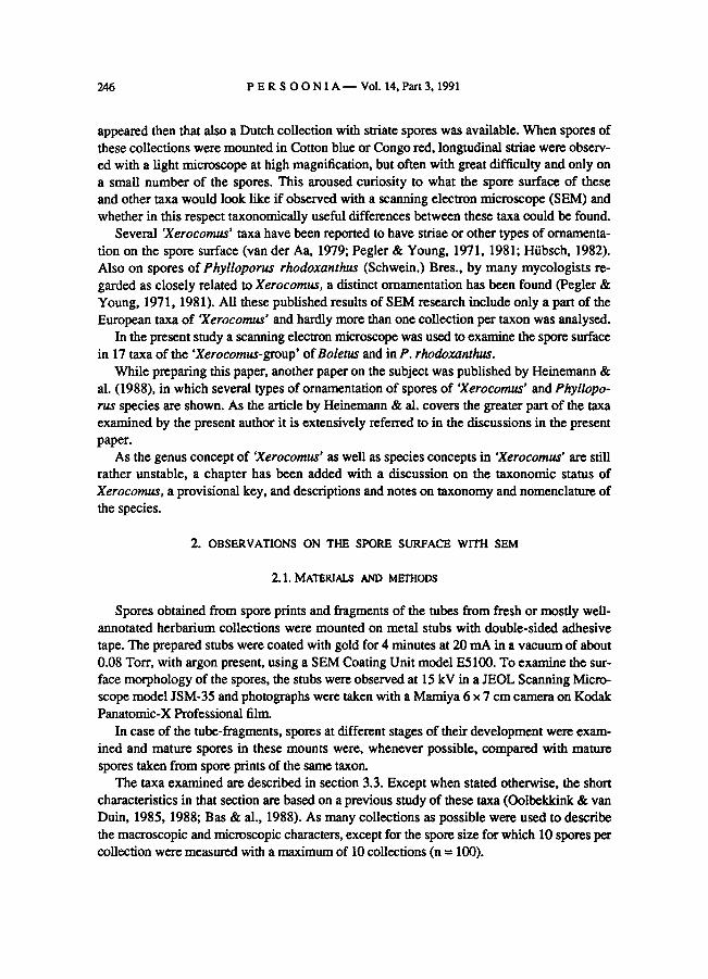
remnants (Fig. 9) to entirely smooth (Fig. 10). A very few spores in one collection show a
'blistered' surface (Fig. 11, arrow). In spore prints the spores are smooth or with floccose
remnants. Young spores appear to be smooth.
Boletus subtomentosus var. luteolus with pileipellis-type I as well as with type II (see
section 3.3) has smooth spores with floccose remnants to entirely smooth spores (Fig. 12).
In B. ferrugineus floccose (Fig. 13) and smooth with floccose remnants (Fig. 14) to
smooth spores (Fig. 15) are found.
In the taxa B. chrysenteron (Figs. 16, 17), B. rubellus (Fig. 18), B. fraternus (Figs. 19,
20), B. pruinatus (Figs. 21, 22), B. fragilipes (Fig. 23), B. porosporus (Figs. 24, 25) and
B. truncatus (Fig. 27) most spores have faint to distinct longitudinal striae, usually anas-
tomosing and often fading at the apex. Frequently the spores ofB. porosporus have only a
faint striation and on a very few spores in one collection a 'blistered' surface has been
observed (Fig. 26). A small number ofthe mature spores of the taxa is smooth. Very young
spores are all smooth, but they already develop ridges when still attached to the sterigmata.There is no difference in spore ornamentationamong basidiocarps with the two pileipellis-
types (see section 3.3) ofB. chrysenteron and B. rubellus, and among basidiocarps with the
three pileipellis-types ofB. fragilipes.
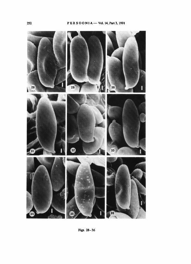
In B. armeniacus floccose (Fig. 28) to smooth spores (Fig. 29) have been found. Some-
times spores in the sample examinedwere covered with strange structures, perhaps offoreign
origin (Fig. 28). Boletus bubalinus, sp. nov., has smooth spores with floccose remnants (Fig.
30) to entirely smooth spores (Fig. 31).
The spores of B. moravicus (Fig. 32) and B. pulverulentus (Fig. 33) are smooth. In B.
ich-nusanus, comb, nov., the spore surface appears to be smooth, but there were some
difficultiesin establishing this, because in the sample examined it was covered with a strange
substance considered offoreign origin (Fig. 34).

PERSOONIA— Vol. 14, Part 3, 1991248
The spores of Phylloporus rhodoxanthus show a floccose (Fig. 35) or a pitted-and-floc-
cose ornamentation (Fig. 36). The spores in the sample examined in Fig. 35 show also some
strange structures probably of foreign origin. See also footnote in the discussion (2.3).
2.3. DISSCUSION
The sporal layers in ‘Xerocomus’ taxa were not studied by the present author, but for a
better understanding ofthe probable origin of the ornamentationof spores of 'Xerocomus' as
describedin this paper, it is necessary to take the wall layers of the spore into consideration.
The terminology of these layers may cause confusion, as several authors use a different ter-
minology. Since Perreau-Bertrand (1961, 1964, 1965, 1967; Perreau & Heim, 1969) exten-
sively examined the structure of the spore wallof Boletales, her terminology is applied here.
Perreau-Bertrand (1967:676) demonstratedthat five wall layers are present in the spores
of Boletales. As the spores mature, the surface ornamentation can be formed (i) by remnants
of the distintegrating two outer wall layers, viz. the ectosporium and perisporium, or (ii) by
outgrowths ofthe exosporium. It is difficult to decide on the most probable of these two ori-
gins for the various taxa in this study, because it is hardly possible to make a distinctionbe-
tween an ornamentationofperisporial origin (remnants) or an ornamentationofexosporial
origin on SEM photographs, as was also noticed by Perreau & Heim (1969: 330).
The spores of Boletus parasiticus show an ornamentation that varies from reticulately
venose to (elongately) pitted (Figs. 3-6). A somewhat comparable ornamentationhas been
found inPhylloporus rhodoxanthus (Fig. 36), except that the latter is also floccose whereas
the edges of the pits of spores of B. parasiticus are smooth. Perreau-Bertrand (1967: 676)
supposed that the spores ofthese taxa were smooth, but she studied the spore surface with a
light microscope only and therefore was unableto see any ofthese very fine structures. Pegler
& Young (1981: 117) foundan ornamentationin B. parasiticus with SEM that they described
as 'minute rugulose'. After comparing their fig. 60 with Fig. 3 in this paper, it is concluded
that the type of ornamentation is similar, but the terminology applied is different. Exactly the
same can be said about fig. 27 in Heinemann& al. (1988: 531) and Fig. 3 in this paper. Hei-
nemann & al., however, placed B. parasiticus in a group with vroughened to smooth spores,
—3-6.
— 1-2. B. parasiticus. B. subtomentosus
Figs. 10-18. Spores of — 10-11. — 12.
B. ferrugineus. B. chrysenteron.
Figs. 1-9. Spores of
— 13-15. (Fig. 16, x 6600;Figs. 10-15, 17,
and 18, x 6000. Bar = 1 μm).
B. badius.
—19-20. B. fraternus.
(Figs. 1, 2, 4, and 6-9, x 6000; Fig. 3, x 5400; Fig. 5, x 4800. Bar = 1 μm).
—23.
Boletus.
Boletus.
B. subtomentosus.
(Figs. 24 and 25, x 6600; Figs. 20-22,26, and 27, x 6000;
Fig. 23, x 5400; Fig. 19, x 4000. Bar = 1 μm).
B. subtomentosus var. luteolus.
— 7-9.
—16-17.
Boletus and Phylloporus. B. armeniacus. — 30-31. B. bubalinus.
— 33.
—21-22.
B. pulverulentus.
B. fragilipes.
— 35-36.
—18. B. rubellus
Boletus.Figs. 19-27. Spores of
B. moravicus.
— 27.B. porosporus.— 24-26.
B. pruinatus.
—32. (Figs. 28-36,
x 6000. Bar = 1 μm).
P. rhodoxanthus
B. truncatus
—34.
Figs. 28-36. Spores of
B. ichnusanus.
— 28-29.

OOLBEKKINK: Spore ornamentation in Xerocomus 249
Figs. 1-9
250 PERSOONIA— Vol. 14, Part 3, 1991
Figs. 10-18

OOLBEKKINK: Spore ornamentation in Xerocomus 251
Figs. 19-27
252 PERSOONIA— Vol. 14, Part 3, 1991
Figs. 28-36

OOLBEKKINK: Spore ornamentation in Xerocomus 253
but stated also that it can have certain spores showing a very faintornamentationresembling
the striaeofweakly ornamentedBoletellus spores. The reason why these authors did not also
find more pronouncedly pitted spores (see Figs. 4-6) probably lies in the fact that in each
case only one or two collections were examined.
The surface structure of spores of P. rhodoxanthus (Figs. 35, 36) remains a puzzle. Not
only theornamentationobserved in this study (floccose or pitted-and-floccose) is in disagree-
ment with the one found by Pegler & Young, also theirobservations of 1971 and 1981, of the
same collection, disagree with each other. They reported in 1971 (: 158) a spore surface with
numerous, rod-like structures comparable to those observed in Oudemansiella, but in 1981
(: 119) they reported a fine rugulose ornamentation. Heinemann & al. (1988: 517) found
bacillate('bacillées') spores in P. rhodoxanthus. Some confusion is caused by the different
terminology. The term bacillate is considered deceptive by the present author, because the
spore surface is covered by numerous very fine structures, of which only a part can be
described as rod-like. Consequently Heinemann& al. display a rather broad concept of ba-
cillate. One part ofthe structures covered by their term can be described as rod-like (as Pegler
& Young do) or as fibrinöse(this paper), but the otherpart as floccose (this paper). Therefore
the spore surface of P. rhodoxanthus as described by Heinemann & al. agrees well with
the data of Pegler & Young of 1971, but also with the spore surface depicted in Fig. 35 in
this paper. The pitted-and-floccose spore surface (Fig. 36) is only reported in this paper.1
It is supposed that on the spores depicted in the Figs. 3 and 35 the ecto- and perisporium
are beginning to disintegrate. The ornamentation showed in the Figs. 4-6 and 36 may be
formed (i) by remnants of a much further disintegrated ecto- and perisporium, (ii) by the
exosporium which becomes visible as these two outer layers disappear, or (iii) by both (i) and
(ii). As all the spores in the Figs. 3-6,35, and 36 are probably mature, it seems possible that
the degree of ecto- and perisporium disintegration is not always equal in different collections.
An ornamentationalmost certainly formed by remnants of the two outer layers, is found
in the taxa of the B. subtomentosus-complex and B. bubalinus, sp. nov. The gradual break-
down ofthe ecto- and perisporium, which eventually disappears to leave a smooth exosporial
surface, can be observed in the Figs. 7 to 10. All degrees of disintegration may be found in
mature spores, however, smooth spores and spores with floccose remnants are found in both
spore prints and tube-fragments, but fibrillose to floccose spores only in tube-fragments.
This strongly supports the supposition that the ornamentation in the B. subtomentosus-
complex is caused by disintegration of the outer layers.
The ornamentations of the spore surface Heinemann & al. (1988: 524, 532) found in
B. subtomentosus (bacillate) and B. ferrugineus (bacillate or roughened to smooth), are simi-
lar to stages foundin this study, bearing in mind that their 'bacillate' is comparable to 'fibril-
lose to floccose', here. Besides smooth spores, Perreau-Bertrand (1965: 4247) and Pegler &
Young (1971: 156) also observed striate spores in some collections of B. subtomentosus.
This is contrary to the observations of Heinemann& al. (1988) and the present author, that
indicate that striate spores occur only in taxa of the B. chrysenteron-complex.
1 Photographs, made at a later lime and therefore not reproduced here, of one of the collections also
examined earlier (coll. Schreurs 464) show spores, some still attached to the sterigmata, with a fibrillose sur-
face similar to the rod-like structures found by Pegler& Young (1971:158).

PERSOONIA— Vol. 14, Part 3, 1991254
In all the taxa of the B. chrysenteron-complex studied here, a majority of the spores show
fine, usually anastomosing, longitudinal striae, distinguishing themfrom the other taxa of the
Xerocomus- group studied. Up to the start of the present study only ‘B. fragilipes’ (Pouzar,
1981: 8; Hubsch, 1982: 62) and B. truncatus (Pegler & Young, 1981: 116) were known to
have spores with fine longitudinal striae, and B. chrysenteron was suspected to have them
(van der Aa, 1979: 207). Recently Heinemann & al. (1988: 522) have observed this type of
ornamentationin B. chrysenteron, B. porosporus and B. pruinatus. They also notice that the
ornamentationof spores of B. porosporus often is faint. However, they describe B. versi-
color as having roughened to smooth spores. This is in disagreement with the observation of
the present authorand could be caused by a differentconceptofB. versicolor (a synonym of
B. rubellus). That striate spores are typical for the B. chrysenteron-complex, is made more
plausible by the fact that the spores of the North American Boletus zelleri (Murr.) Murr., a
taxon with a 'deceptive external similarity' to B. chrysenteron, have a similar fine striation
(Snell & al., 1960: 575; Perreau & Heim, 1969: 333; Pegler & Young, 1981: 116). Because
of this striation, some mycologists consider this taxon to belong to Boletellus (Boletellus zel-
leri (Murr.) Sing., Snell & Dick).
Boletusfragilipes is a taxon reinstated by Pouzar to accommodate collections with striate
spores and that are close to, but somewhat aberrant from B. chrysenteron (see section 3.4).
As the taxa examined in the B. chrysenteron-complex all show striate spores and as there
seem to be no other reliable characters to distinguish B. fragilipes from B. chrysenteron,
B. fragilipes is consideredhere a nomen dubium.
The ornamentationof the spores in the B. chrysenteron-complex is probably of exosporial
origin, as was pointed out for B. zelleri (Perreau-Bertrand, 1967: 680; Pegler&Young, 1971:
162; 1981: 116). The reason why a number of spores are smoothor only faintly striate, can
be explained by an overlying ecto- and perisporium obscuring the exosporial ornamentation
(e. g. Figs. 20,25). Smooth and faintly to distinctly striate spores can all be found in a spore
sample of one specimen of the various taxa, except B. porosporus, which frequently has only
smooth and faintly striate spores.
The spores of B. armeniacus (Figs. 28,29) have a floccose to smooth surface comparable
to and probably of the same origin as that of spores in the B. subtomentosus-complex and
B. bubalinus. Heinemann& al. (1988: 526) consider the spore surface to be roughened to
smooth, probably similar to what is describedin this paper. Therefore B. armeniacus seems
not to belong to the B. chrysenteron-complex, as initially thought (Oolbekkink & van Duin,
1988: 1). It is, however, not easy to place this taxon elsewhere. Although the colour of the
cap, the pink zone under the pileipellis and the pileipellis-structure suggest a relationship with
B. chrysenteron, the surface structure of the spores, the ridges on the stem and the hardly
blueing flesh suggest a relationship with B. subtomentosus.
Smooth spores without any trace of remnants have been found by the present author in
B. badius, B. pulverulentus, B. moravicus, and B. ichnusanus. This has been confirmed for
B. badius by both Perreau-Bertrand (1967: 676) and Pegler & Young (1981: 117), and for
B. pulverulentus only by the former. Pegler & Young (1971: 156) are the only authors report-
ing occasional faint longitudinal striae on the spores ofB. pulverulentus. Heinemann & al.
(1988: 526, 528) place B. badius and B. moravicus in a group with roughened to smooth

OOLBEKKINK: Spore ornamentation in Xerocomus 255
spores, but also state that B. badius shows very faint veins on a small number ofspores when
enlarged to 20,000 times, and that the spore wall of B. moravicus is longitudinally wrinkled
(I.e.: 530). As yet no data on the spore surface ofB. ichnusanus were available in the litera-
ture.
On a very few spores in one collectionof each B. badius, B. subtomentosus,and B. poro-
sporus, a surface with rounded swellings ('blistered') has been observed. As these species
are distinctly differentand the number of spores involved is very small, it is considered not
characteristic for any of these taxa and possibly externally caused.
This study and the one by Heinemann& al. (1988) show that more ‘Xerocomus’ species
have ornamented spores than could be expected from the available data in earlier literature.
Summarizing the above, the most striking results ofthis study are that the taxa of the B. chry-
senteron-complex can be distinguished from the B. subtomentosus-complex and the other
taxa examined, by their striate spores, and that B. parasiticus can be distinguished by its con-
spicuously pitted spore surface. Ifthe presence of remnants ofan outer layer on a large num-
ber ofspores ofB. bubalinus, B. armeniacus, and the taxa in the B. subtomentosus-complex
is a constant feature, then they can be distinguished from taxa with only entirely smooth
spores. The SEM study of the spores has not altered the author's concept of the taxa dis-
cussed in section 3.3, except for '‘B.fragilipes’,which is now considered a nomen dubium,
and B. armeniacus, which seems better placed outside the B. chrysenteron-complex.
3. TAXONOMY AND NOMENCLATURE
3.1. THETAXONOMIC STATUS OF XEROCOMUS QUÉL.
Although part of the specimens in several taxa ofXerocomus can be distinguished from
part of the specimens in several taxa of Boletus by the characters given by Quélet (1888: 417),
it is impossible to totally delimitall the specimens in all of the taxa of the former from those of
the latter, because noneof the characters used is exclusively reserved for eitherof them.
Singer and other mycologists consider the structure of the hymenophoral trama the main
criterionfor separating Xerocomus from Boletus. This has produced, however, a rather arti-
ficial systematic arrangement; e.g. Singer emended Boletus sect. Subpruinosi (see Singer,1986: 777) to include species with xerocomoid features of the fruit-body but with a boletoid
trama,within Boletus. In such a systematic arrangement it is impossible to place closely re-
sembling taxa, such as B. chrysenteron and B. pruinatus, near to each other.
The present author does not regard the occurrence of a hymenophoral trama ofthe Phyllo-
porus-type or the Boletus-type as such a fundamentalcharacter, because the distinction be-
tween the two is often not sharp and it is often difficult to designate eitherof them to the vari-
ous taxa of Xerocomus. This view is supported by observations of otherauthors and by the
author's own experience.In Singer (1986: 58) is stated that towards the end of the sporulation of Boletus fruit-
bodies, the Boletus-type changes into a tramai type similar to the Phylloporus-type. The re-
verse is also possible, as is suggested to occur in some species of Pulveroboletus Murr.,

256 PERSOONIA— Vol. 14, Part 3, 1991
where the Phylloporus-type found in youngspecimens changes through further development
into the Boletus-type (l.c: 771). A tramal structure intermediatebetween phylloporoid and
boletoid, has been found in B. badiusand related taxa (I.e.: 764).
Corner (1972: 19) noticed in his study of Malaysian species that in a very young stage all
boletes seem to be phylloporoid. Moreover, he observed (1.e.: 9, 18) several species with
intermediatetrama where a sharp distinction between phylloporoid and boletoid trama could
not be made, and he suggested that there may be several, not merely two, variations in tramai
structure. In a previous study (Oolbekkink & van Duin, 1985) on the morphology oftaxa of
the Xerocomus-group, the Phylloporus-type and the Boletus-type, but also intermediate struc-
tures were observed. For instance: in B. badius, the Phylloporus-type and the Boletus-type,
and an intermediatetramalstructure were found; in B. porosporus and B. pruinatus, the Phyl-
loporus-type. and an intermediate structure were found, and in B. fraternus the Phylloporus-
type and the Boletus-type. As these tramai types were all found in mature fruit-bodies, it
seems that there is no obvious relation between the hymenophoral trama-type and the stage of
development of the fruit-body.
Earlier also Watling (1968: 304) pointed out irregularities of the hymenophoral trama in
several taxa. Smith & Thiers (1971: 11) considered the differences between the tramal types
not sufficiently constant to be of aid in distinguishing genera or species in their study of
Michigan boletes. The present author agrees with Smith & Thiers, because the hymenophoral
trama displays such a variation in structure during the short existence of the fruit-body, that
no great value can be attached to it as distinguishing character.
Another difference between the two genera has been mentioned, namely the fact that many
taxa ofXerocomus are facultatively ectomycorrhizal, whereas Boletus taxa are constantly
obligatorily ectomycorrhizal (Pegler & Young, 1981: 116, 123; Singer, 1986: 762). Besides
the fact that it is very difficult in most cases to establish whether a taxon is obligatorily or
facultatively mycorrhizal, the present author wonders ifthis differenceis of any value for the
taxonomy. According to Harley & Smith (1983: 114) the variability of many proved mycor-
rhiza-forming species in culture suggests that the indication 'facultatively mycorrhizal' should
be treated with caution.
The discussion above has led to the conclusion that Xerocomus should not be separated
from Boletus at genus level.
3.2. PROVISIONAL KEY TO THE SPECIES AND VARIETIES OF
‘THE XEROCOMUS-GROUP’ OF BOLETUS
la. Basidiocarp growing on Scleroderma;cap yellow-brown with olivaceous tinge; spores 11.6-18.0 pm long
(mean value 15.7 ±1.3 pm); pileipellis a strongly intricate trichoderm of branched narrow hyphae, (3.5-)
4.5-9.0 pm broad, with apical cells often curving inwards, often slightly inflated at septa and there up to
11.0 pm broad 2. B. parasiticus
b. Basidiocarp onthe ground 2
2a. Basidiocarps usually growing in tufts; stem covered over two-third from apex downwards by a brownish
red to brown net; base usually prolonged into soil by root-like strand of mycelium; spores long and broad,
12—18(—23)x5-7(-7.5) pm; probably restricted to the Mediterranean 16. B. ichnusanus

OOLBEKKINK: Spore ornamentation in Xerocomus 257
b. Basidiocarp never with a prominent net on the greaterpart of the stem; base neverprolonged; spores
smaller 3
3a. Many spores with a truncate apex 4
b. Spores not truncate 5
4a. Cap dark yellowish brown to brown or olive-brown, with surface usually cracking and showing whitish
or yellowish flesh underneath;no red on stem, ormore rarely ared-brown or deepred zoneat apex
10. B. porosporus
b. Cap dark olive to olive-brown at first, but very soonred to reddish throughout, with surface cracking and
then showing pink to red flesh; stem soon pink to red from base upwards 11. B. truncatus
5a. Spores short and broad, Q = 1.7-2.4; cap ochraceous orpale brown; pores pale cream-coloured at first,
later yellow to yellowish-ochraceous; flesh never changing to blue 14. B. moravicus
b. Spore size larger, minimum Q value never below 2.0, maximum Q value greater than 2.4 6
6a. Cap and stem bruising dark blue to black on handling;flesh strongly turning blue to dark blue through-
out 15. B. pulverulentus
b. Cap and stem not or not so prominent bruising blue; flesh not or weakly changing,or turning (greenish)
blue irregularly and usually more slowly 7
7a. Cap viscid when wet; pileipellisan intricate ixotrichoderm of branched, narrow hyphae, (3.5-)5.5-7.0
(-9.0) pm broad 1. B. badius
b. Cap never viscid; pileipellisan intricate, irregularor epithelioidtrichoderm 8
8a. Pileipellis an intricate to strongly intricate trichoderm of branched narrow hyphae, about 5.5-13.0 pm
broad, with age at surface sometimes tending to change into a cutis; terminal cells not or slightly in-
flated 9
b. Pileipellisa trichoderm of wider hyphae, about 7.0-18.0 pm broad; terminal and/or subterminal cells
often distinctly inflated and broader than 14.5 pm; terminal cells can be cystidioid or globose 11
9a. Cap buff, pale brown, yellow-brownorolive-brown; stem buff or yellow-brown,on the upper half with
reddish brown or brownish tinges or yellowish brown, brown or reddish brown, sometimes slightly
anastomosing ridges; NH4OH oncap producing a somewhat orange-brownspot with dark brown to dark
purple ring 3. B. subtomentosus
b. Cap yellow to yellow-ochre or (dark) brown to (dark) reddish brown; stem yellow or brownish....
10
10a. Cap yellow to yellow-ochre; stem yellow, sometimes with yellowridgesat apex
4. B. subtomentosus var. luteolus
b. Cap (dark) brown to (dark) reddish brown; stem on upper part merely (dark) reddish brown to brown dot-
ted, or with concolorous dots and ridges and these hardly anastomosing or forminga coarse net; lower
part pale yellow to pale brown; NH4OH on cap reddish brown to dark brown with a dark ring, or
fleeting dark (greenish) blue, leaving a whitish spot (colour disappearing) with a dark reddish brown to
blackring 5. B. ferrugineus
11a. Pileipellisan intricate to irregular trichoderm with remarkably large terminal cells, 25.0-63.0 (-81.0)
pm long and 8.0-23.5(-30.5)pm broad; cap yellow to yellow-ochre; (see 10 also)
4. B. subtomentosus var. luteolus
b. Pileipellis an irregularor epithelioid trichoderm with terminal cells usually not longer than 50.0 pm, but
when longer, terminal cells usually not broader than 20.0 pm; cap several tints of brown, pink or red,
never yellow 12
12a. Pileipellis an epithelioid trichoderm 13
b. Pileipellisan irregular trichoderm 14
13a. Pileipellisanepithelioid trichoderm of many inflated terminal and subterminal cells, respectively (9.0-)
14.5-25.0 and (9.0-)13.5-35.0(-45.0)pm broad; hyphae narrowing downwards, (7.0-)9.0-17.0
(-20.5)pm broad; no red line under pileipellis; cap red, with surface often minutely cracking and then
showing (deep) yellow flesh; NH4OH on cap producing an orange-yellow spot (red colour disappearing)8. B. fraternus

PERSOONIA— Vol. 14, Part 3, 1991258
b. Pileipellis an epithelioid trichoderm with many chains of almost globose cells on hardly narrower
hyphae; terminal cells (5.5—)10.0—25.0(—31.0)pm; subterminal cells (7.0—>11.5—23.5(—31.0) pm
broad; cells underneath 10.0-18.0(-22.0)pm broad; red line under pileipellis 17
14a. Pileipellis a thin, 80-110(-145)pm thick, somewhat irregular trichoderm with many rather narrow ter-
minal cells, (5.5-)8.0-14.0(-15.5)pm broad, and often slightly inflated subterminal cells, (7.0-) 10.0-
16.0(-18.0) pm broad; cap dark brown with a red to purplish red colour shining through, covered with a
hoary bloom easily destroyedby handlingor by rain 9. B. pruinatus
b. Pileipellis thicker, 135-280(-420)pm; terminal cells often broader than 15.5 pm; subterminal cells of-
ten broader than 16.0 pm 15
15a. Pileipellis an irregular trichoderm often with branching elements in upper part and usually rather narrow
terminal cells, (5.5-)7.0-18.0(-27.0)pm broad; subterminal cells (5.5-)7.0-18.0(-22.0)pm broad;
cap buff, (dark) yellow-brown or pale brown, usually with pink flush appearing locally, with age
becoming very smooth, often with surface cracking and showing yellowish flesh underneath; pores
bright yellow when young, with age becoming dark (greenish) yellow or sometimes brownish and then
somewhat orange-brown 13. B. bubalinus
b. Pileipellis an irregular trichoderm usually with slightly inflated,but sometimes cystidioid terminal cells,
(5.5-)8.0-18.0(-28.5)pm broad; subterminal cells (5.5-)9.0-23.5(-31.0) pm broad; hyphae usuallynarrowing downwards; with few to many incrustations 16
16a. Cap pink when young, becoming pale ochraceous brown or a mixture of reddish pink and brownish-
ochraceous to ochraceous buff; stem bright to golden yellow, paler yellow at base, often with ridges
especially near apex and these sometimes reddish orange 12. B. armeniacusb. Cap drab, pale brown, dark yellowish brown, dull to dark red, or variegated dark red and dark yellowish
brown 17
17a. Cap drab, pale brown or dark yellowish brown, often with red flush particularly when mature, usually
with irregularly cracking surface and showing reddish, sometimes whitish or yellowish flesh underneath
6. B. chrysenteron
b. Cap dull red, with age dark red, often variegated dark red and dark yellowishbrown, sometimes with sur-
face cracking particularly at margin and then showing pinkish, sometimes whitish flesh (considered ared-
coloured variant ofB. chrysenteron) 7. ‘B. rubellus’
3.3. DESCRIPTIONS OF AND NOTES ON TAXA OF THE XEROCOMUS-GROUP
1. Boletus badius (Fr.) Fr.: Fr.
Boletus badius (Fr.) Fr.: Fr., Elench. fung.: 126. 1828.
Cap orange-brown, (dark) reddish brown or dark brown, tomentose when young, soon
becoming smooth, viscid when wet. Pores pale yellow to pale greenish yellow, bruising blue.
Stem at apex concolorous with pores, elsewhere concolorous with cap but usually paler.Flesh (yellowish) white to pinkish white in cap, greyish white or brownish white in stem,
turning blue particularly over the tubes. NH4OH on cap producing an orange-brown or red-
dish brown spot with spreading and fading blackish brown or blackish green ring, on flesh
negative, seldom yellowish.
Spores (n = 100) 13.5± 1.1 x 4.7±0.4 (10.7-15.8 x 4.3-6.3) pm, Q = 2.9±0.2 (2.4-
3.5), smooth with SEM. Pileipellis an intricate ixotrichoderm of branched, narrow hyphae,(3.5-)5.5-7.0(-9.0) pm broad, difficult to observe in older specimens probably because of
gelatinizing hyphal walls; pigment incrustations present.
Collections examine d.—NETHERLANDS: Lage Vuursche, 29 Sept. 1983, Oolbekkink & van
Duin 113; Hulshorst, 11 Oct. 1983, van Duin & Oolbekkink 133 and 134; Gooisch Natuurreservaat, 18 Oct.
1984,Oolbekkink & van Duin 171 and 173.

OOLBEKKINK: Spore ornamentation in Xerocomus 259
The taxon B. badius was first described by Fries as B. castaneus ß Badius in the Obser-
vationes mycologicae (1818: 247) and again in 1821 (: 392) as B. castaneus ß B. badius in the
Systema mycoiogicum. Gams (1984: 227) pointed out that this epithet is of uncertain infra-
specific rank. Fortunately Fries described the taxon once more in the Elenchus fungorum
(1828: 126),but now as a species with the name B. badius and thus this name is sanctioned.
See also the notes underB. moravicus.
2. Boletus parasiticus Bull.: Fr.
Boletus parasiticus Bull.: Fr., Syst. mycol. 1: 389. 1821.
Cap pale yellow-brown with olivaceous tinge to dirty yellowish brown, with surface often
cracking particularly at centre, dry. Pores yellow, brownish yellow becoming reddish brown
(rust-coloured), not blueing. Stem concolorous with cap or paler, often curved. Flesh yellow,
unchanging. NH4OHreaction on cap or flesh not recorded.
Spores (n = 70) 15.7±1.3 x 4.9±0.4 (11.6-18.0 x 4.2-5.4) pm, Q = 3.210.3 (2.6-
4.0), pitted to reticulately venose with SEM. Pileipellis a strongly intricate trichoderm of
branched narrow hyphae, (3.5-)4.5-9.0 pm broad, apical cells often curving inwards, often
slightly inflated at septa, then up to 11.0 pm broad; incrustations absent.
Fruit-bodies growing on Scleroderma citrinum Pers.
Collections examined. —NETHERLANDS: Het Gooi, 29 July 1956,Smiv, Breda, 12 Oct.
1958, Jarisen-, Swalmen, Hillenraad, 7 Oct. 1962, Bas 2844\ Breda, Liesbos, 14 Sept. 1968, Goos\ Arnhem,
Vijverberg, 4 Aug. 1975, van derLaan.
3. Boletus subtomentosus L.: Fr.
Boletus subtomentosus L.: Fr., Syst. mycol. 1: 389. 1821.
Cap buff, pale brown, yellow-brown or olive-brown with dark yellowish brown, orange-
brown or reddish brown patches where bruised, tomentose, sometimes with surface cracking
particularly at margin and then showing (very) pale yellow, never pink or red, flesh. Pores
bright yellow, darker with age and sometimes more greenish yellow, seldom slightly blueingon bruising. Stem buff or yellow-brown, on the upper half with brown or reddish brown
tinges or yellowish brown, brown or reddish brown, sometimes slightly anastomosing ridges.Flesh yellowish white to pale yellow, with (reddish) brown line underpileipellis, often with
pinkish tinge in cap, sometimes turning brownish or slightly bluish green locally. NH4OH on
cap producing a ± orange-brown spot with a dark brown to dark purple ring, on flesh negative.
Spores (n = 100) 12.211.2 x 4.410.4 (10.0-15.3 x 3.6-5.4) pm, Q = 2.810.2 (2.2-
3.5), fibrilloseto floccose or smooth with or without floccose remnants with SEM. Pileipellisan intricate to strongly intricate trichoderm of branched narrow hyphae, about 5.5-13.0 pm
broad, with age at its surface sometimes tending to change into a cutis; terminal cells not or
slightly inflated, (5.5—)8.0—14.5 (—18.0) pm broad, rarely curving inwards; without infla-
tions at septa; sometimeswith a few incrustations.
Collections examined.—NETHERLANDS:Vogelenzang,A.W.-dunes,9Oct.1983,Bos 737; Bergen,
15 Oct. 1984 ,Ypelaar YP8461B;Bergen, Zwarte Weg,21 Oct. 1984,Ypelaar 174; Castricum,Noordhollands
Duinreservaat, 26 Sept. 1987,Oolbekkink 203.—
LUXEMBURG: Dillingen, 29 Oct 1987, Oolbekkink 209.
See notes under B. subtomentosus var. luteolus, B. ferrugineus, and B. chrysenteron.

PERSOONIA— Vol. 14, Part 3, 1991260
4. Boletus subtomentosus var. luteolus Velen.
Boletus subtomentosus var. luteolus Velen., Ceskfi Houby: 717. 1922.
Xerocomus subtomentosus var. xanthus E. J. Gilb., Les Bolets: 142. 1931; ?Xerocomus flavus Sing. &
Kuthan in Ceskâ Mykol. 30: 153. 1976.
Cap yellow to yellow-ochre, tomentose. Pores bright yellow. Stem at apex concolorous
with pores, elsewhere pale yellow, sometimeswith yellow ridges at apex. Flesh yellowishwhite to pale yellow, often with pinkish tinge in cap. NH4OH reaction on cap or flesh not
recorded.
Two pileipellis-types could be distinguished in the collections examined: (I) Pileipellis as
in B. subtomentosus. Spores (n = 100) 12.311.1 x 4.910.4 (9.2-14.9 x 4.0-5.7) pm, Q= 2.510.2 (2.0-3.2), smooth with or without floccose remnants with SEM. (II) Pileipellis
an intricate to irregular trichoderm with remarkably large terminalcells, (16.0-)25.0-63.0
(-81.0) pm long and (6.5-)8.0-23.5(-30.5) pm broad; subterminal cells 7.0-18.0(-29.5)
pm broad; incrustations absent. Spores (n = 20) 12.611.0x4.910.5(10.7-14.9 x 3.9-5.5)
pm, Q = 2.610.4 (2.2-3.5), smooth with or without floccose remnants with SEM.
Collections examine d.—NETHERLANDS: roadside (Koningsweg) between Utrecht and Bunnik,
31 Aug. 1969,Arnolds 350; Hardenberg, 20 Sept. 1969,Hengstmengel 169 (both type I); Breda, Liesbos, 1
Sept. 1959, Bas 1742 ; Breda, Liesbos, 8 Aug. 1975, Jansen (both type II).
Velenovsky has given a more extended description of his taxon in Latin in 1939 (: 159).
Singer & Kuthan (1976: 154) consider X. flavus to be very close to X. spadicus and X.
lanatus and possibly very similar to yellowish forms ofX. subtomentosus. They describe a
striking chemical character for X. flavus, viz. NH4OH on surfaces produces an immediate,
very slowly fading blue-green reaction. The NH4OH reaction is not recorded for the speci-
mens described above. The structure of the pileipellis of X. flavus as described by Singer
& Kuthan does not fit either of the pileipellis-types described here. Therefore it is doubtful
whether X.flavus is identical with B. subtomentosus var. luteolus.
The present author has found two pileipellis-types in otherwise similar collections. This
was concluded after comparing the annotations of the collections, as no fresh material was
available. More studies are needed to solve this problem. There are three possibilities: (i) the
annotations are inaccurate and two taxa are involved; (ii) variationin the pileipellis-structure is
caused by genetic variationwithin one taxon; (iii) variation in the pileipellis-structure is caused
by abiotic factors, e.g. the weather conditions during the development of the fruit-bodies.
5. Boletus ferrugineus Schaeff.
Boletus ferrugineusSchaeff., Fung. Bavariae 4: 85. 1774 (as B. decimus nonus in vol. 2: Tab. 126. 1763).
Boletus spadiceus Fr., Epicr.: 415. 1838; Boletus lanatus Rostk. in Sturm (ed.), Deutschl. Fl. (Pilze),Abth. Ill, 5: 77. 1844.
Cap (dark) reddish brown to (dark) brown, (dark) reddish brown patches where bruised,
with (pale) yellow, soon collapsing tomentum. Pores bright yellow to deep golden yellow,sometimes with greenish tinge, seldom slightly blueing on bruising. Stem on upperpart mere-
ly (dark) reddish brown to brown dotted,or with concolorous dots and ridges and these hard-
ly anastomosing or forming a coarse net; lowerpart pale yellow to pale brown. Flesh whitish

OOLBEKKINK: Spore ornamentation in Xerocomus 261
to pale yellowish, often more yellowish in stem, with dark reddish brown to brown lineunder
pileipellis, often with pinkish to pale brownish pink tinges in cap and stem, seldom turning
pale bluish locally. NH4OH on cap reddish brown to dark brown with a dark ring, or fleeting
dark (greenish) blue, leaving a whitish spot (colour disappearing) with a dark reddish brown
to black ring. NH4OH on flesh not recorded.
Spores (n = 90) 12.8±1.2 x 4.510.5 (10.8-15.8 x 3.6-6.1) pm, Q = 2.810.3 (2.0-
3.5), floccose or smooth with or without floccose remnants with SEM. Pileipellis as in
B. subtomentosus.
Collections examine d.—NETHERLANDS: Bunnik, 23 Aug. 1967,Arnolds; Amersfoort.Pine-
tum Birkhoven, 11 Oct. 1980, Wisman', Amersfoort, Birkhoven, 7 July 1984, Wisman. — AUSTRIA: Sattnitz-
ridge, south of Klagenfurth, nearGöltschach, 3 Oct 1978, Bas 7394.
The description ofB. spadiceus by Fries (1838: 415) was based on the species of Boletus
described by Schaeffer in 1763 (: PI. 126), because Fries referred to this publication in his
description and because he mentioned this taxon as ‘B. spadiceus Schaeff.' in the index of
the Hymenomycetes europaei (Fries, 1874: 746). Fries neglected the fact that Schaefferhad
already given his nineteenth species of Boletus the name B. ferrugineus in 1774 (: 85). There-
fore B. ferrugineus Schaeff. is the correct name for this species.
When the original descriptions are compared, the differences between the taxa of the
B. subtomentosus-complex can be summarized as follows: B. subtomentosus has an oliva-
ceous cap and somewhat ridged, furrowed or grooved stem; B. ferrugineus has a (dark) red-
dish brown cap and no ridges on the stem; B. lanatus has a brown cap and reddish brown
wrinkled, almost nettedstem. Unfortunately these differences are in reality not very useful, as
the taxa show an overlap of characters, which was shown in an earlier study of the concern-
ing taxa (Oolbekkink & van Duin, 1985, 1988). During that study specimens were found
with yellow-ochre, yellow-brown, olivaceous or reddish brown to (dark) brown cap, without
or with yellow, yellowish brown, reddish brown or brown ridges sometimes (slightly) anas-
tomosing or forming a coarse net.
On account of the fortuitousoccurrence of, sometimes anastomosing, ridges on the stem
of any of the specimens with the above described colours of the cap, the emphasis in
grouping them is laid here on the colour ofthe cap and not on the occurrence of ridges. This
implicates that B. ferrugineus can have ridges, although they were not described bySchaeffer. Schaeffer and also Rostkovius considered the presence or absence of ridges or
anastomosing ridges on the stem a good reason to distinguish several 'different' taxa, many
of which are quite similar to B. subtomentosus or B. ferrugineus.Fries (1874: 503), who also attached much value to the presence of ridges on the stem,
mentionedRostkovius' B. lanatus under B. subtomentosus on account ofits ridges, but also
stated that B. lanatus closely resembles B. spadiceus. In this paper B. lanatus is considered a
synonym of B. ferrugineus. The differences between these two reported by Watling (1970:
26, 40), viz. the pileipellis-structure and the NH4OH reaction on the cap, couldnot be con-
firmed by Oolbekkink & van Duin (1985). The pileipellis-structure appeared to be similar in
all the examined taxa of the B. subtomentosus-complex. (except in part of the collections of
B. subtomentosus var. luteolus) and the blue NH4OH reaction can occur in specimens with
a reddish brown to (dark) brown cap, both with and without ridges. Singer (1965: 97) and

PERSOONIA— Vol. 14, Part 3, 1991262
Watling (1970: 41) disagree on the colour ofthe NH4OHreaction of the cap ofB. ferrugi-
neus, as they also disagree in their concept of B. lanatus, probably caused by the above-
mentionedoverlap of characters. Anyhow, it seems doubtful whether an inconsistently blue
NH4OHreaction alone can be used to separate taxa.
Therefore additional observations on the NH4OH reaction of the cap, but also on the
structure of the caulocystidia in the taxa ofthe B. subtomentosus-complex are still necessary.
The latter because Grund & Harrison (1976: 94-96) have found in their studies on Nova
Scotian boletes that the caulocystidia of B. subtomentosus are often multiseptate, whereas
those ofB. ferrugineus are not. But this does not automatically imply that the same applies to
the European taxa.
See also the notes under B. subtomentosus var. luteolus.
6. Boletus chrysenteron Bull.
Boletus chrysenteron Bull.,Hist, champ. France: 328. 1791.
Cap drab, pale brown, dark yellowish brown or dingy brown, often with paler margin, of-
ten with red flushparticularly when mature, usually with irregularly cracking surface and then
showing reddish, sometimes whitish or yellowish flesh. Pores pale yellow becoming (dark)
greenish yellow with age, often bruising (dark) bluish green.Stem (pale) yellow at apex, of-
ten almost totally pink to (dark) red elsewhere, often bluish to bluish green on handling; old
bruises becoming brown. Flesh in cap whitish to yellowish white or pale yellow, in stem
brownish or yellowish brown with pink, red or purplish red streaks or patches, often with
pink to red line underpileipellis, sometimesalso understipitipellis, turning faintly (greenish)blue in cap, blue to dark greenish blue in stem. NH4OH reaction on cap and flesh negative.
Two pileipellis-types could be distinguished in the collectionsexamined: (I) Pileipellis an
irregular trichodermwith not or usually slightly inflated, but sometimescystidioid terminal
cells, (5.5-)8.0-18.0(-28.5) pm broad, subterminal cells (5.5-)9.0-23.5(-31.0) pm
broad; hyphae branched, usually narrowing downwards; with few to many incrustations;
pileipellis usually rather thick, (70-)135-280(-420) pm. Spores (n = 100) 13.6± 1.5 x
4.9±0.5 (10.5-18.1 x 4.0-6.8) pm, Q = 2.8+0.3 (2.2-3.3), with faint to distinct striae
with SEM. (II) Pileipellis an epithelioid trichodermwith many chains of almost globose cells
on hardly narrower hyphae; terminal cells (5.5-) 10.0-25.0(-31.0) pm, subterminal cells
(7.0—)11.5—23.5(—31.0) pm broad, cells underneath 10.0-18.0(-22.0) pm broad; hyphaenot branched; few to many incrustations; pileipellis usually thin, (50-)80-130(-170) pm.
Spores (n = 20) 12.4± 1.3 x 4.7±0.5 (10.5-15.8 x 3.6-5.4) pm, Q = 2.610.3 (2.2-
3.2), with faint to distinct striae with SEM.
Collections examine d. —NETHERLANDS: Lage Vuursche, 29 Sept. 1983, van Duin & Ool-
bekkink 114\ Vogelenzang,A.W.-dunes, 2 Oct. 1984, Oolbekkink & van Duin 153\ Hilversum, near Larense
Weg, 18 Oct 1984, van Duin & Oolbekkink 168B\ Castricum, Noordhollands Duinreservaat, 26 Sept. 1987,
Oolbekkink 201 (all type I); Hilversum, near Larense Weg, 18 Oct. 1984, Oolbekkink & van Duin 168A
(type II). — GERMANY: Dillingerbriick, 25 Oct. 1987,Oolbekkink 208 (type I).
Fries' description of,B. subtomentosus L. in 1821 (: 389) includedB. chrysenteron Bull.,
because Bulliard's (1791: 328) concept of B. chrysenteron was very broad and included
amongst others B. subtomentosus. Later, in 1838(: 415), Fries separated them and described
B. chrysenteron as it is interpreted today.

OOLBEKKINK: Spore ornamentation in Xerocomus 263
The present author has found two pileipellis-types in otherwise identical collections. As
the collections looked identical in fresh condition, the variation in type of structure can be
caused by genetic variation within the taxon or by abiotic factors (see also the relevant note
underB. subtomentosus var. luteolus).
See also the notes under ‘B. rubellus’.
7. ‘Boletus rubellus Krombh.’
‘Boletus rubellus Krombh.', Naturgetr. Abbild. SchwSmme 5: 12. 1836.
Boletus versicolor Rostk. in Sturm (ed.), Deutschl. Fl. (Pilze), Abth. Ill, 5: 55. 1844.
Excluded. —Boletus rubellus sensu Singer, ROhrl. 2 in Pilze Mitteleur. 6: 45. ('1967') 1966
(= Boletus fraternus Peck).
Cap dull red, with age dark red, often variegated dark red and dark yellowish brown, or
with dark yellowish brown centre and dark red margin, sometimes with surface (minutely)
cracking particularly at margin and then showing pinkish, sometimes whitish flesh. Pores
pale yellow becoming greenish yellow with age, often blueing on bruising. Stem (pale) yel-low at apex, often almost totally red or dark red elsewhere with yellow to yellow-brown base.
Flesh whitish to pale yellow in cap, in stem pale yellow with pink to red streaks or patches,with red line under pileipellis, often turning (greenish) blue. NH4OH reaction on cap nega-
tive, on flesh not recorded.
Two pileipellis-types could be distinguished in the collections examined: (I) Pileipellisas in B. chrysenteron type I. Spores (n = 100) 12.1± 1.0 x 5.1±0.5 (9.5-15.8x4.3-6.8)
Jim, Q = 2.410.3 (1.8-3.1). (II) Pileipellis as in B. chrysenteron type II. Spores (n = 50)
12.8± 1.4 x 4.7±0.4 (10.2-15.0 x 4.0-5.7) pm, Q = 2.7±0.3 (2.2-3.8). In both types
spores with faint to distinct striae with SEM.
Collections examined.—
NETHERLANDS: Oegstgeest, Oud-Poelgeest, 27 July 1954, Bas
541a\ roadside (Koningsweg) between Utrecht and Bunnik, 31 Aug. 1969, Arnolds 351 (both type I); Cal-
lantsoog, Zwanewater, 26 Sept. 1981, Schreurs 643\ Aardenbrug,De PIa6te,near Bakkersdam, 24 Aug. 1982,
de Meijer602 (both type II).
After careful comparison of the description and plate ofB. rubellus by Krombholz (1836:
12) with B. versicolor by Rostkovius (1844: 55), it is evident that both concern the same
fungus. Both taxa are describedas having a dull red cap, a red with brownish yellow stem,
yellow pores, a red line under the pileipellis and yellowish flesh with red in the centre of the
stem. Therefore, if the material described above represents an independent species, B. rubel-
lus is the correct name for this fungus, as it is the oldest one. This is fortunate, because the
name B. versicolor is preoccupied. It was used by several 18th and 19thcentury mycologists
(e.g. Gray, 1821: 642) for various polypores that were includedin Boletus at the time.
Fries (1874) regarded B. versicolor as totally different from B. rubellus and placed these
taxa widely apart in his systematic arrangement on account ofsome characters that cannot be
found in the original descriptions. As he did not see any material himself, but studied only
descriptions and plates, Fries' opinion on this matter can hardly be considered authoritative.
During their observations on B. chrysenteron and B. rubellus, Oolbekkink & van Duin
(1985) found as only difference between these two the dark red cap and usually redder
stem of the latter. At the time it led to the conclusion that B. rubellus was merely a variety of
PERSOONIA— Vol. 14, Part 3, 1991264
B. chrysenteron. This view altered when the present author came across a well-annotated
collection in the herbariumof specimens fromone locality with dark yellowish brown to red
caps and several colour variants in between. As these specimens, all of approximately the
same age, exhibited a gradual colour range from dark yellowish brown to red, it seems logical
to suppose that B. rubellus represents only an extreme colour variant ofB. chrysenteron, but
this possibility needs corroboration.
Oolbekkink & van Duin (1988: 8) thought B. rubellus specifically different fromB. versi-
color on account of the description of the former given by Singer (1966: 45) and ofthe latter
given by Watling (1970:42).This view needs to be corrected.Thedescription by Watling con-
cerns B. versicolor of Rostkovius and this is a synonym ofB. rubellus Krombh., as is dem-
onstrated above. Singer's description of B. rubellus, however, does not agree with the one
by Krombholz, but quite well with Peck's (1897: 145) original description of B. fraternus. A
comparison of the description by Peck with those by Krombholzand Rostkovius, makes clear
that B. fraternus cannot be a synonym of B. rubellus, alias B. versicolor, as Singer thought.
The comprehensive description of B.fraternus by Coker & Beers (1943: 60), who compared
their material with Peck's type and foundit identical, confirms this point of view.
The present author has found two pileipellis-types in otherwise identical collections of
B. rubellus'. The collections were not in fresh condition when examined. The variation
in pileipellis structure may have been caused by one of the same factors as mentioned for B.
chrysenteron (see also the note on this subject underB. subtomentosus var. luteolus).
8. Boletus fraternus Peck
Boletus fraternus Peck in Bull. Torrey bot. Club 24: 145. 1897.
Misapplied nam e.—Boletus rubellus sensu Singer, Röhrl. 2, in Pilze Mitteleur. 6:45. ('1967')
1966.
Cap red to deep red when young, becoming somewhatpaler with age, but only losingmuch of the red colour on drying (in dried state yellowish brown or reddish brown with
almost no red), with surface often minutely cracking and then showing (deep) yellow flesh.
Pores bright yellow to deep yellow, later often with greenish flush, finally pale yellow-brown, bruising greenish blue. Stem at upperpart concolorous with pores, at lower part or
only at base red to dark red (concolorous with cap), dark greenish blue on handling. Flesh
pale yellow to (deep) yellow, sometimes red in lowerpart of stem, turning greenish blue,
without red line under pileipellis. NH4OH on cap producing an orange-yellow spot (red
colour disappearing), on flesh greenish brown.
Spores (n = 20) 13.5+1.1 x 4.9±0.5 (11.8-15.8 x 4.5-5.7) pm, Q = 2.810.2 (2.2-3.2), with faint to distinct striae with SEM. Pileipellis a rather thick, 135-180pm, epithelioidtrichoderm of many inflated terminaland subterminal cells, respectively (9.0-)14.5-25.0
and (9.0-) 13.5-35.0(-45.0) pm broad; hyphae narrowing downwards, (7.0-)9.0-17.0
(-20.5) pm broad; hyphae rarely branched, withoutor with scarce incrustations.
Collections examine d.—NETHERLANDS: Aerdenhoul, A.W.-dunes, 'Naaldenbos', 21 Sept.
1983,van Duin & Oolbekkink 702; Kortenhoef, 22 Sept. 1983, Daams 83-28.
Oolbekkink & van Duin (1985, 1988) have been the first to report the occurrence of
B. fraternus in the Netherlands. It is easy to distinguish B. fraternus from the red coloured
OOLBEKKINK: Spore ornamentation in Xerocomus 265
variant of B. chrysenteron (= ‘B. rubellus’) by the vivid red of the cap, the deep yellow flesh
in the cracks of the cap and the absence of a red line under the pileipellis. Observations on
specimens collected in the Netherlands show that two important features can be added, viz.
the orange-yellow (disappearing of red) NH4OH reaction on the surface of the cap and the
pileipellis being an epipthelioid trichoderm.
See also the notes underB. rubellus.
9. Boletus pruinatus Fr.
Boletus pruinatus Fr., Boleti fung. gen.: 9. 1835.
Misapplied name.—;Boletellus fragilipes sensu Dermek in Fung. rar. Ic. col. 16: 20. 1987.
Cap dark brown with a (dark) red or purplish red colour shining through, giving it the
appearance of being dark reddish brown or purplish red-brown, covered with a hoary bloom
easily destroyed by rain or by handling (handling causes also intensifying of dark red or pur-
plish red colour), never viscid, surface never cracking. Pores pale to bright yellow, later some-
times with orange flush, slightly blueing on bruising. Stem yellow at apex, deep red else-
where or variegated yellow and deep red or entirely deep yellow, sometimes with orange
flush, covered with fine, not densely distributed, orange, yellowish green or red to reddish
brown dots, brownish at base, sometimesblueing on handling; old bruises becoming brown.
Flesh in cap pale yellow, in stem yellow but brownish in base, with pinkish to purplish red
line under pileipellis, often turning greenish blue over tubes and in stem. NH4OH reaction on
cap and flesh negative.
Spores (n = 60) 12.7+1.2 x 5.010.6 (10.2-15.8 x 4.2-6.8) pm, Q = 2.610.3 (2.0-
3.4), with faint to distinct striae with SEM. Pileipellis a thin, 80-110(-145) pm, somewhat
irregular trichoderm with many rather narrow terminal cells, (5.5-)8.0-14.0(-15.5) pm
broad, and often slightly inflated subterminal cells, (7.0-) 10.0-16.0(-18.0) pm broad; in-
crustations scarce.
Collections examine d. — NETHERLANDS: Bunnik, Amelisweerd, 20 Oct 1984, Bos 8335.
GERMANY: Dillingerbruck, 25 Oct 1987, Oolbekkink 207.
There seems to be some doubt among mycologists whether B. pruinatus as described byRea (1922: 565), Pearson (1952: 122) and Watling (1970: 33) is identical with the one orig-
inally described by Fries (1835: 9; 1838: 414; 1874: 504). This doubt is caused by a dis-
agreement on the colour of the flesh, as the other characters agree amazingly well. Fries did
not mention the colourof the flesh in the original description of 1835 (which should be at-
tributed to him only and not to Fries & Hök), but later he described it as whitish or white.
However, to illustrate B. pruinatus Fries (1838) referred to Bulliard s Plate 393 fig. B and C
(1791, B. communis), in which the flesh is distinctly yellow, although Bulliard described the
flesh of his specimens as whitish or yellowish. This B. communis has a good resemblance
with B. pruinatus sensu Rea, Pearson, and Watling.
It happened more often that Fries describedthe colourof the flesh of a taxon as white or
whitish, while it is nowadays known that it can also be yellowish (e.g. in B.subtomentosus).
Considering all this, it seems logical to attach more value to theother characters that do agree
with Fries' description and to use the name B. pruinatus for the taxon described by Rea,
Pearson, and Watling. Phillips (1981: 204) has excellently illustrated this species. A subject
PERSOONIA— Vol. 14, Part 3, 1991266
for discussion could be whether B. pruinatus or B. communis, which Fries referred to in his
description, is the correct name for the taxon. According to Petersen (1977: 159) B. commu-
nis is a name given by Ventenat to part of Bulliard's B. chrysenteron.
See also the notes under ‘B. fragilipes’ (section 3.4).
10. Boletus porosporus (Imler ex Imler) Watl.
Boletus porosporus (Imler ex Imler) Wall, in Notes R. boL Gdn. Edinb. 28: 305. 1968.
Xerocomus porosporus Imler ex Imler in Watl. in Notes R. bot. Gdn. Edinb. 28: 304. 1968.
Cap (dull) dark yellowish brown to (dark) brown or olive-brown, with surface usually
cracking sometimes deeply into flesh, and then always showing whitish or yellowish flesh.
Pores (pale) yellow becoming greenish yellow with age, bruising greyish blue to greenishblue. Stem at apex bright yellow to deep yellow sometimes with a red-brown or deep red
zone, elsewhere pale brown to dark yellowish brown or striped with these colours on a palebrownish yellow ground-colour, brown at base. Flesh in cap pale yellow, in upper halfof
stem (deep) yellow, towards base yellow-brown to brown sometimes with pinkish to purplishred tinges, never with red line under pileipellis, turning blue or greenish blue particularly over
tubes. NH4OH reaction on cap or flesh negative.
Spores (n = 100) 14.7±1.5 x 5.510.6 (11.5-19.8 x 4.3-6.8) pm, Q = 2.710.2 (2.1-
3.3), many with truncate apex, with faint to distinct striae with SEM. Pileipellis a rather
regular and rather thick (140-210(-320) pm) trichoderm with protruding, never cystidioidand usually not inflated terminal cells, 7.0-20.0(-28.5) pm broad; subterminal cells 11.0-
20.0 pm broad;all its elements strongly pigment-incrusted.
Collections examined.—NETHERLANDS: Duin en Kruidberg, 27 Sept. 19S3, van Duin &
Oolbekkink 110\ Aerdenhout, A.W.-dunes, 'Oranjekom*, 14 Oct. 1983,Oolbekkink & van Duin 147: Castri-
cum, Noordhollands Duinreservaat, 26 Sept. 1987,Oolbekkink 202.
Imler(1958: 97) failed to designate a type as well in his Latin description as in his French
description (Imler, 1964: Atlas PI. 141-142) of Xerocomus porosporus. Thereforethe name
was invalidly published. Watling (1968: 304), while transferring Imler's species to Boletus,
corrected this by publishing a personal communicationby Imlerstating that the collectionof
10 July 1963, Brasschaat (Belgium) should be considered the type ofX. porosporus. This
collection was describedand illustrated with a coloured plate by Imler in 1964. Thereforethe
most complete author citation would be Boletus porosporus (Imler (1958) ex Imler in Watl.
(1968)) Watl. (1968), but (Imler ex Imler) Watl. is sufficient.
See also the notes under B. truncatus.
11. Boletus truncatus (Sing. & al.) Pouzar
Boletus truncatus (Sing., Snell & Dick) Pouzar (non sensu Pouzar) in Ceskd Mykol. 20: 2. 1966.
Macroscopic description (free after Smith & Thiers, 1971: 288): Cap dark olive to olive-
brown, very soon red to reddish throughout or along the margin, with surface cracking and
then usually showing pink to red flesh. Pores pale yellow when young, later becoming green-ish yellow, bruising greenish blue. Stem(pale) yellow at apex, soon pink to red from base

OOLBEKKINK: Spore ornamentation in Xerocomus 267
upwards. Flesh whitish to pale yellow, pinkish red underpileipellis, turning blue. NH4OH
reaction on cap or flesl) not recorded.
Spores (n = 20) 11.0-14.0 x 4.5-6.0 |xm, Q = 2.3-2.7, many with truncate apex, with
faint to distinct striae with SEM. Pileipellis not yet thoroughly examined, but probably similar
to that of B. chrysenteron type I.
Collection examined. — U.S.A.: California, Contra Costa County, Indian Creek Valley,
3 Dec. 1968, Rademacher.
The description of B. truncatus by Pouzar (1966: 2) includes the characters of B. poro-
sporus, because that author supposed that these two taxa were conspecific. This, however, is
incorrect. If the original descriptions are compared, one of the differences between the two
is the almost total lack ofred or pink in B. porosporus. This species is never red or pink on
the cap, or in the cracks ofthe cap and rarely on the stem, where there can be a red-brown or
deep red zone at the apex. It also has yellower flesh and probably a differentpileipellis-struc-
ture. Boletus truncatus closely resembles B. chrysenteron, except for the truncate spores and
the usually more slender fruit-bodies.
Pouzar's (1966: 6) records of B. truncatus from Czechoslovakia refer to B. porospo-
rus. Hiibsch (1982: 63) seems to record the true B. truncatus for Germany.
12. Boletus armeniacus Quél.
Boletus armeniacus Qu61. in C.r. Ass. fr. Avang. Sci. 13: 281. ('1884') 1885.
Cap pink when young, becoming pale ochraceous brown or a mixtureof reddish pink and
brownish-ochraceous to ochraceous buff, granular felted, with surface often (minutely) crack-
ing and showing yellowish flesh. Pores bright yellow to golden yellow, becoming greenish
yellow with age, bruising greenish blue. Stem bright to golden yellow, paler yellow at base,
often with ridges especially near apex and these sometimes reddish orange. Flesh in cap paleyellow to pale golden yellow, in stem (pale) golden yellow but deep golden yellow in base,
sometimes with pink zone under pileipellis, not or hardly blueing in cap, never blueing in
stem. NH4OH reaction on cap or flesh not recorded.
Spores (n = 80) 10.5-17.4x 3.6-6.3 pm, Q = 2.0-3.3, floccose to smooth with SEM.
Pileipellis as in B. chrysenteron type I.
Collections examin ed.—NETHERLANDS: Vogelenzang,A.W.-dunes, 13 Aug. 1970, Bas 5264;
Wassenaar, Wassenaarseslag, 21 Oct. 1979, Bas 7472.
13. Boletus bubalinus Oolbekkink & Duin, sp. nov.
Boleli species xerocomoidea. Pileus pallide bubalinus vel ochraceo-brunneus, plcrumque in parte roseo-
tinctus, velutinus, posterius valde glabrescens et rimosus. Pori e clare luteo virido-lutei, tacti virido-caerules-
centes. Stipes flavus vel pallide flavo-brunneus, supra roseo-tinctus vel roseo-striatus, infra brunneo-striatus.
Caro albida vel pallide lutea, in stipitepallide flavo-brunnea,supra tubulos caerulescens, in pileo roseolescens.
Sporae 10.8-16.8 x 4.0-5.8 pm, in cumulo olivaceo-brunneae.
Typus: 'G. Oolbekkink & W. van Duin 145, 14 Oct. 1983, Netherlands,prov. Noord-Holland, Aerden-
hout, A.W.-dunes, near Ezelenvlak' (L).
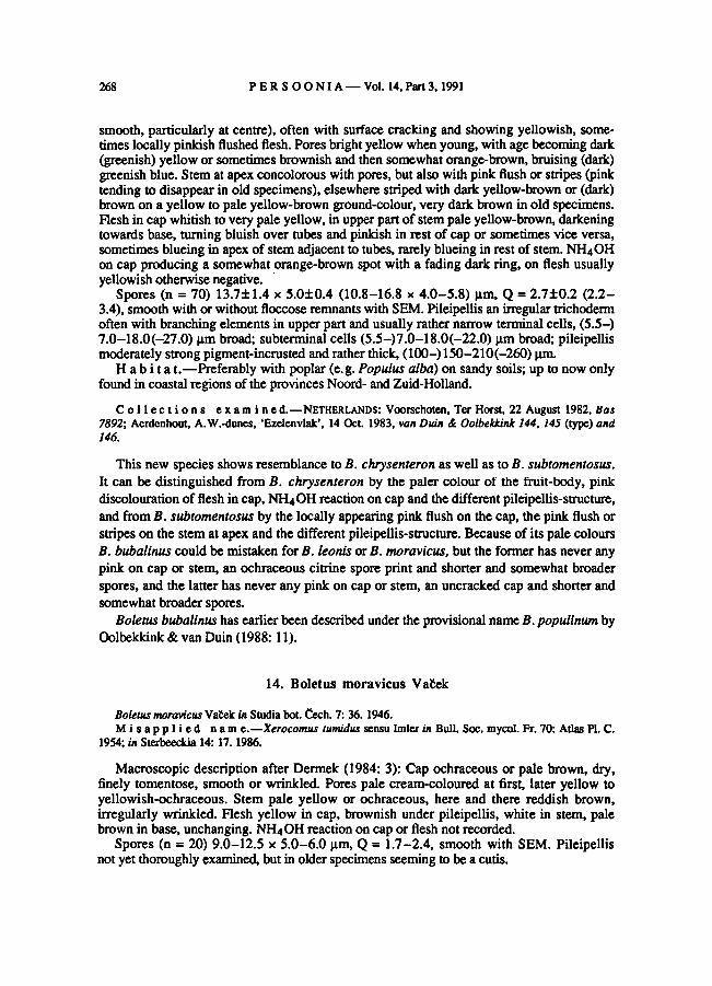
Cap buff, pale to dark yellow-brown or pale brown, usually with pink flush appearing
locally, tomentose when young, later becoming very smooth (in dried state almost shining
PERSOONIA— Vol. 14, Part 3, 1991268
smooth, particularly at centre), often with surface cracking and showing yellowish, some-
times locally pinkish flushed flesh. Pores bright yellow when young, with age becoming dark
(greenish) yellow or sometimes brownish and then somewhat orange-brown, bruising (dark)
greenish blue. Stem at apex concolorous with pores, but also with pink flush or stripes (pink
tending to disappear in old specimens), elsewhere striped with dark yellow-brown or (dark)
brown on a yellow to pale yellow-brown ground-colour, very dark brown in old specimens.Flesh in cap whitish to very pale yellow, in upperpart ofstem pale yellow-brown, darkeningtowards base, turning bluish over tubes and pinkish in rest of cap or sometimes vice versa,
sometimes blueing in apex of stem adjacent to tubes, rarely blueing in rest of stem. NH4OH
on cap producing a somewhat orange-brown spot with a fading dark ring, on flesh usually
yellowish otherwise negative.
Spores (n = 70) 13.7±1.4 x 5.0±0.4 (10.8-16.8 x 4.0-5.8) ptn, Q = 2.7±0.2 (2.2-3.4), smooth with or without floccose remnants with SEM. Pileipellis an irregular trichoderm
often with branching elements in upper part and usually rather narrow terminal cells, (5.5-)
7.0-18.0(-27.0) |xm broad; subterminal cells (5.5-)7.0-18.0(-22.0) |im broad; pileipellis
moderately strong pigment-incrusted and rather thick, (100-) 150-210(-260) prn.
H ab i t a t. —Preferably with poplar (e.g. Populus alba) on sandy soils; up to now onlyfoundin coastal regions of the provinces Noord- andZuid-Holland.
Collections examine d.—NETHERLANDS: Voorschoten, Ter Horst, 22 August 1982, Bas
7892; Aerdenhout, A.W.-dunes, 'Ezelenvlak', 14 Oct. 1983, van Duin & Oolbekkink 144, 145 (type) and
146.
This new species shows resemblance to B. chrysenteron as well as to B. subtomentosus.
It can be distinguished from B. chrysenteron by the paler colour of the fruit-body, pink
discolourationofflesh in cap, NH4OH reaction on cap and the differentpileipellis-structure,
and from B. subtomentosus by the locally appearing pink flush on the cap, the pink flush or
stripes on the stem at apex and the different pileipellis-structure. Because of its pale colours
B. bubalinus could be mistaken for B. leonis or B. moravicus, but the former has never any
pink on cap or stem, an ochraceous citrine spore print and shorter and somewhat broader
spores, and the latter has never any pink on cap or stem, an uncracked cap and shorter and
somewhat broader spores.
Boletusbubalinus has earlierbeen described under the provisional name B. populinum by
Oolbekkink & van Duin (1988: 11).
14. Boletus moravicus Vaček
Boletus moravicus Vatek in Studia bot. Cech. 7: 36. 1946.
Misapplied nam e.— Xerocomus tumidus sensu Imler in Bull. Soc. mycol. Fr. 70: Atlas PI. C.
1954; in Sterbeeckia 14: 17. 1986.
Macroscopic description after Dermek (1984: 3): Cap ochraceous or pale brown, dry,
finely tomentose, smooth or wrinkled. Pores pale cream-coloured at first, later yellow to
yellowish-ochraceous. Stem pale yellow or ochraceous, here and there reddish brown,
irregularly wrinkled. Flesh yellow in cap, brownish under pileipellis, white in stem, palebrown in base, unchanging. NH4OHreaction on cap or flesh not recorded.
Spores (n = 20) 9.0-12.5 x 5.0-6.0 pm, Q = 1.7-2.4, smooth with SEM. Pileipellisnot yet thoroughly examined, but in older specimens seeming to be a cutis.

OOLBEKKINK: Spore ornamentation in Xerocomus 269
Collections examined.—CZECHOSLOVAKIA: 20 Aug. 1945, Valek
PRM 203553. —FRANCE: Touraine, Bois de Montrdsor, 24 Sept. 1955, Imler (as X. tumidus).
Fries described B. tumidus in 1874 (: 501) with a red-brown, viscid cap and placed it to-
gether with e.g. B. badius in Boletus sect. Viscipelles. The specimens determined as B. tu-
midus by Imler (1954, 1986, as Xerocomus) do not agree with Fries' taxon, as they had a
brownish and dry cap. It is rather improbable that Fries made a mistake in establishing such a
striking character as a viscid cap. The convincing reasoning of Kallenbach(1942: 155) shows
that B. tumidus Fr. is merely a variant ofB. badius.
The description and plate of Imler's B. tumidus agree with B. moravicus described by
Vabek (1946). Observations of the present author on the collections of Imlerand Vabek cited
above confirmedtheirconspecificity. Therefore B. moravicus is the correct name for Imler's
specimens.
15. Boletus pulverulentus Opat. in Wiegm.
Boletus pulverulentus Opat. in Wiegm. in Arch. Naturgesch. Meckl. 2: 27. 1836.
Cap drab to brown, with or without red flush, tomentose then smooth, strongly bruisingblue to almost black on handling. Pores (deep) yellow, bruising blue to dark blue. Stem at
apex yellow to yellowish orange with fine red dots, elsewhere brownish with red streaks,
on handling dark blue and finally black. Flesh in cap (pale) yellow, in stem deep yellowsometimes with red patches, in base red, immediately and strongly turning blue to dark blue
throughout when cut. NH4OH reaction on cap or flesh not recorded.
Spores (n = 20) 10.0-14.0x 4.0-6.0 |xm, smooth with SEM.Pileipellis not yet exam-
ined.
Collections examine d.—NETHERLANDS: Valburg, Oosterhout, 26 Sept. 1971, Schreurs
346\ Bunde, Bunderbos, 21 Sept 1979, Schreurs 343.
As the present author does not regard the structure of the hymenophoral trama as a distin-
guishing character, it seems logical to includeB. pulverulentus in theXerocomus-group of
Boletus because of the xerocomoid features of its fruit-bodies. This view is supported by the
fact that other taxa with xerocomoidfeatures are arranged together with B. pulverulentus in
one section by both Singer and Watling. Singer (1986: 777) has placed B. pulverulentus to-
gether with e. g. B. rubellus and B. fraternus in genus Boletus sect. Subpruinosi. Watling
(1970: 99) has placed it together with B. pruinatus and B. versicolor in Boletus subgenus
Xerocomus sect. Subpruinosi.
16. Boletus ichnusanus (Alessio, Galli & Litt.) Oolbekkink, comb. nov.
Xerocomus ichnusanus Alessio, Galli & Litt. in Alessio in Boll. Gruppo micol. G. Bresadola,Trento 27:
170. 1984 (basionym).
Macroscopic and microscopic description after Alessio (1984:170; 1985:596):
Cap reddish brown (chestnut-coloured) to brown tinged with reddish pink, becoming darker
with age, dry, finely tomentose, smooth with age. Pores golden yellow, becoming reddish

PERSOONIA— Vol. 14, Part 3, 1991270
brown (rust-coloured), bruising green-blue. Stem at upperpart bright yellow to golden yel-
low, at lowerpart brownish red, covered over two-third from apex downwards by a brown-
ish red to brown net; base brown, usually prolonged into soil by root-like strand of myce-
lium. Flesh yellowish white with pink spots in lower part of stem, turning blue. Spore printbrown. NH4OH reaction on cap or flesh not recorded.
Spores 12—18(—23) x 5—7(—7.5) pm. Pileipellis not examined.
H a b i ta t.—With broad-leaved trees and shrubs (Quercus and Cistus) in the Mediter-
ranean (possibly restricted to Sardinia), usually growing in groupsor tufts.
Collection examine d.—ITALY: Sardinia, Arzachena, 23 Oct. 1987,Kuyper.
3.4. THE INSUFFICIENTLY KNOWN TAXON BOLETUS FRAGILIPES
Boletus fragilipes C. Martin in Bull. Soc. bot. Genfeve 7: 189. 1894.—
Boletus subtomentosus subsp.
fragilipes (C. Martin) C. Martin in Mat6r. Fl. cryptog. Suisse 2: PI. 18. 1903. — Boletellus fragilipes (C.
Martin) Kuthan in Ph'r. Sb. Ostr. Muz. Ostrava 26: 162. 1982.
Macroscopic description after Martin (1894: 189; 1903: PI. 18): Cap olivaceous brown
mixed with purple and green, subtomentose. Pores yellow stained with red. Tubes brownish
yellow. Stem thickened at base, narrow at apex with tendency to break there, at upper partwith dark red to purple stripes, at lower part yellow with brown and red-brown. Flesh in cap
pale yellow, purplish stained with yellow or green in upper part of stem, golden yellow in
lowerpart, turning blue in cap. NH4OH reaction on capof flesh not recorded.
In the three collectionsreceived under this name there was hardly any difference in spore
size, but there were large differences in pileipellis-structure. Spores (n = 30) 12.5-18.0 x
4.0-6.0 pm, Q = 2.2-3.6; with great difficulty a fine striation is visiblewith a light micro-
scope at highest magnification in a number of spores mounted in Congo red or Cotton blue.
According to Pouzar (1981) this character is typical for this taxon. Each collection has its own
pileipellis-type: (i) a pileipellis as in B. chrysenteron type I was found in a collectionfrom the
Netherlands: Apeldoorn, *t Loo, 8 Oct. 1960,Bas 2268; (ii) a pileipellis as in B. chrysenteron
type II was found in a collection from Czechoslovakia: Klabianskd Magura, Slovakia, Maid
Fatra, 18 Oct. 1984, Pouzar, (iii) a pileipellis as in B. pruinatus was found in a collection from
Czechoslovakia: Bohemia, Karlovy Vary, 11 Nov. 1984, Valter (but determinedby Pouzar).
Boletusfragilipes is a taxon created by Martin in 1894, later regarded as a subspecies of
B. subtomentosus in his publication of 1903. Pouzar (1981) came across this forgotten taxon
and reinstated it, because he thought its description and plate fitted a taxon with striate spores,
considered differentfrom B. chrysenteron (see paperof Hübsch, 1982). Attributing a striking
but (sub)microscopical character (striate spores) to an old taxon of which no authenticmaterial
is availableand thereby giving it a new status, is rather risky, as is shown by the discovery of
such ornamentedspores in all taxa examinedof the B. chrysenteron-complex.
In recent publications (e.g. Dermek, 1987: 20) the striation of spores is considered the
fundamental characterof B. fragilipes, but the characters given by Martin are ignored. Boletus
fragilipes sensu Pouzar, or Boletellusfragilipes sensu Kuthan (1982: 162), who transferred it
on account of its striate spores, have become a mixture of taxa from the B. chrysenteron-
complex which explains why collections with differentcharacters are found under the name
B. fragilipes.
OOLBEKKJNK: Spore ornamentation in Xerocomus 271
For that reason the macroscopic description by Martin is reproduced above. The taxon of
Martin is, however, insufficiently known and thus B.fragilipes is considered a nomen
dubium.
3.5. DESCRIPTION OF PHYLLOPORUS RHODOXANTHUS
Phylloporus rhodoxanthus (Schwein.) Bres., Fungi Trid. 2: 95. 1900.
Cap reddish brown to brown often with olivaceous flush, tomentose then smoother, some-
times with slightly cracking surface. Gills distant, decurrent, golden yellow, often weaklyto strongly anastomosing, reddish brown on bruising. Stem reddish brown granular, dotted
or streaked on a yellow to golden yellow ground-colour. Flesh yellowish white in cap but
flushed reddish brown under pileipellis, brownish red in stem. NH4OH reaction on cap or
flesh not recorded.
Spores (n = 30)11.0-13.5 x 3.5-5.0 pm, fibrillose, floccose or pitted-and-floccose with
SEM. Pileipellis not yet examined.
Collections examine d. —NETHERLANDS: Baarn, 1 Aug. 1953, de Vries; Apeldoom, t Loo,
18 Sept. 1971, de Kleuver-Schreuters\ Stokkem, 28 Aug. 1980, Schreurs 464.
ACKNOWLEDGEMENTS
The author wishes to thank Mrs. M. Hoek and Dr. C. Bas for their constructive comments
on the manuscript, the latterand Dr. R.A. Maas Geesteranus for correcting the Latin descrip-tion and Mrs. B.J. van Heuven for technical assistance.
REFERENCES
AA, H. A. VAN DER (1979). Het Centraalbureau voor Schimmelcultures bestaat 75 jaar. In Natura 76: 205-210.
ALESSIO, C.L. (1984). Xerocomus ichnusanus Alessio, Galli et Litlini sp. nov. In Boll. Gruppo micol. G.
Bresadola, Trento 27: 166-170.
— (1985). Boletus Dill, ex L. Fungi europaei 2. Saronno.
BAS, C., OOLBEKKINK, G. & DUIN, W. VAN (1988). Un rapporto preliminare sul valore tassonomico della
struttura della cuticola nel gruppo Xerocomus del genere Boletus.. In Fungo 7 (6, suppl.): 41-46.
BULLIARD, J.B.F. (1791). Histoire des champignons de la France 1.
COKER, W.C. & BEERS, A.H. (1943). The Boletaceae of North Carolina. Chapel Hill. (Reprinted by Dover,
New York in 1974 as: The Boleti of North Carolina.)
CORNER, E.J.H. (1972). Boletus in Malaysia. Botanic Gardens, Singapore.
DERMEK, A. (1984).Fungorum rariorum Icones coloratae 13. Vaduz.
— (1987). Fungorumrariorum Icones coloratae 16. Berlin.
FRIES, E.M. (1818). Observationes mycologicae 2. Hafniae.
— (1821). Systema mycologicum 1. Lundae.
— (1828). Elenchus fungorum 1. Gryphiswaldiae.— (1835). Boleti, fungorum generis, illustratio. Upsaliae.— (1838). Epicrisis systematis mycologici. Upsaliae.
— (1874). Hymenomycetes europaei. Upsaliae.GAMS, W. (1984). An index to fungal names and epithets sanctioned by Persoon and Fries. In Mycotaxon 19:
219-270.
GRAY, S.F. (1821). Natural arrangement of Bridsh plants 1. London.

PERSOONIA Vol. 14, Part 3, 1991272
GRUND, D.W.& HARRISON, A.K. (1976). Nova Scotian Boletes. Vaduz.
HARLEY, J.L. & SMITH, S.E. (1983). Mycorrhizal symbiosis. London.
HEINEMANN, P., RAMMELOO, J. & RULLIER, E. (1988). L'ornementation sporale des Xerocomaceae & spores
dites lisses. In Bull. Jard. bot. natn. Belg. 58: 513-534.
HOBSCH, P. (1982). tlber RotfuBchen mit abweichenden Sporen. In Boletus 6: 61-66.
IMLER.L. (1954). Xerocomus tumidus (Fr.) sensu Peltereau et Gilbert. In Bull. Soc. mycol. Fr.70: Atlas PI. C.
— (1955). Notes critiques. In Bull. Soc. mycol. Fr. 71: 19-25.
— (1958). Notes critiques. In Bull. Soc. mycol. Fr. 74: 94-98.
— (1964). Xerocomus porosporus. In Bull. Sex:, mycol. Fr. 80: Allas PI. 141 and 142.
— (1986). Xerocomus tumidus (Fr.) Gilb. In Sterbeeckia 14: 17-19.
KALLENBACH, F. (1942). Die ROhrlinge. Die Pilze Mitteleuropas 1. Leipzig.
KROMBHOLZ, J.V. (1836). NaturgetreueAbbildungen und Beschreibungen der essbaren, schadlichen und ver-
dachtigen Schwamme 5. (Text and atlas.) Prag.
KUTHAN, J. (1982).Einige Bemerkungcn zu Funde von seltenen und interessanten Grosspilzen im Stadtgelande
von Ostrava. In Pfir. Sb. Ostr. Muz. Ostrava 26: 153-166.
MARTIN, C.E. (1894). Contribution A la flore mycologique genovois. In Bull. Soc. boL Genbve 7: 189-192.
— (1903). Le ‘Boletus subtomentosus’ de la fegion genevoise. Maferiaux pour la flore cryptogamique Suisse
2(1). Berne.
OOLBEKKINK, G.T. & DUIN, W.E. VAN(1985). De taxonomische betekenis van de hoedhuidtypen in het genus
Xerocomus. Rijksherbarium, Leiden. Unpublishedreport.
—&
— (1988). De taxonomische betekenis van de hoedhuidtypenin Xerocomus. In Coolia 31: 1-11,48
(errata).
PEARSON, A. A. (1952). New records and observations 5. In Trans Br. mycol. Soc. 35: 97-122.
PECK, C.H.(1897). Boletus fraternus.. In Bull. Torrey bot. Club 24: 145.
PEGLER, D.N. & YOUNG, T.W.K. (1971). Basidiospore morphology in the Agaricales. In Beih. Nova Hed-
wigia 35.
—&
— (1981). A natural arrangementof the Boletales, with reference to spore morphology. In Trans. Br.
mycol. Soc. 76: 103-146.
PERREAU-BERTRAND, J. (1961). Recherches sur les ornementations sporales et la sporogenèse chez quelques
espèces des genres Boletellus et Strobilomyces (Basidiomycètes). In Annls Sei. nat. (Bot.) 2: 399-489.
PERREAU, J. (1964). Complémentà l'étude des ornementations sporales dans le genreBoletellus. In Annls Sei.
nat. (Bot.) 5: 753-766.
PERREAU-BERTRAND, J. (1965). Structure membranaire et différenciations apicales chez les spores des genres
Xerocomus, Boletellus, Heimiella et Strobilomyces. In C. r. hebd. Séanc. Acad. Sei., Paris 260: 4245-
4248.
— (1967). Recherches sur la différenciation et la structure de la paroi sporale chez les Homobasidiomycètes a
spores ornées. In Annls Sei. nat. (Bot) 8: 639-745.
PERREAU, J. & HEIM, R. (1969). L'ornementation des basidiospores au microscope électronique à balayage.
In Rev. Mycol. 33: 329-340.
PETERSEN, R. H. (1977). An annotated index for Bulliard's 'Histoire des Champignons'. In Mycotaxon 6: 127-
166.
PHILLIPS, R. (1981). Mushrooms and other fungi ofGreat Britain and Europe. London.
POUZAR, Z. (1966). Two interesting species of the family Boletaceae: Boletus truncatus and Boletus sub-
tomentosus var. leguei. In Ceskâ Mykol. 20: 1-7.
— (1981). Co jsou podzimni 'babky'? In Mykol. listy 3: 8-14.
QuÉLET, L. (1888).Flore mycologiquede la France et des pays limitrophes. Paris.
REA, C. (1922). British Basidiomycetae. (A handbook to the larger British fungi.) Cambridge.
ROSTKOVIUS, F.W.T. (1844). Die Pilze Deutschlands 5. In Deutschlands Flora, Abth. 3. J. Sturm (ed.). Nürn-
berg.
SCHAEFFER, J.C. (1763). Fungorum qui in Bavaria et Palatinatu circa Ratisbonam nascuntur icones nativis
coloribus expressae 2. Ratisbonae.
— (1774). Fung. Bavaria 4. Ratisbonae.

OOLBEKKINK: Spore ornamentation in Xerocomus 273
SINGER, R. (1965). Die ROhrlinge 1. In Pilze Mitteleuropas 5. Bad Heilbrunn.
— (1966). Die ROhrlinge 2. In Pilze Mitteleuropas 6. Bad Heilbrunn.' 1967'.
— (1986). The Agaricales in modern taxonomy. Koenigstein.
SINGER, R. & KUTHAN, J. (1976). Notes on Boletes. In Ceskd Mykol. 30: 143-155.
SMITH, A.H. & THIERS, H.D. (1971). The Boletes of Michigan. Ann Arbor.
SNELL, W.H., SINGER, R. & DICK, E.A. (1960). Notes on Boletes 11. In Mycologia 51: 564-577. '1959'.
VACEK, V. (1946). Boletus moravicus, species nova Cechoslovaca. In Studia boL Cech. 7: 36-37.
VELENOVSKY, J. (1939). Novitates mycologicae. Pragae.
WATUNG, R. (1968). Records of Boleti and notes on their taxonomic position. In Notes R. bot. Gdn. Edinb.
28: 301-315.
— (1970). British fungus flora: Agarics and Boleti. 1. Boletaceae;Gomphidiaceae; Paxillaceae. Royal Botanic
Garden, Edinburgh.