Hypercapnia via Reduced Rate and Tidal Volume Contributes to Lipopolysaccharide-induced Lung Injury

SYMPOSIUM: INTENSIVE CARE
Permissive hypercapnia inprotective lung ventilatorystrategies
Brendan D Higgins
Joseph F Costello
Martina Ni Chonghaile
John G Laffey
Abstract
Hypercapnia has traditionally been avoided in paediatric critical illness;
indeed, traditional approaches advocated hypocapnia in a number of
disease states. However, recent advances in understanding of the role of
excessive tidal stretch has prompted clinicians to avoid high tidal
volumes or plateau pressures, and to tolerate the resulting ‘permissive’
hypercapnia. Advances in understanding of the biology of hypercapnia
have led to consideration of an active role for hypercapnia in the
pathogenesis of inflammation and tissue injury. Newer data suggest that
elevated CO2 may be protective, but in some experimental situations can
cause harm. This review assesses the role of ventilatory strategies
involving permissive hypercapnia in the management of neonates and
children with acute severe respiratory failure. The physiological effects of
hypercapnia on the lung and systemic organs are discussed, and
evidence from laboratory models of lung and systemic organ injury is
considered, demonstrating the potential for hypercapnia to modulate the
injury process. The role of permissive hypercapnia in various clinical
settings relevant to neonatal and paediatric practice, and the risks
and benefits of hypercapnia in specific clinical situations are also
considered.
Brendan D Higgins BSc PhD is Postdoctoral Fellow at the Department of
Anaesthesia Clinical Sciences Institute and National Centre for
Biomedical Engineering Sciences, National University of Ireland, Galway,
Ireland.
Joseph F Costello MB FCARCSI is Research Fellow at the Department of
Anaesthesia, Clinical Sciences Institute and National Centre for
Biomedical Engineering Sciences, National University of Ireland, Galway,
Ireland.
Martina Ni Chonghaile MB FCARCSI is Research Fellow at the Department of
Anaesthesia, Clinical Sciences Institute and National Centre for
Biomedical Engineering Sciences, National University of Ireland, Galway,
Ireland.
John G Laffey MD MA BSc FCARCSI MB FCARCSI is Professor at the Department of
Anaesthesia, Clinical Sciences Institute and National Centre for
Biomedical Engineering Sciences, National University of Ireland, Galway,
Ireland.
PAEDIATRICS AND CHILD HEALTH 17:3 94
Keywords hypercapnia; acidosis; mechanical ventilation; acute lung
injury; acute respiratory distress syndrome; congenital heart disease;
congenital diaphragmatic hernia; asthma; neonatal respiratory distress
syndrome; pulmonary hypertension; intracranial pressure; buffering
Introduction
Traditional approaches to the management of CO2 in neonates
and children with acute respiratory failure have focused on the
potential deleterious effects of hypercapnia and therefore on
targeted normocapnia or even hypocapnia. Support for this
strategy is derived from the link between hypercapnia and
adverse outcome in diverse clinical contexts, including cardiac
arrest1 sepsis2 and neonatal asphyxia.3 However, it has been
increasingly questioned. Accumulating evidence from experi-
mental and clinical studies supports the contention that
mechanical ventilation may directly injure the lungs, a phenom-
enon termed ‘ventilator-induced lung injury’. Permissive hyper-
capnia (PHC) is a ventilatory strategy in which relatively high
PaCO2 is tolerated in an effort to avoid high tidal volumes and
pulmonary over-distension, thereby potentially reducing lung
injury and increasing survival.4,5 PHC has been progressively
accepted in the critical care of patients requiring mechanical
ventilation.
Conventionally, the protective effect of ventilatory strategies
incorporating PHC is considered to be solely due to reductions in
lung stretch, with hypercapnia permitted to achieve this goal.
However, protective ventilatory strategies involving hypoventila-
tion result in both limitation of lung stretch and elevation of
systemic PCO2. Lung stretch is distinct from elevated PCO2, and
by manipulation of respiratory parameters (frequency, tidal
volume, dead-space, inspired CO2) can, at least to some extent,
be separately controlled. Thus, it is important to determine
whether hypercapnia might exert direct effects in critically ill
children. If hypercapnia were proved to have independent
benefits, deliberately elevating PaCO2 (therapeutic hypercapnia)
could provide an additional advantage over low tidal volume
strategies alone. Conversely, in patients managed with conven-
tional PHC, adverse effects of elevated PaCO2 might be concealed
by the benefits of lower lung stretch. Because ICU outcome might
be related to the development of multi-organ failure (as opposed
to simply lung injury) it is also necessary to determine the effects
of hypercapnia on systemic organs.
This review assesses the role of ventilatory strategies involving
PHC in the management of neonates and children with acute
respiratory failure. The physiological effects of hypercapnia in the
lung and systemic organs are discussed, and evidence from
laboratory models of lung and systemic organ injury is
considered, demonstrating the potential for hypercapnia to
modulate the injury process. The role of PHC in various clinical
settings relevant to neonatal and paediatric practice, and the risks
and benefits of PHC are also considered in specific clinical
situations.
Physiological effects of hypercapnia
If PHC is to be used rationally and safely in critically ill neonates
and children, its physiological effects must be considered. These
r 2007 Elsevier Ltd. All rights reserved.

Figure 1 Brain tissue oxygen tension, in addition to cerebral perfusion,
is progressively increased with increases in inspired CO2 concentration.
(Reproduced with permission, Hare GM, et al. Can J Anaesth 2003; 50:
1061–8.)
SYMPOSIUM: INTENSIVE CARE
effects are diverse and incompletely understood, however, and
direct effects are often counterbalanced by indirect effects. In
addition, the net effect of hypercapnia may occur as a function of
acidosis or of CO2 per se.
Pulmonary system
In the normal lung, CO2 may either regulate regional ventilation
in response to primary changes in perfusion, or alter regional
perfusion to match primary changes in ventilation. An example is
the phenomenon of hypocapnic bronchoconstriction that occurs
following acute regional pulmonary artery occlusion.6
It is of concern that hypercapnia can increase pulmonary
vascular resistance. This might aggravate primary pulmonary
hypertension in newborns managed with a strategy of PHC.
Reassuringly, however, data from an animal model of chronic
hypoxia-induced pulmonary hypertension suggests that the effect
of hypercapnia on pulmonary vascular resistance does not appear
to be exacerbated in the setting of pre-existing pulmonary
hypertension.7 Administration of CO2 improves matching of
ventilation and perfusion and increases arterial oxygenation by
this mechanism in both health8,9 and disease.10 A dose–response
relationship exists whereby increased FiCO2 results in progressive
augmentation of PaO29,11 (Figure 1). In acute respiratory distress
syndrome (ARDS), the potential for PHC to increase shunt is due
to reduced tidal volume and airway closure rather than
hypercapnia per se.12
Hypercapnia has been variably reported to either increase13 or
decrease14 airway resistance. These effects may be explained by
the direct dilatation of small airways and indirect (vagally
mediated) large airway constriction.6 These opposing balanced
airway actions of CO2 may result in little net alteration in airway
resistance. Parenchymal lung compliance increases in response to
hypercapnic acidosis. This may be due to increased surfactant
secretion or more effective surface tension-lowering properties
under acidic conditions.15
Central nervous system
Hypercapnic acidosis increases cerebral tissue oxygen tension
through both augmentation of PaO2 and increased cerebral blood
flow9 (Figure 1). Hypercapnia is a potent ventilatory stimulant.16
A modest increase in ventilatory chemosensitivity has been
shown in response to acute hypoxia, but no change occurred with
acute elevations in CO2.
Cardiovascular system
Hypercapnic acidosis directly reduces the contractility of cardi-
ac17 and vascular smooth muscle.6 This is counterbalanced by
the hypercapnia-mediated sympathoadrenal effects of increased
preload and heart rate, increased myocardial contractility and
decreased afterload, leading to a net increase in cardiac output.6
Hypercapnia results in a complex interaction of altered cardiac
output, hypoxic pulmonary vasoconstriction, and intrapulmon-
ary shunt to produce a net increase in PaO2. Because hypercapnia
generally elevates cardiac output, global oxygen delivery is
increased. Regional (including mesenteric) blood flow is also
increased,18 thereby increasing organ oxygen delivery. Hyper-
capnia and acidosis shifts the hemoglobin–oxygen dissociation
curve to the right, reducing the oxygen affinity of haemoglobin,
and may cause an elevation in haematocrit, further increasing
tissue oxygen delivery. Acidosis may reduce cellular respiration
PAEDIATRICS AND CHILD HEALTH 17:3 95
and oxygen consumption, which may further benefit a supply/
demand imbalance.19
Insights from laboratory studies
It is not currently feasible to examine the direct effects of
hypercapnic acidosis in humans independent of alterations in
ventilatory strategies. An alternative approach, which has been
the focus of much recent attention, is to determine the effects of
hypercapnic acidosis in various animal models of experimental
acute lung and systemic organ injury.
Pulmonary
Hypercapnic acidosis attenuates the increased lung permeability
seen following free radical-mediated20 and ischaemia–reperfu-
sion-induced reperfusion-induced lung injury.20,21 Hypercapnic
acidosis preserved lung mechanics, attenuated protein leakage,
reduced pulmonary oedema and improved oxygenation com-
pared with control conditions following in vivo pulmonary
ischaemia–reperfusion22 as well as in secondary reperfusion-
induced lung injury.11 Such protective effects of hypercapnic
acidosis are not mediated via a decrease in pulmonary artery
resistance; indeed, protection occurred despite elevated pulmon-
ary artery pressures.11
r 2007 Elsevier Ltd. All rights reserved.

SYMPOSIUM: INTENSIVE CARE
Hypercapnic acidosis appears to attenuate the development of
pulmonary hypertension and vascular remodelling induced by
chronic hypoxia in newborn rats.23 Newborn rats were main-
tained under atmospheric CO2 of less than 0.5% (normocapnia),
5.5% or 10% during exposure from birth for 14 days to normoxia
or moderate hypoxia (13%). Inspired CO2 attenuated the increase
in pulmonary arterial resistance, right ventricular hypertrophy
and dysfunction, medial thickening of pulmonary resistance
arteries and distal arterial muscularisation that occurred in rats
exposed to hypoxia. A dose–response was seen; 10% CO2
significantly attenuated pulmonary vascular remodelling and
alterations in pulmonary arterial resistance, and both increased
concentrations of CO2 normalised right ventricular performance.
Exposure to 10% CO2 also reduced lung oxidative injury, and
prevented up-regulation of endothelin-1, a critical mediator of
pulmonary vascular remodelling.23
The effect of hypercapnia in ventilator-induced lung injury
(VILI) is more complex. In two key studies, addition of inspired
CO2 reduced VILI in isolated rabbit lung24 and in rabbits in vivo.25
However, not all data are so positive. Supplemental CO2 has more
modest protective effects in the context of more clinically relevant
tidal stretch. Strand et al. showed that significant hypercapnic
acidosis (mean PaCO2 95 mmHg) was well tolerated in preterm
lambs, and appeared to reduce lung injury.26 In the context of a
clinically relevant high tidal volume strategy (tidal volume
12 mL/kg, positive end-expiratory pressure (PEEP) 0 cmH2O, rate
42/min) in an adult model, hypocapnia was potentially deleter-
ious and hypercapnic acidosis somewhat protective.11 However,
inspired CO2 did not significantly attenuate lung injury induced
by an atelectasis-prone model of lung injury, which may mimic
neonatal respiratory distress syndrome more closely than pure
stretch models of injury.27 Hypercapnia produced by hypoventi-
lation rather than by increased inspired CO2 did not protect
against stretch-induced injury.28 In a subsequent study, hyper-
capnia was found to minimise the adverse effects of high-volume
ventilation on vascular barrier function, but it impaired the
ability of lung cells to repair the stretch-induced injury.29 Taken
together, these findings suggest that while hypercapnic acidosis
substantially attenuates injury due to excessive stretch, its effects
in the context of more clinically relevant lung stretch or extensive
atelectasis are modest, and there are concerns regarding the
effects of hypercapnia on cellular repair following injury.
ARDS commonly develops in the context of severe sepsis in
children. Hypercapnia may have deleterious effects by impairing
the immune system response to bacterial sepsis.30,31 The
mechanisms of lung injury in sepsis-induced ARDS are distinct
from those in many experimental models. Lipopolysaccharide
(LPS), a key endotoxin of Gram-negative bacteria, initiates lung
injury by activating a specific receptor called toll-like receptor-4.
Hypercapnia appears to have different effects in lung injury
caused by pulmonary vs systemic administration of endotoxin.
Hypercapnic acidosis induced by the administration of CO2 has
been shown to directly protect against acute lung injury induced
by intratracheal instillation of endotoxin.32 Conversely, hyper-
capnia induced by reduced tidal volume and respiratory rate
appears to worsen the lung damage induced by systemic
administration of endotoxin.33 These issues underline the need
to take into account both the means of achieving hypercapnia
and the diversity of experimental models.
PAEDIATRICS AND CHILD HEALTH 17:3 96
Prolonged hypercapnia may impair diaphragmatic function in
rats by interfering with neuromuscular transmission and histo-
logical structure.34 Alterations in diaphragmatic function could
lead to delayed weaning from ventilatory support. However,
clinical trials of PHC in infants and children to date do not
support this concern.
Cardiovascular
Hypercapnic acidosis appears to have beneficial effects on the
myocardium, and protects the heart from ischaemia–reperfusion
injury.35 Both hypercapnic and metabolic acidosis have been
shown to reduce infarct size in an in vivo canine model of
coronary artery ischemia–reperfusion.36 Possible mechanisms for
the protective effects of acidosis include reduction of calcium
loading to the myocardium through H+ inhibition of calcium
uptake, and, in the case of hypercapnic acidosis, the induction of
coronary vasodilatation. Nomura et al. found that the greatest
coronary artery blood flow occurred with maximal hypercap-
nia,35 while acute hypercapnia increased both collateral and
global coronary blood flow in a swine model of chronic coronary
artery obstruction.37 However, normocapnic acidosis protected
the myocardium from coronary artery occlusion-induced ischae-
mia–reperfusion injury in a canine model.38 In contrast,
hypercapnic acidosis was found to reduce the success of
resuscitation following ventricular fibrillation arrest in a rodent
model.39
Neurological
Several studies have demonstrated protective effects of hyper-
capnia in brain injury. Hypercapnic acidosis protects the new-
born porcine brain from hypoxia/reoxygenation-induced
injury.40,41 Hypercapnia also attenuates hypoxic–ischaemic brain
injury in immature rats, while hypocapnia is deleterious.42,43
Cerebral blood flow was better preserved during hypercapnia,
and the greater oxygen delivery promoted cerebral glucose
utilisation and oxidative metabolism for optimal maintenance
of high-energy phosphate reserves in the tissues.42 However, an
important dose–response phenomenon was found; mild-to-
moderate hypercapnia (PaCO2 40–55 mmHg) was significantly
more neuroprotective than higher PaCO2 (470 mmHg).43 Indeed,
extremely high PaCO2, which is not generally seen in the clinical
context, appeared to have detrimental effects on the developing
brain.44 Potential mechanisms underlying these protective effects
include reduced levels of glutamate, an excitatory amino acid
neurotransmitter, in the CSF,42 free radical inhibition40,41 and
attenuation of neuronal apoptosis.45
Hypercapnia may contribute to the pathogenesis of retino-
pathy of prematurity, an important concern in the context of
neonatal respiratory failure. Hypercapnia causes retinal vasodi-
latation and increases retinal oxygen delivery in newborn (and
adult) rats.46 However, hypercapnia was shown to produce
preretinal neovascularisation similar to that seen in oxygen-
induced retinopathy in a neonatal rat model.47
Others
Acidosis markedly delays the onset of cell death in isolated
hepatocytes exposed to anoxia48 or chemical hypoxia.49 Buffering
of the pH accelerated cell death. This phenomenon may represent
a protective adaptation against hypoxic and ischaemic stress.
Isolated renal cortical tubules exposed to anoxia have greater ATP
r 2007 Elsevier Ltd. All rights reserved.

Figure 2 Hypercapnia suppresses the degradation of IkB-a (panel A) but
not IkB-b (panel B) following exposure to lipopolysaccharide, thereby
inhibiting the nuclear translocation of NF-kB and downstream cytokine
production. The effects of isocapnic acidosis and buffered hypercapnia
(panel C) on IkB-a degradation were intermediate between normocapnic
control and hypercapnic acidosis conditions. BH, buffered hypercapnia;
HA, hypercapnic acidosis; IA, isocapnic acidosis; LPS, lipopolysacchar-
ide; NC, normocapnia; NF-kB, nuclear factor kappa-B. (Reproduced with
permission, Takeshita K, et al. Am J Respir Cell Mol Biol 2003; 29:
124–32.)
SYMPOSIUM: INTENSIVE CARE
levels on reoxygenation at acidotic pH compared with tubules
incubated at pH 7.548. In contrast, the combination of hypoxia
and hypercapnia induced apoptosis in rat renal tubular cell
cultures.50
Cellular and molecular effects of hypercapnia
A clear understanding of the cellular and biochemical mechan-
isms underlying the effects of hypercapnia is essential. It is a
prerequisite for the successful translation of laboratory findings
to the bedside, and allows prediction of potential side effects,
enabling identification of those in whom hypercapnia should be
avoided.
Acidosis vs hypercapnia
The protective effects of hypercapnic acidosis in experimental
lung and systemic organ injury appear to be primarily a function
of the acidosis.21,51 In isolated lung, the protective effect of
hypercapnic acidosis in ischemia–reperfusion was greatly atte-
nuated if the pH was buffered towards normal.21 The myocardial
protective effects of hypercapnic acidosis are also seen with
metabolic acidosis in both ex vivo52 and in vivo36,38 models.
Furthermore, as discussed earlier, metabolic acidosis exerts
protective effects in other organs, including the liver and the
kidney.48,49
Anti-inflammatory effects
Several key components of the inflammatory response appear to
be attenuated by hypercapnic acidosis. It inhibits the release of
tumour necrosis factor-a (TNF-a) and interleukin (IL)-1 from
stimulated macrophages in vitro53 and reduces bronchoalveolar
lavage levels of TNF-a following in vivo pulmonary ischaemia–
reperfusion.22 Both hypercapnia and acidosis impair intracellular
pH regulation by neutrophils, potentially overwhelming their
capacity (especially when activated54) to regulate cytosolic pH.
This failure impairs important neutrophil functions such as
chemotaxis55 and the release of IL-8 following stimulation by
LPS.56 Such effects also occur in vivo; lung neutrophil recruit-
ment is inhibited during ventilator-induced25 and endotoxin-
induced32 lung injury.
Free radical generation and activity
In common with most biological enzymes, the enzymes that
produce oxidising free radicals function optimally at physiologi-
cal pH. Generation of oxidants by both basal and stimulated
neutrophils appears to be regulated by ambient CO2 levels;
oxidant generation is reduced by hypercapnia and increased by
hypocapnia.56 The production of superoxide by stimulated
neutrophils in vitro is decreased at acidic pH.57 In the brain,
hypercapnic acidosis attenuates glutathione depletion and lipid
peroxidation,40 which reflect free radical activity and tissue
damage, respectively. In the lung, hypercapnic acidosis reduces
free radical tissue injury following ischaemia–reperfusion22 and
attenuates production of the higher oxides of NO (e.g. NO2, NO3)
following both ventilator-induced24 and endotoxin-induced32
injury. Hypercapnic acidosis inhibits injury mediated by xanthine
oxidase, and directly inhibits the enzyme.20
There are concerns regarding the potential for hypercapnia to
potentiate tissue nitration by peroxynitrite, a potent free radical.
Buffered hypercapnia promotes the formation of nitration
PAEDIATRICS AND CHILD HEALTH 17:3 97
products from peroxynitrite in vitro.51 The potential for hyper-
capnic acidosis to promote nitration of lung tissue in vivo appears
to depend on the injury process. Hypercapnic acidosis decreased
tissue nitration following pulmonary ischaemia–reperfusion,22
but increased nitration following endotoxin exposure.32,33,51
Regulation of gene expression
Hypercapnic acidosis appears to regulate the expression of genes
central to the inflammatory response in models of cell injury.
Nuclear factor kappa B (NF-kB) is a key regulator of the
expression of multiple genes involved in the inflammatory
response, and its activation is a pivotal early step. Hypercapnic
acidosis inhibits endotoxin-induced NF-kB activation and DNA
binding in pulmonary endothelial cells by decreasing IkB-adegradation (Figure 2).58 Hypercapnic acidosis also suppressed
endothelial production of intercellular adhesion molecule-1 and
IL-8, which are critically regulated by the NF-kB pathway.58
Role of PHC in specific clinical settings
The use of PHC in neonatal and paediatric respiratory failure has
long been recognised. The potential deleterious effects of
barotrauma on the developing lung were first noted almost 30
years ago.
Neonatal respiratory distress syndrome
Acute respiratory failure in the preterm newborn results from
parenchymal stiffness due to immaturity and surfactant defi-
ciency, and may be complicated by adverse events such as sepsis
and aspiration of meconium. Lung injury remains a leading cause
of morbidity in neonates who receive ventilatory support.59 The
duration and intensity of mechanical ventilation may be
important determinants of the development of bronchopulmon-
ary dysplasia (BPD)/chronic lung disease (CLD). The immature
lung may be particularly susceptible to barotrauma. The risk of
r 2007 Elsevier Ltd. All rights reserved.

SYMPOSIUM: INTENSIVE CARE
CLD in premature neonates may be lowered if barotrauma is
reduced by the acceptance of higher PaCO2.60 Conversely, the
presence of hypocapnia at 48 and 96 hours of life in neonates
with respiratory failure has been demonstrated to be the best
predictor of BPD.61
In the first randomized controlled trial of PHC, Mariani et al.
reported beneficial effects of hypercapnia in infants with neonatal
respiratory distress syndrome.59 Preterm infants (birth weight
8547163 g, gestational age 2671.4 weeks) were randomly
allocated to a target PaCO2 of 35–45 mmHg or 45–55 mmHg for
the first 96 hours of life. Infants randomised to the higher PaCO2
required less intensive ventilation and were weaned from
mechanical ventilation significantly faster. The total number of
days on assisted ventilation was 2.5 (1.5–11.5) in the PHC group
and 9.5 (2.0–22.5) in the control group (p ¼ 0.17), and the
number of infants requiring assisted ventilation during the first
96 hours was lower (po0.005) in the PHC group (Figure 3). No
obvious adverse effects were seen, though such a small study
would not detect a low incidence of adverse effects.
A larger, multi-centre trial of PHC randomised extremely low
birth weight infants (501–1000 g) mechanically ventilated before
12 hours of life to a target PaCO2 of below 48 mmHg (routine) or
above 52 mmHg (PHC group), and a tapered course of
dexamethasone or a saline placebo, using a 2� 2 factorial
design, for the first 10 post-natal days.62 Unfortunately, the trial
was stopped early, because of unanticipated non-respiratory
adverse events related to the dexamethasone therapy. There was
a trend towards a lower incidence of death and CLD in the PHC
group. Importantly, only 1% of the PHC group required
mechanical ventilation at 36 weeks’ gestational age, compared
with 16% in the routine group (po0.01). However, the reduced
Figure 3 Duration of mechanical ventilation in neonates with respiratory
failure randomised to conventional therapy or permissive hypercapnia.
(Reproduced with permission from Mariani G et al. Pediatrics 1999; 104:1082–8.)
PAEDIATRICS AND CHILD HEALTH 17:3 98
sample size limits the conclusions that can be drawn from
this study.
A prospective multi-centre study of extremely premature
neonates in Denmark (1994–1995) reported that a ventilatory
strategy incorporating PHC, early use of nasal continuous
positive airway pressure and surfactant significantly reduced
the incidence of CLD.63
Persistent pulmonary hypertension of the newborn (PPHN)
PPHN is a complication of neonatal sepsis, aspiration of
meconium or severe neonatal respiratory failure, or occurs in
an idiopathic form in term or near-term neonates. Traditional
management has emphasised hyperventilation to reduce pul-
monary arterial pressure. To achieve a significant effect with a
resultant increase in oxygenation, extreme reduction of PaCO2
(420 mmHg) resulting in a pH of more than 7.60 has been
advocated.64 However, the resultant hypocapnia has been clearly
associated with adverse neurological outcome in the survivors of
PPHN, in terms of sensorineural hearing loss and low psycho-
motor developmental scores.65,66
In marked contrast to this traditional approach, Wung et al.67
described lower than previous mortality, and a reduced incidence
of CLD, in 15 neonates with persistent foetal circulation in severe
respiratory failure. PaCO2 was allowed to increase as high as
60 mmHg, while hyperventilation and muscle relaxants were
avoided. All of the neonates survived, and only one developed
CLD as defined by a need for supplemental oxygen beyond 30
days of life.67 In a more recent study, Marron et al.68 reported
100% survival in a case series of 34 infants with severe PPHN and
severe respiratory failure at birth managed with PHC. Subsequent
detailed neurological and audiological testing of 27 of these
patients revealed a good neurological outcome, with average IQ
in the normal range, no cases of sensorineural hearing loss, and a
relatively low incidence of neurological abnormalities not
attributable to birth asphyxia. Only two infants developed BPD,
and neither required supplemental oxygen at follow-up.68
Congenital diaphragmatic hernia
Permissive hypercapnia is playing an increasing role in the
ventilatory management of infants with congenital diaphragmatic
hernia.69 This contrasts sharply with traditional management
strategies, which involved aggressive hyperventilation with the
aim of producing systemic alkalinisation. However, high levels of
barotrauma, poor long-term respiratory outcomes and poor
survival rates led to the recognition that it is the hypoplastic
lung that is the major pathophysiological defect. Accordingly,
avoidance of barotrauma has assumed increasing importance,
and ventilation strategies involving PHC are increasingly used in
this clinical setting. A retrospective analysis of the effect of three
treatment protocols on the outcome in high-risk infants with
congenital diaphragmatic hernia reported that prioritizing PHC
was associated with a substantial increase in survival, reduced
barotrauma, and reduced morbidity at 6 months. In contrast,
earlier introduction of high-frequency oscillatory ventilation
(which readily controls PaCO2) appeared to have a minimal
impact. Despite limitations of this study, the finding of a clear
survival benefit with a treatment protocol in which PHC appears
to be the sole addition is persuasive.70
r 2007 Elsevier Ltd. All rights reserved.

SYMPOSIUM: INTENSIVE CARE
Congenital heart disease
Control of CO2 has traditionally played an integral role in the
management of patients with complex congenital heart defects.
In the context of single ventricle physiology, pulmonary vascular
resistance can be controlled by inducing alveolar hypoxia or
alveolar hypercapnia. The potential of hypercapnia to improve
brain and other systemic organ oxygenation is increasingly
recognised. In neonates with severe congenital heart defects, low
cerebral blood flow has been associated with periventricular
leucomalacia and adverse neurological outcome.71 These deficits
in cerebral blood flow were reversible when CO2 was adminis-
tered.71 Furthermore, addition of inspired CO2 increased cerebral
oxygenation and mean arterial pressure compared with reducing
FiO2 in hypoplastic left heart syndrome72 and following
cavopulmonary connection,73 respectively. Hypoventilation has
also been shown to improve systemic oxygenation after bidirec-
tional superior cavopulmonary connection, potentially via a
hypercarbia-induced decrease in cerebral vascular resistance,
thereby increasing cerebral, superior vena caval and pulmonary
blood flow.74 A more detailed recent study showed that, without
altering tidal volume or mean airway pressure, addition of CO2 to
the inspired gas resulted in improved cerebral blood flow and
systemic oxygenation following cavopulmonary connection.75
Taken together, these studies raise the possibility that inhaled
CO2 might have a future therapeutic role in this context.
Acute severe asthma
Although much of the current research on ventilatory strategies
involving PHC concentrates on its therapeutic potential in lung
injury, its use was first described in patients with status
asthmaticus.76 PHC facilitates reduction of dynamic hyperinfla-
tion during mechanical ventilation in acute severe asthma, by
allowing increased expiratory time, reduced inspiratory flow rate,
and reduced tidal volume. This reduces the end-inspiratory lung
volume and the risk of auto-PEEP. Others have supported the
case for morbidity and mortality being reduced when PHC is used
in patients with severe asthma who require mechanical ventila-
tion,77 and modest PHC (mean highest levels 62 mmHg) is
routinely used in patients with acute severe asthma admitted to
ICUs in mainland Europe.78
ARDS
The potential for protective lung ventilation strategies, with
varying degrees of PHC, to improve survival in patients with
acute lung injury and ARDS was suggested initially by Hickling
et al.5,79 Two studies by this group, one retrospective79 and one
prospective,5 strongly indicated that a low tidal volume approach
was beneficial. Of the five prospective randomized controlled
trials of protective ventilation strategies4,80–83 conducted in the
last decade, two showed an impact of ventilatory strategy on
mortality,4,80 though three did not.81–83 To some extent, PHC
developed in all of the trials, though there was much variability.
In the four major trials, the post-randomization ranges of PaCO2
(mean7SD, mmHg) in the control (i.e. higher tidal volume)
groups were 35.878.0,80 36.071.5,4 41.077.582 and 46.0710.81
In contrast, the post-randomisation ranges of PaCO2 in the
protective (i.e. lower tidal volume) groups were 40.0710,80
58.073.0,4 59.571582 and 54.5719.81 Thus, though it is clear
that ventilation strategy can affect mortality (in the positive
PAEDIATRICS AND CHILD HEALTH 17:3 99
trials), these dats show no discernable relationship between level
of hypercapnia and survival.
The database of the largest of these studies80 has been
subsequently analysed to determine whether, in addition to the
effect of tidal volume, there might also have been an independent
effect of hypercapnic acidosis.84 Mortality was examined as a
function of PHC on the day of enrolment, and using multivariate
analysis and controlling for other co-morbidities and the severity
of the lung injury. It was found that PHC reduced mortality in
patients randomised to the higher tidal volume, but not in those
receiving lower tidal volumes.84 If these data are supported, there
may be a good case for concluding that hypercapnic acidosis
directly attenuates ventilator-associated lung injury, rather than
simply being a side effect tolerated to reduce lung stretch.
Balancing risks and benefits
Although hypercapnia and acidosis exert a myriad of biologically
important effects, in practice there are few complications.
Nevertheless, in certain clinical contexts, the potential deleter-
ious effects of hypercapnia must be carefully considered.
PHC and the immature brain
Rapid changes in CO2 levels in very low birth weight infants may
lead to substantial fluctuations in cerebral blood flow, and
predispose to intraventricular haemorrhage in the immature
brain.85 In one study of very low birth weight infants,
hypercapnia was associated with progressive loss of cerebral
autoregulation.86 As a consequence, fluctuations in blood
pressure in premature neonates in the presence of hypercapnia
may increase the risk of intraventricular haemorrhage. This risk
may be reduced by avoiding abrupt changes in PCO2, and by
modest rather than severe hypercapnia (or hypocapnia). Reassur-
ingly, neither Mariani et al.,59 nor Carlo et al.62 found any
increase in intraventricular haemorrhage rates in their rando-
mised controlled trials of PHC in preterm infants with respiratory
failure.
Hypercapnia may protect the immature brain. As discussed
above, there is laboratory evidence that hypercapnia may directly
protect the immature brain from hypoxic�ischaemic injury.42,43
Of potentially more importance, PHC may indirectly protect the
developing brain by avoiding accidental hypocapnia. Preterm
infants exposed to severe hypocapnia (PaCO2o15 torr (o2 kPa)),
even of relatively short duration, develop considerable long-term
neurological abnormalities,87 probably due to reduced cerebral
perfusion. In addition, abrupt termination of hyperventilation
can result in reactive cerebral hyperaemia, and precipitate
intracranial haemorrhage.88
PHC and intracranial pressure (ICP) regulation
A key concern regarding PHC is the potential for hypercapnia-
induced increases in cerebral blood flow to critically elevate ICP
in situations where intracranial compliance is diminished.89
However, clinical conditions predisposing to intracranial hyper-
tension constitute a relative rather than an absolute contra-
indication to PHC. Consideration should be given to the insertion
of an ICP monitor or a jugular venous oximetry catheter, which
can facilitate the gradual titration (or avoidance) of PHC in
patients with a brain injury. The successful use of such an
approach was described in the management of a child with
r 2007 Elsevier Ltd. All rights reserved.

Figure 4 Chest radiograph and brain CT scan of a child with both acute
respiratory distress syndrome and cerebral oedema, illustrating the
‘trade-off’ involved in instituting permissive hypercapnia to protect
against ventilator-associated lung injury. (Reproduced with permission,
Tasker RC and Peters MJ. Intensive Care Med 1998; 24: 616–9.)
SYMPOSIUM: INTENSIVE CARE
meningococcal septicaemia complicated by both significantly
elevated ICP and severe acute lung injury (Figure 4).89
PHC and pulmonary vascular resistance
Clinical conditions predisposing to pulmonary hypertension
should be considered a relative rather than an absolute contra-
indication to PHC strategies. As discussed above, PHC is
increasingly used in the setting of severe neonatal respiratory
failure resulting in persistent fetal circulation and pulmonary
hypertension.67 In addition, laboratory studies have shown that
hypercapnic acidosis may retard the development of hypoxia-
induced pulmonary hypertension in newborn rodents.23 Con-
cerns about significant pulmonary hypertension can be most
rationally dealt with by assessing the degree of pulmonary
hypertension or its sequelae (e.g. right ventricular failure,
tricuspid regurgitation, increased right to left shunting) and the
effect of hypercapnia on pulmonary vascular resistance, and
titrating the degree of hypercapnia accordingly. In this context,
monitoring by transthoracic echocardiography or the placement
of a pulmonary artery catheter may be indicated.
The role of buffering
Buffering of the acidosis induced by hypercapnia remains a
common, albeit controversial clinical practice. Buffering with
sodium bicarbonate was permitted in the ARDS Network tidal
volume study.80 The need to consider the effects of buffering of
hypercapnic acidosis is emphasized by the fact that both
hypercapnia and acidosis per se may have distinct biological
effects. However, as discussed above, there is evidence that the
protective effects of hypercapnic acidosis in ARDS are a function
of the acidosis rather than the elevated CO2 per se.21,51 Buffering
may simply ablate any protective effects, while not addressing
the primary problem.
There are specific concerns regarding sodium bicarbonate, the
buffer used most commonly in clinical settings. The effectiveness
of bicarbonate infusion as a buffer depends on the ability to
excrete CO2, rendering it less effective in buffering hypercapnic
acidosis. Indeed, bicarbonate may further raise PaCO2 when
alveolar ventilation is limited, such as in ARDS.90 Although
bicarbonate may correct arterial pH, it may worsen intracellular
PAEDIATRICS AND CHILD HEALTH 17:3 100
acidosis91 because the CO2 produced when bicarbonate reacts
with metabolic acids diffuses readily across cell membranes,
whereas bicarbonate cannot.92
Conclusion
Ventilatory strategies involving hypercapnia are widely used in
critically ill neonates and children, with the aim of realising the
benefits of reduced lung stretch. The potential for hypercapnia to
directly contribute to the beneficial effects of protective lung
ventilatory strategies is clear from experimental studies demon-
strating protective effects in models of acute lung and systemic
organ injury. These findings raise the possibility that hypercapnia
might be induced for therapeutic effect in certain clinical
contexts. However, concerns persist regarding the potential
deleterious effects of hypercapnia and/or acidosis, and the need
for caution before extrapolation to clinical settings must be
emphasised.
The optimal ventilatory strategies, and the precise contribution
of hypercapnia to them, remains unclear. At present, clinicians
must continue to decide the benefits and costs of avoiding high
tidal volumes and the associated hypercapnia in individual
patients. A clearer understanding of the effects and the
mechanisms of action of hypercapnia is central to determining
its safety and therapeutic utility. ~
REFERENCES1 Jorgensen E O, Holm S. The course of circulatory and cerebral
recovery after circulatory arrest: influence of pre-arrest, arrest and
post-arrest factors. Resuscitation 1999; 42: 173–82.
2 Balakrishnan I, Crook P, Morris R, Gillespie S H. Early predictors of
mortality in pneumococcal bacteraemia. J Infect 2000; 40: 256–61.
3 Anyaegbunam A, Fleischer A, Whitty J et al. Association between
umbilical artery cord pH, five-minute Apgar scores and neonatal
outcome. Gynecol Obstet Invest 1991; 32: 220–3.
4 Amato M B, Barbas C S, Medeiros D M et al. Effect of a protective-
ventilation strategy on mortality in the acute respiratory distress
syndrome. N Engl J Med 1998; 338: 347–54.
5 Hickling K G, Walsh J, Henderson S, Jackson R. Low mortality rate in
adult respiratory distress syndrome using low-volume, pressure-
limited ventilation with permissive hypercapnia: a prospective study.
Crit Care Med 1994; 22: 1568–78.
6 Kregenow D A, Swenson E R. The lung and carbon dioxide:
implications for permissive and therapeutic hypercapnia. Eur Respir
J 2002; 20: 6�11.
7 Lee K J, Hernandez G, Gordon J B. Hypercapnic acidosis and
compensated hypercapnia in control and pulmonary hypertensive
piglets. Pediatr Pulmonol 2003; 36: 94–101.
8 Swenson E R, Robertson H T, Hlastala M P. Effects of inspired carbon
dioxide on ventilation-perfusion matching in normoxia, hypoxia, and
hyperoxia. Am J Respir Crit Care Med 1994; 149: 1563–9.
9 Hare G M, Kavanagh B P, Mazer C D et al. Hypercapnia increases
cerebral tissue oxygen tension in anesthetized rats. Can J Anaesth
2003; 50: 1061–8.
10 Keenan R J, Todd T R, Demajo W, Slutsky A S. Effects of hypercarbia
on arterial and alveolar oxygen tensions in a model of Gram-negative
pneumonia. J Appl Physiol 1990; 68: 1820–5.
r 2007 Elsevier Ltd. All rights reserved.

SYMPOSIUM: INTENSIVE CARE
11 Laffey J G, Jankov R P, Engelberts D et al. Effects of therapeutic
hypercapnia on mesenteric ischemia-reperfusion injury. Am J Respir
Crit Care Med 2003; 168: 1383–90.
12 Feihl F, Eckert P, Brimioulle S et al. Permissive hypercapnia impairs
pulmonary gas exchange in the acute respiratory distress syndrome.
Am J Respir Crit Care Med 2000; 162: 209–15.
13 Rodarte J R, Hyatt R E. Effect of acute exposure to CO2 on lung
mechanics in normal man. Respir Physiol 1973; 17: 135–45.
14 van den Elshout F J, van Herwaarden C L, Folgering H T. Effects of
hypercapnia and hypocapnia on respiratory resistance in normal and
asthmatic subjects. Thorax 1991; 46: 28–32.
15 Wildeboer-Venema F. The influences of temperature and humidity
upon the isolated surfactant film of the dog. Respir Physiol 1980; 39:
63–71.
16 Crosby A, Talbot N P, Balanos G M et al. Respiratory effects in
humans of a 5-day elevation of end-tidal PCO2 by 8 Torr. J Appl
Physiol 2003; 95: 1947–54.
17 Tang W C, Weil M H, Gazmuri R J et al. Reversible impairment of
myocardial contractility due to hypercarbic acidosis in the isolated
perfused rat heart. Crit Care Med 1991; 19: 218–24.
18 Cardenas V J, Zwischenberger J B, Tao W et al. Correction of blood pH
attenuates changes in hemodynamics and organ blood flow during
permissive hypercapnia. Crit Care Med 1996; 24: 827–34.
19 Laffey J G, Kavanagh B P. Carbon dioxide and the critically ill – too
little of a good thing? (Hypothesis paper). Lancet 1999; 354:
1283–6.
20 Shibata K, Cregg N, Engelberts D et al. Hypercapnic acidosis may
attenuate acute lung injury by inhibition of endogenous xanthine
oxidase. Am J Respir Crit Care Med 1998; 158: 1578–84.
21 Laffey J G, Engelberts D, Kavanagh B P. Buffering hypercapnic
acidosis worsens acute lung injury. Am J Resp Crit Care Med 2000;
161: 141–6.
22 Laffey J G, Tanaka M, Engelberts D et al. Therapeutic hypercapnia
reduces pulmonary and systemic injury following in vivo lung
reperfusion. Am J Respir Crit Care Med 2000; 162: 2287–94.
23 Kantores C, McNamara P J, Teixeira L et al. Therapeutic hypercapnia
prevents chronic hypoxia-induced pulmonary hypertension in the
newborn rat. Am J Physiol Lung Cell Mol Physiol 2006; 291: L912–22.
24 Broccard A F, Hotchkiss J R, Vannay C et al. Protective effects of
hypercapnic acidosis on ventilator-induced lung injury. Am J Respir
Crit Care Med 2001; 164: 802–6.
25 Sinclair S E, Kregenow D A, Lamm W J et al. Hypercapnic acidosis is
protective in an in vivo model of ventilator-induced lung injury. Am
J Respir Crit Care Med 2002; 166: 403–8.
26 Strand M, Ikegami M, Jobe A H. Effects of high PCO2 on ventilated
preterm lamb lungs. Pediatr Res 2003; 53: 468–72.
27 Rai S, Engelberts D, Laffey J G et al. Therapeutic hypercapnia is not
protective in the in vivo surfactant-depleted rabbit lung. Pediatr Res
2004; 55: 42–9.
28 Billert H, Drobnik L, Makowski A. The influence of acute hypercapnia
on the quantity and oxidative metabolism of bronchoalveolar lavage-
derived leukocytes in the mechanically ventilated rabbit. Med Sci
Monit 2003; 9: BR8–BR15.
29 Doerr C H, Gajic O, Berrios J C et al. Hypercapnic acidosis impairs
plasma membrane wound resealing in ventilator-injured lungs. Am
J Respir Crit Care Med 2005; 171: 1371–7.
30 Swenson E R. Therapeutic hypercapnic acidosis: pushing the
envelope. Am J Respir Crit Care Med 2004; 169: 8–9.
PAEDIATRICS AND CHILD HEALTH 17:3 101
31 Laffey J G, O’Croinin D, McLoughlin P, Kavanagh B P. Permissive
hypercapnia – role in protective lung ventilatory strategies. Intensive
Care Med 2004; 30: 347–56.
32 Laffey J G, Honan D, Hopkins N et al. Hypercapnic acidosis attenuates
endotoxin-induced acute lung injury. Am J Respir Crit Care Med 2004;
169: 46–56.
33 Lang J D, Figueroa M, Sanders K D et al. Hypercapnia via reduced rate
and tidal volume contributes to lipopolysaccharide-induced lung
injury. Am J Respir Crit Care Med 2005; 171: 147–57.
34 Shiota S, Okada T, Naitoh H et al. Hypoxia and hypercapnia affect
contractile and histological properties of rat diaphragm and hind
limb muscles. Pathophysiology 2004; 11: 23–30.
35 Nomura F, Aoki M, Forbess J M, Mayer Jr. J E. Effects of hypercarbic
acidotic reperfusion on recovery of myocardial function after
cardioplegic ischemia in neonatal lambs. Circulation 1994; 90:
II321–7.
36 Kitakaze M, Takashima S, Funaya H et al. Temporary acidosis during
reperfusion limits myocardial infarct size in dogs. Am J Physiol 1997;
272: H2071–8.
37 Arellano R, Jiang M T, O’Brien W et al. Acute graded hypercapnia
increases collateral coronary blood flow in a swine model of chronic
coronary artery obstruction. Crit Care Med 1999; 27: 2729–34.
38 Preckel B, Schlack W, Obal D et al. Effect of acidotic blood
reperfusion on reperfusion injury after coronary artery occlusion in
the dog heart. J Cardiovasc Pharmacol 1998; 31: 179–86.
39 von Planta I, Weil M H, von Planta M et al. Hypercarbic acidosis
reduces cardiac resuscitability. Crit Care Med 1991; 19: 1177–82.
40 Barth A, Bauer R, Gedrange T et al. Influence of hypoxia and hypoxia/
hypercapnia upon brain and blood peroxidative and glutathione
status in normal weight and growth-restricted newborn piglets. Exp
Toxicol Pathol 1998; 50: 402–10.
41 Rehncrona S, Hauge H N, Siesjo B K. Enhancement of iron-catalyzed
free radical formation by acidosis in brain homogenates: differences
in effect by lactic acid and CO2. J Cereb Blood Flow Metab 1989; 9:
65–70.
42 Vannucci R C, Brucklacher R M, Vannucci S J. Effect of carbon dioxide
on cerebral metabolism during hypoxia–ischemia in the immature
rat. Pediatr Res 1997; 42: 24–9.
43 Vannucci R C, Towfighi J, Heitjan D F, Brucklacher R M. Carbon
dioxide protects the perinatal brain from hypoxic–ischemic damage:
an experimental study in the immature rat. Pediatrics 1995; 95:
868–74.
44 Vannucci R C, Towfighi J, Brucklacher R M, Vannucci S J. Effect of
extreme hypercapnia on hypoxic–ischemic brain damage in the
immature rat. Pediatr Res 2001; 49: 799–803.
45 Xu L, Glassford A J, Giaccia A J, Giffard R G. Acidosis reduces neuronal
apoptosis. Neuroreport 1998; 9: 875–9.
46 Berkowitz B A. Adult and newborn rat inner retinal oxygenation
during carbogen and 100% oxygen breathing. Comparison using
magnetic resonance imaging delta Po2 mapping. Invest Ophthalmol
Vis Sci 1996; 37: 2089–98.
47 Holmes J M, Zhang S, Leske D A, Lanier W L. Carbon dioxide-
induced retinopathy in the neonatal rat. Curr Eye Res 1998; 17:
608–16.
48 Bonventre J V, Cheung J Y. Effects of metabolic acidosis on viability of
cells exposed to anoxia. Am J Physiol 1985; 249: C149–59.
49 Gores G J, Nieminen A L, Wray B E et al. Intracellular pH during
‘‘chemical hypoxia’’ in cultured rat hepatocytes. Protection by
r 2007 Elsevier Ltd. All rights reserved.
SYMPOSIUM: INTENSIVE CARE
intracellular acidosis against the onset of cell death. J Clin Invest
1989; 83: 386–96.
50 Hotter G, Palacios L, Sola A. Low O2 and high CO2 in LLC-PK1 cells
culture mimics renal ischemia-induced apoptosis. Lab Invest 2004;
84: 213–20.
51 Lang Jr J D, Chumley P, Eiserich J P et al. Hypercapnia induces injury
to alveolar epithelial cells via a nitric oxide-dependent pathway. Am
J Physiol Lung Cell Mol Physiol 2000; 279: L994–L1002.
52 Kitakaze M, Weisfeldt M L, Marban E. Acidosis during early
reperfusion prevents myocardial stunning in perfused ferret hearts.
J Clin Invest 1988; 82: 920–7.
53 West M A, Baker J, Bellingham J. Kinetics of decreased LPS-
stimulated cytokine release by macrophages exposed to CO2. J Surg
Res 1996; 63: 269–74.
54 Hackam D J, Grinstein S, Nathens A et al. Exudative neutrophils show
impaired pH regulation compared with circulating neutrophils. Arch
Surg 1996; 131: 1296–301.
55 Demaurex N, Downey G P, Waddell T K, Grinstein S. Intracellular pH
regulation during spreading of human neutrophils. J Cell Biol 1996;
133: 1391–402.
56 Coakley R J, Taggart C, Greene C et al. Ambient pCO2 modulates
intracellular pH, intracellular oxidant generation, and interleukin-8
secretion in human neutrophils. J Leukoc Biol 2002; 71: 603–10.
57 Leblebicioglu B, Lim J S, Cario A C et al. pH changes observed in the
inflamed gingival crevice modulate human polymorphonuclear
leukocyte activation in vitro. J Periodontol 1996; 67: 472–7.
58 Takeshita K, Suzuki Y, Nishio K et al. Hypercapnic acidosis
attenuates endotoxin-induced nuclear factor-[kappa]B activation. Am
J Respir Cell Mol Biol 2003; 29: 124–32.
59 Mariani G, Cifuentes J, Carlo W A. Randomized trial of permissive
hypercapnia in preterm infants. Pediatrics 1999; 104: 1082–8.
60 Avery M E, Tooley W H, Keller J B et al. Is chronic lung disease in low
birth weight infants preventable? A survey of eight centers. Pediatrics
1987; 79: 26–30.
61 Kraybill E N, Runyan D K, Bose C L, Khan J H. Risk factors for chronic
lung disease in infants with birth weights of 751 to 1000 grams.
J Pediatr 1989; 115: 115–20.
62 Carlo W A, Stark A R, Wright L L et al. Minimal ventilation to prevent
bronchopulmonary dysplasia in extremely-low-birth-weight infants.
J Pediatr 2002; 141: 370–4.
63 Kamper J, Feilberg Jorgensen N, Jonsbo F et al. The Danish national
study in infants with extremely low gestational age and birthweight
(the ETFOL study): respiratory morbidity and outcome. Acta Paediatr
2004; 93: 225–32.
64 Drummond W H, Gegory G A, Heymann M A, Phibbs R A. The
independent effects of hyperventilation, tolazoline, and dopamine
on infants with persistent pulmonary hypertension. J Pediatrics 1981;
98: 603–11.
65 Ferrara B, Johnson D E, Chang P N, Thompson T R. Efficacy and
neurologic outcome of profound hypocapneic alkalosis for the
treatment of persistent pulmonary hypertension in infancy. J Pediatr
1984; 105: 457–61.
66 Leavitt A M, Watchko J F, Bennett F C, Folsom R C. Neurodevelop-
mental outcome following persistent pulmonary hypertension of the
neonate. J Perinatol 1987; 7: 288–91.
67 Wung J T, James L S, Kilchevsky E, James E. Management of infants
with severe respiratory failure and persistence of the fetal circulation,
without hyperventilation. Pediatrics 1985; 76: 488–94.
PAEDIATRICS AND CHILD HEALTH 17:3 102
68 Marron M J, Crisafi M A, Driscoll Jr. J M et al. Hearing and
neurodevelopmental outcome in survivors of persistent pul-
monary hypertension of the newborn. Pediatrics 1992; 90:
392–6.
69 Bohn D. Congenital diaphragmatic hernia. Am J Respir Crit Care Med
2002; 166: 911–5.
70 Bagolan P, Casaccia G, Crescenzi F et al. Impact of a current
treatment protocol on outcome of high-risk congenital diaphragmatic
hernia. J Pediatr Surg 2004; 39: 313–8.
71 Licht D J, Wang J, Silvestre D W et al. Preoperative cerebral blood flow
is diminished in neonates with severe congenital heart defects.
J Thorac Cardiovasc Surg 2004; 128: 841–9.
72 Tabbutt S, Ramamoorthy C, Montenegro L M et al. Impact of inspired
gas mixtures on preoperative infants with hypoplastic left heart
syndrome during controlled ventilation. Circulation 2001; 104:
I159–64.
73 Ramamoorthy C, Tabbutt S, Kurth C D et al. Effects of inspired
hypoxic and hypercapnic gas mixtures on cerebral oxygen saturation
in neonates with univentricular heart defects. Anesthesiology 2002;
96: 283–8.
74 Bradley S M, Simsic J M, Mulvihill D M. Hypoventilation improves
oxygenation after bidirectional superior cavopulmonary connection.
J Thorac Cardiovasc Surg 2003; 126: 1033–9.
75 Hoskote A, Li J, Hickey C et al. The effects of carbon dioxide on
oxygenation and systemic, cerebral, and pulmonary vascular
hemodynamics after the bidirectional superior cavopulmonary
anastomosis. J Am Coll Cardiol 2004; 44: 1501–9.
76 Darioli R, Perret C. Mechanical controlled hypoventilation in status
asthmaticus. Am Rev Respir Dis 1984; 129: 385–7.
77 Tuxen D V, Williams T J, Scheinkestel C D et al. Use of a measurement
of pulmonary hyperinflation to control the level of mechanical
ventilation in patients with acute severe asthma. Am Rev Respir Dis
1992; 146: 1136–42.
78 Gupta D, Keogh B, Chung K F et al. Characteristics and outcome for
admissions to adult, general critical care units with acute severe
asthma: a secondary analysis of the ICNARC Case Mix Programme
Database. Crit Care 2004; 8: R112–21.
79 Hickling K G, Henderson S J, Jackson R. Low mortality associated with
low volume pressure limited ventilation with permissive hypercapnia
in severe adult respiratory distress syndrome. Intensive Care Med
1990; 16: 372–7.
80 Ventilation with lower tidal volumes as compared with traditional
tidal volumes for acute lung injury and the acute respiratory distress
syndrome. The Acute Respiratory Distress Syndrome Network. N Engl
J Med 2000; 342: 1301–8.
81 Stewart T E, Meade M O, Cook D J et al. Evaluation of a ventilation
strategy to prevent barotrauma in patients at high risk for acute
respiratory distress syndrome. Pressure- and Volume-Limited Venti-
lation Strategy Group. N Engl J Med 1998; 338: 355–61.
82 Brochard L, Roudot-Thoraval F, Roupie E et al. Tidal volume
reduction for prevention of ventilator-induced lung injury in acute
respiratory distress syndrome. The Multicenter Trail Group on Tidal
Volume reduction in ARDS. Am J Respir Crit Care Med 1998; 158:
1831–8.
83 Brower R G, Shanholtz C B, Fessler H E et al. Prospective,
randomized, controlled clinical trial comparing traditional versus
reduced tidal volume ventilation in acute respiratory distress
syndrome patients. Crit Care Med 1999; 27: 1492–8.
r 2007 Elsevier Ltd. All rights reserved.
SYMPOSIUM: INTENSIVE CARE
84 Kregenow D A, Rubenfeld G D, Hudson L D, Swenson E R.
Hypercapnic acidosis and mortality in acute lung injury. Crit Care
Med 2006; 34: 1–7.
85 Wallin L A, Rosenfeld C R, Laptook A R et al. Neonatal intracranial
hemorrhage: II. Risk factor analysis in an inborn population. Early
Hum Dev 1990; 23: 129–37.
86 Kaiser J R, Gauss C H, Williams D K. The effects of hypercapnia on
cerebral autoregulation in ventilated very low birth weight infants.
Pediatr Res 2005; 58: 931–5.
87 Greisen G, Munck H, Lou H. Severe hypocarbia in preterm infants and
neurodevelopmental deficit. Acta Paediatr Scand 1987; 76: 401–4.
88 Gleason C A, Short B L, Jones M D. Cerebral blood flow and
metabolism during and after prolonged hypocapnia in newborn
lambs. J Pediatr 1989; 115: 309–14.
89 Tasker R C, Peters M J. Combined lung injury, meningitis and cerebral
edema: how permissive can hypercapnia be? Intensive Care Med
1998; 24: 616–9.
90 Sun J H, Filley G F, Hord K et al. Carbicarb: an effective substitute for
NaHCO3 for the treatment of acidosis. Surgery 1987; 102: 835–9.
91 Shapiro J I, Whalen M, Kucera R et al. Brain pH responses to sodium
bicarbonate and Carbicarb during systemic acidosis. Am J Physiol
1989; 256: H1316–21.
92 Goldsmith D J, Forni L G, Hilton P J. Bicarbonate therapy and
intracellular acidosis. Clin Sci 1997; 93: 593–8.
PAEDIATRICS AND CHILD HEALTH 17:3 103
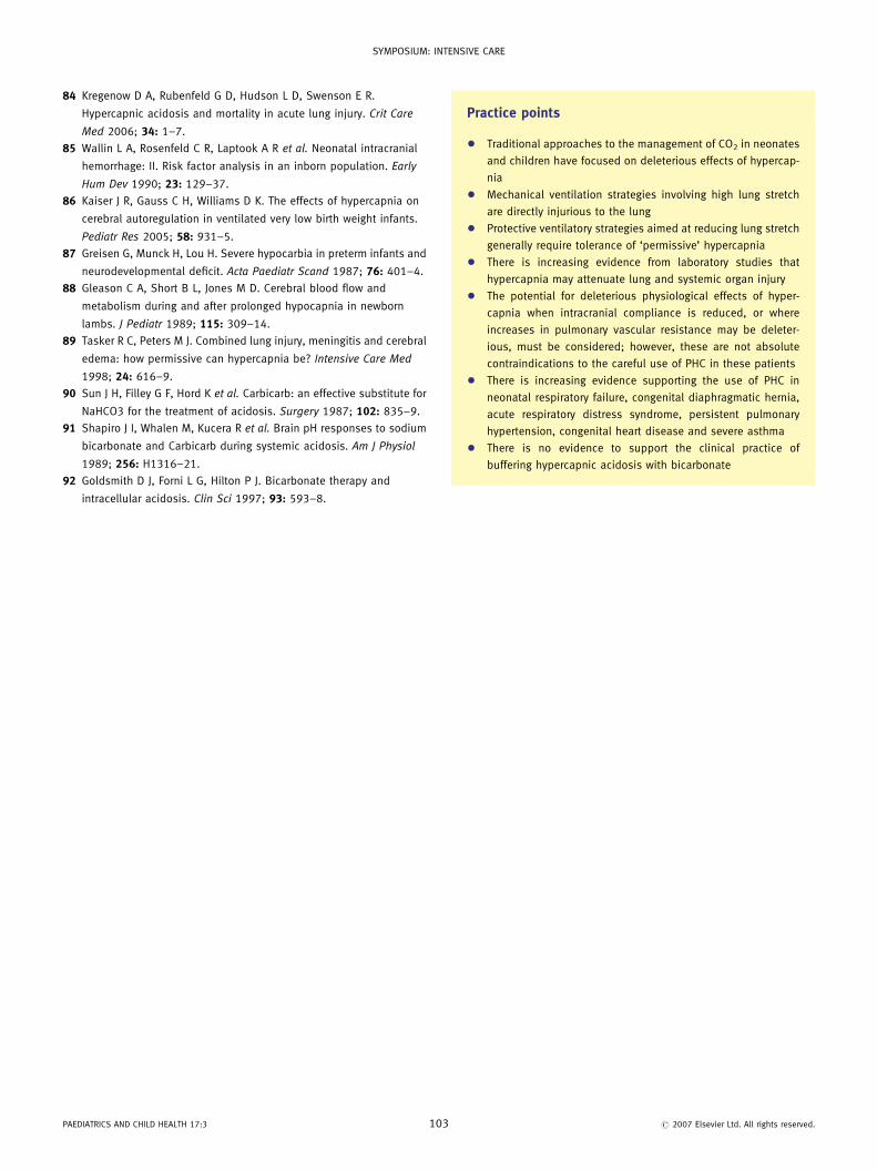
Practice points
� Traditional approaches to the management of CO2 in neonates
and children have focused on deleterious effects of hypercap-
nia
� Mechanical ventilation strategies involving high lung stretch
are directly injurious to the lung
� Protective ventilatory strategies aimed at reducing lung stretch
generally require tolerance of ‘permissive’ hypercapnia
� There is increasing evidence from laboratory studies that
hypercapnia may attenuate lung and systemic organ injury
� The potential for deleterious physiological effects of hyper-
capnia when intracranial compliance is reduced, or where
increases in pulmonary vascular resistance may be deleter-
ious, must be considered; however, these are not absolute
contraindications to the careful use of PHC in these patients
� There is increasing evidence supporting the use of PHC in
neonatal respiratory failure, congenital diaphragmatic hernia,
acute respiratory distress syndrome, persistent pulmonary
hypertension, congenital heart disease and severe asthma
� There is no evidence to support the clinical practice of
buffering hypercapnic acidosis with bicarbonate
r 2007 Elsevier Ltd. All rights reserved.
Copyright © 2022 FDOKUMEN