removals of licensed persons/registered institutions ... - SFC

Pentoxifylline, Inflammation, and Endothelial Function inHIV-Infected Persons: A Randomized, Placebo-ControlledTrialSamir K. Gupta1*, Deming Mi2, Michael P. Dube3, Chandan K. Saha2, Raymond M. Johnson1,
James H. Stein4, Matthias A. Clauss5, Kieren J. Mather6, Zeruesenay Desta7, Ziyue Liu2
1 Division of Infectious Diseases, Department of Medicine, Indiana University School of Medicine, Indianapolis, Indiana, United States of America, 2 Department of
Biostatistics, Indiana University School of Medicine, Indianapolis, Indiana, United States of America, 3 Division of Infectious Diseases, Department of Medicine, University of
Southern California Keck School of Medicine, Los Angeles, California, United States of America, 4 Division of Cardiovascular Medicine, Department of Medicine, University
of Wisconsin School of Medicine and Public Health, Madison, Wisconsin, United States of America, 5 Department of Cellular & Integrative Physiology, Center for Vascular
Biology, Indiana University School of Medicine, Indianapolis, Indiana, United States of America, 6 Division of Endocrinology & Metabolism, Department of Medicine,
Indiana University School of Medicine, Indianapolis, Indiana, United States of America, 7 Division of Clinical Pharmacology, Department of Medicine, Indiana University
School of Medicine, Indianapolis, Indiana, United States of America
Abstract
Background: Untreated HIV may increase the risk of cardiovascular events. Our preliminary in vitro and in vivo researchsuggests that pentoxifylline (PTX) reduces vascular inflammation and improves endothelial function in HIV-infected personsnot requiring antiretroviral therapy.
Methods: We performed a randomized, placebo-controlled trial of PTX 400 mg orally thrice daily for 8 weeks in 26participants. The primary endpoint was change in flow-mediated dilation (FMD) of the brachial artery after 8 weeks.Nitroglycerin-mediated dilation (NTGMD) and circulating markers of inflammation, cellular immune activation, coagulation,and metabolism were also assessed.
Results: The difference in mean absolute change (SD) in FMD after 8 weeks between the placebo [21.06 (1.45)%] and PTX[21.93 (3.03)%] groups was not significant (P = 0.44). No differences in NTGMD were observed. The only significantbetween-group difference in the changes in biomarkers from baseline to week 8 was in soluble tumor necrosis factorreceptor-1 (sTNFRI) [283.2 pg/mL in the placebo group vs. +65.9 pg/mL in the PTX group; P = 0.03]. PTX was generally well-tolerated.
Conclusions: PTX did not improve endothelial function and unexpectedly increased the inflammatory biomarker sTNFRI inHIV-infected participants not requiring antiretroviral therapy. Additional interventional research is needed to reduceinflammation and cardiovascular risk in this population.
Trial Registration: ClinicalTrials.gov NCT00796822
Citation: Gupta SK, Mi D, Dube MP, Saha CK, Johnson RM, et al. (2013) Pentoxifylline, Inflammation, and Endothelial Function in HIV-Infected Persons: ARandomized, Placebo-Controlled Trial. PLoS ONE 8(4): e60852. doi:10.1371/journal.pone.0060852
Editor: Shilpa J. Buch, University of Nebraska Medical Center, United States of America
Received November 1, 2012; Accepted March 1, 2013; Published April 9, 2013
Copyright: � 2013 Gupta et al. This is an open-access article distributed under the terms of the Creative Commons Attribution License, which permitsunrestricted use, distribution, and reproduction in any medium, provided the original author and source are credited.
Funding: This work was supported by the National Heart, Lung & Blood Institute at the National Institutes of Health [R01HL095149, R01HL09526]. Additionalsupport was provided by the Indiana Clinical and Translational Sciences Institute funded in part from the National Center for Advancing Translational Sciences,Clinical and Translational Sciences Award [Grant Number TR000006] and from the National Center for Research Resources [RR020128] at the National Institutes ofHealth. The funders had no role in study design, data collection and analysis, decision to publish, or preparation of the manuscript.
Competing Interests: SKG reports having received unrestricted research grant support from Gilead Sciences, Inc., Merck & Co., and Janssen (Tibotec)Therapeutics and receives consultant fees from Bristol-Myers Squibb. MPD reports having received research grant support from ViiV Healthcare/GlaxoSmithKlineand grant support from Serono. CKS reports having received statistical consulting fees from Merck & Co. and serves on a Data and Safety Monitoring Committeefor Merck & Co. JHS serves on the Data and Safety Monitoring Committees for Abbott, Lilly, and Takeda. KJM reports having received research support forunrelated projects from Merck & Co. All other authors have declared that no competing interests exist. This does not alter the authors’ adherence to all the PLOSONE policies on sharing data and materials.
* E-mail: [email protected]
Introduction
HIV-infected patients are at an increased risk for developing
cardiovascular disease (CVD) compared to the uninfected
population [1,2]. Much of this increased risk had previously been
attributed to the dysmetabolic effects associated with the use of
antiretroviral therapy (ART) [3]. However, recent data strongly
suggest that HIV-related systemic inflammation also contributes to
this excess CVD risk as well as overall mortality, especially in those
not receiving ART [4,5,6,7].
Endothelial dysfunction is a key and initial promoter of
atherosclerosis in the general population [8,9]. Because systemic
inflammation is associated with endothelial dysfunction [10],
PLOS ONE | www.plosone.org 1 April 2013 | Volume 8 | Issue 4 | e60852

reduction of inflammation may improve endothelial health and
consequently reduce the risk of future CVD events [11,12].
We have previously shown that pentoxifylline (PTX), a
phosphodiesterase inhibitor, can downregulate the endothelial
activation marker vascular cell adhesion molecule-1 (VCAM-1) in
an in vitro HIV-1 endothelial cell model [13]. In addition, we
demonstrated in an open-label, single arm, 8 week pilot trial of
PTX given to HIV-infected patients not requiring ART that PTX
use was well-tolerated and reduced circulating levels of sVCAM-1
and interferon-c-induced protein 10 (IP-10). Moreover, flow-
mediated dilation (FMD) of the brachial artery [14], a validated
measure of in vivo endothelial function and predictor of future
cardiovascular events [11,12], improved significantly. Taken
together, these data suggest that PTX may inhibit leukocyte
adhesion pathways that are involved in vascular inflammation and
dysfunction in those with HIV infection. If so, then PTX would
potentially be an inexpensive, safe, and readily available therapy to
reduce systemic inflammation and improve the cardiovascular risk
profile in the HIV-infected population. On the basis of these initial
observations, we conducted a randomized, double-blind, placebo-
controlled trial of PTX in HIV-infected participants not on ART
to test the hypothesis that PTX would reduce systemic inflamma-
tion and improve FMD in this population.
Methods
Study DesignThe protocol and informed consent form for this trial and
supporting CONSORT checklist are available as supporting
information; see Protocol and Consent S1 and Checklist S1.
We performed a randomized, double-blind, placebo-controlled,
parallel group trial of oral PTX 400 mg given thrice daily for 8
Figure 1. Flow of participants through the trial.doi:10.1371/journal.pone.0060852.g001
Pentoxifylline and Endothelial Function in HIV
PLOS ONE | www.plosone.org 2 April 2013 | Volume 8 | Issue 4 | e60852

weeks in HIV-infected study participants not requiring ART per
DHHS Guidelines at the time of the study (ClinicalTrials.gov
NCT00796822). We excluded those on ART in order to isolate
the effects of PTX on untreated HIV and to avoid the potentially
confounding effects of ART on endothelial function [15,16] and to
assess why lack of ART may predispose to endothelial dysfunction.
Participants had study procedures performed at baseline, 4 weeks,
and 8 weeks. Participants underwent assessment for eligibility at a
screening visit and within 21 days were then randomized 1:1 via a
computerized random-generated list (with a block size of 4 for the
first 24 participants and a block size of 2 for the remaining 2
participants) to either PTX or matching placebo. PTX was given
as 400 mg extended-release tablets purchased commercially. Both
PTX and placebo were over-encapsulated with gelatin capsules
with cellulose backfill to provide matching color, taste, size, smell,
and texture. Adherence was assessed by 3-day recall of missed
doses at each study visit. Adverse events were assessed at each visit
and in between scheduled visits as needed. Assessment of
successful blinding was performed at the end of trial participation
by asking the participants if they thought they knew to which study
arm they were assigned.
The protocol underwent one major amendment and revision in
February 2009 to clarify eligibility criteria. Over the course of the
trial, there were 8 protocol deviations (5 for out of window study
visits to accommodate participant schedules, 2 for out of window
measurements of HIV-1 RNA levels due to faulty lab equipment,
1 for not having advanced flow cytometry measurements
performed at one study visit).
Study PopulationParticipants were recruited from the HIV outpatient clinics
associated with the Indiana University Health medical system.
Primary inclusion criteria included documented HIV-1 infection,
age $18 years, CD4 cell count $350/mL at screening, no receipt
of ART within 6 months of screening and no anticipated need for
ART during the course of trial participation. Major exclusion
criteria included diagnosed cardiovascular disease, diabetes,
hypertension, thyroid abnormalities, other systemic inflammatory
disease (although hepatitis B or C co-infection was allowed);
pregnancy or breastfeeding during the trial; known intolerance to
PTX or other methylxanthines; creatinine clearance ,50 mL/
min, hemoglobin ,9.0 g/dL, alanine (ALT) or aspartate (AST)
aminotransferase .3 times upper limit of normal, total bilirubin
.2.5 times upper limit of normal; ongoing fever or active
infection/malignancy during a study visit; or use of anti-
inflammatory (including aspirin or non-steroidal anti-inflammato-
ry drugs), lipid-lowering, or anticoagulation agent at screening or
during the trial.
Study ProceduresParticipants were required to fast and not smoke for at least 8
hours prior to all study procedures. FMD and nitroglycerin-
mediated dilation (NTGMD) studies were performed at all study
visits according to recommended guidelines [17] by a single
registered vascular ultrasonographer who was certified by the
University of Wisconsin Atherosclerosis Imaging Research Pro-
gram Core Laboratory. After resting supine for 10-minutes in a
temperature-controlled room, a blood pressure cuff was placed on
the widest part of proximal right forearm approximately 1 cm
distal to the antecubital fossa. Using a 10 MHz resolution linear
array vascular ultrasound transducer with an Acuson CV70
ultrasound machine, the brachial artery was located above the
elbow and scanned in longitudinal sections. After recording
baseline B-mode digital images of the brachial artery and spectral
Doppler images of flow, the forearm cuff was inflated to
250 mmHg for 5 minutes to induce reactive hyperemia. Imme-
diately after deflation, spectral Doppler images are obtained to
verify hyperemia. FMD of the brachial artery was measured 60
and 90 seconds after cuff deflation. The relative FMD (%) was
calculated as the ratio between the largest post-cuff release and the
baseline diameter. Fifteen minutes later, repeat brachial artery
images were obtained and 400 mcg of sublingual nitroglycerin was
administered. The artery was re-imaged 3 minutes later. NTGMD
was calculated in an analogous fashion. Images were sent
electronically to the University of Wisconsin core imaging
laboratory for quality control and interpretation by a blinded,
single, experienced technician using Access Point Web software
(Freeland Systems, Westminster, CO).
Cellular immune activation, defined as circulating proportions
of CD3+CD8+CD38+HLA-DR+ cells, was assessed by flow
cytometry using fresh whole blood on the same day as the
collection at baseline and at week 8. Circulating serum levels of
PTX were measured using an in-house high performance liquid
chromatography assay. Circulating inflammatory markers [high
sensitivity C-reactive protein (hsCRP), serum interleukin-6 (IL-
6), soluble tumor necrosis factor-a receptors I and II (sTNFRI,
sTNFRII), tissue inhibitor of metalloproteinase-1 (TIMP-1),
monocyte chemoattractant protein-1 (MCP-1), interferon-c-
induced protein 10 (IP-10)], a coagulation marker [plasminogen
activating inhibitor antigen-1 (PAI-1 Ag)], an endothelial
activation marker [soluble vascular cell adhesion molecule-1
(sVCAM-1)], and metabolic markers [lipid fractions, insulin,
glucose] were measured in batches from archived frozen
samples (kept at 280uC) at the University of Vermont
Laboratory for Clinical Biochemistry Research. The homeostasis
model assessment-insulin resistance (HOMA-IR) was used to
estimate insulin resistance from fasting glucose and insulin
measures [18]. hsCRP, IL-6, IP-10, lipids, and insulin were
measured from serum, MCP-1, sTNFRI and II, sVCAM-1, and
TIMP-1 were measured from EDTA plasma, and PAI-1 Ag was
measured from citrated plasma. Safety laboratories were
assessed at the Indiana University Health commercial laboratory
at each study visit.
Statistical AnalysisThe sample size was determined based on a two-sample,
independent, two-tailed t-test with 5% type I error for the primary
Table 1. Baseline Characteristics of the Trial Participants(N = 26).
CharacteristicPlacebo(N = 13)
Pentoxifylline(N = 13)
Age, years 34 (10.9) 40 (11.6)
Male 11 (85%) 8 (62%)
Black race 8 (62%) 8 (62%)
Hispanic ethnicity 1 (8%) 0 (0%)
Current smoker 5 (38%) 6 (46%)
Body mass index, kg/m2 27.7 (5.9) 26.1 (4.6)
CD4 cell count/mL 583 (175) 524 (165)
HIV–1 RNA level, log10copies/mL 4.0 (1.2) 4.0 (0.7)
Notes: Data presented as means (standard deviations) or as No. (%). Nostatistically significant differences were found between these baselinecharacteristics.doi:10.1371/journal.pone.0060852.t001
Pentoxifylline and Endothelial Function in HIV
PLOS ONE | www.plosone.org 3 April 2013 | Volume 8 | Issue 4 | e60852

endpoint of change in FMD from baseline to week 8. Using the
results from our pilot trial [14], we conservatively estimated a
predicted absolute change in FMD of 3.5% with PTX (assuming
no change in placebo-treated participants) and we assumed a
common standard deviation of 2.6%. A sample size of 10 per
group was estimated to provide at least 80% power to detect this
effect size. Allowing for a 20% dropout rate, we planned to recruit
13 subjects per group.
Continuous variables were summarized by treatment groups
using descriptive statistics. Categorical variables were summarized
using frequency counts and percentages. Baseline clinical and
demographic data were compared between two treatment groups.
Continuous variables were summarized by treatment groups using
descriptive statistics. Categorical variables were summarized using
frequency counts and percentages. Baseline clinical and demo-
graphic data were compared between two treatment groups.
Categorical variables were examined using Fisher’s exact test. We
employed Student’s t-test for comparisons of continuous measures
as we found no evidence of violation of the normality assumption
for these variables; of note, HIV-1 RNA level, HOMA-IR, and
hsCRP required logarithmic transformation to approximate
normal distributions prior to such analysis. Independent two-
sample Student’s t-test was used to compare the mean changes in
FMD from baseline to week 8 between the placebo and PTX
groups. To account for missing week 8 FMD measures, Lachin’s
worst-rank analysis approach [19] was used as part of the intent-
to-treat analysis.
As we specifically wished to evaluate the effects of covariates on
the primary outcomes, we also performed multiple linear
regressions adjusted for treatment on the changes in FMD and
NTGMD for 4 and 8 weeks. In these models, the indicator
variable for PTX was kept regardless of its significance, while other
potential baseline covariates, including age, sex, race, body mass
index, FMD, and laboratory data, were included one at a time.
The computerized randomization list was generated by the
study statistician and kept by the study pharmacist who then
Figure 2. Changes in flow-mediated dilation (FMD) of the brachial artery and soluble tumor necrosis factor receptor-1 (sTNFRi)from baseline to week 8. Panel A shows the changes in FMD; Panel B shows the changes in sTNFRI. Circles indicate actual values. Top and bottomof boxes indicate 75th and 25th percentiles, respectively. Internal horizontal lines indicate median values and plus-signs indicate mean values. Externalhorizontal lines/whiskers indicate 25th or 75th percentiles 6 (1.5 times interquartile range).doi:10.1371/journal.pone.0060852.g002
Pentoxifylline and Endothelial Function in HIV
PLOS ONE | www.plosone.org 4 April 2013 | Volume 8 | Issue 4 | e60852
provided study drug or placebo in matching containers. Study
participants, study personnel, and all outcome assessors were
blinded to the allocation. Analyses were performed as intention to
treat but without corrections for multiple testing for the secondary
analyses. Two-sided P-values ,0.05 were considered statistically
significant. All analyses were performed in SAS 9.3 (SAS Inc.,
Cary, NC).
Ethics StatementThis trial was approved by the Indiana University Institutional
Review Board. All participants provided written, informed consent
prior to screening.
Results
Study Cohort CharacteristicsFigure 1 outlines the flow of the study participants through
the trial. Thirty-one potential participants underwent screening.
Study recruitment, enrollment, and follow-up assessments were
performed from May 2009 through October 2011. Of these, 1
was found to be pregnant, 1 required a prohibited medication,
1 could not provide blood samples (difficult venipuncture), and
2 withdrew consent prior to randomization. The characteristics
of the remaining 26 participants are shown in Table 1. The
majority of participants were non-Hispanic, non-smoking, black
men. Of note, no white women enrolled into the trial. The
mean (standard deviation, SD) CD4 cell count and HIV-1 RNA
level for the entire study group was 555 (169)/mL and 4.0 (0.9)
log10copies/mL, respectively. None had active hepatitis B or C
co-infection. There were no significant differences in the
baseline characteristics between arms. All 13 participants in
the placebo arm completed the 8 week trial. However, 2 of the
PTX participants were lost to follow-up by week 4, 1 developed
Grade 2 neutropenia at week 4 and was subsequently
withdrawn, and 1 had poor ultrasound data quality at week
8. Thus, 11 and 9 participants, respectively, of the 13 initial
PTX participants had evaluable vascular imaging data at the
weeks 4 and 8 study visits; 11 and 10, respectively, in the PTX
group and samples available for biomarker analysis.
At the 4 week visit, 15 of the 24 evaluable study participants,
respectively, claimed no missed study drug and the remainder
claimed no more than 3 missed doses in the 3 days prior to the
study visit. At week 8, 12 of the evaluable 22 participants claimed
no missed doses and 7 of the remaining 10 claimed no more than 3
missed doses in the 3 days prior to the study visit. At weeks 4 and
8, 3 PTX participants at each time point had no measurable PTX
drug concentration; 2 of the 3 PTX participants had no
measurable drug concentration at both weeks 4 and 8.
Because one participant in the PTX group was removed due to
an adverse event, unblinding of this participant’s randomization
assignment may have occurred. To assess potential selection bias
after withdrawal of this participant, the Berger-Exner test [20] was
performed and was found to have a P-value of 0.11, suggesting no
significant selection bias.
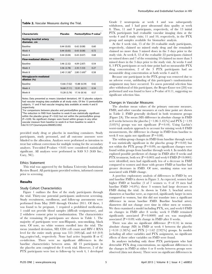
Changes in Vascular MeasuresThe absolute mean values of the primary outcome measure,
FMD, and other vascular measures at each time point are shown
in Table 2. FMD generally declined in both treatment groups
(Figure 2A). The mean (SD) difference in absolute change in FMD
at 8 weeks between the placebo [21.06 (1.45)%] and PTX [21.93
(3.03)%] groups was not significant (P = 0.44). Using Lachin’s
worst-rank analysis approach to account for missing week 8 FMD
measurements, the difference in change in FMD from baseline to
week 8 was again not significant (P = 0.08).
The within-group changes in FMD from baseline through week
8 was statistically significant in the placebo group (P = 0.02) but
not within the PTX group (P = 0.09); no significant changes were
found within groups from baseline through week 4. In models that
explored possible predictors of change in FMD after 8 weeks and
PTX treatment, both sex (P = 0.003) and week 0 FMD (P,0.0001)
were identified; men had significantly less of a decrease in FMD
compared to women and those with greater FMD at baseline had
greater decreases in FMD. Of note, smoking status was not
associated with FMD change.
A post-hoc exploratory analysis of differences in FMD by sex
and baseline FMD is shown in Figure 3. As expected, women had
higher FMD at baseline (3 of 7 women vs. 0 of 19 men had
baseline FMD .6.0%); these 3 women had large decreases in
FMD during the trial. As shown in Table 3, brachial artery
diameters at baseline were, as expected, significantly larger in men
compared to women, but this did not translate into a significant
difference in mean baseline FMD. Baseline brachial artery
diameters did not change over time in either men or women.
We then examined a model including PTX, sex, and week 0 FMD
on changes in FMD after 8 weeks; week 0 FMD remained
significantly associated (P = 0.0009) and sex was marginally
associated (P = 0.09) with change in FMD after 8 weeks.
There was also no significant difference (P = 0.14) in mean
absolute change (SD) in FMD at week 4 between the placebo
[20.10 (1.58)%] and PTX [21.62 (2.92)%] groups. In models
including all other variables and PTX assignment, no significant
effects of these other variables on FMD were found.
In analyses including only those PTX participants who had
detectable PTX drug concentrations, no significant differences in
the changes in FMD at either 4 or 8 weeks between groups were
observed (data not shown). There were no significant differences in
Table 2. Vascular Measures during the Trial.
Characteristic Placebo Pentoxifylline P-value1
Resting brachial arterydiameter, cm
Baseline 0.44 (0.05) 0.42 (0.08) 0.60
Week 4 0.44 (0.05) 0.43 (0.08) 0.72
Week 8 0.44 (0.04) 0.43 (0.07) 0.68
Flow-mediated dilation (%)
Baseline 3.46 (2.32) 4.09 (2.87) 0.54
Week 4 3.36 (2.78) 2.35 (1.53) 0.27
Week 8 2.40 (1.58)2 2.80 (1.69)2 0.58
Nitroglycerin-mediateddilation (%)
Baseline 13.66 (7.02) 15.88 (6.59) 0.42
Week 4 16.68 (7.11) 13.91 (6.51) 0.34
Week 8 15.28 (5.76) 17.16 (8.16) 0.57
Notes: Data presented as means (standard deviation). All 13 placebo subjectshad vascular imaging data available at all study visits. Of the 13 pentoxifyllinesubjects, 11 and 9 had vascular imaging data available at weeks 4 and 8respectively.1For comparisons between placebo and pentoxifylline groups.2The reduction in FMD from baseline to week 8 was statistically significantwithin the placebo group (P = 0.02) but not within the pentoxifylline group(P = 0.09). No significant changes were found within groups in any othervascular measure from baseline to week 8 or from baseline to week 4.doi:10.1371/journal.pone.0060852.t002
Pentoxifylline and Endothelial Function in HIV
PLOS ONE | www.plosone.org 5 April 2013 | Volume 8 | Issue 4 | e60852

change in FMD at week 8 in those who did have and did not have
detectable PTX levels (data not shown).
There were no significant changes in NTGMD at either week 4
or week 8 between the two groups.
Changes in BiomarkersThe absolute mean values of the immunologic, virologic, inflam-
matory, coagulation, and metabolic parameters assessed during the
trial are presented in Table 4. PAI-1 Ag was significantly higher in the
PTX group at both baseline and at week 8. However, there were no
significant differences in the changes in PAI-1 Ag between groups. As
shown in Figure 2B, the only significant difference in the changes in
these biomarkers from baseline to week 8 was in sTNFRI [283.2 pg/
mL in the placebo group vs. 65.9 pg/mL in the PTX group; P = 0.03].
This change in sTNFRI was not significantly correlated with the
change in FMD. Of note, there were no significant within-group
changes in any biomarker through week 8.
SafetyThere were no serious adverse events during the trial. As shown
in Table 5, most study participants reported at least one physical
symptom during the trial, none of which were treatment-limiting.
There were no significant differences between the number of total
reported symptoms or laboratory abnormalities in the placebo
group compared to the PTX group. All toxicities were considered
grade 1 and not treatment-limiting except for one grade 2
neutropenia which prompted study discontinuation in 1 partici-
pant at week 4.
Discussion
PTX did not reduce circulating markers of inflammation and
did not improve arterial FMD, a measure of endothelial function,
in this randomized, placebo-controlled trial of HIV-infected
persons not requiring ART. PTX also did not reduce the levels
Figure 3. Individual changes in FMD by treatment group (pentoxifylline vs. placebo) and by sex (female vs. male).doi:10.1371/journal.pone.0060852.g003
Pentoxifylline and Endothelial Function in HIV
PLOS ONE | www.plosone.org 6 April 2013 | Volume 8 | Issue 4 | e60852

of the leukocyte adhesion molecules sVCAM-1 or IP-10, although
this effect was found in our smaller, open-label pilot study [14] and
in our in vitro endothelial cell model combining TNF-a with HIV-
secreted proteins [13]. The inflammatory marker sTNFRI
surprisingly increased in the PTX arm compared to placebo.
We cannot discount the possibility that the increase in sTNFRI
in the PTX group would have an adverse clinical impact if PTX
were continued as a chronic intervention. However, we did not
find a correlation between change in sTNFRI and FMD in this
study, thus making it difficult to make any inference of the clinical
relevance of the change in sTNFRI as it relates to endothelial
function. In addition, no changes in other inflammatory markers
were significantly different between groups, making the change in
this one biomarker potentially a chance finding due to multiple
testing. Previous studies have suggested that PTX reduces TNF-aexpression in vitro and in vivo by inhibiting nuclear factor-kappa B
and may thus even inhibit HIV replication [21,22,23,24].
However, Clerici et al. found that TNF-a expression may actually
increase during the first 12 weeks of use of PTX 400 mg thrice
daily [25], a finding which is concordant our trial’s finding of
significant increases in sTNFRI, a more stable circulating marker
of TNF-a production than circulating TNF-a itself. These
contrasting results may be explained by the fact that the study
by Clerici et al. and the current trial included asymptomatic
patients with relatively preserved CD4 cell counts who were not
receiving ART as opposed to the previously mentioned studies
that involved severely immunocompromised patients with higher
levels of TNF-a. This suggests that PTX may be more beneficial in
patients with reduced CD4 counts and who may have a greater
inflammatory burden, a group we are currently studying in a
separate PTX trial in patients initiating ART (NCT00864916).
Despite its purported beneficial effects on the endothelium, the
influence of oral PTX on vascular function in humans has only
previously been studied in HIV-negative, type 2 diabetics [26]. In
that study, similar to the current report, there was no benefit of
PTX on endothelial function. The characteristics of the partici-
pants in our smaller pilot study [14] were generally similar to those
who enrolled in this larger and more definitive trial. The only
appreciable differences were the greater number of black
participants in the current trial compared to the pilot study
(62% vs. 33%) and the greater body mass index in the current trial
participants (26.9 vs. 20.6 kg/m2). However, neither black race
nor body mass index were predictors of FMD change in this trial
and so were unlikely to have led to these discrepant results. In
addition, the PTX drug concentrations found at weeks 4 and 8
were comparable to those seen in our pilot trial. Of note,
NTGMD did not change in either arm during this trial, suggesting
that there were no changes in the inherent ability of the vascular
endothelium to react to nitric oxide. Therefore, it is not clear why
we observed the impressive improvements in FMD in our pilot
trial and yet negative results in the current trial, although we
cannot rule out the possibilities that the pilot trial’s results were
due to chance or biased due to the open-label design.
FMD generally declined in this study cohort, with the largest
reductions in FMD occurring in those with the highest FMD at
baseline, thus, regression to the mean cannot be excluded. When
examining baseline FMD and sex together, baseline FMD
remained significantly associated with changes in FMD after 8
weeks with sex only marginally associated with this change. A
larger sample size may have led to finding that sex remained
independently associated with change in FMD. Of note, we
cannot exclude the possibility that PTX may be beneficial in white
HIV-infected women as none were enrolled in this trial.
Although this study was relatively small, the sample size was
conservatively and justifiably based on our positive pilot results.
Moreover, given the FMD outcomes observed, it would be
unlikely to find differences between arms, let alone a positive
benefit of PTX, in a larger trial. This sample had sufficient power
to find differences in the inflammatory markers found to be
significantly reduced in our pilot trial, namely sVCAM-1 and IP-
10. However, we recognize that a larger sample potentially could
have provided greater power to find differences in these secondary
endpoint measures and allowed better assessments of the changes
in FMD and biomarkers in specific subgroups.
Overall, PTX was well-tolerated. Compared to placebo, the
PTX participants did not report a greater frequency of gastroin-
testinal adverse events which have been previously associated with
the use of this drug [22]. Although there was not a statistically
significant increase in laboratory toxicities with PTX, the two
neutropenia events, one of which led to drug discontinuation,
suggests that this particular adverse event should be monitored for
closely in other trials of PTX. In addition, the two participants
who were lost to follow-up were both randomized to the PTX
arm, so we cannot exclude the possibility of PTX-related adverse
events in these two participants.
Our trial is unique in that changes in FMD and the biomarkers
purportedly associated with cardiovascular disease in the HIV-
infected population were assessed over time without the con-
founding influence of ART. This was possible as the treatment
guidelines at the time at which this study was performed
recommended ART initiation only in those with CD4 cell counts
,350/mL. With current recommendations to initiate ART
immediately without regard to CD4 cell count, such studies will
likely not be possible in the future. We also note that the negative
results of this current trial do not necessarily extend to those
receiving ART, although we will examine this possibility in the
aforementioned second trial of PTX.
In conclusion, PTX did not reduce systemic inflammation or
improve endothelial function in HIV-infected persons not
requiring antiretroviral therapy. Additional research investigating
the utility of other anti-inflammatory interventions is clearly
needed.
Table 3. Vascular Measures during the Trial by Sex.
Characteristic Men Women P-value1
Resting brachial arterydiameter, cm
Baseline 0.45 (19; 0.05) 0.38 (7; 0.06) 0.04
Week 4 0.45 (18; 0.05) 0.37 (6; 0.06) 0.02
Week 8 0.44 (17; 0.04) 0.39 (5; 0.07) 0.15
Flow-mediateddilation (%)
Baseline 3.20 (19; 1.42) 5.33 (7; 4.21) 0.24
Week 4 2.54 (18; 1.57) 3.98 (6; 3.76) 0.40
Week 8 2.56 (17; 1.52) 2.59 (5; 2.04) 0.97
Notes: Data presented as means (N; standard deviations). All 13 placebosubjects had vascular imaging data available at all study visits. Of the 13pentoxifylline subjects, 11 and 9 had vascular imaging data available at weeks 4and 8 respectively.1For comparisons between men and women.doi:10.1371/journal.pone.0060852.t003
Pentoxifylline and Endothelial Function in HIV
PLOS ONE | www.plosone.org 7 April 2013 | Volume 8 | Issue 4 | e60852

Table 4. Immunologic, Virologic, Inflammatory, Coagulation, Metabolic, and Pentoxifylline Concentration Levels during the Trial.
Characteristic Placebo Pentoxifylline P-value1
CD4 cell count/mL
Baseline 583 (175) 524 (165) 0.39
Week 4 534 (206) 601 (269) 0.52
Week 8 526 (165) 494 (208) 0.72
CD3+CD8+CD38+HLA-DR+proportions (%)
Baseline 36 (19) 39 (15) 0.69
Week 8 36 (19) 41 (17) 0.52
HIV-1 RNA, log10copies/mL
Baseline 4.0 (1.2) 4.0 (0.7) 0.88
Week 4 3.9 (1.1) 4.0 (4.5) 0.72
Week 8 4.0 (1.0) 4.0 (0.8) 0.87
hsCRP, log10mg/L
Baseline 0.27 (0.41) 0.25 (0.57) 0.95
Week 4 0.36 (0.57) 0.37 (0.80) 0.74
Week 8 20.01 (0.45) 0.15 (0.81) 0.58
IL-6, pg/ml
Baseline 1.91 (1.53) 2.61 (2.59) 0.41
Week 4 3.44 (6.52) 2.91 (2.45) 0.79
Week 8 1.7 (1.55) 5.26 (10.2) 0.30
TIMP-1, ng/ml
Baseline 99 (17) 117 (35) 0.12
Week 4 98 (20) 112 (31) 0.21
Week 8 91 (14) 109 (25) 0.06
sVCAM-1, ng/mL
Baseline 1037 (399) 1108 (273) 0.60
Week 4 1081 (419) 1156 (283) 0.6
Week 8 1098 (462) 1130 (281) 0.84
sTNFRI, pg/mL
Baseline 1125 (249) 851 (379) 0.04
Week 4 1047 (244) 805 (298) 0.04
Week 8 1042 (205) 805 (298) 0.12
sTNFRII, pg/mL
Baseline 7239 (2606) 7736 (2141) 0.60
Week 4 7163 (2106) 7762 (2562) 0.54
Week 8 7455 (2405) 7549 (1945) 0.92
MCP-1, pg/mL
Baseline 237 (75) 202 (76) 0.24
Week 4 193 (55) 205 (100) 0.71
Week 8 217 (55) 207 (106) 0.80
IP-10, pg/mL
Baseline 432 (330) 552 (331) 0.36
Week 4 386 (283) 528 (324) 0.27
Week 8 377 (295) 440 (182) 0.53
PAI-1 Ag, ng/ml
Baseline 17.3 (6.9) 43.3 (27.5) 0.01
Week 4 20.3 (13.0) 22.4 (14.5) 0.72
Week 8 18.6 (8.6) 29.4 (10.6) 0.02
Total cholesterol, mg/dL
Baseline 150 (34) 149 (27) 0.92
Pentoxifylline and Endothelial Function in HIV
PLOS ONE | www.plosone.org 8 April 2013 | Volume 8 | Issue 4 | e60852

Acknowledgments
We thank Ms. Beth Zwickl, NP for study coordination, Mr.
Jeffrey Waltz, RDCS for performing the vascular ultrasonography
studies, and Mr. Jonathon Mathews, BS for data management. We
also thank Dr. Homer Twigg and Ms. Patricia Smith for
performing the flow cytometry studies, Dr. Russell Tracy and
Ms. Elaine Cornell for performing the batched biomarker assays,
and Ms. Bonnie Klank and Denise Cox for preparing the study
drug and matching placebo. Most of all, we thank the study
participants for their generous participation.
Supporting Information
Checklist S1 CONSORT checklist.(DOC)
Protocol and Consent S1 The revised (February 5, 2011)trial protocol and informed consent form.(DOC)
Author Contributions
Conceived and designed the experiments: SKG MPD CKS RMJ MAC
KJM ZD. Performed the experiments: SKG JHS ZD. Analyzed the data:
SKG DM MPD CKS JHS KJM ZD ZL. Contributed reagents/materials/
analysis tools: DM CKS JHS ZD ZL. Wrote the paper: SKG DM MPD
CKS RMJ JHS MAC KJM ZL.
Table 4. Cont.
Characteristic Placebo Pentoxifylline P-value1
Week 4 145 (38) 152 (31) 0.67
Week 8 146 (26) 142 (34) 0.78
HDL-C, mg/dL
Baseline 39 (11) 38 (11) 0.68
Week 4 38 (10) 39 (12) 0.85
Week 8 39 (11) 35 (10) 0.33
LDL-C, mg/dL
Baseline 95 (29) 90 (28) 0.65
Week 4 92 (31) 95 (31) 0.83
Week 8 90 (21) 86 (28) 0.73
Triglycerides, mg/dL
Baseline 81 (29) 108 (45) 0.08
Week 4 77 (30) 89 (41) 0.43
Week 8 84 (33) 112 (78) 0.31
HOMA-IR
Baseline 2.04 (2.19) 2.96 (3.97) 0.48
Week 4 1.97 (1.82) 2.28 (2.99) 0.78
Week 8 2.19 (1.72) 2.85 (2.92) 0.54
Pentoxifylline concentration, ng/mL
Week 4 0 (0) 89 (120)
Week 8 0 (0) 61 (65)
Notes: Data presented as means (standard deviations) or as No. (%). All 13 placebo subjects had samples available at all study visits. Of the 13 pentoxifylline subjects, 11and 10 had samples available at weeks 4 and 8, respectively. hsCRP, high sensitivity C-reactive protein; IL-6, interleukin-6; HDL-C, high density lipoprotein-cholesterol;LDL-C, low density lipoprotein-cholesterol; TIMP-1, tissue inhibitor of metalloproteinase-1; sVCAM-1, soluble vascular cell adhesion molecule-1; sTNFRI and sTNFRII,soluble tumor necrosis factor-a receptors I and II; MCP-1, monocyte chemoattractant protein-1; IP-10, interferon-c-induced protein-10; PAI-1 Ag, plasminogen activatinginhibitor antigen-1; HOMA-IR, homeostatic model assessment-insulin resistance.1For comparisons between placebo and pentoxifylline groups.doi:10.1371/journal.pone.0060852.t004
Table 5. Adverse Events during the Trial.
Characteristic Placebo Pentoxifylline P-value1
Symptoms
Total 21 14 0.73
Gastrointestinal 8 7
Rash 2 1
Flushing 2 0
Headache 1 2
Cough 2 0
Other 6 4
Laboratory Abnormalities
Total 2 6 0.86
Neutropenia 0 2
Hypokalemia 0 1
Hyperkalemia 1 0
Elevated liver function tests 1 1
Hyperglycemia 0 1
Dipstick proteinuria 0 1
Notes: Data are cumulative number of adverse events.1For comparisons between placebo and pentoxifylline groups.doi:10.1371/journal.pone.0060852.t005
Pentoxifylline and Endothelial Function in HIV
PLOS ONE | www.plosone.org 9 April 2013 | Volume 8 | Issue 4 | e60852

References
1. Triant VA, Lee H, Hadigan C, Grinspoon SK (2007) Increased acute
myocardial infarction rates and cardiovascular risk factors among patients withhuman immunodeficiency virus disease. J Clin Endocrinol Metab 92: 2506–
2512.2. Obel N, Thomsen HF, Kronborg G, Larsen CS, Hildebrandt PR, et al. (2007)
Ischemic heart disease in HIV-infected and HIV-uninfected individuals: a
population-based cohort study. Clin Infect Dis 44: 1625–1631.3. Friis-Moller N, Reiss P, Sabin CA, Weber R, Monforte A, et al. (2007) Class of
antiretroviral drugs and the risk of myocardial infarction. New England Journalof Medicine 356: 1723–1735.
4. Calmy A, Gayet-Ageron A, Montecucco F, Nguyen A, Mach F, et al. (2009)
HIV increases markers of cardiovascular risk: results from a randomized,treatment interruption trial. AIDS 23: 929–939.
5. Kuller LH, Tracy R, Belloso W, De Wit S, Drummond F, et al. (2008)Inflammatory and coagulation biomarkers and mortality in patients with HIV
infection. PLoS Med 5: e203.6. El-Sadr WM, Lundgren JD, Neaton JD, Gordin F, Abrams D, et al. (2006)
CD4+ count-guided interruption of antiretroviral treatment. N Engl J Med 355:
2283–2296.7. Grunfeld C, Delaney JAC, Wanke C, Currier JS, Scherzer R, et al. (2009)
Preclinical atherosclerosis due to HIV infection: carotid intima-medial thicknessmeasurements from the FRAM study. AIDS 23: 1841–1849.
8. Gokce N, Keaney JF, Jr., Hunter LM, Watkins MT, Nedeljkovic ZS, et al.
(2003) Predictive value of noninvasively determined endothelial dysfunction forlong-term cardiovascular events in patients with peripheral vascular disease.
Journal of the American College of Cardiology 41: 1769–1775.9. Suwaidi JA, Hamasaki S, Higano ST, Nishimura RA, Holmes DR, Jr., et al.
(2000) Long-term follow-up of patients with mild coronary artery disease andendothelial dysfunction. Circulation 101: 948–954.
10. Kang SM, Chung N, Kim JY, Koo BK, Choi D, et al. (2002) Relation of
vasodilator response of the brachial artery to inflammatory markers in patientswith coronary artery disease. Echocardiography 19: 661–667.
11. Suessenbacher A, Frick M, Alber HF, Barbieri V, Pachinger O, et al. (2006)Association of improvement of brachial artery flow-mediated vasodilation with
cardiovascular events. Vasc Med 11: 239–244.
12. Modena MG, Bonetti L, Coppi F, Bursi F, Rossi R (2002) Prognostic role ofreversible endothelial dysfunction in hypertensive postmenopausal women.
Journal of the American College of Cardiology 40: 505–510.13. Green LA, Kim C, Gupta SK, Rajashekhar G, Rehman J, et al. (2012, in press)
Pentoxifylline Reduces Tumor Necrosis Factor-alpha and HIV-InducedVascular Endothelial Activation. AIDS Research and Human Retroviruses.
14. Gupta SK, Johnson RM, Mather KJ, Clauss M, Rehman J, et al. (2010) Anti-
inflammatory treatment with pentoxifylline improves HIV-related endothelialdysfunction: a pilot study. AIDS 24: 1377–1380.
15. Hsue PY, Hunt PW, Wu Y, Schnell A, Ho JE, et al. (2009) Association of
abacavir and impaired endothelial function in treated and suppressed HIV-
infected patients. AIDS 23: 2021–2027.
16. Gupta SK, Shen C, Moe SM, Kamendulis LM, Goldman M, et al. (2012, in
press.) Worsening endothelial function with efavirenz compared to protease
inhibitors: A 12-month prospective study. PLOS ONE.
17. Corretti MC, Anderson TJ, Benjamin EJ, Celermajer D, Charbonneau F, et al.
(2002) Guidelines for the ultrasound assessment of endothelial-dependent flow-
mediated vasodilation of the brachial artery: a report of the International
Brachial Artery Reactivity Task Force. Journal of the American College of
Cardiology 39: 257–265.
18. Matthews DR, Hosker JP, Rudenski AS, Naylor BA, Treacher DF, et al. (1985)
Homeostasis model assessment: insulin resistance and beta-cell function from
fasting plasma glucose and insulin concentrations in man. Diabetologia 28: 412–
419.
19. Lachin JM (1999) Worst-rank score analysis with informatively missing
observations in clinical trials. Controlled Clinical Trials 20: 408–422.
20. Berger VW, Exner DV (1999) Detecting selection bias in randomized clinical
trials. Controlled Clinical Trials 20: 319–327.
21. Dezube BJ, Lederman MM (1995) Pentoxifylline for the treatment of HIV
infection and its complications. Journal of Cardiovascular Pharmacology 25
Suppl 2: S139–142.
22. Dezube BJ, Lederman MM, Spritzler JG, Chapman B, Korvick JA, et al. (1995)
High-dose pentoxifylline in patients with AIDS: inhibition of tumor necrosis
factor production. National Institute of Allergy and Infectious Diseases AIDS
Clinical Trials Group. Journal of Infectious Diseases 171: 1628–1632.
23. Dezube BJ, Pardee AB, Chapman B, Beckett LA, Korvick JA, et al. (1993)
Pentoxifylline decreases tumor necrosis factor expression and serum triglycerides
in people with AIDS. NIAID AIDS Clinical Trials Group. Journal of Acquired
Immune Deficiency Syndromes 6: 787–794.
24. Fazely F, Dezube BJ, Allen-Ryan J, Pardee AB, Ruprecht RM (1991)
Pentoxifylline (Trental) decreases the replication of the human immunodefi-
ciency virus type 1 in human peripheral blood mononuclear cells and in cultured
T cells. Blood 77: 1653–1656.
25. Clerici M, Piconi S, Balotta C, Trabattoni D, Capetti A, et al. (1997)
Pentoxifylline improves cell-mediated immunity and reduces human immuno-
deficiency virus (HIV) plasma viremia in asymptomatic HIV-seropositive
persons. Journal of Infectious Diseases 175: 1210–1215.
26. Bilsborough W, O’Driscoll G, Stanton K, Weerasooriya R, Dembo L, et al.
(2002) Effect of lowering tumour necrosis factor-alpha on vascular endothelial
function in Type II diabetes. Clin Sci 103: 163–169.
Pentoxifylline and Endothelial Function in HIV
PLOS ONE | www.plosone.org 10 April 2013 | Volume 8 | Issue 4 | e60852
Copyright © 2022 FDOKUMEN