Obesogenic Family Types Identified through Latent Profile Analysis
Upload
independentCategory
view
1download
0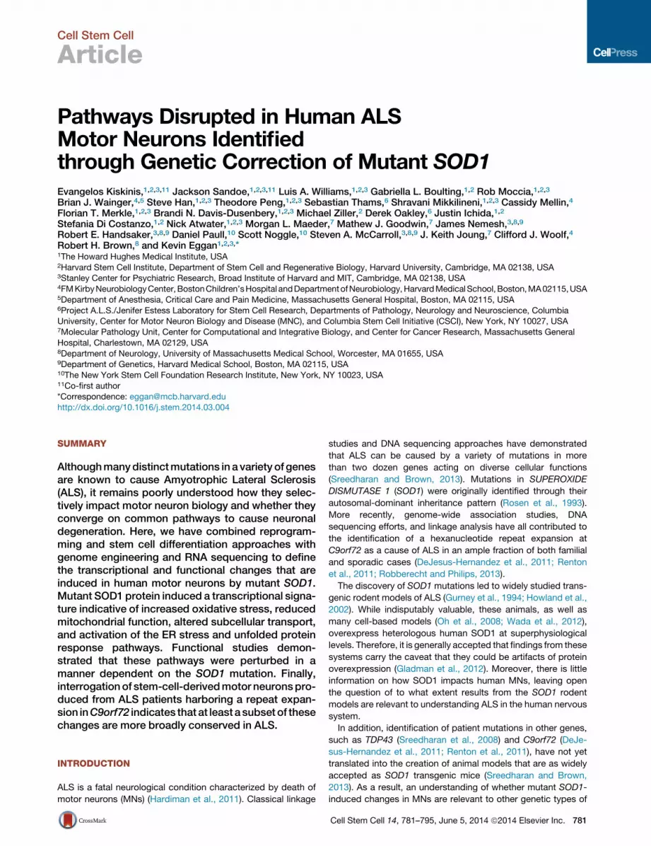
Cell Stem Cell
Article
Pathways Disrupted in Human ALSMotor Neurons Identifiedthrough Genetic Correction of Mutant SOD1Evangelos Kiskinis,1,2,3,11 Jackson Sandoe,1,2,3,11 Luis A. Williams,1,2,3 Gabriella L. Boulting,1,2 Rob Moccia,1,2,3
Brian J. Wainger,4,5 Steve Han,1,2,3 Theodore Peng,1,2,3 Sebastian Thams,6 Shravani Mikkilineni,1,2,3 Cassidy Mellin,4
Florian T. Merkle,1,2,3 Brandi N. Davis-Dusenbery,1,2,3 Michael Ziller,2 Derek Oakley,6 Justin Ichida,1,2
Stefania Di Costanzo,1,2 Nick Atwater,1,2,3 Morgan L. Maeder,7 Mathew J. Goodwin,7 James Nemesh,3,8,9
Robert E. Handsaker,3,8,9 Daniel Paull,10 Scott Noggle,10 Steven A. McCarroll,3,8,9 J. Keith Joung,7 Clifford J. Woolf,4
Robert H. Brown,8 and Kevin Eggan1,2,3,*1The Howard Hughes Medical Institute, USA2Harvard Stem Cell Institute, Department of Stem Cell and Regenerative Biology, Harvard University, Cambridge, MA 02138, USA3Stanley Center for Psychiatric Research, Broad Institute of Harvard and MIT, Cambridge, MA 02138, USA4FMKirbyNeurobiologyCenter,BostonChildren’sHospital andDepartmentofNeurobiology,HarvardMedical School,Boston,MA02115,USA5Department of Anesthesia, Critical Care and Pain Medicine, Massachusetts General Hospital, Boston, MA 02115, USA6Project A.L.S./Jenifer Estess Laboratory for Stem Cell Research, Departments of Pathology, Neurology and Neuroscience, ColumbiaUniversity, Center for Motor Neuron Biology and Disease (MNC), and Columbia Stem Cell Initiative (CSCI), New York, NY 10027, USA7Molecular Pathology Unit, Center for Computational and Integrative Biology, and Center for Cancer Research, Massachusetts General
Hospital, Charlestown, MA 02129, USA8Department of Neurology, University of Massachusetts Medical School, Worcester, MA 01655, USA9Department of Genetics, Harvard Medical School, Boston, MA 02115, USA10The New York Stem Cell Foundation Research Institute, New York, NY 10023, USA11Co-first author*Correspondence: [email protected]
http://dx.doi.org/10.1016/j.stem.2014.03.004
SUMMARY
Althoughmanydistinctmutations in a variety of genesare known to cause Amyotrophic Lateral Sclerosis(ALS), it remains poorly understood how they selec-tively impact motor neuron biology and whether theyconverge on common pathways to cause neuronaldegeneration. Here, we have combined reprogram-ming and stem cell differentiation approaches withgenome engineering and RNA sequencing to definethe transcriptional and functional changes that areinduced in human motor neurons by mutant SOD1.Mutant SOD1 protein induced a transcriptional signa-ture indicative of increased oxidative stress, reducedmitochondrial function, altered subcellular transport,and activation of the ER stress and unfolded proteinresponse pathways. Functional studies demon-strated that these pathways were perturbed in amanner dependent on the SOD1 mutation. Finally,interrogationof stem-cell-derivedmotor neuronspro-duced from ALS patients harboring a repeat expan-sion inC9orf72 indicates that at least a subset of thesechanges are more broadly conserved in ALS.
INTRODUCTION
ALS is a fatal neurological condition characterized by death of
motor neurons (MNs) (Hardiman et al., 2011). Classical linkage
studies and DNA sequencing approaches have demonstrated
that ALS can be caused by a variety of mutations in more
than two dozen genes acting on diverse cellular functions
(Sreedharan and Brown, 2013). Mutations in SUPEROXIDE
DISMUTASE 1 (SOD1) were originally identified through their
autosomal-dominant inheritance pattern (Rosen et al., 1993).
More recently, genome-wide association studies, DNA
sequencing efforts, and linkage analysis have all contributed to
the identification of a hexanucleotide repeat expansion at
C9orf72 as a cause of ALS in an ample fraction of both familial
and sporadic cases (DeJesus-Hernandez et al., 2011; Renton
et al., 2011; Robberecht and Philips, 2013).
The discovery of SOD1 mutations led to widely studied trans-
genic rodent models of ALS (Gurney et al., 1994; Howland et al.,
2002). While indisputably valuable, these animals, as well as
many cell-based models (Oh et al., 2008; Wada et al., 2012),
overexpress heterologous human SOD1 at superphysiological
levels. Therefore, it is generally accepted that findings from these
systems carry the caveat that they could be artifacts of protein
overexpression (Gladman et al., 2012). Moreover, there is little
information on how SOD1 impacts human MNs, leaving open
the question of to what extent results from the SOD1 rodent
models are relevant to understanding ALS in the human nervous
system.
In addition, identification of patient mutations in other genes,
such as TDP43 (Sreedharan et al., 2008) and C9orf72 (DeJe-
sus-Hernandez et al., 2011; Renton et al., 2011), have not yet
translated into the creation of animal models that are as widely
accepted as SOD1 transgenic mice (Sreedharan and Brown,
2013). As a result, an understanding of whether mutant SOD1-
induced changes in MNs are relevant to other genetic types of
Cell Stem Cell 14, 781–795, June 5, 2014 ª2014 Elsevier Inc. 781
B C
E
A
ISL / TUJ1 / DNA
Day 30
Day 3 Day 30
18a
-con
trol
Day 3
39b-
SO
D1+/
A4V
Fixed, stained and analyzed
on days 3, 15, 21, 30
day 3 day 15 day 30
iPSCs
embryoid bodies (EBs)
Motor neuron specification
Neuralization
motor neurons (MNs)
day -24 day -23 day -17 day 0
Patientfibroblasts
OCT4, SOX2, KLF4
Quality control
Neu
ron
num
ber r
elat
ive
to d
3 (%
)/ i el cun
eviti sopL
EN
UT)
%( i el cunl at ot
MN
num
ber r
elat
ive
to d
3 (%
)
D
F
Day 30
Day 3 Day 30
Day 3
ISL / TUJ1 / DNA
GC
umul
ativ
e fre
quen
cy (%
) day 30
H I
J
+
MN
num
ber r
elat
ive
to d
3 (%
)
39bRB9d
11a18a
0
20
40
60
80
120
100 150 200 250 300
K
Motor neuron differentiation Motor neuron disease analysis
EB dissociation & plating of MNs
on glial monolayer
EBs
day 21
p < 0.0005
TUNEL positive nuclei
*day 21
10
20
30
039bSOD1+/A4V
RB9d11a 18aControl
18a
-con
trol
39b-
SO
D1+/
A4V
*
20
40
60
80
100
0
day 30
Relative percentage of ISL+/TUJ1+
BrdU negative motor neurons
39bSOD1+/A4V
RB9d11a 18aControl
20406080
100
0
120140
day 30
Relative percentage of ISL negative neurons
n.s.
39bSOD1+/A4V
RB9d11a 18aControl
day 30
0
Relative percentage of ISL+/TUJ1+ motor neurons
20
40
60
80
100 *
39bSOD1+/A4V
RB9d11a 18aControl
Nor
mal
ized
som
a si
ze (%
)
day 3 day 15 day 30200
0
40
80
120
160
39b RB9d11a 18a
Change in soma size inISL+/TUJ1+ motor neurons
**
**
SOD1+/A4VControl
Average soma size ofISL+/TUJ1+ motor neurons
*day 30
39bSOD1+/A4V
RB9d11a 18aControl
20
40
60
80
0
100
120
Nor
mal
ized
som
a si
ze ( %
)
day 30
Average soma size of ISL negative neurons
n.s.
39bSOD1+/A4V
RB9d11a 18aControl
20
40
60
80
0
100
120
Nor
mal
ized
som
a si
ze (%
)
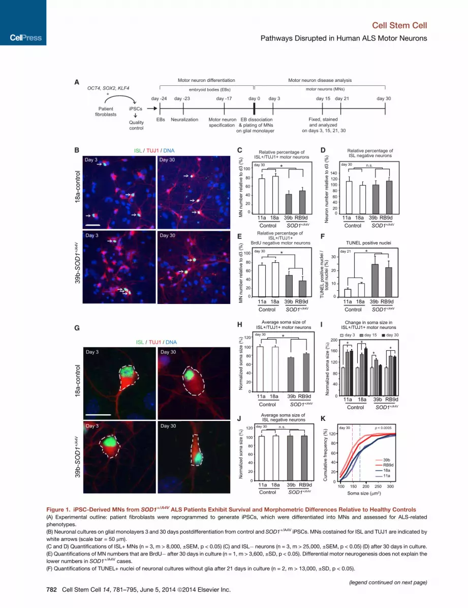
Figure 1. iPSC-Derived MNs from SOD1+/A4V ALS Patients Exhibit Survival and Morphometric Differences Relative to Healthy Controls
(A) Experimental outline: patient fibroblasts were reprogrammed to generate iPSCs, which were differentiated into MNs and assessed for ALS-related
phenotypes.
(B) Neuronal cultures on glial monolayers 3 and 30 days postdifferentiation from control and SOD1+/A4V iPSCs. MNs costained for ISL and TUJ1 are indicated by
white arrows (scale bar = 50 mm).
(C and D) Quantifications of ISL+ MNs (n = 3, m > 8,000, ±SEM, p < 0.05) (C) and ISL� neurons (n = 3, m > 25,000, ±SEM, p < 0.05) (D) after 30 days in culture.
(E) Quantifications of MN numbers that are BrdU� after 30 days in culture (n = 1, m > 3,600, ±SD, p < 0.05). Differential motor neurogenesis does not explain the
lower numbers in SOD1+/A4V cases.
(F) Quantifications of TUNEL+ nuclei of neuronal cultures without glia after 21 days in culture (n = 2, m > 13,000, ±SD, p < 0.05).
(legend continued on next page)
Cell Stem Cell
Pathways Disrupted in Human ALS Motor Neurons
782 Cell Stem Cell 14, 781–795, June 5, 2014 ª2014 Elsevier Inc.
Cell Stem Cell
Pathways Disrupted in Human ALS Motor Neurons
ALS has been slow to develop. Identification of shared mecha-
nisms of MN disease could inform the selection of pathways
for therapeutic intervention with the greatest relevance to a
broader patient population.
We and others have proposed that induced pluripotent stem
cells (iPSCs) from ALS patients and their differentiation into
spinal MNs (Dimos et al., 2008) could complement existing
animal models, allowing hypotheses to be tested in human
MNs with the patients’ unique genetic constellation (Bilican
et al., 2012; Donnelly et al., 2013; Egawa et al., 2012; Sareen
et al., 2013).
Here, we have combined reprogramming and stem cell differ-
entiation approaches with genome engineering and RNA
sequencing (RNA-seq) technologies to identify the transcrip-
tional and functional changes induced by theSOD1A4Vmutation
in human MNs. In addition to supporting hypotheses concerning
the actions of mutant SOD1 protein developed using transgenic
mouse models, such as the disruption of mitochondrial function
and transport, our studies identified novel mechanisms that may
contribute to MN dysfunction. Notably, we found that mutant
SOD1 disrupts a delicate balance between ER stress and
neuronal excitability that is inherent to MNs. Finally, studies
using iPSCs derived from patients harboring C9orf72 repeat
expansions indicate that at least a subset of the changes
induced by mutant SOD1 in human MNs are relevant to both
forms of ALS.
RESULTS
Generation of iPSCs and FunctionalMNs fromSOD1+/A4V
ALS PatientsWe derived skin fibroblasts from two female ALS patients (study
participants 39 and RB9) carrying the same dominantly acting
SOD1A4V mutation (SOD1+/A4V). We then generated iPSCs via
retroviral transduction of OCT4, SOX2, and KLF4 and validated
their integrity through a battery of standard pluripotency assays
(Table S1). We then obtained differentiated spinal MNs through
modest modifications to a previously reported protocol (Fig-
ure 1A and Figure S1A available online) (Boulting et al., 2011),
which resulted in highly neuralized cultures (>97% TUJ1+),
with significant percentages of ISL+ and HB9+ postmitotic
MNs (Figures S1 and S2). Our MN cultures were electrophysio-
logically active (Figures S1D–S1G) and could functionally inte-
grate into the developing chick spinal cord (Figures S2D–S2E).
Increased Apoptosis and Altered Morphometry inSOD1+/A4V MNsTo ask whether SOD1+/A4V MNs manifest a phenotype under
standard culture conditions, we compared them with MNs pro-
duced in parallel from two control iPSC lines (11a and 18a),
which were similar in their neuronal differentiation capacity, re-
(G) Representative images of measured soma size (white-dotted circumference)
(H) Quantifications of ISL+ MN soma size with values normalized to those of con
(I) MN soma size after 3, 15, and 30 days in culture normalized to day 3 for each cel
lesser degree in SOD1+/A4V cases.
(J) Quantifications of ISL� neuron soma size with values normalized to those of
(K) Cumulative frequency graphs of MN soma size after 30 days in culture. Dotte
n.s., not significant; n = experiment; m = cell number.
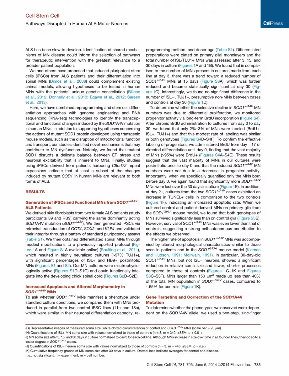
programming method, and donor age (Table S1). Differentiated
preparations were plated on primary glial monolayers and the
total number of ISL/TUJ1+ MNs was assessed after 3, 15, and
30 days in culture (Figures 1A and 1B). We found that in compar-
ison to the number of MNs present in cultures made from each
line at day 3, there was a trend toward a reduced number of
SOD1+/A4V MNs at 15 days (Figure S3A), which was further
reduced and became statistically significant at day 30 (Fig-
ure 1C). Interestingly, we found no significant difference in the
number of ISL�, TUJ1+, presumptive non-MNs between cases
and controls at day 30 (Figure 1D).
To determine whether the selective decline in SOD1+/A4V MN
numbers was due to differential proliferation, we monitored
progenitor activity via long-term BrdU incorporation (Figure S4).
After chronic BrdU administration to cultures from day 0 to day
30, we found that only 2%–3% of MNs were labeled (BrdU+,
ISL+, TUJ1+) and that this modest rate of labeling was similar
in both genotypes (Figures S4D–S4F). To confirm the effective
labeling of progenitors, we administered BrdU from day �17 of
directed differentiation until day 0, finding that the vast majority
of MNs (>95%) were BrdU+ (Figures S4A–S4C). These results
suggest that the vast majority of MNs in our cultures were
postmitotic prior to day 0 and that the reduced SOD1+/A4V MN
numbers were not due to a decrease in progenitor activity.
Importantly, when we specifically quantified only the MNs born
before day 0, we again found that significantly more SOD1+/A4V
MNswere lost over the 30 days in culture (Figure 1E). In addition,
at day 21, cultures from the two SOD1+/A4V cases exhibited an
increase in TUNEL+ cells in comparison to the two controls
(Figure 1F), indicating an increased apoptotic rate. When we
cultured control and patient-derived MNs on primary glia from
the SOD1G93A mouse model, we found that both genotypes of
MNs survived significantly less than on control glia (Figure S3B).
However, survival of SOD1+/A4VMNswas even lower than that of
controls, suggesting a strong cell-autonomous contribution to
the effects we observed.
The higher rate of apoptosis in SOD1+/A4VMNswas accompa-
nied by altered morphological characteristics similar to those
seen in patients and in the SOD1G93A mouse model (Kiernan
and Hudson, 1991; McIlwain, 1991). In particular, 30-day-old
SOD1+/A4V MNs, but not ISL� neurons, showed a significant
reduction in relative soma size and fewer, shorter processes
compared to those of controls (Figures 1G–1K and Figures
S3C–S3F). MNs larger than 150 mm2 made up less than 40%
of the total MN population in SOD1+/A4V cases, compared to
�65% for controls (Figure 1K).
Gene Targeting and Correction of the SOD1A4VMutationTo determinewhether the phenotypeswe observedwere depen-
dent on the SOD1A4V allele, we used a two-step, zinc-finger
of control and SOD1+/A4V MNs (scale bar = 20 mm).
trols (n = 3, m = 340, ±SEM, p < 0.01).
l line. AlthoughMNs increase in size over time in all four cell lines, they do so to a
controls (n = 3, m = 446, ±SEM, p = n.s.).
d lines indicate averages for control and disease.
Cell Stem Cell 14, 781–795, June 5, 2014 ª2014 Elsevier Inc. 783
A B
MN
num
ber r
elat
ive
to d
3 (%
)
= FRT site = A4V mutation = ZFN siteTargeting vector
SOD1 genomic locus
800bp 3’ Homology Arm800bp 5’ Homology Arm
PshAI
Homologous recombination
Flp recombination
Exon 1
Exon 1
Exon 1
Exon 1
PGK::Puromycin
polyA
PGK::Puromycin
polyA
C
D
α-tubulinRIPA soluble
SOD1:RIPA soluble
SOD1:UREA soluble
+MG132:
SOD1+/+SOD1+/A4V
+ +- -
39b motor neuronculture
PshAI: + -
39bSOD1+/A4V
39b SOD1+/+
+ -
0.0
0.5
1.0
1.5
2.0
2.5
SOD1 copy number in iPSCs
Rel
ativ
e ab
unda
nce
of g
enom
ic S
OD
1
0.0
0.5
1.0
1.5
2.0
2.5
Rel
ativ
e ex
pres
sion
SOD1 transcript in iPSCs
α-tubulin
SOD115kDa
50kDa
+/++/-+/A4V
SOD1 in 39b-iPSCs
1 0.52 1.21
39b-SOD1+/+39b-SOD1+/A4V 39b-SOD1+/+39b-SOD1+/A4V
15kDa
15kDa
E
G H I
F
39b-SOD1+/A4V
39b-SOD1+/+
CG GGAA XCG GTGT
CG GGAA CCG GTGT
day 30
Relative percentage of ISL+/TUJ1+ motor neurons
0
20
40
60
80
100 *
Nor
mal
ized
som
a si
ze (%
)
0
40
80
120 day 30*
Average soma size ofISL+/TUJ1+ motor neuron
100
60
20
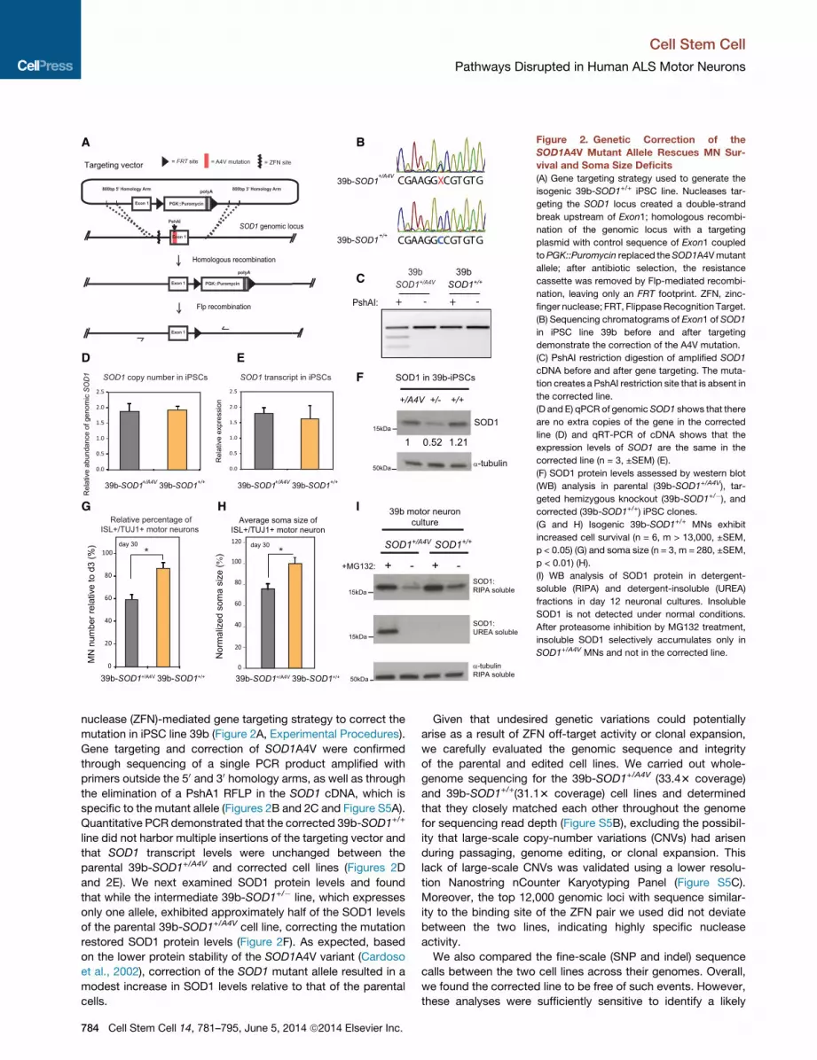
Figure 2. Genetic Correction of the
SOD1A4V Mutant Allele Rescues MN Sur-
vival and Soma Size Deficits
(A) Gene targeting strategy used to generate the
isogenic 39b-SOD1+/+ iPSC line. Nucleases tar-
geting the SOD1 locus created a double-strand
break upstream of Exon1; homologous recombi-
nation of the genomic locus with a targeting
plasmid with control sequence of Exon1 coupled
toPGK::Puromycin replaced the SOD1A4Vmutant
allele; after antibiotic selection, the resistance
cassette was removed by Flp-mediated recombi-
nation, leaving only an FRT footprint. ZFN, zinc-
finger nuclease; FRT, Flippase Recognition Target.
(B) Sequencing chromatograms of Exon1 of SOD1
in iPSC line 39b before and after targeting
demonstrate the correction of the A4V mutation.
(C) PshAI restriction digestion of amplified SOD1
cDNA before and after gene targeting. The muta-
tion creates a PshAI restriction site that is absent in
the corrected line.
(D and E) qPCR of genomic SOD1 shows that there
are no extra copies of the gene in the corrected
line (D) and qRT-PCR of cDNA shows that the
expression levels of SOD1 are the same in the
corrected line (n = 3, ±SEM) (E).
(F) SOD1 protein levels assessed by western blot
(WB) analysis in parental (39b-SOD1+/A4V), tar-
geted hemizygous knockout (39b-SOD1+/�), andcorrected (39b-SOD1+/+) iPSC clones.
(G and H) Isogenic 39b-SOD1+/+ MNs exhibit
increased cell survival (n = 6, m > 13,000, ±SEM,
p < 0.05) (G) and soma size (n = 3, m = 280, ±SEM,
p < 0.01) (H).
(I) WB analysis of SOD1 protein in detergent-
soluble (RIPA) and detergent-insoluble (UREA)
fractions in day 12 neuronal cultures. Insoluble
SOD1 is not detected under normal conditions.
After proteasome inhibition by MG132 treatment,
insoluble SOD1 selectively accumulates only in
SOD1+/A4V MNs and not in the corrected line.
Cell Stem Cell
Pathways Disrupted in Human ALS Motor Neurons
nuclease (ZFN)-mediated gene targeting strategy to correct the
mutation in iPSC line 39b (Figure 2A, Experimental Procedures).
Gene targeting and correction of SOD1A4V were confirmed
through sequencing of a single PCR product amplified with
primers outside the 50 and 30 homology arms, as well as through
the elimination of a PshA1 RFLP in the SOD1 cDNA, which is
specific to the mutant allele (Figures 2B and 2C and Figure S5A).
Quantitative PCR demonstrated that the corrected 39b-SOD1+/+
line did not harbor multiple insertions of the targeting vector and
that SOD1 transcript levels were unchanged between the
parental 39b-SOD1+/A4V and corrected cell lines (Figures 2D
and 2E). We next examined SOD1 protein levels and found
that while the intermediate 39b-SOD1+/� line, which expresses
only one allele, exhibited approximately half of the SOD1 levels
of the parental 39b-SOD1+/A4V cell line, correcting the mutation
restored SOD1 protein levels (Figure 2F). As expected, based
on the lower protein stability of the SOD1A4V variant (Cardoso
et al., 2002), correction of the SOD1 mutant allele resulted in a
modest increase in SOD1 levels relative to that of the parental
cells.
784 Cell Stem Cell 14, 781–795, June 5, 2014 ª2014 Elsevier Inc.
Given that undesired genetic variations could potentially
arise as a result of ZFN off-target activity or clonal expansion,
we carefully evaluated the genomic sequence and integrity
of the parental and edited cell lines. We carried out whole-
genome sequencing for the 39b-SOD1+/A4V (33.43 coverage)
and 39b-SOD1+/+(31.13 coverage) cell lines and determined
that they closely matched each other throughout the genome
for sequencing read depth (Figure S5B), excluding the possibil-
ity that large-scale copy-number variations (CNVs) had arisen
during passaging, genome editing, or clonal expansion. This
lack of large-scale CNVs was validated using a lower resolu-
tion Nanostring nCounter Karyotyping Panel (Figure S5C).
Moreover, the top 12,000 genomic loci with sequence similar-
ity to the binding site of the ZFN pair we used did not deviate
between the two lines, indicating highly specific nuclease
activity.
We also compared the fine-scale (SNP and indel) sequence
calls between the two cell lines across their genomes. Overall,
we found the corrected line to be free of such events. However,
these analyses were sufficiently sensitive to identify a likely
A B
C
D
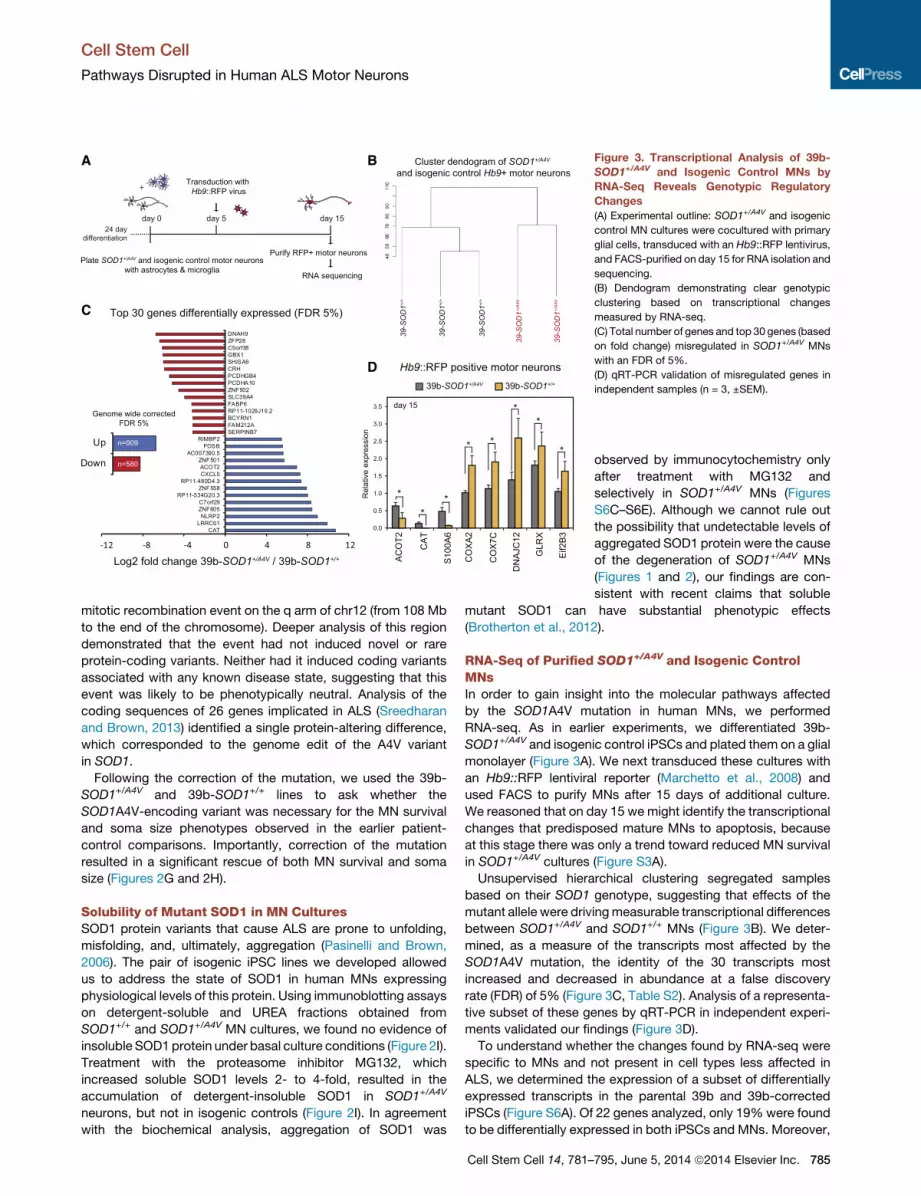
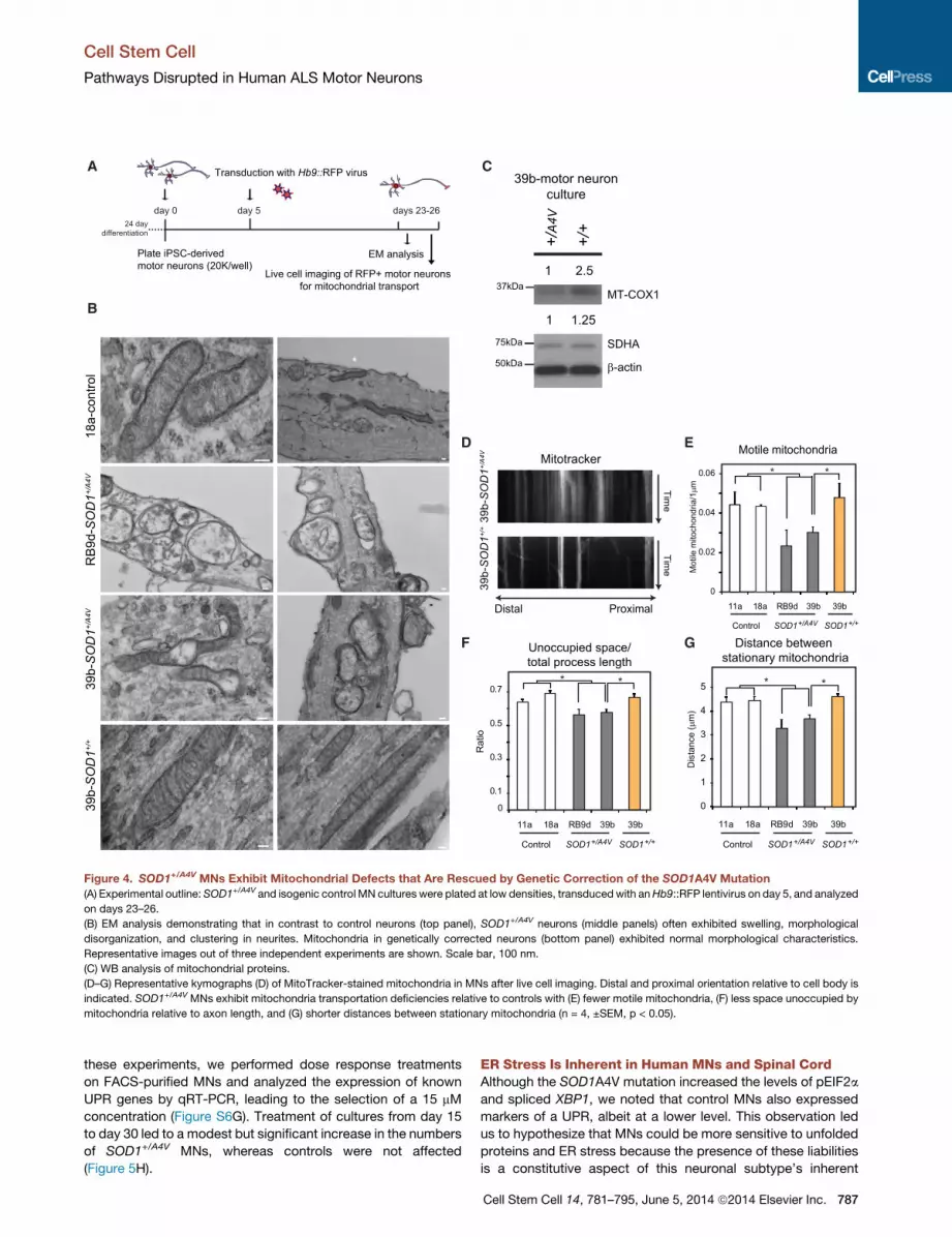
Figure 3. Transcriptional Analysis of 39b-
SOD1+/A4V and Isogenic Control MNs by
RNA-Seq Reveals Genotypic Regulatory
Changes
(A) Experimental outline: SOD1+/A4V and isogenic
control MN cultures were cocultured with primary
glial cells, transduced with an Hb9::RFP lentivirus,
and FACS-purified on day 15 for RNA isolation and
sequencing.
(B) Dendogram demonstrating clear genotypic
clustering based on transcriptional changes
measured by RNA-seq.
(C) Total number of genes and top 30 genes (based
on fold change) misregulated in SOD1+/A4V MNs
with an FDR of 5%.
(D) qRT-PCR validation of misregulated genes in
independent samples (n = 3, ±SEM).
Cell Stem Cell
Pathways Disrupted in Human ALS Motor Neurons
mitotic recombination event on the q arm of chr12 (from 108 Mb
to the end of the chromosome). Deeper analysis of this region
demonstrated that the event had not induced novel or rare
protein-coding variants. Neither had it induced coding variants
associated with any known disease state, suggesting that this
event was likely to be phenotypically neutral. Analysis of the
coding sequences of 26 genes implicated in ALS (Sreedharan
and Brown, 2013) identified a single protein-altering difference,
which corresponded to the genome edit of the A4V variant
in SOD1.
Following the correction of the mutation, we used the 39b-
SOD1+/A4V and 39b-SOD1+/+ lines to ask whether the
SOD1A4V-encoding variant was necessary for the MN survival
and soma size phenotypes observed in the earlier patient-
control comparisons. Importantly, correction of the mutation
resulted in a significant rescue of both MN survival and soma
size (Figures 2G and 2H).
Solubility of Mutant SOD1 in MN CulturesSOD1 protein variants that cause ALS are prone to unfolding,
misfolding, and, ultimately, aggregation (Pasinelli and Brown,
2006). The pair of isogenic iPSC lines we developed allowed
us to address the state of SOD1 in human MNs expressing
physiological levels of this protein. Using immunoblotting assays
on detergent-soluble and UREA fractions obtained from
SOD1+/+ and SOD1+/A4V MN cultures, we found no evidence of
insoluble SOD1protein under basal culture conditions (Figure 2I).
Treatment with the proteasome inhibitor MG132, which
increased soluble SOD1 levels 2- to 4-fold, resulted in the
accumulation of detergent-insoluble SOD1 in SOD1+/A4V
neurons, but not in isogenic controls (Figure 2I). In agreement
with the biochemical analysis, aggregation of SOD1 was
Cell Stem Cell 14, 781–
observed by immunocytochemistry only
after treatment with MG132 and
selectively in SOD1+/A4V MNs (Figures
S6C–S6E). Although we cannot rule out
the possibility that undetectable levels of
aggregated SOD1 protein were the cause
of the degeneration of SOD1+/A4V MNs
(Figures 1 and 2), our findings are con-
sistent with recent claims that soluble
mutant SOD1 can have substantial phenotypic effects
(Brotherton et al., 2012).
RNA-Seq of Purified SOD1+/A4V and Isogenic ControlMNsIn order to gain insight into the molecular pathways affected
by the SOD1A4V mutation in human MNs, we performed
RNA-seq. As in earlier experiments, we differentiated 39b-
SOD1+/A4V and isogenic control iPSCs and plated them on a glial
monolayer (Figure 3A). We next transduced these cultures with
an Hb9::RFP lentiviral reporter (Marchetto et al., 2008) and
used FACS to purify MNs after 15 days of additional culture.
We reasoned that on day 15 we might identify the transcriptional
changes that predisposed mature MNs to apoptosis, because
at this stage there was only a trend toward reduced MN survival
in SOD1+/A4V cultures (Figure S3A).
Unsupervised hierarchical clustering segregated samples
based on their SOD1 genotype, suggesting that effects of the
mutant allele were drivingmeasurable transcriptional differences
between SOD1+/A4V and SOD1+/+ MNs (Figure 3B). We deter-
mined, as a measure of the transcripts most affected by the
SOD1A4V mutation, the identity of the 30 transcripts most
increased and decreased in abundance at a false discovery
rate (FDR) of 5% (Figure 3C, Table S2). Analysis of a representa-
tive subset of these genes by qRT-PCR in independent experi-
ments validated our findings (Figure 3D).
To understand whether the changes found by RNA-seq were
specific to MNs and not present in cell types less affected in
ALS, we determined the expression of a subset of differentially
expressed transcripts in the parental 39b and 39b-corrected
iPSCs (Figure S6A). Of 22 genes analyzed, only 19%were found
to be differentially expressed in both iPSCs and MNs. Moreover,
795, June 5, 2014 ª2014 Elsevier Inc. 785
Cell Stem Cell
Pathways Disrupted in Human ALS Motor Neurons
RNA-seq analysis on fibroblast cultures isolated from five
healthy control individuals and the two ALS patients harboring
the SOD1A4V mutation failed to segregate transcriptomes
based on genotype after undergoing unsupervised hierarchical
clustering (Figure S6B). Importantly, a number of genes we
identified to be misregulated in SOD1+/A4V MNs have not, to
our knowledge, previously been implicated as being modulated
by mutant SOD1 (Table S2).
Ontology of Transcripts Modulated in SOD1+/A4V MNsIn order to probe the RNA-seq data for biological meaning, we
utilized two bioinformatic tools that query for enriched gene
ontology terms (Table S3). We first performed gene-annotation
enrichment analysis with DAVID (Huang et al., 2009), using
all the genes that were significantly altered (909 upregulated
and 580 downregulated) in SOD1+/A4V MNs at an FDR of 5%
(Table S3A and S3B). A total of 27 and 65 gene terms
were enriched when increased and reduced transcripts, respec-
tively, were considered. Transcripts implicated in cytoskeleton
organization were among the most significantly induced in
SOD1+/A4V MNs, consistent with the morphological alterations
that we observed in these cells relative to isogenic controls
(Figure 2H). Transcripts involved in transcriptional regulation
and motor proteins were also induced as a result of the SOD1
mutation. Among the significantly decreased transcripts in
SOD1+/A4V MNs, there was a very strong enrichment for genes
implicated in mitochondrial function and structure. In particular,
60% of all downregulated ontology terms were related to mito-
chondria, while genes implicated in protein translation were
also repressed (Table S3B).
As an alternative approach for querying our RNA-seq data,
we performed Gene Set Enrichment Analysis (GSEA) (Mootha
et al., 2003; Subramanian et al., 2005). GSEA identified 16
gene sets that were significantly induced in SOD1+/A4V MNs
(NES < 1.5; Table S3C). Among these gene sets were the motor
proteins kinesins. GSEA also identified 100 gene sets to be
significantly repressed in SOD1+/A4V MNs (NES < 1.5), and
notably, gene sets associated with mitochondrial function and
protein translation were again among the most significantly
suppressed (Table S3D).
SOD1+/A4V MNs Exhibit Disturbances in MitochondrialMorphology and MotilityTo determine whether the transcriptional changes in mitochon-
drial genes that we identified by RNA-seq in SOD1+/A4V MNs
(Table S2 and Table S3) were indicative of actual disturbances
to mitochondria, we initially performed electron microscopy
(EM) studies (Figures 4A and 4B). Whereas mitochondrial
morphology was normal in MNs derived from a control cell line
(18a), mitochondria in SOD1+/A4V MNs (39b and RB9d) were
commonly deranged and more vacuolar in appearance. These
differences were mostly apparent in neuronal processes. We
concluded that distortion in mitochondrial morphology was
mediated by expression of the SOD1A4V mutant allele because
correction of the mutation eliminated this phenotype (Figure 4B).
To further validate mitochondrial damage in SOD1+/A4V MNs, we
used immunoblotting assays to quantify the levels of two mito-
chondrial proteins: SDHA, which is encoded in the nucleus,
and MT-COX1, encoded by mitochondrial DNA. We found that
786 Cell Stem Cell 14, 781–795, June 5, 2014 ª2014 Elsevier Inc.
correction of the SOD1A4V allele in 39b-SOD1+/+MNs increased
the protein levels of both SDHA and MT-COX1 relative to the
parental 39b-SOD1+/A4V MNs (Figure 4C).
We next sought to measure the movements of mitochondria
within the axons of our in vitro-derived MNs (Figure 4A). MN
cultures differentiated from control and SOD1+/A4V iPSCs were
colabeled with Hb9::RFP and MitoTracker-Green, which selec-
tively stained mitochondria. We then carried out live cell
imaging to register mitochondrial movement along MN pro-
cesses over the course of 5min, andwe generated time/distance
kymographs for further analysis (Figure 4D). We found that the
A4V mutation resulted in a significant decrease in the number
of motile mitochondria (Figure 4E). This was coupled to an
increase in mitochondrial density in processes as measured
by the shorter distance between stationary mitochondria and
the significantly smaller amount of space unoccupied by these
organelles (Figures 4F and 4G).
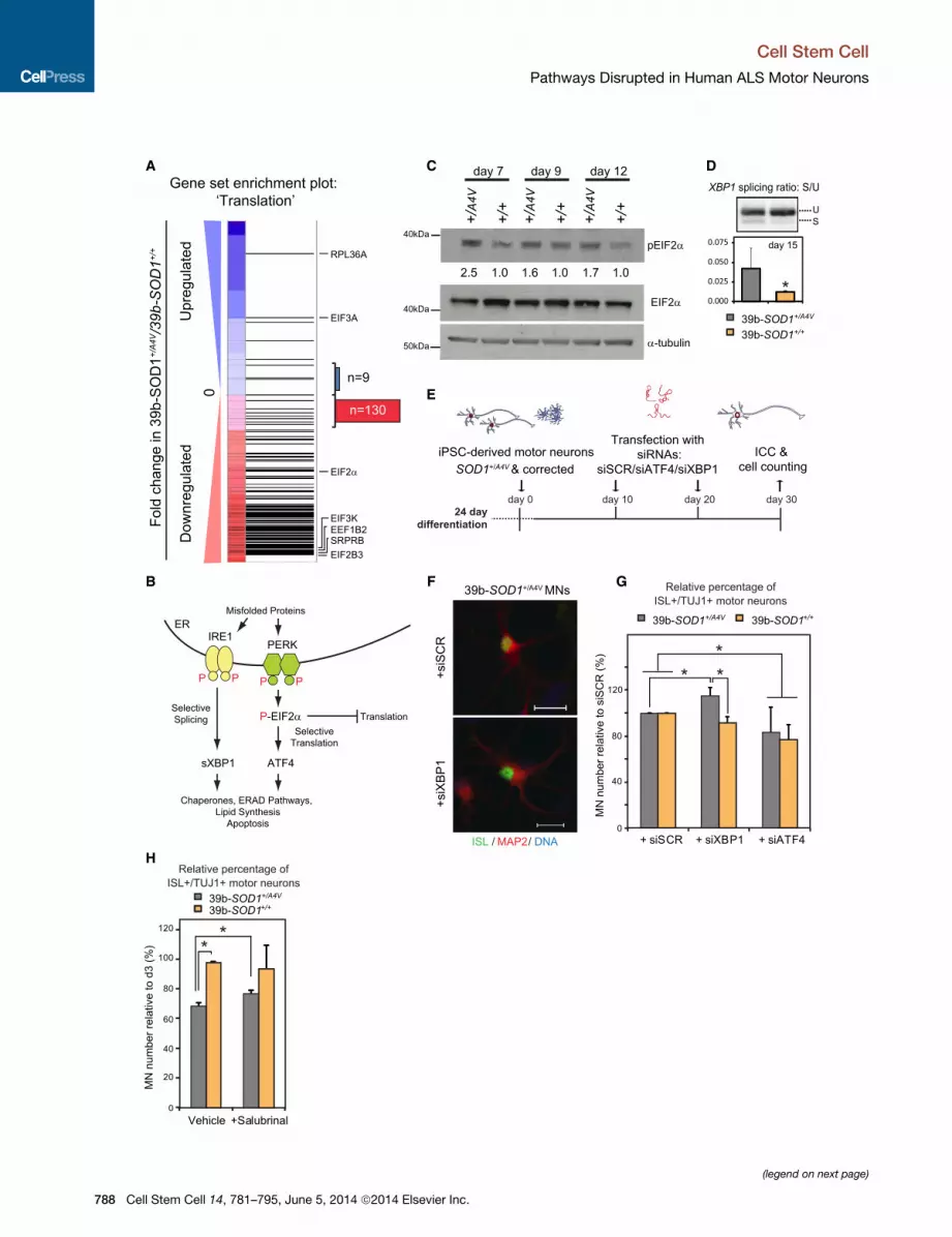
SOD1+/A4V MNs Exhibit Signatures of an UnfoldedProtein Response and ER Stress InductionRNA-seq analysis showed that 93%of all transcripts assigned to
the protein translation gene-set were reduced in SOD1+/A4VMNs
(Figure 5A). Translational inhibition is a well-established hallmark
of ER stress and the unfolded protein response (UPR), which is
activated upon accumulation of misfolded proteins (Trusina
et al., 2008). While the UPR is initially cytoprotective, its persis-
tent activation can lead to apoptosis. In one of the branches of
the pathway, pEIF2a leads to a global attenuation of translation
and to selective translation of ATF4, while in another branch,
IRE1 cleaves the mRNA of XBP1, creating its active spliced
form (sXBP1) (Figure 5B) (Ron and Walter, 2007). The transcrip-
tion factors sXBP1 and ATF4 modulate expression of
downstream effectors including chaperone proteins. Impor-
tantly, RNA-seq analysis identified the heat-shock proteins
DNAJC12 and HSBP1, the prefoldin subunits PFDN2 and
PFDN5, and the chaperonin subunits CCT4 and CCT7 as being
differentially expressed in SOD1+/A4V MNs (Table S2). In
addition, SOD1+/A4V MNs exhibited increased levels of
pEIF2a (Figure 5C), as well as significantly elevated sXBP1
transcript (Figure 5D) relative to isogenic controls, consistent
with an active UPR.
To test if ER stress directly contributed to mutant SOD1-
mediated toxicity in our culture system, we genetically manipu-
lated the two UPR branches using siRNA knockdown of XBP1
andATF4 and assessed the effect onMNsurvival (Figure 5E, Fig-
ure S7B). Knockdown of XBP1 resulted in a modest but signifi-
cant increase in the number of SOD1+/A4V MNs after 30 days in
culture (Figures 5F and 5G). In contrast, there was a trend for
reduced MN numbers in the isogenic controls, suggesting that
XBP1might provide a protective function in this context. Knock-
down of ATF4 depressed survival of both SOD1+/A4V and
SOD1+/+ MNs, implying that this protein plays a protective role
in both contexts (Figure 5G). Given that a reduction in ATF4
levels was detrimental to the survival of MNs, we asked whether
a further induction of pEIF2a would confer MN protection. Salu-
brinal is a selective inhibitor of phosphatases, which dephos-
phorylate pEIF2a (Boyce et al., 2005), and has previously been
shown to extend the survival of the SOD1G93A mouse model
(Saxena et al., 2009). To identify the optimal concentration for
A
Mot
ile m
itoch
ondr
ia/1μm
Plate iPSC-derived motor neurons (20K/well)
day 0 day 5 days 23-26 24 daydifferentiation
Transduction with Hb9::RFP virus
Live cell imaging of RFP+ motor neurons for mitochondrial transport
EM analysis
B
Time
Mitotracker
39b-
SO
D1+/
A4V
39b-
SO
D1+/
+
C
D E
F G
39b-
SO
D1+/
A4V
39
b-S
OD
1+/+
18a-
cont
rol
Dis
tanc
e (μ
m)
0
1
2
3
4
5
11a 18a RB9d 39b
Control SOD1+/A4V SOD1+/+
39b
Distance between stationary mitochondria
**
Motile mitochondria
0
0.02
0.04
0.06
11a 18a RB9d 39b
Control SOD1+/A4V SOD1+/+
39b
**
Unoccupied space/total process length
0
0.1
0.3
0.5
0.7
Rat
io
11a 18a RB9d 39b
Control SOD1+/A4V SOD1+/+
39b
**
Distal Proximal
Time
RB
9d-S
OD
1+/A
4V
SDHA
β-actin
MT-COX1
75kDa
50kDa
37kDa
+/A
4V
+/+
39b-motor neuron culture
1 2.5
1 1.25
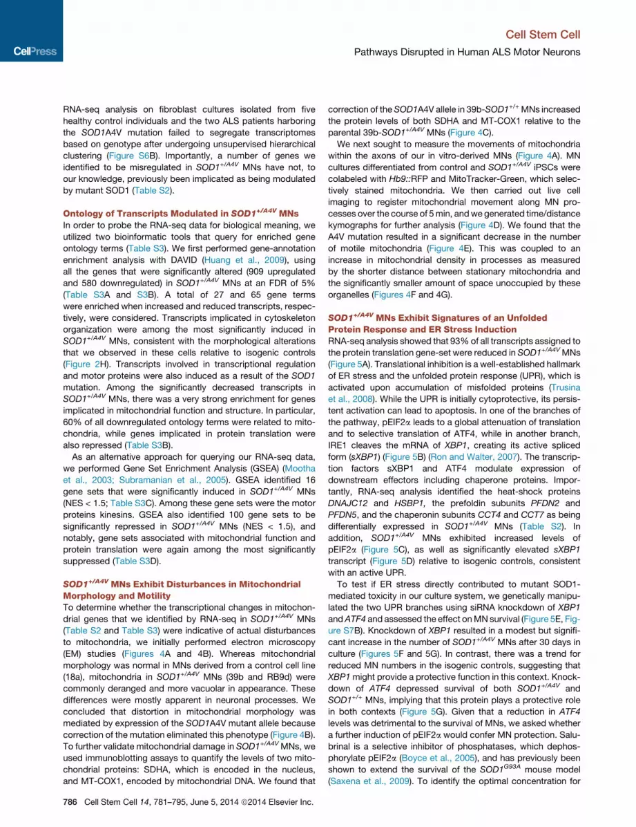
Figure 4. SOD1+/A4V MNs Exhibit Mitochondrial Defects that Are Rescued by Genetic Correction of the SOD1A4V Mutation
(A) Experimental outline: SOD1+/A4V and isogenic control MN cultures were plated at low densities, transduced with anHb9::RFP lentivirus on day 5, and analyzed
on days 23–26.
(B) EM analysis demonstrating that in contrast to control neurons (top panel), SOD1+/A4V neurons (middle panels) often exhibited swelling, morphological
disorganization, and clustering in neurites. Mitochondria in genetically corrected neurons (bottom panel) exhibited normal morphological characteristics.
Representative images out of three independent experiments are shown. Scale bar, 100 nm.
(C) WB analysis of mitochondrial proteins.
(D–G) Representative kymographs (D) of MitoTracker-stained mitochondria in MNs after live cell imaging. Distal and proximal orientation relative to cell body is
indicated. SOD1+/A4V MNs exhibit mitochondria transportation deficiencies relative to controls with (E) fewer motile mitochondria, (F) less space unoccupied by
mitochondria relative to axon length, and (G) shorter distances between stationary mitochondria (n = 4, ±SEM, p < 0.05).
Cell Stem Cell
Pathways Disrupted in Human ALS Motor Neurons
these experiments, we performed dose response treatments
on FACS-purified MNs and analyzed the expression of known
UPR genes by qRT-PCR, leading to the selection of a 15 mM
concentration (Figure S6G). Treatment of cultures from day 15
to day 30 led to a modest but significant increase in the numbers
of SOD1+/A4V MNs, whereas controls were not affected
(Figure 5H).
ER Stress Is Inherent in Human MNs and Spinal CordAlthough the SOD1A4V mutation increased the levels of pEIF2a
and spliced XBP1, we noted that control MNs also expressed
markers of a UPR, albeit at a lower level. This observation led
us to hypothesize that MNs could be more sensitive to unfolded
proteins and ER stress because the presence of these liabilities
is a constitutive aspect of this neuronal subtype’s inherent
Cell Stem Cell 14, 781–795, June 5, 2014 ª2014 Elsevier Inc. 787
A C D
E
B F G
H
(legend on next page)
Cell Stem Cell
Pathways Disrupted in Human ALS Motor Neurons
788 Cell Stem Cell 14, 781–795, June 5, 2014 ª2014 Elsevier Inc.
Cell Stem Cell
Pathways Disrupted in Human ALS Motor Neurons
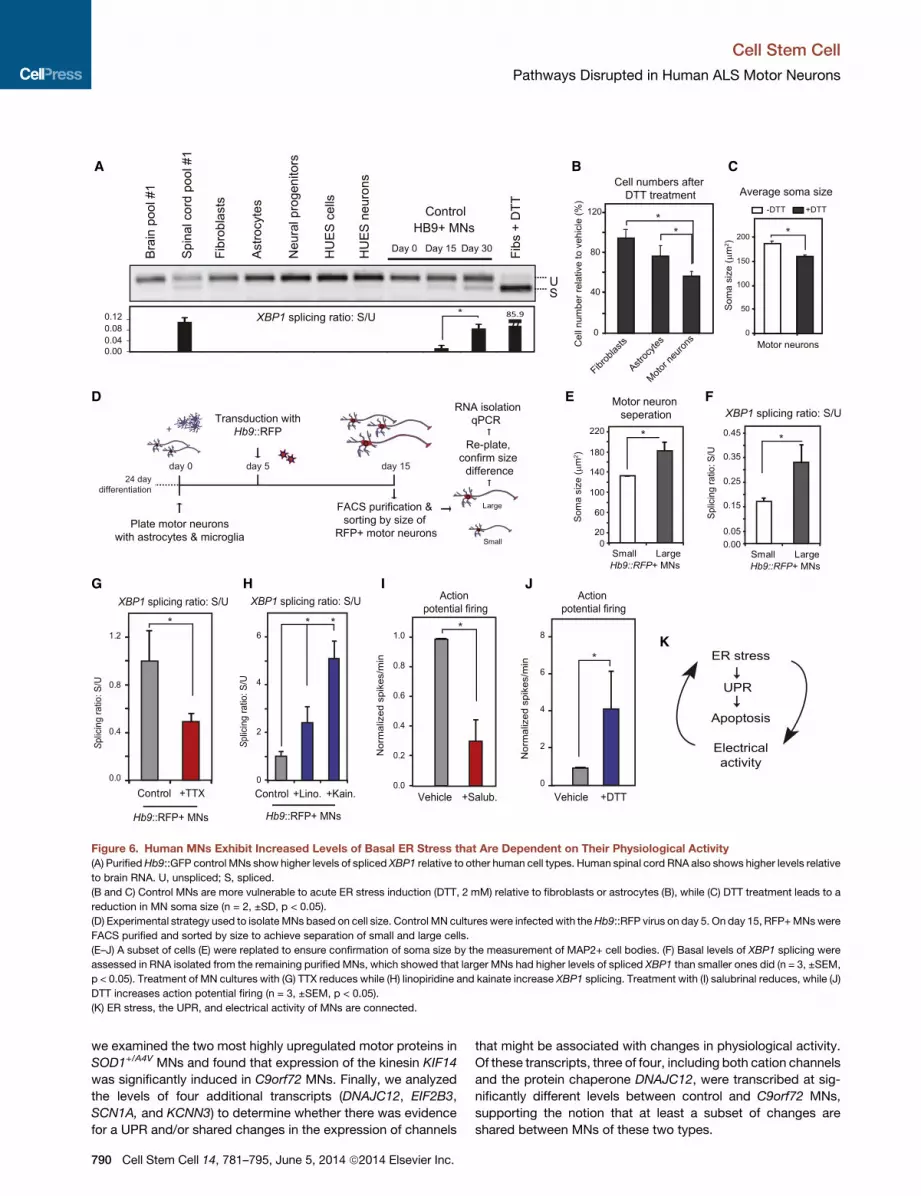
biology. To test this, we compared the levels of XBP1 splicing in
purified, control HUES3 Hb9::GFP+ MNs (Di Giorgio et al., 2008)
with levels found in a range of other cell types, including astro-
cytes, embryonic stem cells (ESCs), neural progenitors, fibro-
blasts, Hb9::GFP-negative neurons and ESC-derived anterior
neurons. Of all the cell types examined, only the control MNs
displayed detectable levels of sXBP1 that increased as MNs
matured in culture (Figure 6A and Figure S7A). When we
compared the levels of sXBP1 in RNA isolated from human brain
and spinal cord (n = 2; each replicate was pooled RNA from 22
nonoverlapping healthy controls), we strikingly saw little or no
evidence in the brain, while in the spinal cord there was amarked
and significant accumulation of the spliced transcript (Figure 6A
and Figure S7A).
We wondered whether this ongoing activation of the UPR
pathway was associated with an increased sensitivity to ER-
stress-inducing agents such as DTT. Control MNs indeed
exhibited greater susceptibility to DTT administration than astro-
cytes and fibroblasts did (Figure 6B). When we analyzed the area
of the soma of MNs after treatment, we found that the average
soma size decreased substantially (Figure 6C), which given the
acute nature of the treatment seemed most consistent with the
death of the largest MNs. These observations prompted us to
investigate whether MN size correlated with basal ER stress
levels. To address this, we purified Hb9::RFP+ MNs and sepa-
rated large and small populations (Figure 6D). We validated
this approach by replating a subset of the purified MNs and
measuring their soma size (Figure 6E). We then isolated RNA
and examined the levels of sXBP1 as an indicator of ER stress.
Larger MNs showed significantly higher levels of sXBP1 than
smaller ones (Figure 6F), suggesting that an increased constitu-
tive ER stress may contribute to their increased vulnerability in
our cell-culture model.
Inherent ER Stress in HumanMNs Is Dependent on TheirElectrical ActivityWainger and colleagues have found that the SOD1+/A4V human
MNs that we report on here are hyperexcitable in comparison
to controls (Wainger et al., 2014). Given that the XBP1 splicing
levels increased as MNs matured in culture and became excit-
able (Figure 6A), we reasoned that a relationship might exist
between the inherent ER stress we found in MNs and their elec-
trophysiological activity. Treatment of MN cultureswith sufficient
tetrodotoxin (TTX) to effectively block action potentials (Figures
Figure 5. SOD1+/A4V MNs Exhibit Signatures of a UPR and Are Selectiv(A) GSEA of transcriptional changes in SOD1+/A4V MNs shows strong downregu
capacity. Horizontal black bars represent individual genes with representative
downregulated, respectively, in SOD1+/A4V relative to isogenic control MNs. Out o
130 were downregulated, consistent with activation of the UPR pathway.
(B) Diagram illustrating the canonical UPR.
(C)WB analysis demonstrates increased levels of phosphorylated EIF2a, a marker
relative to control samples for each time point are shown.
(D) SOD1+/A4V MNs exhibit increased levels of XBP1 splicing, a marker of ER stre
3, ±SEM, p < 0.05). U, unspliced; S, spliced.
(E) Experimental strategy used to assess the contribution of XBP1 and ATF4 in t
(F) Representative images of untreated and treated MN cultures are shown. Sca
(G) SOD1+/A4V MN numbers selectively increase after XBP1 knockdown, while AT
cases (n = 3, m > 9,800 and m > 8,500, ±SEM, p < 0.05).
(H) Salubrinal has a modest but positive effect on survival of SOD1+/A4V MNs (n
S6C and S6D) significantly reduced spliced XBP1 (Figure 6G).
Reciprocally, treatment of cultures with the glutamatergic
agonist kainate, which depolarizes MNs, led to a significant in-
crease in spliced XBP1 (Figure 6H). In addition, linopiridine, a
compound that blocks Kv7 voltage-gated potassium channels
(Brown and Passmore, 2009), and increased MN activity (Fig-
ure S7D) also increased XBP1 splicing (Figure 6H).
We next addressed the inverse question and assessed
whether manipulating ER stress would affect the electrical
activity of MNs. Treatment with salubrinal resulted in a relative
reduction in the number of spikes per minute (Figure 6I).
Conversely, an acute treatment of MN cultures with DTT, which
robustly induced sXBP1 (Figure S6F), resulted in an increase in
the number of spikes per minute (Figure 6J). These two data
sets suggest that ER stress, the UPR, and the physiological
activity of human MNs are interconnected and that alterations
in one of these pathways can affect the others (Figure 6K).
A Subset of Transcriptional Changes in SOD1+/A4V AreShared in C9orf72 MNsA central question in the ALS field is whether mutations in the
diverse causative genes converge on shared molecular path-
ways. To begin to address this question, we focused on the
most prevalent genetic ALS type and selected two familial
patients (19, RB8) that carried GGGGCC repeat expansions in
theC9orf72 locus.We generated iPSCs, confirmed the presence
of the expansion in both the parental fibroblasts and in multiple
passages of the resulting iPSC lines, and demonstrated the
ability of these lines to differentiate into ISL/HB9+ MNs (Figures
7A–7C, Table S1). To determine whether these MNs exhibited
transcriptional disturbances similar to the ones that we identified
in SOD1+/A4V MNs, we simultaneously differentiated them with
six iPSC lines originating from five healthy individuals (Boulting
et al., 2011) and FACS-purified Hb9::RFP+ MNs in multiple
biological replicates.
Using qRT-PCR we interrogated a subset of transcripts repre-
sentative of pathways or cellular functions, which we had found
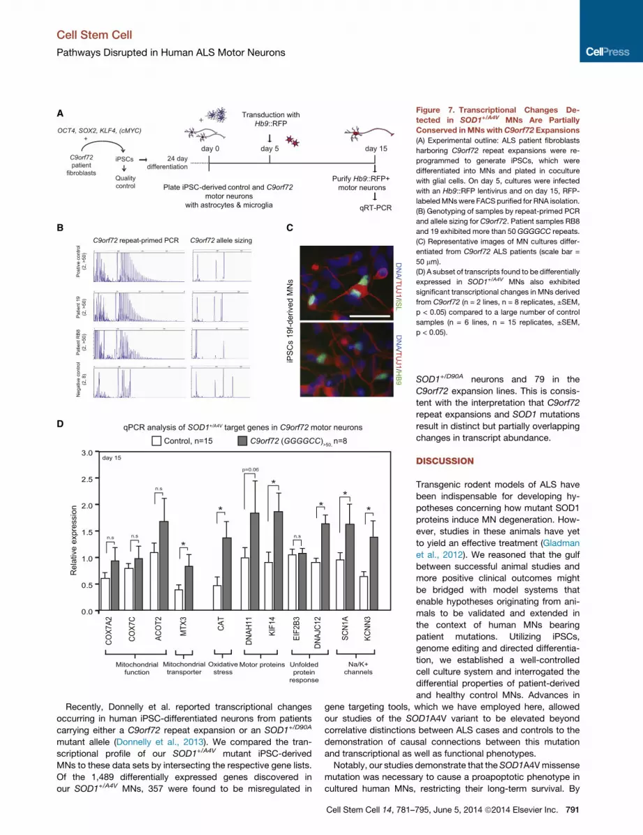
to be altered by theSOD1A4Vmutation (Figure 7D). Interestingly,
inC9orf72mutant MNs, we did not detect a significant change in
the transcript levels of genes implicated in electron transport in
mitochondria, but we did detect a significant change in levels
of the mitochondrial transporter MTX3. We also found a signifi-
cant induction of catalase (CAT), indicative of oxidative stress.
To determine whether intracellular transport might be impacted,
ely Vulnerable to ER Stress Inductionlation (normalized enrichment score: �3.31) for genes involved in translational
examples indicated. Blue or red represents genes that were upregulated or
f 139 genes detected that are annotated as involved in regulation of translation,
for activation of the UPR pathway, in SOD1+/A4VMNcultures. The percentages
ss. RNA was isolated from purified Hb9::RFP MNs after 15 days in culture (n =
he survival of SOD1+/A4V MNs.
le bar, 20 mm.
F4 knockdown significantly decreases numbers in both control and SOD1+/A4V
= 2, m > 10,000, ±SEM, p < 0.05).
Cell Stem Cell 14, 781–795, June 5, 2014 ª2014 Elsevier Inc. 789
A B C
D E F
G H I J
K
Figure 6. Human MNs Exhibit Increased Levels of Basal ER Stress that Are Dependent on Their Physiological Activity
(A) PurifiedHb9::GFP control MNs show higher levels of spliced XBP1 relative to other human cell types. Human spinal cord RNA also shows higher levels relative
to brain RNA. U, unspliced; S, spliced.
(B and C) Control MNs are more vulnerable to acute ER stress induction (DTT, 2 mM) relative to fibroblasts or astrocytes (B), while (C) DTT treatment leads to a
reduction in MN soma size (n = 2, ±SD, p < 0.05).
(D) Experimental strategy used to isolate MNs based on cell size. Control MN cultures were infected with theHb9::RFP virus on day 5. On day 15, RFP+MNswere
FACS purified and sorted by size to achieve separation of small and large cells.
(E–J) A subset of cells (E) were replated to ensure confirmation of soma size by the measurement of MAP2+ cell bodies. (F) Basal levels of XBP1 splicing were
assessed in RNA isolated from the remaining purified MNs, which showed that larger MNs had higher levels of spliced XBP1 than smaller ones did (n = 3, ±SEM,
p < 0.05). Treatment of MN cultures with (G) TTX reduces while (H) linopiridine and kainate increase XBP1 splicing. Treatment with (I) salubrinal reduces, while (J)
DTT increases action potential firing (n = 3, ±SEM, p < 0.05).
(K) ER stress, the UPR, and electrical activity of MNs are connected.
Cell Stem Cell
Pathways Disrupted in Human ALS Motor Neurons
we examined the two most highly upregulated motor proteins in
SOD1+/A4V MNs and found that expression of the kinesin KIF14
was significantly induced in C9orf72 MNs. Finally, we analyzed
the levels of four additional transcripts (DNAJC12, EIF2B3,
SCN1A, and KCNN3) to determine whether there was evidence
for a UPR and/or shared changes in the expression of channels
790 Cell Stem Cell 14, 781–795, June 5, 2014 ª2014 Elsevier Inc.
that might be associated with changes in physiological activity.
Of these transcripts, three of four, including both cation channels
and the protein chaperone DNAJC12, were transcribed at sig-
nificantly different levels between control and C9orf72 MNs,
supporting the notion that at least a subset of changes are
shared between MNs of these two types.
A
B C
D
-derived
Figure 7. Transcriptional Changes De-
tected in SOD1+/A4V MNs Are Partially
Conserved inMNswithC9orf72 Expansions
(A) Experimental outline: ALS patient fibroblasts
harboring C9orf72 repeat expansions were re-
programmed to generate iPSCs, which were
differentiated into MNs and plated in coculture
with glial cells. On day 5, cultures were infected
with an Hb9::RFP lentivirus and on day 15, RFP-
labeledMNswere FACS purified for RNA isolation.
(B) Genotyping of samples by repeat-primed PCR
and allele sizing for C9orf72. Patient samples RB8
and 19 exhibited more than 50 GGGGCC repeats.
(C) Representative images of MN cultures differ-
entiated from C9orf72 ALS patients (scale bar =
50 mm).
(D) A subset of transcripts found to be differentially
expressed in SOD1+/A4V MNs also exhibited
significant transcriptional changes in MNs derived
from C9orf72 (n = 2 lines, n = 8 replicates, ±SEM,
p < 0.05) compared to a large number of control
samples (n = 6 lines, n = 15 replicates, ±SEM,
p < 0.05).
Cell Stem Cell
Pathways Disrupted in Human ALS Motor Neurons
Recently, Donnelly et al. reported transcriptional changes
occurring in human iPSC-differentiated neurons from patients
carrying either a C9orf72 repeat expansion or an SOD1+/D90A
mutant allele (Donnelly et al., 2013). We compared the tran-
scriptional profile of our SOD1+/A4V mutant iPSC-derived
MNs to these data sets by intersecting the respective gene lists.
Of the 1,489 differentially expressed genes discovered in
our SOD1+/A4V MNs, 357 were found to be misregulated in
Cell Stem Cell 14, 781–
SOD1+/D90A neurons and 79 in the
C9orf72 expansion lines. This is consis-
tent with the interpretation that C9orf72
repeat expansions and SOD1 mutations
result in distinct but partially overlapping
changes in transcript abundance.
DISCUSSION
Transgenic rodent models of ALS have
been indispensable for developing hy-
potheses concerning how mutant SOD1
proteins induce MN degeneration. How-
ever, studies in these animals have yet
to yield an effective treatment (Gladman
et al., 2012). We reasoned that the gulf
between successful animal studies and
more positive clinical outcomes might
be bridged with model systems that
enable hypotheses originating from ani-
mals to be validated and extended in
the context of human MNs bearing
patient mutations. Utilizing iPSCs,
genome editing and directed differentia-
tion, we established a well-controlled
cell culture system and interrogated the
differential properties of patient-derived
and healthy control MNs. Advances in
gene targeting tools, which we have employed here, allowed
our studies of the SOD1A4V variant to be elevated beyond
correlative distinctions between ALS cases and controls to the
demonstration of causal connections between this mutation
and transcriptional as well as functional phenotypes.
Notably, our studies demonstrate that the SOD1A4Vmissense
mutation was necessary to cause a proapoptotic phenotype in
cultured human MNs, restricting their long-term survival. By
795, June 5, 2014 ª2014 Elsevier Inc. 791
Cell Stem Cell
Pathways Disrupted in Human ALS Motor Neurons
employing RNA-seq, we defined the transcriptional differences
between human SOD1+/A4V and isogenic control MNs. Curating
these data supported the view that patient-specific ALS iPSC-
derived MNs display hallmarks of disease found in both patients
and animal models. We identified defects in mitochondrial trans-
port and morphology, oxidative and ER-related stress, and an
activated UPR, all of which were dependent on the presence
of the SOD1A4V mutation. Importantly, we also identified candi-
date genes that were significantly affected by this disease-
causing mutation. These identified genes will require further
investigation because they may represent potential therapeutic
targets. Our functional validation of the pathways in which these
genes act suggests that they will serve as an invaluable resource
for many future studies of ALS.
We also found that other cell and neural types were relatively
unaffected by the SOD1 mutation. As is observed in ALS
patients, our molecular and pharmacological studies suggest
that human MNs were more susceptible to mutant SOD1. We
propose that this susceptibility may originate from a pre-existing
burden of ER stress that we found to be constitutively present in
healthy, physiologically active MNs, but absent from a variety of
other cell types. Interestingly, this inherent ER stress positively
correlated with MN size, drawing parallels to the fact that the
largest a-MNs are the most vulnerable to degeneration in ALS
patients (Kiernan and Hudson, 1991). It has previously been
proposed that combinations of stressorsmay converge and rein-
force each other, leading to dysfunction and eventual degenera-
tion of vulnerable neurons (Saxena and Caroni, 2011). We have
found that the UPR, ER stress, and electrical activity of MNs
appear interconnected. Therefore distinct categories of com-
pounds may be of substantial therapeutic benefit to ALS
patients: those that support folding of proteins generally, those
that specifically aid MNs in handling ER stress, and finally those
that alter MNphysiological activity. Our studies with salubrinal as
well as those of Wainger and colleagues (Wainger et al., 2014)
with retigabine support this view. It is noteworthy that neither
treatment with salubrinal nor knockdown of XBP1 alone resulted
in a complete rescue of the survival deficit, implying that perhaps
ER stress is only one of many components that contribute to MN
death in our system.
While the function of the C9ORF72 protein remains unknown,
the mechanism by which the hexanucleotide repeat expansion
predisposes individuals to ALS has been suggested to range
from haploinsufficiency to toxic gain-of-function properties of
the mutant RNA or protein (Ash et al., 2013; DeJesus-Hernandez
et al., 2011; Mori et al., 2013; Renton et al., 2011). Our studies
indicate a partial conservation of transcriptional changes be-
tween SOD1+/A4V and C9orf72 cases. Of particular note are
transcripts reflecting a heightened oxidative stress response,
reduced mitochondrial function, and changes in cation channels
and motor proteins. Our discoveries of transcriptional and func-
tional aberrations particularly in relation to mitochondria and ER
stress in these patient-specific MNs could potentially relate to
the typically late clinical onset of ALS, because these pathways
are known to be involved in aging. Taken together with themanu-
script by Wainger et al. (2014), our work validates the utility of
iPSCs and genome engineering strategies for probing relation-
ships between the genetic variants responsible for ALS in the
MNs that selectively degenerate in this disease.
792 Cell Stem Cell 14, 781–795, June 5, 2014 ª2014 Elsevier Inc.
EXPERIMENTAL PROCEDURES
Cell Culture
Stem cells were maintained on Matrigel (BD Biosciences) with mTeSR1 media
(Stem Cell Technologies) and passaged by dispase (GIBCO, 1 mg/ml). All cell
cultures were maintained at 37�C, 5% CO2.
Derivation of Human Fibroblasts and iPSC Generation
Fibroblasts were generated from 3 mm forearm dermal biopsies following
informed consent as described previously (Dimos et al., 2008). Generation of
iPSCs was done as reported previously by retroviral transduction of KLF4,
SOX2, OCT4, and cMYC (Boulting et al., 2011).
MN Differentiation
MN differentiation was carried out as previously described (Boulting et al.,
2011) with a few modifications (see also Figure S1). Briefly, iPSC colonies
were dissociated to single cells with accutase and plated in suspension in
low-adherence flasks, at a 400,000/ml density with 10 mM ROCK inhibitor
(Sigma) in mTeSR1 media for 24 hr. Embryoid bodies (EBs) were formed and
media was gradually diluted (50% on day 3 and 100% on day 4) with KOSR
(DMEM/F12, 15% KOSR) between days 1 and 4 and with a neural induction
medium (NIM: DMEM/F12 with L-glutamine, NEAA, Heparin [2 mg/ml], N2
supplement [GIBCO]) on days 5–24. Treatment with small molecules and
recombinant proteins was as follows: on day 1–6, 10 mM SB431542 (Sigma) +
1 mM Dorsmorphin (Stemgent); and on day 5–24, 10 ng/ml BDNF (R&D),
0.4 mg/ml ascorbic acid (AA, Sigma), 1 mM Retinoic Acid (RA, Sigma) and
1 mM Smoothened Agonist 1.3 (SAG 1.3, Calbiochem). At day 24 EBs
were dissociated to single cells with Papain/DNase (Worthington Bio) and
plated onto lysine/laminin-coated surfaces (BD Biosciences) for relevant
experiments.
MN Survival Assay
Twenty-thousand differentiated cells were plated on 8-well chamber slides
(BD Biosciences) containing a confluent monolayer of primary cortical mouse
glia. Primary glial preparations from P0–P2 mouse pups were obtained as
described previously (Di Giorgio et al., 2008). Cocultures were maintained in
Neurobasal media (NB, Invitrogen), supplemented with B27 and N2 supple-
ment (GIBCO); 10 ng/ml each of BDNF, GDNF, and CNTF (R&D); and
0.4 mg/ml ascorbic acid (Sigma) andwere fed every 2–3 days. Slideswere fixed
at various time points, cultures were stained, and cell numbers were assessed.
Whole-well images were quantified in a manner blinded to the genotype and
condition of the experiment. Neuronal numbers on day 3 were set as 100%
and numbers on subsequent time points were expressed as a percentage of
day 3. To evaluate cell death, neuronal cultures were plated without glia on
coverslips and live cells were assayed using the In Situ Cell Death Kit (Roche
Diagnostics) according to manufacturer’s instructions.
Electrophysiology Recordings
MNs were plated at 20,000 cells/cm2 on coverslips in the presence of primary
mouse glia and allowed to mature for 2–4 weeks. MNs were identified by RFP
fluorescence after transduction with the Hb9::RFP lentivirus (Marchetto et al.,
2008). Whole-cell voltage-clamp or current-clamp recordings were made
using a Multiclamp 700B (Molecular Devices). Data were digitized with a
Digidata 1440A A/D interface and recorded using pCLAMP 10_software
(Molecular Devices). For MEA recordings, equal numbers of MN cultures
were plated on lysine/laminin-coated M768-GLx 12-well plates (Axion
BioSystems) at typical densities of 40,000–80,000 per well and were recorded
after approximately 14 days using an Axion Maestro device and analyzed
using Axion Integrated Studio software.
RNA Preparation, qRT-PCR, and RNA-Seq
After a total of 15 days of coculture withmouse glia, RFP+MNs for RNA assays
were purified by FACS 8–10 days after transduction with Hb9::RFP lentiviral
reporter. Total RNA was isolated using Trizol LS (Invitrogen) according to the
manufacturer’s instructions. A total of 300–1,000 ng was used to synthesize
cDNA by reverse transcription according to the iSCRIPT kit (Bio-Rad). qRT-
PCR was then performed using SYBR green (Bio-Rad) and the iCycler system
(Bio-Rad). Quantitative levels for all genes were normalized to the average
Cell Stem Cell
Pathways Disrupted in Human ALS Motor Neurons
levels of three housekeeping genes (GAPDH/b-Actin/YWHAZ) and expressed
relative to the relevant control samples or the lowest expressing sample in the
experiment. For RNA-seq, libraries were generated from �250 ng total RNA
using the illumina TruSeq RNA kit v2, according to the manufacturer’s direc-
tions. Libraries were sequenced at the Harvard Bauer Core Sequencing facility
on a HiSeq 2000. All FASTQ files were analyzed using FastQC software
(v 0.10.1) to confirm that Phred scores were acceptable at all read positions
(median Phred score > 25 and lower quartile > 20). The FASTQ files were
aligned to the GRCh37/hg19 reference genome using Tophat (v 2.0.7). Data
analysis was performed by DSeq.
Mitochondrial Transport Assays and EM Analysis
Hb9::RFP+ MNs were stained with 50 nM MitoTracker Green FM (Invitrogen)
and transferred to a custom observation chamber mounted on the stage of
a Nikon Eclipse Ti microscope equipped with an automated stage and In Vivo
Scientific incubator. Mitochondrial movements were recorded for 5 min with
4 s time-lapse intervals using NIS-Elements (Nikon) using a 633 lens. Kymo-
graphs were generated from each video using NIS-Elements Analyzing Soft-
ware (Nikon). Mitochondria were considered motile if they traveled faster
than 0.017 mm/s. For EM analysis, �60 nm thick sections of MN cultures
were fixed with 2.5% glutaraldehyde-2% paraformaldehyde in 0.1M sodium
cacodylate buffer (pH 7.4) and maintained at 4�C O/N. Following postfixing,
cells were then embedded in plastic and �60 nm thick sections were cut,
stained with lead citrate, and analyzed in a JEOL 1200EX Transmission Elec-
tron Microscope. At least three independent differentiation experiments
were analyzed in each case and pictures were taken by a technician blinded
for sample IDs.
XBP1 Splicing Assay
Three hundred nanograms of RNAwas used to generate cDNA. PCR products
were analyzed after electrophoresis on a 2% low-melting agarose gel. The
ratio of spliced/unspliced bands was quantified using Image J software.
Gene Targeting
ZFNs targeting the SOD1 locus were constructed using a modified version of
the OPEN method (Maeder et al., 2008) and their nuclease activity was vali-
dated in HEK293 cells. For genetic correction of the 39b-SOD1+/A4V iPSC
line, 2.5 million cells were nucleofected (AmaxaTM) with 1 mg of ZFN plasmid
and 5 mg of targeting plasmid. Forty-eight hours later, puromycin selection
was applied for one week. Surviving colonies were expanded and PCR was
used to confirm proper targeting. To remove the puromycin cassette from
the intermediate SOD1+/� cells, 2.5 million cells were nucleofected with 5 mg
of a mammalian expression plasmid containing the Flp recombinase.
Sequencing of the genomic DNA was used to confirm removal of the puromy-
cin cassette. Copy number qPCR was performed as described previously
(D’haene et al., 2010) to rule out random integration events.
Genome Sequencing and Analysis
DNA samples were obtained from the parental 39b-SOD1+/A4V cell line and the
gene-corrected clone using phenol chloroform extraction. The sequencing
libraries were made with 50 ng of genomic DNA using the Illumina Nexterra
DNA kit. Deep (303) whole-genome sequencing was performed using the Illu-
mina HiSeq 2500 Platform (500 bp library, 101 bp reads). All subsequent align-
ments and analysis were performed with hg19 as a reference. To investigate
whether there were changes in copy number, we used Genome STRiP (Hand-
saker et al., 2011). To look for regions of copy number change, we evaluated
the ratio of normalized read depth in the derived cell line compared to the
parental cell line in each window. To find rare coding SNPs in ALS genes,
we annotated coding variants called by Haplotype caller with SNPeff (Abeca-
sis et al., 2012). SNPs classified as missense, silent, or nonsense were re-
tained. We then integrated allele frequencies for the European population
from the thousand genomes project (Cingolani et al., 2012). Variants were
selected that overlapped target genes for ALS. To find variants that differed
between cell lines, we compared the genotypes of both lines in a stringent
manner similar to the methodologies used to discover de novo mutations.
To examine the off-target effects of the designed nuclease, variants within
the top 12,000 potential off-target nuclease cut sites were selected from this
filtered set of confident variants.
Immunocytochemistry
Cell cultures were fixed in 4% paraformaldehyde for 15 min at 4�C, permeabi-
lized with 0.2% Triton-X in PBS for 45 min, and blocked with 10% donkey
serum in PBS-T (Triton 0.1%). Cells were then incubated in primary antibody
overnight and in secondary antibodies for 1 hr in 2% donkey serum in
PBS-T after several washes in between. See Supplemental Experimental
Procedures for a full list of antibodies.
Western Blot Assays
For analysis of Phospho-eIF2a protein, cells were lysed in RIPA buffer with
protease and phosphatase inhibitors (Roche). Twenty micrograms of protein
was separated by SDS-PAGE and transferred to nitrocellulose membranes.
Anti-Phospho-eIF2a (#3597, Cell Signaling Technology), anti-a-Tubulin
(abcam, ab4074) and anti-eIF2a (Cell Signaling Technology, #9722) antibodies
were used. For SOD1 protein, detergent-soluble (RIPA buffer) and detergent-
insoluble (UREA buffer) fractions were obtained and 5 mg of protein was sepa-
rated by SDS-PAGE, transferred to PDVF membranes, and probed with anti-
SOD1 (Agrisera #AS09 540) and anti-a-Tubulin (Sigma Aldrich # T6199). For
mitochondrial biogenesis analysis, 6 mg of protein was analyzed using the Mi-
toBiogenesisTM Western Blot Cocktail (ab123545).
Statistical Analysis
Statistical significance was assessed by a standard Student’s t test (one-tail
and two-tail); p < 0.05 was considered significant. Two-tailed, unpaired tests
were used except to confirm specific hypotheses, in which case, one-tailed,
unpaired tests were used.
ACCESSION NUMBERS
The GEO submission number for RNA-seq described in this paper is
GSE54409.
SUPPLEMENTAL INFORMATION
Supplemental Information for this article includes Supplemental Experimental
Procedures, seven figures, and three tables and can be found with this article
online at http://dx.doi.org/10.1016/j.stem.2014.03.004.
ACKNOWLEDGMENTS
We thank H. Mitsumoto and D. McKenna-Yasek for performing skin biopsies;
K. Koszka for maintainingmice; all members of the Eggan Lab; and C. Hender-
son and M.W. Amoroso for helpful comments on the manuscript. R.H.B.
acknowledges generous support from the ALS Therapy Alliance, Project
ALS, P2ALS, the Angel Fund, the Pierre L. de Bourgknecht ALS Research
Foundation, the Al-Athel ALS Research Foundation, the ALS Family Charitable
Foundation, and the NIH/NINDS (1R01NS050557 and NINDS ARRA Award
RC2-NS070-342). This work was funded by Target ALS, Project A.L.S.,
P2ALS, NINDS GO grant (5RC2NS069395-02), NINDS R24 (1U24NS078736-
01), the HHMI, and the NIH Director’s Pioneer Award DP1 (OD006862). E.K.
is a Charles A. King Trust Postdoctoral fellow, and K.E. is an HHMI early career
scientist. J.K.J. has financial interests in Editas Medicine and Transposagen
Biopharmaceuticals. J.K.J.’s interests were reviewed and are managed by
Massachusetts General Hospital and Partners HealthCare in accordance
with their conflict of interest policies.
Received: October 1, 2013
Revised: December 18, 2013
Accepted: March 11, 2014
Published: April 3, 2014
REFERENCES
Abecasis, G.R., Auton, A., Brooks, L.D., DePristo, M.A., Durbin, R.M.,
Handsaker, R.E., Kang, H.M., Marth, G.T., and McVean, G.A.; 1000
Genomes Project Consortium (2012). An integrated map of genetic variation
from 1,092 human genomes. Nature 491, 56–65.
Cell Stem Cell 14, 781–795, June 5, 2014 ª2014 Elsevier Inc. 793
Cell Stem Cell
Pathways Disrupted in Human ALS Motor Neurons
Ash, P.E., Bieniek, K.F., Gendron, T.F., Caulfield, T., Lin, W.L., Dejesus-
Hernandez, M., van Blitterswijk, M.M., Jansen-West, K., Paul, J.W., 3rd,
Rademakers, R., et al. (2013). Unconventional translation of C9ORF72
GGGGCC expansion generates insoluble polypeptides specific to c9FTD/
ALS. Neuron 77, 639–646.
Bilican, B., Serio, A., Barmada, S.J., Nishimura, A.L., Sullivan, G.J., Carrasco,
M., Phatnani, H.P., Puddifoot, C.A., Story, D., Fletcher, J., et al. (2012). Mutant
induced pluripotent stem cell lines recapitulate aspects of TDP-43 proteinopa-
thies and reveal cell-specific vulnerability. Proc. Natl. Acad. Sci. USA 109,
5803–5808.
Boulting, G.L., Kiskinis, E., Croft, G.F., Amoroso, M.W., Oakley, D.H., Wainger,
B.J., Williams, D.J., Kahler, D.J., Yamaki, M., Davidow, L., et al. (2011). A func-
tionally characterized test set of human induced pluripotent stem cells. Nat.
Biotechnol. 29, 279–286.
Boyce, M., Bryant, K.F., Jousse, C., Long, K., Harding, H.P., Scheuner, D.,
Kaufman, R.J., Ma, D., Coen, D.M., Ron, D., and Yuan, J. (2005). A selective
inhibitor of eIF2alpha dephosphorylation protects cells from ER stress.
Science 307, 935–939.
Brotherton, T.E., Li, Y., and Glass, J.D. (2012). Cellular toxicity of mutant SOD1
protein is linked to an easily soluble, non-aggregated form in vitro. Neurobiol.
Dis. 49C, 49–56.
Brown, D.A., and Passmore, G.M. (2009). Neural KCNQ (Kv7) channels. Br. J.
Pharmacol. 156, 1185–1195.
Cardoso, R.M., Thayer, M.M., DiDonato, M., Lo, T.P., Bruns, C.K., Getzoff,
E.D., and Tainer, J.A. (2002). Insights into Lou Gehrig’s disease from the struc-
ture and instability of the A4V mutant of human Cu,Zn superoxide dismutase.
J. Mol. Biol. 324, 247–256.
Cingolani, P., Platts, A., Wang, L., Coon, M., Nguyen, T., Wang, L., Land,
S.J., Lu, X., and Ruden, D.M. (2012). A program for annotating and predict-
ing the effects of single nucleotide polymorphisms, SnpEff: SNPs in the
genome of Drosophila melanogaster strain w1118; iso-2; iso-3. Fly (Austin)
6, 80–92.
D’haene, B., Vandesompele, J., and Hellemans, J. (2010). Accurate and objec-
tive copy number profiling using real-time quantitative PCR. Methods 50,
262–270.
DeJesus-Hernandez, M., Mackenzie, I.R., Boeve, B.F., Boxer, A.L., Baker,
M., Rutherford, N.J., Nicholson, A.M., Finch, N.A., Flynn, H., Adamson, J.,
et al. (2011). Expanded GGGGCC hexanucleotide repeat in noncoding region
of C9ORF72 causes chromosome 9p-linked FTD and ALS. Neuron 72,
245–256.
Di Giorgio, F.P., Boulting, G.L., Bobrowicz, S., and Eggan, K.C. (2008).
Human embryonic stem cell-derived motor neurons are sensitive to the toxic
effect of glial cells carrying an ALS-causing mutation. Cell Stem Cell 3,
637–648.
Dimos, J.T., Rodolfa, K.T., Niakan, K.K., Weisenthal, L.M., Mitsumoto, H.,
Chung, W., Croft, G.F., Saphier, G., Leibel, R., Goland, R., et al. (2008).
Induced pluripotent stem cells generated from patients with ALS can be differ-
entiated into motor neurons. Science 321, 1218–1221.
Donnelly, C.J., Zhang, P.W., Pham, J.T., Heusler, A.R., Mistry, N.A., Vidensky,
S., Daley, E.L., Poth, E.M., Hoover, B., Fines, D.M., et al. (2013). RNA toxicity
from the ALS/FTD C9ORF72 expansion is mitigated by antisense intervention.
Neuron 80, 415–428.
Egawa, N., Kitaoka, S., Tsukita, K., Naitoh, M., Takahashi, K., Yamamoto, T.,
Adachi, F., Kondo, T., Okita, K., Asaka, I., et al. (2012). Drug screening for ALS
using patient-specific induced pluripotent stem cells. Sci. Transl. Med. 4,
145ra104.
Gladman,M., Cudkowicz,M., and Zinman, L. (2012). Enhancing clinical trials in
neurodegenerative disorders: lessons from amyotrophic lateral sclerosis.
Curr. Opin. Neurol. 25, 735–742.
Gurney, M.E., Pu, H., Chiu, A.Y., Dal Canto, M.C., Polchow, C.Y., Alexander,
D.D., Caliendo, J., Hentati, A., Kwon, Y.W., Deng, H.X., et al. (1994). Motor
neuron degeneration in mice that express a human Cu,Zn superoxide dismut-
ase mutation. Science 264, 1772–1775.
794 Cell Stem Cell 14, 781–795, June 5, 2014 ª2014 Elsevier Inc.
Handsaker, R.E., Korn, J.M., Nemesh, J., and McCarroll, S.A. (2011).
Discovery and genotyping of genome structural polymorphism by sequencing
on a population scale. Nat. Genet. 43, 269–276.
Hardiman, O., van den Berg, L.H., and Kiernan, M.C. (2011). Clinical diagnosis
and management of amyotrophic lateral sclerosis. Nat. Rev. Neurol. 7,
639–649.
Howland, D.S., Liu, J., She, Y., Goad, B., Maragakis, N.J., Kim, B., Erickson,
J., Kulik, J., DeVito, L., Psaltis, G., et al. (2002). Focal loss of the glutamate
transporter EAAT2 in a transgenic rat model of SOD1 mutant-mediated
amyotrophic lateral sclerosis (ALS). Proc. Natl. Acad. Sci. USA 99, 1604–
1609.
Huang, W., Sherman, B.T., and Lempicki, R.A. (2009). Systematic and integra-
tive analysis of large gene lists using DAVID bioinformatics resources. Nat.
Protoc. 4, 44–57.
Kiernan, J.A., and Hudson, A.J. (1991). Changes in sizes of cortical and lower
motor neurons in amyotrophic lateral sclerosis. Brain 114, 843–853.
Maeder, M.L., Thibodeau-Beganny, S., Osiak, A., Wright, D.A., Anthony,
R.M., Eichtinger, M., Jiang, T., Foley, J.E., Winfrey, R.J., Townsend,
J.A., et al. (2008). Rapid ‘‘open-source’’ engineering of customized zinc-
finger nucleases for highly efficient gene modification. Mol. Cell 31,
294–301.
Marchetto, M.C., Muotri, A.R., Mu, Y., Smith, A.M., Cezar, G.G., and Gage,
F.H. (2008). Non-cell-autonomous effect of human SOD1 G37R astrocytes
on motor neurons derived from human embryonic stem cells. Cell Stem Cell
3, 649–657.
McIlwain, D.L. (1991). Nuclear and cell body size in spinal motor neurons. Adv.
Neurol. 56, 67–74.
Mootha, V.K., Bunkenborg, J., Olsen, J.V., Hjerrild, M., Wisniewski, J.R., Stahl,
E., Bolouri, M.S., Ray, H.N., Sihag, S., Kamal, M., et al. (2003). Integrated anal-
ysis of protein composition, tissue diversity, and gene regulation in mouse
mitochondria. Cell 115, 629–640.
Mori, K., Weng, S.M., Arzberger, T., May, S., Rentzsch, K., Kremmer, E.,
Schmid, B., Kretzschmar, H.A., Cruts, M., Van Broeckhoven, C., et al.
(2013). The C9orf72 GGGGCC repeat is translated into aggregating dipep-
tide-repeat proteins in FTLD/ALS. Science 339, 1335–1338.
Oh, Y.K., Shin, K.S., Yuan, J., and Kang, S.J. (2008). Superoxide dismutase 1
mutants related to amyotrophic lateral sclerosis induce endoplasmic stress in
neuro2a cells. J. Neurochem. 104, 993–1005.
Pasinelli, P., and Brown, R.H. (2006). Molecular biology of amyotrophic lateral
sclerosis: insights from genetics. Nat. Rev. Neurosci. 7, 710–723.
Renton, A.E., Majounie, E., Waite, A., Simon-Sanchez, J., Rollinson, S., Gibbs,
J.R., Schymick, J.C., Laaksovirta, H., van Swieten, J.C., Myllykangas, L., et al.;
ITALSGEN Consortium (2011). A hexanucleotide repeat expansion in
C9ORF72 is the cause of chromosome 9p21-linked ALS-FTD. Neuron 72,
257–268.
Robberecht, W., and Philips, T. (2013). The changing scene of amyotrophic
lateral sclerosis. Nat. Rev. Neurosci. 14, 248–264.
Ron, D., and Walter, P. (2007). Signal integration in the endoplasmic reticulum
unfolded protein response. Nat. Rev. Mol. Cell Biol. 8, 519–529.
Rosen, D.R., Siddique, T., Patterson, D., Figlewicz, D.A., Sapp, P., Hentati, A.,
Donaldson, D., Goto, J., O’Regan, J.P., Deng, H.X., et al. (1993). Mutations in
Cu/Zn superoxide dismutase gene are associated with familial amyotrophic
lateral sclerosis. Nature 362, 59–62.
Sareen, D., O’Rourke, J.G., Meera, P., Muhammad, A.K., Grant, S.,
Simpkinson, M., Bell, S., Carmona, S., Ornelas, L., Sahabian, A., et al.
(2013). Targeting RNA foci in iPSC-derived motor neurons from ALS patients
with a C9ORF72 repeat expansion. Sci. Transl. Med. 5, 208ra149.
Saxena, S., and Caroni, P. (2011). Selective neuronal vulnerability in neurode-
generative diseases: from stressor thresholds to degeneration. Neuron 71,
35–48.
Saxena, S., Cabuy, E., and Caroni, P. (2009). A role for motoneuron subtype-
selective ER stress in disease manifestations of FALS mice. Nat. Neurosci. 12,
627–636.
Cell Stem Cell
Pathways Disrupted in Human ALS Motor Neurons
Sreedharan, J., and Brown, R.H., Jr. (2013). Amyotrophic lateral sclerosis:
Problems and prospects. Ann. Neurol. 74, 309–316.
Sreedharan, J., Blair, I.P., Tripathi, V.B., Hu, X., Vance, C., Rogelj, B.,
Ackerley, S., Durnall, J.C., Williams, K.L., Buratti, E., et al. (2008). TDP-43
mutations in familial and sporadic amyotrophic lateral sclerosis. Science
319, 1668–1672.
Subramanian, A., Tamayo, P., Mootha, V.K., Mukherjee, S., Ebert, B.L.,
Gillette, M.A., Paulovich, A., Pomeroy, S.L., Golub, T.R., Lander, E.S., and
Mesirov, J.P. (2005). Gene set enrichment analysis: a knowledge-based
approach for interpreting genome-wide expression profiles. Proc. Natl.
Acad. Sci. USA 102, 15545–15550.
Trusina, A., Papa, F.R., and Tang, C. (2008). Rationalizing translation attenua-
tion in the network architecture of the unfolded protein response. Proc. Natl.
Acad. Sci. USA 105, 20280–20285.
Wada, T., Goparaju, S.K., Tooi, N., Inoue, H., Takahashi, R., Nakatsuji, N., and
Aiba, K. (2012). Amyotrophic lateral sclerosis model derived from human
embryonic stem cells overexpressing mutant superoxide dismutase 1. Stem
Cells Transl. Med. 1, 396–402.
Wainger, B., Kiskinis, E., Mellin, C., Wiskow, O., Han, S., Sandoe, J., Perez, N.,
Williams, L., Lee, S., Boulting, G., et al. (2014). Intrinsic Membrane
Hyperexcitability of ALS Patient-Derived Motor Neurons. Cell Reports, in
press. Published online April 3, 2014. http://dx.doi.org/10.1016/j.celrep.
2014.03.019.
Cell Stem Cell 14, 781–795, June 5, 2014 ª2014 Elsevier Inc. 795
Copyright © 2022 FDOKUMEN