One to one atrioventricular conduction during atrial pacing at rates of 300/minute in absence of...
8
REVIEWS One to One Atrioventricular Conduction During Atrial Pacing at Rates of 300/Minute in Absence of Wolff-Parkinson-White Syndrome FEDERICO MOLEIRO, MDt IVAN J. MENDOZA, MDt VICTOR MEDINA-RAVELL, MDT AGUSTIN CASTELLANOS, MD, FACC’ ROBERT J. MYERBURG, MD, FACC’ Miami, Florida Caracas. Venezuela One to one atrioventricular (A-V) or atrio-His bundle (A-H) conduction occurred during right atrial pacing at rates of 300/min in two patients with short P-R (and A-H) intervals, narrow QRS complexes and recurrent su- praventricular tachyarrhythmias. Patient 1 had episodes of reciprocating A-V tachycardla and of atrial fibrillation with very fast rates (270 to 290/min) that were slowed to 100 to 135Imin after administration of in- travenous verapamil. Enhanced A-V (A-H) conduction was exposed only during stimulation from the high right atrium, but not from the low lateral right atrium or coronary sinus. Patient 2 had episodes of atrial flutter with 1:l A-V conduction and rates of 290/min. The H-V interval was short (25 ms) during sinus rhythm and atrial pacing presumably because conduction occurred through an atrio-“distal” His bundle (atriofascicular) tract. In contrast, the H-V interval was normal (40 ms) in echo beats or when the “proximal” His bundle was stimulated. In these two patients, having as “common denominators” short P-R (and A-H) intervals, narrow QRS complexes and recurrent supraven- tricular tachyarrhythmias, enhanced A-V (A-H) conduction was (1) possibly due to different electrogenetic mechanisms: (2) pacing-site dependent; (3) manifested, during atrial fibrillation and atrial flutter, by extremely fast ventricular rates; and (4) unrelated to the rate of recipro- cating A-V tachycardias because the latter was predominantly a function of anterograde conduction through the “slow” nodal pathway. From the Division of Cardiology, University of Miami School of Medicine, Miami, Florida’ and the Universidad Central de Venezuela, Caracas, Venezuela.t Manuscript received October 15, 1980; revised manuscript received April 13, 1961, accepted April 17. 1961. Address for reprints: Agustin Castellanos. MD, Division of Cardiology (D-39) University of Miami School of Medicine, P.O. Box 016960. Miami. Florida 33101. A review of published reports1-21 shows that the occurrence of 1:l A-V conduction during atria1 pacing at rates of 300lmin seems to be excep- tional in patients without manifest Wolff-Parkinson-White syndrome. It therefore appears justified to discuss two patients with short P-R in- tervals, narrow QRS complexes and recurrent supraventricular tachy- arrhythmias in whom this unique finding was observed. Methods Specialized electrophysiologic studies were performed in two patients with persistently short P-R intervals, normal P wave axis, narrow QRS complexes and recurrent (sustained) supraventricular tachyarrhythmias.g A 6F tripolar electrode catheter and a 4F quadripolar electrode catheter were introduced percutaneously by way of the right femoral vein to record and pace from the His bundle region and low lateral right atrium. A sextapolar electrode catheter was introduced through the left antecubital vein to stimulate and record from the high right atrium (through the atria1 electrode pairs) and from the right ven- tricular apex (through the ventricular electrode pair). Finally, a fourth quadri- polar electrode catheter was introduced by way of the left antecubital vein and advanced to the coronary sinus. Standard electrocardiographic leads I, II and Vi were recorded simultaneously with filtered (40 to 400 hertz) bipolar leads from the high right atrium, low lateral right atrium, His bundle region, coronary sinus and right ventricular apex. Paper speed was 100 mm/s. October 1961 The American Journal of CARDIOLOGY Volume 48 769
-
Upload
independent -
Category
Documents
-
view
6 -
download
0
Transcript of One to one atrioventricular conduction during atrial pacing at rates of 300/minute in absence of...

REVIEWS
One to One Atrioventricular Conduction During Atrial
Pacing at Rates of 300/Minute in Absence of
Wolff-Parkinson-White Syndrome
FEDERICO MOLEIRO, MDt IVAN J. MENDOZA, MDt
VICTOR MEDINA-RAVELL, MDT AGUSTIN CASTELLANOS, MD, FACC’ ROBERT J. MYERBURG, MD, FACC’
Miami, Florida
Caracas. Venezuela
One to one atrioventricular (A-V) or atrio-His bundle (A-H) conduction occurred during right atrial pacing at rates of 300/min in two patients with short P-R (and A-H) intervals, narrow QRS complexes and recurrent su- praventricular tachyarrhythmias. Patient 1 had episodes of reciprocating A-V tachycardla and of atrial fibrillation with very fast rates (270 to 290/min) that were slowed to 100 to 135Imin after administration of in- travenous verapamil. Enhanced A-V (A-H) conduction was exposed only during stimulation from the high right atrium, but not from the low lateral right atrium or coronary sinus. Patient 2 had episodes of atrial flutter with 1:l A-V conduction and rates of 290/min. The H-V interval was short (25 ms) during sinus rhythm and atrial pacing presumably because conduction occurred through an atrio-“distal” His bundle (atriofascicular) tract. In contrast, the H-V interval was normal (40 ms) in echo beats or when the “proximal” His bundle was stimulated.
In these two patients, having as “common denominators” short P-R (and A-H) intervals, narrow QRS complexes and recurrent supraven- tricular tachyarrhythmias, enhanced A-V (A-H) conduction was (1) possibly due to different electrogenetic mechanisms: (2) pacing-site dependent; (3) manifested, during atrial fibrillation and atrial flutter, by extremely fast ventricular rates; and (4) unrelated to the rate of recipro- cating A-V tachycardias because the latter was predominantly a function of anterograde conduction through the “slow” nodal pathway.
From the Division of Cardiology, University of Miami School of Medicine, Miami, Florida’ and the Universidad Central de Venezuela, Caracas, Venezuela.t Manuscript received October 15, 1980; revised manuscript received April 13, 1961, accepted April 17. 1961.
Address for reprints: Agustin Castellanos. MD, Division of Cardiology (D-39) University of Miami School of Medicine, P.O. Box 016960. Miami. Florida 33101.
A review of published reports1-21 shows that the occurrence of 1:l A-V conduction during atria1 pacing at rates of 300lmin seems to be excep- tional in patients without manifest Wolff-Parkinson-White syndrome. It therefore appears justified to discuss two patients with short P-R in- tervals, narrow QRS complexes and recurrent supraventricular tachy- arrhythmias in whom this unique finding was observed.
Methods
Specialized electrophysiologic studies were performed in two patients with persistently short P-R intervals, normal P wave axis, narrow QRS complexes and recurrent (sustained) supraventricular tachyarrhythmias.g A 6F tripolar electrode catheter and a 4F quadripolar electrode catheter were introduced percutaneously by way of the right femoral vein to record and pace from the His bundle region and low lateral right atrium. A sextapolar electrode catheter was introduced through the left antecubital vein to stimulate and record from the high right atrium (through the atria1 electrode pairs) and from the right ven- tricular apex (through the ventricular electrode pair). Finally, a fourth quadri- polar electrode catheter was introduced by way of the left antecubital vein and advanced to the coronary sinus. Standard electrocardiographic leads I, II and Vi were recorded simultaneously with filtered (40 to 400 hertz) bipolar leads from the high right atrium, low lateral right atrium, His bundle region, coronary sinus and right ventricular apex. Paper speed was 100 mm/s.
October 1961 The American Journal of CARDIOLOGY Volume 48 769

1:l CONDUCTION AT 300/MIN-MOLEIRO ET AL.
aVR aVL aVF 1
The technique of electrophysiologic study consisted of incremental atria1 pacing from different sites (Cases 1 and 2); atria1 pacing with the extrastimulus technique (Case 1); and ventricular pacing with the extrastimulus technique (Cases 1 and 2). Stimulus duration and intensity were 2 ms and three times diastolic threshold, respectively.
Case 1
Case Reports
This 21 year old woman without heart disease had a short P-R interval, narrow QRS complex (Fig. 1, left panel) in the electrocardiogram and a history of recurrent supraventricular tachyarrhythmias since age 13. Intracavitary recordings performed during sinus rhythm (Fig. 1, right panel) showed that the duration of the pertinent intervals in milliseconds was as follows: P-R 105, A-H 50, H-V 35 and QRS 80.
Atria1 pacing: In this patient, the high right atrium could be captured at a rate of 300/min (Fig. 2, left panel). Moreover, 1:l A-V conduction was still possible at this high rate. Whereas the A-H interval had increased (from 50 ms in Fig. 1) to 85 ms, the duration of the H-V interval and of the QRS complexes remained unchanged. However, 1:l A-V conduction up to rates of 300/min was not possible when the low lateral right atrium and the coronary sinus were stimulated. Figure 2 (right panel), obtained during low lateral right atria1 pacing at a rate of
I FIGURE 1. Case 1. Twelve lead electrocardiogram (left panel) and intracardiac leads (right panel) taken during sinus rhythm and showing short P-R intervals, short A-H intervals, normal H-V intervals and narrow QRS complexes. CS = coronary sinus; HBE = His (H) bundle electrographic lead; HRA = high right atrium; WA = right ventricular apex. Intervals between time markers equal 1,000 ms.
HRA MING (RATE: 3OO/MlN)
I I /-’
240/min, shows that the A-H intervals increased from 85 to 190 ms before pacing was stopped.
Atria1 pacing with extrastimulus technique: Figure 3 shows the graph relating Al-As intervals to Hi-Hz intervals during pacing with the extrastimulus technique from the high right atrium, low lateral right atrium and distal coronary sinus. The basic cycle length was 600 ms. The high right atria1 curve first descended parallel (and in close proximity) to the oblique line of identity, from late in diastole to an Al-As interval of 240 ms. Thereafter, it ascended slightly until the effective re- fractory period of the A-H region was reached at an Al-A2 interval of 190 ms. The minimal Hi-Hz interval was 265 ms.
The low lateral right atria1 curve first descended parallel to the oblique line of identity until the Al-As interval was 360 ms, thereafter becoming horizontal until reaching the effective refractory period of the A-H region (from this site) at an Ai-A2 interval of 300 ms. The minimal Hi-Hz interval was 395 ms.
Finally, the coronary sinus curve descended more or less parallel to the oblique line of identity until the Al-AZ interval was 270 ms. However, in contrast to what happened to the other curves, it therefore ascended abruptly so that the Hi-Hz intervals were f500 ms until the effective refractory period of the A-H region was reached (at an Al-A2 interval of 235 ms).
These findings can be explained as follows: (1) Impulses initiated at the high right atrium were conducted only through
LOW LATERAL RIGHT ATRIA1 ?AClN6 (RATE: 240/MIN.)
FIGURE 2. Case 1. High right atrial (HRA) pacing with 1:l A-V (A-H) conduction at a rate of 300/min (left panel). Although the A-H interval showed a minimal increase (from 50 ms in Fig. 1) to 85 ms, the H-V interval kept the same value. Low lateral right atrial pacing at a rate of 240/min showing an increase of A-H interval from 85 to 195 ms (right panel). Intervals be- tween time markers equal 1,000 ms.
790 October 1981 The American Journal of CARDIOLOGY Volume 48

500 ! +++
400 1
)I
HI-H, _’ \y
0 200 300 400 500 600
A, -AZ FIGURE 3. Case 1. Graph relating the At-AZ intervals to the HI-HP in- tervals during pacing with the extrastimulus technique from the high right atrium (HRA), low lateral right atrium (LLRA) and distal coronary sinus. The basic cycle length was 600 ms.
a “very fast” intranodal, or extranodal, pathway with an ef- fective refractory period of 190 ms; (2) impulses initiated at the low lateral right atrium were conducted only through a “fast” A-V nodal pathway with an effective refractory period of 395 ms; and (3) impulses initiated at the coronary sinus were conducted late in the cycle through a “fast” A-V nodal path- way and earlier in the cycle through a “slow” A-V nodal pathway.
Atria1 fibrillation: During the study, pacing-induced and spontaneous episodes of sustained atria1 fibrillation with very fast ventricular rates developed. This is seen in Figure 4, left panel, where the ventricular rates ranged between 270 and 290/min. All QRS complexes (except the last) showed aber- ration with a right bundle branch block pattern. The H-V intervals measured 35 ms (as in Fig. 1, right panel, and Figure 2). In this patient, 10 mg of intravenous verapamil decreased the ventricular rates to 130 to 135/min before sinus rhythm was reestablished (Fig. 4, right panel).
FIGURE 4. Case 1. The left panel shows atrial fibrillation with very fast (270 to 2907min) ventricular rates. All QRS complexes, except the last one, had aberration with a right bundle branch block configuration. The right panel, obtained after administration of 10 mg of verapamil. shows that the ventricular rate had slowed to 130 to 135/min. The H-V intervals were 35 ms in both panels. Intervals between time markers equal 1,000 ms. CL = cycle length.
ATRIA1 FlBRlllATlON WITH ABERRATION (RATE: 270-290/MIN.)
I ,m
October 1981
I:1 CONDUCTION AT 300/MIN-MOLEIRO ET AL.
Ventricular pacing: The retrograde (V-A) activation of the premature ventricular beats (during pacing with the ex- trastimulus technique) (Fig. 5, left panel) showed Hz-A2 and Hz-HRAs intervals of 40 and 80 ms, respectively. The atria1 activation sequence was characterized by inscription of As before that of CSs and HRAs.
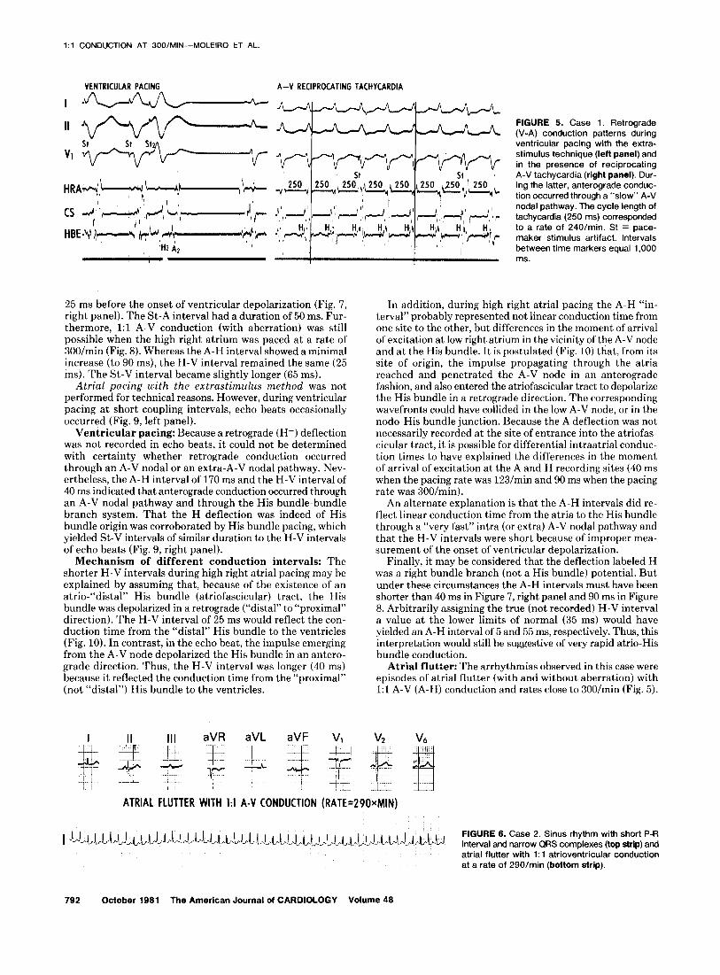
Reciprocating tachycardia: Occasionally, atria1 pacing from the low lateral right atrium and coronary sinus resulted in episodes of reciprocating A-V tachycardias during which anterograde conduction occurred through a “slow” A-V nodal pathway and retrograde conduction through a “fast” pathway (Fig. 5, right panel). Because the retrograde atria1 (A-) de- flection in the His bundle electrocardiographic lead was in- scribed simultaneously with the ventricular electrograms (recorded in the same lead), it was not possible to determine the exact duration of the H-A- interval. However, the H- HRA- interval had (as in the left panel of this Figure 5) a duration of 80 ms. The rate of the tachycardia was 240/min (cycle length 250 ms).
To summarize, in this patient, electropharmacologic studies did not permit the determination as to whether the anatomic substrate was an atrionodal tract22 or a “fast” intranodal pathway.20,23j24 However, enhanced A-V (A-H) conduction indeed occurred electrophysiologically.14J7 This phenomenon was (1) only suspected during sinus rhythm, (2) proved by high right atria1 pacing, (3) manifested during atria1 fibrillation as extremely fast ventricular rates, and (4) pacing site-depen- dent.g
Case 2
An 18 year old youth with mild aortic stenosis had a short P-R interval, and narrow QRS complex in the electrocardio- gram and recurrent supraventricular tachyarrhythmias (Fig. 6). The paroxysms were considered refractory to pharmaco- logic therapy, responding only to cardioversion.
During sinus rhythm, the P-R and P-A intervals measured 100 ms and 50 ms, respectively (Fig. 7, left panel). The H de- flection could not be identified with certainty because of the short A-V interval (50 ms). In this patient the H-V intervals were measured in the His bundle electrogram because the onset of ventricular depolarization occurred in this intracar- diac lead earlier than in the surface leads.
Atria1 pacing: When the high right atrium was paced at a rate of 123/min, the A deflection became separated by an interval of 40 ms from the H deflection, which in turn occurred
AFTER VERAPAMIL CL 450-600
The American Journal of CARDIOLOGY Volume 48 791
1:l CONDUCTION AT 300/MIN-MOLEIRO ET AL.
VENTRICULAR PACING A-V~RECIPROCATING TACHYCARDIA
25 ms before the onset of ventricular depolarization (Fig. 7, right panel). The St-A interval had a duration of 50 ms. Fur- thermore, 1:l A-V conduction (with aberration) was still possible when the high right atrium was paced at a rate of 300/min (Fig. 8). Whereas the A-H interval showed a minimal increase (to 90 ms), the H-V interval remained the same (25 ms). The St-V interval became slightly longer (65 ms).
Atria1 pacing with the extrastimulus method was not performed for technical reasons. However, during ventricular pacing at short coupling intervals, echo beats occasionally occurred (Fig. 9, left panel).
Ventricular pacing: Because a retrograde (H-) deflection was not recorded in echo beats, it could not be determined with certainty whether retrograde conduction occurred through an A-V nodal or an extra-A-V nodal pathway. Nev- ertheless, the A-H interval of 170 ms and the H-V interval of 40 ms indicated that anterograde conduction occurred through an A-V nodal pathway and through the His bundle-bundle branch system. That the H deflection was indeed of His bundle origin was corroborated by His bundle pacing, which yielded St-V intervals of similar duration to the H-V intervals of echo beats (Fig. 9, right panel).
Mechanism of different conduction intervals: The shorter H-V intervals during high right atria1 pacing may be explained by assuming that, because of the existence of an atrio-“distal” His bundle (atriofascicular) tract, the His bundle was depolarized in a retrograde (“distal” to “proximal” direction). The H-V interval of 25 ms would reflect the con- duction time from the “distal” His bundle to the ventricles (Fig. 10). In contrast, in the echo beat, the impulse emerging from the A-V node depolarized the His bundle in an antero- grade direction. Thus, the H-V interval was longer (40 ms) because it reflected the conduction time from the “proximal” (not “distal”) His bundle to the ventricles.
FIGURE 5. Case 1. Retrograde (V-A) conduction patterns during ventricular pacing with the extra- stimulus technique (left panel) and in the presence of reciprocating A-V tachycardia (right panel). Dur- ing the latter, anterograde conduc- tion occurred through a “slow” A-V nodal pathway. The cycle length of tachycardia (250 ms) corresponded to a rate of 240/min. St = pace- maker stimulus artifact. Intervals between time markers equal 1,000 ms.
In addition, during high right atria1 pacing the A-H “in- terval” probably represented not linear conduction time from one site to the other, but differences in the moment of arrival of excitation at low right atrium in the vicinity of the A-V node and at the His bundle. It is postulated (Fig. 10) that, from its site of origin, the impulse propagating through the atria reached and penetrated the A-V node in an anterograde fashion, and also entered the atriofascicular tract to depolarize the His bundle in a retrograde direction. The corresponding wavefronts could have collided in the low A-V node, or in the nodo-His bundle junction. Because the A deflection was not necessarily recorded at the site of entrance into the atriofas- cicular tract, it is possible for differential intraatrial conduc- tion times to have explained the differences in the moment of arrival of excitation at the A and H recording sites (40 ms
when the pacing rate was 123/min and 90 ms when the pacing rate was 300/min).
An alternate explanation is that the A-H intervals did re- flect linear conduction time from the atria to the His bundle through a “very fast” intra (or extra) A-V nodal pathway and that the H-V intervals were short because of improper mea- surement of the onset of ventricular depolarization.
Finally, it may be considered that the deflection labeled H was a right bundle branch (not a His bundle) potential. But under these circumstances the A-H intervals must have been shorter than 40 ms in Figure 7, right panel and 90 ms in Figure 8. Arbitrarily assigning the true (not recorded) H-V interval a value at the lower limits of normal (35 ms) would have yielded an A-H interval of 5 and 55 ms, respectively. Thus, this interpretation would still be suggestive of very rapid atrio-His bundle conduction.
Atria1 flutter: The arrhythmias observed in this case were episodes of atria1 flutter (with and without aberration) with 1:1 A-V (A-H) conduction and rates close to 300/min (Fig. 5).
ATRIA1 FLUTTER WITH I:1 A-V CONDUCTION (RATE=290xMIN)
FIGURE 6. Case 2. Sinus rhythm with short P-R Interval and narrow QRS comolexes (too strip) and . . atrial flutter with 1: 1 atrioventricular conduction at a rate of 290/min (bottom strip).
792 October 1961 The American Journal of CARDIOLOGY Volume 46

1:l CONDUCTION AT 300/MIN-MOLEIRO ET AL.
SINUS RHYTHM ATRIAL PACING (RATE: I~~/MIN.)
HRA -i+
HBE'-e 500 MSEC.
P-R: 100 P-A: 50 A-H: 25(?) H-V: 25(?)
FIGURE 7. Case 2. Sinus rhythm (left panel) and high right atrial pacing at a rate of 123/min (right panel). The H deflection could not be identified with certainty in sinus beats. However, during atrial pacing a deflection (presumably His bundle in origin) was recorded 40 ms after the A de- flection and 25 ms before the onset of ventricular depolarization (V).
Although these episodes did not respond to 15 mg of verapamil or 10 mg of tensilon, cardioversion with a 50 watt-second shock promptly reestablished sinus rhythm.
To summarize, in this patient, electrophysiologic studies suggested (but did not conclusively prove) the existence of an atrio-His bundle (atriofascicular) tract. However, the findings showed that (regardless of its exact mechanism) enhanced A-V (A-H) conduction occurred.
Discussion
The clinical and surface electrocardiographic fea- tures, the underlying anatomic substrates and the electrophysiologic categorization of patients with the so-called Lown-Ganong-Levine syndrome have been subjects of considerable debate and speculations. Un- fortunately, these issues are still unresolved after a
ATRIA1 PACING: RATE: 300/MIN. SINUS
A- H: 90 H - V: 25
FIGURE 8. Case 2. High right atrial pacing at a rate of 300/min with 1:l atrioventricular (A-H) conduction. The QRS complex shows aberration with a right bundle branch block configuration. The duration of the H-V interval was 25 ms. V = onset of ventricular depolarization. Intervals between time markers equal 1,000 ms.
VENTRICULAR PACING ECHO HIS BUNDLE PACING
St
II,
11 i-
VP - F
HRAT
H-V: 40 St-v: 40
FIGURE 9. Case 2. Echo beat during ventricular pacing with the ex- trastimulus technique at a short coupling interval (left panel). An- terograde conduction of the echo beat occurred through an A-V nodal pathway (A-H interval of 170 ms) and the His bundle-bundle branch system (H-V interval of 40 ms). The His bundle origin of the H deflection was validated by proximal His bundle stimulation (right panel) because the St-V interval (40 ms) was equal to the H-V interval of the echo beat. Intervals between driving pacemaker stimuli in the left panel equal 500 ms.
decade of extensive and exhaustive studies.1-21,2s~24 Therefore, attempts to clarify the areas of agreement, disagreement and misinterpretations appear justi- fied.
The syndrome described by Lown, Ganong and Levine: In 1952, these authors25 discussed the findings in 34 patients who had a “short P-R interval, narrow QRS complexes and paroxysmal heart action.” That Lown, Ganong and Levine used the term “paroxysmal heart action” in reference to recurrent sustained su- praventricular tachyarrythmias is well known. What has received less attention (especially in recent reports) is
FIGURE 10. Case 2. Diagrammatic representation of a mechanism that could explain the different conduction intervals in Case 2. An impulse arriving at the “distal” His bundle through an atriofascicular tract (as in Fig. 8) would reach the ventricles earlier than an impulse arriving at the “proximal” His bundle after traversing the atrioventricular node (as in Fig. 9, left panel). This occurs because the “distal” His to V con- duction time is shorter than the “proximal” His to V conduction time. Because the existence of an atriofascicular tract would also imply retrograde (“distal” to ” proximal”) His bundle activation, the corre- sponding A-H interval would represent not linear conduction time from A to H but differences in the moment of activation of the atrial recording site and His bundle.
October 1981 The American Journal of CARDIOLOGY Volume 48 793

I:1 CONDUCTION AT 300/MIN-MOLEIRO ET AL.
that of the 34 patients studied “six experienced bouts of auricular fibrillation and four had atria1 flutter.“25
Therefore, if the eponym “Lown-Ganong-Levine syndrome” is to be used at all, it should be applied to a surface electrocardiographic pattern consisting of a short P-R interval, narrow ventricular complexes and recurrent, sustained supraventricular tachyarrhyth- mias, which could be reciprocating A-V tachycardias as well as atria1 fibrillation and atria1 flutter.
Anatomic substrates of the short P-R interval: The abbreviated A-H conduction times have been at- tributed to faster than normal conduction through atrionoda122,26 or atrio-His bundle (atriofascicular) tracts.16,27m2g In cases in which atrio-His bundle (atri- ofascicular) tracts were not found and propagation through a “normal” atrionodal tract was inferred, morphologic studies could not exclude propagation through a “fast” intranodal pathway.26,2g
Nevertheless, the electrophysiologic substrates in the specimens showing connections between atria1 fibers and the His bundle (which were considered as “acces- sory A-V nodes”) have not been well defined.30-33
Enhanced A-V (A-H) conduction: Regardless of the underlying anatomic substrates, the findings in our two patients indicated that, electrophysiologically, enhanced A-V conduction occurred. It should be stressed that, by itself, the existence of a short A-H in- terval in sinus beats does not mean that enhanced A-V (A-H) conduction is present. The latter should be con- sidered in the following circumstances:14-l7 (1) 1:l A-V conduction can be maintained during atria1 pacing at rates of 200/min or more; (2) the maximal A-H prolon- gation does not exceed 100 ms; and (3) the A-V nodal (or more properly “A-H”) refractory periods are shorter than they should be for the paced cycle length and age.34 These conditions were fulfilled in our cases. Moreover, as in previous reports,g,35 we observed that the capacity to penetrate (in an anterograde direction) the pathway through which enhanced A-V conduction occurred was not only refractory-dependent, but also pacing site- related (Fig. 2 and 3). However, in these studies, the impulses arising in the high right atrium always pene- trated the slow A-V nodal pathway.g,35 Exposure of enhanced A-V conduction during stimulation of some but not all paced atria1 sites can reflect, alone or in combination, changes in the mode, sites, or moment of arrival of excitation at the corresponding pathways.g,35 Therefore, failure to expose enhanced A-V conduction in any electrophysiologic study in which atria1 pacing is performed from only a single site should not be con- strued to indicate that this patient lacks the potential for enhanced A-V conduction. The search for this phenomenon should include stimulation from at least three atria1 sites.
Furthermore, exposure of enhanced A-V conduction in our two patients was possible because (among other factors) the atria could be stimulated at rates of 300/ min. In our two cases, consistent atria1 capture at cycle lengths of 200 ms was surprising because 2:1, 3:l and
“Wenckebach” types of intranodal conduction distur- bances usually develop at rates less than 300/min.36-40 This finding may have occurred because of the relatively young age of the patients (18 and 21 years, respectively), the absence of organic or functional (manifest or pac- ing-exposed) atria1 disease or the techniques used. In- troduction of the hexapolar catheter electrode through the left antecubital vein allowed the catheter to be looped so as to permit a close (and persistent) contact between the pacing electrodes and atria1 endocar- dium.40
“Pharmacology” of enhanced A-V (A-H) con- duction: In most cases pharmacologic evaluation has shown that the pathways through which enhanced A-V conduction occurs responds as A-V nodal tis- sue.20,23,24,41p43 Slowing of ventricular rates by drugs acting on the A-V node probably excludes an atrio-His bundle (atriofascicular) tract, but this effect can occur with either an atrionodal tract or a “fast” intranodal pathway.
However, even if the existence of an intranodal pathway were proved, it could still be argued whether the distinct electrophysiologic properties of this path- way represent an “abnormality” or an extreme variation from normal. If these unusual properties represent an “abnormality,” then the pathway is not the normal “fast” intranodal pathway but another “very fast” (also intranodal) pathway. However, if one considers that these properties reflect an extreme variation from normal one implies that the normal “fast” intranodal pathway can have (as accessory pathways of the Kent bundle type) “very short” as well as “normally short” conduction times and refractory periods (or that “the fast intranodal pathway is a little faster”).18 Therefore, the discussion as to whether enhanced A-V conduction occurs through an atrionodal tract or an intranodal pathway has not been settled and, in fact, may be (from the electrophysiologic viewpoint) sophistic and aca- demic.
Types of recurrent supraventricular tachyar- rhythmias which have been recorded during elec- trophysiologic evaluation of patients with short P-R intervals and narrow QRS complexes: A review of published data1-21,44 reveals that the patient popu- lation studied by different groups has varied consider- ably. For example, in a recent study of 14 patients with short P-R intervals (who had reciprocating A-V tachycardia but not atria1 fibrillation or flutter), and of 51 patients with normal P-R intervals (also with re- ciprocating A-V tachycardia only), Bauernfeind et a1.24 found no difference in the cycle lengths of the tachy- cardia among the two subgroups. They observed that these 65 patients (with reciprocating A-V tachycardia) had a continuum of P-R (and A-H) intervals during sinus rhythm. Hence, these authors concluded “that a diagnosis of Lown-Ganong-Levine syndrome in these patients may be arbitrary and artificial.”
However, in patients with short P-R intervals and narrow QRS complexes (as well as in those with mani-
794 October 1991 The American Journal of CARDIOLOGY Volume 49

fest Wolff-Parkinson-White syndrome) the rates of most established reciprocating A-V tachycardias bear no relation to the rates that can occur during estab- lished atria1 fibrillation (or atria1 flutter). The former are mostly dependent on the conduction velocity through the “slow” A-V nodal pathway, and the latter on the conduction velocity of the pathway through which enhanced A-V conduction runs (whether it is intranodal or extranodal). Therefore, the existence of short P-R (and A-H) intervals in sinus beats has little prognostic significance with regard to the ventricular rates that these patients may manifest during estab- lished reciprocating A-V tachycardia. If one considers that the only arrhythmias that patients with short P-R intervals and narrow QRS complexes can have are re- ciprocating A-V tachycardias, the eponym Lown- Ganong-Levine syndrome has, of course, no rele- vance.
In contrast, the short P-R (and A-H) interval may (but need not always) be of some prognostic significance in those patients who manifest atria1 fibrillation or flutter and who also have enhanced A-V conduction capable of being manifested by very fast ventricular rates (Fig. 2 and 10). In these cases, use of the eponym Lown-Ganong-Levine syndrome may be helpful.
Clinical implications: A review of the original article by Lown, Ganong and Levine25 shows that if the ep- onym Lown-Ganong-Levine syndrome is to be used at all, it should be used in reference to patients with short P-R intervals and narrow QRS complexes who may have atria1 fibrillation or atria1 flutter as well as. reciprocating A-V tachycardias. 25 Limiting the syndrome to patients with reciprocating A-V tachycardias only would be the equivalent of limiting the designation of “Wolff-Par- kinson-white syndrome” to patients who only have reciprocating tachycardias (therefore excluding those with atria1 fibrillation and atria1 flutter). This discussion is not academic because some patients with short P-R intervals and narrow QRS complexes (Fig. 2) do mani- fest atria1 fibrillation and atria1 flutter with extremely fast (from 200 to 300/min) ventricular rates capable of
1:l CONDUCTION AT 300/MIN-MOLEIRO ET AL.
leading to ventricular tachycardia and ventricular flutter (Fig. 2 and 10).13J5J7Jg
Although in most (but not all) cases the ventricular rates can be decreased with drugs that act on the A-V node (propranolol, ouabain, verapamil), the very fact that such rapid ventricular rates can be sustained for variable periods of time may be clinically significant. For example, when organic heart disease is present, there are clinical situations and physiologic conditions causing hemodynamic and electrical instability (such as transient ischemia or acute myocardial infarction) where these rapid rates can lead to ventricular fibrilla- tion.17 The origin of the latter can be exclusively elec- trical (R on T phenomenon) or hemodynamic (poor coronary perfusion), as suggested in a previous study in which the onset of ventricular fibrillation was docu- mented.17 It is interesting that the two sudden deaths occurring in the report of Lown, Ganong and Levine25 occurred in patients with “paroxysmal auricular fi- brillation,” Furthermore, it is not known whether the dosages required to decrease these extremely fast ven- tricular rates are the same dosages as those needed to slow the ventricular rates of the (common) types of atria1 fibrillation occurring in patients who, during sinus rhythm, have normal P-R (and A-H) inter- vals.
Future directions: Further (prospective) studies of patients with a short P-R interval, narrow QRS complex and recurrent supraventricular tachyarrhythmias are required to determine (1) the incidence of various types of tachyarrhythmias, (2) the incidence of enhanced A-V (A-H) conduction during atria1 pacing while stimulating from at least three atria1 sites, (3) the ventricular rates of atria1 flutter and fibrillation (when present) and their relation to the rate during reciprocating A-V tachy- cardias (if they occur in the same patient), and (4) the drug dosages required to slow these rates. Unfortu- nately, until (and while) this is done, discussions re- garding the association of a short P-R interval, narrow QRS complex and recurrent tachyarrhythmias are likely to continue unabated.
References
1. Durrer D. Electrical aspects of human cardiac activity: A clinical- Am J Cardiol 1973;31:245-53. physiological approach to excitation and stimulation. Cardiovasc 7. lannone LA. Electrophysiology of atrial pacing in patients with short Res 1968;2:1-18. P-R interval, normal QRS complex. Am Heart J 1975;89:74-8.
2. Castellanos A, Castlllo CA, Agha AS, Tessler M. His bundle 8. Blssett JK, deSoyza N, Kane JJ, Murphy ML. Altered refractory electrograms in patients with short P-R intervals, narrow QRS periods in patients with short P-R intervals and normal QRS com- complexes and paroxysmal tachycardias. Circulation 197 1;43: plex. Am J Cardiol 1975;35:487-91. 667-78. 9. Aranda J, Castellanos A, Molelro J, Befeler B. Effects of the pacing
3. Coumel PH, Waynberger M, Slama Ft. Bouvraln Y. Le syndrome site on A-H conduction and refractoriness in patients with short P-R court, avec complexes QRS normal. Particularites electro- P-R intervals. Circulation 1976;53:33-9. cardiographiques. Arch Mal Coeur 1972;65:161-81. 10. Selpel L, Brelthardl G, Both A. Atrioventricular (AV) and ven-
4. Mandel WJ, Danzlg R, Hayakawa H. Lown-Ganong-Levine syn- triculo-atrial (VA) conduction pattern in patients with short P-R drome. A study of His bundle electrograms. Circulation 1971; intervals and normal QRS complex. In: Heidelberg LB, ed. Cardiac 44:696-708. Pacing: Diagnostic and Therapeutic Tools. Springer, 197G:
5. Blssett JK, Thompson AJ, deSoyza N, Murphy ML. Atrioventricular 275-88. conduction in patients with short PR intervals and normal QRS 11. Denes P, Wu D, Rosen KM. Demonstration of dual A-V pathways complexes. Br Heart J 1973;35: 123-7. in patients with Lown-r&tong-Levine syndrome. Chest 1974;
6. Caracta AR, Damato AN, Gallagher JJ, et al. Electrophysiologic 65~343-6. studies in the syndrome of short P-R interval, normal QRS complex. 12. Befeler B, Castellanos A, Aranda J, Gutlerrez R, Lazzara R. In-
October 1981 The American Journal of CARDIOLOGY Volume 48 795

I:1 CONDUCTION AT 300/MIN-MOLEIRO ET AL.
termittent bundle-branch block in patients with accessory atri@His Ganong-Levine. Arch Mal Coeur 1974;67:507-20. or atrio-nodal pathways. Variants of the Lown-Ganong-Levine 29. Anderson RH, Becker AH, Brechenmacher C, Davies MJ, Rossi syndrome. Br Heart J 1976;38: 173-9. L. Ventricular preexcitation: a proposed nomenclature for its
13. Casfellanos A, Vagueiro MC, Befeler B, Myerburg RJ. Syndrome substrates. Eur J Cardiol 1975;3:27-36. of short P-R, narrow QRS and repetitive supraventricular tachy- 30. Mockenberg JG. Zur entwicklungsyeschichte des atrioventrikular arrhythmias: the possible occurrence of the R-on-T phenomenon system. Verh Dtsch Pathol Ges 1913;16:228-31. and the limiting of this syndrome. Eur J Cardiol 1975;2:337-42. 3 1. Lev M, Fielding RT, Zaeske D. Mixed levocardia with ventricular
14. Gallagher JJ, Seafy WC, Kasell J, Wallace AG. Multiple accessory inversion (corrected transposition) with complete atrioventricular pathways in patients with the pre-excitation syndrome. Circulation block. A histopathologic study of the conduction system. Am J 1976;54:571-91. Cardiol 1963; 12:875-83.
15. McRae RJ, Wagner GS, Rogers MC, Canent RV. Paroxysmal fa- miliar ventricular fibrillation. J Pediat 1974;84:515-9.
16. Brechenmacher C. Atrio-His bundle tracts. Br Heart J 1975;37: 853-5.
17. Benditf DG, Pritchett ECL, Smith WY, Wallace AG, Gallagher JJ. Characteristics of atrioventricular conduction and the spectrum of arrhythmias in the Lown-Ganong-Levine syndrome. Circulation 1978;57:454-65.
32. Anderson RH, Thapar MK, Arnold R, Jones RS. A study of the conducting tissue in a case of ventricular preexcitation. Br Heart J 1973;35:566-9.
33. Castellanos A, Balais M, Lemberg L, Salhanick L, Castellanos A Jr. Double ectopic left atrial rhythms. Dis Chest 1967;52:87- 91.
18. Josephson ME, Seides SF. Clinical Cardiac Electrophysiology. Techniques and Interpretations. Philadelphia: Lea & Febiger, 1979:232-40.
34. Denes P, Wu D, Dhingra R, Pietras RJ, Rosen KM. The effects of cycle length on cardiac refractory periods in man. Circulation 1974;49:32-41.
19. Myerburg RJ, Sung RJ, Castellanos A. Ventricular tachycardia and ventricular fibrillation in patients with short P-R intervals and narrow QRS complexes. Pace 1979;2:568-78.
20. Josephson ME, Kastor JA. Supraventricular tachycardia in the Lown-Ganong-Levine syndrome: atrionodal versus intranodal reentry. Am J Cardiol 1977;40:521-7.
21. Holmes DR, Hartzler GO, Maloney JD. Concealed retrograde bypass tracts and enhanced atrioventricular conduction: an unusual subset of patients with refractory supraventricular tachycardias. Am J Cardiol 1980;45: 1053-60.
35. Castellanos A, Agha AS, Mendoza IJ, Sung RJ. Evaluation of in- tracardiac recordings in diagnosis of impulse formation and con- cealed conduction in atrioventricular nodal bypass tracts. Br Heart J 1977;36:726-32.
22. James TN. Morphology of the human atrioventricular node, with remarks pertinent to its electrophysiology. Am Heart J 1961;62: 756-71.
36. Castellanos A Jr, lyengar R, Agha AS, Castillo CA. Wenckebach phenomenon within the atria. Br Heart J 1972;34:1121-6.
37. Puech P. Atrioventricular block: the value of intracardiac record- ings. In: Krikler DM, Goodwin JF, eds. Cardiac Arrhythmias. The Modern Electrophysiological Approach. Philadelphia: WB Saunders, 1975:81-115.
38. Narula OS, Runge M, Samet P. Second degree Wenckebach type AV block due to block within the atrium. Br Heart J 1972;34: 1127-36.
23. Denes P, Wu D, Dhlngra RC, Chuquimia R, Rosen KM. Demon- stration of dual A-V nodal pathways in patients with paroxysmal supraventricular tachycardia. Circulation 1973:48:549-55.
24. Bauernfeind RA, Ayres BF, Wyndham CC, et al. Cycle length in atrioventricular nodal reentrant paroxysmal tachycardia with ob- servations on the Lown-Ganong-Levine syndrome. Am J Cardiol 1980;45: 1148-53.
39. Puech P, Grolleau R, Cabasson J, Baissus C, Latour H. Blocs auriculo-ventriculaire du premier et deuxieme degree par trouble de conduction intra-auriculaire. Arch Mal Coeur 1975;68:19- 23.
25. Lown B, Ganong WF, Levine SA. the syndrome of short P-R in- terval, normal QRS complex and paroxysmal rapid heart action. Circulation 1952;5:693-706.
40. Castellanos A, Sung RJ, Mallon SM, Bloom MG, Myerburg RJ. Effects of proximal intra-atrial Wenckebach on distal atrioven- tricular nodal, and His-Purkinje, block. Am Heart J 1978;95: 228-34.
41. Wft AL, Damato AN, Weiss MB, Steiner C. Phenomenon of the gap in atrioventricular conduction in the human heart. Circ Res 1970;27:679-89.
26. Lev M, Fox SM Ill, Bharati S, Greenfleld JC Jr, Rosen KM, Pick A. Mahaim and James fibers as a basis for a unique variety of ventricular preexcitation. Am J Cardiol 1975;36:880-8.
27. Lev M, Leffler WB, Langendrof R, Pick A. Anatomic findings in a case of ventricular pre-excitation (WPW) terminating in complete atrioventricular block. Circulation 1966;34:718-33.
28. Brechenmacher C, Laham J, Iris L, Gerbaux A, Lenegre J. Etude histologic des voies anormales de conduction dans un syndrome de Wolff-Parkinson-White et dans un syndrome de Lown-
42. Wu D, Denes P, Dhingra R, Khan A, Rosen KM. The effects of propranolol on induction of A-V nodal reentrant paroxysmal tachycardia. Circulation 1974;50:665-77.
43. Wu D, Wyndham CR, Amat-y-Leon F, Denes P, Dhingra RC, Rosen KM. The effects of ouabain on induction of atrioventricular nodal re-entrant paroxysmal supraventricular tachycardia. Circu- lation 1975;52:201-7.
44. Sherf L, Neufeld HN. The Pre-Excitation Syndrome: Facts and Theory. New York: Yorke Medical Books, 1980:216-9.
796 October 1981 The American Journal of CARDIOLOGY Volume 48