Novel symptomatology and changing epidemiology of domoic acid toxicosis in California sea lions...
10
Novel symptomatology and changing epidemiology of domoic acid toxicosis in California sea lions (Zalophus californianus): an increasing risk to marine mammal health T. Goldstein 1,2, * , J. A. K. Mazet 2 , T. S. Zabka 1,2 , G. Langlois 3 , K. M. Colegrove 4 , M. Silver 5 , S. Bargu 5 , F. Van Dolah 6 , T. Leighfield 6 , P. A. Conrad 3 , J. Barakos 7 , D. C. Williams 8 , S. Dennison 1 , M. Haulena 1 and F. M. D. Gulland 1 1 The Marine Mammal Center, 1065 Fort Cronkhite, Sausalito, CA 94965, USA 2 Wildlife Health Center, School of Veterinary Medicine, University of California, 1 Shields Avenue, Davis, CA 95616, USA 3 California Department of Public Health, 850 Marina Bay, Parkway, Richmond, CA 94804, USA 4 Department of Pathology, Microbiology and Immunology, School of Veterinary Medicine, University of California, 1 Shields Avenue, Davis, CA 95616, USA 5 Ocean Sciences Department, University of California, Santa Cruz, CA 95064, USA 6 National Oceans Services, SC219 Fort Johnston Road, Charleston, SC 29412, USA 7 California Pacific Medical Center, University of California, San Francisco, CA 94143, USA 8 Veterinary Medical Teaching Hospital, University of California, 1 Garrod Drive, Davis, CA 95616, USA Harmful algal blooms are increasing worldwide, including those of Pseudo-nitzschia spp. producing domoic acid off the California coast. This neurotoxin was first shown to cause mortality of marine mammals in 1998. A decade of monitoring California sea lion (Zalophus californianus) health since then has indicated that changes in the symptomatology and epidemiology of domoic acid toxicosis in this species are associated with the increase in toxigenic blooms. Two separate clinical syndromes now exist: acute domoic acid toxicosis as has been previously documented, and a second novel neurological syndrome characterized by epilepsy described here associated with chronic consequences of previous sub-lethal exposure to the toxin. This study indicates that domoic acid causes chronic damage to California sea lions and that these health effects are increasing. Keywords: amnesic shellfish poisoning; California sea lions; domoic acid; hippocampal atrophy; seizures; harmful algal blooms 1. INTRODUCTION Domoic acid is a potent marine neurotoxin produced by some diatom species of the genus Pseudo-nitzschia that appear to be becoming more frequent along the California coast (Allen 1922; Fryxell et al. 1997; Jeffery et al. 2004). Blooms of other toxigenic algae worldwide also appear to be increasing in extent, frequency and type in recent years, although the reasons for this are unclear ( Van Dolah 2000; HARNESS 2005). Possible explanations include oceano- graphic regime shifts, overfishing, movement and dis- charge of ballast waste, eutrophication of marine waters and global climate change (Anderson 1997; Landsberg 2002; Chavez et al. 2003). As harmful algal blooms increase, it is likely that their impacts on marine fauna and human health will also increase, yet data on these impacts are rare due to the logistical difficulties associated with studying these effects. Domoic acid is water soluble and has a high affinity for the a-amino-5-hydroxy-3-methyl-4-isoxazole propionic acid (AMPA) and kainate subclasses of neuronal glutamate ionotropic receptors ( Jeffery et al. 2004). Following binding of glutamate receptors, domoic acid acts as an excitotoxin by causing massive cell depolarization with subsequent increase in intracellular calcium, resulting in cell dysfunction and death (Olney et al. 1979). Domoic acid is the cause of amnesic shellfish poisoning in humans, first recognized in Canada in 1987 following consumption of contaminated shellfish ( Perl et al. 1990). Clinical signs included gastrointestinal distress and neurological symp- toms including seizures, agitation, coma and death ( Teitelbaum et al. 1990). Histological changes in four people who died 7–90 days after the event were consistent with exposure to an excitotoxin and included neuronal necrosis or loss in all sectors of the hippocampal formation except in cornu ammonis sector CA2, and astrocytosis in one patient ( Teitelbaum et al. 1990). One year after the acute episode had resolved, one patient developed complex partial seizures (Cendes et al. 1995). Electroencephalo- grams (EEG) showed periodic epileptiform discharges over both temporal lobes and magnetic resonance imaging (MRI) revealed bilateral hippocampal atrophy. Autopsy over 3 years following intoxication revealed bilateral hippocampal sclerosis (i.e. astrocytosis), demonstrating that long-term neurological damage persisted years after initial exposure to domoic acid. Proc. R. Soc. B (2008) 275, 267–276 doi:10.1098/rspb.2007.1221 Published online 15 November 2007 * Author and address for correspondence: Wildlife Health Center, 1 Shields Ave, School of Veterinary Medicine, University of California, Davis, California 95616, USA ([email protected]). Received 6 September 2007 Accepted 12 October 2007 267 This journal is q 2007 The Royal Society
Transcript of Novel symptomatology and changing epidemiology of domoic acid toxicosis in California sea lions...
Proc. R. Soc. B (2008) 275, 267–276
doi:10.1098/rspb.2007.1221
Novel symptomatology and changing epidemiologyof domoic acid toxicosis in California sea lions(Zalophus californianus): an increasing risk
to marine mammal healthT. Goldstein1,2,*, J. A. K.Mazet2, T. S. Zabka1,2, G. Langlois3, K.M. Colegrove4,
M. Silver5, S. Bargu5, F. Van Dolah6, T. Leighfield6, P. A. Conrad3, J. Barakos7,
D. C. Williams8, S. Dennison1, M. Haulena1 and F. M. D. Gulland1
1The Marine Mammal Center, 1065 Fort Cronkhite, Sausalito, CA 94965, USA2Wildlife Health Center, School of Veterinary Medicine, University of California, 1 Shields Avenue, Davis, CA 95616, USA
3California Department of Public Health, 850 Marina Bay, Parkway, Richmond, CA 94804, USA4Department of Pathology, Microbiology and Immunology, School of Veterinary Medicine,
University of California, 1 Shields Avenue, Davis, CA 95616, USA5Ocean Sciences Department, University of California, Santa Cruz, CA 95064, USA6National Oceans Services, SC219 Fort Johnston Road, Charleston, SC 29412, USA
7California Pacific Medical Center, University of California, San Francisco, CA 94143, USA8Veterinary Medical Teaching Hospital, University of California, 1 Garrod Drive, Davis, CA 95616, USA
Published online 15 November 2007
*AuthoShieldsDavis, C
ReceivedAccepted
Harmful algal blooms are increasing worldwide, including those of Pseudo-nitzschia spp. producing domoic
acid off the California coast. This neurotoxin was first shown to cause mortality of marine mammals in 1998. A
decade of monitoring California sea lion (Zalophus californianus) health since then has indicated that changes
in the symptomatology and epidemiology of domoic acid toxicosis in this species are associated with the
increase in toxigenic blooms. Two separate clinical syndromes now exist: acute domoic acid toxicosis as has
been previously documented, and a second novel neurological syndrome characterized by epilepsy described
here associated with chronic consequences of previous sub-lethal exposure to the toxin. This study indicates
that domoic acid causes chronic damage to California sea lions and that these health effects are increasing.
Keywords: amnesic shellfish poisoning; California sea lions; domoic acid; hippocampal atrophy; seizures;
harmful algal blooms
1. INTRODUCTION
Domoic acid is a potent marine neurotoxin produced by
some diatom species of the genus Pseudo-nitzschia that
appear to be becoming more frequent along the California
coast (Allen 1922; Fryxell et al. 1997; Jeffery et al. 2004).
Blooms of other toxigenic algae worldwide also appear to
be increasing in extent, frequency and type in recent years,
although the reasons for this are unclear (Van Dolah 2000;
HARNESS 2005). Possible explanations include oceano-
graphic regime shifts, overfishing, movement and dis-
charge of ballast waste, eutrophication of marine waters
and global climate change (Anderson 1997; Landsberg
2002; Chavez et al. 2003). As harmful algal blooms
increase, it is likely that their impacts on marine fauna and
human health will also increase, yet data on these impacts
are rare due to the logistical difficulties associated with
studying these effects.
Domoic acid is water soluble and has a high affinity for
the a-amino-5-hydroxy-3-methyl-4-isoxazole propionic
acid (AMPA) and kainate subclasses of neuronal glutamate
r and address for correspondence: Wildlife Health Center, 1Ave, School of Veterinary Medicine, University of California,alifornia 95616, USA ([email protected]).
6 September 200712 October 2007
267
ionotropic receptors ( Jeffery et al. 2004). Following
binding of glutamate receptors, domoic acid acts as an
excitotoxin by causing massive cell depolarization with
subsequent increase in intracellular calcium, resulting in
cell dysfunction and death (Olney et al.1979). Domoic acid
is the cause of amnesic shellfish poisoning in humans, first
recognized in Canada in 1987 following consumption of
contaminated shellfish (Perl et al. 1990). Clinical signs
included gastrointestinal distress and neurological symp-
toms including seizures, agitation, coma and death
(Teitelbaum et al. 1990). Histological changes in four
people who died 7–90 days after the event were consistent
with exposure to an excitotoxin and included neuronal
necrosis or loss in all sectors of the hippocampal formation
except in cornu ammonis sector CA2, and astrocytosis in one
patient (Teitelbaum et al. 1990). One year after the acute
episode had resolved, one patient developed complex
partial seizures (Cendes et al. 1995). Electroencephalo-
grams (EEG) showed periodic epileptiform discharges
over both temporal lobes and magnetic resonance imaging
(MRI) revealed bilateral hippocampal atrophy. Autopsy
over 3 years following intoxication revealed bilateral
hippocampal sclerosis (i.e. astrocytosis), demonstrating
that long-term neurological damage persisted years after
initial exposure to domoic acid.
This journal is q 2007 The Royal Society
268 T. Goldstein et al. Chronic effects: domoic acid on sea lion
Domoic acid has more recently been associated with
neurological signs in seabirds and California sea lions off
the California coast (Work et al. 1993; Scholin et al. 2000).
In 1998, it was estimated that over 400 sea lions were
exposed to the toxin through contaminated prey. During
the event, domoic acid was detected in anchovies, sardines,
phytoplankton, and blood and urine samples from affected
sea lions (Scholin et al. 2000). Lesions in sea lions that died
acutely were consistent with limbic system injury charac-
terized by ischaemic neuronal necrosis in the hippocampal
formation, especially granule cells in the dentate gyrus and
pyramidal cells in sectors CA4, CA3 and CA1, and
progressed to neuronal loss with parenchymal atrophy
and marked gliosis (glial cell type not specified) in sea lions
dying after 15 days (Silvagni et al. 2005).
Experimental exposure of laboratory animals to
domoic acid has produced clinical signs and brain lesions
mostly consistent with those in naturally exposed humans
and sea lions, and the effects of acutely lethal toxic doses of
domoic acid have been well described in multiple species
(Tryphonas et al. 1990; Scallet et al. 1993; Schmued et al.
1995). However, the effects of low doses and repeated
exposures are not as well defined, with single and repeated
low-dose exposure experiments conducted in rats, mice
and primates obtaining varied results. Low doses adminis-
tered by systemic injection produced clinical signs as well
as mild localized lesions and gliosis in the hippocampus
(Scallet et al. 1993; Sobotka et al. 1996). However, oral
dosing produced mild to no clinical signs and histological
examination revealed minimal to no morphologic changes
in the brain (Truelove et al. 1996, 1997). Peng et al.
(1997) did observe clinical signs and associated changes at
low doses in mice, but repeated exposures failed to
enhance symptomatic toxicity.
This paucity of information on the effects of single and
repeated low doses of domoic acid on mammals is of
concern, because as the frequency of domoic acid-
producing algal blooms increases, the likelihood also
increases that humans and marine mammals may ingest
prey contaminated with low levels of the toxin below the
currently accepted dose for seafood closures to prevent
acute poisoning. California sea lions range from Baja
California to British Columbia and feed on species that
often enter the human seafood market, such as anchovies,
sardines, hake, rockfish, salmon and market squid (King
1983). This prey base that is shared with humans, and their
accessibility, mammalian physiology and longevity make
California sea lions excellent sentinels of the potential effects
of toxins in seafood on human health. The United States’
sea lion population breeds primarily on the California
Channel Islands between May and July, which numbers
approximately 200 000 animals (Forney et al. 2000). Adult
females from San Miguel Island, the most northwestern of
the rookery islands, primarily forage north of the island from
Point Conception along the coast of central California
(Melin et al. 2000), but the foraging range of juveniles is
unknown. Little information is available on bloom dynamics
in the areas where sea lions feed, however, conditions
currently appear to favour the dominance of Pseudo-
nitzschia australis and production of domoic acid off the
central and southern California coast (Anderson et al.
2006). This may be related to algal nutrient physiology and
availability associated with wind-driven upwelling, as well as
eddy circulation and entrainment promoting conditions for
Proc. R. Soc. B (2008)
bloom formation and triggers for toxin production. There-
fore, sea lions are likely to be naturally exposed to varying
levels of domoic acid in their prey.
Here we present data from a decade of monitoring
California sea lion health which suggest changes in the
symptomatology and epidemiology of domoic acid
toxicosis associated with increasing harmful algal blooms.
2. MATERIAL AND METHODS(a) Animals
Sea lions found ill along the California coast between Ventura
and Humboldt Counties were taken to The Marine Mammal
Center in Sausalito, CA (TMMC) for clinical examination,
treatment and rehabilitation for release back to the wild when
possible. Three additional cases that originally stranded in Los
Angeles county were transferred from another facility for
treatment. Sex determination was based on genital morphology
and age class was estimated using a combination of body length,
tooth size and stage of sagittal crest development (Greig et al.
2005). Animals were housed in individual pens with fresh water
pools, observed hourly 07.00–23.00, and fed thawed, pre-
viously frozen, herring offered three times daily. Blood samples
were collected for haematology, serum biochemistry and
serology (Dierauf & Gulland 2001).
Sea lions that exhibited chronic neurological signs
were evaluated clinically and by EEG and MRI. Animals
were sedated for EEG with medetomidine at a dose of
0.07 mg kgK1 intramuscularly. Needle electrode (Grass/
Astro-Med, Inc.) placement was based on that used in canine
patients (Holliday & Williams 1998). Fifteen EEG, four
electro-ocular (EOG), two electrocardiac (EKG) and one
ground electrode were placed for each recording. A Nihon
Kohden model 2110 digital EEG system was used for recording
all EEGs which were displayed via a modified double banana
montage. Display sensitivities varied between 7 and
20 mV mmK1, with the time constant at 0.1 s and a high-
frequency cut-off of 70 Hz. Duration of EEG recordings ranged
from 10 to 61 min.
MRI was performed following anaesthesia using
medetomidine (0.07 mg kgK1) and tiletamine–zolazepam
(1 mg kgK1) as described by Haulena & Gulland (2001). All
MR studies were performed on a 1.5-T General Electric MR
system by using a quadrature knee coil. Following the localizer
scan, five sequences were acquired, including coronal fast spin
echo (FSE), fat-suppressed (FS) T2, FS FSE proton density
(PD), fluid-attenuated inversion recovery (FLAIR), sagittal
FSE T2 and FS three-dimensional spoiled gradient-recalled
echo T1 (3D volume SPGR T1). The first three sequences
(FSE T2, FSE PD and FLAIR) were obtained along a plane
perpendicular to the long axis of the temporal lobe and
ventricular horn. This oblique dorsal (coronal) orientation was
selected to optimize the characterization of the hippocampal
formation, as well as to align MR images with pathological
sectioning. Hippocampal changes were classified as normal,
mild, moderate or severe atrophy based on subjective analysis
using the following imaging characteristics: generalized or focal
volume loss, corresponding ex-vacuo temporal ventricular horn
enlargement and loss of grey white differentiation of hippo-
campal structures. The MR images were interpreted by a
neuroradiologist ( J.B.) without knowledge of the clinical or
pathological data. To determine the effect of intraobserver
variation on the imaging interpretations, all examinations were
re-reviewed by the same neuroradiologist three months later
and interpreted in a blinded fashion.
Chronic effects: domoic acid on sea lion T. Goldstein et al. 269
Urine and serum samples submitted for domoic acid
testing were screened by direct competitive domoic acid
ELISA (Biosense Laboratories, Bergen, Norway), and
positive samples were confirmed by liquid chromatog-
raphy–tandem mass spectrometry (LC-MS/MS; Van Dolah
et al. 1997; Maucher & Ramsdell 2005). Faeces were
examined for toxic P. australis diatoms by scanning electron
microscopy and positive samples were tested for domoic acid
using high-performance liquid chromatography (HPLC;
Lefebvre et al. 1999).
After the animals died, a necropsy was performed and
representative tissue samples of brain, eyes and all viscera
were collected, immersed in 10% neutral buffered formalin,
paraffin embedded, sectioned at 4–5 mm, stained with
haematoxylin and eosin and examined by light microscopy.
Brains were serially sectioned and gross abnormalities were
documented when examined by a pathologist. Brain sections
sampled consistently and examined histologically were
regions incorporating (i) anterior frontal lobes, (ii) anterior
cingulated gyrus, (iii) both anterior to mid hippocampal
formations–-parahippocampal gyri–thalamus–insular gyrus,
(iv) midbrain, (v) medulla oblongata at the level of the
anterior velum, (vi) cerebellum, and (vii) cervical spinal cord.
Additional sections sampled in some cases included regions
incorporating (i) amygdaloid nuclei, (ii) olfactory structures
at the level of the olfactory tubercle and gyrus, (iii) optic
structures at the level of the optic tract and chiasm,
(iv) posterior hippocampal formation, and (v) occipital lobes.
(b) Domoic acid-producing algal blooms
The California Department of Public Health (CDPH)
manages a volunteer-based programme that monitors for the
occurrence of toxic algal blooms along the California coast as a
part of the Marine Biotoxin Monitoring Program. There were
230 sampling sites within the study range and these were
sampled at varying frequencies, from 1 to 490 times over the 8
years of this study. An estimate of the relative abundance of
Pseudo-nitzschia spp. was reported based on sampling effort,
per cent composition and cell mass of the diatom in each
sample. Two index sites, Santa Cruz pier (Santa Cruz county,
centre of sea lion stranding range) and Goleta pier (Santa
Barbara county, southern edge of sea lion stranding range),
were chosen for comparison of monthly Pseudo-nitzschia spp.
abundance estimates in water samples with sea lion stranding
data as both were sampled consistently during the study
period. Domoic acid analyses were performed on mussel
samples (collected at 11 sites between Marin and Los Angeles
counties) according to the methods of Quilliam et al. (1995) by
CDPH when blooms of the diatoms were detected.
(c) Statistical analysis
The distribution by year, sex and age class for animals that
stranded with acute toxicosis and with chronic neurological
signs were evaluated by the two-sided Chi-square test for
independence and with the Fisher’s exact test when an expected
cell frequency was less than 5 using EPI INFO 2000 software
(v. 1.1.2, June 2000, Centers for Disease Control and
Prevention, Atlanta, GA, USA). Strength of associations was
estimated by the odds ratio. Temporal associations between
stranding of acute and chronic neurological cases were
evaluated using time-series cross-correlation analysis (Box &
Jenkins 1976; SPSS for Windows, v. 11.0, SPSS, Inc., Chicago,
IL). The number of strandings for both groups was summed for
each week and month, and cross-correlations were evaluated
Proc. R. Soc. B (2008)
for 12 forward and 12 backward week and month time periods.
Temporal associations between the monthly relative abundance
index ofPseudo-nitzschia spp. and sea lions stranding with acute
toxicity were also evaluated using time-series cross-correlation
analysis. Cross-correlation coefficients were considered to be
significant at p!0.05. Santa Cruz & Goleta pier counts were
used for comparison when acute events occurred north or south
of Point Sur (Monterey County), respectively. Spatial and
temporal clustering was evaluated for stranding events
associated with acute toxicosis. The space–time statistic with
the Space-Time Permutation model (Kulldorff et al. 2005) was
used to test whether these cases were randomly distributed
along TMMC’s response area and in time (SATSCAN Software:
Kulldorff M. and Information Management Services, Inc.
SATSCAN v. 7.0: software for the spatial and space-time scan
statistics).
3. RESULTS(a) Clinical case characterization
A total of 2963 sea lions were admitted to The Marine
Mammal Center during the study period. Examination of
715 sea lions stranding along the California coast with
neurological signs between 1998 and 2006 revealed two
separate clinical syndromes: acute domoic acid toxicosis as
documented by Scholin et al. (2000) and Gulland et al.
(2002; nZ551); and a novel chronic epileptic syndrome
characterized by behavioural changes, seizures and
atrophy of the hippocampal formation (nZ164). Acute
cases were characterized by clinical signs that included
ataxia, head weaving, seizures or coma which varied in
severity but were continuous during the period of toxicosis,
lasting about one week followed by recovery, if treated, or
death (Gulland et al. 2002); they stranded in clusters and
had histopathological findings that included hippocampal
neuronal necrosis. Chronic neurological cases were
characterized by animals that developed intermittent
seizures (suffering from seizures at least two weeks apart
and/or seizures after at least two weeks following admission
to The Marine Mammal Center) but were asymptomatic
between seizures, exhibited unusual behaviours, stranded
individually and/or had chronic pathological changes. The
latter were consistent and more extensive than those
previously described for acute cases that survived longer in
rehabilitation, which were described as neuronal loss with
hippocampal atrophy and marked gliosis (glial cell types
not specified; Silvagni et al. 2005).
Clinical signs in the 164 animals with chronic neuro-
logical disease included seizures, periods of marked lethargy
and inappetence, vomiting, muscular twitching, central
blindness blepharospasm (often unilateral) and abnormal
behaviour. Duration of clinical signs from initial presen-
tation to death varied from 25 to 1525 days. Seizures were
observed in 140 of these cases at intervals varying from
hours to weeks, often progressing from simple (not
impairing the level of consciousness), partial (focal) to
secondary (following a simple seizure) generalized (invol-
ving loss of consciousness) seizures. Partial seizures were
characterized by muscle twitching of the head (sometimes
just the eyelids and vibrissae) or flippers and were
occasionally unilateral. Generalized seizures were most
commonly clonic–tonic, characterized by lateral recum-
bency and extended flippers and neck. Seizures were often
preceded by vomiting, pacing or swimming in tight circles.
F3-A1A1-T5T5-O1F3-C3C3-P3P3-O1Fz-CzCz-PzF4-C4C4-P4P4-O2F4-A2A2-T6T6-O2
ECGL EOGR EOG
F3-A1A1-T5T5-O1F3-C3C3-P3P3-O1Fz-CzCz-PzF4-C4C4-P4P4-O2F4-A2A2-T6T6-O2ECG
L EOGR EOG
1 s
100 uV 1 s
(a)
(b)
200 uV
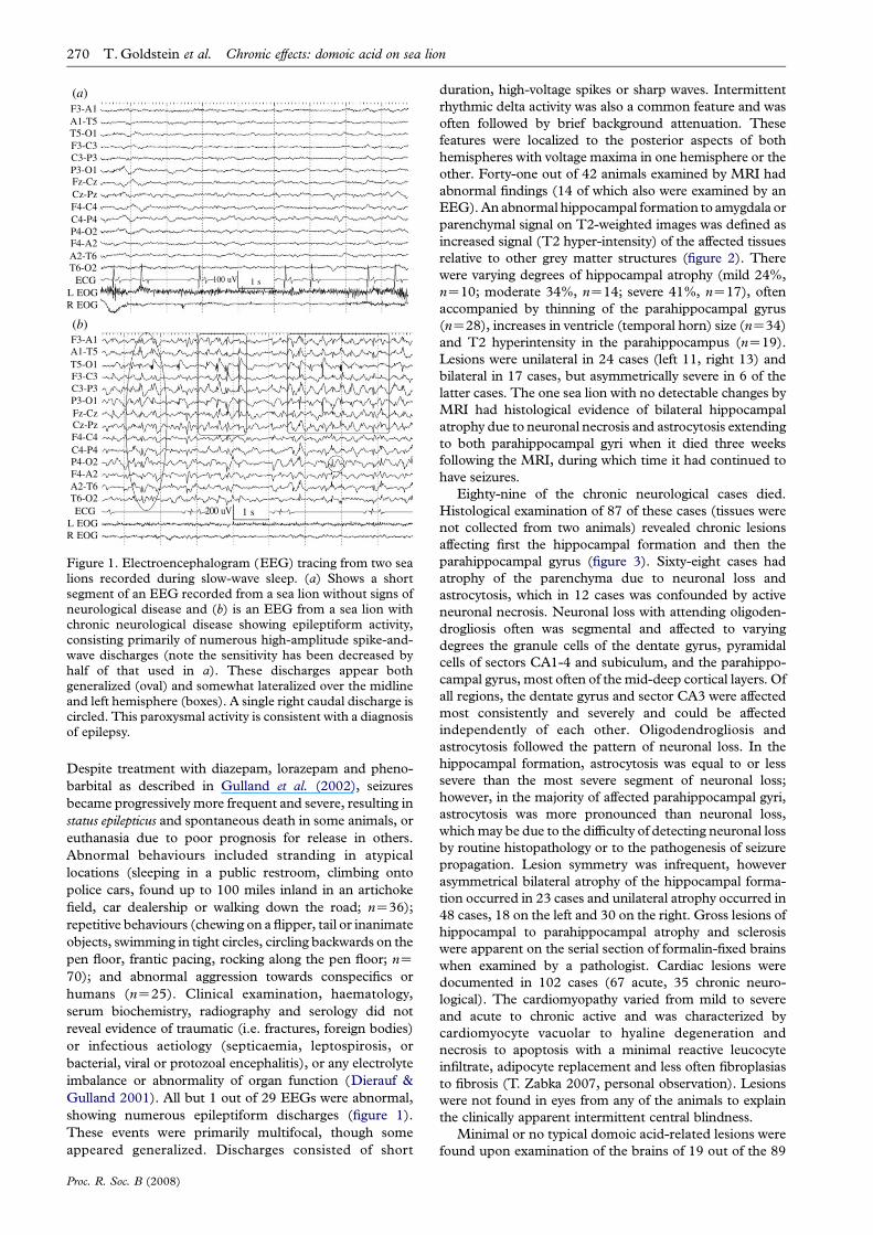
Figure 1. Electroencephalogram (EEG) tracing from two sealions recorded during slow-wave sleep. (a) Shows a shortsegment of an EEG recorded from a sea lion without signs ofneurological disease and (b) is an EEG from a sea lion withchronic neurological disease showing epileptiform activity,consisting primarily of numerous high-amplitude spike-and-wave discharges (note the sensitivity has been decreased byhalf of that used in a). These discharges appear bothgeneralized (oval) and somewhat lateralized over the midlineand left hemisphere (boxes). A single right caudal discharge iscircled. This paroxysmal activity is consistent with a diagnosisof epilepsy.
270 T. Goldstein et al. Chronic effects: domoic acid on sea lion
Despite treatment with diazepam, lorazepam and pheno-
barbital as described in Gulland et al. (2002), seizures
became progressively more frequent and severe, resulting in
status epilepticus and spontaneous death in some animals, or
euthanasia due to poor prognosis for release in others.
Abnormal behaviours included stranding in atypical
locations (sleeping in a public restroom, climbing onto
police cars, found up to 100 miles inland in an artichoke
field, car dealership or walking down the road; nZ36);
repetitive behaviours (chewing on a flipper, tail or inanimate
objects, swimming in tight circles, circling backwards on the
pen floor, frantic pacing, rocking along the pen floor; nZ70); and abnormal aggression towards conspecifics or
humans (nZ25). Clinical examination, haematology,
serum biochemistry, radiography and serology did not
reveal evidence of traumatic (i.e. fractures, foreign bodies)
or infectious aetiology (septicaemia, leptospirosis, or
bacterial, viral or protozoal encephalitis), or any electrolyte
imbalance or abnormality of organ function (Dierauf &
Gulland 2001). All but 1 out of 29 EEGs were abnormal,
showing numerous epileptiform discharges (figure 1).
These events were primarily multifocal, though some
appeared generalized. Discharges consisted of short
Proc. R. Soc. B (2008)
duration, high-voltage spikes or sharp waves. Intermittent
rhythmic delta activity was also a common feature and was
often followed by brief background attenuation. These
features were localized to the posterior aspects of both
hemispheres with voltage maxima in one hemisphere or the
other. Forty-one out of 42 animals examined by MRI had
abnormal findings (14 of which also were examined by an
EEG). An abnormal hippocampal formation to amygdala or
parenchymal signal on T2-weighted images was defined as
increased signal (T2 hyper-intensity) of the affected tissues
relative to other grey matter structures (figure 2). There
were varying degrees of hippocampal atrophy (mild 24%,
nZ10; moderate 34%, nZ14; severe 41%, nZ17), often
accompanied by thinning of the parahippocampal gyrus
(nZ28), increases in ventricle (temporal horn) size (nZ34)
and T2 hyperintensity in the parahippocampus (nZ19).
Lesions were unilateral in 24 cases (left 11, right 13) and
bilateral in 17 cases, but asymmetrically severe in 6 of the
latter cases. The one sea lion with no detectable changes by
MRI had histological evidence of bilateral hippocampal
atrophy due to neuronal necrosis and astrocytosis extending
to both parahippocampal gyri when it died three weeks
following the MRI, during which time it had continued to
have seizures.
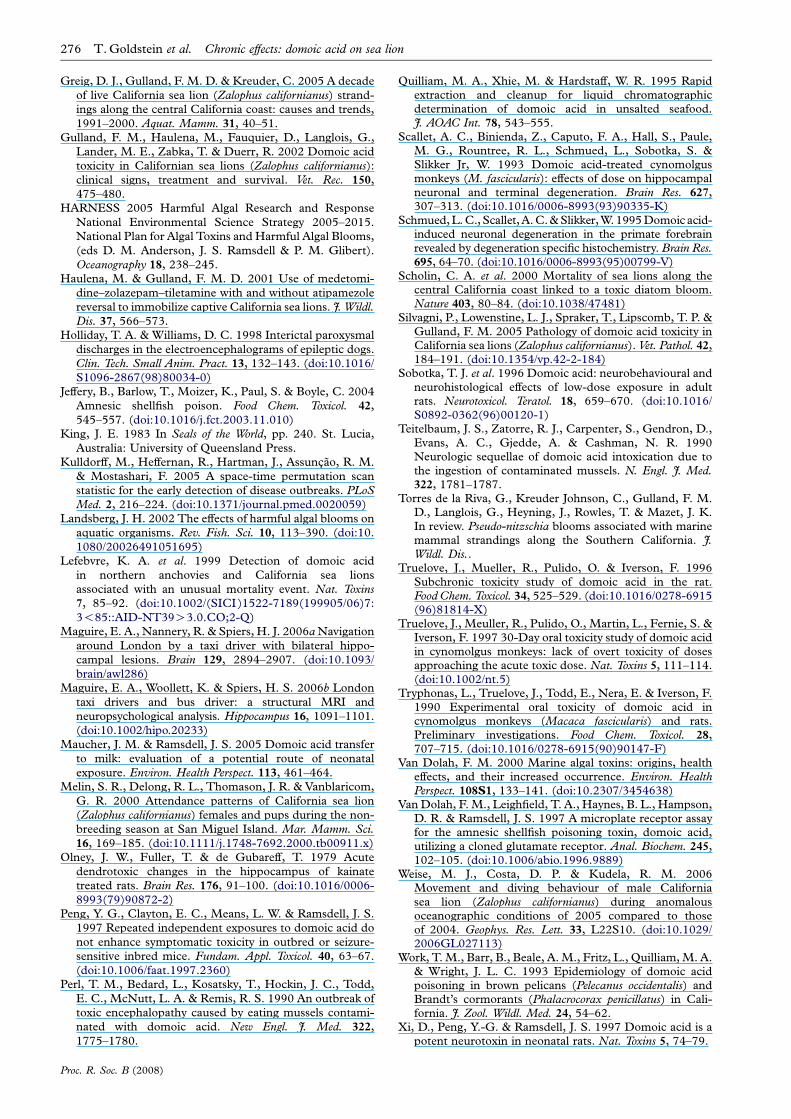
Eighty-nine of the chronic neurological cases died.
Histological examination of 87 of these cases (tissues were
not collected from two animals) revealed chronic lesions
affecting first the hippocampal formation and then the
parahippocampal gyrus (figure 3). Sixty-eight cases had
atrophy of the parenchyma due to neuronal loss and
astrocytosis, which in 12 cases was confounded by active
neuronal necrosis. Neuronal loss with attending oligoden-
drogliosis often was segmental and affected to varying
degrees the granule cells of the dentate gyrus, pyramidal
cells of sectors CA1-4 and subiculum, and the parahippo-
campal gyrus, most often of the mid-deep cortical layers. Of
all regions, the dentate gyrus and sector CA3 were affected
most consistently and severely and could be affected
independently of each other. Oligodendrogliosis and
astrocytosis followed the pattern of neuronal loss. In the
hippocampal formation, astrocytosis was equal to or less
severe than the most severe segment of neuronal loss;
however, in the majority of affected parahippocampal gyri,
astrocytosis was more pronounced than neuronal loss,
which may be due to the difficulty of detecting neuronal loss
by routine histopathology or to the pathogenesis of seizure
propagation. Lesion symmetry was infrequent, however
asymmetrical bilateral atrophy of the hippocampal forma-
tion occurred in 23 cases and unilateral atrophy occurred in
48 cases, 18 on the left and 30 on the right. Gross lesions of
hippocampal to parahippocampal atrophy and sclerosis
were apparent on the serial section of formalin-fixed brains
when examined by a pathologist. Cardiac lesions were
documented in 102 cases (67 acute, 35 chronic neuro-
logical). The cardiomyopathy varied from mild to severe
and acute to chronic active and was characterized by
cardiomyocyte vacuolar to hyaline degeneration and
necrosis to apoptosis with a minimal reactive leucocyte
infiltrate, adipocyte replacement and less often fibroplasias
to fibrosis (T. Zabka 2007, personal observation). Lesions
were not found in eyes from any of the animals to explain
the clinically apparent intermittent central blindness.
Minimal or no typical domoic acid-related lesions were
found upon examination of the brains of 19 out of the 89
(a)HP
R L
FA
HP MRI brain256*256
MR
13/18T2 FLAIR COR
1.44 : 1none100%
1.44 : 1none100%
R L
FA
(b)
Figure 2. The magnitude of hippocampal atrophic changeswas classified from normal to severe. T2-weighted imagesshowing a sea lion with (a) bilaterally normal hippocampi and(b) a chronic neurological case with severe bilateral hippo-campal atrophy, moderate bilateral thinning of the para-hippocampus accompanied by bilateral temporal hornenlargement.
CA2
CA1CA4CA3
subiculum dentategyrus
parahippocampalgyus
(a)
CA1
CA2
CA3
CA4
dentate gyrus
(b)
Figure 3. Sea lion brain, HE stain. (a) Normal hippocampalformation and parahippocampal gyrus to demonstrate thehippocampal substructures, as demarcated by lines andarrow, and the relative numbers of neurons and glial cellsfor comparison with chronic histopathology from domoicacid exposure (scale barZ500 mm). (b) Chronic domoic acid-associated histopathology in the hippocampal formation withsubstructures demarcated by lines and arrow. Characteristicfeatures that cause parenchymal atrophy, which is especiallynotable as contraction at the medial aspect, are the loss ofgranule cells in the dentate gyrus and pyramidal cells in all CAsectors, with the most notable loss in the dentate gyrus andCA3, and the astrocytosis (asterisks) that contributes to theoverall increased cellular appearance, most prominent inregions with greatest neuronal loss. Increased cellularity alsois due to oligodendrogliosis that attends the neuronal loss(scale barZ300 mm).
Chronic effects: domoic acid on sea lion T. Goldstein et al. 271
animals that died with chronic neurological signs, all of
which were immature animals (1 pup, 3 yearlings and 15
juveniles or subadults). Lesions in these 19 sea lions were
non-specific, including focal meningeal haemorrhage,
mild to severe diffuse brain oedema and mild scattered
non-suppurative meningoencephalitis limited to extra-
(para) hippocampal regions such as the medulla oblon-
gata, cingulate gyrus, thalamus and occipital lobes. These
findings were milder than expected in the light of the
abnormal neurological presentation of these cases includ-
ing seizures, and severe behavioural abnormalities. Also,
these lesions were present variably in chronic cases with
typical brain lesions.
Of the 164 chronic neurological cases, 112 developed
epilepsy (intermittent seizures) while under care, 52 had
severe behavioural abnormalities and 68 out of the 87
examined by histology had chronic histopathological
changes. Additionally, 32 animals initially admitted and
treated for acute domoic acid exposure developed epilepsy
between 16 and 85 days after apparent recovery from
acute toxicosis. An additional six animals that had
detectable domoic acid in clinical samples upon their
first admission tested negative when readmitted following
their second stranding and were exhibiting epilepsy and
abnormal behaviour.
When possible, samples were collected within 24 hours
of admission from animals upon admission for domoic acid
testing. Domoic acid was detected in 54% (60/112) of
samples collected from 80 out of the 551 acute cases (urine
43, serum 10, faeces 7) and from only 5 out of 31 samples
submitted from cases with chronic neurological sequellae
(urine 2, faeces 2, serum 1; table 1). Twenty out of these 80
acute animals that tested positive went on to develop
neurological sequellae typical of chronic cases.
(b) Epidemiology
For epidemiological analyses, an acute domoic acid
stranding event was identified when at least five cases
were admitted into rehabilitation over 48 hours which
exhibited signs as described previously by Gulland et al.
(2002), the strandings were clustered spatially within
80 km of coastline of each other and histopathological
findings included hippocampal necrosis in at least one
case. The above criteria were chosen based on estimates in
humans and primates for time from ingestion of the
toxin to manifestation of neurological signs (within
Proc. R. Soc. B (2008)
48 hours; Teitelbaum et al. 1990; Tryphonas et al. 1990),
the distance off the coast where sea lions may feed
(30–60 km) and the distance they can travel in 24 hours
(60–80 km; Feldcamp et al. 1989; Melin et al. 2000; Weise
et al. 2006). Animals that stranded with neurological signs
within two weeks before and after the cluster of cases were
included in the event as Torres de la Riva et al. (in review)
have found this time lag to correlate significantly with
associated sea lion strandings during a bloom. Often clusters
were adjacent to each other resulting in events that spanned
multiple two-week periods.
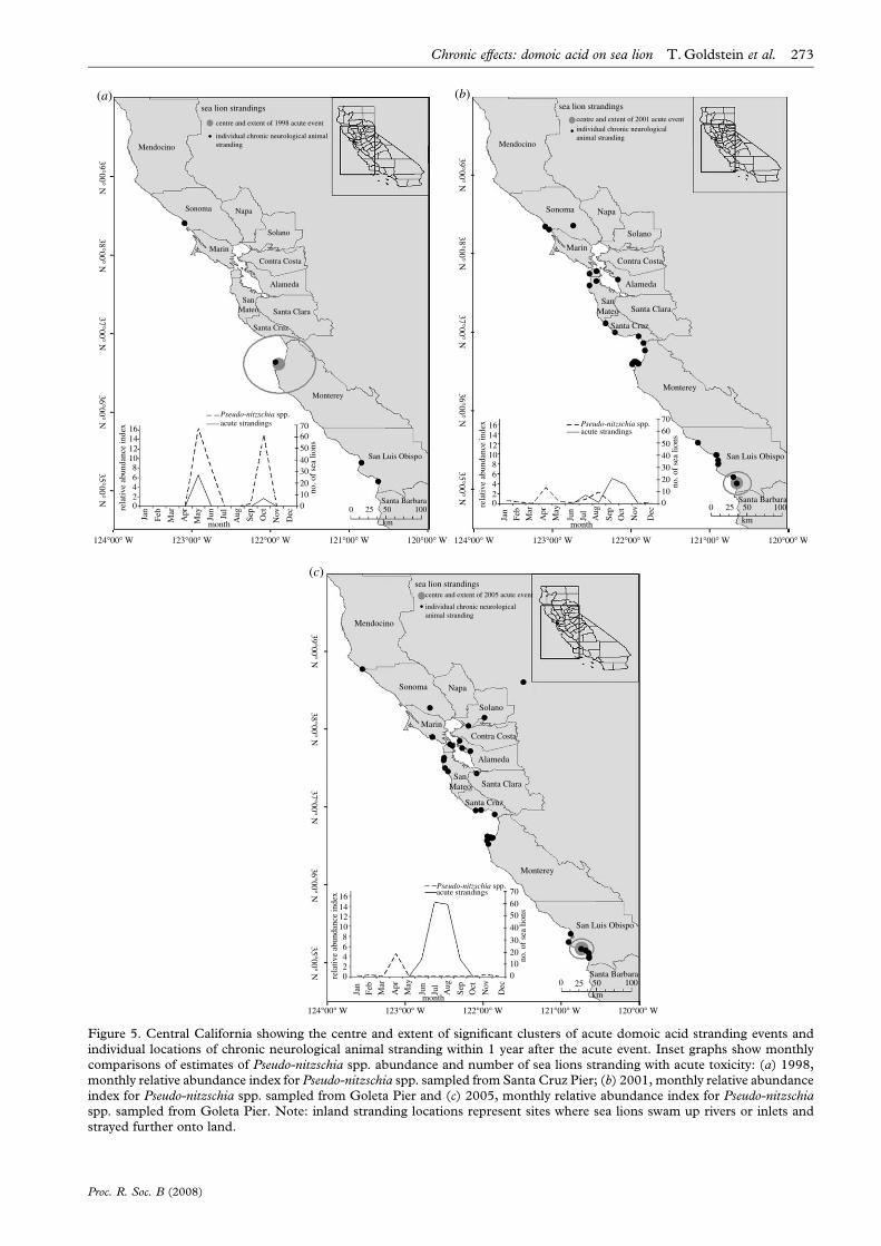
The 551 sea lions that stranded with acute toxicity
during a bloom event and 164 sea lions that stranded as
chronic neurological cases between 1998 and 2006 were
found along the California coast between 38.788 N,
123.548 W and 34.218 N, 199.268 W. Overall, the preva-
lence of domoic acid-related strandings increased signi-
ficantly from 1998 to 2006 (c2Z27.66, p!0.001). The
number of acute cases fluctuated each year and did not
show an overall increasing trend, whereas the number of
chronic cases increased significantly each year (c2Z71.82,
p!0.001). There was a four-month time lag ( pZ0.003)
between acute events and strandings of subsequent chronic
neurological cases (figure 4). Five significant clusters
( p!0.01) of acute domoic acid-related stranding events
were identified, one in the spring of 1998, while the other
four occurred in the mid to late summer of 2000, 2001,
2002 and 2005 (figure 5). The 1998 event was centred in
Monterey Bay (figure 5a), while the other four were all
centred off the coast of San Luis Obispo and Santa Barbara
counties (figure 5b,c; data not shown for 2000 and 2002).
The 1998 stranding event was tightly correlated temporally
with the phytoplankton bloom off Monterey Bay detected
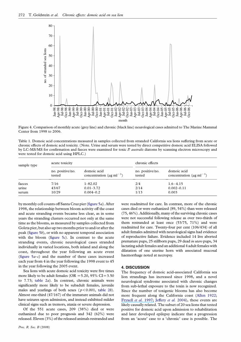
Table 1. Domoic acid concentrations measured in samples collected from stranded California sea lions suffering from acute orchronic effects of domoic acid toxicity. (Note. Urine and serum were tested by direct competitive domoic acid ELISA followedby LC-MS/MS for confirmation and faeces were examined for toxic P. australis diatoms by scanning electron microscopy andwere tested for domoic acid using HPLC.)
sample typeacute toxicity chronic effects
no. positive/no.tested
domoic acidconcentration (mg mlK1)
no. positive/no.tested
domoic acidconcentration (mg mlK1)
faeces 7/16 1–82.02 2/4 1.6–4.15urine 43/67 0.01–3.72 2/14 0.002–0.11serum 10/29 0.004–0.2 1/13 0.003
0
10
20
30
40
50
60
70
80
Jan-
98A
pr-9
8Ju
l-98
Oct
-98
Jan-
99A
pr-9
9Ju
l-99
Oct
-99
Jan-
00A
pr-0
0Ju
l-00
Oct
-00
Jan-
01A
pr-0
1Ju
l-01
Oct
-01
Jan-
02A
pr-0
2Ju
l-02
Oct
-02
Jan-
03A
pr-0
3Ju
l-03
Oct
-03
Jan-
04A
pr-0
4Ju
l-04
Oct
-04
Jan-
05A
pr-0
5Ju
l-05
Oct
-05
Jan-
06A
pr-0
6Ju
l-06
Oct
-06
month
no. o
f st
rand
ed s
ea li
ons
Figure 4. Comparison of monthly acute (grey line) and chronic (black line) neurological cases admitted to The Marine MammalCenter from 1998 to 2006.
272 T. Goldstein et al. Chronic effects: domoic acid on sea lion
by monthly cell counts off Santa Cruz pier (figure 5a). After
1998, the relationship between bloom activity off the coast
and acute stranding events became less clear, as in some
years the stranding clusters occurred not only at the same
time as the blooms, as measured by samples collected from
Goleta pier, but also up two months prior to and/or after the
peak (figure 5b), or with no apparent temporal association
with the bloom (figure 5c). In contrast to the acute
stranding events, chronic neurological cases stranded
individually in varied locations, both inland and along the
coast, throughout the year following an acute event
(figure 5a–c) and the number of these cases increased
each year from 4 in the year following the 1998 event to 45
in the year following the 2005 event.
Sea lions with acute domoic acid toxicity were five times
more likely to be adult females (OR Z5.20, 95% CIZ3.50
to 7.73; table 2a). In contrast, chronic animals were
significantly more likely to be subadult females, juvenile
males and yearlings of both sexes (p!0.001, table 2b).
Almost one-third (47/167) of the immature animals did not
have seizures upon admission, and instead exhibited milder
clinical signs such as tremors, ataxia or severe depression.
Of the 551 acute cases, 209 (38%) died or were
euthanized due to poor prognosis and 342 (62%) were
released. Eleven (3%) of the released animals restranded and
Proc. R. Soc. B (2008)
were readmitted for care. In contrast, more of the chronic
cases died or were euthanized (89, 54%) than were released
(75, 46%). Additionally, many of the surviving chronic cases
were not successful following release as over two-thirds of
them restranded at least once (53/75, 71%) and were
readmitted for care. Twenty-four per cent (106/434) of all
adult females admitted with neurological signs had evidence
of reproductive failure. Evidence included 14 live aborted
premature pups, 25 stillborn pups, 29 dead in utero pups, 34
lactating adult females and an additional 4 adult females with
dilatation of one uterine horn with associated mucosal
haemorrhage noted at necropsy.
4. DISCUSSIONThe frequency of domoic acid-associated California sea
lion strandings has increased since 1998, and a novel
neurological syndrome associated with chronic changes
from sub-lethal exposure to the toxin is now recognized.
Since the number of toxigenic blooms has also become
more frequent along the California coast (Allen 1922;
Fryxell et al. 1997; Jeffery et al. 2004), these events are
likely causally related. The subset of 20 sea lions that tested
positive for domoic acid upon admission to rehabilitation
and later developed epilepsy indicate that a progression
from an ‘acute’ case to a ‘chronic’ case is possible. The
Mendocino
sea lion strandings
centre and extent of 1998 acute event
individual chronic neurological animalstranding
Sonoma Napa
Contra Costa
Alameda
Santa Clara
Santa Cruz
Monterey
San Luis Obispo
Santa Barbara0 25 50
km
100
SanMateo
Solano
Marin
124°00° W
35°00° N36°00° N
37°00° N38°00° N
39°00° N
123°00° W 122°00° W 121°00° W 120°00° W
16
Pseudo-nitzschia spp.acute strandings
rela
tive
abun
danc
e in
dex
14121086420
706050403020100
Jan
Feb
Mar
Apr
May
month
Jun
Jul
Aug Sep
Oct
Nov
Dec
(a)
no. o
f se
a lio
ns
Mendocino
sea lion strandings
centre and extent of 2001 acute eventindividual chronic neurological animal stranding
Sonoma Napa
Contra Costa
Alameda
Santa Clara
Santa Cruz
Santa Barbara
San Luis Obispo
Monterey
0 25 50
km
100
SanMateo
Solano
Marin
124°00° W
35°00° N36°00° N
37°00° N38°00° N
39°00° N
123°00° W 122°00° W 121°00° W 120°00° W
16 Pseudo-nitzschia spp.acute strandings
rela
tive
abun
danc
e in
dex
14121086420
7060
50403020100
(b)
no. o
f se
a lio
ns
Jan
Feb
Mar
Apr
May
month
Jun
Jul
Aug
Sep
Oct
Nov
Dec
Mendocino
sea lion strandingscentre and extent of 2005 acute event
individual chronic neurologicalanimal stranding
Sonoma Napa
Contra Costa
Alameda
Santa Clara
Santa Cruz
Monterey
San Luis Obispo
Santa Barbara0 25 50
km100
SanMateo
Solano
Marin
124°00° W
35°00° N36°00° N
37°00° N38°00° N
39°00° N
123°00° W 122°00° W 121°00° W 120°00° W
16Pseudo-nitzschia spp.acute strandings
rela
tive
abun
danc
e in
dex
14121086420
70
6050
40
30
20
no. o
f se
a lio
ns
100
(c)
Jan
Feb
Mar
Apr
May
month
Jun
Jul
Aug
Sep
Oct
Nov
Dec
Figure 5. Central California showing the centre and extent of significant clusters of acute domoic acid stranding events andindividual locations of chronic neurological animal stranding within 1 year after the acute event. Inset graphs show monthlycomparisons of estimates of Pseudo-nitzschia spp. abundance and number of sea lions stranding with acute toxicity: (a) 1998,monthly relative abundance index for Pseudo-nitzschia spp. sampled from Santa Cruz Pier; (b) 2001, monthly relative abundanceindex for Pseudo-nitzschia spp. sampled from Goleta Pier and (c) 2005, monthly relative abundance index for Pseudo-nitzschiaspp. sampled from Goleta Pier. Note: inland stranding locations represent sites where sea lions swam up rivers or inlets andstrayed further onto land.
Chronic effects: domoic acid on sea lion T. Goldstein et al. 273
Proc. R. Soc. B (2008)
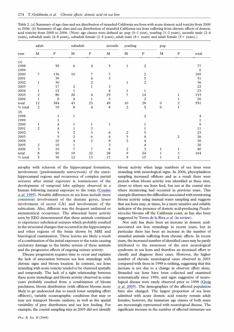
Table 2. (a) Summary of age class and sex distribution of stranded California sea lions with acute domoic acid toxicity from 2000to 2006. (b) Summary of age class and sex distribution of stranded California sea lions suffering from chronic effects of domoicacid toxicity from 2000 to 2006. (Note: age classes were defined as: pup (0–1 year), yearling (1–2 years), juvenile male (2–4years), subadult male (4–8 years), subadult female (2–5 years), adult male (8C years) and adult female (5C years).)
adult subadult juvenile yearling pup
year M F M F M M F M F total
(a)1998 59 6 4 5 1 2 771999 02000 7 136 10 7 7 2 1692001 39 4 3 5 512002 1 30 2 1 2 362003 17 2 1 2 222004 1 15 1 4 1 1 232005 2 80 20 4 25 7 14 1 1532006 1 8 4 1 3 2 1 20total 12 384 43 23 49 10 28 0 2 551% total 2 70 8 4 8 2 5 0 1
(b)1998 1 1 2 41999 1 12000 1 4 1 1 1 1 92001 2 4 1 3 1 112002 5 2 3 6 2 5 232003 5 2 6 4 3 4 2 262004 10 4 2 8 2 4 302005 2 10 1 3 4 202006 3 10 7 7 6 2 3 1 1 40total 8 50 19 22 28 9 24 1 3 164% total 5 30 12 13 17 5 15 1 2
274 T. Goldstein et al. Chronic effects: domoic acid on sea lion
atrophy with sclerosis of the hippocampal formation,
involvement (predominantly astrocytosis) of the extra-
hippocampal regions and recurrence of complex partial
seizures after initial exposure is reminiscent of the
development of temporal lobe epilepsy observed in a
human following natural exposure to the toxin (Cendes
et al. 1995). Notable differences in sea lions include more
consistent involvement of the dentate gyrus, lesser
involvement of sector CA1 and involvement of the
subiculum. Also, different was the frequent unilateral or
asymmetrical occurrence. The abnormal burst activity
seen by EEG demonstrated that these animals continued
to experience subclinical seizures which probably resulted
in the structural changes that occurred in the hippocampus
and other regions of the brain shown by MRI and
histological examination. These lesions are likely a result
of a combination of the initial exposure to the toxin causing
excitatory damage to the limbic system of these animals
and the progressive effects of ongoing seizure activity.
Disease progression requires time to occur and explains
the lack of association between sea lion strandings with
chronic signs and bloom location. In contrast, sea lions
stranding with acute toxicity tended to be clustered spatially
and temporally. The lack of a tight relationship between
these acute strandings and bloom activity observed in some
years probably resulted from a combination of bloom
patchiness, bloom distribution (with offshore blooms more
likely to go undetected due to much lower sampling effort
offshore), variable oceanographic conditions that may or
may not transport blooms onshore, as well as the spatial
variability of prey distribution and sea lion feeding. For
example, the coastal sampling sites in 2005 did not identify
Proc. R. Soc. B (2008)
bloom activity when large numbers of sea lions were
stranding with neurological signs. In 2006, phytoplankton
sampling increased offshore and as a result there were
periods when bloom activity was identified at these sites,
closer to where sea lions feed, but not at the coastal sites
where monitoring had occurred in previous years. This
example illustrates the difficulties associated with monitoring
bloom activity using manual water sampling and suggests
that sea lions may, at times, be a more sensitive and reliable
indicator of the presence of domoic acid-producing Pseudo-
nitzschia blooms off the California coast, as has also been
suggested by Torres de la Riva et al. (in review).
Not only has there been an increase in domoic acid-
associated sea lion strandings in recent years, but in
particular there has been an increase in the number of
stranded animals suffering from chronic effects. In recent
years, the increased number of identified cases may be partly
attributed to the awareness of the new neurological
syndrome in sea lions and therefore an increased effort to
classify and diagnose these cases. However, the higher
number of chronic neurological cases observed in 2001
compared with those in 1998 is striking, suggesting that this
increase is not due to a change in observer effort alone.
Stranded sea lions have been collected and examined
systematically since 1990, and signs suggestive of neuro-
logical disease were rarely observed prior to 1998 (Greig
et al. 2005). The demographics of the affected population
have also changed. The largest proportion of sea lions
admitted with acute domoic acid toxicity remain adult
females; however, the immature age classes of both sexes
are increasingly represented with neurological disease. The
significant increase in the number of affected immature sea
Chronic effects: domoic acid on sea lion T. Goldstein et al. 275
lions in recent years is of interest as some of these animals
differed from adults both in clinical presentation and
structural changes seen in the brains. The reasons for these
differences may be related to dose; age; seizure frequency,
intensity and duration; or differential distribution of brain
receptors for the toxin. Age and dose effects associated with
domoic acid exposure have been demonstrated in humans,
rats and monkeys, with adults being shown to be more
susceptible to the toxin than juveniles (Scallet et al. 1993;
Truelove et al. 1996). Additionally, both intrauterine and
neonatal exposure in rats resulted in permanent behavioural
and morphologic changes associated with reduced seizure
threshold and decreased ability to perform memory tasks (Xi
et al. 1997; Doucette et al. 2004). Morphologic changes
included hippocampal neuronal loss and mossy fibre
sprouting (indicative of new synapse formation) in the rat
neonates and the authors hypothesize that this reorgan-
ization provided a substrate for hippocampal excitability,
resulting in the observed behavioural changes. Previous work
has shown that domoic acid crosses the placenta of
California sea lions as the toxin has been detected in
amniotic fluid, foetal urine and foetal gastric fluid (Brodie
et al. 2006). Thus it is possible that in exposed cases where
the sea lion foetus survives to birth, exposure to the toxin in
utero could result in developmental abnormalities leading to
neurological and behavioural deficits as the animals age.
Additionally, neonatal sea lions may be exposed to the toxin
after birth by exposure in milk, which has been demonstrated
to be possible in rats (Maucher & Ramsdell 2005). Further
work is warranted to examine the effects of domoic acid on
brain development as well as association between beha-
vioural abnormalities and hippocampal and extra-hippo-
campal lesions in sea lions.
The reasons for the occurrence of unilateral brain lesions
in the chronic animals are unclear but may explain, in part,
the varied behavioural presentations noted in affected
animals. More sea lions exhibited unilateral damage than
bilateral damage, and right-sided hippocampal atrophy was
most common. Changes in hippocampal volume have been
associated with navigational ability in humans (Burwell et al.
2004; Maguire et al. 2006a,b). Thus, unilateral atrophy of
the hippocampal formation may have important impli-
cations on their ability to navigate and could explain the
unusual stranding locations and abnormal behaviour
following release in some of these animals.
Inconclusion, although the historyof exposure todomoic
acid in individual wild sea lions is mostly unknown, this
study demonstrates that impacts of domoic acid on
California sea lions are increasing and that effects can be
more subtle than death, such as epilepsy and severe
behavioural changes. Atrophyof the hippocampal formation
and parahippocampal gyrus results from a combination of
neuronal loss and astrocytosis and may be a result of the
initial toxic insult and the progressive and cumulative effects
from seizure propogation. Neurological and behavioural
abnormalities are now observed in free-ranging juvenile
animals rather than primarily in adult females, suggesting
that toxin exposure in utero and through milk may be
important. These cases of epilepsy in sea lions suggest that
this free-living marine mammal naturally exposed to domoic
acid in its environment may serve as a useful model for the
pathogenesis of medial temporal lobe epilepsy in humans,
especially the sclerotic form, as well as serve as an important
Proc. R. Soc. B (2008)
food safety sentinel for the presence of harmful algal blooms
off the California coast.
The work was authorized under the US Marine MammalProtection Act by Scientific Research permit no. 932-1489-00.
This study was supported by funds from the US NationalMarine Fisheries Service, Oceans and Human Health Initiativegrant number NA04OAR4600200 and through a MorrisAnimal Foundation Fellowship Training grant numberD05ZO-401. Thanks are due to the staff and volunteers ofThe Marine Mammal Center for caring for the animals. Theauthors would also like to thank many others for performingclinical diagnostics and sample analyses, especially RandyWhite, Ann Melli, Andrea Packham, Portia Cox, TenayaNorris, Kevin Randeni, Elizabeth Wheeler, Kate Thomas andLinda Lowenstine as well as Nihon Kohden America for theirgenerous donation of an EEG machine.
REFERENCESAllen, W. E. 1922 Observations on surface distributions of
marine diatoms between San Diego and Seattle. Ecology 3,
140–145. (doi:10.2307/1929148)
Anderson, D. M. 1997 Turning back the harmful red tide.
Nature 6642, 513–514. (doi:10.1038/41415)
Anderson, C. R., Brzezinski, M. A., Wasburn, L. & Kudela,
R. 2006 Circulation and environmental conditions during
a toxigenic Pseudo-nitzschia australis bloom in the Santa
Barbara Channel, California. Mar. Ecol. Prog. Ser. 327,
119–133. (doi:10.3354/meps327119)
Brodie, E. C., Gulland, F. M. D., Greig, D. J., Hunter, M.,
Jaakola, J., St. Leger, J., Leighfield, T. & Van Dolah, F. M.
2006 Domoic acid causes reproductive failure in Cali-
fornia sea lions (Zalophus californianus). Mar. Mamm. Sci.
22, 700–707. (doi:10.1111/j.1748-7692.2006.00045.x)
Burwell, R. D., Saddoris, M. P., Bucci, D. J. & Wigg, K. A.
2004 Corticohippocampal contributions to spatial and
contextual learning. J. Neurosci. 24, 3826–3838. (doi:10.
1523/JNEUROSCI.0410-04.2004)
Cendes, F., Andermann, F., Carpenter, S., Zatorre, R. J. &
Cashman, N. R. 1995 Temporal lobe epilepsy caused by
domoic acid intoxication: evidence for glutamate receptor
mediated excitotoxicity in humans. Ann. Neurol. 37,
123–126. (doi:10.1002/ana.410370125)
Chavez, F. P., Ryan, J., Lluch-Cota, S. E. & Niquen, C. M.
2003 From anchovies to sardines and back: multidecadal
changes in the Pacific Ocean. Science 299, 217–221.
(doi:10.1126/science.1075880)
Dierauf, L. & Gulland, F. M. D. (eds) 2001 Handbook
of marine mammal medicine, p. 10633. Boca Raton, FL:
CRC Press.
Doucette, T. A., Bernard, P. B., Husum, H., Perry, M. A.,
Ryan, C. L. & Taker, R. A. 2004 Low doses of domoic acid
during postnatal development produce permanent
changes in rat behaviour and hippocampal morphology.
Neurotox. Res. 6, 555–563.
Feldcamp, S. D., Delong, R. L. & Antonelis, G. A. 1989
Diving patterns of the California sea lion, Zalophus
californianus. Can. J. Zool. 67, 872–883.
Forney, K. A., Barlow, J., Muto, M., Lowry, M., Baker, J.,
Cameron, G., Mobley, J., Stinchcomb, C. & Carretta, J. V.
2000 U.S. Pacific marine mammal stock assessments: 2000,
pp. 1–7. NOAA-TM-NMFS-SWFSC-300, Washington,
DC: NOAA.
Fryxell, G. A., Villac, M. C. & Shapiro, L. P. 1997 The
occurrence of the toxic diatom genus Pseudo-nitzschia
(Bacillariophyceae) on the west coast of the USA, 1920–
1996: a review. Phycologia 36, 419–437.
276 T. Goldstein et al. Chronic effects: domoic acid on sea lion
Greig, D. J., Gulland, F. M. D. & Kreuder, C. 2005 A decade
of live California sea lion (Zalophus californianus) strand-
ings along the central California coast: causes and trends,
1991–2000. Aquat. Mamm. 31, 40–51.
Gulland, F. M., Haulena, M., Fauquier, D., Langlois, G.,
Lander, M. E., Zabka, T. & Duerr, R. 2002 Domoic acid
toxicity in Californian sea lions (Zalophus californianus):clinical signs, treatment and survival. Vet. Rec. 150,
475–480.
HARNESS 2005 Harmful Algal Research and Response
National Environmental Science Strategy 2005–2015.
National Plan for Algal Toxins and Harmful Algal Blooms,
(eds D. M. Anderson, J. S. Ramsdell & P. M. Glibert).
Oceanography 18, 238–245.
Haulena, M. & Gulland, F. M. D. 2001 Use of medetomi-
dine–zolazepam–tiletamine with and without atipamezole
reversal to immobilize captive California sea lions. J.Wildl.
Dis. 37, 566–573.
Holliday, T. A. & Williams, D. C. 1998 Interictal paroxysmal
discharges in the electroencephalograms of epileptic dogs.
Clin. Tech. Small Anim. Pract. 13, 132–143. (doi:10.1016/
S1096-2867(98)80034-0)
Jeffery, B., Barlow, T., Moizer, K., Paul, S. & Boyle, C. 2004
Amnesic shellfish poison. Food Chem. Toxicol. 42,
545–557. (doi:10.1016/j.fct.2003.11.010)
King, J. E. 1983 In Seals of the World, pp. 240. St. Lucia,
Australia: University of Queensland Press.
Kulldorff, M., Heffernan, R., Hartman, J., Assuncao, R. M.
& Mostashari, F. 2005 A space-time permutation scan
statistic for the early detection of disease outbreaks. PLoSMed. 2, 216–224. (doi:10.1371/journal.pmed.0020059)
Landsberg, J. H. 2002 The effects of harmful algal blooms on
aquatic organisms. Rev. Fish. Sci. 10, 113–390. (doi:10.
1080/20026491051695)
Lefebvre, K. A. et al. 1999 Detection of domoic acid
in northern anchovies and California sea lions
associated with an unusual mortality event. Nat. Toxins7, 85–92. (doi:10.1002/(SICI)1522-7189(199905/06)7:
3!85::AID-NT39O3.0.CO;2-Q)
Maguire, E. A., Nannery, R. & Spiers, H. J. 2006a Navigation
around London by a taxi driver with bilateral hippo-
campal lesions. Brain 129, 2894–2907. (doi:10.1093/
brain/awl286)
Maguire, E. A., Woollett, K. & Spiers, H. S. 2006b London
taxi drivers and bus driver: a structural MRI and
neuropsychological analysis. Hippocampus 16, 1091–1101.
(doi:10.1002/hipo.20233)
Maucher, J. M. & Ramsdell, J. S. 2005 Domoic acid transfer
to milk: evaluation of a potential route of neonatal
exposure. Environ. Health Perspect. 113, 461–464.
Melin, S. R., Delong, R. L., Thomason, J. R. & Vanblaricom,
G. R. 2000 Attendance patterns of California sea lion
(Zalophus californianus) females and pups during the non-
breeding season at San Miguel Island. Mar. Mamm. Sci.
16, 169–185. (doi:10.1111/j.1748-7692.2000.tb00911.x)
Olney, J. W., Fuller, T. & de Gubareff, T. 1979 Acute
dendrotoxic changes in the hippocampus of kainate
treated rats. Brain Res. 176, 91–100. (doi:10.1016/0006-
8993(79)90872-2)
Peng, Y. G., Clayton, E. C., Means, L. W. & Ramsdell, J. S.
1997 Repeated independent exposures to domoic acid do
not enhance symptomatic toxicity in outbred or seizure-
sensitive inbred mice. Fundam. Appl. Toxicol. 40, 63–67.
(doi:10.1006/faat.1997.2360)
Perl, T. M., Bedard, L., Kosatsky, T., Hockin, J. C., Todd,
E. C., McNutt, L. A. & Remis, R. S. 1990 An outbreak of
toxic encephalopathy caused by eating mussels contami-
nated with domoic acid. New Engl. J. Med. 322,
1775–1780.
Proc. R. Soc. B (2008)
Quilliam, M. A., Xhie, M. & Hardstaff, W. R. 1995 Rapidextraction and cleanup for liquid chromatographicdetermination of domoic acid in unsalted seafood.J. AOAC Int. 78, 543–555.
Scallet, A. C., Binienda, Z., Caputo, F. A., Hall, S., Paule,M. G., Rountree, R. L., Schmued, L., Sobotka, S. &Slikker Jr, W. 1993 Domoic acid-treated cynomolgusmonkeys (M. fascicularis): effects of dose on hippocampalneuronal and terminal degeneration. Brain Res. 627,307–313. (doi:10.1016/0006-8993(93)90335-K)
Schmued, L. C., Scallet, A. C. & Slikker, W. 1995 Domoic acid-induced neuronal degeneration in the primate forebrainrevealed by degeneration specific histochemistry. Brain Res.695, 64–70. (doi:10.1016/0006-8993(95)00799-V)
Scholin, C. A. et al. 2000 Mortality of sea lions along thecentral California coast linked to a toxic diatom bloom.Nature 403, 80–84. (doi:10.1038/47481)
Silvagni, P., Lowenstine, L. J., Spraker, T., Lipscomb, T. P. &Gulland, F. M. 2005 Pathology of domoic acid toxicity inCalifornia sea lions (Zalophus californianus). Vet. Pathol. 42,184–191. (doi:10.1354/vp.42-2-184)
Sobotka, T. J. et al. 1996 Domoic acid: neurobehavioural andneurohistological effects of low-dose exposure in adultrats. Neurotoxicol. Teratol. 18, 659–670. (doi:10.1016/S0892-0362(96)00120-1)
Teitelbaum, J. S., Zatorre, R. J., Carpenter, S., Gendron, D.,Evans, A. C., Gjedde, A. & Cashman, N. R. 1990Neurologic sequellae of domoic acid intoxication due tothe ingestion of contaminated mussels. N. Engl. J. Med.322, 1781–1787.
Torres de la Riva, G., Kreuder Johnson, C., Gulland, F. M.D., Langlois, G., Heyning, J., Rowles, T. & Mazet, J. K.In review. Pseudo-nitzschia blooms associated with marinemammal strandings along the Southern California. J.Wildl. Dis..
Truelove, J., Mueller, R., Pulido, O. & Iverson, F. 1996Subchronic toxicity study of domoic acid in the rat.Food Chem. Toxicol. 34, 525–529. (doi:10.1016/0278-6915(96)81814-X)
Truelove, J., Meuller, R., Pulido, O., Martin, L., Fernie, S. &Iverson, F. 1997 30-Day oral toxicity study of domoic acidin cynomolgus monkeys: lack of overt toxicity of dosesapproaching the acute toxic dose. Nat. Toxins 5, 111–114.(doi:10.1002/nt.5)
Tryphonas, L., Truelove, J., Todd, E., Nera, E. & Iverson, F.1990 Experimental oral toxicity of domoic acid incynomolgus monkeys (Macaca fascicularis) and rats.Preliminary investigations. Food Chem. Toxicol. 28,707–715. (doi:10.1016/0278-6915(90)90147-F)
Van Dolah, F. M. 2000 Marine algal toxins: origins, healtheffects, and their increased occurrence. Environ. HealthPerspect. 108S1, 133–141. (doi:10.2307/3454638)
Van Dolah, F. M., Leighfield, T. A., Haynes, B. L., Hampson,D. R. & Ramsdell, J. S. 1997 A microplate receptor assayfor the amnesic shellfish poisoning toxin, domoic acid,utilizing a cloned glutamate receptor. Anal. Biochem. 245,102–105. (doi:10.1006/abio.1996.9889)
Weise, M. J., Costa, D. P. & Kudela, R. M. 2006Movement and diving behaviour of male Californiasea lion (Zalophus californianus) during anomalousoceanographic conditions of 2005 compared to thoseof 2004. Geophys. Res. Lett. 33, L22S10. (doi:10.1029/2006GL027113)
Work, T. M., Barr, B., Beale, A. M., Fritz, L., Quilliam, M. A.& Wright, J. L. C. 1993 Epidemiology of domoic acidpoisoning in brown pelicans (Pelecanus occidentalis) andBrandt’s cormorants (Phalacrocorax penicillatus) in Cali-fornia. J. Zool. Wildl. Med. 24, 54–62.
Xi, D., Peng, Y.-G. & Ramsdell, J. S. 1997 Domoic acid is apotent neurotoxin in neonatal rats. Nat. Toxins 5, 74–79.