Guloptiloides: an Extraordinary New Carnivorous Genus of Baetidae (Ephemeroptera
Upload
independentCategory
view
1download
0
Acta Zoologica
(Stockholm)
90
: 87–98 (January 2009) doi: 10.1111/j.1463-6395.2008.00367.x
© 2008 The AuthorsJournal compilation © 2009 The Royal Swedish Academy of Sciences
87
Abstract
Ubero-Pascal, N. and Puig, M. A. 2009. New type of egg attachment structurein Ephemeroptera and comparative analysis of chorion structure morphologyin three species of Ephemerellidae. —
Acta Zoologica
(Stockholm)
90
: 87–98
Ephemeroptera eggs vary greatly in their morphology, a characteristic that hasbeen used to solve systematic problems. The eggs of Ephemerellidae may beconsidered among the most studied in mayfly families, especially with regardto the number of species for which they have been described (approximately32 species corresponding to 12 genera). However, this study provides newdetails of the egg morphology of three species of this family,
Serratella ignita
(Poda 1761),
S. spinosa
(Ikonomov 1961) and
Torleya major
(Klapalek 1905).The main finding was that the morphology and organization of the lateralattachment structures do not resemble the type normally associated with
‘knobterminated coiled thread
’ (KCT). The particular characteristics of this structure,never before described for Ephemeroptera, led us to propose a new type ofattachment structure, the ‘
multithread-folded with terminal fibre cluster
’ (MFT).We also provide a detailed description of the chorion morphology of the eggsof the three species, identifying a tear-shaped variation in the oval sperm guideand confirming the type III polar caps in the two species of
Serratella
Walsh,1862.
Dr Nicolás Ubero Pascal, Departamento de Zoología y Antropología Física, Facultad de Biología, Universidad de Murcia, Campus de Espinardo s/n, 30100, Espinardo-Murcia, Spain.E-mail: [email protected]
Blackwell Publishing Ltd
New type of egg attachment structure in Ephemeroptera and comparative analysis of chorion structure morphology in three species of Ephemerellidae
N. Ubero-Pascal
1
and M. A. Puig
2
1
Departamento de Zoología y Antropología Física, Facultad de Biología, Universidad de Murcia, Campus de Espinardo s/n, 30100 Espinardo-Murcia, Spain;
2
Departamento de Ecología Continental, Centro de Estudios Avanzados de Blanes (CEAB-CSIC), Acceso a la Cala San Francesc, 14, 17300 Blanes (Gerona), Spain
Keywords:
chorion morphology, attachment structure, serretella, Torleya, micropyle
Accepted for publication:
28 March 2008
Introduction
Regional complexity was proposed by Margaritis (1985), todescribe, basically, the position and arrangement of func-tional chorionic structures in eggs of the dipteran
Drosophilamelanogaster
Meigen, 1830. This complexity in Ephemeropteraeggs is related to three basic types of chorionic structure,already defined by Koss and Edmunds (1974): the attach-ment structures, the micropyle and chorionic sculpturing.The characteristics of a structure, or a combination thereof,may be particular to different taxonomic levels, so that theyhave frequently been used to establish phylogenetic relation-ships (McCafferty and Wang 1994; Studemann and Landolt1997; Domínguez and Cuezzo 2002). Moreover, given thatthe egg is completely mature at the end of the nymphstage, the characteristics of the chorion allow us to associate
nymphs and female imagos, which is very useful since the latterusually lack taxonomically valid morphological characters(Koss 1968). The chorion morphology of Ephemeropteraeggs has been studied in numerous families (Degrange 1960;Koss and Edmunds 1974; Kopelke and Müller-Liebenau1981a,b, 1982; Malzacher 1982; Gaino
et al
. 1987, 1993;Kluge
et al
. 1995; Klonowska-Olejnik 1997; Klonowska-Olejnik & Jazdzewska 2003; Ubero-Pascal 2004; Ubero-Pascal and Puig 2007), although the number of species inwhich they have been studied in depth is still low.
Attachment structures are essential for egg viability afterovoposition, since they are responsible for fixing eggs firmlyto the substrate and so prevent them from drifting (Gainoand Rebora 2003); they are also important from a morpho-logical point of view since it is the chorionic structures thatshow greatest variability. This variability is evident in their

New egg attachment structure in mayflies •
Ubero-Pascal and Puig Acta Zoologica
(Stockholm)
90
: 87–98 (January 2009)
© 2008 The Authors
88
Journal compilation © 2009 The Royal Swedish Academy of Sciences
ultrastructure, arrangement and position or distribution onthe egg surface. Koss and Edmunds (1974) proposed a classifica-tion of Ephemeroptera attachment structures, although theyalso stated that this could change as the egg morphology ofmore species became known. Indeed, given their biologicalimportance and morphological diversity, attachment structureshave been the main focus in a large number of the studies onEphemeroptera eggs (Gaino and Mazzini 1989; Gaino andBongiovanni 1991, 1992; Gaino and Rebora 2001; Ubero-Pascal 2004; Ubero-Pascal and Puig 2007), the findings ofwhich have provided more detail and shown the need toupdate Koss and Edmunds’ classification.
Compared with other Ephemeroptera, such as Potaman-thidae, Ephemeridae and Oligoneuriidae, the egg morphologyof Ephemerellidae has been studied in a considerablenumber of species (Ubero-Pascal and Puig 2007). Thecharacteristics of the chorion have been established both byoptical microscopy and scanning electron microscopy (SEM)in approximately 32 species, belonging to 11 of the 22 generaforming this family according to Hubbard (1990); however,most of the descriptions given have been quite superficial(Bengtsson 1913; Smith 1935; Degrange 1956, 1960; Koss1968; Koss and Edmunds 1974; Studemann and Tomka1987; Gaino and Bongiovanni 1992; Kang and Yang 1995;Studemann
et al
. 1995; Studemann and Landolt 1997;Haybach 2003; Jacobus and Sartori 2004; Jacobus
et al
.2004). In systematics, the application of the morphology ofEphemerellidae eggs has provided patchy results, although itdoes seem that egg characteristics are species-specific, forwhich reason they are of great taxonomic value at this level,but not so much at supra-species level or for clarifying theirphylogeny (McCafferty and Wang 1994; Studemann
et al
.1995; Studemann and Landolt 1997).
In this study, we analyse in detail the egg morphologyof three species of Ephemerellidae,
Serratella ignita
(Poda,1761),
S. spinosa
(Ikonomov, 1961) and
Torleya major
(Klapalek, 1905), using optical microscopy and SEM. Theresults enabled us to confirm the most common chorionicpattern in the eggs of this family and the specific pattern ofeach species. However, we found differences in the ultrastruc-ture and organization of the attachment structures, especiallyin the lateral attachment structures, which do not fit the patternof ‘
knob terminated coiled thread
’ (KCT). Furthermore, thecharacteristics observed in the lateral attachment in thesethree species do not reflect any types established by Koss andEdmunds (1974), leading us to denominate a new type ofstructure. After studying the figures in several works pub-lished on Ephemerellidae eggs, we consider that this type ofstructure may appear in many other species and could beconsidered specific to this family.
Materials and methods
The three species whose eggs have been studied were col-lected in the Segura river basin, S. E. Spain (Ubero-Pascal
et al
. 1998): three nymphs and one imago of
S. ignita
fromthe Segura river near La Graya village (615 m, 20 March 1994and 13 Nov. 1994, respectively); three nymphs of
S. spinosa
from the Mundo river near Los Chorros (985 m, 10 July1994); and two nymphs of
T. major
from the Madera streamnear Segura de la Sierra village (1180 m, 10 July 1994).Female mature nymphs (with black wingpads) were fixed in4% formaldehyde and conserved in 70% ethanol, whereasimagos were fixed and preserved in 70% ethanol. Eggs fromthe imago were studied only in the case of
S. ignita
.The eggs of each species were obtained directly from the
abdomen of specimens and were processed for SEM bycleaning in a Branson 3510 ultrasound bath for 5 min anddehydrating in increasing concentrations of ethanol (80%,90%, 95%) until absolute ethanol or acetone. They werethen dried using the critical point method or air-dried afterhexamethyldisilizane/tetramethylsilane treatment (see Ubero-Pascal
et al
. 2005), mounted on stubs with conductive stickytape, and sputter coated with gold–palladium in a PolaronSputter Coater. Finally, the eggs were observed by scanningelectron microscopes (Jeol JSM 6100 and Hitachi S3500N)with working voltages of 20 and 10 kV, respectively. Forty tofifty eggs of each specimen were prepared for SEM analysis,checking the morphological characteristics of the chorion inat least 10 of them.
For light microscopy analysis, the eggs were processed inorder to obtain the cleared and stained effect producedby CMC-S mounting-medium that Koss (1968) used.As this mounting-medium is no longer commercially avail-able, the eggs were stained with neutral red and mounted onslides with ‘
hoyer
’ mounting-medium. Eggs on slides wereanalysed 3 or 4 weeks after preparation by a Leica DMRBBmicroscope.
The terminology and classification of the chorion struc-tures proposed by Koss and Edmunds (1974) were followedin the descriptions of egg morphology. Koss (1968) used themesh length to compare reticle size in different species,considering this parameter as the longest inside dimension ofthe mesh unit (excluding the strand). However, we considerthis parameter to be of little use in geometric reticle com-posed of regular and irregular polygons, or only irregular, asoccurs in some of the species studied. We therefore think thatcomparisons of mesh size should be based on three para-meters: the mean length of the polygon sides, the meandistance between opposite vertices and the mean distancebetween opposite sides.
Results
Morphology and arrangement of lateral attachment structure
In Ephemerellidae the lateral attachment structures arefibrous and take on the circular shape of the depression whenpacked (Fig. 1A,B). These structures are mainly situated inthe subpolar zones of the egg and vary in number (Fig. 4),

Acta Zoologica
(Stockholm)
90
: 87–98 (January 2009)
Ubero-Pascal and Puig
• New egg attachment structure in mayflies
© 2008 The AuthorsJournal compilation © 2009 The Royal Swedish Academy of Sciences
89
although they may occasionally be shifted towards the equa-torial area or the pole without a cap. The packed fibres ineach unit are arranged in a slight depression in the chorion,although this depression is only visible when they are absentor unravelled (Fig. 1C,E,F); the whole unit is kept in placeby a thin film known as the extrachorion (Figs 1A,B). Whenthis extrachorion is broken or becomes detached, the fibrousmaterial unravels and reveals its ultrastructure and organiza-tion (Figs 1E,D and 3A). Basically, each lateral attachmentstructure is made up of at least two threads and a circularcushion (Fig. 1C–F). The threads are joined to the chorionjust at the edge of the depression at different insertion points(usually diametrically opposed to each other; Figs 1E,F).Each thread is formed of three or four flattish filaments,weakly twisted and attached to the edge of the depression inseveral places (Fig. 1E). The threads are joined distally to thesame place on the centre of the lower side of the circularcushion. The cushion is formed of numerous microfilamentsthat give it a velvety appearance (Fig. 1B,D).
What is surprising about this structure is the way in whichthe packed threads are organized, since they do not show thespiral common to other attachment structures in Ephemeroptera.This organization is represented schematically in Fig. 2,where the zigzag folding of the threads within the depression
is depicted. This folding completely covers the surface of thedepression, in the centre of which the cushion can be seen(Fig. 1B). This particular arrangement of the threads is moreevident when the structure has unfolded (Fig. 1E,D),
Fig. 1—Ultrastructure and organization of multithreads-folded with terminal fibre cluster (MFT). —A, B. Packing arrangement covers partially of extrachorion. —C, D. Unravelled configuration. —E, F. Detail of threads joined to the chorion at the edge of circular depression. S. ignita (A and C). T. major (B and F). S. spinosa (D and E). Cd, circular depression; Cs, chorion surface; Ex, extrachorion; Fl, filament; Hf, hairpin folds; La, lateral attachment structure; T, terminal fibre cluster; Th, thread.
Fig. 2—Schematic representation of basic organization of MFT and detail of proximal end of thread. Cd, circular depression; Fl, filament; T, terminal fibre cluster; Th, thread.

New egg attachment structure in mayflies •
Ubero-Pascal and Puig Acta Zoologica
(Stockholm)
90
: 87–98 (January 2009)
© 2008 The Authors
90
Journal compilation © 2009 The Royal Swedish Academy of Sciences
although it also is clear when the extrachorion permits it tobe seen (Figs 1B and 3A). In this last case, since the cushionterminal is smaller than the complete structure, the hairpinfolds that make up the thread when it is folded can be seen(Figs 1B and 3A).
Until now, these attachment structures have been regardedas being of the’KCT type. These KCTs are common in theeggs of Heptageniidae and are formed of one thread made upof tightly entwined filaments (Fig. 3C), which finish in acircular knob (Figs 3B,C). In this type of structure the threadis coiled (Fig. 3B), giving it the form of a spring when itexpands (Fig. 3D). The same type of structure may also beassociated with a circular depression, in which case the threadis joined to the centre of the depression (Fig. 3C). Clearly,these morphological characteristics differ substantially from
those described above in the Ephemerellidae species studied(Figs 1 and 3A). For this reason, we do not think they shouldshare the same name and propose they should be designated‘
multithread-folded with terminal fibre clusters
’ (MFT), and, assuch, be included in Koss and Edmunds’ classification (1974)as a new fibrous structure type, as depicted in Table 1.
The size of the MFT in the species studied cannot beconsidered uniform (Table 2), and is considerably smaller in
S. ignita
than in
S. spinosa
and
T. major
. The same may be saidof the thread thickness (Table 2), although the diameterof the circular cushion is more homogeneous and almostidentical in the case of
S. ignita
and
T. major
(Table 2). Inaddition, we have seen that in the eggs of
S. spinosa
the lateralattachment structures in the subpolar region near the cap arelarger (14.6 µm mean diameter) than the structure near the
Fig. 3—Comparison of organization and structure of MFT and KCT. —A. S. ignita, MFT partially unravelled. —B. Ecdyonurus dispar (Curtis, 1834), packed KCT. —C. Rhithogena iridina (Kolenati, 1839), partially unravelled KCT with detail of twisted filaments of the thread. —D. E. dispar, uncoiled KCT showing the thread spiralled. Cd, circular depression; Fl, filament; Hf, hairpin folds; K, knob; T, terminal fibre cluster; Th, thread.
Table 1 Koss and Edmunds (1974) attachment structures with the new attachment structure described
I.- Non-fibrous Sucker-like disc or platesProjectionsLayers
II.- Fibrous II.A.- Non-coiled II.A.1.- LayersII.A.2.- Threads or fibres Lateral
Polar cap type IPolar cap type II
II.B. Folded Multi-thread with terminal fibre clustersII.C.- Coiled II.B.1.- Fibres Without terminal fibre clusters
With terminal fibre clustersWith terminal knob
II.B.2.- Collectively coiled threads Polar cap type IIIII.B.3.- Individually coiled threads II.B.3.a.- Knobless Lateral
Polar cap type IVII.B.3.b.- Knob-terminated Lateral
Polar cap type V

Acta Zoologica
(Stockholm)
90
: 87–98 (January 2009)
Ubero-Pascal and Puig
• New egg attachment structure in mayflies
© 2008 The AuthorsJournal compilation © 2009 The Royal Swedish Academy of Sciences
91
opposite pole (11.21 µm mean diameter). Such a positionalvariation in the size of the lateral attachment structure wasnot observed in the other two species studied.
Egg morphology of studied species
The eggs of the three species studied (Fig. 4) share the samemorphotype, characterized by their ovoid shape, one polarcap, numerous lateral attachment structures and numeroustagenoform-type micropyles, although the chorionic sculp-turing varies. The mean egg size of
S. ignita
is considerablesmaller than in the other two species,
S. spinosa
having thelargest eggs. In general, the mean egg size in our study was
considerably smaller than the values recorded in the litera-ture (Table 3).
Optical microscope and SEM images of the polar caps ofthe eggs of the three species show them to be made up ofinnumerable filaments, bulbous at one end – spherical in
S. ignita
and
T. major
(Fig. 5A) and bell-shaped in
S. spinosa
(Fig. 5D). The polar cap is made up internally of a denseamorphous-looking matrix, which forms a central nucleusfrom which the filaments arise (Fig. 5E). The nucleus isjoined to the chorion by means of a conical projection(Fig. 5G,H). The filaments are densely packed because ofthe extrachorion that covers them, although it can be seenthat they are weakly wound around a central nucleus.
Table 2 Chorion structure measurements
Torleya major Serratella ignita Serratella spinosa
Multithread-folded with terminal fibre cluster
Number of structures* 4–5 11 8–9Diameter of terminal fibre cluster 6.4 6.2 7.2Diameter of Circular depression 13.2 9.8 13.6Thread thickness 0.6 0.38 –
Micropyles
Units number* 2–3 4–7 5Sperm guide form Circular Oval/Teardrop† TeardropSperm guide size 14 Long Large Long Large
8.4/10.6† 8.7/6.3† 18.25 10.7
Netlike Chorionic sculpturing
Side length 9.6 – 9.5Length between sides 16.9 – 16.5Length between apexes 19.2 – 18.6
All average data given in µm. n = 15.*Units observed directly in the eggs, nonextrapolable data of maximum number of units in the whole egg.†Teardrop sperm guide measurements.
Table 3 Comparison of egg size in studied species
Torleya major Serratella ignita Serratella spinosa
Length Width Length Width length Width
Our data 168* 125 145* 109 189* 141178† 160† 210†
Degrange (1960) 203–222* 144–156 175–211* 136–160 – –Studemann et al. (1995) 200‡ – 250‡ – 340‡ –Gaino and Bongiovanni (1992) – – – 132 – –
– 213† – –
All data are given in µm. All data indicated are mean data, except Degrage’s data. n = 15.*Egg length without measuring the polar cap length.†Egg length including the polar cap length of bulged type according to Gaino and Bongiovanni (1992).‡Average length of egg considering polar caps of different types and lengths.

New egg attachment structure in mayflies •
Ubero-Pascal and Puig Acta Zoologica
(Stockholm)
90
: 87–98 (January 2009)
© 2008 The Authors
92
Journal compilation © 2009 The Royal Swedish Academy of Sciences
This winding is much more evident in the polar cap of theeggs of
Serratella
Walsh, 1862 than of
T. major
(Fig. 5B–D).In the former therefore we can speak of a type III polar cap(
‘Multi-threaded coiled cap’
), while in
T. major
the polar cap ismore like type I (
‘Non-coiled, single unit cap’
). The shape and
size of the polar cap in
S. ignita
vary considerably (Fig. 5F),and all three types described by Gaino and Bongiovanni(1992) were observed: short, bulging, and long; in
S. spinosa
and
T. major
, on the other hand, the same two parametersremain quite stable. The polar cap is bell-shaped in
S. spinosa
and flat in
T. mayor
(Figs 4B and 5C).The micropyles are usually set around the central area of
the egg, occupying both the equatorial and subpolar regions(Fig. 4), although, occasionally, they may be more nearer thepoles. The sperm guide and micropylar opening are the onlyparts of the micropyle visible by SEM, since the micropylarcanal is completely internal (Fig. 6). The micopylar opening,a simple orifice at the beginning of the micropylar canal,shows no morphological variations in the three speciesstudied (Fig. 6A,E,F); the only point to note is that in
T. major
it is situated below the mesh strand (Fig. 6F). The spermguide, on the other hand, clearly differs both in shape andsize. In
T. major
it is completely circular (Fig. 6F), while in
S. ignita
and
S. spinosa
it is oval (Fig. 6A,C,E). In turn,in
S. spinosa
this structure is teardrop-shaped (version of theoval shape) where the proximal end is narrow and drawn outbefore the micropylar canal begins (Fig. 6E). This type ofsperm guide also occurs in
S. ignita
(Fig. 6C), although lessfrequently and in combination with the oval shape (Fig. 6B).As with the lateral attachment structures, the dimensions ofthe sperm guide vary between the species studied, and followthe same tendency, i.e. in
S. ignita
they are smaller (even thetear-shape) than in
S. spinosa
and
T. major
(Table 2). Thenumber of micropyles in the egg also varies (Table 2),although, in this case,
T. major
has the lowest number peregg.
The chorionic sculpturing differs considerably amongthe three species (Fig. 4), despite the fact that the eggs of
S. spinosa
and
T. major
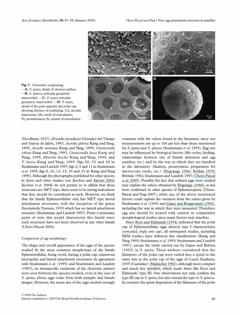
share a reticular chorionic pattern.The chorionic sculpturing of each species is characterized asfollows:– Serratella ignita. The chorion appears to be completely
smooth (Fig. 4A), but above 10,000x irregular shallowfurrows can be observed, from which arise smallprotuberances about 0.35 µm thick (Fig. 7A). Theseprotuberances tend to lean sideways and to be joined toeach other, although they are not visible when theextrachorion is present.
– Serratella spinosa. The chorion forms a geometric macroreliefformed of a large-meshed reticulation covering the wholeegg surface (Fig. 4B), although nearer to the poles theymay lose their regular appearance and present othergeometric shapes. Each mesh unit is perfectly delimitedby a very narrow furrow approximately 1.3 µm wide,which makes up the strand. The perimeter of each meshunit is raised (3.9–2.0 µm high) and surrounds a centralcrater of varying diameter (8.6–4.5 µm). The externaledge of the raised perimeter keeps the polygonal shape ofthe mesh unit, while the internal edge completely losesthis shape. The size of the mesh units is quite homogeneous
Fig. 4—Overall view of the eggs of the studied species: —A. S. ignita. —B. S. spinosa. —C. T. major. Cd, circular depression; La, lateral attachment structure; Mp, micropyle; Pc, polar cap; R, reticulation.

Acta Zoologica (Stockholm) 90: 87–98 (January 2009) Ubero-Pascal and Puig • New egg attachment structure in mayflies
© 2008 The AuthorsJournal compilation © 2009 The Royal Swedish Academy of Sciences 93
regardless of the geometry to the individual cells (Table 3).The presence of an extrachorion does not obscure thechorion sculpturing, although it might impede observationof the strand.
– Torleya major. The chorion forms a geometric macroreliefconstituted by a large-meshed reticulation (Fig. 4C) thatextends over the whole egg surface except the polar zones,although it is more evident in the pole without a cap(Fig. 7D). Normally, the mesh units are not delimited bya furrow (Fig. 7C), so that the raised area of the chorion(6.3 µm wide) constitutes the strand of the reticle and thecircular depressions (11.5 µm diameter) the mesh (Fig. 7C).At very large magnification, a very weak furrow can justbe made out and the cells are seen to have a regularhexagonal shape (Fig. 7C), except near the poles. Thepresence of the extrachorion does not obscure the chorionicsculpturing.
Discussion
Among Ephemeroptera, the egg morphology of Ephemerel-lidae has been the most widely studied, at least with regardto the number of species. However, SEM descriptions tendto be very general or contain little detail, as is the case of
S. spinosa and T. major (Studemann et al. 1995; Studemannand Landolt 1997). An exception to this is S. ignita sinceboth the regional and radial complexity of the chorion is wellknown from light microscopic (Degrange 1960) and SEMstudies (Gaino and Bongiovanni 1992). Despite this, our studyof the above species has provided new and surprising data,especially with regard to the lateral attachment structure.
Multithread-folded with terminal fibers cluster
The fibrous nature of the lateral attachment structures in theeggs of several species of Ephemerellidae was described byBengtsson (1913) and Smith (1935). Much later, Degrange(1960) described their structure and organization in detail.Later, Koss and Edmunds (1974) confirmed these findingsand included them in the KCT type, alongside the lateralattachment structures observed in the eggs of other familiesof Ephemeroptera, such as Heptageniidae, Siphlonuriidae,Potamanthidae and Oligoneuriidae. Given that the descrip-tion and classification of lateral attachment structures inEphemeroptera provided by Koss and Edmunds (1974) areconsidered the main reference in chorion morphologicalstudies in these insects, SEM-based morphological studies ofthe eggs of the family Ephemerellidae have continued to
Fig. 5—Morphology and organization of polar cap. —A, D. Details of bulbous end in filaments. —B, C. General disposition of filaments in polar cap. —E. Internal organization of polar cap. —F. Varying of size and shape of polar cap. —G. Polar chorionic structure with polar cap attached. —H. Polar chorionic structure with no polar cap. T. major (A, B and H); S. spinosa (C and D); S. ignita (E, F and G). Light microscopy scale: 40× (E, G and H); 10× (F). Co, conical projection; Fl, filament; Nu, nucleus; Pc, polar cap.

New egg attachment structure in mayflies • Ubero-Pascal and Puig Acta Zoologica (Stockholm) 90: 87–98 (January 2009)
© 2008 The Authors94 Journal compilation © 2009 The Royal Swedish Academy of Sciences
regard lateral attachment structures as KCT (Gaino andBongiovanni 1991, 1992; Kang and Yang 1995; Studemannet al. 1995; Studemann and Landolt 1997). However, as inthe case of the lateral attachment structures of the eggs ofPotamanthus luteus (Linnaeus, 1767) (Potamanthidae), inwhich SEM studies revealed morphological characteristicsthat cannot be considered KCTs (Ubero-Pascal and Puig2007), the morphological characteristics of the lateral attach-ment structures of Ephemerellidae eggs also lead us to questionthe type to which they have traditionally been assigned.
The KCT consists of a thread composed of several twistedfilaments that support a terminal knob (Koss and Edmunds1974; Gaino and Bongiovanni 1991). Recently, Gaino andRebora (2003) made a detailed study of the morphology andorganization of KCTs using SEM and TEM in the speciesEcdyonurus venosus (Fabricius, 1775) (Heptageniidae), wherebythese observations were confirmed and new interestinginformation as regards the way in which eggs are fixed to thesubstrate was provided. Comparison of the SEM descriptionof a KCT given by these authors and our observation oflateral attachment structures in the eggs of the three speciesof the Ephemerellidae studied, suggest a totally differentultrastructure and packing arrangement. This arrangementconstists of a fibrous ultrastructure formed of threads, each
made up of a few weakly twisted filaments that, proximally,are fixed to the chorion at diametrically opposite sites of theedge of a circular depression and, distally, to a multifibrillarcircular cushion. The whole structure is packed within thedepression by multiple zigzag foldings of threads, leavingthe cushion in a central position. This description clearlydifferentiates the structure observed from a KCT and anyother structure proposed by Koss and Edmunds (1974).Bearing this in mind, we propose a new lateral attachmentstructure, provisionally denominated MFT. In this way andwith regard to their organization and packing, the fibrousattachment structures defined by Koss and Edmunds (1974)could be classified as noncoiled, coiled and folded.
Although the morphological characteristics of the lateralattachment structures in Ephemerellidae species have notbeen described previously by SEM, analysis of the photo-graphs that have been published clearly points to some of thecharacteristics we describe above, both in the species that wehave studied (S. ignita, S. spinosa and T. major) and in another12. Therefore, we propose that the following species shouldbe considered as having MFT type lateral attachment struc-tures: Ephemerella oroni Eaton, 1908 (cited as Ephemerellaaurivillii Bengtsson, 1908), Drunella submontana (Brodsky,1930), Drunella colouradensis (Dodds, 1923), Drunella doddsi
Fig. 6—Sperm guide morphology and micropyles distribution in eggs. —A. Oval sperm guide. —B. Distribution of micropyles with both teardrop-shaped and oval sperm guides visible. —C, E. Teardrop-shaped sperm guide. —D. Distribution of micropyles in the reticulation. —F. Circular sperm guide. S. ignita (A and C); S. spinosa (D and E); T. major (F). Mo, micropylar opening; Mpt, teardrop-shaped micropyle; Mpv, oval-shaped micropyle; Sg, sperm guide; St, strand of reticulation.
Acta Zoologica (Stockholm) 90: 87–98 (January 2009) Ubero-Pascal and Puig • New egg attachment structure in mayflies
© 2008 The AuthorsJournal compilation © 2009 The Royal Swedish Academy of Sciences 95
(Needham, 1927), Drunella paradinasi Gónzalez del Tánagoand Garcia de Jalón, 1983, Acerella glebosa Kang and Yang,1995, Acerella montana Kang and Yang, 1995, Cincticostellacolossa Kang and Yang, 1995, Cincticostella fusca Kang andYang, 1995, Eburella brocha Kang and Yang, 1995, andT. lutosa Kang and Yang, 1995 (figs 50, 52 and 54 inStudemann and Landolt 1997; figs 2, 5 and 11 in Studemannet al. 1995; figs 8, 10, 12, 15, 19 and 23 in Kang and Yang1995). Although the photographs published for other speciesin these and other studies (see Jacobus and Sartori 2004;Jacobus et al. 2004) do not permit us to affirm that thesestructures are MFT type, there seem to be strong indicationsthat they should be considered as such. However, we thinkthat the family Ephemerellidae only has MFT type lateralattachment structures, with the exception of the genusEurylophella Tiensuu, 1935 which has no lateral attachmentstructure (Studemann and Landolt 1997). From a systematicpoint of view, this would characterize this family sincesuch structures have not been observed in any other family(Ubero-Pascal 2004).
Comparison of egg morphology
The shape and overall appearance of the eggs of the speciesstudied fit the most common morphotype of the familyEphemerellidae, being ovoid, having a polar cap, numerousmicropyles and lateral attachment structures. In agreementwith Studemann et al. (1995) and Studemann and Landolt(1997), no intraspecific variations of the chorionic patternwere seen between the species studied, even in the case ofS. ignita, whose eggs come from both nymphs and femaleimagos. However, the mean size of the eggs studied strongly
contrasts with the values found in the literature, since ourmeasurements are up to 100 µm less than those mentionedfor S. ignita and S. spinosa (Studemann et al. 1995). Egg sizemay be influenced by biological factors (life cycles, feeding,relationships between size of female abdomen and eggnumber, etc.) and by the way in which they are handledin the laboratory (fixation, preservation, preparation formicroscope study, etc.) (Degrange 1960; Soldan 1979;Brittain 1982; Studemann and Landolt 1997; Ubero-Pascalet al. 2005). Possibly the fact that unfixed eggs were studiedmay explain the values obtained by Degrange (1960), as hasbeen confirmed in other species of Ephemeroptera (Ubero-Pascal and Puig 2007), while any of the above mentionedfactors could explain the variation from the values given byStudemann et al. (1995) and Gaino and Bongiovanni (1992),including the way in which they were measured. Therefore,egg size should be treated with caution in comparativemorphological studies since many factors may interfere.
Since Koss and Edmunds (1974) indicated that the polarcap of Ephemerellidae eggs showed type I characteristics(noncoiled, single unit cap), all subsequent studies, includingSEM studies, have followed this classification (Kang andYang 1995; Studemann et al. 1995; Studemann and Landolt1997), except the study carried out by Gaino and Rebora(1992) in S. ignita. These authors considered that thefilaments of the polar cap were curled into a spiral in thesame way as the polar cap of the eggs of Caenis Stephens,1835 (Caenidae) (Malzacher 1982), although more compactand much less spiralled, which made them like Koss andEdmunds’ type III. Our observations not only confirm thetype III cap in S. ignita, but also extend the type to S. spinosa.In contrast, the spiral disposition of the filaments of the polar
Fig. 7—Chorionic sculpturing. —A. S. ignita, detail of chorion surface. —B. S. spinosa, reticular geometric macrorelief. —C. T. major, reticular geometric macrorelief. —D. T. major, detail of the pole opposite the polar cap showing absence of sculturing. Cd, circular depression; Ms, mesh of reticulation; Pr, protuberance; St, strand of reticulation.

New egg attachment structure in mayflies • Ubero-Pascal and Puig Acta Zoologica (Stockholm) 90: 87–98 (January 2009)
© 2008 The Authors96 Journal compilation © 2009 The Royal Swedish Academy of Sciences
cap of T. major is not so clear when observed by SEM,although it is just possible to see it by light microscopy.The SEM study made by Kang and Yang (1995) shows theloosely spiral nature of the filaments in other species ofTorleya Lestage, 1917, although this is not specified in theirdescription, while in other genera (Cincticostella Allen, 1971and Eburella Kang and Yang, 1995) the filaments of the polarcap are clearly type I. It is evident, then, that both types ofpolar cap can exist in the family Ephemerellidae.
Although the shape and size of the polar cap is more orless constant in Ephemerellidae eggs, some species presentconsiderable variability in these parameters (Studemannand Landolt 1997). In S. ignita this variability has beenmentioned in several studies (Bengtsson 1913; Degrange1960; Gaino and Bongiovanni 1992; Studemann et al.1995), and is confirmed by our observations. On the otherhand, in contrast to Studemann and Landolt (1997), we didnot find this variability in S. spinosa. The variation in egg polarcap length and shape of some species of Ephemeropterameans that these characteristics should be treated with careif used for taxonomic purposes (Gaino and Bongiovanni1992; Ubero-Pascal et al. 2005; Ubero-Pascal and Puig 2007).Our studies confirm the presence of a chorionic structurein the form of a circular plate in the eggs of the familyEphemerellidae (already mentioned by Degrange 1960), overwhich the filaments of the polar cap are disposed. Further-more, the shape of the end swellings of the cap filaments mayalso change, as seen in the differences between S. ignitaand S. spinosa, although a more exhaustive study is necessarybefore any significant variability can be described.
The micropyles in the eggs of the family Ephemerellidaeare tagenoform, with a completely interiorized micropylarcanal, and only the sperm guide is visible by SEM. The shapeof the sperm guide is usually oval or ovoid in the eggs of thefamily Ephemerellidae (Studemann et al. 1995; Studemannand Landolt 1997; Jacobus et al. 2004), although they may bemore circular in some species such as T. major. A teardrop-shaped sperm guide, as well as the oval form, is quitecommon in species of Ephemerellidae (Kang and Yang 1995;Studemann and Landolt 1997), but in S. spinosa may be theonly type. The number of micropyles in the egg varies withspecies within the family Ephemerellidae but, as with eggsize, there were substantial differences between our findingsand the data found in the literature for S. spinosa. For example,we counted a maximum of five on the half egg visible bySEM, while Studemann et al. (1995) found up to 8 or even12, although it is unclear whether this range corresponds tothose visible on half an egg or whether it is an estimate of thetotal number of micropyles per egg, which is what we referto. Different observations in eggs with multiple micropyles,both in species belonging to Ephemerellidae and otherfamilies of Ephemeroptera, lead us to think that the distri-bution around the egg is not totally uniform and that theyare probably grouped on one side of the egg. Therefore,any extrapolation of the number of micropyles from the
number observed by SEM on half an egg must be made withcaution.
Chorionic sculpturing shows great variation in the eggs ofEphemerellidae, although, in agreement with the observationsof Studemann and Landolt (1997), this variation can bereduced to two basic patterns: apparently smooth eggs with auniform chorion, as in S. ignita, and eggs with a geometricmacrorelief, as in S. spinosa and T. major. The ornamentationof the species Eurylophella and Timpanoga Needham, 1927 fitthis last pattern. Eggs with a geometric macrorelief are foundin 10 genera, while apparently smoth eggs are found in four(Kang and Yang 1995; Studemann and Landolt 1997; Jacobusand Sartori 2004; Jacobus et al. 2004). The great variability inthe morphotype of eggs with a geometric macrorelief meansthat chorionic sculpturing in this family can be consideredspecies-specific (Kang and Yang 1995; Studemann et al.1995). However, the characteristic found on the chorionicsurface of S. ignita eggs, which have a uniform and smoothmorphotype, suggests that specific characteristics mayoccur in other species of the same morphotype, so that amore detailed study is necessary. In addition, the absence ofany sculpturing at the pole opposite the polar cap in T. majorseems to be shared by other species of the genus Torleya(Kang and Yang 1995; Jacobus et al. 2004).
Morphological studies of Ephemerellidae eggs havebasically been put to systematic ends, either from a purelytaxonomic point of view (Degrange 1960; Studemann et al.1995) or phylogenic (Studemann and Landolt 1997), in anattempt to resolve the problems inherent in classifying thisfamily. However, such attempts have not been very success-ful to date; furthermore, McCafferty and Wang (1994) didnot consider egg morphology to be of much use in a phylo-genetic study of the Timpanoga-complex since little informa-tion was available for some species. They considered that adeeper study of egg structures was needed and that the eggmorphology of Ephemeroptera is subject to a degree ofconvergence. Studemann et al. (1995) and Studemann andLandolt (1997), on the other hand, concluded that eggmorphology was useful taxonomically at species level althoughit does not serve much purpose for characterizing genera.Despite the wealth of studies on the egg morphology of thisfamily, our contribution provides new information, includingthe type of lateral attachment structure, which seems only tooccur in this family (Ubero-Pascal 2004). It is necessary,then, to study egg morphology in greater depth even in thoseeggs that are well known, as well as in other species, toconfirm our findings and to ascertain whether they can beuseful in systematics. With greater knowledge of the organ-ization of the polar cap, the appearance of the bulbous endof the filaments, micropyle morphology or the characteristicsof the chorion surface (probably obtained by greater magni-fication), it should be possible to resolve the problems ofidentification posed by Studemann et al. (1995) between theeggs of E. aurivillii and E. ignita, on the one hand, and thoseof T. major and D. paradinasi on the other.

Acta Zoologica (Stockholm) 90: 87–98 (January 2009) Ubero-Pascal and Puig • New egg attachment structure in mayflies
© 2008 The AuthorsJournal compilation © 2009 The Royal Swedish Academy of Sciences 97
Acknowledgements
This study was supported by a research project (05804/PPC/07) of Programa de Generación de Conocimiento Científicode Excelencia de la Fundación Seneca, Agencia de Ciencia yTecnolog’a de la Región de Murcia, and by the RETRO-CALMED project (CGL2006-11339/BOS). We also wouldlike to thank the referees’ commentaries of this article.
References
Bengtsson, S. 1913. Undersöknigar öfver äggen hos Ephemeriderna. – Entomologisk Tidskrift 34: 271–320.
Brittain, J. E. 1982. Biology of mayflies. – Annual Review of Entomology27: 119–147.
Degrange, C. 1956. Sur les micropyles des oeufs des Ephéméroptères. – Bulletin de la Société Entomologique de France 61: 147–148.
Degrange, C. 1960. Recherches sur la reproduction des Ephémeroptères.– Travaux Du Laboratoire d’Hydrobiologie et de Pisciculture del’Université de Grenoble 51: 7–193.
Domínguez, E. and Cuezzo, M. G. 2002. Ephemeroptera egg chorioncharacters: a test of their importance in assessing phylogeneticrelationships. – Journal of Morphology 253: 148–165.
Gaino, E., Belfiore, C. and Mazzimi, M. 1987. Ootaxonomicinvestigation of the italian species of the genus Electrogena(Ephemeroptera, Heptagenudae). – Bolletino Di Zoologia 54:169–175.
Gaino, E. and Bongiovanni, E. 1991. A comparison of differentchorionic adhesive devices in mayfly eggs (Insects, Ephemeroptera).In SIME (Ed.): Atti dell XVIII Congresso. Nationale della SocietaItaliana de Microscopia Elettronica, Padova pp. 109–110.
Gaino, E. and Bongiovanni, E. 1992. Comparative morphology ofepithemata (polar chorionic structures) in the eggs of Ephemerellaignita (Ephemeroptera: Ephemerellidae). – Transactions of theAmerican Microscopical Society 111: 255–265.
Gaino, E. and Mazzini, M. 1989. Chorionic adhesive material ofthe egg of the mayfly Habrophlebia eldae (Ephemeroptera,Leptophlebiidae): morphology and synthesis. – Bolletino DiZoologia 56: 291–298.
Gaino, E., Mazzini, M. and Sartori, M. 1993. Comparative analysisof the chorionic pattern in Habroleptoides species (Ephemeroptera,Leptophlebiidae). – Bolletino Di Zoologia 60: 155–162.
Gaino, E. and Rebora, M. 2001. Synthesis and function of the fibrouslayers covering the eggs of Siphlonurus lacustris (Ephemeroptera,Siphlonuridae). – Acta Zoologica (Stockholm) 82: 41– 48.
Gaino, E. and Rebora, M. 2003. Adhesiveness of the eggs ofEcdyonurus venosus to siliceous and calcareous substrate. InGaino, E. (Ed.): Research Update on Ephemeroptera & Plecoptera,University of Perugia, Perugia pp. 437– 443.
Haybach, A. 2003. Egg-structure of German mayflies (Insecta,Ephemeroptera). – an overview. – Lauterbornia 47: 41–58.
Hubbard, M. D. 1990. Mayflies of the world. A Catalogof the Familyand Genus Group Taxa (Insecta: Ephemeroptera). – Flora and FaunaHandbook nº8. Sandhill Crane Press, Inc., Florida.
Jacobus, L. M. and Sartori, M. 2004. Review of the genus Hyrtanella(Ephemeroptera: Ephemerellidae). – Zootaxa 785: 1–12.
Jacobus, L. M., Zhou, C. F. and McCafferty, W. P. 2004. Revisionarycontribution to the genus Torleya (Ephemeroptera: Ephemerellidae).– Journal of New York Entomologic Society 112: 153 –175.
Kang, S. C. and Yang, C. T. 1995. Ephemerellidae of Taiwan (Insecta,Ephemeroptera). – Bulletin of National Museum of Natural Science5: 95–116.
Klonowska-Olejnik, M. 1997. The use of egg morphology in thetaxonomy of some species of the genus Rhithrogena (Ephemeroptera:Heptageniidae). In Landolt, P. and Sartori, M. (Eds): Ephemeropteraand Plecoptera: Biology – Ecology – Systematic, pp. 457–462. MTL,Fribourg.
Klonowska-Olejnik, M. and Jazdzewska, T. 2003. Scanning ElectronMicroscopy Study of the Eggs of Some Rare Mayfly (Ephemeroptera)species: Ametropus fragilis, Isonychia ignota, Neoephemera maxima.In Gaino, E. (Ed.): Research Update on Ephemeroptera & Plecoptera,University of Perugia, Perugia pp. 147– 462.
Kluge, N. J., Studemann, D., Landolt, P. and Gonser, T. 1995. Areclassification of Siphlonuroidea (Ephemeroptera). – Bulletin dela Société Entomologique Suisse 68: 103–132.
Kopelke, J. P. and Müller-Liebenau, I. 1981a. Eistrukturen beiEphemeroptera un deren Bedeutung für die Aufstellung vonArtengruppen am Beispiel der europäischen Arten der GattungBaetis Leach, 1815 (Insecta: Baetidae). Teil II: rhodani-, vernus- undfusctus-Gruppe. – Spixiana 4: 39–54.
Kopelke, J. P. and Müller-Liebenau, I. 1981b. Eistrukturen bei Ephe-meroptera un deren Bedeutung für die Aufstellung von Artengruppenam Beispiel der europäischen Arten der Gattung Baetis Leach, 1815(Insecta: Baetidae). Teil III: buceratus-, atrebatinus-, niger-, gracilis-und muticus-Gruppe. – Deutsche Entomologische Zeitschrift 28: 1–3.
Kopelke, J. P. and Müller-Liebenau, I. 1982. Eistrukturen beiEphemeroptera un deren Bedeutung für die Aufstellung vonArtengruppen am Beispiel der europäischen Arten der GattungBaetis Leach, 1815 (Insecta: Baetidae). Teil I: alpinus-, lutheri- undlapponicus-Gruppe. – Gewässer und Abwässer 68/69: 7–25.
Koss, R. W. 1968. Morphology and taxonomic use of Ephemeropteraeggs. – Annals of the Entomological Society of America 61: 696–721.
Koss, R. W. and Edmunds, G. F. Jr. 1974. Ephemeroptera eggs andtheir contribution to phylogenetic studies of the order. – ZoologicalJournal of the Linnean Society 55: 267–349.
Malzacher, P. 1982. Eistrukturen europäischer Caenidae (Insecta:Ephemeroptera). – Stuttgarter Beiträge Zur Naturkunde, a, Biologie356: 1–15.
Margaritis, L. 1985. Structure and physiology of the eggshell. InKerkut, G. A. and Gilbert, L. I. (Eds): Comprehensive Insect Phys-iology, Biochemistry and Pharmacology, Plenum Press, New Yorkpp. 153 –230.
McCafferty, W. P. and Wang, T. Q. 1994. Phylogenetics and theclassification of the Timpanoga complex (Ephemeroptera:Ephemerellidae). – Journal of the North America BenthologicalSociety 13: 569–579.
Smith, O. R. 1935. The egg and eggs-laying habits of NorthAmerican Mayflies. In Needham, J. G., Traver, J. R. and Hsu, Y. C.(Eds): The Biology of Mayflies, Ithaca, New York. pp. 67–89.
Soldan, T. 1979. The structure and development of the female internalreproductive system in six european species of Ephemeroptera. – Acta Entomologica Bohemoslovaca 76: 353–365.
Studemann, D. and Landolt, P. 1997. Eggs of Ephemerellidae(Ephemeroptera). In Landolt, P. and Sartori, M. (Eds):Ephemeroptera and Plecoptera: Biology – Ecology – Systematic, MTL,Fribourg. pp. 362–371.
Studemann, D., Landolt, P. and Tomka, I. 1995. Eggs of the europeanEphemerellidae (Ephemeroptera). In Corkum, L. D. and Ciborowski,J. H. (Eds): Current Direction in Research on Ephemeroptera,Canadian Scholars’ Press Inc, Toronto. pp. 407– 422.
Studemann, D. and Tomka, I. 1987. Contribution to the study ofEuropean Ephemerellidae (Ephemeroptera). I. Completion ofdescription of three endemic Iberian species. – Bulletin de laSociété Entomologique de Suisse 60: 361–378.
Ubero-Pascal, N. 2004. Estudio Morfológico del huevo en los Órdenes

New egg attachment structure in mayflies • Ubero-Pascal and Puig Acta Zoologica (Stockholm) 90: 87–98 (January 2009)
© 2008 The Authors98 Journal compilation © 2009 The Royal Swedish Academy of Sciences
Ephemeróptera y Plecóptera. Aplicación Taxonómica en la cuenca delRío Segura (S. E. de la Península Ibérica). Unpublished DPhilThesis, Murcia University, Murcia.
Ubero-Pascal, N., Fortuño, J. M. and Puig, M. A. 2005. Newapplication of air-drying techniques for studiying Ephemeropteraand Plecoptera eggs by Scanning Electron Microscopy. – Microscopy Research and Technique 68: 264–271.
Ubero-Pascal, N. and Puig, M. A. 2007. Egg morphology update
based on new chorionic data of Potamanthus luteus (Linnaeus),Ephemera danica Müller and Oligoneuriella rhenana (Imhoff ) (Insecta,Ephemeroptera) obtained by scanning electron microscopy. – Zootaxa 1465: 15–29.
Ubero-Pascal, N., Puig, M. A. and Soler, A. 1998. Los Efemerópterosde la cuenca del río Segura (S.E. de España): 1. Estudio Faunístico(Insecta: Ephemeroptera). – Boletín de la Asociación Española deEntomología 22: 151–170.
Copyright © 2022 FDOKUMEN