New Destruxins from the Marine-derived Fungus Beauveria felina
11
Abstract Chemical investigation of the cytotoxic and anti-tuberculosis active butanone extract obtained from the growth media of the marine-derived fungus Beauveria felina led to the isolation of two new destruxins, [b -Me- Pro] destruxin E chlorohydrin (1) and pseudodestruxin C (3), along with five known cyclic depsipeptides. The structures of the new destruxin derivatives were established by analysis of spectroscopic data, while the absolute configuration of the common amino acid residues was established by Marfey’s analysis. The absolute configuration of the 2(R),4(S)-5-chloro-2,4-dihydroxypentanoic acid residue in 1 could be established by application of a J-based configuration method followed by derivatization with R-MPA-Cl and NMR analysis. Keywords Fungi, Beauveria felina, marine micro- organisms, destruxins Introduction During the past fifteen years, marine fungi have received increasing attention as a source of biologically active secondary metabolites [1, 2]. We have recently initiated the first research program in Brazil to search for new biologically active secondary metabolites from marine- derived bacteria and fungi [3, 4]. Chemical and biological screening of extracts obtained from 57 marine-derived fungal strains led to chemical studies of a cytotoxic and anti-tuberculosis active extract from the culture media of a marine-derived Beauveria felina. This chemical investigation led to the isolation of two new cyclodepsipeptides, [b -Me- Pro] destruxin E chlorohydrin (1) and pseudodestruxin C (3), along with the known destruxin E chlorohydrin (2), [Phe 3 , N-Me-Val 5 ] destruxin B (4), roseotoxin B (5), roseocardin (6) and isariin B (7). Herein we report the isolation and structure determination of compounds 1 and 3, including the absolute stereochemistry of the 2(R),4(S)- 5-chloro-2,4-dihydroxypentanoic acid residue in 1. Experimental General Experimental The NMR data were recorded in 5 : 1 MeCN - d 3 /DMSO-d 6 on a Varian INOVA spectrometer equipped with a Nalorac New Destruxins from the Marine-derived Fungus Beauveria felina Simone P. Lira, Aline M. Vita-Marques, Mirna H. R. Seleghim, Tim S. Bugni, Daniel V. LaBarbera, Lara D. Sette, Sandra R. P. Sponchiado, Chris M. Ireland, Roberto G. S. Berlinck Dedicated to the memory of Professor Kenneth L. Rinehart. Received: June 2, 2006 / Accepted: September 13, 2006 © Japan Antibiotics Research Association J. Antibiot. 59(9): 553–563, 2006 ORIGINAL ARTICLE THE JOURNAL OF ANTIBIOTICS R. G. S. Berlinck (Corresponding author), S. P. Lira, A. M. Vita-Marques, M. H. R. Seleghim: Instituto de Química de São Carlos, Universidade de São Paulo, CP 780, CEP 13560-970, São Carlos, SP, Brazil, E-mail: [email protected] T. S. Bugni, D. V. LaBarbera, C. M. Ireland: Department of Medicinal Chemistry, University of Utah, Salt Lake City, Utah 84112, USA L. D. Sette: Divisão de Recursos Microbianos, Centro Pluridisciplinar de Pesquisas Químicas, Biológicas e Agrícolas, Universidade Estadual de Campinas, CP 6171, 13083-970, Campinas, SP, Brazil S. R. P. Sponchiado: Departamento de Bioquímica e Tecnologia Química, Universidade Estadual Paulista, Araraquara, SP, Brazil
-
Upload
independent -
Category
Documents
-
view
1 -
download
0
Transcript of New Destruxins from the Marine-derived Fungus Beauveria felina

Abstract Chemical investigation of the cytotoxic andanti-tuberculosis active butanone extract obtained from thegrowth media of the marine-derived fungus Beauveriafelina led to the isolation of two new destruxins, [b -Me-Pro] destruxin E chlorohydrin (1) and pseudodestruxin C (3), along with five known cyclic depsipeptides. Thestructures of the new destruxin derivatives were establishedby analysis of spectroscopic data, while the absoluteconfiguration of the common amino acid residues wasestablished by Marfey’s analysis. The absolute configurationof the 2(R),4(S)-5-chloro-2,4-dihydroxypentanoic acidresidue in 1 could be established by application of a J-based configuration method followed by derivatizationwith R-MPA-Cl and NMR analysis.
Keywords Fungi, Beauveria felina, marine micro-organisms, destruxins
Introduction
During the past fifteen years, marine fungi have receivedincreasing attention as a source of biologically active
secondary metabolites [1, 2]. We have recently initiated thefirst research program in Brazil to search for newbiologically active secondary metabolites from marine-derived bacteria and fungi [3, 4]. Chemical and biologicalscreening of extracts obtained from 57 marine-derivedfungal strains led to chemical studies of a cytotoxic andanti-tuberculosis active extract from the culture media of amarine-derived Beauveria felina. This chemical investigationled to the isolation of two new cyclodepsipeptides, [b-Me-Pro] destruxin E chlorohydrin (1) and pseudodestruxin C(3), along with the known destruxin E chlorohydrin (2),[Phe3, N-Me-Val5] destruxin B (4), roseotoxin B (5),roseocardin (6) and isariin B (7). Herein we report theisolation and structure determination of compounds 1 and3, including the absolute stereochemistry of the 2(R),4(S)-5-chloro-2,4-dihydroxypentanoic acid residue in 1.
Experimental
General ExperimentalThe NMR data were recorded in 5 : 1 MeCN - d3/DMSO-d6
on a Varian INOVA spectrometer equipped with a Nalorac
New Destruxins from the Marine-derived Fungus Beauveria felinaSimone P. Lira, Aline M. Vita-Marques, Mirna H. R. Seleghim, Tim S. Bugni, Daniel V. LaBarbera, Lara D. Sette, Sandra R. P. Sponchiado, Chris M. Ireland,Roberto G. S. Berlinck
Dedicated to the memory of Professor Kenneth L. Rinehart.
Received: June 2, 2006 / Accepted: September 13, 2006© Japan Antibiotics Research Association
J. Antibiot. 59(9): 553–563, 2006
ORIGINAL ARTICLETHE JOURNAL OF
ANTIBIOTICS
R. G. S. Berlinck (Corresponding author), S. P. Lira, A. M. Vita-Marques, M. H. R. Seleghim: Instituto de Química de São Carlos, Universidade de São Paulo, CP 780, CEP 13560-970,São Carlos, SP, Brazil, E-mail: [email protected]. S. Bugni, D. V. LaBarbera, C. M. Ireland: Department ofMedicinal Chemistry, University of Utah, Salt Lake City, Utah84112, USA
L. D. Sette: Divisão de Recursos Microbianos, CentroPluridisciplinar de Pesquisas Químicas, Biológicas e Agrícolas,Universidade Estadual de Campinas, CP 6171, 13083-970,Campinas, SP, BrazilS. R. P. Sponchiado: Departamento de Bioquímica e TecnologiaQuímica, Universidade Estadual Paulista, Araraquara, SP, Brazil

MDBG 3 mm probe operating at 500 MHz for 1H and125 MHz for 13C using standard pulse sequences, or inDMSO-d6 and MeOH-d4 on a Bruker DRX500 11.7 T,operating at 500.13 MHz for 1H and 125.76 MHz for 13C,respectively. The gHETLOC was recorded on a 600 MHzVarian INOVA spectrometer equipped with a 5 mmcryoprobe. NMR spectra were recorded using either TMSor the signal of the residual non-deuterated solvent asreference. Mass spectrometry was performed using aMicromass Q-Tof micro equipped with lockspray. Accuratemass measurements for isariin B were performed usingleucine enkephalin as a lock mass. Data were processedusing MassLynx 4.0. Optical rotations were measured on aJasco DIP-370 polarimeter. IR spectra were recorded on aJasco FTIR-420 spectrophotometer. UV spectra wereobtained using a Perkin Elmer UV/VIS spectrometerLambda2. All commercially available reagents and aminoacid standards were purchased and used without additionalpurification. All solvents were HPLC grade.
Biological MaterialThe fungus was obtained from a Caulerpa sp. alga. Thealga was collected with sterile tweezers at Cabelo Gordo de Fora beach, São Sebastião, São Paulo state northcoastline (S 23°49�656�; W 45°25�351�) and stored insterile seawater. The algal sample was vortexed for oneminute, and 200 m l of the aqueous sample was inoculatedon MF agar, and the plates were incubated for 7 days at25°C. After exhaustive purification, pure isolates werepreserved with silica-gel in sealed eppendorf tubes andstored at 4°C.
Seed cultures were prepared in two culture tubes (12 ml)containing 5 ml of MF agar and incubated for 7 days at25°C. After incubation, inoculum was prepared by adding a0.85% NaCl solution to each tube and scraping the agarsurface. The inoculum was transferred to 250 ml of MFproduction medium in a 500 ml erlenmeyer. The fungi werestatically grown for 30 days at 25°C.
Extraction and IsolationTwo hundred and fifty milliliters of methylethylketone(MEK) was added to each culture, and the resultingmixture (medium, mycelia, and MEK) was homogenized ina blender. After 24 hours of extraction the mixture wasfiltered, and the supernatant was transferred to a separatoryfunnel to separate the aqueous and organic layers. Theorganic extract was concentrated and dried in vacuo. Thecrude extract was fractionated using a Waters Sep-Paksilica gel column (10 g) and eluted with a gradient of 9 : 1 EtOAc - MeOH in CH2Cl2. A second columnchromatography in the same conditions provided a peptide-
enriched fraction. The peptide-containing fractions werepurified by HPLC with a Phenomenex Prodigy Phenyl-3column (5 m , 100 Å, 4.6�250 mm) eluting with MeCN -H2O 45 : 55, flow rate 1 ml/minute, to give 8.7 mg of [b -Me-Pro] destruxin E chlorohydrin (1), 4.5 mg of destruxinE chlorohydrin (2), 26.8 mg of pseudodestruxin C (3),47.4 mg of [Phe3, N-Me-Val5] destruxin B (4), 7.9 mg ofroseotoxin B (5), 1.2 mg of roseocardin (6) and 4.5 mg ofisariin B (7).
TaxonomyPreliminary identification of all strains was made bymacro- and microscopic analysis. Strains AcSS8 andAcSS13 were identified by molecular and conventionalmethods, including DNA isolation, ITS amplification andsubsequently sequencing. DNA was isolated following amethod already described [21]. ITS1/ITS2 regions wereamplified with the primers ITS1 (5�-CCGTAGGTGA-ACCTGCGG-3�) and ITS4 (5�-TCCTCCGCTTATTGATA-TGC-3�). Polymerase chain reaction DNA amplificationswere performed in final reaction mixtures (25 m l)containing 5�25 ng genomic DNA, 0.4 mM of each primer,0.2 mM dNTPs (Amersham Biosciences), 1.5 mM MgCl2
(Invitrogen), 2.0 U Taq polymerase (Invitrogen) and 1.0Xreaction buffer (Invitrogen). Amplification reactions wereperformed in a PCR Sprint (Hybaid) with the followingcycling conditions: initial denaturation for 5 minutes at 94°C followed by 30 cycles of 30 seconds at 94°C, 30 seconds at 55°C and 1 minute at 72°C with a finalextension for 10 minutes at 72°C and cooling to 4°C.Amplified products were purified, quantified and subjectedto sequencing using the DYEnamic ET Dye TerminatorCycle Sequencing Kit for an automated MegaBacesequencer (Amersham Biosciences). ITS1 and ITS4primers were used for sequencing.
Sequences were compared with ITS1/ITS2 sequencedata from strains available at the public database Genbank(http://www.ncbi.nem.nih.gov) by using the BLAST Nsequence match routines. The sequences were alignedusing the CLUSTAL X program [22] and phylogenetic andmolecular evolutionary analyses were conducted usingMEGA version 3.0 [23]. The Kimura two-parameter model[24] was used to estimate evolutionary distance. Thephylogenetic reconstruction was done using the neighbor-joining (NJ) algorithm, with bootstrap values calculatedfrom 1,000 replicate runs, using the software routinesincluded in the MEGA software.
After identificaton, B. felina strains AcSS8 and AcSS13 were deposited at the Brazilian Collection ofMicroorganisms from Environment and Industry (CBMAI,Campinas, SP, Brazil) under the numbers CBMAI 738 and
554

CBMAI 739, respectively.
General Procedure for Hydrolysis and FDAA DerivitizationTo a thick walled micro reaction vial containing 50 mg(0.078 mmoles) of each peptide was added 150 m l of 6 NHCl. The vial was sealed and heated at 105°C for 24 hours.The reaction vessel was allowed to cool to rt and thesolvent was removed in vacuo. The resulting residue wastaken up into 126 m l of 0.1 M aqueous sodium bicarbonatefollowed by the addition of 126 mg (6 equivalents,0.47 mmoles) of 1-fluoro-2,4-dinitrophenyl-5-l-alanineamide (FDAA) in 126 m l of acetone. The reaction washeated at 50°C under argon for 1 hour.
FDAA-derivatized Amino Acid HPLC AnalysisThe reaction mixtures from the hydrolysis and FDAAderivitizations of each peptide, 1 and 3, were used without
workup for HPLC analysis. A portion of the reactionmixture 15 m l was analyzed using a reversed phase C18 (4.6�250 mm) column. The sample was eluted with a gradient of 10% MeCN in 0.1 M ammonium acetate(pH�5) to 20% MeCN over 60 minutes. The gradient was then increased to 50% MeCN over 90 minutes. Theabsolute stereochemistry were determined by comparingretention times of L and DL amino acid standards derivatizedwith L-FDAA with the natural product derivatized aminoacids. HPLC retention times in minutes for the amino acidstandards: L-N-methylalanine (47.27), D-N-methylalanine(56.17), L-isoleucine (73.45), D-isoleucine (92.93), L-N-methylvaline (79.0), D-N-methylvaline (90.65), L-phenylalanine (81.00), D-phenylalanine (95.77).
[b-Me-Pro] Destruxin E Chlorohydrin (1)HPLC retention times in minutes: L-N-methylalanine
555
Fig. 1 Structures of the new destruxins 1 and 3 and of known destruxins 2, 4�6, isolated from B. felina.
* Indicates relative stereochemistry.

(46.10), L-isoleucine (74.62), L-N-methylvaline (80.66).
Psuedodestruxin C (3)HPLC retention times in minutes: L-N-methylvaline(79.07), L-phenylalanine (81.93).
Physico-chemical Properties[b-Me-Pro] Destruxin E Chlorohydrin (1)C30H51ClN5O8 MW 644.19 (see Figure 1); [a ]D
25��0.2(c�0.09, CHCl3); UV (CH2Cl2) lmax nm (e) 242 (6,508);IR nmax (NaCl plate) cm�1 3286, 2965, 1726, 1677, 1658,1641, 1629, 1548, 1443, 1181, 755; ESIMS [M�H]��m/z644.3426, calcd. for C30H50ClN5O8 (D�1.1 ppm).
Psuedodestruxin C (3)C36H55N5O7 MW 669.85 (see Figure 1); [a ]D
25��122.4(c�0.02, MeOH); UV (CH2Cl2) lmax nm (e) 230 (6,511);IR n max (NaCl plate) cm�1 2991, 1724, 1690, 1665, 1641, 1611, 1549, 1529, 1462, 1442,; FABMS[M�H]��m/z 670.4149, calcd. for C36H56N5O7 670.4179(D��4.5 ppm).
Preparation of R-Methoxyphenylacetyl Chloride [25, 26]DMF (1.2 mmoles) was added to R-methoxyphenylaceticacid (0.012 mmoles) followed by the addtion of 3.0 ml of anhydrous hexanes. Subsequently, oxalyl chloride(0.12 mmoles) was added, and the solution was stirred for 2 days. The solvent was removed in vacuo and the productwas immediately used in the preparation of the R-MPAester of 1.
R-MPA Ester of [b-Me-Pro] Destruxin E Chlorohydrin (1)A solution of freshly prepared R-MPA-Cl (369.2 mg,0.002 mmoles) in 100 m l of anhydrous CH2Cl2 was added to a solution of 1 (0.5 mg, 0.00078 mmoles) in 200 m l of anhydrous CH2Cl2 followed by the addition of 2 m l of pyridine. The addition of only a small amount ofpyridine without DMAP or Et3N led to a low yield(�0.17�0.20 mg) of the R-MPA ester, but the formation of an epoxide from the chlorhydrin was not observed. Thesolution was left at rt for 16 hours, and the solvent was removed under a stream of argon. The MPA ester of 1 was purified by HPLC using a Luna phenylhexyl column (10�250 mm). A linear 30 minute gradient(4.0 ml/minute) from 40% MeCN/H2O to 100% MeCN wasemployed. [b -Me-Pro] Destruxin E chlorohydrin (1)(�0.33 mg) starting material eluted at 8.6 minutes and theR-MPA-1 ester eluted at 15.7 minutes. The amount ofrecovered starting material and product was estimated fromthe HPLC integration of each peak at 210 nm. ESIMS m/z[M�H]� 792.3.
Results and Discussion
The crude extract obtained from the culture medium of amarine-derived B. felina strain displayed anti-tuberculosisactivity against Mycobacterium tuberculosis H37Rv (MICat 65 mg/ml) and cytotoxic activity against MCF-7 (breast),HCT-8 (colon) and B16 (murine melanoma) cancer celllines, at 125 mg/ml. Isolation was initially bioassay guided,and monitored by 1H NMR during the HPLC separations,but the purified peptides were essentially inactive. Thecyclodepsipeptide [b-Me-Pro] destruxin E chlorohydrin (1)was isolated as a white amorphous solid. On the basis ofaccurate mass measurements and NMR data, its molecularformula was assigned as C30H50ClN5O8. The intense[M�2�H]� peak observed in the ESI mass spectrum of 1further supported the presence of one chlorine atom. The1H NMR data was very suggestive of a cyclic peptide, witha protons between d 4.00 and d 5.00, two amide protons(d N–H 8.05 and d N–H 6.90) and two N-methyl groups (d H
3.20 and d H 2.63). A search in the Chemical Abstractsdatabase, revealed two compounds with similar structuralfeatures destruxin-A4 chlorohydrin [5] and destruxin Echlorohydrin (2) [6].
A detailed inspection of the NMR data and a 14 AMUdifference between 1 and 2 suggested that 1 was [b -Me-Pro] destruxin E chlorohydrin. The amino acid sequencewas established by ROESY and HMBC correlations asidentical to that of destruxin E chlorohydrin (2), with theexception of the presence of the b -Me-Pro methyl groupsignal at d 1.09 (d, 7.0 Hz) and a b-methine multiplet at d2.54. The sequence of the residues in [b-Me-Pro] destruxinE chlorohydrin (1) was established as follows. A ROESYcorrelation was observed between the proton at the a -position of the pentanoic acid residue (d 5.14, t, 7.0 Hz)and both protons at the d-position of the b-Me-Pro aminoacid (at d 3.83, ddd, 3.9, 9.1, 9.1 Hz and d 3.93, m).Analysis of 1H-1H COSY and HMBC spectra enabled us toconstruct the complete b-Me-Pro spin system, from the d-position protons to the a -position proton at d 4.04 (d,2.0 Hz). HMBC correlations from both the b -Me-Pro a -proton and the Ile amide proton (d 6.90, d, 9.0 Hz) to thecarbonyl at d 169.1 confirmed that an Ile residue followedthe b-Me-Pro. From the Ile a-proton (d 4.83, dd, 6.5 and9.5 Hz), a ROESY correlation to the subsequent N-Megroup (d 3.20) of the N-Me-Val residue confirmed thesequence as (b -Me-Pro)-(Ile)-(N-Me-Val). Further NOEcorrelations observed in the ROESY spectrum from the N-Me-Val a-proton (d 4.99, d, 11.0 Hz) to the methyl groupof N-Me-Ala residue (at d 2.63), as well as a strongcorrelation from the N-Me-Ala a-proton (d 5.18, t, 7.0 Hz)
556

to the amide proton of the b-Ala (at d 8.05, br d, 9.0 Hz)confirmed the amino acid and a-hydroxy acid sequence asshown in Figure 1 for [b-Me-Pro] destruxin E chlorohydrin(1) (Tables 1 and 2). Additionally, the ROESY datasuggested that the relative stereochemistry between the a-and the b -proton of b -Me-Pro was trans, since a strong
NOE correlation between the b -methyl and the a -protonwas observed. The stereochemistry of the Ile, N-Me-Val,and N-Me-Ala residues was established as S by Marfey’sanalysis [7]. No commercial standards were available toassign the absolute stereochemistry of the b -Me-Proresidue.
557
Table 1 1H NMR data for [b -Me-Pro] destruxin E chlorohydrin (1) and pseudodestruxin C (3)
1 3
Position1H d mult. (J in Hz)a 1H d mult. (J in Hz)b
5-Chloro-2,4-dihydroxy pentanoic acid 2-Hydroxy-4-methylpentanoic acida 5.14 t (7.0) 5.29 dd (1.5, 11.5)b ,b� 2.07 ddd (7.0, 8.3, 13.8), 1.35 ddd (1.5, 10.0, 14.5),
2.13 ddd (4.8, 7.0, 13.8) 1.87 ddd (3.5, 11.5, 15.0)g 3.76 ddd (4.5, 4.8, 5.6, 8.3) 1.96 md 3.61 dd (5.6, 11.2) 1.01 d (5.4)d� 3.65 dd (4.5, 11.2) 1.03 d (5.0)
b -Alanine b -Alaninea ,a� 3.01 m, 3.92 m 3.31 m, 4.06 dt (3.4, 13.7)b ,b� 2.46 ddd (2.0, 11.5, 18.0), 2.69 dd (4.5, 18.0) 2.50 m, 2.74 dt (3.0, 15.0)N-H 8.05 (br d, 9.0)
N-Me-alanine N-Me-valine (1)a 5.18 t (7.0) 4.42 d (10.5)b 1.23 d (7.0) 2.40 mg 0.95 d (6.5)d 0.91 d (6.5)c
N-Me 2.63 s 2.95 sN-Me-valine N-Me-valine (2)
a 4.99 d (11.0) 5.17 d (10.6)b 2.27 dqq (11.0, 7.0, 7.0) 2.41 mg 0.90 d (7.0) 0.89 d (6.5)c
d 0.93 d (7.0) 0.90 d (6.5)c
N-Me 3.20 s 3.08 sIsoleucine Phenylalanine
a 4.83 dd (6.5, 9.5) 4.71 dd (5.2, 10.8)b 1.90 m 2.97 dd (10.8, 13.8)b� 3.16 dd (5.2, 13.8)g ,g� 1.31m, 1.49 md ,d� 0.83 d (7.0) 7.30 me ,e� 0.84 d (7.0) 7.27 mf 7.24 mN-H 6.90 d (9.0)
b -Methylproline b -Methylprolinea 4.04 d (2.0) 3.72 d (2.5)b 2.54 m 2.40 mg ,g� 1.70 ddd (3.9, 7.2, 12.8), 2.0 m 1.49 md ,d� 3.83 ddd (3.9, 9.1, 9.1), 3.93 m 3.33 m, 3.68 dt (8.8, 11.9)b -Me 1.09 d (7.0) 1.09 d (7.0)
a Recorded in 3 : 1 MeCN-d3/DMSO-d6; b recorded in MeOH-d4;
c assignments are interchangeable.

Our approach to solve the absolute stereochemistry ofthe 5-chloro-2,4-dihydroxypentanoic acid residue in 1employed a combination of the J-based configurationmethod [8, 9] and variable temperature NMR analysis ofthe corresponding R-MPA ester derivative [10]. The goal ofthe J-based analysis was to relate the stereochemistry of theC-4 hydroxyl group to that of the a-methine group in the 5-chloro-2,4-dihydroxypentanoic acid residue. After the
heteronuclear coupling constants were measured for the 5-chloro-2,4-dihydroxypentanoic acid side chain using a sensitivity-enhanced gHETLOC [11], each of the C-3methylene protons (Hl and Hh in Figure 2) wasdiastereotopically labeled relative to the Ca -methinehydrogen. All hydrogens in the 5-chloro-2,4dihydroxypentanoic acid rotamers of Figures 2 and 3 have been labeled in agreement with those published
558
Table 2 13C NMR data for [b -Me-Pro] destruxin E chlorohydrin (1) and pseudodestruxin C (3)
1 3
Position13C (d ) 13C (d )
5-Chloro-2,4-dihydroxy pentanoic acid 2-Hydroxy-4-methylpentanoic acida 70.6 74.6b 35.2 40.1g 66.9 26.1d ,d� 49.2 21.4, 23.8C�O 170.6 172.5
b -Alanine b -Alaninea 32.8 35.9a
b 34.2 36.9C�O 173.5 175.6
N-Me-alanine N-Me-valine (1)a 55.1 67.9b 14.7 29.3b
g ,g� 19.5, 20.4c
N-Me 27.4 30.0C�O 169.1 170.5
N-Me-valine N-Me-valine (2)a 57.5 59.1b 26.9 29.0b
g ,g� 18.8, 18.7 20.8c, 20.0c
N-Me 30.4 30.4C�O 170.6 171.8
Isoleucine Phenylalaninea 52.9 55.5b 36.9 36.0a
g 24.0 138.1d ,d� 10.7 129.9d
e ,e� 14.7 130.2d
f 128.4C�O 173.5 171.8
b -Methylproline b -Methylprolinea 66.9 69.3b 36.9 41.4g 30.4 29.8d ,d� 44.9 46.9b -Me 18.4 19.3C�O 169.1 173.3
a Recorded in 3 : 1 MeCN-d3/DMSO-d6; b recorded in MeOH-d4;
c,d assignments are interchangeable.
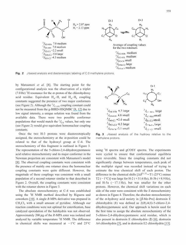
by Matsumori et al. [8]. The starting point for theconfigurational analysis was the observation of a triplet(7.0 Hz) 1H resonance for the a-proton of the chlorohydroxyacid residue. Equivalent Ha–Hl and Ha–Hh couplingconstants suggested the presence of two major conformers(see Figure 2). Although the 3JC1-H3 coupling constant couldnot be measured from the g-BIRD-HSQMBC [8, 12] due tolow signal intensity, a unique solution was found from theavailable data. There were two possible conformerpopulations that would match the 2JCH values, but only one(see Figure 2) would give equivalent homonuclear couplingconstants.
Once the two H-3 protons were diastereotopicallyassigned, the stereochemistry at the a -position could berelated to that of the hydroxyl group at C-4. Thestereochemistry of this fragment is outlined in Figure 3.The representation of the 5-chloro-2,4-dihydroxypentanoicacid relative stereochemistry and its major conformer in theNewman projection are consistent with Matsumori’s model[8]. The observed coupling constants were consistent withthe presence of mainly one rotamer since the homonuclearcoupling constants were quite different. However, themagnitude of these couplings was consistent with a smallpopulation of a second rotamer similar to that described inFigure 2. Overall, the coupling constants were consistentwith the rotamer shown in Figure 3.
The absolute stereochemistry at C-4 was establishedusing the 1H NMR method described by Riguera andcoworkers [10]. A single R-MPA derivative was prepared inCH2Cl2 with a small amount of pyridine. Although ourreaction conditions were not optimal for esterification, basecatalyzed epoxidation of the halohydrin was not observed.Approximately 200 mg of the R-MPA ester was isolated andanalyzed by variable temperature 1H NMR. The differencein chemical shifts was measured at �1°C and 25°C
using 1H spectra and gCOSY spectra. The experimentswere cycled to ensure that conformational equilibria were reversible. Since the coupling constants did notsignificantly change between temperatures, each peak ofthe multiplet signal was recorded instead of trying toestimate the true chemical shift of each proton. Thedifference in the chemical shifts [DdT1,T2�T1 (25°C) minusT2 (�1°C)] was large for H-2 (�31.6 Hz), H-3b (�8.9 Hz),and H-5a (�17.1 Hz), but was smaller for the otherprotons. However, the chemical shift variations on eachside of the ester were consistent with the S stereochemistryas shown in Figure 4. Therefore, the absolute stereochemistryof the a -hydroxy acid moiety in [b -Me-Pro] destruxin Echlorohydrin (1) was defined as 2(R),4(S)-5-chloro-2,4-dihydroxypentanoic acid. Our approach is herein used forthe first time to assign the absolute stereochemistry of the5-chloro-2,4-dihydroxypentanoic acid residue, which isalso present in destruxin E chlorohydrin (2) [6], destruxinA4 chlorohydrin [5], and in destruxin E2 chlorohydrin [13].
559
Fig. 2 J-based analysis and diastereotopic labeling of C-3 methylene protons.
Fig. 3 J-based analysis of the hydroxy relative to themethylene protons.

Pseudodestruxin C (3) was isolated as an optically activewhite solid, [a]D
25 �122.4 (c 0.0213, MeOH). The molecularformula C36H56N5O7 was assigned by the HRFABMSanalysis of the [M�H]� peak at m/z 670 (meas. 670.4149;calc. 670.4179) and was consistent with the NMR data. TheNMR data showed that pseudodestruxin C (3) containedtwo N-methyl groups (at d 2.95 and 3.08) and a b -Alaresidue consistent with other members of the destruxinclass of cyclodepsipeptides. The 1H NMR clearly indicatedthe presence of a monosubstituted aromatic ring belongingto a phenylalanine residue (d 7.30 m; 7.27 m; 7.24 m).Additionally, analysis of the 1H-1H COSY and 80 msTOCSY spectrum showed spin systems consistent with twoN-Me-Val residues and a b-Me-Pro residue. The Ha of thehydroxy acid residue could easily be identified in thegHSQC spectrum due to the downfield shift of the 13Cresonance, typically assigned to Ca (at d 74.6). The 1H-1Hcouplings observed in both COSY and TOCSY spectrashowed a spin system related to that of leucine, therebyelucidating the 2-hydroxy-4-methylpentanoic acid residue.Once the individual amino acid spin systems were assigned,the sequence could be assembled using HMBC data andROESY correlations in a manner similar to that describedfor [b-Me-Pro] destruxin E chlorohydrin (1), indicating theplanar structure 3 for pseudodestruxin C, which is the b -Me-Pro analog of [Ph3, N-Me-Val5] destruxin B (4)previously isolated from the same fungal strain Beauveriafelina [14]. The absolute stereochemistry of the N-Me-Valand Phe residue has been established as S by derivatizationwith Marfey’s reagent followed by HPLC analysis. No attempt has been made to establish the absolutestereochemistry of the 2-hydroxy-4-methylpentanoic acidresidue. The relative stereochemistry of the b -Me-Pro
residue is proposed as Ca(S*), Cb(S*), the same of [b-Me-Pro] destruxin E chlorohydrin (1), since the couplingconstant for the Ha -Hb in the b -Me-Pro residue ispractically the same for both compounds 1 (J�2.54 Hz)and 3 (J�2.4 Hz), and closely related to the same couplingobserved in roseotoxin B [15]. We have named compound 3pseudodestruxin C in agreement with Gloer’s namingsystem for pseudodestruxins A and B [16]. In addition tothe new compounds, we have isolated the known destruxinsdestruxin E chlorohydrin (2) [6] [Ph3, N-Me-Val5] destruxinB (4) [14], roseotoxin B (5) [15, 17] and roseocardin (6)[18]. The structures of the known cyclodepsipeptides havebeen established by analysis of MS and NMR data andcomparison with literature data. Interestingly, a non-destruxin peptide, isariin B (7), was also obtained from our marine-derived strain of B. felina. Initial inspection ofthe NMR data suggested that this peptide contained a methylene similar to that of a b -Ala. However, a detailed analysis showed that this methylene was a glycine and indicated the peptide was not a destruxin-like cyclodepsipeptide. Accurate mass measurements gavean [M�H]� at m/z 596.4025 indicating the molecularformula C30H54N5O7 for the quasi-molecular ion [M�H]�
(calcd 596.4023, D 0.3 ppm). On the basis of the molecularformula, the compound was the known fungal metaboliteisariin B (see Figure 5) [19, 20]. In order to confirm thestructure without relying on extensive analysis of the NMR data, MS studies were performed on the [M�Na]�
ion using a quadrupole orthogonal acceleration time of flight tandem mass spectrometer (Q-Tof) with �ESI. After fragmentation, the spectrum was calibrated using the residual parent [M�Na]� ion to obtain accurate mass measurements on the fragment ions (see Figure 6).
560
Fig. 4 Single derivative MPA ester analysis [10].

A molecular formula could easily be assigned to thefragments, but the sequence required knowledge of thefragmentation route. Therefore, in source CID fragmentationof the [M�Na]� ion was performed and resulted in aspectrum that was similar to the MS/MS spectrum. Each ofthe resulting fragment ions was filtered by the quadrupole,fragmented in the collision cell, and analyzed by the TOFmass analyzer. From these analyses, m/z 335 and m/z 236were identified as daughter ions of m/z 448, with m/z 236being the only daughter ion observed upon fragmentationof m/z 335. Additionally, m/z 289 was a daughter ion of m/z402. These results confirmed not only the sequence of theamino acids, but also the molecular formula for each
fragment ion. This methodology allows pseudo MS3
experiments while maintaining mass accuracy and shouldbe widely applicable to the analysis of a variety of naturalproducts, enabling the dereplication of peptide type naturalproducts without the need for NMR analysis to confirm theamino acid sequence. The method can be summarized as itfollows. In the first step, a lock mass is used to obtainaccurate mass on the molecular ion. Once a molecularformula is confirmed, the MS/MS spectra can be masscorrected using the residual signal from the parent ion toachieve accurate mass on the fragments. Then, in sourceCID can be applied to allow mass filtering for MS/MSstudies to track daughter ions, thereby allowing MS3 andefficient compound dereplication.
In conclusion, the present investigation reports theisolation of two new destruxin derivatives, [b -Me-Pro]destruxin E chlorohydrin (1) and pseudodestruxin C (3).Furthermore, the absolute stereochemistry of the 5-chloro-2,4-dihydroxypentanoic acid in [b -Me-Pro] destruxin Echlorohydrin (1) has been assigned for the first time using acombination of the J-based configuration method [10, 11]in conjunction with the variable temperature NMR analysisof the corresponding R-MPA ester derivative. Finally, anMS3 method using in source CID to allow mass filtering for
561
Fig. 5 Structure of isariin B (7).
Fig. 6 MS sequencing of isariin B (7).

additional MS/MS analysis to track daughter ions wasdeveloped for the rapid dereplication of natural products.Finally, the major compounds isolated from the marine-derived B. felina, pseudodestruxin C (3), [Phe3, N-Me-Val5]destruxin B (4) and rosetoxin B (5), have been testedagainst M. tuberculosis H37 Rv and in cytotoxicitybioassays against SF 295 (human CNS), MDA-MB435(human breast), HCT8 (colon) and HL60 (leukemia) cancer cell lines. Only roseotoxin B displayed moderatecytotoxicity at 1.09, 1.30, 0.90 and 0.14 mg/ml against eachof the cell lines, respectively.
Acknowledgments The authors thank Dr. Álvaro EstevesMigotto, Director of the Centro de Biologia Marinha of theUniversidade de São Paulo, for providing facilities for marinesample collection, Prof. Gil V. J. Silva and Virginia H. B. Glass(Departamento de Química, Faculdade de Filosofia, Ciências eLetras de Ribeirão Preto, Universidade de São Paulo) forobtaining the NMR spectra on the Bruker DRX500 NMRequipment, and Professor Brent R. Copp (Department ofChemistry, University of Auckland, New Zealand) for obtainingthe MS data for compound 3. Financial support was provided bythe FAPESP grant 03/08899-0 and the CNPq grant 470481/04-8to RGSB, a FAPESP scholarship 03/06471-3 to SPL, a CAPESscholarship to AMV-M, a USP-PROCONTES contract to MHRSand NIH grant CA36622 to CMI. The following NIH and NSFgrants funded the Varian NMR instrumentation, RR06262,RR13030, and DBI-0002806. The authors dedicate this work tothe memory of Professor Kenneth Lloyd Rinehart, for hisoutstanding achievements in the discovery of biologically activenatural products.
References
1. Bugni TS, Ireland CM. Marine-derived fungi: a chemicallyand biologically diverse group of microorganisms. Nat ProdRep 21: 143–163 (2004)
2. Kelecom A. Secondary metabolites from marinemicroorganisms. An Acad Bras Ciências 74: 151–170(2002)
3. Hernandez ILC, Godinho MJL, Magalhães A, Schefer AB,Ferreira AG, Berlinck RGS. N-Acetyl-g -hydroxyvalinelactone, an unusual amino acid derivative from a marinestreptomycete. J Nat Prod 63: 664–665 (2000)
4. Hernandez ILC, Macedo ML, Berlinck RGS, Ferreira AG,Godinho MJl. Dipeptide metabolites from the marinederived bacterium Streptomyces acrymicini. J Braz ChemSoc 15: 441–444 (2004)
5. Cai P, Smith D, Katz B, Pearce C, Venables D, Houck D.Destruxin-A4 chlorohydrin, a novel destruxin from fungusOS-F68576: isolation, structure determination, andbiological activity as an inducer of erythropoietin. J NatProd 61: 290–293 (1998)
6. Gupta S, Roberts DW, Renwick JAA. Insecticidalcyclodepsipeptides from Metarrhizium anisopliae. J ChemSoc Perkin Trans I 2347–2357 (1989)
7. Bhushan R, Bruckner H. Marfey’s reagent for chiral aminoacid analysis: a review. Amino Acids 27: 231–247 (2004)
8. Matsumori N, Kaneno D, Murata M, Nakamura H,Tachibana K. Stereochemical determination of acyclicstructures based on carbon-proton spin-coupling constants.A method of configuration analysis for natural products. JOrg Chem 64: 866–876 (1999)
9. Murata M, Matsuoka S, Matsumori N, Paul GK, TachibanaK. Absolute configuration of amphidinol 3, the first completestructure determination from amphidionol homologues:Application of a new configuration analysis based oncarbon-hydrogen spin-coupling constants. J Am Chem Soc121: 870–871 (1999)
10. Latypov SK, Seco JM, Quiñoá E, Riguera R. Are both the(R)- and the (S)-MPA esters really needed for the assignmentof the absolute configuration of secondary alcohols byNMR? The use of a single derivative. J Am Chem Soc 120:877–882 (1998)
11. Uhrin D, Batta G, Hruby VJ, Barlow PN, Kover KE.Sensitivity- and gradient-enhanced hetero (w1) half-filteredTOCSY experiment for measuring long-range heteronuclearcoupling constants. J Mag Res 130: 155–161 (1998)
12. Williamson RT, Boulanger A, Vulpanovici A, Roberts MA,Gerwick WH. Structure and absolute stereochemistry ofphomidolide, a new toxic metabolite from the marinecyanobacterium Phormidium sp. J Org Chem 67: 7927–7936(2002)
13. Yeh SF, Pan W, Ong GT, Chiou AJ, Chuang CC, Chiou SH,Wu SH. Study of structure-activity correlation in destruxins,a class of cyclodepsipeptides possessing suppressive effecton the generation of hepatitis B virus surface antigen inhuman hepatoma cells. Biochem Biophys Res Commun 229:65–72 (1996)
14. Kim HS, Jung MH, Ahn S, Lee CW, Kim SN, Ok JH.Structure elucidation of a new cyclic hexadepsipeptide fromBeauveria felina. J Antibiot 55: 598–601 (2002)
15. Springer JP, Cole RJ, Dorner JW, Cox RH, Richard JL,Barnes CL, Van der Helm D. Structure and conformation ofroseotoxin B. J Am Chem Soc 106: 2388–2392 (1984)
16. Che Y, Swenson DC, Gloer JB, Koster B, Malloch D.Pseudodestruxins A and B: new cyclic depsipeptides fromthe Coprophilous fungus Nigrosabulum globosum. J NatProd 64: 555–558 (2001)
17. Engstrom GW, DeLance JV, Richard JL, Baetz AL.Purification and characterization of roseotoxin B, a toxiccyclodepsipeptide from Trichothecium roseum. J Agric FoodChem 23: 244–253 (1975)
18. Tsunoo A, Kamijo M, Taketomo N, Sato Y, Ajisaka K.Roseocardin, a novel cardiotonic cyclodepsipeptide fromTrichothecium roseum TT103. J Antibiot 50: 1007–1013(1997)
19. Baute R, Deffieux G, Merlet D, Baute MA, Neveu A. New
562

insecticidal cyclodepsipeptides from the fungus Isariafelina. I. Production, isolation and insecticidal properties ofisariins B, C and D. J Antibiot 34: 1261–1265 (1981)
20. Deffieux G, Merlet D, Baute R, Bourgeois G, Baute MS,Neveu A. New insecticidal cyclodepsipeptides from thefungus Isaria felina. II. Structure elucidation of isariins B, Cand D. J Antibiot 34: 1266–1270 (1981)
21. Raeder J, Broda P. Rapid preparation of DNA fromfilamentous fungi. Lett Appl Microbiol 1: 17–20 (1985)
22. Thompson JD, Higgins DG, Gibson TJ, Clustal W.Improving the sensitivity of progressive multiple alignmentthrough sequence weighting, positions-specific gap penaltiesand weight matrix choice. Nucl Acids Res 22: 4673–4680(1994)
23. Kumar S, Tamura K, Jakobsen IB, Nei M. MEGA2:Molecular Evolutionary Genetics Analysis software,Arizona State University, Tempe, Arizona, USA. Bioinform17: 1244–1245 (2001)
24. Kimura M. A simple method for estimating evolutionaryrate of base substitutions through comparative studies ofnucleotide sequences. J Mol Evol 16: 111–120 (1980)
25. Abad J-L, Fabrias G, Camps F. Synthesis of dideuterated andenantiomers of monodeuterated tridecanoic acids at C-9 andC-10 positions. J Org Chem 65: 8582–8588 (2000)
26. Ward DE, Rhee CK. A simple method for the microscalepreparation of Mosher’s acid chloride. Tetrahedron Lett 32:7165–7166 (1991)
563