Neuroprotection by BDNF against glutamate-induced apoptotic cell death is mediated by ERK and...
15
Neuroprotection by BDNF against glutamate-induced apoptotic cell death is mediated by ERK and PI3-kinase pathways RD Almeida 1 , BJ Manadas 1 , CV Melo 1 , JR Gomes 1 , CS Mendes 1 , MM Gra ˜os 1 , RF Carvalho 1 , AP Carvalho 1 and CB Duarte* ,1 1 Center for Neuroscience and Cell Biology and Department of Zoology, University of Coimbra, 3004-517 Coimbra, Portugal * Corresponding author: CB Duarte, Center for Neuroscience and Cell Biology, Department of Zoology, University of Coimbra, 3004-517 Coimbra, Portugal. Tel: þ 351 239 480209; Fax: þ 351 239 480208; E-mail: [email protected] Received 08.11.04; revised 29.3.05; accepted 06.4.05; published online 20.5.05 Edited by M Miura Abstract Neurotrophins protect neurons against glutamate excitotoxi- city, but the signaling mechanisms have not been fully elucidated. We studied the role of the phosphatidylinositol 3-kinase (PI3-K) and Ras/mitogen-activated protein kinase (MAPK) pathways in the protection of cultured hippocampal neurons from glutamate induced apoptotic cell death, characterized by nuclear condensation and activation of caspase-3-like enzymes. Pre-incubation with the neurotrophin brain-derived neurotrophic factor (BDNF), for 24 h, reduced glutamate-evoked apoptotic morphology and caspase-3-like activity, and transiently increased the activity of the PI3-K and of the Ras/MAPK pathways. Inhibition of the PI3-K and of the Ras/MAPK signaling pathways abrogated the protective effect of BDNF against glutamate-induced neuronal death and similar effects were observed upon inhibition of protein synthesis. Moreover, incubation of hippocampal neurons with BDNF, for 24 h, increased Bcl-2 protein levels. The results indicate that the protective effect of BDNF in hippocampal neurons against glutamate toxicity is mediated by the PI3-K and the Ras/MAPK signaling pathways, and involves a long- term change in protein synthesis. Cell Death and Differentiation (2005) 12, 1329–1343. doi:10.1038/sj.cdd.4401662; published online 20 May 2005 Keywords: BDNF; apoptosis; extracellular signal-regulated kinase (ERK); phosphatidylinositol 3-kinase; glutamate; hippo- campal neurons; Akt (PKB); Bcl-2 Abbreviations: BDNF, brain-derived neurotrophic factor; CREB, cAMP response-element binding protein; DTT, dithio- threitol; ERK, extracellular signal-regulated kinase; IC, ischemic core; IP, ischemic penumbra; JNK, c-Jun N-terminal kinase; MAPK, mitogen-activated protein kinase; MEK, MAPK/ERK kinase; NGF, nerve growth factor; NT-3, neurotrophin-3; PI, propidium iodide; PI3-K, phosphatidylinositol 3-kinase; PMSF, phenylmethylsulfonyl fluoride Introduction Neurotrophins play an important role in the development of the nervous system, by influencing cell survival, differentiation and cell death. 1 Furthermore, gradients of neurotrophins are able to steer growth cones in vitro, acting as chemoattractants or chemorepellents, depending on the cyclic nucleotide levels within the neurons. 2,3 In vivo brain-derived neurotrophic factor (BDNF) has been shown to rescue different types of neurons from ischemic, traumatic and toxic brain injury. 4–10 Blocking endogenous BDNF activity leads to aggravated death of a subpopulation of hippocampal neurons after global forebrain ischemia. 11 The neuroprotective role of endogenous BDNF is further supported by the observed correlation between BDNF protein levels and resistance to ischemic damage in the various hippocampal subregions. 12 Furthermore, intravenous administration of BDNF 30 min after middle cerebral artery occlusion reduced infarct size in the cerebral cortex and counter-regulated Bcl-2 and Bax expression. 8 Intraventricular injection of BDNF also increased survival of rat hippocampal CA1 pyramidal neurons following transient forebrain ischemia. 4 In addition to the long-term effects, neurotrophins also exhibit acute effects on neurotransmitter release, synaptic strength and connectivity. 13,14 The cellular actions of neurotrophins are mediated through the activation of the Trk family of receptors, TrkA-C, and the p75 neurotrophin receptor. The latter receptor binds to all neurotrophins with a similar affinity, whereas the TrkA, TrkB and TrkC receptors are activated preferentially by nerve growth factor (NGF), BDNF and neurotrophin-3 (NT-3), respectively. 14 Once activated, the Trk receptors autophos- phorylate specific tyrosine residues, which serve as interac- tion sites for proteins endowed with PTB/SH2 domains. The consequences of Trk receptor activation include the activation of Ras, mediated by the adapter proteins Shc/Grb2/SOS, leading to the activation of extracellular signal-regulated kinases (ERK), members of the mitogen-activated protein kinase (MAPK) family, and of phospholipase C-g and phosphatidylinositol 3-kinase (PI3-K). Phospholipase C-g generates diacylglycerol and inositol 1,4,5-trisphosphate, and PI3-K forms 3 0 -phosphorylated phosphoinositides, which allow the activation of pH-containing proteins, including Akt/PKB. 1 Activation of Akt has acute effects on cell survival, due to phosphorylation of Bad, which in the phosphorylated form is sequestered in the cytosol by 14-3-3, precluding its proapoptotic effects on the mitochondria. 15 Furthermore, active ERKs and Akt are translocated to the nucleus where they can phosphorylate certain transcription factors, thereby regulating the expression of specific genes that contribute to cell survival. 1,16 Neurotrophins were shown to protect cultured hippocampal neurons from glutamate-evoked cell death, by upregulating the antioxidant defense system. 17 During overstimulation of Cell Death and Differentiation (2005) 12, 1329–1343 & 2005 Nature Publishing Group All rights reserved 1350-9047/05 $30.00 www.nature.com/cdd
Transcript of Neuroprotection by BDNF against glutamate-induced apoptotic cell death is mediated by ERK and...
Neuroprotection by BDNF against glutamate-inducedapoptotic cell death is mediated by ERK and PI3-kinasepathways
RD Almeida1, BJ Manadas1, CV Melo1, JR Gomes1, CS Mendes1,
MM Graos1, RF Carvalho1, AP Carvalho1 and CB Duarte*,1
1 Center for Neuroscience and Cell Biology and Department of Zoology,University of Coimbra, 3004-517 Coimbra, Portugal
* Corresponding author: CB Duarte, Center for Neuroscience and Cell Biology,Department of Zoology, University of Coimbra, 3004-517 Coimbra, Portugal.Tel: þ 351 239 480209; Fax: þ 351 239 480208; E-mail: [email protected]
Received 08.11.04; revised 29.3.05; accepted 06.4.05; published online 20.5.05Edited by M Miura
AbstractNeurotrophins protect neurons against glutamate excitotoxi-city, but the signaling mechanisms have not been fullyelucidated. We studied the role of the phosphatidylinositol3-kinase (PI3-K) and Ras/mitogen-activated protein kinase(MAPK) pathways in the protection of cultured hippocampalneurons from glutamate induced apoptotic cell death,characterized by nuclear condensation and activation ofcaspase-3-like enzymes. Pre-incubation with the neurotrophinbrain-derived neurotrophic factor (BDNF), for 24 h, reducedglutamate-evoked apoptotic morphology and caspase-3-likeactivity, and transiently increased the activity of the PI3-K andof the Ras/MAPK pathways. Inhibition of the PI3-K and of theRas/MAPK signaling pathways abrogated the protective effectof BDNF against glutamate-induced neuronal death andsimilar effects were observed upon inhibition of proteinsynthesis. Moreover, incubation of hippocampal neurons withBDNF, for 24 h, increased Bcl-2 protein levels. The resultsindicate that the protective effect of BDNF in hippocampalneurons against glutamate toxicity is mediated by the PI3-Kand the Ras/MAPK signaling pathways, and involves a long-term change in protein synthesis.Cell Death and Differentiation (2005) 12, 1329–1343.doi:10.1038/sj.cdd.4401662; published online 20 May 2005
Keywords: BDNF; apoptosis; extracellular signal-regulated
kinase (ERK); phosphatidylinositol 3-kinase; glutamate; hippo-
campal neurons; Akt (PKB); Bcl-2
Abbreviations: BDNF, brain-derived neurotrophic factor;
CREB, cAMP response-element binding protein; DTT, dithio-
threitol; ERK, extracellular signal-regulated kinase; IC, ischemic
core; IP, ischemic penumbra; JNK, c-Jun N-terminal kinase;
MAPK, mitogen-activated protein kinase; MEK, MAPK/ERK
kinase; NGF, nerve growth factor; NT-3, neurotrophin-3; PI,
propidium iodide; PI3-K, phosphatidylinositol 3-kinase; PMSF,
phenylmethylsulfonyl fluoride
Introduction
Neurotrophins play an important role in the development ofthe nervous system, by influencing cell survival, differentiationand cell death.1 Furthermore, gradients of neurotrophins areable to steer growth cones in vitro, acting as chemoattractantsor chemorepellents, depending on the cyclic nucleotide levelswithin the neurons.2,3 In vivo brain-derived neurotrophic factor(BDNF) has been shown to rescue different types of neuronsfrom ischemic, traumatic and toxic brain injury.4–10 Blockingendogenous BDNF activity leads to aggravated death of asubpopulation of hippocampal neurons after global forebrainischemia.11 The neuroprotective role of endogenous BDNF isfurther supported by the observed correlation between BDNFprotein levels and resistance to ischemic damage in thevarious hippocampal subregions.12 Furthermore, intravenousadministration of BDNF 30 min after middle cerebral arteryocclusion reduced infarct size in the cerebral cortex andcounter-regulated Bcl-2 and Bax expression.8 Intraventricularinjection of BDNF also increased survival of rat hippocampalCA1 pyramidal neurons following transient forebrain ischemia.4
In addition to the long-term effects, neurotrophins also exhibitacute effects on neurotransmitter release, synaptic strengthand connectivity.13,14
The cellular actions of neurotrophins are mediated through
the activation of the Trk family of receptors, TrkA-C, and the
p75 neurotrophin receptor. The latter receptor binds to all
neurotrophins with a similar affinity, whereas the TrkA, TrkB
and TrkC receptors are activated preferentially by nerve
growth factor (NGF), BDNF and neurotrophin-3 (NT-3),
respectively.14 Once activated, the Trk receptors autophos-
phorylate specific tyrosine residues, which serve as interac-
tion sites for proteins endowed with PTB/SH2 domains. The
consequences of Trk receptor activation include the activation
of Ras, mediated by the adapter proteins Shc/Grb2/SOS,
leading to the activation of extracellular signal-regulated
kinases (ERK), members of the mitogen-activated protein
kinase (MAPK) family, and of phospholipase C-g and
phosphatidylinositol 3-kinase (PI3-K). Phospholipase C-ggenerates diacylglycerol and inositol 1,4,5-trisphosphate,
and PI3-K forms 30-phosphorylated phosphoinositides, which
allow the activation of pH-containing proteins, including
Akt/PKB.1 Activation of Akt has acute effects on cell survival,
due to phosphorylation of Bad, which in the phosphorylated
form is sequestered in the cytosol by 14-3-3, precluding its
proapoptotic effects on the mitochondria.15 Furthermore,
active ERKs and Akt are translocated to the nucleus where
they can phosphorylate certain transcription factors, thereby
regulating the expression of specific genes that contribute to
cell survival.1,16
Neurotrophins were shown to protect cultured hippocampalneurons from glutamate-evoked cell death, by upregulatingthe antioxidant defense system.17 During overstimulation of
Cell Death and Differentiation (2005) 12, 1329–1343& 2005 Nature Publishing Group All rights reserved 1350-9047/05 $30.00
www.nature.com/cdd

glutamate receptors, the mitochondria become de-energizedand produce reactive oxygen species due to the accumulationof large amounts of Ca2þ .18–21 It has been proposed that theability of mitochondria to recover their energetics after aglutamate insult determines the mode of cell death, byapoptosis or by necrosis.18 Neuronal death by apoptosis ischaracterized by the release of cytochrome c, which allowsthe activation of caspase-9. This caspase acts upstream ofcaspase-3, the major executioner caspase in neurons.22–25
Although neurotrophins protect hippocampal neurons in vivoand in vitro, the signaling mechanisms responsible for thisprotection are still not fully understood. In the present work, wedetermined the role of the PI3-K pathway and of the Ras/ERKpathway in neuroprotection by BDNF from glutamate toxicity.The results show that BDNF protects hippocampal neurons atthe level or upstream of the activation of caspase-3-likeenzymes, by a mechanism involving both signaling pathways.
Results
BDNF protects cultured hippocampal neuronsfrom glutamate-induced cell death
Cultured hippocampal neurons were challenged with 125 mMglutamate and cell death was accessed by analyzing nuclearmorphology, 7 h after the excitotoxic insult, using thefluorescent dyes Syto-13 and propidium iodide. After theexcitotoxic insult with glutamate about 40–45% of the cellsdisplayed an apoptotic morphology (Figure 1), characterizedby chromatin condensation and loss of neurites (not shown).Pre-incubation of the cells with BDNF (Figure 1b) or NT-3(Figure 1c), for 24 h, before the glutamate insult, reduced thenumber of apoptotic cells in a dose-dependent manner. BDNFwas more efficient in protecting the cells than NT-3 at themaximal concentration used (200 ng/ml), reducing the toxiceffect of glutamate by about 60%. As expected, NGF (20–100 ng/ml) did not protect hippocampal neurons from gluta-mate toxicity, in agreement with the known absence of TrkAreceptors in the preparation.26
Activation of caspase-3 is a hallmark of apoptotic cell deathand precedes changes in nuclear morphology.27 Pre-incuba-tion of hippocampal neurons with Z-DEVD-FMK, a cellpermeable irreversible inhibitor of caspase-3-like enzymes,28
significantly reduced glutamate-induced cell death(Figure 2a). However, at the maximal concentration used,the caspase inhibitor did not fully protect the cells. In control
experiments hippocampal neurons were treated only with thecaspase inhibitor (50–100mM), and no toxicity was observed.
To further confirm that in our model system glutamateinduced an apoptotic-like form of neuronal cell death, we
Figure 1 BDNF and NT-3, but not NGF, protect hippocampal neurons fromglutamate-induced cell death. Hippocampal neurons were treated with NGF (a),BDNF (b) or NT-3 (c), for 24 h, and then challenged with 125 mM glutamate for15 min, in fresh Neurobasal medium containing B27 supplement, and thenreturned to the original medium. Cell death was accessed 7 h after the insult byfluorescence microscopy, using the fluorescence dyes Syto-13 and PI. Controlcells were incubated for 15 min, with fresh glutamate-free Neurobasal mediumcontaining B27 supplement, and were then returned to the original culturemedium. Data are presented as the mean7s.e.m. of the indicated number ofexperiments, performed in independent preparations. ***Po0.001 as comparedwith control cells; þ þ þPo0.001 as compared with cells stimulated withglutamate alone
Signaling mechanisms in neuroprotection by BDNFRD Almeida et al
1330
Cell Death and Differentiation

measured caspase-3-like activity in extracts prepared fromhippocampal cultures treated or not with glutamate, using thechromogenic substrate Ac-DEVD-pNA. The results show thatexcitotoxic stimulation with glutamate increased caspase-3-like activity, but this effect was not observed in cells pre-incubated with BDNF for 24 h, before the toxic insult. Theseresults indicate that BDNF protects hippocampal neurons atthe level or upstream of the activation of caspase-3-likeenzymes.
The Ras/MAPK pathway is involved inneuroprotection by BDNF against glutamatetoxicity
Since BDNF was more potent in protecting hippocampalneurons against glutamate toxicity than NT-3, we furtherinvestigated the signaling pathways responsible for the effectof the former neurotrophin. Activation of Trk receptors by
BDNF was required for neuroprotection, since no effect of theneurotrophin on glutamate toxicity was observed when theexperiments were conducted in the presence of K252a(200 nM), an inhibitor of this family of receptors29 (Figure 3aand b). Control experiments showed that K252a did not affectglutamate toxicity in the absence of BDNF. The effect ofK252a on Trk receptors was confirmed by Western blot, usingan antibody that recognizes phosphotyrosine 490 in TrkA, aresidue conserved in the other Trk receptors. Stimulation ofcultured hippocampal neurons with 100 ng/ml BDNF, for5 min, increased Trk (presumably TrkB) phosphorylation,and this effect was inhibited by K252a (Figure 3c).
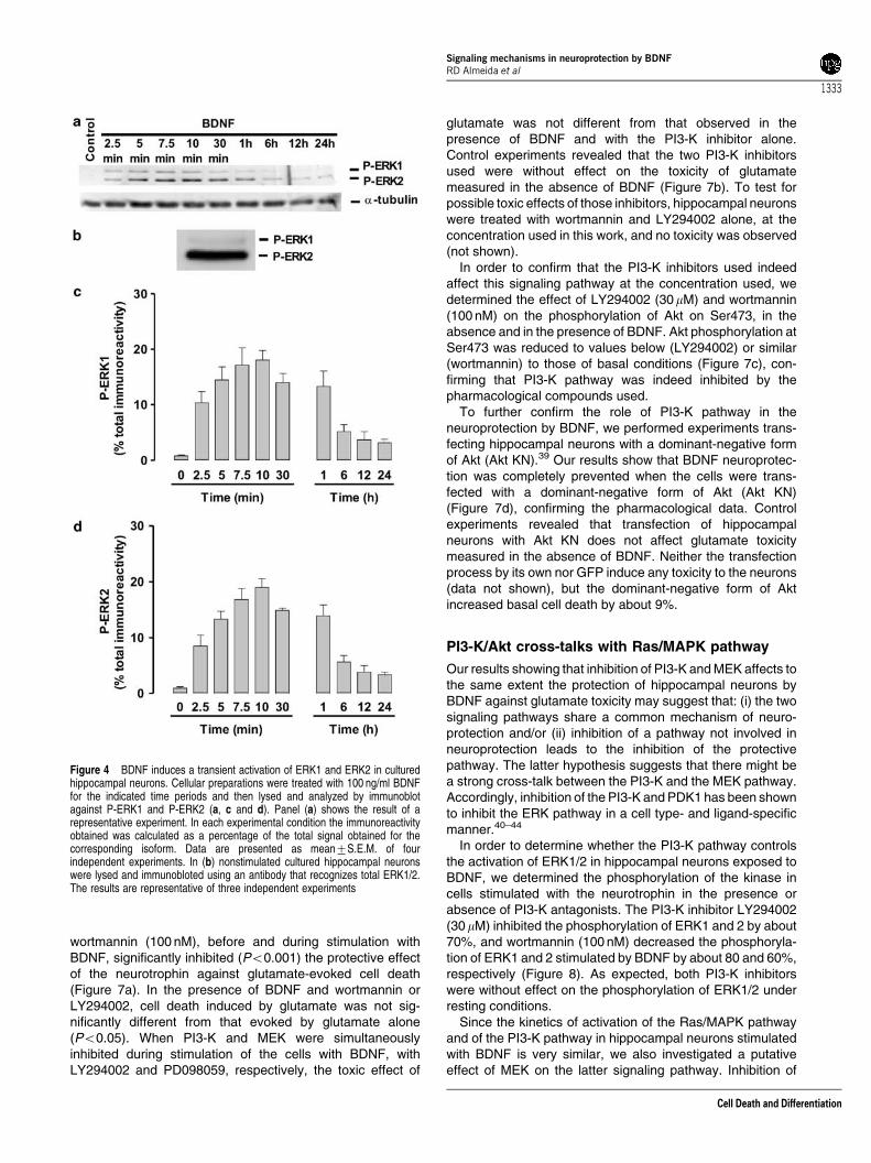
The kinetics of activation of the Ras/ERK pathway by BDNFwas followed by Western blot, using an antibody thatspecifically recognizes the dually phosphorylated (active)form ERK1 and 2.30 The activity of ERK 1 and 2 in culturedhippocampal neurons was very low under resting conditions,but it was transiently increased upon stimulation of the cellswith 100 ng/ml BDNF. The kinetics of activation of the twokinases was similar (Figure 4c and d), although the increase inphospho-ERK2 immunoreactivity was more significant thanthat obtained for phospho-ERK1 (Figure 4a). The total amountof ERK2 present in the cells was also higher than theexpression of ERK1 protein (Figure 4b). The maximalphosphorylation of ERKs was attained after 7.5–10 min ofstimulation, and the activity of the kinases decreased after-wards, towards a plateau close to basal levels. After 24 hstimulation of hippocampal neurons with BDNF, an incubationperiod that confers neuroprotection (Figure 1), the phosphory-lation of ERK1 and 2 was not significantly different from basalphosphorylation (P40.05).
In order to test if the activation of the ERK pathway wasinvolved in the neuroprotective effect of BDNF, we inhibitedpharmacologically MAPK/ERK Kinase (MEK), the upstreamkinase of ERK, with PD098059 (20 mM)31 or U0126(300 nM).32 In the presence of the MEK inhibitors, theneuroprotective effect of BDNF against glutamate-evokedcell death was completely abrogated (Figure 5a), as observedby analysis of nuclear morphology, indicating that the ERKpathway mediates the neuroprotective effect of BDNF againstglutamate toxicity (Po0.001). When PD098059 andLY294002, a PI3-K inhibitor, were present together, noadditive effect was observed. Control experiments revealedthat the two MEK inhibitors used were without effect onglutamate toxicity measured in the absence of BDNF(Figure 5b). To test for possible toxic effects of thoseinhibitors, hippocampal neurons were treated independentlywith PD098059 or U0126, at the concentration used in thiswork, and no toxicity was observed (not shown).
The concentrations of the MEK inhibitors used in thepresent work are below the range of concentrations that fullyblock the kinase in in vitro assays.33 However, we could notuse higher concentrations of the antagonists because of theirtoxic effects. Since the effect of inhibitors of MEK activation invivo may also depend on how potently the signaling pathwayis activated by any particular agonist,31 we determined theeffect of PD098059 and of U0126 on the phosphorylation ofERK1/2 in hippocampal neurons stimulated with BDNF. Theresults of Figure 5c show that PD098059 inhibited thephosphorylation of ERK1 and 2 in response to stimulation
Figure 2 BDNF inhibits caspase-3-like activity induced by glutamate. (a)Hippocampal neurons were challenged with 125mM glutamate, for 15 min, in thepresence or absence of the indicated concentrations of the caspase-3 likeinhibitor Z-DEVD-FMK. Control cells were incubated with the inhibitor alone, atthe indicated concentrations, and no toxicity was observed. The experimentswere conducted as described in the caption of Figure 1 and cell death wasassessed 7 h after the insult. (b) Hippocampal neurons were treated or not with100 ng/ml BNDF, for 24 h, and challenged with 125 mM glutamate or not (control),for 15 min. Caspase-3-like activity in cellular extracts was assessed 5 h after theinsult by following the cleavage of the Ac-DEVD-pNA substrate. Data arepresented as mean7S.E.M. of the indicated number of experiments, performedin independent preparations. ***Po0.001 as compared to control; þ þPo0.01,þ þ þPo0.001 as compared with cells stimulated with glutamate alone
Signaling mechanisms in neuroprotection by BDNFRD Almeida et al
1331
Cell Death and Differentiation

with 100 ng/ml BDNF by about 65%. U0126 also inhibited thephosphorylation of ERK1 and 2 by about 60 and 20%,respectively.
In order to further confirm the role of the ERK pathway in theneuroprotection by BDNF, we performed experiments trans-fecting hippocampal neurons with a dominant-negativeconstruct of MEK (MEK KN).34 When hippocampal neuronswere transfected with the dominant-negative form of MEK, theneuroprotective effect of BDNF was abolished, in agreementwith the results obtained with the pharmacological inhibitors(Figure 5d). Control experiments revealed that transfection ofhippocampal neurons with the MEK KN does not affectglutamate toxicity measured in the absence of BDNF. The
transfection by its own did not induce any toxicity, as well asthe transfection of neurons with GFP plus MEK KN or GFPalone (data not shown). Taken together, the results indicatethat the ERK pathway mediates the protective effect of BDNFagainst excitotoxic insults in cultured hippocampal neurons.
The PI3-K/Akt pathway is involved in BDNFneuroprotection against glutamate
In order to evaluate the effect of BDNF on the PI3-K pathwayin cultured hippocampal neurons, we determined the kineticsof phosphorylation of Akt, a kinase that is a downstreameffector of PI3-K. Akt is activated by phosphorylation onThr308, Ser473 and on a tyrosine residue, by differentkinases, and phosphorylation of these sites is necessary forfull activation of the kinase.35,36 Therefore, we assessed thephosphorylation of Akt on Ser473 and on Thr308, by Westernblot, as a measurement of the activity of the PI3-K pathway.Under resting conditions there was a basal phosphorylation ofAkt on Ser473 (Figure 6b) and on Thr308 (Figure 6c), whichcontrasts with the low phosphorylation of ERK1/2 shown inFigure 4. Upon stimulation of the cells with BDNF, there was a2.5-fold increase in the phosphorylation of Akt on Thr308 anda 3.2-fold increase in Ser473 phosphorylation. This increasein Akt activity was transient, peaking after 7.5–10 min ofstimulation, and decreasing afterwards towards a plateau.After 24 h stimulation of hippocampal neurons withBDNF, an incubation period that leads to neuroprotectionagainst glutamate toxicity (Figure 1), the phosphorylationof Akt was not significantly different from basal phosphory-lation (P40.05).
The role of the PI3-K pathway in the protection by BDNFagainst glutamate toxicity was investigated using two chemi-cally different inhibitors of the enzyme: wortmannin, a fungaltoxin that covalently binds to and blocks the activity of thecatalytic p110 subunit of PI3-K,37 and LY294002, a syntheticbioflavenoid that reversibly binds to and inhibits p110.38 Pre-incubation of the cells with LY294002 (30 mM) or with
Figure 3 The neuroprotective effect of BDNF is mediated through theactivation of Trk receptors. (a) Hippocampal neurons were pretreated or not withthe Trk inhibitor, K252a (200 nM), 30 min before they were incubated for 6 h, with100 ng/ml BDNF. This pre-incubation period provides maximal protection byBDNF (see Figure 10). The cells were then challenged with glutamate (125 mM)during 20 min. Cell death was accessed 14 h later by fluorescence microscopy,using the fluorescent dye Hoechst 33342. Data are presented as mean7S.E.M.of three experiments, performed in independent preparations, and are expressedas a percentage of cell death observed in response to glutamate stimulationalone. **Po0.01 as compared with cells stimulated with glutamate alone;þPo0.05. The nuclear morphology of control cells, and the effect excitotoxicstimulation with glutamate, in the presence or in the absence of BDNF, are shownin (b). The inhibition of neuroprotection by BDNF in the presence of K252a is alsoshown in (b). (c) Hippocampal neurons were pretreated or not with 200 nMK252a, for 30 min, and then incubated with 100ng/ml BDNF, for 5 min, with orwithout the Trk inhibitor. Cells were then lysed and total extracts (30 mg) wereanalysed by Western blot, using an anti-phospho-Trk (phosphotyrosine 490)antibody. Protein loading was checked by stripping and reprobing the membranewith an anti-actin antibody. The immunoreactivity obtained in each experimentalcondition was calculated as a percentage of the control. The results of arepresentative experiment are also shown. Data are presented as mean7S.E.M. of three different experiments, performed in independent preparations.***Po0.001 as compared with the control, nonstimulated cells; þ þ þPo0.001
Signaling mechanisms in neuroprotection by BDNFRD Almeida et al
1332
Cell Death and Differentiation
wortmannin (100 nM), before and during stimulation withBDNF, significantly inhibited (Po0.001) the protective effectof the neurotrophin against glutamate-evoked cell death(Figure 7a). In the presence of BDNF and wortmannin orLY294002, cell death induced by glutamate was not sig-nificantly different from that evoked by glutamate alone(Po0.05). When PI3-K and MEK were simultaneouslyinhibited during stimulation of the cells with BDNF, withLY294002 and PD098059, respectively, the toxic effect of
glutamate was not different from that observed in thepresence of BDNF and with the PI3-K inhibitor alone.Control experiments revealed that the two PI3-K inhibitorsused were without effect on the toxicity of glutamatemeasured in the absence of BDNF (Figure 7b). To test forpossible toxic effects of those inhibitors, hippocampal neuronswere treated with wortmannin and LY294002 alone, at theconcentration used in this work, and no toxicity was observed(not shown).
In order to confirm that the PI3-K inhibitors used indeedaffect this signaling pathway at the concentration used, wedetermined the effect of LY294002 (30mM) and wortmannin(100 nM) on the phosphorylation of Akt on Ser473, in theabsence and in the presence of BDNF. Akt phosphorylation atSer473 was reduced to values below (LY294002) or similar(wortmannin) to those of basal conditions (Figure 7c), con-firming that PI3-K pathway was indeed inhibited by thepharmacological compounds used.
To further confirm the role of PI3-K pathway in theneuroprotection by BDNF, we performed experiments trans-fecting hippocampal neurons with a dominant-negative formof Akt (Akt KN).39 Our results show that BDNF neuroprotec-tion was completely prevented when the cells were trans-fected with a dominant-negative form of Akt (Akt KN)(Figure 7d), confirming the pharmacological data. Controlexperiments revealed that transfection of hippocampalneurons with Akt KN does not affect glutamate toxicitymeasured in the absence of BDNF. Neither the transfectionprocess by its own nor GFP induce any toxicity to the neurons(data not shown), but the dominant-negative form of Aktincreased basal cell death by about 9%.
PI3-K/Akt cross-talks with Ras/MAPK pathway
Our results showing that inhibition of PI3-K and MEK affects tothe same extent the protection of hippocampal neurons byBDNF against glutamate toxicity may suggest that: (i) the twosignaling pathways share a common mechanism of neuro-protection and/or (ii) inhibition of a pathway not involved inneuroprotection leads to the inhibition of the protectivepathway. The latter hypothesis suggests that there might bea strong cross-talk between the PI3-K and the MEK pathway.Accordingly, inhibition of the PI3-K and PDK1 has been shownto inhibit the ERK pathway in a cell type- and ligand-specificmanner.40–44
In order to determine whether the PI3-K pathway controlsthe activation of ERK1/2 in hippocampal neurons exposed toBDNF, we determined the phosphorylation of the kinase incells stimulated with the neurotrophin in the presence orabsence of PI3-K antagonists. The PI3-K inhibitor LY294002(30 mM) inhibited the phosphorylation of ERK1 and 2 by about70%, and wortmannin (100 nM) decreased the phosphoryla-tion of ERK1 and 2 stimulated by BDNF by about 80 and 60%,respectively (Figure 8). As expected, both PI3-K inhibitorswere without effect on the phosphorylation of ERK1/2 underresting conditions.
Since the kinetics of activation of the Ras/MAPK pathwayand of the PI3-K pathway in hippocampal neurons stimulatedwith BDNF is very similar, we also investigated a putativeeffect of MEK on the latter signaling pathway. Inhibition of
Figure 4 BDNF induces a transient activation of ERK1 and ERK2 in culturedhippocampal neurons. Cellular preparations were treated with 100 ng/ml BDNFfor the indicated time periods and then lysed and analyzed by immunoblotagainst P-ERK1 and P-ERK2 (a, c and d). Panel (a) shows the result of arepresentative experiment. In each experimental condition the immunoreactivityobtained was calculated as a percentage of the total signal obtained for thecorresponding isoform. Data are presented as mean7S.E.M. of fourindependent experiments. In (b) nonstimulated cultured hippocampal neuronswere lysed and immunobloted using an antibody that recognizes total ERK1/2.The results are representative of three independent experiments
Signaling mechanisms in neuroprotection by BDNFRD Almeida et al
1333
Cell Death and Differentiation

MEK with PD098059 or with U0126 during stimulation ofhippocampal neurons with BDNF had a minor effect on theactivation of the PI3-K pathway, as determined by thephosphorylation of Akt on Ser473. Furthermore, both inhibi-tors were without effect on the phosphorylation of the kinaseon Ser473 under resting conditions (Figure 8b). Therefore, theresults indicate that the PI3-K pathway contributes to theactivity of the Ras/MAPK pathway in hippocampal neuronsstimulated with BDNF.
BDNF does not downregulate glutamate-evokedactivation of the p38 MAPK and c-Jun N-terminalkinase (JNK) signaling pathways
Recent studies showed that activation of the p38 MAPKmediates glutamate-induced apoptotic death of culturedcerebellar granule neurons.45,46 Excitotoxic stimulation ofglutamate receptors was also shown to activate JNK, and this
pathway may also contribute to cell death.45,47,48 Therefore,we investigated whether a downregulation of p38 MAPK and/or JNK signaling pathways could account for neuroprotectionby BDNF. Stimulation of cultured hippocampal neurons withglutamate induced a transient phosphorylation of p38 MAPK(Thr180/Tyr182), with a maximal effect at 5 min (not shown).However, the effect of glutamate was not significantly differentin cells pre-incubated with BDNF for 24 h (Figure 9a). Inaddition to p38 MAPK, there was also a transient increasein the phosphorylation of a JNK isoform (Thr183/Tyr185), withan apparent molecular mass of 46 kDa, in hippocampalneurons stimulated with glutamate. The maximal effect wasobserved after 5 min of stimulation with glutamate (notshown), and the phosphorylation (activation) of this JNKisoform was not affected in cells pre-incubated with BDNF for24 h (Figure 9b). The antibody used also recognized two otherbands, with a higher apparent molecular mass, whoseimmunoreactivity did not change in response to glutamatestimulation. These results indicate that the protection by
Figure 5 The neuroprotective effect of BDNF is mediated through the Ras/MAPK pathway. Hippocampal neurons were pretreated with the MEK inhibitors, PD098059(20 mM) and U0126 (300 nM), for 15 min, before they were incubated (a), or not (b), for 24 h, with 100 ng/ml BDNF, and then challenged with glutamate. Cell death wasassessed 7 h later by fluorescence microscopy using the fluorescence dyes Syto-13 and PI. (c) Hippocampal neurons were pretreated with 20 mM PD098059 or 300 nMU0126, for 15 min, and then incubated with BDNF, for 7.5 min, with or without MEK inhibitors. Cells were lysed and total cell extracts (12.5 mg protein) were analyzed byWestern blot using an anti-phospho-ERK1/2 antibody. Protein loading was checked by stripping and reprobing the membrane with an anti-tubulin (a-tubulin) antibody.The data are representative of three independent experiments. In (d), hippocampal neurons were co-transfected with a dominant-negative construct of MEK (MEK KN)and GFP, for 24 h, in a proportion 5 : 1, respectively, and then treated with 100 ng/ml BDNF. After 24 h incubation with BDNF, the cells were challenged with glutamateand death of GFP-positive neurons was assessed 14 h later by fluorescence microscopy, using the fluorescent dye Hoechst 33342. Data are presented asmean7S.E.M. of the indicated number of experiments, performed in independent preparations, and are expressed as a percentage of cell death, by apoptosis,observed in response to glutamate stimulation alone. ***Po0.001 as compared to glutamate-induced cell death
Signaling mechanisms in neuroprotection by BDNFRD Almeida et al
1334
Cell Death and Differentiation

BDNF under excitotoxic conditions is not due to an effect onthe activation of stress-activated protein kinases.
The neuroprotective effect of BDNF is proteinsynthesis dependent
Considering that the BDNF treatment induces a rapid andtransient activation of both ERK and Akt, and that theneuroprotective effect of BDNF in the experiments reportedabove was observed after a 24 h incubation period, wehypothesized that neuroprotection by BDNF could be due toa change in transcription and/or translation. Since long (24 h)incubations with transcription or translation inhibitors are toxicto hippocampal neurons, we determined the effect ofanisomycin, an inhibitor of protein synthesis, on the protectionby BDNF, using 6 h pre-incubation with the neurotrophin.
Figure 10a shows that the maximal neuroprotection by BDNFwas already observed at this period of incubation. Whenhippocampal neurons were pre-incubated with anisomycin,together with BDNF, the neuroprotection by the neurotrophinwas completely blocked (Figure 10b). Control experimentsshowed that 5 mM anisomycin fully inhibited the incorporationof [32S]cysteine and [32S]methionine into proteins in culturedhippocampal neurons (not shown). Also, incubation of thecells with anisomycin (5 mM), during a period corresponding tothe total duration of the experiments, did not affect cellviability, as determined by fluorescence microscopy, using thefluorescent dye Hoechst 33342 (not shown). Our data indicatethat BDNF neuroprotective effect against glutamate toxicity ismediated through a protein synthesis-dependent mechanism.
BDNF increases Bcl-2 protein levels in culturedhippocampal neurons
The Bcl-2 family proteins are key players in modulatingneuronal survival and death.49 We hypothesized that theprotein synthesis-dependent neuroprotective effect of BDNFcould be due to a change in the abundance of an antiapoptoticmember of this family of proteins. Cells were treated withBDNF, for 24 h, and Bcl-2, Bcl-xL, Bad and Bax protein levelswere analyzed in cultured hippocampal neurons by Westernblot. Our results indicate that BDNF significantly increased theabundance of Bcl-2 (Figure 11) in hippocampal neuronswhen compared to control cells (Po0.01), to about 124% ofthe control. In contrast, Bax, Bcl-xL and Bad protein levelswere not altered under the same experimental conditions(Figure 11). Taken together, our results suggest that Bcl-2may mediate, to some extent, the long-term protective effectsprovided by BDNF against glutamate toxicity.
Discussion
In this work, we have shown that pre-incubation with BDNF orNT-3 protects cultured hippocampal neurons against gluta-mate toxicity, by acting at the level or upstream of theactivation caspase-3-like enzymes. Neuroprotection by BDNFwas mediated through a transient activation of the Ras/MAPKpathway and of the PI3-K/Akt pathway, which was detected bythe phosphorylation of its mediators ERK1/2 and Akt,respectively. Blocking each of these pathways, with specificpharmacological inhibitors or with dominant-negative con-structs of MEK and Akt, prevented the neuroprotective effectof BDNF. Our results also show that de novo protein synthesisis required for neuroprotection by BDNF and, accordingly,incubation of hippocampal neurons with the neurotrophinincreased Bcl-2 protein levels. The effects of BDNF and NT-3observed in the present work are in agreement with thereported presence of TrkB and TrkC receptors in thehippocampus from early in development,50–52 in contrastwith the lack of TrkA receptors.26 Accordingly, pre-incubationof hippocampal neurons with NGF did not affect glutamatetoxicity.
In ischemia and hypoxia, the massive release of glutamateinduces neuronal injury of the neuronal population in thearea of insult, but the mode of cell death (apoptosis
Figure 6 BDNF induces a transient activation of Akt in cultured hippocampalneurons. Cellular preparations were treated with 100 ng/ml BDNF for theindicated time periods and then lysed and analyzed by immunoblot against P-Akt(Ser473) (a and b). The membranes were then stripped and reprobed againstP-Akt (Thr308) (a and c). Protein loading (12.5 mg protein) was checked bystripping and reprobing the membrane with an anti-tubulin (a-tubulin) antibody.Panel (a) shows the result of a representative experiment. In each experimentalcondition the immunoreactivity obtained was calculated as a percentage of thetotal signal obtained for P-Akt (Ser473) (b) or P-Akt (Thr308) (c). Data arepresented as mean7S.E.M. of four independent experiments
Signaling mechanisms in neuroprotection by BDNFRD Almeida et al
1335
Cell Death and Differentiation

versus necrosis) changes during development53 and dependson the extent of the insult.54 In the present work, we showedthat BDNF promotes survival of cultured hippocampalneurons under excitotoxic conditions, where cell deathoccurs by apoptosis. Glutamate-induced cell death waspartially prevented by Z-DEVD-FMK, and the cleavage ofAc-DEVD-pNA, a substrate of caspase-3-like enzymes, wascompletely abrogated in extracts prepared from neuronspre-treated with BDNF, for 24 h, before the excitotoxicinsult (Figure 2). The protective effect of BDNF observed inthis work, mediated by the activation of Trk receptors,contrasts with the toxicity of neurotrophins in culturedcerebrocortical55 and hippocampal neurons.56 In the latter
study, neurotrophins targeted mainly a subpopulation ofneurons that express p75 receptors and lack Trk neurotrophinreceptors. Since the majority of hippocampal neurons presentin the cultures used in this work express Trk receptors (notshown), in agreement with previously reported resultsshowing that about 90% of cultured hippocampal neuronsexpress these receptors,52 this may account for the apparentdiscrepancy between our findings and the results reported byFriedman.56 This difference in the expression of Trk receptorsmay be due to developmental changes in the abundance ofthe receptors, since we have used 7–8 DIV cells andneurotrophin toxicity was observed in five DIV neurons.Differences in the composition of the culture medium may
Figure 7 The neuroprotective effect of BDNF is mediated through the PI3-K/Akt pathway. Hippocampal neurons were pretreated with the PI3-K inhibitors, LY294002(30 mM) or wortmannin (100 nM), 15 min before they were incubated (a), or not (b), for 24 h, with 100 ng/ml BDNF. The cells were then challenged with glutamate and celldeath was accessed 7 h later by fluorescence microscopy, using the fluorescence dyes Syto-13 and PI. (c) Hippocampal neurons were pretreated with 30 mM LY294002or 100 nM wortmannin, for 15 min, and then incubated with BDNF, for 7.5 min, with or without PI3-K inhibitors. Cells were lysed and total cell extracts were analyzed byWestern blot, using an anti-phospho-Akt (Ser473) antibody. Protein loading was checked by stripping and reprobing the membrane with an anti-tubulin (a-tubulin)antibody. The data are representative of three independent experiments. In (d), hippocampal neurons were co-transfected with a dominant-negative construct of Akt(Akt KN) and GFP (5 : 1), for 24 h, and then treated with 100 ng/ml BDNF, for 24 h. After incubation with BDNF the cells were challenged with 125 mM glutamate, for20 min, and death of GFP-positive neurons was assessed 14 h later, by fluorescence microscopy, using the fluorescent dye Hoechst 33342. Data are presented asmean7S.E.M. of the indicated number of experiments, performed in independent preparations, and are expressed as a percentage of cell death, by apoptosis,observed in response to glutamate stimulation alone. Akt KN increased basal cell death by about 9%; subtraction of basal death and normalization to 100% cell death inthe glutamate treated-only condition overcame this problem. ***Po0.001 as compared to glutamate-induced cell death
Signaling mechanisms in neuroprotection by BDNFRD Almeida et al
1336
Cell Death and Differentiation

also explain the discrepancy in the effect of neurotrophins incultured hippocampal neurons.
The transient phosphorylation (activation) of ERK1/2 inhippocampal neurons stimulated with BDNF, observed in thepresent work, is in agreement with previous findings in thesame preparation,52 and with the relatively fast rate ofdesensitization of TrkB receptors.57 A downregulation of TrkBreceptors under sustained exposure to BDNF may alsoaccount for the transient phosphorylation (activation) of Akt,which followed a kinetics similar to that observed for ERK1/2phosphorylation. The main difference between the two path-ways was that Akt phosphorylation was already significantunder resting conditions. This is in agreement with thefundamental role played by Akt in neuronal survival (reviewedby Downward15), although the kinase is probably not the onlytarget of the PI3-K-induced survival activity.58,59
The mechanism by which BDNF exerts its neuroprotectiverole is still controversial, and may depend on the insult. Wehave found that the pharmacological inhibitors of MEK,
Figure 8 Cross-talk between the PI3-K/Akt pathway and the Ras/MAPKpathway. (a) Hippocampal neurons were pretreated with vehicle, 30 mMLY294002 or 100 nM wortmannin, for 15 min, and then with 100 ng/ml BDNF,for 7.5 min, with or without PI3-K inhibitors. In the control experiments the cellswere maintained in the absence of BDNF, without (control) or with inhibitors, forthe same period of time. Cells were lysed and total cell extracts were analyzed byWestern blot, using an anti-phospho-ERK1/2 antibody. (b) Hippocampal neuronswere pretreated with vehicle, 20 mM PD098059 or 300 nM U0126, for 15 min, andthen with 100 ng/ml BDNF, for 7.5 min, with or without MEK inhibitors. In thecontrol experiments the cells were maintained in the absence of BDNF, in culturemedium lacking (control) or containing the inhibitors, for the same period of time.Cells were lysed and total cell extracts were analyzed by Western blot, using ananti-phospho-Akt (Ser473) antibody. Protein loading was checked by strippingand reprobing the membranes with an anti-tubulin (a-tubulin) antibody.Quantification of the immunoreactivity is shown in the lower panels. Data arerepresentative of three independent experiments
Figure 9 BDNF does not prevent the glutamate-evoked activation of JNK andp38 MAPK. Hippocampal neurons were incubated or not with 100 ng/ml BDNF,for 24 h, and then challenged with glutamate (125 mM), for 5 min. Cells were thenlysed and analyzed by immunoblot against phospho-p38 (Thr180/Tyr182) (a),and P-JNK (Thr183/Tyr185) (b). In (A) protein loading (30 mg) was checked bystripping and reprobing the membrane with an anti-actin antibody. In (b), the lackof change in immunoreactivity in the heavier bands was used as a loadingcontrol. In each experimental condition the immunoreactivity obtained wascalculated as a percentage of the control. The immunoblots show representativeexperiments. Data are presented as mean7S.E.M. of 3–4 different experiments,performed in independent preparations. **Po0.01 and ***Po0.001 ascompared with control nonstimulated cells
Signaling mechanisms in neuroprotection by BDNFRD Almeida et al
1337
Cell Death and Differentiation

PD098059 and U0126, inhibited the neuroprotection providedby BDNF against the glutamate toxicity, and similar resultswere obtained using a dominant-negative form of MEK.Similarly, LY294002 and Wortmannin, two distinct inhibitorsof PI3-K, abrogated BDNF protection of hippocampal neuronsand transfection experiments with a dominant-negative formof Akt confirmed the pharmacological data. The participationof the PI3-K pathway, together with the MEK/ERK pathway,as a survival mechanism stimulated by BDNF in culturedhippocampal neurons subjected to an excitotoxic insultcontrasts with the relative role of those signaling cascades
in neuroprotection in studies of neuronal injury from trophicsupport withdrawal. Indeed, PI3-K was identified as the majorregulator of neurotrophin-mediated survival responses inNGF-dependent PC12 cells,60 and in cerebellar,61 cortical,62
sympathetic,63–65 sensory66 and motor neurons.67 In contrast,the MEK/ERK pathway does not contribute or plays a minorrole in NGF-dependent survival of PC12 cells68 and sensoryneurons,64,65 and in the BDNF-dependent survival of cere-bellar granule neurons.34,69 This signaling pathway plays a
Figure 11 BDNF increases Bcl-2 protein levels in cultured hippocampalneurons. Cultured hippocampal neurons were pretreated with 100 ng/ml BDNF,for 24 h. Cells were then lysed and total cell extracts were analyzed by Westernblot, using antibodies against the indicated proteins of the Bcl-2 family. Proteinloading was checked by stripping and reprobing the membranes with an anti-tubulin (a-tubulin) antibody. The lower panel shows the quantification of theimmunoreactivity obtained in the upper panel. Data are the average7S.E.M.four independent experiments, performed in independent preparations. In BDNF-treated cultures, the mean Bcl-2 immunoreactivity was 124.073.3% (n¼ 4) ofthe control (**Po0.01, as determined using the Students t-test)
Figure 10 BDNF protects hippocampal neurons against glutamate toxicitythrough a protein synthesis-dependent mechanism. (a) Cultured hippocampalneurons were pretreated with 100 ng/ml BDNF for the indicated time periods andthen challenged with 125 mM glutamate, for 20 min. Cell death was assessed14 h after the excitotoxic insult, by looking at nuclear morphology, using thefluorescent dye Hoechst 33342. (b) Cultured hippocampal neurons werepretreated with 5mM anisomycin, for 15 min, and then stimulated or not with100 ng/ml BDNF, for 6 h. Cells were then challenged with glutamate, as indicatedabove, and returned to conditioned medium without anisomycin, in the presenceor in the absence of BDNF, as indicated. Cell death was assessed by looking atnuclear morphology, using the fluorescent dye Hoechst 33342. Data arepresented as mean7S.E.M. of the indicated number of experiments, performedin independent preparations, and are expressed as a percentage of cell death, byapoptosis, in response to glutamate stimulation alone. ***Po0.001 as comparedto glutamate-evoked cell death under control conditions. þ þ þPo0.001 ascompared to glutamate-induced cell death in the presence of BDNF
Signaling mechanisms in neuroprotection by BDNFRD Almeida et al
1338
Cell Death and Differentiation

major role in the protective effect of BDNF against DNA-damaging agents in cerebrocortical neurons62 and againstin vivo hypoxic-ischemic brain injury.70 The Ras/ERK pathwaystimulated by NGF also inhibits apoptotic death of sympa-thetic neurons subjected to cytosine arabinoside.65 Thesefindings suggest that the major role of MEK/ERK is to protectneurons from death due to injury or toxicity. The fact that boththe MEK/ERK and the PI3-K contribute to the protection ofhippocampal neurons by BDNF from glutamate toxicity maybe explained by the activation of multiple lethal reactionsduring excitotoxic cell death.71,72 After hypoxia-ischemia,there is an increase in double-stained cells for p-ERK andp-Akt in the ischemic penumbra (IP), where cells die mainlyby apoptosis, in contrast to the ischemic core (IC), whichsuggests a cooperative role of both signals for survival inthe IP.73
The simultaneous contribution of the PI3-K and MEK/ERKto the protection of hippocampal neurons by BDNF fromglutamate-evoked apoptotic death may also be due to thecross-talk between the two pathways. Indeed, when the effectof BDNF on ERK1/2 phosphorylation was assessed in thepresence of LY294002 or wortmannin, we observed a potentdecrease in the activation of the kinases. These resultsindicate that the PI3-K/Akt pathway contributes to theactivation of the Ras/MAPK pathway, in agreement with arecent report showing that PDK1 directly binds and activatesMEK, therefore contributing to the activation of this pathway.40
PI3-K is also responsible for maintaining constitutive ERK1/2activity in different cell lines, where basal PI3-K and ERKactivities are required to prevent cell death.74 Other studieshave shown that the effect of PI3-K on the Ras/MAPKpathway may occur both upstream of Ras and between Rasand ERK2.41–44,75 In contrast, Akt phosphorylates Raf-1 onSer-259 and negatively controls its activity in MCF-7 cellsstimulated with high but not with low doses of mitogenicstimuli, suggesting that the Raf-Akt cross-talk is regulated in aconcentration- and ligand-dependent manner.76 The cell typeand ligand specificities of the effects of PI3-K inhibitors onMEK/ERK activity, and the fact that a similar inhibition wasobserved in the present work with two chemically distinctcompounds, strongly suggest that this is a specific effect. Thedifferences observed in the cross-talk between the Ras/MAPK and PI3-K/Akt pathways in distinct models couldtherefore be due to differences in the type and dose of theagonist and in the cellular background used.
In addition to the cross-talk between the Ras/MAPK and thePI3-K pathways that may account for their role in neuropro-tection by BDNF under excitotoxic conditions, the results arealso compatible with a role for a common mechanism actingdownstream of the two pathways. Accordingly, the Ras/MAPKand the PI3-K pathways are known to stimulate the serumresponse factor (SRF), which plays a role in neuronalsurvival.77
Neurotrophins activate other transcription factors, whichcan also upregulate the expression of several target genes.Akt was also shown to inhhibit Forkhead transcription factors,inhibiting their ability to induce the expression of death genes.Furthermore, Akt may also induce the expression of survivalgenes (Bcl-2, Bcl-xL, and several inhibitor of apoptosisproteins), by activating cAMP response-element binding
protein (CREB) and nuclear factor-kB.15 BDNF also upregu-lates the antioxidant defenses in cultured hippocampalneurons by a still unknown mechanism.17 We showed thatanisomycin, a protein synthesis inhibitor, completely blockedthe neuroprotective effect of BDNF in cultured hippocampalneurons. In agreement with the role of protein synthesis andthe Ras/MAPK and PI3-K signaling pathways in neuroprotec-tion by BDNF reported here, chemical inhibitors of PI3-K andMEK fully blocked protein synthesis induced by BDNF incultured cerebrocortical neurons.78
We also showed that upon treatment with 100 ng/ml BNDF,for 24 h, Bcl-2 protein levels were increased when comparedto the control, suggesting that this protein may be, at least inpart, responsible for the protection by the neurotrophin. This isin agreement with a previous report79 where NGF-mediatedsurvival of sympathetic neurons was shown to be mediated bya mechanism requiring CREB family transcription factors andBcl-2 expression. Although the levels of the other Bcl-2 familymembers tested, Bcl-xL, Bad and Bax, were not altered inhippocampal neurons stimulated with BDNF for 24 h, wecannot exclude the possibility that BDNF may have alsoprevented the translocation from the cytoplasm to themitochondria of proapoptotic proteins, such as Bad, Bax andBak. Pre-incubation of hippocampal neurons with BDNF, for24 h, did not affect glutamate-induced phosphorylation of JNKand p38 MAPK. Therefore, although these stress-activatedkinases have been shown to contribute to apoptotic cell death,by upregulating proapoptotic BH3-only Bcl-2 family members(e.g., Whitfield et al.80), BDNF is unlikely to modulate thisdeath pathway under the experimental conditions used in thiswork. Recent studies have shown that activation of TrkBreceptors elicits a complex program of changes in geneexpression,81 which will cause multiple changes in theproteome. Therefore, in addition to Bcl-2, other proteins arelikely to contribute to neuroprotection by BDNF underexcitotoxic conditions.
Although neurotrophic factors protect CNS neurons fromexcitotoxic injury, their clinical use is made difficult due toseveral drawbacks that generally affect large polypeptidesapplied as drugs. Therefore, there is currently an activesearch for delivery strategies of neurotrophic factors and forneurotrophic factor mimetics.82,83 A strategy that could beenvisaged is based on agents that selectively target thesignaling pathways that contribute to neuroprotection from theexcitotoxic injury. The results reported here suggest thatagents that selectively target the PI3-K and/or the MEK/ERKpathways may be clinically useful in the protection of neuronsfrom glutamate toxicity.
Materials and Methods
Materials
Neurobasal medium, the B27 supplement, trypsin and the plasmidmaxiprep system were purchased from GIBCO Invitrogen (Paisley, UK).Nerve growth factor (NGF) was obtained from Alomone (Jerusalem, Israel)and BDNF and NT-3 were kind gifts of Regeneron (Tarrytown, NY, USA).Syto-13 and PI were from Molecular Probes (Leiden, The Netherlands).The anti-active MAPK antibody was from Promega (Madison, WI,USA) and the anti-phospho-Akt (Ser473), anti-phospho-Akt (Thr 308),
Signaling mechanisms in neuroprotection by BDNFRD Almeida et al
1339
Cell Death and Differentiation
anti-phospho-p38 MAPK, anti-phospho-JNK, anti-Bcl-xL, anti-Bad andanti-Bax antibodies were from Cell Signaling Technology (Beverly, MA,USA). The rabbit monoclonal anti-ERK1/2 antibody (MK12) was from BDBiosciences (San Jose, CA, USA). The anti-Bcl-2 and the anti-a-tubulinantibodies were from Zymed (San Francisco, CA, USA), and the mouseanti-actin antibody was from Chemicon (Temecula, CA, USA). The rabbitanti-phospho-TrkA (phosphotyrosine 490) antibody was from SigmaChemical Co. (St. Louis, MO, USA). LY294002, wortmannin, PD098 059and U0126 were purchased from Calbiochem (La Jolla, CA, USA), Ac-DEVD-pNA was from Bachem (Bubendorf, Switzerland) and Z-DEVD-FMK from Enzyme Systems Products (Livermore, CA, USA). Reagentsused in immunoblotting experiments were purchased from Bio-Rad(Hercules, CA, USA), with the exception of the polyvinylidene difluoride(PVDF) membranes, the alkaline phosphatase-linked anti-mouse andanti-rabbit secondary antibodies, and the enhanced chemifluorescence(ECF) reagent, which were obtained from Amersham Biosciences(Buckinghamshire, England). All other reagents were from SigmalChemical Co. or from Merck KGaA (Darmstadt, Germany). MEK KNand Akt KN were generously provided by Michael Greenberg (HarvardMedical School and Children’s Hospital, Boston, USA).
Preparation and culture of hippocampal neurons
Primary cultures of rat hippocampal neurons were prepared from thehippocampi of E18-E19 Wistar rat embryos, after treatment with trypsin(0.5 mg/ml, 15 min, 371C) and deoxyribonuclease I (0.20 mg/ml), in Ca2þ -and Mg2þ -free Hank’s balanced salt solution (HBSS; 137 mM NaCl,5.36 mM KCl, 0.44 mM KH2PO4, 0.34 mM Na2HPO4 � 2H2O, 4.16 mMNaHCO3, 5 mM glucose, supplemented with 0.001% phenol red, 1 mMsodium pyruvate and 10 mM HEPES, pH 7.4).84 The hippocampi werethen washed in HBSS supplemented with 10% FCS in order to stop trypsinactivity and, after centrifugation at 140� gav, for 1 min, the cells weremechanically dissociated in HBSS. Hippocampal cultures were maintainedin serum-free Neurobasal medium, supplemented with B27 supplement,glutamate (25 mM), glutamine (0.5 mM) and gentamicin (0.12 mg/ml). Thecells were kept at 371C in a humidified incubator of 5% CO2/95% air, for7–8 days, the time required for maturation of hippocampal neurons. Theglial content of hippocampal cultures maintained in Neurobasal medium,supplemented with B27 supplement, was estimated to be about 0.5% ofthe total cell population.85
Cell viability experiments
The cells were cultured for 7 days, on poly-D-lysine-coated glasscoverslips, at a density of 45� 103 cells/cm2, before pre-incubation, for24 h, in the presence or in the absence of neurotrophins, added to theculture medium. The neurotrophin stock solutions were prepared in sterilephosphate-buffered saline (PBS). The cells were then incubated with125 mM glutamate in supplemented Neurobasal medium, for 15 min at371C, in a humidified incubator. After stimulation with glutamate, the cellswere further incubated with the original culture medium, containing ornot neurotrophins, for 7 h at 371C. Cell viability was then measured bystaining with the fluorescent dyes Syto-13 (1.2 mM) and propidium iodide(2.9 mg/ml), in Naþ medium (in mM: 140 mM, 5 KCl, 1 CaCl2, 1 MgCl2, 5.5glucose, 20 HEPES and 1 NaH2PO4, pH 7.4), for 2–3 min at roomtemperature. Syto-13 is membrane permeant and stains DNA and RNA ofcells with intact plasma membranes. PI is membrane impermeant andstains the nuclei of cells with disrupted plasma membranes.18 Cells werevisualized on a Nikon Diaphot-TMD fluorescence microscope, using theOMEGAt three-dye filter set XF 63. In the experiments where the effect of
MEK inhibitors or PI-3 inhibitors was investigated, the compounds wereadded to the culture medium 15 min before stimulation with BDNF.
Alternatively, the cells were incubated with 125 mM glutamate insupplemented Neurobasal medium, for 20 min at 371C, in a humidifiedincubator. After stimulation with glutamate, the cells were furtherincubated with the original culture medium, containing or not neuro-trophins, for 14 h at 371C. Cell viability was then measured by fixing thecells in 4% paraformaldehyde, for 15 min at room temperature. Fixedcells were washed and stained with the fluorescent dye Hoechst 33342(0.5 mg/ml). The coverslips were mounted on glass slides and examinedunder a Zeiss Axiovert 200 fluorescence microscope.
Measurement of caspase-3-like activity
The cells were cultured for 7 days, on poly-D-lysine-coated 24-well clusterplates, at a density of 0.2� 106 cells/cm2, before pre-incubation, for 24 h,in the presence or in the absence of BDNF, added to the culture medium.The cells were then incubated with 125 mM glutamate in supplementedNeurobasal medium, for 15 min at 371C, in a humidified incubator. Incontrol experiments the cells were incubated in glutamate-freesupplemented Neurobasal medium, for the same time period. Afterstimulation with glutamate, the cells were further incubated with theoriginal culture medium, containing or not BDNF, for 5 h at 371C. The cellswere then washed twice with KPM (in mM: 50 KCl, 50 PIPES, 10 EGTAand 2 MgCl2, pH 7.0), and once with KPM supplemented with CLAP(chymostatin, leupeptin, antipain and pepstatin A, at 1 mg/ml), phenyl-methylsulfonyl fluoride (PMSF) (100mM) and dithiothreitol (DTT) (1 mM).The extracts were prepared in the same medium, supplemented with 0.5%Triton X-100, and the protein content was determined by the Bio-Radmethod. Caspase-3-like activity was measured at 371C, in microtiterplates, with a DEVD-pNA concentration of 100mM and a total proteinconcentration of 150 mg/ml. The cleavage of the substrate was measuredevery 15 min, at 405 nm, using a microtiter plate reader (SLT Spectra,Salzburg, Austria).
Immunoblotting
The cells were cultured for 7 days, on poly-D-lysine-coated four-well(200� 103 cells/cm2, for the time-course experiments) or six-well(90� 103 cells/cm2, for the experiments with the inhibitors) cluster plates,before pre-incubation in the presence or in the absence of BDNF. Whenthe effect of inhibitors was tested, the cells were pre-incubated with thecompounds for 15 min before stimulation with the neurotrophin. The cellextracts were prepared as indicated in the previous section, with theexception that KPM was supplemented with 50 mM sodium fluoride and1.5 mM sodium orthovanadate. The samples were diluted with a 2�concentrated sample buffer (100 mM Tris, 100 mM glyine, 4% SDS, 8%b-mercaptoethanol, 8 M urea and 1.5 mM sodium orthovanadate), andequal amounts of protein, as determined by the Bio-Rad method, werethen separated by electrophoresis on 12% SDS-polyacrylamide gels(SDS-PAGE) (or 7.5% gels when pTrk receptors were detected). Theproteins were transferred electrophoretically to PVDF membranes, whichwere then blocked for 1 h at room temperature, in Tris-buffered saline(137 mM NaCl, 20 mM Tris-HCl, pH 7.6) containing 0.1% Tween 20 (TBS-T) and 5% low-fat milk. The membranes were incubated overnight at 41C(or 1 h at room temperature), with rabbit anti-active MAPK (1 : 3500), anti-phospho-Akt (Ser473) (1 : 5000), anti-phospho-Akt (Thr 308) (1 : 5000),anti-phospho-p38 MAPK (1 : 1000) or anti-phospho-JNK (1 : 1000) anti-bodies, or with a mouse anti-ERK (pan) antibody (1 : 5000), diluted in TBS-T containing 1% low-fat milk. Incubation with the anti-phospho-TrkA
Signaling mechanisms in neuroprotection by BDNFRD Almeida et al
1340
Cell Death and Differentiation

(phosphotyrosine 490) (1 : 500) antibody was performed in TBS-T with 1%BSA for the same period of time. After extensive washing with TBS-T,membranes were incubated with the alkaline phosphatase-linked anti-rabbit or anti-mouse sera, diluted 1 : 20 000 in TBS-T with 1% low-fat milk,for 1 h at room temperature. Protein immunoreactive bands werevisualized by enhanced chemifluorescence (ECF) on a Storm 860 Geland Blot Imaging System (Amersham Biosciences), following incubation ofthe membranes with ECF reagent for 5 min. Where indicated, themembranes were stripped and reprobed with mouse anti-a-tubulin(1 : 1000) or anti-actin (1 : 1000) antibodies.
Immunoblotting experiments for detection of Bcl-2 family members wereperformed as described above, with slight modifications. The samples werediluted with a 2� concentrated sample buffer (125 mM Tris, pH 6.8, 100 mMglycine, 4% SDS, 200 mM DTT, 20% glycerol and 0.01% bromophenol blue)and the membranes were incubated with rabbit anti-Bcl-xL (1 : 1000), anti-Bax (1 : 1000) or anti-Bad (1 : 1000) rabbit antibodies, or with a mouse anti-Bcl-2 (1 : 500) antibody. Where indicated, the membranes were stripped andreprobed with a mouse anti-a-tubulin antibody (1 : 1000).
Transfection experiments
Hippocampal neurons were transiently transfected at DIV4 using a calciumphosphate co-precipitation protocol.34 Cells at a density of 45� 103 cells/cm2 were washed twice with DMEM and then incubated with 250 ml offresh DMEM, for 45 min, in a humidified incubator with 5% CO2 at 371C.During this period of time, the DNA/calcium phosphate precipitate wasprepared by mixing one volume of DNA in 250 mM CaCl2 with an equalvolume of 2�HBS solution (274 mM NaCl, 10 mM KCl, 1.4 mMNa2HPO4, 15 mM D-glucose, 42 mM HEPES, pH 7.06). For each well ofa 24 multiwell plate, the amount of DNA used was 2 mg for MEK KN or AktKN, and 0.4 mg for GFP. In the experiments where GFP toxicity was testedalone, 1 mg of GFP DNA was used. The precipitate was let to form for20 min at room temperature and then added to cells, which were returnedto the incubator for another 20 min. The cells were then washed threetimes with BME supplemented with 10% FCS, to stop transfection, andreturned to the incubator for another hour. Neurons were then washedtwice with plain DMEM and the conditioned medium was added back.Experimental treatments started 24 h after transfection.
Statistical analysis
Results are presented as means7S.E.M. of the indicated number ofexperiments, performed in independent preparations. All the results wereanalyzed using one-way ANOVA, followed by Newman–Keuls multiplecomparison test except in Figure 11, where Student’s t-test was used.
Acknowledgements
Ramiro D Almeida was supported by FCT, Portugal. This work wassupported by FCT, Portugal (Grants 32631/99 and 46441/2002) and bythe Bissaya Barreto Foundation. We are grateful to Dr. Michael Greenbergand Dr. Anne West for comments and technical assistance with thetransfection experiments. We also thank Elisabete Carvalho for assistancein the preparations of cell cultures.
References
1. Huang EJ and Reichardt LF (2003) Trk receptors: roles in neuronal signaltransduction. Annu. Rev. Biochem. 72: 609–642
2. Song HJ, Ming GL and Poo MM (1997) cAMP-induced switching in turningdirection of nerve growth cones. Nature 388: 275–279
3. Song HJ and Poo MM (1999) Signal transduction underlying growth coneguidance by diffusible factors. Curr. Opin. Neurobiol. 9: 355–363
4. Beck T, Lindholm D, Castren E and Wree A (1994) Brain-derived neurotrophicfactor protects against ischemic cell damage in rat hippocampus. J. Cereb.Blood Flow Metab. 14: 689–692
5. Schabitz WR, Schwab S, Spranger M and Hacke W (1997) Intraventricularbrain-derived neurotrophic factor reduces infarct size after focal cerebralischemia in rats. J. Cereb. Blood Flow Metab. 17: 500–506
6. Yamashita K, Wiessner C, Lindholm D, Thoenen H and Hossmann KA (1997)Post-occlusion treatment with BDNF reduces infarct size in a model ofpermanent occlusion of the middle cerebral artery in rat. Metab. Brain Dis. 12:271–280
7. Wu D and Pardridge WM (1999) Neuroprotection with noninvasiveneurotrophin delivery to the brain. Proc. Natl. Acad. Sci. USA 96: 254–259
8. Schabitz WR, Sommer C, Zoder W, Kiessling M, Schwaninger M and SchwabS (2000) Intravenous brain-derived neurotrophic factor reduces infarct size andcounterregulates Bax and Bcl-2 expression after temporary focal cerebralischemia. Stroke 31: 2212–2217
9. Zhang Y and Pardridge WM (2001) Neuroprotection in transient focal brainischemia after delayed intravenous administration of brain-derived neurotrophicfactor conjugated to a blood–brain barrier drug targeting system. Stroke 32:1378–1384
10. Kazanis I, Giannakopoulou M, Philippidis H and Stylianopoulou F (2004)Alterations in IGF-I, BDNF and NT-3 levels following experimental brain traumaand the effect of IGF-I administration. Exp. Neurol. 186: 221–234
11. Larsson E, Nanobashvili A, Kokaia Z and Lindvall O (1999) Evidence forneuroprotective effects of endogenous brain-derived neurotrophic factor afterglobal forebrain ischemia in rats. J. Cereb. Blood Flow Metab. 19: 1220–1228
12. Kokaia Z, Nawa H, Uchino H, Elmer E, Kokaia M, Carnahan J, Smith ML, SiesjoBK and Lindvall O (1996) Regional brain-derived neurotrophic factor mRNAand protein levels following transient forebrain ischemia in the rat. Brain Res.Mol. Brain Res. 38: 139–144
13. Poo MM (2001) Neurotrophins as synaptic modulators. Nat. Rev. Neurosci. 2:24–32
14. Chao MV (2003) Neurotrophins and their receptors: a convergence point formany signalling pathways. Nat. Rev. Neurosci. 4: 299–309
15. Downward J (2004) PI 3-kinase, Akt and cell survival. Semin. Cell Dev. Biol. 15:177–182
16. Hanada M, Feng J and Hemmings BA (2004) Structure, regulation and functionof PKB/AKT – a major therapeutic target. Biochim. Biophys. Acta 1697: 3–16
17. Mattson MP, Lovell MA, Furukawa K and Markesbery WR (1995) Neurotrophicfactors attenuate glutamate-induced accumulation of peroxides, elevation ofintracellular Ca2+ concentration, and neurotoxicity and increase antioxidantenzyme activities in hippocampal neurons. J. Neurochem. 65: 1740–1751
18. Ankarcrona M, Dypbukt JM, Bonfoco E, Zhivotovsky B, Orrenius S, LiptonSA and Nicotera P (1995) Glutamate-induced neuronal death: a successionof necrosis or apoptosis depending on mitochondrial function. Neuron 15:961–973
19. Castilho RF, Hansson O, Ward MW, Budd SL and Nicholls DG (1998)Mitochondrial control of acute glutamate excitotoxicity in cultured cerebellargranule cells. J. Neurosci. 18: 10277–10286
20. Kruman II and Mattson MP (1999) Pivotal role of mitochondrial calcium uptakein neural cell apoptosis and necrosis. J. Neurochem. 72: 529–540
21. Nicholls DG (2004) Mitochondrial dysfunction and glutamate excitotoxicitystudied in primary neuronal cultures. Curr. Mol. Med. 4: 149–177
22. Kuida K, Zheng TS, Na S, Kuan C, Yang D, Karasuyama H, Rakic P and FlavellRA (1996) Decreased apoptosis in the brain and premature lethality in CPP32-deficient mice. Nature 384: 368–372
23. Zou H, Henzel WJ, Liu X, Lutschg A and Wang X (1997) Apaf-1, a humanprotein homologous to C. elegans CED-4, participates in cytochrome c-dependent activation of caspase-3. Cell 90: 405–413
24. Gervais FG, Xu D, Robertson GS, Vaillancourt JP, Zhu Y, Huang J, LeBlanc A,Smith D, Rigby M, Shearman MS, Clarke EE, Zheng H, Van Der Ploeg LH,Ruffolo SC, Thornberry NA, Xanthoudakis S, Zamboni RJ, Roy S andNicholson DW (1999) Involvement of caspases in proteolytic cleavage ofAlzheimer’s amyloid-beta precursor protein and amyloidogenic A beta peptideformation. Cell 97: 395–406
Signaling mechanisms in neuroprotection by BDNFRD Almeida et al
1341
Cell Death and Differentiation

25. Earnshaw WC, Martins LM and Kaufmann SH (1999) Mammalian caspases:structure, activation, substrates, and functions during apoptosis. Annu. Rev.Biochem. 68: 383–424
26. Ip NY, Li Y, Yancopoulos GD and Lindsay RM (1993) Cultured hippocampalneurons show responses to BDNF, NT-3, and NT-4, but not NGF. J. Neurosci.13: 3394–3405
27. Degterev A, Boyce M and Yuan J (2003) A decade of caspases. Oncogene 22:8543–8567
28. Ekert PG, Silke J and Vaux DL (1999) Caspase inhibitors. Cell Death Differ. 6:1081–1086
29. Knusel B and Hefti F (1992) K-252 compounds: modulators of neurotrophinsignal transduction. J. Neurochem. 59: 1987–1996
30. English JD and Sweatt JD (1996) Activation of p42 mitogen-activatedprotein kinase in hippocampal long term potentiation. J. Biol. Chem. 271:24329–24332
31. Alessi DR, Cuenda A, Cohen P, Dudley DT and Saltiel AR (1995) PD 098059 isa specific inhibitor of the activation of mitogen-activated protein kinase kinase invitro and in vivo. J. Biol. Chem. 270: 27489–27494
32. Favata MF, Horiuchi KY, Manos EJ, Daulerio AJ, Stradley DA, Feeser WS, VanDyk DE, Pitts WJ, Earl RA, Hobbs F, Copeland RA, Magolda RL, Scherle PAand Trzaskos JM (1998) Identification of a novel inhibitor of mitogen-activatedprotein kinase kinase. J. Biol. Chem. 273: 18623–18632
33. Davies SP, Reddy H, Caivano M and Cohen P (2000) Specificity andmechanism of action of some commonly used protein kinase inhibitors.Biochem. J. 351: 95–105
34. Bonni A, Brunet A, West AE, Datta SR, Takasu MA and Greenberg ME (1999)Cell survival promoted by the Ras-MAPK signaling pathway by transcription-dependent and -independent mechanisms. Science 286: 1358–1362
35. Hill MM, Andjelkovic M, Brazil DP, Ferrari S, Fabbro D and Hemmings BA(2001) Insulin-stimulated protein kinase B phosphorylation on Ser-473 isindependent of its activity and occurs through a staurosporine-insensitivekinase. J. Biol. Chem. 276: 25643–25646
36. Chen R, Kim O, Yang J, Sato K, Eisenmann KM, McCarthy J, Chen H and QiuY (2001) Regulation of Akt/PKB activation by tyrosine phosphorylation. J. Biol.Chem. 276: 31858–31862
37. Powis G, Bonjouklian R, Berggren MM, Gallegos A, Abraham R, Ashendel C,Zalkow L, Matter WF, Dodge J and Grindey G (1994) Wortmannin, a potent andselective inhibitor of phosphatidylinositol-3-kinase. Cancer Res. 54: 2419–2423
38. Vlahos CJ, Matter WF, Hui KY and Brown RF (1994) A specific inhibitor ofphosphatidylinositol 3-kinase, 2-(4-morpholinyl)-8-phenyl-4H-1-benzopyran-4-one (LY294002). J. Biol. Chem. 269: 5241–5248
39. Datta SR, Dudek H, Tao X, Masters S, Fu H, Gotoh Y and Greenberg ME(1997) Akt phosphorylation of BAD couples survival signals to the cell-intrinsicdeath machinery. Cell 91: 231–241
40. Sato S, Fujita N and Tsuruo T (2004) Involvement of 3-phosphoinositide-dependent protein kinase-1 in the MEK/MAPK signal transduction pathway.J. Biol. Chem. 279: 33759–33767
41. Welsh GI, Foulstone EJ, Young SW, Tavare JM and Proud CG (1994)Wortmannin inhibits the effects of insulin and serum on the activities ofglycogen synthase kinase-3 and mitogen-activated protein kinase. Biochem. J.303 (Part 1): 15–20
42. Cross DA, Alessi DR, Vandenheede JR, McDowell HE, Hundal HS and CohenP (1994) The inhibition of glycogen synthase kinase-3 by insulin or insulin-likegrowth factor 1 in the rat skeletal muscle cell line L6 is blocked by wortmannin,but not by rapamycin: evidence that wortmannin blocks activation of themitogen-activated protein kinase pathway in L6 cells between Ras and Raf.Biochem. J. 303 (Part 1): 21–26
43. Duckworth BC and Cantley LC (1997) Conditional inhibition of the mitogen-activated protein kinase cascade by wortmannin. Dependence on signalstrength. J. Biol. Chem. 272: 27665–27670
44. Wennstrom S and Downward J (1999) Role of phosphoinositide 3-kinase inactivation of ras and mitogen-activated protein kinase by epidermal growthfactor. Mol. Cell. Biol. 19: 4279–4288
45. Chen RW, Qin ZH, Ren M, Kanai H, Chalecka-Franaszek E, Leeds P andChuang DM (2003) Regulation of c-Jun N-terminal kinase, p38 kinase andAP-1 DNA binding in cultured brain neurons: roles in glutamate excitotoxicityand lithium neuroprotection. J. Neurochem. 84: 566–575
46. Cao J, Semenova MM, Solovyan VT, Han J, Coffey ET and Courtney MJ(2004) Distinct requirements for p38a and c-Jun N-terminal kinase stress-
activated protein kinases in different forms of apoptotic neuronal death. J. Biol.Chem. 279: 35903–35913
47. Yang DD, Kuan CY, Whitmarsh AJ, Rincon M, Zheng TS, Davis RJ, Rakic Pand Flavell RA (1997) Absence of excitotoxicity-induced apoptosis in thehippocampus of mice lacking the Jnk3 gene. Nature 389: 865–870
48. Schwarzschild MA, Cole RL and Hyman SE (1997) Glutamate, but notdopamine, stimulates stress-activated protein kinase and AP-1-mediatedtranscription in striatal neurons. J. Neurosci. 17: 3455–3466
49. Antonsson B (2004) Mitochondria and the Bcl-2 family proteins in apoptosissignaling pathways. Mol. Cell. Biochem. 256–257: 141–155
50. Ernfors P, Merlio JP and Persson H (1992) Cells expressing mRNA forneurotrophins and their receptors during embryonic rat development. Eur. J.Neurosci. 4: 1140–1158
51. Ringstedt T, Lagercrantz H and Persson H (1993) Expression of members ofthe trk family in the developing postnatal rat brain. Brain Res. Dev. Brain Res.72: 119–131
52. Marsh HN, Scholz WK, Lamballe F, Klein R, Nanduri V, Barbacid M and PalfreyHC (1993) Signal transduction events mediated by the BDNF receptor gp145trkB in primary hippocampal pyramidal cell culture. J. Neurosci. 13: 4281–4292
53. Liu CL, Siesjo BK and Hu BR (2004) Pathogenesis of hippocampal neuronaldeath after hypoxia-ischemia changes during brain development.Neuroscience 127: 113–123
54. Nicotera P, Leist M and Manzo L (1999) Neuronal cell death: a demise withdifferent shapes. Trends Pharmacol. Sci. 20: 46–51
55. Koh JY, Gwag BJ, Lobner D and Choi DW (1995) Potentiated necrosis ofcultured cortical neurons by neurotrophins. Science 268: 573–575
56. Friedman WJ (2000) Neurotrophins induce death of hippocampal neurons viathe p75 receptor. J. Neurosci. 20: 6340–6346
57. Sommerfeld MT, Schweigreiter R, Barde YA and Hoppe E (2000) Down-regulation of the neurotrophin receptor TrkB following ligand binding. Evidencefor an involvement of the proteasome and differential regulation of TrkA andTrkB. J. Biol. Chem. 275: 8982–8990
58. Wiese S, Digby MR, Gunnersen JM, Gotz R, Pei G, Holtmann B, Lowenthal Jand Sendtner M (1999) The anti-apoptotic protein ITA is essential for NGF-mediated survival of embryonic chick neurons. Nat. Neurosci. 2: 978–983
59. LaCasse EC, Baird S, Korneluk RG and MacKenzie AE (1998) The inhibitorsof apoptosis (IAPs) and their emerging role in cancer. Oncogene 17:3247–3259
60. Yao R and Cooper GM (1995) Requirement for phosphatidylinositol-3 kinase inthe prevention of apoptosis by nerve growth factor. Science 267: 2003–2006
61. Skaper SD, Floreani M, Negro A, Facci L and Giusti P (1998) Neurotrophinsrescue cerebellar granule neurons from oxidative stress-mediated apoptoticdeath: selective involvement of phosphatidylinositol 3-kinase and the mitogen-activated protein kinase pathway. J. Neurochem. 70: 1859–1868
62. Hetman M, Kanning K, Cavanaugh JE and Xia Z (1999) Neuroprotectionby brain-derived neurotrophic factor is mediated by extracellular signal-regulated kinase and phosphatidylinositol 3-kinase. J. Biol. Chem. 274:22569–22580
63. Crowder RJ and Freeman RS (1998) Phosphatidylinositol 3-kinase and Aktprotein kinase are necessary and sufficient for the survival of nerve growthfactor-dependent sympathetic neurons. J. Neurosci. 18: 2933–2943
64. Mazzoni IE, Said FA, Aloyz R, Miller FD and Kaplan D (1999) Ras regulatessympathetic neuron survival by suppressing the p53-mediated cell deathpathway. J. Neurosci. 19: 9716–9727
65. Xue L, Murray JH and Tolkovsky AM (2000) The Ras/phosphatidylinositol 3-kinase and Ras/ERK pathways function as independent survival modules eachof which inhibits a distinct apoptotic signaling pathway in sympathetic neurons.J. Biol. Chem. 275: 8817–8824
66. Klesse LJ and Parada LF (1998) p21 ras and phosphatidylinositol-3 kinase arerequired for survival of wild-type and NF1 mutant sensory neurons. J. Neurosci.18: 10420–10428
67. Dolcet X, Egea J, Soler RM, Martin-Zanca D and Comella JX (1999) Activationof phosphatidylinositol 3-kinase, but not extracellular-regulated kinases, isnecessary to mediate brain-derived neurotrophic factor-induced motoneuronsurvival. J. Neurochem. 73: 521–531
68. Creedon DJ, Johnson EM and Lawrence JC (1996) Mitogen-activated proteinkinase-independent pathways mediate the effects of nerve growth factor andcAMP on neuronal survival. J. Biol. Chem. 271: 20713–20718
Signaling mechanisms in neuroprotection by BDNFRD Almeida et al
1342
Cell Death and Differentiation

69. Gunn-Moore FJ, Williams AG, Toms NJ and Tavare JM (1997) Activation ofmitogen-activated protein kinase and p70S6 kinase is not correlated withcerebellar granule cell survival. Biochem. J. 324 (Part 2): 365–369
70. Han BH and Holtzman DM (2000) BDNF protects the neonatal brain from hypoxic-ischemic injury in vivo via the ERK pathway. J. Neurosci. 20: 5775–5781
71. Mattson MP, Culmsee C and Yu ZF (2000) Apoptotic and antiapoptoticmechanisms in stroke. Cell Tissue Res. 301: 173–187
72. White BC, Sullivan JM, DeGracia DJ, O’Neil BJ, Neumar RW, Grossman LI,Rafols JA and Krause GS (2000) Brain ischemia and reperfusion: molecularmechanisms of neuronal injury. J. Neurol. Sci. 179: 1–33
73. Li F, Omori N, Jin G, Wang SJ, Sato K, Nagano I, Shoji M and Abe K (2003)Cooperative expression of survival p-ERK and p-Akt signals in rat brainneurons after transient MCAO. Brain Res. 962: 21–26
74. Versteeg HH, Evertzen MW, van Deventer SJ and Peppelenbosch MP (2000)The role of phosphatidylinositide-3-kinase in basal mitogen-activated proteinkinase activity and cell survival. FEBS Lett. 465: 69–73
75. Conway AM, Rakhit S, Pyne S and Pyne NJ (1999) Platelet-derived-growth-factorstimulation of the p42/p44 mitogen-activated protein kinase pathway in airwaysmooth muscle: role of pertussis-toxin-sensitive G-proteins, c-Src tyrosine kinasesand phosphoinositide 3-kinase. Biochem. J. 337 (Part 2): 171–177
76. Moelling K, Schad K, Bosse M, Zimmermann S and Schweneker M (2002)Regulation of Raf-Akt cross-talk. J. Biol. Chem. 277: 31099–31106
77. Chang SH, Poser S and Xia Z (2004) A novel role for serum response factor inneuronal survival. J. Neurosci. 24: 2277–2285
78. Takei N, Kawamura M, Hara K, Yonezawa K and Nawa H (2001) Brain-derivedneurotrophic factor enhances neuronal translation by activating multiple
initiation processes: comparison with the effects of insulin. J. Biol. Chem. 276:42818–42825
79. Riccio A, Ahn S, Davenport CM, Blendy JA and Ginty DD (1999) Mediation by aCREB family transcription factor of NGF-dependent survival of sympatheticneurons. Science 286: 2358–2361
80. Whitfield J, Neame SJ, Paquet L, Bernard O and Ham J (2001) Dominant-negative c-Jun promotes neuronal survival by reducing BIM expression andinhibiting mitochondrial cytochrome c release. Neuron 29: 629–643
81. Schulte JH, Schramm A, Klein-Hitpass L, Klenk M, Wessels H, Hauffa BP, EilsJ, Eils R, Brodeur GM, Schweigerer L, Havers W and Eggert A (2005)Microarray analysis reveals differential gene expression patterns and regulationof single target genes contributing to the opposing phenotype of TrkA- andTrkB-expressing neuroblastomas. Oncogene 24: 165–177
82. Thoenen H and Sendtner M (2002) Neurotrophins: from enthusiasticexpectations through sobering experiences to rational therapeuticapproaches. Nat. Neurosci. 5 (Suppl): 1046–1050
83. Saragovi HU and Zaccaro MC (2002) Small molecule peptidomimetic ligands ofneurotrophin receptors, identifying binding sites, activation sites and regulatorysites. Curr. Pharm. Des. 8: 2201–2216
84. Ambrosio AF, Silva AP, Malva JO, Mesquita JF, Carvalho AP and Carvalho CM(2000) Role of desensitization of AMPA receptors on the neuronal viability andon the [Ca2+]i changes in cultured rat hippocampal neurons. Eur. J. Neurosci.12: 2021–2031
85. Brewer GJ, Torricelli JR, Evege EK and Price PJ (1993) Optimized survival ofhippocampal neurons in B27-supplemented Neurobasal, a new serum-freemedium combination. J. Neurosci. Res. 35: 567–576
Signaling mechanisms in neuroprotection by BDNFRD Almeida et al
1343
Cell Death and Differentiation