Neural representation of the parent–child attachment from infancy to ...
16
Social Cognitive and Affective Neuroscience, 2021, 00, 1–16 DOI: https://doi.org/10.1093/scan/nsab132 Advance Access Publication Date: 10 December 2021 Original Manuscript Neural representation of the parent–child attachment from infancy to adulthood Adi Ulmer-Yaniv, 1,2 Shani Waidergoren, 1 Ariel Shaked, 2 Roy Salomon, 2 and Ruth Feldman 1,3 1 Center for Developmental Social Neuroscience, Interdisciplinary Center Herzliya, Herzliya 4610101, Israel 2 Gonda Brain Research Center, Bar-Ilan University, Ramat Gan 5290002, Israel 3 Child Study Center, Yale University, New Haven, CT 06519, USA Correspondence should be addressed to Ruth Feldman Center for Developmental Social Neuroscience, Interdisciplinary Center Herzliya, 8 Ha’Universita st., Herzliya 4610101, Israel. E-mail: [email protected]. Adi Ulmer-Yaniv, Shani Waidergoren, Roy Salomon and Ruth Feldman contributed equally to this study. Abstract Attachment theory is built on the assumption of consistency; the mother–infant bond is thought to underpin the life-long represen- tations individuals construct of attachment relationships. Still, consistency in the individual’s neural response to attachment-related stimuli representing his or her entire relational history has not been investigated. Mothers and children were followed across two decades and videotaped in infancy (3–6 months), childhood (9–12 years) and young adulthood (18–24 years). In adulthood, participants underwent functional magnetic resonance imaging while exposed to videos of own mother–child interactions (Self) vs unfamiliar interactions (Other). Self-stimuli elicited greater activations across preregistered nodes of the human attachment network, including thalamus-to-brainstem, amygdala, hippocampus, anterior cingulate cortex (ACC), insula and temporal cortex. Critically, self-stimuli were age-invariant in most regions of interest despite large variability in social behavior, and Bayesian analysis showed strong evidence for lack of age-related differences. Psycho–physiological interaction analysis indicated that self-stimuli elicited tighter connectivity between ACC and anterior insula, consolidating an interface associating information from exteroceptive and interceptive sources to sustain attachment representations. Child social engagement behavior was individually stable from infancy to adulthood and linked with greater ACC and insula response to self-stimuli. Findings demonstrate overlap in circuits sustaining parental and child attachment and accord with perspectives on the continuity of attachment across human development. Key words: attachment; longitudinal studies; mother–child relationship; ACC Introduction Since the discovery of social bonding by Lorenz (1935), the ensu- ing research of the ethologists (Tinbergen, 1963) and Bowlby’s (1969) adaptation of these findings into a comprehensive the- ory of human development, the mother–child attachment has been a key lens for understanding human nature, generating extant research across ages, cultures and pathological condi- tions (Bretherton, 2010; Dykas and Cassidy, 2011; Zimmermann and Iwanski, 2015; Keller, 2016). Among the central proposi- tions of attachment theory is the consistency of attachment across development; the nature of the bond formed between mother and infant defines the trajectory of their relationship and predicts a host of child social, emotional, cognitive and physiological outcomes (Sroufe et al., 1999; Waters et al., 2000; Valadez et al., 2020). In parallel, disruptions to the mother– infant attachment carry long-term negative consequences that are moderated, at least in part, by the consistency of attach- ment over time (Sroufe, 2005). The consistency hypothesis has been described not only throughout development but also across attachment relationships. Conceptual models (Mikulincer and Shaver, 2005, 2012) and empirical studies demonstrate continu- ity from the parent–infant to romantic attachment (Selcuk et al., 2010; G¨ obel et al., 2019) and friendship quality (Freeman and Brown, 2001; Feldman et al., 2013), indicating that the mother– infant bond shapes the individual’s later attachments through- out life (Verhage et al., 2016). Consistency has also been found across measures. Behavioral markers of attachment in infancy (i.e. the ‘strange situation’) have been associated with narrative quality of the parent–adolescent attachment (Hamilton, 2000; Chae et al., 2018) and the adult child’s romantic attachment (Weinfield et al., 2004; Pascuzzo et al., 2013), and the parent’s attachment-related narrative during pregnancy was found to pre- dict the infant’s behavior-based attachment classification (Fonagy et al., 1991) despite a significant difference in measure. The long-term impact of the mother–infant attachment on the adult human appears to draw from its substantial consistency—across Received: 4 June 2021; Revised: 23 September 2021; Accepted: 9 December 2021 © The Author(s) 2021. Published by Oxford University Press. This is an Open Access article distributed under the terms of the Creative Commons Attribution-NonCommercial License (https://creativecommons.org/licenses/by-nc/4.0/), which permits non-commercial re-use, distribution, and reproduction in any medium, provided the original work is properly cited. For commercial re-use, please contact [email protected] Downloaded from https://academic.oup.com/scan/advance-article/doi/10.1093/scan/nsab132/6459276 by guest on 17 January 2022
-
Upload
khangminh22 -
Category
Documents
-
view
1 -
download
0
Transcript of Neural representation of the parent–child attachment from infancy to ...
Social Cognitive and Affective Neuroscience, 2021, 00, 1–16
DOI: https://doi.org/10.1093/scan/nsab132Advance Access Publication Date: 10 December 2021
Original Manuscript
Neural representation of the parent–child attachmentfrom infancy to adulthoodAdi Ulmer-Yaniv,1,2 Shani Waidergoren,1 Ariel Shaked,2 Roy Salomon,2 and Ruth Feldman1,3
1Center for Developmental Social Neuroscience, Interdisciplinary Center Herzliya, Herzliya 4610101, Israel2Gonda Brain Research Center, Bar-Ilan University, Ramat Gan 5290002, Israel3Child Study Center, Yale University, New Haven, CT 06519, USACorrespondence should be addressed to Ruth Feldman Center for Developmental Social Neuroscience, Interdisciplinary Center Herzliya, 8 Ha’Universita st.,Herzliya 4610101, Israel. E-mail: [email protected] Ulmer-Yaniv, Shani Waidergoren, Roy Salomon and Ruth Feldman contributed equally to this study.
Abstract
Attachment theory is built on the assumption of consistency; the mother–infant bond is thought to underpin the life-long represen-tations individuals construct of attachment relationships. Still, consistency in the individual’s neural response to attachment-relatedstimuli representing his or her entire relational history has not been investigated. Mothers and children were followed across twodecades and videotaped in infancy (3–6months), childhood (9–12years) and young adulthood (18–24years). In adulthood, participantsunderwent functional magnetic resonance imaging while exposed to videos of own mother–child interactions (Self) vs unfamiliarinteractions (Other). Self-stimuli elicited greater activations across preregistered nodes of the human attachment network, includingthalamus-to-brainstem, amygdala, hippocampus, anterior cingulate cortex (ACC), insula and temporal cortex. Critically, self-stimuliwere age-invariant inmost regions of interest despite large variability in social behavior, and Bayesian analysis showed strong evidencefor lack of age-related differences. Psycho–physiological interaction analysis indicated that self-stimuli elicited tighter connectivitybetween ACC and anterior insula, consolidating an interface associating information from exteroceptive and interceptive sources tosustain attachment representations. Child social engagement behavior was individually stable from infancy to adulthood and linkedwith greater ACC and insula response to self-stimuli. Findings demonstrate overlap in circuits sustaining parental and child attachmentand accord with perspectives on the continuity of attachment across human development.
Key words: attachment; longitudinal studies; mother–child relationship; ACC
IntroductionSince the discovery of social bonding by Lorenz (1935), the ensu-ing research of the ethologists (Tinbergen, 1963) and Bowlby’s(1969) adaptation of these findings into a comprehensive the-ory of human development, the mother–child attachment hasbeen a key lens for understanding human nature, generatingextant research across ages, cultures and pathological condi-tions (Bretherton, 2010; Dykas and Cassidy, 2011; Zimmermannand Iwanski, 2015; Keller, 2016). Among the central proposi-tions of attachment theory is the consistency of attachmentacross development; the nature of the bond formed betweenmother and infant defines the trajectory of their relationshipand predicts a host of child social, emotional, cognitive andphysiological outcomes (Sroufe et al., 1999; Waters et al., 2000;Valadez et al., 2020). In parallel, disruptions to the mother–infant attachment carry long-term negative consequences thatare moderated, at least in part, by the consistency of attach-ment over time (Sroufe, 2005). The consistency hypothesis has
been described not only throughout development but also acrossattachment relationships. Conceptual models (Mikulincer andShaver, 2005, 2012) and empirical studies demonstrate continu-ity from the parent–infant to romantic attachment (Selcuk et al.,2010; Gobel et al., 2019) and friendship quality (Freeman andBrown, 2001; Feldman et al., 2013), indicating that the mother–infant bond shapes the individual’s later attachments through-out life (Verhage et al., 2016). Consistency has also been foundacross measures. Behavioral markers of attachment in infancy(i.e. the ‘strange situation’) have been associated with narrativequality of the parent–adolescent attachment (Hamilton, 2000;Chae et al., 2018) and the adult child’s romantic attachment(Weinfield et al., 2004; Pascuzzo et al., 2013), and the parent’sattachment-related narrative during pregnancy was found to pre-dict the infant’s behavior-based attachment classification (Fonagyet al., 1991) despite a significant difference in measure. Thelong-term impact of the mother–infant attachment on the adulthuman appears to draw from its substantial consistency—across
Received: 4 June 2021; Revised: 23 September 2021; Accepted: 9 December 2021
© The Author(s) 2021. Published by Oxford University Press.This is an Open Access article distributed under the terms of the Creative Commons Attribution-NonCommercial License(https://creativecommons.org/licenses/by-nc/4.0/), which permits non-commercial re-use, distribution, and reproduction in any medium, provided the originalwork is properly cited. For commercial re-use, please contact [email protected]
Dow
nloaded from https://academ
ic.oup.com/scan/advance-article/doi/10.1093/scan/nsab132/6459276 by guest on 17 January 2022

2 Social Cognitive and Affective Neuroscience, 2021, Vol. 00, No. 00
the individual’s developmental history, across attachment rela-tionships and across modes of dialogue between partners andmeasurements of attachment.
More recent studies tapped the brain basis of attachment todescribe the neural networks that underpin the parent–infantbond. This line of research typically examined parents’ neu-ral response to own-infant stimuli as compared with unfamil-iar infant. To target the attachment context, numerous studiesacross several labs exposed parents to naturalistic video vignettesof their own parent–child interaction as compared with unfamil-iar interaction (Noriuchi et al., 2008; Atzil et al., 2011; Abrahamet al., 2014, 2016a; Elmadih et al., 2016). This experimentalparadigm is based on two conceptual tenets in ethology andattachment theory. First, that humans form exclusive attach-mentwith their infants, hence their brain reacts distinctly to one’sown child, and second, that in order to study bonding one mustgo to the natural habitat and observe bonding-related features inthe natural ecology (Feldman, 2016). Cumulative evidence fromthis research has delineated the neural structures that underpinhuman attachment (Swain et al., 2014; Feldman, 2015b, 2017).The amygdala, ventral tegmental area (VTA) along other subcor-tical regions of the dopamine network (Strathearn et al., 2009)and hippocampus, areas rich in oxytocin receptors (Boccia et al.,2013; Raam et al., 2017), play a key role in mammalian moth-ering and are causally involved in bond formation (Insel andHarbaugh, 1989; Oxley and Fleming, 2000; Numan, 2020). Inhumans, these subcortical regions are similarly implicated inmaternal attachment (Strathearn et al., 2009) and are connectedvia multiple ascending and descending projections with severalcortical regions into an integrated caregiving network that sus-tains human attachment. Among the key cortical structures thatunderpin attachment are the anterior insula (AI), which supportsinteroception (Craig, 2003; Zaki et al., 2012; Salomon et al., 2016)and empathy (Singer et al., 2009; Weisz and Zaki, 2018; Lammet al., 2019), the anterior cingulate cortex (ACC), a higher-orderlimbic interface of social, affective and representational functions(Bush et al., 2000; Fan et al., 2011), and temporal regions impli-cated in social understanding, embodiment and mentalization(Frith and Frith, 2003; Mar, 2011; Qin et al., 2020). In anteriortemporal regions, dissociable patterns of activation were foundin response to one’s parent stimuli, compared to other attach-ment figures (Laurita et al., 2017). Importantly, while these circuitsare not specific to attachment and support multiple affective pro-cesses of both social and non-social nature (Eslinger et al., 2021),research has repeatedly implicated these regions in the formationand maintenance of the parent-to-child attachment (Swain et al.,2004, 2007), and, consistent with prior work, we cautiously termit in the following as an ‘attachment network’.
Studies on the neural basis of human attachment lend fur-ther support to the consistency hypothesis by showing that otherattachments throughout life, including romantic love and friend-ship, are underpinned by the same neural circuits (Feldman,2017). The same neural structures that activate in parents inresponse to attachment reminders also activate when individ-uals observe their romantic partner, co-parent or close friend(Bartels and Zeki, 2004; Acevedo et al., 2012; Abraham et al., 2017;Laurita et al., 2017), suggesting continuity fromparental to roman-tic to filial attachment. Similar to the parent–infant attachment,the neural basis of other attachments has been linked with oxy-tocin levels and sensitive inter-partner behavior (Abraham et al.,2017; Scatliffe et al., 2019). Still, very little research assessedthe neural underpinnings of the child’s attachment to the par-ent. For instance, a magnetoencephalography study showed that
adolescents exhibited increased activations in a wide corticalcluster of the attachment network, including the insula, cingu-late cortex and superior temporal sulcus (STS)/superior temporalgyrus (STG), in response to own mother–child interaction com-pared to unfamiliar interactions (Pratt et al., 2018). These acti-vations were predicted by mother–child behavioral synchrony inearly childhood and decreased in cases of maternal postpartumdepression (Pratt et al., 2019), supporting the consistency hypoth-esis. Similar findings emerged from a study of 10-year-olds’ neuralresponse to own mother’s picture compared with a stranger’spicture in a sample receiving attachment-based intervention ininfancy. Findings showed that mother’s picture activated nodesof the attachment network and following attachment interven-tion children displayed increased activation in the cingulate cor-tex and hippocampus (Valadez et al., 2020). Similarly, stimuliinvolving narrative measures of attachment elicited activationsin nodes of the attachment network (Buchheim et al., 2006). Yet,despite the reported consistency in the neural network sustain-ing human attachments, consistency in the adult child’s neuralresponse to cues representing his or her relational history frominfancy to adulthood has not been examined.
In this study, we utilized a longitudinal cohort followed forover two decades from infancy to young adulthood. Employingthe same paradigm used to test the neural basis of parentalattachment (Swain et al., 2007), naturalistic mother–child inter-actions were videotaped in the home ecology at three agegroups: infancy (3–6months), childhood (10–12 years) and youngadulthood (18–24 years). In young adulthood, participants werescanned in functional magnetic resonance imaging (fMRI) whileobserving these ecological videos spanning three stages in theirdevelopment in addition to three vignettes of unfamiliar gender-and age-matched dyads. Such longitudinal stimuli spanning theirentire attachment history were thought to tap the consistentrepresentations adults construct of their attachment relation-ship (Goossens et al., 1986; Lewis et al., 2000; Fraley, 2002).Notably, while mothers were not scanned in the current study,we expected that the same regions previously found to underpinthe parent’s attachment to the child would also underlie the adultchild’s neural response to attachment-related stimuli, includ-ing the amygdala, hippocampus, VTA, temporal cortex, insulaand ACC (Feldman, 2017), and that these regions would showhigher blood oxygen level dependent (BOLD) activity when view-ing Self-stimuli across the ages as compared with similar videosof unfamiliar dyads (hypothesis 1). We also examined whetherthese regions would show consistent, age-invariant responses toattachment-related stimuli from the three age groups presentedin adulthood, as compared with unfamiliar interactions (Other)from infancy, childhood and adulthood (hypothesis 2).
Our third hypothesis considered functional connectivity. Stud-ies showed increased connectivity in the parent’s brain inresponse to attachment stimuli (Leibenluft et al., 2004), and weexplored whether Self-stimuli would elicit increased connectiv-ity among nodes that sustain the parent–child attachment. Wewere particularly interested in the coupling of the ACC with otherregions of the attachment network. The ACC is an integrativeinterface of sensation, cognition, emotion, arousal and neuro-modulation (Peterson et al., 1999; Bush et al., 2000; Rolls, 2019;Vassena et al., 2020) and is among the most interconnected hubsin the brain (Margulies et al., 2007), with both upstreamand down-stream connections (Rushworth et al., 2007; Pavlvlovic et al., 2009).The ACC contains evolutionary-conserved reciprocal projectionswith all subcortical regions of the attachment network, includingamygdala, hippocampus, ventral striatum and VTA, which enable
Dow
nloaded from https://academ
ic.oup.com/scan/advance-article/doi/10.1093/scan/nsab132/6459276 by guest on 17 January 2022
A. Ulmer-Yaniv et al. 3
the regulation of emotion and motivation within attachmentcontexts (Beauregard et al., 2001; Burgos-Robles et al., 2019). Wehypothesized that the coupling between the ACC and otherregions that underpin the parent–child attachment may con-tribute to consolidation of the long-term representation of attach-ment relationships (hypothesis 3).
Finally, we tested whether the strength of neural activationsin response to Self-stimuli would correlate with the degree ofobserved maternal sensitivity and child social engagement inthe presented interactions. Maternal sensitivity and child socialengagement are two key behavioral markers of the mother–childattachment that have shown individual stability from infancyto adolescence (Feldman, 2010) and support child resilience,well-being and adaptation (Ulmer-Yaniv et al., 2017; Feldman,2020). Since the inception of attachment theory (Ainsworth et al.,1978), maternal sensitivity has been consistently shown to predictattachment security (Meins et al., 2001; Belsky and Fearon, 2002),and activation of nodes in the parental brain has been linkedwith both maternal sensitivity (Atzil et al., 2011; Kim et al., 2011)and child social engagement (Shimon-Raz et al., 2021). We thusexpected that these behavioral markers would correlate with theparticipants’ neural response to attachment cues and focused oncorrelations with the ACC and insula (hypothesis 4). Activation ofthe ACC and insula in the maternal brain was found to correlatewith sensitive caregiving (Atzil et al., 2014), and insular activationsin the child’s brain were predicted by engaged and synchronousmother–child interactions across development (Pratt et al., 2019;Ulmer Yaniv et al., 2021), suggesting that greater activation of theACC and insula may correlate with more sensitive and engagedinteractions.
Materials and methodsParticipantsParticipants included 65 young adults (mean age=20.03 years,s.d.=2.0, 33 males) who were followed from infancy and partic-ipated in two visits at the adult stage, a home visit and a brainimaging session. All participants were healthy, without chronicmedical or psychiatric conditions, and completed at least 12 yearsof education. All children were born to middle-class, two-parentfamilies, and their parents completed high-school education,were above 21 years at the child’s birth, were above poverty lineand were screened for psychiatric or psychosocial conditions. Allparticipants were of Israeli–Jewish ethnicity.
Longitudinal study designFamilies were recruited in infancy to participate in a longitudi-nal study on mother–infant attachment and its developmentalconsequences. Home visits were conducted at three timepoints:infancy (3–6months), childhood (9–12 years) and young adult-hood (18–25 years). Visits were scheduled for the afternoon orevening hours. We were specifically interested in observing nat-uralistic interactions between mother and child that reflect, asmuch as possible, their habitual daily interactions. For all inter-actions, mother and child were videotaped in a face-to-face posi-tion. The cameras were placed at 1.2m from the interactingdyad, and filming tried to capture participants’ faces as much aspossible.
At the adult stage, two visits were conducted within a∼6weekperiod, including a home visit conducted in the evening hoursthat lasted ∼3.5h and a magnetic resonance imaging (MRI) scansession. The study was approved by the Bar-Ilan University’s Insti-tutional Review Board (IRB). Both mother and young adult signed
an informed consent and received a gift card of 250 NIS (∼60 USD)for their participation.
Our pre-registered study included two cohorts. Cohort 1 wasused for region-of-interest (ROI) definition and included 15 partic-ipants who were randomly assigned to this cohort 1. Cohort 2, ourmain study, included 50 participants with a full dataset. Resultsreported here are based on data from these 50 participants.
Overall, 81 participants had data from all three stages of thestudy, including the MRI scan. Ten subjects were discarded dueto misplacement in scanner, one for no visual activity and onefor bad scan. Four additional subjects were discarded due toproblems with the video stream during the scan (video stuck ormistakenly doubled), leaving the final cohort of 65 participants.No differences on demographic information emerged betweenparticipants with and without valid data.
Home observation of mother–child interactionsIn infancy (mean age=4.8months, s.d.=1.1), mother and infantwere videotaped at home, and instructions were ‘play withyour infant as you normally do’ for 5 min. In childhood(mean age=10.9 years, s.d.=1.2) and young adulthood (meanage=20.03 years, s.d.=2), mother and child engaged in a vali-dated conversation-based positive interaction for 7 min (Feldmanet al., 2014; Ulmer-Yaniv et al., 2017, 2018). Interactions were usedfor offline coding and as fMRI stimuli. For the fMRI task, 2 minfrom the middle of the interaction was selected.
Coding of social behaviorVideos of mother–child interactions were coded with the CodingInteractive Behavior (CIB) manual (Feldman, 1998). The CIB is aglobal rating system for social interactions with multiple codesfor parent, child and dyadic behavior and several manuals forcoding social behavior from newborns to adults that are based,whenever possible, on the same codes and similar conceptualprinciples. The CIB has been validated in hundreds of studies withinfants, children, adolescents and adults across multiple culturesand high-risk conditions. The system has good psychometricproperties, including construct validity, test–retest reliability andpredictive validity (Feldman, 2012, 2021).
In the current study we used the following two constructs:Maternal sensitivity—was the averaged codes of mother’s
acknowledgment of child communication, constant gaze, warmpositive affect, warm vocalization, appropriate range of affec-tive expression and consistent style from the three age groups(alpha=0.92).
Child positive engagement–was the averaged of codes relatedto social engagement, affection and trust toward parent, posi-tive affect and involvement from the three stages (alpha=0.86).The construct includes only child, not adult behaviors. In infancy,codes included child positive affect and child social alertness. Inlate childhood and young adulthood, in addition to these codes,the construct also includes the following codes: child affection toparent, child trust and openness to parent, child involvement inthe conversation and child warmth.
Coding was conducted by two coders blind to other informa-tion and trained to 90% reliability. Inter-rater reliability, com-puted for 20% of the videos averaged 94% (k=0.87).
MRI data acquisitionMRI data were collected using a 3T scanner (SIEMENS MAGNE-TOM Prisma syngo MR D13D, Erlangen, Germany) located at theTel Aviv Sourasky Medical Center. Scanning was conducted witha 20-channel head coil for parallel imaging. Head motion was
Dow
nloaded from https://academ
ic.oup.com/scan/advance-article/doi/10.1093/scan/nsab132/6459276 by guest on 17 January 2022
4 Social Cognitive and Affective Neuroscience, 2021, Vol. 00, No. 00
minimized by padding the head with cushions, and participantswere asked to lie still during the scan. High-resolution anatom-ical T1 images were acquired using magnetization preparedrapid gradient echo sequence: repetition time (TR)=1860ms,echo time (TE)=2.74ms, field of view (FoV)=256mm, voxelsize=1×1×1mm, flip angle=8◦. Following, functional imageswere acquired using echo-planar imaging (EPI) gradient echosequence. TR=3000ms, TE=35ms, 44 slices, slice thick-ness=3mm, FoV=220mm, voxel size=2.3 × 2.3 × 3mm, flipangle=90◦. In total, 300 volumes were acquired during the func-tional scan, and the first three functional volumes, before sig-nal stabilization, were automatically discarded by the scannerto allow for T1 equilibrium (resulting in 277 volumes). Videoswere displayed to subjects inside the scanner, using a projector(Epson PowerLite 74C, resolution=1024×768), and were back-projected onto a screen mounted above subjects’ heads and seenby the subjects via an angled mirror. The stimuli were deliv-ered using windows media player software (Microsoft Corpora-tion). The study was approved by the Bar-Ilan University’s IRBand by the Helsinki committee of the Sourasky medical center,Tel Aviv (ethical approval no. 0161-14-TLV). Before participating,participants signed an informed consent according to protocolsapproved by the ethics committee of the Tel-Aviv Sourasky Med-ical Center. Participants received a gift certificate of 300 NIS(∼85 USD) for their participation in the scan session.
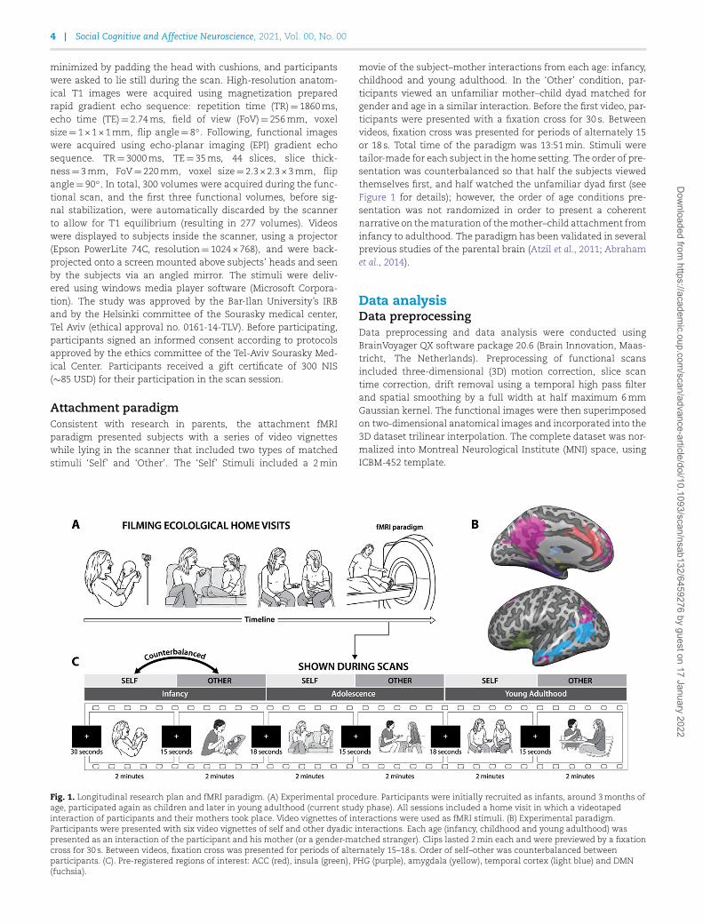
Attachment paradigmConsistent with research in parents, the attachment fMRIparadigm presented subjects with a series of video vignetteswhile lying in the scanner that included two types of matchedstimuli ‘Self’ and ‘Other’. The ‘Self’ Stimuli included a 2min
movie of the subject–mother interactions from each age: infancy,childhood and young adulthood. In the ‘Other’ condition, par-ticipants viewed an unfamiliar mother–child dyad matched forgender and age in a similar interaction. Before the first video, par-ticipants were presented with a fixation cross for 30 s. Betweenvideos, fixation cross was presented for periods of alternately 15or 18 s. Total time of the paradigm was 13:51min. Stimuli weretailor-made for each subject in the home setting. The order of pre-sentation was counterbalanced so that half the subjects viewedthemselves first, and half watched the unfamiliar dyad first (seeFigure 1 for details); however, the order of age conditions pre-sentation was not randomized in order to present a coherentnarrative on thematuration of themother–child attachment frominfancy to adulthood. The paradigm has been validated in severalprevious studies of the parental brain (Atzil et al., 2011; Abrahamet al., 2014).
Data analysisData preprocessingData preprocessing and data analysis were conducted usingBrainVoyager QX software package 20.6 (Brain Innovation, Maas-tricht, The Netherlands). Preprocessing of functional scansincluded three-dimensional (3D) motion correction, slice scantime correction, drift removal using a temporal high pass filterand spatial smoothing by a full width at half maximum 6mmGaussian kernel. The functional images were then superimposedon two-dimensional anatomical images and incorporated into the3D dataset trilinear interpolation. The complete dataset was nor-malized into Montreal Neurological Institute (MNI) space, usingICBM-452 template.
Fig. 1. Longitudinal research plan and fMRI paradigm. (A) Experimental procedure. Participants were initially recruited as infants, around 3months ofage, participated again as children and later in young adulthood (current study phase). All sessions included a home visit in which a videotapedinteraction of participants and their mothers took place. Video vignettes of interactions were used as fMRI stimuli. (B) Experimental paradigm.Participants were presented with six video vignettes of self and other dyadic interactions. Each age (infancy, childhood and young adulthood) waspresented as an interaction of the participant and his mother (or a gender-matched stranger). Clips lasted 2min each and were previewed by a fixationcross for 30 s. Between videos, fixation cross was presented for periods of alternately 15–18 s. Order of self–other was counterbalanced betweenparticipants. (C). Pre-registered regions of interest: ACC (red), insula (green), PHG (purple), amygdala (yellow), temporal cortex (light blue) and DMN(fuchsia).
Dow
nloaded from https://academ
ic.oup.com/scan/advance-article/doi/10.1093/scan/nsab132/6459276 by guest on 17 January 2022

A. Ulmer-Yaniv et al. 5
Whole-brain analysisMulti-subject general linearmodel (GLM)was computedwith ran-dom effects, with separate subject predictors, in which the differ-ent conditions (videos or fixation) were defined as predictors andconvoluted with a standard hemodynamic response predictor.Following, a whole brain, two factors (Attachment (Self/Other)× Age (Infancy/Childhood/Young Adulthood)) repeated measuresanalysis of variance (ANOVA) was performed. Whole-brain mapswere created and voxelwise corrected for false discovery rate(FDR) of q<0.050 (Benjamini and Hochberg, 1995). For visual-ization of results, the group contrasts were overlaid on a MNI-transformed anatomical brain scan of a single participant.
ROI definition and preregistrationBased on cohort 1 and a priori theory-based selection, eightROIs were selected and pre-registered at Open Science Frame-work: hippocampus and parahippocampal gyrus (PHG), amyg-dala, VTA, ACC, insula, a temporal cluster encompassing thesuperior temporal sulcus and gyrus, from the occipito-parietalborder to the temporal pole, and the default mode network (DMN)as a network. Pre-registration was made following the comple-tion of data collection and is available at https://osf.io/2ndxr/?view_only=ba738b07cad249e0b1f08c2f458ddb35.
Cohort 1 included a group of 15 subjects (mean age 18.93 yearsold (s.d.=0.88), 46.7% males, 86.7% right-handed). Fixed effectsmulti-subject GLM activationmapswere used for ROI definition ofthe amygdala and thalamus. The DMNwas defined based on indi-vidual functional connectivity maps with seed in the precuneus,which were superimposed to create a 70% mutual probabilitymap. The temporal cortex region was defined based on the pilotmap combined with STS region from the Glasser atlas (Glasseret al., 2016). The insula, ACC and hippocampus–PHG were alsotaken from the Glasser atlas. VTA was defined by three 5mmspheres based on coordinates from the literature (Murty et al.,2014). Figure 1 shows the ROIs and Supplementary Figure S1shows cohort 1 Self >Other map.
Psycho–physiological interaction analysisClassic pyscho–physiological interaction (PPI) analysis (Fristonet al., 1997) was done using PPI plugin for BrainVoyager (V1.30)to asses PPI predictors and confounds as follows: pre-registeredACC was defined as the seed region, and psychological conditionswere defined as Self >Other, for all timepoints. For each condition,weight was assigned in such a way that the resulting time coursewill be zero centered (self adult+1, self child +1, self infant +1,other adult −1, other child −1, other infant −1). Fixation weightwas set to zero.
For each subject, the time course of the ACC ROI wasextracted, Z-transformed and then convolved with the hemo-dynamic response function. Then, it was multiplied TR by TRwith the task time course (task time course was based on theprotocol associated with the data) to create the ACC PPI pre-dictor. Additionally, for each subject, a psychological regressor,based on the associated protocol, an ACC predictor, based onthe ACC time course correlation, and a complementary regres-sor were created. Additional motion correction predictors wereadded and Z-transformed. The resulting set of four PPI predic-tors for each subject were used in a multi-subject GLM analysis.The PPI ACC predictor allows to create a group map of voxelsthat increase their interaction with the ACC for the self con-ditions compared to other conditions, over and above what is
explained by the task itself (Self >Other contrast; psychologi-cal component) and by the global functional connectivity of theACC (physiological component). Multi-subject GLM analysis wasrestricted to our pre-registered ROIs, using a mask. Then, theACC-PPI maps were corrected using Monte Carlo cluster levelstatistical threshold estimator, with 1000 simulations to estimatecluster-level probabilities (Forman et al., 1995).
Statistical analysisStatistical analysis was conducted using JASP (Version 0.12.1 forwindows, JASP Team, 2020), SPSS (SPSS statistics V25, IBM Corp.)and R version 4.0.0 (R Core Team, 2020) with Tidyverse pack-age (Wickham et al., 2019). Null effects were assessed usingBayesian statistics (Keysers et al., 2020). Greenhouse–Geiser cor-rection was used for sphericity violations. Repeated measuresBayesian ANOVA was used to evaluate the evidence for the nulleffect found using the standard repeated measures (RM) ANOVAanalysis. Of note, throughout the analysis, we used the exclusionBayes factor (BF). As such, a low value for BF signifies support forthe inclusion of the effect (i.e. evidence for the effect): BF <0.33denotes moderate evidence, BF <0.1 denotes strong evidence andBF <0.03 denotes very strong evidence for the inclusion of themodel (Kelter, 2020).
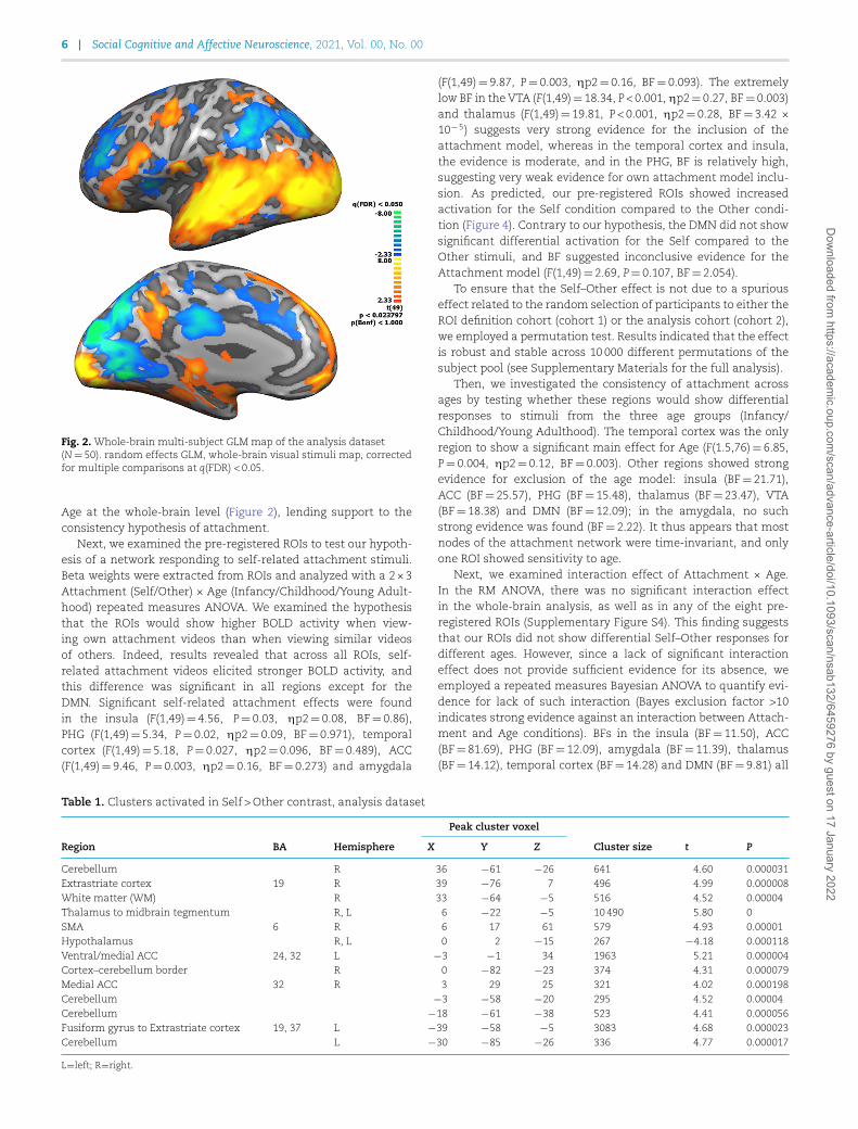
ResultsAs a first step, we examined the overall brain response to attach-ment stimuli of naturalistic mother–child interactions vs thebaseline fixation condition. A whole-brain map comparing theepochs of audio-visual stimulation to fixation of the analysisdataset (50 subjects; Figure 2), is parallel to the map of cohort1 dataset (15 subjects; Supplementary Figure S2A). As expected,both maps show wide activations in the visual cortex, spreadingto the temporal cortex. Additional activations were observed inthe DMN, and in limbic regions such as the PHG, and amygdala.Note that the two maps were highly similar despite differences inthe number of subjects.
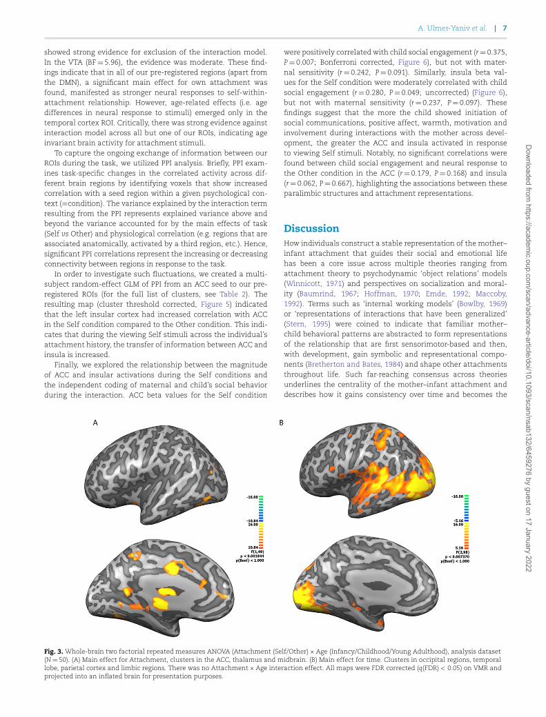
Next, we examined the experimental factors using two-factorial ANOVA analysis of Attachment (Self/Other) × Age(Infancy/Childhood/Young Adulthood) on whole-brain activity(N=50). The ANOVA map of the Attachment main effect(Figure 3A) revealed that the main regions showing differentialBOLD responses between the Self and Other conditions includethe ACC, thalamus and midbrain. Investigation of Self >Othercontrast map indicated stronger activity for the Self conditionin the middle ACC (Brodmann area (BA) 32), posterior ACC (BA24), and a large activation cluster extending from the thalamusventrally to the brain stem. Additional bilateral activations werefound in the visual association regions—peristriate cortex (BA19), supplementary motor area (SMA) (BA 6) and cerebellum. Inaddition, a bilateral deactivation was found in the hypothalamus(Supplementary Figure S2, Table 1).
The main effect of Age was associated with activation acrossoccipito-temporal regions, mainly in the visual associationregions (BA 18) as well as limbic regions (PHG and amygdala)and a parietal cluster (Figure 3B). Random effects GLM mapsof the Age contrasts (infancy> childhood, infancy>young adult-hood, Supplementary Figure S3) show that visual associationregions are activated across all contrasts. There was considerableresemblance between the Adulthood> Infancy and Childhood>Infancy contrast maps, while in the Childhood>Adulthood con-trast map, the activations were weaker and sparse. Critically,there was no significant interaction between Attachment and
Dow
nloaded from https://academ
ic.oup.com/scan/advance-article/doi/10.1093/scan/nsab132/6459276 by guest on 17 January 2022
6 Social Cognitive and Affective Neuroscience, 2021, Vol. 00, No. 00
Fig. 2. Whole-brain multi-subject GLM map of the analysis dataset(N=50). random effects GLM, whole-brain visual stimuli map, correctedfor multiple comparisons at q(FDR) < 0.05.
Age at the whole-brain level (Figure 2), lending support to theconsistency hypothesis of attachment.
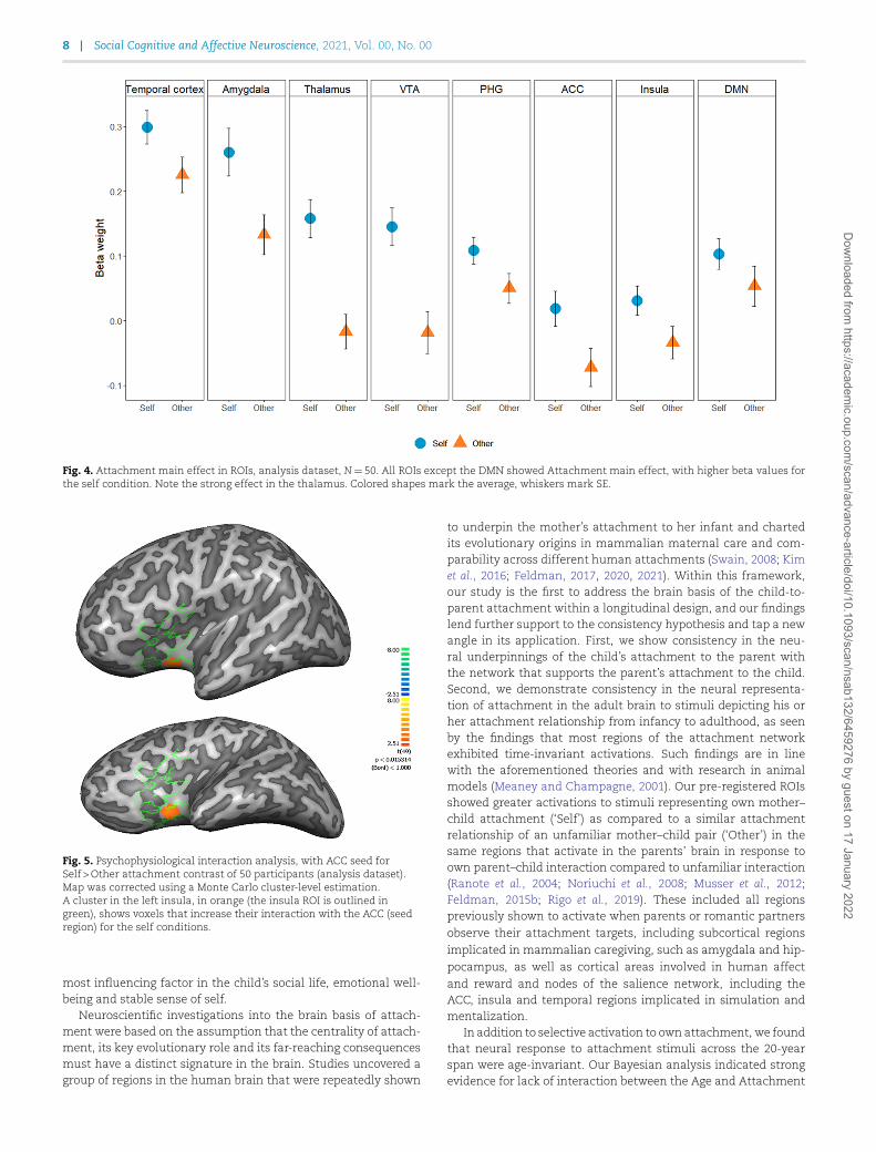
Next, we examined the pre-registered ROIs to test our hypoth-esis of a network responding to self-related attachment stimuli.Beta weights were extracted from ROIs and analyzed with a 2×3Attachment (Self/Other) × Age (Infancy/Childhood/Young Adult-hood) repeated measures ANOVA. We examined the hypothesisthat the ROIs would show higher BOLD activity when view-ing own attachment videos than when viewing similar videosof others. Indeed, results revealed that across all ROIs, self-related attachment videos elicited stronger BOLD activity, andthis difference was significant in all regions except for theDMN. Significant self-related attachment effects were foundin the insula (F(1,49)=4.56, P=0.03, ηp2=0.08, BF=0.86),PHG (F(1,49)=5.34, P=0.02, ηp2=0.09, BF=0.971), temporalcortex (F(1,49)=5.18, P=0.027, ηp2=0.096, BF=0.489), ACC(F(1,49)=9.46, P=0.003, ηp2=0.16, BF=0.273) and amygdala
(F(1,49)=9.87, P=0.003, ηp2=0.16, BF=0.093). The extremelylow BF in the VTA (F(1,49)=18.34, P<0.001, ηp2=0.27, BF=0.003)and thalamus (F(1,49)=19.81, P<0.001, ηp2=0.28, BF=3.42 ×10⁻⁵) suggests very strong evidence for the inclusion of theattachment model, whereas in the temporal cortex and insula,the evidence is moderate, and in the PHG, BF is relatively high,suggesting very weak evidence for own attachment model inclu-sion. As predicted, our pre-registered ROIs showed increasedactivation for the Self condition compared to the Other condi-tion (Figure 4). Contrary to our hypothesis, the DMN did not showsignificant differential activation for the Self compared to theOther stimuli, and BF suggested inconclusive evidence for theAttachment model (F(1,49)=2.69, P=0.107, BF=2.054).
To ensure that the Self–Other effect is not due to a spuriouseffect related to the random selection of participants to either theROI definition cohort (cohort 1) or the analysis cohort (cohort 2),we employed a permutation test. Results indicated that the effectis robust and stable across 10 000 different permutations of thesubject pool (see Supplementary Materials for the full analysis).
Then, we investigated the consistency of attachment acrossages by testing whether these regions would show differentialresponses to stimuli from the three age groups (Infancy/Childhood/Young Adulthood). The temporal cortex was the onlyregion to show a significant main effect for Age (F(1.5,76)=6.85,P=0.004, ηp2=0.12, BF=0.003). Other regions showed strongevidence for exclusion of the age model: insula (BF=21.71),ACC (BF=25.57), PHG (BF=15.48), thalamus (BF=23.47), VTA(BF=18.38) and DMN (BF=12.09); in the amygdala, no suchstrong evidence was found (BF=2.22). It thus appears that mostnodes of the attachment network were time-invariant, and onlyone ROI showed sensitivity to age.
Next, we examined interaction effect of Attachment × Age.In the RM ANOVA, there was no significant interaction effectin the whole-brain analysis, as well as in any of the eight pre-registered ROIs (Supplementary Figure S4). This finding suggeststhat our ROIs did not show differential Self–Other responses fordifferent ages. However, since a lack of significant interactioneffect does not provide sufficient evidence for its absence, weemployed a repeated measures Bayesian ANOVA to quantify evi-dence for lack of such interaction (Bayes exclusion factor >10indicates strong evidence against an interaction between Attach-ment and Age conditions). BFs in the insula (BF=11.50), ACC(BF=81.69), PHG (BF=12.09), amygdala (BF=11.39), thalamus(BF=14.12), temporal cortex (BF=14.28) and DMN (BF=9.81) all
Table 1. Clusters activated in Self >Other contrast, analysis dataset
Peak cluster voxel
Region BA Hemisphere X Y Z Cluster size t P
Cerebellum R 36 −61 −26 641 4.60 0.000031Extrastriate cortex 19 R 39 −76 7 496 4.99 0.000008White matter (WM) R 33 −64 −5 516 4.52 0.00004Thalamus to midbrain tegmentum R, L 6 −22 −5 10490 5.80 0SMA 6 R 6 17 61 579 4.93 0.00001Hypothalamus R, L 0 2 −15 267 −4.18 0.000118Ventral/medial ACC 24, 32 L −3 −1 34 1963 5.21 0.000004Cortex–cerebellum border R 0 −82 −23 374 4.31 0.000079Medial ACC 32 R 3 29 25 321 4.02 0.000198Cerebellum −3 −58 −20 295 4.52 0.00004Cerebellum −18 −61 −38 523 4.41 0.000056Fusiform gyrus to Extrastriate cortex 19, 37 L −39 −58 −5 3083 4.68 0.000023Cerebellum L −30 −85 −26 336 4.77 0.000017
L=left; R=right.
Dow
nloaded from https://academ
ic.oup.com/scan/advance-article/doi/10.1093/scan/nsab132/6459276 by guest on 17 January 2022
A. Ulmer-Yaniv et al. 7
showed strong evidence for exclusion of the interaction model.In the VTA (BF=5.96), the evidence was moderate. These find-ings indicate that in all of our pre-registered regions (apart fromthe DMN), a significant main effect for own attachment wasfound, manifested as stronger neural responses to self-within-attachment relationship. However, age-related effects (i.e. agedifferences in neural response to stimuli) emerged only in thetemporal cortex ROI. Critically, there was strong evidence againstinteraction model across all but one of our ROIs, indicating ageinvariant brain activity for attachment stimuli.
To capture the ongoing exchange of information between ourROIs during the task, we utilized PPI analysis. Briefly, PPI exam-ines task-specific changes in the correlated activity across dif-ferent brain regions by identifying voxels that show increasedcorrelation with a seed region within a given psychological con-text (=condition). The variance explained by the interaction termresulting from the PPI represents explained variance above andbeyond the variance accounted for by the main effects of task(Self vs Other) and physiological correlation (e.g. regions that areassociated anatomically, activated by a third region, etc.). Hence,significant PPI correlations represent the increasing or decreasingconnectivity between regions in response to the task.
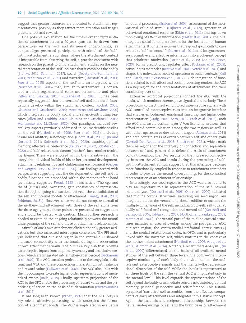
In order to investigate such fluctuations, we created a multi-subject random-effect GLM of PPI from an ACC seed to our pre-registered ROIs (for the full list of clusters, see Table 2). Theresulting map (cluster threshold corrected, Figure 5) indicatedthat the left insular cortex had increased correlation with ACCin the Self condition compared to the Other condition. This indi-cates that during the viewing Self stimuli across the individual’sattachment history, the transfer of information between ACC andinsula is increased.
Finally, we explored the relationship between the magnitudeof ACC and insular activations during the Self conditions andthe independent coding of maternal and child’s social behaviorduring the interaction. ACC beta values for the Self condition
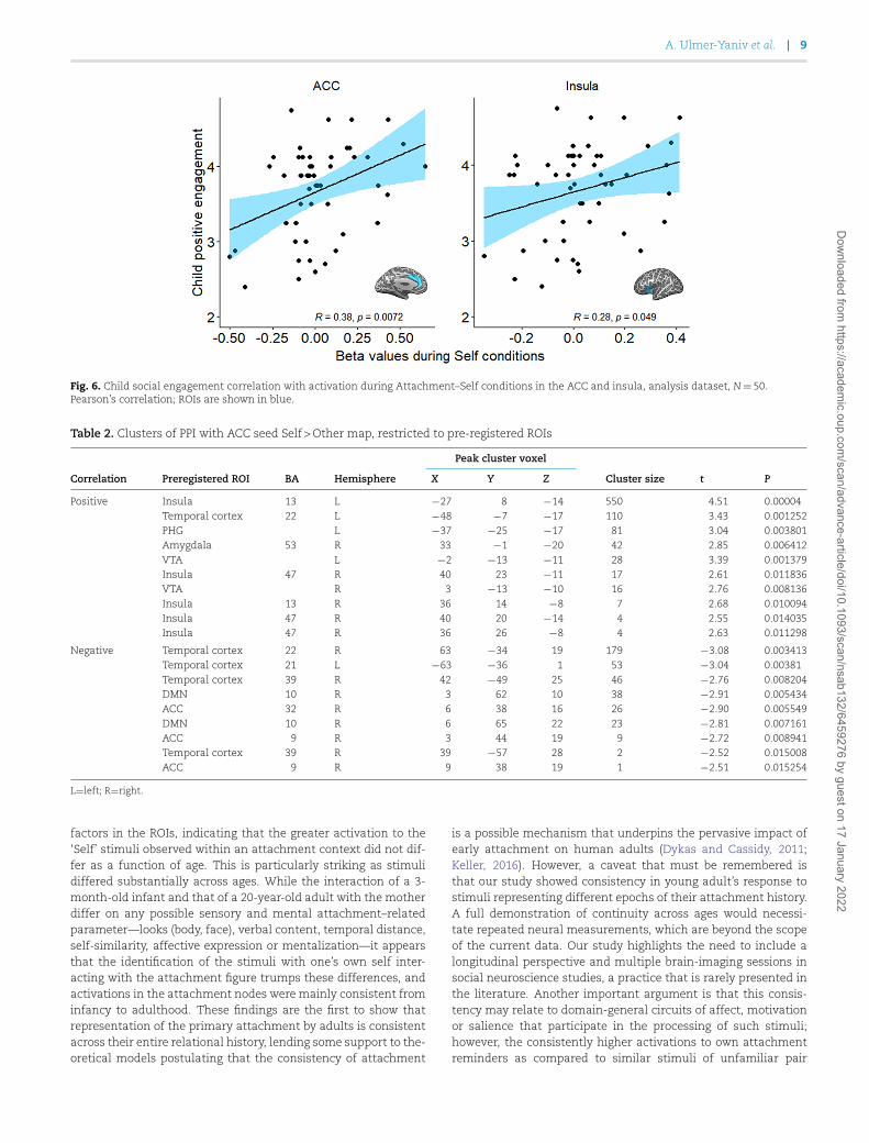
were positively correlated with child social engagement (r=0.375,P=0.007; Bonferroni corrected, Figure 6), but not with mater-nal sensitivity (r=0.242, P=0.091). Similarly, insula beta val-ues for the Self condition were moderately correlated with childsocial engagement (r=0.280, P=0.049; uncorrected) (Figure 6),but not with maternal sensitivity (r=0.237, P=0.097). Thesefindings suggest that the more the child showed initiation ofsocial communications, positive affect, warmth, motivation andinvolvement during interactions with the mother across devel-opment, the greater the ACC and insula activated in responseto viewing Self stimuli. Notably, no significant correlations werefound between child social engagement and neural response tothe Other condition in the ACC (r=0.179, P=0.168) and insula(r=0.062, P=0.667), highlighting the associations between theseparalimbic structures and attachment representations.
DiscussionHow individuals construct a stable representation of the mother–infant attachment that guides their social and emotional lifehas been a core issue across multiple theories ranging fromattachment theory to psychodynamic ‘object relations’ models(Winnicott, 1971) and perspectives on socialization and moral-ity (Baumrind, 1967; Hoffman, 1970; Emde, 1992; Maccoby,1992). Terms such as ‘internal working models’ (Bowlby, 1969)or ‘representations of interactions that have been generalized’(Stern, 1995) were coined to indicate that familiar mother–child behavioral patterns are abstracted to form representationsof the relationship that are first sensorimotor-based and then,with development, gain symbolic and representational compo-nents (Bretherton and Bates, 1984) and shape other attachmentsthroughout life. Such far-reaching consensus across theoriesunderlines the centrality of the mother–infant attachment anddescribes how it gains consistency over time and becomes the
Fig. 3. Whole-brain two factorial repeated measures ANOVA (Attachment (Self/Other) × Age (Infancy/Childhood/Young Adulthood), analysis dataset(N=50). (A) Main effect for Attachment, clusters in the ACC, thalamus and midbrain. (B) Main effect for time. Clusters in occipital regions, temporallobe, parietal cortex and limbic regions. There was no Attachment × Age interaction effect. All maps were FDR corrected (q(FDR) < 0.05) on VMR andprojected into an inflated brain for presentation purposes.
Dow
nloaded from https://academ
ic.oup.com/scan/advance-article/doi/10.1093/scan/nsab132/6459276 by guest on 17 January 2022
8 Social Cognitive and Affective Neuroscience, 2021, Vol. 00, No. 00
Fig. 4. Attachment main effect in ROIs, analysis dataset, N=50. All ROIs except the DMN showed Attachment main effect, with higher beta values forthe self condition. Note the strong effect in the thalamus. Colored shapes mark the average, whiskers mark SE.
Fig. 5. Psychophysiological interaction analysis, with ACC seed forSelf >Other attachment contrast of 50 participants (analysis dataset).Map was corrected using a Monte Carlo cluster-level estimation.A cluster in the left insula, in orange (the insula ROI is outlined ingreen), shows voxels that increase their interaction with the ACC (seedregion) for the self conditions.
most influencing factor in the child’s social life, emotional well-being and stable sense of self.
Neuroscientific investigations into the brain basis of attach-ment were based on the assumption that the centrality of attach-ment, its key evolutionary role and its far-reaching consequencesmust have a distinct signature in the brain. Studies uncovered agroup of regions in the human brain that were repeatedly shown
to underpin the mother’s attachment to her infant and chartedits evolutionary origins in mammalian maternal care and com-parability across different human attachments (Swain, 2008; Kimet al., 2016; Feldman, 2017, 2020, 2021). Within this framework,our study is the first to address the brain basis of the child-to-parent attachment within a longitudinal design, and our findingslend further support to the consistency hypothesis and tap a newangle in its application. First, we show consistency in the neu-ral underpinnings of the child’s attachment to the parent withthe network that supports the parent’s attachment to the child.Second, we demonstrate consistency in the neural representa-tion of attachment in the adult brain to stimuli depicting his orher attachment relationship from infancy to adulthood, as seenby the findings that most regions of the attachment networkexhibited time-invariant activations. Such findings are in linewith the aforementioned theories and with research in animalmodels (Meaney and Champagne, 2001). Our pre-registered ROIsshowed greater activations to stimuli representing own mother–child attachment (‘Self’) as compared to a similar attachmentrelationship of an unfamiliar mother–child pair (‘Other’) in thesame regions that activate in the parents’ brain in response toown parent–child interaction compared to unfamiliar interaction(Ranote et al., 2004; Noriuchi et al., 2008; Musser et al., 2012;Feldman, 2015b; Rigo et al., 2019). These included all regionspreviously shown to activate when parents or romantic partners
observe their attachment targets, including subcortical regions
implicated in mammalian caregiving, such as amygdala and hip-
pocampus, as well as cortical areas involved in human affect
and reward and nodes of the salience network, including theACC, insula and temporal regions implicated in simulation andmentalization.
In addition to selective activation to own attachment, we foundthat neural response to attachment stimuli across the 20-yearspan were age-invariant. Our Bayesian analysis indicated strongevidence for lack of interaction between the Age and Attachment
Dow
nloaded from https://academ
ic.oup.com/scan/advance-article/doi/10.1093/scan/nsab132/6459276 by guest on 17 January 2022
A. Ulmer-Yaniv et al. 9
Fig. 6. Child social engagement correlation with activation during Attachment–Self conditions in the ACC and insula, analysis dataset, N=50.Pearson’s correlation; ROIs are shown in blue.
Table 2. Clusters of PPI with ACC seed Self >Other map, restricted to pre-registered ROIs
Peak cluster voxel
Correlation Preregistered ROI BA Hemisphere X Y Z Cluster size t P
Positive Insula 13 L −27 8 −14 550 4.51 0.00004Temporal cortex 22 L −48 −7 −17 110 3.43 0.001252PHG L −37 −25 −17 81 3.04 0.003801Amygdala 53 R 33 −1 −20 42 2.85 0.006412VTA L −2 −13 −11 28 3.39 0.001379Insula 47 R 40 23 −11 17 2.61 0.011836VTA R 3 −13 −10 16 2.76 0.008136Insula 13 R 36 14 −8 7 2.68 0.010094Insula 47 R 40 20 −14 4 2.55 0.014035Insula 47 R 36 26 −8 4 2.63 0.011298
Negative Temporal cortex 22 R 63 −34 19 179 −3.08 0.003413Temporal cortex 21 L −63 −36 1 53 −3.04 0.00381Temporal cortex 39 R 42 −49 25 46 −2.76 0.008204DMN 10 R 3 62 10 38 −2.91 0.005434ACC 32 R 6 38 16 26 −2.90 0.005549DMN 10 R 6 65 22 23 −2.81 0.007161ACC 9 R 3 44 19 9 −2.72 0.008941Temporal cortex 39 R 39 −57 28 2 −2.52 0.015008ACC 9 R 9 38 19 1 −2.51 0.015254
L=left; R=right.
factors in the ROIs, indicating that the greater activation to the‘Self’ stimuli observed within an attachment context did not dif-fer as a function of age. This is particularly striking as stimulidiffered substantially across ages. While the interaction of a 3-month-old infant and that of a 20-year-old adult with the motherdiffer on any possible sensory and mental attachment–relatedparameter—looks (body, face), verbal content, temporal distance,self-similarity, affective expression or mentalization—it appearsthat the identification of the stimuli with one’s own self inter-acting with the attachment figure trumps these differences, andactivations in the attachment nodes were mainly consistent frominfancy to adulthood. These findings are the first to show thatrepresentation of the primary attachment by adults is consistentacross their entire relational history, lending some support to the-oretical models postulating that the consistency of attachment
is a possible mechanism that underpins the pervasive impact ofearly attachment on human adults (Dykas and Cassidy, 2011;Keller, 2016). However, a caveat that must be remembered isthat our study showed consistency in young adult’s response tostimuli representing different epochs of their attachment history.A full demonstration of continuity across ages would necessi-tate repeated neural measurements, which are beyond the scopeof the current data. Our study highlights the need to include alongitudinal perspective and multiple brain-imaging sessions insocial neuroscience studies, a practice that is rarely presented inthe literature. Another important argument is that this consis-tency may relate to domain-general circuits of affect, motivationor salience that participate in the processing of such stimuli;however, the consistently higher activations to own attachmentreminders as compared to similar stimuli of unfamiliar pair
Dow
nloaded from https://academ
ic.oup.com/scan/advance-article/doi/10.1093/scan/nsab132/6459276 by guest on 17 January 2022
10 Social Cognitive and Affective Neuroscience, 2021, Vol. 00, No. 00
suggest that greater resources are allocated to attachment rep-resentations, possibly as they attract more attention and triggergreater affect and reward.
One possible explanation for the time-invariant representa-tion of attachment across a 20-year span can be drawn fromperspectives on the ‘self’ and its neural underpinnings, asour paradigm presented participants with stimuli of the ‘self–within–attachment relationships’ where the attachment contextis inseparable from observing the self, a practice consistent withresearch on the parent-to-child attachment. Studies on the neu-ral representation of the ‘self’ indicate that it combines the bodily(Blanke, 2012; Salomon, 2017), social (Decety and Sommerville,2003; Yeshurun et al., 2021) and narrative (Christoff et al., 2011;Peer et al., 2015) aspects of the ‘self’ into an integrated whole(Northoff et al., 2006) that, similar to attachment, is consid-ered a stable organizational construct across time and place(Allen and Tsakiris, 2018; Rubianes et al., 2020). It has beenrepeatedly suggested that the sense of self and its neural foun-dations develop within the attachment context (Rochat, 2003;Ciaunica and Crucianelli, 2019; Montirosso and McGlone, 2020)which integrates its bodily, social and salience-attributing fea-tures (Allen and Tsakiris, 2018; Ciaunica and Crucianelli, 2019;Montirosso and McGlone, 2020). Our paradigm, included sev-eral key aspects previously addressed in neuroscientific studieson the self (Northoff et al., 2006; Peer et al., 2015), includingvisual and auditory self-recognition (Uddin et al., 2005; Qin andNorthoff, 2011; Salomon et al., 2012, 2020), autobiographicalmemory, affective self-relevance (Kelley et al., 2002; Schafer et al.,2020) and ‘self-relatedness’ factors (mother’s face, mother’s voice,my home). These were encored within the ‘narrative self’, the‘story’ the individual builds of his or her personal development,attachment relationships and childrearing environment (Gergenand Gergen, 1988; Miller et al., 1990). Our findings accord withperspectives suggesting that the development of the self and itsbodily functions are embedded within the mother–infant bond(as initially suggested Freud, 1923 in his article ‘The ego andthe id (1923)’) and, over time, gain consistency of representa-tion through ongoing transactions between the consolidation ofthe self and internal models of attachment (Fonagy et al., 2007;Feldman, 2015a). However, since we did not compare stimuli ofthe mother–child attachment with those of the self alone fromthe three age groups, these points are presented as suggestionsand should be treated with caution. Much further research isneeded to examine the ongoing relationship between the neuralunderpinnings of the self and those of attachment relationships.
Stimuli of one’s own attachment elicited not only greater acti-vations but also increased inter-region coherence. The PPI anal-ysis indicated that our seed region in the ventral ACC showedincreased connectivity with the insula during the observationof own attachment stimuli. The ACC is a key hub that receivesexteroceptive inputs from all primary sensory and associative cor-tices, which are integrated into a higher-order percept (Beckmannet al., 2009). The ACC contains projections to the amygdala, stria-tum, and VTA and these connections mark stimuli with salienceand reward value (Fujiwara et al., 2009). The ACC also links withthe hippocampus to create higher-order representations of mem-orized events (Rolls, 2019). Finally, upstream projections of theACC to the OFC enable the processing of reward value and the pri-oritizing of action on the basis of such valuation (Burgos-Robleset al., 2019).
It has long been known (Papez, 1937) that the ACC plays akey role in affective processing, which underpins the forma-tion of attachment bonds. The ACC is implicated in evaluative
emotional processing (Esslen et al., 2004), assessment of the moti-vational value of stimuli (Fujiwara et al., 2009), generation ofbehavioral emotional response (Etkin et al., 2011) and top–downmonitoring of affective information (Carter et al., 2001). The ACCintegrates social functions relevant for the formation of humanattachments. It contains neurons that respond specifically to cuesrelated to ‘self’ vs ‘nonself’ (Sturm et al., 2013) and integrates sen-sory, cognitive and affective information into a coherent perceptthat prioritizes motivation (Porter et al., 2019; Lee and Reeve,2020), forms predictions, regulates affect (Ochsner et al., 2009),consolidates memories (Restivo et al., 2009; Vetere et al., 2011) andshapes the individual’s mode of operation in social contexts (Krilland Platek, 2009; Vassena et al., 2017). Such integration of func-tions related to self, affect and social processing renders the ACCas a key region for the representations of attachment and theirconsistency over time.
Extensive reciprocal projections connect the ACC with theinsula, whichmonitors interoceptive signals from the body. Theseprojections connect insula-monitored interoceptive signals withACC-controlled exteroceptive cues into an integrative interfacethat enables embodiment, emotional mirroring, and higher-orderrepresentation (Craig, 2009; Seth, 2013; Park et al., 2018). Boththe ACC and insula contain layer V Von Economo neurons thatafford rapid communication among the two regions as well aswith other upstream or downstream targets (Allman et al., 2011)and both contain areas of overlap between self and others’ pain(Corradi-Dell’Acqua et al., 2016; Smith et al., 2021), which markthem as regions for the interplay of connection and separationbetween self and partner that define the basis of attachmentbonds throughout life. Our results on the increased connectiv-ity between the ACC and insula during the processing of self–within–attachment stimuli suggest that this interface becomesmore functionally coupled in response to attachment remindersin order to provide the neural underpinnings for the consistentrepresentation of attachment relationships.
Interestingly, our seed region in the ACC was also found toplay an important role in representation of the self. Severalmeta-analyses (Northoff et al., 2006; Qin et al., 2020) indicatedthat midline cortical structures, including our seed region, areintegrated across the ventral and dorsal midline to sustain themultiple dimensions of the self, including proto-self, self-‘qualia’,bodily self, facial self-recognition and mental self (Northoff andBermpohl, 2004; Uddin et al., 2007; Northoff and Panksepp, 2008;Moran et al., 2009). The ventral part of the midline cortical struc-tures includes an area of overlap among the post-genual ACC,our seed region, the ventro-medial prefrontal cortex (vmPFC)and the medial orbitofrontal cortex (mOFC), and is particularlylinked with the narrative self, which matures in the context ofthe mother–infant attachment (Northoff et al., 2006; Araujo et al.,2013; Salomon et al., 2014). Notably, a recent meta-analysis (Qinet al., 2020) differentiated on the basis of all available neuralstudies of the self between three levels: the bodily—the intero-ceptive monitoring of one’s body, the environmental—the self-relevant exteroceptive signals and the mental—the representa-tional dimension of the self. While the insula is represented atall three levels of the self, the ventral ACC is implicated only inthe mental level. This level expands the representations of theself beyond the bodily or immediate sensory into autobiographicalmemory, personal perspective and self-reference. This autobi-ographical ‘narrative’ self assembles from the affective compo-nents of early attachments and integrates into a stable concept.Again, the parallels and reciprocal relationships between theneural underpinnings of self and the brain basis of attachment
Dow
nloaded from https://academ
ic.oup.com/scan/advance-article/doi/10.1093/scan/nsab132/6459276 by guest on 17 January 2022
A. Ulmer-Yaniv et al. 11
require much further research, and we present these thoughtsonly as directions for future research, as our study did not sep-arate the neural response to the self from the neural responseto attachment reminders and the associations between self andattachment could not be empirically tested.
Connectivity between the ACC and AI, which subserves keyfunctions such as interoception and affective processing (Craig,2009; Seth, 2013), appears to play an important role in theparent-to-child attachment. Connectivity of the ACC and AI in theparental brain has been associated with parent–infant synchronyin infancy and predicted the child’s emotion regulation and corti-sol reactivity in preschool and lower behavior problems at 7 years(Abraham et al., 2016b; Abraham and Feldman, 2018). The cou-pling between the ACC and insula in response to attachmentstimuli shown in our study anchors the representation of themother–child attachment in the bodily and non-verbal sensory,as seen by the insular involvement, yet integrates this interocep-tive level into adult representation, as seen by our seed region inthe ACC (Morita et al., 2014). Insular activations in the maternalbrain are thought to provide external-regulatory function for theinfant’s emerging ability to recognize his/her own bodily signalsand, over time, develop interoceptive representations (Atzil et al.,2018). Furthermore, as part of the sociotemporal brain (Schirmeret al., 2016), the insula monitors the duration and patterns ofsocial events, including the early patterns of mother–infant non-verbal synchronous interactions that later expand into symbolicand verbal exchanges that are individually stable and provide thebackground for consistent attachment relationships from infancyto adulthood (Ulmer Yaniv et al., 2021).
It is interesting to note that the same brain regions sustain-ing human attachment overlap with the so-called ‘interoceptivenetwork’, particularly, as recent models on interoception suggestthat viewing the self often triggers activation in this ‘interocep-tive network’ (Chen et al., 2021). For adults, viewing the selfwhile interacting with the mother as an infant may be an espe-cially strong reminder of caregiving and bodily contact. It hasbeen suggested (Chen et al., 2021) that interoceptive informa-tion is first processed in the brainstem nucleus of the solitarytract and then projects to the thalamus, from where it is relayedto higher targets: the amygdala, insula and ACC. As seen inour data (Figure 2), the same nodes of the interoceptive net-work were found here to differentiate Self from Other’s attach-ment: thalamus-to-brainstem, amygdala, hippocampus, insulaand ACC. This suggests some overlap between the attachmentnetwork and the interoception network and raises the possibil-ity that the primary attachment and the experience of caregivingprovide the earliest context for the infant’s capacity to first sense,then identify and finally form representations of signals from thebody. Further study on the overlap in the brain basis of intero-ception and attachment may be a fruitful avenue of research andmay shed further light on this important topic.
Finally, the magnitude of ACC and insular activations corre-lated with the degree of child social engagement, which definesthe degree of positive involvement, motivation for social con-nection and initiation of social communication the child exhibitsduring interactions with the mother across the 20-year span.Child social engagement is an important feature of the mother–child interaction that has received significantly less research ascompared with maternal sensitivity, albeit its role in shapingsocial-emotional competencies and the social brain appears tobe just as critical. Child social engagement is an individuallystable disposition that develops on the basis of both maternal
sensitivity and the child’s temperamental sociality (Feldman et al.,2010; Feldman, 2021). No study, to our knowledge, tracked theexpression of children’s social engagement during interactionwith the mother across two decades, and thus, our findings thatshow stability in this orientation from infancy to adulthood arenovel and important and likely represent the longest timespanfor which such stability in children’s behavioral social orientationis reported. This suggests that features of the infant’s behaviorwithin the first social relationship may persist throughout life,and thus, high-risk conditions associated with dampened childsocial engagement, such as maternal postpartum depression orhigh contextual risk, should receive intervention that help moth-ers increase infant social engagement already in early infancy.Notably, activation of the ACC and insula was not significantlyrelated to maternal sensitivity, the key variable in attachmentresearch that often views attachment from the mother’s perspec-tive (van Ijzendoorn et al., 1995), but with the child’s own behavioras an infant, child and adult. Interestingly, while maternal sen-sitivity has been repeatedly shown to serve as a buffer againstharsh rearing conditions, beginning in late childhood the child’ssocial engagement charts a unique pathway to resilience, inde-pendent of the maternal path (Halevi et al., 2017), suggesting thatchildren’s own social behavior becomes more important begin-ning in late childhood. Our findings indicate that in adulthood, theneural representation of the mother–child attachment, particu-larly the paralimbic interface that becomes functionally coupledin response to attachment cues, shows closer associations withmarkers of the ‘self’ and its contribution to the relationship,including the degree of engagement, valuation andmotivation forthe interaction, than with the mother’s behavior. These resultshighlight, again, the ongoing relationship between representa-tions of the ‘self’ and internalization of the primary attachmentas they mutually evolve across development and cohere into atime-invariant representation.
Several study limitations should be considered. First, we didnot measure mothers’ neural response to the same stimuli, andour ROIs were based on prior research with mothers using thesame naturalistic paradigm. Only a design that includes bothmother and child can definitively demonstrate that own attach-ment stimuli trigger similar activations in both mother and child.Second, similar to all neural studies of the self, it is possiblethat self-related stimuli are allotted more attentional resourcesthan non-self-relevant stimuli. However, this attentional accountwould also suggest that the novel attachment stimuli from theearlier ages would probably elicit greater attention; still our datashow no difference between ages, suggesting that these find-ings do not stem from differential attentional engagement. Third,stimuli presentation order was counterbalanced for ‘attachment’(Self vs Other), but not for ‘Age’ presentation order. This stemmedfrom our desire to present a coherent narrative account ofthe mother–child relationship from infancy to adulthood anddescribe the unfolding of the attachment relationship across theindividual’s developmental history. Still, the lack of counterbal-ance in age is a study limitation and should be taken into accountwhen interpreting the Age effect. Additionally, as in all ecologi-cal studies, our stimuli varied on numerous visual and auditoryproperties. Despite these limitations, we found consistent activa-tion across our pre-registered ROI, which speak to the robustnessof the effect above and beyond the specific stimuli. Much furtherresearch is needed to characterize the development of the neu-ral basis of attachment across ages and relationships, understandits impact on the consolidation of the self and representation of
Dow
nloaded from https://academ
ic.oup.com/scan/advance-article/doi/10.1093/scan/nsab132/6459276 by guest on 17 January 2022

12 Social Cognitive and Affective Neuroscience, 2021, Vol. 00, No. 00
the bodily milieu, and tease apart the impact of culture, con-text, habit and risk conditions on the maturation of the neuralrepresentation of attachment bonds throughout life.
AcknowledgementsWe would like to thank the mothers and children for their partic-ipation and cooperation.
FundingThe study was supported by the Simms/Mann Chair to RuthFeldman.
Conflict of interestThe authors declare no competing financial interests.
Supplementary dataSupplementary data is available at SCAN online.
ReferencesAbraham, E., Hendler, T., Shapira-Lichter, I., et al. (2014). Father’s
brain is sensitive to childcare experiences. Proceedings of theNational Academy of Sciences, 111, 9792–7.
Abraham, E., Hendler, T., Zagoory-Sharon, O., et al. (2016a). Networkintegrity of the parental brain in infancy supports the devel-opment of children’s social competencies. Social Cognitive andAffective Neuroscience, 11, 1707–18.
Abraham, E., Hendler, T., Zagoory-Sharon, O., et al. (2016b). Networkintegrity of the parental brain in infancy supports the devel-opment of children’s social competencies. Social Cognitive andAffective Neuroscience, 11, 1707–18.
Abraham, E., Gilam, G., Kanat-Maymon, Y., et al. (2017). The humancoparental bond implicates distinct corticostriatal pathways:longitudinal impact on family formation and child well-being.Neuropsychopharmacology, 42, 2301–13.
Abraham, E., Feldman, R. (2018). The neurobiology of human allo-maternal care; implications for fathering, coparenting, and chil-dren’s social development. Physiology and Behavior, 193, 25–34.
Acevedo, B.P., Aron, A., Fisher, H.E., et al. (2012). Neural correlatesof long-term intense romantic love. Social Cognitive and AffectiveNeuroscience, 7, 145–59.
Ainsworth, M.D., Blehar, M.C., Waters, E., Wall, S. (1978). Patterns ofAttachment: A Psychological Study of the Strange Situation. Hillsdale,NJ: Erlbaum.
Allen, M., Tsakiris, M. (2018). The body as first prior: interoceptivepredictive processing and the primacy of self-models.
Allman, J.M., Tetreault, N.A., Hakeem, A.Y., et al. (2011). The vonEconomo neurons in the frontoinsular and anterior cingulatecortex. Annals of the New York Academy of Sciences, 1225, 59–71.
Araujo, H.F., Kaplan, J., Damasio, A. (2013). Cortical midlinestructures and autobiographical-self processes: an activation-likelihood estimation meta-analysis. Frontiers in Human Neuro-science, 7, 548.
Atzil, S., Hendler, T., Feldman, R. (2011). Specifying the neurobiolog-ical basis of human attachment: brain, hormones, and behaviorin synchronous and intrusive mothers. Neuropsychopharmacology:Official Publication of the American College of Neuropsychopharmacol-ogy, 36, 2603–15.
Atzil, S., Hendler, T., Feldman, R. (2014). The brain basis of socialsynchrony. Social Cognitive and Affective Neuroscience, 9, 1193–202.
Atzil, S., Gao, W., Fradkin, I., et al. (2018). Growing a social brain.Nature Human Behaviour, 2, 624–36.
Bartels, A., Zeki, S. (2004). The neural correlates of maternal andromantic love. NeuroImage, 21, 1155–66.
Baumrind, D. (1967). Child care practices anteceding three patternsof preschool behavior. Genetic Psychology Monographs, 75, 43–88.
Beauregard, M., Levesque, J., Bourgouin, P. (2001). Neural correlatesof conscious self-regulation of emotion. Journal of Neuroscience,21(18), RC165.
Beckmann, M., Johansen-Berg, H., Rushworth, M.F.S. (2009).Connectivity-based parcellation of human cingulate cortex andits relation to functional specialization. Journal of Neuroscience, 29,1175–90.
Belsky, J., Fearon, P. (2002). Early attachment security, subsequentmaternal sensitivity, and later child development: does con-tinuity in development depend upon continuity of caregiving?Attachment and Human Development, 4, 361–87.
Benjamini, Y., Hochberg, Y. (1995). Controlling the false discov-ery rate: a practical and powerful approach to multiple testing.Journal of the Royal Statistical Society. Series B (Methodological), 57,289–300.
Blanke, O. (2012). Multisensory brain mechanisms of bodily self-consciousness. Nature Reviews Neuroscience, 13, 556–71.
Boccia, M.L., Petrusz, P., Suzuki, K., et al. (2013). Immunohis-tochemical localization of oxytocin receptors in human brain.Neuroscience, 253, 155–64.
Bowlby, J. (1969). Attachment and loss, Volume 1: Attachment.Bretherton, I. (2010). Fathers in attachment theory and research: a
review. Early Child Development and Care, 180, 9–23.Bretherton, I., Bates, E. (1984). The development of representa-
tion from 10 to 28 months. In: Continuities and Discontinuities inDevelopment, Boston, MA: Springer. pp. 229–61.
Buchheim, A., Erk, S., George, C., et al. (2006). Measuring attach-ment representation in an fMRI environment: a pilot study.Psychopathology, 39, 144–52.
Burgos-Robles, A., Gothard, K.M., Monfils, M.H., et al. (2019). Con-served features of anterior cingulate networks support obser-vational learning across species. Neuroscience and BiobehavioralReviews, 107, 215–28.
Bush, G., Luu, P., Posner, M.I. (2000). Cognitive and emotional influ-ences in anterior cingulate cortex. Trends in Cognitive Sciences, 4,215–22.
Carter, C.S., MacDonald, A.W., Ross, L.L., et al. (2001). Anteriorcingulate cortex activity and impaired self-monitoring of per-formance in patients with schizophrenia: an event-related fMRIstudy. American Journal of Psychiatry, 158, 1423–8.
Chae, Y., Goodman, M., Goodman, G.S., et al. (2018). How childrenremember the strange situation: the role of attachment. Journalof Experimental Child Psychology, 166, 360–79.
Chen, W.G., Schloesser, D., Arensdorf, A.M., et al. (2021). The emerg-ing science of interoception: sensing, integrating, interpreting,and regulating signals within the self. Trends in Neurosciences, 44,3–16.
Christoff, K., Cosmelli, D., Legrand, D., et al. (2011). Specifying the selffor cognitive neuroscience. Trends in Cognitive Sciences, 15, 104–12.
Ciaunica, A., Crucianelli, L. (2019). Minimal self-awareness fromwithin: a developmental perspective. Journal of Consciousness Stud-ies, 26, 207–26.
Corradi-Dell’Acqua, C., Tusche, A., Vuilleumier, P., et al. (2016).Cross-modal representations of first-hand and vicarious pain,disgust and fairness in insular and cingulate cortex. Nature Com-munications, 7, 1–12.
Dow
nloaded from https://academ
ic.oup.com/scan/advance-article/doi/10.1093/scan/nsab132/6459276 by guest on 17 January 2022
A. Ulmer-Yaniv et al. 13
Craig, A.D. (2003). Interoception: the sense of the physiologicalcondition of the body. Current Opinion in Neurobiology, 13, 500–5.
Craig, A.D. (2009). How do you feel - now? The anterior insula andhuman awareness. Nature Reviews Neuroscience, 10, 59–70.
Decety, J., Sommerville, J.A. (2003). Shared representations betweenself and other: a social cognitive neuroscience view. Trends inCognitive Sciences, 7, 527–33.
Dykas, M.J., Cassidy, J. (2011). Attachment and the processing ofsocial information across the life span: theory and evidence.Psychological Bulletin, 137, 19–46.
Elmadih, A., Wan, M.W., Downey, D., et al. (2016). Natural variationinmaternal sensitivity is reflected inmaternal brain responses toinfant stimuli. Behavioral Neuroscience, 130, 500–10.
Emde, R.N. (1992). Social referencing research. In: Social Referencingand the Social Construction of Reality in Infancy. US: Springer, 79–94.
Eslinger, P.J., Anders, S., Ballarini, T., et al. (2021). The neuroscienceof social feelings: mechanisms of adaptive social functioning.Neuroscience and Biobehavioral Reviews, 128, 592–620.
Esslen, M., Pascual-Marqui, R.D., Hell, D., et al. (2004). Brain areas andtime course of emotional processing. NeuroImage, 21, 1189–203.
Etkin, A., Egner, T., Kalisch, R. (2011). Emotional processing in ante-rior cingulate and medial prefrontal cortex. Trends in CognitiveSciences, 15, 85–93.
Fan, Y., Duncan, N.W., de Greck, M., et al. (2011). Is there a core neuralnetwork in empathy? An fMRI based quantitative meta-analysis.Neuroscience and Biobehavioral Reviews, 35, 903–11.
Feldman, R. (1998). Coding Interactive Behavior. Ramat Gan BarIlanUniversity Israel, 1–54.
Feldman, R. (2010). The relational basis of adolescent adjustment:trajectories of mother-child interactive behaviors from infancyto adolescence shape adolescents’ adaptation. Attachment andHuman Development, 12, 173–92.
Feldman, R., Gordon, I., Zagoory-Sharon, O. (2010). The cross-generation transmission of oxytocin in humans. Hormones andBehavior, 58, 669–76.
Feldman, R. (2012). Parent-infant synchrony: a biobehavioral modelof mutual influences in the formation of affiliative bonds. Mono-graphs of the Society for Research in Child Development, 77, 42–51.
Feldman, R., Bamberger, E., Kanat-Maymon, Y. (2013). Parent-specific reciprocity from infancy to adolescence shapes children’ssocial competence and dialogical skills. Attachment and HumanDevelopment, 15, 407–23.
Feldman, R., Golan, O., Hirschler-Guttenberg, Y., et al. (2014). Parent-child interaction and oxytocin production in pre-schoolers withautism spectrum disorder. British Journal of Psychiatry, 205,107–12.
Feldman, R. (2015a). Mutual influences between child emotion regu-lation and parent-child reciprocity support development acrossthe first 10 years of life: implications for developmental psy-chopathology. Development and Psychopathology, 27, 1007–23.
Feldman, R. (2015b). Sensitive periods in human social development:new insights from research on oxytocin, synchrony, and high-riskparenting. Development and Psychopathology, 27, 369–95.
Feldman, R. (2016). The neurobiology of mammalian parenting andthe biosocial context of human caregiving.Hormones and Behavior,77, 3–17.
Feldman, R. (2017). The neurobiology of human attachments. Trendsin Cognitive Sciences, 21, 80–99.
Feldman, R. (2020). What is resilience: an affiliative neuroscienceapproach. World Psychiatry, 19, 132–50.
Feldman, R. (2021). Social behavior as a transdiagnostic marker ofresilience. Annual Review of Clinical Psychology, 17, 153–80.
Fonagy, P., Steele, H., Steele, M. (1991). Maternal representations ofattachment during pregnancy predict the organization of infant-mother attachment at one year of age. Child Development, 62,891–905.
Fonagy, P., Gergely, G., Target, M. (2007). The parent-infant dyad andthe construction of the subjective self. Journal of Child Psychologyand Psychiatry and Allied Disciplines, 48, 288–328.
Forman, S.D., Cohen, J.D., Fitzgerald, M., et al. (1995). Improvedassessment of significant activation in functional magnetic res-onance imaging (fMRI): use of a cluster-size threshold. MagneticResonance in Medicine, 33, 636–47.
Fraley, R.C. (2002). Attachment stability from infancy to adulthood:meta-analysis and dynamic modeling of developmental mecha-nisms. Personality and Social Psychology Review, 6, 123–51.
Freeman, H., Brown, B.B. (2001). Primary attachment to parentsand peers during adolescence: differences by attachment style.Journal of Youth and Adolescence, 30, 653–74.
Freud, S. (1923). The ego and the id (J. Strachey, Trans.). The Stan-dard Edition of the Complete Psychological Works of Sigmund Freud, 19,12–66.
Friston, K.J., Buechel, C., Fink, G.R., et al. (1997). Psychophysiologi-cal and modulatory interactions in neuroimaging. NeuroImage, 6,218–29.
Frith, U., Frith, C.D., Frith, C.D., Wolpert, D.M. (2003). Developmentand neurophysiology of mentalizing. Philosophical Transactions ofthe Royal Society B: Biological Sciences, 358, 459–73.
Fujiwara, J., Tobler, P.N., Taira, M., et al. (2009). Segregated and inte-grated coding of reward and punishment in the cingulate cortex.Journal of Neurophysiology, 101, 3284–93.
Gergen, K.J., Gergen, M.M. (1988). Narrative and the self as relation-ship. Advances in Experimental Social Psychology, 21, 17–56.
Glasser, M.F., Coalson, T.S., Robinson, E.C., et al. (2016). A multi-modal parcellation of human cerebral cortex. Nature, 536, 171–8.
Gobel, A., Barkmann, C., Arck, P., et al. (2019). Couples’ prenatalbonding to the fetus and the association with one’s own and part-ner’s emotional well-being and adult romantic attachment style.Midwifery, 79, 102549.
Goossens, F.A., Van Ijzendoorn, M.H., Tavecchio, L.W.C., et al. (1986).Stability of attachment across time and context in a dutch sam-ple. Psychological Reports, 58, 23–32.
Halevi, G., Djalovski, A., Kanat-Maymon, Y., et al. (2017). The socialtransmission of risk: maternal stress physiology, synchronousparenting, and well-being mediate the effects of war exposureon child psychopathology. Journal of Abnormal Psychology, 126,1087–103.
Hamilton, C.E. (2000). Continuity and discontinuity of attachmentfrom infancy through adolescence. Child Development, 71, 690–4.
Hoffman, M.L. (1970). Conscience, personality, and socializationtechniques. Human Development, 13, 90–126.
Insel, T.R., Harbaugh, C.R. (1989). Lesions of the hypothalamic par-aventricular nucleus disrupt the initiation of maternal behavior.Physiology and Behavior, 45, 1033–41.
JASP Team (2020). JASP (Version 0.12) [Computer software].Keller, H. (2016). Attachment. A pancultural need but a cultural
construct. Current Opinion in Psychology, 8, 59–63.Kelley, A.W.M., Macrae, C.N., Wyland, C.L., et al. (2002). Finding the
self? An event-related fMRI study. Journal of Cognitive Neuroscience,14, 785–94.
Kelter, R. (2020). Bayesian alternatives to null hypothesis significancetesting in biomedical research: a non-technical introduction toBayesian inference with JASP. BMC Medical Research Methodology,20, 1–13.
Dow
nloaded from https://academ
ic.oup.com/scan/advance-article/doi/10.1093/scan/nsab132/6459276 by guest on 17 January 2022

14 Social Cognitive and Affective Neuroscience, 2021, Vol. 00, No. 00
Keysers, C., Gazzola, V., Wagenmakers, E.J. (2020). Using Bayes fac-tor hypothesis testing in neuroscience to establish evidence ofabsence. Nature Neuroscience, 23, 788–99.
Kim, P., Feldman, R., Mayes, L.C., et al. (2011). Breastfeeding, brainactivation to own infant cry, and maternal sensitivity. Journal ofChild Psychology and Psychiatry and Allied Disciplines, 52, 907–15.
Kim, P., Strathearn, L., Swain, J.E. (2016). The maternal brain and itsplasticity in humans. Hormones and Behavior, 77, 113–23.
Krill, A., Platek, S.M. (2009). In-group and out-group membershipmediates anterior cingulate activation to social exclusion. Fron-tiers in Evolutionary Neuroscience, 1, 1.
Lamm, C., Rutgen, M., Wagner, I.C. (2019). Imaging empathy andprosocial emotions. Neuroscience Letters, 693, 49–53.
Laurita, A.C., Hazan, C., Spreng, R.N. (2017). Dissociable patternsof brain activity for mentalizing about known others: a rolefor attachment. Social Cognitive and Affective Neuroscience, 12,1072–82.
Lee, W., Reeve, J. (2020). Remembering pleasure and personalmeaning from episodes of intrinsic motivation: an fMRI study.Motivation and Emotion, 44, 810–8.
Leibenluft, E., Gobbini, M.I., Harrison, T., et al. (2004). Mothers’ neu-ral activation in response to pictures of their children and otherchildren. Biological Psychiatry, 56, 225–32.
Lewis, M., Feiring, C., Rosenthal, S. (2000). Attachment over time.Child Development, 71, 707–20.
Lorenz, K. (1935). Der Kumpan in der Umwelt des Vogels. Journal furOrnithologie, 83, 289–413.
Maccoby, E.E. (1992). The role of parents in the socialization ofchildren: an historical overview. Developmental Psychology, 28(6),1006.
Mar, R.A. (2011). The neural bases of social cognition and storycomprehension. Annual Review of Psychology, 62, 103–34.
Margulies, D.S., Kelly, A.M.C., Uddin, L.Q., et al. (2007). Mapping thefunctional connectivity of anterior cingulate cortex. NeuroImage,37, 579–88.
Meaney, M.J., Champagne, F.A. (2001). Like mother, like daugh-ter: evidence for non-genomic transmission of parental behav-ior and stress responsivity. Progress in Brain Research, 133,287–302.
Meins, E., Fernyhough, C., Fradley, E., et al. (2001). Rethinking mater-nal sensitivity: mothers’ comments on infants’ mental processespredict security of attachment at 12 months. Journal of ChildPsychology and Psychiatry, 42, 637–48.
Mikulincer, M., Shaver, P.R. (2005). Attachment theory and emo-tions in close relationships: exploring the attachment-relateddynamics of emotional reactions to relational events. PersonalRelationships, 12, 149–68.
Mikulincer, M., Shaver, P.R. (2012). An attachment perspective onpsychopathology. World Psychiatry, 11, 11–5.
Miller, P.J., Potts, R., Fung, H., et al. (1990). Narrative practices and thesocial construction of self in childhood. American Ethnologist, 17,292–311.
Montirosso, R., McGlone, F. (2020). The body comes first. Embodiedreparation and the co-creation of infant bodily-self. Neuroscienceand Biobehavioral Reviews, 113, 77–87.
Moran, J.M., Heatherton, T.F., Kelley, W.M. (2009). Modulation ofcortical midline structures by implicit and explicit self-relevanceevaluation. Social Neuroscience, 4, 197–211.
Morita, T., Tanabe, H.C., Sasaki, A.T., et al. (2014). The anterior insu-lar and anterior cingulate cortices in emotional processing forself-face recognition. Social Cognitive and Affective Neuroscience, 9,570–9.
Murty, V.P., Shermohammed, M., Smith, D.V., et al. (2014). Restingstate networks distinguish human ventral tegmental area fromsubstantia nigra. NeuroImage, 100, 580–9.
Musser, E.D., Kaiser-Laurent, H., Ablow, J.C. (2012). The neuralcorrelates of maternal sensitivity: an fMRI study. DevelopmentalCognitive Neuroscience, 2, 428–36.
Noriuchi, M., Kikuchi, Y., Senoo, A. (2008). The functional neu-roanatomy of maternal love: mother’s response to infant’sattachment behaviors. Biological Psychiatry, 63, 415–23.
Northoff, G., Heinzel, A., de Greck, M., et al. (2006). Self-referentialprocessing in our brain: a meta-analysis of imaging studies onthe self. NeuroImage, 31, 440–57.
Northoff, G., Bermpohl, F. (2004). Cortical midline structures and theself. Trends in Cognitive Sciences, 8, 102–7.
Northoff, G., Panksepp, J. (2008). The trans-species concept of selfand the subcortical-cortical midline system. Trends in CognitiveSciences, 12, 259–64.
Numan, M. (2020). The Parental Brain: Mechanisms, Development, andEvolution. Oxford University Press.
Ochsner, K.N., Hughes, B., Robertson, E.R., et al. (2009). Neural sys-tems supporting the control of affective and cognitive conflicts.Journal of Cognitive Neuroscience, 21, 1842–55.
Oxley, G., Fleming, A.S. (2000). The effects of medial preoptic areaand amygdala lesions on maternal behavior in the juvenile rat.Developmental Psychobiology, 37, 253–65.
Papez, J.W. (1937). A proposed mechanism of emotion. Archives ofNeurology and Psychiatry, 38, 725–43.
Park, H.D., Bernasconi, F., Salomon, R., et al. (2018). Neural sourcesand underlying mechanisms of neural responses to heartbeats,and their role in bodily self-consciousness: an intracranial EEGstudy. Cerebral Cortex (New York, N.Y.: 1991), 28, 2351–64.
Pascuzzo, K., Cyr, C., Moss, E. (2013). Longitudinal associationbetween adolescent attachment, adult romantic attachment,and emotion regulation strategies. Attachment and Human Devel-opment, 15, 83–103.
Pavlvlovic, D.M., Pavlvlovic, A.M., Lackovic, M. (2009). The anteriorcingulate cortex. Archives of Biological Sciences, 61, 659–73.
Peer, M., Salomon, R., Goldberg, I., et al. (2015). Brain system formental orientation in space, time, and person. Proceedings of theNational Academy of Sciences of the United States of America, 112,11072–7.
Peterson, B.S., Skudlarski, P., Gatenby, J.C., et al. (1999). An fMRIstudy of stroop word-color interference: evidence for cingulatesubregions subserving multiple distributed attentional systems.Biological Psychiatry, 45, 1237–58.
Porter, B.S., Hillman, K.L., Bilkey, D.K. (2019). Anterior cingulate cor-tex encoding of effortful behavior. Journal of Neurophysiology, 121,701–14.
Pratt, M., Goldstein, A., Feldman, R. (2018). Child brain exhibits amulti-rhythmic response to attachment cues. Social Cognitive andAffective Neuroscience, 13, 957–66.
Pratt, M., Zeev-Wolf, M., Goldstein, A., et al. (2019). Exposureto early and persistent maternal depression impairs the neu-ral basis of attachment in preadolescence. Progress in Neuro-Psychopharmacology and Biological Psychiatry, 93, 21–30.
Qin, P., Wang, M., Northoff, G. (2020). Linking bodily, environmen-tal and mental states in the self—a three-level model based on ameta-analysis. Neuroscience and Biobehavioral Reviews, 115, 77–95.
Qin, P., Northoff, G. (2011). How is our self related to midline regionsand the default-mode network? NeuroImage, 57, 1221–33.
R Core Team. (2020). R: a language and environment for statisticalcomputing.
Dow
nloaded from https://academ
ic.oup.com/scan/advance-article/doi/10.1093/scan/nsab132/6459276 by guest on 17 January 2022
A. Ulmer-Yaniv et al. 15
Raam, T., McAvoy, K.M., Besnard, A., et al. (2017). Hippocampal oxy-tocin receptors are necessary for discrimination of social stimuli.Nature Communications, 8, 1–14.
Ranote, S., Elliott, R., Abel, K.M., et al. (2004). The neural basis ofmaternal responsiveness to infants: an fMRI study. NeuroReport,15, 1825–9.
Restivo, L., Vetere, G., Bontempi, B., et al. (2009). The formation ofrecent and remote memory is associated with time-dependentformation of dendritic spines in the hippocampus and anteriorcingulate cortex. Journal of Neuroscience, 29, 8206–14.
Rigo, P., Kim, P., Esposito, G., et al. (2019). Specific maternal brainresponses to their own child’s face: an fMRI meta-analysis. Devel-opmental Review, 51, 58–69.
Rochat, P. (2003). Five levels of self-awareness as they unfold earlyin life. Consciousness and Cognition, 12(4), 717–31.
Rolls, E.T. (2019). The cingulate cortex and limbic systems for emo-tion, action, and memory. Brain Structure and Function, 224,3001–18.
Rubianes, M., Muñoz, F., Casado, P., et al. (2020). Am I the sameperson across my life span? An event-related brain potentialsstudy of the temporal perspective in self-identity. Psychophysiol-ogy, 58(1), e13692.
Rushworth, M.F., Buckley, M.J., Behrens, T.E., et al. (2007). Func-tional organization of the medial frontal cortex. Current Opinionin Neurobiology, 17, 220–7.
Salomon, R., van Elk, M., Aspell, J.E., et al. (2012). I feel whoI see: visual body identity affects visual-tactile integration inperipersonal space. Consciousness and Cognition, 21, 1355–64.
Salomon, R., Levy, D.R., Malach, R. (2014). Deconstructing thedefault: cortical subdivision of the default mode/intrinsic sys-tem during self-related processing. Human Brain Mapping, 35,1491–502.
Salomon, R., Ronchi, R., Donz, J., et al. (2016). The insula mediatesaccess to awareness of visual stimuli presented synchronouslyto the heartbeat. Journal of Neuroscience, 36, 5115–27.
Salomon, R. (2017). The assembly of the self from sensory andmotorfoundations. Social Cognition, 35, 87–106.
Salomon, R., Progin, P., Griffa, A., et al. (2020). Sensorimotor induc-tion of auditory misattribution in early psychosis. SchizophreniaBulletin, 46, 947–54.
Scatliffe, N., Casavant, S., Vittner, D., et al. (2019). Oxytocin and earlyparent-infant interactions: a systematic review. International Jour-nal of Nursing Sciences, 6, 445–53.
Schafer, S., Wentura, D., Frings, C. (2020). Creating a network ofimportance: the particular effects of self-relevance on stimulusprocessing. Attention, Perception, and Psychophysics, 82, 3750–66.
Schirmer, A., Meck, W.H., Penney, T.B. (2016). The socio-temporalbrain: connecting people in time. Trends in Cognitive Sciences, 20,760–72.
Selcuk, E., Gunaydin, G., Sumer, N., et al. (2010). Self-reportedromantic attachment style predicts everyday maternal caregiv-ing behavior at home. Journal of Research in Personality, 44, 544–9.
Seth, A.K. (2013). Interoceptive inference, emotion, and the embod-ied self. Trends in Cognitive Sciences, 17, 565–73.
Shimon-Raz, O., Salomon, R., Bloch, M., et al. (2021). Mother brain iswired for social moments. eLife, 10, 1–32.
Singer, T., Critchley, H.D., Preuschoff, K. (2009). A common role ofinsula in feelings, empathy and uncertainty. Trends in CognitiveSciences, 13, 334–40.
Smith, M.L., Asada, N., Malenka, R.C. (2021). Anterior cingulateinputs to nucleus accumbens control the social transfer of painand analgesia. Science, 371, 153–9.
Sroufe, L.A., Carlson, E.A., Levy, A.K., et al. (1999). Implications ofattachment theory for developmental psychopathology. Develop-ment and Psychopathology, 11, 1–13.
Sroufe, L.A. (2005). Attachment and development: a prospective, lon-gitudinal study from birth to adulthood. Attachment and HumanDevelopment, 7, 349–67.
Stern, D.N. (1995). The Motherhood Constellation. In: A Uni-fied View of Parent-Infant Psychotherapy, New York (Basic Books)1995.
Strathearn, L., Fonagy, P., Amico, J., et al. (2009). Adult attachmentpredicts maternal brain and oxytocin response to infant cues.Neuropsychopharmacology: Official Publication of the American Collegeof Neuropsychopharmacology, 34, 2655–66.
Sturm, V.E., Sollberger, M., Seeley, W.W., et al. (2013). Role ofright pregenual anterior cingulate cortex in self-conscious emo-tional reactivity. Social Cognitive and Affective Neuroscience, 8,468–74.
Swain, J., Leckman, J., Mayes, L., et al. (2004). Neural substrates ofhuman parent-infant attachment in the postpartum.
Swain, J.E., Lorberbaum, J.P., Kose, S., et al. (2007). Brain basis of earlyparent? Infant interactions: psychology, physiology, and in vivofunctional neuroimaging studies. Journal of Child Psychology andPsychiatry, 48, 262–87.
Swain, J.E. (2008). Baby stimuli and the parent brain: functional neu-roimaging of the neural substrates of parent-infant attachment.Psychiatry (Edgmont (PA.: Township), 5, 28–36.
Swain, J.E., Kim, P., Spicer, J., et al. (2014). Approaching the biology ofhuman parental attachment: brain imaging, oxytocin and coor-dinated assessments ofmothers and fathers. Brain Research, 1580,78–101.
Tinbergen, N. (1963). On aims and methods of ethology. Zeitschrift furTierpsychologie, 20, 410–33.
Uddin, L.Q., Kaplan, J.T., Molnar-Szakacs, I., et al. (2005). Self-facerecognition activates a frontoparietal ‘mirror’ network in theright hemisphere: an event-related fMRI study. NeuroImage, 25,926–35.
Uddin, L.Q., Iacoboni, M., Lange, C., et al. (2007). The self andsocial cognition: the role of corticalmidline structures andmirrorneurons. Trends in Cognitive Sciences, 11, 153–7.
Ulmer Yaniv, A., Salomon, R., Waidergoren, S., et al. (2021). Syn-chronous caregiving from birth to adulthood tunes humans’social brain. Proceedings of the National Academy of Sciences, 118,e2012900118.
Ulmer-Yaniv, A., Djalovski, A., Yirmiya, K., et al. (2017). Mater-nal immune and affiliative biomarkers and sensitive parentingmediate the effects of chronic early trauma on child anxiety.Psychological Medicine, 48, 1020–33.
Ulmer-Yaniv, A., Djalovski, A., Priel, A., et al. (2018). Maternal depres-sion alters stress and immune biomarkers in mother and child.Depression and Anxiety, 35, 1145–57.
Valadez, E.A., Tottenham, N., Tabachnick, A.R., et al. (2020). Earlyparenting intervention effects on brain responses to maternalcues among high-risk children. American Journal of Psychiatry, 177,818–26.
van Ijzendoorn, M.H., Juffer, F., Duyvesteyn, M.G.C. (1995). Breakingthe intergenerational cycle of insecure attachment: a review ofthe effects of attachment-based interventions onmaternal sensi-tivity and infant security. Journal of Child Psychology and Psychiatry,36, 225–48.
Vassena, E., Holroyd, C.B., Alexander, W.H. (2017). Computationalmodels of anterior cingulate cortex: at the crossroads betweenprediction and effort. Frontiers in Neuroscience, 11, 316.
Dow
nloaded from https://academ
ic.oup.com/scan/advance-article/doi/10.1093/scan/nsab132/6459276 by guest on 17 January 2022
16 Social Cognitive and Affective Neuroscience, 2021, Vol. 00, No. 00
Vassena, E., Deraeve, J., Alexander, W.H. (2020). Surprise, valueand control in anterior cingulate cortex during speeded decision-making. Nature Human Behaviour, 4, 412–22.
Verhage, M.L., Schuengel, C., Madigan, S., et al. (2016). Narrowingthe transmission gap: a synthesis of three decades of researchon intergenerational transmission of attachment. PsychologicalBulletin, 142, 337–66.
Vetere, G., Restivo, L., Cole, C.J., et al. (2011). Spine growth in theanterior cingulate cortex is necessary for the consolidation ofcontextual fear memory. Proceedings of the National Academy ofSciences of the United States of America, 108, 8456–60.
Waters, E., Hamilton, C.E., Weinfield, N.S. (2000). The stabil-ity of attachment security from infancy to adolescence andearly adulthood: general introduction. Child Development, 71,678–83.
Weinfield, N.S., Whaley, G.J., Egeland, B. (2004). Continuity, dis-continuity, and coherence in attachment from infancy to late
adolescence: sequelae of organization and disorganization.Attachment and Human Development, 6, 73–97.
Weisz, E., Zaki, J. (2018). Motivated empathy: a social neuroscienceperspective. Current Opinion in Psychology, 24, 67–71.
Winnicott, D.W. (1971). Playing and reality. In: Playing and Reality,London: Tavistock Publications. pp. 1–156.
Wickham, H., Averick, M., Bryan, J., et al. (2019). Welcome to thetidyverse. Journal of Open Source Software, 4, 1686.
Yeshurun, Y., Nguyen, M., Hasson, U. (2021). The default mode net-work: where the idiosyncratic self meets the shared social world.Nature Reviews Neuroscience, 22, 181–92.
Zaki, J., Davis, J.I., Ochsner, K.N. (2012). Overlapping activity inanterior insula during interoception and emotional experience.NeuroImage, 62, 493–9.
Zimmermann, P., Iwanski, A. (2015). Attachment in middle child-hood: associationswith information processing.NewDirections forChild and Adolescent Development, 2015, 47–61.
Dow
nloaded from https://academ
ic.oup.com/scan/advance-article/doi/10.1093/scan/nsab132/6459276 by guest on 17 January 2022