Neural correlates of depressive realism - An fMRI study on causal attribution in depression
10
This article appeared in a journal published by Elsevier. The attached copy is furnished to the author for internal non-commercial research and education use, including for instruction at the authors institution and sharing with colleagues. Other uses, including reproduction and distribution, or selling or licensing copies, or posting to personal, institutional or third party websites are prohibited. In most cases authors are permitted to post their version of the article (e.g. in Word or Tex form) to their personal website or institutional repository. Authors requiring further information regarding Elsevier’s archiving and manuscript policies are encouraged to visit: http://www.elsevier.com/copyright
-
Upload
independent -
Category
Documents
-
view
1 -
download
0
Transcript of Neural correlates of depressive realism - An fMRI study on causal attribution in depression
This article appeared in a journal published by Elsevier. The attachedcopy is furnished to the author for internal non-commercial researchand education use, including for instruction at the authors institution
and sharing with colleagues.
Other uses, including reproduction and distribution, or selling orlicensing copies, or posting to personal, institutional or third party
websites are prohibited.
In most cases authors are permitted to post their version of thearticle (e.g. in Word or Tex form) to their personal website orinstitutional repository. Authors requiring further information
regarding Elsevier’s archiving and manuscript policies areencouraged to visit:
http://www.elsevier.com/copyright
Author's personal copy
Research report
Neural correlates of depressive realism — An fMRI study on causalattribution in depression
Eva-Maria Seidel a,b,c,d,⁎, Theodore D. Satterthwaite e,f, Simon B. Eickhoff a,b,g,h,Frank Schneider a,b, Ruben C. Gur e,f, i, Daniel H. Wolf e, Ute Habel a,b, Birgit Derntl a,b,d
a Department of Psychiatry, Psychotherapy and Psychosomatics, RWTH Aachen University, Aachen, Germanyb JARA, Translational Brain Medicine, Aachen, Germanyc Interdisciplinary Centre for Clinical Research “BIOMAT.” within the Faculty of Medicine at the RWTH Aachen University, Aachen, Germanyd Institute for Clinical, Biological and Differential Psychology, Faculty of Psychology, University of Vienna, Vienna, Austriae Neuropsychiatry Division, Department of Psychiatry, University of Pennsylvania, Philadelphia, PA, United Statesf Department of Psychiatry, Philadelphia Veterans Administration Medical Center, Philadelphia, PA, United Statesg Institute for Neuroscience and Medicine (INM −2), Research Center Jülich, Jülich, Germanyh Institute for Clinical Neuroscience and Medical Psychology, Heinrich-Heine University, Düsseldorf, Germanyi Department of Radiology, University of Pennsylvania, Philadelphia, PA, United States
a r t i c l e i n f o a b s t r a c t
Article history:Received 2 September 2011Received in revised form 21 December 2011Accepted 23 January 2012Available online 27 February 2012
Background: Biased causal attribution is a critical factor in the cognitive model of depression.Whereas depressed patients interpret events negatively, healthy people show a self-servingbias (internal attribution of positive events and external attribution of negative events).Methods: Using fMRI, depressed patients (n=15) and healthy controls (n=15) were con-fronted with positive and negative social events and made causal attributions (internal vs. ex-ternal). Functional data were analyzed using a mixed effects model.Results: Behaviourally, controls showed a self-serving bias, whereas patients demonstrated a bal-anced attributional pattern. Analysis of functional data revealed a significant group difference in afronto-temporal network. Higher activation of this network was associated with non self-servingattributions in controls but self-serving attributions in patients. Applying a psycho-physiologicalinteraction analysis, we observed reduced coupling between a dorsomedial PFC seed region andlimbic areas during self-serving attributions in patients compared to controls.Limitations: Results of the PPI analysis are preliminary given the liberal statistical threshold.Conclusions: The association of the behaviourally less frequent attributional pattern with activa-tion in a fronto-temporal network suggests that non self-serving responses may produce a self-related response conflict in controls, while self-serving responses produce this conflict in patients.Moreover, attribution-modulated coupling between the dorsomedial PFC and limbic regions wasweaker in patients than controls. This preliminary finding suggests that depression may be asso-ciated with disturbances in fronto-limbic coupling during attributional decisions. Our results im-plicate that treatment of major depression may benefit from approaches that facilitatereinterpretation of emotional events in a more positive, more self-serving way.
© 2012 Elsevier B.V. All rights reserved.
Keywords:Locus of controlEmotionsEmotional disturbancesAffective disordersReappraisalCognitive control
1. Introduction
Beck's cognitive model of depression (Beck, 2008) postu-lates that negatively biased information processing is criticalin the pathogenesis and maintenance of depressive symp-toms. A critical element in this model is a change in causal at-tribution, i.e., the inference one draws regarding the cause of
Journal of Affective Disorders 138 (2012) 268–276
⁎ Corresponding author at: Institute for Clinical, Biological and DifferentialPsychology, Faculty of Psychology, University of Vienna, Liebiggasse 5, 1010Vienna, Austria.
E-mail address: [email protected] (E.-M. Seidel).
0165-0327/$ – see front matter © 2012 Elsevier B.V. All rights reserved.doi:10.1016/j.jad.2012.01.041
Contents lists available at SciVerse ScienceDirect
Journal of Affective Disorders
j ourna l homepage: www.e lsev ie r .com/ locate / jad
Author's personal copy
an event. One can feel responsible (internal attribution) for anevent or one can attribute the cause of an event to other peopleor the situation (external attribution). Healthy people display aso-called “self-serving” bias: they feel responsible for positiveevents, but assign external causes to negative events (Snyderand Uranowitz, 1978). Although this self-serving bias oftenmay not reflect reality accurately, it is thought to serve a psy-chologically protective function (Chandler et al., 1997). In con-trast, depressed patients show a so-called “depressive realism”
(Moritz et al., 2007) or a “non self-serving” bias (Diez-Alegriaet al., 2006). The non self-serving bias is characterized by inter-nal attribution of negative events in combination with externalattribution of positive events. Despite the critical role this attri-butional bias is thought to play in major depression, the neuralcorrelates of this cognitive process have not been examined.
In a previous fMRI study (Seidel et al., 2010) healthy sub-jects viewed short sentences describing positive and negativesocial events. Theywere asked to imagine the event happening,decide the main cause and assign it to one of two categories:themselves (internal) or another person/situation (external).Here, the self-serving tendency was associated with activationin the dorsal part of the striatum, suggesting that self-servingattributions have a rewarding effect (e.g., Hikosaka et al.,2008).
Based on these findings and behavioural studies in depres-sion (Diez-Alegria et al., 2006; Moritz et al., 2007), in the cur-rent experiment we expected depressed patients to show less“self-serving” attributions on the behavioural level. On thebrain level, we hypothesized that a reduced activity of the re-ward system in depressed patients (e.g., Pizzagalli et al.,2009) is associatedwith the frequency of “self-serving” attribu-tional decisions in depressed patients. Since self-related pro-cesses are required by the task, we predicted, based onprevious evidence (Grimm et al., 2009; Lemogne et al., 2009),that depressed patients will show altered neural responses inbrain regions associatedwith self-processing, such as themedi-al PFC (Andrews-Hanna et al., 2010; Northoff et al., 2006).
Finally, we used an exploratory psycho-physiological in-teraction (PPI) analysis (Friston et al., 1997) to examine aber-rant interactions among fronto-limbic brain regions. Here, wehypothesize fronto-limbic neural circuits to be altered in de-pressed patients (Johnstone et al., 2007; Sheline et al., 2009).
2. Methods and materials
2.1. Participants
Fifteen depressed patients (9 females) meeting DSM-IV cri-teria for major depression and 15 healthy controls (9 females)matched for gender, age and education participated. All sub-jects were native German speakers and right-handed asassessed by the Edinburgh Inventory (Oldfield, 1971). Thestudy was approved by the local Institutional Review Boardand conducted according to the Declaration of Helsinki(1989). Written informed consent was obtained and all sub-jects were paid for their participation (€30).
Patients were recruited from the inpatient units of the De-partment of Psychiatry and Psychotherapy, University HospitalAachen. They had no substance abuse for the last six monthsand no co-morbid psychiatric (axis I or II) or neurological ill-ness, as assessed by the German version of the Structured
Clinical Interview for DSM (SCID; Wittchen et al., 1998).None of these patients experienced psychotic symptoms duringthe current or previous episodes. Severity of affective symptomswas assessedwith the Beck Depression Inventory (mean=25.6,SD=6.05; BDI; Beck et al., 1961) and the 17-item Hamilton De-pression Rating Scale (mean=19.87, SD=7.30; HAMD;Hamilton, 1960). The mean age of onset was 29.47 years(SD=10.81), with mean illness duration of 4.67 years(SD=7.12) and mean number of previous episodes 1.39(SD=0.65). All but two of the depressed patients were takingpsychotropic medication at the time of testing (SSRI [n=2],SNRI [n=5], SSRI+SNRI [n=4], SNRI+quetiapine [n=2]).
The non-psychiatric control group consisted of 15 healthyadults with no history of psychiatric or neurological illness(including head trauma with loss of consciousness) in them-selves and in their first degree relatives. They were recruitedvia advertisements.
All participants completed a neuropsychological test bat-tery assessing executive functions (TMT-A/B; Reitan, 1956),and working memory (digit span, WAIS III; Von Aster et al.,2006). To estimate the subjects' self-esteem we applied theGerman version (Ferring and Filipp, 1996) of the RosenbergSelf-Esteem Scale (RSES; (Rosenberg, 1965). For demograph-ic and neuropsychological data see Table 1.
2.2. Task
The experimental paradigm has been validated and de-scribed in detail in our previous study (Seidel et al., 2010).Briefly, participants were presented with 80 sentences de-scribing 40 positive (e.g., “A friend sent you a postcard.”)and 40 negative (e.g., “A friend ignored you.”) social events.Participants were asked to imagine the event happening tothem and select the most likely cause with a button press:self (internal), and another person/ situation (external). In aresponse triggered design, these stimuli, together with theresponse categories, were presented maximally 10 s or untila response was given, which was on average after around4 s. Each trial was followed by a jittered inter-stimulus inter-val (1.9–4.3 s) of a fixation cross.
2.3. Behavioural data analysis
Behavioural statistical analyses were performed using SPSS16.0 (Statistical Packages for the Social Sciences, Version 16.0,SPSS Inc., USA) with a level of significance of p=0.05. We per-formed 2 (diagnosis)×2 (valence)×2 (attribution) repeatedmeasures ANOVAs on the percentage of self-serving and non
Table 1Demographic and neuropsychological data of depressed patients (DP) andhealthy controls (HC). Standard deviations are given in parentheses.
DP (n=15) HC (n=15) t-values p-values
Gender (M:F) 6:9 6:9 – –
Age (range) 34.13 (11.95) 32.87 (10.93) 0.303 0.764Years of education 16.13 (3.72) 17.00 (4.00) 0.614 0.544TMT-A (seconds) 19.19 (3.76) 20.61 (6.54) 0.716 0.480TMT-B (seconds) 40.05 (13.54) 38.29 (11.28) 0.370 0.714Digit span(raw score)
15.60 (4.24) 15.62 (4.52) 0.009 0.993
Self-esteem 12.20 (4.57) 23.47 (3.60) 7.498 b 0.001
269E.-M. Seidel et al. / Journal of Affective Disorders 138 (2012) 268–276
Author's personal copy
self-serving attributions and on the reaction times (RT).Planned post-hoc group comparisons regarding attributionalbiases and reaction times (RT) were performed by between-group independent t-tests comparing both groups as well aspaired-samples t-tests comparing percentages of self-servingand non self-serving attributions.Within-group correlation an-alyses between attributional data and BDI, HAM-D, and RSESwere performed using the Pearson product–moment correla-tion coefficient.
2.4. Image acquisition and preprocessing
Functional MR images were acquired on a Siemens Trio 3Tesla system (Erlangen, Germany) using a standard echopla-nar sequence. fMRI data were quality controlled, pre-processed, and analysed using FEAT (fMRI Expert AnalysisTool) Version 5.9, part of FSL (FMRIB's Software Library,www.fmrib.ox.ac.uk/fsl) with standard fMRI settings (for de-tails on image acquisition and pre-processing see supple-mentary methods).
Subject level analysis was carried out using FILM (FMRIB'sImproved General Linear Model). Analysis of functional datawas dependent on the individual response given by a partic-ular subject which determined the type of event. The generallinear model (GLM) included four response-based event vari-ables (external attribution of positive events, internal attri-bution of positive events, external attribution of negativeevents, internal attribution of negative events) that wereconvolved with a canonical hemodynamic response function.We have chosen the onset of the stimulus (sentence and re-sponse categories) as the onset time point and the RT as theduration of the event. Temporal derivatives and six rigidbody movement parameters were included as nuisancevariables.
2.5. Statistical image analysis
Whole-brain mixed effects analyses were performed withFLAME 2 (FMRIB's local analysis of mixed effects). Subjectlevel contrast maps were entered into group-level one andtwo-sample t-tests. As in our previous study (Seidel et al.,2010) the main contrast of interest compared self-serving vs.non self-serving attributions. This contrast was composed of:[positive situation, internal attribution+negative situation,external attribution] vs. [positive situation, external attribu-tion+negative situation, internal attribution], and is equiva-lent to a within-subject valence-attribution interaction effect.We evaluated this bidirectional contrast on a within-groupand between-group basis. For completeness, other evaluatedcontrasts included task (all conditions vs. resting baseline), va-lence (positive vs. negative) and attribution (internal vs. exter-nal) and are presented in the online supplement.
We calculated correlations between neural activation(“self-serving” / “non self-serving” contrasts) and symptom se-verity in depressed patients aswell as self-esteem in thewholegroup by including mean centred rating scale/questionnairevalues as additional covariates of interest in the same model.
Based on previous evidence (Johnstone et al., 2007;Ochsner and Gross, 2005) the dorsomedial PFC seems to be acritical node involved in the appraisal of emotional eventsand regulation of affective responses. In order to explore
interactions between the dorsomedial PFC and other brainregions, we conducted a PPI analysis (Friston et al., 1997)examining differential connectivity during self-serving vs. nonself-serving attributions. This seed was a 4 mm radius spheredefined around a local maximum (−6, 38, 24) of activationacross groups in the contrast of interest (non self-serving>self-serving). More details on the PPI analysis are given in theonline supplement.
Our prior results (Seidel et al., 2010) indicated neural ac-tivation in the dorsal striatum associated with self-serving at-tributions. Based on previous evidence in depression (Epsteinet al., 2006; Pizzagalli et al., 2009) showing reduced reward-related responses in both the dorsal and ventral striatum, wedecided to select the whole striatum as our a priori ROI. Thesetwo regions were anatomically defined using the Harvard-Oxford subcortical atlas at a threshold of 0.75 probability.Within these ROIs, we corrected for multiple comparisonsusing a small volume correction (pb0.05, FWE corrected;(Friston, 1997) to identify clusters of at least 5 contiguousvoxels.
For whole-brain analyses we corrected for multiple com-parisons using a Monte Carlo method with AFNI AlphaSim(R. W. Cox, National Institutes of Health) at a z threshold of2.33 and a probability of spatial extent at a p valueb0.05.For the PPI analysis, we decided to apply a secondary moreliberal threshold of pb0.001 uncorrected to increase sensitiv-ity. This more liberal threshold reduces risk of type two error,while increasing risk of type one error, therefore we inter-preted the findings based on this threshold as tentative andpreliminary. Identified clusters were then labelled accordingto anatomical regions using the Harvard-Oxford atlas. Coor-dinates are reported in MNI space. Images are presentedusing Mango (J.L. Lancaster and J. Martinez, San Antonio,Texas).
3. Results
3.1. Behavioural data
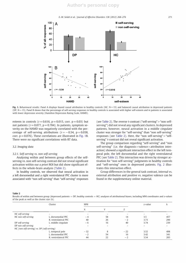
The repeated measures ANOVA of attributional decisionsshowed a significant diagnosis×valence×attribution interac-tion (F(1,28)=8.998, p=0.006). To disentangle this interac-tion, we performed planned post-hoc comparisons toexamine group differences regarding attributional biases. Thisrevealed that controls show significantly (t(28)=3.021,p=0.005) more self-serving attributions (mean=69.5%,SD=11.39) compared to depressed patients (mean=52.91%,SD=17.05). Directly comparing the percentage of self-serving vs. non self-serving responses within each grouprevealed that controls are more self-serving (t(14)=6.633,pb0.001) whereas patients show a balanced attributionalstyle (t(14)=0.645, p=0.529). These effects are illustratedin Fig. 1A. The repeated measures ANOVA on the RT showedno significant main effect of diagnosis (F(1,28)=1.317,p=0.261) indicating both groups responded equally fast.Also all interactions with the factor diagnosis (diagnosis by va-lence, diagnosis by attribution) remained non-significant. (ForANOVA main effects and interactions without the diagnosisfactor see the online supplement).
In the within-group correlation analyses, we observed apositive correlation of self-serving attributions with self-
270 E.-M. Seidel et al. / Journal of Affective Disorders 138 (2012) 268–276
Author's personal copy
esteem in controls (r=0.616, p=0.015, corr. p=0.03) butnot patients (r=0.077, p=0.784). In patients, symptom se-verity on the HAMD was negatively correlated with the per-centage of self-serving attributions (r=−0.54, p=0.038,corr. p=0.076). These correlations are illustrated in Fig. 1B.There were no significant correlations with RT data.
3.2. Imaging data
3.2.1. Self-serving vs. non self-servingAnalysing within and between group effects of the self-
serving vs. non self-serving contrast did not reveal significantactivation within our a priori ROI but did show significant ef-fects in the whole-brain analysis (Table 2).
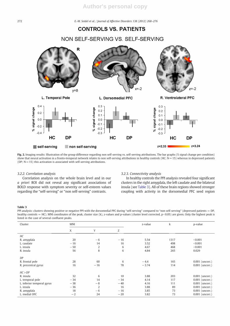
In healthy controls, we observed that neural activation ina left dorsomedial and a right ventrolateral PFC cluster is moreassociated with “non self-serving” than “self-serving” responses
(see Table 2). The reverse t-contrast (“self-serving”>“non self-serving”) did not reveal any significant clusters. In depressedpatients, however, neural activation in a middle cingulatecluster was stronger for “self-serving” than “non self-serving”responses (see Table 2). Here, the “non self-serving”>“self-serving” t-contrast did not reveal significant activation.
The group comparison regarding “self-serving” and “nonself-serving” (i.e. the diagnosis×valence×attribution inter-action) showed a significant interaction effect in the left tem-poral pole, the left dorsomedial and the right ventrolateralPFC (see Table 2). This interaction was driven by stronger ac-tivation for “non self-serving” judgments in healthy controlsand “self-serving” ones in depressed patients. Fig. 2 illus-trates this interaction effect.
Group differences in the general task contrast, internal vs.external attribution and positive vs. negative valence can befound in the supplementary online material.
Fig. 1. Behavioural results: Panel A displays biased causal attribution in healthy controls (HC; N=15) and balanced causal attribution in depressed patients(DP; N=15). Panel B shows that the percentage of self-serving responses in healthy controls is associated with higher self-esteem and in patients is associatedwith lower depression severity (Hamilton Depression Rating Scale, HAMD).
Table 2Results of within and between group (depressed patients = DP, healthy controls = HC) analyses of attributional biases, including MNI coordinates and z-valuesof the peak as well as the cluster size (k).
Cluster MNI z-value k
X Y Z
HC self-serving – – – – – –
HC non self-serving L. dorsomedial PFC −4 58 14 3.5 457R. ventrolateral PFC 40 26 −2 3.73 290
DP self-serving L. middle cingulate −6 16 40 3.74 239DP non self-serving – – – – – –
HC (non self-serving) vs. DP (self-serving)L. temporal pole −32 8 −22 3.52 498L. dorsomedial PFC −2 54 16 3.42 341R. ventrolateral PFC 44 22 −2 3.54 283
271E.-M. Seidel et al. / Journal of Affective Disorders 138 (2012) 268–276
Author's personal copy
3.2.2. Correlation analysisCorrelation analysis on the whole brain level and in our
a priori ROI did not reveal any significant associations ofBOLD response with symptom severity or self-esteem valuesregarding the “self-serving” or “non self-serving” contrasts.
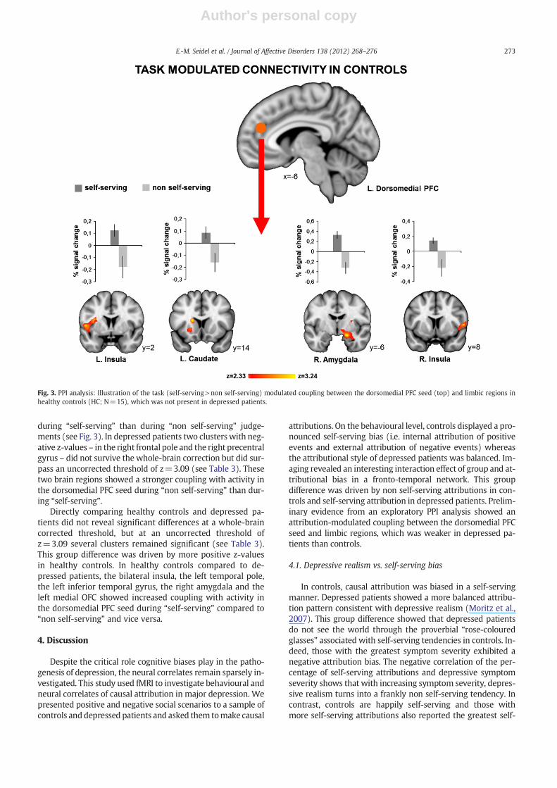
3.2.3. Connectivity analysisIn healthy controls the PPI analysis revealed four significant
clusters in the right amygdala, the left caudate and the bilateralinsula (see Table 3). All of these brain regions showed strongercoupling with activity in the dorsomedial PFC seed region
Table 3PPI analysis: clusters showing positive or negative PPI with the dorsomedial PFC during “self-serving” compared to “non self-serving” (depressed patients = DP,healthy controls = HC). MNI coordinates of the peak, cluster size (k), z-values and p-values (cluster level corrected, pb0.05) are given. Only the highest peak islisted in the case of several confluent peaks.
Cluster MNI z-value k p-value
X Y Z
HCR. amygdala 20 −6 −16 5.54 1317 b0.001L. caudate −16 14 16 3.52 498 b0.001L. insula −50 2 6 4.67 468 b0.001R. insula 56 8 6 4.84 265 0.029
DPR. frontal pole 28 60 6 −4.4 165 0.001 (uncorr.)R. precentral gyrus 16 −16 78 −3.74 114 0.001 (uncorr.)
HC>DPR. insula 32 6 10 3.88 203 0.001 (uncorr.)L. temporal pole −34 14 −34 4.14 117 0.001 (uncorr.)L. inferior temporal gyrus −38 −8 −40 4.16 111 0.001 (uncorr.)L. insula −36 2 16 3.88 89 0.001 (uncorr.)R. amygdala 18 −6 −16 3.85 73 0.001 (uncorr.)L. medial OFC −2 24 −20 3.82 73 0.001 (uncorr.)
Fig. 2. Imaging results: Illustration of the group difference regarding non self-serving vs. self-serving attributions. The bar graphs (% signal change per condition)show that neural activation in a fronto-temporal network relates to non self-serving attributions in healthy controls (HC; N=15) whereas in depressed patients(DP; N=15) this activation is associated with self-serving attributions.
272 E.-M. Seidel et al. / Journal of Affective Disorders 138 (2012) 268–276
Author's personal copy
during “self-serving” than during “non self-serving” judge-ments (see Fig. 3). In depressed patients two clusters with neg-ative z-values – in the right frontal pole and the right precentralgyrus – did not survive the whole-brain correction but did sur-pass an uncorrected threshold of z=3.09 (see Table 3). Thesetwo brain regions showed a stronger coupling with activity inthe dorsomedial PFC seed during “non self-serving” than dur-ing “self-serving”.
Directly comparing healthy controls and depressed pa-tients did not reveal significant differences at a whole-braincorrected threshold, but at an uncorrected threshold ofz=3.09 several clusters remained significant (see Table 3).This group difference was driven by more positive z-valuesin healthy controls. In healthy controls compared to de-pressed patients, the bilateral insula, the left temporal pole,the left inferior temporal gyrus, the right amygdala and theleft medial OFC showed increased coupling with activity inthe dorsomedial PFC seed during “self-serving” compared to“non self-serving” and vice versa.
4. Discussion
Despite the critical role cognitive biases play in the patho-genesis of depression, the neural correlates remain sparsely in-vestigated. This study used fMRI to investigate behavioural andneural correlates of causal attribution in major depression. Wepresented positive and negative social scenarios to a sample ofcontrols anddepressed patients and asked them tomake causal
attributions. On the behavioural level, controls displayed a pro-nounced self-serving bias (i.e. internal attribution of positiveevents and external attribution of negative events) whereasthe attributional style of depressed patients was balanced. Im-aging revealed an interesting interaction effect of group and at-tributional bias in a fronto-temporal network. This groupdifference was driven by non self-serving attributions in con-trols and self-serving attribution in depressed patients. Prelim-inary evidence from an exploratory PPI analysis showed anattribution-modulated coupling between the dorsomedial PFCseed and limbic regions, which was weaker in depressed pa-tients than controls.
4.1. Depressive realism vs. self-serving bias
In controls, causal attribution was biased in a self-servingmanner. Depressed patients showed a more balanced attribu-tion pattern consistent with depressive realism (Moritz et al.,2007). This group difference showed that depressed patientsdo not see the world through the proverbial “rose-colouredglasses” associated with self-serving tendencies in controls. In-deed, those with the greatest symptom severity exhibited anegative attribution bias. The negative correlation of the per-centage of self-serving attributions and depressive symptomseverity shows that with increasing symptom severity, depres-sive realism turns into a frankly non self-serving tendency. Incontrast, controls are happily self-serving and those withmore self-serving attributions also reported the greatest self-
Fig. 3. PPI analysis: Illustration of the task (self-serving>non self-serving) modulated coupling between the dorsomedial PFC seed (top) and limbic regions inhealthy controls (HC; N=15), which was not present in depressed patients.
273E.-M. Seidel et al. / Journal of Affective Disorders 138 (2012) 268–276
Author's personal copy
esteem. However, these correlations do not allow us to deter-mine whether one or both causal directions are operating.
4.2. Conflictive attributions activate a fronto-temporal networkin controls and depressed patients
Functional imaging revealed neural group differences thatparalleled the behavioural differences in attributional style.This group difference in activation of the dorsomedial PFC,the ventrolateral PFC and the temporal pole was driven bynon self-serving attributions in controls and self-serving at-tributions in depressed patients. It is notable that in eachgroup, the less frequent attributional pattern was associatedwith greater activation within this network. This may reflectthat in controls non self-serving and in depressed patientsself-serving attributions are in greater conflict with the pre-vailing self-concept. This conflict may require higher degreesof cognitive control (Krusemark et al., 2008) to inhibit theprepotent tendency toward either self-serving or non self-serving responses, leading to greater recruitment of fronto-temporal brain regions.
Such an interpretation is consistent with prior literature re-garding the functions associated with these regions. The dor-somedial PFC has been associated with self-relatedness(Andrews-Hanna et al., 2010; Northoff et al., 2006), and intro-spective self-referential processing (Gallagher and Frith, 2003;Schmitz et al., 2004). In depressed patients this brain regionhas been implicated in mediating abnormal self-relatednessand the characteristically negative self-concept (Grimm et al.,2009; Lemogne et al., 2009). In addition to its role in self-related processes, the dorsomedial PFC is involved in conflictmonitoring and cognitive control (e.g., Amodio and Frith, 2006;Etkin et al., 2006). Similarly, the ventrolateral PFC has also beenimplicated in control-related processes such as response inhibi-tion when a pre-potent response is withheld (e.g., Aron et al.,2004; Rubia et al., 2003). Analogously, activation in the temporalpole may reflect higher effort in the retrieval of socially-relevantsemantic knowledge (Zahn et al., 2007) when a less automatedattributional decision is made. When taken together with priorresults, our current findings suggest that depressed patientsshow a different pattern of activation in a fronto-temporal net-work during causal attributions due to the emotional contextof their own negative self-concept, resulting in greater conflictwhen a self-serving judgment is made.
4.3. Prefrontal-limbic connectivity is modulated by attribution
The exploratory PPI analysis revealed a group differencein fronto-limbic connectivity modulated by attributional de-cisions. However, this finding should be considered prelimi-nary given the liberal statistical threshold. Compared tocontrols, depressed patients showed decreased connectivitybetween the dorsomedial PFC seed and limbic regions includ-ing the amygdala during self-serving attributions. During nonself-serving attributions, however, the PFC-limbic couplingwas increased in patients.
We speculate that causal attributionmay serve a regulato-ry function influencing emotional reactions to positive andnegative events (Terbeck et al., 2008). In response to positiveevents internal attribution may help induce a positive emo-tional reaction, whereas in response to negative events
external attribution could prevent a negative emotional reac-tion (also called distancing). Therefore, the decreased PFC-limbic coupling during non self-serving attributions, whichwas stronger in controls compared to patients, might reflecta stronger suppression of limbic activation by the PFC to en-able distancing. This speculative interpretation is consistentwith previous research suggesting that the PFC exerts a regu-latory influence on limbic structures, such as the amygdala(Quirk and Beer, 2006; Urry et al., 2006). Specifically, the dor-somedial PFC has been implicated in self-focused cognitivereappraisal (Ochsner et al., 2004; for a review see Ochsnerand Gross, 2008).
This modulation of connectivity was weaker in patientsthan controls, suggesting a disturbance of fronto-limbic regu-lation during the attribution process in depressed patients.This result parallels previously reported abnormalities inthe suppression of limbic responses during the reappraisalof negative pictures in depressed patients (Johnstone et al.,2007; Sheline et al., 2009). However, this result is based ona rather liberal statistical threshold and therefore is tentativepending replication in larger samples.
4.4. Limitations and future prospects
This study has several limitations that should be noted.First, we were primarily interested in examining group differ-ences in attributional biases;we therefore combined individualvalence-attribution conditions into self-serving and non self-serving categories. We did not separately examine the effectsof each individual condition, which would have been difficultgiven the small number of events in particular conditions forsome of our subjects. Second, we did not replicate the findingfrom our previous study (Seidel et al., 2010) of activations inthe striatum in healthy subjects associatedwith self-serving re-sponses. Type II error may contribute to the lack of striatal ef-fects in the current study: the current control sample was halfthe size of the previous sample. Furthermore, the current find-ing of an association between front-temporal activation andnon-self serving attributions was not observed in the previousstudy. Differences in the control samples of the two studiesmay contribute to the difference in results. The first study in-cluded only college students, while the current study used acommunity control samplewhichwas significantly older. Com-pared to the previous student sample, the current communitysample also showed amore pronounced self-serving bias, asso-ciatedwith greater self-esteem. Future studieswith larger sam-ples of heterogeneous healthy subjects should seek to identifysignificant individual difference variables. Third, this studywas not designed to evaluate effects of antidepressant medica-tion on the processes studied. However, future neuroimagingstudies should highlight how and where antidepressants influ-ence the neural correlates of causal attribution.
Notwithstanding these limitations, the current study pro-vides novel data on the neural correlates of causal attributionin major depression. Our results suggest that the attributionalstyle of depressive realism seen in patients is associated withabnormalities in fronto-temporal brain regions. Specifically,increased activation to self-serving judgments in these re-gions may reflect conflict associated with such attributions;this stands in sharp contrast to the increased responses ofthese same brain regions to non self-serving judgments in
274 E.-M. Seidel et al. / Journal of Affective Disorders 138 (2012) 268–276
Author's personal copy
controls. Furthermore, as suggested by the preliminary re-sults of the exploratory PPI analysis, these changes may berelated to alterations of fronto-limbic connectivity.
These results have implications for treatments of majordepression, such as cognitive therapy, designed to reducenegatively biased causal attributions by facilitating reinter-pretation of emotional events in a more positive way. Inves-tigating the effects of psychotherapy and pharmacotherapyon fronto-limbic abnormalities elicited by causal attributionin depressed patients will be an important avenue for futurestudies.
Role of funding sourceThis study was funded by the Medical Faculty of the RWTH Aachen Uni-
versity (START 690811). EMS was supported by the Interdisciplinary Centrefor Clinical Research (IZKF, NWW11-SP3) within the Faculty of Medicine atthe RWTH Aachen University. TDS was supported by NIMH grants MH19112 and 5R25MH60490. SBE was supported by the Human Brain Project(R01-MH074457-01A1) and the Helmholtz-Initiative on Systems Biology(“Human Brain Model”). R.C.G. was supported by the NIMH grant MH60722. TDS and DHW are supported by NARSAD and APIRE. UH and BDwere supported by the German Research Foundation (DFG, IRTG 1328).
All these funding sources had no further role in study design; in the col-lection, analysis and interpretation of data; in the writing of the report; andin the decision to submit the paper for publication.
Conflict of interestEach author declares that they have no potential conflict of interest.
AcknowledgementsThis study was funded by the Medical Faculty of the RWTH Aachen Uni-
versity (START 690811). EMS was supported by the Interdisciplinary Centrefor Clinical Research (IZKF, NWW11-SP3) within the Faculty of Medicine atthe RWTH Aachen University. TDS was supported by NIMH grants MH19112 and 5R25MH60490. SBE was supported by the Human Brain Project(R01-MH074457-01A1) and the Helmholtz-Initiative on Systems Biology(“Human Brain Model”). R.C.G. was supported by the NIMH grant MH60722. TDS and DHW are supported by NARSAD and APIRE. UH and BDwere supported by the German Research Foundation (DFG, IRTG 1328).
Appendix A. Supplementary data
Supplementary data to this article can be found online atdoi:10.1016/j.jad.2012.01.041.
References
Amodio, D.M., Frith, C.D., 2006. Meeting of minds: the medial frontal cortexand social cognition. Nature Reviews Neuroscience 7, 268–277.
Andrews-Hanna, J.R., Reidler, J.S., Sepulcre, J., Poulin, R., Buckner, R.L., 2010.Functional–anatomic fractionation of the brain's default network. Neu-ron 65, 550–562.
Aron, A.R., Robbins, T.W., Poldrack, R.A., 2004. Inhibition and the right inferi-or frontal cortex. Trends in Cognitive Sciences 8, 170–177.
Beck, A.T., 2008. The evolution of the cognitive model of depression and itsneurobiological correlates. The American Journal of Psychiatry 165,969–977.
Beck, A.T., Erbaugh, J., Ward, C.H., Mock, J., Mendelsohn, M., 1961. An inventoryfor measuring depression. Archives of General Psychiatry 4, 561–571.
Chandler, T.A., Lee, M.S., Pengilly, J.W., 1997. Self-esteem and causal attribu-tions. Genetic, Social, and General Psychology Monographs 123, 479–491.
Diez-Alegria, C., Vazquez, C., Nieto-Moreno, M., Valiente, C., Fuentenebro, F.,2006. Personalizing and externalizing biases in deluded and depressedpatients: are attributional biases a stable and specific characteristic ofdelusions? British Journal of Clinical Psychology 45, 531–544.
Epstein, J., Pan, H., Kocsis, J.H., Yang, Y.H., Butler, T., Chusid, J., Hochberg, H.,Murrough, J., Strohmayer, E., Stern, E., Silbersweig, D.A., 2006. Lack ofventral striatal response to positive stimuli in depressed versus normalsubjects. The American Journal of Psychiatry 163, 1784–1790.
Etkin, A., Egner, T., Peraza, D.M., Kandel, E.R., Hirsch, J., 2006. Resolving emo-tional conflict: a role for the rostral anterior cingulate cortex in modulat-ing activity in the amygdala. Neuron 51, 871–882.
Ferring, D., Filipp, S.-H., 1996. Messung des Selbstwertgefühls: Befunde zurReliabilität, Validität und Stabilität der Rosenberg-Skala. Diagnostica42, 284–292.
Friston, K.J., 1997. Testing for anatomically specified regional effects. HumanBrain Mapping 5, 133–136.
Friston, K.J., Buechel, C., Fink, G.R., Morris, J., Rolls, E., Dolan, R.J., 1997. Psy-chophysiological and modulatory interactions in neuroimaging. Neuro-Image 6, 218–229.
Gallagher, H.L., Frith, C.D., 2003. Functional imaging of ‘theory of mind’.Trends in Cognitive Sciences 7, 77–83.
Grimm, S., Ernst, J., Boesiger, P., Schuepbach, D., Hell, D., Boeker, H., Northoff,G., 2009. Increased self-focus in major depressive disorder is related toneural abnormalities in subcortical–cortical midline structures. HumanBrain Mapping 30, 2617–2627.
Hamilton, M., 1960. A rating scale for depression. Journal of Neurology, Neu-rosurgery, and Psychiatry 23, 56–62.
Hikosaka, O., Bromberg-Martin, E., Hong, S., Matsumoto, M., 2008. New in-sights on the subcortical representation of reward. Current Opinion inNeurobiology 18, 203–208.
Johnstone, T., van Reekum, C.M., Urry, H.L., Kalin, N.H., Davidson, R.J., 2007.Failure to regulate: counterproductive recruitment of top-downprefrontal-subcortical circuitry in major depression. Journal of Neurosci-ence 27, 8877–8884.
Krusemark, E.A., Campbell, W.K., Clementz, B.A., 2008. Attributions, decep-tion, and event related potentials: an investigation of the self-servingbias. Psychophysiology 45, 511–515.
Lemogne, C., le Bastard, G., Mayberg, H., Volle, E., Bergouignan, L., Lehericy,S., Allilaire, J.F., Fossati, P., 2009. In search of the depressive self: extend-ed medial prefrontal network during self-referential processing in majordepression. Social Cognitive and Affective Neuroscience 4, 305–312.
Moritz, S., Woodward, T.S., Burlon, M., Braus, D.F., Andresen, B., 2007. Attri-butional style in schizophrenia: evidence for a decreased sense of self-causation in currently paranoid patients. Cognitive Therapy and Re-search 31, 371–383.
Northoff, G., Heinzel, A., Greck, M., Bermpohl, F., Dobrowolny, H., Panksepp,J., 2006. Self-referential processing in our brain — a meta-analysis of im-aging studies on the self. NeuroImage 31, 440–457.
Ochsner, K.N., Gross, J.J., 2005. The cognitive control of emotion. Trends inCognitive Sciences 9, 242–249.
Ochsner, K.N., Gross, J.J., 2008. Cognitive emotion regulation: insights fromsocial cognitive and affective neuroscience. Current Directions in Psy-chological Science 17, 153–158.
Ochsner, K.N., Ray, R.D., Cooper, J.C., Robertson, E.R., Chopra, S., Gabrieli,J.D.E., Gross, J.J., 2004. For better or for worse: neural systems supportingthe cognitive down- and up-regulation of negative emotion. Neuro-Image 23, 483–499.
Oldfield, R.C., 1971. The assessment and analysis of handedness: the Edinburghinventory. Neuropsychologia 9, 97–113.
Pizzagalli, D.A., Holmes, A.J., Dillon, D.G., Goetz, E.L., Birk, J.L., Bogdan, R.,Dougherty, D.D., Iosifescu, D.V., Rauch, S.L., Fava, M., 2009. Reduced cau-date and nucleus accumbens response to rewards in unmedicated individ-uals with major depressive disorder. The American Journal of Psychiatry166, 702–710.
Quirk, G.J., Beer, J.S., 2006. Prefrontal involvement in the regulation of emo-tion: convergence of rat and human studies. Current Opinion in Neuro-biology 16, 723–727.
Reitan, R.M., 1956. Trail Making Test: manual for administration, scoring andinterpretation. Indiana University Press, Indianapolis.
Rosenberg, M., 1965. Society and the adolescent self-image. Princeton Uni-versity Press, Princeton, NJ.
Rubia, K., Smith, A.B., Brammer, M.J., Taylor, E., 2003. Right inferior prefrontalcortex mediates response inhibition while mesial prefrontal cortex is re-sponsible for error detection. NeuroImage 20, 351–358.
Schmitz, T.W., Kawahara-Baccus, T.N., Johnson, S.C., 2004. Metacognitiveevaluation, self-relevance, and the right prefrontal cortex. NeuroImage22, 941–947.
Seidel, E.-M., Eickhoff, S.B., Kellermann, T., Schneider, F., Gur, R.C., Habel, U.,Derntl, B., 2010. Who is to blame? Neural correlates of causal attributionin social situations. Social Neuroscience 5, 335–350.
Sheline, Y.I., Barch, D.M., Price, J.L., Rundle, M.M., Vaishnavi, S.N., Snyder, A.Z.,Mintun, M.A., Wang, S., Coalson, R.S., Raichle, M.E., 2009. The default modenetwork and self-referential processes in depression. Proceedings of the Na-tional Academy of Sciences of the United States of America 106, 1942–1947.
Snyder, M., Uranowitz, S.W., 1978. Reconstructing past— some cognitiveconsequences of person perception. Journal of Personality and SocialPsychology 36, 941–950.
Terbeck, S., Chesterman, P., Fischmeister, F.P.S., Leodolter, U., Bauer, H., 2008.Attribution and social cognitive neuroscience: a new approach for the“online-assessment” of causality ascriptions and their emotional conse-quences. Journal of Neuroscience Methods 173, 13–19.
275E.-M. Seidel et al. / Journal of Affective Disorders 138 (2012) 268–276

Author's personal copy
Urry, H.L., van Reekum, C.M., Johnstone, T., Kalin, N.H., Thurow, M.E.,Schaefer, H.S., Jackson, C.A., Frye, C.J., Greischar, L.L., Alexander, A.L.,Davidson, R.J., 2006. Amygdala and ventromedial prefrontal cortex areinversely coupled during regulation of negative affect and predict the di-urnal pattern of cortisol secretion among older adults. Journal of Neuro-science 26, 4415–4425.
Von Aster, M., Neubauer, A., Horn, R., 2006. Hamburg-Wechsler-Intelligenz-Test für Erwachsene III. Harcourt, Frankfurt.
Wittchen, H.-U., Zaudig, M., Fydrich, T., 1998. Strukturiertes Klinisches Inter-view für DSM-IV. Hogrefe, Göttingen.
Zahn, R., Moll, J., Krueger, F., Huey, E.D., Garrido, G., Grafman, J., 2007. Socialconcepts are represented in the superior anterior temporal cortex. Pro-ceedings of the National Academy of Sciences of the United States ofAmerica 104, 6430–6435.
276 E.-M. Seidel et al. / Journal of Affective Disorders 138 (2012) 268–276