national disaster management manual
136
-
Upload
khangminh22 -
Category
Documents
-
view
4 -
download
0
Transcript of national disaster management manual

133


NATIONAL DISASTER MANAGEMENT MANUAL
onMEDICAL MANAGEMENT
ofNUCLEAR AND RADIOLOGICAL
EMERGENCIES

Manual on Medical Management ofNuclear and Radiological Emergencies
A publication of:National Disaster Management AuthorityGovernment of IndiaNDMA BhawanA-1, Safdarjung EnclaveNew Delhi - 110029
When citing this manual, the following citation should be used: Manual on Medical Management of Nuclear and Radiological Emergencies.A publication of the National Disaster Management Authority, Government of India.February 2019, New Delhi
Manual on Medical Management of Nuclear and Radiological Emergencies is formulated by NDMA, in consultation with various stakeholders and domain experts from across the country.

NATIONAL DISASTER MANAGEMENT MANUAL
onMEDICAL MANAGEMENT
ofNUCLEAR AND RADIOLOGICAL
EMERGENCIES
National Disaster Management AuthorityGovernment of India


PREFACE
There are a number of Nuclear and Radiological facilities operating in the country. These nuclear and radiological facilities are designed, built and operated with utmost care and safety. Possibilities of emergency situations arising out of operations of these facilities leading to radiation exposure to public are very remote but cannot be entirely ruled out. Similarly, members of the public may receive high radiation doses as a result of handling or being in the vicinity of lost, stolen or orphan radiation sources. Moreover, the general public and also emergency workers could be exposed to radiation or may get contaminated as a consequence of malicious acts involving radioactive material. Without adequate awareness, training and preparedness of the medical community for such radiation emergencies, medical management of the situation could be ineffective.
Radiological accidents and disasters can have a prolonged impact on public health. Hospitals should, therefore, be prepared to respond to radiation emergencies, as determined by risk assessment, based on local and regional radioactive hazards, threats and vulnerabilities. Approach to medical management of multiple combined radiation injury victims requires attention to casualty triage, decontamination and prevention of secondary contamination, radiation safety of healthcare personnel, trauma care system, availability of medical staff trained in the treatment of radiation-related injuries and also availability of pharmaco-therapeutic options.
This manual aims to serve as a practical resource guide for management of a nuclear or radiological emergency. It also explains the roles and responsibilities of the members of the emergency medical response organization which includes the Response Initiation Team, the Emergency Medical Personnel on scene and the Hospital Radiological Response Team (HRRT).
The confidence of the members of the public is of paramount importance while managing such emergency situations. It is needless to say that the medical fraternity has an edge over others in building this confidence. It is necessary that the members of the public affected or likely to be affected, by the radiological emergencies are made aware of not only the effects of the radiation, but also of the fact that the fear arising out of ignorance is far greater than that the effects of the radiation. This information may be given by the medics and paramedics of the state, on a regular basis, during their door-to-door visits for various governmental programs viz. immunization, family planning, hygiene drives etc.
Since the radiological emergencies due to ‘orphan radioactive sources’ leading to cases of inadvertent exposure to members of public is considered more likely and is a concern internationally, the preparedness by the medical community, though addressed for dealing with nuclear emergencies through this manual, will also help in handling such issues and in strengthening national level preparedness. Incidents similar to the Mayapuri, (New Delhi), in the year 2011 and many radiological incidents reported internationally, have led to severe radiation injuries and casualties to the public, due to lack of timely medical support. This document includes information on Acute Radiation Syndrome (ARS), and its medical management, internal decontamination, Radiation Burns, Bio-dosimetry etc. Some of these may not be feasible at the level of Primary Health Centre (PHC) or Community Health Centre (CHC) and may need specialised designated hospitals/facilities.

jk"Vªh; vkink izca/u izkf/dj.kNational Disaster Management Authority
Hkkjr ljdkjGovernment of India
Foreword
The National Disaster Management Authority has undertaken numerous initiatives for Disaster Risk Reduction and capacity building for disaster management in conformity with its mandate under the DM Act, 2005.
The manual on Medical Management of Nuclear and Radiological Emergencies can be seen as an effort to provide guidance for precise handling of patients related to mass or sentinel incident of acute or chronic illnesses resulting from radiation exposure, be it intentional or accidental. The manual which has been reviewed by several experts, will be a very handy compendium on this important aspect.
Though Nuclear and Radiological Emergencies have a low probability of occurrence, managing of such incidents require expertise and skills. This document is intended to boost the capacity building of the professionals specializing in the area.
The first draft of the manual was single handly, prepared by Late Dr. Raghavendra Deolalikar, Certifying Surgeon, NAPS, NPCIL.
We take this opportunity to express our deep appreciation of the commitment to the team of experts from NDMA, DAE, BARC, AERB, DRDO and various stakeholders who extended their willing support and cooperation to our efforts and cause by devoting their professional approach and for their valuable contributions in developing and reviewing this document.
We are sanguine that this effort will go a long way in enhancing preparedness and Disaster Risk Reduction in the county for Nuclear and Radiological emergencies.
Shri Kamal Kishore Dr. D.N. Sharma Lt. Gen. N.C. Marwah (Retd.) Member, NDMA Member, NDMA Member, NDMA

8
NDMA MANuAl oN MeDicAl MANAgeMeNt of NucleAr AND rADiologicAl eMergeNcies
DISCLAIMER
This is a document intended for education and practical use while responding to radiation emergency situations. The references used in preparing the document are enlisted at the end. The publishers NDMA do not intend to derive any commercial benefits whatsoever and, hence, all re-productions and references used are done in good faith and the teams do not feel the necessity for any Written Permission from the authors and copyright owners of the original articles. However, due credit to the original articles are rendered wherever it is felt necessary even within the text, in addition to being enlisted in bibliographical references.
As this in an educative material, the trade names of any medicines used in this document are for purpose of easy reference and do not, in any way, advocate or promote the use of the same brand of medicines.

9
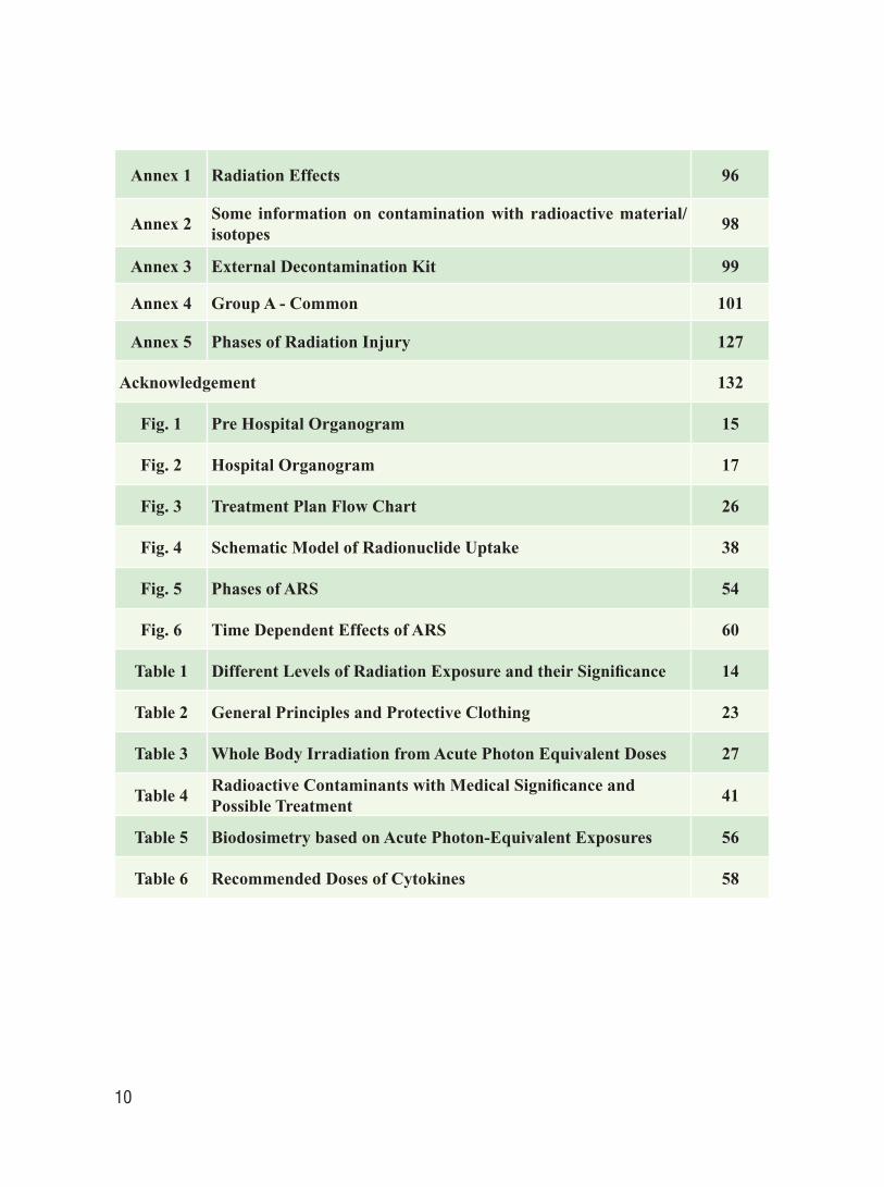
C O N T E N T S
SECTION TOPIC PAGE
1 Introduction 11
2 Type of Events, Effects of Radiation and Types of Exposure 13
3 Organization and Structure with Role & Responsibilities 15
4 Resources and Infrastructure 19
5 Medical Management – Principles and Plan 23
6 External Decontamination 29
7 Internal Contamination – Principles of Uptake and Clearance 37
8 Internal Decontamination 39
9 Follow up of Internally Decontaminated Patients 43
10 Radiation Burns 45
11 Admission 49
12 Writing Notes 51
13 Acute Radiation Syndrome (ARS) 53
14 Bio-Dosimetry in ARS 55
15 Medical Management of ARS 57
16 Post Disaster Counseling 63
17 Maintenance and Records 65
18 Appendix I General Instructions for Awareness 67
19 Appendix II Table of Half Lives 72
20 References 73
W.S. 1 Worksheet for Issue of Thermo Luminescence Dosimetry (TLD) 75
W.S. 2 Worksheet for TLD Assessment Report 76
W.S. 3 Worksheet for Decontamination / Treatment Case Sheet 77
W.S. 4Worksheet for Recording the movement of Radio-Active Waste Material
94
W.S. 5 Worksheet for Report on Status of Radio-Active Waste Material 95
10
NDMA MANuAl oN MeDicAl MANAgeMeNt of NucleAr AND rADiologicAl eMergeNcies
Annex 1 Radiation Effects 96
Annex 2Some information on contamination with radioactive material/isotopes
98
Annex 3 External Decontamination Kit 99
Annex 4 Group A - Common 101
Annex 5 Phases of Radiation Injury 127
Acknowledgement 132
Fig. 1 Pre Hospital Organogram 15
Fig. 2 Hospital Organogram 17
Fig. 3 Treatment Plan Flow Chart 26
Fig. 4 Schematic Model of Radionuclide Uptake 38
Fig. 5 Phases of ARS 54
Fig. 6 Time Dependent Effects of ARS 60
Table 1 Different Levels of Radiation Exposure and their Significance 14
Table 2 General Principles and Protective Clothing 23
Table 3 Whole Body Irradiation from Acute Photon Equivalent Doses 27
Table 4Radioactive Contaminants with Medical Significance and Possible Treatment
41
Table 5 Biodosimetry based on Acute Photon-Equivalent Exposures 56
Table 6 Recommended Doses of Cytokines 58

11
Medical management of radiological emergencies involves the medical fraternity and the emergency response organization. Although the cause of emergencies arising from Radiation and Nuclear facilities may be different, the management of exposure cases may be similar. The most important consideration in the medical evaluation and preparedness for response of a radiation event is the relative magnitude of the situation i.e. quantum of exposure, number of persons involved and the resources needed to address the emergency. In a radiation emergency, victims may have been harmed by one or more of the following causes: external exposure (localized, partial and whole body), contamination (external/internal) and conventional trauma. The same general principles of medical care apply at the scene of the emergency as at hospital, but the details and extent of medical care differ.
Patients contaminated by radioactive material generally pose no danger to healthcare personnel, if adequate precautions are taken. However, contaminated excreta or vomit can spread contamination to equipment, environment and attending staff. Using appropriate procedures could, therefore, prevent spreading of contamination. Hence, medical professionals must be prepared to provide prompt treatment for conventional trauma complicated by exposure to ionizing radiation or radioactive contamination. Two principles are of paramount importance in the medical management of the contaminated patient: early estimation of the magnitude of the radiation exposure and identification of the radioisotope(s) in question. These principles strongly influence subsequent treatment decisions.
Following a Radiological Mass Casualty Incident (RMCI), a surge of patients is expected and hospitals will need to rapidly reorganize and systematically manage their resources for patients’ care. Recognised documents on the subject describe a concept of surge capacity which is known as the “3 S System” — the 3 “S” standing for “Staff”, “Stuff” and “Structure”. By considering these key components when preparing for disaster, health care facilitators can respond better during such exigencies.
This manual is intended to act as operational guidance to doctors and other healthcare professionals for the proper medical management of persons affected or suspected to be exposed in a nuclear or radiological emergency.
Introduction1

12
NDMA MANuAl oN MeDicAl MANAgeMeNt of NucleAr AND rADiologicAl eMergeNcies
13
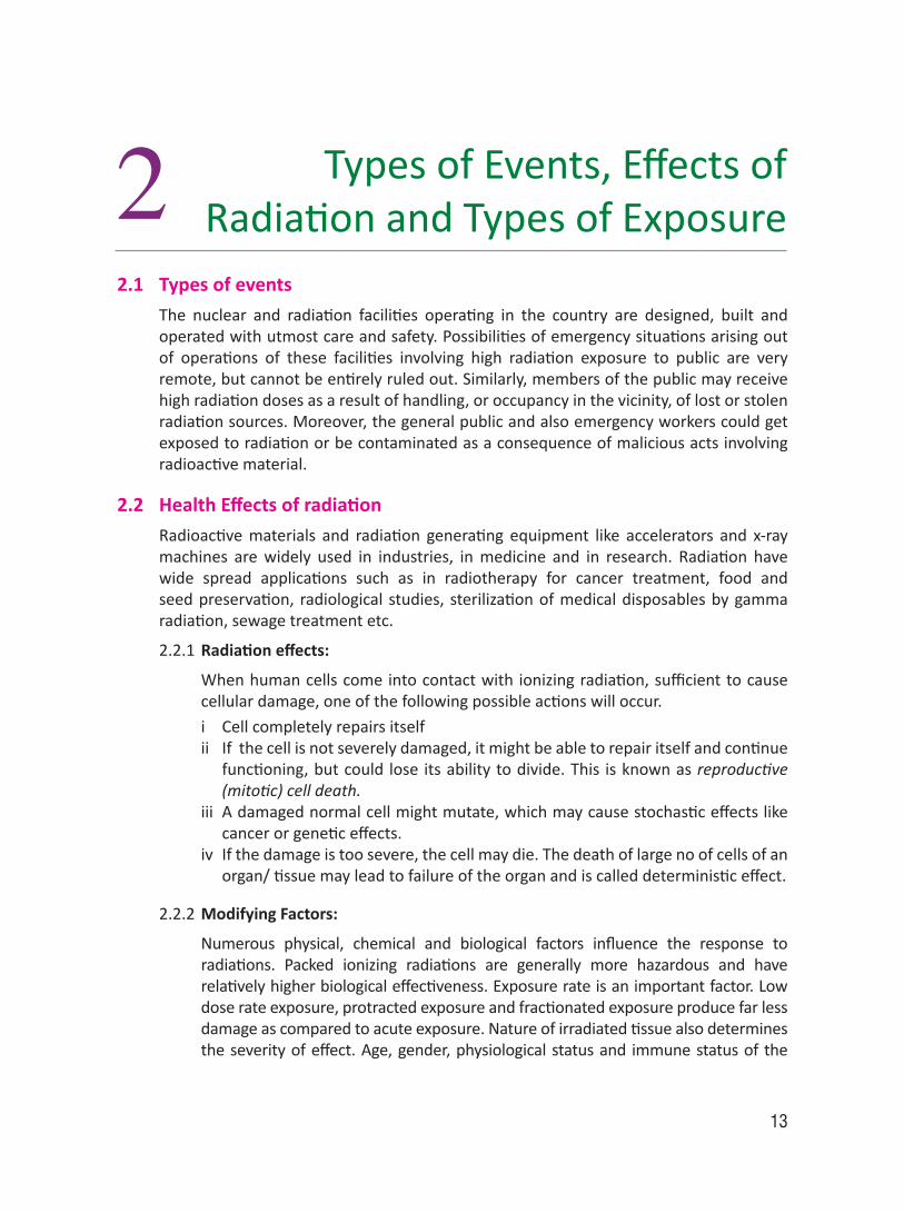
2.1 Types of events The nuclear and radiation facilities operating in the country are designed, built and
operated with utmost care and safety. Possibilities of emergency situations arising out of operations of these facilities involving high radiation exposure to public are very remote, but cannot be entirely ruled out. Similarly, members of the public may receive high radiation doses as a result of handling, or occupancy in the vicinity, of lost or stolen radiation sources. Moreover, the general public and also emergency workers could get exposed to radiation or be contaminated as a consequence of malicious acts involving radioactive material.
2.2 Health Effects of radiation Radioactive materials and radiation generating equipment like accelerators and x-ray
machines are widely used in industries, in medicine and in research. Radiation have wide spread applications such as in radiotherapy for cancer treatment, food and seed preservation, radiological studies, sterilization of medical disposables by gamma radiation, sewage treatment etc.
2.2.1 Radiation effects:
When human cells come into contact with ionizing radiation, sufficient to cause cellular damage, one of the following possible actions will occur.
i Cell completely repairs itselfii If the cell is not severely damaged, it might be able to repair itself and continue
functioning, but could lose its ability to divide. This is known as reproductive (mitotic) cell death.
iii A damaged normal cell might mutate, which may cause stochastic effects like cancer or genetic effects.
iv If the damage is too severe, the cell may die. The death of large no of cells of an organ/ tissue may lead to failure of the organ and is called deterministic effect.
2.2.2 Modifying Factors:
Numerous physical, chemical and biological factors influence the response to radiations. Packed ionizing radiations are generally more hazardous and have relatively higher biological effectiveness. Exposure rate is an important factor. Low dose rate exposure, protracted exposure and fractionated exposure produce far less damage as compared to acute exposure. Nature of irradiated tissue also determines the severity of effect. Age, gender, physiological status and immune status of the
Types of Events, Effects of Radiation and Types of Exposure2

14
NDMA MANuAl oN MeDicAl MANAgeMeNt of NucleAr AND rADiologicAl eMergeNcies
individual also determine the extent and severity of radiation effects. Infants and children are more sensitive to effects of radiation, particularly due to the active process of division of cells and development of organs occuring in early childhood.
Radiation effects depend upon large no of factors and amount of dose of radiation. The details are discussed in ANNEX-1. The following table gives exposure ranges for different effects.
Table 1: Different Levels of Radiation Exposure and their Significance:
Exposure Significance1-2 mSv / year Background Radiation at sea level outdoors0.5 – 5 mSv Most Diagnostic Radiological Examinations1 mSv / year Limit for Non-Occupational Exposure20 mSv / year Limit for Occupational Exposure - Whole Body150 mSv / year Exposure Limit for Eye Lens [Under Review]500 mSv / year Limit for exposure of skin and extremities10 mGy (in utero) 2 childhood cancers in 10,000 pregnancies
100 mGy whole body Detectable increase in chromosomal aberrations. No detectable clinical injury
100 – 200 mGy (in utero) Malformations and 1st trimester abortions1 Gy acute whole body Threshold for Radiation sickness in 5-10%
1 Gy Reproductive System Doubling dose Temporary Sterility in Males
2-3 Gy acute whole bodyThreshold for Epilation, Cataract [Under Review], Radiation sickness for most, Transient Erythema and LeukopeniaDeath 20-30%
3-5 Gy acute whole body LD 50/60 untreated. Severe Leucopenia, Purpura, Hemorrhage, Epilation, Infection
6 GyPermanent Sterility both the genders, Fixed Erythema50% Death with best Rx
> 10 Gy - Skin Dry Desquamation> 20 Gy - Skin Wet Desquamation40-60 Gy Total Radiation dose used in Fractional Radiotherapy of Cancer
2.3 Types of Accidental Exposure Types of radiation exposure which could occur during an emergency situation are
discussed in the following sections.i External contaminationii Internal contaminationiii Skin injury and radiation burns (Cutaneous radiation injury)iv Acute radiation syndrome (Whole body irradiation)v Combined injury (Concomitant conventional and radiation injury)
15
Organization and Structure with Roles and Responsibilities3
3.1 Pre-Hospital Organogram: It is imperative to have defined organizational levels (Organogram) for medical response
in emergencies. There has to be one organizational level for tackling the pre-hospital response and another for managing the hospital response.
i Medical Response Initiator: As the name suggests, is the person who initiates the emergency response after notification of a real or suspected radiation emergency. In case of off-site emergency arising from a Nuclear Power Plant [NPP], it will be the District Chief Medical Officer [CMO] on orders from the District Magistrate /
Fig. 1: Pre Hospital Organogram

16
NDMA MANuAl oN MeDicAl MANAgeMeNt of NucleAr AND rADiologicAl eMergeNcies
District Collector who is the Responsible Officer / Incident Commander. In case of emergency arising from other than NPP as in the case of detonations or orphan sources, the Doctor will notify the Chief Medical Officer (CMO) of the District. The CMO will get the cases confirmed and inform the District Magistrate [DM] / District Collector [DC]. The DM/DC will get the source traced and initiate the emergency response in concerned areas.
ii First Responder: First Responder(s) are the people who will proceed to the field under instructions from the Response Initiator. They will be responsible for informing the people about the dos and don’ts, the precautions and the distribution of Tab Potassium Iodide, if required.
iii Public Health Advisor: S/he will be the person who will instruct the people about sanitation, food and grain handling, proper storage, proper care of water resources and its storage etc. S/he will in association with the first responders, arrange to facilitate the evacuation of the people to Resting Shelters.
iv Radiological Assessment Team: Will carry out the external radiological assessment with the help of dosimeters and quick frisking. Internal dose assessment will be carried out by portable Whole Body Counter [vehicle mounted].
v Triage Team: Triage team will comprise of trained para-medics and with the help of the Radiological Assessment Team they will categorize the patients based on urgency of treatment and level of care required. Depending upon these factors, they will shift the patient either to the Decontamination Centre in a Primary Health Center [PHC] / Community Health Center [CHC] or to a District Hospital [DH] or directly to a tertiary care hospital.
vi Medical Transport Team: This team will be responsible for transport of patients to shelters, PHC/CHC or to District Hospitals or to tertiary care hospitals on the advice from the Triage Team and under instructions from the Public health advisor.
vii Decontamination Team: This will comprise of Doctors who are trained in decontamination. They will carry out the external or internal decontamination of the people. This team will again decide in association with the Radiological Assessment Team whether the patient, after decontamination, needs to be sent to a specialized center.
viii Waste Management Team: Will be responsible for proper collection of solid waste, liquid wastes, proper labeling of bags / bins / tanks etc. and proper disposal, as per national regulatory requirements.

17
3.2 Hospital Organogram:
Fig. 2: Hospital Organogram
i Emergency Medical Manager: At the hospital level, the Emergency Medical Manager is In-Charge of the concerned medical facility. S/he could be the Medical Officer in-charge of PHC / CHC, or the Medical Superintendent of the District Hospital or the tertiary care hospital. Upon instruction from the Chief Medical Officer of the district, the Emergency Medical Manager of the concerned facility will prepare the health center / hospital in complete readiness, for managing the patient. He/ She will ensure that all the teams under him/her will set the ball rolling.
ii Emergency Response Department Team: This is normally the Casualty Team, the Blood Bank and the Operation Theatre Teams. These Teams will be responsible for taking care and managing cases where medical emergencies override radiological emergencies. This team, upon receiving instructions from the Emergency Medical Manager, shall keep all the paraphernalia in the Casualty, Blood Bank and Operation Theatre in a complete state of readiness.
iii Specialist Team: This will comprise of doctors and para-medics. This team will have two components – (a) Decontamination / Decorporation Team which will be responsible for the decontamination and decorporation of radionuclides, both external and internal; and (b) Specialist team which will comprise of doctors from other specialties viz. Surgeon, Physician, Anesthetist, Ophthalmologist, Gynecologist, Pediatrician etc. for managing the co-morbid conditions of respective faculties. They will be assisted by the para-medics.
OrganizatiOn and Structure with rOleS and reSpOnSibilitieS

18
NDMA MANuAl oN MeDicAl MANAgeMeNt of NucleAr AND rADiologicAl eMergeNcies
iv Radiological Assessment Team: This team will comprise of people trained in monitoring the doses among suspected exposure cases. They will help the doctors in deciding the radiological assessment of reduction of contamination levels during the decontamination process. This team will have two components – (a) Physical dosimetry team, which will monitor the dose rate with the help of the monitors and estimate likely exposure; and (b) Bio-Dosimetry team which will collect the biological samples of the patients viz. Urine, Feces, Swabs from orifices, hair and nail samples, Blood sample etc., label them properly and send them to concerned bio-assay laboratories. Every District should have at least one such lab which will have facilities for bio-assay. This team will be responsible for proper dispatch of samples and receipt of reports. On receiving the reports, this team will intimate and hand over the reports to the Decontamination Team.
v Waste Management Team: Will be responsible of proper collection of solid and liquid wastes and its proper labeling and disposal, as per the national regulatory requirements.

19
Resources and Infrastructure44.1 Personnel Monitoring Devices and Protective Clothing: Personnel monitoring devices are of various types, but the most useful in field purposes
would be the Thermo-Luminescence Dosimeter [TLD]. This has to be encased in a plastic apron. Details of its usage are elaborated in Section-5
Protective clothing have also been detailed layer wise in Section-5.
4.2 Decontamination Facilities: The decontamination (Brief description is given in Annex-2) of the affected people needs
to be done at the level of the nearest PHC / CHC / District Hospital. Referrals, only for decontamination, to other places are to be avoided for two reasons:
4.2.1 The earlier the decontamination is done the better it is for the individual and hence the nearest facility should be capable of doing this, and
4.2.2 Sending the contaminated individuals elsewhere means spreading the contamination to newer areas instead of containing it.
For developing capabilities to effect successful decontamination, the medical centers should be equipped with the following:
i Infrastructure capable of catering to a volume of patients: Space and infrastructure needed here, will mainly be bathing facility, decontamination tables in a large hall and another hall for waiting-in patients. Care should be taken that the outlet drain of these bathrooms should be connected to make-shift collection tanks like PVC tanks which can be transported to a suitable radioactive waste collection facility of the state government approved by the AERB.
ii Necessary Decontamination Consumables: Paraphernalia needs to be adequate to meet the demand. Stock all paraphernalia in wooden/ metal boxes; store them in a room safely under lock and key, at these hospitals. This paraphernalia are herein referred to as “Decontamination Kits – External decontamination and internal decontamination kits”. (See Annex-3)
The Facility needs to be under the charge of the Medical Officer in Charge of that PHC/CHC/Hospital. This will ensure proper upkeep and accountability.
iii Trained manpower: All the doctors and para-medics should be trained to manage medical aspects of radiological / nuclear emergencies. There should

20
NDMA MANuAl oN MeDicAl MANAgeMeNt of NucleAr AND rADiologicAl eMergeNcies
be refresher training for all the staff every three years. Mock-patients should be handled during off-site emergency exercises. There should be a Physician and a Surgeon posted at all PHCs / CHCs. In addition, One Hematologist at District Hospitals and Civil Hospitals (available on call from tertiary care centers as needed) and mandatory in Medical colleges.
iv Investigation Facilities:
a All PHCs/ CHCs should have laboratory facility for complete blood count including platelet count, complete biochemistry investigations, blood grouping, Rh typing, urine and stool examination for routine and microscopic, urine-test for pregnancy and semen analysis.
b All District and Civil Hospitals should have the following facilities over and above available at the PHC/CHC: Blood Bank facility with component facility, burns ward, I.C.U. with ventilators, monitors and ABG analysis.
c There should be bone marrow transplant facility with proper Isolation wards one each in 5 regions of the country i.e north, south, east, west and central region.
d Every district/ a cluster of districts should have an accredited laboratory for bio-dosimetry and bio-assay which can reverse assess the blood / urine / stool.
e Every district / a cluster of districts should have a BARC Accredited TLD reading laboratory
f Every district or a cluster of districts should develop a radiological waste disposal facility for solid and liquid wastes, as per national regulatory requirements.
4.3 Identification Labels: While decontamination is under process, the samples collected need to be correctly
labeled in order to attribute the correct dose received by an individual after the dosimetry is done.
4.3.1 Individual Patient’s Waste collecting bag Label:
Name: Age: Sex:
Resident of: Relief Camp No: Patient No:
Type of Waste: (Please specify Linen, gauge, etc.)
Quantity: Date and Time of Collection:
Whether suspected Radioactive Yes/No
Name and Signature of Paramedic PHC/CHC: Bag No:

21
4.3.2 Individual Patient’s Bio-Assay sample Collection Label:
Name: Age: Sex:
Resident of: Relief Camp No: Patient No:
Date and Time of Event:
Type of Specimen: (Please specify Blood, Stool, Urine, Swabs, etc.)
Quantity: Date of Collection: Time of Collection:
Name and Signature of Paramedic on Duty:
Sample No:
4.4 Solid and Liquid Wastes: Decontamination requires a lot of water and other chemical solutions along with
consumables. Liquid waste generated as a result of decontamination need to be collected in waste tank and its proper disposal is equally important. As mentioned earlier, the outlet of the bathrooms used for bathing should be connected temporarily to PVC tanks of sufficient capacity. Also all liquids - water or chemical solutions – used during the process of decontamination should be discharged / drained into these tanks. These tanks will contain all the liquid wastes. Similarly, Solid wastes generated during the decontamination including the contaminated clothing of individuals should be collected in large yellow colored plastic bags as it is the standard accepted color to denote radioactive demarcation. They should be sealed. The waste tank and plastic bags should be labeled, as given below, on filling and the same shall be collected by the designated district authorities for proper and safe disposal / delivery to the nearest Radioactive waste management facility.
4.4.1 Label for waste collecting tank/bag following decontamination:
CHC* / Place of collection / MM / YYYY / RDD# / SW¥ / Sl. Number
* Community Health Centre [Alternating – PHC for Primary Health Centre, DH for District Hospital, MC for Medical College]
# Radiological Dispersal Device [Alternating – NPP for Emergency arising from Nuclear Power Plant or OS for Orphan Source]
¥ Solid Waste [alternating LW for Liquid waste]
Necessary instructions vide below should be printed on opposite surfaces in Hindi / Regional Language
i Radio-active waste
ii Do not break the seal
iii To be handled only by Authorized Staff
reSOurceS and infraStructure

22
NDMA MANuAl oN MeDicAl MANAgeMeNt of NucleAr AND rADiologicAl eMergeNcies
iv Public should maintain safe distance.
The movement of all the waste collecting bags / tanks should be properly logged. There should be signed worksheet that should accompany the bag/tank and should serially move along with the bag/tank until proper disposal. (Please see Work Sheet 4 [WS.4] for details). The waste facility should report back to MO I/c Health Centre. All districts should develop such a waste decontamination / disposal facility.

23
Medical Management Principle and Plan5
5.1 General Principles and Protective Clothing
Table 2: General Principles and Protective Clothingi Life saving measures will take precedence and preference over
decontamination procedures.ii Principles of Triage should be followediii Stabilize the patients’ vital parameters firstiv Treat any acute exacerbations / exaggerations of any chronic illness and their
complications which you feel need attention on priority basisv Treat Injuriesvi External Decontamination procedures to be carried outvii Internal Decontamination procedures to be carried out. This will take
precedence over external decontamination if contamination is suspected with Iodine or confirmed for any other radionuclide.
viii Any medical/paramedical personnel who are going to handle the patients should wear protective clothing.
ix Remove all ornaments, wrist watches or any other artifacts from the upper limbs, elbow downwards and from the foot
x Remove footwear and keep them aside, wear the canvas shoes / slippers provided in the PHC/CHC. This is in your interest. If your own shoes get contaminated, you may have to part with them
xi Wear shoe covers.
xii Wear a cap to cover your head completelyxiii Wear a face mask
xiv Wear a plastic apron over itxv Wear a surgical gown over itxvi Wear surgical / latex gloves / polypropylene. Pull the cuffs of the gloves up
in order to tuck into the sleeves of the gownxvii Pin the Thermo-Luminescence Dosimeter [TLD] on your chest, inside the
plastic apronxviii Please enter your name and TLD number in the TLD Register along with
other details as specified in Worksheet [W.S. 1]xix If the decontamination process continues for another day, the same TLD is to
be used. This will maintain the continuity of monitoring.xx If on the next day a new member joins the team s/he should use a new TLDxxi Once all the decontamination procedures are over and the activities are
termed as “Closed” the TLDs should be sent to the TLD reading laboratories which are BARC accredited.
24
NDMA MANuAl oN MeDicAl MANAgeMeNt of NucleAr AND rADiologicAl eMergeNcies
xxii Designated TLD reading laboratory will determine the dose levels of each individual TLD and record the same assigned to the respective individual. The lab I/c will fill in Worksheet 2 [WS 2] and send it back to the original health centre. A copy of this shall also be sent to O/o CMO of the district.
xxiii CMO will maintain the data base of such records and will also forward a copy of the same to the D.M. of the District.
5.2 Radiation Emergency – Types of Casualties A Doctor working with the health care unit during a nuclear / radiological emergency
would be dealing with the following types of contaminations among Adults, Senior Citizens or Children
i only External Contamination
ii without External Contamination but with only Internal Contamination – likely to occur only by route of inhalation or Ingestion or cuts/ wounds by shrapnel with contamination
iii Both External Contamination and Internal Contamination
iv Open Wounds [Injuries] with or without contamination
v Co-Morbid Conditions with or without Contamination
vi Only Exposure and no contamination whatsoever.
All the patients would be referred to the doctors only by the RSO or by the radiation monitoring staff after confirmation. As a doctor, one would have to prioritize the treatment plan for a large number of patients following a specified plan.
5.2.1 Triage: Triage should be conducted based on traditional Medical and Surgical considerations. Generally, radiation dose is not immediately life threatening, hence co-morbid medical/surgical conditions requiring priority attention should be addressed first.
Injuries will amplify the effects of radiation due to concomitant damage. For externally irradiated patients without trauma, patients receiving a high dose can be differentiated from those with a dose < 1Gy using two criteria – (a) Neutrophil / Lymphocyte [N/L] ratio; and (b) Whether Emesis has occurred. A Triage Score “T” is assigned as follows
T = N / L + E,
where E = 0 if no emesis, E=2 if emesis.
In a normal healthy human population, N/L ratio from a Complete Blood Count [CBC] with differential has been found to be approximately 2.1. For time > 4h post event, T is significantly elevated for doses > 1Gy.

25
5.2.2 Case Priority
i MedicalTriageOverriding–nomatterwhattheTscoreis
a Priority One: Any Medical / Surgical Life Threatening Condition
b Priority Two: Any Medical/ Surgical instability.
ii Radiological Triage Overriding – Higher the T value, more the priority.FollowingprioritiesassumingTissame.
a Priority Three class A: Patient Stable with only Exposure, no contamination – treatment on the lines of Acute Radiation Syndrome – send immediately to higher centers if dose estimation is more than 1 Gy
b Priority Three class B: Patient Stable but with Internal Contamination
c Priority Four: Patient Stable with External Contamination with Open wounds. All wounds are to be considered contaminated unless proven otherwise.
d Priority Five: Patient Stable with only External Contamination and without any wounds. Here decontamination of natural orifices should be done first.
iii Bio-dosimetryhelpsinmonitoringandcategorizingthesecasesefficiently.
a Baseline CBC with Differential count. Repeat every 6 hours for first 48 hours - Lymphocyte depletion is dose-dependent, whereas N/L ratio increases over the first few days.
b Time to Emesis [TE] is another good clinical dose estimator for whole body doses. At doses 3Gy and more TE is less than 2 hours, whereas for 4-6 Gy it is less than 1 hour.
c Serum amylase baseline reading every 24 hours. Dose-dependent increase in amylase is expected after 24 hours.
iv Investigationswhichcanaddvalueatdistrictcentres/clusterofcentresare –
a Blood FLT-3 ligand levels – Markers for Hematopoeitic damage
b Blood Citrulline: Decreasing citrulline indicates GI damage
c Cytogenic studies with over-dispersion index to evaluate for partial body exposure
d Interleukin-6 [IL-6]: Marker increased at higher radiation dose
e Quantitative G-CSF: Marker increased at higher radiation dose
f C-reactive protein [CRP]: Increases with dose, capable of discriminating patients for cases between minimally and heavily exposed.
Medical ManageMent principle and plan

26
NDMA MANuAl oN MeDicAl MANAgeMeNt of NucleAr AND rADiologicAl eMergeNcies
5.3 Treatment Plan Flow Chart
Radiation Incident with Trauma or illness
Life Threatening Problem
Stabilise
Externally Contaminated Admit to Decontamination ward
Possible External Irradiation/Internal
Contamination
Stabilize Patient
Assess and Treat Medical problems
Survey and Document
Collect Samples for Radiological Analysis
C. Identify Decontamination Priority
Collects Samples & Decontaminate
Contamination Reduced to Acceptable Levels
Wounds Orifices Intact Skin
Identify Contamination
Baseline CBC, Amylase, Collect 24 hr Urine, Facilitate Excretion of Contaminant
Vomiting/Erythema/Fever
CBC 6 hourly Amylase at 24 hr
Severe Lymphopenia/ Other Medical Conditions
Follow Up: Med Ev/Rx, Dose assessment Whole Body Count
Medical/ Radiological Follow Up
Cytogenic Biodosimetry
Still Externally Contaminated
Confirmatory Survey of Entire Body
Other Contaminated Areas?
Resurvey
Back To C
Back To C Back To B
Discharge
Vomiting in 24 hr
Observe
Admit to Controlled Area Standard
Treatment
Remove Clothing
Yes
Yes
Yes
Yes
YesYes
Yes
A. Yes
No
No
No
No
No
No
No
B. No
Fig. 3: Treatment Plan Flow Chart

27
Phases of Syndrome
Survivability: High Survivable Survivable to Lethal Lethal
Degree of ARS: MildModerate to
Severe Very Severe Lethal
Dose Range [centi Gy]* 0-100 100-200 200-600 600-800 800-3000 >3000
Initial or Prodromal
Vomiting: 5-50% of Total Cases Exposed
50-100% of Total Cases
Exposed
75-100% of Total Cases
Exposed
98-100% of Total Cases
Exposed100% of Total Cases Exposed
Time of Onset: 3-6 Hours 1-6 Hrs < 2 Hrs < 1 Hr < 1 Hr
Duration: < 24 Hrs < 24 Hrs < 48 Hrs < 48 Hrs < 48 Hrs
Lymphocyte Count [cells/mm³]: < 1400 at Day 4
< 1400 at 48 Hrs
< 1000 at 24 Hrs
< 800 at 24 Hrs
CNS Function:No
Impairment No Impairment
Routine Task Performance.
Cognitive Impairment for
6-20 Hrs
Simple and Routine Task Performance.
Cognitive Impairment for
> 24 Hrs Transient Incapacitation
Latent Duration: N/A 7-15 days 0-21 days 0-2 days 0-2 days
Manifest (Obvious)
Illness
Signs and Symptoms: NoneModerate
Leukopenia
Severe leucopenia, purpura, haemorrhage, pneumonia, Hair
Loss after 300Rads [cGy]
Severe Diarrhoea,
Fever, Electrolyte Imbalance
Convulsions, Ataxia, Tremor,
Lethargy
Time of Onset: >2 weeks 2 days - 2 weeks 0-2 days
Critical Period: None 4-6 weeks 5-14 days 1-48 Hrs
Principal Organ System: None HaemotopoeticHaemopoetic and Gastro-
intestinal
Gastro-Intestinal - Mucosal
Surface CNS
Hospitalisation: %: Duration: 0< 5%
45-60days90%
60-90days100%
90 + days100%
2 weeks100% 2 days
Fatality: 0% 0% 0-80% 80-100% 98-100%
Time of Death: 3 - 12 weeks 1-2 weeks 1-2 days
AdaptedfromTM8-125NuclearHandbookforMedicalServicePersonnel,USArmy,1969.TabulateddataforFatalityincidenceassumesNoTreatment.
Medical ManageMent principle and plan
Table 3: Whole Body Irradiation from Acute Photon Equivalent Doses

28
NDMA MANuAl oN MeDicAl MANAgeMeNt of NucleAr AND rADiologicAl eMergeNcies

29
External Decontamination66.1. Generally, removing the suspected contaminated clothing and a proper bath gives
as much as 90% successful external decontamination. Only patients with persistent contamination or stubborn contamination will normally be sent to the treatment area of PHC / CHC etc.
6.2 Decontamination procedure – both external and internal, will be done only after confirmation of respective contamination, the area/ body part affected, and the radionuclide involved by the radiation safety expert staff monitoring the contamination.
6.3 The Objective: The objective of decontamination is to remove as much of radionuclide as practicable in
order to –
a Reduce the surface dose
b Prevent activity from entering the body
c Prevent spread of contamination to others/ area
6.4 Getting Started:6.4.1 If the make shift decontamination facility has Fans or Blowers then please turn
them off. They will spread the contamination from the surface of the patient to the surrounding.
6.4.2 To start with, take the patient into the bathroom of the decontamination area.
6.4.3 Normally all ornaments like rings, chains, wrist-bands or wrist-watches are to be removed at relief camps. However, if they are still on the body, ask him/her to remove them. Give him/her a polythene pouch to keep them. Ask the monitoring staff to monitor the same. If they carry contamination on them, inform the patient about it while handing them over, so that he can co-ordinate with the authorities for proper decontamination/ disposal.
6.4.4 Remove his contaminated clothes that he has come in.
6.4.5 Collect these clothes in a yellow collection bag, seal the bag after collecting all the clothes, tag it well and label it with date and time. Note: this bag will be sent either for proper decontamination and laundering, or for disposal. Label it as “Contaminated Linen”
6.4.6 Cover the open wounds with water proof dressing while bathing.

30
NDMA MANuAl oN MeDicAl MANAgeMeNt of NucleAr AND rADiologicAl eMergeNcies
6.4.7 If the patient desires to pass urine, give the individual a 2.5 or 3.0 liter plastic container with a proper lid. Ask him to collect 24 hours urine, thereafter, in the same container. This may be needed for Bio-assay. Secure the lid properly and label it before handing over to concerned authorities.
6.4.8 Similarly, give the individual a large mouthed plastic container that fits into the aluminum container of the bed-side commode. This is to collect the stool sample. Seal the lid properly and label it. This also may be needed for bio-assay. Hand over the container to concerned authorities.
6.4.9 If the monitoring staff finds high contamination levels entrapped in the hair, then shave the concerned area with razor. Use Shaving foam / gel to minimize the trauma to the skin. Shaving may be done by the concerned individual on accessible areas, or he may be assisted in doing it. N.B: Care should be taken while shaving to prevent any cuts or injuries, which may convert the external contamination to internal contamination.
6.4.10 Collect the shaved hair in small polythene bag. Label the bag properly. This may be required by monitoring staff for purpose of dosimetry. If not, then they may be discarded in solid waste collection bags.
6.4.11 Discard the razor and the blade in the solid waste collection bag. Note that shaving for each patient should be done with a separate razor and the blade.
6.4.12 Give the patient a thorough bath in the bathroom / or designated make-shift bath area. Patient is to be provided with a soap and a soft brush or a loofah.
6.4.13 After the thorough bath, give a towel for drying.
6.4.14 After proper drying, give a set of new boiler suit / to wear.
6.4.15 Keep the brush and the loofah separately, in a small polythene pouch. These should also be sent for disposal as solid waste.
6.4.16 If there is evidence of contamination entering through the body orifices, take the swabs from the respective orifices. Use sterile swab sticks for this. Collect these swabs in sterile test tubes. Collect them in separate test tubes if multiple orifices are involved. Label each test tube (for labels see section 4.3). These test tubes may be collected by the dosimetry monitoring staff if required for dosimetry. If monitoring staff does not need them, they can be discarded in solid waste collection bags.
6.4.17 Similarly take a swab from open wounds and follow the same procedure as detailed in the preceding paragraph.
6.4.18 Position the patient either on a stool, chair, stretcher or examination table as per body areas requiring decontamination. Decontamination is a repetitive procedure

31
and hence it is essential that both – the patient and the doctor – should be in a comfortable posture to avoid fatigue.
6.5 The Procedure6.5.1 Identify the areas of contamination – called as Hot Spots - and mark them out. This
demarcation will help you to carry out the procedure within the specified area and thus prevent the spread of contamination while attempting decontamination. Radioactive substances are usually trapped in a thin film of oil which covers the skin. Decontamination procedure is therefore aimed on removal of this film first.
6.5.2 Simplest measures which are less harmful to the skin should be used first.
6.5.3 Decontamination should be done from periphery to the center. Remember this is just the opposite of Surgical scrubbing which is carried out from the center to the periphery.
6.5.4 Areas with abrasions or wounds will take priority over other areas. This is because the absorption of radionuclide is faster from an open wound than the intact skin. All wounds should be considered as “Contaminated” unless proven otherwise.
6.5.5 Decontamination of orifices and peri-orifices should then be done with paper napkin i.e. dry wiping, to be followed by wet wiping.
6.5.6 Coming to decontamination of Hot Spots, wash with soap and water. Do it for 3-5 minutes. The area is then dried and monitored. Continue doing the same in spells of 3-5 min each until there is no further appreciable drop in successive reading
6.5.7 Next, clean with 1% cetrimide solution in the same manner. For the hair, 4 % cetrimide shampoo can be used.
6.5.8 Next use 5 % sodium hypochlorite solution (household bleach) for resistant contamination. For the face, the same solution should be used in 1:5 dilution (with water?). Remember this is further dilution of the 5 % solution.
6.5.9 If contamination still persists, use saturated solution of Potassium permanganate (KMnO4). This is an oxidizing agent and removes the horny layer of the skin. The solution is left to stand for a few minutes and then washed with water.
6.5.10 Skin pigmentation or stains of KMnO4 if any, are treated with 10% solution of Sodium bi-sulphite. Care should be taken that this solution is not allowed to remain in contact for more than 2 minutes. This however should not be used on face and perineal region.
6.5.11 These procedures should be continued, till they fail to yield substantial reduction in levels of contamination.
external decOntaMinatiOn
32
NDMA MANuAl oN MeDicAl MANAgeMeNt of NucleAr AND rADiologicAl eMergeNcies
6.6 Specific External Decontaminants6.6.1 If radioactive contamination is with rare earths, e.g., plutonium and transplutonics,
use 1% DTPA solution for cleaning the area. Aqueous HCl of pH 1 may also be used if DTPA is not available. Continue as long as you get appreciable reduction in contamination status each time.
6.6.2 For alkalis (Na, Cs, K) and alkaline earths (Ca, Sr, Br) washing with soap and water is enough. However if there is wound contamination with Strontium, then use Potassium Rhodizonate crystals to in-solubilize Sr. (N.B.: Solution of Potassium Rhodizonate is unstable)
6.6.3 For contamination with Uranium, use 1.4 % Soda-bicarb solution
6.6.4 For contamination with radionuclides (viz.:, Cesium 137Cs, Barium 140Ba,) use 1% DTPA or dilute HCl
6.6.5 For contamination with Iodine, use Lugol’s solution. It has to be followed by application of sodium hyposulphite and then rinsed with water.
6.6.6 Contamination with Phosphorous 32P, use Acetic Acid solution (5%, pH 4 to 5) or simply Vinegar. Wash and then rinse with water.
6.6.7 For contamination with Cobalt, use dilute acid solution of 1% DTPA (pH 4)
6.6.8 After completion, dress the area with Lanolin containing sterile dressing.
6.7 Stubborn Contaminations on skin6.7.1 For stubborn contaminations, saturated solution of KMnO4 and 0.2 N H2SO4 is used.
6.7.2 In some cases, localized hot spots of insoluble material embedded in the horny layer of the skin can be removed by sand paper or by sticking tape. Apply the sticking tape on the area, adhere it closely and then peel it off.
6.7.3 Skin clears itself by shedding the horny layer every two-three weeks, therefore residual skin contamination will gradually vanish over the time.
6.8 External Decontamination – Broken Skin/ Open Wounds6.8.1 Initial assessment of severity of injury and degree of contamination should be
done.
6.8.2 Decontamination of broken skin or wounds should always take precedence over the intact skin
6.9 Uptake and deposition6.9.1 The contaminant from the wound may get absorbed into the circulatory system
and may get deposited in regional lymph nodes. The speed of uptake from the wound would depend on –

33
a pH
b solubility of the radionuclide
c tissue reactivity
d particle size
6.10 Abrasions6.10.1 For Abrasions, clean with soap and water. If the process is painful, apply local
application of 4% Xylocaine. The residual contamination if any would come out with the scab which should be monitored for the activity.
6.11 Lacerations 6.11.1 In lacerations, the contamination may be along the irregular margins or edges.
It may also be in deeper planes and make detection little difficult. At times surgical excision of wounds may be necessary.
6.12 Wounds 6.12.1 The contaminated wound should be isolated from a clean skin by plastic
drapes.
6.12.2 Obtain wound biopsy, remove wound exudates, blood etc. and collect them in a sterile container or vials for evaluation and analysis
6.12.3 Wound is then irrigated, cleaned and debrided as per normal surgical procedures.
6.12.4 If necessary, trim the wound edges to remove contaminant. This yields significant reductions in contamination levels.
6.12.5 If the wounds are severe in nature and if you feel that attempts to further decontaminate them would further aggravate the injury, then the wound may be first closed and decontamination attempted later.
6.12.6 For wounds contaminated with plutonium and americium, irrigate the wound with 25%DTPA solution. Excision of wounds may be required after the chelation is over.
6.12.7 For wounds contaminated with uranium, irrigate the wound with soda-bicarb solution
6.12.8 For wounds contaminated with strontium or radium, sprinkle the wound with the crystals of potassium rhodizonate. This will insolubilize the strontium and it can be flushed out of the wound by irrigating it with water. Always remember that once a radioactive contaminant is rendered insoluble, its absorption and systemic distribution gets delayed or discontinued. For best results, begin treatment in first 15 minutes.
6.12.9 For wounds contaminated with iodine, use Lugol’s solution to irrigate the
external decOntaMinatiOn

34
NDMA MANuAl oN MeDicAl MANAgeMeNt of NucleAr AND rADiologicAl eMergeNcies
wound. Carry out irrigation with Lugol’s and then with copious amounts of water. Repeat if necessary.
6.12.10 For wounds contaminated with cobalt, use 25 % DTPA solution to irrigate the wounds. Later flush with water.
6.12.11 For wounds contaminated with tritium, copious irrigation with water is necessary. Continue irrigation until satisfactory reductions in levels are noted.
6.12.12 For wounds contaminated with phosphorous, use 5% acetic acid solution (pH 4 to 5) or simply vinegar to irrigate adequately. Then rinse with water.
6.12.13 Close the wound after decontamination / termination of procedure. Suture the wound if necessary. Alternatively, dress the wound with a proper anti-septic ointment.
6.12.14. In certain cases, at a later stage, split skin removal or a full thickness skin removal may become necessary along with a suitable skin graft. While considering this, preservation of function and cosmetic appearance should be taken into account. Before subjecting to such a surgical procedure, the monitoring staff should always be consulted to weigh the pros and cons.
6.13 Termination of Decontamination procedure: 6.13.1 Decontamination procedure of intact skin should be terminated if -
a the area is successfully decontaminated to an acceptable level and so approved by the monitoring staff
b 3-4 successive washings and drying procedures do not decrease the contamination levels,
c If severe redness or irritation occurs.
In such a situation, cover the area with sterile Lanolin containing dressings, and attempt decontamination the next day or when the skin shows signs of proper recovery.
6.13.2 Decontamination procedure of broken skin or wounds should be terminated if –
a The area is successfully decontaminated to an acceptable level and so approved by the Health Physics staff.
b 3-4 successive washings and drying procedures do not decrease the contamination levels,
c If wound starts bleeding and shows signs of deepening or worsening
d If surgical interventions like skin grafting etc. are required
In such a situation, suture the wound if required, or cover the wound with a

35
sterile anti-septic dressing. Attempt decontamination the next day or when the wound shows signs of proper recovery.
6.13.3 Record the readings of contamination monitoring at the termination time, and make a note of it. The difference of reading between the starting and termination time will give us the level of decontamination achieved.
6.13.4 On the next sitting for decontamination, again obtain the starting contamination reading. It may so happen that the starting reading at the second sitting may be higher than the termination reading of the first sitting. Do not get worried about this. It is because the lower layers of stratum corneum possess a sponge-like capacity to fill and empty. Hence, the contamination trapped there at the end of day one, may resurface at the start of day two, thus giving a higher reading. This is more common with alpha emitters.
At times certain hot particles, beta / gamma emitters, which are insoluble in water like Cobalt 60Co, have a tendency to move from one surface to another due to their electrostatic charges. This also explains the change in values for two different sittings.
external decOntaMinatiOn

36
NDMA MANuAl oN MeDicAl MANAgeMeNt of NucleAr AND rADiologicAl eMergeNcies

37
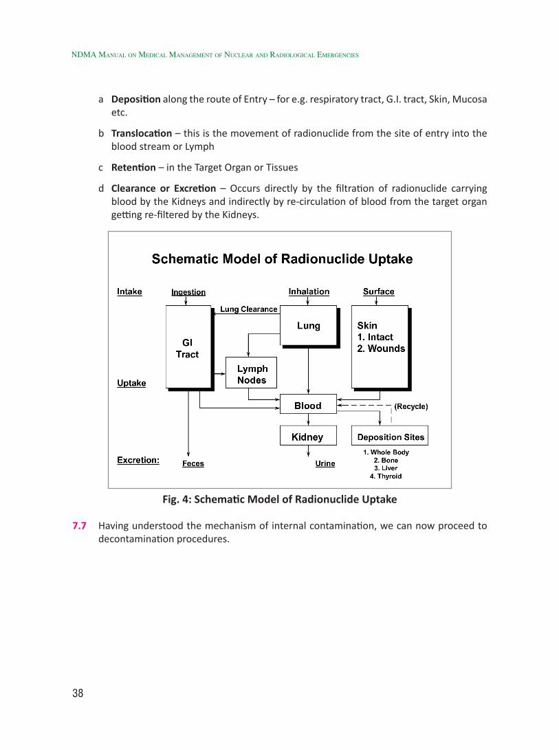
7.1 Before we proceed on to the internal decontamination, a brief account of Internal contamination will help us understand the decontamination procedures better.
7.2 It occurs due to accidental intake of radioisotope.
7.3 Radioisotope can enter the body by any of the following routes
(a) Inhalation, (b) Ingestion, (c) Injection, (d)Absorption through intact / broken skin and eyes
7.4 The hazards and their impact will depend upon the following:
a Amount of activity
b Site of deposition
c Type and energy of radiation emitted
d Sensitivity of specific tissues to penetration
e Effective half life
f Physico-Chemical nature of the contaminant
7.5 Effective Half Life is calculated by the formula
Radioactive half Life X Biological Half Life≠
Effective Half Life = ___________________________________ Radioactive half Life +Biological Half Life
* RadioactiveHalfLife: the time required for a quantity of a radioisotope to decay by half. e.g.: The radioactive half life of I131 is Eight days, hence if a sample of I131 has 10 m Ci of activity on January 1st, then Eight days later, i.e. on January 9th, its activity will be 5 mCi.
≠ BiologicalHalf Life: the time required for one half amount of the substance, such as a radionuclide, to be expelled from the body by natural metabolic processes, not counting the radioactive decay, once it has been taken in through inhalation, ingestion or absorption.
7.6 Internal contamination includes the following successive stages
Internal Contamination –Priciples of Uptake and Clearance7
38
NDMA MANuAl oN MeDicAl MANAgeMeNt of NucleAr AND rADiologicAl eMergeNcies
a Deposition along the route of Entry – for e.g. respiratory tract, G.I. tract, Skin, Mucosa etc.
b Translocation – this is the movement of radionuclide from the site of entry into the blood stream or Lymph
c Retention – in the Target Organ or Tissues
d Clearance or Excretion – Occurs directly by the filtration of radionuclide carrying blood by the Kidneys and indirectly by re-circulation of blood from the target organ getting re-filtered by the Kidneys.
Fig. 4: Schematic Model of Radionuclide Uptake
7.7 Having understood the mechanism of internal contamination, we can now proceed to decontamination procedures.

39
Internal Decontamination88.1 General treatment Plan
8.1.1 General treatment plan of internal decontamination will depend on the contamination route.
8.1.2 Contamination should be dealt with at the point of entry, particularly for those elements for which there is no effective therapy available. Hence, fixation or blocking of the radionuclide at the site of entry should be attempted so that blood uptake does not occur
8.1.3 Next, trapping it in blood during translocation and re-routing it towards its natural excretion should be attempted. This is important because the deposition in target organs starts as soon as the radionuclide circulates in the blood. Effectiveness of this treatment decreases with time, as more and more deposition will take place with passage of time.
8.1.4 The above two plans are the best methods, none the less, a third step of prevention of deposition in target organs can be most valuable at times, as in the case of 131I where, administering stable Potassium Iodide blocks the Thyroid and prevents the uptake of 131I.
8.2 Methods of Systemic Treatment8.2.1 Decontamination of G.I. Tract:
i Radioactive material may enter the G.I. tract either by ingestion through oral route or as a result of broncho-ciliary clearance mechanism following the inhalation.
ii As a principle it would be appropriate to remove or enhance the transit of the gastro-intestinal contents. Carry out the following to achieve this.
iii Carry out a Gastric Lavage through a Naso-Gastric tube. Emesis can be attempted in a conscious patient. However this may be done as a First Aid method in the absence of Medical help. Gastric Lavage is preferable. This is because, during gastric lavage, the radionuclide is removed through the tube, thereby preventing its possible re-deposition along the upper G.I. mucosa that may occur during the attempted emesis. Laxatives may be used to hasten the elimination of the radionuclide and to minimize the intestinal irradiation and absorption.
iv Magnesium Sulphate is a saline purgative which produces a relatively insoluble sulphate with radium and thus reduces its absorption.

40
NDMA MANuAl oN MeDicAl MANAgeMeNt of NucleAr AND rADiologicAl eMergeNcies
v Enemas may be considered where quick emptying of colon is desired.
vi Isotopic dilution method consists of giving large quantities of non–radioactive ion which competes with radioactive materials for absorption e.g. KI for Radioactive Iodine or Stable Phosphate for 32P.
vii Displacement therapy is a special form of dilution therapy. In this, a non-radioactive element of different atomic number, competes with the radionuclide for up take sites, e.g. Oral or IV Calcium increases excretion of Strontium
viii Specific therapeutic agents such as ion exchange resins, gels, antacids are also used to reduce intestinal absorption of radioactive material.
ix Certain mobilizing agents, increase natural turn-over process and help in enhancing elimination of radionuclide from the body tissues. e.g.
a Chelating agents like Ca DTPA, Zn DTPA are used for Plutonium contamination.
b Anti-thyroid drugs as Propyl thiouracil or Methimazole are administered when treatment with stable iodine may not be considered to be effective in advanced cases or if radioactive doses are high enough to justify their use.
c Ammonium chloride given orally is effective in mobilizing radio-strontium. Its effectiveness can be increased by simultaneous use of I.V. Ca gluconate.
8.2.2 Decontamination of Respiratory Tract:
i It is important to note that soluble particles (less than 5 microns) are translocated to blood and are deposited in the appropriate target organ
ii Insoluble particles get deposited in lung parenchyma and may get translocated to other organs at a low rate over many months or years or they may migrate to regional Lymph nodes by phagocytosis and the lymphoid channels
iii Contamination by inhalation may occur with Krypton, Xenon, radioactive Iodine and Plutonium. Krypton and Xenon need no treatment as they are short lived
iv In case of soluble particles which may be rapidly translocated to blood, treatment may be directed to trapping the radionuclide in the blood stream and enhancing their natural excretion.
v Use of inhalation with specific antidotes may be advocated e.g. DTPA aerosol inhalation in Plutonium.
vi Pulmonary lavage may be considered in cases of heavy non transportable radionuclide inhalation. However, risk benefit assessment should be done. The procedure should be considered only in high exposure cases in which reduction of dose can be expected to prevent acute or sub-acute effects such as radiation Pneumonitis or Fibrosis.

41
vii Before we proceed to the internal decontamination procedure for specific radionuclides, following is a table of the Radionuclides and their treatment.
Table 4: Radioactive Contaminants with Medical Significance and Possible Treatment
Radioactive Contaminant
Radiation Type
Target Organ
Contamination Mode* Treatment
Americium-241 α, γ Bone I / W Ca-DTPA, Zn-DTPA†Californium-252 γ, α, η Bone I / W Ca-DTPA, Zn-DTPA†Cerium-141, 144 β, γ GI, lung I / GI Ca-DTPA, Zn-DTPA†Cesium-137 β, γ Total body I / S / GI Prussian blue£
Cobalt- 60 Β, γTotal body / Lung
I / S / G.I.DTPA, D-Penicillamine
Curium-244 α, γ, η Bone I / GI Ca-DTPA, Zn-DTPA†
Iodine-131 β, γ Thyroid I / GI / S KI ¥ KIO3 Lugol’s iodine
Plutonium-239, 238 α, γ Bone I /W Ca-DTPA, Zn-DTPA†Polonium-210 α Lung I Dimercaprol‡
Phosphorous-32 Β Bones I / S / G.I.Stable Phosphate, Aluminum hydroxide antacid
Strontium-89, 90 β , γ Bone I / GI AlPO4** Tritium (3H) β Total body I / S / GI Forced H2O
§
Uranium-238, 235, 239 α, β, γ Bone I / S / W NaHCO3*** * Contamination Mode: I - inhalation; GI - gastrointestinal absorption; S- skin absorption; W-wound absorption ** The antacid aluminum phosphate in gel form used as a gastrointestinal adsorbent for radio strontium *** Sodium bicarbonate to maintain alkalinity of urine used in conjunction with diuretics † Calcium- and Zinc-DTPA, metal complexes of di-ethylene-tri-amine-penta-acetate. The calcium form is recommended for the first decontaminating dose, followed with the zinc form for subsequent doses.
‡ A mercury and arsenic poisoning chelation agent (very toxic)
¥ Agent blocking radioiodine absorption in tissues resulting in its dilution
§ Simple forced intake of water, resulting in tritium dilution
£ A dye used as an ion exchanger.
internal decOntaMinatiOn

42
NDMA MANuAl oN MeDicAl MANAgeMeNt of NucleAr AND rADiologicAl eMergeNcies
viii Facilities for Internal decontamination of specific radionuclides may not be available at PHC/CHC
ix Decision for transfer to higher centers shall be taken by the Doctor I/C of the PHC/CHC.
x Decontamination of Individual Radionuclide:
It is important to note that there is NO SINGLE ALL-PURPOSE DECORPORATION DRUG (COCKTAIL) that will protect against all internal radio contamination possibilities
Various radionuclides could be categorized into two groups, based on the probability of contamination occurring – (A) Common and (B) Less Common. Once the body gets internally contaminated by these, the decontamination/removal from the body needs to be carried out using specific decontaminants and decorporation agents., Their properties, deposition in the body organs and the clearance using specific agents is given in Annex-4.

43
9.1 For patients who were shown to have internal contamination levels below the ALI, no medical follow-up is required as there is no evidence of adverse effects. They need reassurance, possibly repeated reassurance, but no further studies or work-up.
9.2 Those who received decorporation drugs should have repeat measurements to determine whether or not treatment needs to be continued. These measurements may also help to establish biological half-life or half-lives, which could later be used in making dosimetry estimates.
9.3 Patients who received contamination levels above the ALI, and those to whom decorporation drugs were administered, need fairly accurate measurements of internal radioactivity levels and then calculated dosimetry estimates.
9.4 Health Physicist / Designated Monitoring staff will co-ordinate for these dosimetry.
9.5 In the event of some patients absorbing high doses of radiation, high enough to manifest the acute radiation syndrome, the patient should be referred to a Hematologist-Oncologist, as this is the specialty most capable of treating the acute bone marrow syndrome.
Follow Up of Internally Decontaminated Patients9

44
NDMA MANuAl oN MeDicAl MANAgeMeNt of NucleAr AND rADiologicAl eMergeNcies

45
Radiation Burns1010.1 The most common form of radiation burn is Sun Burn, caused due to over exposure to
UV radiation which is a non-ionizing radiation.
10.2 However, the term “Radiation burn” used in this chapter is the damage to skin or other biological tissues caused by exposure to ionizing radiation.
10.3 Caused mainly due to localized over exposure to ionizing radiation, e.g. in Industrial Radiography, inadvertent manual handling of Radioactive source or in cases of Radiotherapy.
10.4 Radiation burns evolve slowly over weeks to months. Hence detailed history of event and physical dosimetry may help in their management.
10.5 Effects of Ionizing Radiation on Skin:
10.5.1 Alpha particles do not penetrate the horny outer layer of skin, i.e. the epidermis. The main problem associated with the alpha emitters is the possibility of transfer into the body by absorption through intact or broken skin, inhalation or ingestion while eating with contaminated hands.
10.5.2 Beta particles penetrate the epidermis and cause intense irradiation of tissues and structures beneath the epidermis and therefore are a major health hazard.
10.5.3 Electro-magnetic radiation [X-Rays and Gamma rays] can cause damage, but because of their greater penetration they deposit less energy locally than beta rays. Low energy rays can cause more biological damage superficially than gamma rays.
10.6 Symptoms: Symptoms usually are
i Sensation of warmth
ii Onset of pains and paresthesia, pain could be intense and continuous
iii Disturbances to tactile and heat sensitivity
10.7 Signs: Following an acutely delivered single dose for 3 cm diameter fields, the threshold doses are in the following ranges:
i Erythema (Transient) : Between 2 and 3 Gyii Fixed Erythema : 6 Gy

46
NDMA MANuAl oN MeDicAl MANAgeMeNt of NucleAr AND rADiologicAl eMergeNcies
iii Dry Desquamation : 10 Gyiv Blisters : 12 Gyv Wet Desquamation : 15 Gyvi Ulceration : 20 Gyvii Necrosis : 25 Gyviii Gangrene : 30 Gy
10.8 Severity : severity of burns depends on
i Total dose received,
ii Type of Radiation, i.e. based on photon energy, e.g. Gamma radiation causes deep Gamma Burns, whereas Beta particles are not able to penetrate deep, hence produce shallow burns.
10.9 Investigations : Whenever a suspected case of Radiation Burns is under study, carry out the following investigations :
i Complete Blood Count
ii Chromosomal Aberration Analysis (Biological Dosimetry)
iii Color Doppler Studies / thermography / vascular scintigraphy
iv Semen Analysis within few days [earlier the better], second sample after 60 days post exposure.
v Slit Lamp examination of eyes, serially done at regular intervals to assess development of cataract
vi Collection of material from septic foci for culture and antibiotic sensitivity tests
vii Serial Color Photography to assess development and evolution of signs
viii Physical re-construction of accident – Physical dosimetry.
Most of these investigation facilities are available only in advanced centers, hence such cases may be referred there.
10.10 Medical Management: Medical management entails a comprehensive clinical approach with a team of Certifying Surgeon, General Surgeon, Dermatologist, Plastic surgeon,Hematologist, Oncologist, Ophthalmologist and Health Physicist.
10.10.1 Management involves a prolonged follow up spread over weeks and months, not only for treatment reasons but to assess the development of late sequelae if any and their treatment.
10.11 Specific treatment
10.11.1 For Erythema, both transient and fixed, use bland lotion such as calamine lotion, or Steroid Ointment, or Steroid with anti-biotic ointment like Neosporin Hydrocortisone.
10.11.2 Sterile protective dressings with silver sulphadiazine / Framycetin ointment /

47
any other anti-septic solution or ointment. Dressings should ideally be changed twice a day. In treatment of Radiation Burns following Radiotherapy, change of dressings have helped in reducing pain.
10.11.3 Pain can be reduced by the use of analgesics, those which do not cause bone marrow damage. Morphine at times is necessary.
10.11.4 Use of systemic broad spectrum antibiotics for control and treatment of any bacterial infections
10.11.5 Use of anti-fungal drugs like Fluconazole in standard doses to control or treat fungal infections if any.
10.12 Surgical treatment
10.12.1 Based on the nature of burn, following surgical interventions may be necessary
i Excision of wound
ii Escharectomy
iii Ulcerectomy
iv Necretomy
v Amputation
10.12.2 If the involved area is more than 2-3 sq.cm, skin grafting will be necessary. Partial or full thickness graft may be necessary depending upon the severity.
10.12.3 In cases with beta burns, early excision and skin grafting helps in relieving the pain
10.12.4 Larger areas involving extremities with necrosis or gangrene may require amputation.
10.12.5 Amputation, which is usually the terminal resort, is determined by the following factors –
i Intractable pain due to Ischemia
ii Size and Location of burn
iii Associated secondary infections
iv Extent of vascular damage
v Loss of functional value of the body part
10.12.6 Late Sequelae of radiation Burns: Following lesions may develop over a period of several months to years. They constitute the late sequelae, and hence long term follow up patients with Radiation burns is necessary.
i Chronic Radio-dermatitis – particularly if the dose is around 10 Gy and above
ii Keratosis
radiatiOn burnS

48
NDMA MANuAl oN MeDicAl MANAgeMeNt of NucleAr AND rADiologicAl eMergeNcies
iii Dry, fragile and brittle skin which may show areas of hypopigmentation / hyperpigmentation
iv Squamous or Basal cell carcinoma of skin,
v Cataract – if there is localized exposure to face or eyes
vi Sterility – if there is localized exposure to Gonads.

49
Admission1111.1 Admission to higher center/s will depend on the following factors:
a General condition of the patient
b Dose more than 1Gy
c Underlying chronic conditions exaggerated by the event directly or indirectly due to fear
d Level of radioactive contamination left behind on / in the body at the end of decontamination procedures. This will be viewed as “Risk to self” and as “Risk to others”.
e Concomitant injuries and infections needing Isolation.
f Social conditions like – presence of infants or pregnant lady at home.
11.2 Decision for admission will be jointly taken by doctors and health physicists/monitoring staff.
11.3 Once the admission is advised, the patient will be referred to center with admission facilities. Admission paper in that hospital will be made as routinely done for any indoor patient.
11.4 Patient will be kept in isolation.
11.5 Patient will be provided with hospital linen including the patient’s clothing. Kindly note that the linen will be changed daily and for internally contaminated patients, the soiled linen will be collected in separate yellow polythene bags. These will be sent to concerned authorities for proper decontamination and subsequent cleaning.
11.6 One nurse shall be deputed in around-the-clock shift for attending to the admitted patient(s).
11.7 These nurses will practice “barrier nursing techniques” while attending to the patient(s).
11.8 The adjoining ward shall be the supply station for these patients’ requirements.
11.9 The movement of equipment / consumables into this isolation ward shall be uni-directional. No material brought into this isolation ward will go back to general use, unless the material is properly decontaminated and certified to be contamination-free by the radiation monitoring staff. If any material continues to remain contaminated, it shall be disposed, as radioactive contaminated solid waste.

50
NDMA MANuAl oN MeDicAl MANAgeMeNt of NucleAr AND rADiologicAl eMergeNcies
11.10 All entries in the admission papers should be made with proper date and time. All instructions should be carried out ad-verbatim.
11.11 No visitors should be allowed inside the isolation area. This also includes the hospital staff that is not on duty in the isolation ward at a given point in time.
11.12 The ward personnel should also be told to wear protective clothing while attending to the requirements in the isolation ward. They should be properly instructed to preserve the urine and stool samples for investigations.
11.13 Treatment of admitted patient will include:
a Treatment for any specific co-morbid medical conditions
b Decontamination treatment,
c Any specific / specialized treatment – like transfusion, grafting etc.
d Medicines for any chronic illness, if the patient is taking them,
e Suitable antibiotics and supportive treatment
f Antacids, Laxatives as needed
g Vitamins and Anti-oxidants
h Pain killers and Anxiolytics
i Any others as per requirement.
11.14 Food provided to the patient should be –
a Fresh and nutritious
b Fully cooked. No uncooked food like salads, raita, fruits etc. should be given
c Food specifications of his / her underlying illness, viz. Salt restricted, Diabetic, etc. should be borne in mind
d Water should be properly filtered and boiled for at least 20 minutes to kill all the spores in it. It should be preserved in proper container and given to patient as needed. Authenticated sterile sealed Mineral water may be used as a safe alternative.
11.15 Based on the patient(s)’condition and the decontamination success, s/he may be either referred to higher center for further treatment, or may be discharged from the ward.
11.16 All papers such as case papers, investigation reports, radiological films (if any) etc. will be handed over to the state health authorities for proper filing.
11.17 Discharge papers – elaborately written - will be handed over to the patient. One copy of the same will be retained by the state health authorities and one copy will be sent to the Medical Superintendent / Certifying Surgeon of the nearest NPP Hospital.

51
Writing Notes1212.1 While writing notes make detailed entry of areas and readings (level) of contamination
before starting the decontamination procedure. Mark the body areas on the figure on the card – both on anterior and posterior surfaces.
12.2 Enter the notes regarding general examination, systemic examination, injuries if any.
12.3 Make a note of any past history and past medications.
12.4 Make a note of any drug allergies.
12.5 Write down step wise, each procedure carried out.
12.6 Make an entry of all the specimens collected and sent for bio-assay.
12.7 Make an entry of all investigations ordered.
12.8 Make entries of progressive developments.
12.9 Keep the spouse of the patient informed. This helps in reducing undesired anxiety.
12.10 At the end of each sitting of decontamination procedure, make detailed entry of areas and readings (level) of contamination before starting and at the end of the decontamination procedure. Mark the body areas on the figure on the card – both on anterior and posterior surfaces.
12.11 Keep all the investigation reports intact. Make a note of the reports in daily case sheet. Draw trends for investigation. It gives a good estimate of the prognosis.
12.12 All papers of the decontamination procedures and admission papers, discharge summary, investigations and follow up records should be neatly filed separately for each patient and submitted to the Office of Chief Medical Officer of the District.
12.13 All patients requiring follow up should be properly instructed regarding the time, the date and the place of follow-up. The follow-up should be done at the place of last decontamination / decorporation.
12.14 O/o the Chief Medical Officer of the district will retain these papers in hard copies and soft copies – scanned and converted into PDF files, until the death of the person / 30 years beyond the treatment completion / Age of 90 years of the person, whichever is later.
12.15 Please fill in all the Work sheets given at the end of this document meticulously. WS 3 deals with treatment of the patient from initial reporting to discharge.

52
NDMA MANuAl oN MeDicAl MANAgeMeNt of NucleAr AND rADiologicAl eMergeNcies

53
Acute Radiation Syndrome (ARS)13
13.1 The acute radiation syndrome occurs after whole-body or significant partial-body irradiation of greater than 1 Gy delivered at a relatively high-dose rate. The most replicative cells are the most sensitive to the acute effects of radiation, particularly, lympho-hematopoietic elements and intestinal crypt cells. The inherent sensitivity of these cells results in a constellation of clinical syndromes that predominate within a predictable range of doses of whole-body or significant partial-body exposure.
13.2 Clinical components of the acute radiation syndrome include the hematopoietic, gastrointestinal and neurovascular syndromes. The time course and severity of clinical signs and symptoms for the component syndromes at different dose ranges have been elaborated in Section. Each syndrome can be divided into 4 phases: prodromal, latent, manifest illness and recovery or death.
13.3 The following diagram is a Reference from: Annals of Internal Medicine. CLINICAL GUIDELINES. Medical management of the Acute Radiation Syndrome: Recommendations of the Strategic National Stockpile Radiation Working Group. Jamie K. Waselenko et al; 15 June 2004/ Vol 140/Issue 12/ Pg 1037 – 1051).
Approximate time course of clinical manifestations: Shown in the diagram is the approximate time for hematopoietic, gastrointestinal (GI), and central nervous system (CNS) symptoms at different ranges of dose of whole-body radiation for exposed, living persons. Hematopoietic changes include development of lymphopenia, granulocytopenia or thrombocytopenia. Gastrointestinal symptoms include nausea, vomiting or diarrhea and may also be accompanied by headaches. Cerebrovascular signs and symptoms include headache, impaired cognition, disorientation, ataxia, seizures, prostration and hypotension. Note that the signs and symptoms of different organ systems significantly overlap at each radiation dose and that cerebrovascular symptoms do not appear until exposure to a high whole-body dose. The relative severity of signs and symptoms is measured on an arbitrary scale.
13.4 Phases of ARS: Depending on the absorbed dose, symptoms appear within minutes, hours to weeks, following a predictable clinical course.
i Prodromal Phase: The prodromal phase of the acute radiation syndrome usually occurs in the first 48 hours but may develop up to 6 days after exposure.
ii Latent phase: The latent phase is a short period characterized by improvement of symptoms, as the person appears to have recovered. Unfortunately, this effect is transient, lasting for several days to a month.

54
NDMA MANuAl oN MeDicAl MANAgeMeNt of NucleAr AND rADiologicAl eMergeNcies
iii Manifest Illness Phase: Symptoms of manifest illness then appear and may last for weeks. This stage is characterized by intense immuno-suppression and is the most difficult to manage. If the person survives this stage, recovery is likely. Individuals exposed to a supra-lethal dose of radiation may experience all of these phases over a period of hours, resulting in early death.
iv Recovery Phase: This phase shows signs of recovery and patients regain normal health over a period of time.
These phases under different acute exposure conditions leading to various syndromes are detailed in Annex-5.
Fig. 5: Phases of ARS

55
Bio-Dosimetry in Acute Radiation Syndrome14
14.1 Individual bio-dosimetry is essential for predicting the clinical severity, treatment and survivability of exposed individuals and triaging those with minimal or no exposure. The 3 most useful elements for calculating the exposure dose are (a) time to onset of vomiting, (b) lymphocyte depletion kinetics and (c) the presence of chromosome dicentrics.
14.2 The rate of decline and nadir of the absolute lymphocyte count (ALC) over the initial 12 hours to 3 days after exposure is a function of cumulative dose. Lymphocyte depletion kinetics predict dose assessment for a photon-equivalent dose range between 1 and 10 Gy with an exposure resolution of approximately 2 Gy. Ideally, a complete blood cell count with leukocyte differential should be obtained immediately after exposure, 3 times per day for the next 2 to 3 days and then twice a day for the next 3 to 6 days. Day of 500 (of ALC) can be used to identify the severity of hematopoeitic syndrome and its prognosis.
14.3 It is recommended that 6 (and a minimum of 3) complete blood counts with differential be obtained within the initial 4 days after exposure, to calculate a slope for lymphocyte decline that can be used to estimate exposure dose. Complete blood counts with differential should then be obtained weekly or twice weekly until a nadir in neutrophil count is defined.
14.4 The chromosome-aberration cytogenetic bioassay, primarily the lymphocyte dicentrics assay introduced by Bender and Gooch, remains the gold standard for bio-dosimetry. A peripheral blood sample should be obtained without delay (for uniform whole body exposure) or at 24 hours after exposure (in cases of suspected non-uniform exposure) or later. The results will be available after 48 to 72 hours. The blood sample has to be collected in Li-heparin vials (color coded as green-cap vial).
14.5 Table showing Bio-dosimetry (Ref: Annals of Internal Medicine. CLINICAL GUIDELINES. Medical management of the Acute Radiation Syndrome: Recommendations of the Strategic National Stockpile Radiation Working Group. Jamie K. Waselenko et al; 15 June 2004/ Vol 140/Issue 12/ Pg 1037 – 1051)

56
NDMA MANuAl oN MeDicAl MANAgeMeNt of NucleAr AND rADiologicAl eMergeNcies
Dose Victims with Time to Absolute Lymphocyte Count Rate Constant Dicentrics in HumanEstimate Vomiting Onset of for Lymphocyte Peripheral Blood Vomiting Depletion Lymphocytes ______________________________________________ ________________ Day 0.5 Day 1 Day 2 Day 4 Day 6 Day 8 Per 50 Per 1000 Cells CellsGy % h X109 cells/L K n 0 – – 2.45 2.45 2.45 2.45 2.45 2.45 – 0.05-0.1 1-21 19 2.30 2.16 1.90 1.48 1.15 0.89 0.126 4 882 35 4.63 2.16 1.90 1.48 0.89 0.54 0.33 0.252 12 2343 54 2.62 2.03 1.68 1.15 0.54 0.25 0.12 0.378 22 4394 72 1.74 1.90 1.48 0.89 0.33 0.12 0.044 0.504 35 7035 86 1.27 1.79 1.31 0.69 0.20 0.06 0.020 0.63 51 10246 94 0.99 1.68 1.15 0.54 0.12 0.03 0.006 0.7567 98 0.79 1.58 1.01 0.42 0.072 0.012 0.002 0.8818 99 0.66 1.48 0.89 0.33 0.044 0.006 <0.001 1.019 100 1.56 1.39 0.79 0.25 0.030 0.003 <0.001 1.1310 100 0.48 1.31 0.70 0.20 0.020 0.001 <0.001 1.26
Table 5: Biodosimetry based on Acute Photon-Equivalent Exposures*
*Depicted above are the 3 most useful elements of biodosimetry. Dose range is based on acute photon-equivalent exposures. The second column indicates the percentage of people who vomit, based on dose received and time to onset. The middle section depicts the time frame for development of lymphopenia. Blood lymphocyte counts are determined twice to predict a rate constant that is used to estimate exposure dose. The final column represents the current gold standard, which requires several days before results are known. Colony-stimulating factor thereapy should be initiated when onset of vomiting or lymphocyte depletion kinetics suggests an exposure dose for which treatment is recommended (see Table 7 (of reference 13)). Therapy may be discontinued if results from chromosome dicentrics analysis indicate a lower estimate of whole-body dose.
Normal range, 1.4–3.5 3 109 cells/L. Numbers in boldface fall within this range.
The lymphocyte depletion rate is based on the model Lt = 2.45 3 109 cells/L 3 e– k(D)t, where Lt equals the lymphocyte count (3 109 cells/L), 2.45 3 109 cells/L equals a constant representing the consensus mean lymphocyte count in the general population, k equals the lymphocyte depletion rate constant for a specific acute photon dose, and t equals the time after exposure (days).
Number of dicentric chromosomes in human peripheral blood lymphocytes.

57
15.1 Treatment of acute radiation syndrome is not indicated when exposure dose is very low (<1 Gy) or very high (>10 Gy). Supportive and comfort care is indicated for people with an exposure dose greater than 10 Gy because their prognosis is grave.
15.2 Thus the medical management shall be applicable largely to the Hematopoietic Syndrome which falls between doses more than 1 Gy and less than 10 Gy.
15.3 Medical Management of Hematopoietic Syndrome
15.3.1 Treatment of radiological victims with hematopoietic syndrome varies with dose estimates, exposure scenarios and presenting symptoms.
15.3.2 Short-term therapy with cytokines is appropriate when the exposure dose is relatively low (<3 Gy).
15.3.3 Prolonged therapy with cytokines, blood component transfusion and even stem-cell transplantation may be appropriate when exposure dose is high (>7 Gy) or when traumatic injury or burns are also present.
15.3.4 Cytokine Therapy: Colony Stimulating Factors (CSFs) are the Hematopoietics which are widely used. The rationale for the use of CSFs in the radiation setting is derived from enhancement of neutrophil recovery in patients with cancer on chemotherapy who are treated with CSFs.
i Treatment should commence when the Absolute Lymphocyte Count drops to 500/ cu.mm and should continue until the Absolute Neutrophil Count rises to 1000/cu. mm
ii Others used are recombinant forms of (a) granulocyte macrophage colony-stimulating factor (rhGM-CSF) e.g. Sargramostim in dose 5-10 µg/Kg/d subcutaneously or equivalent dose of 200-400 µg/m2/d, and (b) granulocyte colony-stimulating factor (rhG-CSF) e.g. Filgrastim in dose 2.5 – 5 µg/Kg/d subcutaneously or equivalent 100-200 µg/m2/d. Also the mg pegylated form of G-CSF (pegylated G-CSF or pegfilgrastim) can be used in the dose of 6 once subcutaneously.
Medical Management of Acute Radiation Syndrome15
Note: (Kindly note that treatment of Acute Radiation Syndrome requires specialized centers., Hence, such patients are to be referred to Tertiary Care Facilities for expert management. However, the details of ARS in earlier chapters and the management mentioned here are for purpose of knowledge sharing).

58
NDMA MANuAl oN MeDicAl MANAgeMeNt of NucleAr AND rADiologicAl eMergeNcies
iii The value of CSFs in the treatment of radiation-induced myelo-suppression of the bone marrow lies in their ability to increase the survival, amplification, and differentiation of granulocyte progenitors. Both rhGM-CSF and rhG-CSF activate or prime neutrophils to enhance their functions, such as microbicidal activity. Both have been shown to hasten neutrophil recovery by approximately 3 to 6 days in humans after intensely myelotoxic therapies, including bone marrow and stem-cell transplantation.
15.3.5 Doses of Cytokines: (Ref: Annals of Internal Medicine. CLINICAL GUIDELINES. Medical management of the Acute Radiation Syndrome: Recommendations of the Strategic National Stockpile Radiation Working Group. Jamie K. Waselenko et al; 15 June 2004/ Vol 140/ Issue 12/ Pg 1037 – 1051)
Table 6: Recommended Doses of Cytokines*
Cytokine Adults Children Pregnant Women
Precautions
G-CSF or filgrastim Subcutaneous administration of 5 µg/kg of body weight per day, continued until ANC>1.0x10⁹ cells/L
Subcutaneous administration of 5 µg/kg per day, continued until ANC>1.0x10⁹ cells/L
Class C (same as adults)
Sickle-cell hemoglobinopathies, significant coronary artery disease, ARDS; consider discontinuation if pulmonary infiltrates develop at neutrophil recovery
Pegylated G-CSF or pegfilgrastim
1 subcutaneous dose, 6 mg
For adolescent >45 kg: 1 subcutaneous dose, 6 mg
Class C (same as adults)
Sickle-cell hemoglobinopathies, significant coronary artery disease, ARDS
GM-CSF or sargramostim Subcutaneous administration of 250 µg/m2 per day, continued until ANC >1.0x10⁹ cells/L
Subcutaneous administration of 250 µg/m2 per day, continued until ANC >1.0x10⁹ cells/L
Class C (same as adults)
Sickle-cell hemoglobinopathies, significant coronary artery disease, ARDS; consider discontinuation if pulmonary infiltrates develop at neutrophil recovery
*ANC = absolute neutrophic count; ARDS = acute respiratory distress syndrome; G-CSF = granulocyte colony-stimulating factor; GM-CSF = granulocyte-macrophage colony-stimulating factor.
Experts in biodosimetry must be consulted. Any pregnant patient with exposure to radiation should be evaluated by a health physicist and maternal-fetal specialist for an assessment of risk to the fetus. Class C refers to U.S. Food and Drug Administration Pregnancy Category C, which indicates that studies have shown animal, teratogenic, or embryocidal effects, but there are no adequate controlled studies in women; or no studies are available in animals or pregnant women.
i People at extremes of age (children < 12 years and adults > 60 years) may be more susceptible to irradiation and have a lower LD50/60. Therefore, a lower threshold exposure dose (2 Gy) for initiation of CSF therapy is

59
appropriate in such persons and in those who have major trauma, injuries or burns.
15.3.6 Transfusion:
Transfusion of cellular components, such as packed red blood cells and platelets, is required for patients with severe bone marrow damage. Fortunately, this complication does not typically occur for 2 to 4 weeks after the exposure, thereby permitting time for rapid mobilization of blood donors. Blood component replacement therapy is also required for trauma resuscitation. All cellular products must be leuko-reduced by irradiating to 25 Gy to prevent transfusion-associated graft-versus-host disease in the irradiated (and therefore immune-suppressed) patient. It may be difficult to distinguish transfusion-associated graft-versus-host disease from radiation-induced organ toxicity, which may include fever, pancytopenia, skin rash, desquamation, severe diarrhea and abnormalities on liver function tests (in particular, hyper-bilirubinemia).
15.3.7 Stem-Cell Transplantation:
Matched related and unrelated allogeneic stem-cell transplantations are life-saving and potentially curative treatments in patients with certain predominantly hematologic malignant conditions. A small number of radiation accident victims have undergone allogeneic transplantation from a variety of donors in an attempt to overcome radiation-induced aplasia.
If resources allow, transplantation should be considered in people with an exposure dose of 7 to 10 Gy who do not have significant burns or other major organ toxicity and who have an appropriate donor. Individuals with a granulocyte count exceeding 0.500x 10⁹ cells/L and a platelet count of more than 100 x 109 cells/l at 6 days after exposure may appear to have residual hematopoiesis and may not be suitable candidates for transplantation.
In the unusual circumstances where a syngeneic donor is available or previously harvested autologous marrow is available, a stem-cell infusion may be considered in patients with exposures exceeding 4 Gy.
15.4 Medical Management of Other Conditions and Special Care
15.4.1 Supportive Care: Supportive care includes the administration of antimicrobial agents, anti-emetic agents, anti-diarrheal agents, fluids, electrolytes, analgesic agents and topical burn creams. Transfusion of fresh irradiated platelets can be done.
15.4.2 Infections:
Susceptibility to infection results from a breach in the integument or mucosal barriers, as well as immune suppression consequent to a decline in lympho-hematopoietic elements. Several studies have indicated that administration
Medical ManageMent Of acute radiatiOn SyndrOMe

60
NDMA MANuAl oN MeDicAl MANAgeMeNt of NucleAr AND rADiologicAl eMergeNcies
of antibiotics reduces mortality rates in irradiated dogs in the LD50/30 range. Controlling infection during the critical neutropenic phase is a major contributing factor for successful outcome. In non-neutropenic patients, antibiotic therapy should be directed towards foci of infection and the most likely pathogens. Fluoroquinolones have been used extensively for prophylaxis in neutropenic patients. In patients who experience significant neutropenia (absolute neutrophil count < 0.500 x 10⁹ cells/l), broad-spectrum prophylactic antimicrobial agents should be given during the potentially prolonged neutropenia period. Prophylaxis should include a fluoroquinolone with streptococcal coverage or a fluoroquinolone without streptococcal coverage plus penicillin (or a congener of penicillin), antiviral drugs (acyclovir or one of its congeners) and antifungal agents (fluconazole).
Time since Radiation d
Physical dosimetry Dose from cytogenics
Sternum: 500 cGy
Ilacbone marrow, left front1000 cGy
Right back:150 cGy
6-8h
48-72h
Epilation11d
Secondary erythema19,49d
49d
9d
Dry epidermatitis18-20d
Exudativeepidermatitis25-27d
Radioepithelitis1-6, 8-16d
Gastritis63-73d
Interitis5-18, 22-30, 56-61, 68-74d.
Hepatitis79-93d
The begining of Primary erythema The begining of secondary skin changes
Body position at the moment of catastrophe(biological dosimetry data)
Head: 800 cGY
Chest: 950 cGY
1 5 10 15 20 25 30 35 40
10
8
6
4
2
100
Leuc
ocyt
e C
ount
cel
ls/m
m3
Stomach: 900 cGY
Fig. 6: Time Dependent Effects of ARS

61
Antimicrobial agents should be continued until they are clearly not effective (for example, the patient develops neutropenic fever) or until the neutrophil count has stabilized, i.e. absolute neutrophilic count more than 1.000 x 10 9 cells / l.
15.4.3 Comfort Measures
People with a high exposure dose, whose outcome is grim, must be identified for appropriate management. Since there is no chance of survival after irradiation with a dose of more than 10 to 12 Gy, it is appropriate for definitive care to be withheld from such individuals. Rather than being treated aggressively, these patients should be provided with comfort measures. This includes attention to pain management and general comfort as well as administration of anti-emetic and anti-diarrheal agents. In this devastating situation, psychological support is essential not only for the patient but also for family and friends, who may experience traumatic grief.
15.5 Summary of Medical Record of a radiation exposed patient:
Shown are the absolute leukocyte count (top left panel), estimated organ dose (top right panel), areas of skin injury (middle panels), injury to oral cavity and gastrointestinal system (bottom left panel), and body position relative to the radioactive source (bottom right panel) as a function of time after the exposure. To convert cells/mm3 to x109 cells/l, multiply by 0.001.
(Ref: Annals of Internal Medicine. CLINICAL GUIDELINES. Medical management of the Acute Radiation Syndrome: Recommendations of the Strategic National Stockpile Radiation Working Group. Jamie K. Waselenko et al; 15 June 2004/ Vol 140/ Issue 12/ Pg 1037 – 1051)
Medical ManageMent Of acute radiatiOn SyndrOMe

62
NDMA MANuAl oN MeDicAl MANAgeMeNt of NucleAr AND rADiologicAl eMergeNcies

63
Post Disaster Counselling1616.1 Introduction: It has been found that people who have witnessed the disaster, or have
been victims of it, have a long lasting impression on their minds. The disaster catastrophe, the losses during the disaster and the post disaster scene of destruction and the ensuing epidemics, all make their lasting impressions on one’s mind. This can happen even after individual calamities in the family, but are significantly pronounced in large scale disasters where the canvas of destruction is larger than expected. Compounding the effect is the sense of hollowness / emptiness that creeps in when one has lost everything s/he had. It is indeed a horrifying experience, when one has to restart life all over again amidst the pain of losing everything, at times even the pain of losing loved ones. These are very trying times and they affect practically every neighbor of the victim. The degree of pain and the tolerance of an individual to absorb the shock is what make the difference. It is during these times if apt help is rendered in the form of psychological support through means of counselling, it can help many to tide over the losses rather stoically. Hence by now PTSD (Post Traumatic Stress Disorder) is an accepted entity and all efforts should be taken to minimize the effects of PTSD.
16.2 Effects of Disaster: Effects of disaster can be
i Psychological: Impact includes loss of house or loved ones, fear, anxiety and uncertainty in facing one’s future and that of the family; trauma, and feeling of helplessness.
ii Physical impact is obvious in the form of crippling and physical dysfunction (which may cause psychological problem such as loss of self-confidence).
iii Social impact appears as a feeling of insecurity, a feeling of being treated unfairly, anxiety caused by uncertainty and higher sensitivity to rumor.
iv Economic impact is the loss of living resources, marketplace and customers.
v Housing and Environmental impact appears as the damage to buildings and environmental setting and facilities.
16.3 Post-Traumatic Stress Disorder: It is an anxiety disorder which some people suffer from, after living through a traumatic event. Fortunately all people who are exposed to traumatic events do not develop PTSD. Disaster and crisis counseling serve to prevent the development of PTSD through safety, stabilization, self-care and coping skills.
16.4 Aim: The Aim of counseling is to create a sense of safety and security in the midst of chaos. People who are affected may react with severe emotions or remorse. There can be sadness and aloofness both together. People should be convinced that Time is a Great Healer.

64
NDMA MANuAl oN MeDicAl MANAgeMeNt of NucleAr AND rADiologicAl eMergeNcies
Judith Herman’s triphasic model of trauma recovery should be borne in mind by every counselor. The elements of triphasic model are (a) safety and stabilization, (b) remembering and mourning, and (c) reconnecting and healing.
16.5 Helping clients cope:
In the immediate aftermath, always make the person feel safe and secure. S/he should be emotionally stabilized by helping him/her regain his/her confidence.
Subsequently help the client to remember the role and responsibilities played by his/her deceased dear ones and the need for the client to understand that a part of those roles and responsibilities now need to be performed by him/her. Life ahead has to be lived and lived productively.
It is important to remember that counselors should emotionally support the client but should never become emotionally a part of the client’s world.
All counselling activities should be continuous. WHO does not recommend single-session psychological debriefing to the general population as an early intervention. Most acute mental health problems during the acute emergency phase are best managed without medication following the principles of ‘psychological first aid’ (i.e., listen, convey compassion, assess needs, ensure basic physical needs are met, do not force them to talk, provide or mobilize company from family or friends).
Psychological debriefing as an early intervention after trauma is likely to be ineffective and some evidence suggests that some forms of debriefing may be counterproductive by slowing down natural recovery.
In order to assist the counselors, a very established and world-wide accepted tool is a scale called CAPS, i.e. Clinician Administered PTSD Scale. It is a questionnaire based analysis wherein 30 standard questions are to be asked and graded. This has been developed by the National Centre for Post-Traumatic Stress Disorder, Behavior Science Division, Boston VA Medical Centre and the Neurosciences Division West Haven VA Medical Centre, USA.
CAPS is highly recommended by all Psychologists and Psychiatrists world-wide to diagnose and grade the PTSD.
In developing mutual aid within the affected people as the care givers, one should always keep in mind that it is essential to help and guide rather than do the things for the victim. Care should be taken to see that the person is not shifted from one disaster to another, i.e. disaster of dependency.

65
Maintenance and Records1717.1 All medicines, kits, disposables, decorporating agents, chemicals, protective clothing,
waste bags/ baskets/ containers, stationery, shall be maintained at every PHC/ CHC/ District Hospital/ Civil Hospital/ Medical College/ Super-specialty Medical College/ Hospital.
17.2 It shall be ensured by the CMO of every district that each medical centre under her/ his jurisdiction is in a state of readiness round the clock.
17.3 All materials and staff shall be provided by the CMO of the district.
17.4 The Medical Officer I/c or the Medical superintendent of the hospital shall in turn keep her/ his centre in a state of readiness.
17.5 The M.O I/c or Medical superintendent shall carry out monthly inspection in the last week of every month of all requisite things, check stock taking, and submit a report to District CMO latest by the 7th of the next month. S/he shall make a list of all material requiring replacement and send it to the District CMO who, in turn, shall ensure prompt and timely replacement. It shall be ensured by the District CMO that, at no point of time, the centers fall deficient on any account.
17.6 All inspections and actions taken should be properly logged.
17.7 CMO shall prepare a plan of mock exercises between the health centers of her/ his district.
17.8 CMO shall in turn keep the DM of the district informed of every development.
17.9 All patient related documents are to be retained in the O/o CMO until 30 years post completion of treatment of an individual (Mandatory) if patient is alive and regular follow up is on, or Death of the patient.

66
NDMA MANuAl oN MeDicAl MANAgeMeNt of NucleAr AND rADiologicAl eMergeNcies

67
Appendix – I
General Instructions for Awareness in a Large Scale Radiological Emergency Scenario
In the event of an emergency situation, it is natural that apprehensions and panic will override rationality. It will be the onus of the medics and the paramedics to alleviate these fears. These can be achieved by – (i) educating the people, distributing information booklets through interactions with school / college children and (ii) dissemination of correct information during the crisis hours.
As for the first part, the state health department may do this as part of their weekly activities and along with other health programs on Immunization, Family welfare, Sanitation and Hygiene etc.
As for the second part, the doctors and para-medics should correctly dissipate the information they receive from the emergency centres. They should discourage gossip and speculative talk, and instead instill confidence in the public. All non-governmental channels of public media, both print and mass media, should co-operate with governmental agencies in broadcasting the information given to them by governmental agencies only, and refrain from broadcasting speculative news or views.
During regular interactions with the public through various programs, they should inform the people about the following dos and don’ts:
• Firstly it should be emphasized that the precautions to be taken are similar to those which would be required to be taken during any chemical/ industrial releases/ leaks.
• The only difference in the situation is that, unlike chemical releases, air borne radioactive releases cannot be seen, cannot be smelt and cannot be physically felt.
• Thus any person would find it hard to believe that there is any radioactive spread, but none the less, they should follow instructions.
• Whenever there is information about air borne spread, all the people who are outdoors should cover their mouths and noses with a wet handkerchief. This will prevent any air borne contamination from entering the respiratory and gastro-intestinal tracts.
• While being outdoors, they should not eat anything, particularly food bought from open shops or vendors.
• All those who are outdoors, should try and return home as soon as possible or else enter into the nearest available closed shelters/ homes.

68
NDMA MANuAl oN MeDicAl MANAgeMeNt of NucleAr AND rADiologicAl eMergeNcies
• Before one runs away from the emergency area, one has to ascertain the wind direction. This can be done by a simple method. Take a piece of cloth or a handkerchief and suspend it in the air by holding one corner tip. The cloth will sway towards the wind direction. One should always run perpendicular to this direction to be least affected.
• Shop keepers should close down their shops. Shops selling open eatables like vegetables, fruits, cooked food etc. should immediately cover their goods with a plastic sheet or a tarpaulin, as is commonly done during the rainy season to prevent the goods from getting wet. Therefore shop keepers and vendors should realize the importance of this plastic sheet, and thus keep it with them round the year instead of only for the rainy season.
• After reaching the shelter, please discard the handkerchief in a plastic pouch / polythene bag and keep it aside. The relief officers may require this for assessing the contamination levels. Do not use it again. Keep it out of reach of children, in particular.
• While indoors, please close all the windows and doors.
• Cover all the open food and water cans. Please also cover open food drums/ cans for cattle fodder.
• The village elders or the members of the panchayat should cover any open well with a large tarpaulin and properly secure it with ropes so that it is not displaced by blowing winds. A placard bearing the message “This well is covered on the orders of the Village Panchayat / competent authority can be placed near the well. Please do not remove the cover or consume water for drinking / washing / irrigation / any other purpose until authorized by the Government agencies / Panchayat”. This sign should be in vernacular and pictorial for easy understanding.
• Please listen to Public information notices issued by government agencies only given on Radio, Television or by rescue officers over loud speakers. Please follow the instructions given by them ad verbatim, as they will be relaying the most authentic information and in your interest.
• The information will normally be dynamic, meaning that the instructions will change as per the change in situation. The personnel issuing notices over loudspeakers should number every announcement along with the time of issue. So that villagers do not get confused as to which instruction they are supposed to follow.
• As a standard procedure, the following sequence is to be adhered to: Stay Indoors > Tab Potassium Iodide Distribution > Sheltering > Evacuation > Relief Centres.
a Stay Indoors:
➢ Whenever there is an announcement for staying indoors, all members of the family should be indoors.
➢ If possible, keep your cattle inside.
➢ If the cattle cannot be kept inside, cover their sheds with a plastic sheet or tarpaulin sheet on the windward side so as to block the air borne contamination from settling on and around the cattle.

69
➢ Villagers store grains for the family’s year-long requirement, and farmers store the harvested grain before it is shifted to open markets for sale. Please cover all such stored grains with plastic sheets or tarpaulin.
➢ While indoors, please, use power (electricity consumption) sparingly. Only use those appliances as required. Power supply interruption is expected. If you have multiple cell phones in your family, keep only one in use at a time to conserve battery power. If there is power supply, all cell phones should be charged. This will ensure battery availability in emergency.
➢ Do not panic.
➢ One should keep listening to radio / T.V. / Public announcements on loudspeakers for further instructions.
b Tab Potassium Iodide Distribution: The relief team may visit the houses to distribute the Tablets of Potassium Iodide, if such a situation is warranted.
➢ These tablets will protect from developing the ill effects of Radioactive Iodine. This is not a panacea for all radioactive agents. Hence after taking this, one should not have a false sense of confidence that they are fully protected and don’t need to follow any further instructions.
➢ When the relief teams enquire about the number of family members available in the house, one should say the exact number. The dose of this tablet is one full tablet for adults – both genders, half tablet for children between age from 3 years to fourteen years – both genders, and one fourth tablet for children below the age of three years for both the genders.
➢ This medicine is not to be used for cattle. This information is particularly important for members of the public in rural areas where cattle are treated like family members. Pregnant ladies can and must take this tablet.
➢ This medicine is safe, and does not have any adverse effects, hence should be taken as and when asked to take. At the time of actual distribution, the person advising may not be a Doctor, but could be any member of the relief team authorized to carry out the task.
➢ These tablets are normally available at the nearest PHC or CHC. (The actual number of tablets, based on the population of the affected sectors will be made available through the Emergency control centres for distribution in the target sectors only.
c Sheltering: Sheltering is a step prior to evacuation. People from the affected sector are asked to gather at temporary shelter areas. This is a pre-designated place, viz. a school, college, or any government office building large enough to house the population. Whenever there is an announcement, people should be asked to gather at the shelter area and observe the following:
➢ They should not run and cause a stampede.
➢ They should keep calm. This step is generally taken as a preventive measure and it does not mean that everyone is in danger.
➢ All the family members should stay together. Particular care should be taken of the
appendix – i

70
NDMA MANuAl oN MeDicAl MANAgeMeNt of NucleAr AND rADiologicAl eMergeNcies
children, so that they are not far away from the elders of the family. It would be better if an identity badge is pinned on the clothes of children with their names, parents’ names, addresses and one contact telephone (cell) number if available.
➢ Cattle should be left behind. They will be evacuated through suitable means by the veterinary authorities. As a routine practice, evacuation of cattle should precede evacuation of humans, so that the cattle owners have an idea as to who has taken their cattle and where they have been kept for safety. The cattle owners should also be informed about the collection / delivery of their cattle after the emergency. The cattle owners should be encouraged to suitably tag their cattle with identification so that while receiving them back, there are no disputes. Bar coding for identification should be used wherever possible. Safe evacuation of cattle before humans will instill confidence amongst the villagers and their co-operation and compliance with state orders will be better.
➢ Houses should be locked properly; police force should be deputed to protect the belongings of the evacuated people.
➢ During evacuation from homes to shelter areas, if possible, plastic or rubber raincoats / rain suits along with cap should be worn. Raincoats / rain suits are advisable, because they cover the complete body and thus any airborne particles settling down would settle on this and would not come in contact with the body. Also, they have an added advantage that radiation (α and β) emitted from the released particles can be blocked by these plastic raincoats.
➢ Raincoats/ rain suits are to be disposed of when told to do so by authorities in a Yellow colored collection bin. Plastic/ rubber is easier to be decontaminated, hence they are preferred.
➢ From the shelter point, one should go to relief camps in buses arranged for the purpose by the authorities.
➢ At the entry to relief camps, raincoats should be removed. They should be disposed properly, without dusting, into the containers placed for the purpose.
➢ Removing the raincoat will remove most of the externally deposited air borne contamination, if any.
➢ After this, frisking by radiation detection monitors is to be done.
➢ All people should maintain calm, and should not try to jump the queue, so that the frisking can be completed in an orderly manner and within a shorter time frame. Any person found with external contamination, should be sent to the nearest PHC / CHC / district hospital for decontamination.
➢ Decontamination is discussed in the main document (Section 6).
➢ Others should continue to stay in relief camps until governmental orders to return to their places of residence.
➢ While in relief camps, instructions are to be strictly followed and hygiene maintained at personal level so that cumulatively the place is maintained clean and hygienic. It is to be

71
borne in mind that crowded places, if unhygienic, form an ideal platform for the spread of infectious diseases.
d Return / Relocation: After the government gives the green signal to return to respective residences, the following procedures need to be observed.
➢ Take a proper bath with soap and water.
➢ Do not consume the food left behind.
➢ Do not consume stored water.
➢ Do not pluck fresh vegetables from the shrubs and consume, unless they are cleared for consumption by suitable agencies.
➢ Please remove the covers (plastic sheets / tarpaulin sheets) with which you had covered the stored grain / harvested crop / fodder etc., only when permitted by the authorized monitoring agencies.
➢ Keep these sheets neatly folded, so that the exterior surface is turned inwards, and dispose them in suitable containers. While folding them, care should be taken to see that the sheets are not dusted. This will prevent the settled contaminated dust from spreading around.
➢ Please ensure that the cattle are given a proper bath after they are returned.
➢ These cattle will also be monitored by suitable agencies.
➢ If the cattle is internally contaminated, the authorities may advise not to consume the milk in case of Cow, Buffalo, Goat, Camel etc. In that case, please avoid consumption of milk and also do not sell it. Care should also be taken to see that the offspring of the cattle are not allowed to suckle. This is because radioactive elements are secreted in the milk in case of internally contaminated animals.
➢ The same don’ts are applicable to cattle meant for meat consumption / trading.
➢ Water from wells should be consumed only after it is approved for consumption.
➢ It may so happen that, the authorities may advise to avoid consumption of hand pumps/ submersible pumps after a lapse of a few days. This delay will be due to delay in contamination of ground water. Normally it may take a few days for the ground water to get contaminated. In this case, do not panic if you have already consumed the water earlier in the interim period. In all probability, it was not contaminated.
appendix – i
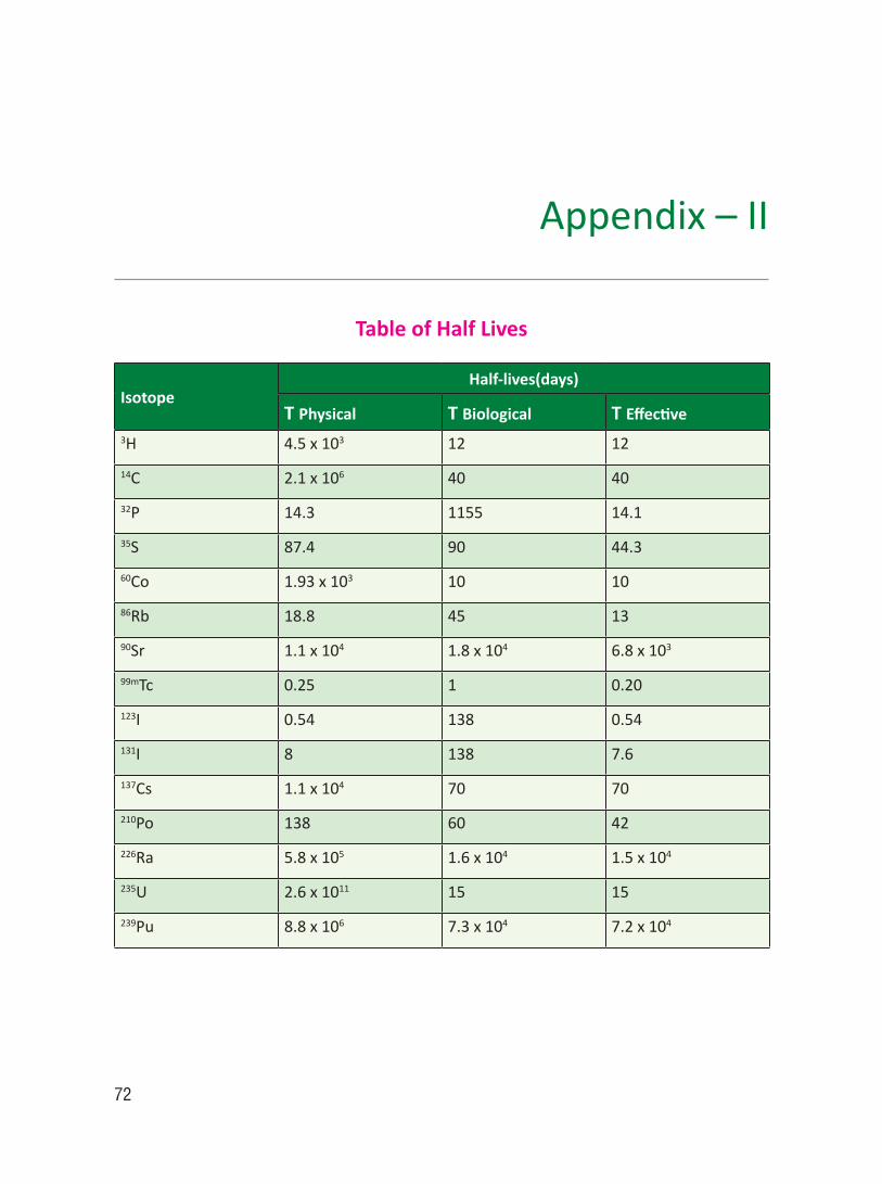
72
NDMA MANuAl oN MeDicAl MANAgeMeNt of NucleAr AND rADiologicAl eMergeNcies
Appendix – II
Table of Half Lives
IsotopeHalf-lives(days)
T Physical T Biological T Effective3H 4.5 x 10³ 12 12
14C 2.1 x 10⁶ 40 40
32P 14.3 1155 14.1
35S 87.4 90 44.3
60Co 1.93 x 10³ 10 10
86Rb 18.8 45 13
90Sr 1.1 x 10⁴ 1.8 x 10⁴ 6.8 x 10³
99mTc 0.25 1 0.20
123I 0.54 138 0.54
131I 8 138 7.6
137Cs 1.1 x 10⁴ 70 70
210Po 138 60 42
226Ra 5.8 x 10⁵ 1.6 x 10⁴ 1.5 x 10⁴
235U 2.6 x 10¹¹ 15 15
239Pu 8.8 x 10⁶ 7.3 x 10⁴ 7.2 x 10⁴

73
References
1. K. Kostial, B. Karga, et al. The effect of composite oral treatment for Internal contamination with several radio nuclides on 131I thyroid uptake in humans. Journal of Applied Toxicolgy 2006; Vol 6, Issue 2; Pg 109 – 111.
2. Sonawane, V. R. ; Jagtap, V. S.; Pahuja, D. N.; Rajan, M. G. R. ; Samuel, A. M. Difficulty in dislodging in vivo fixed radio strontium. Health Physics. 87(1):46-50, July 2004.
3. Lugol’s Solution of Iodine 2 % - M.S.D.S.
4. Asaf Durakoviæ, Medical Effects of Internal Contamination with Uranium. Croatian Medical Journal v.40, n.1, Mar99.
5. Radiation Emergencies – Fact Sheet on DTPA. Issued by Dept.of Health and Human services, Center for Disease Control and Prevention, U.S.A. Oct.2006
6. Basic Disaster Life Support, Chapter 4, Nuclear and Radiological Events. American Medical Association
7. Guidance for Industry Internal Radioactive Contamination — Development of Decorporation Agents. U.S. dept of Health and Human Services,, Food and Drug Administrator, Center for Drug Evaluation and Research. March 2006.
8. I.A.E.A. Module 15, External and Internal Contamination – Decontamination and Decorporation.
9. Tareg Bey, Prof. Dept of Emergency Medicine, University of California, Irvin, USA. “ Review of Most Commonly used Antidotes and Decorporation Agents” – A Paper presented at Taipei, Taiwan, June 2007
10. Medical Management of Radiological Casualties,- Second Edition- A handbook by Military Medical Operations, Armed Forces Radiobiology Research Institute, Bethesda, Maryland USA. – April 2003
11. Carol S. Marcus, Richard B. Sparks, et al. Medical Management of Internally Radio-contaminated Patients. Los Angeles County Dept. of Health Services, Emergency Medical Service Agency. June 2006
12. Medical management of Internally radiocontaminated patients– By Bruce Beach Nov 2006.

74
NDMA MANuAl oN MeDicAl MANAgeMeNt of NucleAr AND rADiologicAl eMergeNcies
13. Medical management of Acute Radiation Syndrome: Recommendations of the Strategic National stockpile Radiation Working Group. Walter Reed Army Medical center and Catholic University of America, Washington D.C.; USA. – Annals of Internal Medicine, 2004, June15; 140 (12); 1037 – 51.
14. T.Hirama, S.Tanosaki, S.Kandastu et al. Initial medical management of patients severely irradiated in Tokai Mura criticality accident; British Journal of Radiology; ( 2003), 76; 246 – 53
15. Istvan Turai, atalin Veress, Bengul Gunalp, Gennadi Souchkevitch .- Medical response to radiation incidents and radio nuclear threats.; B.M.J.; 2004; 328 : 568 –572 ( 6 March)
16. Handbook for Medical management of persons exposed in radiation Accidents – AERB Safety Manual No: AERB/SM/MED –2
17. Guide on Medical management of Persons Exposed in Radiation Accidents. AERB Guide No. SG/MED – 1. Issued – April 1990.
18. Lecture Notes on an Approach to Medical Management of Radiation. Emergencies, Published by BARC Medical Division.
19. I.C.R.P. 30, 54, 61, 68, 78.
20. World Health Organisation (2003) Mental Health in Emergencies: Mental and Social Aspects of Health of Populations Exposed to Extreme Stressors. Department of Mental Health and Substance Dependence, WHO, Geneva
21. van Emmerik et al. (‘Single session debriefing after psychological trauma: a meta-analysis’. Lancet. 2002 Sep 7; 360: 766-71),
22. Rose et al. (‘Psychological debriefing for preventing post traumatic stress disorder [PTSD] [Cochrane Review]’. In: The Cochrane Library, Issue 2, 2004. Chichester, UK: Wiley),
23. National Institute of Mental Health (‘Mental Health and Mass Violence: Evidence-based Early Psychological Interventions for Victims/survivors of Mass Violence. A Workshop to Reach Consensus on Best Practices’. NIH Publication No. 02-5138. Washington: US Government Printing Office, 2002
24. Medical Management of Radiological Casualties – Chapter-Emergency response. Online Third Edition June 2010, AFFRI Special publication 10-1
25. Generic Procedures for medical response during a nuclear or radiological emergency – IAEA April 2005 – co-sponsored by IAEA and WHO

75
WO
RK S
HEET
- 1
Mon
th /
YEA
R
WO
RKSH
EET
FOR
ISSU
E O
F TH
ERM
O-L
UM
INES
CEN
CE D
OSI
MET
ERS
[TLD
]N
.B.:
TLD
are
to b
e nu
mbe
red
as: N
o: T
ype
of H
ealth
Car
e Fa
cilit
y*/N
ame
of P
lace
or L
ocati
on/
Nam
e of
Dis
tric
t/ N
umbe
r * P
HC
/ CH
C /
DH
/ M
C fo
r Pr
imar
y H
ealth
Cen
ter;
Com
mun
ity H
ealth
Cen
ter;
Dis
tric
t Hos
pita
l; M
edic
al C
olle
ge re
spec
tivel
y
To b
e se
nt to
TLD
Lab
in D
uplic
ate.
Sec
ond
copy
to b
e si
gned
by
TLD
lab
I/c
and
retu
rned
bac
k to
the
Prin
cipa
l Dis
patc
her
Sl.N
oTL
D
No.
[E
xam
ple
give
n be
low
]N
ame
of
the
Pers
onD
ate
of
Issu
eTi
me
of
Issu
eD
ate
of
Retu
rnTi
me
of
Retu
rnTo
tal N
o of
Hou
rs
Use
d in
Hou
rs &
M
inut
es H
H:M
M
Sign
atur
eRe
mar
ks
1CH
C/D
ibai
/BSR
/12
- do
- /2
3 4 5 6 7 8 9 10 N.B
: (1)
Thi
s W
orks
heet
nee
ds t
o be
fille
d fo
r Ea
ch d
ay o
f th
e U
se. (
2) If
the
Dec
onta
min
ation
exe
rcis
e co
ntinu
es o
n su
bseq
uent
day
s, t
he
empl
oyee
s sh
ould
use
the
sam
e TL
D o
n ea
ch s
ucce
ssiv
e da
y. (3
) If a
new
mem
ber j
oins
the
team
on
succ
essi
ve d
ay, s
/he
shou
ld u
se n
ew T
LD.
(4) O
nce
the
com
plet
e de
cont
amin
ation
is o
ver
for
all p
atien
ts a
nd th
e M
edic
al P
roce
dure
s te
rmed
as
“Com
plet
e”, t
he T
LDs
shou
ld b
e se
nt
to T
LD re
adin
g La
bora
torie
s au
thor
ized
by
Atom
ic E
nerg
y Re
gula
tory
Boa
rd. T
hese
labs
will
rece
ive
the
used
TLD
s fo
r ana
lysi
s an
d is
sue
fres
h TL
Ds
to th
e us
er. R
e-nu
mbe
ring
of T
LDs
shou
ld b
e sa
me
as m
entio
ned
earli
er. (
5) In
eve
nt o
f “no
use
”, th
ese
TLD
s sh
ould
be
sent
to th
e sa
me
labo
rato
ries
on la
st w
orki
ng d
ay o
f eve
ry m
onth
for i
ssue
of f
resh
TLD
s.
Sign
atur
e of
the
I/c
DD
/MM
/YYY
Y D
D/M
M/Y
YYY
Sign
atur
e of
Dec
onta
min
ation
Tea
m
[Dat
e of
Dis
patc
h of
all
Abo
ve T
LDs
[D
ate
of R
ecei
pt o
f all
Abo
ve T
LDs
TL
D L
ab In
-Cha
rge
to
TLD
Lab
aut
hori
zed
by A
ERB]
by
TLD
Lab
aut
hori
zed
by A
ERB]
wOrk Sheet - 1

76
NDMA MANuAl oN MeDicAl MANAgeMeNt of NucleAr AND rADiologicAl eMergeNcies
WO
RK S
HEET
- 2
REPO
RT F
OR
THE:
Mon
th /
YEA
R
WO
RKSH
EET
FOR
THER
MO
-LU
MIN
ESCE
NCE
DO
SIM
ETER
S [T
LD] A
NAL
YSIS
REP
ORT
N.B
.: TL
D a
re t
o be
num
bere
d as
: Typ
e of
Hea
lth C
are
Faci
lity*
/Nam
e of
Pla
ce o
r Lo
catio
n/ N
ame
of D
istr
ict/
Num
ber
* PH
C /
CHC
/ D
H/
MC
for
Prim
ary
Hea
lth C
ente
r; C
omm
unity
Hea
lth C
ente
r; D
istr
ict H
ospi
tal;
Med
ical
Col
lege
resp
ectiv
ely
To b
e se
nt t
o I/
cDec
onta
min
ation
Tea
m/
Prin
cipa
l Dis
patc
her
from
who
m r
ecei
ved
in D
uplic
ate.
Sec
ond
copy
to
be s
igne
d by
D
econ
Tea
m I/
c an
d re
turn
ed b
ack
to th
e TL
D la
b
Sl.N
oTL
D
No.
[E
xam
ple
give
n be
low
]N
ame
of th
e Pe
rson
Tota
l N
o of
H
ours
U
sed
in
Hou
rs
&
Min
utes
HH
:MM
Dat
e of
R
ece
ipt
of T
LD
Dat
e of
A
na
lysi
s of
TLD
Dos
e in
mSv
Sign
atur
eRe
mar
ks
1CH
C/D
ibai
/BSR
/12
- do
- /2
3 4 5 6 7 8 9 10 N.B
: (1)
Thi
s W
orks
heet
nee
ds t
o be
fille
d fo
r ea
ch d
ay o
f th
e U
se. (
2) If
the
Dec
onta
min
ation
exe
rcis
e co
ntinu
es o
n su
bseq
uent
day
s, t
he
empl
oyee
s sh
ould
use
the
sam
e TL
D o
n ea
ch s
ucce
ssiv
e da
y. (3
) If a
new
mem
ber j
oins
the
team
on
succ
essi
ve d
ay, s
/he
shou
ld u
se n
ew T
LD.
(4) O
nce
the
com
plet
e de
cont
amin
ation
is o
ver
for
all p
atien
ts a
nd th
e M
edic
al P
roce
dure
s te
rmed
as
“Com
plet
e”, t
he T
LDs
shou
ld b
e se
nt
to T
LD re
adin
g La
bora
torie
s au
thor
ized
by
Atom
ic E
nerg
y Re
gula
tory
Boa
rd. T
hese
labs
will
rece
ive
the
used
TLD
s fo
r ana
lysi
s an
d is
sue
fres
h TL
Ds
to th
e us
er. R
e-nu
mbe
ring
of T
LDs
shou
ld b
e sa
me
as m
entio
ned
earli
er. (
5) In
eve
nt o
f “no
use
”, th
ese
TLD
s sh
ould
be
sent
to th
e sa
me
labo
rato
ries
on la
st w
orki
ng d
ay o
f eve
ry m
onth
for i
ssue
of f
resh
TLD
s.
Sign
atur
e of
the
I/c
DD
/MM
/YYY
Y D
D/M
M/Y
YYY
Si
gnat
ure
ofTL
D L
ab
[Dat
e of
Dis
patc
h of
Rep
ort o
f TLD
s
Dat
e of
Rec
eipt
of a
ll A
bove
TLD
I/
c D
econ
Tea
m
t I/c
Dec
on T
eam
] Re
port
s by
I/c
Dec
on T
eam

77
WORKSHEET 3DECONTAMINATION / TREATMENT CASE SHEET
1 Name of Decon / Treatment Facility : [e.g] CHC / Dibai / BSR [short for District
Bulandshahr]
2 Nature of Emergency : NPP / RDD / OS [Nuclear Power Plant / Radio- Logical dispersal Device / Orphan source
3 Likely radio-Isotopes Involved :
[Information to be provided by Radiological Monitoring Authorities]
4 Date & Time of Emergency Declared : DD/MM/YYYY at HHMM in 24 hour clock
5 Name of the Patient :
6 Sex : M / F If Female: Pregnancy : Yes / No
7 Age : _____Years Date of Birth [If Av] : DD/MM/YYYY
8 Address :
9 Sector around NPP : “A” to “P”
[mention mandatorily in event of Emergency due to NPP]
10 Identification Document no : ___________________________
[If Av. Viz. Aadhar/Voter ID etc]____________________________(Specify type)
11 Patient Number : CHC/Dibai/BSR/REM*/OPD/Number dated
DD/MM/YYYY
* radiological emergency management
12 Likely Date & Time of Exposure : DD/MM/YYYY at HHMM Hrs in 24 hour &
[T1] Minute mode [This shall be provided by the Au-thorities tracking the spread of contamination in association with Environmental Survey Reports]
13 Change of Clothes and Bath Given : Yes / No If Yes: mention place If No : Do it
14 Oral Tablet Potassium Iodide : Yes / No If Yes: mention Date & Time Given Dose Given: 1 Tab / ½ Tab / ¼ Tab If No : Give stat
15 Whether Wet Cloth Used to cover
the Nostrils : Yes / No
16 Past History : DM / HT / Chronic Pulmonary Diseases / CVA / IHD / Hypothyroid / Hyperthyroid / Malignan-cies / Congenital Anomalies / Others / NIL
Details if Yes : _________________________________________________________
17 Additional History for Females : L.M.P.: DD/MM/YYYY
LCB. : DD/MM/YYYY
Obstetrics History : G__P__A__L__ [ M__, F ___]
wOrk Sheet - 3

78
NDMA MANuAl oN MeDicAl MANAgeMeNt of NucleAr AND rADiologicAl eMergeNcies
18 History of Known Drug allergies : ______________________________________
19 Regular Treatment : (if any) ________________________________
______________________________________
20 Classify Present Case as : (Please Tick the relevant Box)
• Only External Contamination [Take up for Decontamination]
• No External Contamination but with only Internal Contamination – likely to occur only by route of inhalations or Ingestion [InternalDecontamination at Primary/Secondary Heath Center if Dose <1Gy, at Tertiary Center if Dose > 1 Gy. Initiate decontamination before transfer]
• Both External Contamination & Internal Contamination [Simultaneous External & Internal Decontamination at Primary/Secondary Heath Center if Dose <1Gy, at Tertiary Center if Dose > 1 Gy. Initiate decontamination before transfer]
• Open Wounds [Injuries] with contamination [Decontaminate wounds as priority]
• Open Wounds [Injuries] without contamination [Clean & Cover wounds with waterproof dressing to prevent contamination from entering]
• Only Exposure and no contamination whatsoever [OPD Treatment if Dose < 1 Gy; transfer to tertiary care center if Dose > 1 Gy.]
• Co-Morbid Conditions with Contamination [Attend to Co-morbid Life threatening conditions first, Take up Decontamination when patient is stable]
• Co-Morbid Conditions without Contamination [Attend to medical emergencies, if no emergency treat on OPD with counseling]
21 Possible Routes of Exposure : Absorption / Inhalation / Ingestion
22 Physical Dosimetry Doses : External ______m Sv; Internal ____m Sv; To-tal___ m Sv
23 Present Complaints : Nausea / Vomiting / Loose Motions / fever / Headache / Tingling Numbness
Nausea : Yes / No Time of Onset__________
Vomiting : Yes / No [T2]Time of Onset__________ Frequency: ____nos / hr
Loose Motions : Yes / No Time of Onset__________ Frequency: ____nos / hr
Fever : Yes / No Time of Onset__________
Headache : Yes / No Time of Onset__________
Headache Location : Frontal / Temporal / Occipital / Parietal / Complete
Side affected : Unilateral - Right / Left; OR Bilateral
Associated Neck Pain : Yes / No

79
Giddiness : Yes / No
24 Clinical Findings:
General condition : Healthy / Weak / Debilitated
Level of Consciousness : Conscious / Drowsy / Stupor / Confabulated / Coma / Uncon-scious
Breathlessness : Yes / No
Pulse : _________ / min
B.P. : _________ mm Hg
Temp. : _________° F
Body Weight : _________ Kgs
Cyanosis : Yes / No
Pallor : Yes / No
Clubbing : Yes / No
Congenital Anomalies : Yes / No Details:____________________________
Breath Sounds : Normal / Adventitious
Heart Sounds : Normal / Added Sounds
If Normal – Rhythm: Regular / Irregular
Goitre : Yes / No
25 Send Following Sample for Assessment: Please tick the tests for which the samples are sent as checklist
Sample Investigation Advised
Quantity Container First Sample At what Time
Frequency thereafter
Dispatch To
Blood CBC; including Differential Count and Absolute Lymphocy te Count
2 cc Ethyl Diamine Tetra Acetic acid [EDTA] Vaccutainer
Within 3 Hrs of Exposure
Once very 6 Hourly upto 48 Hrs. Thereafter as per need
Can be done locally in PHC also
- do - RBS, 2 cc Sodium Flouride Vaccutainer
On Admission
Clinician’s Decision
- do -
- do - Urea, Creatinine, LFT, Electrolytes
3 cc Plain Vaccutainer OR SST[Serum Separation Tube]
On Admission
Clinician’s Decision
- do -
wOrk Sheet - 3

80
NDMA MANuAl oN MeDicAl MANAgeMeNt of NucleAr AND rADiologicAl eMergeNcies
Urine Routine Microscopic & Creatine – creatinine clearance ratio
5 cc Clean Plain Test Tube
On admission
Clinician’s Decision
- do -
- do - Baseline metabolites of Radionuclide
10 cc In a Sterile Test Tube
On Admission
24 hour collection every 24 Hourly until internal decontamina-tion successfully termed as OVER
To Laboratories Authorized by CMO
Stool / Faeces
Baseline metabolites of Radionuclide
Complete Void
Clean Container
First Sample after admission
24 hour collection every 24 Hourly until internal decontamination successfully termed as OVER
To Laboratories Authorized by CMO
Swabs from body orifices
To assess possibility of Internal contamination–
Single swab
Clean Test Tube
Before Deconta-mination
After deconta-mination,
Radiological Monitoring Team
Swabs from Wound
- do - Single swab
- do - - do - - do - - do -
26 Additional Tests :
• ECG for elderly or if clinical condition warrants
• Oxygen saturation by Pulse Oximetry – if Contamination by Inhalation is suspected or if clinical condition warrants
• Penicillin sensitivity Test before commencing treatment with Cap DPenicillamine.
• Semen Examination for Males:
a First sample within Forty days of Exposure [for base line studies. Earlier the better] dt________________
Total Sperm count__________ Live Sperm Count__________, Dead Sperm Count ______________ Motility Percentage___________
b Second sample after 60 days of exposure: dt _______________
Total Sperm count__________ Live Sperm Count__________, Dead Sperm Count ______________ Motility Percentage___________
• Serial colored photography for Skin Burns: [Please number each photo serially with date for respective patients]

81
• Plethysmography• Thermography• Color Doppler of the affected limb in case of skin Burns• EEG• Electron Spin Resonance• Chromosomal Aberrations Studies. Send the patient to the lab for sample collection
27 Based on the requirement commence decontamination procedures as detailed in respective chapters of the main document. You can also refer to Checklist 1 for external Decontamination and Checklist 2 for Internal Decontamination as ready reckoner
28 Proceed and Fill in the Supplements to this WS 3 for easy management
Supplement 1 of WS 3
DETAILS OF WOUNDS AND CONTAMINATION
Name :
Patient No:[vide No11 on Pg 1]
Before Decontamination / After Decontamination
Date: DD/MM/YYYY Decontamination Attempt No: ___________
Time of Commencement: HHMM Time of Termination: HHMM
Reasons for Termination: _______________________________________
• Please mark on the diagram the areas of wounds / injuries and contamination.• Please fill this supplement 1 at the start and end of Each sittings [attempts] of
Decontamination• For Detailed Decontamination Procedure refer to the main document
wOrk Sheet - 3

82
NDMA MANuAl oN MeDicAl MANAgeMeNt of NucleAr AND rADiologicAl eMergeNcies
WO
RKSH
EET
3Su
pple
men
t 2 o
f WS
3
Nam
e :
Pa
tient
No:
[vid
e N
o11
on P
g 1]
INVE
STIG
ATIO
N C
HA
RT fo
r fr
eque
ntly
requ
ired
inve
stiga
tions
Dat
e
Tim
e*
Hou
rs fr
om
Expo
sure
#
Test
s
TLC
DC
Abs
olut
e Ly
mph
ocyt
e Co
unt
Abs
olut
e N
eutr
ophi
l Co
unt
Hb
Plat
elet
s
RBC
Ure
a
Crea
tinin
e
Pota
ssiu
m
Sodi
um
Chlo
ride
*Tim
e of
Sam
ple
Colle
ction
. # H
ours
at s
ampl
e co
llecti
on s
ince
T1
[col
umn
12] N
.B.:
Plea
se m
aint
ain
this
to p
lot t
rend
on
Gra
ph

83
Supplement 2 of WS 3
N.B.: Please plot the Absolute Lymphocyte Count of the patient and compare with the standard Andrew’s nomogram given above to ascertain the extent of Radiation Injury
wOrk Sheet - 3
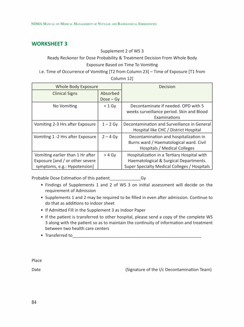
84
NDMA MANuAl oN MeDicAl MANAgeMeNt of NucleAr AND rADiologicAl eMergeNcies
WORKSHEET 3Supplement 2 of WS 3
Ready Reckoner for Dose Probability & Treatment Decision From Whole Body
Exposure Based on Time To Vomiting
i.e. Time of Occurrence of Vomiting [T2 from Column 23] – Time of Exposure [T1 from
Column 12]
Whole Body Exposure DecisionClinical Signs Absorbed
Dose – GyNo Vomiting < 1 Gy Decontaminate if needed. OPD with 5
weeks surveillance period. Skin and Blood Examinations
Vomiting 2-3 Hrs after Exposure 1 – 2 Gy Decontamination and Surveillance in General Hospital like CHC / District Hospital
Vomiting 1 -2 Hrs after Exposure 2 – 4 Gy Decontamination and hospitalization in Burns ward / Haematological ward. Civil
Hospitals / Medical CollegesVomiting earlier than 1 Hr after Exposure [and / or other severe symptoms, e.g.: Hypotension]
> 4 Gy Hospitalization in a Tertiary Hospital with Haematological & Surgical Departments.
Super Specialty Medical Colleges / Hospitals
Probable Dose Estimation of this patient_____________Gy
• Findings of Supplements 1 and 2 of WS 3 on initial assessment will decide on the requirement of Admission
• Supplements 1 and 2 may be required to be filled in even after admission. Continue to do that as additions to indoor sheet
• If Admitted Fill in the Supplement 3 as Indoor Paper
• If the patient is transferred to other hospital, please send a copy of the complete WS 3 along with the patient so as to maintain the continuity of information and treatment between two health care centers
• Transferred to______________________________________________________
Place
Date (Signature of the I/c Decontamination Team)

85
WORKSHEET 3Supplement 3 of WS 3
INDOOR ADMISSION PAPERImp.: This should always be filled in as a continuation to the WS 3 with its Supplements 1 & 2.
Not to be filled as a stand-alone form
Admitted from the OPD of the same Center : Yes / No
If No – Center from where transferred In : ___________________________________
Date and Time of Transfer in : DD/MM/YYYY at HHMM
Patient Number [OPD Treatment] : [vide column 11]
Name of Indoor Admitting Center : [e.g.] CHC / Dibai / BSR [short for District Bulandshahr]
Date and Time of Admission : DD/MM/YYYY at HHMM Hrs
Indoor Admission Number : CHC/Dibai/BSR/REM*/IPD/Number
* Radiological emergency Management
---------------------------------------------------------------------------------------------------------------------------
Status of External Contamination – Yes / No.
Decontamination Successful : Yes / No
If Yes : Treat for the effects of radiation if dose exposure > 1 Gy
If No : Carry out Decontamination until successful
---------------------------------------------------------------------------------------------------------------------------
Status of Internal Contamination – Yes / No Probable Route of Entry: Inhalation / Ingestion / Absorption
If Yes : Respiratory / G.I.
Decontamination Done : Yes / No
Decontamination successful : Yes / No
If Yes : Management of Effects of Radiation depending upon Dose
If No : Carry out Internal Decontamination until successful
---------------------------------------------------------------------------------------------------------------------------
For Procedures of Decontamination follow the main document chapters
Supplement 4 of WS 3
CONTINUATION SHEET FOR DAILY NOTES FOR INDOOR SHEET
[Please make your daily clinical notes entry with date and time. Use additional sheets of this supplement 4 as needed until discharge
wOrk Sheet - 3
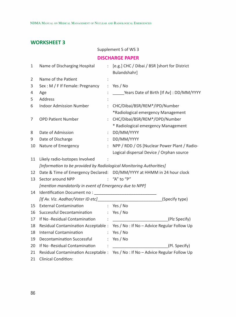
86
NDMA MANuAl oN MeDicAl MANAgeMeNt of NucleAr AND rADiologicAl eMergeNcies
WORKSHEET 3Supplement 5 of WS 3
DISCHARGE PAPER1 Name of Discharging Hospital : [e.g.] CHC / Dibai / BSR [short for District
Bulandshahr]
2 Name of the Patient :
3 Sex : M / F If Female: Pregnancy : Yes / No
4 Age : _____Years Date of Birth [If Av] : DD/MM/YYYY
5 Address :
6 Indoor Admission Number : CHC/Dibai/BSR/REM*/IPD/Number
*Radiological emergency Management
7 OPD Patient Number : CHC/Dibai/BSR/REM*/OPD/Number
* Radiological emergency Management
8 Date of Admission : DD/MM/YYYY
9 Date of Discharge : DD/MM/YYYY
10 Nature of Emergency : NPP / RDD / OS [Nuclear Power Plant / Radio-
Logical dispersal Device / Orphan source
11 Likely radio-Isotopes Involved :
[Information to be provided by Radiological Monitoring Authorities]
12 Date & Time of Emergency Declared: DD/MM/YYYY at HHMM in 24 hour clock
13 Sector around NPP : “A” to “P”
[mention mandatorily in event of Emergency due to NPP]
14 Identification Document no : ___________________________
[If Av. Viz. Aadhar/Voter ID etc]____________________________(Specify type)
15 External Contamination : Yes / No
16 Successful Decontamination : Yes / No
17 If No -Residual Contamination : ________________________(Plz Specify)
18 Residual Contamination Acceptable : Yes / No : If No – Advice Regular Follow Up
18 Internal Contamination : Yes / No
19 Decontamination Successful : Yes / No
20 If No -Residual Contamination : ________________________(Pl. Specify)
21 Residual Contamination Acceptable : Yes / No : If No – Advice Regular Follow Up
21 Clinical Condition:

87
WORKSHEET 3
Parameter Presenting Initially [on OPD]
On Admission On Discharge
Pulse Rate
B.P.
Resp. Rate
Level of Consciousness
Conscious/Drowsy/Stupor/Confabulated/Coma/Unconscious
Conscious/Drowsy/Stupor /Confabulated/Coma/ Unconscious
Conscious
Body Weight Kgs Kgs Kgs
Cyanosis : Yes / No
Pallor : Yes / No
Clubbing : Yes / No
Congenital Anomalies [Specify]
Breath Sounds
Heart Sounds Normal/ Added Sounds
Normal/ Added Sounds
Normal/ Added Sounds
If Normal – Rhythm: Regular / Irregular Regular / Irregular Regular / Irregular
Goitre
TLC / c.c
DC P L M E B P L M E B P L M E B
Absolute Lymphocyte Count
Absolute Neutrophil Count
Platelets
Hb
wOrk Sheet - 3
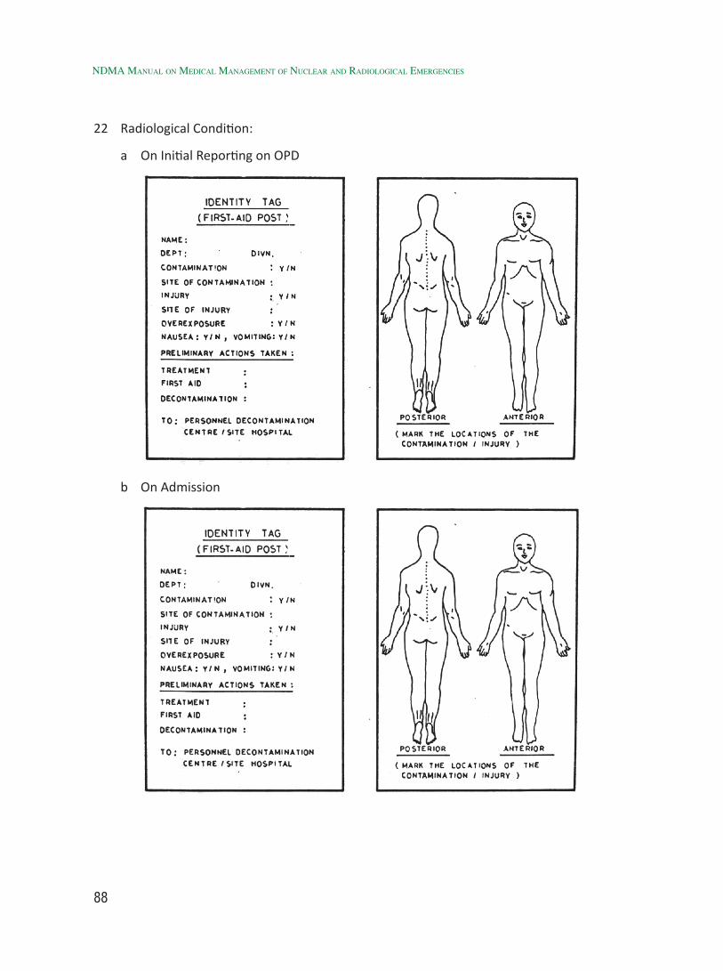
88
NDMA MANuAl oN MeDicAl MANAgeMeNt of NucleAr AND rADiologicAl eMergeNcies
22 Radiological Condition:
a On Initial Reporting on OPD
b On Admission

89
c On Discharge
23 Status of External Contamination:
Area of the Body [including orifices]
Radionuclide Type of Radiation
On Initial Reporting on OPD in cps
On Admission in cps
On Discharge in cps
wOrk Sheet - 3
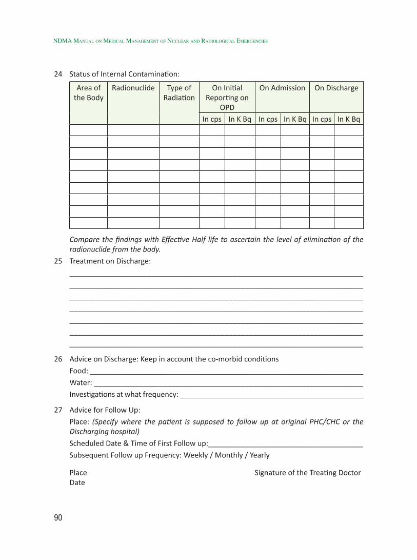
90
NDMA MANuAl oN MeDicAl MANAgeMeNt of NucleAr AND rADiologicAl eMergeNcies
24 Status of Internal Contamination:
Area of the Body
Radionuclide Type of Radiation
On Initial Reporting on
OPD
On Admission On Discharge
In cps In K Bq In cps In K Bq In cps In K Bq
Compare the findings with Effective Half life to ascertain the level of elimination of the radionuclide from the body.
25 Treatment on Discharge:
________________________________________________________________________
________________________________________________________________________
________________________________________________________________________
________________________________________________________________________
________________________________________________________________________
________________________________________________________________________
________________________________________________________________________
26 Advice on Discharge: Keep in account the co-morbid conditions
Food: ___________________________________________________________________
Water: __________________________________________________________________
Investigations at what frequency: _____________________________________________
27 Advice for Follow Up:
Place: (Specify where the patient is supposed to follow up at original PHC/CHC or the Discharging hospital)
Scheduled Date & Time of First Follow up:______________________________________
Subsequent Follow up Frequency: Weekly / Monthly / Yearly
Place Signature of the Treating Doctor Date
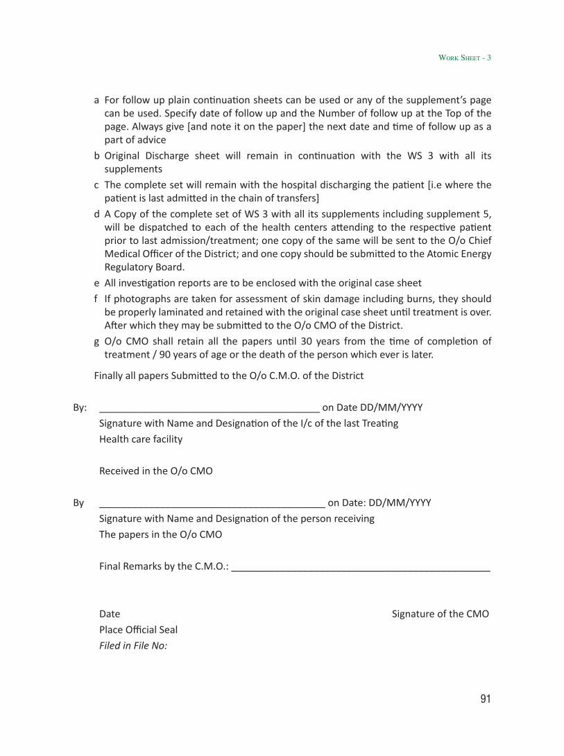
91
a For follow up plain continuation sheets can be used or any of the supplement’s page can be used. Specify date of follow up and the Number of follow up at the Top of the page. Always give [and note it on the paper] the next date and time of follow up as a part of advice
b Original Discharge sheet will remain in continuation with the WS 3 with all its supplements
c The complete set will remain with the hospital discharging the patient [i.e where the patient is last admitted in the chain of transfers]
d A Copy of the complete set of WS 3 with all its supplements including supplement 5, will be dispatched to each of the health centers attending to the respective patient prior to last admission/treatment; one copy of the same will be sent to the O/o Chief Medical Officer of the District; and one copy should be submitted to the Atomic Energy Regulatory Board.
e All investigation reports are to be enclosed with the original case sheet
f If photographs are taken for assessment of skin damage including burns, they should be properly laminated and retained with the original case sheet until treatment is over. After which they may be submitted to the O/o CMO of the District.
g O/o CMO shall retain all the papers until 30 years from the time of completion of treatment / 90 years of age or the death of the person which ever is later.
Finally all papers Submitted to the O/o C.M.O. of the District
By: ________________________________________ on Date DD/MM/YYYY
Signature with Name and Designation of the I/c of the last Treating
Health care facility
Received in the O/o CMO
By _________________________________________ on Date: DD/MM/YYYY
Signature with Name and Designation of the person receiving
The papers in the O/o CMO
Final Remarks by the C.M.O.: _______________________________________________
Date Signature of the CMO
Place Official Seal
Filed in File No:
wOrk Sheet - 3

92
NDMA MANuAl oN MeDicAl MANAgeMeNt of NucleAr AND rADiologicAl eMergeNcies
Checklist 1 for WS 3 - External Decontamination1 Wounds : Clean with wet swabs, dose assessment of swabs, discard as solid
waste. Flush wounds with normal saline, Evert the edges to clean undersurface of edges. Flush until decontamination is complete or e/o imminent bleeding from the wound floor is observed. Flush specific decontamination solution depending upon radio-nuclide. If painful use 4% Xylocaine jelly. All scabs should be collected for Dose assessment.
2 Ears : Dry wipe with Ear Buds. Wet wipe with Moist Ear bud [moist--ioned with Distilled water / NS. Collect in labeled plastic pouch. Dose assessment. Discard as solid waste. Do not syringe the ear. In Case of perforated Tympanic membrane, contamination from External Ear will enter Middle ear. Decontaminating Middle Ear Would be difficult
3 Nose : Blow the nose on Tissue paper, dose assessment, discard in labeled plastic pouch as solid waste. Dry wipe with Buds followed by wet buds as above. Dose assessment and discard as solid waste
4 Oral cavity : Rinse with water. Collect the liquid, dose assessment, discard as Liquid waste. Repeat rinse with medicated gargles. Discard all Collections as liquid waste.
5 Eyes : Eye wash with distilled water or Normal saline. Wash from Medial canthus to Lateral canthus as opening of lacrimal duct is close to medial canthus. Through lacrimal duct contamination can spread to pharynx and further to GI tract. While Eye wash cover the Ipsilateral ear complete with a water proof covering. Collect the eye wash liquid and discard as Liquid waste.
6 Hair : Decontaminate using 4% Cetrimide Shampoo / Shave and collect in labeled plastic pouch. Dose assessment. Discard as Solid waste
7 Nails : Nail Clipping – collect in labeled plastic pouch. Dose assessment. Discard as solid waste
8 Nail Beds : Decontamination with 1% Cetrimide solution
9 Hot Spots : Mark the hot spots. Clean from periphery to center. Priority to areas with higher contamination. All gauze pieces, cotton balls etc used are to be discarded as solid waste. If any solutions are used, discard them as liquid waste.
Order of General cleansing to be done with
a 1% cetrimide solution,
b 5 % Sodium hypochlorite [for face further dilution to 1:5 ratio],
c Saturated solution of Potassium Permangnate,
d For removing stains of KMnO4 use 10% solution of Sodium bisulphite

93
For specific Cleansing –
a 1% DTPA solution for Plutonium & transplutonics, Iodine, Cesium, Rruthenium, Barium, Lanthanium, and Cobalt
b Potassium Rhodizonate crystals for Wounds with Strontium
c 1.4% Soda-Bicarbonate solution for Uranium
d Lugol’s Solution for Iodine
e Acetic acid solution at pH 4 to 5 OR Vinegar for Phosphorous.
Finally dress the area with Lanolin dressings
Checklist 2 for WS 3 – Internal DecontaminationG.I. Tract : a Insert Ryle’s Tube. Give Gastric Lavage with normal saline or Water.
Collect all lavage liquid. Dose assessment. Discard as Liquid waste. Discard RT as solid waste.
b Start I.V. Line – Normal saline / Isolyte M / 5 % DNS
c Give Mucaine gel / Magaldrate / Sucralfate antacid 30 ml
d Mag Sulph solution [saline purgative] for Radium.
e Give Enema for enhancing elimination from colon
f Potassium Iodide for Iodine. Propyl thiouracil or Methimazole if very high contamination
g Stable phosphate for 32P
h Oral / I.V. Calcium Gluconate for Strontium. Oral ammonium chloride along with IV Cal Gluconate is more effective
i Ca DTPA / Zn DTPA for Plutonium contamination
j Discard all tubing and disposables as solid waste
Resp. Tract : a DTPA aerosol for Plutonium inhalation
b Monitor Oxygen Saturation by Pulse Oximeter
c Oxygen Inhalation SOS.
d Pulmonary Lavage weighing Risk V/s Benefits Decision jointly to be taken by Surgeon/Physician/Anaesthetist For patients with Age below 30 yrs a maximum permissible Lung burden [MPLB] of 100 shall be the safe criterion. Can Also be done with MPLB of 50 with due risks explained. First lavage ideally within 1 hour or after 2-3 days. Thereafter twice a week for two weeks, followed by once a week for total ten lavages. Lavage with Normal Saline [9gms NaCL / Ltr]. DTPA [1gm / Litre] may be added to lavage fluid. Each washing should be for 3 minutes. Collect all lavage fluid in labeled containers and send for Histological and Radio-toxical studies.
e discard all tubing and catheters as solid waste.
wOrk Sheet - 3

94
NDMA MANuAl oN MeDicAl MANAgeMeNt of NucleAr AND rADiologicAl eMergeNcies
WO
RKSH
EET
4
Mov
emen
t Rec
ord
of R
adio
-acti
ve W
aste
Mat
eria
lN
ame
of H
ealth
Cen
tre
Gen
erati
ng W
aste
:
CHC/
Dib
ai/B
SR
Was
te C
onsi
gnm
ent N
o.
: __
____
____
____
_
Dat
e of
Dis
patc
h :
DD
/MM
/YYY
Y
Sl.N
o.Ba
g /
Cont
aine
r N
oTy
pe o
f was
te –
Sol
id /
Liq
uid
Dat
e of
Col
lecti
onRe
mar
k –
Dec
onta
min
ate
/ D
ispo
se
Dat
e Si
gnat
ure
of S
iste
r I/
c Se
nt T
o: N
ame
of A
ERB
Aut
hori
zed
Was
te D
econ
tam
inati
on /
D
ispo
sal O
R M
O I/
c H
ealth
Cen
tre
Cent
re o
f the
Dis
tric
t [W
DC]
Rece
ived
at W
DC
:
On
: DD
/MM
/YYY
Y Si
gnat
ure
of th
e I/
c W
DC
N.B
: Fill
thi
s in
Dup
licat
e. O
ne c
opy
shou
ld b
e si
gned
by
I/c
WD
C on
rec
eipt
of b
ags/
cont
aine
rs a
nd s
ent
back
to
MO
I/c
Of t
he
Hea
lth c
entr
e se
ndin
g th
e w
aste
. One
cop
y of
this
sho
uld
be s
ent t
o O
/o C
MO
by
the
I/c
WD
C al
ong
with
the
repo
rt a
fter
ana
lysi
s on
WS
5

95
WO
RKSH
EET
5
Repo
rt o
f Sta
tus o
f Rad
io-a
ctive
Was
te M
ater
ial
Nam
e of
AER
B A
utho
rize
d W
aste
Dec
onta
min
ation
/ D
ispo
sal C
entr
e of
the
Dis
tric
t [W
DC]
:
Nam
e of
Hea
lth C
entr
e G
ener
ating
Was
te :
CHC/
Dib
ai/B
SR
Was
te C
onsi
gnm
ent N
o: _
____
____
____
____
____
____
Dat
e of
Dis
patc
h of
repo
rt
: D
D/M
M/Y
YYY
Repo
rt N
o: _
____
____
____
____
____
____
____
____
___
Sl.N
o.Ba
g /
Cont
aine
r N
oTy
pe o
f was
te –
Sol
id /
Liqu
idD
ate
of
Dec
onta
min
ation
/ D
ispo
sal
Mat
eria
l D
econ
tam
inat
ed &
Re
turn
ed fo
r re
use
Rem
ark
– D
econ
tam
inat
ed/
Dis
pose
d
Dat
e Si
gnat
ure
of I/
c W
DC
Dis
patc
hed
to :
Hea
lth C
entr
e G
ener
ating
Was
te
Repo
rt R
ecei
ved
at H
ealth
Cen
tre
:
O
n: D
D/M
M/Y
YYY
Si
gnat
ure
of th
e M
.O. I
/c H
ealth
Cen
tre
N.B
: Fill
this
in D
uplic
ate.
One
cop
y sh
ould
be
sign
ed b
y M
.O. I
/c H
ealth
Cen
tre
on r
ecei
pt o
f thi
s an
d se
nt b
ack
to I/
c W
DC
One
co
py s
houl
d be
sub
mitt
ed o
O/o
CM
O b
y M
O I/
c H
ealth
Cen
tre
wOrk Sheet - 5

96
NDMA MANuAl oN MeDicAl MANAgeMeNt of NucleAr AND rADiologicAl eMergeNcies
Annex-1
Radiation Effects
Deterministic Effects: These effects are known to occur at high radiation doses received over a short period. They have a threshold dose below which they do not occur. The severity of these effects is proportional to the dose and is generally associated with the death of large fraction of cells in a specific organ or tissue e.g. Radiation Sickness, Hematopoietic/Gastro-Intestinal/Neuro-Vascular Syndromes, Cataract, Sterility- either temporary or permanent, and Skin Burns.
Stochastic Effects:They are probabilistic in nature and are observed for exposures in excess of 100mSv but as a conservative estimate treated as not having any threshold. Probability of incidence increases with dose rather than severity. If the non-lethal genetic alteration occurs in somatic cells, it is a precursor for carcinogenesis, and if it occurs in germ cells, it may result in genetic disorders in progeny of exposed individuals.
Radio-sensitivity:Radio-sensitivity varies in different types of tissue. While all cells can be destroyed by a high enough radiation dose, highly radiosensitive cells or tissue exhibit deleterious effects at much lower doses than others. As stated in the Bergonié-Tribondeau law, rapidly dividing, undifferentiated cells in tissue are most sensitive to radiation effects. Several of the most sensitive tissues and systems follow this law.
Highly radiosensitive tissue- lymphoid, bone-marrow elements, gastrointestinal epithelium, gonads (testis and ovary) and foetal tissue.
Moderately radiosensitive tissue- skin, vascular endothelium, lung, kidney, liver, lens and thyroid glands in childhood.
Least radiosensitive tissue- central nervous system, endocrine (except gonad), thyroid glands in adults, muscle, bone and cartilage and connective tissue. The least radiosensitive tissue, although radio-resistant, is less capable of cell renewal than highly sensitive tissue. Some - especially neurons, glial cells of the brain and muscle cells - have essentially no ability to regenerate. Once these cells are killed, the area is repaired by fibrosis or scarring.
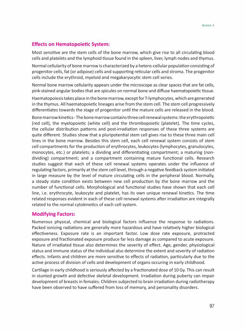
97
Effects on Hematopoietic System:Most sensitive are the stem cells of the bone marrow, which give rise to all circulating blood cells and platelets and the lymphoid tissue found in the spleen, liver, lymph nodes and thymus.
Normal cellularity of bone marrow is characterized by a hetero-cellular population consisting of progenitor cells, fat (or adipose) cells and supporting reticular cells and stroma. The progenitor cells include the erythroid, myeloid and megakaryocytic stem cell series.
Normal bone marrow cellularity appears under the microscope as clear spaces that are fat cells, pink-stained angular bodies that are spicules on normal bone and diffuse haematopoietic tissue.
Haematopoiesis takes place in the bone marrow, except for T-lymphocytes, which are generated in the thymus. All haematopoietic lineages arise from the stem cell. The stem cell progressively differentiates towards the stage of progenitor until the mature cells are released in the blood.
Bone marrow kinetics - The bone marrow contains three cell renewal systems: the erythropoietic (red cell), the myelopoietic (white cell) and the thrombopoietic (platelet). The time cycles, the cellular distribution patterns and post-irradiation responses of these three systems are quite different. Studies show that a pluripotential stem cell gives rise to these three main cell lines in the bone marrow. Besides this stem cell, each cell renewal system consists of stem cell compartments for the production of erythrocytes, leukocytes (lymphocytes, granulocytes, monocytes, etc.) or platelets; a dividing and differentiating compartment; a maturing (non-dividing) compartment; and a compartment containing mature functional cells. Research studies suggest that each of these cell renewal systems operates under the influence of regulating factors, primarily at the stem cell level, through a negative feedback system initiated in large measure by the level of mature circulating cells in the peripheral blood. Normally, a steady state condition exists between new cell production by the bone marrow and the number of functional cells. Morphological and functional studies have shown that each cell line, i.e. erythrocyte, leukocyte and platelet, has its own unique renewal kinetics. The time related responses evident in each of these cell renewal systems after irradiation are integrally related to the normal cytokinetics of each cell system.
Modifying Factors:Numerous physical, chemical and biological factors influence the response to radiations. Packed ionizing radiations are generally more hazardous and have relatively higher biological effectiveness. Exposure rate is an important factor. Low dose rate exposure, protracted exposure and fractionated exposure produce far less damage as compared to acute exposure. Nature of irradiated tissue also determines the severity of effect. Age, gender, physiological status and immune status of the individual also determine the extent and severity of radiation effects. Infants and children are more sensitive to effects of radiation, particularly due to the active process of division of cells and development of organs occuring in early childhood.
Cartilage in early childhood is seriously affected by a fractionated dose of 10 Gy. This can result in stunted growth and defective skeletal development. Irradiation during puberty can impair development of breasts in females. Children subjected to brain irradiation during radiotherapy have been observed to have suffered from loss of memory, and personality disorders.
annex-1

98
NDMA MANuAl oN MeDicAl MANAgeMeNt of NucleAr AND rADiologicAl eMergeNcies
Annex-2
Some information on contamination with radioactive material/isotopesRadioactive ContaminationBrief information about contamination with radioisotopes are:
1 Radioactive contamination is the presence of radioactive material – either in the form of dust particles, suspended particulate matter or liquid or gas – on or in the body of a person.
2 When the radioactive dust, particles, liquids or gases are deposited on the external part of human subject or on the skin, it is termed as External Contamination
3 When the radioisotopes had gained entry inside the body through ingestion, inhalation or absorption through wounds or intact skin, it is called as Internal Contamination.
4 A contaminated person is a source of radiation to himself and to others in the vicinity and may lead to the spread of contamination.
5 Usually, radioactive contamination is not immediately threatening to life and its effects will depend upon the type and energy of radiation.
6 Alpha particles are heavy and positively charged and ionize the matter densely and do not penetrate much. Alpha particles have a propagation range of no more than about 5 cm in the air and can be obstructed even by a moderately thin sheet of paper. The stratum corneum layer of the skin effectively shields the basal layer of skin since alpha particles have a range of 0.04 mm in soft tissue and, hence, do not reach Basal layer of skin. Contamination with alpha emitters can be of concern if inhaled, ingested or absorbed through broken skin. Internal contamination with alpha emitters poses a severe hazard because they affect the target organs severely.
7 Beta particles comprise of negatively charged particles with a little mass. The propagation range of beta particles in air is approximately 3.65 m / MeV of kinetic energy. Beta particle of 70 KeV energy or more can penetrate the Epidermis and can cause the irradiation of subcutaneous tissues and can also cause “Beta Burns”. It is important to note that passage through one millimeter of tissue will reduce most beta radiation by a factor of 2 or more. Hence the dose of beta radiation in the subcutaneous tissue is much less than that recorded on surface.Internal contamination with beta emitters poses hazard because they transfer a significant fraction of their energy to the organ in which they penetrate, i.e., the target organ.
8 X-rays and Gamma rays are non-particulate electromagnetic radiation having no mass and charge. They can penetrate deep into the body tissues and deep seated organs. Higher the energy absorbed in the target tissue, greater is the damage caused.
9 Maximum permissible level allowed for fixed skin contamination is –0.4 Bq / cms2 - For Alpha emitters and4.0 Bq / cms2 - For Beta and Gamma emitters.
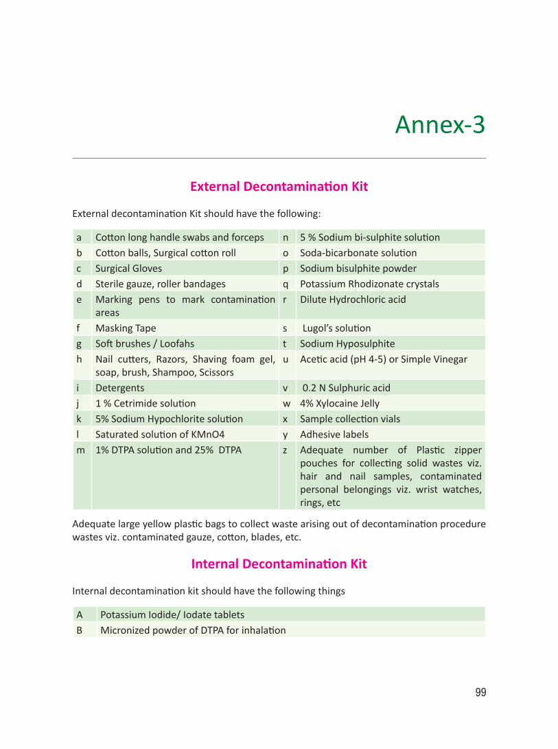
99
Annex-3
External Decontamination Kit
External decontamination Kit should have the following:
a Cotton long handle swabs and forceps n 5 % Sodium bi-sulphite solutionb Cotton balls, Surgical cotton roll o Soda-bicarbonate solutionc Surgical Gloves p Sodium bisulphite powderd Sterile gauze, roller bandages q Potassium Rhodizonate crystalse Marking pens to mark contamination
areasr Dilute Hydrochloric acid
f Masking Tape s Lugol’s solutiong Soft brushes / Loofahs t Sodium Hyposulphiteh Nail cutters, Razors, Shaving foam gel,
soap, brush, Shampoo, Scissorsu Acetic acid (pH 4-5) or Simple Vinegar
i Detergents v 0.2 N Sulphuric acidj 1 % Cetrimide solution w 4% Xylocaine Jellyk 5% Sodium Hypochlorite solution x Sample collection vialsl Saturated solution of KMnO4 y Adhesive labelsm 1% DTPA solution and 25% DTPA z Adequate number of Plastic zipper
pouches for collecting solid wastes viz. hair and nail samples, contaminated personal belongings viz. wrist watches, rings, etc
Adequate large yellow plastic bags to collect waste arising out of decontamination procedure wastes viz. contaminated gauze, cotton, blades, etc.
Internal Decontamination Kit
Internal decontamination kit should have the following things
A Potassium Iodide/ Iodate tabletsB Micronized powder of DTPA for inhalation

100
NDMA MANuAl oN MeDicAl MANAgeMeNt of NucleAr AND rADiologicAl eMergeNcies
C Aerosol generator (Nebulizer) for inhaling DTPAD Ca DTPA / Zn DTPA ampoules and DTPA aerosolsE Colloidal Prussian Blue or Cap RadiogardaseF Aluminium hydroxide gel Antacids- e.g. Gelusil Plus, Logacid etcG Potassium RhodizonateH Inj. Soda bicarbonate ampoulesI Oral Magnesium Sulphate / Barium sulphateJ Oral Strontium lactate / Strontium Gluconate / Oral or Inj. Calcium GluconateK Stable Phosphates – Aluminum phosphateL Propyl thiouracil OR MethimazoleM Oral Ammonium chlorideN Cap. D- Penicillamine 50 mgO Inj. DimercaprolP Inj. LasixQ I.V. Fluids – Normal Saline, 5 % Dextrose, 5 % Dextrose Normal Saline, Ringer’s Lactate,
Isolyte M/PR Inj. Avil and Inj. Hydrocortisone – for treating any allergic reactionsS Syr. Cremaffin or Proctoglycerine Enema

101
Annex-4
Group A - Common
1 Americium (Am)-241:1.1 Half Life: Am-241 has a physical half-life of 432.2 years and decays by α emission (it
emits several photons which can be detected, most notably a gamma ray with an energy of 59.54 keV).
1.2 A.L.I.: The annual limit of intake (ALI) for 241Am due to inhalation for type M compound is 740 Bq as per limits prescribed by the Atomic Energy Regulatory Board (AERB).
1.3 Deposition and Clearance: Americium is deposited in the pulmonary parenchyma after inhalation of the oxide and is mostly cleared with a half-time of 10-20 days (80%), but the clearance half-time of the remaining material has been estimated to vary between a few tens of days to almost 1000 days. These differences may reflect the degree of solubility of the Am-241 in lung fluids which, in turn, is a reflection of the composition of the oxide. According to ICRP 30, a clearance half-time of 28 days was estimated in a worker who had inhaled Am-241 in the oxide form.
1.4 Recommended Treatment: Parenteral Ca-DTPA, Zn-DTPA. (For details of regime and administration of Ca and Zn DTPA please see 8.28.5.).
2 Cesium (Cs)-137:2.1 Half Life: Cs-137 has a physical half-life of 30 years and decays by β and γ emission.
Dosimetry methods used for radio- cesium are based on the concepts of ICRP 30. From the blood, the activity is distributed uniformly in the body with no organ or tissues exhibiting a higher concentration.
2.2 A.L.I.: The annual limit on intake (ALI) for 137Cs due to inhalation for Type F compound is 3000 kBq as per Atomic Energy Regulatory Board (AERB) limits.
2.3 Deposition and Clearance: Cesium-137 is assumed to be completely and rapidly absorbed into the systemic circulation from both the respiratory and GI tracts. Some of the cesium is passed into the intestine, absorbed from the gut into the blood, then goes to the liver, where some of it is excreted via bile into the intestine, reabsorbed from the gut into the blood, then to the liver again, where some is excreted again into the gut (entero-hepatic circulation).
The body retention of 137Cs is described as consisting of two components. Using the ICRP 30 model - two component bio-kinetic model with 10% of the initial intake exhibiting a clearance half-time of 2 days and 90% exhibiting a longer half-time of 110 days.

102
NDMA MANuAl oN MeDicAl MANAgeMeNt of NucleAr AND rADiologicAl eMergeNcies
The ICRP 30 systemic model is also used in the more recent ICRP publications 68 and 78. Publication 78 notes that the biological clearance half-time from the transfer compartment to the systemic compartment is 0.25 days and that, females may exhibit significantly shorter retention half-times in the long-term compartment than males. For systemic excretion, according to ICRP 54, it is assumed that 80% of the 137Cs intake is excreted in the urine and 20% in feces since the main pathway of 137Csexcretion is known to be through glomerular filtration in the kidneys.
2.4 Treatment Recommended: Oral Prussian blue. Insoluble Prussian blue for Human consumption is available by the name Radiogardase. It is available as capsules.
2.4.1 Effects of treatment are promising. The untreated mean whole body effective half-life of Cs-137 is 80 days in adults, 62 days in adolescents and 42 days in children. Insoluble Prussian blue reduces the mean whole body effective half-life of Cs-137 by 69% in adults, by 46% in adolescents and by 43% in children. Dose and clearance ratio observed in cases after 1987 incident in Goiânia are as follows -
Cesium-137 Effective Half life during and after treatment with insoluble Prussian blue (In Days, by age, and dose of Insoluble Prussian blue)
Group Age (Years)
Insoluble Prussian blue dose (grams/day)
No. of Pts.
During Insoluble Prussian blue Treatment - 137Cs T½
Off Insoluble Prussian blue Treatment - 137Cs T½
Adults >18 10 5 26 + 6 days 18 + 15 days (all 21 adult patients)Adults >18 6 10 25 + 15 days
Adults >18 3 6 25 + 9 daysAdolescents 12 - 14 <10 5 30 + 12 days 62 + 14 daysChildren 4 - 9 <3 7 24 + 3 days 42 + 4 days
2.4.2 Indication and Usage: Insoluble Prussian blue is indicated for treatment of patients with known or suspected internal contamination with radioactive cesium and /or radioactive or non-radioactive thallium to increase their rates of elimination.

103
2.4.3 Pharmacokinetics: 99% of the administered Insoluble Prussian blue is excreted unchanged in Feces.
2.4.4 Food Effects: The effect of food was not identified through published literature. In animal studies, insoluble Prussian blue was not significantly absorbed. Food may increase the effectiveness of insoluble Prussian blue by stimulating bile secretion. Food is known to increase the bile production and entero-hepatic circulation. The increase in entero-hepatic circulation may increase the amount of cesium and thallium in the gastrointestinal lumen and may increase the amounts available for binding with insoluble Prussian blue.
2.4.5. Renal and Hepatic Functions: Insoluble Prussian blue is not systemically bio-available and does not rely on renal elimination or hepatic metabolism; therefore, the use of insoluble Prussian blue is not contraindicated in groups of patients having deranged hepatic or renal functions. However, insoluble Prussian blue may be less effective in patients with impaired liver function due to decreased excretion of cesium and thallium in the bile.
2.4.6 Contra-indications: None
2.4.7 Precautions: Insoluble Prussian blue can cause constipation. Decreased gastrointestinal motility will slow the transit time of 137Cs bound to insoluble Prussian blue in the gastro-intestinal tract and may increase the radiation absorbed dose to the gastrointestinal mucosa. Constipation occurring during insoluble Prussian blue treatment may be corrected with a fiber-based laxative and /or a high fiber diet. Insoluble Prussian blue should be used with caution in patients with disorders associated with decreased gastrointestinal motility.
2.4.8 Information to patient:
a Cesium-137 is excreted in the urine and feces. Appropriate safety measures should be taken to minimize radiation exposure to others. When possible, a toilet should be used instead of a urinal and it should be flushed several times after each use. Spilled urine or feces should be cleaned up completely and patients should wash their hands thoroughly. If blood or urine gets onto clothing, such clothing should be washed separately.
b In patients with constipation, a fiber-based laxative and /or high fiber diet is recommended during treatment with insoluble Prussian blue.
c Patients taking insoluble Prussian blue should be informed that their stools might become blue colored.
d In patients who cannot swallow capsules, the capsules are opened and the contents are mixed with food and eaten. The mouth and teeth might become blue colored.
2.4.9 Caution: Insoluble Prussian blue may bind electrolytes found in the gastrointestinal tract. Asymptomatic hypokalemia, with serum potassium values
annex-4

104
NDMA MANuAl oN MeDicAl MANAgeMeNt of NucleAr AND rADiologicAl eMergeNcies
of 2.5-2.9 (normal 3.5-5.0), were observed in 7% cases after 1987 incident in Goiânia. Hence, serum electrolytes should be closely monitored during insoluble Prussian blue treatment. Caution should be exercised when treating patients with pre-existing cardiac arrhythmias or electrolyte imbalances.
2.4.10 Pregnancy and treatment: Since insoluble Prussian blue is not absorbed from the gastro-intestinal tract, effects on the fetus are not expected. In one case, the patient became pregnant 3 years and 8 months after being treated with insoluble Prussian blue for internal contamination with 137Cs (8 mCi). No complications or birth defects were identified through published reports.
i Cesium-137 is known to penetrate the human placenta. One patient, in Goiânia was contaminated with 0.005 mCi 137Cs during her 4thmonth of pregnancy. She was not treated with insoluble Prussian blue. During delivery, the concentration of 137Cs was the same both in the mother and the infant.
ii Thallium penetrates the human placenta. Reported fetal effects in the reviewed literature include fetal death, failure to thrive, alopecia or in some instances outwardly normal development. The risk of toxicity from untreated radioactive cesium or thallium exposure is expected to be greater than the reproductive toxicity risk of insoluble Prussian blue.
2.4.11 Nursing Mothers: Studies to determine if insoluble Prussian blue is excreted in human milk have not been conducted. Since insoluble Prussian blue is not absorbed from the gastrointestinal tract, its excretion in milk is highly unlikely. However, cesium and thallium are transmitted from mother to infant in breast milk. Women internally contaminated with cesium or thallium should not breast feed.
2.4.12 Doses and Administration :
Adults and Adolescents: The recommended dose of insoluble Prussian blue is 1 to 3 grams orally three times a day.
Pediatrics (2 - 12 years):i The recommended dose of insoluble Prussian blue is 1 gram orally three
times a day.
ii In patients who cannot tolerate swallowing a large number of capsules, the capsules may be opened and mixed with bland food or liquids. This may result in blue discoloration of the mouth and teeth.
iii Insoluble Prussian blue capsules may be taken with food to stimulate excretion of cesium or thallium through bile.
iv When the internal radioactivity is substantially decreased the insoluble Prussian blue dose may be decreased to 1 or 2 grams TID to improve gastro-intestinal tolerance.

105
2.4.13 Adverse Reactions: Only constipation and undefined gastric distress have been reported. Severe adverse reactions or death shave not been reported with Insoluble Prussian blue.
2.4.14 Drug Availability: Radiogardase™ is supplied as 0.5 gram blue powder in gelatin capsules for oral administration. It is packaged in brown glass bottles containing 30 capsules each. The product is manufactured by Haupt Pharma Berlin GmbH for distribution by HEYL Chemisch-pharmazeutische Fabrik GmbH and Co. KG, Berlin.
2.5 Bio-assay:
The radioactivity counts in urine and fecal samples should be measured and recorded weekly to monitor 137Cs elimination rate.
3 Iodine:3.1 Half Life: I-131 has a physical half-life of 8.04 days and decays by β, γ- emission
3.2 A.L.I.: The annual limit on intake (ALI) for 131I due to inhalation for Type F compounds is 2 MBq as per AERB limits.
3.3 Deposition and Clearance: The gastrointestinal uptake (f1) factor for all forms of iodine is 1.0. Of the iodine entering the systemic compartment, a fraction, 0.3, is assumed to be translocated to the thyroid, while the remainder (0.7) is assumed to go directly to excretion. Iodine in the thyroid is assumed to be retained with a biological half-life of 80 days. Inhaled Iodine reaches equilibrium with the body fluids within 30 minutes and 30 % of this is in the Thyroid gland.
3.4 Treatment: Potassium Iodide within first 4 hours. If KI or its equivalent (Lugol’s Iodine) isn’t used within four to six hours, it will have significantly decreased effectiveness and that effectiveness will approach zero after about 12-24 hours. This blocks the thyroid. (Stable) Iodine works by saturating the uptake mechanism in the thyroid gland and “competes” with radioactive iodine (I-131). Early administration of antidotes is the key element for successful therapy.
OR
3.4.1 Lugol’s Iodine consists of 5% Iodine (I2) and 10% Potassium Iodide (KI) in 85% distilled water with a total iodine content of 130 mg/ml. Potassium iodide makes the iodine water soluble through the formation of the I3- ion. Synonyms for Lugol’s solution are IKI (Iodine-Potassium Iodide); Iodine, Strong solution (Systemic); Aqueous Iodine Solution BP. Lugol’s Iodine 2% has a composition of Iodine 2%, Potassium Iodide 4% and distilled water 94%.
3.4.2 Treatment can be done by mobilization of the radio-iodine through the use of anti-thyroid drugs.
annex-4

106
NDMA MANuAl oN MeDicAl MANAgeMeNt of NucleAr AND rADiologicAl eMergeNcies
3.4.3 Doses:
a Adults and children above the age of 1 : KI 130 mg daily for 7 –1 4 daysb Children below the age of 1: 65 mg per day, daily for 7 –1 4 days.
4 Phosphorous (P)- 324.1 Half Life: P-32 has a physical half-life of 14.26 days and decays by β-emission (its
bremsstrahlung photons may be detectable); specific bremsstrahlung constant, ÃP-32 = 4.05 x 10⁻³ R cm2 /mCi h in soft tissue and 1.08 x 10⁻² R cm2/mCi h in bone (Zanzonico et al. JNM 1999; 40:1024-1028).
4.2 A.L.I.: The annual limits on intake (ALI) for 32P due to inhalation for Type F compounds is 20 MBq, and for Type M compound is 7 MBq as per AERB.
4.3 Deposition and Clearance: The bio-kinetic model described in ICRP 30 (Full form with reference) is used to estimate the whole body retention, R(t), of phosphorus. Phosphorus entering the transfer compartment is assumed to be retained there with a half-life of 0.5 days. Of this, 15% is assumed to go directly to excretion, 15% to intracellular fluids where it is retained with a half-life of 2 days, 40% to soft tissue where it is assumed to be retained with a half-life of 19 days and 30% to mineral bone where it is assumed to be permanently retained. P-32 going either to intracellular fluids or to soft tissues is assumed to be uniformly distributed throughout all organs and tissues of the body excluding mineral bone, where it is assumed to be retained on the bone surfaces.
4.4 Treatment:
a Oral stable phosphates are given. Sodium phosphate or Potassium phosphate or Glycerol phosphate is given.
b Aluminum Hydroxide can be given to reduce the G.I. uptake.
4.4.1 Dose: Stable Phosphates are given 1 gram orally on daily basis for as long as required.
5 Plutonium (Pu)-2395.1 Half Life: Pu-239 has a physical half-life of 24,110 years and decays by α emission.
5.2 A.L.I.: The annual limit on intake (ALI) for 239Pu due to inhalation for Type M compounds is 625 Bq and for Type S compounds is 2400 Bq, as per AERB.
5.3 Deposition and Clearance : For dissolved (ionic form) plutonium reaching the transfer compartment (i.e., the bloodstream), the ICRP 30 model distributes 45% to the bone surfaces from which it clears with a biological half-time of 50 years and 45% to the liver with a biological clearance half-time of 20 years.
5.3.1 The activity deposition in bone is assumed to be uniformly distributed over the bone surfaces of both cortical and trabecular bone. A small radioactivity fraction is permanently retained in the gonads (0.035% for testes and 0.011%

107
for ovaries). The remaining 10% is assumed to go directly to excretion; for purposes of dosimetry, this component is considered to be an insignificant contributor to effective dose equivalent and is generally ignored.
5.4 Treatment: Parenteral Ca-DTPA, Zn-DTPA. Di-ethylene-Tri-amine-Penta-Acetate (DTPA). DTPA is available only in parenteral form. Ca DTPA and Zn DTPA should not be administered together. DTPA may be given through intravenous infusion or as aerosol therapy of Zn DTPA in cases of internal contamination of lungs only by inhalation. The safety and effectiveness of the intramuscular route have not been established for Ca-DTPA or Zn-DTPA.
5.4.1 If both products are available, then the treatment should be started with Ca DTPA within 24 hours. If additional treatment becomes necessary then it should be changed to Zn DTPA. This is because Ca DTPA is more effective (roughly 10 times) in the first 24 hours than Zn DTPA. After 24 hours the efficacy of both is similar, but Ca-DTPA causes more loss of essential metals, such as zinc, from the body. Therefore, Zn-DTPA is preferred for maintenance therapy.
5.4.2 Mode of Action: DTPA is a chelating agent. It chelates plutonium (also americium, and curium) and then excretes them through urine. Treatment should be started as soon as possible. It is observed that if started within 1 hour, the retention of Plutonium in Liver is reduced from 14 % in control to 0.47 % in treated cases. Similarly retention of plutonium in skeleton is reduced from 57% in control to 5.9% in treated cases. The removal of plutonium from bones can be as high as 90% if treated within one hour.
5.4.3 Limitations: DTPA cannot bind all of the radioactive materials that might get into a person’s body. DTPA cannot reverse the health effects caused by radioactive materials once these materials have entered the body. After 24 hours, plutonium, americium, and curium are harder to chelate. However, DTPA can still work to remove these radioactive materials from the body several days or even weeks after a person has been internally contaminated.
5.4.4 Treatment plan: The treatment plan should be jointly drawn out by the doctors and the health physicists. For various categories of patients it should be as follows:
i Infants (including breastfed infants) and children <12 years of age
Either Ca-DTPA or Zn-DTPA may be given to infants and children. The dosage of DTPA to be given should be based on the child’s size and weight.
ii Adults and adolescents
Adults and adolescentsinternally contaminated with plutonium, americium or curium should receive Ca-DTPA within the first 24 hours after contamination. After 24 hours, if additional treatment is needed, adults should receive Zn-DTPA. If Zn-DTPA is not available, patients may receive
annex-4
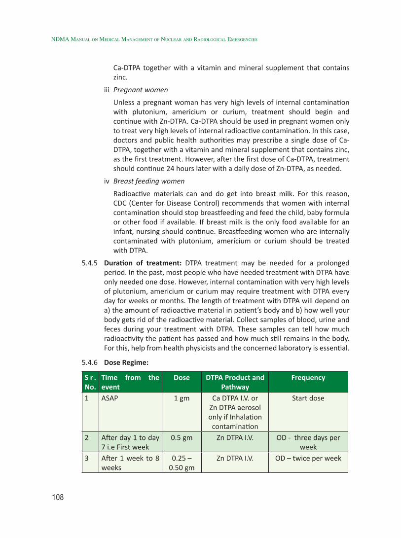
108
NDMA MANuAl oN MeDicAl MANAgeMeNt of NucleAr AND rADiologicAl eMergeNcies
Ca-DTPA together with a vitamin and mineral supplement that contains zinc.
iii Pregnant women
Unless a pregnant woman has very high levels of internal contamination with plutonium, americium or curium, treatment should begin and continue with Zn-DTPA. Ca-DTPA should be used in pregnant women only to treat very high levels of internal radioactive contamination. In this case, doctors and public health authorities may prescribe a single dose of Ca-DTPA, together with a vitamin and mineral supplement that contains zinc, as the first treatment. However, after the first dose of Ca-DTPA, treatment should continue 24 hours later with a daily dose of Zn-DTPA, as needed.
iv Breast feeding women
Radioactive materials can and do get into breast milk. For this reason, CDC (Center for Disease Control) recommends that women with internal contamination should stop breastfeeding and feed the child, baby formula or other food if available. If breast milk is the only food available for an infant, nursing should continue. Breastfeeding women who are internally contaminated with plutonium, americium or curium should be treated with DTPA.
5.4.5 Duration of treatment: DTPA treatment may be needed for a prolonged period. In the past, most people who have needed treatment with DTPA have only needed one dose. However, internal contamination with very high levels of plutonium, americium or curium may require treatment with DTPA every day for weeks or months. The length of treatment with DTPA will depend on a) the amount of radioactive material in patient’s body and b) how well your body gets rid of the radioactive material. Collect samples of blood, urine and feces during your treatment with DTPA. These samples can tell how much radioactivity the patient has passed and how much still remains in the body. For this, help from health physicists and the concerned laboratory is essential.
5.4.6 Dose Regime:
S r . No.
Time from the event
Dose DTPA Product and Pathway
Frequency
1 ASAP 1 gm Ca DTPA I.V. or Zn DTPA aerosol only if Inhalation
contamination
Start dose
2 After day 1 to day 7 i.e First week
0.5 gm Zn DTPA I.V. OD - three days per week
3 After 1 week to 8 weeks
0.25 – 0.50 gm
Zn DTPA I.V. OD – twice per week

109
4 8 weeks To 12 weeks
- - -
5 Subsequently 0.25 – 0.50 gm
Zn DTPA I.V. Once per week for 4 weeks. Cycle to
be continued with a pause of 2-3 weeks
after each 4 weeks of treatment.
5.4.7 For large quantities of Plutonium inhalation, (50 times the maximum permissible lung burden in patients aged 30 years or less) or more than 1.5 µ Ci, bronchial lavage is recommended soon after second or third day. This should be undertaken in specialized centers only where expertise exists.
5.4.8 Caution: There are no medical reasons why a person who is internally contaminated with plutonium, americium or curium should not be treated with Ca-DTPA or Zn-DTPA, except nephritic syndrome. However, keep the following guidelines in mind:
i Because radioactive materials chelated to DTPA are passed out of the body in the urine, DTPA must be used carefully in people whose kidneys do not function properly.
ii Ca-DTPA should be used carefully in people who have a disease called “hemochromatosis.” (Hemochromatosis is a genetic disease that causes the body to absorb too much iron from foods and other sources, such as vitamins containing iron.)
iii Aerosol treatments using DTPA may not be safe for some people with asthma. If a person with asthma requires treatment with DTPA, the drug should be injected.
iv DTPA should not be used to treat people who are internally contaminated with the radioactive materials uranium or neptunium.
5.4.9 Side Effects: DTPA does not build up in the body or cause long-term health effects. People who are given repeat doses of Ca-DTPA within a short period of time may have nausea, vomiting, diarrhea, chills, fever, itching and muscle cramps. Other side effects may include headache, lightheadedness, chest pain and a metallic taste in the mouth. Ca-DTPA (and Zn-DTPA) can chelate certain important minerals that the body needs (zinc, magnesium and manganese). For example, the body needs zinc to make red blood cells, white blood cells and platelets. Therefore, DTPA treatment may interfere with the normal production of blood cells. As a precaution, patients receiving long-term treatment with DTPA should be given a vitamin and mineral supplement containing zinc.
5.5 Bio-assay samples: Blood, urine and stool samples need to be collected for bio-assay.
annex-4

110
NDMA MANuAl oN MeDicAl MANAgeMeNt of NucleAr AND rADiologicAl eMergeNcies
6 Strontium 906.1 Half Life : As regards Sr-90, it is important to realize that Sr-90 decays into Yttrium
Y-90, that Y-90 is radioactive and much easier to detect than Sr-90, that Y-90 has a much shorter half-life (64 hrs) than Sr-90 (28 yrs) and that Sr-90 and Y-90 activities reach equilibrium after about two weeks starting with pure Sr-90. This means that if you start with a 1000 Ci source of Sr-90, after about two weeks the source will also contain about 1000 Ci of Y-90 and that this equilibrium will remain the same as the Sr-90 decays. Sr-90 has a physical half-life of 29.12 years.
6.2 A.L.I: The annual limit on intake (ALI) for 90Sr due to inhalation for Type F compounds is 670 kBq and for Type S compounds is 260 kBq, as per AERB.
6.2.1 Deposition and Clearance: The bio-kinetic model used for the distribution, retention and excretion of stable strontium is the ICRP alkaline earth model. It is assumed that stable strontium is uniformly distributed throughout the bone volume, where it is retained and internally recycled according to a series of exponential terms. The alkaline earth excretion model assumes that the fraction of excreted uptake occurring by the urinary pathway and by the fecal pathway is 0.8 and 0.2 respectively.
6.3 Treatment:
a Reduction of absorption : Aluminum phosphate gel antacids or sodium / calcium alginate or Barium sulphate
b Blockage: strontium lactate or Strontium gluconate
c Displacement: Oral phosphate
d Mobilization: ammonium chloride or parathyroid extract (dynamics)
6.3.1 Doses:
i Sodium / Calcium Alginate in a single dose of 10 – 20 gm dissolved in sugared water orally. Sugared water, because it is otherwise very unpalatable.
ii Aluminum phosphate 60 gm colloidal gel 20% orally
iii Barium sulphate 300 gm in aqueous solution orally as a single dose.
iv Oral Strontium Lactate 500 mg OR Slow infusion of Strontium gluconate 600 mg in 500 ml of 5 % Dextrose solution.
v Although stable strontium is the ideal choice, but as Strontium is quite toxic, it cannot be used for clearing skeletal strontium. Calcium, besides being a congener of strontium, is also an important and major component of skeleton. Thus Calcium compounds have been considered a better choice in curtailing Strontium uptake. Commonly used Calcium compounds are easy to administer and do not have any untoward effect on body metabolism even at a higher dose.

111
vi Oral Calcium gluconate 6-10 gm in divided doses over the day OR Inj Calcium gluconate 1-5 gm in 500 ml of 5 % Dextrose solution as slow infusion.
vii Acidification by oral Ammonium chloride 6 gm in 3 divided doses (4 tabs each dose) over the day will help mobilization of Strontium.
viii Newer drug – newer chelating agent - Phosphoric acid derivative (APDA) developed by the Shanghai Institute of Materia Medica has worked well. Parenteral Ca APDA in doses of 600 mg / Kg body weight for three days. Excretion route post Ca APDA is through kidneys and hepato-biliary system.
Oral Ca APDA in the dose of 600mg/ Kg body weight is also effective in chelating the GI intake. Excretion is through Feces.
6.3.2 Contraindications: Same as for Radium (Pl. see Section 8.32.4.2)
6.3.3 Caution: Same as for Radium (Pl. see Section 8.32.4.3)
6.3.4 Side Effects: Same as for Radium (Pl. see Section 8.32.4.4)
6.3.5 Bio-assay Sample: Urine and Feces.
7 Tritium (H)-37.1 Half Life: H-3 has a physical half-life of 12.33 years and decays by β- emission.
7.2 A.L.I.: The annual limit on intake (ALI) for 3H due to inhalation is 1 GBq, as per AERB.
7.3 Deposition and Clearance: The metabolic model for tritium is described in ICRP 30. (Complete reference). Tritiated water is assumed to be uniformly distributed among all soft tissues at any time following intake. Its retention, R(t), is described as a single exponential with an effective clearance half-time of 10 days:
7.4. Treatment: Force water to promote dieresis
7.4.1 Dose:
a Oral fluids 6-8 L / day OR Intravenous fluids 4-6 L per day with forced diuresis.Excess fluids help dilution and forced diuresis helps renal clearance.
b For forced Diuresis, use Inj Lasix 40 mg i.v. or more if necessary. Use Inj Lasix 40 mg i.v. for every 2-3 l of fluids. Use Lasix and Fluids alternatively.
Treatment should be continued for 1 week.
7.4.2 Investigations: Monitor Electrolytes, particularly Sodium, if hypotonic solutions are used.
7.4.3 Caution: Assess cardiac status of the patient before loading extra fluids. If the patient is already in Congestive Cardiac Failure (CCF), extra caution is needed.
7.5 Bio-assay Samples: Urine sample.
annex-4

112
NDMA MANuAl oN MeDicAl MANAgeMeNt of NucleAr AND rADiologicAl eMergeNcies
8 Uranium (U)-234, 235, and 2388.1 Half Life: U-234 has a physical half-life of 2.455x10⁵ years and decays by α emission;
U-235 has a physical half-life of 7.038x10⁸ years and decays by α emission; and U-238 has a physical half-life of 4.468x10⁹ years and decays by α emission.
8.2 A.L.I.: The annual limits on intake (ALI) for 234U due to inhalation is for Type F compounds 31250 Bq, for Type M compounds 9500 Bq, and for Type S compounds 2900 Bq as per AERB.
The annual limits on intake (ALI) for 235U due to inhalation is for Type F compounds 33333 Bq, for Type M compounds 11111 Bq and for Type S compounds 3280 Bq, as per AERB.
The annual limits on intake (ALI) for 238U due to inhalation is for Type F compounds 34500Bq, for Type M compounds 12500 Bq and for Type S compounds 3500 Bq, as per AERB.
8.3 Deposition and Clearance: For material entering the systemic circulation, fractions of 0.2 and 0.023 are assumed to enter the bone mineral and remains there with half-lives of 20 and 5000 days, respectively; fractions of 0.12 and 0.00052 are assumed to enter the kidneys and remains with half-lives of 6 and 1500 days, respectively; and fractions of 0.12 and 0.00052 are assumed to enter all other tissues of the body. The remaining fraction of the uranium entering the systemic circulation, 0.54, is assumed to be excreted directly.
8.4 Metabolic Pathway of Uranium: Uranium is nephrotoxic with predominant changes described by necrosis in the proximal convoluted tubule and a moderate degree of inflammatory and fibrotic changes, resulting in scarred kidneys. In cases of non-lethal poisoning, damaged tubular epithelium is rapidly regenerated, with subsequent tolerance to large doses of uranium. Regenerated epithelium is of metaplastic histologic type, different from the normal epithelium and the postulated tolerance mechanism was the inability of uranium compounds to interact with renal tubular cells.
Toxic effects were also observed in the liver, central nervous system and blood.
There are three major routes of internal contamination with uranium: 1) gastrointestinal system; 2) skin and wounds; and 3) inhalation and trans-alveolar transfer to the blood stream.
Gastrointestinal Absorption: Gastrointestinal absorption of uranium isotopes is relatively low in the adult human, but still presents a considerable biomedical hazard because of their long half-lives, nephrotoxicity and retention in skeletal tissue being the main hazards. It was recognized that, although uranium predominantly enters the animal or human organism by the respiratory route, it may be swallowed, gaining entry to the gastrointestinal system. Fasting state enhances the absorption of uranium.

113
Inhalation: About 25% of the radioactive particles are deposited in the bronchial tree, 25% are immediately exhaled, whereas 50% are translocated translocated to the naso-pharynx and swallowed, with subsequent handling by the mechanisms of gastrointestinal absorption. The intestinal absorption of Depleted Uranium (DU) is negligible, placing the respiratory pathway in the category of major radio-toxicological hazard. One of the therapeutic aims in internal DU contamination should include the movement of the inhaled particles to the extra pulmonary pathways. The deposition of DU particles on the alveolar surfaces will result in their absorption, depending on their solubility, with approximately 10% of the particles retained in the lungs and reaching systemic circulation, and the remaining 15% ascending to the naso-pharynx by expectoration and ending in the gastrointestinal tract. Soluble components of uranium absorbed from the pulmonary tree are deposited in the skeleton within a few weeks, with a biological half-life in the lungs of 120 days. A considerably longer pulmonary retention of 1,470 days is expected in the case of inhalation of uranium oxides.
8.5 Treatment: The principal aim in the therapeutic management of patients with internally deposited uranium is to prevent the absorption from the site of entry and eliminate uranium from the blood stream or target organs.
8.5.1 The method of gastric lavage is very useful in therapy or early exposure by ingestion. Wash the stomach several times with water or physiological saline by negative pressure, until the aspirate is declared free of the contaminant.
8.5.2 The use of laxatives is a common therapeutic approach in reducing internal contamination. Laxative use is contraindicated in acute abdominal syndrome or non-diagnosed pain in the stomach. Numerous side effects include tachypnea, dyspnea, tachy-arrhythmias, intestinal irritation, exanthema and syncopal attacks.
8.5.3 Treatment of patients who have been contaminated by inhalation of uranium compounds includes the use of therapeutic agents which decrease viscosity of endobronchial mucosa. The use of mucolytic substances, which have the effect on muco-polysacharides and nucleoproteins in the respiratory tree, enable the elimination of actinides by expectoration. Mucolytics like, Carbocysteine (e.g. Syr. Mucodyne), Bromohexine HCL (e.g Syr Bromohexine) or Ambroxol HCL (e.g. Syr Ambrodil ct) are very effective.
8.5.4 Ethylene-diamine-tetra-acetic acid (EDTA) has proven efficacy in the treatment of lead, zinc, copper, chromium, manganese and nickel poisoning and in contamination with transuranic elements. Na-EDTA is used in a dose of 50 mg/kg. The total quantity should not exceed 300 mg during 6 days of treatment. It is not administered by oral or intramuscular application. Parenteral use of Na-EDTA may lead to hypocalcemia. The use of Ca-EDTA in the therapeutic dose of 15-30 mg/kg does not have a hypocalcemic effect, and hence is preferred. It is essential to evaluate kidney function before the beginning of the treatment because its use is contraindicated in patients with renal disease.
annex-4

114
NDMA MANuAl oN MeDicAl MANAgeMeNt of NucleAr AND rADiologicAl eMergeNcies
8.5.5 Inj Soda Bicarb 1.4 % in 250 ml as slow infusion. This forms a stable complex with uranyl ions which is excreted through kidneys.
8.5.6 Caution: Kidney functions and electrolyte monitoring is needed.
8.5.7 Bio-assay: Urine and Feces. Also Gastric Lavage of contents.
Group B – Less Common9 Cobalt 60
9.1 Cobalt- 60 is beta / gamma energy emitter.
9.2 A.L.I.: The annual limits on intake (ALI) for 60Co due to inhalation for Type M compound is 2800 kBq and for Type S compound is 1200 kBq as per AERB.
9.3 Treatment:
9.4 There is no good decorporation agent recognized for radionuclides of cobalt.
9.4.1 Results using Penicillamine were notconclusive in mice.
9.4.2 Cobaltous DTPA reduced radioactive cobalt concentration by about 1/3 in mice, but it has never been tried in humans and is not presently available.
9.4.3 However, for treatment with Ca and Zn DTPA, please refer to Plutonium decontamination (Section 8.31.5).
9.4.4 Aerosol therapy for decorporation of inhalation contamination is done using DTPA aerosol. 1gm/ 4 ml amp. or 100 mg of micronised powder.
9.4.5 Before giving Penicillamine, carry out the following tests. Complete Blood Count [CBC], Liver Function Tests [LFT], Kidney Function Tests [KFT] and Penicilline sensitivity test.
9.4.5.1 D-Penicillamine is given in a dose of 250 mg Four times daily, in between meals. Better results are obtained when given as early as possible.
9.4.5.2 D-Penicillamine is a chelating agent and will chelate other metals and minerals. Hence it is given in between meals, so that the dietary minerals are not chelated.
9.4.5.3 None the less supplements of Muti-vitamins, minerals containing zinc and calcium supplements are recommended.
10 Iridium ( Ir) 19210.1 Half Life: Ir-192 is a photon emitter which may be identified by its spectrum. Ir-192
has a physical half-life of 73.831 days and decays by electron capture and α emission. The bio-kinetic model described in ICRP 30 is used to estimate the whole body retention, R(t), of iridium.
10.1 A.L.I.: The annual limits on intake (ALI) for 192Ir due to inhalation for Type F

115
compounds is 9 MBq, for type M compound is 5 MBq, and for Type S compound is 4 MBq, as per AERB.
10.3 Deposition and Clearance: It is assumed that, of the iridium leaving the transfer compartment, 20%, 4 % and 2 % is translocated to liver, kidney and spleen respectively. A further 54 %, is assumed to be uniformly distributed throughout all other organs and tissues of the body. The remaining 20% of iridium leaving the transfer compartment is excreted directly. Of iridium deposited in any organ or tissue of the body, 20 % and 80 % are assumed to be retained with biological half-lives of 8 and 200 days respectively.
10.3.1 Treatment: Unfortunately, there is no known decorporation drug for Iridium. However Oral Penicillamine might work. For details of dose and administration please refer to Section 3.4.
11 Palladium (Pd) 10311.1 Half Life: Pd-103 has a physical half-life of 16.991 days and decays by electron
capture (it emits x-rays that may be detectable with energies of approximately 20 keV). The bio-kinetic model described in ICRP 30 is used to estimate the whole body retention, R(t), of palladium.
11.1.1 A.L.I : The annual limits of intake (ALI) for 103Pd due to inhalation for Class D compounds is 1 x 10⁸Bq, for Class W compounds is 5 x 10⁷Bq, and for Class Y compounds is 4 x 10⁷Bq. Whereas for Ingestion, the A.L.I. is 7 x 10⁷Bq for Class D, Class W and Class Y compounds, as per ICRP 61.
11.1.2 Deposition and Clearance: The retention of palladium in the body is assumed to be approximated by a single exponential with a biological half-life of 15 days. Of the palladium leaving the transfer compartment, it is assumed that 30% goes directly to excretion, 45% is translocated to the liver, 15% is translocated to the kidneys, 7% is translocated to mineral bone (Pd-103 is assumed to be uniformly distributed throughout the volume of mineral bone) and 3% is uniformly distributed throughout all other organs and tissues of the body. Palladium translocated to any organ or tissue is assumed to remain there with a biological half-life of 15 days.
11.1.3 Treatment: There is no known decorporation drug for palladium. However oral penicillamine could be tried. For details of doses etc. please refer to Section 3.4.
12 Radium (Ra)-22612.1 Half Life: Ra-226 has a physical half-life of 1600 years and decays by α emission
12.2 A.L.I.: The annual limit on intake (ALI) for 226Ra due to inhalation for Type M compounds is 1700 Bq.
annex-4

116
NDMA MANuAl oN MeDicAl MANAgeMeNt of NucleAr AND rADiologicAl eMergeNcies
12.3 Deposition and Clearance: Since radium is an alkaline earth element, it can be assumed that the bio-kinetic model is the same as for strontium.
12.3.1 Treatment
a Oral calcium to reduce gastrointestinal absorption and increase urinary excretion.
b Alginates are also useful to reduce gastrointestinal absorption.
c Oral Magnesium Sulphate forms sulphates with Radium.
12.3.2 Doses
a Magnesium Sulphate: Single dose of 10 gm in 100 ml of water produces insoluble sulphate with radium and thus reduces its absorption.
b Barium Sulphate can also be used instead of Mag. Sulphate. This drug is commonly used in Radiography and hence is easily available. A single dose of 200 ml of 100% barium sulphate should be administered.
c Calcium gluconate orally 6-10 g in 3 divided doses over a day can be given. Or else Inj. Calcium gluconate 1-5 g in 500 ml of 5% Dextrose solution as a slow infusion.
d Oral colloidal Aluminum phosphate 60 gm colloidal gel also works similar to Barium Sulphate.
e Acidification by oral administration of Ammonium chloride 6 g in 3 divided doses over a day.
12.4 Contra-indications
* While using Barium sulphate / magnesium sulphate or Aluminum phosphate, care should be taken to ascertain that the patient is not suffering from suspected colonic obstruction, acute GI hemorrhage, inflammation and perforation. Hypersensitivity to any of these products is a contra-indication.
12.4.1 Caution:
* While using Ba Sulphate or other similar drugs caution should be exercised in following cases - Pregnancy, appendicitis, diverticulitis, intussusception, malignancy, granuloma, ulcerative colitis and helminthiasis and any condition with a risk of GI perforation. Also marked hypertension or advanced cardiac disease, severe debility, history of food aspiration, bronchial asthma and atopy.
* Administration of Inj. Calcium gluconate should always be done under cardiac monitoring. Observe for any arrhythmias. Go slow while infusion. If arrhythmias are significant and likely to be fatal, discontinue infusion until cardiac rhythm reverses to normal. Restart infusion only if cardiac conditions permit.
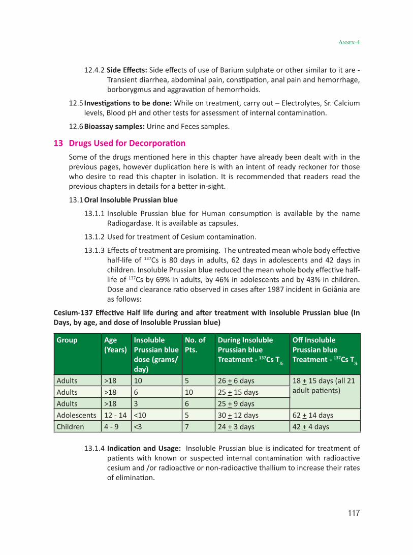
117
12.4.2 Side Effects: Side effects of use of Barium sulphate or other similar to it are - Transient diarrhea, abdominal pain, constipation, anal pain and hemorrhage, borborygmus and aggravation of hemorrhoids.
12.5 Investigations to be done: While on treatment, carry out – Electrolytes, Sr. Calcium levels, Blood pH and other tests for assessment of internal contamination.
12.6 Bioassay samples: Urine and Feces samples.
13 Drugs Used for Decorporation Some of the drugs mentioned here in this chapter have already been dealt with in the
previous pages, however duplication here is with an intent of ready reckoner for those who desire to read this chapter in isolation. It is recommended that readers read the previous chapters in details for a better in-sight.
13.1 Oral Insoluble Prussian blue
13.1.1 Insoluble Prussian blue for Human consumption is available by the name Radiogardase. It is available as capsules.
13.1.2 Used for treatment of Cesium contamination.
13.1.3 Effects of treatment are promising. The untreated mean whole body effective half-life of 137Cs is 80 days in adults, 62 days in adolescents and 42 days in children. Insoluble Prussian blue reduced the mean whole body effective half-life of 137Cs by 69% in adults, by 46% in adolescents and by 43% in children. Dose and clearance ratio observed in cases after 1987 incident in Goiânia are as follows:
Cesium-137 Effective Half life during and after treatment with insoluble Prussian blue (In Days, by age, and dose of Insoluble Prussian blue)
Group Age (Years)
Insoluble Prussian blue dose (grams/day)
No. of Pts.
During Insoluble Prussian blue Treatment - 137Cs T½
Off Insoluble Prussian blue Treatment - 137Cs T½
Adults >18 10 5 26 + 6 days 18 + 15 days (all 21 adult patients)Adults >18 6 10 25 + 15 days
Adults >18 3 6 25 + 9 daysAdolescents 12 - 14 <10 5 30 + 12 days 62 + 14 daysChildren 4 - 9 <3 7 24 + 3 days 42 + 4 days
13.1.4 Indication and Usage: Insoluble Prussian blue is indicated for treatment of patients with known or suspected internal contamination with radioactive cesium and /or radioactive or non-radioactive thallium to increase their rates of elimination.
annex-4

118
NDMA MANuAl oN MeDicAl MANAgeMeNt of NucleAr AND rADiologicAl eMergeNcies
13.1.5 Pharmacokinetics: 99 % of the administered Insoluble Prussian blue is excreted unchanged in Feces.
13.1.6 Food Effects: Food effect studies were not identified through published literature. In animal studies, insoluble Prussian blue was not significantly absorbed. Food may increase the effectiveness of insoluble Prussian blue by stimulating bile secretion. Food is known to increase bile production and entero-hepatic circulation. The increase in entero-hepatic circulation may increase the amount of cesium and thallium in the gastrointestinal lumen and may increase the amounts available for binding with insoluble Prussian blue.
13.1.7 Renal and Hepatic Functions: Insoluble Prussian blue is not systemically bioavailable and does not rely on renal elimination or hepatic metabolism; therefore, the use of insoluble Prussian blue is not contraindicated in groups of patients having deranged Hepatic or Renal functions. However, insoluble Prussian blue may be less effective in patients with impaired liver function due to decreased excretion of cesium and thallium in the bile.
13.1.8 Contra-indications: None
13.1.9 Precautions: Insoluble Prussian blue can cause constipation. Decreased gastrointestinal motility will slow the transit time of 137Cs bound to insoluble Prussian blue in the gastrointestinal tract and may increase the radiation absorbed dose to the gastrointestinal mucosa. Constipation occurring during insoluble Prussian blue treatment may be treated with a fiber-based laxative and /or a high fiber diet. Insoluble Prussian blue should be used with caution in patients with disorders associated with decreased gastrointestinal motility.
13.1.10 Information for the patient: (should be a part of patient Dos and Dont’s separately)
a Cesium-137 is excreted in the urine and feces. Appropriate safety measures should be taken to minimize radiation exposure to others. When possible, a toilet should be used instead of a urinal, and it should be flushed several times after each use. Spilled urine or feces should be cleaned up completely and patients should wash their hands thoroughly. If blood or urine gets onto clothing, such clothing should be washed separately.
b In patients with constipation, a fiber-based laxative and /or high fiber diet is recommended during treatment with insoluble Prussian blue.
c Patients taking insoluble Prussian blue should be informed that their stools might become blue colored.
d In patients who cannot swallow capsules, the capsules are opened and the contents are mixed with food and eaten. The mouth and teeth might become blue colored.
13.1.11 Caution: Insoluble Prussian blue may bind electrolytes found in the

119
gastrointestinal tract. Asymptomatic hypokalemia, with serum potassium values of 2.5-2.9 (normal 3.5-5.0), were observed in 7% cases after 1987 incident in Goiânia. Hence Serum electrolytes should be closely monitored during insoluble Prussian blue treatment. Caution should be exercised when treating patients with pre-existing cardiac arrhythmias or electrolyte imbalances.
13.1.12 Pregnancy and treatment: Since insoluble Prussian blue is not absorbed from the gastrointestinal tract, effects on the fetus are not expected. In one patient that became pregnant 3 years and 8 months after being treated with insoluble Prussian blue for internal contamination with 137Cs (8 mCi), complications or birth defects were not identified in the literature report.
a Cesium-137 is known to cross the human placenta. One patient, in Goiânia, was contaminated with 0.005 mCi 137Cs during her 4th month of pregnancy. She was not treated with insoluble Prussian blue. At birth the concentration of 137Cs was the same in the mother and the infant.
b Thallium crosses the human placenta. Reported fetal effects in the reviewed literature include fetal death, failure to thrive, alopecia, or in some instances outwardly normal development. The risk of toxicity from untreated radioactive cesium or thallium exposure is expected to be greater than the reproductive toxicity risk of insoluble Prussian blue.
13.1.13 Nursing Mothers: Studies to determine if insoluble Prussian blue is excreted in human milk have not been conducted. Since insoluble Prussian blue is not absorbed from the gastrointestinal tract, its excretion in milk is highly unlikely. However, cesium and thallium are transmitted from mother to infant in breast milk. Women internally contaminated with cesium or thallium should not breast feed.
13.1.14 Doses and Administration:
Adults and Adolescents:
The recommended dose of insoluble Prussian blue is 3 grams orally three times a day.
Pediatrics (2 - 12 years):
a The recommended dose of insoluble Prussian blue is 1 gram orally three times a day.
b In patients who cannot tolerate swallowing large numbers of capsules, the capsules may be opened and mixed with bland food or liquids. This may result in blue discoloration of the mouth and teeth.
c Insoluble Prussian blue capsules may be taken with food to stimulate excretion of cesium or thallium through bile.
d When the internal radioactivity is substantially decreased the insoluble
annex-4

120
NDMA MANuAl oN MeDicAl MANAgeMeNt of NucleAr AND rADiologicAl eMergeNcies
Prussian blue dose may be decreased to 1 or 2 grams TID to improve gastrointestinal tolerance.
13.1.15 Adverse Reactions: Only constipation and undefined gastric distress are reported. Death or any severe adverse reactions are not reported with Insoluble Prussian blue.
13.1.16 Drug Availability: Radiogardase™ is supplied as 0.5 gram blue powder in gelatin capsules for oral administration. It is packaged in brown glass bottles containing 30 capsules each. The product is manufactured by Haupt Pharma Berlin GmbH for distribution by HEYL Chemisch-pharmazeutische Fabrik GmbH and Co. KG, Berlin.
13.1.17 Bio-assay: The radioactivity counts in urine and fecal samples should be measured and recorded weekly to monitor 137Cs elimination rate.
14 Potassium Iodide / Potassium Iodate / Lugol’s Iodine14.1 Used for treatment of Iodine contamination.
14.2 Treatment: Potassium Iodide within first 4 hours. If KI or its equivalent (Lugol’s Iodine) isn’t used within four to six hours, it will have significantly decreased effectiveness and that effectiveness will approach zero after about 12-24 hours. This blocks the thyroid. (Stable) Iodine works by saturating the uptake mechanism in the thyroid gland and “competes” with radioactive iodine (I-131). Early administration of radionuclide antidotes is the key element for successful therapy.
OR
14.3 Lugol’s Iodine consists of 5% Iodine (I2) and 10% Potassium Iodide (KI) in 85% distilled water with a total iodine content of 130 mg/ml. Potassium iodide makes the iodine water soluble through the formation of the I3- ion. Synonyms for Lugol’s solution are IKI (Iodine-Potassium Iodide); Iodine, Strong solution (Systemic); Aqueous Iodine Solution BP. Lugol’s Iodine 2% has a composition of Iodine 2%, Potassium Iodide 4% and distilled water 94%.
14.3.1 Treatment can be done by mobilization of the radio-iodine by use of anti-thyroid drugs
14.4 Doses:
a Adults and children above the age of 1 : KI 130 mg daily for 7 –1 4 days
b Children below the age of 1 : 65 mg per day, daily for 7 –1 4 days
15 Diethylene-triamine-penta-acetate (DTPA)15.1 Used: Used in the treatment of contaminations with Americium - 241Am and Plutonium
239Pu
15.2 Treatment: Parenteral Ca-DTPA, Zn-DTPA. Di-ethylene-Triamine-Penta-Acetate (DTPA).

121
DTPA is available only in parenteral form. Ca DTPA and Zn DTPA should not be administered together. DTPA may be given intravenous infusion or as aerosol therapy of Zn DTPA in cases of internal contamination of lungs only by inhalation. The safety and effectiveness of the intramuscular route have not been established for Ca-DTPA or Zn-DTPA
15.2.1 If both products are available, then the treatment should be started with Ca DTPA if within 24 hours. If additional treatment is necessary then it should be changed to Zn DTPA. This is because Ca DTPA is more effective (roughly 10 times) in the first 24 hours than Zn DTPA. After 24 hours the efficacy of both is similar, but Ca-DTPA causes more loss of essential metals, such as zinc, from the body. Therefore, Zn-DTPA is preferred for maintenance therapy.
15.2.2 Mode of Action: DTPA is a chelating agent. It chelates plutonium (also americium, and curium) and then excretes them in urine. Treatment should be started as soon as possible. It is observed that if started within 1 hour the retention of Plutonium in Liver is reduced from 14 % in control to 0.47 % in treated cases. Similarly retention of plutonium in skeleton is reduced from 57% in control to 5.9% in treated cases. The removal of plutonium from bones can be as high as 90% if treated within one hour.
15.2.3 Limitations: DTPA cannot bind all of the radioactive materials that might get into a person’s body. DTPA cannot reverse the health effects caused by radioactive materials once these materials have entered the body. After 24 hours, plutonium, americium, and curium are harder to chelate. However, DTPA can still work to remove these radioactive materials from the body several days or even weeks after a person has been internally contaminated.
15.3 Treatment plan: The treatment plan should be jointly drawn out by the doctors and the health physicists. For various categories of patients it should be as follows
a Infants (including breastfed infants) and children <12 years of age
Either Ca-DTPA or Zn-DTPA may be given to infants and children. The dosage of DTPA to be given should be based on the child’s size and weight.
i Adults and adolescents
Adults and adolescentsinternally contaminated with plutonium, americium, or curium should receive Ca-DTPA if treated within the first 24 hours after contamination. After 24 hours, if additional treatment is needed, adults should receive Zn-DTPA. If Zn-DTPA is not available, patients may receive Ca-DTPA together with a vitamin and mineral supplement that contains zinc.
ii Pregnant women
Unless a pregnant woman has very high levels of internal contamination with plutonium, americium, or curium, treatment should begin and continue with Zn-DTPA. Ca-DTPA should be used in pregnant women only to treat very high levels of internal radioactive contamination. In this case, doctors and public health
annex-4
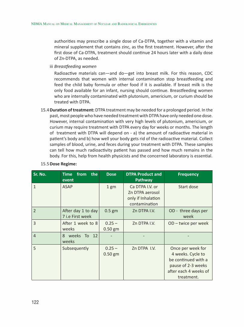
122
NDMA MANuAl oN MeDicAl MANAgeMeNt of NucleAr AND rADiologicAl eMergeNcies
authorities may prescribe a single dose of Ca-DTPA, together with a vitamin and mineral supplement that contains zinc, as the first treatment. However, after the first dose of Ca-DTPA, treatment should continue 24 hours later with a daily dose of Zn-DTPA, as needed.
iii Breastfeeding women
Radioactive materials can—and do—get into breast milk. For this reason, CDC recommends that women with internal contamination stop breastfeeding and feed the child baby formula or other food if it is available. If breast milk is the only food available for an infant, nursing should continue. Breastfeeding women who are internally contaminated with plutonium, americium, or curium should be treated with DTPA.
15.4 Duration of treatment: DTPA treatment may be needed for a prolonged period. In the past, most people who have needed treatment with DTPA have only needed one dose. However, internal contamination with very high levels of plutonium, americium, or curium may require treatment with DTPA every day for weeks or months. The length of treatment with DTPA will depend on - a) the amount of radioactive material in patient’s body and b) how well your body gets rid of the radioactive material. Collect samples of blood, urine, and feces during your treatment with DTPA. These samples can tell how much radioactivity patient has passed and how much remains in the body. For this, help from health physicists and the concerned laboratory is essential.
15.5 Dose Regime:
Sr. No. Time from the event
Dose DTPA Product and Pathway
Frequency
1 ASAP 1 gm Ca DTPA I.V. or Zn DTPA aerosol only if Inhalation
contamination
Start dose
2 After day 1 to day 7 i.e First week
0.5 gm Zn DTPA I.V. OD - three days per week
3 After 1 week to 8 weeks
0.25 – 0.50 gm
Zn DTPA I.V. OD – twice per week
4 8 weeks To 12 weeks
- - -
5 Subsequently 0.25 – 0.50 gm
Zn DTPA I.V. Once per week for 4 weeks. Cycle to
be continued with a pause of 2-3 weeks
after each 4 weeks of treatment.

123
15.6 For large quantities of Plutonium inhalation, (50 times the maximum permissible lung burden in patients aged 30 years or less) or more than 1.5 µ Ci, bronchial lavage is recommended soon after second or third day. This should be undertaken in specialized centers only where expertise exists.
15.7 Caution: There are no medical reasons why a person who is internally contaminated with plutonium, americium, or curium should not be treated with Ca-DTPA or Zn-DTPA, except nephritic syndrome. However, keep the following guidelines in mind:
i Because radioactive materials chelated to DTPA are passed out of the body in the urine, DTPA must be used carefully in people whose kidneys do not function properly.
ii Ca-DTPA should be used carefully in people who have a disease called “hemochromatosis.” (Hemochromatosis is a genetic disease that causes the body to absorb too much iron from foods and other sources, such as vitamins containing iron.)
iii Aerosol treatments using DTPA may not be safe for some people with asthma. If a person with asthma requires treatment with DTPA, the drug should be injected.
iv DTPA should not be used to treat people who are internally contaminated with the radioactive materials uranium or neptunium.
15.8 Side Effects: DTPA does not build up in the body or cause long-term health effects. People who are given repeat doses of Ca-DTPA within a short period of time may have nausea, vomiting, diarrhea, chills, fever, itching, and muscle cramps. Other side effects may include headache, lightheadedness, chest pain, and a metallic taste in the mouth.
15.8.1 Ca-DTPA (and Zn-DTPA) can chelate certain important minerals that the body needs (zinc, magnesium, and manganese). For example, the body needs zinc to make red blood cells, white blood cells, and platelets. Therefore, DTPA treatment may interfere with the normal production of blood cells. As a precaution, patients receiving long-term treatment with DTPA should be given a vitamin and mineral supplements containing zinc.
15.9 Bio-assay samples: Blood, urine and stool samples need to be collected for bio-assay. Repeat of earlier Sections).
15.10 Some other drugs of decontamination include DMPS (Di-mercapto-propan-sulphonate), DMSA, Dimercaprol (also called as BAL), Desferoxamine (DFOA). These are used for heavy metals like, Mercury, Lead, etc. and also for Polonium contamination.
16 DMPS16.1 This is a mercury chelator, like DMSA.
16.2 More dangerous since it can pull other useful metals out of the body (e.g. zinc and
annex-4

124
NDMA MANuAl oN MeDicAl MANAgeMeNt of NucleAr AND rADiologicAl eMergeNcies
copper) and it can dump much mercury into the kidney and liver and permanently damage them.
16.3 Start with several small test doses to make sure that patient can tolerate them (e.g. 10mg 1st time, 50mg 2nd time, 250mg 3rd time; and wait 2wks between each test).
16.4 It is recommended that DMPS is not to be used unless there are very compelling reasons to do so, since it is dangerous and has damaged many people.
17 DMSA17.1 This is a drug chelator (binds to mercury and then pulls it out of the body).
17.2 It is recommended to take several small test doses first to make sure that the patient can tolerate them (e.g. 20mg 1st time, 100mg 2nd time, 500mg 3rd time; and wait 2wks between each test).
17.3 LONG TERM treatment approach is to take 100mg a day for 3days and then wait 3weeks, and then repeat the cycle; and do this for a year or so.
17.4 While doing this, take a zinc/copper tablet each day (e.g. 25mg zinc, 2.5mg copper; the 10:1 ratio is important).
17.5 DMSA is sometimes coupled with Lipoate, however, if one has multiple sclerosis (MS), DMSA is to be used with caution since those with MS are at a greater risk of a bad reaction.
17.6 Also, if one has high levels of methyl-mercury (inorganic mercury from fish), the Lipoate can hurt as well.
18 Dimercaprol ( BAL – British Anti-Lewisite)18.1 Synonyms:
Dimercaprol, Dicaptol, Sulfactin, Dithioglycerol, British Anti-Lewisite, BAL
18.2 Presentation/formulation:i Ampoules (2 ml) containing 50 mg/ml (100 mg/ampoule) (BAL- Boots)
(Dimercaprol 5% solution in peanut oil and 10% benzyl benzoate).
ii Ampoules (3 ml) containing 100 mg/ml (300 mg/ampoule) (BAL- Hynson, Westcott and Dunning, Inc.) (Dimercaprol 10% solution in peanut oil and 10% benzyl benzoate)
iii Ampoules (2 ml) containing 100 mg/ml (200 mg/ampoule) (BAL- Sociétél’Arguenon) (Dimercaprol 10% solution in peanut oil with butacaine 1 mg).
18.3 First Aid Measures and treatment principles:
i Interrupt parenteral administration by lowering the dosage or increasing the time between doses.

125
ii Anti-histamines may possibly alleviate some of the adverse effects.
iii Strict clinical observation and monitoring of blood pressure and diuresis.
Note: Dimercaprol should be given with caution in hypertensive patients and in patients who have renal and hepatic dysfunction.
18.4 Storage conditions
18.4.1 Stability in light
Dimercaprol must be protected from light. The addition of benzyl benzoate increases its stability (and solubility).
18.4.2 Thermal stability
Dimercaprol must be stored at between 2°C and 10 °C in small vials that are hermetically sealed and completely filled.
18.5 Indications: Dimercaprol may be found useful in the treatment of poisonings due to:
i arsenic (organic and inorganic)
ii gold
iii inorganic mercury.
It should be noted that in any of the above types of poisoning the use of dimercaprol (even if DMSA/DMPS are not available) is not an absolute indication.
18.6 Doses:
i Adults
Dimercaprol should always be administered as soon as possible by deep intramuscular injection (never intravenous or subcutaneous) and rotating sites. The generally recommended doses are similar for arsenic, gold and inorganic mercury poisoning
ii Mild cases
2.5 mg/kg every 4 hours on the lst day
2.5 mg/kg every 6 hours on the 2nd day
2.5 mg/kg every 12 hours on the 3rd day
2.5 mg/kg every 24 hours for 10 days (or until clinical recovery).
iii Severe cases
3 to 4 mg/kg every 4 hours on the first 2 days
3 to 4 mg/kg every 6 hours on the 3rd day
3 to 4 mg/kg every 12 hours for 10 days
annex-4

126
NDMA MANuAl oN MeDicAl MANAgeMeNt of NucleAr AND rADiologicAl eMergeNcies
(doses of 5 mg/kg can be employed in the most severe cases).
(IPCS dimercaprol antidote monograph, 1994).
iv Children
Dimercaprol is well tolerated by children. The dosage should be calculated according to body weight, using the same unit-dose per kilogram of body weight as for adults under similar clinical conditions.
18.7 Contra-indications:
i Dimercaprol cannot be used in poisonings due to iron, cadmium, tellurium, selenium, vanadium and uranium. It is also contraindicated in poisonings due to elemental mercury vapor, because it can further increase the metal in the brain (Berlin and Ullberg, 1963).
ii Dimercaprol should not be given in case of acute renal failure (anuria) or extensive hepatic insufficiency (Cameron et al., 1947), and should be used with special care in hypertensive patients.
Note: Dimercaprol is not effective in massive, severe poisonings because its antidotal effect is surpassed by the toxicity of the toxic metal.
If given 7 days after arsenic exposure, it has little or no effect on the subsequent course of the neuropathy.

127
Annex-5
Phases of Radiation InjuryDose Range, Gy Prodrome Manifestation of Illness Prognosis (without Therapy)0.5-1.0 Mild Slight decrease in blood cell counts Almost certain survival
1.0-2.0 Mild to moderate
Early signs of bone marrow damage
Highly probable survival (>90% of victims)
2.0-3.5 Moderate Moderate to severe bone marrow damage
Probable survival
3.5-5.5 Severe Severe bone marrow damage; slight GI damage
Death within 3.5-6 wk (50% of victims)
5.5-7.5 Severe Pancytopenia and moderate GI damage
Death probable within 2-3 wk
7.5-10.0 Severe Marked GI and bone marrow damage, hypotension
Death probable within 1-2.5 wk
10.0-20.0 Severe Severe GI damage, pneumonitis, altered mental status, cognitive dysfunction
Death certain within 5-12 d
20.0-30.0 Severe Cerebrovascular collapse, fever, shock Death certain within 5-12 d
* Modified from Walker RI, Cerveny RJ, eds. (21). GI = gastrointestinal.
1 Hematopoietic Syndrome:1.1 Irradiation of bone marrow stem and progenitor cells at increasing doses results in
exponential cellular death. The hematopoietic syndrome is seen with significant partial-body or whole-body radiation exposures exceeding 1 Gy and is rarely clinically significant below this level. Mitotically active hematopoietic progenitors have a limited capacity to divide after a whole-body radiation dose greater than 2 to 3 Gy. In the ensuing weeks after exposure, a hematologic crisis occurs, characterized by hypoplasia or aplasia of the bone marrow. These changes result in pancytopenia, predisposition to infection, bleeding, and poor wound healing, all of which may contribute to death.
1.2 While most bone marrow progenitors are susceptible to cell death after sufficiently intense radiation doses, subpopulations of stem cells or accessory cells are selectively more radio resistant, presumably because of their largely non-cycling state. These radio resistant cells may play an important role in recovery of hematopoiesis after exposure to doses as high as 6 Gy, albeit with a reduced capacity for self-renewal. Another critical determinant for reconstitution is inhomogeneity of the dose with sparing of marrow sites that become foci of hematopoietic activity.

128
NDMA MANuAl oN MeDicAl MANAgeMeNt of NucleAr AND rADiologicAl eMergeNcies
1.3 Lymphopenia is common and occurs before the onset of other cytopenias. A predictable decline in lymphocytes occurs after irradiation. In fact, a 50% decline in absolute lymphocyte count within the first 24 hours after exposure, followed by a further, more severe decline within 48 hours, characterizes a potentially lethal exposure.
The above figure shows Leukocyte count based on exposure dose in patients exposed to radiation in Chernobyl. Note the abortive rise (transient increase before the fall) in counts of leukocytes, which are primarily composed of granulocytes, in doses less than 5 Gy. Neutropenia may not occur for weeks, especially with lower exposures and its duration may be prolonged. To convert cells/mm3 to x109 cells/litre, multiply by 0.001. Due to large inter-individual variation of leuco-counts, it is necessary to observe the kinetics. However, in severe cases, bone marrow cellularity needs to be checked to ascertain the viability of bone marrow and its recovery.
(Ref: Annals of Internal Medicine. CLINICAL GUIDELINES. Medical management of the Acute Radiation Syndrome: Recommendations of the Strategic National Stockpile Radiation Working Group. Jamie K. Waselenko et al; 15 June 2004/ Vol 140/Issue 12/ Pg 1037 – 1051)
2 Gastro-Intestinal Syndrome:2.1 Radiation induces loss of intestinal crypts and breakdown of the mucosal barrier. The
onset of gastro-intestinal syndrome is at 6-8 Gy whole body and results in abdominal pain, diarrhea, nausea and vomiting and predispose patients to infection. At doses

129
exceeding 12 Gy, the mortality rate of the gastrointestinal syndrome exceeds that of the hematopoietic syndrome. Severe nausea, vomiting, watery diarrhea and cramps occur within hours after high-dose (>10 Gy) irradiation.
Latent period follows, lasting for 5 to 7 days, during which symptoms reduce.
2.2 During manifest illness, vomiting and severe diarrhea associated with high fever occur. Systemic effects may include malnutrition from mal-absorption, bowel obstruction from ileus, dehydration, cardiovascular collapse and electrolyte derangements from fluid shifts. Damage to the intestinal mucosa and microcirculation, causing gastrointestinal bleeding, leads to anemia. Subsequently it can cause sepsis and acute renal failure.
3 The Neurovascular Syndrome3.1 The Neurovascular syndrome is less well defined than other syndromes, and its
stages are compressed. Individuals presenting with fever, hypotension and major impairment of cognitive function will most likely have had a supra-lethal exposure. These symptoms may be observed in those receiving more than 15 to 30 Gy of radiation.
3.2 The prodromal phase is characterized by disorientation, confusion and prostration and may be accompanied by loss of balance and seizures. Physical examination may show papilledema, ataxia and reduced or absent deep tendon and corneal reflexes.
3.3 During the latent period, apparent improvement occurs for a few hours and is followed by severe manifest illness. Within 5 to 6 hours, watery diarrhea, respiratory distress, hyperpyrexia and cardiovascular shock can occur. This rapid decline mimics the clinical course of acute sepsis and septic shock, both of which must be considered. The ensuing circulatory complications of hypotension, cerebral edema, increased intracranial pressure and cerebral anoxia can lead to death within 2 days.
4 The Cutaneous Syndrome4.1 Cutaneous injury from thermal or radiation burns is characterized by loss of
epidermis and, at times, dermis. Injuries to the skin may cover small areas but extend deeply into the soft tissue, even reaching underlying muscle and bone. They may be accompanied by profound local edema and place the patient at risk for a compartment syndrome. Patients presenting with burns immediately after exposure may have had thermal rather than radiation burns. Significant injuries to the integument decrease the LD50/60 and amplify the risk of death at any radiation exposure dose. Patients with the hematopoietic syndrome have a more complicated course of the cutaneous syndrome as a result of bleeding, infection and poor wound healing.
4.2 Table showing the Grading system for Response of Neurovascular, Gastro-Intestinal Systems.
(Ref: Annals of Internal Medicine. CLINICALGUIDELINES. Medical management of the Acute Radiation Syndrome : Recommendations of the Strategic National Stockpile
annex-5

130
NDMA MANuAl oN MeDicAl MANAgeMeNt of NucleAr AND rADiologicAl eMergeNcies
Radiation Working Group. Jamie K. Waselenko et al; 15 June 2004/ Vol 140/Issue 12/ Pg 1037 – 1051)
Grading System of Response of Neurovascular, Gastrointestinal and cutaneous Systems*
Symptom Degree 1 Degree 2 Degree 3 Degree 4Neurovascular SystemNausea Mild Moderate Intense Excruciating
Vomiting Occasional (once per day
Intermittent (2-5 times per day)
Persistent (6-10 times per day)
Refractory (>10 times per day
Anorexia Able to eat Intake decreased Intake minimal Parenteral nutrition
Fatigue syndrome
Able to work Impaired work ability
Needs assistance for ADLs
Cannot perform ADLs
Temperature, 0C <38 38-40 >40 for <24 h >40 for <24 h
Headache Minimal Moderate Intense Excruciating
Hypotension Heart rate>100 beats/min; blood pressure > 100/170 mm Hg
Blood pressure <100/70 mm Hg
Blood pressure <90/60 mm Hg; transient
Blood pressure <80/?mm Hg; persistent
Neurologic deficits
Barely detectable Easily detectable Prominent Lifte-threatening, loss of consciousness
Cognitive deficits
Minor loss Moderate loss Major impairment Complete imapirment
Gastrointestinal system Diarrhea
Frequency, stools/d
2-3 4-6 7-9 >10
Consistency Bulky Loose Loose Watery
Bleeding Occult Intermittent Persistent Persistent with large amount
Abdominal cramps or pain
Minimal Moderate Intense Excruciating
Cutaneous system
Erythema Minimal, transient Moderate (<10% body surface area)
Marked (10%–40% body surface area)
Severe (>40% body surface area)
Sensation or itching
Pruritus Slight and intermittent pain
Moderate and persistent pain
Severe and persistent pain
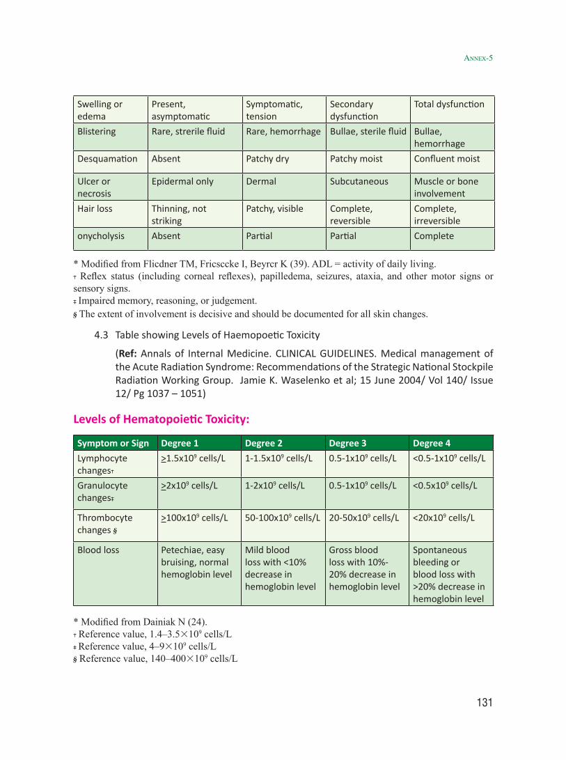
131
Swelling or edema
Present, asymptomatic
Symptomatic, tension
Secondary dysfunction
Total dysfunction
Blistering Rare, strerile fluid Rare, hemorrhage Bullae, sterile fluid Bullae, hemorrhage
Desquamation Absent Patchy dry Patchy moist Confluent moist
Ulcer or necrosis
Epidermal only Dermal Subcutaneous Muscle or bone involvement
Hair loss Thinning, not striking
Patchy, visible Complete, reversible
Complete, irreversible
onycholysis Absent Partial Partial Complete
* Modified from Flicdner TM, Fricsccke I, Beyrcr K (39). ADL = activity of daily living. Reflex status (including corneal reflexes), papilledema, seizures, ataxia, and other motor signs or
sensory signs. Impaired memory, reasoning, or judgement. The extent of involvement is decisive and should be documented for all skin changes.
4.3 Table showing Levels of Haemopoetic Toxicity
(Ref: Annals of Internal Medicine. CLINICAL GUIDELINES. Medical management of the Acute Radiation Syndrome: Recommendations of the Strategic National Stockpile Radiation Working Group. Jamie K. Waselenko et al; 15 June 2004/ Vol 140/ Issue 12/ Pg 1037 – 1051)
Levels of Hematopoietic Toxicity:
Symptom or Sign Degree 1 Degree 2 Degree 3 Degree 4Lymphocyte changes
>1.5x10⁹ cells/L 1-1.5x10⁹ cells/L 0.5-1x10⁹ cells/L <0.5-1x10⁹ cells/L
Granulocyte changes
>2x10⁹ cells/L 1-2x10⁹ cells/L 0.5-1x10⁹ cells/L <0.5x10⁹ cells/L
Thrombocyte changes
>100x109 cells/L 50-100x109 cells/L 20-50x109 cells/L <20x109 cells/L
Blood loss Petechiae, easy bruising, normal hemoglobin level
Mild blood loss with <10% decrease in hemoglobin level
Gross blood loss with 10%-20% decrease in hemoglobin level
Spontaneous bleeding or blood loss with >20% decrease in hemoglobin level
* Modified from Dainiak N (24). Reference value, 1.4–3.53109 cells/L Reference value, 4–93109 cells/L Reference value, 140–4003109 cells/L
annex-5

132
NDMA MANuAl oN MeDicAl MANAgeMeNt of NucleAr AND rADiologicAl eMergeNcies
Acknowledgements
Prepared By : Late : Dr Raghavendra Deolalikar Certifying Surgeon, NAPS, NPCIL and Member W.G.
Reviewed / Edited By:
Shri B. Bhattacharjee Hon’ble ex Member, NDMA & Chairman, High Power Committee, NDMA Shri S.A. Hussain Ex Head RSD, AERB and Convener W.G.Late Dr M. C. Abani former Sr. Specialist (Nuclear), NDMA and Member W.GShri Shivajee Singh former Sr. Specialist [NDRF], NDMA and Member W.G.Shri D.K.Shukla Head, ROD, BARC and Member W.G.Dr Pradeep Kumar, Head, ERS and MS, BARC and Member W.G.Shri S.M.Maskey, O.S., TAPS 1and2, NPCIL and Member W.G.Shri P.C. Meghnani S.O. NPCIL and Member W.G.Shri A.P. Garg Scientific Officer AERB, and Member W.G.Dr N. Roy former Head, Surgical Unit, BARC Hospital, and Member W.G.Shri S.K.Pawar Scientific Officer AERB, and Member W.G. Shri Shyam Vyas Scientific Officer AERB, and Member W.G.Dr M.M. Adtani Retd. Scientific Officer BARC, and Member W.G.Shri P.K. Bhatia Scientific Officer NPCIL, and Member-Secretary W.G.Dr Hemant Haldavnekar, (OIC) Trombay Dispensary, BARCDr Rajiv Sarin, Prof. TMH, ACTRECDr M.G.R. Rajan, BARC, Former Head, FMCDr (Smt) Birajalaxmi Das, S.O. BARCDr Pankaj Tandon, S.O. AERB Dr Ajay Chaubey, Head Radiology Dept. BARC HospitalDr Anjali Godse, Medical Officer, BARCDr Aseem Bhatnagar, S.O. INMASDr Rakesh Kumar, Prof. AIIMS
NDMA
Dr D N Sharma, Member, NDMABrig. Ajay Gangwar, JS (Mitigation), NDMASh Pushkar Sahay, JA (MP & P)Sh S K Mishra , Sr Consultant (N & R), NDMADr Saurabh Dalal, Consultant (Medical Preparedness), NDMA
133


136
NDMA MANuAl oN MeDicAl MANAgeMeNt of NucleAr AND rADiologicAl eMergeNcies