Efficient in vivo delivery of DNA to pulmonary cells using the novel lipid EDMPC
Upload
independentCategory
view
2download
0
Trends in Food Science & Technology 23 (2012) 13e27
Review
* Corresponding author.
0924-2244/$ - see front matter � 2011 Elsevier Ltd. All rights reserved.doi:10.1016/j.tifs.2011.08.003
Nanoencapsulation
of food ingredients
using lipid based
delivery systems
Milad Fathia,*, M.R. Mozafarib
and M. Mohebbia
aDepartment of Food Science and Technology,
Ferdowsi University of Mashhad,
PO Box: 91775-1163, Iran(e-mail: [email protected])
bAustralasian Nanoscience and Nanotechnology
Initiative, Monash University LPO, P.O. Box 8052,
Wellington Road, Clayton, Victoria 3800, Australia
e
Sustained release
Delayed release
Nanoencapsulation allows protection of the sensitive bioactive
food ingredients from unfavorable environmental conditions,
eradication of incompatibilities, solubilization, or masking of
unpleasant taste or odor. This paper reviews the present state
of the art of lipid based carriers including nanoemulsions,
nanoliposomes, solid lipid nanoparticles (SLNs) and novel
generation of encapsulation system namely nanostructure lipid
carriers (NLCs) regarding their production method, physico-
chemical properties, functionalities, stabilization techniques,
potential advantages and limitations and delivery mechanisms.
In the last section, mathematical models for predication of bio-
active release kinetics from lipid based nanocarriers, which
can be applied for optimization of encapsulation systems,
are presented and some future developments in the area of
nanoencapsulation are discussed.
Bio
acti
ve r
elea
s
IntroductionNanotechnology is defined as creation, utilization and
manipulation of materials, devices or systems in thenanometer scale (smaller than 100 nm). In recent years nano-technology has found innumerable applications in differentfood industries (Aguilera et al., 2008; Fathi & Mohebbi,
2010; Neethirajan & Jayas, 2010; Rizvi, Moraru,Bouwmeester, & Kampers, 2010; Sanguansri & Augustin,2006). Some potential applications of this technology innanoencapsulation and delivery of bioactive componentshave been documented in pharmaceutics, as well as cos-metics and food sciences (Farokhzad & Langer, 2009; Liu,Jiao, Wang, Zhou, & Zhang, 2008; M€uller, Petersen,Hommoss, & Pardeike, 2007; Sagalowicz, Leser, Watzke,&Michel, 2006; Shah et al., 2007; Shimoni, 2009). Deliverysystem is defined as one in which a bioactive material isentrapped in a carrier to control the rate of bioactive release.Nanocarriers can protect a bioactive component from unfa-vorable environmental conditions e.g. oxidation and pHand enzymes degradation (Fang & Bhandari, 2010;Ghosh, Mandal, Sarkar, Panda, & Das, 2009; Zimet &Livney, 2009). Nanocarriers provide more surface area andhave the potential to enhance solubility, improve bioavail-ability and ameliorate controlled release and targeting ofthe encapsulated food ingredients, in comparison tomicro-size carriers (Mozafari, 2006a; Weiss, Gaysinsky,Davidson, & McClements, 2009).
Generally two controlled release mechanisms (Fig. 1)can be observed during delivery of a bioactive (Lakkis,2007). (i) Delayed release, which is a mechanism by whichthe release of a bioactive substance is delayed froma bounded “lag time” up to a point when/where its releaseis preferred and is no longer obstructed. This mechanismcould be used for flavor release in ready-meals, colorrelease in beverages or protection of nutrition compoundsin gastric condition and their release in the intestine.
Time
Fig. 1. Models of bioactive release from nanocarrier systems.

14 M. Fathi et al. / Trends in Food Science & Technology 23 (2012) 13e27
(ii) Sustained release, which is a mechanism engineered tokeep constant concentration of a bioactive at its target site.This system can be employed for extending the release ofthe encapsulated material, including flavor or certain drugssuch as insulin, in chewing gum. Many variables over-shadow bioactive release of the encapsulated material.These include shape and dimensions of carrier, bioactivediffusivity and solubility in encapsulant and environmentalmedia, erosion rate, polymorphism status of lipid based car-riers, porosity and tortuosity, bioactive ratio between carrierand aqueous medium, encapsulation load (weight ratio ofencapsulant to lipid) and loading efficiency (weight ratioentrapped to free encapsulant) and pH value of the medium(Barat, Crane, & Ruskin, 2008; Briones & Sato, 2010;Jalsenjak, 1992; Sant, Nadeau, & Hildgen, 2005;Siepmann, Faisant, & Benoit, 2002; Yang & Washington,2006; Zhang, Yang, Chow, & Wang, 2003). Havinga high encapsulation efficiency is always favorable, albeitencapsulation load more than 50% is not proper due to in-crease risk of bioactive leakage in case of more surface de-fect (Madene, Jacquot, Scher, & Desobry, 2006).
Typically, food applicable nanocarrier systems can becarbohydrate, protein or lipid based. Despite of differentadvantages of carbohydrate and protein based nanocap-sules, they do not have potential of fully scale up due to re-quirement of applying different complicated chemical orheat treatments which cannot be completely controlled.On the other hand, lipid based nanocarriers have possibilityof industrial production and bear advantage of more encap-sulation efficiency and low toxicity. In this paper we willprovide an overview of recent developments in different as-pects (e.g. production methods, physicochemical proper-ties, stabilization techniques, release mechanisms,advantages and disadvantages) of four famous lipid basedcarriers namely nanoemulsions, nanoliposomes, solid lipidnanoparticles (SLNs) and nanostructure lipid carriers(NLCs) (Table 1). In the last section of the paper, mathe-matical models for predication of bioactive release kineticsof lipid based nanocarriers are presented.
Nano delivery systemsNanoemulsions
Nanoemulsions (also known as miniemulsions or submi-cron emulsions) are nanoscale droplets of multiphase col-loidal dispersions formed by dispersing of one liquid inanother immiscible liquid by physical share-induced rup-turing (Liu, Sun, Li, Liu, & Xu, 2006; Mason, Wilking,Meleson, Chang, & Graves, 2006; Meleson, Graves, &Mason, 2004; Russel, Saville, & Schowalter, 1989). Differ-ent size ranges have been reported in the literature for nano-emulsions; e.g. less than 100 nm (Guo et al., 2007; Porras,Solans, Gonzلlez, & Guti�errez, 2008; Shakeel & Ramadan,2010), 10e100 nm (Talegaonkar, Mustafa, Akhter, & Iqbal,2010), 100e500 nm (Anton, Benoit, & Saulnier, 2008;Constantinides, Chaubal, & Shorr, 2008; Rossi & Leroux,2007; Tadros, Izquierdo, Esquena, & Solans, 2004; Unger
et al., 2004) and 100e600 nm (Sakulku et al. 2009;Solans et al., 2003). However, the most appropriate onesbased on nanotechnology definition is having size rangesof less than 100 nm and possessing different propertiesthan ordinary emulsions. Nanoemulsions have some inter-esting physical properties that can be applied to distinguishthem from microemulsions. For instance, microemulsionstypically show multiple scattering of visible light, andtherefore, have a white opaque appearance. In contrast,the droplet sizes in nanoemulsions are much smaller thanvisible wavelengths; hence most nanoemulsions appear op-tically transparent (McClements & Li, 2010; Shakeel &Ramadan, 2010). This is a very favorable feature of nano-emulsions for applying them as the nutrient carriers in bev-erages. Rheological properties of nanoemulsions are alsodifferent from micoemulsion. For example, Mason andRai (2003) reported a rapid increase in the shear modulusof nanoemulsions by decreasing emulsion droplet size. Fur-thermore, at very high droplet volume fraction (4), wherethe droplets commence to deform, the elastic shear modu-lus of repulsive emulsions was reported to be proportionalto the Laplace pressure of the undeformed droplets leadingto extraordinary elastic modulus of nanoemulsion. Like-wise, at the same droplet volume fraction, nanoemulsionsenhance shelf stability against the gravity over the microe-mulsions due to Brownian motion of nano sized droplets,caused by entropic driving forces (Mason et al., 2006). Un-like the microemulsions that are thermodynamically stableand form spontaneously, nanoemulsions are kinetically sta-ble (Henry, Fryer, Frith, & Norton, 2010; Tadros et al.,2004). An interesting feature of nanoemulsions in contrastto microemulsions is that, they are metastable and can bediluted with water without change in the droplet size distri-bution (Gutieırrez, Gonzaılez, Sole’, Pey, & Nolla, 2008).
Production methodThe methods used to produce nanoemulsions can be di-
vided into the mechanical and non-mechanical techniques.Mechanical (high-energy) methods include high-pressurehomogenization, microfluidization and ultrasonication(Anton, Saulnier, Beduneau, & Benoit, 2007; Tadroset al., 2004).
To form a fine stable emulsion using high-pressure ho-mogenization, the coarse dispersion of the oil and aqueousphase and emulsifier is passed through a small inlet orificeat pressures in the range of 500e5000 psi. Microfluidiza-tion uses a very high pressure (up to 20,000 psi) to forcethe liquid through the interaction chamber, which consistsof microchannels of a special configuration. The emulsionfeeds through the microchannels into a collision chamberwhich leads to formation of fine nano-scale emulsion drop-lets. The emulsification mechanism is based on combina-tion of cavitation and mechanical shear. The operatingpressure and the number of passing cycles of the coarsepreemulsion in the microfluidizer or homogenizer havestrong effect on the particle size of the formed
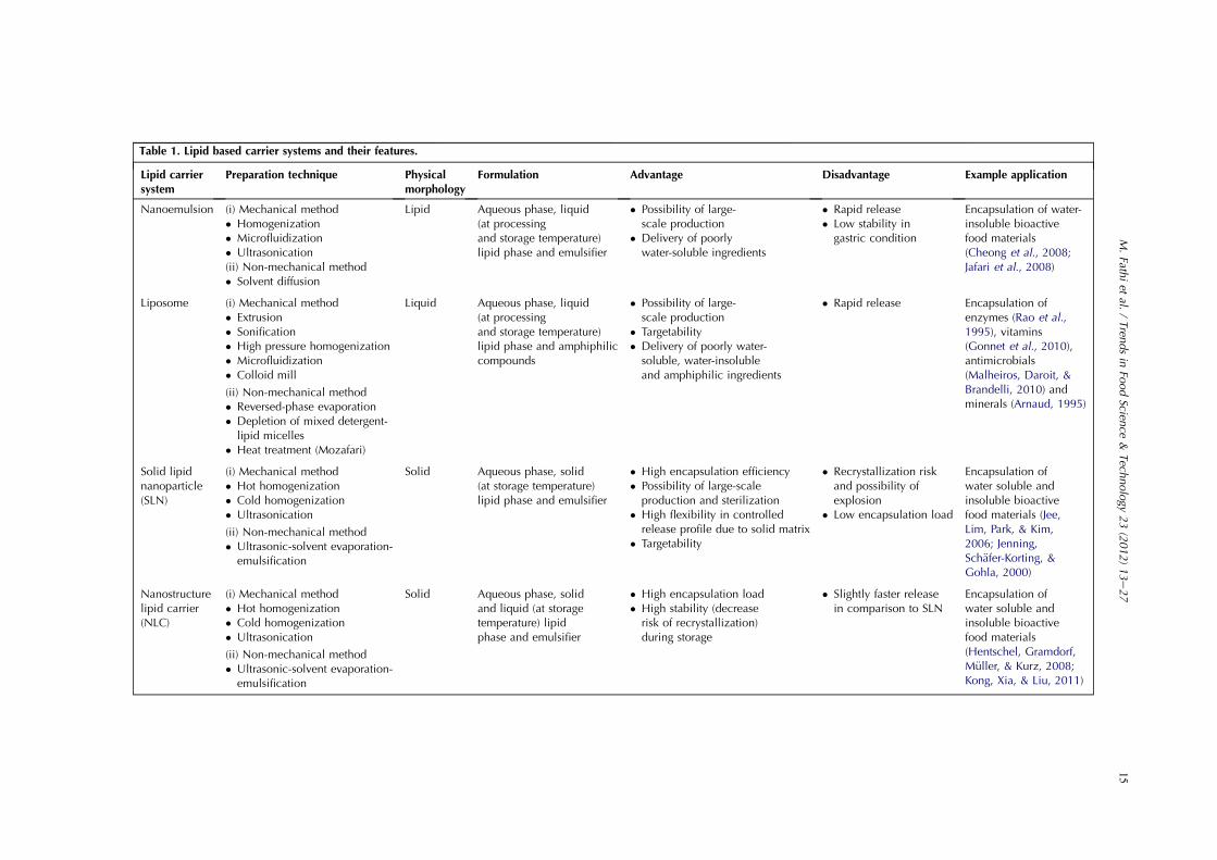
Table 1. Lipid based carrier systems and their features.
Lipid carriersystem
Preparation technique Physicalmorphology
Formulation Advantage Disadvantage Example application
Nanoemulsion (i) Mechanical method� Homogenization� Microfluidization� Ultrasonication(ii) Non-mechanical method� Solvent diffusion
Lipid Aqueous phase, liquid(at processingand storage temperature)lipid phase and emulsifier
� Possibility of large-scale production
� Delivery of poorlywater-soluble ingredients
� Rapid release� Low stability in
gastric condition
Encapsulation of water-insoluble bioactivefood materials(Cheong et al., 2008;Jafari et al., 2008)
Liposome (i) Mechanical method� Extrusion� Sonification� High pressure homogenization� Microfluidization� Colloid mill
(ii) Non-mechanical method� Reversed-phase evaporation� Depletion of mixed detergent-
lipid micelles� Heat treatment (Mozafari)
Liquid Aqueous phase, liquid(at processingand storage temperature)lipid phase and amphiphiliccompounds
� Possibility of large-scale production
� Targetability� Delivery of poorly water-soluble, water-insolubleand amphiphilic ingredients
� Rapid release Encapsulation ofenzymes (Rao et al.,1995), vitamins(Gonnet et al., 2010),antimicrobials(Malheiros, Daroit, &Brandelli, 2010) andminerals (Arnaud, 1995)
Solid lipidnanoparticle(SLN)
(i) Mechanical method� Hot homogenization� Cold homogenization� Ultrasonication
(ii) Non-mechanical method� Ultrasonic-solvent evaporation-
emulsification
Solid Aqueous phase, solid(at storage temperature)lipid phase and emulsifier
� High encapsulation efficiency� Possibility of large-scaleproduction and sterilization
� High flexibility in controlledrelease profile due to solid matrix
� Targetability
� Recrystallization riskand possibility ofexplosion
� Low encapsulation load
Encapsulation ofwater soluble andinsoluble bioactivefood materials (Jee,Lim, Park, & Kim,2006; Jenning,Sch€afer-Korting, &Gohla, 2000)
Nanostructurelipid carrier(NLC)
(i) Mechanical method� Hot homogenization� Cold homogenization� Ultrasonication
(ii) Non-mechanical method� Ultrasonic-solvent evaporation-
emulsification
Solid Aqueous phase, solidand liquid (at storagetemperature) lipidphase and emulsifier
� High encapsulation load� High stability (decreaserisk of recrystallization)during storage
� Slightly faster releasein comparison to SLN
Encapsulation ofwater soluble andinsoluble bioactivefood materials(Hentschel, Gramdorf,M€uller, & Kurz, 2008;Kong, Xia, & Liu, 2011)
15
M.Fath
iet
al./Tren
dsin
FoodScien
ce&
Tech
nology
23(2012)13e27

16 M. Fathi et al. / Trends in Food Science & Technology 23 (2012) 13e27
nanoemulsion (Constantinides et al., 2008; Maa & Hsu,1999; Quintanilla-Carvajal et al., 2010). The mechanismof nanoemulsion generation using ultrasonication is attrib-uted to bubble cavitation. However, this mechanism is notfully understood yet (Bondy & Sollner, 1935; Mason,1992). The ultrasound waves (at ultrasonic frequencies typ-ically 20 kHz or larger and high power intensity) result insequential formation, growth and collapse of microscopicvapor bubbles in the liquid. Subsequently, the collapse ofthese cavities provides sufficient energy to increase surfacearea of droplets (Patil & Pandit, 2007). Efficiency of nano-emulsification by sonication (considering the size of thenanoemulsion droplets and required time to attain thissize), depends both on the emulsion properties (lipid, en-capsulated bioactive, surfactant and cosurfactant) and thepower of the device (Leong, Wooster, Kentish, &Ashokkumar, 2009). Kentish et al. (2008) produced nanoe-mulsion using flax seed oil and tween 40 (C16) as an emul-sifier by ultrasonication method. Results showed that thereis an optimum power input level above which droplet coa-lescence and cavitational bubble cloud formation adverselyaffect the efficiency, reproducibility, quality of nanoemul-sion production. Increasing sonication time reduces dropletsizes to a point, but continued sonication more than one tofive minutes is useless. The produced nanoemulsions arecomparable to those emulsions prepared using a microfluid-izer operated at 100 MPa. In spite of high potential of son-ication for research purposes, it does not appear to bepractical for industrial applications. In this case usinghigh-pressure homogenizer or microfluidization are oftenpreferred (Samer & Schork, 1999). It is remarkable thatwhen the aim of the processing is the encapsulation of sen-sitive molecules such as enzymes, using ultrasonication andhigh-pressure homogenization may cause degradation, de-naturation or loss of activity of the bioactive agents(Anton et al., 2008). Pinnamaneni, Das, and Das (2003)compared oil/water submicron emulsions manufacturedby microfluidization and homogenization methods. The mi-crofluidization was found to be more effective than the ho-mogenization, since submicron emulsions prepared by thisprocess had smaller droplet diameters and exhibited lessdroplet diameter growth over time. Jafari, He, andBhandari (2006) studied the efficiency of producing nano-emulsions using sonication and microfluidization tech-niques. Their results showed that, whether both methodswere able to form nanoemulsions of the sizes rangingfrom 150 to 700 nm, the microfluidizer produced emulsionswith restricted size distributions and sonication was morecommodious regarding to operation control and cleaning.Jafari, Assadpoor, Bhandari, and He (2008) produced nano-emulsion using microfluidization and ultrasonication forencapsulation of fish oil. Their results showed that micro-fluidization is a competent emulsification technique leadingto fish oil-encapsulated powder with the lower unencapsu-lated oil at the surface of particles in comparison to ultra-sonication method.
Nanoemulsions can be formed using non-mechanicalmethods; e.g. solvent diffusion technique (Anton et al.,2007; Tadros et al., 2004; Unger et al., 2004). Cheong,Tan, Man, and Misran (2008) applied emulsifica-tioneevaporation technique to prepare nanoemulsion(90e120 nm) of a-tocopherol. In this method a-tocopherolis first dissolved in an organic solvent. The resulting coarsedispersion is passed through a high pressure homogenizerand then the solvent is removed from fine emulsion byevaporation (Jaiswal, Gupta, & Kreuter, 2004; Mainardes& Evangelista, 2005). However, the a-tocopherol contentof prepared nanoemulsion was significantly decreased dur-ing storage. Lee and McClements (2010) produced oil/wa-ter nanoemulsions coated by whey protein usingcombination of emulsification, solvent displacement and/or solvent evaporation approaches. A limitation of this ap-proach is the requirement of large amounts of organic sol-vent to prepare emulsions and consequently the necessity ofusing expensive equipment to remove organic solvent be-fore consumption. Generally, due to use of organic solvent,application of low energy methods is limited in foodsectors.
Applications and featuresNanoemulsions are good candidate for delivery of poorly
water-soluble food ingredients, such as fish oil and lipophilicvitamins, because of their ability to improve bioactive solu-bilization and potential for enhancing absorption in the gas-trointestinal (GI) tract, caused by surfactant inducedpermeability changes. After ingestion, droplets readily dis-perse in stomach to small droplet of nanoemulsion, whichpromotes wide distribution of the encapsulated bioactivethroughout the GI condition (Talegaonkar et al., 2010).Yuan, Gao, Mao, and Zhao (2008) found that the optimumproduction conditions for the preparation of b-carotenenanoemulsions are a homogenization pressure of 129 MPa;a homogenization temperature of 47 �C; a b-carotene con-centration of 0.82% and an emulsifier concentration of8.2%. Soybean oil-based nanoemulsion has been shown tohave bactericidal properties against Gram-positive bacteria(Hamouda & Baker, 2000) and fungistatic, but not fungi-cidal, property (Hamouda et al., 2001). Teixeira et al.(2007) prepared BCTP nanoemulsion (an O/W nanoemul-sion of soybean oil and tri-n-butyl phosphate emulsifiedwith Triton X-100) and showed that it was effective in reduc-ing the cell numbers of Listeria monocytogenes.
Wulff-Perez, Torcello-Gomez, Galvez-Ruız, and Martın-Rodrıguez (2009) reported that destabilization of the nanoe-mulsions takes place above certain surfactant concentration.This phenomenon can be described as a depletionefloccula-tion effect caused by non-adsorbed micelles. They presenteda theoretical mathematical model based on experimental pa-rameters to determine the optimum value of surfactant con-centration. A bioactive material might have differentlocalizations within an emulsion (Fig. 2), and this localiza-tion can obviously be influenced by the production

Low temperature and low number of homogenization cycles
Increased temperature and increased number of homogenization cycles
Stabilizer film
Fig. 2. Principles of chemical stabilization of bioactive ingredient en-capsulated in nanoemulsion. Applying Low temperature and low num-ber of homogenization cycles (Left) lead to localization of thebioactive more toward the inner oil phase (bioactive is not protectedagainst chemical degradation). Increasing temperature and homogeni-zation cycles (right) allow diffusion of the bioactive into the outer sta-bilizer film (more protection against chemical degradation) (Baspinar,
et al., 2010).
17M. Fathi et al. / Trends in Food Science & Technology 23 (2012) 13e27
parameters, e.g. number of homogenization cycles and pro-duction temperature. The location of a drug, or any other bio-active compound, within the capsule or carrier influences thestability, release and thus the bioavailability of the nanocar-rier formulation. Baspinar, Keck, and Borchert (2010) pre-pared a nanoemulsion formulation, using polysorbate andless purified egg lecithin as emulsifiers, through the highpressure homogenization method. Interestingly, their resultsshowed that the highest chemical stability was obtained withlowhomogenization pressures but higher numbers of homog-enization cycles and higher temperatures (e.g. 300 bar, 10 cy-cles and 50 �C). This could be attributed to an immigration ofthe bioactive from inner oil to the stable surfactant outerlayer. However, the release profiles of these nanoemulsionswere not investigated.
Maruno and da Rocha-Filho (2010) produced non-foodgrade nanoemulsion using lipophilic (Sorbitan monooleate;HLB ¼ 4.3) and hydrophilic (Ceteth-10, HLB ¼ 12.9) sur-factants. The accelerated tests based on measurement ofzeta potential showed that nanoemulsions are stable for15 years. It was demonstrated that nanoemulsions stabilizedby lecithin/polyol or lecithin/carbohydrate matrix havedroplet sizes ranging from 30 to 60 nm. In other studies,it was shown that the bioavailability of the nanoemulsion-encapsulated vitamin E was 10-fold more than the bioavail-ability of the same vitamin contained in commercial gelatincapsules (Gonnet, Lethuaut, & Boury, 2010; Wajda, 2003;Wajda, Zirkel, & Schaffer, 2007).
Nanoemulsions have low stability in acidic conditions.Klinkesorn and McClements (2009) applied chitosan forenhancing the stability of nanoemulsions. Their resultsshowed that the zeta potential of the oil droplets inlecithin-chitosan stabilized nanoemulsions changed frompositive to negative and the emulsions droplets can be de-graded by lipase under simulated GI conditions. Therefore,chitosan-coated lipid droplets can be potentially applied asuseful carriers for the oral delivery of lipophiliccompounds.
Finally, it should be mentioned that advantages of nano-emulsions include toxicological safety and a high contentof the lipid phase as well as the possibility of large-scaleproduction using high-pressure homogenization (HPH).However, controlled drug release from nanoemulsions isvery unlikely because of the small size and the liquid stateof the carrier.
LiposomeHaving a number of benefits, e.g. possibility of large-scale
production using natural ingredients and entrapment and re-lease of water-soluble, lipid-soluble, and amphiphilic mate-rials as well as targetability (Huwiler, Kolter, Pfeilschifter,& Sandhoff, 2000; Mozafari, Johnson, Hatziantoniou, &Demetzos, 2008; Thompson, Hindmarsh, Haisman, Rades,& Singh, 2006), liposomes have been widely used in foodsector both in research and industry. Notable examples are li-posome formulations of antimicrobials (Malheiros, Daroit,& Brandelli, 2010; Malheiros, Daroit, Silveira, &Brandelli, 2010; Taylor, Bruce, Weiss, & Davidson, 2008;Taylor, Davidson, Bruce, & Weiss, 2005b; Taylor,Gaysinsky, Davidson, Bruce, & Weiss, 2007; Were, Bruce,Davidson, & Weiss, 2004; Were, Bruce, & Weiss, 2003), li-pophilic vitamins (Gonnet et al., 2010; Padamwar &Pokharkar, 2006), enzymes (Dufour, Laloy, Vuillemard, &Simard, 1996; Rao, Chawan, & Veeramachaneni, 1995)and minerals (Arnaud, 1995). Similar to nanoemulsions, li-posomes are kinetically stable. The mechanism of liposomeformation is based on the unfavorable interactions occur be-tween amphiphilic compounds (mainly phospholipids) andwater molecules, where the polar head groups of phospho-lipids are subjected to the aqueous phases of the inner andouter media, and the hydrophobic hydrocarbon tails are asso-ciated into the bilayer and spherical core shell structures areformed (Goyal et al., 2005; Jesorka & Orwar, 2008).
Liposomes can be produced using natural ingredients onan industrial scale and have the capability of entrappingmaterials with different solubilities (Mozafari &Khosravi-Darani, 2007; Yurdugul & Mozafari, 2004). An-other important advantage of liposomes (also known aslipid vesicles) is targetability. Lipid vesicles can be tailoredto deliver and release their load in the target site inside andoutside the body (Mozafari, 2006b).
Production methodDifferent procedures have been developed to produce
nano-sized liposomes. These include mechanical (extru-sion, where the liposomes are forced through filters withwell defined pore sizes under moderate pressures, sonifica-tion, high pressure homogenization, microfluidization andcolloid mill) and non-mechanical (reversed-phase evapora-tion and depletion of mixed detergent-lipid micelles)methods (Chonn & Cullis, 1998; Gregoriadis, 1993; Mui& Hope, 2007; Schroeder, Kost, & Barenholz, 2009;Watwe & Bellare, 1995). It was reported that induced me-chanical shear by ultrasonic cavitation, results in the narrow

18 M. Fathi et al. / Trends in Food Science & Technology 23 (2012) 13e27
size distribution of droplets (Maulucci et al., 2005; Moranet al., 2006; Pereira-Lachataignerais et al., 2006). Thereare several recent articles that describe methods of lipo-some preparation in detail (Gregoriadis, 2007; Mozafari,2010; Taylor, Davidson, Bruce, & Weiss, 2005a).
A method for liposome preparation is presented by Mo-zafari based on heating treatment (Mozafari, 2005a, 2005b;Mozafari, Reed, Rostron, Kocum, & Piskin, 2002). In thistechnique the liposomal ingredients are added to a preheatedmixture of bioactive compound and glycerol (about 60 �C).The mixture is further heated while stirring under nitrogenatmosphere. At this stage multilamellar vesicle liposomes(MLV) are formed. If nano-sized vesicles (nanoliposomes)required, the samples are then consecutively extrudedthrough membrane filters above the phase transition tem-perature (Tc) of the liposomes. Finally, the product is leftat a temperature above Tc under nitrogen to be stabilized.Colas et al. (2007) applied this method for the preparationof nanoliposomes for the encapsulation of nisin Z. Their re-sults showed that stability of nanoliposome-encapsulatednisin enhanced for at least 14 months for the vesicles storedat 4 �C and for 12 months for those stored at 25 �C.
More recently, a further developed method was intro-duced by the group of Mozafari through which nano-sizedvesicles and a number of nanocarrier systems (e.g. Archaeo-somes, nanocochleates, niosomes) can be manufactured ina single step using a single vessel, without employing toxicsolvents (Khosravi-Darani, Pardakhty, Honarpisheh, Rao,&Mozafari, 2007;Mozafari &Khosravi-Darani, 2007; Patel& Chen, 2006; Sahin, 2007).
Applications and featuresLiposomes are classified based on their number of bila-
yers and size. According to their bilayer structure, vesiclescan be classified as unilamellar vesicles (ULV), multilamel-lar vesicles (MLV) that consist of one or more concentriclipid bilayer(s) (Nagle & Tristram-Nagle, 2000; Yung,Shek, & Stanacev, 1985). Another type of liposomes isknown as multivesicular vesicles (MVV), which includessome small non-concentric vesicles entrapped within a sin-gle lipid bilayer. Vesicles can be further categorized bytheir size, as small unilamellar vesicle (SUV) characterizedby diameters ranging between 20 and 100 nm with a narrowsize distribution, and large unilamellar vesicle (LUV) withparticle sizes reaching up to few micrometers. SUVs aregenerally formed by sequential delamination of the outerlayers of MLV (Barenholz et al., 1977; Zasadzinski,1986). SUVs have a low aqueous core volume to lipid ratio;thus they are not efficient encapsulants of large functionalfoods and nutraceutical compounds (Sharma & Sharma,1997). However, nano-sized liposomes have the ability toentrap hydrophilic molecules in their interior volume, andhydrophobic compounds in the hydrophobic part of thelipid bilayer, simultaneously (Acosta, 2008; Mozafari &Mortazavi, 2005). On the other hand, LUVs and MVVs,which have a large aqueous core volume to lipid ratio;
can carry large loads of encapsulated water-soluble com-pounds in their internal core and therefore are more appro-priate for the encapsulation of large hydrophilic compounds(Augustin, Sanguansri, Margetts, & Young, 2001; Gibbs,Kermasha, Alli, & Mulligan, 1999). In contrast to ULVs,MVVs provide sustained release profile of the encapsulatedmaterial (Ye, Asherman, Stevenson, Brownson, & Katre,2000). Compared to other encapsulation technologies, lipo-somes can generally provide higher chemical stability andprotection to sensitive bioactives such as ascorbic acidand glutathione at high water-activity conditions (Kirby,1993; Suntres & Shek, 1996).
Temperature-sensitive liposomes (also known asthermo-sensitive liposomes) can be produced by modifica-tion of the lipid bilayers with specific polymers. The poly-mer should have a critical temperature at which it turns towater-soluble below and becomes water-insoluble abovethat temperature. The polymer-coated liposomes are desta-bilized above the critical temperature due to interaction be-tween the liposome membrane and the hydrophobicpolymer chains, leading to release of the encapsulated ma-terial (Hayashi, Kono, & Takagishi, 1996; Kitano, Maeda,Takeuchi, Ieda, & Aizu, 1994; Kono, Hayashi, &Takagishi, 1994; Kono, Nakai, Morimoto, & Takagishi,1999). These kinds of carriers are ideal for flavor releaseby increasing cocking temperature of the ready meals.
Recent studies showed that pH-sensitive liposomes canbe prepared using amphiphilic lipid molecules such as un-saturated phosphatidylethanolamine (PE) and oleic acidwhich destabilize the liposome at the acidic condition andrelease the encapsulated bioactive (Cho et al., 2009).More, it has been shown that pH-sensitive polymers (e.g.poly methacrylic acid-co-stearyl methacrylate) can beadded to liposomes by mixing lipids and polymers duringthe preparation of vesicles. Hence, the stimulus-responsive function of these liposomes chiefly depends onthe structural properties of the polymer surrounding theouter surface of the vesicles (Sudimack, Guo, Tjarks, &Lee, 2002; Zignani, Drummond, Meyer, Hong, & Leroux,2000). To our knowledge, the pH-sensitive liposomeshave not been employed in the encapsulation of food ingre-dients to date. However, it seems they have significant po-tential in food industry for example for the release ofantimicrobials upon pH changes as a result of increased mi-crobial activity.
Many methods for the stabilization of liposomes havebeen investigated. These include lyophilization, freezing,spray-drying and supercritical fluid (SCF) technology(Kadimi, Balasubramanian, Ganni, Balaraman, &Govindarajulu, 2007; Lo, Tsai, & Kuo, 2004; Mishima,2008). It was shown that the stability of liposomes pro-duced using SCF is more than ones manufactured applyingultrasonication method (Kadimi et al., 2007). It might bedue to the contamination of the liposomes with the probesonicator which is the major drawback ultrasonicationmethod. However, among these methods of stabilization,

19M. Fathi et al. / Trends in Food Science & Technology 23 (2012) 13e27
lyophilization is the main approach used to prolong theshelf-life of liposomes, especially for liposomes containingthermosensitive compounds (Chen, Han, Cai, & Tang,2010). In this case, addition of lyoprotectants is essentialto avoid phase transition and loss of the encapsulated bio-active from the liposomes. Many sugars have been usedas lyoprotectants during lyophilization, including monosac-charides, disaccharides, polysaccharides and synthetic sac-charides. It is should be noted that trehalose usuallyexhibits the best protective effect among the disaccharides(Heikal et al., 2009; Kawai & Suzuki, 2007).
In spite of having different advantages, nanoliposomeshave short release time. To overcome this limitation, chito-san coating can be employed by dropwise addition of chi-tosan solution to the liposome dispersion (Zaru, Manca,Fadda, & Antimisiaris, 2009). Research in the fields ofpharmaceutical and food sciences showed that chitosancoating changed the liposome surface charge and slightlyincreased its particle size, while the liposome displayeda prolonged in vitro release profile and an enhanced stabil-ity (Laye, McClements, & Weiss, 2008; Li et al., 2009).The use of long chain saturated acyl chains (such as dister-oylphosphatidylcholine) or hydrogenated soy phosphatidyl-choline and the presence of an optimal level of cholesterolin the liposome membrane minimize membrane defects.Moreover, cholesterol dries the lipidewater interface,which leads to the enhancement of the chemical stabilityof the liposomal membrane against peroxidation and acylester hydrolysis (Parasassi, Di Stefano, Loiero, Ravagnan,& Gratton, 1994; Samuni, Lipman, & Barenholz, 2000). Li-posomes coated with polyethylene glycol have been foundto be resistant to digestion by bile salts (Iwanaga et al.,1999; H. Li, Song, Park, & Han, 2003) and are thereforeuseful for increasing the bioavailability of the encapsulatednutritional compounds.
Solid lipid nanoparticles (SLN)Solid lipid nanoparticles (SLN) have attracted increasing
scientific and commercial attention during the last few yearsin pharmaceutical and food sciences (Awad et al., 2008;Gallarate, Trotta, Battaglia, & Chirio, 2009; Taylor et al.,2007; Varshosaz et al., 2010; Varshosaz, Tabbakhian, &Mohammadi, 2009). SLNs are particles consisting of amatrixmade of solid lipid shell (M€uller, Dingler, Schneppe, &Gohla, 2000). Compared to nanoemulsions and liposomes,SLNs have some distinct advantages (M€ader & Mehnert,2005; Mehnert & Mader, 2001; M€uller & Runge, 1998;Saupe & Rades, 2006), which include:
� Having high encapsulation efficiency.� Avoiding use of organic solvents in their preparation.� Possibility of large-scale production and sterilization.� Providing high flexibility in controlling the release pro-
file due to solid matrix.� Slower degradation rate allows bioactive release for pro-
longed times.
� The solid matrix can (but need not) protect the incorpo-rated bioactive ingredients against chemical degradation.
In support of above advantages, it should be mentionedthat bioactive ingredient release from nanoemulsions,which takes place based on the partitioning coefficientand the phase ratios of oil and water phases, is too fast(Washington, 1998). Longer release times can be achievedwith liposomes. However, it is not yet appropriate for deliv-ery of bioactive food ingredients. Compared to these car-riers, release period for SLN is longer because of increaseof degradation time of solid matrix. Solid matrix is ableto provide more protection against chemical reactionssuch as oxidation (M€uller et al., 2000).
Production methodSeveral methods have been reported for SLN production
in pharmaceutics (Sch€afer-Korting & Mehnert, 2005).However, only two basic production techniques are likelyto be used for large-scale production of SLN in food pro-cessing: (i) Hot homogenization and (ii) Cold homogeniza-tion (M€uller et al., 2000; M€ullers, Schwarz, Mehnert, &Lucks, 1993).
In hot homogenization method, the lipid is melted at ap-proximately 5e10 �C above its melting point, the bioactivecompound is dissolved in the melted lipid and the producedliquid is dispersed in an aqueous surfactant solution withthe same temperature. The obtained emulsion is thenpassed through a high-pressure homogenizer at thecontrolled temperature. The result of this process is a hotO/W emulsion. Cooling of the emulsion leads to the recrys-tallization of the lipid and the formation of solid lipid nano-particles. Recrystallization can also be initiated bylyophilization (M€uller, Mader et al., 2000). Due to loss ofhydrophilic bioactive ingredients to the water phase, thehot homogenization technique cannot efficiently be em-ployed to incorporate these components into solid matrix.On the other hand, this method cannot be applied for ultraheat sensitive food components such as enzymes. Cold ho-mogenization can be applied to overcome these obstacles.Similar to the hot homogenization technique, the bioactivecompound is incorporated into a melted lipid. Then thelipid melt is cooled and after solidification grounded bya mortar mill. The obtained lipid microparticles are dis-persed in a cold surfactant solution at room temperature.The produced lipid suspension is then homogenized atroom temperature, or even lowers (e.g. 0 �C). The solidstate of the matrix minimizes partitioning of the drug tothe water phase. In this method special care must be takendue to temperature increase during homogenization (e.g.10e20 �C per homogenization cycle) and milling. A majorproblem with delivery of actives using solid lipid nanopar-ticles is the burst release which is due to the presence ofbioactive compounds in the outer shell. Using low produc-tion temperature and low surfactant concentration lead todecrease the burst effect (M€uller, Radtke, & Wissing,

20 M. Fathi et al. / Trends in Food Science & Technology 23 (2012) 13e27
2002b; zur M€uhlen, Schwarz, & Mehnert, 1998). Recently,Weiss et al. (2008) reviewed important parameters (e.g.lipid composition, surfactant type, surfactant concentration,droplet size and cooling conditions) affecting the structureand properties of SLN prepared for the delivery of bioactivefood components.
SLNs can also be prepared easily on laboratory scale byemulsification-evaporation followed by sonification method.In this technique, coarse emulsion is formed by mixing oilphase, organic solvent, emulsifier and bioactive component.The pre-emulsion is then sonicated at a temperature abovethe melting point of lipid for appropriate period of time. Fi-nally, the SLNs are produced by adding the resultant nanoe-mulsion to cold water containing surfactant and mixing toallow solvent evaporation (Varshosaz et al., 2010, 2009;Varshosaz et al., 2010).
Applications and featuresGenerally, there are three models for the incorporation
of bioactive components into SLNs: (i) Homogeneous ma-trix model; (ii) Bioactive-enriched shell model; and (iii)Bioactive-enriched core model. The type of obtained modeldepends basically on the formulation components (lipid, li-pophilic or hydrophilic bioactive compound and surfactant)and the production conditions (hot or cold homogeniza-tion). A homogeneous matrix (Fig. 3a) is mainly obtainedwhen applying the cold homogenization method andwhen incorporating very lipophilic actives into SLNs withthe hot homogenization technique. Release of the bioactivecompound in this model is based on dissolution mecha-nism. A bioactive-enriched shell (Fig. 3b) might be ob-tained if phase separation occurs during the coolingprocess from the liquid oil droplet. This model showsa burst release behavior. A bioactive-enriched core(Fig. 3c) can be formed while the opposite phenomenonof bioactive-enriched shell model comes about, whichmeans the bioactive compound starts precipitating firstand therefore, the shell have less encapsulated components.This structure model leads to a membrane controlled re-lease governed by the Fick’s law of diffusion (see Section3) (M€uller et al., 2002b).
An important parameter affecting physical stability ofcolloidal dispersion is surface charge that can be quantified
Homogeneous matrix Bioactive-enrshell
a b
Fig. 3. Structural models for the incorporatio
by zeta potential (Bunjes, 2005; Cavalli, 1997; Lim & Kim,2002). In general (but not always), nanoparticle aggregationis less likely to come along for charged particles (highvalues of zeta potential) due to electric repulsion(Mehnert & Mader, 2001; Wissing & M€uller, 2002). Thegastrointestinal environment (ionic strength and strong pHchanges) may destabilize the particles and lead to aggrega-tion and size growth. Zimmermann and M€uller (2001) re-ported that it is possible to produce stable SLNs in GIcondition. SLNs are required to have zeta potentials largerthan 8e9 mV in combination with steric stabilization thatcan be achieved by optimizing surfactant composition.
Solubility of bioactive compounds in melted lipid,chemical and physical structures of solid lipid matrix andpolymeric state of lipid material are important factors af-fecting loading capacity of the carrier system. Adding sol-ubilizers such as mono and diglycerides in the lipid mixtureboosts bioactive solubilization and consequently increasesloading capacity. The chemical structure of the lipid isalso critical; for example applying lipids which form highlycrystalline particles with a perfect lattice (e.g. monoacid tri-glycerides) lead to expulsion of bioactive ingredient(Westesen, Bunjes, & Koch, 1997). Crystallization of thelipid in nanoparticles is different compared to the bulk ma-terial. Lipid nanoparticles recrystallize at least partially inthe a-form, whereas bulk lipids tend to recrystallize prefer-entially in the b0-modification and transform rapidly intothe b-form (Westesen, Siekmann, & Koch, 1993). Produc-tion of stable SLN suspensions requires delaying of thea to b polymorphic transition (e.g. by changing fat typeor surfactant surface coverage using surfactants with hydro-carbon tails that crystallize prior to the lipid phase), prevent-ing of particle aggregation by increasing zeta potential aswell as storing at low temperatures (Awad et al., 2008;Helgason, Awad, Kristbergsson, McClements, & Weiss,2009a, 2009b).
Lippacher and his coworkers (Lippacher, M€uller, &Mader, 2004; Lippacher, M€uller, & Mن; der, 2002) studiedrheological properties of a non-food semisolid, containingSLNs. Their results proved that the existence of SLNs isa prerequisite to form a semisolid dispersion having theappropriate consistency and elastic modulus. Rheologicalfeatures have strong effects on sensory and physical
iched Bioactive-enriched core
c
n of bioactive components into SLNs.

21M. Fathi et al. / Trends in Food Science & Technology 23 (2012) 13e27
properties of food systems. However, to date there is nopublished data on rheological properties of food systemscontaining SLNs.
It has been shown that particle size of aqueous SLN dis-persions might be stable over 12e36 months (Mehnert &Mader, 2001). Nevertheless, an increase in particle sizeand instability may occur during the storage of SLN disper-sion. Lyophilization is a promising technique to improvechemical and physical stability of SLN. However, freezingof the sample might cause instability due to the freezing outeffect which results in changes of the osmolarity and zetapotential. This impediment can be overcome by using cryo-protective agents such as sorbitol, mannose, trehalose andglucose that decrease the osmotic activity of water and fa-vor the glassy state of the frozen sample (Crowe et al.,1986; Hauser & Strauss, 1988; Schwarz & Mehnert,1997). Spray drying has been rarely used as an alternativemethod to lyophilization for increasing the stability ofSLNs (Freitas & M€uller, 1998). However, due to the possi-bility of aggregation as a result of applying high tempera-ture and shear forces, using high melting-point lipids(higher than 70 �C) is recommended (Mehnert & Mader,2001).
Nanostructure lipid carrier (NLC)In spite of having different advantages, SLNs have some
potential problems such as low encapsulation load and pos-sibility of explosion during storage. With increasing the pu-rity of applied lipid, less space is available to accommodatedrug and nutraceutical molecules, hence encapsulation effi-ciency decreases and explosion risks increases due to for-mation of a and b0 into perfect b transition form(Westesen et al., 1997).
Radtke and M€uller (2001) developed a novel carriernamely nanostructure lipid carrier (NLC) for overcomingthe limitations of SLNs. NLC can be produced by mixingvery different lipid molecules i.e. solid lipids with liquidlipids (oils) based on preparation methods described forSLN. The produced matrix of the lipid particles demon-strates a melting point depression compared to the originalsolid lipid. In fact by giving the lipid matrix a certain nano-structure, the encapsulation load of bioactive ingredient isenhanced and expulsion phenomenon during storage is lim-ited by preventing the formation of perfect crystals (Chenet al., 2010; M€uller, Radtke, & Wissing, 2002a; M€ulleret al., 2002b). It is also reported that NLCs have smallerparticle sizes compared to SLNs (Fang, Fang, Liu, & Su,2008). A pharmaceutical study by Teeranachaideekul,M€uller, and Junyaprasert (2007) for the investigation ofchemical stability enhancement of ascorbyl palmitate(AP) after incorporation into NLCs showed that additionof antioxidants as well as selection of suitable surfactantsand solid lipids improved the chemical stability of AP. In-vestigation physicochemical properties proved that NLCshave zeta potentials ranging from �13.4 to �23.5 andshow a sustained release mechanism and no obviously burst
release in gastrointestinal condition (Zhuang et al., 2010).This study demonstrates the possibility of NLC applicationfor the encapsulation of lipophilic nutrients such vitamin Eand omega 3 fatty acids.
Modeling bioactive release of nanoscale deliverysystems
Towards augmenting the controlled delivery systems,mathematical modeling of the release process plays an im-portant role as it demonstrates the mechanism(s) of bioac-tive release, provides a scenario for the optimization ofthe carrier systems and avoids excessive experimentation.Generally, the bioactive release is governed by one or com-bination of three different mechanisms, i.e. (i) diffusion; (ii)erosion; and (iii) swelling. However, the two latter ones aremore likely to occur in hydrophilic (carbohydrate and pro-tein based) carriers. Therefore, the mathematical models re-garding the diffusion mechanism are discussed for lipidbased nanocarriers. The process of mass transfer kineticin nanocarriers can be modeled using Fick’s lows of diffu-sion. The Fick’s first low (Eq. (1)) assumes constant diffu-sion coefficient and boundaries. The independency ofdiffusion to concentration is not correct in real situations.The concentration dependence of the diffusion coefficientof the bioactive can be written as the Fick’s second low(Eq. (2)). Fick’s equations can be solved analytically ornumerically.
vc
vt¼ D
v2c
vz2ð1Þ
vc
vt¼ v
vz
�DðcÞvc
vz
�ð2Þ
where c is the concentration of the bioactive compound, t istime, z is the position in the carrier and D is the diffusioncoefficient of the bioactive ingredient. Higuchi (1963) de-veloped a square root of time model to describe releasephenomenon from spherical devices carrying a bioactivecomponent:
Mt ¼ffiffiffiffiffiffiffiffiffiffiffiffiffiffiffiffiffiffiffiffiffiffiffiffiffiffiffiffiDcmð2ct � ccÞt
pð3Þ
In this expression, cm is the solubility of the bioactive in theencapsulant matrix, ct the initial bioactive concentration,and cs is the solubility of bioactive in the sink phase. Anapproximate relation derived from the Higuchi equation de-scribes the release of a dispersed solute from a rigid matrixof the spherical shaped carriers where there is no swellingor erosion mechanisms can be written as:
Mt ¼ 4r2p
� ffiffiffiffiffiffiffiffiffiffiffiffiffiffiffiffiffiffiffiffiffiffiffiffiffiffiffiffi2ðc0 � csÞDcst
p� 4csD
9r
�cs
2c0� cs� 3
�t
�ð4Þ
where, c0 is initial bioactive concentration. In large valuesof release time (t) the portion of the second term becomessignificant, and hence the particle size of the encapsulation

22 M. Fathi et al. / Trends in Food Science & Technology 23 (2012) 13e27
system becomes important. Another form of Higuchi modelcan be written based on Eq. (5):
J ¼ ½2Dcsεðcm � 0:5csεÞ�0:5�t0:5 ¼ kHffiffit
p ð5Þ
where J is the amount of bioactive released in time t, ε isthe porosity and kH is the Higuchi constant. The aboveequation can be expressed as:
Mt
MN
¼ kH � ffiffit
p ð6Þ
Thus, the bioactive release rate is proportional to the in-verse of the square root of time. Haidar, Hamdy, andTabrizian (2008) used this model for the investigation ofbovine serum albumin release kinetics from alginate andchitosan-coated liposomes. The results showed high abilityof this model to kinetically investigate the release rate.Based on this model two distinct regions were observed,which were related to the initial phase (shell release) anda terminal phase (core release). Abdel-Mottaleb,Neumann, and Lamprecht (2010) showed zero order likerelease kinetics (Fick’s first low) can be used for modelingof bioactive release from lipid nanocapsules. The fluxeswere calculated and found to increase from 1.5 to 7.0(mg � cm�2 � min�1) with increasing the temperaturefrom 4 to 50 �C.
The release kinetics can also be modeled using monoex-ponential (Eq. (4)) and biexponential (Eq. (8)) equations.
C¼ C0e�kt ð7Þ
C¼ ae�k1t þ be�k2t ð8Þ
The release rate constants are k, k1 (related to burst phases)and k2 (related to sustained phases) and the initial bioactiveconcentrations are C0, a (for burst phases) and b (for sus-tained phases). Based on the values of a and b the valueof the bioactive entrapped within the nanocarrier can be de-termined. Mathematical modeling of the release profiles fornon-food bioactives was conducted according to theseequations. The results showed that presence of the polymerincreases the half-lives of the burst phases (2.7 min) whilethe presence of the oil phase increases the half-lives of thesustained phases (147.5 min) of nanoemulsions (Cruz et al.,2006). In spite of the importance of release kinetic model-ing in nanoencapsulated food ingredients, the numbers ofpublished data are scarce.
Concluding and future remarksDespite the strong upsurge in the investigations of nano-
delivery systems and proven role of nanoencapsulation inenhancing bioavailability, solubility and protection offood ingredients, there is no comprehensive informationon different aspects of lipid-based nanocarriers. In this pa-per we attempted to provide an overview of latter develop-ments of four lipid based encapsulation systems namely
nanoemulsions, liposomes, solid lipid nanoparticles andnanostructure lipid carriers. Recent studies revealed that ap-plying the two latter nanocarriers have considerable advan-tages such as having more stability, longer release time andsustained release profile over the conventional encapsula-tion systems. However, future trends in nano-delivery sys-tems should focus more on investigations pertaining tothe physicochemical properties of the nanocarriers as wellas the properties and interactions of food systems incorpo-rating nanoencapsulated bioactives. On the other hand,more studies are necessary for modeling the release kineticsof nanoencapsulated food components using the availableequations as well as the recent novel models. This com-prises one of the future objectives of our research team.
References
Abdel-Mottaleb, M. M. A., Neumann, D., & Lamprecht, A. (2010).In vitro drug release mechanism from lipid nanocapsules (LNC).International Journal of Pharmaceutics, 390, 208e213.
Acosta, E. (2008). Testing the effectiveness of nutrient deliverysystems. In N. Garti (Ed.), Delivery and controlled release ofbioactives in foods and nutraceuticals. Boca Raton: CRC Press.
Aguilera, J. M., Lillford, P. J., Lee, J., Wang, X., Ruengruglikit, C.,Gezgin, Z., et al. (2008). Nanotechnology in food materialsresearch. In: Food materials science (pp. 123e144). New York:Springer.
Anton, N., Benoit, J. P., & Saulnier, P. (2008). Design and productionof nanoparticles formulated from nano-emulsion templatesdareview. Journal of Controlled Release, 128, 185e199.
Anton, N., Saulnier, P., Beduneau, A., & Benoit, J.-P. (2007). Salting-out effect induced by temperature cycling on a water/nonionicsurfactant/oil system. The Journal of Physical Chemistry B, 111,3651e3657.
Arnaud, J. P. (1995). Pro-liposomes for the food industry. FoodTechnology in Europe, 2, 30e34.
Augustin, M. A., Sanguansri, L., Margetts, C., & Young, B. (2001).Microencapsulation of food ingredients. Food Australia, 53,220e223.
Awad, T. S., Helgason, T., Kristbergsson, K., Decker, E. A., Weiss, J.,& McClements, D. J. (2008). Effect of cooling and heating rateson polymorphic transformations and gelation of tripalmitin solidlipid nanoparticle (SLN) suspensions. Food Biophysics, 3,155e162.
Barat, A., Crane, M., & Ruskin, H. J. (2008). Quantitative multi-agentmodels for simulating protein release from PLGA bioerodiblenano- and microspheres. Journal of Pharmaceutical andBiomedical Analysis, 48, 361e368.
Barenholz, Y., Gibbes, D., Litman, B. J., Goll, J., Thompson, T. E., &Carlson, R. D. (1977). A simple method for the preparation ofhomogeneous phospholipid vesicles. Biochemistry, 16,2806e2810.
Baspinar, Y., Keck, C. M., & Borchert, H. H. (2010). Development ofa positively charged prednicarbate nanoemulsion. InternationalJournal of Pharmaceutics, 383, 201e208.
Bondy, C., & Sollner, K. (1935). On the mechanism of emulsificationby ultrasonic waves. Transactions of the Faraday Society, 31,835e843.
Briones, A. V., & Sato, T. (2010). Encapsulation of glucose oxidase(GOD) in polyelectrolyte complexes of chitosan-carrageenan.Reactive and Functional Polymers, 70, 19e27.
Bunjes, H. (2005). Characterization of solid lipid nano andmicroparticles. In: Lipospheres in drug targets and delivery. CRCPress.

23M. Fathi et al. / Trends in Food Science & Technology 23 (2012) 13e27
Cavalli, R.e.a. (1997). Sterilization and freeze-drying of drug-free anddrug-loaded solid lipid nanoparticles. International Journal ofPharmecutical, 148, 47.
Chen, C.-C., Tsai, T.-H., Huang, Z.-R., & Fang, J.-Y. (2010). Effects oflipophilic emulsifiers on the oral administration of lovastatin fromnanostructured lipid carriers: physicochemical characterizationand pharmacokinetics. European Journal of Pharmaceutics andBiopharmaceutics, 74, 474e482.
Chen, C., Han, D., Cai, C., & Tang, X. (2010). An overview ofliposome lyophilization and its future potential. Journal ofControlled Release, 142, 299e311.
Cheong, J. N., Tan, C. P., Man, Y. B. C., & Misran, M. (2008).a-Tocopherol nanodispersions: preparation, characterization andstability evaluation. Journal of Food Engineering, 89, 204e209.
Cho, E. C., Lim, H. J., Kim, H. J., Son, E. D., Choi, H. J., Park, J. H.,et al. (2009). Role of pH-sensitive polymer-liposome complex inenhancing cellular uptake of biologically active drugs. MaterialsScience and Engineering: C, 29, 774e778.
Chonn, A., & Cullis, P. R. (1998). Recent advances in liposometechnologies and their applications for systemic gene delivery.Advanced Drug Delivery Reviews, 30, 73e83.
Colas, J. S., Shi, W., Malleswara Rao, V. S. N., Omri, A.,Mozafari, M. R., & Singh, H. (2007). Microscopical investigationsof nisin-loaded nanoliposomes prepared by Mozafari method andtheir bacterial targeting. Micron, 38, 841e847.
Constantinides, P. P., Chaubal, M. V., & Shorr, R. (2008). Advances inlipid nanodispersions for parenteral drug delivery and targeting.Advanced Drug Delivery Reviews, 60.
Crowe, L. M., Womersley, C., Crowe, J. H., Reid, D., Appel, L., &Rudolph, A. (1986). Prevention of fusion and leakage in freeze-dried liposomes by carbohydrates. Biochimica et Biophysica Acta(BBA) - Biomembranes, 861, 131e140.
Cruz, L., Soares, L. U., Costa, T. D., Mezzalira, G., da Silveira, N. P.,Guterres, S. S., et al. (2006). Diffusion and mathematical modelingof release profiles from nanocarriers. International Journal ofPharmaceutics, 313, 198e205.
Dufour, P., Laloy, E., Vuillemard, J. C., & Simard, R. (1996). Liposomesin cheesemaking. In D. Lasic, & Y. Barenholz (Eds.), Handbook ofnonmedical applications of liposomes. Boca Raton: CRC Press.
Fang, J.-Y., Fang, C.-L., Liu, C.-H., & Su, Y.-H. (2008). Lipid nanoparticlesas vehicles for topical psoralen delivery: Solid lipid nanoparticles(SLN) versus nanostructured lipid carriers (NLC). European Journal ofPharmaceutics and Biopharmaceutics, 70, 633e640.
Fang, Z., & Bhandari, B. (2010). Encapsulation of polyphenols -a review. Trends in Food Science & Technology, 21, 510e523.
Farokhzad, O. C., & Langer, R. (2009). Impact of nanotechnology ondrug delivery. ACS Nano, 3, 16e20.
Fathi, M., & Mohebbi, M. (2010). Increasing food safety usingnanotechnology. Magazine of Nanotechnology Initative Council,143, 16e18.
Freitas, C., & M€uller, R. H. (1998). Spray-drying of solid lipidnanoparticles (SLN). European Journal of Pharmaceutics andBiopharmaceutics, 46, 145e151.
Gallarate, M., Trotta, M., Battaglia, L., & Chirio, D. (2009).Preparation of solid lipid nanoparticles from W/O/W emulsions:preliminary studies on insulin encapsulation. Journal ofMicroencapsulation: Micro and Nano Carriers, 26, 394e402.
Ghosh, A., Mandal, A. K., Sarkar, S., Panda, S., & Das, N. (2009).Nanoencapsulation of quercetin enhances its dietary efficacy incombating arsenic-induced oxidative damage in liver and brain ofrats. Life Sciences, 84, 75e80.
Gibbs, B. F., Kermasha, S., Alli, I., & Mulligan, C. N. (1999).Encapsulation in the food industry: a review. International Journalof Food Science and Nutrition, 50, 213e224.
Gonnet, M., Lethuaut, L., & Boury, F. (2010). New trends inencapsulation of liposoluble vitamins. Journal of ControlledRelease. doi:10.1016/j.jconrel.2010.1001.1037.
Goyal, P., Goyal, K., Kumar, S. G. Y., Singh, A., Ktare, O. P., &Mishra, D. N. (2005). Liposomal drug delivery systems e clinicalapplications. Acta pharmaceutica, 55, 1e25.
Gregoriadis, G. (1993). Liposome preparation and related techniques.In G. Gregoriadis (Ed.), Liposome technology, Vol. 1 (pp. 1e63).Boca Raton: CRC Press.
Gregoriadis, G. (2007). Liposome technology. In: Liposomepreparation and related techniques, Vol. I. Healthcare: Informa.
Guo, H., Wilking, J. N., Liang, D., Mason, T. G., Harden, J. L., &Leheny, R. L. (2007). Slow, nondiffusive dynamics in concentratednanoemulsions. Phsical Review E, 75, 1e8.
Gutieırrez, J. M., Gonzaılez, M., Sol�e, I., Pey, C. M., & Nolla, J.(2008). Nano-emulsions: new applications and optimization oftheir preparation. Current Opinion in Colloid & Interface Science,13, 245e251.
Haidar, Z. S., Hamdy, R. C., & Tabrizian, T. (2008). Protein releasekinetics for coreeshell hybrid nanoparticles based on the layer-by-layer assembly of alginate and chitosan on liposomes.Biomaterials, 29, 1207e1215.
Hamouda, T., & Baker Jr., J. R. (2000). Antimicrobial mechanism ofaction of surfactant lipid preparations in enteric Gram-negativebacilli. Journal of Applied Microbiology, 89, 397e403.
Hamouda, T., Myc, A., Donovan, B., Shih, A. Y., Reuter, J. D., &Baker Jr., J. R. (2001). A novel surfactant nanoemulsion witha unique non-irritant topical antimicrobial activity againstbacteria, enveloped viruses and fungi. Microbiological Research,156, 1e7.
Hauser, H., & Strauss, G. (1988). Stabilization of small, unilamellarphospholipid vesicles by sucrose during freezing and dehydration.Advances in Experimental Medicine And Biology, 238, 71e80.
Hayashi, H., Kono, K., & Takagishi, T. (1996). Temperature-controlledrelease property of phospholipid vesicles bearing a thermo-sensitive polymer. Biochimica et Biophysica Acta (BBA) -Biomembranes, 1280, 127e134.
Heikal, A., Box, K., Rothnie, A., Storm, J., Callaghan, R., & Allen, M.(2009). The stabilisation of purified, reconstituted P-glycoproteinby freeze drying with disaccharides. Cryobiology, 58, 37e44.
Helgason, T., Awad, T. S., Kristbergsson, K., McClements, D. J., &Weiss, J. (2009a). Effect of surfactant surface coverage onformation of solid lipid nanoparticles (SLN). Journal of Colloid andInterface Science, 334, 75e81.
Helgason, T., Awad, T. S., Kristbergsson, K., McClements, D. J., &Weiss, J. (2009b). Influence of polymorphic transformations ongelation of tripalmitin solid lipid nanoparticle suspensions. Journalof American Oil Chemistry Society, 85, 501e511.
Henry, J. V. L., Fryer, P. J., Frith, W. J., & Norton, I. T. (2010). Theinfluence of phospholipids and food proteins on the size andstability of model sub-micron emulsions. Food Hydrocolloids, 24,66e71.
Hentschel, A., Gramdorf, S., M€uller, R. H., & Kurz, T. (2008).b-Carotene-loaded nanostructured lipid carriers. Journal of FoodScience, 73, N1eN6.
Higuchi, T. (1963). Mechanism of sustained-action medication:theoretical analysis of rate of release of solid drugs dispersed insolid matrices. Journal of Pharmaceutical Sciences, 52,1145e1149.
Huwiler, A., Kolter, T., Pfeilschifter, J., & Sandhoff, K. (2000).Physiology and pathophysiology of sphingolipid metabolism andsignaling. Biochimica ET Biophysica Acta, 1485, 63e99.
Iwanaga, K., Ono, S., Narioka, K., Kakemi, M., Morimoto, K.,Yamashita, S., et al. (1999). Application of surface-coatedliposomes for oral delivery of peptide: effect of coating theliposome’s surface on the GI transit of insulin. Journal ofPharmaceutical Sciences, 88, 248e252.
Jafari, S. M., Assadpoor, E., Bhandari, B., & He, Y. (2008). Nano-particle encapsulation of fish oil by spray drying. Food ResearchInternational, 41, 172e183.

24 M. Fathi et al. / Trends in Food Science & Technology 23 (2012) 13e27
Jafari, S. M., He, Y., & Bhandari, B. (2006). Nano-emulsion productionby sonication and microfluidization - a comparison. InternationalJournal of Food Properties, 9, 475e485.
Jaiswal, J., Gupta, S. K., & Kreuter, J. (2004). Preparation ofbiodegradable cyclosporinenanoparticles by high-pressureemulsification-solvent evaporation process. Journal of ControlledRelease, 96, 169e178.
Jalsenjak, I. (1992). Invitro release frommicrocapsules andmicrospheres.In M. Donbrow (Ed.),Microcapsules and nanoparticles in medicineand pharmacy (pp. 193e218). Boca Raton: CRC Press.
Jee, J.-P., Lim, S.-J., Park, J.-S., & Kim, C.-K. (2006). Stabilization ofall-trans retinol by loading lipophilic antioxidants in solid lipidnanoparticles. European Journal of Pharmaceutics andBiopharmaceutics, 63, 134e139.
Jenning, V., Sch€afer-Korting, M., & Gohla, S. (2000). Vitamin A-loadedsolid lipid nanoparticles for topical use: drug release properties.Journal of Controlled Release, 66, 115e126.
Jesorka, A., & Orwar, O. (2008). Liposomes: technologies andanalytical applications. Annual ReviewofAnalyticalChemistry, 1,801e832.
Kadimi, U. S., Balasubramanian, D. R., Ganni, U. R., Balaraman, M.,& Govindarajulu, V. (2007). In vitro studies on liposomalamphotericin B obtained by supercritical carbon dioxide-mediated process. Nanomedicine, 3, 273e280.
Kawai, K., & Suzuki, T. (2007). Stabilizing effect of four types ofdisaccharide on the enzymatic activity of freeze-dried lactatedehydrogenase: step by step evaluation from freezing to storage.Pharmaceutical Research, 24, 1883e1890.
Kentish, S., Wooster, T. J., Ashokkumar, M., Balachandran, S.,Mawson, R., & Simons, L. (2008). The use of ultrasonics fornanoemulsion preparation. Innovative Food Science and EmergingTechnologies, 9, 170e175.
Khosravi-Darani, K., Pardakhty, A., Honarpisheh, H., Rao, V. S. N. M.,& Mozafari, M. R. (2007). The role of high-resolution imaging inthe evaluation of nanosystems for bioactive encapsulation andtargeted nanotherapy. Micron, 38, 804e818.
Kirby, C. J. (1993). Controlled delivery of functional food ingredients:opportunities for liposomes in the food industry. In G. Gregoriadis(Ed.), Liposome technology. London: CRC Press.
Kitano, H., Maeda, Y., Takeuchi, S., Ieda, K., & Aizu, Y. (1994).Liposomes containing amphiphiles prepared by using a lipophilicchain transfer reagent: responsiveness to external stimuli.Langmuir, 10, 403e406.
Klinkesorn, U., & McClements, D. J. (2009). Influence of chitosan onstability and lipase digestibility of lecithin-stabilized tuna oil-in-water emulsions. Food Chemistry, 114, 1308e1315.
Kong, R., Xia, Q., & Liu, G. Y. (2011). Preparation andcharacterization of vitamin A palmitate-loaded nanostructuredlipid carriers as delivery systems for food products. AdvancedMaterials Research, 236-238, 1818e1823.
Kono, K., Hayashi, H., & Takagishi, T. (1994). Temperature-sensitiveliposomes: liposomes bearing poly (N-isopropylacrylamide).Journal of Controlled Release, 30, 69e75.
Kono, K., Nakai, R., Morimoto, K., & Takagishi, T. (1999).Thermosensitive polymer-modified liposomes that release contentsaround physiological temperature. Biochimica et Biophysica Acta(BBA) - Biomembranes, 1416, 239e250.
Lakkis, J. M. (2007). Introduction. In J. M. Lakkis (Ed.), Encapsulationand controlled release: Technologies in food systems (pp. 1e12).Iowa: Blackwell Publishing.
Laye, C., McClements, D. J., & Weiss, J. (2008). Formation ofbiopolymer-coated liposomes by electrostatic deposition ofchitosan. Journal of Food Science, 73, 7e15.
Lee, S., & McClements, D. J. (2010). Fabrication of protein-stabilizednanoemulsions using a combined homogenization andamphiphilic solvent dissolution/evaporation approach. FoodHydrocolloids, 24, 560e569.
Leong, T. S. H., Wooster, T. J., Kentish, S. E., & Ashokkumar, M.(2009). Minimising oil droplet size using ultrasonic emulsification.Ultrasonics Sonochemistry, 16, 721e727.
Li, H., Song, J., Park, J., & Han, K. (2003). Polyethylene glycol-coatedliposomes for oral delivery of recombinant human epidermalgrowth factor. International Journal of Pharmaceutics, 258,11e19.
Li, N., Zhuang, C., Wang, M., Sun, X., Nie, S., & Pan, W. (2009).Liposome coated with low molecular weight chitosan and itspotential use in ocular drug delivery. International Journal ofPharmaceutics, 379, 131e138.
Lim, S. J., & Kim, C. K. (2002). Formulation parameters determiningthe physicochemical characteristics of solid lipid nanoparticlesloaded with all-trans retinoic acid. International Journal ofPharmecutical, 243, 135e146.
Lippacher, A., M€uller, R. H., & Mader, K. (2004). Liquid and semisolidSLN(TM) dispersions for topical application: rheologicalcharacterization. European Journal of Pharmaceutics andBiopharmaceutics, 58, 561e567.
Lippacher, A., M€uller, R. H., & Mنder, K. (2002). SemisolidSLN(TM) dispersions for topical application: influence offormulation and production parameters on viscoelasticproperties. European Journal of Pharmaceutics andBiopharmaceutics, 53, 155e160.
Liu, W., Sun, D., Li, C., Liu, Q., & Xu, J. (2006). Formation andstability of paraffin oil-in-water nano-emulsions prepared by theemulsion inversion point method. Journal of Colloid and InterfaceScience, 303, 557e563.
Liu, Z., Jiao, Y., Wang, Y., Zhou, C., & Zhang, Z. (2008).Polysaccharides-based nanoparticles as drug delivery systems.Advanced Drug Delivery Reviews, 60, 1650e1662.
Lo, Y. L., Tsai, J. C., & Kuo, J. H. (2004). Liposomes and disaccharidesas carriers in spray-dried powder formulations of superoxidedismutase. Journal of Controlled Release, 94, 259e272.
Maa, Y.-F., & Hsu, C. C. (1999). Performance of sonication andmicrofluidization for liquidaV"liquid emulsification.Pharmaceutical Development and Technology, 4, 233e240.
Madene, A., Jacquot, M., Scher, J., & Desobry, S. (2006). Flavourencapsulation and controlled release - a review. InternationalJournal of Food Science and Technology, 41, 1e21.
M€ader, K., & Mehnert, W. (2005). Solid Lipid nanoparticles -concepts, procedures, and physicochemical aspects. In:Lipospheres in drug targets and delivery. CRC Press.
Mainardes, R. M., & Evangelista, R. C. (2005). PLGA nanoparticlescontaining praziquantel: effect of formulation variables on sizedistribution. International Journal of Pharmaceutics,290, 137e144.
Malheiros, P. S., Daroit, D. J., & Brandelli, A. (2010). Foodapplications of liposome-encapsulated antimicrobial peptides.Trends in Food Science & Technology, 21, 284e292.
Malheiros, P. S., Daroit, D. J., Silveira, N. P., & Brandelli, A. (2010).Effect of nanovesicle-encapsulated nisin on growth of Listeriamonocytogenes in milk. Food Microbiology, 27, 175e178.
Maruno, M., & da Rocha-Filho, P. A. (2010). O/W nanoemulsion after15 years of preparation: a suitable vehicle for pharmaceutical andcosmetic applications. Journal of Dispersion Science andTechnology, 31, 17e22.
Mason, T. G., & Rai, P. K. (2003). Shear-induced elastification ofconcentrated emulsions probed by sinusoidal amplitude variationrheometry. Journal of Rheology, 47, 513e533.
Mason, T. G., Wilking, J. N., Meleson, K., Chang, C. B., &Graves, S. M. (2006). Nanoemulsions: formation, structure, andphysical properties. Journal of Physics: Condensed Matter, 18,R635eR666.
Mason, T. J. (1992). Industrial sonochemistry: potential andpracticality. Ultrasonics, 30, 192e196.
Maulucci, G., De Spirito, M., Arcovito, G., Boffi, F., Castellano, A. C.,& Briganti, G. (2005). Particle size distribution in DMPC vesicle

25M. Fathi et al. / Trends in Food Science & Technology 23 (2012) 13e27
solutions undergoing different sonication times. BiophysicalJournal, 88, 3545e3550.
McClements, D. J., & Li, Y. (2010). Structured emulsion-based deliverysystems: controlling the digestion and release of lipophilic foodcomponents. Advances in Colloid and Interface Science, 59(2),213e228.
Mehnert, W., & Mader, K. (2001). Solid lipid nanoparticlesproduction, characterization and applications. Advanced DrugDelivery Reviews, 47, 165e196.
Meleson, K., Graves, S., & Mason, T. G. (2004). Formation ofconcentrated nanoemulsions by extreme shear. Soft Mater, 2,109e123.
Mishima, K. (2008). Biodegradable particle formation for drug andgene delivery using supercritical fluid and dense gas. Advance inDrug Delivery Reviews, 60, 411e432.
Moran, C., Ross, J., Cunningham, C., Butler, M., Anderson, T.,Newby, D., et al. (2006). Manufacture and acousticalcharacterisation of a highfrequency contrast agent for targetingapplications. Ultrasound in Medicine and Biology, 32, 421e428.
Mozafari, M. R. (2005a). Liposomes: an overview of manufacturingtechniques. Cellular and Molecular Biology Letters, 10, 711e719.
Mozafari, M. R. (2005b). Method and apparatus for producing carriercomplexes. In U. Patent (Ed.), (Vol. No. GB 0404993.8).
Mozafari, M. R. (2006a). Bioactive entrapment and targeting usingnanocarrier technologies: an introduction. In M. R. Mozafari (Ed.),Nanocarrier technologies (pp. 1e16). Netherlands: Springer.
Mozafari, M. R. (2006b). Nanocarrier technologies: Frontiers ofnanotherapy. Dordrecht: Springer.
Mozafari, M. R. (2010). Nanoliposomes: preparation and analysis. InJ. M. Walker (Ed.), Liposomes, Vol. 605 (pp. 29e50). HumanaPress.
Mozafari, M. R., Johnson, C., Hatziantoniou, S., & Demetzos, C.(2008). Nanoliposomes and their applications in foodnanotechnology. Journal of Liposome Research, 18, 309e327.
Mozafari, M. R., & Khosravi-Darani, K. (2007). An overview ofliposome-derived nanocarrier technologies. In M. R. Mozafari(Ed.), Nanomaterials and nanosystems for biomedical applications(pp. 113e123). Dordrecht: Springer.
Mozafari, M. R., & Mortazavi, S. M. (2005). Nanoliposomes: fromfundamentals to recent developments. Oxford: Trafford.
Mozafari, M. R., Reed, C. J., Rostron, C., Kocum, C., & Piskin, E.(2002). Construction of stable anionic liposome-plasmid particlesusing the heating method: a preliminary investigation. Cellularand Molecular Biology Letters, 7, 923e927.
Mui, B., & Hope, M. J. (2007). Formation of large unilamellar vesiclesby extrusion. In G. Gregoriadis (Ed.), Liposome technology.Liposome preparation and related techniques, Vol. I. Healthcare:Informa.
M€uller, R. H., Dingler, A., Schneppe, T., & Gohla, S. (2000). Large-scale production of solid lipid nanoparticles (SLN) andnanosuspensions. In D. L. Wise (Ed.), Handbook ofpharmaceutical controlled release technology (pp. 377e392).New York: CRC Press.
M€uller, R. H., Lippacher, A., & Gohla, S. (2000). Solid lipidnanoparticles (SLN) as a carrier system for the controlled release ofdrugs. In D. L. Wise (Ed.), Handbook of pharmaceutical controlledrelease technology (pp. 377e392). New York: CRC Press.
M€uller, R. H., Mader, K., & Gohla, S. (2000). Solid lipid nanoparticles(SLN) for controlled drug delivery - a review of the state of the art.European Journal of Pharmaceutics and Biopharmaceutics, 50,161es177.
M€uller, R. H., Petersen, R. D., Hommoss, A., & Pardeike, J. (2007).Nanostructured lipid carriers (NLC) in cosmetic dermal products.Advanced Drug Delivery Reviews, 59, 522e530.
M€uller, R. H., Radtke, M., & Wissing, S. A. (2002a). Nanostructuredlipid matrices for improved microencapsulation of drugs.International Journal of Pharmaceutics, 242, 121e128.
M€uller, R. H., Radtke, M., & Wissing, S. A. (2002b). Solid lipidnanoparticles (SLN) and nanostructured lipid carriers (NLC) incosmetic and dermatological preparations. Advanced DrugDelivery Reviews, 54, 131e155.
M€uller, R. H., & Runge, S. A. (1998). Solid lipid nanoparticles (SLN)for controlled drug delivery. In S. Benita (Ed.), Submicronemulsions in drug targeting and delivery. Amsterdam: HarwoodAcademic Publishers.
M€ullers, R. H., Schwarz, C., Mehnert, W., & Lucks, J. S. (1993).Production of solid lipid nanoparticles (SLN) for controlled drugdelivery. Proceeding of International Symposium ControlledRelease Bioactive Materials, 20, 480e481.
zur M€uhlen, A., Schwarz, C., & Mehnert, W. (1998). Solid lipidnanoparticles (SLN) for controlled drug delivery - drug release andrelease mechanism. European Journal of Pharmaceutics andBiopharmaceutics, 45, 149e155.
Nagle, J. F., & Tristram-Nagle, S. (2000). Structure of lipid bilayers.Biochimica et Biophysica Acta (BBA) - Reviews onBiomembranes, 1469, 159e195.
Neethirajan, S., & Jayas, D. (2010). Nanotechnology for the Foodand Bioprocessing Industries. Food and Bioprocess Technology,1e9.
Padamwar, M. N., & Pokharkar, V. B. (2006). Development of vitaminloaded topical liposomal formulation using factorial designapproach: drug deposition and stability. International Journal ofPharmaceutics, 320, 37e44.
Parasassi, T., Di Stefano, M., Loiero, M., Ravagnan, G., & Gratton, E.(1994). Influence of cholesterol on phospholipid bilayers phasedomains as detected by Laurdan fluorescence. BiophysicalJournal, 66, 120e132.
Patel, G. B., & Chen, W. (2006). Archaeosomes as drug and daccinedanodelivery systems. In M. R. Mozafari (Ed.), Nanocarriertechnologies (pp. 17e40). Netherlands: Springer.
Patil, M. N., & Pandit, A. B. (2007). Cavitation - a novel technique formaking stable nano-suspensions. Ultrasonics Sonochemistry, 14,519e530.
Pereira-Lachataignerais, J., Pons, R., Panizza, P., Courbin, L.,Rouch, J., & Lopez, O. (2006). Study and formation of vesiclesystems with low polydispersity index by ultrasound method.Chemistry and Physics of Lipids, 140, 88e97.
Pinnamaneni, S., Das, N. G., & Das, S. K. (2003). Comparison of oil-in-water emulsions manufactured by microfluidization andhomogenization. Pharmazie, 58, 554e558.
Porras, M., Solans, C., Gonzلlez, C., & Guti�errez, J. M. (2008).Properties of water-in-oil (W/O) nano-emulsions prepared bya low-energy emulsification method. Colloids and Surfaces A:Physicochemical and Engineering Aspects, 324, 181e188.
Quintanilla-Carvajal, M., Camacho-D�ıaz, B., Meraz-Torres, L.,Chanona-P�erez, J., Alamilla-Beltr�an, L., Jimen�ez-Aparicio, A.,et al. (2010). Nanoencapsulation: a new trend in food engineeringprocessing. Food Engineering Reviews, 2, 39e50.
Radtke,M., &M€uller, R.H. (2001).NLSTM nanostructured lipid carriers:the new generation of lipid drug carriers. New Drugs, 2, 48e52.
Rao, D. R., Chawan, C. B., & Veeramachaneni, R. (1995). Liposomalencapsulation of beta-galactosidase - comparison of two methodsof encapsulation and in vitro lactose digestibility. Journal of FoodBiochemistry, 18, 239e251.
Rizvi, S. S. H., Moraru, C. I., Bouwmeester, H., & Kampers, F. W. H.(2010). Nanotechnology and food safety. In E. B. Christine,S. Aleksandra, O. Sangsuk, & L. M. L. Huub (Eds.), Ensuring globalfood safety (pp. 263e280). San Diego: Academic Press.
Rossi, J., & Leroux, J. C. (2007). Principles in the development ofintravenous lipid emulsions. In K. Wasan (Ed.), Role of lipidexcipients in modifying oral and parenteral drug delivery(pp. 88e123). New Jersey: Wiley-Interscience.
Russel, W. B., Saville, D. A., & Schowalter, W. R. (1989). Colloidaldispersions. Cambridge University Press.

26 M. Fathi et al. / Trends in Food Science & Technology 23 (2012) 13e27
Sagalowicz, L., Leser, M. E., Watzke, H. J., & Michel, M. (2006).Monoglyceride self-assembly structures as delivery vehicles.Trends in Food Science & Technology, 17, 204e214.
Sahin, N. O. (2007). Niosomes as nanocarrier systems. InM. R. Mozafari (Ed.), Nanomaterials and nanosystems forbiomedical applications (pp. 67e81). Netherlands: Springer.
Sakulku, U., Nuchuchua, O., Uawongyart, N., Puttipipatkhachorn, S.,Soottitantawat, A., & Ruktanonchai, U. (2009). Characterizationand mosquito repellent activity of citronella oil nanoemulsion.International Journal of Pharmaceutics, 372, 105e111.
Samer, C. J., & Schork, F. (1999). The role of high shear in continuousminiemulsion polymerization. Industrial & Engineering ChemistryFundamentals, 38, 1801e1807.
Samuni, A., Lipman, A., & Barenholz, Y. (2000). Damage toliposomal lipids: protection by antioxidants and cholesterol-mediated dehydration. Chemistry and Physics of Lipids, 105,121e134.
Sanguansri, P., & Augustin, M. A. (2006). Nanoscale materialsdevelopment - a food industry perspective. Trends in Food Science& Technology, 17, 547e556.
Sant, S., Nadeau, V., & Hildgen, P. (2005). Effect of porosity on therelease kinetics of propafenone-loaded PEG-g-PLA nanoparticles.Journal of Controlled Release, 107, 203e214.
Saupe, A., & Rades, T. (2006). Solid lipid nanoparticles. InM. R. Mozafari (Ed.), Nanocarrier technologies: Frontiers ofnanotherapy (pp. 41e50). Dordrecht: Springer.
Sch€afer-Korting, M., & Mehnert, W. (2005). Delivery of lipophiliccompounds with lipid nanoparticles - applications in dermaticsand for transdermal therapy. In: Lipospheres in drug targets anddelivery. CRC Press.
Schroeder, A., Kost, J., & Barenholz, Y. (2009). Ultrasound, liposomes,and drug delivery: principles for using ultrasound to control therelease of drugs from liposomes. Chemistry and Physics of Lipids,162, 1e16.
Schwarz, C., & Mehnert, W. (1997). Freeze-drying of drug-free anddrug-loaded solid lipid nanoparicles. International Journal ofPharmaceutics, 157, 171e179.
Shah, R. B., Zidan, A. S., Funck, T., Tawakkul, M. A., Nguyenpho, A.,& Khan, M. A. (2007). Quality by design: characterization of self-nano-emulsified drug delivery systems (SNEDDs) using ultrasonicresonator technology. International Journal of Pharmaceutics, 341,189e194.
Shakeel, F., & Ramadan, W. (2010). Transdermal delivery ofanticancer drug caffeine from water-in-oil nanoemulsions.Colloids and Surfaces B: Biointerfaces, 75, 356e362.
Sharma, A., & Sharma, U. S. (1997). Liposomes in drug delivery:progress and limitations. International Journal of Pharmaceutics,154, 123e140.
Shimoni, E. (2009). Nanotechnology for foods: delivery systems. In B.-C. Gustavo, M. Alan, L. David, S. Walter, B. Ken, & C. Paul (Eds.),Global issues in food science and technology (pp. 411e424). SanDiego: Academic Press.
Siepmann, J., Faisant, N., & Benoit, J.-P. (2002). A new mathematicalmodel quantifying drug release from bioerodible microparticlesusing monte carlo simulations. Pharmaceutical Research, 19,1885e1893.
Solans, C., Esquena, J., Forgiarini, A., Uson, N., Morales, D.,Izquierdo, P., et al. (2003). Absorption and aggregation ofsurfactants in solution. In K. L. Mittal, & O. S. Dinesh (Eds.), Nano-emulsions: Formation, properties and applications (pp. 525e554).New York: Marcel Dekker.
Sudimack, J. J., Guo, W., Tjarks, W., & Lee, R. J. (2002). A novel pH-sensitive liposome formulation containing oleyl alcohol.Biochimica et Biophysica Acta (BBA) - Biomembranes, 1564,31e37.
Suntres, Z. E., & Shek, P. N. (1996). Alleviation of paraquat-inducedlung injury by pre-treatment with bifunctional liposomes
containing alpha-tocopherol and glutathione. BiochemicalPharmacology, 52, 1515e1520.
Tadros, T., Izquierdo, P., Esquena, J., & Solans, C. (2004). Formationand stability of nano-emulsions. Advances in Colloid and InterfaceScience, 108e109, 303e318.
Talegaonkar, S., Mustafa, G., Akhter, S., & Iqbal, Z. I. (2010). Designand development of oral oil-in-water nanoemulsion formulationbearing atorvastatin: in vitro assessment. Journal of DispersionScience and Technology, 31, 690e701.
Taylor, T. M., Bruce, B. D., Weiss, J., & Davidson, P. M. (2008). Listeriamonocytogenes and Escherichia coli O157:H7 inhibition in vitroby liposome-encapsulated nisin and ethylene diaminetetraaceticacid. Journal of Food Safety, 28, 183e197.
Taylor, T. M., Davidson, P. M., Bruce, B. D., & Weiss, J. (2005a).Liposomal nanocapsules in food science and agriculture.Critical Reviews in Food Science and Nutrition, 45,587e605.
Taylor, T. M., Davidson, P. M., Bruce, B. D., & Weiss, J. (2005b).Ultrasonic spectroscopy and differential scanning calorimetry ofliposomal-encapsulated nisin. Journal of Agricultural and FoodChemistry, 53, 8722e8728.
Taylor, T. M., Gaysinsky, S., Davidson, P. M., Bruce, B. D., & Weiss, J.(2007). Characterization of antimicrobial-bearing liposomes byz-potential, vesicle size, and encapsulation efficiency. FoodBiophysics, 2, 1e9.
Teeranachaideekul, V., M€uller, R. H., & Junyaprasert, V. B. (2007).Encapsulation of ascorbyl palmitate in nanostructured lipidcarriers (NLC)eEffects of formulation parameters onphysicochemical stability. International Journal of Pharmaceutics,340, 198e206.
Teixeira, P. C., Leite, G. M., Domingues, R. G., Silva, J., Gibbs, P. A., &Ferreira, J. P. (2007). Antimicrobial effects of a microemulsion anda nanoemulsion on enteric and other pathogens and biofilms.International Journal of Food Microbiology, 118, 15e19.
Thompson, A. K., Hindmarsh, J. P., Haisman, D., Rades, T., &Singh, H. (2006). Comparison of the structure and properties ofliposomes prepared from milk fat globule membrane and soyphospholipids. Journal of Agriculture and Food Chemistry, 54,3704e3711.
Unger, E. C., Porter, T., Culp, W., Labell, R., Matsunaga, T., &Zutshi, R. (2004). Therapeutic applications of lipid-coatedmicrobubbles. Advanced Drug Delivery Reviews, 56,1291e1314.
Varshosaz, J., Ghaffari, S., Khoshayand, M. R., Atyabi, F., Azarmi, S.,& Kobarfard, F. (2010). Development and optimization of solidlipid nanoparticles of amikacin by central composite design.Journal of Liposome Research, 20, 97e104.
Varshosaz, J., Minayian, M., & Moazen, E. (2010). Enhancement oforal bioavailability of pentoxifylline by solid lipid nanoparticles.Journal of Liposome Research, 20, 115e123.
Varshosaz, J., Tabbakhian, M., & Mohammadi, M. Y. (2009).Formulation and optimization of solid lipid nanoparticles ofbuspirone HCl for enhancement of its oral bioavailability. Journalof Liposome Research1e11.
Wajda, R. (2003). Emulsive water soluble concentrates. In (Vol.Patent.WO2004/047791 A2).
Wajda, R., Zirkel, J., & Schaffer, T. (2007). Increase of bioavailabilityof coenzyme Q(10) and vitamin E. Journal of Medicinal Food, 10,731e734.
Washington, C. (1998). Drug release and interfacial structure inemulsions. In R. H. Miller, S. Benita, & B. B€ohm (Eds.), Emulsionsand nanosuspensions for the formulation of poorly soluble drugs.Stuttgart: Medpharm.
Watwe, R. M., & Bellare, J. R. (1995). Manufacture of liposomes:a review. Current Science, 68, 715e724.
Weiss, J., Decker, E. A., McClements, D. J., Kristbergsson, K.,Helgason, T., & Awad, T. (2008). Solid lipid nanoparticles as

27M. Fathi et al. / Trends in Food Science & Technology 23 (2012) 13e27
delivery systems for bioactive food components. Food Biophysics,3, 146e154.
Weiss, J., Gaysinsky, S., Davidson, M., & McClements, J. (2009).Nanostructured encapsulation systems: food antimicrobials. InB.-C. Gustavo, M. Alan, L. David, S. Walter, B. Ken, & C. Paul(Eds.), Global issues in food science and technology(pp. 425e479). San Diego: Academic Press.
Were, L. M., Bruce, B., Davidson, P. M., & Weiss, J. (2004).Encapsulation of nisin and lysozyme in liposomes enhancesefficacy against Listeria monocytogenes. Journal of FoodProtection, 67, 922e927.
Were, L. M., Bruce, B. D., Davidson, P. M., & Weiss, J. (2003). Size,stability, and entrapment efficiency of phospholipid nanocapsulescontaining polypeptide antimicrobials. Journal of Agricultural andFood Chemistry, 51, 8073e8079.
Westesen, K., Bunjes, H., & Koch, M. H. J. (1997). Physicochemicalcharacterisation of lipid nanoparticles and evaluation of their drugloading capacity and sustained release potential. Journal ofControlled Release, 48, 223e236.
Westesen, K., Siekmann, B., & Koch, M. H. J. (1993). Investigations onthe physical state of lipid nanoparticles by synchrotron radiationX-ray diffraction. International Journal of Pharmecutical, 93,189e199.
Wissing, S. A., & M€uller, R. H. (2002). Solid lipid nanoparticles ascarrier for sunscreens: in vitro release and in vivo skin penetration.Journal of Controlled Release, 81, 225e233.
Wulff-Perez, M., Torcello-Gomez, A., Galvez-Ruız, M. J., & Martın-Rodrıguez, A. (2009). Stability of emulsions for parenteral feeding:Preparation and characterization of o/w nanoemulsions withnatural oils and Pluronic f68 as surfactant. Food Hydrocolloids,23, 1096e1102.
Yang, S., & Washington, C. (2006). Drug release frommicroparticulate systems. In S. Benita (Ed.), Microencapsulation:Methods and industrial applications. Boca Raton: CRC Press.
Ye, Q., Asherman, J., Stevenson, M., Brownson, E., & Katre, N. V.(2000). DepoFoamTM technology: a vehicle for controlled delivery
of protein and peptide drugs. Journal of Controlled Release, 64,155e166.
Yuan, Y., Gao, Y., Mao, L., & Zhao, J. (2008). Optimisation ofconditions for the preparation of b-carotene nanoemulsions usingresponse surface methodology. Food Chemistry, 107, 1300e1306.
Yung, B., Shek, P. N., & Stanacev, N. Z. (1985). Topological distributionof bovine serum albumin in multilamellar and unilamellarliposomes. Chemistry and Physics of Lipids, 37, 283e295.
Yurdugul, S., & Mozafari, M. R. (2004). Recent advances in micro-and nano-encapsulation of food ingredients. Cellular andMolecular Biology Letters, 9, 64e65.
Zaru, M., Manca, M. L., Fadda, A. M., & Antimisiaris, S. G. (2009).Chitosan-coated liposomes for delivery to lungs by nebulisation.Colloids and Surfaces B: Biointerfaces, 71, 88e95.
Zasadzinski, J. A. (1986). Transmission electron microscopyobservations of sonication-induced changes in liposome structure.Biophysical Journal, 49, 1119e1130.
Zhang, M., Yang, Z., Chow, L. L., & Wang, C. H. (2003). Simulation ofdrug release from biodegradable polymeric microspheres withbulk and surface erosions. Journal of Pharmaceutical Sciences, 92,2040e2056.
Zhuang,C.-Y., Li,N.,Wang,M.,Zhang,X.-N.,Pan,W.-S.,Peng, J.-J., et al.(2010). Preparation and characterization of vinpocetine loadednanostructured lipid carriers (NLC) for improved oral bioavailability.International Journal of Pharmaceutics, 394, 179e185.
Zimet, P., & Livney, Y. D. (2009). Beta-lactoglobulin and itsnanocomplexes with pectin as vehicles for u-3 polyunsaturatedfatty acids. Food Hydrocolloids, 23, 1120e1126.
Zignani, M., Drummond, D. C., Meyer, O., Hong, K., & Leroux, J.-C.(2000). In vitro characterization of a novel polymeric-based pH-sensitive liposome system. Biochimica et Biophysica Acta (BBA) -Biomembranes, 1463, 383e394.
Zimmermann, E., & M€uller, R. H. (2001). Electrolyte- and pH-stabilities of a queous solid lipid nanoparticle (SLNTM) dispersionsin artificial gastrointestinal media. European Journal ofPharmaceutics and Biopharmaceutics, 52, 203e210.
Copyright © 2022 FDOKUMEN