
Protein Localization in Silica Nanospheres Derived via Biomimetic Mineralization

Multiple Presentation of Scfv800E6 on Silica Nanospheres EnhancesTargeting Efficiency Toward HER-2 Receptor in Breast Cancer CellsSerena Mazzucchelli,‡,§,† Paolo Verderio,§,† Silvia Sommaruga,‡,§ Miriam Colombo,‡,§ Agnese Salvade,̀‡
Fabio Corsi,‡ Patrizia Galeffi,∥ Paolo Tortora,§ and Davide Prosperi*,§,⊥
‡Dipartimento di Scienze Cliniche “Luigi Sacco”, Universita ̀ di Milano, Ospedale L. Sacco, Via G.B. Grassi 74, 20157 Milano, Italy§Dipartimento di Biotecnologie e Bioscienze, Universita ̀ di Milano-Bicocca, Piazza della Scienza 2, 20126 Milano, Italy∥UTAGRI-GEN, ENEA, R. C. Casaccia, Via Anguillarese 301, 00123 Roma, Italy⊥Istituto di Scienze e Tecnologie Molecolari, CNR, via Fantoli 16/15, 20138 Milano, Italy
*S Supporting Information
ABSTRACT: Spherical silica nanoparticles (SNP) have been synthe-sized and functionalized with anti-HER-2 scFv800E6 antibody by bothlocalized histidine-tag recognition, leading to an oriented proteinligation, and glutaraldehyde cross-linking, exploiting a statisticalreactivity of lysine amine groups in the primary sequence of themolecule. The targeting efficiency of nanocomplexes in comparison withfree scFv was evaluated by flow cytometry using a HER-2 antigen-positive MCF-7 breast cancer cell line, exhibiting a 4-fold increase inscFv binding efficacy, close to the affinity of intact anti-HER-2monoclonal antibody, which suggests the effectiveness of presentingmultiple scFv molecules on nanoparticles in improving antigenrecognition. Unexpectedly, the conjugation method did not affect thebinding efficacy of scFv, suggesting a structural role of lysines in the scFvmolecule. Confocal laser scanning microscopy confirmed the binding of nanocomplexes to HER-2 and also provided evidence oftheir localization at the cell surface.
■ INTRODUCTION
Monoclonal antibodies (mAbs) are versatile and uniquemolecules that have found applications in the investigation,diagnosis, and treatment of many diseases, including cancer.1
Although mAbs display high affinity and selectivity for thetarget, antibody-based therapies are often less effective towardsolid tumors, because only a small amount of mAbs can indeedaccumulate at the tumor tissue, due to their highimmunogenicity and low penetration. Moreover, mAbs remaincirculating for an extended time because their large sizeprevents excretion by renal clearance.2 Recently, recombinantantibodies with modified properties have been designed inorder to improve tissue penetration and biodistribution.3
Among them, small antibody fragments consisting of thevariable VH and VL regions connected through a synthetic loop,called single-chain fragment variable recombinant antibodies(scFv), hold great promise.4 Target-specific scFvs are usuallyobtained through genetic engineering by phage displaytechnology.5 ScFv display has improved biodistributioncompared to intact IgGs due to small size (typically in the20−30 kDa range) and absence of a highly immunogenic stem.However, a poor retention time and a decreased affinity andspecificity caused by their monovalent binding strongly limittheir application in cancer immunotherapy. Hence, whileimmunogenicity is remarkably reduced and scFv clearance is
accelerated, the binding efficacy is often reduced compared totheir parent mAb resulting in a remarkably lower affinity for thereceptor.Multimerization has been recently envisaged as a strategy to
enhance the functional affinity (avidity) of scFv increasing theka by 2−3 orders relative to the monovalent fragment.6−8
Different multimerization strategies have been proposed tocouple monovalent domains to produce multivalent antibodies,by chemical manipulation or using protein engineeringapproaches, including formation of diabodies, tribodies, tetra-bodies, and minibodies.9,10 Multimerized scFvs combinefeatures from mAb and scFv that improve tumor penetration,as low immunogenicity and high affinity toward their receptor,and increase circulation time due to their large size, whichprevents renal excretion.10 So far, the conjugation of antibodyfragments to nanoparticles has been seldom exploited. It hasbeen observed that scFv having low affinity toward theepidermal growth factor receptor, when present in multiplecopies on a liposome, allowed for tight receptor binding, thusfilling the affinity gap between scFv and a high-affinity mutantobtained by molecular evolution.11 However, functionalization
Received: July 4, 2011Revised: October 14, 2011Published: October 20, 2011
Article
pubs.acs.org/bc
© 2011 American Chemical Society 2296 dx.doi.org/10.1021/bc200352x |Bioconjugate Chem. 2011, 22, 2296−2303
of immunoliposomes does not offer a suitable control ofnumber and orientation of the scFv ligand displayed on theirsurface. Such control of scFv presentation could be betteroptimized by using inorganic nanoparticles.12−14 Here, wepresent a strategy to enhance scFv target binding efficacy thatmakes use of silica nanoparticles as a multimerization scaffold.As proof of concept, we chose scFv800E6 produced in Pichiapastoris, that recognizes the breast cancer membrane markerHER-2.15 Among the numerous examples of inorganicnanoparticles available so far, silica nanoparticles have beenextensively studied, due to their broad application and the easeof synthesis and stable functionalization with organicligands.16,17 In this work, we have explored synthesis andsurface functionalization of silica nanoparticles (SNP) in orderto immobilize scFv800E6 by two different approaches
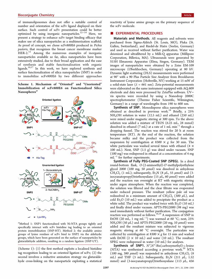
(Scheme 1): (1) the first method exploits a localized histidinetag recognition leading to an oriented ligation of scFv; (2) thesecond involves a reductive amination strategy via glutaralde-hyde cross-linking on the nanoparticle exploiting a statistical
reactivity of lysine amine groups on the primary sequence ofthe scFv molecule.
■ EXPERIMENTAL PROCEDURESMaterials and Methods. All reagents and solvents were
purchased from Sigma-Aldrich (St. Louis, MO), Fluka (St.Gallen, Switzerland), and Riedel-de Hae ̈n (Seelze, Germany)and used as received without further purification. Water wasdeionized and ultrafiltered by a Milli-Q apparatus (MilliporeCorporation, Billerica, MA). Ultrasounds were generated byS15H Elmasonic Apparatus (Elma, Singen, Germany). TEMimages of nanoparticles were obtained by a Zeiss EM-109microscope (Oberkhochen, Germany) operating at 80 kV.Dynamic light scattering (DLS) measurements were performedat 90° with a 90 Plus Particle Size Analyzer from BrookhavenInstrument Corporation (Holtsville, NY) working at 15 mW ofa solid-state laser (λ = 661 nm). Zeta-potential measurementswere elaborated on the same instrument equipped with AQ-809electrode and data were processed by ZetaPlus software. UV−vis spectra were recorded by using a Nanodrop 2000Cspectrophotometer (Thermo Fisher Scientific, Wilmington,Germany) in a range of wavelengths from 190 to 600 nm.Synthesis of SNP. Monodisperse silica nanospheres were
obtained as described in previous work.18 Briefly, a 25%NH4OH solution in water (12.5 mL) and ethanol (250 mL)were mixed under magnetic stirring at 300 rpm. To the abovesolution was added a mixture of TEOS (6.25 mL, 28 mmol)dissolved in ethanol (7 mL) at a rate of 1 mL min−1 through adropping funnel. The reaction was stirred for 20 h at roomtemperature (RT). At the end of the reaction, the solutionbecame milky and the product was collected from thesuspension by centrifugation at 8300 × g for 30 min. Thewhite particulate was washed several times with ethanol (4 ×100 mL). Next, SNP (1.5 g) was dried under vacuum. SNP(100 mg) was redispersed in ethanol at a concentration of 4 mgmL−1 for further experiments.Synthesis of Fully PEG-Coated SNP (SPEG). In a dried
round-bottom flask, O-(2-aminoethyl)-O′-methylpolyethyleneglycol 2000 (100 mg, 50 μmol) was dissolved in anhydrousCH2Cl2 (1 mL); subsequently, Et3N (8 μL, 55 μmol) and (3-isocyanatopropyl)triethoxysilane (15 μL, 60 μmol) were addedand the reaction run overnight at RT with magnetic stirringunder argon atmosphere. When the reaction was completed,the solution was filtered and the clear filtrate was evaporatedunder reduced pressure. The resultant yellow pale oil wasredissolved in a minimum amount of CH2Cl2 (300 μL), andcold Et2O (10 mL) was added to precipitate the product as awhite solid. The product was washed twice with Et2O (10 mL)and finally dried under vacuum. APTS-PEG2000 (98 mg) wasused immediately without further purification. The conjugationreaction was performed as follows.18,19 A suspension of SNP inEtOH (20 mL, 1 mg mL−1) was warmed at 60 °C; next, 25%NH4OH (50 μL) and APTS-PEG2000 (20 mg, 10 μmol) wereadded and the resultant mixture was subjected to vigorousmagnetic stirring at 60 °C overnight. The particulate wascollected by centrifugation at 8300 × g for 15 min and washedwith EtOH (2 × 10 mL) and water (10 mL). The resultantSPEG were redispersed in water (10 mL) for analyses.Synthesis of SNP1. Nα,N α-Bis(carboxymethyl)-L-lysine
(NTA) was synthesized according a previous work.20 NTA(100 mg, 380 μmol) was suspended in anhydrous CH2Cl2 (8mL) and THF (3 mL). Subsequently, Et3N (211 μL, 1.52mmol) and (3-isocyanatopropyl)triethoxysilane (113 μL, 456
Scheme 1. Mechanism of “Oriented” and “Unoriented”Immobilization of scFv800E6 on Functionalized SilicaNanospheresa
aMethod 1: SNP1 functionalized with Ni-NTA groups tightly andspecifically interact with scFv histidine tag leading to an orientedprotein immobilization (SNP-HT). Method 2: the available aminogroups of lysine residues of scFv bind to SNP2 via the aldehydicgroups, which have been generated on the surface of nanoparticles byglutaraldehyde addition, resulting in a random ligation (SNP-UT).
Bioconjugate Chemistry Article
dx.doi.org/10.1021/bc200352x |Bioconjugate Chem. 2011, 22, 2296−23032297
μmol) were added and the reaction run overnight at RT withmagnetic stirring under argon atmosphere. The resultingsuspension was filtered, and the clear filtrate was evaporatedunder reduced pressure. The pale yellow oil was redissolved ina minimum amount of MeOH (500 mL) and cold Et2O (10mL) was added to precipitate the product as a white solid. Theproduct was washed twice with Et2O (10 mL) and finally driedunder vacuum. APTS-NTA (132 mg) was used immediatelywithout further purification.For the conjugation reaction,18,19 a suspension of SNP in
EtOH/H2O 1:1 (20 mL, 1 mg mL−1) was warmed at 60 °C;25% NH4OH (50 μL), APTS-NTA (10 mg, 20 μmol), andAPTS-PEG2000 (10 mg, 5 μmol) were added, and the mixturewas left under vigorous stirring at 60 °C overnight. Next, theparticulate was collected by centrifugation at 8300 × g for 15min and washed with EtOH (2 × 10 mL) and water (10 mL).Nanoparticles were resuspended in water (20 mL) for furtherchelation with nickel ions. A green solution of 0.1 MNiCl2·6H2O in deionized water (2 mL) was added to theabove suspension of nanoparticles; the pH was adjusted to pH8.0 with 0.1 N NaOH, and the resultant mixture were keptunder magnetic stirring for 30 min at RT. Then, particles werecollected by centrifugation and the pale blue supernatant wasdiscarded. The pale green particulate was washed thrice withwater (10 mL) and, finally, SNP1 were resuspended in water (5mL) and stored for further conjugation with His-taggedscFv800E6.Synthesis of SNP2. The conjugation reaction18,19 was
performed as follows. A suspension of SNP in EtOH (20 mL, 1mg mL−1) was warmed at 60 °C; then, 25% NH4OH (50 μL),APTS (5 μL, 20 μmol), and APTS-PEG2000 (10 mg, 5 μmol)were added and the resultant mixture was kept under vigorousmagnetic stirring at 60 °C overnight. The particulate wascollected by centrifugation at 8300 × g for 15 min and washedwith EtOH (2 × 10 mL) and water (10 mL). The resultantSNP2 were stored in water (10 mL) for further conjugationwith scFv.Determination of Amine Groups on SNP2. Following a
method described in the literature,21 an aqueous mixturecontaining 1 M picrylsulfonic acid TNBS (10 μL) and 0.05 MNa2B4O7 (1.5 mL) was added to a sample of aminated SNP2 (6mg). The suspension was sonicated for 1 min and then heatedat 70 °C for 10 min. At the end of reaction, the mixture wasallowed to cool to RT; SNP2 was then separated from thesupernatant by centrifugation at 11 200 × g and washed withwater (1 mL), 50% acetone in water (1 mL), 100% acetone (1mL), and water (2 × 1 mL). SNP2 was then suspended inNaOH 1 M (5 mL) and heated to 70 °C under vigorousstirring for 10 min. The suspension was cooled to RT andSNP2 were separated by centrifugation. An aliquot of theparticle-free supernatant (1 mL) was then withdrawn and itsabsorbance read at 410 nm. Each particle contained silica (d =2.2 × 10⁶ g m−3) with an average radius of 40 nm = 4.0 × 10⁻⁸
m. The average volume and mass of SiO2 nanoparticles were2.68 × 10−22 m3 and 5.90 × 10⁻¹⁶ g, respectively. Hence, 1 mgof SiO2 contained 1.02 × 10¹⁴ particles. By determination ofresidual absorbance due to picric acid released from reaction(Supporting Information Scheme S4), we established that 0.012μmol of ligand APTS were immobilized on the particle surfacecorresponding to about 708 NH2 groups/particle.Dynamic Light Scattering and ζ-Potential Measure-
ments. Viscosity and refractive index of deionized water wereused to characterize the solvent. Nanoparticles were dispersed
in water under sonication for several minutes before analyses;sporadically, to avoid the formation of large aggregates, thesuspension was filtered through a 0.45 μm cellulose acetatefilter. The final sample concentration used for measurementswas typically 0.025 mg mL−1 (Figure S1). The same procedurewas followed for hydrodynamic size distribution behavior ofnanoparticles in deionized water by different Na+ concen-trations (1 mM to 20 mM) (Figure S2).Strains and Plasmids. P. pastoris KM71H (arg4;
aox1::ARG4) (Invitrogen) was used as host for expressingscFv800E6 gene. Plasmid pPICZαA (Invitrogen) was used forconstructing plasmid vector, as previously described.15
Purification of scFv800E6. The clone KM71H-pPIC-ZαA-scFv800E6-4 was grown in 10 mL YPD medium (1%yeast extract, 2% peptone, 2% dextrose) at 30 °C overnightwith shaking at 250 rpm. The cultures were centrifuged at 1500× g for 4 min, and then, the pellets were resuspended in 200mL of BMMY (1% yeast extract, 2% peptone, 100 mMpotassium phosphate pH 6.0, 1.34% YNB, 0.00004% biotin,0.5% methanol) with 0.8% glycerol yielding an initial OD 600value of 10. The culture was induced by daily addition ofmethanol to a final concentration of 0.5%. After 48 h ofmethanol treatment, the culture supernatant was filteredthrough 0.22 μm filters and dialyzed overnight in 50 mMsodium phosphate pH 8.0, 300 mM NaCl. The dialyzedmedium was loaded at a flow rate of 0.5 mL/min onto a Ni-NTA Agarose (Qiagen) column (bed volume 0.5 mL) pre-equilibrated with 50 mM sodium phosphate pH 8.0, 300 mMNaCl, and 10 mM imidazole. The column was washed with 50mM sodium phosphate pH 8.0, 300 mM NaCl, 20 mMimidazole, and the protein eluted with a stepwise imidazolegradient, 100 mM to 200 mM, in the same buffer. Fractionswere collected and analyzed by SDS-PAGE. SDS-PAGE wasperformed according to Laemmli,22 using 12% (v/v)polyacrylamide gels. The proteins were detected by CoomassieBrilliant Blue R-250 staining. Protein content was determinedboth by measuring absorbance at 280 nm and by using theCoomassie Plus Protein Assay Reagent (Termo FisherScientific) and bovine plasma immunoglobulin G as thestandard protein.Conjugation of His-tag scFv800E6 (SNP-HT). In a
plastic tube, SNP1 (1 mg) were incubated with purifiedscFv800E6 (50 μg) in phosphate-buffered saline (PBS;EuroClone) in a final volume of 1000 μL and the mixturewas stirred on an orbital shaker for 10 min at RT. SNP-HTwere isolated from unreacted scFv800E6 by centrifugation at11 200 × g for 5 min and the supernatant was discarded.Nanoparticles were washed three times with PBS (500 μL) andstored in PBS at 4 °C for further experiments. By measuring theabsorbance at 280 nm, we determined an amount of scFv800E6immobilized on nanoparticles of 26 μg per mg of SNP-HT.Unoriented Conjugation of scFv800E6 (SNP-UT).
Particle-glutaraldehyde cross-linking,23 followed by iminereduction24 with NaCNBH3 were performed as follows. In aplastic tube, amino SNP2 (1 mg) was dispersed in borate bufferpH 7.6 (600 μL). A 5 mM glutaraldehyde solution in the samebuffer (400 μL, 20 μmol) was added under stirring at RT. After2 h, particles were isolated by centrifugation and washed oncewith borate buffer pH 7.6 (1 mL) and twice with PBS pH 7.4(1 mL). At the end of the washing, nanoparticles wereresuspended at a concentration of 1 mg mL−1 in the samebuffer (1 mL) and the suspension cooled at 4 °C with an icebath. scFv800E6 (50 μg in PBS) was added to the suspension
Bioconjugate Chemistry Article
dx.doi.org/10.1021/bc200352x |Bioconjugate Chem. 2011, 22, 2296−23032298
and the resultant mixture was stirred on the orbital shaker for 3h at 4 °C. ScFv-functionalized nanoparticles were recoveredafter centrifugation and washed twice with PBS (1 mL).Subsequently, a NaBH3CN solution (10 μL, 1 mg mL−1) inPBS buffer pH 7.4 was added to the nanoparticle suspension inthe same medium (1 mL) and incubated for 3 h at 4 °C. Aftercentrifuging, SNP-UT were washed several times with PBS (3× 1 mL) and finally were stored in the same buffer at 4 °C forfurther experiments. By measuring the absorbance at 280 nm,we determined an amount of scFv800E6 of 43 μg per mg ofSNP-UT.Dot Blot Assay. Dot blot was performed by filtering
proteins and/or nanoparticles onto PVDF membranes, utilizinga Manifold I dot blot apparatus (GE Healthcare), andincubating in blocking solution (5% skim milk in PBS,Tween 0.05%) for 1 h at RT. The membrane was then probedfor 1 h at RT in blocking solution using rabbit anti-Myc-HRPantibody (Invitrogen) at a 1:5000 dilution. Membranes wererinsed thrice in 0.05% Tween in PBS for 10 min.Immunoreactive spots were revealed using ECL Westernblotting reagent (GE Healthcare).Cell Cultures. MCF-7 and MDA cell lines were used as
HER-2 positive and HER-2 negative targets, respectively. Cellswere cultured in 50% Dulbecco’s Modified Eagle’s Medium(DMEM) and 50% F12, supplemented with 10% fetal bovineserum, L-glutamine (2 mM), penicillin (50 UI mL−1), andstreptomycin (50 mg mL−1) at 37 °C and 5% CO2 in ahumidified atmosphere and subcultured prior to confluenceusing trypsin/EDTA. Cells culture medium and chemicals werepurchased from EuroClone.Flow Cytometry. Cells were cultured on a multiwell dish
until subconfluence. Then, equal aliquots were incubated 15min at 37 °C in the presence of one of the following: (i) 5 μgmL−1 scFv, (ii) 25.6 μg mL−1 TZ, (iii) SNP-HT, and (iv) SNP-
UT. For each sample, 0.171 nmole of antibody, either free orimmobilized on nanoparticles, was used. After incubation time,cells were washed twice with PBS and treated with typsin for 3min. Digestion with trypsin was stopped with culture medium,cells were transferred in FACS tubes and washed twice withPBS. Then, cells were incubated for 30 min at 4 °C in blockingsolution (PBS, 2% Bovine Serum Albumin) and immunodeco-rated with 1 μL of FITC-conjugated antibody to whole murineIgG (MP Biomedicals) for 30 min at 4 °C. The excesssecondary antibody was removed by washing cells six timeswith PBS. Labeled cells were resuspended with 0.5 mL of PBSand analyzed on a FACS Calibur flow cytometer (BectonDickinson). Ten thousand events were acquired for eachanalysis, after gating on viable cells, and isotype-controlantibodies were used to set the appropriate gates.Stability Assay of scFv Conjugation on SNP-HT. Five
micrograms of scFv immobilized on SNP-HT was incubated at37 °C with 0.1 mL of culture medium. After incubation time,SNP-HT was centrifuged 15 min at 15 000 × g at 4 °C, andsupernatants (S1, S4, S24, and S48) were filtered onto a PVDFmembrane utilizing a Manifold I dot blot apparatus (GEHealthcare). The membrane was incubated in blocking solution(5% skim milk in PBS, Tween 0.05%) for 1 h at RT, thenprobed 1 h at RT in blocking solution using rabbit anti-Myc-HRP antibody (Invitrogen) at a 1:5000 dilution. Membraneswere rinsed thrice in 0.05% Tween in PBS for 10 min.Immunoreactive spots were revealed using ECL Westernblotting reagent (GE Healthcare). Five micrograms of scFvimmobilized on SNP-HT and 0.1 mL of culture medium wereused, respectively, as positive and negative control.Confocal Laser Scanning Microscopy. Cells were
cultured on collagen (Sigma) precoated coverglass slides untilsubconfluence. Cells were incubated 1 h at 37 °C with 20 μg/mL of free-scFv800E6, and with equal amounts of scFv800E6
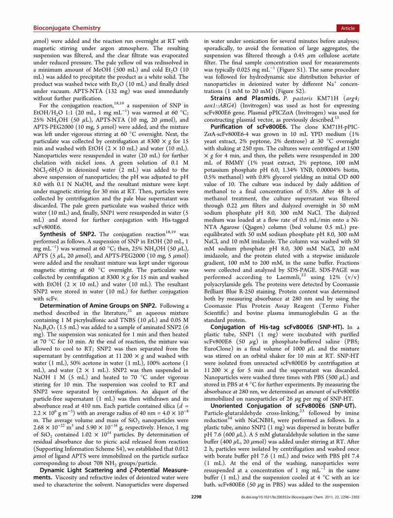
Figure 1. Characterization of modified silica nanoparticles: (A) TEM (Inset: magnification) and (B) SEM images of as-synthesized SNP. (C) DLSand (D) ζ-potential measurements of SNP, SPEG, SNP1, and SNP2 as a function of pH in aqueous solution.
Bioconjugate Chemistry Article
dx.doi.org/10.1021/bc200352x |Bioconjugate Chem. 2011, 22, 2296−23032299
immobilized on SNP-HT and SNP-UT. Then, cells werewashed with PBS, fixed for 10 min with 4% paraformaldehyde(Sigma), and treated for 10 min with 0.1 M glycine (Sigma) inPBS. A blocking step was performed for 1 h at RT with asolution containing 2% bovine serum albumin (Sigma) and 2%goat serum in PBS. ScFv was revealed by a FITC-conjugatedantibody to whole murine IgG (MP Biomedicals) at a 1:300dilution by incubating for 2 h at RT. Nuclei were stained withDAPI (4′,6-diamidino-2-phenylindole, Invitrogen) at 0.2 μgmL−1 in PBS with 0.1% Saponin (Sigma) for 20 min at RT.Membranes were stained with DiD oil (Invitrogen) at a 1:300dilution by incubating 30 min at 37 °C. Microscopy analysiswas performed with a Leica SP2 AOBs microscope confocalsystem. Images were acquired with 63× magnification oilimmersion lenses at 1024 × 1024 pixel resolution.
■ RESULTS AND DISCUSSION
Uniform 60 nm spherical SNPs were synthesized by hydrolysisand condensation of tetraethyl orthosilicate (TEOS) in a 25%NH4OH ethanolic solution.18 In method 1, previously preparedAPTS-PEG2000-OMe25 and APTS-NTA in a 1:1 molar ratiowere co-condensed on SNP in alkaline ethanol resulting inNTA-functionalized SNP1 (Schemes S1,2 in SI).19 Accordingto previous reports,26 PEG chains enhanced the nanoconjugatesolubility in buffered media and prevented nonspecificadsorption of proteins. ScFv800E6 protein containing a 6 ×His-affinity tag was produced in P. pastoris, secreted in culturemedium, and purified through affinity chromatography, aspreviously described.15 Subsequent Ni2+ chelation by nitrilo-acetic acid groups of SNP1 promoted the active Ni2+-NTAaffinity-oriented immobilization of His-tagged scFv (SNP-HT)by incubation at room temperature. In method 2, APTS-PEG2000-OMe and APTS in a 1:1 molar ratio were co-condensed on SNP (Scheme S3 in SI).23 Next, the covalentattachment of scFv was performed via glutaraldehyde cross-linking resulting in functional SNP2, stabilized by reduction ofthe diimine intermediate with NaCNBH3,
27 to give SNP-UT.In Figure 1, TEM (A) and SEM (B) images show that the coresize of SNP was 60 ± 5 nm indicating a homogeneousformulation of silica nanospheres. The hydrodynamic diameterof SNP in ethanol was 78.3 ± 2.2 nm, as determined bydynamic light scattering (DLS, Figure S1 in SI). The pH-dependent behavior of SNP, SNP1, SNP2, and fully PEG-coated nanosilica (SPEG) was investigated by DLS in the 2−9pH range (Figure 1C). All the nanosilica tested did not exhibitthe formation of critical aggregates between pH 4 and 9. Zetapotential (ζ) of SNP, SPEG, SNP1, and SNP2 in water in the2−9 pH range was also investigated (Figure 1D). From pH 5 to9, SNP were strongly negatively charged (−35.85 ± 0.64 mV),whereas at pH 4 and lower, the charge approached neutrality(−6.40 ± 1.77 mV). Similar behavior was also observed forSPEG, although they were only −25.01 ± 0.38 mV at pH 7because of the charge-shielding effect of the PEG layer on thenanoparticle surface. In contrast, SNP1 exhibited a remarkablylow surface charge at pH 7 (−13.64 ± 0.74 mV), in accordancewith the presence of the Ni2+ chelates on the external carboxylicgroups of NTA. This charge did not appreciably change in thepH range tested. SNP2 were negatively charged (−25.23 ± 1.03mV) at pH 7−9. However, below pH 5, a gradual shift of thecharge to positive values (+16.39 ± 1.35 mV at pH 3) wasobserved, in line with the presence of protonated amino groupsof APTS in acidic conditions.
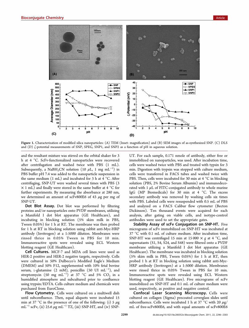
After conjugation, the amount of scFv800E6 on SNP-HTand SNP-UT was quantified by protein assay of supernatants at280 nm using a calculated ε 280 nm of 55 600 M−1. Theimmobilization of scFv on SNP-HT was also confirmed by dot-blot assay (Figure 2). SNP-HT (5 μg) and free scFv (0.5, 0.2,
0.1, and 0.05 μg) were filtered onto a polyvinylidene fluoride(PVDF) membrane and then probed with an anti-Myc-HRPantibody. Intensities of immunoreactive spots of SNP-HT and0.1 μg of free scFv were comparable. This reasonably fits withthe amount of bound scFv that we inferred from thedetermination of unbound protein, i.e., 0.125 μg.To assess the effect of multivalent presentation of scFv on
silica nanoparticles, SNP-HT and SNP-UT binding towardHER-2 receptor in breast cancer cells was evaluated by flowcytometry. Free scFv, the commercial anti-HER-2 mAb(trastuzumab, TZ), SNP-HT, and SNP-UT, containing equalamounts of conjugated scFv, were incubated 15 min with HER-2-positive MCF-7 cells.28 Flow cytometry evidenced a right-shift of fluorescence signal accounting for an increase in scFvbinding efficacy upon multimerization due to SNP conjugation(Figure 3). Values reported in Figure 3B show a 3-fold increasein mean fluorescence intensity of SNP-UT sample incomparison with free scFv at the same scFv concentration.Moreover, scFv multimerized on SNP via glutaraldehyde cross-linking (SNP-UT) exhibited a mean fluorescence intensityvalue very close to that of intact TZ, indicating that there was asignificant improvement in receptor binding capability, whichcan be attributed to avidity effect. However, the conjugationstrategy exploited for scFv multimerization on SNP surfaces(method 1 vs method 2) did not prove to be crucial inenhancing scFv binding efficiency. Indeed, SNP-HT and SNP-UT exhibited similar fluorescence intensities when assessed byflow cytometry. To account for this unexpected result, weinspected the localization of lysines in scFv sequence. Multiplesequence alignment of scFv800E6 with other scFv sequencesavailable in the NCBI database revealed that all lysine residuesare highly conserved. This clearly indicates that these residuesare not directly involved in HER-2 binding. Instead, quite likelythey only play a structural role. Therefore, an involvement ofthis residue in glutaraldehyde cross-linking is not expected tointerfere significantly with HER-2 binding.Moreover, to exclude the release of scFv from SNP-HT and
assess the stability of this conjugation method, we haveincubated SNP-HT for 1, 4, 24, and 48 h at 37 °C and 5% CO2atmosphere. Supernatants of incubation were then filtered onPVDF membrane and probed with an anti-Myc-HRP antibody.Release of scFv800E6 in cell culture medium was not observedunder the conditions tested, confirming that scFv conjugationon SNP-HT was stable within 48 h of incubation at 37 °C(Figure 4).To validate flow cytometry data, the specificity of binding
between SNP-HT or SNP-UT and HER-2 was assessed by
Figure 2. Dot-blot assay of SNP-HT conjugated with scFv. Differentamounts of scFv800E6 (0.5, 0.2, 0.1, and 0.05 μg) and an aliquot ofSNP-HT were filtered on a PVDF membrane, then probed with anti-Myc-HRP antibody and revealed with an ECL substrate.
Bioconjugate Chemistry Article
dx.doi.org/10.1021/bc200352x |Bioconjugate Chem. 2011, 22, 2296−23032300
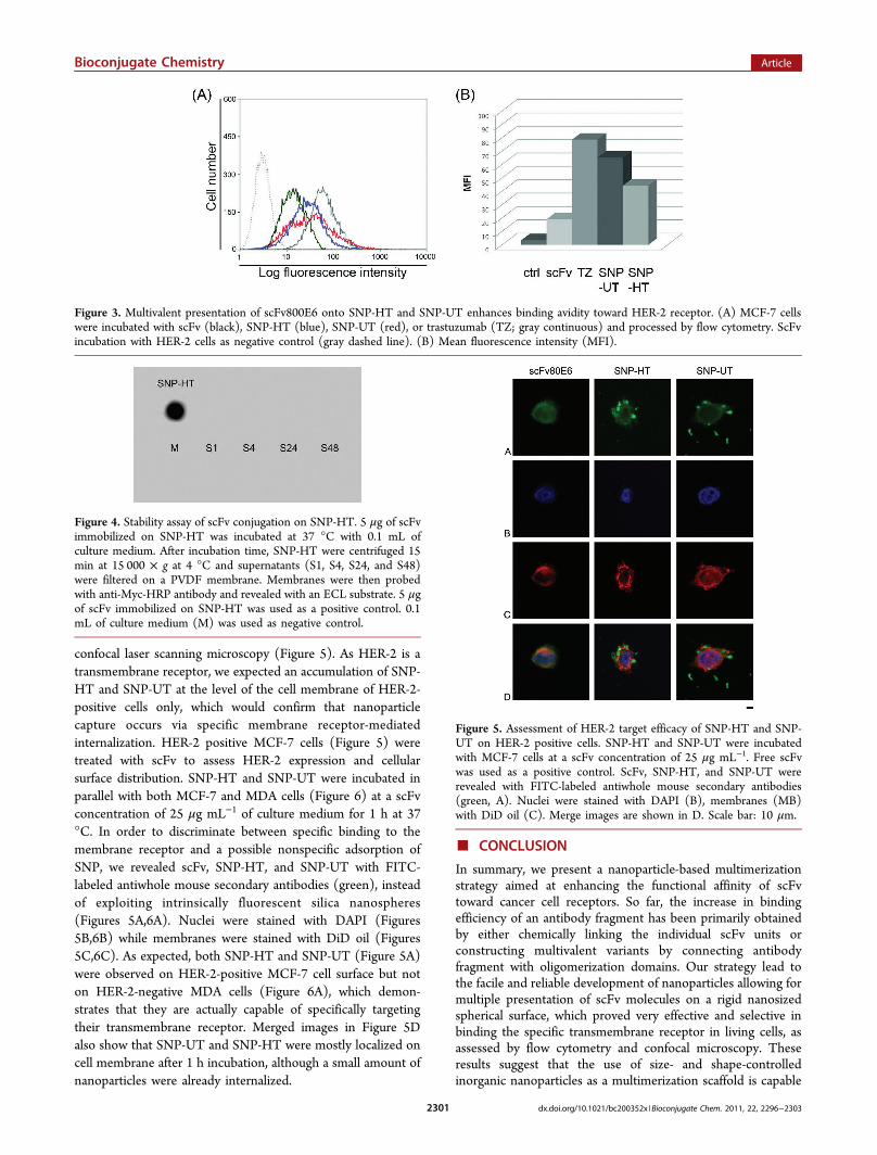
confocal laser scanning microscopy (Figure 5). As HER-2 is atransmembrane receptor, we expected an accumulation of SNP-HT and SNP-UT at the level of the cell membrane of HER-2-positive cells only, which would confirm that nanoparticlecapture occurs via specific membrane receptor-mediatedinternalization. HER-2 positive MCF-7 cells (Figure 5) weretreated with scFv to assess HER-2 expression and cellularsurface distribution. SNP-HT and SNP-UT were incubated inparallel with both MCF-7 and MDA cells (Figure 6) at a scFvconcentration of 25 μg mL−1 of culture medium for 1 h at 37°C. In order to discriminate between specific binding to themembrane receptor and a possible nonspecific adsorption ofSNP, we revealed scFv, SNP-HT, and SNP-UT with FITC-labeled antiwhole mouse secondary antibodies (green), insteadof exploiting intrinsically fluorescent silica nanospheres(Figures 5A,6A). Nuclei were stained with DAPI (Figures5B,6B) while membranes were stained with DiD oil (Figures5C,6C). As expected, both SNP-HT and SNP-UT (Figure 5A)were observed on HER-2-positive MCF-7 cell surface but noton HER-2-negative MDA cells (Figure 6A), which demon-strates that they are actually capable of specifically targetingtheir transmembrane receptor. Merged images in Figure 5Dalso show that SNP-UT and SNP-HT were mostly localized oncell membrane after 1 h incubation, although a small amount ofnanoparticles were already internalized.
■ CONCLUSION
In summary, we present a nanoparticle-based multimerizationstrategy aimed at enhancing the functional affinity of scFvtoward cancer cell receptors. So far, the increase in bindingefficiency of an antibody fragment has been primarily obtainedby either chemically linking the individual scFv units orconstructing multivalent variants by connecting antibodyfragment with oligomerization domains. Our strategy lead tothe facile and reliable development of nanoparticles allowing formultiple presentation of scFv molecules on a rigid nanosizedspherical surface, which proved very effective and selective inbinding the specific transmembrane receptor in living cells, asassessed by flow cytometry and confocal microscopy. Theseresults suggest that the use of size- and shape-controlledinorganic nanoparticles as a multimerization scaffold is capable
Figure 3. Multivalent presentation of scFv800E6 onto SNP-HT and SNP-UT enhances binding avidity toward HER-2 receptor. (A) MCF-7 cellswere incubated with scFv (black), SNP-HT (blue), SNP-UT (red), or trastuzumab (TZ; gray continuous) and processed by flow cytometry. ScFvincubation with HER-2 cells as negative control (gray dashed line). (B) Mean fluorescence intensity (MFI).
Figure 4. Stability assay of scFv conjugation on SNP-HT. 5 μg of scFvimmobilized on SNP-HT was incubated at 37 °C with 0.1 mL ofculture medium. After incubation time, SNP-HT were centrifuged 15min at 15 000 × g at 4 °C and supernatants (S1, S4, S24, and S48)were filtered on a PVDF membrane. Membranes were then probedwith anti-Myc-HRP antibody and revealed with an ECL substrate. 5 μgof scFv immobilized on SNP-HT was used as a positive control. 0.1mL of culture medium (M) was used as negative control.
Figure 5. Assessment of HER-2 target efficacy of SNP-HT and SNP-UT on HER-2 positive cells. SNP-HT and SNP-UT were incubatedwith MCF-7 cells at a scFv concentration of 25 μg mL−1. Free scFvwas used as a positive control. ScFv, SNP-HT, and SNP-UT wererevealed with FITC-labeled antiwhole mouse secondary antibodies(green, A). Nuclei were stained with DAPI (B), membranes (MB)with DiD oil (C). Merge images are shown in D. Scale bar: 10 μm.
Bioconjugate Chemistry Article
dx.doi.org/10.1021/bc200352x |Bioconjugate Chem. 2011, 22, 2296−23032301

to improve scFv target binding efficacy, reaching an affinityvalue very close to that of native TZ, the mAb currently utilizedin clinical practice.
■ ASSOCIATED CONTENT
*S Supporting InformationExperimental details for fully PEG-coated SNP (SPEG), SNP1and SNP2, scheme of conjugation of SNP-HT and SNP-UT,determination of amine groups on SNP2, Dynamic LightScattering and ζ-potential measurements. This material isavailable free of charge via the Internet at http://pubs.acs.org.
■ AUTHOR INFORMATION
Corresponding Author*E-mail: [email protected]; Phone: (+39)0264483302; Fax: (+39)0264483565.
Author Contributions†These authors contributed equally to the research.
■ ACKNOWLEDGMENTSWe thank R. Allevi for TEM images, S. Citterio for helpfuldiscussion of cell cytometry result. This work was partlysupported by “Assessorato alla Sanita”̀, Regione Lombardia, andSacco Hospital (NanoMeDia Project), “Fondazione Romeo edEnrica Invernizzi” and “Centro di Microscopia Elettronica perlo sviluppo delle Nanotecnologie applicate alla medicina”(CMENA, Univ. of Milan). M.C. and S.M. acknowledge“Centro di Microscopia Elettronica per lo sviluppo delleNanotecnologie applicate alla medicina” (CMENA, University
of Milan) for doctoral and postdoctoral fellowships, respec-tively.
■ REFERENCES(1) Sanz, L., Cuesta, A. M., Compte, M., and Alvarez-Vallina, L.
(2005) Antibody engineering: facing new challenges in cancer therapy.Acta Pharmacol. Sin. 26, 641−648.(2) Holliger, P., and Hudson, P. J. (2005) Engineered antibody
fragments and the rise of single domains. Nat. Biotechnol. 9, 1126−1136.(3) Goel, A., Augustine, S., Baranowska-Kortylewicz, J., Colcher, D.,
Booth, B. J. M., Pavlinkova, G., Tempero, M., and Batra, S. K. (2001)Single-dose versus fractionated radioimmunotherapy of human coloncarcinoma xenografts using 131I-labelled multivalent CC49 singlechain fvs. Clin. Cancer Res. 7, 175−184.(4) Sanz, L., Blanco, B., and Alvarez-Vallina, L. (2004) Antibodies
and gene therapy: teaching old “magic bullets” new tricks. TrendsImmunol. 25, 85−91.(5) Bayly, A. M., Kortt, A. A., Hudson, P. J., and Power, B. E. (2002)
Large-scale bacterial fermentation and isolation of scFv multimersusing a heat-inducible bacterial expression vector. J. Immunol. Methods262, 217−227.(6) Wittel, U. A., Jain, M., Goel, A., Chauhan, S. C., Colcherand, D.,
and Batra, S. K. (2005) The in vivo characteristics of geneticallyengineered divalent and tetravalent single-chain antibody constructs.Nucl. Med. Biol. 32, 157−164.(7) Gall, L. F., Kipriyanov, S. M., Moldenhauer, G., and Little, M.
(1999) Di-, tri- and tetrameric single chain Fv antibody fragmentsagainst human CD19: effect of valency on cell binding. FEBS Lett. 453,164−168.(8) Ravn, P., Danielczyk, A., Jensen, K. B., Kristensen, P.,
Christensen, P. A., Larsen, M., Karsten, U., and Goletz, S. (2004)Multivalent scFv display of phagemid repertoires for the selection ofcarbohydrate-specific antibodies and its application to the Thomsen−Friedenreich antigen. J. Mol. Biol. 343, 985−996.(9) Natarajan, A., Du, W., Xiong, C-Y, DeNardo, G. L., DeNardo, S.
J., and Gervay-Hague, J. (2007) Construction of di-scFv through atrivalent alkyne−azide 1,3-dipolar Cycloaddition. Chem. Commun. 695,685−697.(10) Devey, S. M., and Lebedenko, E. N. (2008) Multivalency: the
hallmark of antibodies used for optimization of tumor targeting bydesign. BioEssays 30, 904−918.(11) Zhou, Y., Drummond, D. C., Zou, H., Hayes, M. E., Adams, G.
P., Kirpotin, D. B., and Marks, J. D. (2007) Impact of single-chain Fvantibody fragment affinity on nanoparticle targeting of epidermalgrowth factor receptor-expressing tumor cells. J. Mol. Biol. 371, 934−947.(12) Ackerson, C. J., Jadzinsky, P. D., Jensen, G. J., and Kornberg, R.
D. (2006) Rigid, specific, and discrete gold nanoparticle/antibodyconjugates. J. Am. Chem. Soc. 128, 2635−2640.(13) Yang, L., Mao, H., Wang, A., Cao, Z., Peng, X., Wang, X., Duan,
H., Ni, C., Yuan, Q., Adams, G., Smith, M. Q., Wood, W. C., Gao, X.,and Nie, S. (2009) Single chain epidermal growth factor receptorantibody conjugated nanoparticles for in vivo tumor targeting andimaging. Small 5, 235−243.(14) Huang, X., Peng, X., Wang, Y., Wang, Y., Shin, D. M., El-Sayed,
M. A., and Nie, S. (2010) A reexamination of active and passive tumortargeting by using rod-shaped gold nanocrystal and covalentlyconjugated peptide ligands. ACSNano 4, 5887−5896.(15) Sommaruga, S., Lombardi, A., Salvade,̀ A., Mazzucchelli, S.,
Corsi, F., Galeffi, P., Tortora, P., and Prosperi, D. (2011) Highlyefficient production of anti-HER2 scFv antibody targeting for tumortargeting breast cancer cells. Appl. Microbiol. Biotechnol. 91, 613−621.(16) Burns, A., Ow, H., and Weisner, U. (2006) Fluorescent core-
shell silica nanoparticles: towards “lab on a particle” architectures fornanobiotechnology. Chem. Soc. Rev. 35, 1028−1042.(17) Rossi, M. L., Shi, L., Rosenzweig, N., and Rosenzweig, Z. (2006)
Fluorescent silica nanosphere for digital counting assay of the breastcancer marker HER2/neu. Biosens. Bioelectron. 21, 1900−1906.
Figure 6. Assessment of HER-2 target efficacy of SNP-HT and SNP-UT on HER-2 negative cells. SNP-HT and SNP-UT were incubatedwith MDA cells at a scFv concentration of 25 μg mL−1. ScFv on SNP-HT and SNP-UT was revealed with FITC-labeled antiwhole mousesecondary antibodies (green, A). Nuclei were stained with DAPI (B),membranes (MB) with DiD oil (C). Merge images are shown in D.Scale bar: 10 μm.
Bioconjugate Chemistry Article
dx.doi.org/10.1021/bc200352x |Bioconjugate Chem. 2011, 22, 2296−23032302
(18) Wu, T., Zhang, Y., Wang, X., and Liu, S. (2008) Fabrication ofhybrid silica nanoparticles densely grafted with thermoresponsivepoly(N-isopropylacrylamide) brushes of controlled thickness viasurface-initiated atom transfer radical polymerization. Chem. Mater.20, 101−109.(19) Kim, S. H., Jeyakumar, M., and Katznellenbogen, J. A. (2007)
Dual-mode fluorophore-doped nickel nitriloacetic acid-modified silicananoparticles combine histidine-tagged protein purification with site-specific fluorophore labeling. J. Am. Chem. Soc. 129, 13254−13264.(20) Scmitt, L., Dietrich, C., and Tampe,̀ R. (1994) Synthesis and
characterization of chelator-lipids for reversible immobilization ofengineered proteins at self-assembled lipid interfaces. J. Am. Chem. Soc.116, 8485−8491.(21) Halling, P. J., and Dunnill, P. (1979) Improved nonporous
magnetic supports for immobilized enzymes. Biotechnol. Bioeng. 21,393−416.(22) Laemmli, U. K. (1970) Cleavage of structural proteins during
the assembly of the head of bacteriophage T4. Nature 227, 680−85.(23) Corsi, F., De Palma, C., Colombo, M., Allevi, R., Nebuloni, M.,
Ronchi, S., Rizzi, G., Tosoni, A., Trabucchi, E., Clementi, E., andProsperi, D. (2009) Towards ideal magnetofluorescent nanoparticlesfor bimodal detection of breast-cancer cells. Small 5, 2555−2564.(24) Han, H. J., Kannan, R. M.., Wang, S., Mao, G., Kusanovic, J. P.,
and Romero, R. (2010) Multifunctional dendrimer template antibodypresentation on biosensor surfaces for improved biomarker detection.Adv. Funct. Mater. 20, 409−421.(25) Kohler, N., Fryxell, G. E., and Zhang, M. (2004) A bifunctional
poly(ethylene glycol) silane immobilized on metallic oxide-basednanoparticles for conjugation with cell targeting agents. J. Am. Chem.Soc. 126, 7206−7211.(26) Lin, W., Garnett, M. C., Schacht, E., Davis, S. S., and Illum, L.
(1999) Preparation and in vitro characterization of HSA-mPEGnanoparticles. Int. J. Pharm. 189, 161−170.(27) Hu, F., Wei, L., Zhou, Z., Ran, Y., Li, Z., and Gao, M. (2006)
Preparation of biocompatibile magnetite nanocrystals for in vivomagnetic resonance detection of cancer. Adv. Mater. 18, 2553−2556.(28) Mazzucchelli, S., Colombo, M., De Palma, C., Salvade,̀ A.,
Verderio, P., Coghi, M. D., Clementi, E., Tortora, P., Corsi, F., andProsperi, D. (2010) Single-domain protein A-engineered magneticnanoparticles: a step towards a universal strategy to site-specificlabeling of antibodies for targeted detection of tumor cells. ACS Nano10, 5693−5702.
Bioconjugate Chemistry Article
dx.doi.org/10.1021/bc200352x |Bioconjugate Chem. 2011, 22, 2296−23032303
Copyright © 2022 FDOKUMEN