M.Sc. BOTANY LAB MANUAL - Midnapore city college
43
M.Sc. BOTANY LAB MANUAL MIDNAPORE CITY COLLEGE Prepared By Biological Science Dept. Botany 1st Semester
-
Upload
khangminh22 -
Category
Documents
-
view
0 -
download
0
Transcript of M.Sc. BOTANY LAB MANUAL - Midnapore city college

M.Sc. BOTANY LAB MANUAL
MIDNAPORE CITY COLLEGE
Prepared By
Biological Science Dept.Botany
1st Semester

Botany Lab Manual Dept. of Biological Science
MIDNAPORE CITY COLLEGE
PREFACE TO THE FIRST EDITION
This is the first edition of Lab Manual for PG Botany first Semester. Hope this edition will help
you during practical. This edition mainly tried to cover the whole syllabus. Some hard core
instrument based topic are not present here that will be guided by responsive teachers at the time
of practical.

Botany Lab Manual Dept. of Biological Sciences
MIDNAPORE CITY COLLEGE
ACKNOWLEDGEMENT
We are really thankful to our students, teachers , and non teaching staffs to make this effort little bit complete.
Mainly thanks to Director and Principal Sir to motivate for making this lab manual.

Laboratory Practice Safety Rules
1. Use safety glass when dealing with fire and chemical.
2. Should use front cover clothes during biochemistry practical.
3. Always use hand wash after dissection and any type of chemical use.
4. Carefully handle needles , forceps, microscope and any other dissecting instrument.
Botany Lab Manual Dept. of Biological Sciences
MIDNAPORE CITY COLLEGE

Botany Lab Manual Dept. of Biological Sciences
BOT-105, Microbiology Phycology and Bryology Microbiology Gram Staining
INDEX
Sl. No. CONTENT PAGE No.
Microbiology Phycology and Bryology Microbiology Gram Staining 011.
Negative Staning Technique 032.
Study of the curd organism 043.
Endospore Staining 064.
PhycologyDescription and workout of algal specimen: A 09
5.
Work Out of Specimen: B 106.
Work Out of Specimen: C
Work Out of Specimen: D 147.
BryologyWork Out of Specimen: A 15
8.
Work Out of Specimen: B 179.
Work Out of Specimen: C 2010.
Work Out of Specimen: A 2312.
Work Out of Specimen: C 2513.
Work Out of Specimen: B 2714.
Gymnosperms 3615.
126.
BOT-106, Mycology and Plant Pathology, Pteridophytes and Gymnosperms Mycology
Plant PathologyStudy of Diseased Plant Specimen: A 26
5.
Gymnosperms 3715.

MicrobiologyPhycology and Bryology
MicrobiologyGram Staining
Principle : Different staining requires the use of at least three chemical reagent that are applied sequencetilly to a hit fixed smear. The first reagent is called primary stain. Its function is to impart its colour to all cells. In order to establish acolour contrast, the second reagent use is the decolouring agent. Based on the chemical composition of celluler components, the decolouring agent may or may not remove the primary stain from the entire cell on olly from certain cell structure. The final reagent, the counter stain has a contrastin colour than that of the primary stain. The following decolourization, if the primary stain is not washed out the counter stain cannot be observe and the cells on their components will retain the colour of the primary stain. If the primary stain is removed, the colourized celluler components will accept and assume the contrasting colour of counter stain. In this may, cell types or their structure can be distinguished from each other on the basis of the stain that the cells retained.
Purpose : to became familiar with;
1. The chemical & theoretical bases for differential staining procedure.
2. The chemical basis of gram stain.
3. Performance of the procedure for differentiating between the two principle groups of bacteria:
-Gram +ve bacteria
-Gram –ve bacteria
Materials :
Cultures: 24 hours nutrient agar stain culture of sample 1.
Reagents:
Crystal violet (Primary stain)
Gram's iodine (mordant)
70% Ethyl alcohol (Decolourising agent)
Safranin (Counter stain)
Equipments:
Bunsen burner ,inoculating loop, staining tray, glass slide, bibulous, Paper, lens paper and microscope.
BOT – 105
Botany Lab Manual Dept. of Biological Sciences
MIDNAPORE CITY COLLEGE 1

Procedure:
1.obtain 4 clean glass slides.
2.using sterile technique prepared smears of each of the 2 sample organisms.the smear if prepared by placing a drop of water on the slide,then transferring each organism separately to the drop water with a sterile cooled inoculating loop.mix and spread he organism by the means of circular motion by inoculating loop.
3. Allow smears to air dry then heat fixed the smear by the Bunsen burner.
4.Gently flood the smear with crystal violet and let stand for 1 min.
5.Gently then wash it with tap water.
6.Gently flood smear with the gram's iodine mordant and let it stand for 1 min.
7. Gently wash it again with tap water.
8. decolourised it with 90% ethyl alcohol.
(caution: do not over decolourise,add reagent drop by drop until alcohol runs almost clear, showing only a blue colouration.)
9. Gently wash it with tap water is when done ,then the next step is to apply gently or counter stain gently with safranin for 45 sec.
10. Blot dry with bibulous paper and examine under oil immersion microscope.
Observation and result:
Draw a representative field.
Cell morphology;
1)Shape — Round shaped
2)Arrangement — single
3)Cell colour — crystal violet (purple)
4) Gram Reaction — gram positive
Comment:
According to the above result ,the sample contain gram positive stain ,because it retains the crystal violet stain colour, hence it is a gram positive bacteria.
Botany Lab Manual Dept. of Biological Sciences
MIDNAPORE CITY COLLEGE 2

NEGATIVE STANING TECHNIQUE
Botany Lab Manual Dept. of Biological Sciences
MIDNAPORE CITY COLLEGE 3
Principle: In negative staining ,the background but not the bacteria are stained by the use of an acidic stain which carries a negative charge on its surface and is used by the bacteria, that too carry a negative charge on their surface.In negative staining the bacterial growth is mixed with loopful of the stain on a clear slide and the cover laid with a coverslip. The capsule appears as a clear one between the cell wall and the background under high-dry and oil-immersion objective .
Requirements:
1) 36-48hours culture of Lactobacillus fermentum.
2) Indian ink.
3) Absorbent or blotting paper.
4) Clean glass slides.
5) Glass cover slip.
6) Inoculating loop.
7) Bunsen burner.
Procedure:
1. Take a clean glass slide.
2. Put a drop of Indian ink close to one end of the clean glass slide.
3. Add two loopful of a broth culture into the drop of stain and mixed it with the loop.
4. Prepare a smear of the suspended organism using edge of the second slide held at 30 degree angle and pushed away to the other end of the slide.
5. Air dry the smear.
Observation:
Examine the slide under oil immersion objection .
Result:
Bacilli will appear as a clear zone with dark background.
Comment:
in the staining , Indian ink is repelled by the bacterial cell ,therefore, the cell appear will transport onward the blue background.
Botany Lab Manual Dept. of Biological Sciences
MIDNAPORE CITY COLLEGE 3

Study of the curd organism
Introduction: Basically curd is a milk product produced due to fermentation i.emicrobilogical activity in milk .The bacteria i.eLactobacillus produce lactic acid in the milk and causes souring and milk is ultimately converted into curd .In addition to Lactobaccillus sp. Curd also contain the Streptococcus and Yeast.Curd organisms are studied by gram stainng procedure which is a double staining procedure discovered by cristian gram applicable for bacteria, not in any microbes.
Procedure of Gram staining
Requirements:
a) Agrease free slide
b) Crystal violet solution(Gram stain)
c) Lugal's iodine
d) 70% ethanol
e) Safranin solution(counter stain)
f) Oil emmersion
g) Compound microscope
h) Gentle tap water
I) Spirit lamp
j) Needle
Procedure:
a) Atfuirstagrease free slide is taken
b) A drop of curd suspension is taken on this slide
c) Smear the suspension by using needle
d) Then the smear is dried or heat fixed by placing,it in the gentle flame of the spirit lamp
e) A few drop of crystal violet solution is added over the smear and keep it for 30sec.
f) Then few drop of iodine and added for 90sec.safranin.
But incase of gram(-ve) type, bacteria took crystal violet stain, & due to cell wall composition, they loose this stain by ethanol treatment.so,they are stain by safranini.e counter stain & finally they looks red or pink.
g) Now ,the stain is poured and the slide is washed with 70% or 90% ethynol alcohol and dry it.
h) Th4enafew drop of safranin solution are added to smear and keep it for 2mints.
i) Now,the slide is washed under gentle stream of tap water and let it for dry
j) Then the dry slide observed under 10X,40X,100X ( oil emmersion)microscopic vie
Botany Lab Manual Dept. of Biological Sciences
MIDNAPORE CITY COLLEGE 4

Observation:
i.e, Lactobacillus sp. retain colour of crystal violet,so there gram positive.
Disscussion of result:
Actually gram(+ve) type of bacteria,they took crystal violet stain as they form crystal violet +iodine complex(CVI)& when treat with ethanol then they do not loose their stain &there is no effect of second counter stain i.e
Precautions:
I) The timing of staining & washing done in appropriate manner.
ii) The slide must be grease free
iii) Heat fixation should not provide high temp. as bacteria from the endospore.
Botany Lab Manual Dept. of Biological Sciences
MIDNAPORE CITY COLLEGE 5

ENDOSPORE STAINING
Principle:
Some bacteria are capable of changing in to dormant structure that are metabolically inactivate and do not grow or reproduce since these structure are formed inside the cells hence are known as endospore .
During sporutation a vegetative cell give rise to a new intracellular structure termed as endospore that is surrounds by a impermeable layer called aspore coat.
Example: Bacillus, Clostridium, Coniella, Desulyoyomacula, thermoactiinomycetes and sporo.
These spore are differentially stained by using special procedure that help the dyes to penetrate the spore wall.An aqueous primary stain (malachite green ) is applied and is steamed to enhance penetration of the impermeable spore coats ,once endospore are stained they do not readlydocolorize and appear green with red cells .
Requriements:
i) 48 hours nutrient agar cultures of Bacillicercusor Bacilli subtilisand Staphylococcus aureus.
ii) Malachite green(5%aqueous)
iii) Safranin(o.5%aqueous)
iv) Staning tray
v) Glass slides
vi) Inoculam loop
vii) Blotting paper
Viii) sprit lamp
ix) microscope.
Procedure:
i) Make smears of Bacillus sutilis and staphylococcus aureus on separate clean slide.
ii) Air dry and heat fixed the smnear
iii) Flood the smear with malachite green
iv) Heat the slide to steaming and stem for 5 minute adding move stain to yhe smear fron time to time.
v) Wash the slides under slowly running tap water.
vi) Counter stain with saffranin for 30 seconds.
Vii) Wash the smear with distilled water.
Viii) Bolt dry the slids that is wet with disttiled water
Botany Lab Manual Dept. of Biological Sciences
MIDNAPORE CITY COLLEGE 6

Observation :
Examine the slids microscopically uunder oil immersion objects from a reppresentitive microscopic field of each preparation warks drawing indicate the position amnd size of the endospoore within individual cell as well as the size and also the exat shape of the free spore
Result:
In Bacillus subtilis the endospore staning and the vegetation cells stains red .The vegetation cells eae rod-shaped each containing an elliptical centrally located spores.
Comment:
From observation and result we can conclude that supplied sample contain endospore staning some cells have taken malachite green stain and from greenish colour and some cells have taken saffranin colour and from pink colour .So from above experiment we can concluded tha greenish cells area spore and pink cells are vegetation cells.
Botany Lab Manual Dept. of Biological Sciences
MIDNAPORE CITY COLLEGE 7
Staining procedure for algal specimen
1. Put the species in clean slide and one drop cotton blue taken on it
2. After preparation the slide heated by sprit lamp and waiting for 3-5 minutes.
3. Added lacto phenol on the slide and spread well.
4. After preparation the specimen is covered by cover slip and given pressure on the cover slip for clean the bubbles.
5. At last seal the cover slip.
Botany Lab Manual Dept. of Biological Sciences
MIDNAPORE CITY COLLEGE 8

Phycology
Description and workout of algal specimen: A
Thallus Structure:
plant body is blue – green in colour,free floating colonical or attached to a substratum. Each colony contains a number of trichomes embedded with in a matrix (common sheath) forming little blass Nostoc balls. The trichoms consist of a single series of uniform, often torulose, bead-like ,ellipsoidal cells more or less depressed wich are often contorted and some times from densely interwoven masses.Cells of each trichomes are joined end to end to from moni liform (bead like)chains sheaths of individual trichomes are hetrocysts are bpresent . Heterocysts are distinguished from the vegetative cell by thick –walls, transparent contents,larger size and two polar nodules at two ends.Heterocysts separate the hormogonium
Reproductive structure:
Akinetes – Thy are different in appearance from vegetative cells. Akinetes are spherical or oblong and much larger than vegetative cells with dens Protoplasm.
IDENTIFICATION:
Thallus blue green in colour,cells devoid of conspicuous nucleus, absence of any sex organ ,presence of gelatinous sheath around the cells (in most case)absence of organised cell organelles like plastids.
Thallus unbrunched, filamentous, presence of hormogonium, hetrocyst present some genera only.
The specimen belong the class: CYANOPHYCEAE.
Trichomes unbrunched ,interwoven and surrounded by sheath presence of heterocyst and akinets.
The specimen belong the order : NOSTOCALES.
Trichomes with single series of unifrome ,ellipsoidal(bead-like) cells presence of intercalary heterocysts and akinets.
The specie s belonging the family: NOSTOCACEAE.
Genus : Nostoc
Botany Lab Manual Dept. of Biological Sciences
MIDNAPORE CITY COLLEGE 9

WORK OUT OF SPECIMEN: B
Materials :-The specimen supplied from laboratory.
The plant body:-
I) Plant body of this specimen is filamentous, much brunched coenocytic and saponaceous thallus.
II) The coenocytic body contains many nuclei, septa may from during injury on the development of sex organ .
III) In terrestrial species the plant body remains attached to the soil surface with much branched thread like structure ,the rhizoid are either absent ort ill -developed.
IV) The filamentous body has a thin outer wall. which is less elastic .it is made up of outer pectin and inner cellulosic layers
IV) In the centre of the filament a continuous vacuole is present except at the apical region , which is filled with cell sap.
Sexual Reproduction :-
I) The sexual reproduction in specimen in specimen is of organous type.
ii) It takes place by antheridium ,the male sex organ and oogonium, the female sex organ
Development of the sex organ:
Antheridium or male sex organ:
I) The nuclei of antheridium aggregate in the continue and divide Mibtically.
II) Each nucleus along with along with some cytoplasm metamorphoses into sivgta spindle shaped bi flagellate antherozoid.
III) The flagella are unequal in length ,dissimilar cone whiplash and othr tinsel and laterally in sorted.
Iv) The antherozoids are generally liberated though on opening developed at the apical region of antheridium
Oogonium or Femal sex organ :-
I) Identatically a small protuberance developes at on near the base of antheridial branch,due to occumulation of cytoplasm.
(II) The cytoplasm of this region is colourless which has many nucli and without any chromatophores .
III) The mature Oogonium contain a large nucleus at the centre with many chromatophores and all drof dispersed through out the cytoplasm
IV) The protoplast along with nucleus round of and froms single ovum or egg.
V) It has an hyaline area towards the anterior known as receptive spot
IDENTIFICATION :-
I) Thallus yellowish green in colour filamentous cenocytic brunched.
II) Presence of oil as reserve food sexual reproduction complex Oogamous type.
Botany Lab Manual Dept. of Biological Sciences
MIDNAPORE CITY COLLEGE 10

Hence, the specimen is under the class – XANTHOPHYCEAE.
I) Plant body filamentous a sexuality reproduce by multiflagellate zoospore
Hence the supplied specimen is under the order – HETRROSIPHOOONALES
I) Filaments irregularly brunched .
II) Antheridia curved cylindrical and Oogonia.
Hence the specimen under the family –VAUCHERTIACERAE.
I) Filament long tubular spiraly brunched.
II) Antheridia hook like curved an d oogonium sessile on short stalked with a beak.
Hence the specimen is under the Genus – VAUCHERIA
So the supplied specimen is Vaucheria sp
Botany Lab Manual Dept. of Biological Sciences
MIDNAPORE CITY COLLEGE 11

WORK OUT OF SPECIMEN –C
Specimen: Supplied the specimen from the laboratory
Thallus structure:-
I) The plant body is promentous , brunched laterally or dichotomously and brownish red to purple red is colour.
II) The main axis and brunches process of polysiphonia appearance as the central axis cell is surrounded by pericentral cell of advisable number.
III) The cell is pronominent cell to cell organism connection and each cell has one nucleus and many discoid plastids embedded in dense cytoplasm. ultimate brunche are uniseriate structure and are known as trichoblast.
Reproductive structure :
Spermatangium :
The lateral brunches of the male plant bear antheridium known as spermatangia in denseThe spermatangiaare short stalked colourless and spherical oval structure .it contain a singale.
Cystocarps :
It ius an unshaped structure formed cynoimoblast filament surrounded by sterile filaments . The terminal cells of the gonimoblast filament produce a carposporangium with one carpospores.
Tetrasporasngium :
It is brone on the central axis of the specialised filament called tetyrasporoohysis filamnent .Each sporangium contains four tetraspore.
Identification :
Presence of gelatinous material in the thallus cells contain chloroplast with pyrenoid. Presence of characteristic post lygotic structure called cystoicarp.
Hence the specimen belongs the class-RODOPHY CEAE.
Plant body hetrotrichous uniarial ios the growing and multi axial in the mature region carposporagium developing on filamentous gonimoblasts derived directly from the fertilized carposporangia.uni or multiaxias construction of thallus presence of tetrasporangia with tetraspores.
Hence the specimen belong the order –SERAMIALES.
Polysiphonous brunched thallus cystocarp urn shaped with a pore tetrasporangia with tetraspores
Hence the specimen belongs the family-RHODOMELACEAE
Botany Lab Manual Dept. of Biological Sciences
MIDNAPORE CITY COLLEGE 12

Cells of the central axis polysiphonus surrounded by peericentral cells presence of spermatangia on separate filaments
Hence specimen belong the genus POLYSIPHONIA
So the supplied specimen is Polysiphonia sp.
Botany Lab Manual Dept. of Biological Sciences
MIDNAPORE CITY COLLEGE 13

WORK OUT OF SPECIMEN –D
Material:-
I) The plant body is filamentous and the filament are green, simple unbrunched consisting of row of cylindrical cells
II) The filamentous are usually free flating
III) The filamentous are silky hair like structures which are smooth to touch.
IV) The cells are generally more is length then in breath.
V) The protoplast is differentiated into structures such as plasma membrane chloloplasts pyrenoids central vascular other cytoplast is nucleus.
VI) The cytoplast is surrounded by plasma membrane and it encloves a long vacuole fillec with tannin containing cell shape.
VII) The most prominante feature of this cells the presence of spinal or rtibbow shaped chloroplast which are partial in position and remain cubedded in the cytoplasm.
VIII) Chloroplast contain man pyrenoids.
Identifying character:-
I) Unbrunched filamentous type.
II) The filamentous forms are free floating
III) Filamentous forms are Silky hair like structure which are smooth touch.
IV) Cells are generally more in breath than in breadth.
V) Chloroplasts contain many pyrenoids
So, the supplied specimen is belongs the genus-Spirogyra sp.
Botany Lab Manual Dept. of Biological Sciences
MIDNAPORE CITY COLLEGE 14

Bryology
WORK OUT OF SPECIMEN: A
Material:
Specimen are supplied from the laboratory.
Vegetative structure:-
Plant body is like thallus dorsiventrally differentiated prostate with dichotomous branching. There is a consioicious longitudin al furrow on the dorsal side .The ventral surface bears a correspondin ridge and a transverse roe of scale one cell is thicknest lower down the scale are in two marginal rows violet in colour. In addition there are two types of rhizoids smooth and tuberculate being on the ventral ridge of the thallus.
T.S of Thallus :-
In T.S through the thallus .the flowing layers can be seen-
a) Dorsal side bears a tissue in which there are a few vertical rows of chiorophyllose cells separated by narrow vertical air canal .This is chlorophyllose or assimilatory tissue.
b) Ventral region of the thallus is formed by compact colourless parenchymatous tissue unicellular rhizoid are develope from the outer most layerd with colourless cell. Air pores are bounded by four epidermis cells lower epidermis cells lower epidermis is a continuous layer
Reproductive Structure:-
Plants are mostly monoecious but rarely dioecious .sex organs develop singaly and acropetaly in a linear row on the dorsal median furrow.
Archegonium :
The mature antheridium is stalked pear shaped and its single layered jacked encloses a number of sperm or androcyte mother cells.
Sporophyte:
Sporangium is some round sac like a single layered jacked enclosing many spores which are often in tetvales .Mature spore 3- layers exosporangium megasporangium and endosporium spores after remain in tetrads.
Identification :
Thallus dorsiventrally flattened and prostrate sporophytes simple and always of limited growth collumella absent inside capsule.
Hence the specimen belong the class HEPATICOPSIDA
Pant body ribbon shaped, dichotomously brunched dorsal tissue layers green and with air canals or chamber thallus with scales and rhizoid on ventral surface sex organs on dorsal surface.
Hence the specimen belongs the order MARCHANTIALES.
Botany Lab Manual Dept. of Biological Sciences
MIDNAPORE CITY COLLEGE 15

Thallus dichotomously brunched with sex organ along the entire langth of the medium furrow.
Hence the specimen belongs the family RICCIACEAE
Thallus linear to wedge shaped air canal present in between two vertical rows of cells .sporophyte sac like and having one iayered jascked surrounding spores and nurse cells.
Hence the specimen belongs the genus is Riccia sp.
Botany Lab Manual Dept. of Biological Sciences
MIDNAPORE CITY COLLEGE 16

WORK OUT OF SPECIMEN – B
THE PLANT BODY:
a) External structure :
The gamotophyte of Marchantia is dichotomously branched prostrate dorsiventral thallus. The dorsal surface of the thallus show many regular rhomboidal or polygonial areas .Each area has a poreat the centre. A distinct medium grove is present on the thallus with a corresponding ridge on the ventral surface .The branches grow identify by means a growing point situated in the terminal grove.
The scale are membranous one layered thick usually violet in colour due to the presence of anthocyanin pigments. Beside the scales the ventral surface on the thallus bears rhizoids between the scales. The rhizoid performe the function of anchorage to the substratum as well absorption of water and nutrient from soil.
The sexually mature thalli bear specialised erect branches called gametophores and gametophores which bear sex organ .There branches are umbrella shaped and aries from the aopicl notch.Thre for athallus diocious or heterothallic either antheridiophore or archegoniophore.
b) Internal feature :
A section of the thallus shows three distinct region the epidermal region the photosynthetic region and the storage region.
The epidermal region it consists of a well defined upper and lower epidermis. The epidermis is formed of quadrate cell containing a few chloroplastid. An air chamber of schizogenous origion is present just below the polygonal area.
The photosynthetic region ; The uppr dorsal epidermis contains a few chloropkast .The air chamber is demarcated from othr by single layered partitions of celss containing chloroplastids.these horizontal row of cells from the main photosynthetic tissue of the marchentia .
The storage region : The ventral tissue lies immediately below the air chamber from the storage region it is a compact zone comprised of several layered of thin walled polygonal parenchymatous cells devoid of chloroplast.The midrib of the thallus up of cells elongated trangetionally showing reticulate thicking.
Structure of female gametophores :
In Marchatia the production of sex organs is dependent on environmental condition like day length humidity excess or nitrogenous substance etc. The sex organ are borne on special receptacles bearing male (Antheridium) and female (Archegonium) sex organs are called antheridiophore and archegfoniophore or carpocephallum respectively. These are developed on separate plants, so that the Marchantia is dioecious or heterothallic although .the gametophores are erect branching they are actually the direct extension of the prostrate vegetative thallus .This is clearly evident from the dorsiventral nature of the erect shot with air chamber, airpores and rhizoid.
Botany Lab Manual Dept. of Biological Sciences
MIDNAPORE CITY COLLEGE 17

Archegonium: Like Riccia the archegonium of specimen –B also develop from a single superficial dorsal cell called the archegonial initial.The conical shaped initial cell become conspicuous ,increase in size and project above the surface .the initial cell divides by atransverse wall to from an upper primary archegonial cell and a lower primary archegonial cell by a few irregular division forms a short but distinct multicellular stalk of the archegonium .The primary archegonial initial undergoes several phases or regular division to from the archegonium.
A mature archegonium is a pendulous flask shaped structure with a short stalk which attach tothe lower (ventral) surface of the archegonial disc. A sin gal aechegonium has a basal sdwollen venter containing an egg cell a ventral canal cell and row neck canalo cell inside the elongated inside the elongated neck.
Identification :
Thallus dorsiventrally flattend and prostat sporophyte simple and with limited growth columella absent inside the capsule.
Hence the specimen belong the class HEPATICOPSIDA
Plant body prostate ribbon shaped dichotomously branched presence of dorsal chamber sex organs on dorsal surface sporophyte devoid of columella or eaterophore.
Hence the specimen belong the order MARCHANTIALES
Antheridia and archegonia localized on special branches antheridio and archegoniophore sporophyte differentiated into foot seta and capsule elaters present.
Hence the specimen belong the family MARCHANTIACEAE.
Air chambers present in a now pores elevated photosynthetic filament present presence of antheridiophore and archegoniophore, sporophyte with foot seta and capsule .capsule bears spores and elaters
Hence the specimen belong the genus is Marchantia sp.
Botany Lab Manual Dept. of Biological Sciences
MIDNAPORE CITY COLLEGE 18

Botany Lab Manual Dept. of Biological Sciences
MIDNAPORE CITY COLLEGE 19

WORK OUT OF SPECIMEN – C
Material:
Supplied from the laboratory.
The plant body
External feature:
a) The erect gametophytic plant is about an inch high with on erect leafly axis or stem attached to the substratum by rhizoid.
b) The axis is radial often branched axially or extra axillary.
c) The axis is covered with spirally arranged leaves which are more crowded near tha apex forming a rosette
d) The leaves are simple sessile ovate with pointed apex smoothed margin attached to the stem by a broad base
e) The mature leaves have midribs but younger leaves are devoid of midrib.
Structure of the mature sporophyte:
The immature sporophyte is differentiated into food along seta and a pear shaped of the tip.
Foot: It is a poorly developed conical structure embedded in the apex of archegonial branch.
Seta
I) Seta is a long green in colour when young but becomes reddish brown of maturity .
ii) A Central conducting strand of thin walled cells surrounded by a coretex made up of comparatively thick walls.
iii) Seta helps in conducting starnd of thin walled to capsule .
CAPSULE:
i) The mature capsule is pear shaped material.
ii) It is divided into three distinct parts the sterile basal region the apophysis, the central fertile region the apical region.
CAPSULE WALL:
i) The capsule wall is many layered.
ii) The single layered outer most wall forms the epidermis which is followed by 2-3 layered parenchymatous hypodermis.
iii) The inner 2-3 layerd of parenchymatous cells are chlorophyllous which consistitute the photosynthetic tissue of the capsule.
Spore sac
i) The columella is surrounded by two elongated spore sac
ii) The spore sac has a inner wall of one layer of cells and an outer wall of 3-4 layer of such cells
iii) The spore sacs are formed forme the single layered archesporium.
Botany Lab Manual Dept. of Biological Sciences
MIDNAPORE CITY COLLEGE 20

AIR CHAMBER:
I) The outer of the spore sac is followed by a big cylindrical air chamber.
ii) It is transverse by string of filaments of elongated green cell known as trabiculle.
iii) Trabecule is bridge the air space between the outer wall of the spore sac and the innermost layer of the capsule wall.
COLUMELLA
i) It is the central axial part of the fertile zone comprising of thin walled colourless compact parebchymatous cells constricted of the base just above the apophysis.
ii) The distal part of the columella is cone shaped which projects inti the connectivity of the operculam.
THE APICAL REGION:
i) The operculam is an obliquely placed dome shaped lid that closes the mouth of the capsule.
STRUCTURE OF PERISTOME TEETH:
i) The peristome teeth lies that below the operculum and are attached beneath the edge of thje diapharagm
ii) It consists of two ring of long triangular teeth one within the other
iii) The teeth are not cellurar in nature and are made up of cuticle.
iv) Each ring of peristome possesses 16 teeth.
Identification:
Absence of vascular tissue without any differentiation on root stem and leaf .sporophyte dependent on gametophyte presence of archegonia.
Hence the specimen is under the division –BRYOPHYTA
Gametophyte erect, rhizoids are multicellular presence of peristoned teeth.
Hence the specimen is under the order-Funariales
Sporophyte differentiated from foot, seta and capsule
Hence the specimen under the family-Funariaceae.
Septa presence on the capsule has a lower sterile part called apophysis, trabiculae present.
Hence the specimen belong the genus-Funaria.
Plants prefer moist shady place and in close tufts an damp on alkaline soil.
Hence the supplied specimen is the specimen is Funaria hygrometrica
Botany Lab Manual Dept. of Biological Sciences
MIDNAPORE CITY COLLEGE 21

Botany Lab Manual Dept. of Biological Sciences
MIDNAPORE CITY COLLEGE 22

Specimen: Supplied from the laboratory.
Somatic Structures:
1. Thallus is mycelial type.
2. Mycelium is profusely branched and septate.
3. Mycelium ramifies along the surface and sub-surface of the substratum producing sex organs at maturity.
Reproductive Structures:
1) Ascocarp-
It is apothecial type. The apothecia are sessile or subtipitate grow superficially or partially immersed in the substratum.They are soft, fleshy or waxy. A T.S through the ascocarp shows pseudo parenchymatous hypothecium and concave hymenium ,dotted with ends of asci.
2) Ascus-
Asci are relatively broad,cylindrical or clavate, unitunicate and operculate.they are protruding at maturity from the hymenial layer.Each ascus bears 4-8 ascospores which are arranged in two rows.
3) Ascospore-
They are brownish to blackish at maturity and ellipsoidal to subglobose.
4) Paraphyses-
They are septate, hyaline, slender, adhering together or scarcely extended upwards.
Identification:
Thallus mycelia type,mycelium septate,presence of sac like ascus with endogenously formed ascospores.
—hence the supplied specimen belongs to the class- Ascomycetes.
Asci produced from ascogenous hyphae and enclosed in well developed Ascocarps.
—hence,the supplied specimen belongs to the sub-class-Euascomycetes
Ascocarp fleshy,coloured,asci unitunicate and operculate,ascospore elliptical.
—hence, the supplied specimen belongs to order-Pezizaces.
Asci relatively broad and protrude above the general level of the hymenium as they mature.ascospores usually lie in 2 rows in the ascus.
—hence,the supplied specimen belongs to family- Ascobolaceae.
Mycology and Plant Pathology, Pteridophytes and Gymnosperms
Mycology
Work Out Of Specimen- A
BOT – 106
Botany Lab Manual Dept. of Biological Sciences
MIDNAPORE CITY COLLEGE 23

Ascocarp cup-shaped,ascospores dark coloured,eliipsoidal to sub-globose.
—hence,the supplied specimen belongs to genus-Ascobolus.
~ So, the supplied specimen is Ascobolus sp.
Botany Lab Manual Dept. of Biological Sciences
MIDNAPORE CITY COLLEGE 24

Work Out Of Specimen –C
Material: supplied from the laboratory.
Somatic Structures: The thallus is mycelial type.it is composed of numerous,slender,branched and aseptate hyphae.There are two kinds of hyphae-aerial hyphae producing stolons and sporangiophores and prostrate hyphae producing rhizoids.
Reproductive Structures:
Sporangiophore: It is unbranched and arises in tufts from the aerial mycelium. It is terminated by sporangium.
Sporangium: It is small,round and black.Each sporangium has a conspicuous dome shaped columella,overarched by sporangiospores.
Zygospore: It is dark coloured,rounded and thick walled.Its surface is worthy.It is formed by the fusion of two similae gametangia.
Identification:
Thallus mycelia type,mycelium aseptate,spores formed within sporangia,oospore formed sextually.
—hence,the supplied specimen belongs to class-Phycomycetes.
Gametangia morphologically not distinguishable as male and female,zygospore formed by gametangial copulation,asexual reproduction by sporangiospores.
—hence,the supplied specimen belongs to subclass-Zygomycetes.
Assexual reproduction by sporangiospores formed within sporangium.
—hence,the supplied specimen belongs to order- Mucorales.
Sporangiospores liberated by breaking of sporangial wall.
—hence,the supplied specimen belongs to family- Mucoraceae.
Presence of cpnspicuous stolon forming aerial mycelium,sporangiophores formed in tufts from the aerial mycelium.
—hence,the supplied specimen belongs to genus- Rhizopus.
~ So, the supplied specimen is Rhizopus sp.
Botany Lab Manual Dept. of Biological Sciences
MIDNAPORE CITY COLLEGE 25

Plant Pathology
Study Of Diseased Plant Specimen- A
Symptoms:
The symptoms appear on the stem,leaf sheaths.The symptoms are,
1) Leaf sheaths and also on leaves as elongated brown pustules,the uredosori containing huge number of uredospore.
2) The uredosori burst and expose the brown powder like uredospores.
3) Teleutospores develop towards the end of the season in new sorus,the teleutospores contain the teleutosorus or in the same sorus along with uredospores-the mixed sorus.
4) The teleutospori are black in colour and expose the mass of black spores-the teleutospores.
Causal Organism: Puccinia graminis tritici Erikss and Henn, is a macrocyclic, polymorphic and heterocious rust.
Under microscopic view the causal organism i.e Puccinia graminis tritici shows the following spores.
Teleutospores: The teleutosori contain teleutospores which are two celled, black,having thick and smooth wall with 2 germ pores.the germ pore of the apical cell is situated at the round apex,while in the lower cell is at one side just below the septum.
Uredospores: Uredosori contain uredospores which are one celled, dikaryotic, brown in colour,oval in shape.its having thick echinulate wall and four equatorially placed germ pores.
Identification of diseased plant:
1) Uredospores are one celled,dikaryotic,brown in colour and oval in shape,having thick echinulate.
2) Uredosori and teleutosori develop on plant and basidiospores on infected plant debris.
3) Teletospores are two celled,black,having thick and smooth wall with two germpores.
4) Elongated brown postules are found on leaves and leaves sheath.
~Hence,the supplied specimen A is Black stem rust of wheat caused by Puccinia graminis tritici.
Botany Lab Manual Dept. of Biological Sciences
MIDNAPORE CITY COLLEGE 26

Study Of Diseased Plant specimen – B
Symptoms : The symptoms are visible on stem and also on leaf.
On Stem:
1) In early stage of disease development, the affected green stem shows purplish colouration on ring.
2) During the later part of rainy season or still later,the root primordial at the nodal region tends to convert into black dots, the acurvuli.
rd th3) The infected plant shows the death of 3 or 4 leaves and the entire crown becomes dry in
severe attack.
On Leaf:
1) The symptom on leaf appears as elongated bright red lesions on the midrib of leaf blade and reddish patches on leaf sheath.
2) The lesions gradually spread throughout the length of midrib.
3) The bright red lesion gradually changed with greyish centre,surrounded by dark reddish brown margin.
4) The margin become occupied by black dots,the acervuli of the fungus.
Causal Organism:
The causal Organism i.e ,the pathogen is Colletotricum falcatum.Its perfect stage is Glomerella tucumanensis.
Identification:
1. Conidia develop on hyphal tip and also inside the acurvuli.
2. In early stage of diseased developments, affected green stem shows purplish colouration.
3. Reddish purplish colour changed into black colour during maturity.
4. Root primordial at nodal region fends to convert into black dots, the acervuli.
5. Acervulus consists of numerous setae, setae also able to develop conidia like conidiophores.
~Hence,the diseased plant specimen B is Red rot of Sugarcane caused by the pathogen Colletotricum falcutum (perfect stage- Glomerella tucumanensis)
Botany Lab Manual Dept. of Biological Sciences
MIDNAPORE CITY COLLEGE 27

Pteridophytes
Material:- supplied from the laboratory.
Sporophytic Structure:-
A. Root:-
1. Morphological Features:-
a) The primary root is ephemeral& is replaced by a large number of adventitious roots developed all over the surface of the rhizome.
b) The roots are small & branched.
c) Branched, black coloured adventitious roots are present.
B. Rhizome:-
1. Morphological Features:-
a) The rhizome is usually substerranean & creeping but it is erect & semi erect.
b) The rhizome is present, dichotomously branched & covered with scales.
2. Anatomical Features:-
a) The outer layer is epidermis,it is composed of cutinized cells from which multicellular hairs arise.
b) The hypodermis is made up of compactly packed sclerenchyma.
c) The stele is amphiphloic siphonostele.
C. Leaf:-
1. Morphological Features:-
a) The leaves are borne on upper surface of the rhizome.
b) The pinnatly compound leaves are produced in acropetalous succession on the creeping rhizome.
c) The leaves simply uni-pinnate,while the leaves found same sp. the venation is open dichotomous type.
D. Lamina:-
2. Anatomical Features:-
a) It is bound on both the sides by alayer of epidermis,the cells of which contain chloroplast.
b) Paleae are present on the epidermis of some species.
c) In between the upper & lower epidermis layers is present an unidifferentiated mesophyll which is made up of spongy parenchyma.
Botany Lab Manual Dept. of Biological Sciences
MIDNAPORE CITY COLLEGE 28

E. Petiole:-
3. Morphological Features:-
a) The petiole is blackish & brownish in colour.
b) It is soft,erect,branched shaped.
4. Anatomical Features:-
a) It is differentiated internally into epidermis,hypodermis,cortex & stele.
b) The epidermis is single layered & has a thick cuticle on its outer surface.
The stellar regin consists of a single layered endodermis followed by single layered pericycle.
Sporophyte Structure:-(Reproductive)
a) There is no differentiation of the sterile & fertile leaves.
b) Many sporangia are grouped together towards the distal regions.
c) The sori are born superficially towards the distal regions.
d) The sporangia bearing leaves are called sporophylls.
A. Structure of the sporangium:-
a) Each sporangium is oblong in shape & consists of a long multicellular stalk & head or capsule.
b) The stalk is made up of three rows of cells,each row about four cells in length.
c) The wall has a distinct vertical annlus of cells long.
d) Few thin walled cells on one side of annulus of from stomium.
B. Spores:-
a) The spores is the first stage of the gametophytic generation.
b) The spores of sp. are tetrahedral.
c) The spores of having a tri radiate ridge with concave sides.
Identification:-
I. Plant body differentiated into rhizomatous stem.
II. Leaf & root,leaf megaphyllous.
III. Sporangia aggmegate mostly on the leaf margin.
Hence,the supplied sp. belong to the class……Filicopsida.
I. Plants homosporous.
II. Leaf 1-2 pinnate.
III. Sporangia in sori on the ventral side of the leaf.
Hence,the supplied sp. belongs to the order…..Polypodiales.
Botany Lab Manual Dept. of Biological Sciences
MIDNAPORE CITY COLLEGE 29

I. Leaves unipinnate.
II. Sori of true indusium.
III. Sori protected by leaf margin.
Hence,the supplied sp. belongs to the family…..Pteridaceae.
I. Leaves unipinnate.
II. Pinnate long with marginal sori.
III. Sori without true indusium.
Hence,the supplied sp. belongs to the genus…..Adiantum.
So, the supplied specimen 'C' is Adiantum sp.
Botany Lab Manual Dept. of Biological Sciences
MIDNAPORE CITY COLLEGE 30

Material:- Supplied from laboratory.
Sporophytic structure: (Vegetative)
Root:-
Morphological Features:-
a) The primary roots are short lived(ephemeral) & are replaced by adventitious roots.
b) Adventitious roots develop on the outer side at each nodes at the rhizome
Anatomical Features:-
a) In T.S. the root shows tree distinct regions epidermis,cortex & stele.
b) The epidermis is composed of single layered,parenchymatous cells,outer walls are thickly cuticularised.
c) The cortex differentiated into 3 zones & stele is protostelic with diarch & exarch xylem.
Leaves:-
Morphological Features:-
a) The leaves arise from the nodes & are arranged alternately in two rows on the upper side of the creeping rhizome.
b) A mature leaf shows a long petiole on the tip of which there are four leaflets.
c) A young leaf shows circinate vernation the leaflet margin is entire,serrate to crenulate.
Anatomical Features:-
a) In T.s. the petiole differentiates into epidermis,cortex & stele.
b) Epidermis composed of a single layered rectangular parenchymatous cells.Cortex is differentiated into outer & inner cortex.
c) Stele is protostelic with diarch & exarch xylem.
Stem:-
Morphological Features:-
a) Stem is rhizomates,dichotomously branched & creeps on just below the surface at the substratum.
b) Stem is differentiated into nodes & internodes.
Anatomical Features:-
a) In T.S. the stem shows epidermis,cortex & the stele from periphery to the center.
b) Sporocarps are soft & green,but turns dark brown.
c) Cortex differentiated into three layers & stele is amphiphloic solenostelic.
d) Epidermis composed of compactly arranged thick walles cells.
Botany Lab Manual Dept. of Biological Sciences
MIDNAPORE CITY COLLEGE 31
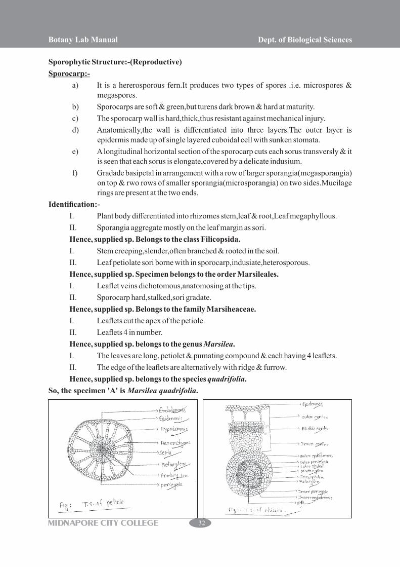
Sporophytic Structure:-(Reproductive)
Sporocarp:-
a) It is a hererosporous fern.It produces two types of spores .i.e. microspores & megaspores.
b) Sporocarps are soft & green,but turens dark brown & hard at maturity.
c) The sporocarp wall is hard,thick,thus resistant against mechanical injury.
d) Anatomically,the wall is differentiated into three layers.The outer layer is epidermis made up of single layered cuboidal cell with sunken stomata.
e) A longitudinal horizontal section of the sporocarp cuts each sorus transversly & it is seen that each sorus is elongate,covered by a delicate indusium.
f) Gradade basipetal in arrangement with a row of larger sporangia(megasporangia) on top & rwo rows of smaller sporangia(microsporangia) on two sides.Mucilage rings are present at the two ends.
Identification:-
I. Plant body differentiated into rhizomes stem,leaf & root,Leaf megaphyllous.
II. Sporangia aggregate mostly on the leaf margin as sori.
Hence, supplied sp. Belongs to the class Filicopsida.
I. Stem creeping,slender,often branched & rooted in the soil.
II. Leaf petiolate sori borne with in sporocarp,indusiate,heterosporous.
Hence, supplied sp. Specimen belongs to the order Marsileales.
I. Leaflet veins dichotomous,anatomosing at the tips.
II. Sporocarp hard,stalked,sori gradate.
Hence, supplied sp. Belongs to the family Marsiheaceae.
I. Leaflets cut the apex of the petiole.
II. Leaflets 4 in number.
Hence, supplied sp. belongs to the genus Marsilea.
I. The leaves are long, petiolet & pumating compound & each having 4 leaflets.
II. The edge of the leaflets are alternatively with ridge & furrow.
Hence, supplied sp. belongs to the species quadrifolia.
So, the specimen 'A' is Marsilea quadrifolia.
Botany Lab Manual Dept. of Biological Sciences
MIDNAPORE CITY COLLEGE 32

Botany Lab Manual Dept. of Biological Sciences
MIDNAPORE CITY COLLEGE 33

Material:- supplied from the laboratory.
Sporophytic Structure:-
A. Root:-
1. Morphological Features:-
a) The primary root is ephemeral,replaced by a large number of adventitious roots.
b) The roots are small & branched.
2. Anatomical Features:-
a) In T.S. the roots shows an outer piliferous layer,cortex & central stele.
b) Cortex is differentiated in a parenchymatous outer cortex & sclerenchymatous inner cortex.
c) The stele is protostelic with diarch & exarch xylem.
B. Rhizome:-
1. Morphological Features:-
a) The rhizome may be creeping or erect which may or may not show brnching.
b) The rhizome is differentiated into nodes & internodes.
2. Anatomical Features:-
a) The rhizome shows an outer single layered epidermis a few layered thick hypodermis,cortex.
b) The stele is solenostelic or dictyostelic.
c) Each stele is bound by its own endodermis.
C. Leaf:-
1. Morphological Features:-
a) The leaves are borne on upper surface of the rhizome.
b) Young leaves are spirally coiled & show circinate vernation.Leaf pinnatly compound,imperripinate.
c) All leaves are fertile,bearing sori along the ventral margin of pinnae,except the apices of the segments.
2. Anatomical Features:-
a) The rachis is transverse by a single C/U/V shaped leaf trace.
b) The lamina is bifacial,hypostomatic.
Reproductive Structure:-
A. Spore producing organ:-
a) It produces by means of spores.
b) It is a homosporous fern.
c) The sorus of pairs is called coenosorus.Coenosorus sori are marginal,borne continuously on a vascular commissure connected with vein ends.
Botany Lab Manual Dept. of Biological Sciences
MIDNAPORE CITY COLLEGE 34

B. Mature Sporangium:-
a) A mature sporangium has a long stalk that terminates in a capsule.
b) The jacket of the capsule is single layered,but with three different types of cells.
c) The spores are dispersed through air to a moderate distance.
Identification:-
I. Plant body differentiated into rhizomatous stem,leaf & root.
II. Leaf megaphyllous.
III. Sporangia aggregate mostly on the leaf margin as sori.
Hence,the supplied sp. belongs to the class……..Filicopsida.
I. Plants homosporous,leaf 1-2 pinnate.
II. Sporangia in sori on the ventral side of the leaf.
III. Sporangia with indusium,sporangial development mixed.
Hence,the supplied sp. belongs to the order……Polypodiales.
I. Stem surface covered with scales,sori devoid of true indusium.
II. Sori protected by leaf margin.
III. Sporangia stalked,annulus vertical,homosporous spore trilete.
Hence,the supplied sp. belongs to the family…Pteridaceae.
I. Leaves unipinnate,pinnae long with marginal sori.
II. Sori without true indusium,annulus oblique.
III. Spores tetrahedral,trilete surface reticulate.
Hence,the supplied sp. belongs to the genus…..Pteris.
I. Indusium is single layer.
Hence,the supplied sp. belongs to the species…..vittata.
So, the supplied specimen 'B' is Pteris vittata.
Botany Lab Manual Dept. of Biological Sciences
MIDNAPORE CITY COLLEGE 35

Gymnosperms
Material: supplied from the laboratory.
Vegetative structure: The plants are mostly evergreen, branched trees. Branchlets are slender, tough,divided near the apex into fine sprat. Smaller branchlets deciduous with the leaves reasons. Leaves are small, scale like, overlapping in 4 rows of 2 opposite sets, the upper & lower ranks flattened or groved the side ranks rounded or keeled. The foliage is usually glandular & aromatic. Thin bark is present,fissured only on old trees, outer bark normally shows scaling in patches of irregular shape but inner bark is fibrous.
Reproductive Structure: Strobili or cones are globose or sub globose.they are formed on different branchlets of the same tree.
Male Strobilis: It is some what reddish, cylindrical or globose. It is normally situated on branchlets near the base of the shoot & composed 3-6 pairs of microsporangia. Pollen grains are not winged.
Female Strobilis: It arises from short terminal branchlets. It is small green to purplish, composed of a few opposite pairs of leaf like scales. Mature cones are solitary & made up of 3-10 pairs of ovuliferous scales.Bract scales are fused with the ovuliferous scales. Cones are dry & ovoid to oblong. Seeds are small, thin & wingless, but attached to the edge of the ovuliferous scale. Cone scales posses thickened or ridged apex & a very fine spine.
Identification: Plant branched trees, leaves simple, microphyllous, well developed.
Hence, The sp. Belongs to the class ... Coniferapsida.
Plant evergreen,stem with secondary wood & resin canals,male & female strobili compact & cone like.
Hence, The sp. Belongs to the order ... Coniferales.
Leaves persistent, small like, usually in rows or whorled, bract scale fuses with the ovuliferous scale, pollen grains not winged.
Hence, the sp. Belongs to the family ... Cupressaceae.
Leaves scale like,usually arranged in 4 ranks of 2 opposite sets,foliage glandular & aromatic female cone ovoid too belong,cone scale with thickened or ridged apex a fine spine seed small thin attached to the edge of the scale.
Hence, The sp. Belongs to the genus ... Thuja.
So, The supplied specimen is Thuja sp.
Botany Lab Manual Dept. of Biological Sciences
MIDNAPORE CITY COLLEGE 36

Gymnosperms
Specimen:-Supplied from the laboratory.
Sporophytic Structure:-
1) Plant looks like a palm tree with a columner aerial trunk & a crown of pinnatly compound leaves.
2) Generally,the stem is unbranched,older tree exibit branching.
3) The stem is covered by an amour of alternating bands of large & small rhomboidal leaf bases.
4) These leaf bases are persistent of which the large bases belong to foliage leaves,while the small ones belong either to scale leaves or to sporophyll.
Leaf(structure):-
External morphology:
1) Leaf dimorphism in possessing green foliage leaves & scale leaves.Scale leaves are small,dry,non green & triangular in shape,covered with ramenta.
2) They have small persistent leaf bases,their only function is to provide protection to apical meristem & other aerial parts.
3) The foliage leaves are large,unipinnately,compound.A single leaf bears 75-100 pairs of leaflets which are arranged on either side of rachis in opposite or alternate manner.
4) Leaflets are sessile,elongated having revolute or flat margins.
5) Each leaflet is provided with a single unbranched mid vein without having any lateral veins.
6) The young rachis as well as leaflets are circinately coiled like those of ferns.
Internal atructure or anatomy:-
1) The T.S. of rachis shows circular outline.The outer most layer is the epidermis which consists of cuticularised thick walled cells inter rupted by sunken stomata.
2) The epidermis is followed by hypodermis,which is composed of a variable mixture of chlorenchyma & sclerenchyma followed by a parenchymatous spony parenchyma.
3) The lower part of the mesophyll consists of spongy parenchymatous cells with intercellular spaces.
4) The vascular bundle is diploxylic in nature,made up of a broad triangular centripetal exarch metaxylem & two small patches of centrifungal endarch primary xylem.The centripetal xylem is separated from centrifungal xylem by a few parenchymatous cells.
5) Some trachidal cells situated on either side of the centripetal cells situated on either side of the centripetal metaxylem are termed transfusion tissue.
6) The phloem is situated on abaxial side below xylem which is made up of sieve cells 7 parenchyma.
Identifying Character:-
1) Green foliage leaves & scale leaves are present.
2) Scale leaves are small,dry, non green,triangular in shape,covered by stomata.
3) Foliage leaves are large,unipinnatly compound.A single leaf bears 75-100 pairs of leaflets.
Botany Lab Manual Dept. of Biological Sciences
MIDNAPORE CITY COLLEGE 37

4) Hypodermis is mixture of chlorenchyma & sclerenchyma followed by the parenchymatous ground tissue with many mucilage canals.
5) Two endarch bandles enters the leaf base & then they splite up into several bundles.There are then dispersed in the ground tissue & arranged in an inverted green letter “omega” like are.
6) The vascular bundles are conjoint,collaral & open bearing both the centrifungal & centripetal xylem,so they are diploxylic or pseudomesarch in nature.Phloem lies on the side of the protoxylem.
7) T.S. of leaflet shows a thick walled,highly cutical arised epidermis.Upper epidermis is continuous,white lower epidermis is interrupted by stomata.
8) Stomata are haplocheilic,situated in pits with overarching rims.The two guard cells are bordered by 8-10 subsidiary cells arrangement in a ring.
9) The hypodermal cells are thick walled,highly lignified.The mesophylls are differentiated into palisade & spongy paren chyma.
10) Transfussion tissue present.
Hence,the supplied specimen 'B' is the leaf of Cycas sp.
Botany Lab Manual Dept. of Biological Sciences
MIDNAPORE CITY COLLEGE 38