Changes in Ciliate Communities Reveal Modification of Lake ...
Upload
independentCategory
view
4download
0
Protistology 4 (1), 5�55 (2005) ProtistologyProtistologyProtistologyProtistologyProtistology
© 2005 by Russia, Protistology
Morphology, ontogenesis and encystment of a soilciliate (Ciliophora, Haptorida), Arcuospathidiumcultriforme (Penard, 1922), with models for theformation of the oral bulge, the ciliary patterns, andthe evolution of the spathidiids
Kuidong Xu 1, 2 and Wilhelm Foissner 1
1 Universitqt Salzburg, FB Organismische Biologie, A�5020 Salzburg, Austria2 Institute of Oceanology, Chinese Academy of Sciences, 266071 Qingdao, PR China
Summary
We studied and reviewed the morphology, ontogenesis and encystment of two closelyrelated spathidiids, Arcuospathidium cultriforme (Penard, 1922) Foissner, 1984 and A.scalpriforme (Kahl, 1930) Foissner, 2003, using live observation, protargol impregnation,morphometry, scanning electron microscopy, populations from different geographicregions, and literature data. Both species are biogeographic flagships. They are 200�300µm long and have a long, steep oral bulge. Our investigations show that they are verysimilar, differing mainly in the length of the oral bulge and the arrangement of the oralbulge extrusomes. Thus, we classify them as subspecies within the Arcuospathidiumcultriforme complex, which includes the following taxa: A. cultriforme cultriforme (Penard,1922), A. cultriforme scalpriforme (Kahl, 1930), A. cultriforme megastoma Foissner et al.,2002, and A. lorjeae Foissner et al., 2002. The latter taxa are likely to have a restrictedgeographic distribution, and there is evidence that the South American and Rwandanpopulations of A. cultriforme cultriforme could represent further distinct (sub)species.During encystment of A. cultriforme, the macronucleus becomes strongly shortened andthe infraciliature appears to be resorbed. The mature cyst is unique in having a very thick,faceted wall. A detailed scenario of the ontogenetic processes is provided, showing thatthe ontogenesis of A. cultriforme matches those of haptorids in general and other spathidiidsin particular. However, there are several characteristic features, for some of which wecould find reasonable explanations which, in turn, provided models for the spathidiidontogenesis and evolution: (i) the “outgrowth model” suggests that the slope of the longspathidiid oral bulge is obtained mainly by faster growth of the dorsal than the ventralside; (ii) the “row detachment model” proposes that the Arcuospathidium andEpispathidium ciliary patterns are variations of the Spathidium pattern which evolved fromthe Protospathidium pattern; and (iii) a model is proposed for a Dileptus�like ancestor ofthe spathidiids.
Key words: Hawaii, outgrowth model, phylogeny, row detachment model, South Africa,Spathidium cultriforme, Spathidium lionotiforme, Spathidium scalpriforme

· Kuidong Xu and Wilhelm Foissner6
Introduction
Both morphology and ontogenesis appear highlysimilar in various haptorid ciliates due to theircomparatively simple organization supporting homo�plasies (Foissner and Foissner, 1988; Xu and Foissner,2003). Only on detailed investigation the differencesbecome evident. Typical examples are the time�honoured genera Trachelophyllum and Spathidium.Trachelophyllum has been split into six genera based onthe structure of the epicortical scales, and there isevidence for many more undescribed genera and species(Foissner, 2005b; Foissner, unpubl. data). The genusSpathidium, which consisted of about 200 species whenDragesco and Dragesco�Kernuis (1979) and Foissner(1984) split it into five genera, now comprises about300 species distributed over 15 genera (Foissner and Xu,2005). Similar, but usually less extreme examples canbe found in all groups of ciliates, for instance, thehypotrichs (Berger, 1999) and colpodids (Foissner,1993; Foissner et al., 2002).
Such data and a recent monograph on soil ciliatesfrom Namibia (Foissner et al., 2002) suggest thathaptorids are heavily undersplit and their diversity ismuch greater than previously assumed. Obviously, thereare thousands of these often inconspicuous species.Thus, Finlay’s (2001) estimation of only 3,000�4,000free�living ciliate morphospecies is flawed by the facts.There is a continuous flow of new families, genera andspecies from limnetic, marine, and soil habitats wheninvestigations are performed by experienced taxo�nomists (Foissner et al., 2002; Song et al., 2003). Wealso disagree with Finlay’s (2001) hypotheses thatprotists are ubiquitous and cosmopolitan. The soilprovides strong arguments against: more than a half ofthe about 800 species has been found only in onebiogeographic region (Foissner, 1998) and manysamples contain one or several flagship species notfound in more than 1,000 other soil samples collectedworld�wide (Foissner, 1995, 1999a, 2004a, b, 2005a,b; Foissner et al., 2002, 2005).
The present study reports the morphology andontogenesis of the Arcuospathidium cultriforme complex,which contains large and thus conspicuous speciesserving as biogeographic flagships. The study providesnot only an evidences of a restricted distribution of sometaxa of the complex, but also new models for variousaspects of the spathidiid ontogenesis and evolution.
Material and Methods
Over the years, many populations of A. cultriformefrom soils all over the world were routinely identifiedaccording to Penard (1922), Kahl (1930b), and Foissner
(1984). The following compilation contains only thosesites where notes and/or preparations of the specimenswere made.
Arcuospathidium cultriforme cultriformeAustria, Burgenland: Upper litter and soil layer (0�
2 cm) of a coniferous forest (Ebenauerwald) in southernBurgenland, eastern Austria. This population wasdescribed by Foissner (1984).
Austria, Linz: Mud and soil from a flat, ephemeralpond at the foot of the Pastlingberg, in the surroundingsof the town of Linz, capital of Upper Austria.
Germany, Kassel: Litter and soil from the upper 0�5 cm layer of a beech forest on the “Kleinen Gudenberg”about 30 km NW of the town of Kassel. Sample kindlyprovided by Dr. M. Bonkowski (Gattingen University).
Dominican Republic, site 22: Litter and soil fromthe upper 0�3 cm layer of a mangrove forest near thevillage of Maimon, about 10 km west of the town ofPuerto Plata. Soil almost black and spongy, contains alot of grass and mangrove roots, slightly acidic (pH 6.2in water) and saline (5‰).
Costa Rica, site 11: Tree mosses from the evergreenrain forest in the Braulio�Carillo National Park,Mirador area; pH 4.7 in water.
South America, Venezuelan site 8: Soil from theupper 3 cm of a banana plantation at the farm of Mr.Eisenberg, surroundings of the town of PuertoAyacucho. The field was founded 15 years ago and hasbeen organically fertilized. The slightly acidic (pH 6 inwater) soil is dark and contains many fine roots, butthe layer is only about 5 cm thick and followed by yellowsand.
South America, Venezuelan site 20: Tree mossesfrom a gallery forest in the surroundings of the town ofPuerto Ayacucho.
Tropical Africa, Rwanda: Savannah litter and soilfrom the surroundings of the village of Gabiro, at thefoot of the mountains with the Gorilla National Park.Sample kindly provided by Dr. E. Stzber (Salzburg).
Hawaii, Big Island site 12: Highly saline (~ 20 ‰)mud and soil from dry rockpools in the lower part of astream between the Kahua Ranch and Kawaihae.
Hawaii, Big Island site 39: Tree bark with mossesand lichens along the Bird Trail in the Volcano NationalPark; pH 7.2 in water.
Hawaii, Oahu Island site 50: Litter and soil fromthe upper 0�3 cm layer of a dry swamp overgrown withferns, Ihi’lhilanгkea crater area, Koko Head, south ofthe Hanauma Bay. Sample kindly provided by Mag.Hubert Blatterer (Linz).
Arcuospathidium cultriforme scalpriformeAustria, Lower Austria: Litter and soil from the
upper 0�5 cm layer of a mixed forest (Asperulo�Fagetum)

· 7ProtistologyProtistologyProtistologyProtistologyProtistology
in the surroundings of the village of Baumgarten. Brown�earth soil with many roots and neutral reaction (pH 7 inwater). This population was described by Foissner (1984).
South America, Brazil site 30: Litter and soil fromthe upper 0�5 cm of the floodplain rainforest near thetown of Manaus, Janauari region. Soil brown, humic,pH 5.1 in water.
Republic of South Africa, site 44: Litter and soilfrom the margin of a flat pond (Sirkelsvlei) in the CapePeninsula National Park. Soil dark, very sandy, withmany grass roots, flooded after heavy rain, pH 5.4 inwater.
All populations developed in non�flooded Petri dishcultures, as described by Foissner et al. (2002). Thestored, air�dried samples were saturated with distilledwater and investigated for ciliates weekly. Sufficientspecimens for preparations usually developed two weeksafter rewetting. No pure cultures were set up, exceptfor the South African population of A. cultriformescalpriforme, where ontogenesis was studied. A semipureculture of this species was obtained by adding somemillilitres of percolate from the non�flooded Petri dishculture to a Petri dish filled with Eau de Volvic (Frenchtable water) and some crushed wheat grains. A denseculture with many dividers and monsters developed,using as food source the natural ciliate communityintroduced with the soil percolate.
Specimens were studied in vivo using a high power,oil immersion objective and differential interferencecontrast optics. The ciliary pattern and variouscytological structures were revealed by Foissner’s orWilbert’s protargol protocol, both described in Foissner(1991). Counts and measurements of stained specimenswere performed at a magnification of +1,000. In vivomeasurements were conducted at magnifications of+100�1,000. Illustrations of live specimens were basedon free�hand sketches and video records, while thoseof cells on preparations were made with a cameralucida. Terminology is mainly according to Corliss(1979) and Foissner (1996).
Scale bars are liked by reviewers and editors.However, in the case of drawings they are usuallycircumstantial and superfluous, if the size of the specimenis indicated in the figure explanation. The old taxonomistsknew that. Imagine Kahl with countless scale bars onthe plates! Thus, we adopted the old style and, whenappropriate, give the size of the specimen in the figureexplanation, not the scale bar in the figure.
The situation is more complex with micrographs.At first glance, scale bars appear indispensable, but whenconsidered critically, they might also be superfluous oreven misleading. A good description provides in vivomeasurements of the main features, such as body lengthand width (first data set); a good description also providesdetailed morphometrics from permanent silver prepa�
rations where, however, the specimens are more or lessshrunken (second data set); and when the descriptioncontains SEM micrographs, a third data set is generatedby scale bars because 20�60% shrinkage is usual in SEMpreparations. Now, we have three different values andscale bars for the same structure! Further bias isgenerated when specimens are more or less flattenedby mild coverslip pressure to get detail�rich micro�graphs. All these problems and the fact that sizes aregiven in the description and the tabulated morpho�metrics suggest that scale bars usually can be omittedfrom micrographs without any loss of importantinformation. Indeed, they are often misleading,indicating higher precision than the actual one. *)
Results
Arcuospathidium cultriforme (Penard, 1922)Foissner, 1984
1922 Spathidium cultriforme Penard, Infusoires, p.25.
1984 Arcuospathidium cultriforme (Penard, 1922)nov. comb. – Foissner, Stapfia, 12: 78 (designation astype species of the genus Arcuospathidium).
2002 Arcuospathidium cultriforme cultriforme(Penard, 1922) Foissner, 1984 nov. stat. – Foissner,Agatha and Berger, Denisia, 5: 299 (improved diagnosisand ranking as a subspecies).
Improved diagnosis: Size 150�450 + 20�90 !m invivo, usually near 240 + 40 !m. Narrowly to verynarrowly knife�shaped with oblique to strongly oblique,very narrowly cuneate to oblong oral bulge occupying1/3, 1/2 or 2/3 of body length. Macronucleus anindistinctly nodulated, tortuous strand, rarely occur twolong pieces, many small nodules or a mixture of nodulesand short strands; multimicronucleate. Extrusomesarranged in rows one row in the right and the left halfof oral bulge or scattered, basically oblong with a sizeof 4�8 + 0.7�1.1 !m in vivo. On the average 25�37 ciliaryrows, 3 anteriorly differentiated to dorsal brushoccupying 23�36%, usually about 28% of body length;bristles up to 5 !m long in vivo. Resting cyst with thick,faceted wall.
Etymology: Not given in the original description.Possibly, cultriforme (knife�shaped) refers to both thebody (“corps allongu en forme de couteau …”) and theexploded toxicysts (“… couteau suudois fermu … maisle couteau s’est ouvert”).
Remarks: The diagnosis is based on Foissner et al.(2002) who distinguish three subspecies according to
*) The editors of the "Protistology" requested to insert the scale bars on the pictures anyway.

· Kuidong Xu and Wilhelm Foissner8
the length of the oral bulge; we add the arrangement ofthe extrusomes: A. cultriforme cultriforme (mouth about1/3 of body length, a row of extrusomes in the rightand the left bulge half), A. cultriforme scalpriforme(mouth about 1/2 of body length, bulge extrusomesscattered), A. cultriforme megastoma (mouth about 2/3of body length, a row of extrusomes each in right andleft bulge half). Arcuospathidium cultriforme cultriformeand A. cultriforme scalpriforme are the subjects of thepresent study, while A. cultriforme megastoma, for whichwe do not have any new data, is described in Foissner etal. (2002).
Arcuospathidium cultriforme cultriforme (Penard,1922) Foissner, 1984 (Figs 1�4a, 5�15; Table 1)
1922 Spathidium cultriforme Penard, Infusoires, p.25.
1930 Spathidium cultriforme Penard – Kahl, Arch.Protistenk., 70: 383 (brief redescription; likely amisidentification).
1930 Spathidium cultriforme Penard, 1922 – Kahl,Tierw. Dtl., 18: 164 (revision).
1975 Spathidium cultriforme Penard, 1922 – Fryd�Versavel, Iftode and Dragesco, Prostistologica, 11: 511�514 (brief redescription from protargol�impregnated,Italian specimens).
1984 Arcuospathidium cultriforme (Penard, 1922)nov. comb. – Foissner, Stapfia, 12: 78 (redescriptionfrom life and after protargol impregnation of Austrianspecimens from Burgenland).
2002 Arcuospathidium cultriforme cultriforme(Penard, 1922) Foissner, 1984 nov. stat. – Foissner,Agatha and Berger, Denisia, 5: 299 (improved diagnosisand ranking as a subspecies).
Improved diagnosis: Mouth (oral bulge) about onethird of body length. A row of extrusomes in the rightand the left bulge half, rarely only a single row in lefthalf.
Type locality: Mosses in Switzerland (Chemin dela Montagne, on an old wall).
Morphology: The description includes the aboveliterature data and the populations mentioned in theMaterial and Methods section. The morphometric datacompiled in Table 1 are repeated only if appropriate.
Size considerably variable within and betweenpopulations, with average values ranging from 192 +28!m to 306 + 45 !m and variation coefficients of up to18% (Table 1). Taking into account the in vivo measu�rements and 10�20% preparation shrinkage, in vivoranges of 150�450 + 20�90 !m have been calculated, withan overall average of 240 + 40 !m, which is close toPenard’s values (200�260 + 40�50 !m). Length:widthratio highly variable within populations (CV 21�37%),but rather constant between populations, ranging from
5.7:1 to 7.2:1, with the average of 6.4:1 matching the invivo ratio of about 6:1. Contractile by up to one third ofbody length, especially in oral region; contracts andextends slowly, size changes thus difficult to recognize.
Usually narrowly to very narrowly knife�shaped orlanceolate (3:1�12:1, on the average about 6:1) due tothe long and strongly slanted oral bulge (40°�80°, onthe average 65°). Oral portion more or less distinctlycurved dorsally and with bluntly pointed anterior end;neck of ordinary distinctness; trunk elongate barrel�shaped, strongly inflated after ingestion of rotifers orlarge ciliates; posterior end rounded or bluntly pointedafter systole of contractile vacuole; laterally flattenedup to 2:1 in hyaline oral region, while only slightlyflattened or unflattened postorally; sometimes twistedabout main body axis, especially when just collectedfrom soil habitat (Figs 1a, g, m, q, 2o, p, u�x, 3a, c, h,5a�d, 6a, 9a, c, 10a, b).
Nuclear apparatus in middle quarters of cell (Figs1a, g, q, 3b, e, 7a, g, 8a, 9a). Macronucleus a long,tortuous, more or less nodulated, 4�8 !m thick strandin over 30 populations routinely identified over theyears, rarely in two or three long, tortuous piecesdividing individually (see A. cultriforme scalpriforme);nucleoli granular, rod�like, or lobate, up to 6 !m in size.Macronucleus pattern very variable in one of the threeHawaiian populations: a highly tortuous strand(occasionally in two pieces) in 58% out of 75 specimensanalysed, a mixture of short strands and small nodulesin 23%, and up to 100 nodules in 19% of specimens(Figs 5a�d, 9a, c�e; Table 1). In a population fromRwanda, all specimens have many nodules (Fig. 6c).Both populations match ordinary cells in all otherfeatures, though the Rwandan specimens are the longeston the average (Table 1). Micronuclei scattered alongmacronucleus strand, 2�4 !m across in protargolpreparations, often difficult to distinguish from similarlysized and impregnated cytoplasmic inclusions; numberthus difficult to determine, likely between 10 and 20.
Contractile vacuole in rear body end, forms severaladventive vacuoles merging to a large vacuole duringdiastole (Figs 1a, c�e). About 10 excretory pores in polearea (Figs 3h, 6a, b). During defecation, the fecal massmigrates through the contractile vacuole and leaves thecell in the pole centre (Figs 2t, x).
One type of extrusomes in oral bulge. Extrusomeshape and size rather similar in the populationsinvestigated, differences recognisable only on carefulobservation; conspicuous both in vivo and in protargolpreparations, though small as compared to body size,because forming a row in the right and the left half oforal bulge, as already described and illustrated byPenard (Fig. 1b), producing a conspicuously bright (invivo) or black (protargol preparations) fringe in ventralanterior third of body; often less narrowly spaced, rarely

· 9ProtistologyProtistologyProtistologyProtistologyProtistology
Figs 1a�q. Arcuospathidium cultriforme cultriforme, alive (1a�n) and after protargol impregnation (1o�q), according toPenard, 1922 (1a�f); Kahl, 1930a (1k�n); Kahl, 1930b (1g�j); and Fryd�Versavel et al., 1975 (1o�q). 1a: Swiss specimen,200�260 µm. 1b: Oral bulge view. 1c�e: Cycle of contractile vacuole. 1f: Exploded toxicyst. 1g, h, m, n: Kahl’s redrawingsof Penard’s figures. 1i�l: A specimen of doubtful identity from the Zillertal, length 180 µm; resting extrusome, 8 µm(j, l). 1o�q: Ventral and left side view of Italian specimens, 140 µm. Figs 2a�y. Arcuospathidium cultriforme cultriforme,alive, according to Foissner, 1984 (2o�u) and new observations (2a�n, v�y). 2a: Dorsal brush, bristles up to 4 µm. 2b�i: Extrusomes from specimens of Austria and Germany (b), Crete (c), Costa Rica (d), Venezuelan site 20 (e, f), andHawaiian sites 39 (g), 12 (h) and 50 (i). Drawn to scale, bar 5 µm. 2j: Exploded toxicyst, 11 µm. 2k: Variability inshape of oral bulge and arrangement of extrusomes. 2l: Surface view of oral bulge. 2m, n: Surface view and opticalsection showing cortical granulation. 2o, p, u: Left side, ventral, and ventrolateral view of Austrian (Burgenland)specimens, length 250 µm. 2q�t: Resting (6 µm) and exploded toxicyst, surface view of oral bulge and somatic cortex,and defecation. 2v�x: Various views of specimens from Hawaiian site 12 (video records). 2y: Resting cyst from anAustrian (Linz) specimen, 70 µm. B (1�3) – dorsal brush (rows), E – extrusomes, FM – fecal mass, G – corticalgranules, MA – macronucleus, OB – oral bulge.
· Kuidong Xu and Wilhelm Foissner10
even lacking in right bulge half, for instance, in 3 outof 43 specimens of the Linz population (Figs 1b, g, 2k,l, r, 3h, 7a, b, d, 8a�e, 9a�c, f, 10g). Size (4�6 + 0.7�1.1!m) very similar in the eight populations investigated;
except for the Venezuelan specimens, shape also highlysimilar, oblong to very narrowly ellipsoidal with roundedends, anterior end conical in some populations, makingextrusomes looking like blunt pencils (Figs 2b�d, g�i,
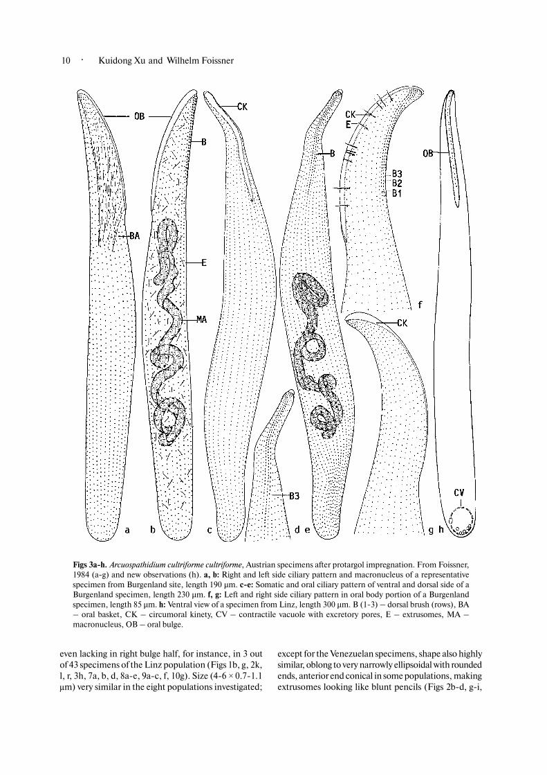
Figs 3a�h. Arcuospathidium cultriforme cultriforme, Austrian specimens after protargol impregnation. From Foissner,1984 (a�g) and new observations (h). a, b: Right and left side ciliary pattern and macronucleus of a representativespecimen from Burgenland site, length 190 µm. c�e: Somatic and oral ciliary pattern of ventral and dorsal side of aBurgenland specimen, length 230 µm. f, g: Left and right side ciliary pattern in oral body portion of a Burgenlandspecimen, length 85 µm. h: Ventral view of a specimen from Linz, length 300 µm. B (1�3) – dorsal brush (rows), BA– oral basket, CK – circumoral kinety, CV – contractile vacuole with excretory pores, E – extrusomes, MA –macronucleus, OB – oral bulge.

· 11ProtistologyProtistologyProtistologyProtistologyProtistology
9h�k); thinner, slightly acicular, and inconspicuouslycurved in the two Venezuelan populations (Figs 2e, f, 7c,10d�f). Fully exploded extrusomes 8�12 !m long andof typical toxicyst structure, that is, with a proximal,
toxin�containing capsule of size and shape similar tothat of the resting organelle, and a distal tube about aslong as capsule bent at up to 90°; between capsule andtube a refractive granule deeply impregnating with
Figs 4a, b. Arcuospathidium cultriforme cultriforme (a) and A. cultriforme scalpriforme (b), ciliary pattern of left side inoral body portion of an Austrian (Linz, a) and South African (b) specimen after protargol impregnation, length ofcord of oral bulge 90 µm and 105 µm, respectively. The specimen of A. cultriforme cultriforme has four dorsal brushrows. Arrowheads mark minute, transversely extending kinetofragments (adesmokineties?) left of the circumoralkinety. B (1�3) – dorsal brush (rows), CK – circumoral kinety, OB – oral bulge. Scale bar 50 µm.

· Kuidong Xu and Wilhelm Foissner12
protargol and staining red with methyl green�pyronin.Partially exploded toxicysts, usually found in preservedspecimens, about 7�10 !m long and often distinctlyknife�shaped, as already mentioned and illustrated byPenard (Figs 1f, p, 2q, 3f, 8d, h, 11e, 12e, f). Developingextrusomes studded in cytoplasm, of similar size andshape as mature ones, sometimes form small bundles,impregnating with protargol (Figs 7g, 8e, 9b, g).
Cortex colourless and very flexible, contains 5�10granule rows between each two kineties. Individualgranules 0.3�0.6 + 0.2�0.3 !m in size, moderately tostrongly refractive, depending on population (Figs 1s,2m, n, 7e, f). Cytoplasm colourless, postorally packedwith lipid droplets 1�6 !m across and food vacuolescontaining ciliates and rotifers; specimens thus turbidand brownish at low magnification ([ +100). Swims andglides moderately rapidly on microscope slides andbetween soil particles, showing great flexibility,especially in the oral area, which moves to and fro, asalready mentioned by Penard (1922). Appears somewhat
flag�like due to the curved oral area and the cylindroidalpostoral portion, when rotating about main body axis.
Somatic cilia 8�10 !m long in vivo, arranged in 24�44, on the average 25�37 equidistant, densely ciliatedrows slightly loosened in oral area. Ciliary rows arrangedin typical Arcuospathidium pattern, that is, curvedorsally on both sides of oral bulge, except for brushkineties which, as usual, curve ventrally (Fryd�Versavelet al., 1975; Figs 1a, g, q, 2o, 3a�g, 4a, 6a�e, 8a, b, e,9a, b, 10a, b; Table 1). Short rows, each composed of1�5 kinetids, extend slightly obliquely from left sidecircumoral kinety either in line with a certain circumoraldikinetid or separated from kinety by a small space;present in all populations of A. cultriforme cultriformeand A. cultriforme scalpriforme, but not in all specimens(Figs 4a, b, 8c, h, 9b): for instance, absent in three outof 12 specimens of the Linz population, indistinct(composed of 1�2 kinetids) in four specimens, anddistinct (composed of 2�5 kinetids) in five specimens;values similar for Hawaiian site (50) population (n 18):6/2/10. These short kineties, which suggest a Dileptus�like ancestor of the spathidiids (see Discussion), werenot mentioned by either Fryd�Versavel et al. (1975) orFoissner (1984), where they are recognisable on amicrograph (Fig. 11c).
Brush exactly on dorsal side, three�rowed, di�kinetidal, heterostichad, and slightly shorter than oralbulge, in vivo inconspicuous, though occupying 25�30%of body length, because bristles only 2�3 !m long andpairs ordinarily spaced (1�1.5 !m apart). Bristle rowsfrequently with small irregularities, such as minutebreaks and/or some kinetids out of line, rarely occurs amore or less complete fourth row right of row 1; all rowscommence with some ordinary cilia anteriorly andcontinue as somatic kineties posteriorly. Brush row 1usually slightly shorter than row 2; row 2 by 20�30%longer (heterostichad) than row 3 which, however, hasa monokinetidal tail extending to second third to halfof body with about 2 !m long bristles. Details of bristlepairs difficult to analyse, even in the scanning electronmicroscope, likely as shown in figure 2a, that is, anteriorbristle of pairs slightly shorter (1.5�2 !m) than clavateposterior bristle (2�3 !m, rarely up to 5 !m), while viceversa in row 3 (Figs 1a, q, 2o, 3b, d�f, 4a, 6b, d, 7d, e,8a, c, f, g, 9a, b, 11a�d; Table 1). Bristle pairs obliquelyarranged in upper half of rows of Venezuelan site (20specimens), a conspicuous feature seemingly doublingrow number (Figs 12a�d, 14b, c).
Oral bulge conspicuous due to the highly refractiveextrusomes and the strong slope (40°�80°, on the average65°, n 15) occupying on the average 31% to 35% of bodylength in five populations; bulge length thus compa�ratively constant, though ranges (25%�45%) approachA. cultriforme scalpriforme; flat to strongly convex, infrontal view very narrowly cuneate to oblong with
Figs 5a�d. Arcuospathidium cultriforme cultriforme,variability of body shape and nuclear apparatus inHawaiian site (50) specimens after protargol im�pregnation, length 200 µm, 200 µm, 170 µm, 280 µm.Two thirds of the specimens have an ordinary, moreor less tortuous macronucleus strand (a, b), while theothers have fragmented nucleus in many nodules (d).Transitions occur (c), showing that they do notrepresent different species. CV – contractile vacuole,MA – macronucleus (nodules), MI – micronuclei,OB – oral bulge.

· 13ProtistologyProtistologyProtistologyProtistologyProtistology
proximal end often bluntly pointed, indistinctly apartfrom body proper because gradually merging intoventral surface and in vivo only about 3 !m high and 5!m wide. Bulge cortex with arrowhead�like pattern ofcortical granules. Circumoral kinety of same shape as
oral bulge, continuous, composed of narrowly spaceddikinetids each associated with a cilium and a basketrod (nematodesma). Oral basket hardly recognisable invivo, prominent after French protargol impregnation(Figs 1o, q), while rather inconspicuous with Foissner’s
Figs 6a�e. Arcuospathidium cultriforme cultriforme, Rwandan specimens after protargol impregnation. About 20specimens are found in the protargol slides, all have many macronucleus nodules (c), similar to some Hawaiian cells(Fig. 5d). Thus, this population might represent a distinct species. a�c: Right and left side ciliary pattern and nuclearapparatus of a representative specimen, length 315 µm. d, e: Ciliary pattern of dorsal and ventral side in oral bodyportion, length of circumoral kinety (oral bulge) 97 µm. B (1, 3) – dorsal brush (rows), BA – oral basket, CK –circumoral kinety, E – extrusomes, EP – excretory pores of contractile vacuole, MA – macronucleus, MI –micronuclei, OB – oral bulge. Scale bars 100 µm (a�c) and 40 µm (d, e).

· Kuidong Xu and Wilhelm Foissner14
Figs 7a�g. Arcuospathidium cultriforme cultriforme,alive (b�g) and after protargol impregnation (a). a:Overview of a specimen from the Linz (Austrian)population, showing the long, tortuous macronucleusand the oral extrusome fringe. b�d: Ventral and lateralview of the oral area of a specimen from Venezuelansite 8. The extrusomes, which are about 6 µm longand comparatively thin in this population (c), form arow in the right and a row in the left bulge half (b, d).Note the minute dorsal brush bristles about 3 µm long. e, f: Surface view showing cortical granulation in a specimenfrom Venezuelan site 8 (e) and Costa Rica (f). The cortical granules, which have a diameter of only 0.2�0.4 µm, arehighly refractive and form long, slightly oblique rows, which are paired in the Costa Rican specimen (f). g: Thecytoplasm of a Hawaiian site (39) specimen is studded with lipid droplets, extrusomes, and the long macronucleus. B� dorsal brush, E � extrusomes, MA � macronucleus, OB � oral bulge. Scale bars 100 µm (a) and 20 µm (b�g).
· 15ProtistologyProtistologyProtistologyProtistologyProtistology
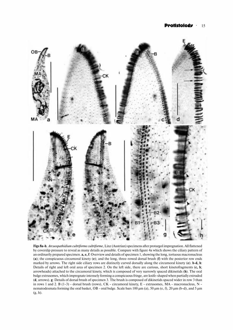
Figs 8a�h. Arcuospathidium cultriforme cultriforme, Linz (Austrian) specimens after protargol impregnation. All flattenedby coverslip pressure to reveal as many details as possible. Compare with figure 4a which shows the ciliary pattern ofan ordinarily prepared specimen. a, e, f: Overview and details of specimen 1, showing the long, tortuous macronucleus(a); the conspicuous circumoral kinety (e); and the long, three�rowed dorsal brush (f) with the posterior row endsmarked by arrows. The right side ciliary rows are distinctly curved dorsally along the circumoral kinety (e). b�d, h:Details of right and left oral area of specimen 2. On the left side, there are curious, short kinetofragments (c, h;arrowheads) attached to the circumoral kinety, which is composed of very narrowly spaced dikinetids (h). The oralbulge extrusomes, which impregnate intensely forming a conspicuous fringe, are knife�shaped when partially extruded(d, arrows). g: Details of dorsal brush of specimen 3. The brush is composed of dikinetids spaced wider in row 3 thanin rows 1 and 2. B (1�3) � dorsal brush (rows), CK � circumoral kinety, E � extrusomes, MA � macronucleus, N �nematodesmata forming the oral basket, OB � oral bulge. Scale bars 100 µm (a), 50 µm (e, f), 20 µm (b�d), and 5 µm(g, h).

· Kuidong Xu and Wilhelm Foissner16
method (Figs 3a, 6a, 8e, 11c), composed of cuneatebundles of nematodesmata extending into second thirdof body. Cytopharyngeal entrance recognisable neitherin vivo nor at preparations (Figs 1a, q, 2k, l, o, p, u�x,3a�c, f�h, 4a, 5a�d, 6a, e, 7a�d, 8a, e, 9a�c, f, 10a�c, g,13a; Table 1).
Resting cyst (Figs 2y, 15a�i): Resting cysts werestudied in the populations from Austria (Linz) and theDominican Republic. They match well, but the ridgesare thinner (Figs 15g, h) and the cysts considerablysmaller (` 68.8 !m, n 22 vs. 45.2 !m, n 6) in theDominican specimens. As concerns the ciliary pattern,see A. cultriforme scalpriforme.
Only the Linz specimens were studied in detail. Forencystment, cells were transferred to Eau de Volvic,where they produced a distinct, but not yet faceted wallwithin six hours; the facets developed slowly over a week.Thus, observations and measurements were performedon two weeks old cysts.
All components of the cyst are colourless at amagnification of ] +400, and brown to yellow�brownat low magnifications ([ +100) due to the strong lightrefraction. The cysts are perfectly spherical and havean outer diameter of 55�95 !m ( ̀68.8 !m, M 65, SD9.6, CV 13.9, n 22), while the inner diameter (cystproper) is considerably smaller ( ̀48.9 !m, M 48, SD8.8, CV 18.1, n 22). Thus, the wall has an averagethickness of 10 !m (!) and its volume is almost thrice(twice when the facet concavities are detracted) that ofthe cyst proper (170,430 !m3 vs. 61,570 !m3). This isan outstanding feature found, for instance, also in desertstrains of Exocolpoda augustini (Foissner et al., 2002).The most conspicuous part of the cyst is the deeply androughly faceted middle layer (mesocyst?) which iscompact, flexible, and highly refractive. A 0.5�1 !mthick membrane each is found on the facets (ectocyst?)and the cyst proper; both are also recognisable insquashed, split cysts, showing that the inner membraneis not the cell cortex, but rather the endocyst. The cystcontents consist of colourless lipid droplets 1�5 !macross and the macronucleus shortened to a semicircularstrand, as described in A. cultriforme scalpriforme. Thecontractile vacuole and all extrusomes disappear.
Ontogenesis (Figs 13a�c, 14a�c, e, f): The SEMmicrographs of dividers from the Venezuelan site (20)population will be included in the detailed descriptionof the ontogenesis of A. cultriforme scalpriforme. Nodifferences are recognisable in the two subspecies.
Occurrence and ecology: There are surprisingly fewrecords in the literature, although A. cultriformecultriforme is rather common in terrestrial habitats,especially in Europe (Foissner et al., 2005). Penard(1922) discovered A. cultriforme cultriforme in wallmosses from Switzerland. Kahl (1930a, b) observed asingle specimen in moss from the Zillertal in Tyrol,
Austria. However, the shape of the extrusomes suggeststhat it was a different species (Figs 1i�l). It was only 50years later, that Fryd�Versavel et al. (1975) reported A.cultriforme cultriforme from Italian moss and provideda brief redescription based on protargol impregnation(Figs 1o�q). Later, Foissner (1984) redescribed A.cultriforme cultriforme from the soil of a coniferous forestin Austria (Figs 2o�t, 3a�g; Table 1). Since then, it wasreported from several terrestrial habitats of Austria(Foissner, 1987a; Aescht and Foissner, 1993; Foissneret al., 2005) and Germany (Goralczyk and Verhoeven,1999; Foissner, 2000b). In 1998, Foissner reported thespecies from all main biogeographic regions, exceptAntarctica. This is substantiated by the records givenin the Materials and Methods section as well as byBlatterer and Foissner (1988) and Foissner (1999a).However, A. cultriforme cultriforme seems to be rare inAfrica because it was not found in 73 samples fromNamibia, where A. cultriforme megastoma was discovered(Foissner et al., 2002).
These data show that A. cultriforme cultriforme andA. cultriforme cultriforme�like taxa are cosmopolites,except Antarctica. In Rwanda and Venezuela, other(sub)species may occur, but more detailed investigationsare required. The ecological range is obviously wide andincludes terrestrial, semiterrestrial, and saline habitats(see Materials and Methods section). However, truelimnetic and extreme habitats, such as alpine soil abovethe timberline (Foissner, 1987b), Antarctica (Foissner,1998), and hot deserts (Foissner et al., 2002) areavoided.
Arcuospathidium cultriforme scalpriforme (Kahl,1930) Foissner, 2003 (Figs 4b, 16�29; Tables 2, 3)
1930 Spathidium scalpriforme Kahl, Arch. Pro�tistenk., 70: 381.
1930 Lionotus scalpriforme Kahl, 1930 – Kahl,Tierw. Dtl., 18: 165 (The figure explanation and theindex indicate that Kahl put it to Lionotus by mistake).
1984 Arcuospathidium lionotiforme (Kahl, 1930)nov. comb. – Foissner, Stapfia, 12: 78 (misidentificationin that and all subsequent papers!)
2002 Arcuospathidium cultriforme lionotiforme(Kahl, 1930) Foissner, 1984 nov. stat. – Foissner,Agatha and Berger, Denisia, 5: 300 (misidentification,see above; ranked as a subspecies).
2003 Arcuospathidium cultriforme scalpriforme(Kahl, 1930) comb. n., stat. n. – Foissner, ActaProtozool., 42: 54 (rectification of misidentification andrank lowering).
Improved diagnosis: Mouth (oral bulge) about halfof body length. Extrusomes scattered in right and leftbulge half.
Type locality: Mosses from the Zillertal in Tyrol,Austria. Kahl (1930a) first observed the species in

· 17ProtistologyProtistologyProtistologyProtistologyProtistology
Figs 9a�k. Arcuospathidium cultriforme cultriforme, Hawaiian sites 50(a�h) and 39 (i�k) specimens from life (g�k) and after protargolimpregnation (a�f). a�e: Two thirds of the Hawaiian site (50) specimenshave an ordinary macronucleus, that is, a long, tortuous strand (a, c),while one third have many macronucleus nodules (d). Transitions occur(e), suggesting that specimens with an ordinary macronucleus andthose with many nodules/that specimens differing as to theirmacronuclei belong to the same species. The oral bulge occupies 30%
(a) to 37% (c) of body length, as typical of this subspecies. Short kinetofragments (b, arrowhead) are attached to theleft side circumoral kinety, as in the European populations (Figs. 8h, 11c). The anterior end of the dorsal brushkineties is slightly curved ventrally (a, b). f: The extrusomes form a row in the right and a row in the left half of the oralbulge, as typical of this subspecies. g: Posterior body portion studded with minute and moderately�sized lipid dropletsand extrusomes. h�k: The oral bulge extrusomes of the Hawaiian site (39) specimens are blunt, 4�5 µm long rods withrounded or bluntly pointed distal end (i�k), just as those occurring in the Hawaiian site (50) specimens (h). B (1�3) �dorsal brush (rows), CK � circumoral kinety, CV � contractile vacuole, E � extrusomes, MA � macronucleus (nodules),MI � micronuclei, OB � oral bulge. Scale bars 100 µm (c�e), 50 µm (a), 25 µm (b), and 10 µm (f, g).
· Kuidong Xu and Wilhelm Foissner18
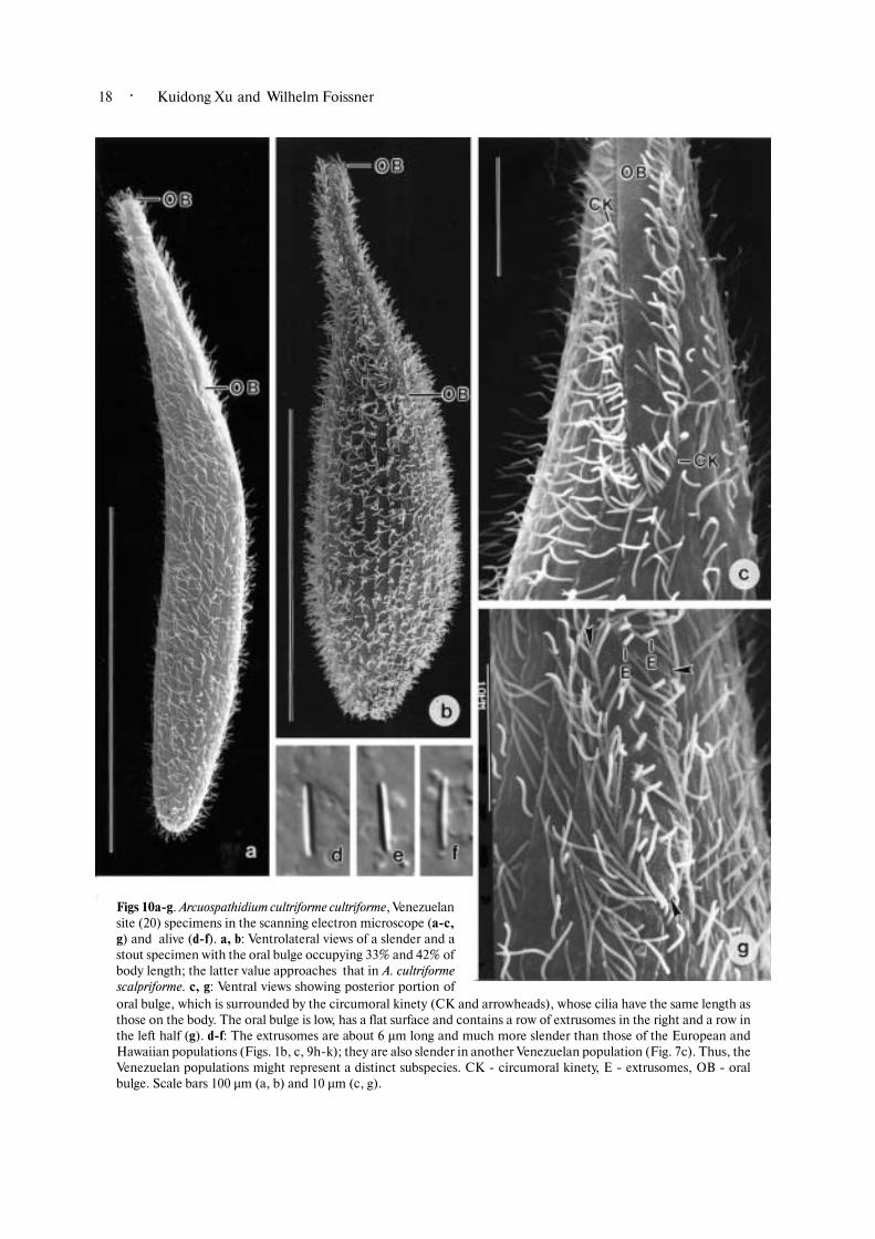
Figs 10a�g. Arcuospathidium cultriforme cultriforme, Venezuelansite (20) specimens in the scanning electron microscope (a�c,g) and alive (d�f). a, b: Ventrolateral views of a slender and astout specimen with the oral bulge occupying 33% and 42% ofbody length; the latter value approaches that in A. cultriformescalpriforme. c, g: Ventral views showing posterior portion oforal bulge, which is surrounded by the circumoral kinety (CK and arrowheads), whose cilia have the same length asthose on the body. The oral bulge is low, has a flat surface and contains a row of extrusomes in the right and a row inthe left half (g). d�f: The extrusomes are about 6 µm long and much more slender than those of the European andHawaiian populations (Figs. 1b, c, 9h�k); they are also slender in another Venezuelan population (Fig. 7c). Thus, theVenezuelan populations might represent a distinct subspecies. CK � circumoral kinety, E � extrusomes, OB � oralbulge. Scale bars 100 µm (a, b) and 10 µm (c, g).

· 19ProtistologyProtistologyProtistologyProtistologyProtistology
Figs 11a�e. Arcuospathidium cultriformecultriforme, Venezuelan site (20) speci�mens in the scanning electron micro�scope (a, b, d) and after protargol impreg�nation (c, e). a, b: Dorsolateral views ofanterior body portion. The bristles(double arrowheads) of the three�roweddorsal brush are distinctly shorter thanordinary somatic cilia (single arrow�heads). c: Left side view of the oral bulgeportion of the Burgenland population investigated by Foissner (1984). Arrowheads mark kinetofragments notmentioned by Foissner (Figs 3b, f). d: Dorsolateral view of anterior body portion. The posterior, clavate bristle of thedikinetids is slightly longer than the anterior (arrow pairs) in rows 1 and (probably) 2, while vice versa in row 3(arrowheads). e: When partially exploded, the extrusomes become knife�shaped (arrowheads). B (1�3) � dorsal brush(rows), CK � circumoral kinety, N � nematodesmata, OB � oral bulge. Scale bars 10 µm.

· Kuidong Xu and Wilhelm Foissner20
Figs 12a�f. Arcuospathidium cultriforme cultriforme, Venezuelan site (20) specimensin the scanning electron microscope (a�d) and alive (e, f). a�d: Dorsal views showingthe dorsal brush in overview (a) and details at higher magnifications (b�d). Thedorsal brush is three�rowed (numerals) and composed of dikinetids with up to 3µm long (in vivo), claviform bristles, whose length gradually decreases from anteriorto posterior end (b�d). The posterior bristle of the dikinetids is slightly longer thanthe anterior one (d, arrows) in rows 1 and 2, while vice versa in row 3 (d, arrowheads).The dikinetids are obliquely arranged in the anterior two thirds of the rows, producing a distinct zigzag pattern of thebristles (b, c). Brush rows 1 and 3 are shorter than row 2, but row 3 has a monokinetidal bristle tail marked by arrowheadsin (a, b). All brush kineties continue posteriorly as ordinary ciliary rows (a, b). e, f: Exploded toxicysts are 10�15 µmlong and knife�shaped, as mentioned and illustrated by Penard (Fig. 1f). Arrows mark a minute globule betweencapsule and shaft. Arrowheads denote empty toxicysts (with content extruded). B (1�3) � dorsal brush (rows). Scalebars 20 µm (a), 10 µm (b, e, f), and 4 µm (c, d).

· 21ProtistologyProtistologyProtistologyProtistologyProtistology
Figs 13a�d. Arcuospathidiumcultriforme cultriforme (a�c)from Venezuelan site (20) andSpathidium stammeri (d) in thescanning electron microscope.a�c: Early dividers showing thestrongly oblique division axis(arrowheads) and the develo�ping division blebs (D) andexcretory pores (EP). Asterisksin a mark the oral bulge. Notegrowing cilia in the formingoral kinetofragments (arrows).d: Division blebs in a early mid�divider of S. stammeri. The forming oral kinetofragments (KF) curve around the rightmargin of the division blebs (D) assuming a concave shape. D � division blebs, EP � excretory pores for the proter,KF � forming oral kinetofragments. Scale bars 50 µm (a), 20 µm (b), and 5 µm (c, d).

· Kuidong Xu and Wilhelm Foissner22
mosses from Mittenwald, a Bavarian village on thenorth border of Tyrol. However, the figure he providedseems to be that of the single specimen he found inZillertal moss (reproduced here as figures 16a�c).Because of this and also because Kahl did not fix a typelocality, we choose the Zillertal.
Synonymy: Foissner (2003b) explains his misiden�tification as follows: “The present study shows that myformer redescription of S. lionotiforme Kahl (1930a, b)is based on an unfortunate misidentification caused byinsufficient experience and the widespread opinion thatSpathidium is highly variable. The species I investigatedin 1984 obviously belongs to the Spathidium cultriformecomplex, specifically to S. scalpriforme Kahl, 1930a, aspecies which I never found in soil and moss (Foissner,1998), although it lives there (Kahl, 1930a, b), simplybecause I continuously misidentified it as S. lionotiforme”.
We adopt this explanation, and the senior authoremphasises that all his previous A. lionotiformeidentifications are wrong (for a review, see Foissner,1998) and belong to A. cultriforme scalpriforme. This isquite reasonable because the “true” Spathidiumlionotiforme has a rather different organisation makingmisidentification unlikely (for a review, see Foissner,2003b); further, S. lionotiforme must be a very rarespecies because it has not been mentioned in theliterature since the original description.
Morphology: The following description incorporatesthe data from Kahl (1930a, b) and Foissner (1984) aswell as new observations on populations from Brazil andthe Cape Peninsula, Republic of South Africa. Kahl(1930a, b) and Gellurt (1956) mention small varietieswhich, in our opinion, belong to other species. Kahl’s“local form” from roof moss in northern Germany is160�180 µm long and has only about half (15) thenumber of ciliary rows typical of this species (Fig. 2d).Gellurt’s variety from moss humus in Hungary is only90 µm long, performs a contractile vacuole cycle within35�40 sec, and feeds on minute zooflagellates.
Arcuospathidium cultriforme scalpriforme is verysimilar to A. cultriforme cultriforme, except for thecharacteristics mentioned in the diagnosis and someother minor features, which will be emphasised in thefollowing description. Some of these might be moreimportant than presently recognised.
Size and length:width ratio highly similar to A.cultriforme cultriforme, that is, about 230 + 40 !m invivo, matching data of Kahl, 1930a, b (250�330 !m,length:width ratio 5:1) and Wenzel, 1953 (length about260 !m); highly dependent on culture conditions, asshown by the South African population: on the average207 + 39 !m in protargol�impregnated specimens froma non�flooded Petri dish (raw) culture, while 314 + 66!m in specimens from a flourishing semipure culture.Number of ciliary rows, in contrast, increases only
slightly from 33 to 35 (Tables 2, 3).Body usually more distinctly knife�shaped than in
A. cultriforme cultriforme due to the longer oral area,widest at proximal end of oral bulge and in mid of trunk(Figs 16a�c, f, g, p�t, 17a, b, 18a, b, d, 22a; Table 2);often similar to certain Dileptus species (e.g. Dileptusconspicuus, redescribed in Foissner, 1989) or variouslarge, limnetic pleurostomatids, for instance, Amphileptuscarchesii and Litonotus varsaviensis (for a review, seeFoissner et al., 1995). Ratio of body and oral bulgelength 0.42 in Austrian and 0.45 in South Africanpopulations, and thus distinctly higher than in the fivepopulations of A. cultriforme cultriforme (0.31�0.35;Tables 1, 2).
Nuclear apparatus as in A. cultriforme cultriforme,but specimens/populations with numerous macronuclearnodules were not observed (Figs 16a, c, f, 17b, d, 18d,22a). Macronucleus strand “broken” into two piecesdividing individually in about 5% of specimens (Figs27a�g). Contractile vacuole and cortex also as in A.cultriforme cultriforme. Cortical granules very distinctand in about 8 rows between each two kineties in SouthAfrican specimens (Figs 20a, b).
Extrusome shape and size very similar in thepopulations investigated and thus rather dissimilar tothose of A. cultriforme cultriforme, where they are shorterand thus comparatively thicker, as already mentionedby Kahl (1930a); data of Foissner (1984) do not meetthe modern standard, but basically match the newobservations. Extrusomes conspicuous both in vivo andat protargol preparations, though small as comparedto body size, because they form a bright stripe in theright and the left half of oral bulge, especially in broaderdorsal half (Figs 16a, c, f�h, o, 18c�f, 20a�e). Size about7 !m in specimens studied by Kahl (1930a), 5 !m inAustrian cells (Foissner, 1984), and 6 + 0.8 !m inBrazilian and 7�8 + 0.7�0.9 !m in South Africanspecimens. Shape rather similar in Brazilian and SouthAfrican specimens, that is, indistinctly acicular to verynarrowly ellipsoidal and somewhat curved and asym�metric (Figs 16v, w). Fully exploded extrusomes 15�20+ 1 !m in size and of typical toxicyst structure (Fig.16u); partially exploded toxicysts less distinctly knife�shaped than in A. cultriforme cultriforme, usually moreor less distinctly clavate (Figs 16h, 18f). Developingextrusomes in cytoplasm as shown in figures 16i, k.
Cytoplasm and movement as described in A.cultriforme cultriforme. Feeds also on rotifers andmiddle�sized hypotrichs, such as Gastrostyla steinii,Gonostomum affine and a large Urosoma species.Appears elegant and conspicuous when gliding onmicroscope slide due to the nicely curved dorsal outlineand the large size.
Somatic ciliature highly similar to that of A.cultriforme cultriforme, except dorsal brush (Figs 4b,

· 23ProtistologyProtistologyProtistologyProtistologyProtistology
Figs 14a�f. Arcuospathidium cultriforme cultriforme, Venezuelan site (20) specimens inthe scanning electron microscope. a�c, e, f: Dorsal overview and details of an earlymid�divider showing the strongly oblique division axis (a, b; arrowheads), the divisionblebs (D), and the growing dorsal brush. There are three dorsal brush rows (B1�3) withzigzagging (= obliquely arranged dikinetids) bristles in the anterior portion, a uniquefeature of the Venezuelan specimens, suggesting, together with the presence of thinextrusomes (Figs 10 d�f), that it is a distinct subspecies. Some ordinary cilia (c; arrows)are at the anterior end of the brush rows. Figure f shows the formation of the brushdikinetids (asterisk): a new bristle is formed anteriorly of the parental somatic cilia,
which become gradually shortened to brush bristles (triangle series). The division blebs (D) are now very conspicuous(b, e), and the cilia of the new oral kinetofragments have reached full length (b, c, e; KF). d: Dorsal view of amorphostatic specimen showing the monokinetidal bristle tail of brush row 3 extending to mid�body (arrowhead);then, the row continues as ordinary somatic kinety to rear body end. The length of the tail of brush row 3 is animportant feature for distinguishing Spathidium s. l. species. B (1�3) � dorsal brush (rows), D � division blebs, KF �kinetofragments. Scale bars 50 µm (a, d), 20 µm (b), and 4 µm (c, e, f).

· Kuidong Xu and Wilhelm Foissner24
Figs 15a�i. Arcuospathidium cultriforme cultriforme, resting cysts (diameter 42�95 µm) of specimens from Linz, Austria(a�f, i) and the Dominican Republic (g, h). a: Optical section of two cysts showing the extremely thick wall delimitedby arrowheads, scale bar 40 µm. b, e: Optical section and surface view of two cysts showing the thick wall and thepolygonally faceted surface, scale bar 40 µm. c, f: Optical section and surface view of same specimen, showing thebroad ridges (f) causing the spotted appearance of the cyst content in optical section (c), scale bar 40 µm. d: Cystcontents mainly consisting of highly refractive lipid droplets 1�5 µm across, scale bar 10 µm. g, h: Optical section andsurface view of a cyst from the Dominican specimen, scale bar 20 µm. Obviously, it is very similar to the Austrianspecimens, except for the thinner ridges (arrowheads) and the smaller diameter (45.2 vs. 68.8 µm on average). i:Surface view of a cyst with facets distinctly larger than those recognisable in (e, f), scale bar 40 µm.
· 25ProtistologyProtistologyProtistologyProtistologyProtistology
16a, f, l, m, 17a�d, 18a, b, d, f; Tables 1, 2). Cilia 8�10!m long, more loosely spaced (~ 3 !m) in oral thanpostoral area (~ 1.5 !m), arranged in about 30 rowsaccording to Kahl (1930a, b), matching averages (32�33) of Austrian and South African populations (Tables1, 2). Row number conspicuously constant, that is,33 rows in South African specimens from the non�flooded Petri dish (raw) culture and 35 in the 100 !mlonger specimens from the semipure culture (Table 2).At right anterior end of dorsal brush of most Austrianspecimens an accumulation of scattered basal bodies(Fig. 16m), likely adesmokineties as in the SouthAfrican specimens (Fig. 4b). Dorsal brush basicallyas in A. cultriforme cultriforme, that is, almost hete�rostichad (row 3 shorter than row 2 by about 20%)occupying 31�35% of body length; rarely occurspecimens with one or two more or less distorted bristlerows right of row 1; tail of row 3 extends into secondbody third (Figs 4b, 16a, f, m, 17c, d, f, 18d, f, i, 19d;Table 2). Details of bristle pairs studied only in SouthAfrican specimens, likely as shown in figure 16n, thatis, slightly clavate anterior bristle longer (3�5 !m) thanposterior (2�3 !m) in rows 1 and 2, while vice versa inrow 3 (Figs 16n, 19a�f).
Oral bulge conspicuous, though only about 3 !mhigh near dorsal end, because almost half as long as body(42�45%; Table 2), strongly slanted (50°�70°, on theaverage 63°, n 10), rather distinctly projecting from bodyproper at proximal end (Figs 16p�s, 17a), and studdedwith refractive extrusomes (Figs 20a, b); in lateral viewslightly to distinctly convex, in frontal view about 5 !mwide and very narrowly cuneate to oblong with proximalend bluntly pointed. Circumoral kinety and oral basketas described in A. cultriforme cultriforme (Figs 16a, c, f,o�t, 17a, 18a, b, d�h, 20a�e, 22a�c, 27b; Table 2).
Encystment (Figs 21a�g): The protargol preparationsfrom the non�flooded Petri dish culture and thesemipure culture contain encysting and encystedspecimens, but only those from the former impregnatedwell, and were used for reconstructing the encystmentprocess. However, few encysting specimens were found,and the description is not very detailed, though definitelymore complete than the data available in the literature.
When encystment commences, cells becomesmaller and stouter (Figs 21a, b). This diminution isassociated with the gradual resorption of somatic andoral kinetids, especially those of the dorsal brush andthe circumoral kinety, both having reduced about halfof their length and kinetids. The macronucleus appearsmore tortuous, but is basically unchanged, while themicronuclei move apart from the macronucleus anddistribute throughout the body, as in early dividers. Thecontractile vacuole is still recognisable.
In the next stage, cells become almost globular andthe number of somatic and oral kinetids is greatly
reduced (Fig. 21c). The remaining, still ciliated kinetidsare somewhat irregularly arranged within the rowswhich loose the Arcuospathidium pattern, and becomemore and more meridionally arranged. The dorsal brushkinetids still have bristles, but loose the dikinetidalappearance. The postciliary microtubule ribbons arevery distinct at this stage. Then a really remarkableprocess commences, a strong shortening of themacronucleus, which is eventually reduced by about80% of its length and becomes semicircular in theencysted specimens (Fig. 21g). This length reductionis not achieved by thickening of the strand or chromatinextrusion, but by resorption of nuclear mass. It is evidentfrom simple volume calculations: the nuclear volumedecreases from about 5,000 µm3 to 1,000 µm3 in themature cyst. The micronuclei are still scattered, andthe contractile vacuole is still recognisable.
The next stage is characterised by the appearanceof the cyst wall, which is about 1 µm thick and does notimpregnate with the protargol method used (Fig. 21e).The specimens are now globular and loose the oralapparatus, while some somatic basal bodies are stillrecognisable, becoming more and more scattered. Theshortening of the macronucleus is now distinct, and themicronuclei arrange around the macronucleus strand.The contractile vacuole is still recognisable.
During the last stages, micronuclei resorptioncommences and the faceted ectocyst develops, asdescribed in A. cultriforme cultriforme. The infraciliatureprobably disappears completely, though this is difficultto prove because the maturing cysts are rather poorlyimpregnated (Fig. 21f). The mature cyst wall, althoughappearing quite stable in vivo, is usually distorted andthe cell is distinctly shrunken at the preparations,leaving a more or less wide space between wall andcortex. The macronucleus is shortened to a semicircularstrand, and about half of the micronuclei have beenresorbed. The nucleoli are very small. The contractilevacuole disappears (Fig. 21g).
Occurrence and ecology: Kahl (1930a) discoveredA. cultriforme scalpriforme in mosses from Mittenwald(a village in southern Bavaria near the Tyrolean border)and the Zillertal in Austria (a valley in the Tyrol Alps).The “local form” from roof moss in northern Germanyappears to belong to another species (see discussion ofsynonymy). The same is supposed for Gellurt’s (1956)variety, which is only 90 µm long. The next reliablerecord is from the surroundings of Erlangen in Bavaria,where Wenzel (1953) observed A. cultriforme scalpriformein only two of over 100 moss samples, that is, in drymoss at pH 5.1 and leaf litter. In 1984, Foissnerredescribed the species from soil of a deciduous forestin Burgenland, Austria. More recently, some unsub�stantiated records were added from Bulgaria (Detcheva,1972b) and from Germany (Foissner, 2000b). Note also

· Kuidong Xu and Wilhelm Foissner26
Table 1. Morphometric data on Arcuospathidium cultriforme cultriforme populations (Pop) from Hawaii (HA50, original; HA12, original), Austria (AUL from Linz, original; AUB from Burgenland, according to
Foissner, 1984 and his original notes), and Rwanda (RW, original).
Characteristics1 Pop × M SD SE CV Min Max n
HA50 222.5 220.0 28.0 8.4 12.6 193.0 280.0 11 HA12 270.3 270.0 30.9 9.3 11.4 233.0 348.0 11 AUL 275.7 282.0 43.5 8.7 15.8 145.0 382.0 25 AUB 191.6 180.0 33.7 8.7 17.6 145.0 245.0 15
Body, length
RW 305.8 300.0 48.5 16.2 15.9 250.0 400.0 9 HA50 36.3 35.0 8.4 2.5 23.2 25.0 56.0 11 HA12 51.5 53.0 14.5 4.4 28.2 34.0 76.0 11 AUL 50.3 49.0 12.0 2.4 23.8 26.0 85.0 25 AUB 27.5 28.0 4.5 1.2 16.6 20.0 34.0 15
Body, width
RW 44.9 47.0 11.0 3.7 24.6 27.0 62.0 9 HA50 6.4 6.4 1.5 0.4 23.2 4.4 9.2 11 HA12 5.7 5.1 2.1 0.6 36.7 3.6 10.2 11 AUL 5.8 5.4 2.1 0.4 35.8 3.2 11.9 25 AUB 7.2 6.6 1.9 0.5 26.7 4.5 11.4 15
Body length:width, ratio
RW 7.1 6.5 1.5 0.5 21.3 5.2 9.7 9 HA50 78.7 75.0 14.6 4.4 18.6 58.0 105.0 11 HA12 84.6 80.0 10.3 3.1 12.2 70.0 107.0 11 AUL 93.9 95.0 17.4 3.5 18.5 56.0 138.0 25 AUB 58.1 60.0 8.3 2.1 14.3 37.0 70.0 15
Oral bulge, length (cord)
RW 102.8 100.0 26.6 8.9 25.9 65.0 145.0 9 HA50 4.0 4.0 0.8 0.4 19.8 3.0 5.0 5 Oral bulge (circumoral kinety), width AUL 3.9 4.0 0.2 0.1 5.7 3.5 4.0 5 HA50 2.4 2.5 0.4 0.1 15.6 2.0 3.0 11 Oral bulge, height AUL 2.3 2.5 0.3 0.1 11.5 2.0 2.5 11 HA50 0.35 0.35 � � 10.4 0.29 0.43 11 HA12 0.32 0.3 � � 13.9 0.26 0.38 11 AUL 0.34 0.34 � � 11.3 0.28 0.43 25 AUB 0.31 0.3 � � 15.7 0.22 0.39 15
Oral bulge length:body length, ratio
RW 0.34 0.31 0.1 � 19.3 0.25 0.45 9 HA50 52.2 52.0 6.5 2.0 12.4 44.0 62.0 11 Circumoral kinety to last dikinetid of brush row
1, distance RW 66.3 67.0 20.3 6.8 30.6 45.0 100.0 9 HA50 58.6 58.0 5.4 1.6 9.3 50.0 67.0 11 AUL 68.5 68.0 9.2 2.6 13.4 51.0 86.0 13 AUB 50.9 49.0 7.9 2.0 15.6 39.0 70.0 15
Circumoral kinety to last dikinetid of (the longest) brush row 2, distance
RW 71.4 73.0 20.6 6.9 28.8 45.0 100.0 9 HA50 47.7 49.0 4.2 1.3 8.8 40.0 54.0 11 Circumoral kinety to last dikinetid of brush row
3, distance RW 51.8 52.0 13.4 4.5 25.9 35.0 70.0 9 Anterior body end to macronucleus, distance HA50 72.8 72.0 10.1 3.0 13.9 58.0 90.0 11
HA50 113.9 110.0 28.8 8.7 25.3 60.0 155.0 11 AUL 158.7 173.0 39.9 9.1 25.1 47.0 220.0 19
Macronucleus figure, length
AUB 100.6 94.0 27.5 7.1 27.3 69.0 160.0 15 Macronucleus nodules, length RW 10.0 9.0 2.7 0.9 26.9 7.0 14.0 9
HA50 5.6 5.0 1.8 0.5 32.0 4.0 9.0 11 Macronucleus, width in mid AUB 4.9 5.0 0.6 0.2 12.3 4.2 5.6 15
Macronucleus nodules, width RW 4.2 4.0 0.9 0.3 22.2 3.0 6.0 9 HA50 see footnote2 HA12 1.0 1.0 0.0 0.0 0.0 1.0 1.0 11 AUL 1.0 1.0 0.0 0.0 0.0 1.0 1.0 25
Macronucleus, number
AUB 1.0 1.0 0.0 0.0 0.0 1.0 1.0 15 Macronucleus nodules, number RW3 96.0 100.0 15.2 6.8 15.8 70.0 110.0 5
HA50 2.6 2.5 0.2 0.1 6.9 2.5 3.0 8 AUL 2.8 3.0 0.4 0.1 15.0 2.0 3.5 19 AUB 2.7 2.7 0.2 0.1 5.6 2.5 2.8 4
Micronuclei, across
RW 2.9 3.0 0.5 0.2 18.9 2.0 4.0 9 HA50 15.0 13.0 � � � 12.0 20.0 3 Micronuclei, number AUL 13.3 12.0 3.3 0.7 24.5 9.0 23.0 19 HA50 31.4 31.0 1.6 0.5 5.0 30.0 35.0 11 AUL 36.7 36.0 3.6 0.8 9.8 30.0 44.0 23 AUB 28.1 29.0 1.3 0.3 4.6 26.0 30.0 15
Somatic kineties, number
RW 28.4 28.0 2.2 0.7 7.7 25.0 31.0 9

· 27ProtistologyProtistologyProtistologyProtistologyProtistology
the records from South Africa and Brazil described inthe Materials and Methods section.
All other records are from limnetic habitats, viz.the mud and Aufwuchs of various polluted rivers inBulgaria (Detcheva, 1972a, c, 1975, 1979a, b, 1986,1993; Russev et al., 1976, 1994) and some naturalfreshwaters and a sewage plant in China (Wang, 1977;Ning et al., 1993; Wang and Ma, 1994). We considerall these unsubstantiated reports as highly questionablebecause the senior author never found this species inover 1,000 river samples investigated over the years. Theabove mentioned authors confused A. cultriformescalpriforme with other ciliates, especially pleuro�stomatids, some of which have a similar size and shape,as mentioned in the descriptive section. Therefore, wedo not document the many abiotic environmental dataprovided by Detcheva and her students. On the otherhand, our South African habitat, the soil from themargin of a flat pond, indicates that this subspecies mayoccur as a guest in the true limnetic habitats.
Reliable records of A. cultriforme scalpriforme areavailable from Germany, Austria, Brazil, and theRepublic of South Africa. Foissner (1998) also lists arecord from Australia. Basically, the reliable recordsindicate that A. cultriforme scalpriforme is a terrestrialcosmopolitan rarer than A. cultriforme cultriforme.
Ontogenesis: Although ontogenesis is a continuousprocess, we distinguish seven stages, each characterisedby specific events, for the sake of clarity. Most stagesdepicted were seen in at least two, usually three or four
specimens. This is also evident from the second seriesshowing division in specimens with two macronucleuspieces (Figs 27a�g). We could not follow the origin ofthe oral basket because the nematodesmata impreg�nated too faintly. The parental oral apparatus and dorsalbrush do not show any changes.
Ordinary specimens (Figs 13, 14, 22�26; Table 3).Stage 1 (very early dividers commencing production
of oral kinetofragments and dorsal brush row 2; Figs22a�c, 23a, b, d, e; Table 3). When ontogenesis begins,the cells are on the average slightly larger thanmorphostatic specimens (Table 3). Basal bodies areproduced distinctly underneath mid�body, as evidentfrom middle and late dividers which show that the proteris considerably longer than the opisthe (Table 3). Thedivision plane is not transverse but slightly oblique fromdorsal to ventral. Soon, some of the new basal bodiesform minute kinetofragments, each probably consistingof monokinetids, except of a dikinetid at anterior end.There is also a basal body proliferation in the opisthe’sciliary rows, as recognisable by the narrow and unevenspacing of the kinetids.
When entering stage 2, a remarkable change occursin body shape � a slight indentation develops in theprospective fission area (Figs 22b, c), as observed alsoin Protospathidium serpens and Arcuospathidiumcoemeterii in which, however, the indentation developsslightly later and is transient. Basal body proliferationcontinues in all opisthe ciliary rows; the oral kinetofrag�ments become dikinetidal and associated with minute
Table 1. (Continuation).
Characteristics1 Pop × M SD SE CV Min Max n
HA50 116.8 113.0 14.5 4.4 12.4 100.0 147.0 11 Basal bodies in a right side kinety, number RW 161.0 165.0 32.5 14.5 20.2 110.0 200.0 5 HA50 3.0 3.0 0.0 0.0 0.0 3.0 3.0 11 HA12 3.0 3.0 0.0 0.0 0.0 3.0 3.0 11 AUL 3.0 3.0 0.0 0.0 0.0 3.0 3.0 25 AUB 3.0 3.0 0.0 0.0 0.0 3.0 3.0 15
Dorsal brush rows, number
RW 3.0 3.0 0.0 0.0 0.0 3.0 3.0 9 HA50 48.7 46.0 11.4 3.4 23.4 35.0 76.0 11 Dikinetids in brush row 1, number RW 64.6 61.0 15.7 5.9 24.3 44.0 91.0 7 HA50 55.3 54.0 8.8 2.6 15.9 48.0 78.0 11 AUL 52.4 50.0 7.8 3.0 15.0 47.0 70.0 7
Dikinetids in brush row 2, number
RW 59.6 58.0 9.9 3.7 16.6 47.0 72.0 7 HA50 40.2 39.0 7.8 2.3 19.3 28.0 51.0 11 Dikinetids in brush row 3, number RW 35.6 38.0 6.1 2.3 17.2 24.0 41.0 7
1 Data based on mounted, protargol�impregnated (Foissner's method), and randomly selectedspecimens from non�flooded Petri dish cultures. Measurements in µm. CV � coefficient of variationin %, M � median, Max � maximum, Min � minimum, n � number of individuals investigated, SD �standard deviation, SE � standard error of arithmetic mean, ` � arithmetic mean.2 Four types are recognizable: Of 75 specimens analysed, 58% have the usual macronucleus pattern,while 19% have many (up to 100) scattered nodules, and 23% have a mixture of nodules and one toseveral short strands.3 See text for explanations.

· Kuidong Xu and Wilhelm Foissner28
Figs 16a�w. Arcuospathidium cultriforme scalpriforme, alive (a�g, j, n�w) and after protargol impregnation (h, i, k�m),according to Kahl, 1930a (a, b); Kahl, 1930b (c, d); Foissner, 1984 (e�m); and new observations of populations fromSouth Africa (n�v) and Brazil (w). a�c: Left side and transverse (b) view of a specimen probably from the Zillertal inAustria, length 260 µm. d: Left side view of another species from northern Germany, length 160 µm. e: Restingextrusome, length 6 µm. f, g: Right side and ventral view of a specimen from Austria, length 270 µm. h: Part of oralbulge of specimen shown in figure 17a. i, k: Cytoplasmic extrusomes, capsule 4 µm. j: Cortical granulation. l, m:Ventrolateral and dorsolateral view of oral area. Arrow marks an accumulation of basal bodies. Arrowheads denoteproximal end of brush rows. Scale bar 50 µm. n: Dorsal brush, bristles (up to 5 µm) and somatic cilia (10 µm) drawnto scale. o: Proximal portion of oral bulge (width 5 µm), showing the scattered extrusomes and the cortical granules,which form an arrowhead�like pattern. p�t: Variability of body shape and length of oral bulge (37�51% of body length)of specimens redrawn from video records. u: Exploded toxicysts, length 15�20 µm. Arrowhead marks refractive granule.v, w: Resting oral bulge extrusomes from populations of South Africa (v) and Brazil (w), length 8 µm and 6 µm. B (1�3) � dorsal brush (rows), C � somatic cilia, CK � circumoral kinety, CV � contractile vacuole, E � extrusomes, G �cortical granules, MA � macronucleus, OB � oral bulge.

· 29ProtistologyProtistologyProtistologyProtistologyProtistology
Figs 17a�f. Arcuospathidium cultriforme scalpriforme, protargol�impregnated ciliary pattern and macronucleus of theAustrian population studied by Foissner (1984). a, d, e: Right and left side view, length 295 µm. The dorsal brush isexactly on the dorsal side of the cell. Therefore, the specimen cannot be Spathidium lionotiforme, as proposed byFoissner (1984), which has the dorsal brush on the left side of the oral area (Kahl, 1930b; Foissner, 2003b). The oralbulge and the cytoplasm are studded with extrusomes shown at higher magnification in figures 16 h, k. b, c, f: Ventraland dorsal view, length 240 µm. Note the long, slightly cuneate oral bulge and the dorsal brush, which is composed ofthree rows of dikinetids with short, bristle�like cilia; arrowheads denote proximal end of brush rows. The brush isisostichad (all rows of similar length) and almost as long as the oral bulge. B (1�3) � dorsal brush (rows), BA � oralbasket made up of nematodesmata originating from the circumoral dikinetids, CK � circumoral kinety, E � extrusomes,EP � excretory pores of the contractile vacuole, MA � macronucleus, OB � oral bulge.

· Kuidong Xu and Wilhelm Foissner30
blebs described below; and some somatic dikinetidsappear to form a short dorsal brush row 2 (Figs 23b, e).The macronucleus appears smoothened and themicronuclei are distributed throughout the cell.
Stage 2 (early dividers separating kinetofragmentsfrom proter kineties and producing brush rows 3 and 1and the excretory pores; Figs 13a�d, 14a�c, 22d, 23c, f,24a, b; Table 3). Early dividers are distinctly longerand narrower than morphostatic specimens (Table 3).Body indentation and division blebs become moredistinct in the prospective fission area (Figs 22d, 23c).
The division blebs are about 5 µm�sized convexities leftand slightly above of the kinetofragments (Figs 13b�d).They disappear in late dividers. The kinetofragment beltis now clearly recognisable and distinctly oblique (30°�45° in three appropriate specimens), which is likely tobe due to faster growth of the dorsal than the ventralside (Figs 13a, 14a, 22d). The newly formed oralkinetofragments, which are longer on the ventral thanthe dorsal side and detach from the proter’s ciliary rowsin a dorsoventral gradient, commence to curverightwards on the dorsal surface and later also on the
Table 2. Morphometric data on Arcuospathidium cultriforme scalpriforme populations (Pop) from South Africa (SA, original) and Austria (AUS, from Foissner, 1984 and his original notes).
Characteristics1 Pop × M SD SE CV Min Max n
SA 206.8 200.0 25.6 5.6 12.4 167.0 275.0 21 Body, length AUS 251.3 260.0 30.8 8.9 12.3 205.0 290.0 12 SA 39.3 42.0 7.7 1.7 19.5 25.0 51.0 21 Body, width AUS 35.3 34.5 3.9 1.1 11.0 30.0 43.0 12 SA 5.4 5.3 1.0 0.2 18.0 4.0 7.8 21 Body length:width, ratio AUS 7.2 7.0 1.2 0.4 16.9 5.2 9.7 12 SA 92.1 94.0 12.8 2.8 13.9 61.0 113.0 21 Oral bulge, length (cord) AUS 105.8 106.0 7.8 2.2 7.4 92.0 120.0 12
Oral bulge, height SA 2.6 2.5 0.3 0.1 11.1 2.0 3.0 21 SA 0.45 0.46 0.1 � 11.8 0.35 0.52 21 Oral bulge length:body length, ratio AUS 0.42 0.43 � � 8.7 0.37 0.49 12
Circumoral kinety to last dikinetid of brush row 1, distance
SA 53.6 53.0 7.9 1.7 14.7 41.0 65.0 21
SA 64.0 63.0 8.1 1.8 12.7 50.0 80.0 21 Circumoral kinety to last dikinetid of brush row 2, distance AUS 88.4 88.0 8.3 2.4 9.4 70.0 100.0 12
Circumoral kinety to last dikinetid of brush row 3, distance
SA 51.6 49.0 9.1 2.0 17.7 35.0 72.0 21
Anterior body end to macronucleus, distance SA 78.8 78.0 16.8 3.7 21.3 47.0 113.0 21 SA 87.5 90.0 21.5 4.7 24.6 46.0 134.0 21 Macronucleus figure, length AUS 106.3 108.0 25.9 7.5 24.4 42.0 142.0 12
Macronucleus, length (spread, thus approximate)
SA 187.4 200.0 � � � 130.0 230.0 21
Macronucleus, width in central third SA 6.1 6.0 1.4 0.3 22.6 4.0 9.0 21 AUS 7.2 7.0 0.8 0.2 10.7 6.0 8.5 12
SA 1.0 1.0 0.0 0.0 0.0 1.0 1.0 21 Macronucleus, number AUS 1.0 1.0 0.0 0.0 0.0 1.0 1.0 12 SA 3.0 3.0 0.3 0.1 10.6 2.5 4.0 21 Micronuclei, across AUS 2.9 2.8 0.1 0.0 4.0 2.8 3.1 12
Micronuclei, number SA 11.5 12.0 2.6 0.6 22.2 7.0 18.0 21 SA 32.7 32.0 3.0 0.7 9.1 27.0 40.0 21 Somatic kineties, number2 AUS 32.5 32.0 1.2 0.4 3.8 31.0 35.0 12
Basal bodies in a right side kinety, number SA 124.4 130.0 25.0 5.5 20.1 85.0 180.0 21 Basal bodies in each somatic kineties of 10 µm
long, number AUS 6.1 6.0 0.9 0.3 14.8 5.0 8.0 12
SA 3.0 3.0 0.0 0.0 0.0 3.0 3.0 21 Dorsal brush rows, number AUS 3.0 3.0 0.0 0.0 0.0 3.0 3.0 12
Dikinetids in brush row 1, number SA 40.6 39.0 7.3 1.6 18.0 30.0 56.0 21 Dikinetids in brush row 2, number SA 55.0 55.0 7.0 1.5 12.8 40.0 67.0 21 Dikinetids in brush row 3, number SA 34.9 35.0 6.1 1.3 17.4 23.0 48.0 21
1 Data based on mounted, protargol�impregnated (Foissner's method), and randomly selectedspecimens from non�flooded Petri dish cultures. Measurements in µm. CV � coefficient of variationin %, M � median, Max � maximum, Min � minimum, n � number of individuals investigated, SD �standard deviation, SE � standard error of arithmetic mean, ` � arithmetic mean.2 Row number in specimens from semipure culture, which are much larger (Table 3): ̀35.0, SD 3.6,CV 10.3, Min 30, Max 41, n 21.

· 31ProtistologyProtistologyProtistologyProtistologyProtistology
ventral one (Figs 13b, 22d, 23c, f). Slightly later, whenthree brush rows are recognisable, the fragments arearranged transversely and become concave on the leftside, while the right side fragments remain almoststraight, but become more distinctly slanted (Figs 24a,b). At this stage, the newly formed kinetofragmentsconsist of six to eleven, on the average eight dikinetidseach, that is, they are almost complete; only onedikinetid is added in middle and late dividers (Table 3).Taking the averages of the fragment dikinetids (9) andciliary rows (35), 315 dikinetids are available for thenew circumoral kinety, which is close to the numberfound in the proter of the dividers (Tables 2, 3). Thus,there is no second round of dikinetid proliferation, noteven during post�divisional growth.
Many new somatic basal bodies have been producedin two regions of the ciliary rows, the posterior area ofthe proter and the anterior area of the opisthe. However,some basal body production is likely to occur throughoutthe division process and even in morphostatic specimens,as suggested by barren kinetids found throughout theciliary rows of morphostatic cells. Dorsal brush row 2is still growing and has now about one third of thedikinetids found in morphostatic specimens (Table 3).Next, brush row 3 is generated, and finally row 1. Thiscurious sequence was observed in several specimens. Allbrush rows have an anterior tail of ordinary mono�kinetids (Figs 14a�c). Now, the nuclear apparatus showssome changes (Fig. 22d): the macronucleus becomesless tortuous, that is, commences to contract, and themicronuclei appear more or less distinctly inflated.Further, a new contractile vacuole and new excretorypores are generated at the proter’s posterior end.Usually, the new pores are located dorsally or dorso�laterally, but in some specimens they are near the ventralside.
Stage 3 (early mid�dividers with condensingmacronucleus and dividing micronuclei; Figs 14a�f,22e, f; Table 3). Early mid�dividers are smaller andstouter than early dividers, but larger than morphostaticcells (Table 3). The indentation underneath theprospective fission area is still recognisable, but becomesindistinct, especially in cells prepared for scanningelectron microscopy (Figs 14a, 22e, f). The macro�nucleus commences to shorten towards the centre, asindicated by the swollen ends; the nucleoli begin todisappear. The micronuclei grow up to 6 µm, that is,double size. In the post�brush area, one can observethe origin of dikinetids within the parental ciliary rows:a bristle is generated ahead of each parental ciliumwhich gradually decreases in length eventuallybecoming a bristle (Fig. 14f).
Stage 4 (mid�dividers with transversely oriented,concave kinetofragments and condensed, globularmacronucleus; Figs 14a�f, 22g, h, 24c, d; Table 3).
These cells are the smallest and stoutest dividers,approaching the length of morphostatic specimens(Table 3). The subequatorial indentation is indistinct,while the division blebs are still distinct (Figs 14a, b, e,22g, h). All oral kinetofragments are now transverselyarranged and more or less distinctly concave. The newbrush rows have about two thirds of their dikinetids(Table 3). No further increase of brush dikinetids occursin late dividers and early post�dividers, suggesting thatthe final number is obtained only in late post�dividersor morphostatic cells. The macronucleus condenses toa globular, fibrous mass with lightly impregnatedkaryoplasm. The dividing micronuclei move apart, stillbeing connected by a bundle of fibres.
Stage 5 (late dividers with forming division furrowand extending macronucleus; Figs 22i, j; Table 3). Latedividers grow in length, the division blebs disappear,and the division furrow becomes recognisable, dividingthe cell into an ellipsoidal opisthe shorter by about onethird than the conspicuously conical proter (Table 3).The body constriction causes the onset of kinetofrag�ment alignment, that is, the distances between theindividual kinetofragments become smaller. Theglobular macronuclear mass of the mid�dividers extendsand grows to a more or less curved rod with fibrouscontents. Most micronuclei have completed fission andcommence to condense.
Stage 6 (very late dividers with aligning kinetofrag�ments, forming oral bulge, and separating daughtercells; Figs 22k, l, 25a, b, 26a; Table 3). Very late dividersmay grow to a length of 565 µm and are distinctlyfurrowed. The developing opisthe oral area is conspicu�ously clavate and the oral bulge, which develops at itsmargin, becomes recognisable as a bare protuberance.The oral kinetofragments align to the new opisthecircumoral kinety. This process is obviously complexbecause many fragments overlap more or less and somebecome strongly curved, as in A. muscorum (Berger etal., 1983). Now, the ciliary rows are distinctly separatefrom the kinetofragments but still straight, showing thatthe genus�specific pattern is obtained only in post�dividers. The macronucleus becomes a long, tortuousstrand and shows fibrous contents with many minuteglobules, probably developing nucleoli (Fig. 22l).Finally, the daughters separate. The anterior daughtercell is longer than the posterior one by about one third;the latter is more or less distinctly pointed in the splitarea. The micronuclei are still scattered through the cell.
Stage 7 (early opisthe post�dividers with small size,pointed anterior macronucleus end, and short dorsalbrush and circumoral kinety/oral bulge with more orless conspicuous irregularities; Figs 26b�g). Very earlyopisthe post�dividers are bursiform and still have arather distorted circumoral kinety and almost meridio�nally arranged ciliary rows (Figs 26d, e). The oral

· Kuidong Xu and Wilhelm Foissner32
Figs 18a�i. Arcuospathidium cultriforme scalpriforme, South African (a, b, e�h) and Austrian (c, d, i; from Foissner,1984) specimens in the scanning electron microscope (a, b, e, g, h) and after protargol impregnation (c, d, f, i). a, b:Ventrolateral overviews showing the oral bulge (ends marked by arrowheads) extending to almost mid�body. c: Leftside oral area showing the comparatively thin, resting extrusomes and the circumoral kinety composed of very narrowlyspaced dikinetids. d: Left side overview showing the dorsally curved ciliary rows in the oral area. e, g, h: Ventral viewsshowing the bluntly pointed proximal end of the oral bulge and circumoral kinety (arrowheads). Note the scatteredextrusomes in right and left bulge half. f: Left side view of oral area showing the dorsal brush and the partially extruded,thin extrusomes. i: Dorsolateral view showing the long dorsal brush. B (1�3) � dorsal brush (rows), CK � circumoralkinety, CV � contractile vacuole, E � extrusomes, K � somatic kineties, MA � macronucleus, N � nematodesmata, OB� oral bulge. Scale bars 100 µm (b, d), 50 µm (a, f, h, i), 20 µm (c), and 5 µm (g).

· 33ProtistologyProtistologyProtistologyProtistologyProtistology
Figs 19a�f. Arcuospathidium cultriforme scalpriforme, dorsal brush area of South African specimens in the SEM. a:Bluntly pointed anterior end, where (brush) ciliary rows curve ventrally. The brush rows have some ordinary cilia atanterior end (arrows). Arrowheads mark short kinetofragments (adesmokineties?) left of circumoral kinety. The brushbristles are three times shorter than the somatic cilia, that is, about 3 µm long in vivo. b: Posterior half of brush. Therows end at similar level, but row 3 has a monokinetidal bristle tail (arrow) extending into second third of body.Arrowheads mark heteromorphic tail of brush rows 1 and 2, where minute bristles irregularly alternate with ordinarycilia. Asterisks denote row 1 dikinetids with a very short anterior bristle and an ordinary posterior cilium. c, f: Mid�portion and end of dorsal brush showing that, in row 3, the anterior bristle of the dikinetids is shorter than the posteriorone (arrows). Arrowheads mark strongly shortened bristles. d: Oral portion showing that brush bristle length decreasesat both ends of rows. e: Posterior third of brush showing that, in row 1, the posterior bristle of the dikinetids is shorterthat the anterior (arrows). B (1�3) � dorsal brush (rows), C � somatic cilia, CK � circumoral kinety, T � tail of brushrow 3. Scale bars 40 µm (d), 20 µm (b), 10 µm (a, e), and 4 µm (c, f).

· Kuidong Xu and Wilhelm Foissner34
Figs 20a�e. Arcuospathidium cultriforme scalpriforme, South African specimens alive (a, b) and in the scanning electronmicroscope (c�e). a, b: Surface views of oral area. The oral bulge appears as a bright band due to the refractive extrusomeswhich are scattered in the bulge halves, while forming a row in the right and a row in the left bulge half of A. cultriformecultriforme (Fig. 7b). The bulge cortex contains oblique rows of cortical granules arranged in an arrowhead�like pattern(arrows). The same granules occur in the somatic cortex, where they form long rows (arrowheads). c�e: In the scanningelectron microscope, the oral bulge is much more distinct than in vivo. The scattered bulge extrusomes are just explodingand appear as distinct granules in surface view (c, e) and as small domes in side view (d). Note the bulge midline(asterisks), the oblique striation (arrows) caused by the cortical granules, and the narrowly spaced cilia of the circumoralkinety (CK). E � extrusomes, OB � oral bulge. Scale bars 10 µm (a, b) and 4 µm (c, d, e).

· 35ProtistologyProtistologyProtistologyProtistologyProtistology
dikinetids are more narrowly spaced than in morpho�static specimens: there are about 17 vs. 11 in an area of10 µm. This explains why the bulge can grow (becomelonger) without a second round of basal body production.
Early (to late) post�dividers are already spatulate,have a distinct but short oral bulge, and the circumoralkinety still shows some small irregularities. The ciliaryrows commence to assume the typical Arcuospathidiumpattern, that is, curve dorsally in the anterior portion.The dorsal brush is still incomplete, as explained in stage4. The macronucleus still has a pointed or even filiformanterior end and grows to a long, tortuous strand withdistinct nucleoli. The micronuclei commence toarrange near the macronucleus (Figs 26b, e, f).
Specimens with two macronucleus strands (Figs 26a,27a�g). Specimens with two, comparatively short ma�cronucleus strands occur in both the non�flooded Petri
dish culture (5%, Figs 27a, b) and the semipure culture,from which we could reconstruct a full division series(Figs 27a�g) showing the following: (i) the twomacronucleus pieces divide individually, that is, do notfuse to a single, globular mass as in ordinary specimens(Figs 22g, h) and multimacronucleate species (Foissneret al., 2002); (ii) the binucleate state is stable andheritable, although figure 27g indicates that the opisthemight become mononucleate; (iii) binucleate specimensare a natural phenomenon in A. cultriforme, that is, arenot caused by chance or injury; and (iv) ontogenesis ofthe oral and somatic ciliary pattern as well as thecontractile vacuole is as in ordinary specimens.
Monsters (Figs 28a�i, 29a�e). Monsters occurredin both the non�flooded Petri dish (raw) culture, thoughvery rarely (< 1%), and in the semipure culture, wherethey were frequent (~14%). We could not clarify the
Figs 21a�g. Arcuospathidium cultriforme scalpriforme, encystment of South African specimens, protargol impregnation.a, b: Early stages showing reduction of body size and diminution of circumoral kinety, length 120 µm and 108 µm. c,d: Next, the cell rounds up and the resorption of the somatic and oral ciliary pattern becomes distinct, length 71 µm.e, f: Formation of the cyst wall and shortening of the macronucleus, diameter 67 µm and 65 µm. Resorption of theciliary structures proceeds. g: Young, completed cyst. The macronucleus is now semicircular, most micronuclei andthe infraciliature have been resorbed, and a faceted cyst wall has developed, diameter 58 µm. Both wall and cell aredistinctly shrunken due to the preparation procedures. B � dorsal brush, CK � circumoral kinety, CV � contractilevacuole, F � (postciliary?) fibres, MA � macronucleus, MI � micronuclei, W � cyst wall.

· Kuidong Xu and Wilhelm Foissner36
Table 3. Morphometric data on Arcuospathidium cultriforme scalpriforme in cultivated morphostatic and dividing specimens from South Africa.
Characteristics1 Stages × M SD SE CV Min Max n
Body, length Morphostatic 313.9 300.0 45.4 10.4 14.5 240.0 410.0 19
Very early 353.9 350.0 50.2 19.0 14.2 270.0 420.0 7
Early 420.0 440.0 � � � 380.0 440.0 3
Early middle 367.1 370.0 44.8 16.9 12.2 295.0 435.0 7
Middle�early late 316.4 330.0 42.2 16.0 13.3 255.0 370.0 7
Body, width (maximum) Morphostatic 65.8 67.0 15.0 3.4 22.7 44.0 95.0 19
Very early 76.1 80.0 12.7 4.8 16.7 55.0 90.0 7
Early 79.3 80.0 � � � 78.0 80.0 3
Early middle 79.6 75.0 17.8 6.7 22.4 58.0 110.0 7
Middle�early late 78.3 78.0 14.5 5.5 18.5 61.0 97.0 7
Body length:width, ratio Morphostatic 4.9 4.9 0.9 0.2 17.4 3.2 6.4 19
Very early 4.7 4.7 0.6 0.2 13.4 3.9 5.7 7
Early 5.3 5.5 � � � 4.8 5.6 3
Early middle 4.8 4.8 1.1 0.4 23.2 3.4 6.5 7
Middle�early late 4.1 4.1 0.4 0.1 9.4 3.5 4.8 7
Proter, length Middle 217.6 220.0 41.0 15.5 18.8 145.0 265.0 7
Late 221.7 205.0 45.9 17.3 20.7 170.0 292.0 7
Proter, width Middle 80.6 76.0 17.1 6.5 21.2 61.0 110.0 7
Late 81.6 80.0 13.2 5.0 16.1 62.0 97.0 7
Proter length:width, ratio Middle 2.8 2.5 0.6 0.2 23.1 2.1 4.0 7
Late 2.7 2.7 0.6 0.2 20.0 2.1 3.5 7
Opisthe, length Middle 145.7 150.0 20.4 7.7 14.0 108.0 165.0 7
Late 143.6 150.0 28.5 10.8 19.9 95.0 180.0 7
Opisthe, width Middle 79.1 75.0 19.3 7.3 24.3 53.0 110.0 7
Late 78.7 80.0 13.2 5.0 16.7 57.0 92.0 7
Opisthe length:width, ratio Middle 1.9 1.9 0.4 0.1 19.9 1.4 2.5 7
Late 1.8 1.8 0.4 0.1 19.3 1.4 2.4 7
Proter:opisthe length, ratio Middle 1.5 1.5 0.1 � 6.1 1.3 1.6 7
Late 1.6 1.5 0.2 0.1 11.7 1.4 1.9 7
Morphostatic 351.4 350.0 � � � 230.0 430.0 7
Very early 368.6 360.0 � � � 320.0 430.0 7
Macronucleus, length (spread, thus approximate)
Early 370.0 390.0 � � � 320.0 400.0 3
Morphostatic about 3 µm
Very early 3.4 3.0 0.8 0.3 22.9 3.0 5.0 7
Micronuclei, largest diameter
Early�middle up to 6 µm
Circumoral dikinetids of proter, number Middle�late 332.8 330.0 24.9 10.2 7.5 305.0 370.0 6
Middle 275.0 285.0 � � � 240.0 300.0 3 Dikinetids in corresponding opisthe’s kinetofragments, number Late 330.0 320.0 � � � 320.0 350.0 3
Early 8.62 8.0 � � � 6.0 11.0 6
Middle 8.82 9.0 � � � 6.0 13.0 9
Dikinetids in opisthe's individual kinetofragments, number
Late 9.32 9.0 � � � 7.0 12.0 9

· 37ProtistologyProtistologyProtistologyProtistologyProtistology
reason(s) of formation of monster, but noted that theywere most common during a “bloom” of the culture.The following observations were made from theprotargol slides: (i) monsters occur in specimens witha single macronucleus strand (Figs 28a, f, 29a, b, e)and in cells with two strands (Fig. 28b), as describedabove; (ii) monsters can divide and grow to ordinarysize, as evidenced, for instance, by post�dividers (Figs29a, b, e) and the mirror�image doublet shown in figure(28a); (iii) the new oral apparatus can develop fromunderneath the parental oral bulge to near mid�bodyon the ventral or dorsal, but never lateral surface (Figs28b, d, g�i); (iv) there is indication that specimens canproduce an ordinary opisthe and a monster proter bygenerating a third oral apparatus above the fission area(Fig. 28b); (v) the new circumoral kinety, probablyinvariably composed of dikinetids, develops not fromsomatic kinetofragments but interkinetally, that is, denovo and grows by proliferation of dikinetids within theforming kinety (Figs 28c, e); and (vi) the new oralapparatus, even if small, always has associatedextrusomes, while ordinary dividers show extrusomesonly at late stages.
Discussion
GENERIC ASSIGNMENT
Arcuospathidium cultriforme is the type of the genusArcuospathidium Foissner, 1984. Arcuospathidium ischaracterised by the arrangement of the ciliary rowswhich are separated from the circumoral kinety andcurve dorsally on both sides of the oral bulge. Further,the dorsal brush is located dorsally, which distinguishesArcuospathidium from Cultellothrix Foissner, 2003, thelatter having the brush located laterally.
NEOTYPIFICATION
We consider neotypification of A. cultriformecultriforme and A. cultriforme scalpriforme as inappropriatebecause the original descriptions are sufficiently detailedfor recognition and their status has not yet settled. Nonethe less, we deposit voucher slides of all populationsshown in Tables 1�3 at the Linz (LI) museum in Austria,where Foissner (1984) stored the protargol slides of hispopulations. If there ever should be a combinedmorphological and gene sequence study of A. cultriformeand A. scalpriforme, these populations should be fixedas neotypes because, basically, their identity isthreatened by A. cultriforme megastoma and populationswith more or less different macronucleus pattern.
SEPARATION OF (SUB)SPECIES IN THE ARCUOSPATHIDIUM
CULTRIFORME COMPLEX
Foissner et al. (2002) and Foissner (2003b)introduced the “Arcuospathidium cultriforme complex”to draw attention to some closely related species, viz.,A. cultriforme (Penard, 1922) Foissner, 1984; A.scalpriforme (Kahl, 1930) Foissner, 2003; A. cultriformemegastoma Foissner et al., 2002; and A. lorjeae Foissneret al., 2002. As concerns the general organisation, A.bulli Foissner, 2000 also belongs to the A. cultriformecomplex, but has two contractile vacuoles indicating aspecial lineage within this or another complex.
Arcuospathidium cultriforme megastoma, a Namibianspecies, differs from A. cultriforme cultriforme in thelength of the oral bulge (60% vs. 33% of body length);it differs from A. cultriforme scalpriforme in the lengthof the oral bulge (60% vs. 45% of body length) and thearrangement of the extrusomes (in a single row in theright and the left bulge half vs. several more or lessscattered rows). Arcuospathidium lorjeae, which has a
Table 3. (Continuation).
Characteristics1 Stages × M SD SE CV Min Max n
Dikinetids in proter’s brush row 2, number Early to late
stages 62.2 62.0 4.9 1.1 7.9 54.0 75.0 21
Early 21.0 20.0 9.9 3.8 47.4 10.0 41.0 7
Middle 40.8 40.0 3.9 1.3 9.6 36.0 49.0 9
Dikinetids in opisthe’s brush row 2, number
Late 40.6 41.0 3.0 1.1 7.4 35.0 45.0 7
1 Data based on mounted and protargol�impregnated (Wilbert's method) specimens from a semipureculture. Measurements in µm. CV � coefficient of variation in %, M � median, Max � maximum, Min� minimum, n � number of individuals investigated, SD � standard deviation, SE � standard error ofarithmetic mean, ̀� arithmetic mean.2 Based on four kinetofragments each from 6 or 9 specimens.

· Kuidong Xu and Wilhelm Foissner38
Figs 22a�l. Arcuospathidium cultriforme scalpriforme, body and nuclear changes of South African specimens duringontogenesis. All drawn to scale, except figure (a), from protargol preparations (Wilbert's method). Arrowheads markregions shown in detail on the following plates. a: Ventral view of a morphostatic specimen, length 325 µm. b, c: Leftside views of very early dividers, which soon become indented in the prospective fission area (c), length 410 µm and352 µm. The macronucleus appears smoothened and the micronuclei distribute throughout the cell. d: Left side viewof an early divider, length 440 µm. Early dividers are conspicuously larger than morphostatic cells (Table 3), have adistinctly oblique division axis, and generate a contractile vacuole in the proter. The macronucleus is smoothenedand less tortuous. e, f: Dorsal and left side view of early mid�dividers, length 295 µm and 335 µm. The macronucleuscommences condensation and the micronuclei begin to divide. g, h: Dorsolateral views of mid�dividers, length 325µm and 255 µm. Mid�dividers are the smallest and stoutest dividers, approaching the length of morphostatic specimens.The macronucleus condensed to a globular mass in mid�body. i, j: Late dividers with extending macronucleus anddeveloping division furrow (arrow), length 295 µm and 275 µm. k, l: Very late dividers with splitting macronucleusand developing oral structures, length 400 µm and 565 µm. The opisthe is considerably smaller than the proter. CK �circumoral kinety, CV � contractile vacuole, EP � excretory pores, MA � macronucleus, MI � micronuclei, OB � oralbulge.

· 39ProtistologyProtistologyProtistologyProtistologyProtistology
similar bulge length as A. cultriforme cultriforme, differsfrom the other members of the complex in the body’slength:width ratio (11.4:1 vs. 5.4�7.2:1), the number ofciliary rows (17 vs. about 30) and, especially, the lengthof the dorsal brush bristles (up to 10 µm vs. up to 5 µm);it is the last feature that makes A. lorjeae so conspicuous.
Literature data and our new observations show thatSpathidium cultriforme Penard and S. scalpriforme Kahlare very similar in all main features, such as body sizeand shape, oral bulge shape, nuclear and contractilevacuole pattern, number of ciliary rows, and the lengthand gross shape of the extrusomes. The distinguishingcharacteristics given in the diagnoses are recognisableonly on detailed observation and one (bulge length) ispurely statistical. Thus, we persist in our opinion thatS. scalpriforme is a subspecies of cultriforme (Foissneret al., 2002; Foissner, 2003b). As concerns recognitionof subspecies, see Foissner et al. (2002) and Xu andFoissner (2005). Contrary to the geographical isolationconcept, we distinguish ciliate subspecies by significantmorphometrics and minor morphological features.
Foissner et al. (2002) distinguish A. cultriformecultriforme and A. cultriforme scalpriforme only by thelength of the oral bulge. Our comparison of severalpopulations suggests two additional features: thearrangement (in rows vs. scattered) of the extrusomesand the brush bristles of row 3 (anterior bristle shorterthan posteriorone in A. cultriforme scalpriforme, whilevice versa in A. cultriforme cultriforme, A. lorjeae, andA. bulli; Figs 11a, d, 12d, 19c, f). However, accuratedata are available only from a few populations of eachspecies. Thus, we cannot yet be entirely confident inthese features, but they are good starters for falsification.The first feature is included in the diagnoses because itwas seen in at least three populations each, while brushbristle details are reliably documented (by SEM) in onlyone population each.
Further studies should painstakingly document thefollowing features: ratio of body and oral bulge length;the macronucleus pattern; the arrangement of theextrusomes (in rows or scattered); shape and size of theextrusomes; the arrangement of the brush dikinetids(straight or oblique); the number of ciliary rows; andbrush row 3 (anterior or posterior bristle of pairsshortened). Certainly, gene sequence data would be veryhelpful.
THE MACRONUCLEUS PATTERN AS A TAXONOMIC FEATURE
The macronucleus pattern is widely used for se�parating species and genera in spathidiids and ciliates ingeneral because it is considered as an important, stablefeature (Kahl, 1930b; Hirshfield et al., 1973; Foissner,1993; Berger, 1999; Foissner et al., 2002; Foissner andXu, 2005). Therefore, it was an unpleasant surprise to
find an A. cultriforme in Hawaii, where one third of thespecimens had many macronucleus nodules, instead ofthe usual, tortuous strand. The situation is even worse inthe population from Rwanda, where all specimens havemacronucleus nodules, although we cannot exclude thepossibility that this population represents a distinctspecies. Further, intergradation between differentmacronucleus patterns occurs, for example, also inEpispathidium papilliferum (Foissner and Xu, 2005) andin Blepharisma spp. (Hirshfield et al., 1973). This isemphasised by specimens with two macronucleus pieces.Our observations show that this pattern occurs in allpopulations, though with very low frequency, and ishereditary. Accordingly, four macronucleus patterns occurin A. cultriforme: a long, tortuous strand (usual), two longpieces (very rare), a mixture of pieces and nodules (in oneof many popu�lations), and many nodules (in one of manypopulations; possibly a distinct species). All this variationis not part of the life cycle; it is not caused by reorganisationprocesses during or after division (this paper) andconjugation (Xu and Foissner, 2004b).
Thus, the macronucleus pattern is as variable asmany other features used for defining species andgenera. Accordingly, it must be applied cautiously.However, the situation is not as hopeless as it appearsat first glance. The populations that contain ciliateswith “unusual” macronucleus patterns constitute onlya small fraction of all the populations studied. Further,some even might represent distinct species, especiallyat molecular level. There are cases when the speciesstatus is suggested not only by a different macronucleuspattern but also by other characteristics. Representativeexamples are Epispathidium ascendens, as redescribedby Foissner (1987b), and E. polynucleatum Foissner etal., 2002. In the descriptions, these species differ onlyin the macronucleus pattern (a long, tortuous strandvs. many small nodules). However, recent investigationsshow a further distinct feature: the resting cyst wall isspiny in the former and smooth in the latter (checkedin two populations from different biogeographicregions; Foissner and Xu, 2005).
BIOGEOGRAPHY
Biogeography of ciliates is strongly impeded by therarity of reliable faunistic data and other shortcomingsdiscussed in Foissner (1999b, 2004a, 2005a) andFoissner et al. (2002). Therefore, we heavily depend onthe so�called “flagship species”, that is, taxa with largebody size and/or conspicuous morphology (Foissner,2004a, b, 2005a, b). However, a restricted geographicdistribution can probably be established for lessconspicuous species as well, if a combination of classicand modern methods is used (Foissner et al., 2003;Fokin et al., 2004).

· Kuidong Xu and Wilhelm Foissner40
Figs 23a�f. Arcuospathidium cultriforme scalpriforme, ciliary pattern of very early and early dividers from South Africanpopulation after protargol impregnation (Wilbert's method). a, d (cp. Fig. 22b): Very early divider commencing basalbody proliferation underneath mid�body. Arrows in (d) mark a dikinetid at the anterior end of the developingkinetofragments. b, e (cp. Fig. 22c): Very early divider developing a body indentation in the prospective fission areaand dorsal brush row 2 (arrows). The newly produced opisthe oral kinetofragments become dikinetidal. c, f: Earlydivider with growing dorsal brush rows 2 and 3, row 1 is generated later. The newly produced opisthe oralkinetofragments separate from the parental ciliary rows in a dorsoventral gradient. The kinetofragments are orientatedventrally and become curved due to the developing division blebs (D, cp. Figs 13b�d). A new proter contractile vacuoleand excretory pores have developed above the opisthe's dorsal brush rows. B2, 3 � dorsal brush rows, D � divisionblebs, EP � excretory pores, KF � oral kinetofragments. Scale bars 20 µm.

· 41ProtistologyProtistologyProtistologyProtistologyProtistology
Figs 24a�d. Arcuospathidium cultriforme scalpriforme, ciliary pattern of an early divider and an early mid�divider fromSouth African population after protargol impregnation (Wilbert's method). a, b (cp. Fig. 22d): Right and left sideview of an early divider with fully developed oral kinetofragments and growing brush rows. The kinetofragments arelonger on the right than on the left side, where they arranged horizontally. c, d (cp. Fig. 22g): Ventrolateral anddorsolateral view of a mid�divider. The opisthe ciliature is almost complete, that is, the oral kinetofragments are nowtransversely arranged and the new brush rows have about two thirds of their dikinetids; the final number is obtained inpost�dividers or morphostatic cells. B1�3 � dorsal brush rows, CV � contractile vacuole, D � division blebs, KF � oralkinetofragments. Scale bars 20 µm.
· Kuidong Xu and Wilhelm Foissner42
As concerns A. cultriforme cultriforme and A. cul�triforme scalpriforme, both appear to be cosmopolites(Foissner, 1998). However, our new data cast doubt onthis view because both South American populations ofA. cultriforme cultriforme have thinner extrusomes andobliquely arranged bristle dikinetids (Figs 2e, f, 7c, d,10d�f, 12a�c), suggesting that they represent a distinct(sub)species, especially if gene sequence data would bein the same line. The extrusome differences becomeimpressive, if the micrographs are compared (Figs 7c,9h�k, 10d�f). Interestingly, the South Americanpopulations of A. cultriforme cultriforme have very similarextrusome shapes as the A. cultriforme scalpriformepopulations from South Africa and Brazil (compareFigs 2d�f and 16v, w). Likewise, the rather differentaverages in the number of ciliary rows (Table 1) suggestthat the complex consists of more (sub)species thatpresently recognized.
The two other taxa of the A. cultriforme complex, A.cultriforme megastoma and A. lorjeae have been found asyet only in Namibia (A. lorjeae recently also in Brazil).They did not occur in over 1,000 other soil samplescollected all over the world (Foissner et al., 2002), sugge�sting a restricted (Gondwanan) distribution. This appliesalso to the A. cultriforme cultriforme population fromRwanda which probably represents a distinct (sub)species, as indicated by the large body size and themultinucleate macronucleus pattern. The Rwandansample contains several undescribed ciliates, especiallyArcuospathidium bulli, a large and very distinct specieswith two contractile vacuoles (Foissner, 2000a). Sampleswith one or several “flagships” are found all over the world(Foissner, 1995, 1999b, 2004a, b, 2005a, b; Foissner etal., 2002, 2005), providing the strongest evidence for arestricted distribution of certain ciliate species.
ONTOGENESIS
Arcuospathidium cultriforme and related speciesshow many interesting ontogenetic processes for someof which we could find reasonable explanations (Table4). Others remain obscure, for instance, the divisionblebs, the indentation in the prospective fission area inearly dividers, and the different length of proter andopisthe; possibly some of the unexplained features areplesiomorphic or species�specific.( Why is the division axis oblique in A. cultriforme?
The long circumoral kinety (mouth) of A. cultriformeneeds a lot of dikinetids. As there is no second (late)round of oral kinetid proliferation, the individual oralkinetofragments must be as long as possible to sufficefor the new kinety. Basically, this is achieved by makingthe division axis oblique so that the larger perimeter ofthe fragment belt becomes larger (Fig. 30; Table 3).Accordingly, blunt spathidiids (e.g., A. muscorum) and
species with short mouth (e.g., Spathidium turgitorum)have a simple, transverse division axis (Berger et al.,1983; Foissner et al., 2002). A further small spaceincrease for dikinetids is possible by curving theindividual kinetofragments. A simple planimetriccalculation shows that oblique division axis andfragment curving increase the useable perimeter byabout 40%. Basically, however, curving of the oralkinetofragments is caused by the division blebs and isindependent of the space available for the growingkinetofragments (see below). Thus, it is unlikely thatcurving of the kinetofragments is a special adaptationto the restricted space available for the growingkinetofragments of A. cultriforme and other haptorids.( Why are the growing oral kinetofragments curved?
The concave shape of the newly produced oral kine�tofragments is a conspicuous and widespread featurein haptorids (Figs 23c, 24c, Table 3). In spathidiids (Figs13b�d) and Homalozoon (Leipe et al., 1992), the kine�tofragments grow around the posterior and right marginof the division blebs, and thus assume the concave shapeobserved. There is little doubt that this applies also toother haptorids, for instance, Dileptus (Goli}ska, 1995).( Why do kinetofragments become disordered in late
dividers? In late and very late dividers of A. cultriforme(Figs 25a, b, 26a) and A. muscorum (Berger et al., 1983),the opisthe’s oral kinetofragments overlap more or lessdistinctly, some even becoming arranged one upon theother or side by side. At first glance, this appears to bean effect of the general disorder caused by cell furrowingand the reduced space available for the fragment belt.However, the fragments never overlap in A. coemeterii,a small species with a comparatively short oral bulge(Foissner and Lei, 2004). Thus, the overlap must haveother reasons in long�mouthed species, probably thelack of a second round of oral dikinetid production,which causes the necessity of generating the oralkinetofragments in full length. This, however, resultsin spatial constraints and disorder during furrowing.( How are the long mouth (oral bulge, circumoral
kinety) and its steep slope obtained? Theoretically andin accordance with most data available, a spathidiidmouth could develop either by ingrowth or outgrowth.The ingrowth model suggests that mouth and slope areobtained by resorption of the anterior portion of theventral ciliary rows or, in other words, the oral bulgegrows into the ventral parental cortex and obtains,almost automatically, the slope. The outgrowth modelassumes that mouth and slope are obtained by someventral growth of the forming oral area (Figs 32a�c) and,in post�dividers, by a faster growth of the dorsal thanthe ventral area (Figs 32d, e).
None of the models can be rejected a priori becausethe monsters show that a new circumoral kinety can begenerated between the ciliary rows, probably involving
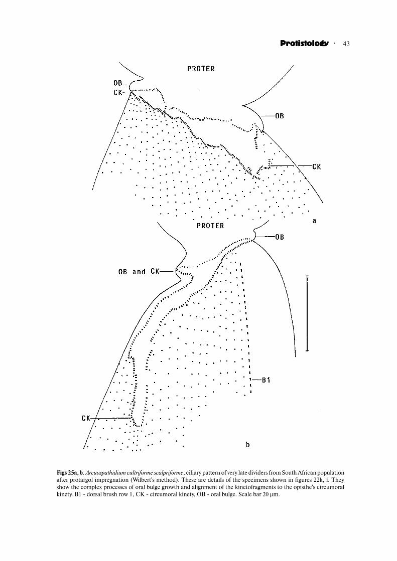
· 43ProtistologyProtistologyProtistologyProtistologyProtistology
Figs 25a, b. Arcuospathidium cultriforme scalpriforme, ciliary pattern of very late dividers from South African populationafter protargol impregnation (Wilbert's method). These are details of the specimens shown in figures 22k, l. Theyshow the complex processes of oral bulge growth and alignment of the kinetofragments to the opisthe's circumoralkinety. B1 � dorsal brush row 1, CK � circumoral kinety, OB � oral bulge. Scale bar 20 µm.
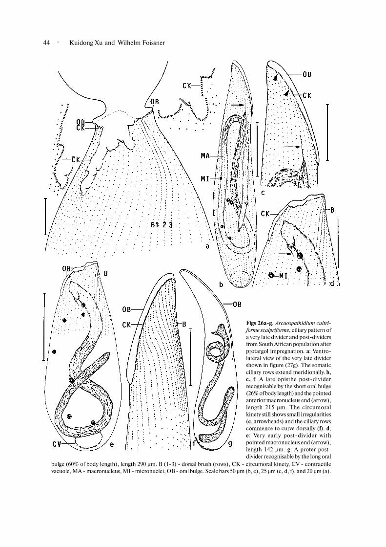
· Kuidong Xu and Wilhelm Foissner44
Figs 26a�g. Arcuospathidium cultri�forme scalpriforme, ciliary pattern ofa very late divider and post�dividersfrom South African population afterprotargol impregnation. a: Ventro�lateral view of the very late dividershown in figure (27g). The somaticciliary rows extend meridionally. b,c, f: A late opisthe post�dividerrecognisable by the short oral bulge(26% of body length) and the pointedanterior macronucleus end (arrow),length 215 µm. The circumoralkinety still shows small irregularities(c, arrowheads) and the ciliary rowscommence to curve dorsally (f). d,e: Very early post�divider withpointed macronucleus end (arrow),length 142 µm. g: A proter post�divider recognisable by the long oral
bulge (60% of body length), length 290 µm. B (1�3) � dorsal brush (rows), CK � circumoral kinety, CV � contractilevacuole, MA � macronucleus, MI � micronuclei, OB � oral bulge. Scale bars 50 µm (b, e), 25 µm (c, d, f), and 20 µm (a).

· 45ProtistologyProtistologyProtistologyProtistologyProtistology
Figs 27a�g. Arcuospathidium cultriforme scalpriforme,body and nuclear ontogenesis of South Africanspecimens with two macronucleus pieces. Thespecimens shown in figures (a, b) are from a non�flooded Petri dish (raw) culture, while all others arefrom a semipure culture. All drawn to scale, exceptfigures (a, b), from protargol preparations. Thearrowheads mark the kinetofragment belt, that is, theforming opisthe circumoral kinety. This series offigures shows, inter alia, that binucleate specimens area natural phenomenon in A. cultriforme; they are notcaused by chance or injury. Ontogenesis of the oraland somatic ciliary pattern as well as the contractilevacuole is as in ordinary specimens. Length ofspecimens: 198 µm (a), 265 µm (b), 420 µm (c), 390µm (d), 340 µm (e), 415 µm (f), 440 µm (g). Scale bar100 µm for c�g. CK � circumoral kinety, CV �contractile vacuole, EP � excretory pores, MA �macronucleus, MI � micronuclei, OB � oral bulge.

· Kuidong Xu and Wilhelm Foissner46
Figs 28a�i. Arcuospathidium cultriforme scalpriforme, protargol�impregnated (Wilbert's method) monsters from SouthAfrican population. a: A doublet with 61 ciliary rows, 270 µm. b, c: A binucleate specimen with a de novo (asterisk) andan ordinarily (arrow) developing oral apparatus, 450 µm. Scale bar 10 µm. d, e: De novo formation of circumoral kinety,390 µm. Scale bar 10 µm. f: A specimen with doubled rear end, 400 µm. g: A specimen with a de novo developing oralapparatus and without opisthe brush rows, 425 µm. h, i: Specimens in which the opisthe oral apparatus develops de novoon the opposite of the proter oral apparatus, 470 µm and 435 µm. B � dorsal brush, CK � circumoral kinety, CV �contractile vacuole, EP � excretory pores, MA � macronucleus, MI � micronuclei, OA � oral apparatus, OB � oral bulge.

· 47ProtistologyProtistologyProtistologyProtistologyProtistology
the resorption of parental somatic kinetids (Fig. 28e).However, in A. cultriforme and some other spathidiids(Berger et al., 1983; Foissner et al., 2002), the ingrowthmodel is disproved by the lack of evidence for resorptionof parental kinetids in the growing mouth area. Further,only half of the basal bodies found in morphostaticspecimens occur in the oral area of early post�dividers,and the distances between the individual basal bodiesare larger orally than postorally. To compensate for thisdeficit, the oral area should not extend into the parentalventral cortex and dissolve existing kinetids, but growas a whole and concomitantly produce new kinetids.Thus, we favour the outgrowth model, where length andslope of the oral bulge are obtained by unequal growthof the dorsal and the ventral side (Figs 32a�e).
As concerns A. cultriforme and other long�mouthedspecies, the oral area not only grows transversely in thefission region, but extends more or less far ventrally(posteriorly) in late dividers (Figs 25a, b, 26a). We
suggest this as a genus and/or species�specific processproducing some space for the long oral kinetofragmentsbecause no growth can occur in the dorsal region wherethe daughters adhere.
The observations and models do not explain thedorsal curve of the ciliary rows in the mouth area of allspathidiids. This curve augments the oral ciliature andtherefore is thus probably related to the feeding process.At first glance, the curve seems to be caused by thesometimes distinct dorsal curve of the spathidiid oralarea. However, such relationship is disproved by manyspecies which have the oral area hardly curved dorsally,for instance, Spathidium spathula and S. turgitorum(Foissner, 1984; Foissner et al., 2002). The lack ofspecific ontogenetic processes suggests that the dorsalcurve is genetically fixed and belongs to the basic(plesiomorphic) equipment of the spathidiids.( How are the spathidiid ciliary patterns obtained?
Foissner (1984) distinguished four spathidiid ciliary
Figs 29a�e. Arcuospathidium cultriforme scalpriforme, protargol�impregnated (Wilbert's method) monster opisthe post�dividers from a semipure culture of the South African population. These figures show that monsters successfullydivide, giving rise to malformed post�dividers and morphostatic cells. a: A slightly distorted specimen with partiallylacking circumoral kinety (arrowheads), length 255 µm. b, c: Left side view of a monster with malformed dorsalbrush, length 248 µm. d, e: Right side view of a monster with malformed circumoral kinety, length 180 µm. Arrowmarks pointed anterior end of macronucleus, showing that it is an opisthe post�divider. B � dorsal brush, CK �circumoral kinety, CV � contractile vacuole, EP � excretory pores, MA � macronucleus, MI � micronuclei. Scale bars50 µm (a, b, e), 30 µm (c), and 10 µm (d).

· Kuidong Xu and Wilhelm Foissner48
patterns. Based on the outgrowth model discussedabove, we suggest the “row detachment model” for theorigin of the spathidiid ciliary patterns (Fig. 31). AnArcuospathidium pattern is generated if the ciliary rowsdetach from the oral kinetofragments in very late andpost�dividers, while a Protospathidium or Spathidiumpattern is generated if the ciliary rows remain attachedto the oral kinetofragments. This explains three of thefour basic spathidiid ciliary patterns. The manyvariations of the patterns found within and betweengenera are likely produced by (i) some remodelling ofthe oral and perioral cortex, (ii) by different growthintensities of the dorsal and ventral oral area, and (iii)by an increased basal body proliferation at the anteriorend of the ciliary rows. The latter produces the typicalcondensation of the cilia at the anterior end of thekineties in many Spathidium and, especially, Epispa�thidium species. Thus, the Epispathidium ciliary patternis a variation of the Spathidium pattern. However,detailed data on Epispathidium are lacking and so it isnot known whether the Epispathidium pattern isgenerated directly from the Spathidium pattern orindirectly via an Arcuospathidium pattern.
The row detachment model also explains some othercharacteristic features of the spathidiid ciliary patterns:(i) that the dorsal brush kineties even in typicalArcuospathidium species are often arranged in Spathidiumpattern (Fig. 3f) and (ii) that the Arcuospathidium patternis frequently indistinct, that is, the left side ciliary rowsare straight or only slightly curved dorsally, sometimeseven ventrally. This is so because the Arcuospathidiumciliary pattern is basically a Spathidium pattern maskedby the separation of the ciliary rows from the circumoralkinety, as shown in our scheme (Fig. 31).
The ontogenetic data and the row detachmentmodel suggest an evolution of the spathidiid ciliarypattern as shown in Figure 31. Most of these patternsevolved several times not only in the spathidiids (Xuand Foissner, 2003, 2004a) but also in related families,such as the Myriokaryonidae (Foissner, 2003a) and
Enchelyodontidae (Foissner et al., 2002; Xu andFoissner, 2003, 2004a).( How is the shape of the oral bulge obtained? When
viewed ventrally, the spathidiid oral bulge may beobovate, oblong, or cuneate (Foissner, 1984; Foissneret al., 2002). We have not found any indication that theseshapes are caused by special ontogenetic constraints,which means that they are genetically determined.( Was the ancestral dorsal brush two�rowed or three�
rowed? The dorsal brush of A. cultriforme develops in aremarkable way unknown in A. muscorum and A.coemeterii (Berger et al., 1983; Foissner and Lei, 2004):first, row 2 is generated, followed by rows 3 and 1 (Figs23b, c, 24a, b). In Spathidium, the brush rows developconcomitantly (Table 4), while a certain population ofProtospathidium serpens generates rows 2 and 3 duringontogenesis and row 1 in post�dividers (Foissner andXu, 2005). Further, most Protospathidium species havebrush row 1 strongly shortened, and in some it is evenlacking (Foissner et al., 2002; Foissner and Xu, 2005).
These observations and the proposed evolution ofthe spathidiids (Fig. 31) suggest an ancestor with onlytwo brush rows. We would not like to generalise thishypothesis, that is, to apply it to other groups ofhaptorids, because ontogenetic data are very sparse, theevolution of the haptorids is obscure, and most species/genera have a three�rowed brush, suggesting this is theplesiomorphic state.( What is the function of the division blebs? Division
blebs are hemispherical protrusions underneath theprospective division furrow. They were first describedin Homalozoon vermiculare, where they contain variousmicrotubules and cortical granules (Leipe et al., 1992).Later, division blebs were described also in Spathidiumturgitorum (Foissner et al., 2002). We found them in A.cultriforme and other Spathidium species (Figs 13b�d,14e), which suggests their widespread occurrence inhaptorid ciliates.
At first glance, the blebs appear to be the precursorsof the prospective oral bulge. However, they are distinct
Fig. 30. The long circumoral kinety of A. cultriforme needs a lot of dikinetids, so that the individual oral kinetofragments(KF) must be as long as possible to suffice for the new circumoral kinety. This is achieved by making the division axisoblique and the fragments curved. Both provisions increase the useable perimeter by about 40%.

· 49ProtistologyProtistologyProtistologyProtistologyProtistology
Fig. 31. An evolutionary scheme of the spathidiid ciliary patterns.

· Kuidong Xu and Wilhelm Foissner50

· 51ProtistologyProtistologyProtistologyProtistologyProtistology
only in early and middle dividers, disappearing duringlate ontogenesis, long before oral bulge formationcommences (Figs 13a�d, 14b; Foissner et al., 2002).Thus, the function of the blebs remains obscure.( Adesmokineties in spathidiids? The term
“adesmokineties” was coined by Jankowski for thepreoral kinetofragments of Dileptus; later the Americansnamed them “paratenes”. Recently, adesmokinety�likekinetofragments were found in Myriokaryon, a relativeof the spathidiids (Foissner, 2003a).
Our observations show adesmokinety�like fragmentsin a typical spathidiid. The ontogenetic analysis revealsthat these fragments are produced during post�divisional growth and shaping of the oral area, possiblyby lateral proliferation of the circumoral kinety or bysome re�arrangement of the somatic kinetids (Figs 4a,b). This would be different from Dileptus, where theadesmokineties are produced by the anterior end of thesomatic kineties (Goli}ska, 1995). However, Goli}ska’s
evidence is not entirely convincing, since it does notrule out production by the circumoral kinety. Generally,there is a tendency to produce extra kinetofragmentsaround the oral opening in haptorids, especially in theendocommensal Buetschlidiidae (Wolska, 1965; Grain,1966). In this group, the fragments are producedinterkinetally, probably de novo, but origin via migratingbasal bodies cannot be excluded, as in the entodinio�morphids (Furness and Butler, 1986; Foissner, 1996).
Only a part of the individuals of A. cultriforme in apopulation have adesmokinety�like fragments, suggestingthat they are vestiges of an ancestral state. Possibly,spathidiids sensu stricto evolved from a Dileptus�likeancestor by reduction of the proboscis and its adesmo�kineties. This hypothesis is supported by two curiousfeatures of the spathidiids: (i) adesmokinety�likefragments occur only on the left side, where Dileptushas these structures, and (ii) the cytostomial opening isnear the dorsal end of the oral bulge. Figure 32f shows
Figs 32a�e. A model of production of the spathidiid oral bulge. In A. cultriforme and several other spathidiids, both theoral bulge and ciliary pattern are generated mainly post�divisionally. Late and very late dividers perform growthmainly in the ventral fission area (a�c). After separation of the daughter cells, faster growth of the dorsal than theventral area makes the oral bulge more or less distinctly oblique and curves the ciliary rows dorsally (d, e). The shapeof the oral bulge is genetically determined. f. A model of a Dileptus�like ancestor of the spathidiids. It is based on theadesmokineties (arrowheads) and the acentric location of the cytopharyngeal entrance (CY) in Spathidium. Thedileptid proboscis becomes reduced. OB � oral bulge.

· Kuidong Xu and Wilhelm Foissner52
an evolutionary scenario, where this pattern is producedby reduction of the proboscis and unequal (ventral)growth of the oral bulge.( Monsters. A huge literature is available on natural
and experimental monsters in ciliates. It was carefullyreviewed by Frankel (1989) who concluded from all theobservations and experiments: “My own view is thatthe true mechanisms of ontogenesis are not yetunderstood, not even in principle, and that answers willbe found by scientists who are willing to think and workunconventionally”. None the less, our observations areof some interest because they confirm and extend earlierdata on other species, mainly Tetrahymema, Parame�cium, Stentor, hypotrichs, and Dileptus. For instance,many processes are obviously controlled epicortically,and a new mouth can be generated only on the ventraland dorsal surface, never laterally. Of particular interestis the observation that the spathidiid circumoral kinetycan develop apokinetally (de novo), that is, withoutsoma�based oral kinetofragments. This provides supportfor the molecular phylogenies proposing a sister�grouprelationship between Gymnostomatea (_ Litostomatea)and Spirotrichea (Lynn and Small, 1997), the soleciliate groups with apokinetal stomatogenesis (Foissner,1996).
Malformed specimens basically divide as ordinaryones. For instance, specimens with two macronucleuspieces divide the pieces individually, so that the offspringalso have two pieces. Wenzel (1955) showed that suchchanges could stabilise in Spathidium and produce a newspecies.
COMPARATIVE ONTOGENESIS
Foissner (1996) briefly reviewed the ontogeneticliterature on haptorids. Although some studies wereadded recently (e.g., Foissner et al., 2002; Foissner andLei, 2004; Table 4), this compilation shows that reliabledata are available on only 6 out of about 1,000 speciesdescribed. None the less, Foissner’s review indicates agreat homogeneity of the division process in haptorids:the macronucleus is homomeric and cell division occursin active (non�encysted) condition; stomatogenesis isholotelokinetal (all ciliary rows produce kinetofrag�ments) except in the pleurostomatids, where it ismonotelokinetal (only one kinety produces the newcircumoral kinety); and the parental oral apparatus isnot reorganised. Arcuospathidium cultriforme perfectlymatches the general pattern, while details may bedifferent even in the same or related genera (Table 4).However, we cannot exclude that some variation iscaused by incomplete or misinterpreted data.
Evolution and classification of the haptorids intoorders and families are poorly understood (Foissner andFoissner, 1988; Lipscomb and Riordan, 1990; Foissner,
1996). Ontogenetic data will certainly contribute to amore rigid system, providing they become available insufficient quality and quantity. For instance, ontogeneticfeatures suggest that the bryophyllids and dileptiids aredistinct groups (Fryd�Versavel et al., 1975; Goli}ska,1995). Likewise, the nuclear apparatus shows interestingvariations. For instance, in multinucleate species themacronucleus nodules fuse to a globular mass in mid�dividers, except for Dileptus where the nodules divideindividually (Jones, 1951). The same is known for somestichotrichine spirotrichs (Foissner, 1996).
ENCYSTMENT AND RESTING CYSTS
Detailed data on encystment of Spathidium are veryscant and time�honoured. Until recently, the sole studyavailable was that from Moore (1924), who provided athorough description of body and nuclear changes inencysting and encysted cells of Spathidium spathula.While the body changes described by Moore (1924) inS. spathula match our observations in A. cultriforme andProtospathidium fraterculum (Xu and Foissner, 2005),the nuclear processes do not. In cysts of S. spathula,the macronucleus strongly fragmentates, while it ismaintained, but distinctly shortened in A. cultriforme.However, Moore’s data are doubtful because she couldstain the nuclear apparatus only after mechanicalrupture of the wall. Certainly, such crude method canproduce many artifacts.
The strong shortening of the macronucleus bycytoplasmic resorption is an outstanding feature of theresting cyst of A. cultriforme. Some reviews (Corliss andEsser, 1974; Gutiurrez et al., 2003) indicate that suchprocess has been not described in other ciliates,although Padnos et al. (1954) observed that 4�year�oldcysts expelled, in three phases, a good half of themacronucleus as well as cytoplasm and deposited thematerial in the cyst wall. In A. cultriforme, a strongreduction of the macronucleus from ~ 5,000 µm3 to ~1,000 µm3 might be associated, at least partially, withthe small cyst volume (~ 45,000 µm3 without the thickwall) as compared to the large cell volume (~ 300,000µm3). In several other spathidiids, the ratio cell:cystvolume is not 6:1, as in A. cultriforme, but only about2:1, for instance, in the Protospathidium serpenscomplex, where the macronucleus is only slightlyshortened during encystment (Xu and Foissner, 2005).
As concerns the ciliary pattern (~ infraciliature),three types of resting cysts have been distinguished inciliates (Gutiurrez et al., 2003): kinetosome�resorbingcysts (KR type), non�kinetosome�resorbing cysts(NKR), and partial�kinetosome�resorbing cysts(PKR). Arcuospathidium cultriforme very prabablyproduces KR cysts, as do, for instance, oxytrichidhypotrichs (Gutiurrez et al., 2003), while some
· 53ProtistologyProtistologyProtistologyProtistologyProtistology
colpodids have PKR cysts (Foissner, 1993). However,definite proof requires transmission electron microscopydata not available on any spathidiid. In related taxa,such as Didinium and Dileptus, some basal bodies andcortical microtubules are maintained (Dippell andGrimes, 1966; Kink, 1973).
Few data are available on resting cyst morphologyof spathidiids. Moore (1924) described a fairly thin,wrinkled wall in Spathidium spathula. The wall is alsothin, but smooth in S. ascendens, S. turgitorum, and S.(now Arcuospathidium) muscorum (Wenzel, 1955;Berger et al., 1983; Foissner et al., 2002). Spathidiumstammeri, in contrast, has a thin wall with manyconspicuous spines about 4 µm long (Wenzel, 1959 andunpubl. observations of the authors). Last but not least,Protospathidium serpens makes smooth or spiny cysts,depending on population. The smooth type has beenclassified as a new species by Xu and Foissner (2005).The resting cysts of the three populations of A.cultriforme are very similar, though there are differencesin size and thickness of the ridges. The cysts are uniquenot only because of the strong shortening of themacronucleus (Figs 2y, 21a�g) but also because of adistinctly faceted cyst wall about 10 µm thick (Figs 15a�i). Obviously, resting cyst morphology is conspicuouslydifferent in Spathidium sensu lato, providing somesupport for the pronounced split of the genus (Foissner,1984; Foissner et al., 2002; Foissner and Xu, 2005).
ACKNOWLEDGEMENTS
Financial support was provided by the AustrianScience Foundation (FWF project P�15017). We thankAndreas Zankl, Birgit Peukert, and Dr. B. Moser fortechnical assistance.
References
Aescht E. and Foissner W. 1993. Effects oforganically enriched magnesite fertilizers on the soilciliates of a spruce forest. Pedobiologia. 37, 321�335.
Berger H. 1999. Monograph of the Oxytrichidae(Ciliophora, Hypotrichia). Monographiae biol. 78, XII+ 1080 pp.
Berger H., Foissner W. and Adam H. 1983.Morphology and morphogenesis of Fuscheria terricolan. sp. and Spathidium muscorum (Ciliophora: Kineto�fragminophora). J. Protozool. 30, 529�535.
Berger H., Foissner W. and Adam H. 1984.Taxonomie, Biometrie und Morphogenese einigerterricoler Ciliaten (Protozoa: Ciliophora). Zool. Jb.Syst. 111, 339�367.
Blatterer H. and Foissner W. 1988. Beitrag zurterricolen Ciliatenfauna (Protozoa: Ciliophora)Australiens. Stapfia (Linz). 17, 1�84.
Corliss J.O. 1979. The Ciliated Protozoa. Charac�terization, Classification and Guide to the Literature.2nd ed. Pergamon Press, Oxford, New York, Toronto,Sydney, Paris, Frankfurt.
Corliss J.O. and Esser S.C. 1974. Comments onthe role of the cyst in the life cycle and survival of free�living protozoa. Trans. Am. microsc. Soc. 93, 578�593.
Detcheva R.B. 1972a. Distribution des especes decilies dans certains affluents Bulgares du Danube, auxeaux polluees. Annls Stn biol. Besse. 6�7 (years 1971/1972), 261�269.
Detcheva R.B. 1972b. Recherches en Bulgarie surles protozoaires du sol. Annls Stn limnol. Besse. 6�7(years 1971/1972), 273�284.
Detscheva R.B. 1972c. Beitrag zur Kenntnis derInfusorienfauna (Protozoa�Ciliata) in den Binnen�gewqssern Bulgariens. Isv. zool. Inst., Sof. 36, 61�79.
Detcheva R. 1975. Pollution influence on theinfusorial fauna (Protozoa, Ciliata) of Bulgarian riversII. The Ogosta river and its affluents. Hydrobiology. 2,19�31 (in Russian with English summary).
Detcheva R. 1979a. Zbersicht zber die Ciliatenfaunader bulgarischen Donauzuflzsse. XXI. Arbeitstagungder Societas Internationalis Limnologiae, Novi Sad,SFR Jugoslawien. pp. 357�364.
Detcheva R. 1979b. Paramitres saprobiologiqueset hydrochimiques pour les cilius de certains affluentsBulgares de la Mer Noire. Hydrobiology. 9, 57�73.
Detcheva R.B. 1986. Composition and distributionof the ciliates (Protozoa, Ciliata) from the Tund,a river.Hydrobiology. 28, 61�65 (in Russian with Englishsummary).
Detcheva R.B. 1993. Les donnues ucologiques pourles cilius (Protozoa�Ciliophora) du bassin de la riviireIskar. Hydrobiology. 38, 33�44 (in Russian with Frenchsummary).
Dippell R.V. and Grimes G.W. 1966. The occur�rence of basal bodies in the cortex of active and encystedDidinium nasutum. J. Protozool. 13 (Suppl.), p. 9.
Dragesco J. and Dragesco�Kernuis A. 1979. Ciliesmuscicoles nouveaux ou peu connus. Acta Protozool.18, 401�416.
Finlay B.J. 2001. Protozoa. Encyclopedia ofBiodiversity. 4, 901�915.
Foissner W. 1984. Infraciliatur, Silberliniensystemund Biometrie einiger neuer und wenig bekannterterrestrischer, limnischer und mariner Ciliaten(Protozoa: Ciliophora) aus den Klassen Kinetofragmi�nophora, Colpodea und Polyhymenophora. Stapfia(Linz). 12, 1�165.
Foissner W. 1987a. Soil protozoa: fundamentalproblems, ecological significance, adaptations in
· Kuidong Xu and Wilhelm Foissner54
ciliates and testaceans, bioindicators, and guide to theliterature. Progr. Protistol. 2, 69�212.
Foissner W. 1987b. Neue terrestrische und limnischeCiliaten (Protozoa, Ciliophora) aus Asterreich undDeutschland. Sber. Akad. Wiss. Wien. 195, 217�268.
Foissner W. 1989. Morphologie und Infraciliatureiniger neuer und wenig bekannter terrestrischer undlimnischer Ciliaten (Protozoa, Ciliophora). Sber. Akad.Wiss. Wien. 196, 173�247.
Foissner W. 1991. Basic light and scanning electronmicroscopic methods for taxonomic studies of ciliatedprotozoa. Europ. J. Protistol. 27, 313�330.
Foissner W. 1993. Colpodea (Ciliophora). Proto�zoenfauna. 4/1, 1�798.
Foissner, W. 1995. Tropical protozoan diversity: 80ciliate species (Protozoa, Ciliophora) in a soil samplefrom a tropical dry forest of Costa Rica, with descriptionsof four new genera and seven new species. Arch.Protistenk. 145, 37�79.
Foissner W. 1996. Ontogenesis in ciliated protozoa,with emphasis on stomatogenesis. In: Ciliates: Cells asorganisms (Eds. Hausmann K. and Bradbury P.C.).Fischer, Stuttgart, Jena, Lzbeck, Ulm. pp. 95�177.
Foissner W. 1998. An updated compilation of worldsoil ciliates (Protozoa, Ciliophora), with ecologicalnotes, new records, and descriptions of new species.Europ. J. Protistol. 34, 195�235.
Foissner W. 1999a. Notes on the soil ciliate biota(Protozoa, Ciliophora) from the Shimba Hills in Kenya(Africa): diversity and description of three new generaand ten new species. Biodiv. Conserv. 8, 319�389.
Foissner W. 1999b. Protist diversity: estimates ofthe near�imponderable. Protist. 150, 363�368.
Foissner W. 2000a. Two new terricolous spathidiids(Protozoa, Ciliophora) from tropical Africa: Arcuo�spathidium vlassaki and Arcuospathidium bulli. Biol.Fertil. Soils. 30, 469�477.
Foissner W. 2000b. A compilation of soil and mossciliates (Protozoa, Ciliophora) from Germany, withnew records and descriptions of new and insufficientlyknown species. Europ. J. Protistol. 36, 253�283.
Foissner W. 2003a. The Myriokaryonidae fam. n.,a new family of spathidiid ciliates (Ciliophora:Gymnostomatea). Acta Protozool. 42, 113�143.
Foissner W. 2003b. Cultellothrix velhoi gen. n., sp.n., a new spathidiid ciliate (Ciliophora: Haptorida)from a Brazilian floodplain soil. Acta Protozool. 42,47�54.
Foissner W. 2004a. Ubiquity and cosmopolitanismof protists questioned. SILnews. 43, 6�7.
Foissner W. 2004b. Some new ciliates (Protozoa,Ciliophora) from an Austrian floodplain soil, includinga giant, red “flagship”, Cyrtohymena (Cyrtohymenides)aspoechi nov. subgen., nov. spec. Denisia. 13, 369�382.
Foissner W. 2005a. Biogeography of microorganisms:a review emphasizing protists. Endocytobiol. Cell Res.(in press).
Foissner W. 2005b. Two new “flagship” ciliates(Protozoa, Ciliophora) from Venezuela: Sleighophryspustulata and Luporinophrys micelae. Europ. J. Protistol.(in press).
Foissner W. and Foissner I. 1988. The fine structureof Fuscheria terricola Berger et al., 1983 and a proposednew classification of the subclass Haptoria Corliss, 1974(Ciliophora, Litostomatea). Arch. Protistenk. 135, 213�235.
Foissner, W. and Lei Y.�L. 2004. Morphology andontogenesis of some soil spathidiids (Ciliophora,Haptoria). Linzer biol. Beitr. 36/1, 159�199.
Foissner W. and Xu K. 2005. Monograph of theSpathidiina (Protozoa, Ciliophora). Kluwer AcademicPublishers (in press).
Foissner W., Agatha S. and Berger H. 2002. Soilciliates (Protozoa, Ciliophora) from Namibia (South�west Africa), with emphasis on two contrastingenvironments, the Etosha region and the Namib Desert.Denisia. 5, 1�1459.
Foissner W., Berger H., Blatterer H. and Kohmann,F. 1995. Taxonomische und akologische Revision derCiliaten des Saprobiensystems � Band IV: Gymnosto�matea, Loxodes, Suctoria. Informationsberichte desBayer. Landesamtes fzr Wasserwirtschaft. 1/95, 1�540.
Foissner W., Berger H., Xu K. and Zechmeister�Boltenstern S. 2005. A huge, undescribed soil ciliate(Protozoa: Ciliophora) diversity in natural forest standsof Central Europe. Biodiv. Conserv. 14, 617�701.
Foissner W., Strzder�Kypke M.C., van der StaayG.W.M., Moon�van der Staay, S.�Y. and Hackstein,J.H.P. 2003. Endemic ciliates (Protozoa, Ciliophora)from tank bromeliads (Bromeliaceae): a combinedmorphological, molecular, and ecological study. Europ.J. Protistol. 39, 365�372.
Fokin S.I., Przybok E., Chivilev S.M., Beier C.L.,Horn M., Skotarczak B., Wodecka B. and FujishimaM. 2004. Morphological and molecular investigationsof Paramecium schewiakoffi sp. nov. (Ciliophora,Oligohymenophorea) and current status of distributionand taxonomy of Paramecium spp. Europ. J. Protistol.40, 225�243.
Frankel J. 1989. Pattern Formation. Ciliate Studiesand Models. Oxford University Press, New York.
Fryd�Versavel G., Iftode F. and Dragesco J. 1975.Contribution a la connaissance de quelques ciliusgymnostomes II. Prostomiens, pleurostomiens:morphologie, stomatogenise. Protistologica. 11, 509�530.
Furness D.N. and Butler R.D. 1986. Morphogenesisof infraciliature in the entodiniomorphid ciliate

· 55ProtistologyProtistologyProtistologyProtistologyProtistology
Eudiplodinium maggii. J. Ultrastruct. Mol. Struct. Res.94, 16�29.
Gellu rt J. 1956. Ciliaten des sich unter demMoosrasen auf Felsen gebildeten Humus. Acta biol.hung. 6, 337�359.
Goli}ska K. 1995. Formation and orientation ofskeletal elements during development of oral territoryin a ciliate, Dileptus. Acta Protozool. 34, 101�113.
Goralczyk K. and Verhoeven R. 1999. Bodengeneseals Standortfaktor fzr Mikrofauna – Ciliaten undNematoden in Dznenbaden. In: Bodenakologieinterdisziplinq r (Eds. Koehler H., Mathes K. andBreckling B.). Springer, Berlin, Heidelberg. pp. 105�118.
Grain J. 1966. Utude cytologique de quelques ciliusholotriches endocommensaux des ruminants et desuquidus (premiire partie). Protistologica. 2, 59�141.
Gutiurrez J.C., Diaz S., Ortega R. and Mart|n�Gonzw lez A. 2003. Ciliate resting cyst walls: acomparative review. Rec. Res. Dev. Microbiol. 7, 361�379.
Hirshfield H.I., Isquith I.R., Di Lorenzo A.E. 1973.Classification, distribution, and evolution. In: Blepha�risma (Ed. Giese A.C.). Stanford Univ. Press, Stanford,California. pp. 304�332.
Jones E.E. Jr. 1951. Encystment, excystment, andthe nuclear cycle in the ciliate Dileptus anser. J. ElishaMitchell scient. Soc. 67, 205�217.
Kahl A. 1930a. Neue und ergqnzende Beobach�tungen holotricher Infusorien. II. Arch. Protistenk. 70,313�416.
Kahl A. 1930b. Urtiere oder Protozoa I: Wim�pertiere oder Ciliata (Infusoria) 1. Allgemeiner Teil undProstomata. Tierwelt Dtl. 18, 1�180.
Kink J. 1973. The organization of fibrillar structuresin the trophic and encysted Dileptus visscheri (Ciliata,Rhabdophorina). Acta Protozool. 12, 173�194.
Leipe D.D., Oppelt A., Hausmann K. and FoissnerW. 1992. Stomatogenesis in the ditransversal ciliateHomalozoon vermiculare (Ciliophora, Rhabdophora).Europ. J. Protistol. 28, 198�213.
Lipscomb D.L. and Riordan G.P. 1990. Theultrastructure of Chaenea teres and an analysis of the
phylogeny of the haptorid ciliates. J. Protozool. 37, 287�300.
Lynn D.H. and Small E.B. 1997. A revisedclassification of the phylum Ciliophora Doflein, 1901.Revta Soc. mex. Hist. nat. 47, 65�78.
Moore E.L. 1924. Endomixis and encystment inSpathidium spathula. J. exp. Zool. 39, 317�337.
Ning Y., Wang S., Ma Z., Gan Y., Yu Z., Geng W.,Gu X., Cai G. and An Q. 1993. Investigations on thefreshwater protozoa in Lanzhou. Chin. J. Zool. 28, 1�3(in Chinese).
Padnos M., Jakowska S. and Nigrelli R.F. 1954.Morphology and life history of Colpoda maupasi,Bensonhurst strain. J. Protozool. 1, 131�139.
Penard E. 1922. Utudes sur les infusoires d&eaudouce. Georg et Cie, Genive.
Russev B., Kovatschev S., Janewa I., Karapetkowa,M., Uzunov J. and Detschewa R. 1976. Vertreter derbulgarischen Flussfauna als limnosaprobe Bioindi�katoren. Hydrobiology. 4, 60�66.
Russev B., Janeva I. and Detcheva R. 1994.Zusammensetzung der Hydrofauna. 3.1.1. WirbelloseTiere. In: Limnologie der bulgarischen Donauzuflzsse,Sofia. pp. 130�186 (in Bulgarian).
Song W., Zhao Y., Xu K., Hu X. and Gong J. 2003Pathogenic Protozoa in Mariculture. Science Press,Beijing. 483 pp. (in Chinese).
Wang J. 1977. Protozoa from some districes ofTibetan Plateau. Acta zool. sin. 23, 131�160 (in Chinesewith English summary).
Wang S. and Ma Z. 1994. The ciliates fromindustrial wastewater of Xining and three indicativespecies of Rhizopoda on high mountains and subhighmountains. J. Northwest Normal Univ. (Nat. Sci.). 30,60�65 (in Chinese with English summary).
Wenzel F. 1953. Die Ciliaten der Moosrasentrockner Standorte. Arch. Protistenk. 99, 70�141.
Wenzel F. 1955. Zber eine Artentstehung innerhalbder Gattung Spathidium (Holotricha, Ciliata). [S.ascendens n. sp. und S. polymorphum n. sp.] Arch.Protistenk. 100, 515�540.
Address for correspondence: Dr. Wilhelm Foissner. Universitqt Salzburg, FB Organismische Biologie,Hellbrunnerstrasse 34. A�5020 Salzburg, Austria. E�mail: [email protected].
Editorial responsibility: Sergei Fokin
Copyright © 2022 FDOKUMEN