Montiel and Molnar 2013 commentary
10
The Impact of Gene Expression Analysis on Evolving Views of Avian Brain Organization Juan F. Montiel, 1,2 * and Zolt an Moln ar 1 * 1 Department of Physiology, Anatomy, and Genetics, Le Gros Clark Building, University of Oxford, Oxford OX1 3QX, United Kingdom 2 Center for Biomedical Research, Faculty of Medicine, Universidad Diego Portales, Santiago, Chile ABSTRACT Recent studies have presented data on adult and devel- oping avian brain organization. Jarvis et al. ([2013] J Comp Neurol. 521:3614–3665) identify four pallial and two subpallial gene expression domains and demon- strate that the mesopallium and adjoining divisions of the hyperpallium (hyperpallium intercalatum and hyper- pallium densocellulare), have very similar gene expres- sion profiles to each other, distinct from those of the nidopallium, the arcopallium, and the more distant divi- sions of the hyperpallium (hyperpallium apicale). The study proposes an update of the current nomenclature (Jarvis et al. [2005] Nat Rev Neurosci. 6:151–159). The authors perform densitometric quantifications of the in situ expression of 50 selected genes, use correlations of distances between vectors that represent these gene expression patterns within the 23 avian brain regions of their study, and group them according to similarity in their expression profiles. The generated cluster tree fur- ther supports their argument for a new terminology. The authors hypothesize that the mesopallium and adjoining divisions of the hyperpallium have a common developmental origin, and in the accompanying paper (Chen et al. [2013] J Comp Neurol. 521:3666–3701) show that these structures/subdivisions initially form continuous gene expression domains. With subsequent development these domains fold into distinct subdivi- sions in the dorsal and ventral avian pallium, forming mirror images to each other. Jarvis et al. ([2013] J Comp Neurol. 521:3614–3665) also demonstrate inter- esting principles of the functional organization of the avian brain by showing that specific sensory stimulation or motor behavior elicits gene expression in functional units perpendicular to the axis of the gene expression reversal and compare their arrangements and cell types with mammalian cortical columns. J. Comp. Neurol. 521:3604–3613, 2013. V C 2013 Wiley Periodicals, Inc. INDEXING TERMS: avian brain nomenclature; transcriptomics; cerebral cortex; wulst; dorsal ventricular ridge; corti- cal column; pallium; mesopallium; hyperpallium In avian and mammalian brain evolution, telence- phalic structures have undergone such significant elab- orations that identifying evolutionary, developmental, structural, or functional homologies has been a great challenge and has produced numerous hypotheses over the last century (Smith, 1919; Holmgren, 1922; John- ston, 1923; Northcutt and Kaas, 1995; Karten, 1997; Butler and Hodos, 2005; Striedter, 2005). On a trans- verse section of an avian or reptilian forebrain, one of the most distinct features is the presence of a large sphere of cells protruding into the lateral ventricle, the dorsal ventricular ridge (DVR) in birds (Karten, 1969; Ulinski, 1983; Puelles et al., 2000; Moln ar and Butler, 2002a,b; Fig. 1A,B). A plausible homologue of the “six- layered” mammalian neocortex (isocortex): located between the three-layered olfactory cortex (lateral pal- lium) and the hippocampal cortex (medial pallium, also bearing three main layers) lies in the position of the hyperpallium of birds. The hyperpallium is a nonlayered structure in which a pseudolayered (columnar) organiza- tion has been described (Medina and Reiner, 2000; Wang et al., 2010; Fig. 1C). The “thorniest questions of comparative neurobiology” (Northcutt, 2003) are whether the hyperpallium and the neocortex are Grant sponsor: The Medical Research Council (to Z.M.); Grant sponsor: Biotechnology and Biological Sciences Research Council (to Z.M.); Grant sponsor: The Wellcome Trust (to Z.M.); Grant sponsor: Becas Chile postdoctoral fellowship (to J.F.M.). *CORRESPONDENCE TO: Juan F. Montiel or Zolt an Moln ar, Department of Physiology, Anatomy, and Genetics, Le Gros Clark Building, University of Oxford, South Parks Road, Oxford OX1 3QX, UK. E-mails: juan.mon- [email protected] and [email protected] Received April 12, 2013; Revised May 15, 2013; Accepted June 21, 2013. DOI 10.1002/cne.23403 Published online July 1, 2013 in Wiley Online Library (wileyonlinelibrary.com) V C 2013 Wiley Periodicals, Inc. 3604 The Journal of Comparative Neurology | Research in Systems Neuroscience 521:3604–3613 (2013) COMMENTARY
-
Upload
independent -
Category
Documents
-
view
0 -
download
0
Transcript of Montiel and Molnar 2013 commentary

The Impact of Gene Expression Analysis onEvolving Views of Avian Brain Organization
Juan F. Montiel,1,2* and Zolt�an Moln�ar1*1Department of Physiology, Anatomy, and Genetics, Le Gros Clark Building, University of Oxford, Oxford OX1 3QX, United Kingdom2Center for Biomedical Research, Faculty of Medicine, Universidad Diego Portales, Santiago, Chile
ABSTRACTRecent studies have presented data on adult and devel-
oping avian brain organization. Jarvis et al. ([2013] J
Comp Neurol. 521:3614–3665) identify four pallial and
two subpallial gene expression domains and demon-
strate that the mesopallium and adjoining divisions of
the hyperpallium (hyperpallium intercalatum and hyper-
pallium densocellulare), have very similar gene expres-
sion profiles to each other, distinct from those of the
nidopallium, the arcopallium, and the more distant divi-
sions of the hyperpallium (hyperpallium apicale). The
study proposes an update of the current nomenclature
(Jarvis et al. [2005] Nat Rev Neurosci. 6:151–159). The
authors perform densitometric quantifications of the in
situ expression of 50 selected genes, use correlations
of distances between vectors that represent these gene
expression patterns within the 23 avian brain regions of
their study, and group them according to similarity in
their expression profiles. The generated cluster tree fur-
ther supports their argument for a new terminology.
The authors hypothesize that the mesopallium and
adjoining divisions of the hyperpallium have a common
developmental origin, and in the accompanying paper
(Chen et al. [2013] J Comp Neurol. 521:3666–3701)
show that these structures/subdivisions initially form
continuous gene expression domains. With subsequent
development these domains fold into distinct subdivi-
sions in the dorsal and ventral avian pallium, forming
mirror images to each other. Jarvis et al. ([2013] J
Comp Neurol. 521:3614–3665) also demonstrate inter-
esting principles of the functional organization of the
avian brain by showing that specific sensory stimulation
or motor behavior elicits gene expression in functional
units perpendicular to the axis of the gene expression
reversal and compare their arrangements and cell types
with mammalian cortical columns. J. Comp. Neurol.
521:3604–3613, 2013.
VC 2013 Wiley Periodicals, Inc.
INDEXING TERMS: avian brain nomenclature; transcriptomics; cerebral cortex; wulst; dorsal ventricular ridge; corti-
cal column; pallium; mesopallium; hyperpallium
In avian and mammalian brain evolution, telence-
phalic structures have undergone such significant elab-
orations that identifying evolutionary, developmental,
structural, or functional homologies has been a great
challenge and has produced numerous hypotheses over
the last century (Smith, 1919; Holmgren, 1922; John-
ston, 1923; Northcutt and Kaas, 1995; Karten, 1997;
Butler and Hodos, 2005; Striedter, 2005). On a trans-
verse section of an avian or reptilian forebrain, one of
the most distinct features is the presence of a large
sphere of cells protruding into the lateral ventricle, the
dorsal ventricular ridge (DVR) in birds (Karten, 1969;
Ulinski, 1983; Puelles et al., 2000; Moln�ar and Butler,
2002a,b; Fig. 1A,B). A plausible homologue of the “six-
layered” mammalian neocortex (isocortex): located
between the three-layered olfactory cortex (lateral pal-
lium) and the hippocampal cortex (medial pallium, also
bearing three main layers) lies in the position of the
hyperpallium of birds. The hyperpallium is a nonlayered
structure in which a pseudolayered (columnar) organiza-
tion has been described (Medina and Reiner, 2000;
Wang et al., 2010; Fig. 1C). The “thorniest questions of
comparative neurobiology” (Northcutt, 2003) are
whether the hyperpallium and the neocortex are
Grant sponsor: The Medical Research Council (to Z.M.); Grantsponsor: Biotechnology and Biological Sciences Research Council (toZ.M.); Grant sponsor: The Wellcome Trust (to Z.M.); Grant sponsor:Becas Chile postdoctoral fellowship (to J.F.M.).
*CORRESPONDENCE TO: Juan F. Montiel or Zolt�an Moln�ar, Departmentof Physiology, Anatomy, and Genetics, Le Gros Clark Building, Universityof Oxford, South Parks Road, Oxford OX1 3QX, UK. E-mails: [email protected] and [email protected]
Received April 12, 2013; Revised May 15, 2013;Accepted June 21, 2013.DOI 10.1002/cne.23403Published online July 1, 2013 in Wiley Online Library(wileyonlinelibrary.com)VC 2013 Wiley Periodicals, Inc.
3604 The Journal of Comparative Neurology | Research in Systems Neuroscience 521:3604–3613 (2013)
COMMENTARY
homologous and whether these mammalian and avian
structures contain similar circuits, but arranged in a dif-
ferent manner.
GENE EXPRESSION REFINESUNDERSTANDING OF AVIAN BRAINORGANIZATION AND DEVELOPMENT
Recent gene expression analyses have contributed to
a better understanding of mammalian and avian brain
organization and development (Dugas-Ford et al., 2012;
Suzuki et al., 2012). Study in sauropsids (birds and rep-
tiles) of selected genes that had a lamina-specific
expression in the mammalian cortex showed surpris-
ingly similar gene expression patterns in the wulst or
DVR, respectively accordingly to the relative position
within the neuronal circuits originally proposed by
Karten (1969; 2013). These studies strongly argue for
the conserved physiological, anatomical, and neuro-
chemical survival in the constituents of mammalian and
avian/reptilian circuits. These circuits share highly simi-
lar properties that are conserved regardless of the lami-
nar or nuclear organization (Karten, 2013). The articles
published in this issue by Jarvis et al. (2013) and Chen
et al. (2013) now go beyond of many of the previous
publications in the numbers of genes observed, regions
studied, developmental stages. The studies use in situ
hybridization on sagittal sections of the adult zebrafinch
and combine this with densitometry and gene cluster
analysis. The data have been collected over more than
a decade, and certain features of gene expression have
been validated in various different avian species, from
available collections, atlases, or websites (e.g., http://
www.zebrafinchatlas.org/). Supplementary histological
data from the articles by Jarvis et al. and Chen et al.
are available as virtual slides or whole slide images
using Biolucida Cloud image streaming technology from
MBF Bioscience. This collection can be accessed online
at http://Wiley.Biolucida.net/JCN521-16Jarvis_Chen.
The Jarvis et al. study (this issue) selected 52 genes
(46 constitutive; 6 activity dependent) to decipher the
molecular and functional relationships between different
adult avian telencephalic subdivisions. The accompany-
ing Chen et al. study (this issue) examined 16 genes at 8
stages and traced the expression of some of these genes
back to embryonic day 4 (E4) to construct an account of
the development of major telencephalic subdivisions.
The genes were selected based on their specific patterns
of expression in particular sectors of the telencephalon,
their relations with mammalian brain structures, or their
functions. They include 21 glutamate receptors, 5 dopa-
mine receptors, the SEMA6A receptor, a cannabinoid
receptor and a retinoic acid b receptor; the protein
ligands for neuritin, BDNF, and SCUBE1; DLX6, FOXP1,
FOXP2, ER81, COUP-TF2, LHX8, LHX9, NKX2.1, PAX6,
EGR1, c-FOS, c-JUN transcription factors; FKBP1A,
DUSP1, and PPAPDC1A enzymes; and a diverse set of
membrane and cytoplasmic genes, including ARPP16,
TMEM100, ARC, CADPS2, and S100B.
The authors followed a rational working hypothesis
according to which homologous or functionally compara-
ble brain areas should express similar gene sets. The
Chen et al. article (this issue) uses the term “anatomical
temporal network” of gene expression for the temporal
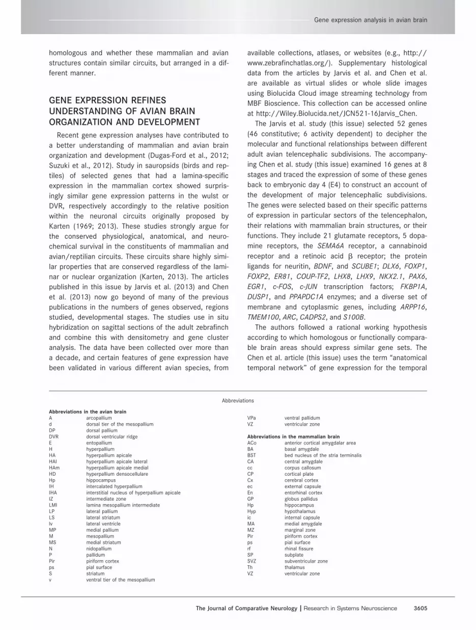
Abbreviations
Abbreviations in the avian brainA arcopalliumd dorsal tier of the mesopalliumDP dorsal palliumDVR dorsal ventricular ridgeE entopalliumH hyperpalliumHA hyperpallium apicaleHAl hyperpallium apicale lateralHAm hyperpallium apicale medialHD hyperpallium densocellulareHp hippocampusIH intercalated hyperpalliumIHA interstitial nucleus of hyperpallium apicaleIZ intermediate zoneLMI lamina mesopallium intermediateLP lateral palliumLS lateral striatumlv lateral ventricleMP medial palliumM mesopalliumMS medial striatumN nidopalliumP pallidumPir piriform cortexps pial surfaceS striatumv ventral tier of the mesopallium
VPa ventral pallidumVZ ventricular zone
Abbreviations in the mammalian brainACo anterior cortical amygdalar areaBA basal amygdaleBST bed nucleus of the stria terminalisCA central amygdalecc corpus callosumCP cortical plateCx cerebral cortexec external capsuleEn entorhinal cortexGP globus pallidusHp hippocampusHyp hypothalamusic internal capsuleMA medial amygdaleMZ marginal zonePir piriform cortexps pial surfacerf rhinal fissureSP subplateSVZ subventricular zoneTh thalamusVZ ventricular zone
Gene expression analysis in avian brain
The Journal of Comparative Neurology | Research in Systems Neuroscience 3605

sequence and location of the expression of selected
genes to study development of brain regions, rather than
gene coexpression or functional networks.
The articles present important new sets of experimen-
tal results with outcomes that are clearly of major impor-
tance to the field. Based on the observations about
pallial hierarchic connectivity and mirror and columnar
expression patterns, the central hypothesis from Jarvis
et al. (this issue) concerns the common developmental ori-
gin of the mesopallium and adjoining divisions of the
hyperpallium. In the Chen et al. study (this issue), the
authors find support in the development of contiguous
gene expression domains that wrap around the lateral ven-
tricle. However, like every landmark study, they produce
more questions than answers. Is the increased sample
size of these genes sufficient to draw these conclusions?
Is the selection of the genes adequate? Could migration of
neurons expressing a gene explain some of the findings
rather than onset of expression of the gene in a static pop-
ulation? How can we integrate the conserved elements of
avian and mammalian cell types with known features of
cell migration and cell lineage? Should the field embrace
or resist the proposed changes in terminology?
MESOPALLIUM AND ADJOINING DIVISIONSOF THE HYPERPALLIUM HAVE SIMILARGENE EXPRESSION PROFILES
The main finding is that the mesopallium and the
adjoining divisions of the hyperpallium have gene
expression profiles that are very similar to each other
and dissimilar from those of the nidopallium, the arco-
pallium, and the more distant divisions of the hyperpal-
lium (Fig. 1). In situ hybridization data for a large
number of the genes studied in several additional bird
species support the original observation made in zebra
finch. The gene expression patterns revealed a continu-
ous zone around a rostral projection of the lateral ven-
tricle that is especially obvious during development and
clearer on sagittal than on coronal sections (Chen
et al., this issue). This evidence raises questions about
the conventional view (Jarvis et al., 2005) of the organi-
zation of the pallial subdivisions and exposes the insuf-
ficiencies of strictly equating the four Cartesian
quadrants of dorsal, ventral, medial, and lateral pallium
with the different brain subdivisions (Fig. 1D).
GENE EXPRESSION AND LINEARREGRESSION NETWORK ANALYSES
In order to quantify these observations, the authors
have translated the gene expression pattern of radioactive
in situ hybridization, using image analysis and processing
for each region of interest, in order to develop a hierarchi-
cal cluster analysis of each pallial and subpallial brain sub-
division according to its gene expression profile. To
generate these results the authors produced an impres-
sive set of in situ hybridization gene expression data.
Although the genes transcribed in brain tissue might reach
tens of thousands, and thousands of these show a layer-
specific expression pattern (Belgard et al., 2011; Moln�ar
and Belgard, 2012; Bernard et al., 2012), it is understand-
able that approaches relying on detailed anatomy and in
situ hybridization require the selection of genes. The justi-
fication for the selection of these genes is still, in the scale
of the transcriptome, a biased/curated selection of rela-
tively few genes. The selection was based on previous lit-
erature that described sharp gene expression boundaries
or specificities for a particular cell group, and this pro-
duced an overrepresentation of some functionally
enriched sets of genes, such as the numerous glutamate
receptors involved in the Jarvis et al. Study (this issue).
The panel of selected genes is not a connected network,
nor a representation of a particular function, or related to
a particular step in morphogenesis, or associated with a
particular cell lineage. One can ask whether the selection
of different sets of markers would produce a different pic-
ture? Dugas-Ford et al. (2012) and Suzuki et al. (2012)
used similar approaches, arguably with a fewer selection
of genes, and drew different conclusions.
Jarvis et al. (this issue) conclude: “Phylo-gene expres-
sion tree and linear regression network analyses impli-
cated single and mixed ancestry of these brain regions.”
The findings support their continuum hypothesis of avian
brain subdivisions around the lateral ventricle and influ-
ence hypotheses on homologies of the avian pallium for
other vertebrates. It is not clear whether the inclusion or
exclusion of a few genes or picking another set of 50
genes would make a significant and noticeable difference
in the clustering and the conclusions. The authors argue
that their observations are solid, that the cladogram is sta-
ble even after the removal of up to 10 genes (Jarvis et al.,
this issue; Supplementary Material, Fig. 5). They claim that
there are only small local changes, but no change to their
conclusions of six major groupings. The issue of the
“random” selection of genes is critical, because typical
long-scale analyses use tens of thousands genes, which
means three orders of magnitude greater. Actually, a
quantitative large-scale transcriptomic analysis of over
5000 most highly expressed one-to-one orthologous
genes, according specificity, coexpression, and marker
gene distribution in the telencephalon of chicken and
mouse exhibits a large divergence, small conservation,
and, exceptionally, convergence between different sectors
of these species (Belgard, Montiel et al., 2013). Belgard,
Montiel et al. (2013) demonstrate a clear transcriptomic
differentiation across avian and mammalian pallial regions,
J.F. Montiel and Z. Molnar
3606 The Journal of Comparative Neurology |Research in Systems Neuroscience

getting a cohort of specific markers for each sector and
other genes with variable shared pattern. For species-spe-
cific and cross-species regional markers for mouse and
chick pallial and striatal domains see Web resource:
http://geserv.anat.ox.ac.uk from Belgard, Montiel et al.,
(2013). A correct interpretation of these results is that dif-
ferent output of similarity would emerge when a few
selected genes are compared at adult stages; neverthe-
less, many of these observations are not significantly sup-
ported when a global transcriptomic involvement is
investigated.
One of the main conclusions of the Jarvis et al. (this
issue) article is that the patterns of constitutively
expressed genes revealed a mirror image expression pat-
tern above and below the lateral ventricle and the associ-
ated lamina (Fig. 2A). This suggests a different relationship
between mesopallium and adjoining divisions of the hyper-
pallium (hyperpallium intercalatum and hyperpallium den-
socellulare). The authors’ definition of dorsal and ventral
pallium is everything positioned above and below the lat-
eral ventricle and the associated lamina mesopallium
intermediate (LMI), respectively, including the renamed
hyperpallium densocellulare (in the dorsal pallium) and
mesopallium (in the ventral pallium) of the 2004-2005
nomenclature or dorsal mesopallium (in dorsal pallium)
and ventral mesopallium (in ventral pallium). Thus the
term ventral is not restricted to the nidopallium or arcopal-
lium; as also involved some olfactory areas as in some
other studies (Medina et al., 2011). The mesopallium,
according to the 2004-2005 nomenclature, is considered
lateral pallium by Medina and Abellan (2009). This is in
contrast to the view of Jarvis and colleagues, who name
the mesopallium region of the 2004-2005 nomenclature
as the ventral mesopallium in the ventral pallium and its
counterpart in the dorsal pallium as the newly named dor-
sal mesopallium, with both parts as a dorsal pallial deriva-
tive during development. They mention that the term
ventral is not restricted to the nidopallium or arcopallium
as done in some previous studies. We do not know any
justification for this. Even more puzzling, the differential
expression patterns between the mesopallium and nido-
pallium (Chen et al., this issue) support a different devel-
opmental origin of the two structures.
Jarvis and colleagues propose including the hyperpallium
densocellulare (HD) in the mesopallium; we agree with
them because of the gene expression patterns, which show
that the most reliable mesopallial marker genes display an
extended expression pattern in this region, (Belgard, Mon-
tiel et al., 2013). Accordingly, Redies et al. (2001) did not
observe a distinguishable gene expression change between
the HD and mesopallium compared with the hyperpallium
intercalatum (the next hyperpallial column in immediate
contact with the HD) from a cadherin expression screen.
However, according to that study (Redies et al., 2001), cad-
herin expression does not support the mirror pallial organi-
zation at least at embryonic day 15, and we believe that
this should be confirmed with a larger set of genes. Simi-
larly, Cheng et al. (2013) do not readily see a mirror image
profile in at these embryonic stages in zebrafinch. Interest-
ingly, the work of Reiner and colleagues (2011) on
Cerebellin-2 (Cbln2) gene expression demonstrated that
there is a Cbln2-rich domain in the mesopallium, but not in
the adjoining HD, and a border can be defined.
INDUCIBLE GENES DEMONSTRATECOLUMN-LIKE EXPRESSION EXTENDINGACROSS SUBDIVISIONS OF THE AVIANWULST
Jarvis et al. (this issue) describe the patterns of func-
tionally regulated genes and observe “functional
Figure 1. Comparisons of telencephalic organizations on transverse
sections of mammalian and avian forebrains. A: The avian brain con-
tains a ball-shaped structure that protrudes into the lateral ventri-
cle, the DVR. B: The mammalian brain organization is different, with
an apparent dominance of the dorsal cortex (Cx). C: According to
the current view of avian telencephalic organization the hyperpal-
lium (H) is a dorsal pallial derivative; with a pseudolayered (colum-
nar) organization, and the mesopallium (M) is considered a lateral
pallial derivative. D: In Jarvis et al.’s (2013) model the former hyper-
pallium densocellulare (HD) becomes part of the mesopallium. The
Jarvis et al. study establishes that the patterns of constitutively
expressed genes reveal a mirror image expression pattern above
and below the lateral ventricle and the associated lamina mesopal-
lium intermediate (LMI), indicated by a black arrow. According to
the expression pattern a semi-mirror pallial organization is proposed
along the axis of the LMI (red arrows). For abbreviations, see list. A
and B are modified from Medina and Abell�an (2009).
Gene expression analysis in avian brain
The Journal of Comparative Neurology | Research in Systems Neuroscience 3607

columns” of gene activation extend through cross-
subdivision boundaries (Fig. 2A). These authors propose
that the “avian columns” are of two types: “those above
the ventricle formed by the hyperpallium, intercalated
hyperpallium, and dorsal mesopallium; and those below
the ventricle formed by the nidopallium, intercalated
nidopallium, and ventral mesopallium.” They suggest
that these functional units extending from above the
ventricle and associated lamina to below the ventricle
and associated lamina in the avian brain are reminis-
cent of columns of the mammalian cortex. The concept
of developmental, anatomical, functional, and transcrip-
tional columns is undergoing a revolution right now
even in the mammalian literature (Moln�ar, 2013). The
distinctions or the relationships among the anatomical,
functional, and developmental columns is not always
defined, and this can lead to misunderstandings (Rakic,
2008). The term “column” is overused in both the mam-
malian and the avian literature.
The gene expression patterns in distinct subdivisions
of the dorsal and ventral avian pallium are arranged as
mirror images to each other. To study the functional
activation of circuits in relation to these sectors, Jarvis
and colleagues elicit changes in functionally regulated
genes (EGR1, C-FOS, C-JUN, DUSP1, and ARC) after
auditory, visual, and somatosensory stimulation or after
singing or motor activity in various avian species. These
coactivated “functional columns” extended into both
the dorsal and the ventral avian pallium and were per-
pendicular to boundaries between pallial sectors. Jarvis
Figure 2. Differences between the avian and mammalian “columnar” organization. A: The gene expression patterns in distinct subdivisions
in the dorsal and ventral avian pallium are arranged as mirror images of each other along the ventricle and an associated mesopallial lam-
ina with the lamina mesopallium intermediate (LMI) as the center. Changes in functionally regulated genes after auditory, visual, and
somatosensory stimulation revealed coactivated units perpendicular to boundaries between pallial sectors (somatosensory, motor, audi-
tory, night and day vision) that extended through different sectors of both dorsal and ventral avian pallium. Jarvis et al. (2013) considered
this organization as “functional columns.” The mammalian and avian “functional columns” both contain similar sets of specialized groups
of neurons, but the overall logic of their developmental origin is very different. In the avian brain the cells of the coactivated networks are
derived from different sectors of the pallial neuroepithelium and assemble across these sectors. B: In birds, neurons are generated in a
spatially regulated manner following a radial migration pattern in different pallial compartments. Modified from Nomura et al., (2008). The
observed arrangement of radial glia implies that neurons of each radial column of the hyperpallium must be born in separate, adjacent
regions of neuroepithelium, from where they migrate toward the pial surface (ps). Distinguishable radial columns are formed parallel to the
orientation of the radial glia and not perpendicular to them as for the laminar organization in mammals and reptiles. The hyperpallium is
therefore considered a nonlayered structure even though the radial columns are interconnected (Medina and Reiner, 2000; Medina and
Abell�an, 2009). C: Mammalian columns are known to extend across the layers of the cerebral cortex perpendicular to the pial surface.
The cells belonging to a functional column in mammals are arranged within the same radial pallial sector that gave rise to them. In the
avian pallium different sectors produce different elements that combine perpendicular to the radial trajectories, whereas in mammals the
diverse elements of the functional columns are produced within the same sector of cortical neuroepithelium and assemble along the radial
trajectories. Figure inspired by Schubert et al. (2007). D: Temporally regulated newly born neurons in mouse migrate from the ventricular
zone alongside the radial glia in an inside-out neurogenetic gradient (K€ull�en, 1953; Tsai et al., 1981). For abbreviations, see list.
J.F. Montiel and Z. Molnar
3608 The Journal of Comparative Neurology |Research in Systems Neuroscience

and colleagues consider the functional units in the
avian pallium as reminiscent of mammalian functional
columnar cortical organization. The word “functional” is
very important in the light of the uncertainties of ana-
tomical, hodological, and developmental columns. The
question remains of how comparable these columns are
across different clades, e.g., mammalian columns pres-
ent a radial arrangement from the lateral ventricle,
against the avian functional columns that are mostly
tangentially organized, and mammalian cortical columns
are confined to a single compartment, e.g., the dorsal
pallium, but avian functional columns run through dor-
sal and ventral pallial compartments (Fig. 2).
Jarvis et al. (this issue) make suggestions about possi-
ble cell types for the avian brain that form circuits similar
to the ones found in mammals. These form four groups: 1)
a primary sensory input population (intercalated pallium);
2) a secondary intrapallial population (nidopallium/hyper-
pallium); 3) a tertiary intrapallial population (mesopallium);
and 4) a quaternary output population (the arcopallium).
They consider that each population contributes portions to
columns that control different sensory or motor systems.
The gene expression results of the selected genes strongly
support Karten’s synthesis of a large amount of circuit
data (Karten, 1969, 1997). The proposed cell populations
are largely equivalent to Karten’s hypothesis about neo-
cortical circuits in the avian dorsal telencephalon (Karten,
1969). The difference is in their placing the HA in the nido-
pallium rather than the arcopallium (N-L hypothesis in
Table 2 of Jarvis et al., this issue).
Although we agree with Jarvis and colleagues that the
two types of functional columns might have conserved fea-
tures, we find this use of the term “column” or “functional
column” misleading. Jarvis and colleagues consider mam-
malian and avian functional units equally as columns:
either columns across different cell populations organized
as layers (in mammals), or more thickened nuclear slabs
(in birds). We would like to emphasize the considerable dif-
ference in the anatomy and development between these
units. The mammalian columns extend across cells that
are the derivatives of the same radial sector of the neuroe-
pithelium, whereas the avian “columns” are across the dif-
ferent sectors of the pallium (Fig. 2). Jarvis et al. (this
issue) argue that regardless of how the cells arrived at
their final destination from the ventricular zone in mam-
mals compared with birds, both functional units contain
similar sets of cells that assemble into a functional circuit.
IS THERE A NEED FOR A NEW AVIANBRAIN NOMENCLATURE NOW?
The classical terminology for the dorsal telencepha-
lon, before the 2004–2005 revisions, identified the con-
stituent nuclei as different varieties of striatum. The
Nomenclature Forum (Jarvis et al., 2005) provided an
important service to the field 10 years ago. The result
was a careful renaming of the dorsal telencephalic
nuclei into varieties of pallium. The renaming was a
consensus, and the new nomenclature preserved as
much as possible the abbreviations employed in the
classical terminology. Most researchers in the field
made serious efforts to be consistent with this new
nomenclature, although it is inevitable that there are
some disagreements and new data will emerge that
may justify modifications.
Jarvis et al. (this issue) argue that just because a
term/definition is in common use, this is not a good
enough reason to continue using it. They propose terti-
ary pallium as a new or alternate name for the meso-
pallium, which refers to the current mesopallium plus
adjoining hyperpallium densocellare and hyperpallium
intercalatum, indicating that this new name reflects
their (implicit) hypothesis that these division share
processing functions. Furthermore, they clearly include
the hyperpallium densocellulare (HD) in a new “dorsal
mesopallium” and exclude the interstitial part of the
hyperpallium apicale (IHA). They slip and slide, however,
about the hyperpallium intercalatum (HI). They state
boldly that the “hyperpallium dorsale [sic], hyperpallium
intercalatum and mesopallium” should be grouped
together, but later state that the lamina LFM, which
separates the HI from the HD (Karten and Hodos,
1967; Reiner et al., 2004), is what separates the “new”
dorsal mesopallium from the rump hyperpallium. An
additional proposed change is to change hyperpallium
apicale (HA) to hyperpallium (H) and the interstitial part
of the hyperpallium apicale (IHA) to the intercalated
hyperpallium (IH).
It is not the custom in neuroanatomy to change
nomenclature with each new discovery because changes
such as those suggested by Jarvis et al. (this issue)
invariably end up confusing the literature when not
everyone accepts the new terms. In the literature on the
cortical development of mammals, there have been
some recent suggestions for changes (radial glia, sub-
ventricular zone, apical–basal directions, various interme-
diate progenitors) (Bystron et al., 2008), but the changes
in nomenclature are gradual and some evolve in parallel.
Perhaps the avian field should also exercise some
restraint until more data generate a general consensus.
WHAT IS THE DEVELOPMENTAL ORIGINOF THE MESOPALLIUM?
The Chen et al. study (this issue) represents a con-
siderable step forward in characterizing the molecular
Gene expression analysis in avian brain
The Journal of Comparative Neurology | Research in Systems Neuroscience 3609

embryology of the zebra finch pallium. The logic of their
approach is to follow back the gene expression patterns
from maturity to embryonic day 4 (E4) and construct, as
far as possible, an account of how these major subdivi-
sions develop. The major finding is that “the mesopallium
and adjoining divisions of the hyperpallium initially form
continuous subdivisions around a rostral projection of
the lateral ventricle” (Fig. 3) and that “distinct subdivi-
sions in the dorsal and ventral avian pallium have gene
expression patterns that are arranged in a mirror image
sequence of each other with the lamina mesopallium
intermediate (LMI) in the center (Figs. 1D, 2A, 3)”.
Accordingly, they hypothesize that the mesopallium and
adjoining divisions of the hyperpallium (hyperpallium
intercalatum and hyperpallium densocellulare) might,
therefore, originate from one continuous region. Indeed,
it has been described that in a primitive reptile (Spheno-
don) DVR and dorsal cortex are cytoarchitectonically con-
tinuous and uniform, and mirror images around the
ventricle (Reiner and Northcutt, 2000).
Several additional ideas are proposed in Chen et al.
(this issue): first, each subdivision of the expression
profile in the avian brain has a different temporal order
of appearance, similar in timing to the order of analo-
gous cell types appearing in the mammalian cortex;
second, like the mammalian pallium, the ventral portion
of the subdivisions become distinct during prehatch
development, whereas the dorsal portions do so during
posthatch development. It is not clear whether the
authors presume that the differences in these regions
could reflect neurogenic gradients rather than genuine
differences in their ontogeny. This kind of data can only
be fully interpreted in conjunction with birth dating and
fate mapping.
The position of Jarvis and colleagues is not currently
supported by developmental evidence that describes
medial, lateral, and ventral patterns of migrations in the
avian pallium (Nomura et al., 2008). This may have a
direct impact on the interpretation of the results
because patterns of migration and early gene
Figure 3. Proposed development of the mirror reversal of the gene expression patterns in distinct subdivisions in the dorsal and ventral
avian pallium by Chen et al. (2013). The Jarvis et al. (2013) paper established that the patterns of constitutively expressed genes revealed
a mirror image expression pattern above and below the lateral ventricle and the associated lamina mesopallium intermediate (LMI). The
Chen et al. (2013) paper suggests that the dorsal and ventral pallium might have closer developmental relationships than previously appre-
ciated. Chen et al. established that most gene expressions in defined pallial subdivisions begin as one continuous ventral or dorsal domain
that later folds around the lateral ventricle only maintaining continuity on the rostralmost sector and only apparent on sagittal sections.
The dorsal and ventral pallial subdivisions and the surrounding mesopallium thus developed the mirror image profile along the ventricular
boundary and associated lamina (black arrow). Chen et al. (2013) also postulate a stream of migration between these regions (blue arrow).
For abbreviations, see list.
J.F. Montiel and Z. Molnar
3610 The Journal of Comparative Neurology |Research in Systems Neuroscience

expression define developmentally independent lateral
and ventral pallial compartments (Fernandez et al.,
1998) in frog, chick, and mouse, whereas the authors
are more supportive of a reorganization of the pallium.
CONSERVATION OR CONVERGENCE OFGENE EXPRESSION PATTERNS?
An alternative hypothesis to the proposed common
origin–initially continuous region of Jarvis et al. (this
issue) is that the pallial populations above and below
the lateral ventricle do not have a shared direct devel-
opmental origin, but originate from different sectors of
the neuroepithelium with different lineages, having par-
allel and possibly even convergent developmental
molecular programs that lead to a similar outcome.
The best hypothesis is generally the one that can
explain the findings with the fewest assumptions.
Although the articles by Jarvis et al. and Chen et al. (this
issue) consider this possibility much less likely, in the
face of a lack of supporting evidence for cell lineages
the idea of convergent evolution of transcriptomic pro-
files for three separate cell populations above and below
the lateral ventricle should be considered. They regard
convergent evolution as a possibility that would require
many more assumptions than either migrating cells or
extensions of those cell populations around the lateral
ventricle. However, if this is so simple, why has such a
common origin not been described during development?
Current evidence on pallial migration shows radial migra-
tion from proliferative compartments to medial, lateral,
and ventral pallial domains (Nomura et al., 2008), but
not a dorsocaudal reorganization, as a proposed wrap-
ping migratory dynamic would require. An alternative
mechanism to impose a convergent gene expression pat-
tern on cell populations with different lineages would be
a central gradient in the limiting region, from where a
morphogenetic signaling center could induce identity
changes in different regions in a mirror pattern. Exam-
ples for such postulated molecular control could be
found in the midbrain–hindbrain boundary organizer
(MHB) (Martinez et al., 1999), or in fibroblast growth
factor manipulation experiments in other systems
(Suzuki-Hirano and Shimogori, 2009).
TEMPORAL VERSUS SPATIAL REGULATIONOF PALLIAL PROGENITORS IN AVIAN ANDMAMMALIAN PALLIUM
Recent evidence suggests that not all progenitors in a
given brain area are similar in their molecular and kinetic
features. There are numerous progenitor compartments
lying along the telencephalic germinal zones, and within
each compartment there are several types. Mammalian
cortical progenitors divide in a temporal sequence to
generate neurons that form different cortical layers in an
inside-first/outside-last fashion (Fig. 2D). These cells are
committed to fates that integrate them into different
functions in the network. Along the entire mammalian
dorsal pallium, the progenitors follow a similar neuro-
genic temporal code, which guides them in the genera-
tion of the cortical layers across the embryonic
development of the neocortex. This temporal code is
independent of their lateromedial position within the dor-
sal pallium. Recent findings by Suzuki and colleagues
(2012) in the developing chick pallium suggest that the
temporal sequence of generation of cortical neurons was
already inherent in the stem amniote, because avian pal-
lial progenitors share the temporal code of generation.
However, this temporal code has been modified during
avian evolution and a spatial code has replaced the tem-
poral one (Nomura et al., 2008; Suzuki et al., 2012). Dif-
ferent sectors of the avian pallium generate different
sets of cells (Fig. 2B). The Chen et al. study (this issue)
notes that “the temporal order of appearance of the
tested gene expression profiles corresponds to a similar
temporal order of appearance of cells with similar con-
nectivity in the mammalian cortex.” These interesting
data point to a drastic modification of pallial progenitors
along the avian pallial neuroepithelium.
INTEGRATED APPROACH TO VIEW CELLTYPES OF THE AVIAN AND MAMMALIANCIRCUITS
The articles by Jarvis et al. and Chen et al. (this issue)
have wide implications and can be considered as pio-
neering studies in the phylogenetic analysis of expression
patterns in avian brains. They introduce several new dis-
tinct hypotheses on brain evolution. Comparative studies
on avian and mammalian brains have generated different
hypotheses on brain evolution and different outcomes for
homology (Karten, 1969, 1997; Jarvis et al., 2005; Suzuki
et al., 2012; Dugas-Ford et al., 2012; Belgard, Montiel et
al., 2013). The similarities between the pallial and subpal-
lial gene expression patterns in mammals and in saurop-
sids have been previously discussed, and this led to the
proposal of the collopallial field hypothesis of Butler and
Moln�ar (2002). This hypothesis emphasized the similar
cellular constituents of the ventral cortex and the claus-
trum/amygdala complex and postulated that they might
originate from the same field (Moln�ar and Butler,
2002a,b). Jarvis et al. and Chen et al. (this issue) provide
support for this hypothesis, but direct analysis of lineage
and cell migration during development is needed for an
ultimate test.
Gene expression analysis in avian brain
The Journal of Comparative Neurology | Research in Systems Neuroscience 3611

While gene expression analysis (Lein et al., 2007;
Miller et al., 2010), especially whole transcriptome
analysis, can be very powerful in revealing homologies,
one has to use integrative approaches to confirm and
validate them. Jarvis et al. and Chen et al. (this issue)
both focus on an avian perspective, but they arrive at
conclusions concerning mammalian brains, two clades
with more than 300 million years of phylogenetic dis-
tance from a common ancestor. Therefore it would
have been useful to summarize the ontogenetic
dynamic of gene expression in some mammals as well,
in order to understand the evolutionary impact. Even
more important, although chickens, belonging to a
basal avian order (Galliformes), have in the past pro-
vided the model system for avian embryology, the
studies under review here are primarily on zebra finch,
from an evolutionarily distant avian group, the song-
birds (order Passeriformes) (Zelenitsky et al., 2011).
Zebra finch have provided a wealth of information on
the functional organization of the relevant brain struc-
tures, and it will be important to study these further in
other birds and reptiles.
The classification of homologous structures might not
rest solely on genes but also on hodology, function,
and developmental origins. Homology is a structural
evolutionary concept for saying “inherited in common
from the common ancestor”—whereas genes, hodology,
function, and lineage are merely criteria by which
homology might be inferred. However, there are not dif-
ferent classes of homology. Novel technologies such as
transcriptomic analysis in the developing brain will have
to be combined with lineage and clonal analyses, and
observations of cell migration and differentiation are
needed to reveal alterations in developmental pro-
grams. Resolution of these issues calls for an integra-
tive view. Also, the fact that there might have been
convergent evolution of the expression of these genes
relating to specific circuit elements does not necessar-
ily imply an evolutionary relationship or common clonal
origin between the particular cells or structures
expressing these genes. It does draw attention to very
general biological principles when one is trying to
understand the overlying logic of neuronal circuit
assembly regardless of the actual laminar structure,
columnar organization, or developmental origin.
ACKNOWLEDGMENTSWe are grateful to Ray Guillery, Denis Jabaudon, and
Anton Reiner for stimulating discussions.
CONFLICT OF INTEREST STATEMENT
The authors have no conflict of interest.
ROLE OF AUTHORS
Both authors had full access to all the data in the
article, take responsibility for the integrity of the data
and the accuracy of the data analysis, and were equally
involved in the concept, analysis, and drafting of the
manuscript.
LITERATURE CITEDBelgard TG, Marques AC, Oliver PL, Abaan HO, Sirey TM,
Hoerder-Suabedissen A, Garc�ıa-Moreno F, Moln�ar Z,Margulies EH, Ponting CP. 2011. A transcriptomic atlasof mouse neocortical layers. Neuron 71:605–616.
Belgard TG, Montiel JF, Wang WZ, Garc�ıa-Moreno F, MarguliesEH, Ponting CP, Moln�ar Z. 2013. Adult pallium transcrip-tomes surprise in not reflecting predicted homologiesacross diverse chicken and mouse pallial sectors. ProcNatl Acad Sci U S A, in press. doi: 10.1073/pnas.1307444110.
Bernard A, Lubbers LS, Tanis KQ, Luo R, Podtelezhnikov AA,Finney EM, McWhorter MME, Serikawa K, Lemon T,Morgan R, Copeland C, Smith K, Cullen V, Davis-Turak J,Lee CK, Sunkin SM, Loboda AP, Levine DM, Stone DJ,Hawrylycz MJ, Roberts CJ, Jones AR, Geschwind DH, LeinES. 2012. Transcriptional architecture of the primateneocortex. Neuron 73:1083–1099.
Butler AB, Hodos W. 2005. Comparative vertebrate neuroanat-omy. New York: Wiley-Liss.
Butler AB, Moln�ar Z. 2002. Development and evolution of thecollopallium in amniotes: a new hypothesis of fieldhomology. Brain Res Bull 57:475–479.
Bystron I, Blakemore C, Rakic P. 2008. Development of thehuman cerebral cortex: Boulder Committee revisited. NatRev Neurosci 9:110–122.
Chen CC, Winkler C, Pfenning A, Jarvis ED. Molecular profiling ofthe developing avian telencephalon: regional timing andbrain subdivision continuities. J Comp Neurol 521:3666–3701.
Dugas-Ford J, Rowell JJ, Ragsdale CW. 2012. Cell-type homolo-gies and the origins of the neocortex. Proc Natl Acad SciU S A 109:16974–16979.
Fernandez AS, Pieau C, Rep�erant J, Boncinelli E, Wassef M.1998. Expression of the Emx-1 and Dlx-1 homeoboxgenes defines three molecularly distinct domains in thetelencephalon of mouse, chick, turtle and frog embryos:implications for the evolution of telencephalic subdivi-sions in amniotes. Development 125:2099–2111.
Holmgren N. 1922. Points of view concerning forebrain mor-phology in lower vertebrates. J Comp Neurol 34:391–459.
Jarvis ED, G€unt€urk€un O, Bruce L, Csillag A, Karten H, KuenzelW, Medina L, Paxinos G, Perkel DJ, Shimizu T, StriedterG, Wild JM, Ball GF, Dugas-Ford J, Durand SE, Hough GE,Husband S, Kubikova L, Lee DW, Mello CV, Powers A,Siang C, Smulders TV, Wada K, White SA, Yamamoto K,Yu J, Reiner A, Butler AB. 2005. Avian brains and a newunderstanding of vertebrate brain evolution. Nat RevNeurosci 6:151–159.
Jarvis ED, Yu J, Rivas M, Horita H, Feenders G, Whitney O,Jarvis S, Jarvis E, Kubikova L, Buck AEP, Siang C, MartinS, McElroy M, Hara E, Mouritsen H, Chen CC, Wada K.2013. A global view of the functional molecular organiza-tion of the avian cerebrum: mirror images and functionalcolumns. J Comp Neurol 521:3614–3665.
Johnston JB. 1923. Further contributions to the study of theevolution of the forebrain. J Comp Neurol 35:337–481.
J.F. Montiel and Z. Molnar
3612 The Journal of Comparative Neurology |Research in Systems Neuroscience

K€ull�en B. 1953. On the nuclear differentiation during ontogene-sis in the avian forebrain and some notes on the amniotestrioamygdaloid complex. Acta Anat (Basel) 17:72–84.
Karten HJ. 1969. The organization of the avian telencephalonand some speculations on the phylogeny of the amniotetelencephalon. Ann N Y Acad Sci 167:164–179.
Karten HJ. 1997. Evolutionary developmental biology meetsthe brain: the origins of mammalian cortex. Proc NatlAcad Sci U S A 94:2800–2804.
Karten HJ. 2013. Neocortical evolution: neuronal circuits ariseindependently of lamination. Curr Biol 23:R12–R15.
Karten HJ, Hodos W. 1967. A stereotaxic atlas of the brain ofthe pigeon. Washington, D C: Johns Hopkins UniversityPress.
Lein ES, Hawrylycz MJ, Ao N, Ayres M, Bensinger A, BernardA, Boe AF, Boguski MS, Brockway KS, Byrnes EJ, Chen L,Chen L, Chen TM, Chin MC, Chong J, Crook BE,Czaplinska A, Dang CN, Datta S, Dee NR, Desaki AL,Desta T, Diep E, Dolbeare TA, Donelan MJ, Dong HW,Dougherty JG, Duncan BJ, Ebbert AJ, Eichele G, Estin LK,Faber C, Facer BA, Fields R, Fischer SR, Fliss TP,Frensley C, Gates SN, Glattfelder KJ, Halverson KR, HartMR, Hohmann JG, Howell MP, Jeung DP, Johnson RA,Karr PT, Kawal R, Kidney JM, Knapik RH, Kuan CL, LakeJH, Laramee AR, Larsen KD, Lau C, Lemon TA, Liang AJ,Liu Y, Luong LT, Michaels J, Morgan JJ, Morgan RJ,Mortrud MT, Mosqueda NF, Ng LL, Ng R, Orta GJ, OverlyCC, Pak TH, Parry SE, Pathak SD, Pearson OC, PuchalskiRB, Riley ZL, Rockett HR, Rowland SA, Royall JJ, Ruiz MJ,Sarno NR, Schaffnit K, Shapovalova NV, Sivisay T,Slaughterbeck CR, Smith SC, Smith KA, Smith BI, SodtAJ, Stewart NN, Stumpf KR, Sunkin SM, Sutram M, TamA, Teemer CD, Thaller C, Thompson CL, Varnam LR,Visel A, Whitlock RM, Wohnoutka PE, Wolkey CK, WongVY, Wood M, Yaylaoglu MB, Young RC, Youngstrom BL,Yuan XF, Zhang B, Zwingman TA, Jones AR. 2007.Genome-wide atlas of gene expression in the adultmouse brain. Nature 445:168–176.
Martinez S, Crossley PH, Cobos I, Rubenstein JL, Martin GR.1999. FGF8 induces formation of an ectopic isthmicorganizer and isthmocerebellar development via a repres-sive effect on Otx2 expression. Development 126:1189–1200.
Medina L, Abell�an A. 2009. Development and evolution of thepallium. Semin Cell Dev Biol 20:698–711.
Medina LL, Reiner AA. 2000. Do birds possess homologues ofmammalian primary visual, somatosensory and motorcortices? Trends Neurosci 23:1–12.
Medina L, Bupesh M, Abell�an A. 2011. Contribution of gen-oarchitecture to understanding forebrain evolution anddevelopment, with particular emphasis on the amygdala.Brain Behav Evol 78:216–236.
Miller JA, Horvath S, Geschwind DH. 2010. Divergence ofhuman and mouse brain transcriptome highlights Alzhei-mer disease pathways. Proc Natl Acad Sci U S A 107:12698–12703.
Moln�ar Z, Belgard TG. 2012. Transcriptional profiling of layersof the primate cerebral cortex. Neuron 73:1053–1055.
Moln�ar Z, Butler AB. 2002a. Neuronal changes during fore-brain evolution in amniotes: an evolutionary developmen-tal perspective. Prog Brain Res 136:21–38.
Moln�ar Z, Butler AB. 2002b. The corticostriatal junction: a cru-cial region for forebrain development and evolution. Bio-essays 24:530–541.
Nomura T, Takahashi M, Hara Y, Osumi N. 2008. Patterns ofneurogenesis and amplitude of Reelin expression are
essential for making a mammalian-type cortex. PLoSONE 3:e1454.
Northcutt RG. 2003. The use and abuse of developmentaldata. Behav Brain Sci 26:565–566.
Northcutt GR, Kaas JH. 1995. The emergence and evolution ofmammalian neocortex. Trends Neurosci 18:373–379.
Puelles L, Kuwana E, Puelles E, Bulfone A, Shimamura K,Keleher J, Smiga S, Rubenstein JLR. 2000. Pallial andsubpallial derivatives in the embryonic chick and mousetelencephalon, traced by the expression of the genes Dlx- 2, Emx - 1, Nkx - 2.1, Pax - 6, and Tbr - 1. J Comp Neu-rol 424:409–438.
Rakic P. 2008. Confusing cortical columns. Proc Natl AcadSci U S A 105:12099–12100.
Redies C, Medina L, Puelles L. 2001. Cadherin expression byembryonic divisions and derived gray matter structuresin the telencephalon of the chicken. J Comp Neurol 438:253–285.
Reiner A, Northcutt RG. 2000. Succinic dehydrogenase histo-chemistry reveals the location of the putative primaryvisual and auditory areas within the dorsal ventricularridge of Sphenodon punctatus. Brain Behav Evol 55:26–36.
Reiner A, Perkel DJ, Bruce LL, Butler AB, Csillag A, Kuenzel W,Medina L, Paxinos G, Shimizu T, Striedter G, Wild M, BallGF, Durand S, G€unt€urk€un O, Lee DW, Mello CV, PowersA, White SA, Hough G, Kubikova L, Smulders TV, WadaK, Dugas-Ford J, Husband S, Yamamoto K, Yu J, Siang C,Jarvis ED. 2004. Revised nomenclature for avian telen-cephalon and some related brainstem nuclei. J CompNeurol 473:377–414.
Reiner A, Yang M, Cagle MC, Honig MG. 2011. Localization ofcerebellin-2 in late embryonic chicken brain: implicationsfor a role in synapse formation and for brain evolution. JComp Neurol 519:2225–2251.
Schubert D, K€otter R, Staiger JF. 2007. Mapping functionalconnectivity in barrel-related columns reveals layer- andcell type-specific microcircuits. Brain Struct Funct 212:107–119.
Smith GE. 1919. A preliminary note on the morphology of thecorpus striatum and the origin of the neopallium. J Anat53:271–291.
Striedter GF. 2005. Principles of brain evolution. Sunderland,MA: Sinauer Associates.
Suzuki IK, Kawasaki T, Gojobori T, Hirata T. 2012. The tempo-ral sequence of the mammalian neocortical neurogeneticprogram drives mediolateral pattern in the chick pallium.Dev Cell 22:863–870.
Suzuki-Hirano A, Shimogori T. 2009. The role of Fgf8 in telen-cephalic and diencephalic patterning. Semin Cell DevBiol 20:719–725.
Tsai HM, Garber BB, Larramendi LM. 1981. 3H-thymidine autora-diographic analysis of telencephalic histogenesis in thechick embryo: II. Dynamics of neuronal migration, displace-ment, and aggregation. J Comp Neurol 198:293–306.
Ulinski PS. 1983. Dorsal ventricular ridge: a treatise on fore-brain organization in reptiles and birds. New York, NY:John Wiley & Sons.
Wang Y, Brzozowska-Prechtl A, Karten HJ. 2010. Laminar andcolumnar auditory cortex in avian brain. Proc Natl AcadSci U S A 107:12676–12681.
Zelenitsky DK, Therrien F, Ridgely RC, McGee AR, Witmer LM.2011. Evolution of olfaction in non-avian theropod dino-saurs and birds. Proc Biol Sci 278:3625–3634.
Gene expression analysis in avian brain
The Journal of Comparative Neurology | Research in Systems Neuroscience 3613