Metabolic control analysis indicates a change of strategy in the treatment of cancer
14
Metabolic control analysis indicates a change of strategy in the treatment of cancer Rafael Moreno-Sánchez a, ⁎, Emma Saavedra a , Sara Rodríguez-Enríquez a , Juan Carlos Gallardo-Pérez a , Héctor Quezada a , Hans V. Westerhoff b,c a Departamento de Bioquímica, Instituto Nacional de Cardiología de México, Mexico b VU University Amsterdam, The Netherlands c The Manchester Centre for Integrative Systems Biology, The University of Manchester, UK abstract article info Article history: Received 22 December 2009 Received in revised form 6 March 2010 Accepted 1 June 2010 Available online 25 June 2010 Keywords: Control and regulation analysis Energy metabolism Rate-limiting step Systems biology Multi-target drug Therapeutic strategies Combination therapy Much of the search for the “magic cancer bullet” or “block buster” has followed the expectation of a single gene or protein as “the rate-limiting step” for tumor persistence. Examples continue to abound: EGFR, VEGFR, Akt/PI3K, HIF-1α, PHD, PDK, or FAS continue to be targeted individually. However, many such attempts to block a metabolic or signal transduction pathway by targeting, specifically, a single rate-limiting molecule have proven to be unsuccessful. Metabolic control analysis (MCA) of cancer cells has generated a generic explanation for this phenomenon: several steps share the control of energy metabolism (for glycolysis: glucose transporter, hexokinase, glycogen synthesis and ATP demand; for oxidative phosphor- ylation: respiratory complex I and ATP demand), i.e., there is no single “rate-limiting step”. Targeting a type of step that does not exist is unlikely to be a successful paradigm for continued research into drug targeting of cancer. MCA establishes how to determine, quantitatively, the degrees of control that the various enzymes in the intracellular network exert on vital flux (or function) and on the concentration of important metabolites, substituting for the intuitive, qualitative and most often erroneous concept of single rate-limiting step. Moreover, MCA helps to understand (i) the underlying mechanisms by which a given enzyme exerts high or low control, (ii) why the control of the pathway is shared by several pathway enzymes and transporters and (iii) what are the better sets of drug targets. Indeed, by applying MCA it should now be possible to identify the group of proteins (and genes) that should be modified to achieve a successful modulation of the intracellular networks of biotechnological or clinical relevance. The challenge is to move away from the design of drugs that specifically inhibit a single controlling step, towards unspecific drugs or towards drug mixtures, which may have multiple target sites in the most exacerbated, unique and controlling pathways in cancer cells. Successful nonspecific drugs should still be specific for the networks of cancer cells over those of normal cells and to establish such cell-type specificity within molecular non-specificity will continue to require sophisticated analyses. Clinical practice has anticipated the latter strategy of mixtures of drugs: combinations of anti-neoplastic drugs are already administered with encouraging results. Therefore, the most promising strategy for cancer treatment seems to be that of a multi-targeted, MCA-advised, therapy. © 2010 Elsevier B.V. and Mitochondria Research Society. All rights reserved. Mitochondrion 10 (2010) 626–639 Abbreviations: ALDO, aldolase; Bcl-2, B-cell lymphoma-2; 3-BrPyr, 3-bromopyruvate; 2DOG, 2-deoxyglucose; EGFR, endothelial growth factor receptor; ENO, enolase; FAS, fatty acid synthase; γ-ECS, gamma-glutamyl cysteine synthetase; GDH, glutamate dehydrogenase; GLUT, glucose transporter; G6P, glucose 6-phosphate; GSH, glutathione; HIF-1, hypoxia-inducible transcriptional factor 1; HK, hexokinase; HPI, hexose phosphate isomerase; LDH, lactate dehydrogenase; MCT, monocarboxylate transporter; mTOR, mammalian target of rapamycin; OxPhos, oxidative phosphorylation; PDH, pyruvate dehydrogenase complex; PDK, pyruvate dehydrogenase kinase; PFK-1, phosphofructokinase 1; PFK-2, phosphofructokinase 2; PGAM, 3-phosphoglycerate mutase; PHD, prolyl hydroxylases; PK, protein kinases; PYK, pyruvate kinase; SDH, succinate dehydrogenase; TPI, triose phosphate isomerase; Tre6P, trehalose 6-phosphate; TRIAL, TNF-related apoptosis inducing ligand; VEGFR, vascular endothelial growth factor receptor; XIAP, X-linked inhibitor of apoptosis protein. ⁎ Corresponding author. Instituto Nacional de Cardiología, Departamento de Bioquímica, Juan Badiano No. 1, Sección XVI, Tlalpan, México D.F. 14080, Mexico. Tel.: +52 55 5573 2911x1298, 1422. E-mail addresses: [email protected], [email protected] (R. Moreno-Sánchez). 1567-7249/$ – see front matter © 2010 Elsevier B.V. and Mitochondria Research Society. All rights reserved. doi:10.1016/j.mito.2010.06.002 Contents lists available at ScienceDirect Mitochondrion journal homepage: www.elsevier.com/locate/mito
-
Upload
independent -
Category
Documents
-
view
0 -
download
0
Transcript of Metabolic control analysis indicates a change of strategy in the treatment of cancer

Mitochondrion 10 (2010) 626–639
Contents lists available at ScienceDirect
Mitochondrion
j ourna l homepage: www.e lsev ie r.com/ locate /mi to
Metabolic control analysis indicates a change of strategy in the treatment of cancer
Rafael Moreno-Sánchez a,⁎, Emma Saavedra a, Sara Rodríguez-Enríquez a, Juan Carlos Gallardo-Pérez a,Héctor Quezada a, Hans V. Westerhoff b,c
a Departamento de Bioquímica, Instituto Nacional de Cardiología de México, Mexicob VU University Amsterdam, The Netherlandsc The Manchester Centre for Integrative Systems Biology, The University of Manchester, UK
Abbreviations: ALDO, aldolase; Bcl-2, B-cell lymphomacid synthase; γ-ECS, gamma-glutamyl cysteine synthhypoxia-inducible transcriptional factor 1; HK, hexokinatarget of rapamycin; OxPhos, oxidative phosphorylatiophosphofructokinase 2; PGAM, 3-phosphoglycerate mphosphate isomerase; Tre6P, trehalose 6-phosphate; TRapoptosis protein.⁎ Corresponding author. Instituto Nacional de Cardiol
2911x1298, 1422.E-mail addresses: [email protected]
1567-7249/$ – see front matter © 2010 Elsevier B.V. andoi:10.1016/j.mito.2010.06.002
a b s t r a c t
a r t i c l e i n f oArticle history:Received 22 December 2009Received in revised form 6 March 2010Accepted 1 June 2010Available online 25 June 2010
Keywords:Control and regulation analysisEnergy metabolismRate-limiting stepSystems biologyMulti-target drugTherapeutic strategiesCombination therapy
Much of the search for the “magic cancer bullet” or “block buster” has followed the expectation of a singlegene or protein as “the rate-limiting step” for tumor persistence. Examples continue to abound: EGFR,VEGFR, Akt/PI3K, HIF-1α, PHD, PDK, or FAS continue to be targeted individually. However, many suchattempts to block a metabolic or signal transduction pathway by targeting, specifically, a single rate-limitingmolecule have proven to be unsuccessful. Metabolic control analysis (MCA) of cancer cells has generated ageneric explanation for this phenomenon: several steps share the control of energy metabolism (forglycolysis: glucose transporter, hexokinase, glycogen synthesis and ATP demand; for oxidative phosphor-ylation: respiratory complex I and ATP demand), i.e., there is no single “rate-limiting step”. Targeting a typeof step that does not exist is unlikely to be a successful paradigm for continued research into drug targetingof cancer.MCA establishes how to determine, quantitatively, the degrees of control that the various enzymes in theintracellular network exert on vital flux (or function) and on the concentration of important metabolites,substituting for the intuitive, qualitative and most often erroneous concept of single rate-limiting step.Moreover, MCA helps to understand (i) the underlying mechanisms by which a given enzyme exerts high orlow control, (ii) why the control of the pathway is shared by several pathway enzymes and transporters and(iii) what are the better sets of drug targets. Indeed, by applying MCA it should now be possible to identifythe group of proteins (and genes) that should be modified to achieve a successful modulation of theintracellular networks of biotechnological or clinical relevance. The challenge is to move away from thedesign of drugs that specifically inhibit a single controlling step, towards unspecific drugs or towards drugmixtures, which may have multiple target sites in the most exacerbated, unique and controlling pathways incancer cells. Successful nonspecific drugs should still be specific for the networks of cancer cells over those ofnormal cells and to establish such cell-type specificity within molecular non-specificity will continue torequire sophisticated analyses. Clinical practice has anticipated the latter strategy of mixtures of drugs:combinations of anti-neoplastic drugs are already administered with encouraging results. Therefore, themost promising strategy for cancer treatment seems to be that of a multi-targeted, MCA-advised, therapy.
© 2010 Elsevier B.V. and Mitochondria Research Society. All rights reserved.
a-2; 3-BrPyr, 3-bromopyruvate; 2DOG, 2-deoxyglucose; EGFR, endothelial growth factor receptor; ENO, enolase; FAS, fattyetase; GDH, glutamate dehydrogenase; GLUT, glucose transporter; G6P, glucose 6-phosphate; GSH, glutathione; HIF-1,se; HPI, hexose phosphate isomerase; LDH, lactate dehydrogenase; MCT, monocarboxylate transporter; mTOR, mammaliann; PDH, pyruvate dehydrogenase complex; PDK, pyruvate dehydrogenase kinase; PFK-1, phosphofructokinase 1; PFK-2,utase; PHD, prolyl hydroxylases; PK, protein kinases; PYK, pyruvate kinase; SDH, succinate dehydrogenase; TPI, trioseIAL, TNF-related apoptosis inducing ligand; VEGFR, vascular endothelial growth factor receptor; XIAP, X-linked inhibitor of
ogía, Departamento de Bioquímica, Juan Badiano No. 1, Sección XVI, Tlalpan, México D.F. 14080, Mexico. Tel.: +52 55 5573
, [email protected] (R. Moreno-Sánchez).
d Mitochondria Research Society. All rights reserved.

627R. Moreno-Sánchez et al. / Mitochondrion 10 (2010) 626–639
1. Introduction
1.1. The concept of the rate-limiting step
Hans Krebs proposed that, in order to begin understanding howa pathway is regulated its “pacemaker” enzyme or “rate-limitingstep” has to be identified (Krebs, 1970). Since then various criteriahave been implemented in attempts to identify the rate-limitingstep, which were indirect and qualitative. These included theassumption that the rate-limiting step had to be the first step inthe pathway, the slowest step in the pathway, the irreversible stepin the pathway, the step that was most displaced from equilibrium,or the most regulated step in the pathway (Rolleston, 1972;Newsholme and Start, 1973). Once a site in a metabolic pathwayhad thus been identified as “the rate-limiting step”, researchershave frequently concluded that such enzyme or transporter is theonly step limiting the metabolic flux through that pathway andextended this conclusion to the same pathway in all cell types andunder all conditions. Herman (1980) proposed that the “basicprinciple of metabolic control was the regulation of at least onerate-limiting step in a given metabolic pathway”. This concept ofthe identifiable single rate-limiting step to which regulation isconfined, is not limited to the world of metabolic pathways. Theconcept also pervades thinking about gene expression regulation,signal transduction and oncogenic transformation.
It is worth noting that the experimental approaches usually yieldedmore than a single proposed rate-limiting step: glucose transporter(GLUT), hexokinase (HK), phosphofructokinase 1 (PFK-1) or pyruvatekinase (PYK) for glycolysis; isocitrate dehydrogenase or citrate synthasefor the Krebs cycle; cytochrome c oxidase (COX), the ATP/ADPtranslocator or the Krebs cycle Ca2+-sensitive dehydrogenases foroxidative phosphorylation (OxPhos) (Rolleston, 1972; Newsholme andStart, 1973; Moreno-Sánchez and Torres-Márquez, 1991; Moreno-Sánchez et al., 2008). These steps were then seen as alternatives andextensive discussions on which proposed step was the actual rate-limiting step ensued. Indeed, there was no way in which toaccommodate that two or more steps in a pathway could be rate-limiting at the same time. Neither as therewas away to understand thatthe most limiting step of the pathway was not necessarily the step thatwas used by the organism to regulate pathway flux. And, there was noway to acknowledge the possibility that theflux through a pathwaywasactually controlled by transcription of a gene rather than by the activityof one of the metabolic enzymes.
Because the concept of the rate-limiting step assumes that there isonly one single enzyme controlling the metabolic pathway flux (orthe concentration of the final product), it should also be noted that inthis approach, full control of the pathway flux or cellular process isassigned to the “key” step and, in consequence, assigns values of zeroto the control exerted by the other enzymes and transporters. Weshall now confirm that all these concepts were intuitive at best andmisleading for sure (Morandini, 2009).
2. Metabolic control analysis (MCA)
2.1. Exit the rate-limiting step concept, enter the control coefficient concept
Thework of three research groups has ruled out theusefulness of theconcept of the single rate-limiting step, even though this has not alwaysreached researchers that begin to consider aspects of regulation andcontrol of cell function: there has been a dearth of informativepublications on the subject, in the literature that is standard to cellbiologists. First, Kacser and Burns (1973) and Heinrich and Rapoport(1973) felt the need not to have to assume that a step was either fullyrate-limiting or not at all rate-limiting. They introduced a more subtleidea to capture the concept of rate-limitation, i.e. the concept of fluxcontrol coefficient (Burns et al., 1985). Although this new concept has
been defined in more precise but also more mathematical terms(Kholodenko et al., 1995), it here suffices to use the followingdefinition:the extent to which an enzyme in a pathway controls the fluxcorresponds to the percentage decrease in flux caused by a 1% decreasein the activity of that enzyme (Bruggeman et al., 2009).
This definition had the advantage that it allowed for the possibilitythat one pathway step would be the rate-limiting step, but also for thepossibility that a pathway had no, ormultiple such steps. In the formercase there would be one enzyme the inhibition of which by 1% wouldequally decrease the flux, whilst the inhibition of no other enzymewould affect the flux. In the latter case there could be more than oneenzyme in the pathway for which the inhibition would affect the flux.Herewith the issue of whether or not a pathway had a single rate-limiting step became an issue to be decided by experimentation. Ofcourse, this left open the possibility that for some reason ofevolutionary optimization in all metabolic pathways there is alwaysa single rate-limiting step.
Recognizing the operational nature of the above definition of fluxcontrol coefficient, Groen and coworkers (1982) provided the coup degrâce to the concept of “the rate-limiting step”. Considering the case ofmitochondrial OxPhos (see above) they used specific inhibitors toinactivate each of a number of the participating enzymes. They foundthat no flux control coefficient equaled 1, that there were variousenzymes for which the flux control coefficients ranged between 0.1 and0.8, and themagnitudes of the flux control coefficients depended on thework load imposed on the mitochondria, i.e., on how much ATP theywere asked to synthesize (Groen et al., 1982). They also showed that theenzyme with the highest flux control coefficient was not the one thatwas irreversible in practice (i.e. COX), nor thefirst step in thepathwayofmitochondrial respiration. The possibility that two steps could becompletely limiting the flux through a pathway was demonstratedexperimentally a few years later (Westerhoff and Arents, 1984).
This demonstration that flux control does not reside in a singlestep has since been extended to multiple other systems, also using amethod differing from the above, i.e. that of computation in realisticmathematical models of biochemical networks (Bakker et al., 1999),with most recent applications to the heart (Cortassa et al., 2009b) andthe cell cycle (Conradie et al., 2010).
2.2. Exit the far-from equilibrium consideration: Control distribution isdetermined by new but well-defined properties of pathway components
In pathways of chemical (and physical) reactions, flux controlresides in the first irreversible step, which is thereby the rate-limitingstep. What is it that makes biochemical pathways different? Oneobvious difference is that all steps in biochemical pathways arecatalyzed by enzymes and transporters. The substrate binding site ofenzymes (and transporters) also binds the product and consequently,reactions tend to be inhibited by high product concentrations. Asecond difference is that evolution may have acted on biochemicalpathways so as to make use of this property and bestow thedownstream ‘demand’ reactions with most of the flux control(Hofmeyr and Cornish-Bowden, 2000). More in general, the distribu-tion of flux and concentration control over the enzymes (andtransporters) in a pathway is determined by the relative extents towhich the enzymes (and transporters) respond to changes in theconcentrations of the metabolites that surround them (Kacser andBurns, 1973; Westerhoff and Chen, 1984). These extents correlateonly loosely with distance from equilibrium and position in thepathway, and even inversely with extent to which an enzyme isregulated (Westerhoff and van Dam, 1987).
2.3. Control and regulation are not the same thing
For a long time, the concepts of regulation and control were usedinterchangeably. Only recently, it was realized that there are actually

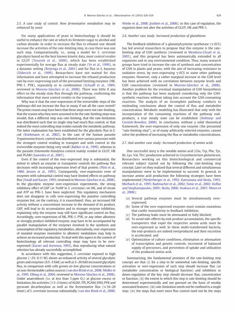
Fig. 1. Experimental determination of flux control coefficient. Small variation in theactivity (aio) of a controlling enzyme (or transporter) significantly affects pathway fluxJo (curve A, position 1), whereas significant variation in the activity of non-controllingenzymes (or transporters) has negligible effect on flux (curve B, position 2).
628 R. Moreno-Sánchez et al. / Mitochondrion 10 (2010) 626–639
two different features at play, and that it should be useful todistinguish between these by giving them two different names. Theword ‘control’ continued to be associated with the extent of limitationas expressed by the control coefficients (for pathway flux, metaboliteconcentration). The word ‘regulation’ was proposed to indicatewhether the organism or cell type under study was actually changingthe amount of an enzyme (by transcriptional, post-transcriptional,translational, and/or post-translational modification processes) whentrying to change the flux through the corresponding process (ter Kuileand Westerhoff, 2001; Bevilacqua et al., 2008). The correspondingquantitative measure of regulation was termed the hierarchicalregulation coefficient. It was defined as the percentage change inthe activity of the enzyme divided to the percentage change in flux.
This regulation coefficient has been determined for a number ofcases where the yeast Saccharomyces cerevisiae regulated its carbonmetabolism to adjust to carbon or nitrogen starvation. The hierarchicalregulation coefficients differed widely between the various glycolyticenzymes in a way that did not correspond with the suspecteddistribution of flux control amongst the glycolytic enzymes (seebelow). The implication is that when the yeast cell regulates itself itdoes not always regulate the steps with the highest control coefficients,neither does it regulate all steps by the same factor (Rossell et al., 2006;van Eunen et al., 2009). Hence, control and regulation are not the same.
2.4. More theory for metabolic control
MCA is an operational framework, in part theoretical (Kacser andBurns, 1973) in part experimental (Groen and Westerhoff, 1990),which rationalizes the quantitative determination of the degree ofcontrol that a given enzyme (or cellular process) exerts on flux (orbiological function) and on the concentration of metabolites. It helpsto identify and design experimental strategies for the molecularmanipulation of a given physiological process in an organism(reviewed in Fell, 1997; Moreno-Sánchez et al., 2008; Westerhoff etal., 2009; 2010). Different approaches have been developed todesigning what has to be done and measured in order to identifyand understand why an enzyme exerts significant or negligiblecontrol on flux and metabolite concentration. Thus, the application ofMCA avoids the “trial and error” experiments for identifying andmanipulating the conceptually wrong and misleading “rate-limitingstep” concept and can readily explain results observed in enzymeover-expression and down-regulation experiments. To understandhow a metabolic pathway is controlled and could be manipulated, itscontrol structure has to be evaluated.
The control structure of a pathway is constituted by (i) thecollection of flux control coefficients (CJ i), each of which is the degreeof control that the activity (a) of a given enzyme i exerts on flux J; (ii)the collection of the concentration control coefficients (CX i), each ofwhich is the degree of control that a given enzyme i exerts on theconcentration of a metabolite (X); and (iii) the elasticity coefficients(Burns et al., 1985). The control coefficients are systemic properties ofthe pathway enzymes and transporters that, in turn, are mechanis-tically determined by their elasticity coefficients (εviX or εaiX), whichare defined as the degree of sensitivity of a given enzyme rate vi or aito variations in the concentration of any ligand X (e.g., the enzyme'sability to change its rate if the concentration of any of its ligands, i.e.substrate, products or allosteric modulators, is varied at constantconcentration of all other ligands).
The flux control coefficient is defined as:
CJi =
δJδai
·aioJo
≈ % change in flux% change in activity of enzyme i
in which the expression δJ/δai describes the variation in flux (J) whenan infinitesimal change is effected in the activity (Vmax) of enzyme i(Burns et al., 1985; Kholodenko et al., 1995). Any other biological
function can also be considered as “flux” whereas a metabolicpathway may become the “enzyme activity”. In practice, infinitesimalchanges J are undetectable, and hence measurable, non-infinitesimalchanges are determined, with extrapolation to very small changes. If asmall change in ai promotes a significant variation in J, then thisenzyme exerts an elevated flux control (Fig. 1, curve A, position 1). Incontrast, if a rather small or negligible change in flux is observedwhenai is greatly varied, then the enzyme does not exert significant fluxcontrol (Fig. 1, curve B, position 2). To obtain dimensionless andnormalized values of (CJ i), the scaling factor ao/Jo is applied, whichrepresents the ratio between the initial magnitudes of enzyme activityand flux (and at which point the slope δJ/δai is calculated). If all (CJ i)sof all enzymes and transporters in the total network (i.e. alsoincluding enzymes in the network but outside the pathway) areadded up, the sum comes to one when J represent flux and to 0 when Jrepresent the concentration of a metabolite, a membrane potential orthe phosphorylation potential of ATP (summation theorem) (Heinrichand Rapoport, 1973; Kacser and Burns, 1973; Westerhoff and vanDam, 1987).
MCA clearly distinguishes between the control exerted by a givenenzyme on flux (flux control coefficient) and on the metaboliteconcentration (concentration control coefficient). Thus, an enzymecan have significant control on a metabolite concentration but not onthe pathway flux or vice versa. This distinction is important forbiotechnological and clinical purposes: The qualitative rate-limitingstep concept for manipulating metabolic pathways does not makesuch differentiation, which probably has contributed to the manyunsuccessful experiments reported in the literature. It should beestablished whether the aim of the project is to increase flux or toincrease a metabolite concentration since MCA establishes for eachaim a different experimental design.
To determine the flux control coefficient of a given enzyme, smallvariations in the enzyme content, or preferentially, in activity arerequired, without altering the rest of the pathway, and then thechanges in flux should be determined. The experimental points areplotted as shown in Fig. 1 and the slope at the reference point ao/Jo iscalculated. This experiment, apparently easy to perform, hasdemanded great intellectual and experimental effort (e.g., Flint etal., 1980; Groen et al., 1982; Mijakovic et al., 2005). Application ofMCA towards a better understanding of cancer biology and identifi-cation of the most susceptible proteins for drug targeting, may wellrequire a deeper knowledge of biochemistry and cell biology.However, the complexity of the disease may demand such anintensified intellectual and scientific effort and its increasingincidence and epidemiology may warrant it.
629R. Moreno-Sánchez et al. / Mitochondrion 10 (2010) 626–639
2.5. A case study of control: How fermentative metabolism may beenhanced by yeast
For many applications of yeast in biotechnology it should beuseful to enhance the rate at which its ferments sugar to alcohol andcarbon dioxide. In order to increase the flux to ethanol one shouldincrease the activities of the rate-limiting step, in case there was onesuch step. Computationally, i.e., using a model for S. cerevisiaeglycolysis based on the best known kinetic data, most control residesin GLUT (Teusink et al., 2000), which has been establishedexperimentally for average flux at steady state (Ye et al., 1999), ina dynamic setting (Reijenga et al., 2001) and for flux in S. bayanus(Diderich et al., 1999). Researchers have not waited for thisinformation and have attempted to increase the ethanol productionrate by over-expressing each of the presumed limiting enzymes (HK,PFK-1, PYK), separately or in combination (Schaaff et al., 1989;reviewed in Moreno-Sánchez et al., 2008). There was little if anyeffect on the steady state flux through the pathway, confirming theinformation that most control resides in the transport.
Why was it that the over-expression of the irreversible steps of thepathways did not increase the flux in many if not all the cases tested?Theprime reasonmayhave been that these stepswere not rate-limiting,that the reasonwhy theywere assumed tobe the rate-limiting stepswasinvalid, that a different step was rate-limiting, that the rate-limitationwas distributed such that no single step had much flux control, or thatindeed themost controlling step resides outside themetabolic pathway.The latter explanation has been established for the glycolytic flux in E.coli (Koebmann et al., 2002). In the case of the human parasiteTrypanosoma brucei, controlwasdistributed over various processeswiththe strongest control residing in transport and with control in theirreversible enzymes being very small (Bakker et al., 1999), whereas inthe parasite Entamoeba histolytica control mainly resided in GLUT, HKand PGAM (Saavedra et al., 2007).
Even if the control of the over-expressed step is substantial, theextent to which an enzyme or transporter controls the pathway fluxdecreases with increasing expression level of that protein (Flint et al.,1980; Jensen et al., 1993). Consequently, over-expression even ofenzymes with substantial control may have limited effects on pathwayflux (Small and Kacser, 1993; reviewed inMoreno-Sánchez et al., 2008).In the glycolytic enzyme over-expression experiments, the stronginhibitory effect of G6P (or Tre6P in S. cerevisiae) on HK, and of citrateand ATP on PFK-1, have been neglected. This regulatory mechanismdoes not disappear in cells over-expressing the putative controllingenzymes but, on the contrary, it is exacerbated; thus, an increased HKactivity without a concomitant increase in the demand of its product,G6P, will lead to its accumulation and to stronger enzyme inhibition,explaining why the enzyme may still have significant control on flux.Accordingly, over-expression of HK, PFK-1, PYK, or any other allostericor strongly product-inhibited enzyme, may have to be accompanied byparallel manipulation of the enzymes involved in the synthesis andconsumptionof the regulatorymetabolites; alternatively, over-expressionof mutated enzymes insensitive to allosteric modulation may help toachieve an increased production. To dealwith this aspect in the context ofbiotechnology all relevant controlling steps may have to be over-expressed (Kacser and Acerenza, 1993), thus reproducing what naturalselection has already successfully accomplished.
In accordance with this suggestion, S. cerevisiae exposed to highglucose (N2%; 0.11 M) shows an enhanced activity of several glycolyticgenes and enzymes (0.5–3 fold) aswell as 5–20 fold increased glycolyticflux, in comparison with cells grown on low glucose concentrations oronnon-fermentable carbonsources (vandenBrink et al., 2008;Müller etal., 1995; Elbing et al., 2004; reviewed in Moreno-Sánchez et al., 2008).Under anaerobiosis (i.e. at less than 5 ppm O2) at glucose excess orlimitation, the activities (1.5–2 times)of ALDO, TPI, PGAM, ENO, PYKandpyruvate decarboxylase as well as the fermentative flux (≈16–20times) of S. cerevisiae increase substantially (van den Brink et al., 2008;
Wiebe et al., 2008; Jouhten et al., 2008); in this case of regulation, theorganism does not alter the activities of GLUT, HK and PFK-1.
2.6. Another case study: Increased production of glutathione
The feedback inhibition of γ-glutamylcysteine synthetase (γ-ECS)has led several researchers to propose that this enzyme is the rate-limiting step of GSH synthesis (reviewed in Mendoza-Cózatl et al.,2005), and this proposal has been automatically extended to allorganisms and to any environmental condition. Thus, many researchgroups have tried to increase the rate of synthesis and concentrationof GSH in plants and yeasts, with the aim of increasing resistance tooxidative stress, by over-expressing γ-ECS or some other pathwayenzymes. However, only a rather marginal increase in the GSH levelhas been achieved with no correlation between enzyme levels andGSH concentration (reviewed in Moreno-Sánchez et al., 2008).Another problem for the eventual manipulation of GSH biosynthesisis that the pathway has been analyzed considering only the GSH-synthetic reactions without taking into account the GSH-consumingreactions. The analysis of an incomplete pathway conducts tomisleading conclusions about the control of flux and metaboliteconcentration. Metabolic modeling has illustrated that only with theincorporation of the consuming reactions of the pathway end-products, a true steady state can be established (Hofmeyr andCornish-Bowden, 2000). In conclusion, without a solid theoreticalframework, the over-expression of only one enzyme (the presumed“rate-limiting step”), or of many arbitrarily selected enzymes, cannotsolve the problem of increasing the flux or metabolite concentrations.
2.7. And another case study: Increased production of amino acids
One successful story is the notable amino acid (Glu, Trp, Phe, Tyr,Ile, Lys, Val, Thr) production achieved in Corynebacterium glutamicum.Researchers working on this biotechnological and commercialrelevant subject started out by following the rate-limiting stepconcept. Later on they realized that several different and simultaneousmanipulations were to be implemented to succeed. In general, toincrease amino acid production the following strategies have beenimplemented (Niederberger et al., 1992; Katsumata and Ikeda, 1993;Morbach et al., 1995; Radmacher et al., 2002; Simic et al., 2002; Koffasand Stephanopoulos, 2005; Ikeda, 2006; Asakura et al., 2007; Moon etal., 2008):
(a) Several pathway enzymes must be simultaneously over-expressed;
(b) Some of the over-expressed enzymes must contain mutationsthat confer insensitivity to feedback inhibition;
(c) The pathway leaks must be attenuated or fully blocked;(d) To avoid side-effects by end-product accumulation, the specific
transporters that expel the respective amino acids must beover-expressed as well. In these multi-transformed bacteria,the end-products are indeed overproduced and their excretionis accelerated; and
(e) Optimization of culture conditions, elimination or attenuationof transcription and genetic controls, increment of balancedsupply of precursors, and prevention of uptake and utilizationof the produced amino acid.
Summarizing, the fundamental premises of the rate-limiting stepconcept are that (i) for a step to be somewhat rate-limiting, specificactivation or over-expression of such step should increase flux (ormetabolite concentration or biological function) and inhibition ordown-regulation of the key step should decrease flux, concentrationor function; (ii) the extent to which this step is rate-limiting should bedetermined experimentally and not guessed on the basis of weaklyassociated features; (iii) rate-limitationneeds not be confined to a singlestep; (iv) the steps that exert more flux control need not be the steps

630 R. Moreno-Sánchez et al. / Mitochondrion 10 (2010) 626–639
that are used by the cell to regulate the flux; and (v) for up-regulatingthe flux, over-expression of all enzymes by the same factor may be agood strategy.
3. The quintessence: Because cells are not a library, they requireintegrative systems biology
Still it is paradoxical. Why is it that a molecular process that isintricately involved in a cellular function need not control itsperformance (flux)?Why does not the H+-translocating ATP synthasedetermine its own function, i.e. the ATP synthesis flux it carries (Groenet al., 1982; Jensen et al., 1993)?Why is not p53 necessarily in controlof cell death just because it is the node in the networks regulating celldeath (Lazebnik, 2002)? The mechanistic reason is that mostmacromolecules that carry out the processes of life are “behavingresponsibly”. They adjust their functioning to signals they receivefrom their intracellular environment (Groen et al., 1982). Themacromolecules we refer to are the enzymes that catalyze metabolicprocesses, the transcription factors that bind DNA and activatetranscription, the enzymes that synthesize and degrade otherenzymes as well as the protein kinases and phosphatases thatmodulate other enzymes. Their responsiveness (i.e. elasticity coeffi-cients) is to changes in the activities or concentrations of othermolecules in the living cell around them, such as intermediarymetabolites or other, phosphorylatable proteins. These changes resultfrom alterations in the activities of the enzymes that produce orconsume them. And these activity changes are again the manifesta-tion of the responsiveness of those enzymes, and of the transcriptionand translation processes setting the concentrations of thoseenzymes. As a consequence possibly all molecules in the cell are atleast indirectly responsive to all othermolecules in the cell. If one suchmacromolecule, say the ATP synthase is activated by a factor of two,then initially the rate at which it synthesizes ATP may increase by thesame factor, but as time goes on its rate will decrease such that thefinal rate is in between the original rate and twice that original rate.The reason is that the increased rate of ATP synthesis will haveincreased the level of ATP and decrease the levels of ADP, Pi andproton motive force, and all these changes will be sensed by theenzymes and make it tone down its rate.
The functional reason why all intracellular processes tend to beresponsible in the above sense is to make the living cell self-sustainable. Whenever an enzyme activity would get too high, itwould adjust itself. In a chemical reaction pathway onewould not findthis phenomenon. There, the first step in the pathway is the only rate-limiting step and the concentration of the penultimate metabolite hasno say. In contrast, in biology there is often at least partial control byend-product demand (Hofmeyr and Cornish-Bowden; 2000).
Because of this responsiveness, most molecules that carry out allthe cellular functions engage strongly with the networks they are in.And because all those networks are again connected, the behavior ofthe molecules is determined by the cellular system as a whole. This iswhy the biology that tries to understand the functions of life in termsof molecules cannot be the biology of the molecules, but must besystems biology (Alberghina andWesterhoff, 2005). A librarywith theinformation of the individual molecules is not sufficient to understandfunction; we shall need a movie in which every molecule is a player.
Due to the complexity of multi-component systems such asmetabolic networks, the analysis, management, integration andcomprehension of the multiple variables involved in their functioning(thermodynamic equilibrium constants; kinetic constants of enzymesand transporters; pathway intermediary concentrations and fluxes;covalent regulation; protein synthesis and degradation; and geneticcontrol and expression) seems impossible for a researcher. Systemsbiology is a combined theoretical–experimental approach that allowsfor the integrative analysis of metabolic networks functioning andhelps the humanmind to understand how complex biological systems
work and how they can be perturbed. Thus, the researcher's expertiseand scientific knowledge on cellular metabolism is potentiated, beingnow able to perform an integrative and dynamic analysis of cellularnetworks. Therefore, systems biology does not add the behaviors ofthe molecules but integrate them. The mathematical procedure ofintegration multiplies the behavior of each molecule for a very shortperiod of time, but will then recalculate that behavior on the basis ofall the new concentrations, before integrating again. The procedure isthen iterated, continuously updating the mathematical behavior inthe light of the development in time of the environment that theysense. This is how the biology works and this is therefore how ourunderstanding of biology should operate, i.e. integratively.
Thanks to infusions from genomics and mathematical biology,integrative systems biology has been able to develop rapidly, with asignificant number of scientific achievements that are relevant for theproblem at hand (Westerhoff and Palsson, 2004). Consensus mapshave beenmade of genome-widemetabolism of yeast (Herrgard et al.,2008) and soon of the human. All possible pathways through thesemaps can now be calculated (Schuster et al., 2002). Through fluxanalysis, the pathways can be measured (Isermann and Wiechert,2003), and through so-called flux-balance analysis, the relative steadystate fluxes through these can be predicted if one assumes that thesystem is optimal from a certain perspective, such as ATP or growthyield (Varma and Palsson, 1993).
Notably, for yeast, the systems biology predictions seem inaccurate,suggesting that other optimization criteria have been important inevolution, or that the cost of certain processes have not yet been takeninto account properly (Molenaar et al., 2009; Westerhoff et al., 2010).The same issue is likely to be relevant for tumor cells. The veryWarburgeffect seems not to have been optimized for producing as much ATP forgrowth as possible, perhaps because other factors than ATP, such asoxygen supply, limit growth. The enzymes and metabolites involved inglycolysis, the Krebs cycle, and OxPhos are all part of a system. The ATPthat is made almost at the end is needed almost at the beginning. TheWarburg effect itself is so hard to understand because it could resultfrom a primary mutation activating glycolysis or from a primarymutation inactivating OxPhos. And if indeed, the genotype of tumors isdetermined by selection for a phenotype then one may expect bothcases to occur at different degrees. Considerations of metabolism, geneexpression and signal transduction need to be integrated; also here thesystems biology requires being integrative.
Ultimately the behavior of systems depends on the responsivenessof all their components. An ultimate version of systems biologytherefore is the silicon cell, i.e. a precise replica of the networks interms of kinetic equations of a component process (Snoep, 2005;Westerhoff et al., 2009). The parameters in those equations shouldstem from accurate experimentation. Consequently, such silicon cellsare still rare whereas kinetic modeling of individual pathways hassurged (for a complete list of available models see http://www.jjj.bio.vu.nl). In turn, kinetic modeling has facilitated:
(i) To quantitatively determine the control coefficients of individ-ual enzymes (or pathways) to identifying those steps (orpathways within networks) with the highest control on fluxand metabolite concentrations (or biological function). Thetechnically difficult experiment of varying the activity of asingle enzymewithout perturbing the rest of the system, whichis essential for determining the control coefficients, as depictedby MCA framework, is easier to perform with the use ofmathematical models;
(ii) To understand the underlying kinetic mechanisms by which apathway enzyme or transporter exerts significant or negligiblecontrol;
(iii) To identifying themolecularmechanisms bywhich all enzymescommunicate with each other (metabolites, and coenzymes) tocontrol their rates;

631R. Moreno-Sánchez et al. / Mitochondrion 10 (2010) 626–639
(iv) To predict pathway and network behavior under differentphysiological relevant conditions (individual or multipleenzyme over-expression or inhibition).
In the post-genomic era, a silicon cell seems now feasible to buildwith the advent of high-throughput screening methods such asgenomics, transcriptomics, proteomics and metabolomics. Thesetechnologies produce a mass of information which by computationalmodeling allows for an integrative analysis to reconstructing themetabolic and signaling cellular networks. For heart energy metab-olism a model has been created that gets close to such silicon cell(Cortassa et al., 2009b). Such silicon cells should enable us tounderstand ultimately how the Warburg effect occurs mechanistical-ly, and should also enable nonlinear flux-balance analyses suggestingwhy it occurs functionally in tumors. In addition, an iterative processof experimentation and modeling can help us to identifying the mostappropriate drug-targets with the highest therapeutic potential.
In the context of cancer, systems biology has been implemented insignal transduction (Hornberg et al., 2006). Here it was shown why itshould not have been surprising that there are so many oncogenes,and why there are so many tumor suppressor genes. The oncogeneproduct may be amongst the worst target for inhibitor action unlessaddiction has taken place (Hornberg et al., 2006).
Before systems biology came along, MCAwas limited to pathways atsteady state. It has since been developed for time dependentphenomena (Acerenza et al., 1989; Bier et al., 1996; Kholodenko et al.,1997; Westerhoff et al., 2009) and for spatial aspects (Francke et al.,2002), although the experimental applications in those fields remainrare. MCA's extension to the genome-wide network includes geneexpression and signal transduction and has been called hierarchicalcontrol analysis (Snoep et al., 2002; Kahn andWesterhoff, 1991). Muchsystems biology beyond MCA is ready to be implemented withenormous potential.
4. The rate-limiting step concept still governs basic research ontargeting cancer cells
Notwithstanding the above observations, researchers working ondrug design for novel and better venues of cancer treatment (and in factof most human diseases), have tended to assigning an essential role totheir single favorite protein. This leads to a less complicated set ofoptions for experimental testing and to clarity and simplicity ofinterpretation. The selection of only one “essential” protein isunderstandable and even justifiable when the cost of the enterprise isevaluated.However, simplicity should onlybeaspired to if reality itself issimple (Westerhoff et al., 2010). Bynow it is clear that the reality ofmostcancers is not simple at all, but requires the amplification of multipleoncogenes (Hanahan and Weinberg, 2000), and cannot just be seen asresulting from p53 amplification alone (Lazebnik, 2002). In addition,even the complete removal of a protein responsible for a given relevantcellular process (by knock-out of the corresponding gene, or nowadaysparticularly by RNA interference technology), rarely leads to thecomplete repression of the corresponding biological function, aphenomenon that has been called redundancy. Any concept of a singlerate-limiting step that determines and should be hit by an anti-tumordrug is a liability for the success of cancer research.
The cytotoxic approach to pathological cells in a human body, suchas cancer cells or parasites, should deal with the fact that the functionsof both the pathological cells and the host are managed by networksrather than single rate-limiting molecules (Bakker et al., 2002). Thebest drug targets may not be the favorite protein or genemolecule butthe networks themselves (Hornberg et al., 2006; Bakker et al., 2002;Saavedra et al., 2007;Westerhoff et al., 2008), or diffuse loops in thosenetworks (Cortassa et al., 2009b). In fact, drugs should target thedifference between the network of the pathological cell and thenetwork of the host (Bakker et al., 2002; Saavedra et al., 2007). The
approach should also reckonwith the fact that oncogene amplificationtends to make the cancer cell more robust with respect toperturbation of the oncogene. Therefore, inhibiting the oncoproteinitself may well harm the healthy host tissue more than the tumor(Hornberg et al., 2006), unless the phenomenon of ‘addiction’ kicks in.
Indeed, side-effects from chemotherapy are always evident and theycan be moderate, severe or lethal. Deletion or full inhibition of anyenzyme or transporter, protein or gene, in a given cellular process isexpected to be successful in cancer treatment only if tumor cells aretargeted specifically whereas normal, healthy cells remain unaffected.Because of the extensive homology between most tumor cells andhealthy cells that are important for the function of the patient, thiscomplete inhibition strategymaywell be unrealistic: at full inhibition ofthe tumor, the healthy tissues of the patient will also be damaged.Complete blockade of an enzyme or transporter activity that followsclassical Michaelis–Menten kinetics requires the addition of more than100 times the Ki value, which in practice is not achieved withoutcompromisinghealth.Moreover, it has been experimentally determinedthat, to significantly alter the pathway flux, activity of non-controllingstepshave to bedecreased beyonda threshold of 60% ormore (Rossignolet al., 2003; reviewed in Moreno-Sánchez et al., 2008). Remarkably, thepercentage of inhibition in protein expression normally achieved byRNA interference is around this threshold value, resulting in an over-interpretation of results when using this tool to supporting hypothesesthat non-controlling transporters or enzymes are indeed the “rate-limiting step”. With such a criterion of effect upon 60% inhibition,multiple steps in a pathway would become “the rate-limiting step”.
Yet, the approach of identifying, and targeting, “the rate-limitingstep” continues today and appears to be the dominant theoreticalsupport for an overwhelming number of studies in drug design andcancer biology fields. Examples spread from hormone receptors andsignal transduction components, transcriptional factors to “metabolicenzymes”. For instance, it has been postulated that vascular endothelialgrowth factor receptor (VEGFR), or endothelial growth factor receptor(EGFR) or downstream protein kinases (PKs), have an “essential” or“pivotal” role in the growth of tumors and in consequence targeting ofthese proteins is pursued actively (reviewed in Benouchan andColombo, 2005; Johnson, 2009). Similar arguments have been used tojustifying the development of specific drugs directed to (i) the “key” or“major” transcription factor HIF-1 (Melillo, 2007; Fulda and Debatin,2007) or (ii) the prolyl-hydroxylases (PHDs; Gottlieb and Tomlinson,2005), the “main” enzymes involved in HIF-1 degradation, (iii) the“master metabolic regulator” Akt/PI3K/mTOR signal transductionpathway (Jones and Thompson, 2009), (iv) the “rate-limiting stepmetabolic enzymes” PDK (Denko, 2008), PFK-2 (Yalcin et al., 2009), LDH(Fantin et al., 2006), fatty acid synthase (FAS; Pizer et al., 2000;Mashimaet al., 2009), ATP citrate lyase (ACL; Hatzivassiliou et al., 2005;Mashimaet al., 2009), or aromatase (P-450) (Bhatnagar, 2007), or (v) “important”pro-apoptotic or anti-apoptotic proteins such as TRIAL andXIAP, or BCL-2, respectively (Fesik, 2005; Fantin and Leder, 2006). In none of thesecases, hard, quantitative experimental data have been producedsupporting the presumed rate-limitation of the above-mentionedselected proteins and processes on tumor growth.
5. Glycolysis in cancer cells
Glycolysis is enhanced in many human and animal neoplasias(reviewed in Moreno-Sánchez et al., 2007). How have these cancersup-regulated their glycolysis?: (i) Have they done this by enhancingthe expression of a single rate-limiting step? (ii) Have they over-expressed all enzymes to the same extent? (iii) Or have they used amore subtle approach? The answer to the first question is clear formost cancers: Yes, all or most of the glycolytic enzymes andtransporters are over-expressed in at least 70% of human cancers(Altenberg and Greulich, 2004). Gene expression of glycolyticproteins (GLUT1, GLUT3, HKI, HKII, PFK1-L, PFKFB-3, ALD-A, ALD-C,

632 R. Moreno-Sánchez et al. / Mitochondrion 10 (2010) 626–639
TPI, GAPDH, PGK1, PGAM-B, ENO-α, PYK-M2, LDH-A, and MCT4) isfurther stimulated by hypoxia in a process mediated by thetranscription factor HIF-1α (hypoxia-inducible factor 1α) (reviewedin Marín-Hernández et al., 2009). The HPI gene is also induced byhypoxia, but through a HIF-1α independent mechanism.
The activity of all glycolytic enzymes is concomitantly enhanced inrat AS-30D hepatoma, 2–4 fold for HPI, ALD, TPI, GAPDH, PGK, PGAM,ENO, and LDH; 8–10 fold for PYK; and 17–300 fold for PFK-1 and HK(Marín-Hernández et al., 2006), in comparison with normal rathepatocytes, the tissue of origin. In rat Morris hepatomas, theactivities of HK, PFK and PYK are 5–500 times higher than in liver(Stubbs et al., 2003). In human breast cancer, the activities of HK, ALD,PYK, and LDH are 3.7–7 times higher than in normal tissue (Balinskyet al., 1984). In human cervix HeLa cells, all enzyme activitiesincluding HK and PFK-1 are enhanced by 2–7 fold, except for PGAMand LDH (Marín-Hernández et al., 2006). Because in all these cases theexpression of more than one glycolytic gene is enhanced, or theactivity of more one enzyme is increased, the answer to the thirdquestion above is ‘No’: Most cancers have not just over-expressed asingle rate-limiting step. And because the extents of over-expressiondiffer significantly between the various enzymes in the same cancer,the answer to the second question is ‘No’ as well: Apparently, cancershave enhanced their glycolysis in subtler ways, perhaps as subtle asnoted in yeast (Rossell et al., 2006).
In summary, the strategy selected by cancer cells to achieve anincreased glycolytic flux to maintaining an adequate ATP supply ishighly similar to that described for amino acid over-producingbacteria:
(a) Over-expression of most glycolytic proteins (reviewed inMoreno-Sánchez et al., 2007);
(b) Isoenzyme expression shift, from the PFK-1 isoenzyme withallosteric inhibitor-sensitivity/low allosteric activator affinityto the PFK-1 isoenzyme with allosteric inhibitor-insensitivity/high allosteric activator affinity (reviewed in Marín-Hernándezet al., 2009);
(c) Decrease in the activity of relevant branching fluxes, e.g.,through the partial inactivation of PDH complex (Denko, 2008)thus diminishing leaks; and
(d) Over-expression of MCT4 (Gallagher et al., 2007), the plasmamembrane transporter responsible for secreting lactate.
In other words, cancer cells do not over-express solely one rate-limiting step to increase glycolytic pathway flux and metaboliteconcentration, but they follow a variety of simultaneous strategies toachieve the desired objective. It is as with microorganisms found in aniche: they have evolved/adapted in whatever way it took to thrive. Itwill be interesting and important to try to understand more preciselywhichways these are, perhaps in order to interfere with them, or withtheir selection.
6. A mishap in the Warburg hypothesis? Functional mitochondriaas anti-cancer targets
We shall now present an example of an analysis of a number ofcancers from a more subtle point of view than the rate-limiting stepconcept. Warburg (1956) originally proposed that the prime cause ofcancer was an energy deficiency caused by an irreversible damage tomitochondrial function, which induced an increased glycolysis. Sincethen, the field of cancer biology research has assumed that theWarburg hypothesis applies to all or most cancer cell types (Pedersen,1978; Atsumi et al., 2002; Rossignol et al., 2004; Robey et al., 2005; Xuet al., 2005; Seyfried and Mukherjee, 2005; Gottlieb and Tomlinson,2005; Matoba et al., 2006; Denko, 2008; Kroemer and Pouyssegur,2008; Jones and Thompson, 2009), because one of the hallmarks ofmany types of cancer is certainly an increased glycolytic capacity,
which persists in the presence of high O2 concentration (reviewed inMoreno-Sánchez et al., 2007, 2009).
However, contribution of glycolysis to cellular ATP supply can beas low as 10% in some cancer types and 50–70% in other cancer types(reviewed in Moreno-Sánchez et al., 2009). In consequence, OxPhosalso significantly contributes to ATP supply in cancer cells. Somewhatsurprisingly, this second aspect of the Warburg hypothesis (“cancercells have impaired mitochondria”) has been ignored or taken as anestablished fact. Because of the absence of hard experimentalevidence for this part of the hypothesis, it has rather become themetabolic central dogma of tumor cells.
According toWarburg's hypothesis, themain oxidizable substrate forcancer cells in the human body should be glucose, and glycolyticinhibitors should potently block tumor growth. Substrates oxidized bymitochondria should not be metabolized by cancer cells and hencemitochondrial inhibitors should be innocuous.Glycolytic inhibitors suchas gossypol, 2DOG, 3-BrPyr, oxamate are indeed effective anti-cancerdrugs but at relatively high doses and only when used in combinationwith drugs targeting other pathways or cellular processes. In addition,they are not specific for the glycolytic enzymes and transporters, andthey are also cytotoxic for non-tumor cells (reviewed in Pelicano et al.,2006; Rodríguez-Enríquez et al., 2009). On the other hand, mitochon-drial inhibitors are potent tumor growth blockerswhen used alone or incombination with other anti-cancer drugs. Therefore, the analysis ofwhole set of available data indicates that glycolysis may not be the rate-limiting pathway for ATP production in some cancer cells, or thatmitochondria have an essential function for tumors other than just ATPproduction. Hence, the usual statement that glycolysis predominatesover OxPhos in terms of ATP supply in tumor cells should be re-evaluated andmost probably verified experimentally for each particulartype of tumor cells.
7. Energy metabolism in mitochondria-deficient cells (rho cells)and stem cells
It has been demonstrated that rho cells survival depends on anenhanced glycolytic activity (60%more vs. parental) which is coupled toan efficient plasma membrane redox system (Hyun et al., 2007), tomaintaining the cytosol redox status. In consequence, rho cells viabilityis drastically abolished by glycolytic inhibitors (2DOG, iodoacetamide,oxamate) at doses 5–10 times lower than those used in wild type cells,whereas significant higher doses (50-times vs. wild type) of mitochon-drial inhibitors (rhodamine 123, safranine O) are required to abolishcellular growth (Hu et al., 2000; Liu et al., 2001; Hyun et al., 2007). Thelactate overproduction and the anti-glycolytic drugs hypersensitizationobserved in rho cells is similar to that observed in parental counterpartcells after OxPhos inhibitors (rhodamine 123, rotenone, antimycin)treatment and in solid tumor hypoxic micro-regions (Rodríguez-Enríquez et al., 2009). These observations indicate that, only whenOxPhos is fully abolished, cells switch over to glycolysis (Liu et al., 2001).On the other hand, the lack of mitochondria, and hence the absence of amitochondrial inner membrane electrical gradient (negative inside),diminishes the ability of rho cells to uptake and accumulate delocalizedlipophilic cation drugs (such as rhodamine 123, 6G, MKT077 and otherpositively charged anti-neoplastic drugs) (Hu et al., 2000; reviewed inRodríguez-Enríquez et al., 2009), thus limiting their anti-proliferationeffect.
The absence of mtDNA in rho cells also brings about (i) higherresistance towards classical anti-neoplastic drugs (paclitaxel, doxoru-bicin, daunomycin) decreasing apoptosis incidence; (ii) development ofa more invasive cancer phenotype in prostate carcinoma and osteosar-coma (Ferraresi et al., 2008; Moro et al., 2008; Mineri et al., 2009); (iii)decrease in the cellular ROS concentration compared to their parentalcounterpart cells (Hyun et al., 2007; Cuperus et al., 2009).
As stem cells are now considered as the precursors of cancer, it isrelevant to analyze how these cells deal with the problem of ATP

633R. Moreno-Sánchez et al. / Mitochondrion 10 (2010) 626–639
supply/demand. Normal human mesenchymal stem cells exhibit (i)higher glycolytic enzymes levels and lactate production rates, (ii)higher sensitivity to glycolytic drugs; and (iii) lower sensitivity toOxPos inhibitors. Upon induction to differentiation, these cellsdevelop: (i) higher number of mtDNA copies, content of respiratorychain enzymes, ATP content and sensitivity to OxPhos inhibitors; (ii)accelerated oxygen consumption rate; and (iii) lower lactateproduction and sensitivity to glycolytic inhibitors (Chen et al.,2008). Thus, these observations suggest an energy metabolismtransition from glycolysis to OxPhos in normal stem cells withdifferentiation (Chen et al., 2008).
No changes in the expression of themajority of the glycolytic gene,together with significant decrease in lactate production and glucoseuptake, is observed in monolayer-cultured oncogenic-transformedmesenchymal stem cells in comparison with their parental stem line.In parallel, oncogenic transformation of stem cells also induces up-regulation of some genes of the Krebs cycle and higher mitochondrialATP-dependence (Funes et al., 2007). In strikingly contrast, tumorsderived from the same cancer stem cells show up-regulation ofglycolytic genes and down-regulation of Krebs cycle genes (probablyinduced by hypoxia), although the glycolytic flux does not signifi-cantly change. Thus, these observations suggest that normal stem cellsare predominantly glycolytic but they shift towards oxidative energymetabolism when experiencing oncogenic transformation, which inturn questions the assumption that aerobic glycolysis is part of theneoplastic transformation (Funes et al., 2007). The complete charac-terization of the energy metabolism setting in cancer stem cells willfacilitate application of MCA and systems biology for target identifi-cation and design of appropriate drugs.
8. Control analysis of cancer energy metabolism
MCA of tumor energy metabolism has been undertaken. Elasticitycontrol analysis of glycolysis in AS-30D hepatocarcinoma (Marín-Hernández et al., 2006) showed that the main flux control (71%)resided in the first part of the glycolytic pathway (i.e. GLUT and HK).The rest of the control (29%) was localized in the ALD–LDH segment,with a negligible contribution of PFK-1 (b6%). Despite its exacerbatedover-expression (100–500 fold in AS-30D hepatocarcinoma), tumorHK was strongly inhibited by its product G6P and hence only an 8–10fold increase in flux was achieved. On the other hand, PFK-1 wasmoderately over-expressed, but the tumor isoenzyme was highlyactivated by F2,6BP and AMP, which surpassed the inhibition bycitrate, ATP, and low pH. These findings provided a mechanisticexplanation for the respective high and low flux control exerted by HKand PFK-1 in tumors. The study also showed that a massive over-expression of glycolytic enzymes does not lead to uncontrolled flux,but rather invokes strict regulatory mechanisms (potent product-inhibition, allosteric modulation), which persist in the tumor cells.Kinetic modeling of glycolysis in AS-30D and HeLa tumor cells hasrevealed that indeed GLUT and HK together with HPI are the mainflux-controlling steps in both tumors.
An integrated modeling of OxPhos (including Krebs cycle,respiratory chain, membrane potential, ATP/ADP exchange, and ATPsynthase) and mitochondrial Ca2+ dynamics in heart showed that thecontrol of the ATP synthesis was distributed among several steps(Cortassa et al., 2009a,b). This feature of distributed control appears tobe conserved in tumor cells: Control analysis of OxPhos in AS-30Dhepatocarcinoma showed that the respiratory chain site 1 (30%) andthe ATP-consuming enzyme block (protein and nucleic acid synthesis;ion ATPases) (34%) were the main controlling sites (Rodríguez-Enríquez et al., 2000). The latter observation shows that the fluxcontrol may reside in part, outside the pathway, supporting theproposal by Hofmeyr and Cornish-Bowden (2000) that the end-product demand (which is usually overlooked in studies of metab-
olism because these metabolites are frequently not considered as partof the pathway) also exerts significant flux control (see also above).
Control analysis of tumor energy metabolism establishing themain sites of control in glycolysis and OxPhos, and Flux Analysisassessing the predominant energy pathway (cf. Westerhoff et al.,2010), may provide a more rational and quantitative approach to theidentification and design of more specific therapeutic strategies.Therefore, it should be desirable to apply MCA of energy metabolism(and of other relevant pathways and cellular processes) tomany otherdifferent types of cancer, avoiding misleading generalizations such as“ATP supply in cancer cells is mainly provided by glycolysis”.
MCA could also be used to improve the selectivity and effectivenessof anti-cancer drugs (Hornberg et al., 2006). For instance, analysis of thecontrol distribution of a commonmetabolic pathway in both tumor andnormal cells, in order to identify the stepswith the highest control in theformer but low control in the latter may allow for the identification ofdrug-targetswith higher therapeutic potential. Thus, selectivity towardsa given anti-neoplastic drug can be determined as follows:
drug selectivity =RJðtumorÞdrug
RJðhostÞdrug
=CJðtumorÞi ·εiðtumorÞ
drug
CJðhostÞi ·εiðhostÞdrug
Where RJdrug are the response coefficients of the flux J (or biologicalfunction) towards the drug (enzyme inhibitor), CJi are the flux controlcoefficients of enzyme i from tumor and normal cells and ε i
drug aretheir corresponding enzyme elasticity coefficients toward the drug.Whether the tumor and host enzymes have the same elasticities forthe drug and are confronted to the same drug concentration, then theselectivity will only depend on the ratio of the flux control coefficientsexerted by the tumor and host enzymes.
The effectiveness of a drug to affect a metabolic pathway in cancercells depends on the response coefficient of the pathwayflux to thedrugwhich in turn, does not only dependonhowefficient is a drug to inhibitsa particular enzyme, but also on the control coefficient that the latter hason the entire pathway (Hornberg et al., 2006). Thus, an anti-neoplasticdrugmay exhibit potent (in the nanomolar concentration) inhibition onisolated enzymeactivity but, if this enzymehas low control on the entirepathway flux (or cellular function), then negligible perturbation of thetumor metabolic flux (or function) can be expected and thus, no effecton the disease treatment. In this regard, a target identificationcomputational program has been introduced for the systematic searchof drug targets, effective inhibitor concentrations and type of inhibitor inkinetic models (Schulz et al., 2009).
9. Clinical treatment of cancer
9.1. Current chemotherapeutic regimes
The majority of chemotherapeutic drugs perturb nuclear DNA,cytoskeletal dynamics or essential metabolic and signal transductionpathways (Table 1). The initial effects are followed by the induction ofapoptosis and eventually, by cell death (Guzman et al., 2002; Xing andOrsulic, 2005). For most cancers however, single-agent treatmentwith a variety of such chemotherapeutic drugs have low responserates (b15%). Part of this may be caused by selection of tumor cellsthat are resistant against the therapeutic. In view of our above analysiswe contend that part of this is also because each drug was aimed at asingle target only. A better approach to cancer treatment should bethe drug combination therapy, in which several different proteins indifferent pathways and cellular processes are targeted simultaneous-ly, in addition to DNA itself (Table 2).
Indeed, oncologists have improved treatment protocols bycombining several drugs: Current clinical treatment of cancer patientsusually combines three or more drugs for attaining high (N20–30%)healing rates or increased index of survival of the five major groups of

Table 1Molecular targets of anti-cancer drugs.
Drug Target
DNA synthesis– Pt-based drugs: cisplatin;carboplatin; oxaliplatin
DNA adducts formation
– Cyclophosphamide– Anthracyclines: doxorubicin(adriamycin); epirubicin;daunorubicin
DNA intercalation
– Methotrexate Dihydrofolate reductase inhibitor– 5-Fluorouracil; capecitabine Thymidylate synthetase inhibitor– Gemcitabine;6-mercaptopurine
Ribonucleotide reductase inhibitor
– Bleomycin DNA strand break induction– Dacarbazine; temozolomide DNA mehylation
Topoisomerase inhibitors:– Topotecan; irinotecan;campthotecina; kaemferol
Isoform I
– Etoposide; doxorubicin;mitoxantrone
Isoform II
Mitotic spindle microtubule assemblyTaxanes: paclitaxel; docetaxel Depolymerization inhibitor– Vinorelbine; vinblastine,vincristine
Polymerization inhibitor
Signal transduction– Tamoxifen; raloxifene Estrogen receptor (ER) inhibitor– Flutamide Androgen uptake inhibitorSmall molecule PK inhibitors:– Imatinib (gleevec) Inhibitor of Bcr-Abl; PDGFR (platelet derived
growth factor receptor); c-Kit-stem cell factorreceptor; EGFR; VEGFR.
– Lapatinib Inhibitor of EGFR (ErbB1) and HER-2 ErbB2tyrosine kinase activities
– Gefitinib; erlotinib EGFR tyrosine kinase inhibitor– Sorafenib Raf-1, VEGFR and PDGFR inhibitor– Rapamycin mTOR kinase inhibitiorMonoclonal antibodies– Trastuzumab (herceptin) Inhibitor of human epidermal growth factor
receptor-2 (HER2)/neu or ErbB-2 receptor in theRaf/MEK/ERK and PI3K/PDK/Akt signaltransduction pathways
Cetuximab (erbitux) EGFR inhibitorBevacizumab VEGFR inhibitorInfliximab TNF-alfa inhibitor
Aromatase inhibitors– Steroidal: exemestane Irreversible binding– Non-steroidal: anastrozole;letrozole
Enzyme activity inhibitor
Data taken from Potier (1989), Navolanic and McCubrey (2005), Kostova (2006), andJohnson (2009).
634 R. Moreno-Sánchez et al. / Mitochondrion 10 (2010) 626–639
tumors: lung, breast, prostate, ovarian and colorectal. For instance, byadding the monoclonal antibody trastuzumab to conventionaladjuvant chemotherapy (see Table 1), a significant reduction indeaths in surgically treated HER-2 positive breast cancer patients, isachieved (Romond et al., 2005). Combination therapies for othertypes of cancer such as aggressive B-cell non-Hodgkin lymphoma,choriocarcinoma, metastatic carcinomas (lung, breast, colon andprostate), testicular cancer, ovarian germ cell tumors, Hodgkinlymphoma, and small-cell lung cancer have also achieved highersurvival rates and retarded relapse, although for some of them curehas remained elusive (Savage et al., 2009). Perhaps, this multi-sitetherapy can be improved by targeting the truly controlling steps of themost relevant cellular processes in tumors, thus decreasing the risk forundesired effects on healthy tissues.
Most current chemotherapeutic drugs are rather unspecific as theyalso perturb other sites in both normal and cancer cells. For instance,doxorubicin is not only inhibiting the division of tumor cells andinducing their apoptosis (Rabbani et al., 2005), it is also cardiotoxicand induces oxidative stress by affecting the mitochondrial respira-tory chain. Many anti-cancer drugs targeting nuclear DNA also affect
mtDNA (Table 1). Topotecan is also a HIF-1 inhibitor. 5-fluorouracilinduces additional inhibition of RNA splicing and mRNA translation.Capecitabine alters ribonucleotide reductase and PKs involved inapoptosis. Gemcitabine up or down regulates multiple membranetransporters. Topoisomerase and microtubule assembly inhibitorsaffect the DNA synthesis or cytoskeleton of all cells in an organism.Monoclonal antibodies like trastuzumab or cetuximab neutralizegrowth factors indispensable for homeostasis of normal cells.Etoposide and cyclophosphamide have inhibition of glycolysis as aside effect. Pt-based drugs are nephrotoxic and cause membrane lipidperoxidation (for detailed descriptions of anti-cancer drugs effects onenergy metabolism see Rodríguez-Enríquez et al., 2009).
9.2. Alternative drug design for cancer treatment
9.2.1. Simultaneous administration of drugs targeting controlling sites ofessential cellular processes
This approach is exactly what oncologists have ended up doing forclinical treatment of cancer patients. However, most of the anti-cancerdrugs used in chemotherapy are rather unspecific for cancer cells.Theymay also affect non-cancer cells by acting on the samemoleculartarget in those cells, or they may affect other targets in healthy cells(reviewed in Rodríguez-Enríquez et al., 2009). Drugs should betargeted at the difference between the intracellular network that isvital for the survival of tumor cells (or that the tumor has in store forits survival) and the network that ensures function of normal hostcells. This is particularly clear by tumors having drug resistancemechanisms in their armor. To avoid the surge of drug resistance insolid tumors, simultaneous targeting of (i) P-glycoprotein and othersimilar multidrug resistance pumps that are over-expressed in cancercells and efficiently expel xenobiotics such as doxorubicin, daunoru-bicin, paclitaxel, vinblastine, vincristine, etoposide and epirubicin(Balimane and Chong, 2005), and (ii) relevant or unique cellularprocesses such as energy metabolism or apoptosis (which is usuallyblocked by over-expressed resistance factors; Gallardo-Pérez et al.,2009), may have substantial success. Protein network-based analysishas also concluded that, to damage a complex biological system, ahigher efficiency is achieved by multiple, moderate attacks onselected targets than by the complete suppression of a single, equallywell-selected, target (Csermely et al., 2005; Kell, 2006).
Some types of cancer cells generate a trans-membrane electricalgradient (negative inside) across both plasma and inner mitochon-drial membranes of a higher magnitude (15–50 mV) than that ofnormal cells (reviewed in Rodríguez-Enríquez et al., 2009). Thissituation provides the required driving force for a greater cytosolicand intra-mitochondrial accumulation of lipophilic cationic moleculesin cancer cells over normal tissues. Indeed, specific targeting of thesecancer cells seems promising with lipophilic drugs that are weakbases with a delocalized net positive charge when they are in theirprotonated state. These would accumulate more into the mitochon-dria and could cause more damage there than in the mitochondria ofnormal cells because of their higher concentration. In addition, thesemolecules should be expected to be able to leave the mitochondriaupon deprotonation, a proton-gradient dissipating cycle resultingthrough which they will act as uncouplers of OxPhos. Examples of thistype of anti-cancer drugs are daunorubicin, doxorubicin as well as therhodamines and casiopeinas (copper-based phenanthroline or bipyr-idene-coordinated molecules). Unfortunately, rhodamines haveshown high nephro-toxicity, indicating that new-generation rhoda-mine derivatives should be more specific for cancer cells and exhibitlower toxicity towards healthy tissues; casiopeinas are in the initialstages of development and except for an attenuated cardiotoxicity incomparison to doxorubicin (Hernández-Esquivel et al., 2006), theirtoxicity has not been evaluated as yet.
Cells resistant against this type of mitochondrial anti-cancer drug,may have decreased the mitochondrial H+ electrochemical gradient

Table 2Current clinical protocols for cancer treatment.
Carcinoma Combination therapy References
Lung – Surgery + cyclophosphamide + cisplatin + doxorubicine; radiation.– Cisplatin or carboplatin + vinblastin or fluorouracil or paclitaxel or docetaxel + radiation, or ± bevacizumab.– Gefitinib; gemcitabine + Pt-based.– Cisplatin + vinorelbin; cituximab (InCan).– Docetaxel + infliximab.
– NSCLC Collaborative Group (1995)– Schiller et al. (2002)– Ohyanagi et al. (2009)
Breast – Doxorubicin or epirubicin + cyclophosphamide; paclitaxel; ± trastuzumab.– Docetaxel or paclitaxel + carboplatin; ± trastuzumab.– Fluorouracil + doxorubicin + cyclophosphamide; paclitaxel (InCan).– Docetaxel; fluorouracil + epirubicin + cyclophosphamide.– Cyclophosphamide + methotrexate + fluorouracil + tamoxifen.– Bevacizumab + capecitabine or paclitaxel or cyclophosphamide + metrhotrexate or doxorubicin + docetaxel.
Banerjee et al. (2007),Dinh et al. (2008),Jones et al. (2009)
Prostate – Surgery + radiation ± hormonotherapy (InCan).– Flutamide + goserelin or leuprolide (gonadotropin releasing hormone agonists)– Docetaxel + prednisone.
– Ahmed et al. (2007)– Seruga and Tannock (2008)– Tannock et al. (2004)
Ovary – Cyclophosphamide or erlotinib or capecitabine + bevacizumab.– Irinotecan + fluoruracil or carboplatin + paclitaxel ± bevacizumab.– Doxorubicin or topotecan + paclitaxel.– Carboplatin + topotecan + doxorubicin (InCan).– Gemcitabine + oxaliplatin.
– Spannuth et al. (2008)– Ray-Coquard et al. (2009)– Kumaran et al. (2009)
Colorectal – Bevacizumab + fluoruracil ± irinotecan.– Irinotecan + cetuximab (InCan) or oxaliplatin or fluorouracil ± bevacizumab.– Fluoruracil or capecitabine + irinotecan or oxaliplatin.
– Chau and Cunningham (2009)– Vasile et al. (2009)
The + symbol indicates simultaneous administration whereas a semicolon mark indicates a subsequent round of treatment. InCan, standard first line treatment protocol followed inthe Instituto Nacional de Cancerología de México (Mohar et al., 2009).
635R. Moreno-Sánchez et al. / Mitochondrion 10 (2010) 626–639
by over-expressing uncoupling protein 2 (UCP2), which acts as amitochondrial H+ channel, collapsing the trans-membrane electricpotential generated by the respiratory chain, reducing the accumu-lation of weak bases (which are electrically positive in theirprotonated form at neutral pH) such as daunorubicin and doxorubicininto the inner mitochondrial membrane, the mitochondrial matrixspace and the mitochondrial DNA. By relying less on mitochondrialOxPhos in the first place, these cells would be less sensitive to theuncoupling effects of the drugs. Possibly because of greater depen-dence on Krebs cycle substrate-level phosphorylation catalyzed bysuccinylCoA synthetase, these drug-resistant cells also increase theutilization of alternative oxidizable substrates such as glutamine, fattyacids and ketone bodies (reviewed in Moreno-Sánchez et al., 2007;Ralph et al., 2010) offering interesting second drug targets. Here, it isuseful to remember that OxPhos requires coupled respiratory activity(e.g., electron transport and H+ pumping catalyzed by the respiratorychain to build up a H+ electrochemical gradient across the innermitochondrial membrane that drives ATP synthesis), whereasoxidation of alternative substrates in the presence of an over-expressed UCP2 may only require uncoupled respiratory activity(e.g., electron transport with no H+ gradient generation) to oxidizethe reducing equivalents produced by the Krebs cycle enzymes,avoiding their accumulation and the inhibition of the pathway flux.
From the perspective of flux control analysis, it appears that GLUTand HK, but not PFK-1 and PYK, provide the best targets fortherapeutic intervention at the level of energy metabolism in atleast some hypoxic and glycolytic tumors. For OxPhos in well-oxygenated, oxidative fast-growing tumors, respiratory complex I andthe ATP demand seem to be the best targets. Thus, specific, potent andcell-permeant inhibitors of these controlling steps of glycolysis andOxPhosmay prove to be suitable targets, along with specific inhibitorsfor other cancer cell processes. Also here similar tumor phenotypes,such as that of an increased activity of glycolysis, may be achieved bymany more than a single combination of oncogenic mutations. Wewould recommend the establishment of the molecular and thensystems biological basis of the phenotype and then engage in tumor(not necessarily patient) specific treatment with a cocktail ofinhibitors.
It may also be possible to exploit themore acidic intracellular pH incertain tumors because some compounds such as α-tocopheryl-
succinate become more potent anti-cancer drugs at lower pH than atneutral pH (Neuzil et al., 2002). For potency, preferred compoundswill be those with low nanomolar range Ki values and drug designshould consider that the compound has to penetrate into the core ofsolid tumors, for which a hydrophobic moiety may prove beneficial.
9.2.2. Multi-site drugs3-BrPyr, an alkylating agent, is effective in killing tumor cell lines
either with high respiratory activity (human HL-60 leukemia; humanlymphoma Raji) or that aremitochondria-deficient (C6F leukemia andRaji/C8 rho cells), with slightly higher potency. The compound is alsoeffective against cancer cells that express a multidrug-resistantphenotype (HL-60/AR), as well as in treatment of tumors implantedin rabbits (Ko et al., 2001; Geschwind et al., 2002; Xu et al., 2005). 3-BrPyr also inhibits the glycolytic enzymes GAPDH, PGK,mitochondria-boundHK-II, themitochondrial enzymes PDH complex, SDH, GDH andthe pyruvate transporter, thus also inhibiting OxPhos and inducingapoptosis (Jones et al., 1995; Ko et al., 2004; Pereira da Silva et al.,2009; Chen et al., 2009). Notwithstanding their low cytotoxic potencyagainst cancer cells (IC50∼50 μM) (Ko et al., 2004; Xu et al., 2005),drugs like BrPyr that affect several different proteins in a variety ofpathways and cellular processes are a rational and encouragingalternative for cancer treatment and warrant further experimenta-tion: they fulfill the requirement that they hit the networkmuchmorethan individual target molecules. What may still be needed is thatthey hit the network in the tumormore than in the healthy cells of thepatients (Bakker et al., 2002; Hornberg et al., 2007). In this regard,multi-functional drug design can be another way to tackle a multi-factorial disease.
The vitamin E analogue α-tocopheryl-succinate (α-TOS) mayspecifically target (and freely diffuse) tumor cells by virtue of themore acidic extra- and intracellular pH. Once inside the cell, α-TOSinduces apoptosis in mesothelioma, bladder, lung and several othercancer cell types and tumors, apparently without much affectingnormal cells, through a variety of effects: by promoting an increasedoxidative stress through the selective inhibition of SDH (Neuzil et al.,2004; Dong et al., 2008, 2009); by acting as a BH3 mimetic (Shiau etal., 2006; Dong et al., 2008); and by causing lysosomal destabilization(Neuzil et al., 1999, 2002).

636 R. Moreno-Sánchez et al. / Mitochondrion 10 (2010) 626–639
Also the copper-based casiopeinas seem multi-site anti-cancerdrugs because they (i) affect DNA synthesis by intercalating with theDNA (Rivero-Müller et al., 2007), (ii) induce apoptosis through acaspase-mediated mechanism (Trejo-Solís et al., 2005), (iii) increaseoxidative stress (Alemón-Medina et al., 2008), (iv) block OxPhos anddecrease cellular ATP content by inhibiting mainly 2-OGDH, PDH andSDH (Marín-Hernández et al., 2003; Rodríguez-Enríquez et al., 2006),and (v) also perturb glycolysis by specifically inhibiting HK. Drugs thattarget glycolysis and OxPhos simultaneously can be advantageous forcancer treatment because of network targeting, but also because it hasbeen described that there may be cellular subpopulations within solidtumors, some with a predominant glycolytic phenotype localizedaway from blood vessels in hypoxic regions, and others with apredominant mitochondrial metabolism localized near to bloodvessels (Sonveaux et al., 2008). Of course here the problem persists,that non-tumor cells in the body of the patientmay also be targeted bythe drugs.
10. Concluding remarks
The frequently recurring idea of only blocking the presumed keyenzyme or rate-limiting step of a pathway or cellular process to stoptumor growth has not yielded the pursued results, except for a veryfew examples (imatinib). Therefore this notion is not a good paradigmfor anti-tumor drug targeting. The signal transduction drugs (seeTables 1 and 2) have shown some success in the treatment of somecancers (leukemia) perhaps because they display potent inhibition ofseveral protein kinases, and because they also affect other cellulartargets. Administration of drugs targeting single key gene products orpathways yields low rates of response and should not be expected tocure cancer (Hayden, 2008). MCA and the oncologic clinical practice,have both demonstrated that control of function is shared by multiplesteps. To achieve some degree of success in the clinical treatment ofcancer, a combination therapy appears mandatory involving, forinstance, conventional drugs such as cisplatin and doxorubicin(Shoshan and Linder, 2008), or the multi-site drugs α-TOS andcasiopeinas, for simultaneous inhibition of multiple cellular targets(see Table 2). In consequence, basic research regarding improvedcancer treatment protocols should focus on identifying the controllingsteps of metabolic pathways and cellular networks and on designingspecific potent drugs targeting such controlling sites, pathways andnetworks. These drugs should be administered simultaneously toachieve higher success rates, or potentially in a dynamic correlationthat is designed by sophisticated systems biological analyses.
Acknowledgements
The present work was partially supported by grants fromCONACyT-México (Nos. 80534 and 83084), from Instituto de Cienciay Tecnología del Distrito Federal (No. PICS08-5), from BBSRC andEPSRC (to the Manchester Centre for Integrative Systems Biology andthe Doctoral Training Centre ISBML), from NWO and from the FP7program (BioSim, EC-MOAN and NucSys).
References
Acerenza, L., Sauro, H.M., Kacser, H., 1989. Control analysis of time-dependentmetabolic systems. J. Theor. Biol. 137, 423–444.
Ahmed, H.U., Pendse, D., Illing, R., Allen, C., van der Meulen, J.P.H., Emberton, M., 2007.Will focal therapy become a standard of care for men with localized prostatecancer? Nat. Clin. Pract. Oncol. 4, 632–642.
Alberghina, L., Westerhoff, H.V., 2005. Systems Biology: Definitions and Perspectives.Springer, Berlin.
Alemón-Medina, R., Muñoz-Sánchez, J.L., Ruiz-Azuara, L., Gracia-Mora, I., 2008.Casiopeina IIgly induced cytotoxicity to HeLa cells depletes the levels of reducedglutathione and is prevented by dimethyl sulfoxide. Toxicol. In Vitro 22, 710–715.
Altenberg, B., Greulich, K.O., 2004. Genes of glycolysis are ubiquitously overexpressedin 24 cancer classes. Genomics 84, 1014–1020.
Asakura, Y., Kimura, E., Usuda, Y., Kawahara, Y., Matsui, K., Osumi, T., Nakamatsu, T., 2007.Altered metabolic flux due to deletion of odhA causes L-glutamate overproduction inCorynebacterium glutamicum. Appl. Environ. Microbiol. 73, 1308–1319.
Atsumi, T., Chesney, J., Metz, C., Leng, L., Donnelly, S., Makita, Z., Mitchell, R., Bucala, R.,2002. High expression of inducible 6-phosphofructo-2-kinase/fructose 2, 6-bisphosphatase (iPFK-2:PFKFB3) in human cancers. Cancer Res. 62, 5881–5887.
Bakker, B.M., Michels, P.A.M., Opperdoes, F.R., Westerhoff, H.V., 1999. What controlsglycolysis in bloodstream form Trypanosoma brucei? J. Biol. Chem. 274,14551–14559.
Bakker, B.M., Assmus, H.E., Bruggeman, F., Haanstra, J.R., Klipp, E., Westerhoff, H., 2002.Network-based selectivity of antiparasitic inhibitors. Mol. Biol. Rep. 29, 1–5.
Balimane, P.V., Chong, S., 2005. A combined cell based approach to identify P-glycoproteinsubstrates and inhibitors in a single assay. Int. J. Pharmacol. 301, 80–88.
Balinsky, D., Platz, C.E., Lewis, J.W., 1984. Enzyme activities in normal, dysplastic, andcancerous human breast tissues. J. Natl Cancer Inst. 72, 217–224.
Banerjee, S., Dowsett, M., Ashworth, A., Martin, L.A., 2007. Mechanisms of Disease:angiogenesis and themanagement of breast cancer. Nat. Clin. Pract. Oncol. 4, 536–550.
Benouchan, M., Colombo, B.M., 2005. Anti-angiogenic strategies for cancer therapy. Int.J. Oncol. 27, 563–571.
Bevilacqua, A., Wilkinson, S.J., Dimelow, R., Murabito, E., Rehman, S., Nardelli, M., vanEunen, K., Rossell, S., Bruggeman, F.J., Blüthgen, N., De Vos, D., Bouwman, J., Bakker,B.M., Westerhoff, H.V., 2008. Vertical systems biology: from DNA to flux and back.SEB Exp. Biol. Ser. 61, 65–91.
Bhatnagar, A.S., 2007. The discovery and mechanism of action of letrozole. BreastCancer Res. Treat. 105, 7–17.
Bier, M., Teusink, B., Kholodenko, B.N., Westerhoff, H.V., 1996. Control analysis ofglycolytic oscillations. Biophys. Chem. 62, 15–24.
Bruggeman, F.J., Bluthgen, N., Westerhoff, H.V., 2009. Noise management by molecularnetworks. PLoS Comput. Biol. 5, e1000506.
Burns, J.A., Cornishbowden, A., Groen, A.K., Heinrich, R., Kacser, H., Porteous, J.W.,Rapoport, S.M., Rapoport, T.A., Stucki, J.W., Tager, J.M., Wanders, R.J.A., Westerhoff,H.V., 1985. Control analysis of metabolic systems. Trends Biochem. Sci. 10, 16.
Chau, I.D., Cunningham, D., 2009. Treatment in advanced colorectal cancer: what, whenand how? Brit. J. Cancer 100, 1704–1719.
Chen, C.T., Shih, Y.V., Kuo, T.K., Lee, O.K., Wei, Y.H., 2008. Coordinated changes ofmitochondrial biogenesis and antioxidant enzymes during osteogenic differenti-ation of human mesenchymal stem cells. Stem Cells 26, 960–968.
Chen, Z., Zhang, H., Lu, W., Huang, P., 2009. Role of mitochondria-associated hexokinase IIin cancer cell death induced by 3-bromopyruvate. BBA-Bioenergetics 1787, 553–560.
Conradie, R., Bruggeman, F.J., Ciliberto, A., Csikasz-Nagy, A., Novak, B., Westerhoff, H.V.,Snoep, J.L., 2010. Restriction point control of themammalian cell cycle via the cyclinE/Cdk2:p27 complex. FEBS J. 277, 357–367.
Cortassa, S., O'Rourke, B., Winslow, R.L., Aon, M.A., 2009a. Control and regulation ofmitochondrial energetics in an integrated model of cardiomyocyte function.Biophys. J. 96, 2466–2478.
Cortassa, S., O'Rourke, B., Winslow, R.L., Aon, M.A., 2009b. Control and regulation ofintegrated mitochondrial function in metabolic and transport networks. Int. J. Mol.Sci. 10, 1500–1513.
Csermely, P., Agoston, V., Pongor, S., 2005. The efficiency of multi-target drugs: thenetwork approach might help drug design. Trends Pharmacol. Sci. 26, 178–182.
Cuperus, R., Leen, R., Tytgat, G.A., Caron, H.N., Kuilenburg, A.B., 2009. Fenretinideinduces mitochondrial ROS and inhibits the mitochondrial respiratory chain inneuroblastoma. Cell. Mol. Life Sci. 67, 807–816.
Denko, N.C., 2008. Hypoxia, HIF1 and glucose metabolism in the solid tumour. Nat. Rev.Cancer 8, 705–713.
Diderich, J.A., Teusink, B., Valkier, J., Anjos, J., Spencer-Martins, I., van Dam, K., Walsh, M.C.,1999. Strategies to determine the extent of control exerted by glucose transport onglycolytic flux in the yeast Saccharomyces bayanus. Microbiology-UK 145, 3447–3454.
Dinh, P., de Azambuja, E., Cardoso, F., Piccart-Gebhart, M.J., 2008. Facts andcontroversies in the use of trastuzumab in the adjuvant setting. Nat. Clin. Pract.Oncol. 5, 645–654.
Dong, L.F., Low, P., Dyason, J., Wang, X.F., Prochazka, L., Witting, P.K., Freeman, R.,Swettenham, E., Valis, K., Liu, J., Zobalova, R., Turanek, J., Spitz, D.R., Domann, F.E.,Scheffler, I.E., Ralph, S.J., Neuzil, J., 2008. α-Tocopheryl succinate induces apoptosisby targeting ubiquinone-binding sites in mitochondrial respiratory complex II.Oncogene 27, 4324–4335.
Dong, L.F., Freeman, R., Liu, J., Zobalova, R., Marín-Hernández, A., Stantic, M., Rohlena, J.,Rodríguez-Enríquez, S., Valis, K., Butcher, B., Goodwin, J., Brunk, U.T., Witting, P.K.,Moreno-Sánchez, R., Scheffler, I.E., Ralph, S.J., Neuzil, J., 2009. Suppression of tumorgrowth in vivo by the mitocan -tocopheryl succinate requires respiratory complexII. Clin. Cancer Res. 15, 1593–1600.
Elbing, K., Ståhlberg, A., Hohmann, S., Gustafsson, L., 2004. Transcriptional responses toglucose at different glycolytic rates in Saccharomyces cerevisiae. Eur. J. Biochem.271, 4855–4864.
Fantin, V.R., Leder, P., 2006. Mitochondriotoxic compounds for cancer therapy.Oncogene 25, 4787–4797.
Fantin, V.R., St-Pierre, J., Leder, P., 2006. Attenuation of LDH-A expression uncovers alink between glycolysis, mitochondrial physiology, and tumor maintenance. CancerCell 9, 425–434.
Fell, D., 1997. Understanding the Control of Metabolism. Portland Press, London. ISBN 185578 047 X ISSN 1353 6516.
Ferraresi, R., Troiano, L., Pinti, M., Roat, E., Lugli, E., Quaglino, D., Taverna, D., Bellizzi, D.,Passarino, G., Cossarizza, A., 2008. Resistance of mtDNA-depleted cells to apoptosis.Cytometry 73, 528–537.
Fesik, S.W., 2005. Promoting apoptosis as a strategy for cancer drug discovery. Nat. Rev.Cancer 5, 876–885.

637R. Moreno-Sánchez et al. / Mitochondrion 10 (2010) 626–639
Flint, H.J., Porteous, D.J., Kacser, H., 1980. Control of the flux in the arginine pathway ofNeurospora crassa. The flux from citrulline to arginine. Biochem. J. 190, 1–15.
Francke, C., Westerhoff, H.V., Blom, J.G., Peletier, M.A., 2002. Flux control of the bacterialphosphoenolpyruvate: glucose phosphotransferase system and the effect ofdiffusion. Mol. Biol. Rep. 29, 21–26.
Fulda, S., Debatin, K.M., 2007. HIF-1-regulated glucose metabolism. A key to apoptosisresistance? Cell Cycle 6, 790–792.
Funes, J.M., Quintero, M., Henderson, S., Martinez, D., Qureshi, U., Westwood, C.,Clements, M.O., Bourboulia, D., Pedley, R.B., Moncada, S., Boshoff, C., 2007.Transformation of human mesenchymañ stem cells increases their dependencyon oxidative phosphorylation for energy production. Proc. Natl Acad. Sci. USA 104,6223–6228.
Gallagher, S.M., Castorino, J.J., Wang, D., Philp, N.J., 2007. Monocarboxylate transporter4 regulates maturation and trafficking of CD147 to the plasma membrane in themetastatic breast cancer cell line MDA-MB-231. Cancer Res. 67, 4182–4189.
Gallardo-Pérez, J.C., Espinosa, M., Ceballos-Cancino, G., Daniel, A., Rodríguez-Enríquez, S.,Aviles, A.,Moreno-Sánchez, R.,Melendez-Zajgla, J., Maldonado, V., 2009. NF-kappaB isrequired for the development of tumor spheroids. J. Cell. Biochem. 108, 169–180.
Geschwind, J.F., Ko, Y.H., Torbenson, M.S., Magee, C., Pedersen, P.L., 2002. Novel therapyfor liver cancer: direct intraarterial injection of a potent inhibitor of ATPproduction. Cancer Res. 62, 3909–3913.
Gottlieb, E., Tomlinson, I.P.M., 2005. Mitochondrial tumour suppressors: a genetic andbiochemical update. Nat. Rev. Cancer 5, 857–866.
Groen, A.K., Westerhoff, H.V. (Eds.), 1990. Modern Control Theories — A Consumer'sTest. Plenum Press, New York.
Groen, A.K., Wanders, R.J., Westerhoff, H.V., van der Meer, R., Tager, J.M., 1982.Quantification of the contribution of various steps to the control of mitochondrialrespiration. J. Biol. Chem. 257, 2754–2757.
Non-small Cell Lung Cancer Collaborative Group, 1995. Chemotherapy in non-small celllung cancer: a meta-analysis using updated data on individual patients from 52randomised clinical trials. Br. Med. J. 311, 899–909.
Guzman, M.L., Swiderski, C.F., Howards, D.S., Grimes, B.A., Rossi, R.M., Szilvassy, S.J.,Jordan, C.T., 2002. Preferential induction of apoptosis for primary human leukemicstem cells. Proc. Natl Acad. Sci. USA 99, 16220–16225.
Hanahan, D., Weinberg, R.A., 2000. The hallmarks of cancer. Cell 100, 57–70.Hatzivassiliou, G., Zhao, F., Bauer, D.E., Andreadis, C., Shaw, A.N., Dhanak, D., Hingorani,
S.R., Tuveson, D.A., Thompson, C.B., 2005. ATP citrate lyase inhibition can suppresstumor cell growth. Cancer Cell 8, 311–321.
Hayden, E.C., 2008. Cancer complexity slows quest for cure. Nature 455, 148.Heinrich, R., Rapoport, T.A., 1973. Linear theory of enzymatic chains; its application for
the analysis of the crossover theorem and of the glycolysis of human erythrocytes.Acta Biol. Med. Ger. 31, 479–494.
Herman, R.H., 1980. The Principles of Metabolic Control. In: Herman, R.H., Cohn, R.M.,McNamara, P.D. (Eds.), Principles of Metabolic Control in Mammalian Systems.Plenum Press, New York, pp. 1–61.
Hernández-Esquivel, L., Marín-Hernández, A., Pavón, N., Carvajal, K., Moreno-Sánchez,R., 2006. Cardiotoxicity of copper-based antineoplastic drugs casiopeinas is relatedto inhibition of energy metabolism. Toxicol. Appl. Pharmacol. 212, 79–88.
Herrgard, M.J., Swainston, N., Dobson, P., Dunn, W.B., Arga, K.Y., Arvas, M., Bluthgen, N.,Borger, S., Costenoble, R., Heinemann,M., Hucka,M., LeNovere,N., Li, P., Liebermeister,W., Mo, M.L., Oliveira, A.P., Petranovic, D., Pettifer, S., Simeonidis, E., Smallbone, K.,Spasic, I., Weichart, D., Brent, R., Broomhead, D.S., Westerhoff, H.V., Kirdar, B., Penttila,M., Klipp, E., Palsson, B.O., Sauer,U., Oliver, S.G.,Mendes, P., Nielsen, J., Kell, D.B., 2008. Aconsensus yeast metabolic network reconstruction obtained from a communityapproach to systems biology. Nat. Biotechnol. 26, 1155–1160.
Hofmeyr, J.H.S., Cornish-Bowden, A., 2000. Regulating the cellular economy of supplyand demand. FEBS Lett. 476, 47–51.
Hornberg, J.J., Bruggeman, F.J., Westerhoff, H.V., Lankelma, J., 2006. Cancer: a systemsbiology disease. Biosystems 83, 81–90.
Hornberg, J.J., Bruggeman, F.J., Bakker, B.M., Westerhoff, H.V., 2007. Metabolic controlanalysis to identify optimal drug targets. Progr. Drug Res. 64, 172–189.
Hu, Y., Moraes, C.T., Savaraj, N., Priebe, W., Lampidis, T.J., 2000. Rho (0) tumor cells: amodel for studying whether mitochondria are targets for rhodamine 123,doxorubicin, and other drugs. Biochem. Pharmacol. 60, 1897–1905.
Hyun, D.H., Hunt, N.D., Emerson, S.S., Hernández, J.O., Mattson, M.P., de Cabo, R., 2000.Up-regulation of plasma membrane-associated redox activities in neuronal cellslacking functional mitochondria. J. Neurochem. 100, 1364–1374.
Ikeda, M., 2006. Towards bacterial strains overproducing L-tryptophan and otheraromatics by metabolic engineering. Appl. Microbiol. Biotechnol. 69, 615–626.
Isermann, N., Wiechert, W., 2003. Metabolic isotopomer labeling systems. Part II:structural flux identifiability analysis. Math. Biosci. 183, 175–214.
Jensen, P.R., Westerhoff, H.V., Michelsen, O., 1993. Excess capacity of H+-ATPase andinverse respiratory control in Escherichia coli. EMBO J. 12, 1277–1282.
Johnson, L.N., 2009. Protein kinase inhibitors: contributions from structure to clinicalcompounds. Quart. Rev. Biophys. 42, 1–40.
Jones, R.G., Thompson, C.B., 2009. Tumor suppressors and cell metabolism: a recipe forcancer growth. Genes Dev. 23, 537–548.
Jones, A.R., Gillan, L., Milmlow, D., 1995. The anti-glycolytic activity of 3-bromopyruvateon mature boar spermatozoa in vitro. Contraception 52, 317–320.
Jones, R.L., Walsh, G., Ashley, S., Chua, S., Agarwal, R., O'Brien, M., Johnston, S., Smith, I.E.,2009. A randomised pilot Phase II study of doxorubicin and cyclophosphamide(AC) or epirubicin and cyclophosphamide (EC) given 2 weekly with pegfilgrastim(accelerated) vs. 3 weekly (standard) for women with early breast cancer. Brit. J.Cancer 100, 305–310.
Jouhten, P., Rintala, E., Huuskonen, A., Tamminen, A., Toivari, M., Wiebe, M.,Ruohonen, L., Penttilä, M., Maaheimo, H., 2008. Oxygen dependence of metabolic
fluxes and energy generation of Saccharomyces cerevisiae CEN.PK113-1A. BMCSyst. Biol. 2, 60.
Kacser, H., Acerenza, L., 1993. A universal method for achieving increases in metaboliteproduction. Eur. J. Biochem. 216, 361–367.
Kacser, H., Burns, J.A., 1973. The control of flux. Symp. Soc. Exp. Biol. 27, 65–104.Kahn, D., Westerhoff, H.V., 1991. Control theory of regulatory cascades. J. Theor. Biol.
153, 255–285.Katsumata, R., Ikeda, M., 1993. Hyperproduction of tryptophan in Corynebacterium
glutamicum by pathway engineering. Nat. Biotechnol. 11, 921–925.Kell, D.B., 2006. Systems biology, metabolic modeling and metabolomics in drug
discovery and development. Drug Discov. Today 11, 1085–1092.Kholodenko, B.N., Molenaar, D., Schuster, S., Heinrich, R., Westerhoff, H.V., 1995.
Defining control coefficients in nonideal metabolic pathways. Biophys. Chem. 56,215–226.
Kholodenko, B.N., Demin, O.V., Westerhoff, H.V., 1997. Control analysis of periodicphenomena in biological systems. J. Phys. Chem. B 101, 2070–2081.
Ko, Y.H., Pedersen, P.L., Geschwind, J.F., 2001. Glucose catabolism in the rabbit VX2tumor model for liver cancer: characterization and targeting hexokinase. CancerLett. 173, 83–91.
Ko, Y.H., Smith, B.L., Wang, Y., Pomper, M.G., Rini, D.A., Torbenson, M.S., Hullihen, J.,Pedersen, P.L., 2004. Advanced cancers: eradication in all cases using 3-bromopyr-uvate therapy to deplete ATP. Biochem. Biophys. Res. Commun. 324, 269–275.
Koebmann, B.J., Westerhoff, H.V., Snoep, J.L., Nilsson, D., Jensen, P.R., 2002. Theglycolytic flux in Escherichia coli is controlled by the demand for ATP. J. Bacteriol.184, 3909–3916.
Koffas, M., Stephanopoulos, G., 2005. Strain improvement by metabolic engineering:lysine production as a case of study for systems biology. Curr. Opin. Biotechnol. 16,361–366.
Kostova, I., 2006. Platinum complexes as anticancer agents. Recent Pat. Anticancer DrugDiscov. 1, 1–22.
Krebs, H.A., 1970. Rate control of the tricarboxylic acid cycle. Adv. Enzyme Regul. 8,335–353.
Kroemer, G., Pouyssegur, J., 2008. Tumor cell metabolism: cancer's Achilles' heel. CancerCell 13, 472–482.
Kumaran, G.C., Jayson, G.C., Clamp, A.R., 2009. Antiangiogenic drugs in ovarian cancer.Brit. J. Cancer 100, 1–7.
Lazebnik, Y., 2002. Can a biologist fix a radio?—or, what I learned while studyingapoptosis. Cancer Cell 2, 179–182.
Liu, H., Hu, Y.P., Savaraj, N., Priebe, W., Lampidis, T.J., 2001. Hypersensitization of tumorcells to glycolytic inhibitors. Biochemistry 40, 5542–5547.
Marín-Hernández, A., Gracia-Mora, I., Ruiz-Ramírez, L., Moreno-Sánchez, R., 2003. Toxiceffects of copper-based antineoplastic drugs (Casiopeinas) on mitochondrialfuctions. Biochem. Pharmacol. 65, 1979–1989.
Marín-Hernández, A., Rodríguez-Enríquez, S., Vital-González, P.A., Flores-Rodríguez, F.L.,Macías-Silva, M., Sosa-Garrocho, M., Moreno-Sánchez, R., 2006. Determining andunderstanding the control of glycolysis in fast-growth tumor cells: flux control by anover-expressed but strongly product-inhibited hexokinase. FEBS J. 273, 1975–1988.
Marín-Hernández, A., Gallardo-Pérez, J.C., Ralph, S.J., Rodríguez-Enríquez, S., Moreno-Sánchez, R., 2009. HIF-1αmodulates energy metabolism in cancer cells by inducingover-expression of specific glycolytic isoforms. Mini-Rev. Med. Chem. 9,1084–1101.
Mashima, T., Seimiya, H., Tsuruo, T., 2009. De novo fatty-acid synthesis and relatedpathways as molecular targets for cancer therapy. Br. J. Cancer 100, 1369–1372.
Matoba, S., Kang, J.G., Patino, W.D., Wragg, A., Boehm, M., Gavrilova, O., Hurley, P.J.,Bunz, F., Hwang, P.M., 2006. p53 Regulates mitochondrial respiration. Science 312,1650–1653.
Melillo, G., 2007. Hypoxia-inducible factor 1 inhibitors. Meth. Enzymol. 435, 385–402.Mendoza-Cózatl, D., Loza-Tavera, H., Hernández-Navarro, A., Moreno-Sánchez, R., 2005.
Sulfur assimilation and glutathione metabolism under cadmium stress in yeast,protists and plants. FEMS Microbiol. Rev. 29, 653–671.
Mijakovic, I., Petranovic, D., Jensen, P.R., 2005. Tunable promoters in systems biology.Curr. Opin. Biotechnol. 16, 329–335.
Mineri, R., Pavelka, N., Fernández-Vizarra, E., Ricciardi-Castagnoli, P., Zeviani, M.,Tiranti, V., 2009. How do human cells react to the absence of mitochondrial DNA?PLoS ONE 4, e5713.
Mohar, A., Bargallo, E., Ramírez, M.T., Lara, F., Beltrán-Ortega, A., 2009. Recursosdisponibles para el tratamiento del cáncer de mama en México. Salud Pública Méx.51, S263–S269.
Molenaar, D., van Berlo, R., de Ridder, D., Teusink, B., 2009. Shifts in growth strategiesreflect tradeoffs in cellular economics. Mol. Syst. Biol. 5, 323.
Moon, S.Y., Hong, S.H., Kim, T.Y., Lee, S.Y., 2008. Metabolic engineering of Escherichia colifor the production of malic acid. Biochem. Eng. J. 40, 312–320.
Morandini, P., 2009. Rethinking metabolic control. Plant Sci. 176, 441–451.Morbach, S., Sahm, H., Eggeling, L., 1995. Use of feedback resistant threonine
dehydratases of Corynebacterium glutamicum to increase carbon flux towards L-isoleucine. Appl. Environ. Microbiol. 61, 4315–4320.
Moreno-Sánchez, R., Torres-Márquez, M.E., 1991. Control of oxidative phosphorylationin mitochondria, cells and tissues. Int. J. Biochem. 23, 1163–1174.
Moreno-Sánchez, R., Rodríguez-Enríquez, S., Marín-Hernández, A., Saavedra, E., 2007.Energy metabolism in tumor cells. FEBS J. 274, 1393–1418.
Moreno-Sánchez, R., Saavedra, E., Rodríguez-Enríquez, S., Olín-Sandoval, V., 2008.Metabolic control analysis: a tool for designing strategies to manipulate metabolicpathways. J. Biomed. Biotechnol. 2008, Art. No. 597913.
Moreno-Sánchez, R., Rodríguez-Enríquez, S., Saavedra, E., Marín-Hernández, A.,Gallardo-Pérez, J.C., 2009. The bioenergetics of cáncer: is glycolysis the main ATPsupplier in all tumor cells? Biofactors 35, 209–225.

638 R. Moreno-Sánchez et al. / Mitochondrion 10 (2010) 626–639
Moro, L., Arbini, A.A., Marra, E., Greco, M., 2008. Mitochondrial DNA depletion reducesPARP-1 levels and promotes progression of the neoplastic phenotype in prostatecarcinoma. Cell. Oncol. 30, 307–322.
Müller, S., Boles, E., May, M., Zimmermann, F.K., 1995. Different internal metabolitestrigger the induction of glycolytic gene expression in Saccharomyces cerevisiae. J.Bacteriol. 177, 4517–4519.
Navolanic, P.M., McCubrey, J.A., 2005. Pharmacological breast cancer therapy. Int. J.Oncol. 27, 1341–1344.
Neuzil, J., Svensson, I., Weber, T., Weber, C., Brunk, U.T., 1999. α-Tocopheryl succinate-induced apoptosis in Jurkat T cells involves caspase-3 activation, and bothlysosomal and mitochondrial destabilisation. FEBS Lett. 445, 295–300.
Neuzil, J., Zhao, M., Ostermann, G., Sticha, M., Gellert, N., Weber, C., Eaton, J.W.,Brunk, U.T., 2002. Alpha-tocopheryl succinate, an agent with in vivo anti-tumour activity, induces apoptosis by causing lysosomal instability. Biochem. J.362, 709–715.
Neuzil, J., Tomasetti, M., Mellick, A.S., Alleva, R., Salvatore, B.A., Birringer, B., Fariss, M.W., 2004. Vitamin analogues: a new class of inducers of apoptosis with selectiveanti-cancer effect. Curr. Cancer Drug Targets 4, 267–284.
Newsholme, E.A., Start, C.S., 1973. Regulation ofMetabolism. JohnWiley and Sons, London.Niederberger, P., Prasad, R., Miozzari, G., Kacser, H., 1992. A strategy for increasing an in
vivo flux by genetic manipulations. The tryptophan system of yeast. Biochem. J. 287,473–479.
Ohyanagi, F., Yamamoto, N., Horiike, A., Harada, H., Kozuka, T., Murakami, H., Gomi, K.,Takahashi, T., Morota, M., Nishimura, T., Endo, M., Nakamura, Y., Tsuya, A., Horai, T.,Nishio, M., 2009. Phase II trial of S-1 and cisplatin with concurrent radiotherapy forlocally advanced non-small-cell lung cancer. Br. J. Cancer 101, 225–231.
Pedersen, P.L., 1978. Tumor mitochondria and the bioenergetics of cancer cells. Prog.Exp. Tumor Res. 22, 190–274.
Pelicano, H., Martin, D.S., Xu, R.H., Huang, P., 2006. Glycolysis inhibition for anticancertreatment. Oncogene 25, 4633–4646.
Pereira da Silva, A.P., El-Bacha, T., Kyaw, N., dos Santos, R.S., da-Silva, W.S., Almeida, F.C.,Da Poian, A.T., Galina, A., 2009. Inhibition of energy-producing of HepG2 cells by 3-bromopyruvate. Biochem. J. 417, 717–726.
Pizer, E.S., Thupari, J., Han, W.F., Pinn, M.L., Chrest, F.J., Frehywot, G.L., Townsend, C.A.,Kuhajda, F.P., 2000. Malonyl-coenzyme-A is a potential mediator of cytotoxicityinduced by fatty-acid synthase inhibition in human breast cancer cells andxenografts. Cancer Res. 60, 213–218.
Potier, P., 1989. The synthesis of navelbine prototype of a new series of vinblastinederivatives. Semin. Oncol. 16, 2–4.
Rabbani, A., Finn, R.M., Ausió, J., 2005. The anthracycline antibiotics: antitumor drugsthat alter chromatin structure. Bioessays 27, 50–56.
Radmacher, E., Vaitsikova, A., Burger, U., Krumbach, K., Sahm, H., Eggeling, L., 2002.Linking central metabolism with increased pathway flux: L-valine accumulation byCorynebacterium glutamicum. Appl. Environ. Microbiol. 68, 2246–2250.
Ralph, S.J., Rodríguez-Enríquez, S., Neuzil, J., Moreno-Sánchez, R., 2010. Bioenergeticmetabolism in tumor mitochondria as a target for cancer therapy and theimportance of the ROS-induced apoptotic trigger. Mol. Aspects Med. 31, 29–59.
Ray-Coquard, I., Weber, B., Cretin, J., Haddad-Guichard, Z., Levy, E., Hardy-Bessard, A.C.,Gouttebel, M.C., Geay, J.-F., Aleba, A., Orfeuvre, H., Agostini, C., Provencal, J., Ferrero,J.M., Fric, D., Dohollou, N., Paraiso, D., Salvat, J., Pujade-Lauraine, E., 2009.Gemcitabine-oxaliplatin combination for ovarian cancer resistant to taxane-platinum treatment: a phase II study from the GINECO group. Br. J. Cancer 100,601–607.
Reijenga, K.A., Snoep, J.L., Diderich, J.A., van Verseveld, H.W., Westerhoff, H.V., Teusink,B., 2001. Control of glycolytic dynamics by hexose transport in Saccharomycescerevisiae. Biophys. J. 80, 626–634.
Rivero-Müller, A., De Vizcaya-Ruiz, A., Plant, N., Ruiz, L., Dobrota, M., 2007. Mixedchelate copper complex, Casiopeina IIgly, binds and degrades nucleic acids: amechanism of cytotoxicity. Chem. Biol. Interact. 165, 189–199.
Robey, I.F., Lien, A.D., Welsh, S.J., Baggett, B.K., Gillies, R.J., 2005. Hypoxia-induciblefactor-1α and the glycolytic phenotype in tumors. Neoplasia 7, 324–330.
Rodríguez-Enríquez, S., Torres-Márquez, M.E., Moreno-Sánchez, R., 2000. Substrateoxidation and ATP supply in AS-30D hepatoma cells. Arch. Biochem. Biophys. 375,21–30.
Rodríguez-Enríquez, S., Vital-González, P.A., Flores-Rodríguez, F.L., Marín-Hernández,A., Moreno-Sánchez, R., 2006. Control of celular proliferation by modulation ofoxidative phosphorylation in human and rodent fast-growing tumor cells. Toxicol.Appl. Pharmacol. 215, 208–217.
Rodríguez-Enríquez, S., Marín-Hernández, A., Gallardo-Pérez, J.C., Carreño-Fuentes, L.,Moreno-Sánchez, R., 2009. Targeting of cancer energy metabolism. Mol. Nutr. FoodRes. 53, 29–48.
Rolleston, F.S., 1972. A theoretical background to the use of measured concentrations ofintermediates in study of the control of intermediary metabolism. Curr. Top. Cell.Reg. 5, 47–75.
Romond, E.H., Perez, E.A., Bryant, J., Suman, V.J., Geyer, C.E., Davidson, N.E., Tan-Chiu, E.,Martino, S., Paik, S., Kaufman, P.A., Swain, S.M., Pisansky, T.M., Fehrenbacher, L.,Kutteh, L.A., Vogel, V.G., Visscher, D.W., Yothers, G., Jenkins, R.B., Brown, A.M.,Dakhil, S.R., Mamounas, E.P., Lingle, W.L., Klein, P.M., Ingle, J.N., Wolmark, N., 2005.Trastuzumab plus adjuvant chemotherapy for operable HER2-positive breastcancer. N Engl J. Med. 353, 1673–1684.
Rossell, S., van der Weijden, C.C., Lindenbergh, A., van Tuijl, A., Francke, C., Bakker, B.M.,Westerhoff, H.V., 2006. Unraveling the complexity of flux regulation: a newmethod demonstrated for nutrient starvation in Saccharomyces cerevisiae. Proc.Natl Acad. Sci. USA 103, 2166–2171.
Rossignol, R., Faustin, B., Rocher, C., Malgat, M., Mazat, J.P., Letellier, T., 2003.Mitochondrial threshold effects. Biochem. J. 370, 751–762.
Rossignol, R., Gilkerson, R., Aggeler, R., Yamagata, K., Remington, J., Capaldi, R.A., 2004.Energy substrate modulates mitochondrial structure and oxidative capacity incancer cells. Cancer Res. 64, 985–993.
Saavedra, E., Marín-Hernández, A., Encalada, R., Olivos, A., Mendoza-Hernaández, G.,Moreno-Saánchez, R., 2007. Kinetic modeling can describe in vivo glycolysis inEntamoeba histolytica. FEBS J. 274, 4922–4940.
Savage, P., Stebbing, J., Bower, M., Crook, T., 2009. Why does cytotoxic chemotherapycure only some cancers? Nat. Clin. Pract. Oncol. 6, 43–52.
Schaaff, I., Heinisch, J., Zimmermann, F.K., 1989. Overproduction of glycolytic enzymesin yeast. Yeast 5, 285–290.
Schiller, J.H., Harrington, D., Belani, C.P., Langer, C., Sandler, A., Krook, J., Zhu, J., Johnson,D.H., 2002. Comparison of four chemotherapy regimens for advanced non-small-cell lung cancer. N Engl J. Med. 346, 92–98.
Schulz, M., Bakker, B.M., Klipp, E., 2009. Tide: a software for the systematic scanning ofdrug targets in kinetic network models. BMC Bioinform. 10, 344.
Schuster, S., Klamt, S., Weckwerth, W., Moldenhauer, F., Pfeiffer, T., 2002. Use ofnetwork analysis of metabolic systems in bioengineering. Bioprocess Biosyst. Eng.24, 363–372.
Seruga, B., Tannock, I.F., 2008. Intermittent androgen blockade should be regarded asstandard therapy in prostate cancer. Nat. Clin. Pract. Oncol. 5, 574–576.
Seyfried, T.N., Mukherjee, P., 2005. Targeting energy metabolism in brain cancer:review and hypothesis. Nutr. Metab. 2, 30–38.
Shiau, C.W., Huang, J.W., Wang, D.S., Weng, J.R., Yang, C.C., Lin, C.H., Li, C., Chen, C.S.,2006. Tocopheryl succinate induces apoptosis in prostate cancer cells in partthrough inhibition of Bcl-xL/Bcl-2 function. J. Biol. Chem. 281, 11819–11825.
Shoshan, M.C., Linder, S., 2008. Target specificity and off-target effects as determinantsof cancer drug efficacy. Exp. Opin. Drug Metab. Toxicol. 4, 273–280.
Simic, P., Willuhn, J., Sahm, H., Eggeling, L., 2002. Identification of glyA (encoding serinehydroxymethyltransferase) and its use together with the exporter ThrE to increaseL-threonine accumulation by Corynebacterium glutamicum. Appl. Environ. Micro-biol. 68, 3321–3327.
Small, J.R., Kacser, H., 1993. Responses of metabolic systems to large changes in enzymeactivities and effectors. 1. The linear treatment of unbranched chain. Eur. J.Biochem. 213, 613–624.
Snoep, J.L., 2005. The Silicon Cell initiative: working towards a detailed kineticdescription at the cellular level. Curr. Opin. Biotechnol. 16, 336–343.
Snoep, J.L., van der Weijden, C.C., Andersen, H.W., Westerhoff, H.V., Jensen, P.R., 2002.DNA supercoiling in Escherichia coli is under tight and subtle homeostatic control,involving gene-expression and metabolic regulation of both topoisomerase I andDNA gyrase. Eur. J. Biochem. 269, 1662–1669.
Sonveaux, P., Végran, F., Schroeder, T., Wergin, M.C., Verrax, J., Rabbani, Z.N., DeSaedeleer, C.J., Kennedy, K.M., Diepart, C., Jordan, B.F., Kelley, M.J., Gallez, B., Wahl,M.L., Feron, O., Dewhirst, M.W., 2008. Targeting lactate-fueled respirationselectively kills hypoxic tumor cells in mice. J. Clin. Invest. 118, 3930–3942.
Spannuth, W.A., Anil, K., Sood, A.K., Coleman, R.L., 2008. Angiogenesis as a strategictarget for ovarian cancer therapy. Nat. Clin. Pract. Oncol. 5, 194–204.
Stubbs, M., Bashford, C.L., Griffiths, J.R., 2003. Understanding the tumor-metabolicphenotype in the genomic era. Curr. Mol. Med. 3, 49–59.
Tannock, I.F., de Wit, R., Berry, W.R., Horti, J., Pluzanska, A., Chi, K.N., Oudard, S.,Theodore, C., James, N.D., Turesson, I., Rosenthal, M.A., Eisenberger, M.A., 2004.Docetaxel plus prednisone or mitoxantrone plus prednisone for advanced prostatecancer. N Engl J. Med. 351, 1502–1512.
ter Kuile, B.H., Westerhoff, H.V., 2001. Transcriptome meets metabolome: hierarchicaland metabolic regulation of the glycolytic pathway. FEBS Lett. 500, 169–171.
Teusink, B., Passarge, J., Reijenga, C.A., Esgalhado, E., van derWeijden, C.C., Schepper, M.,Walsh, M.C., Bakker, B.M., van Dam, K., Westerhoff, H.V., Snoep, J.L., 2000. Can yeastglycolysis be understood in terms of in vitro kinetics of the constituent enzymes?Testing biochemistry. Eur. J. Biochem. 267, 5313–5329.
Trejo-Solís, C., Palencia, G., Zúñiga, S., Rodríguez-Ropón, A., Osorio-Rico, L., Luvia, S.T.,Gracia-Mora, I., Márquez-Rosado, L., Sánchez, A., Moreno-García, M.E., Cruz, A.,Bravo-Gómez, M.E., Ruiz-Ramírez, L., Rodríguez-Enríquez, S., Sotelo, J., 2005. Cas IIgly induces apoptosis in glioma C6 cells in vitro and in vivo through caspase-dependent and caspase-independent mechanisms. Neoplasia 7, 563–574.
Van den Brink, J., Canelas, A.B., van Gulik, W.M., Pronk, J.T., Heijnen, J.J., de Winde, J.H.,Daran-Lapujade, P., 2008. Dynamics of glycolytic regulation during adaptation ofSaccharomyces cerevisiae to fermentative metabolism. Appl. Environ. Microbiol. 74,5710–5723.
Van Eunen, K., Bouwman, J., Lindenbergh, A., Westerhoff, H.V., Bakker, B.M., 2009. Time-dependent regulation analysis dissects shifts betweenmetabolic and gene-expressionregulation during nitrogen starvation in baker's yeast. FEBS J. 276, 5521–5536.
Varma, A., Palsson, B.O., 1993. Metabolic capabilities of Escherichia coli. 2. Optimal-growth patterns. J. Theoretic. Biol. 165, 503–522.
Vasile, E., Masi, G., Fornaro, L., Cupini, S., Loupakis, F., Bursi, S., Petrini, I., Di Donato, S.,Brunetti, I.M., Ricci, S., Antonuzzo, A., Chiara, S., Amoroso, D., Andreuccetti, M.,Falcone, A., 2009. A multicenter phase II study of the combination of oxaliplatin,irinotecan and capecitabine in the first-line treatment of metastatic colorectalcancer. Br. J. Cancer 100, 1720–1724.
Warburg, O., 1956. On the origin of cancer cells. Science 123, 309–314.Westerhoff, H.V., Arents, J.C., 1984. 2 (completely) rate-limiting steps in one metabolic
pathway — resolution of a paradox using bacteriorhodopsin liposomes and controltheory. Biosci. Rep. 4, 23–31.
Westerhoff, H.V., Chen, Y.D., 1984. How do enzyme activities control metaboliteconcentrations — an additional theorem in the theory of metabolic control. Eur. J.Biochem. 142, 425–430.
Westerhoff, H.V., Palsson, B.O., 2004. The evolution of molecular biology into systemsbiology. Nat. Biotechnol. 22, 1249–1252.

639R. Moreno-Sánchez et al. / Mitochondrion 10 (2010) 626–639
Westerhoff, H.V., van Dam, K., 1987. Thermodynamics and Control of Biological Free-Energy Transduction. Elsevier, Amsterdam.
Westerhoff, H.V., Mosekilde, E., Noe, C.R., Clemensen, A.M., 2008. Integrating systemsapproaches into pharmaceutical sciences. Eur. J. Pharmaceut. Sci. 35, 1–4.
Westerhoff, H.V., Kolodkin, A., Conradie, R., Wilkinson, S.J., Bruggeman, F.J., Krab, K., vanSchuppen, J.H., Hardin, H., Bakker, B.M., Mone, M.J., Rybakova, K.N., Eijken, M., vanLeeuwen, H.J.P., Snoep, J.L., 2009. Systems biology towards life in silico:mathematics of the control of living cells. J. Math. Biol. 58, 7–34.
Westerhoff, H.V., Winder, C., Messiha, H., Simeonidis, E., Adamczyk, M., Verma, M.,Bruggeman, F.J., Dunn, W., 2010. Systems biology: the elements and principles oflife. FEBS Lett. 277, 357–367.
Wiebe, M.G., Rintala, E., Tamminen, A., Simolin, H., Salusj rvi, L., Toivari, M., Kokkonen, J.T.,Kiuru, J., Ketola, R.A., Jouhten, P., Huuskonen, A., Maaheimo, H., Ruohonen, L.,Penttil , M., 2008. Central carbon metabolism of Saccharomyces cerevisiae inanaerobic, oxygen-limited and fully aerobic steady-state conditions and followinga shift to anaerobic conditions. FEMS Yeast Res. 8, 140–154.
Xing, D., Orsulic, S., 2005. A genetically defined mouse ovarian carcinoma model for themolecular characterization of pathway-targeted therapy and tumor resistence.Proc. Natl Acad. Sci. USA 102, 6936–6941.
Xu, R., Pelicano, H., Zhou, Y., Carew, J.S., Feng, L., Bhalla, K.N., Keating, M.J., Huang, P.,2005. Inhibition of glycolysis in cancer cells: a novel strategy to overcome drugresistance associated with mitochondrial respiratory defect and hypoxia. CancerRes. 65, 613–621.
Yalcin, A., Clem, B.F., Simmons, A., Lane, A., Nelson, K., Clem, A.L., Brock, E., Siow, D.,Wattenberg, B., Telang, S., Chesney, J., 2009. Nuclear targeting of 6-phosphofructo-2-kinase (PFKFB3) increases proliferation via cyclin-dependent kinases. J. Biol.Chem. 284, 24223–24242.
Ye, L., Kruckeberg, A.L., Berden, J.A., Van Dam, K., 1999. Growth and glucose repressionare controlled by glucose transport in Saccharomyces cerevisiae cells containingonly one glucose transporter. J. Bacteriol. 181, 4673–4675.