Metabolic characterization of a strain (BM90) of Delftia tsuruhatensis showing highly diversified...
15
ORIGINAL PAPER Metabolic characterization of a strain (BM90) of Delftia tsuruhatensis showing highly diversified capacity to degrade low molecular weight phenols Bele ´n Jua ´rez-Jime ´nez • Maximino Manzanera • Bele ´n Rodelas • Maria Victoria Martı ´nez-Toledo • Jesus Gonzalez-Lo ´pez • Silvia Crognale • Chiara Pesciaroli • Massimiliano Fenice Received: 17 June 2009 / Accepted: 18 November 2009 / Published online: 28 November 2009 Ó Springer Science+Business Media B.V. 2009 Abstract A novel bacterium, strain BM90, previ- ously isolated from Tyrrhenian Sea, was metaboli- cally characterized testing its ability to use 95 different carbon sources by the Biolog system. The bacterium showed a broad capacity to use fatty-, organic- and amino-acids; on the contrary, its ability to use carbohydrates was extremely scarce. Strain BM90 was identified and affiliated to Delftia tsuru- hatensis by molecular techniques based on 16S rRNA gene sequencing. D. tsuruhatensis BM90, cultivated in shaken cultures, was able to grow on various phenolic compounds and to remove them from its cultural broth. The phenols used, chosen for their presence in industrial or agro-industrial effluents, were grouped on the base of their chemical charac- teristics. These included benzoic acid derivatives, cinnamic acid derivatives, phenolic aldehyde deriv- atives, acetic acid derivatives and other phenolic compounds such as catechol and p-hydroxyphenyl- propionic acid. When all the compounds (24) were gathered in the same medium (total concentration: 500 mg/l), BM90 caused the complete depletion of 18 phenols and the partial removal of two others. Only four phenolic compounds were not removed. Flow cytometry studies were carried out to under- stand the physiological state of BM90 cells in presence of the above phenols in various conditions. At the concentrations tested, a certain toxic effect was exerted only by the four compounds that were not metabolized by the bacterium. Keywords Delftia tsuruhatensis Biolog Phenolic compounds degradation Flow cytometry GC/MS Introduction Although some phenolic compounds (polyphenols), present in low concentration in various foods and beverages, are well-known healthy substances with antioxidant activity (Ng et al. 2000; Lodovici et al. 2001), in general phenols are very hazardous B. Jua ´rez-Jime ´nez M. Manzanera B. Rodelas M. V. Martı ´nez-Toledo J. Gonzalez-Lo ´pez Section Microbiology, Institute of Water Research of University of Granada, Ramo ´n y Cajal, sn., 18071 Granada, Spain S. Crognale Dipartimento di Agrobiologia e Agrochimica, University of Tuscia, Via San Camillo de Lellis snc, 01100 Viterbo, Italy C. Pesciaroli M. Fenice Dipartimento di Ecologia e Sviluppo Economico Sostenibile, University of Tuscia, Largo dell’Universita `, 01100 Viterbo, Italy M. Fenice (&) Laboratory of Applied Marine Microbiology, Conisma, University of Tuscia, Viterbo, Italy e-mail: [email protected] 123 Biodegradation (2010) 21:475–489 DOI 10.1007/s10532-009-9317-4
Transcript of Metabolic characterization of a strain (BM90) of Delftia tsuruhatensis showing highly diversified...
ORIGINAL PAPER
Metabolic characterization of a strain (BM90) of Delftiatsuruhatensis showing highly diversified capacity to degradelow molecular weight phenols
Belen Juarez-Jimenez • Maximino Manzanera • Belen Rodelas •
Maria Victoria Martınez-Toledo • Jesus Gonzalez-Lopez • Silvia Crognale •
Chiara Pesciaroli • Massimiliano Fenice
Received: 17 June 2009 / Accepted: 18 November 2009 / Published online: 28 November 2009! Springer Science+Business Media B.V. 2009
Abstract A novel bacterium, strain BM90, previ-
ously isolated from Tyrrhenian Sea, was metaboli-cally characterized testing its ability to use 95
different carbon sources by the Biolog system. The
bacterium showed a broad capacity to use fatty-,organic- and amino-acids; on the contrary, its ability
to use carbohydrates was extremely scarce. StrainBM90 was identified and affiliated to Delftia tsuru-hatensis by molecular techniques based on 16S rRNA
gene sequencing. D. tsuruhatensis BM90, cultivatedin shaken cultures, was able to grow on various
phenolic compounds and to remove them from its
cultural broth. The phenols used, chosen for their
presence in industrial or agro-industrial effluents,
were grouped on the base of their chemical charac-teristics. These included benzoic acid derivatives,
cinnamic acid derivatives, phenolic aldehyde deriv-
atives, acetic acid derivatives and other phenoliccompounds such as catechol and p-hydroxyphenyl-propionic acid. When all the compounds (24) weregathered in the same medium (total concentration:
500 mg/l), BM90 caused the complete depletion of
18 phenols and the partial removal of two others.Only four phenolic compounds were not removed.
Flow cytometry studies were carried out to under-
stand the physiological state of BM90 cells inpresence of the above phenols in various conditions.
At the concentrations tested, a certain toxic effect was
exerted only by the four compounds that were notmetabolized by the bacterium.
Keywords Delftia tsuruhatensis !Biolog ! Phenolic compounds degradation !Flow cytometry ! GC/MS
Introduction
Although some phenolic compounds (polyphenols),present in low concentration in various foods and
beverages, are well-known healthy substances with
antioxidant activity (Ng et al. 2000; Lodovici et al.2001), in general phenols are very hazardous
B. Juarez-Jimenez ! M. Manzanera ! B. Rodelas !M. V. Martınez-Toledo ! J. Gonzalez-LopezSection Microbiology, Institute of Water Research ofUniversity of Granada, Ramon y Cajal, sn., 18071Granada, Spain
S. CrognaleDipartimento di Agrobiologia e Agrochimica,University of Tuscia, Via San Camillo de Lellis snc,01100 Viterbo, Italy
C. Pesciaroli ! M. FeniceDipartimento di Ecologia e Sviluppo EconomicoSostenibile, University of Tuscia, Largo dell’Universita,01100 Viterbo, Italy
M. Fenice (&)Laboratory of Applied Marine Microbiology, Conisma,University of Tuscia, Viterbo, Italye-mail: [email protected]
123
Biodegradation (2010) 21:475–489
DOI 10.1007/s10532-009-9317-4
pollutants. High concentration of phenols are presentin effluents and wastes of oil refineries, petrochemical
and coal plants, and other industrial and agro-
industrial processes (Yanase et al. 1992; Garroteet al. 2004; Subramanyam and Mishra 2007). These
phenolic components are often associated with phy-
totoxic and antimicrobial activity (Borja et al. 1995;Aggelis et al. 2003; Casa et al. 2003) and even those
that exert benefits are toxic if present in higher
concentration (Kamaya et al. 2006).Nowadays, a wide range of technological treat-
ments have been tested in order to reduce the
pollution caused by phenolic compounds (Hamdi1993). Nevertheless, due to the inhibition effects on
the microbial growth, biological treatment of phenol-
containing wastewater can be carried out on highlydiluted effluents only (Borja 1994; Quaratino et al.
2007). Various phenols-degrading bacteria have been
isolated and evaluated for the control of phenolpollution (Arrieta and Herndl 2001) but the knowl-
edge on the degradative specificity of the involved
microorganisms is still insufficient.Marine bacteria are less investigated than those
from other environments and represent an extremely
rich repertoire of organisms with potential biodegra-dative properties for pollutant biotransformation
(Munn 2004; Fenice et al. 2007). It is known that,
in most of the oceans, organic matter flux intobacteria is a major pathway (Azam et al. 1983; Cole
et al. 1988; Azam 1998). These marine bacteria are
able to hydrolyze polymers and particles usingvarious hydrolytic enzymes (Martinez et al. 1996;
Arrieta and Herndl 2001; Arnosti et al. 2005). Hence,
oceans and seas can be valid alternative niches tosearch microorganisms with high biodegradative
properties.
Delftia tsuruhatensis, seems to have a broadmetabolic diversity for the biodegradation of various
recalcitrant compounds. Its degrading ability has
been demonstrated for anilines (Liang et al. 2005;Sheludchenko et al. 2005), terephthalate, protoca-
techuate (Shigematsu et al. 2003a, b) and (S)-2,2-dimethylcyclopropanecarboxamide (Zheng et al.
2007). However, no extensive studies have been
carried out to investigate the metabolic capacity ofthis bacterium in particular for its ability to metab-
olize phenolic compounds.
In this work, we performed a large metaboliccharacterization of D. tsuruhatensis strain BM90
testing its ability to use 95 different carbon sources.In addition, we report on its capacity to grow on a
wide array of phenols, very often found in industrial
and agro-industrial effluents and wastes, and toremove them from its cultural medium. Moreover,
cyto-fluorimetric studies, to understand the bacterium
physiological state in presence of different phenols invarious conditions, are reported.
Materials and methods
Microorganism
Delftia tsuruhatensis BM90 was previously isolated
from water samples collected at 90 m deep in theTyrrhenian Sea off the coast of Giglio Island,
Grosseto, Italy (Fenice et al. 2007). The strain has
been deposited to the Spanish Type Culture Collec-tion (CECT): Accession Number, CECT 7427. Dur-
ing the study, the strain was maintained at 4"C on
Plate Count Agar slants (PCA, Oxoid, UK) androutinely sub-cultured.
Morphological, physiological and biochemicaltests
Early exponential phase cells from cultures grown at28"C were used for the morphological, physiological
and biochemical tests. Morphological characteriza-
tion was done using Gram stained cells. Oxidase andcatalase activity were tested as previously described
(Kovac 1956; Whittenbury 1964).
Metabolic competence concerning the use of 95carbon sources (Table 1) (including carbohydrates,
carboxylic acids, polymers/oligomers, amines/amides,
aminoacids and other compounds) was tested by the‘‘Biolog’’ system (Odumeru et al. 1999; Truu et al.
1999). Briefly: Biolog GN2 micro-plates were used to
test the assimilation ability of the strain. Appropriatesuspensions of BM90 (cell density OD = ±0.65 at
560 nm) were inoculated into the micro-well platesand incubated for 24 h as described in the GN2 Biolog
Micro-Plate system manuals. Color development was
automatically recorded using the Biolog micro-platereader using both the 590 and 750 nm wavelength
filters to measure growth and substrate oxidation,
respectively. The results were interpreted with the
476 Biodegradation (2010) 21:475–489
123
Biolog Microlog 4.2 database and software (Biolog,
Hayward, CA, USA) obtaining both metabolic andtaxonomic information.
Media and culture conditions
Starter cultures of BM90 in shaken flasks, obtained
from PCA slants, were incubated on a rotary shakerat 28"C and 180 rpm in Nutrient Broth (NB) (Difco,
USA). After 18 h of incubation, cells were harvested
by centrifugation, washed and resuspended in sterilewater. To obtain the requested final concentration
(ca 106 cells/ml), measured as OD at 600 nm, the
necessary amount of cell suspension was added to
500 ml Erlenmeyer flasks containing 100 ml of amedium composed of 670 mg/l Yeast Nitrogen Base
(YNB) (Difco, USA) and 500 mg/l of various
phenolic compounds as follows (molar concentra-tion, mM/l, of each compound is reported in
brackets):
• MA (pool of benzoic acid derivatives): 3-hy-
droxybenzoic acid (0.452), 4-hydroxybenzoic
acid (0.452), veratric acid (0.343), vanillic acid(0.372), gentisic acid (0.405), 3,4,5-trimethoxy-
benzoic acid (0.294), protocatechuic acid (0.405)
and syringic acid (0.315); 62.5 mg/l each.
Table 1 Use of differentcarbon sources by Delftiatsuruhatensis BM90 asrevealed by the Biologsystem
‘‘?’’, ‘‘±’’ and ‘‘-’’indicate the full ability, thepartial ability and theinability of the bacterium togrow and oxidize thevarious substrates
Carbon source Response
Water (control) -
a-Cyclodextrin/dextrin -
Tween 40/Tween 80 ??
N-acetyl-D-galactosamine/N-acetyl-D-glucosamine -
Adonitol/D-arabitol/D-cellobiose/L-fucose/D-galactose/i-erythritol -
L-arabinose/D-fructose/gentiobiose/D-psicose/glycogen ±
a-D-glucose/m-inositol/a-D-lactose/lactulose/maltose/D-mannitol -
D-mannose/D-melibiose/b-methyl-D-glucoside/D-raffinose/L-rhamnose -
D-sorbitol/sucrose/D-trehalose/turanose/xylitol -
Pyruvic acid methyl ester/succinic acid mono-methyl-ester ??
Acetic acid/formic acid/D-gluconic acid ??
Cis-aconitic acid/citric acid/D-galactonic acid lactone -
D-galacturonic acid/D-glucosaminic acid/D-glucuronic acid -
a- and b-hydroxybutyric acid/p-hydroxy phenylacetic acid ??
c-Hydroxybutyric acid/c-amino butyric acid ±
a-Keto butyric acid/a-keto valeric acid/D,L-lactic acid ??
a-keto glutaric acid/itaconic acid ±
Malonic acid/D-saccharic acid -
Propionic acid/quinic acid/sebacic acid ??
Succinic acid/Br-succinic acid/succinamic acid ??
Glucuronamide/L-alaninamide/D-alanine -
L-alanine/L-alanyl-glycine/glycyl-L-glutamic acid ±
L-asparagine/L-aspartic acid/L-glutamic acid ??
Glycyl-L-aspartic acid/hydroxy-L-proline/L-ornithine -
L-phenylalanine/L-proline/L-threonine/L-histidine/L-leucine ??
D-serine/L-serine/D,L-carnitine -
Urocanic acid/L-pyroglutamic acid ??
Inosine/uridine/thymidine/phenylethyl-amine/putrescine -
2,3-Butanediol ±
Glycerol/2-aminoethanol -
D,L-a-glycerol phosphate/a-D-glucose-1-phosphate/D-glucose-6-phosphate -
Biodegradation (2010) 21:475–489 477
123
• MB (pool of cinnamic acid derivatives): p-coumaric acid (0.761), ferulic acid (0.644), caf-
feic acid (0.694) and esculetin (0.702); 125 mg/leach.
• MC (pool of phenolic aldehyde derivatives):
syringaldehyde (0.686), protocatechuic aldehyde(0.905), vanillin (0.821) and 4-hydroxybenzalde-
hyde (1.023); 125 mg/l each.
• MD (pool of acetic acid derivatives): 3-hydroxy-phenylacetic (1.098), 4-hydroxyphenylacetic acid
(1.098) and 3,4-dihydroxyphenylacetic acid
(0.993), 167 mg/l each.• ME (pool of other phenols): catechol (0.908), 4-
methylcatechol (0.805), tyrosol (0.724), vanillol
(0.649), and 3-p-hydroxyphenylpropionic acid(0.602); 100 mg/l each.
• MF (pool of all the 24 compounds): 3-hydroxyben-
zoic acid (0.152), 4-hydroxybenzoic acid (0.152),veratric acid (0.115), vanillic acid (0.125), gentisic
acid (0.136), 3,4,5-trimethoxybenzoic acid (0.099),
protocatechuic acid (0.136), syringic acid (0.106);p-coumaric acid (0.128), ferulic acid (0.108), caffeic
acid (0.116), esculetin (0.118), syringaldehyde
(0.115), protocatechuic aldehyde (0.152), vanillin(0.138), 4-hydroxybenzaldehyde (0.172), 3-hydroxy-
phenylacetic (0.138), 4-hydroxyphenylacetic acid
(0.138), 3,4-dihydroxyphenylacetic acid (0.125),catechol (0.191), 4-methylcatechol (0.169), tyrosol
(0.152), vanillol (0.136) and 3-p-hydroxyphenylpro-pionic acid (0.126), 21 mg/l each.
Filter sterilized solutions of the various phenols
were added to YNB solution sterilized at 121"C for20 min. Phenols solutions pH was adjusted, before
filtration, to 7.0 using 0.1 N NaOH. Shaken cultures
were carried out in triplicate at 28"C for 96 h andsterile samples withdrawn every 12 h. Controls were
performed submitting non-inoculated media to the
same conditions. Growth was monitored by measur-ing the absorbance at 600 nm with an U-2000
Spectrophotometer (Hitachi, J).
All phenolic compounds were from Fluka withHPLC grade. All other chemicals were of analytical
grade.
Analytical assays
Two different methods were used to determine theconcentration of phenolic compounds. The Folin–
Ciocalteu method was used to measure total phenolcontent as reported by Singleton et al. (1999). Gas
chromatographic analysis was used to determine
concentration of single specific phenols as follows:inoculated and non-inoculated media were cleared by
centrifugation and filtered through 0.22 lm mem-
brane filters. Samples (10 ml) were acidified to pH3.0 with HCl and extracted three times with diethyl
ether followed by solvent removal under vacuum at
35"C. Dried extracts were resuspended in 1 mlmethanol, re-evaporated to dryness and re-dissolved
in 1 ml pyridine. Samples were silanized with
bis(allyldimethylsily1)-trifluoroacetamide (BSTFA)(Fluka, UK). The phenolic compounds were mea-
sured by Gas chromatography using an Agilent 6890
Series GS System gas chromatograph fitted with asplitless injector for a low background HP-5MS fused
silica capillary column (5% phenylmethylsiloxane)
60 m 9 0.25 mm ID; 0.25 lm film thickness (J&WScientific, USA). A silanized injector liner split/
splitless (2 mm ID) was used. Detection was carried
out with a 5,973 mass-selective single quadrupoledetector (Agilent technologies, USA). GC–MS oper-
ations and data process were carried out by Chem-
Station software. The injector temperature was set at250"C. The column oven temperature was held at
90"C for 1 min, then it was increased to 220"C at a
heating rate of 6"C/min, then to 290"C at 10"C/minand held for 1.23 min and finally to 310"C at a rate
40"C/min and the temperature was held for 7.5 min.
The total run time was 38.5 min. The transfer line,manifold and trap temperatures were 260, 40 and
210"C, respectively. The carrier gas used was helium
(purity 99.999%) at a flow rate of 1.0 ml/min.Samples were injected in the splitless mode and the
splitter was opened after 3 min (solvent delay time).
Sample volume in the direct injection mode was 5 ll.The conditions for electron impact ionization (EI)
were: ion energy of 70 eV and the mass range
scanned was 140–465 m/z. The MS was tunedeveryday to m/z 69, 219 and 502 with perfluorotrib-
utylamine (PFTBA) as a calibration standard. Singleion monitoring (SIM) acquisition mode (dwell time
100 ms/ion) was used.
Standard curves for each phenol were calculatedfrom eight different concentrations (0–742.6 mg/l in
YNB medium). The concentration coefficient was
[0.99 and sensitivity of the method was in the rangeof 1–0.0156 mg/ml of phenolic compounds.
478 Biodegradation (2010) 21:475–489
123
Strain identification
BM 90 was identified by genetic analysis of the 16SrRNA gene (Pozo et al. 2002). DNA was extracted as
reported by Sambrook et al. (1989). Primers fD1 and
rD1 (Weisburg et al. 1991), from Sigma-Genosys(UK), were used to amplify the almost full length of
the 16S rRNA gene. Fresh cultured colonies of strain
BM90 grown on PCA plates were lysated adding20 ll of a mixture of NaOH (0.05 M) and SDS
(0.25%) and boiling for 15 min. The lysates were
adjusted to 200 ll with sterile bidistilled water andcentrifuged at 13,000 rpm for 5 min. Cleared lysates
(4 ll) were used as template for amplification. PCR
were done adding to the lysate 19 PCR buffer(GeneCraft, D), 1.5 mM MgCl, 200 lM dNTPs
(Roche Molecular Biochemicals, D), 20 pmol of each
primer, and 1 U of Taq polymerase (GeneCraft). Finalvolume of the reaction tubes was adjusted to 50 ll.Reactions were run using a GeneAmp PCR system
2400 (Perkin-Elmer, USA) following a temperatureprofile already described (Vinuesa et al. 1998). The
amplification product was purified by agarose gel
electrophoresis and extracted with QIAEX II AgaroseGel Extraction kit (Qiagen, D). The nucleotide
sequence of the purified band was determined by the
dideoxy chain termination method, using ABI-PRISMBigDye Terminator Cycle Sequencing Ready Reac-
tion Kit (Applied Biosystems, USA) and automated
sequencer Applied Biosystems ABI373 (AppliedBiosystems). Custom additional primers were syn-
thesised by Sigma-Genosys when needed to complete
the sequence of the 16S rRNA gene of the whole DNAfragments. Sequence data were analysed using the
GCG Wisconsin Package V. 10.1 Programs (Genetics
Computing Group, USA) and were compared tosequences in EMBL bank using FASTA v. 3.3t07
(Pearson and Lipman 1988).
Flow cytometry analysis
The physiological state of the bacterium individualcells was characterized using multi-parameter flow
cytometry in different conditions. Presence of both an
intact polarized cytoplasmic membrane and activetransport systems, essential for a fully functional cell,
was tested by the addition of propidium iodide (PI)
and 3,3-dihexylocarbocyanine iodide (DiOC6). PIbinds to DNA, but cannot cross an intact cytoplasmic
membrane, and DiOC6 accumulates intracellularlywhen membranes are polarized or hyperpolarized
(Muller et al. 1999; Shapiro 2003). Thus, using the
two dyes together, it is possible to verify thephysiological state of individual bacterial cells sub-
mitted to various growth conditions.
Flow cytometry measurements were a variant ofthe method described by Reis and coworkers (Reis
et al. 2005) using a Facs Vantage analyser (Becton
Dickinson Immunocytometry System, USA), with488 nm excitation from an argon-ion laser at 30 mW.
Samples were immediately diluted with phosphate
buffer at pH 7.0 (at least 1:2000, v/v in order toobtain ca 106 cells/ml) and stained with a mixture of
the two fluorescent dyes, PI (Molecular Probes, P-
1304) and DIOC6 (Molecular Probes, D-273). Stocksolutions of each dye were as follows: 10 lg/ml
DIOC6 in dimethyl sulphoxide (DMSO) and PI
2 mg/ml in distilled water. The working concentra-tions of DIOC6 and PI were 5 ng/ml (10 nM) and
1 lg/ml, respectively (Lopes da Silva et al. 2005).
Fresh working solutions were made daily and filtered(0.2 lm, Millipore, USA) to sterilize and remove
possible precipitated materials. In addition, software
discrimination was set on both the light scatteringproperties in the forward angle direction (FALS)
signal and the right angle direction (RALS) signal.
Optical filters were set up to measure PI and DIOC6fluorescence at 585 and 530 nm, respectively.
Spectral overlap between DIOC6 and PI-emitted
fluorescence was compensated using the softwareCell QuestTM ver 3.1 (Becton Dickinson, USA).
The physiological state of D. tsuruhatensis BM90
was tested on samples taken after 0, 24, 48 and 72 hof incubation in media containing 670 mg/l of YNB
and 500 mg/l of different pools of phenols as follows
(molar concentration, mM/l, of each compound isreported in brackets):
• MF (pool of the 24 phenols, see above): 3-hydroxybenzoic acid (0.152), 4-hydroxybenzoic
acid (0.152), veratric acid (0.115), vanillic acid
(0.125), gentisic acid (0.136), 3,4,5-trimethoxyben-zoic acid (0.099), protocatechuic acid (0.136),
syringic acid (0.106); p-coumaric acid (0.128),
ferulic acid (0.108), caffeic acid (0.116), esculetin(0.118), syringaldehyde (0.115), protocatechuic
aldehyde (0.152), vanillin (0.138), 4-hydroxybenz-
aldehyde (0.172), 3-hydroxyphenylacetic (0.138),
Biodegradation (2010) 21:475–489 479
123
4-hydroxyphenylacetic acid (0.138), 3,4-dihydroxy-
phenylacetic acid (0.125), catechol (0.191), 4-
methylcatechol (0.169), tyrosol (0.152), vanillol(0.136) and 3-p-hydroxyphenylpropionic acid
(0.126), 21 mg/l each.
• MG (pool of the 18 compounds that are com-pletely metabolised by BM90) 3-hydroxybenzoic
acid (0.203), 4-hydroxybenzoic acid (0.203),
veratric acid (0.154), vanillic acid (0.166), gen-tisic acid (0.182), protocatechuic acid (0.182),
syringic acid (0.141), p-coumaric acid (0.170),
ferulic acid (0.144), caffeic acid (0.155), proto-catechuic aldehyde (0.203), vanillin (0.184), 4-
hydroxybenzaldehyde (0.229), 3-hydroxyphenyl-
acetic (0.184), 4-hydroxyphenylacetic acid(0.184), 3,4-dihydroxyphenylacetic acid (0.166),
catechol (0.254) and 3-p-hydroxyphenylpropionicacid (0.168): 28 mg/l each.
• MH (pool of the four compounds that BM90 is
not able to metabolise) 3,4,5-trimethoxybenzoic
acid (0.589), esculetin (0.702), tyrosol (0.905)and vanillol (0.811): 125 mg/l each.
Moreover, two controls were added: cells culti-
vated in YNB (without phenols as carbon source) ascontrol under non toxic conditions (C1) and YNB
added with 5 mM Carbonyl cyanide 3-chloro-phen-ylhydrazone as negative control (C2). This is a proton
ionophore that kills the cell producing a membrane
potential collapse.
Statistical analyses
One-way analysis of variance (Anova) and pairwise
multiple comparison procedure (Tukey test) were
carried out using the statistical software SigmaStat2.0 (Jandel, USA).
Results
Morphological and metabolic characterization
Morphology: colonies of strain BM90 were slightly
viscous, round, convex, 0.5–2 mm in diameter (after24 h of incubation), slightly glossy, and white to
cream-colored. Bacteria were Gram-negative
motile rods, with an average size of 0.5–0.6 lm 91.8–2.5 lm.
Metabolic competences: strain BM90 was catalaseand oxidase positive.
The ability to assimilate different carbon sources is
reported in Table 1. The Biolog systems reports theOD at two wavelength in order to measure both the
growth and the assimilation of the carbon sources in
terms of color development. The results are elabo-rated by the Biolog software that renders the capacity
of the strain to use a certain carbon source elaborating
the information of the two readings. Results can onlybe 0, 0.5 or 1 (or ‘‘-’’, ‘‘±’’ and ‘‘?’’) reporting the
incapacity, the slight capacity and the full capacity to
use a substrate, respectively. The mere reading of theOD is not meaningful (see Biolog MicroLog System
User Guide).
Delftia tsuruhatensis BM90 showed the ability touse a wide array of fatty-, carboxylic, amino-acids
and some of their derivatives. Actually it was able to
assimilate 36 out of the 52 compounds tested(Table 1). On the contrary, the scarce ability of
BM90 to use simple sugars was evident. It was only
able to partially use fructose, arabinose and psicoseout of the 27 sugars tested. In addition, a certain
ability to use glycogen was recorded. Although strain
BM90 was unable to use glucose its ability to oxidizegluconic acid was clear (Table 1).
Identification of strain BM90
In a previous work, 95 bacterial isolates, obtained
from various samples collected in the east sector ofCentral Tyrrhenian Sea, were preliminary plate
screened for 12 extracellular enzyme activities (Fe-
nice et al. 2007). Among them, three strains, positivesfor phenol-oxidase, were shaken cultured in media
containing some phenols as carbon sources. Strain
BM90, showing the best capacity of phenol degrada-tion, was selected for further studies (data not
shown). Before further investigations, strain BM90
was taxonomically identified.The Biolog System could be used as a taxonomic
tool due to its database containing the metabolicprofile of a large number of microorganisms. BM90
metabolic profile analyzed with Biolog did not permit
strain classification to the species level. The mostclose microorganism was Delftia acidovorans (70%
of identity). However, D. acidovorans is the only
specie of Delftia present in the Biolog database. Toobtain more detailed taxonomic information, strain
480 Biodegradation (2010) 21:475–489
123
BM90 identification was carried out by molecularmethods.
The nucleotide sequence of the nearly complete
16S rRNA gene (1,417 bp) was determined, submit-ted to GeneBank and deposited with the following
Accession Number: bankit1099429EU779949.
BM90 16S rRNA gene was compared to previ-ously recorded sequences and according to the
BLAST search, the sequence showed 100% similarity
to the 16S rRNA of Delftia tsuruhatensis ZJB-05174(Accession Number DQ864991, Zheng et al. 2007)
and of Delftia tsuruhatensis HR4 (Accession Number
AY302438, Han et al. 2005). Lower similarity wasrecorded with various other Delftia spp. (data not
reported).
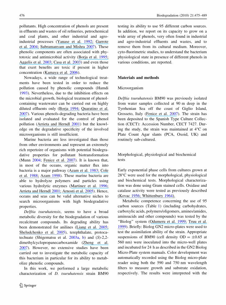
Growth of Delftia tsuruhatensis BM90
on phenolic compounds
Usually, maximum of growth (ca 2 9 109 cell/ml) of
D. tsuruhatensis BM90 in rich medium (NB), was
obtained after 16–18 h of cultivation starting frominocula of ca 106 cell/ml. BM90 was also able to
grow (same inoculum size) in all the YNB-based
media containing different pools of phenolic com-pounds (MA–MF) but in all these media growth was
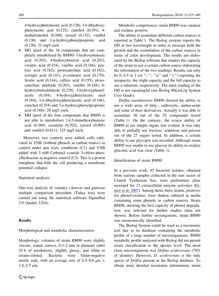
delayed and quantitatively lower (Fig. 1a–f).
BM90 grew rather fast when inoculated into MA(benzoic acid derivatives) (Fig. 1a) and MD (acetic
acid derivatives) (Fig. 1d) showing maximal growth
(6.6 9 108 and 9.31 9 108 cell/ml for MA and MD,respectively) after 48 h of incubation. Similar growth
pattern was found in MF containing a mixture of all
the phenolic derivatives (Fig. 1f). By contrast, aslower growth, with a typical diauxic profile, was
observed when the bacterium was cultivated on MB
(cinnamic acid derivates), MC (phenolic aldehydederivates) and ME (other phenols) (Fig. 1b, c, e). In
all these cases, the first exponential phase started
between the 12th hour and the 24th hour of growth.Then, a second lag phase was evident between the
24th hour and 48th hour of growth to be followed bya further exponential growth. However, maximal cell
growth was always recorded at the 72nd hour of
incubation (8.15 9 108 and 6.03 9 108 cell/ml forMB and MC, respectively). It is worth to note that,
always, to the diauxic growth corresponds a mirror
image in the curve of phenol degradation. This
indicates that phenol degradation occurs in two stepscorresponding to the two bacterial exponential phases
of growth.
In all cases growth of BM90 stopped when phenolsremoval terminated.
Degradation of phenolic compounds
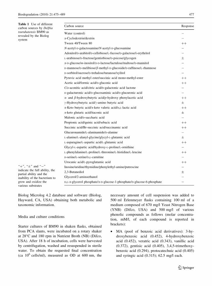
Presence and concentration of the phenolic com-
pounds in the various media was followed along thewhole processes by total phenol determinations
(Fig. 1) and GC/MS (Table 2). GC/MS chromato-
grams are shown for MF only (Fig. 2).With the only exception of ME, the results
indicated that phenols removal was always simulta-
neous with cell growth (Fig. 1a–f). Most of benzoicacid derivatives (MA) were completely depleted
within the first 48 h, except for syringic acid that at
the 48th hour was approximately one sixth of theinitial concentration, and for 3,4,5-trimethoxybenzoic
acid that remained almost at same level throughout
the whole process. Total phenol degradation in MAwas 87.6% (Table 2). Complete phenol removal was
also recorded in MD (acetic acid derivatives) after
48 h. Also cinnamic acid (MB) and phenolic alde-hyde (MC) derivatives were completely eliminated
but only after 96 h. The only exception was esculetin
that at 96 h was only slightly reduced (22.5%).As for ME (other phenolic derivatives), the removal
started after the 48 h of incubation corresponding to the
second exponential phase. Only catechol and 3-p-hydroxyphenylpropionic acidwere completely depleted
by BM90 at the 96th hour. Tyrosol concentration
increased both after 48 and 96 h (31.3 and 11.2%,respectively) while 4-methylcatechol and vanillol
decreased after 48 h of incubation to increase again
after 96 h. However, this occurred in the control also.Total phenol degradation in MB, MC, MD and ME
was 80.6, 100, 100 and 34%, respectively (Table 2).
BM90 behaviour, concerning the removal of eachsingle phenolic compound, was similar either when
they were grouped by chemical characteristics (mediaMA–ME) or when they were pooled together in the
same medium (MF). However, in MF some com-
pounds were completely removed at 48 h instead of96 h: this is probably due to the lower concentration
of each compound. By contrast, in MF syringalde-
hyde was only partially depleted (56.8%) at 96 h
Biodegradation (2010) 21:475–489 481
123
even if its concentration was lower than in MC. The
concentration of 4-methylcatechol in the control
increased of ca 143%. Total phenol degradation inMF was 75.3%.
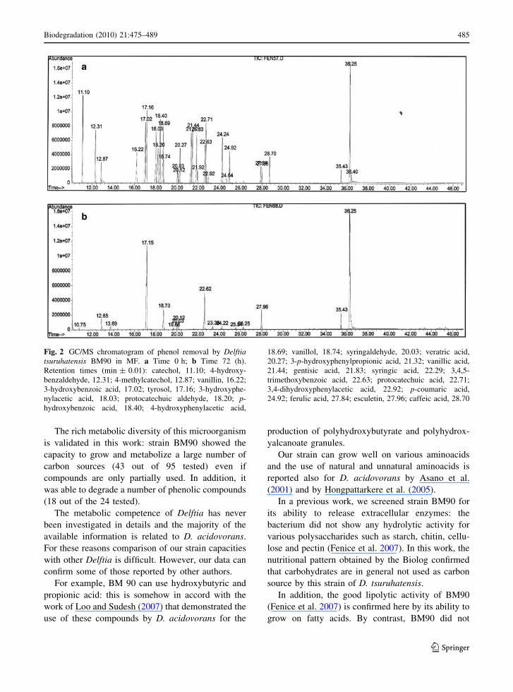
Figure 2 shows the presence of the various phenols
in MF at 0 and 72 h of incubation as detected by GC/MS.
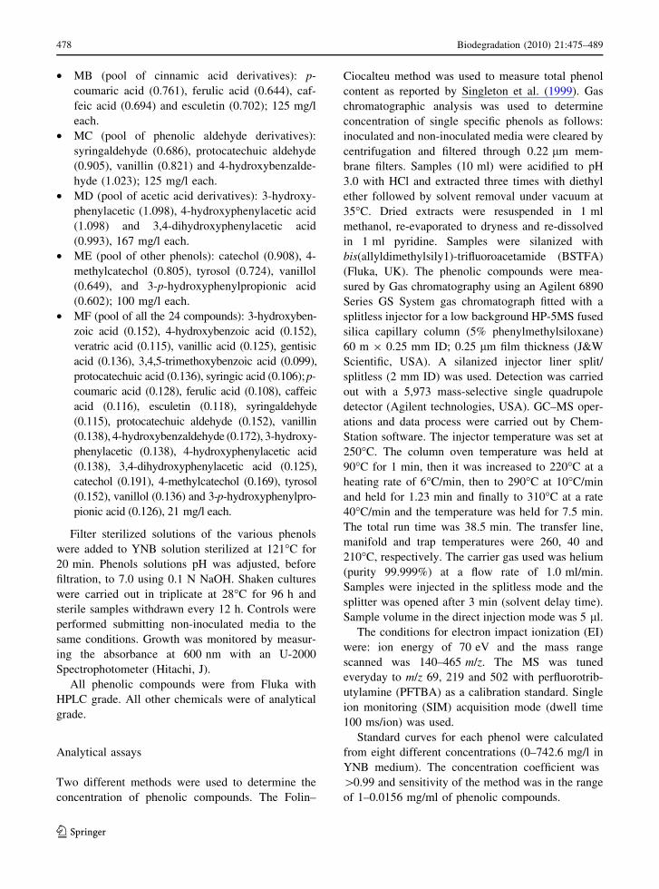
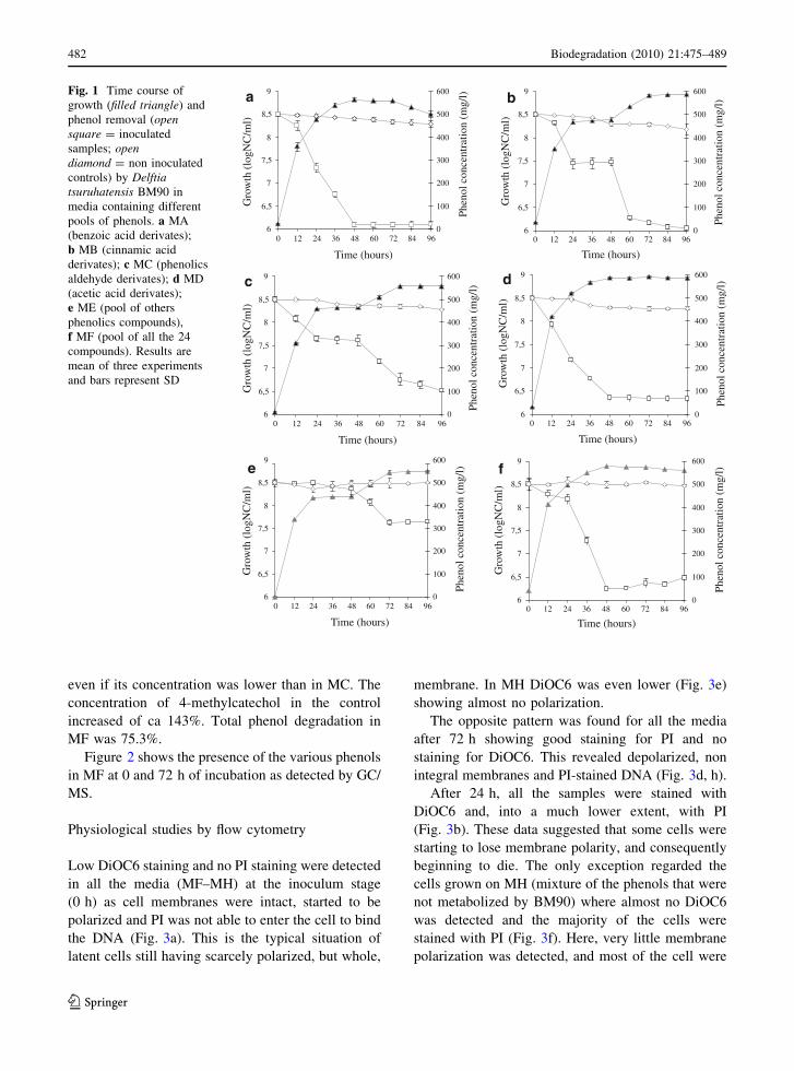
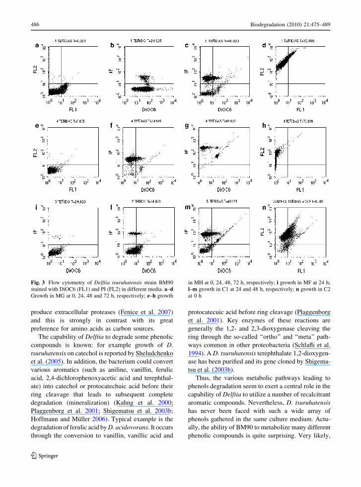
Physiological studies by flow cytometry
Low DiOC6 staining and no PI staining were detected
in all the media (MF–MH) at the inoculum stage(0 h) as cell membranes were intact, started to be
polarized and PI was not able to enter the cell to bind
the DNA (Fig. 3a). This is the typical situation oflatent cells still having scarcely polarized, but whole,
membrane. In MH DiOC6 was even lower (Fig. 3e)
showing almost no polarization.
The opposite pattern was found for all the mediaafter 72 h showing good staining for PI and no
staining for DiOC6. This revealed depolarized, non
integral membranes and PI-stained DNA (Fig. 3d, h).After 24 h, all the samples were stained with
DiOC6 and, into a much lower extent, with PI
(Fig. 3b). These data suggested that some cells werestarting to lose membrane polarity, and consequently
beginning to die. The only exception regarded the
cells grown on MH (mixture of the phenols that werenot metabolized by BM90) where almost no DiOC6
was detected and the majority of the cells were
stained with PI (Fig. 3f). Here, very little membranepolarization was detected, and most of the cell were
a
6
6,5
7
7,5
8
8,5
9
Time (hours)G
row
th (l
ogN
C/m
l)
0
100
200
300
400
500
600
Phen
ol c
once
ntra
tion
(mg/
l)
b
6
6,5
7
7,5
8
8,5
9
Time (hours)
Gro
wth
(log
NC
/ml)
0
100
200
300
400
500
600
Phen
ol c
once
ntra
tion
(mg/
l)
c
6
6,5
7
7,5
8
8,5
9
Time (hours)
Gro
wth
(log
NC
/ml)
0
100
200
300
400
500
600
Phen
ol c
once
ntra
tion
(mg/
l) d
6
6,5
7
7,5
8
8,5
9
Time (hours)
Gro
wth
(log
NC
/ml)
0
100
200
300
400
500
600
Phen
ol c
once
ntra
tion
(mg/
l)
e
6
6,5
7
7,5
8
8,5
9
Time (hours)
Gro
wth
(log
NC
/ml)
0
100
200
300
400
500
600Ph
enol
con
cent
ratio
n (m
g/l) f
6
6,5
7
7,5
8
8,5
9
0 12 24 36 48 60 72 84 96 0 12 24 36 48 60 72 84 96
0 12 24 36 48 60 72 84 96 0 12 24 36 48 60 72 84 96
0 12 24 36 48 60 72 84 96 0 12 24 36 48 60 72 84 96
Time (hours)
Gro
wth
(log
NC
/ml)
0
100
200
300
400
500
600
Phen
ol c
once
ntra
tion
(mg/
l)
Fig. 1 Time course ofgrowth (filled triangle) andphenol removal (opensquare = inoculatedsamples; opendiamond = non inoculatedcontrols) by Delftiatsuruhatensis BM90 inmedia containing differentpools of phenols. a MA(benzoic acid derivates);b MB (cinnamic acidderivates); c MC (phenolicsaldehyde derivates); d MD(acetic acid derivates);e ME (pool of othersphenolics compounds),f MF (pool of all the 24compounds). Results aremean of three experimentsand bars represent SD
482 Biodegradation (2010) 21:475–489
123
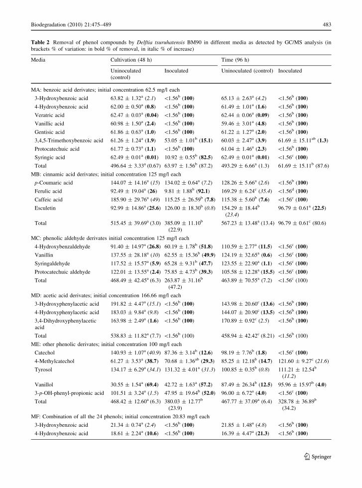
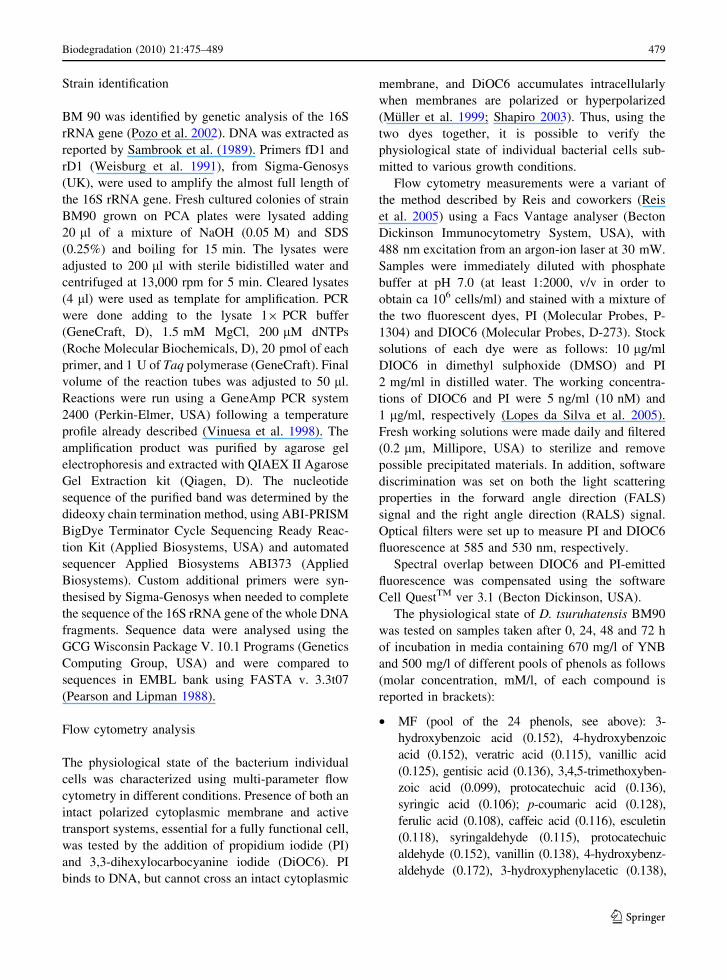
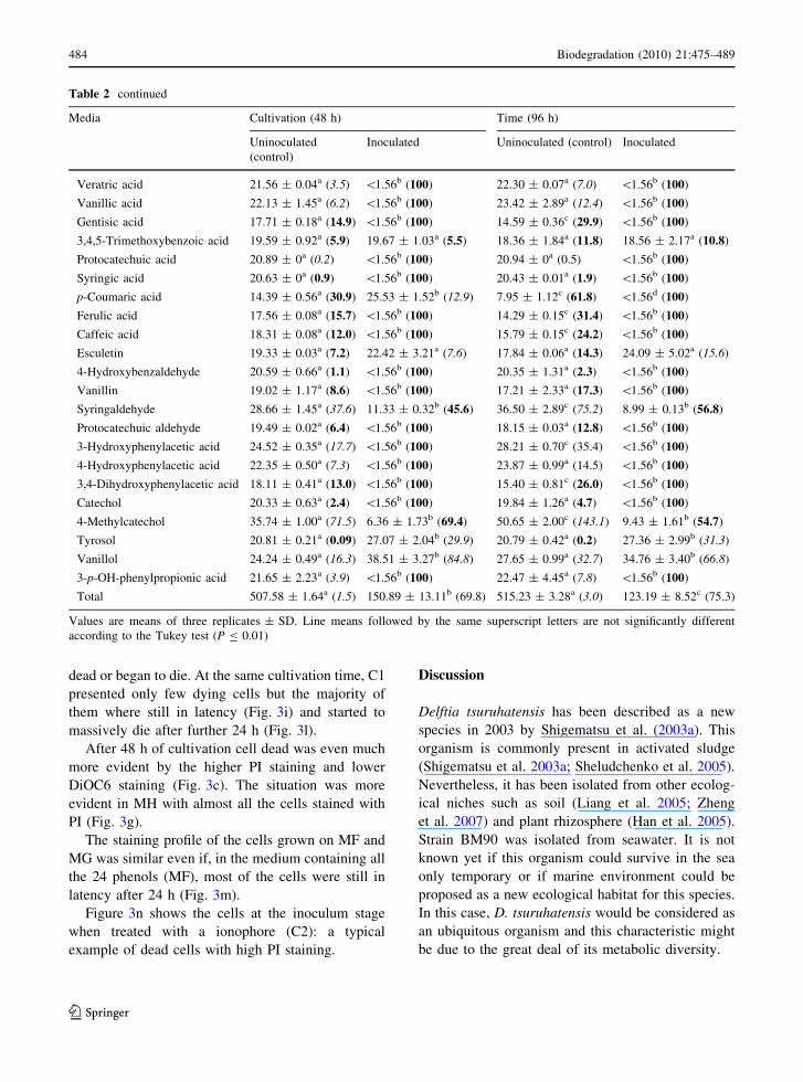
Table 2 Removal of phenol compounds by Delftia tsuruhatensis BM90 in different media as detected by GC/MS analysis (inbrackets % of variation: in bold % of removal, in italic % of increase)
Media Cultivation (48 h) Time (96 h)
Uninoculated(control)
Inoculated Uninoculated (control) Inoculated
MA: benzoic acid derivates; initial concentration 62.5 mg/l each
3-Hydroxybenzoic acid 63.82 ± 1.32a (2.1) \1.56b (100) 65.13 ± 2.63a (4.2) \1.56b (100)
4-Hydroxybenzoic acid 62.00 ± 0.50a (0.8) \1.56b (100) 61.49 ± 1.01a (1.6) \1.56b (100)
Veratric acid 62.47 ± 0.03a (0.04) \1.56b (100) 62.44 ± 0.06a (0.09) \1.56b (100)
Vanillic acid 60.98 ± 1.50a (2.4) \1.56b (100) 59.46 ± 3.01a (4.8) \1.56b (100)
Gentisic acid 61.86 ± 0.63a (1.0) \1.56b (100) 61.22 ± 1.27a (2.0) \1.56b (100)
3,4,5-Trimethoxybenzoic acid 61.26 ± 1.24a (1.9) 53.05 ± 1.01b (15.1) 60.03 ± 2.47a (3.9) 61.69 ± 15.11ab (1.3)
Protocatechuic acid 61.77 ± 0.73a (1.1) \1.56b (100) 61.04 ± 1.46a (2.3) \1.56b (100)
Syringic acid 62.49 ± 0.01a (0.01) 10.92 ± 0.55b (82.5) 62.49 ± 0.01a (0.01) \1.56c (100)
Total 496.64 ± 3.33a (0.67) 63.97 ± 1.56b (87.2) 493.29 ± 6.66a (1.3) 61.69 ± 15.11b (87.6)
MB: cinnamic acid derivates; initial concentration 125 mg/l each
p-Coumaric acid 144.07 ± 14.16a (15) 134.02 ± 0.64a (7.2) 128.26 ± 5.66a (2.6) \1.56b (100)
Ferulic acid 92.49 ± 19.04a (26) 9.81 ± 1.88b (92.1) 169.29 ± 6.24c (35.4) \1.56d (100)
Caffeic acid 185.90 ± 29.76a (49) 115.25 ± 26.59b (7.8) 115.38 ± 5.60b (7.6) \1.56c (100)
Esculetin 92.99 ± 14.86a (25.6) 126.00 ± 18.30b (0.8) 154.29 ± 18.44b
(23.4)96.79 ± 0.61a (22.5)
Total 515.45 ± 39.69a (3.0) 385.09 ± 11.10b
(22.9)567.23 ± 13.48a (13.4) 96.79 ± 0.61c (80.6)
MC: phenolic aldehyde derivates initial concentration 125 mg/l each
4-Hydroxybenzaldehyde 91.40 ± 14.97a (26.8) 60.19 ± 1.78b (51.8) 110.59 ± 2.77a (11.5) \1.56c (100)
Vanillin 137.55 ± 28.18a (10) 62.55 ± 15.36b (49.9) 124.19 ± 32.65a (0.6) \1.56c (100)
Syringaldehyde 117.52 ± 15.57a (5.9) 65.28 ± 9.31b (47.7) 123.55 ± 22.90a (1.1) \1.56c (100)
Protocatechuic aldehyde 122.01 ± 13.55a (2.4) 75.85 ± 4.73b (39.3) 105.58 ± 12.28a (15.5) \1.56c (100)
Total 468.49 ± 42.45a (6.3) 263.87 ± 31.16b
(47.2)463.89 ± 70.55a (7.2) \1.56c (100)
MD: acetic acid derivates; initial concentration 166.66 mg/l each
3-Hydroxyphenylacetic acid 191.82 ± 4.47a (15.1) \1.56b (100) 143.98 ± 20.60c (13.6) \1.56b (100)
4-Hydroxyphenylacetic acid 183.03 ± 9.84a (9.8) \1.56b (100) 144.07 ± 20.90c (13.5) \1.56b (100)
3,4-Dihydroxyphenylaceticacid
163.98 ± 2.49a (1.6) \1.56b (100) 170.89 ± 0.92c (2.5) \1.56b (100)
Total 538.83 ± 11.82a (7.7) \1.56b (100) 458.94 ± 42.42c (8.21) \1.56b (100)
ME: other phenolic derivates; initial concentration 100 mg/l each
Catechol 140.93 ± 1.07a (40.9) 87.36 ± 3.14b (12.6) 98.19 ± 7.76b (1.8) \1.56c (100)
4-Methylcatechol 61.27 ± 3.53a (38.7) 70.68 ± 1.36ab (29.3) 85.25 ± 12.18b (14.7) 121.60 ± 9.27c (21.6)
Tyrosol 134.17 ± 6.29a (34.1) 131.32 ± 4.01a (31.3) 100.85 ± 0.35b (0.8) 111.21 ± 12.54b
(11.2)
Vanillol 30.55 ± 1.54a (69.4) 42.72 ± 1.63a (57.2) 87.49 ± 26.34b (12.5) 95.96 ± 15.97b (4.0)
3-p-OH-phenyl-propionic acid 101.51 ± 3.24a (1.5) 47.95 ± 19.64b (52.0) 96.00 ± 6.72a (4.0) \1.56c (100)
Total 468.42 ± 12.60a (6.3) 380.03 ± 12.77b
(23.9)467.77 ± 37.09a (6.4) 328.78 ± 36.89b
(34.2)
MF: Combination of all the 24 phenols; initial concentration 20.83 mg/l each
3-Hydroxybenzoic acid 21.34 ± 0.74a (2.4) \1.56b (100) 21.85 ± 1.48a (4.8) \1.56b (100)
4-Hydroxybenzoic acid 18.61 ± 2.24a (10.6) \1.56b (100) 16.39 ± 4.47a (21.3) \1.56b (100)
Biodegradation (2010) 21:475–489 483
123
dead or began to die. At the same cultivation time, C1
presented only few dying cells but the majority ofthem where still in latency (Fig. 3i) and started to
massively die after further 24 h (Fig. 3l).
After 48 h of cultivation cell dead was even muchmore evident by the higher PI staining and lower
DiOC6 staining (Fig. 3c). The situation was more
evident in MH with almost all the cells stained withPI (Fig. 3g).
The staining profile of the cells grown on MF and
MG was similar even if, in the medium containing allthe 24 phenols (MF), most of the cells were still in
latency after 24 h (Fig. 3m).
Figure 3n shows the cells at the inoculum stagewhen treated with a ionophore (C2): a typical
example of dead cells with high PI staining.
Discussion
Delftia tsuruhatensis has been described as a new
species in 2003 by Shigematsu et al. (2003a). Thisorganism is commonly present in activated sludge
(Shigematsu et al. 2003a; Sheludchenko et al. 2005).
Nevertheless, it has been isolated from other ecolog-ical niches such as soil (Liang et al. 2005; Zheng
et al. 2007) and plant rhizosphere (Han et al. 2005).
Strain BM90 was isolated from seawater. It is notknown yet if this organism could survive in the sea
only temporary or if marine environment could be
proposed as a new ecological habitat for this species.In this case, D. tsuruhatensis would be considered as
an ubiquitous organism and this characteristic might
be due to the great deal of its metabolic diversity.
Table 2 continued
Media Cultivation (48 h) Time (96 h)
Uninoculated(control)
Inoculated Uninoculated (control) Inoculated
Veratric acid 21.56 ± 0.04a (3.5) \1.56b (100) 22.30 ± 0.07a (7.0) \1.56b (100)
Vanillic acid 22.13 ± 1.45a (6.2) \1.56b (100) 23.42 ± 2.89a (12.4) \1.56b (100)
Gentisic acid 17.71 ± 0.18a (14.9) \1.56b (100) 14.59 ± 0.36c (29.9) \1.56b (100)
3,4,5-Trimethoxybenzoic acid 19.59 ± 0.92a (5.9) 19.67 ± 1.03a (5.5) 18.36 ± 1.84a (11.8) 18.56 ± 2.17a (10.8)
Protocatechuic acid 20.89 ± 0a (0.2) \1.56b (100) 20.94 ± 0a (0.5) \1.56b (100)
Syringic acid 20.63 ± 0a (0.9) \1.56b (100) 20.43 ± 0.01a (1.9) \1.56b (100)
p-Coumaric acid 14.39 ± 0.56a (30.9) 25.53 ± 1.52b (12.9) 7.95 ± 1.12c (61.8) \1.56d (100)
Ferulic acid 17.56 ± 0.08a (15.7) \1.56b (100) 14.29 ± 0.15c (31.4) \1.56b (100)
Caffeic acid 18.31 ± 0.08a (12.0) \1.56b (100) 15.79 ± 0.15c (24.2) \1.56b (100)
Esculetin 19.33 ± 0.03a (7.2) 22.42 ± 3.21a (7.6) 17.84 ± 0.06a (14.3) 24.09 ± 5.02a (15.6)
4-Hydroxybenzaldehyde 20.59 ± 0.66a (1.1) \1.56b (100) 20.35 ± 1.31a (2.3) \1.56b (100)
Vanillin 19.02 ± 1.17a (8.6) \1.56b (100) 17.21 ± 2.33a (17.3) \1.56b (100)
Syringaldehyde 28.66 ± 1.45a (37.6) 11.33 ± 0.32b (45.6) 36.50 ± 2.89c (75.2) 8.99 ± 0.13b (56.8)
Protocatechuic aldehyde 19.49 ± 0.02a (6.4) \1.56b (100) 18.15 ± 0.03a (12.8) \1.56b (100)
3-Hydroxyphenylacetic acid 24.52 ± 0.35a (17.7) \1.56b (100) 28.21 ± 0.70c (35.4) \1.56b (100)
4-Hydroxyphenylacetic acid 22.35 ± 0.50a (7.3) \1.56b (100) 23.87 ± 0.99a (14.5) \1.56b (100)
3,4-Dihydroxyphenylacetic acid 18.11 ± 0.41a (13.0) \1.56b (100) 15.40 ± 0.81c (26.0) \1.56b (100)
Catechol 20.33 ± 0.63a (2.4) \1.56b (100) 19.84 ± 1.26a (4.7) \1.56b (100)
4-Methylcatechol 35.74 ± 1.00a (71.5) 6.36 ± 1.73b (69.4) 50.65 ± 2.00c (143.1) 9.43 ± 1.61b (54.7)
Tyrosol 20.81 ± 0.21a (0.09) 27.07 ± 2.04b (29.9) 20.79 ± 0.42a (0.2) 27.36 ± 2.99b (31.3)
Vanillol 24.24 ± 0.49a (16.3) 38.51 ± 3.27b (84.8) 27.65 ± 0.99a (32.7) 34.76 ± 3.40b (66.8)
3-p-OH-phenylpropionic acid 21.65 ± 2.23a (3.9) \1.56b (100) 22.47 ± 4.45a (7.8) \1.56b (100)
Total 507.58 ± 1.64a (1.5) 150.89 ± 13.11b (69.8) 515.23 ± 3.28a (3.0) 123.19 ± 8.52c (75.3)
Values are means of three replicates ± SD. Line means followed by the same superscript letters are not significantly differentaccording to the Tukey test (P B 0.01)
484 Biodegradation (2010) 21:475–489
123
The rich metabolic diversity of this microorganism
is validated in this work: strain BM90 showed the
capacity to grow and metabolize a large number ofcarbon sources (43 out of 95 tested) even if
compounds are only partially used. In addition, it
was able to degrade a number of phenolic compounds(18 out of the 24 tested).
The metabolic competence of Delftia has never
been investigated in details and the majority of theavailable information is related to D. acidovorans.For these reasons comparison of our strain capacities
with other Delftia is difficult. However, our data canconfirm some of those reported by other authors.
For example, BM 90 can use hydroxybutyric and
propionic acid: this is somehow in accord with thework of Loo and Sudesh (2007) that demonstrated the
use of these compounds by D. acidovorans for the
production of polyhydroxybutyrate and polyhydrox-
yalcanoate granules.
Our strain can grow well on various aminoacidsand the use of natural and unnatural aminoacids is
reported also for D. acidovorans by Asano et al.
(2001) and by Hongpattarkere et al. (2005).In a previous work, we screened strain BM90 for
its ability to release extracellular enzymes: the
bacterium did not show any hydrolytic activity forvarious polysaccharides such as starch, chitin, cellu-
lose and pectin (Fenice et al. 2007). In this work, the
nutritional pattern obtained by the Biolog confirmedthat carbohydrates are in general not used as carbon
source by this strain of D. tsuruhatensis.In addition, the good lipolytic activity of BM90
(Fenice et al. 2007) is confirmed here by its ability to
grow on fatty acids. By contrast, BM90 did not
Fig. 2 GC/MS chromatogram of phenol removal by Delftiatsuruhatensis BM90 in MF. a Time 0 h; b Time 72 (h).Retention times (min ± 0.01): catechol, 11.10; 4-hydroxy-benzaldehyde, 12.31; 4-methylcatechol, 12.87; vanillin, 16.22;3-hydroxybenzoic acid, 17.02; tyrosol, 17.16; 3-hydroxyphe-nylacetic acid, 18.03; protocatechuic aldehyde, 18.20; p-hydroxybenzoic acid, 18.40; 4-hydroxyphenylacetic acid,
18.69; vanillol, 18.74; syringaldehyde, 20.03; veratric acid,20.27; 3-p-hydroxyphenylpropionic acid, 21.32; vanillic acid,21.44; gentisic acid, 21.83; syringic acid, 22.29; 3,4,5-trimethoxybenzoic acid, 22.63; protocatechuic acid, 22.71;3,4-dihydroxyphenylacetic acid, 22.92; p-coumaric acid,24.92; ferulic acid, 27.84; esculetin, 27.96; caffeic acid, 28.70
Biodegradation (2010) 21:475–489 485
123
produce extracellular proteases (Fenice et al. 2007)
and this is strongly in contrast with its great
preference for amino acids as carbon sources.The capability of Delftia to degrade some phenolic
compounds is known: for example growth of D.tsuruhatensis on catechol is reported by Sheludchenkoet al. (2005). In addition, the bacterium could convert
various aromatics (such as aniline, vanillin, ferulic
acid, 2,4-dichlorophenoxyacetic acid and terephthal-ate) into catechol or protocatechuic acid before their
ring cleavage that leads to subsequent complete
degradation (mineralization) (Kahng et al. 2000;Plaggenborg et al. 2001; Shigematsu et al. 2003b;
Hoffmann and Muller 2006). Typical example is the
degradation of ferulic acid byD. acidovorans. It occursthrough the conversion to vanillin, vanillic acid and
protocatecuic acid before ring cleavage (Plaggenborg
et al. 2001). Key enzymes of these reactions are
generally the 1,2- and 2,3-dioxygenase cleaving thering through the so-called ‘‘ortho’’ and ‘‘meta’’ path-
ways common in other proteobacteria (Schlafli et al.
1994). A D. tsuruhatensis terephthalate 1,2-dioxygen-ase has been purified and its gene cloned by Shigema-
tsu et al. (2003b).
Thus, the various metabolic pathways leading tophenols degradation seem to exert a central role in the
capability of Delftia to utilize a number of recalcitrant
aromatic compounds. Nevertheless, D. tsuruhatensishas never been faced with such a wide array of
phenols gathered in the same culture medium. Actu-
ally, the ability of BM90 to metabolize many differentphenolic compounds is quite surprising. Very likely,
Fig. 3 Flow cytometry of Delftia tsuruhatensis strain BM90stained with DiOC6 (FL1) and PI (FL2) in different media. a–dGrowth in MG at 0, 24, 48 and 72 h, respectively; e–h growth
in MH at 0, 24, 48, 72 h, respectively; i growth in MF at 24 h;l–m growth in C1 at 24 and 48 h, respectively; n growth in C2at 0 h
486 Biodegradation (2010) 21:475–489
123
some of these compounds could be immediatelysubmitted to ring cleavage by the dioxygenases
system and, through the ‘‘lower pathway’’, trans-
formed into pyruvate or other intermediates beforepossible mineralization. Others, not directly cleaved
by the dioxygenases, could be transformed in different
substances before ring cleavage and mineralization.However, BM90 seems able to use for its growth
(carbon and energy sources) all the phenols that it
removes from the cultural medium; this is somehowconfirmed by flow cytometry also.
The BM90 biochemical diversity, leading to its
ability to use such a variety of phenols, should bedeeply investigated. However, the various metabolic
pathways, involved in BM90 phenol metabolism,
are probably highly interlinked and regulated byvarious mechanisms and/or factors. This would
explain the different fate of the various phenolic
compounds when they are differently mixed. Inother words, BM90 behaves differently when some
of the phenols are gathered with other similar
derivatives (media MA–ME) or when they are alltogether in MF.
Even if analogous considerations could be made
for other compounds, the most emblematic exampleconcerns 4-methylcatechol. In ME, its concentration
decreased after 48 h of incubation to increase again at
the 96th hour. This could be due to the biotransfor-mation of other phenols in 4-methylcatechol and to
the lack of the appropriate induction of the specific
degradation pathway. On the contrary, in MF, it wasstrongly depleted starting from the 48th hour. This
could be due to the existence of a pathway, able to
degrade 4-methylcatechol, but controlled (induced)by a regulatory protein activated by other phenols. 4-
methylcatechol concentration in the controls (non
inoculated media) slightly decreased (ME control,96th hour) or increased (MF control).
BM90 inability to remove vanillol, tyrosol, escu-
letin or 3,4,5-trimethoxybenzoic acid could be aconsequence of the lack of appropriate enzymatic
machinery and/or regulatory proteins. Anyway, flowcytometry showed that these compound exert a
certain, dose dependent, toxicity also. Almost normal
growth is revealed when they are mixed with otherphenols (MF) and are present in low concentration
(20.83 mg/l). Strong cell inhibition was recorded in
MH: in this case, in fact, their concentration was ca 6time higher (125 mg/l).
Thus, phenol toxicity was recorded only with 4 outof the 24 compounds tested. However, in presence of
all the phenols, BM90 showed a slower growth rate.
A certain amount of dead cells, recorded in variousmedia after 72 h, was probably due to lack of
nutrients. In fact, similar results were obtained with
C1. In this control no phenol is added and there is nocarbon source: the cells remained in latency till the
72 h before starting to die.
A lot of phenolic compounds are recalcitrant andcould cause pollution. In soils and waters they are
originated mainly from plant decomposition or by
agrochemical and industrial activities. Because oftheir antimicrobial and phytotoxic effect (Borja et al.
1995; Quaratino et al. 2007; Juarez et al. 2008) the
application of microbial technologies for bioremedi-ation is very often limited. In fact, only a small
number of bacteria and fungi are able to degrade and
use phenols as carbon source.Our results, showing that D. tsuruhatensis BM90
can use various phenols as growing substrates,
suggest that this organism could be considered verypromising in case of phenol pollution. Experiments in
this direction are in course.
Acknowledgments This work was done in the frame of the‘‘Azioni integrate Italia-Spagna 2006, Ministerodell’Universita e della Ricerca’’ and ‘‘Accion IntegradaEspana-Italia 2006, Ministerio Educacion y Ciencia’’. M.Manzanera was granted by Programa Ramon y Cajal(Ministerio Educacion y Ciencia, Spain and EuropeanRegional Development Fund, European Union). The authorswish to thank the Flow Cytometry Service of University ofGranada (Spain).
References
Aggelis G, Iconomou D, Christou M, Bokas D, Kotzailias S,Christou G, Tsagou V, Papanikolau S (2003) Phenolicremoval in a model olive oil mill wastewater usingPleurotus ostreatus in bioreactor cultures and biologicalevaluation of the process. Water Res 27:3897–3904
Arnosti C, Durkin AS, Jeffrey WH (2005) Patterns of extra-cellular enzyme activities among pelagic marine micro-bial communities: implication for cycling of dissolvedorganic carbon. Aquat Microb Ecol 38:135–145
Arrieta JM, Herndl GJ (2001) Assessing the diversity of marinebacterial glucosidases by capillary electrophoresis zy-mography. Appl Environ Microbiol 67:4896–4900
Asano Y, Umezaki M, Li Y-F, Tsubota S, Lubbehusen TL(2001) Isolation of microorganisms which utilize acidic d-amino acid oligomers. Jpn J Mol Catal B Enzym 12(1–6):53–59
Biodegradation (2010) 21:475–489 487
123
Azam F (1998) Microbial control of oceanic carbon flux: theplot thickens. Science 280:694–696
Azam F, Fenchel T, Field JG, Gray JS, Meyerreil LA,Thingstad F (1983) The ecological role of water-columnmicrobes in the sea. Mar Ecol Prog Ser 10:257–263
Borja R (1994) Comparison of anaerobic filter and anaerobiccontact process for olive mill wastewater previously fer-mented with Geotrichum candidum. Process Biochem29:139–144
Borja R, Alba J, Garrido SE, Martinez L, Garcia MP,Monteoliva M, Ramos-Cormenzana A (1995) Effect ofaerobic pretreatment with Aspergillus terreus on theanaerobic digestion of olive mill wastewater. BiotechnolAppl Biochem 2:233–246
Casa R, D’Annibale A, Pieruccetti F, Stazi SR, Giovannozzi-Sermanni G, Locascio B (2003) Reduction of the phenoliccomponents in olive-mill wastewater and its impact ondurum wheat (Triticum durum Desf.) germinability.Chemosphere 50:959–966
Cole JJ, Findlay S, Pace ML (1988) Bacterial production infresh and saltwater ecosystems—a cross-system overview.Mar Ecol Prog Ser 43:1–10
Fenice M, Gallo AM, Juarez-Jimenez B, Gonzalez-Lopez J(2007) Screening for extracellular enzyme activity bybacteria isolated from samples collected in the TyrrhenianSea. Ann Microbiol 57:93–100
Garrote G, Cruz JM, Moure A, Domınguez H, Parajo JC (2004)Antioxidant activity of byproducts from the hydrolyticprocessing of selected lignocellulosic materials. TrendsFood Sci Technol 15:191–200
Hamdi M (1993) Future prospects and constraints of olive millwastewaters use and treatments: a review. Bioprocess Eng8:209–214
Han J, Sun L, Dong X, Cai Z, Sun X, Yang H, Wang Y, SongW (2005) Characterization of a novel plant growth-pro-moting bacteria strain Delftia tsuruhatensis HR4 both as adiazotroph and a potential biocontrol agent against vari-ous plant pathogens. Syst Appl Microbiol 28:66–76
Hoffmann D, Muller RH (2006) 2, 4-Dichlorophenoxyaceticacid (2, 4-D) utilization by Delftia acidovorans MC1 atalkaline pH and in the presence of Dichlorprop isimproved by introduction of the tfdK Gene. Biodegrada-tion 17:263–273
Hongpattarkere T, Komeda H, Asano Y (2005) Purification,characterization, gene cloning and nucleotide sequencingof d-stereospecific amino acid amidase from soil bacte-rium: Delftia acidovorans. J Ind Microbiol Biotechnol32:567–576
Juarez MJB, Zafra-Gomez A, Luzon-Toro B, Ballesteros-Garcia OA, Navalon A, Gonzalez-Lopez J, Vilchez JL(2008) Gas chromatographic—mass spectrometric studyof the degradation of phenolic compounds in wastewaterolive oil by Azotobacter chroococcum. Biores Technol99:2392–2398
Kahng HY, Kukor JJ, Oh KH (2000) Characterization of strainHY99, a novel microorganism capable of aerobic andanaerobic degradation of aniline. FEMS Microbiol Lett190:215–221
Kamaya Y, Tsuboi S, Takada T, Suzuki K (2006) Growthstimulation and inhibition effects of 4-hydroxybenzoicacid and some related compounds on the freshwater green
alga Pseudokirchneriella subcapitata. Arch EnvironContam Toxicol 51:537–541
Kovac N (1956) Identification of P. pyocyanea by the oxidasereaction. Nature 178:703
Liang Q, Takeo M, Chen M, Zhang W, Xu Y, Lin M (2005)Chromosome-encoded gene cluster for the metabolicpathway that converts aniline to TCA- cycle intermediatesin Delftia tsuruhatensis AD9. Microbiology 151:3435–3446
Lodovici M, Guglielmi F, Meoni M, Dolara P (2001) Effect ofnatural phenolic acids on DNA oxidation in vitro. FoodChem Toxicol 39:1205–1210
Loo C-Y, Sudesh K (2007) Biosynthesis and native granulecharacteristics of poly(3-hydroxybutyrate-co-3-hydro-xyvalerate) in Delftia acidovorans. Int J Biol Macromol40:466–471
Lopes da Silva T, Reis A, Kent CA, Kosseva M, Roseiro JC,Hewitt CJ (2005) Stress-induced physiological responsesto starvation periods as well as glucose and lactose pulsesin Bacillus licheniformis CCMI 1034 continuous aerobicfermentation processes as measured by multi-parameterFlow Cytometry. Biochem Eng J 24:31–41
Martinez J, Smith DC, Steward GF, Azam F (1996) Variabilityin ectohydrolytic enzyme activities of pelagic marinebacteria and its significance for substrate processing in thesea. Aquat Microb Ecol 10:223–230
Muller S, Bley TH, Babel W (1999) Adaptive responses ofRalstonia eutropha to master feast and famine conditionsanalysed by flow cytometry. J Biotechnol 75:81–97
Munn CB (2004) Marine microbiology, ecology and applica-tions. Garland Sciences, New York
Ng TB, Liu F, Wang ZT (2000) Antioxidative activity ofnatural products from plants. Life Sci 66:709–723
Odumeru JA, Steel M, Frhuner L, Larkin C, Jiang J, Mann E,Mcnabb WB (1999) Evaluation of accuracy and repeat-ability of identification of food-borne pathogens byautomated bacterial identification systems. J Clin Micro-biol 37:944–949
Pearson WR, Lipman DJ (1988) Improved tools for biologicalsequence comparison. Proc Natl Acad Sci USA 85:2444–2448
Plaggenborg R, Steinbuchel A, Priefert H (2001) The coen-zyme A-dependent, non-b-oxidation pathway and notdirect deacetylation is the major route for ferulic aciddegradation in Delftia acidovorans. FEMS Microbiol Lett205:9–16
Pozo C, Rodelas B, de la Escalera S, Gonzalez-Lopez J (2002)Hydantoinase activity of an Ochrobactrum anthropistrain. J Appl Microbiol 92:1028–1034
Quaratino D, D’Annibale A, Federici F, Cereti CF, Rossini F,Fenice M (2007) Enzyme and fungal treatments and acombination thereof reduce olive mill wastewater phyto-toxicity on ZeamaysL. seeds. Chemosphere 66:1627–1633
Reis A, Lopes da Silva T, Kent CA, Kosseva M, Roseiro JC,Hewitt CJ (2005) Monitoring population dynamics of thethermophilic Bacillus licheniformis CCMI 1034 in batchand continuous cultures using multi-parameter flowcytometry. J Biotechnol 115:199–210
Sambrook J, Fritsch EF, Maniatis T (1989) Molecular clon-ing—a laboratory manual, 2nd edn. Cold Spring Harbor,New York
488 Biodegradation (2010) 21:475–489
123
Schlafli HR, Weiss MA, Leisinger T (1994) Terephthalate 1, 2-dioxygenase system from Comamonas testosteroni T-2:purification and some properties of the oxygenase com-ponent. J Bacteriol 176:6644–6652
Shapiro HM (2003) Practical flow cytometry, 3rd edn. Alan R.Liss Inc., New York
Sheludchenko MS, Kolomytseva MP, Travkin VM, AkimovVN, Golovleva LA (2005) Degradation of aniline byDelftia tsuruhatensis 14S in batch and continuous pro-cesses. Metallofizika I Noveishie Tekhnologii 27:1659–1676
Shigematsu T, Yumihara K, Ueda Y, Numaguchi M, MorimuraS, Kida K (2003a) Delftia tsuruhatensis sp. nov., a ter-ephthalate-assimilating bacterium isolated from activatedsludge. Int J Syst Evol Microbiol 53:1479–1483
Shigematsu T, Yumihara K, Ueda Y, Numaguchi M, MorimuraS, Kida K (2003b) Purification and gene cloning of theoxygenase component of the terephthalate 1, 2-dioxy-genase system from Delftia tsuruhatensis strain T7. FEMSMicrobiol Lett 220:255–260
Singleton VL, Orthofer R, Lamuela-Raventos RM (1999)Analysis of total phenols and other oxidation substratesand antioxidants by means of Folin-Ciocalteu reagent.Methods Enzymol 299:152–178
Subramanyam R, Mishra IM (2007) Biodegradation of catechol(2-hydroxy phenol) bearing wastewater in an UASBreactor. Chemosphere 69:816–824
Truu J, Talpsep E, Heinaru E, Stottmeister U, Wand H, He-inaru A (1999) Comparison of API 20NE and Biolog GNidentification systems assessed by techniques of multi-variate analysis. J Microbiol Meth 36:193–201
Vinuesa P, Rademaker JLW, Debruijin FJ, Werner D (1998)Genotypic characterisation of Bradyrhizobium strains no-dulating endemic woody legumes of the Canary Island byPCR-restriction fragment length polymorphism analysis ofgenes encoding 16S rRNA (16S rDNA) and 16S–23SrDNA intergenic spacers, repetitive extragenic palin-dromic PCR genomic fingerprinting, and partial 16S rDNAsequencing. Appl Environ Microbiol 64:2096–2104
Weisburg WG, Barns SM, Pelletier DA, Lane DJ (1991) 16Sribosomal DNA amplification for phylogenetic study. JBacteriol 173:697–703
Whittenbury R (1964) Hydrogen peroxide formation and cat-alase activity in the lactic acid bacteria. J Gen Microbiol35:13–26
Yanase H, Zuzan K, Kita K, Sogabe S, Kato N (1992) Deg-radation of phenols by thermophilic and halophilic bac-teria isolated from a marine brine sample. J FermentBioeng 74:297–300
Zheng RC, Wang YS, Liu ZQ, Xing LY, Zheng YG, Shen YC(2007) Isolation and characterization of Delftia tsuruhat-ensis ZJB-05174, capable of R-enantioselective degrada-tion of 2, 2-dimethylcyclopropanecarboxamide. ResMicrobiol 158:258–264
Biodegradation (2010) 21:475–489 489
123