Medical Terminology Systems
620
-
Upload
khangminh22 -
Category
Documents
-
view
0 -
download
0
Transcript of Medical Terminology Systems


Pronunciation GuidelinesHere are guidelines to help you pronounce medical terms as well as understand pronunciation marks usedthroughout this text and in most dictionaries.
Special Sounds
Here are rules regarding certain letter combinations and special sounds attributed to letters based on theirplacement in a medical word:
•For ae and oe, only the second vowel is pronounced.Examples are bursae, pleurae, and roentgen.
•The soft sound of s and j are given to c and g, respectively, before e, i, and y in words of Greek orLatin origin.Examples are cerebrum, circumcision, cycle, gel, gingivitis, giant, and gyrate.
•Before other letters, c and g have a hard sound.Examples are cardiac, cast, gastric, and gonad.
•The letters ch are sometimes pronounced like k.Examples are cholesterol, cholera, and cholemia.
•When pn appears at the beginning of a word, p is silent and only n is pronounced.Examples are pneumonia and pneumotoxin.
•When pn appears in the middle of a word, p and n are pronounced.Examples are orthopnea and hyperpnea.
•When ps appears at the beginning of a word, p is silent and only s is pronounced.Examples are psychology and psychosis.
•When forming the final letter(s) of a word, e and es are commonly pronounced as separate syllables.Examples are syncope, systole, and nares.
•When i appears at the end of a word (to form a plural), it is pronounced eye.Examples are bronchi, fungi, and nuclei.
•All other vowels and consonants have normal English sounds.
Pronunciation Marks
Diacritical marks and capitalization are used to aid pronunciations throughout the text. Diacritical marksare used to show vowel sounds, and capitalization is used to show emphasis.
Diacritical marks are symbols placed above or under vowels. They show vowel sounds. In this text, onlytwo diacritical marks are used: the macron (¯) and the breve (˘ ).
The macron indicates the long The breve indicates the shortsound of vowels, as in: sound of vowels, as in:
a in rate a in applee in rebirth e in everı in isle ı in ito in over o in notu in unite u in cut
Capitalization is used to indicate primary accent. For example, the pronunciation LET-ter indicates thatemphasis should be placed on the first syllable when pronouncing the word letter.
2090_Front_cover.qxd 3/6/09 7:28 PM Page 1

Medical TerminologySystems A Body Systems Approach
SIXTH EDITION
Barbara A. Gylys (GlL-Is), MEd, CMA-A (AAMA)Professor EmeritaCollege of Health and Human ServicesCoordinator of Medical Assisting TechnologyUniversity of ToledoToledo, Ohio
Mary Ellen Wedding, MEd, MT(ASCP), CMA,(AAMA) AAPC
Professor of Health ProfessionsCollege of Health Science and Human ServicesUniversity of ToledoToledo, Ohio
Gylys_FM_I-xxx.qxd 3/6/09 7:36 PM Page 1

CHAPTER OUTLINESto orient students to each chapter’s content
KEY TERMShighlighted in the beginning of each chapter
ABBREVIATIONSfor common terms
Nervous System
Chapter Outline
Objectives
Anatomy and Physio
logy
Anatomy and Physiology Key Terms
Cellular Structure of the Nervous System
Neurons
Neuroglia
Nervous System Divisions
Central Nervous System
Peripheral Nervous System
Connecting Body Systems–Nervous System
Medical Word Elements
Pathology
Radiculopathy
Cerebrovascular D
isease
Seizure Disorders
Parkinson Disease
Multiple Sclerosis
Alzheimer Disease
Mental Illness
Oncology
Diagnostic
, Symptomatic, and Relate
d Terms
Diagnostic
and Therapeutic Procedures
Pharmaco
logy
Abbreviations
Learning Activities
Medical Record Activ
ities
Discharge summary: S
ubarachnoid hemorrhage
Consultation report: A
cute onset paraplegia
Objectives
Upon completion of this ch
apter, you will b
e able to:
• Locate and descri
be the structures of th
e nervous
system.
• Describe the functio
nal relatio
nship between the
nervous sy
stem and other body systems.
• Recognize, pronounce, spell, and build words re
lat
to the nervous sy
stem.
• Describe pathologica
l conditions, diagn
ostic and
therapeutic procedures, an
d other terms re
lat
the nervous sy
stem.
• Explain pharm
acology re
lated to the tre
atm
nervous disorders.
• Demonstrate your knowledge of th
is cha
completing the learning an
d medical rec
activitie
s.
C H A P T E R
14_Ch14_
425-46
4.qxd
3/3/0
9 6:3
7 PM
Page 4
25
Anatomy and
The nervous system is one of the mos
ed systems of the body in both structure and
tion. It senses physical and chemical changes in
the internal and external environments, processes
them, and then responds to maintain homeostasis.
Voluntary activities, such as walking and talking,
and involuntary activities, such as digestion and
circulation, are coordinated, regulated, and inte-
grated by the nervous system. The entire neural
network of the body relies on the transmission
of nervous impulses. Nervous impulses are elec-
a
Cellular Stru
of the Nervous Sys
Despite its complexity, the nervous system
posed of only two principal types of cells: neuron
and neuroglia. Neurons are cells that transmit
Anatomy and Physiology Key Terms
This section introduces i
mportant nervous system term
s and their definitions. Word analyses f
or
selected term
s are also provided.
Term
Definition
afferent
AF-er-ent
blood-brain barri
er
central nervo
us system (CNS)
NER-ves
efferent
EF-e-rent
nerve fiber
neurilemma
nu-rı-LEM-a
peripheral nervo
us system
(PNS)
per-IF-e-ral NER-ves
somatic nervo
us system (SNS)
so-MAT-ık NER-ves
ventricle
VEN-trık-l
ventr: belly, belly side
-ical: pertaining to
Carry or move inward or toward a central structure
The term afferent refers to both vessels and nerves.
Protective mechanism that blocks specific substances fo
und in the blood-
stream from entering delicate brain tiss
ue
Network of nervous tissue found in the brain and spinal cord
Carry or move away from a central str
ucture
The term efferent refers to both vessels and nerves.
Projection of a neuron, especially the axon that transmits i
mpulses
Additional sheath external to myelin that is formed by Schwann cells a
nd
found only on axons in the peripheral nervous system
Because neurilemma does not disintegrate after injury to the axon, its e
nclosed
hollow tube provides an avenue for regeneration of injured axons.
All nervous tissue of th
e body located outside the brain and spinal cord
Functional portion of the peripheral nervous system that sti
mulates vol-
untary actions such as walking and talking, and transmits s
timuli from the
sensory receptors to the brain for integration and interpretation
Chamber or cavity of an organ that receives or holds a fluid
Pronunciation HelpLong Sound
a—rate e—rebirth
ı—isle
Short Sound o—over
u—uniteı—it
a—alone e—ever
o—notu—cut
hypnotics
psychostim
ulants
ag
Hypnotics m
turates. Barbituratey
addiction.
Reduce impulsive behavio
r by increasin
g the
level of neurotransmitte
rs
Psychostimulants have a calming effect on
people with attention def icit hyperactiv
ity
disorder (ADHD) and are also used to treat
narcolepsy.
dek
Dexedrine
methylphenidate
meth-ıl-FEN-ı-dat
Ritalin
Abbreviations
This section introduces n
ervous system–rela
ted abbreviations and their meanings.
AbbreviationMeaning
AbbreviationMeaning
AD
Alzheimer disease
ICP
intracranial pressure
ADHD
attention-deficit hyperactivity LOC
loss of consciousness
disorder
ALS
amyotrophic lateral sclerosis;LP
lumbar puncture
also called Lou Gehrig disease
ANS
autonomic nervous system
BEAM
brain electrical activity
MRA
magnetic resonance angiogram; magnetic
mapping
resonance angiography
CNS
central nervous system
MRI
magnetic resonance imaging
CP
cerebral palsy
MS
musculoskeletal; multiple sclerosis;
mental status; mitral ste
nosis
CSF
cerebrospinal fluid
NCV
nerve conduction velocity
CT
computed tomography
PET
positron emissio
n tomography
CVA
cerebrovascular accident
PNS
peripheral nervous system C
INSIDEMEDICAL TERMINOLOGY SYSTEMSA Body Systems Approach, 6th Edition
What’s
HOW DOES WORD BUILDING WORK?It begins with the basics
Introduces word elements
• Roots• Combining forms• Suffixes• Prefixes
Reviews each element one by one
Applies the principles of word building to each body system
• Learn the parts from which words are built
• Decipher words based on knowledge of word parts
Uses mnemonic devices and interactive activities (in the book and on the CD) to make word building fun and increase retention
CLEAR, CONCISE PRESENTATIONusing the classic word-building and body systems approach to learning.
Gylys Walkthrough_2-09_FINAL.ind2 2Gylys Walkthrough_2-09_FINAL.ind2 2 3/6/09 7:59:58 PM3/6/09 7:59:58 PM

A Unique BlendOF WORDS AND ART
ANATOMYthat’s detailed and precise
BRILLIANT FULL-COLOR ILLUSTRATIONSthat leap from the page and enhance your learning
Gylys Walkthrough_2-09_FINAL.ind3 3Gylys Walkthrough_2-09_FINAL.ind3 3 3/6/09 8:00:24 PM3/6/09 8:00:24 PM

DIAGNOSTIC AND THERAPEUTIC PROCEDURESthat clearly show how diseases and disorders arediagnosed and treated
EXERCISE AND ACTIVITY WORKSHEETSin each chapter that help track your progress and prepare youfor quizzes and tests
COMPLETE MEDICAL RECORDSwith activities that provide real-life examples for each body system
458 CHAPTER 14 • Nervous System
cerebellum hypothalamus parietal lobe
cerebrum medulla pons
corpus callosum midbrain (mesencephalon) temporal lobe
diencephalon (interbrain) occipital lobe thalamus
frontal lobe
Learning Activity 14-2
Building Medical Words
Use encephal/o (brain) to build words that mean:
1. disease of the brain
2. herniation of the brain
3 radiography of the brain
Check your answers by referring to Figure 14–3 on page 434. Review material that you didnot answer correctly.�Enhance your study and reinforcement of word elements with the power of Davis Plus. Visit www.davis-
plus.fadavis.com/gylys/systems for this chapter’s flash-card activity. We recommend you complete theflash-card activity before completing activity 14–2 below.
464CHAPTER 14 • Nervous System
General Hospital
1511 Ninth Avenue ■■ Sun City, USA 12345 ■■ (555) 8022-1887
DISCHARGE SUMMARY
August 16, xx
ADMISSION DATE: July 5, 20xx DISCHARGE DATE: July 16, 20xx
ADMITTING DIAGNOSIS: Severe headaches associated with nausea and vomiting.
DISCHARGE DIAGNOSIS: Subarachnoid hemorrhage.
HISTORY OF PRESENT ILLNESS: Patient is a 61-year-old woman who presents at this time
complaining of an “extreme severe headache while swimming.” She also complains of associated neck
pain, occipital pain, nausea, and vomiting.
A CT scan was obtained that showed blood in the cisterna subarachnoidalis consistent with subarach-
noid hemorrhage. The patient also had mild acute hydrocephalus. Neurologically, the patient was
f und to be within normal limits. A cerebral MRI was performed and no aneurysm was noted.
tient was hospitalized on 7/5/xx. On 7/7/xx, she had sudden worsen-
d vomiting. Also, she was noted to have meningismus
sible rebleed. At the time of the lum-
hnoid hemorrhage. A
Michael R. Saadi,
MD
DISCHARGE SUMMARY: SUBARACHNOID
HEMORRHAGE
h14_429-468.
qxd 1/20/09
8:28 PM P
age 464
Nervous System463
The two medical records included in the following activities use common clinical scenarios
to show how medical terminology is used to document patient care. Complete the termi-
nology and analysis sections for each activity to help you recognize and understand terms
related to the nervous system.Medical Record Activity 14-1 Discharge Summary: Subarachnoid Hemorrhage
TerminologyTerms listed below come from Discharge summary: Subarachnoid hemorrhage that follows.
Use a medical dictionary such as Taber’s Cyclopedic Medical Dictionary, the appendices of this
book, or other resources to define each term. Then review the pronunciations for each term
and practice by reading the medical record aloud.
MEDICAL RECORD ACTIVITIES
Term
DefinitionaneurysmAN-u-r ızm
cerebral MRIcisterna subarach-noidaliss ıs-TER-na sub-a-rak-NOYD-a-l ıs
CSF
CT
hydrocephalu
Page 463
448 CHAPTER 14 • Nervous System
Diagnostic and Therapeutic Procedures—cont’d
Procedure Description
nerve conduction velocity(NCV)NERV kon-DUK-shun
ve-LO-sı-te
Laboratory
cerebrospinal fluid (CSF) analysisser-e-bro-SPI-nal, a-NAL-ı-s ıs
cerebr/o: cerebrumspin: spine-al: pertaining to
Test that measures the speed at which impulses travel through a nerve
In NCV, one electrode stimulates a nerve while other electrodes, placed over dif-
ferent areas of the nerve record an electrical signal (action potential) as it trav-
els through the nerve. This test is used for diagnosing muscular dystrophy and
neurological disorders that destroy myelin.
Series of chemical, microscopic, and microbial tests used to diagnose dis-orders of the central nervous system, including viral and bacterial infec-tions, tumors, and hemorrhage
L3 vertebra
L4 vertebra
Subarachnoidspace containing
cerebrospinalfluid
1 2 3 4 5 6 7 8 9 10
Figure 14-8. Lumbar puncture.
Nervous System 461
Learning Activity 14-4
Matching Procedures, Pharmacology, and Abbreviations
Match the following terms with the definitions in the numbered list.
antipsychotics electromyography NCV
cerebral angiography general anesthetics PET
cryosurgery hypnotics psychostimulants
CSF analysis lumbar puncture tractotomy
echoencephalography myelography trephination
1. tests the speed at which impulses travel through a nerve
2. reduce impulsive behavior by increasing the level of neurotransmitters;treat ADHD and narcolepsy
3. treat psychosis, paranoia, and schizophrenia by altering chemicals in thebrain, including the limbic system, which controls emotions
4. act upon the brain to produce complete loss of feeling with loss of con-sciousness
5. ultrasound technique used to study the intracranial structures of thebrain
Gylys Walkthrough_2-09_FINAL.ind4 4Gylys Walkthrough_2-09_FINAL.ind4 4 3/6/09 8:00:37 PM3/6/09 8:00:37 PM

Audio CD
COMPLETE LEARNING AND TEACHING EXPERIENCE!
The full Medical Terminology Systems package includes the text, TermPlus CD-ROM, and Audio CD.
Audio CD• Listen-and-learn activities for more
than 300 terms
TermPlus 3.0 CD-ROM• Competency based, self-paced• Mac & PC compatible• Interactive exercises, such as anatomy
labeling, crossword puzzles, word drag-and-drop, and word scrambles
Student Resources Online at DavisPlus(No fee—No password—No registration)
• Audio pronunciations—Downloadable to an iPod or MP3 player for study on the go
• Flash Card and Medical Record Activities• Word Search Activities• Animations—almost 20 in all
TABER’S CYCLOPEDIC MEDICAL DICTIONARY, 21ST EDITIONTABER’S CYCLOPEDIC MEDICAL DICTIONARY, 21ST EDITIONEdited by Donald Venes, MD, MSJ
Taber’s brings meanings to life! To thrive in the ever-changing world of healthcare, you need a respected, trusted, and cutting-edge cyclopedic resource. In hand, online, or on your mobile device —anywhere and everywhere—turn to Taber’s 21 and the Taber’sPlus DVD.
Instructor Resources Available Upon Adoption Online at DavisPlus and on CD-ROM
• 25 PowerPoint Slideshows• Wimba Electronic Test Bank—Nearly
1,250 multiple-choice, true/false, short answer, and matching items
• Interactive Teaching Tool—A wealth of activities for each body system—52 in all
• Searchable image bank with approximately 150 images
• Activity Pack including a Resource Kit for uploading to Blackboard or other learning management systems
+
www.fadavis.com
Gylys Walkthrough_2-09_FINAL.ind5 5Gylys Walkthrough_2-09_FINAL.ind5 5 3/6/09 8:00:47 PM3/6/09 8:00:47 PM

F. A. Davis Company
1915 Arch Street
Philadelphia, PA 19103
www.fadavis.com
Copyright © 2009 by F. A. Davis Company
Copyright © 2009 by F. A. Davis Company. All rights reserved. This product is protected by copyright. No part of it may be
reproduced, stored in a retrieval system, or transmitted in any form or by any means, electronic, mechanical, photocopying,
recording, or otherwise, without written permission from the publisher.
Printed in the United States of America
Last digit indicates print number: 10 9 8 7 6 5 4 3 2 1
Senior Acquisitions Editor: Andy McPhee
Manager of Content Development: George W. Lang
Developmental Editor: Brenna H. Mayer
Art and Design Manager: Carolyn O’Brien
As new scientific information becomes available through basic and clinical research, recommended treatments and drug thera-
pies undergo changes. The author(s) and publisher have done everything possible to make this book accurate, up to date, and in
accord with accepted standards at the time of publication. The author(s), editors, and publisher are not responsible for errors or
omissions or for consequences from application of the book, and make no warranty, expressed or implied, in regard to the con-
tents of the book. Any practice described in this book should be applied by the reader in accordance with professional standards
of care used in regard to the unique circumstances that may apply in each situation. The reader is advised always to check prod-
uct information (package inserts) for changes and new information regarding dose and contraindications before administering
any drug. Caution is especially urged when using new or infrequently ordered drugs.
Library of Congress Cataloging-in-Publication Data
Gylys, Barbara A.
Medical terminology systems : a body systems approach / Barbara A. Gylys, Mary Ellen Wedding. — 6th ed.
p. cm.
Includes index.
ISBN 978-0-8036-2090-2
1. Medicine—Terminology. I. Wedding, Mary Ellen. II. Title.
[DNLM: 1. Terminology as Topic—problems and Exercises. W 15 G997ma 2009]
R123.G94 2009
610. 1’4—dc22 2009006473
Authorization to photocopy items for internal or personal use, or the internal or personal use of specific clients, is granted by
F. A. Davis Company for users registered with the Copyright Clearance Center (CCC) Transactional Reporting Service, provid-
ed that the fee of $.10 per copy is paid directly to CCC, 222 Rosewood Drive, Danvers, MA 01923. For those organizations that
have been granted a photocopy license by CCC, a separate system of payment has been arranged. The fee code for users of the
Transactional Reporting Service is: + $.10.
Gylys_FM_I-xxx.qxd 3/6/09 7:36 PM Page vi

This Book is Dedicated with Love to my best friend, colleague, and husband, Julius A. Gylys, and to my children,
Regina Maria and Julius Anthony, and to my grandchildren, Andrew Masters,Julia Masters, Caitlin Masters, Anthony Mychal Bishop-Gylys,
and Matthew James Bishop-Gylys B.A.G.
to my loving grandchildren, Andrew Arthur Kurtz, Katherine Louise Kurtz, Daniel Keith Wedding II, Carol Ann Estelle Wedding,
Jonathan Michael Kurtz, Donald Keith Wedding III,Emily Michelle Wedding, Katelyn Christine Wedding,
and David Michael wedding M.E.W.
Gylys_FM_I-xxx.qxd 3/6/09 7:36 PM Page vii

Gylys_FM_I-xxx.qxd 3/6/09 7:36 PM Page viii

ix
Acknowledgments
The authors would like to acknowledge the valu-able contributions of F. A. Davis’s editorial andproduction team who were responsible for thisproject:
• Andy McPhee, Senior Acquisitions Editor,who provided the overall design and layout forthe sixth edition. His vision and guidancefocused the authors at the onset of the project,and his support throughout this endeavor provided cohesiveness.
• Brenna H. Mayer, Developmental Editor,whose careful and conscientious edits and suggestions for the manuscript are evident throughout the entire work. Herenthusiasm and untiring assistance andsupport during this project are deeply appre-ciated and the authors extend their sincerestgratitude.
In addition, we wish to acknowledge the many,exceptionally dedicated publishing partners thathelped in this publication:
• Stephanie A. Casey, Administrative Assistant• Yvonne N. Gillam, Associate Developmental
Editor• Kate Margeson, Illustrations Coordinator• Frank J. Musick, Developmental Editor,
Electronic Publishing• Bob Butler, Production Manager• Carolyn O’Brien, Art and Design Manager• David Orzechowski, Managing Editor• Kirk Pedrick, Electronic Product Development
Manager, Electronic Publishing• Elizabeth Y. Stepchin, Developmental Associate.
We also extend our sincerest appreciation to NeilK. Kelly, Executive Director of Sales, Sally J. Daluge,Senior Regional Manager, and their staff of sales rep-resentatives whose continued efforts have undoubt-edly contributed to the success of this textbook.
Gylys_FM_I-xxx.qxd 3/6/09 7:36 PM Page ix

Gylys_FM_I-xxx.qxd 3/6/09 7:36 PM Page x

xi
Reviewers
The authors extend a special thanks to the clinicalreviewers who read and edited the manuscript andprovided detailed evaluations and ideas forimproving the textbook.
Collette Bishop Hendler, RN, BS, CCRClinical Leader, Intensive Care UnitAbington Memorial HospitalAbington, Pennsylvania
Judith E. Brevik, RN, MSN, CNORCoordinator and InstructorSurgical Technology ProgramFaulkner State Community CollegeBay Minette, Alabama
Debra Catron, MA, RNInstructorHealth Occupations ProgramMingo Career and Technical CenterDelbarton,West Virginia
Pamela B. Hibbitts, RMA (AMT)Faculty and CoordinatorMedical Assisting ProgramCentral Florida InstituteOrlando, Florida
Marcie C. Jones, BS, CMA (AAMA)DirectorMedical Assisting ProgramGwinnett Technical CollegeLawrenceville, Georgia
Deb Kern, MSN, FNPAdjunct Assistant ProfessorCollege of NursingMontana State UniversityBozeman, Montana
William Leonard, RT(R), MA, BA,AADirector and ProfessorRadiography ProgramBergen Community CollegeParamus, New Jersey
Judy Lichtenberger, CMT, FAAMT, RHITAdjunct FacultyNorthampton Community CollegeBethlehem, Pennsylvania
Christine MacMillan, BS, CMA (AAMA)InstructorMedical Assisting ProgramMiddlesex Community CollegeLowell, Massachusetts
Carole A. Zeglin, MS, BSMT, RMA (AMT)DirectorMedical Assistant ProgramWestmoreland County Community CollegeYoungwood, Pennsylvania
In addition, we wish to acknowledge the followingindividuals who edited portions of the manuscript:
Craig Patrick Black, PhD, RRT-NPSRespiratory Care ProgramUniversity of ToledoToledo, Ohio
Suzanne Spacek, MEd, RRT-NPS, CPFTDirector Respiratory Care ProgramUniversity of ToledoToledo, Ohio
Suzanne Wambold, PhD, RN, RDCS, FASEDirector of Cardiovascular Technology ProgramUniversity of ToledoToledo, Ohio
F.A. Davis Company MedicalAssisting Advisory BoardPat Moeck, PhD, MBA, CMA (AAMA)
DirectorMedical Assisting ProgramEl Centro CollegeDallas,Texas
Sharon Eagle, RN, MSNFacultyNursing ProgramWenatchee Valley Community CollegeWenatchee,Washington
Lorraine Fleming McPhillips, MS, MT(ASCP), CMA(AAMA)
Allied Health Education SpecialistBranford, CT
Marcie Jones, BS, CMA (AAMA)Program ChairMedical Assisting ProgramGwinnett Technical CollegeLawrenceville, Georgia
Gylys_FM_I-xxx.qxd 3/6/09 7:36 PM Page xi

Joanne LemingDirectorAllied Health ProgramsNevada Career InstituteLas Vegas, Nevada
Marti Lewis, EdD, RN, CMA-AC (AAMA)Former Dean (retired)Mathematics, Engineering, Science, and HealthOlympic CollegeBremerton,Washington
Susan Perreira, MS, CMA, RMAAssociate Professor and CoordinatorMedical Assisting ProgramCapital Community CollegeHartford, Connecticut
Marilyn Reeder, MS, CMA (AAMA), CNA, CHUCInstructorHealth Sciences and MedicineGASC Technology CenterFlint, Michigan
Amy Semenchuk, RN, BSNDepartment ChairHealth OccupationsRockford Business CollegeRockford, Illinois
Carol Tamparo, PhD, CMA (AAMA)Former Dean Health Sciences & Business (retired)Lake Washington Technical CollegeTacoma,Washington
Claire Travis, BA, MA (Educ), MBA, CPHQDirectorAllied HealthSalter SchoolWorcester, Massachusetts
LaTanya Young, RMA (AMT), PA-C, MMSc, MPHAssistant Professor and CoordinatorMedical Assisting ProgramClayton State UniversityMorrow, Georgia
xii Reviewers
Gylys_FM_I-xxx.qxd 3/6/09 7:36 PM Page xii

Preface
The sixth edition of Medical Terminology Systems: ABody Systems Approach continues to live up to itswell-established track record of presenting medicalword-building principles based on competency-based curricula. The popular, basic features of theprevious edition have been enhanced and expanded.
Systems is designed with the educational foun-dation of a textbook-workbook that complementsall teaching formats, including traditional lecture,distance learning, and independent or self-pacedstudy. The purpose of the book is to help studentslearn medical terminology so they can effectivelycommunicate with other members of the healthcare team. A variety of pedagogical features helpthem develop a solid foundation in medical termi-nology to broaden their medical vocabulary.Although the study of medical terminologydemands hard work and discipline, various self-paced activities offer interest and variety to thelearning process. A variety of activities andresources are available to adopters of the textbookon DavisPlus at www.davisplus.fadavis.com.
All changes in the sixth edition are structured tohelp in the learning process and improve retention ofmedical terms. Many new, visually impressive, full-color illustrations have been added to this edition.The art work throughout the book is specificallydesigned to present accurate and aesthetically pleas-ing representations of anatomical structures, diseaseconditions, and medical procedures. Illustrationsaugment course content in new and interesting waysand help make difficult concepts clear.
The sixth edition continues to present eponymswithout showing the possessive form, such asBowman capsule, Cushing syndrome, and Parkinsondisease. Medical dictionaries as well as the AmericanAssociation for Medical Transcription and theAmerican Medical Association support thesechanges. The sixth edition contains a summary ofmedical abbreviations and their meanings. New tothis edition is a summary of common symbols aswell as an updated list of “do-not-use” abbrevia-tions. The summaries are found in Appendix B,Common Abbreviations and Symbols.
Each body systems chapter continues to incor-porate the most current technological changes inmedicine. Educators and practitioners in varioushealth care disciplines have offered many helpfulsuggestions for this edition, which have beenincorporated. A newly developed list of keyanatomy and physiology terms, complete withpronunciations and definitions, sets a solid basefor the chapter.
Also new to this edition is a body connectionssection for each body systems chapter. This tableidentifies the interrelationship among the bodysystems and helps put each of them into a clearperspective for the student. Diagnostic and thera-peutic procedures have also been expanded.Finally, pharmacology information has been editedto include drugs most commonly used in medicaltreatment. This section continues to providegeneric and trade names, along with their thera-peutic actions.
Here is a brief summary of chapters:
• Chapter 1 explains the techniques of medicalword-building using basic word elements.
• Chapter 2 categorizes major surgical, diagnostic,symptomatic, and grammatical suffixes.
• Chapter 3 presents major prefixes of position,number and measurement, direction, and otherparameters.
• Chapter 4 introduces anatomical, physiological,and pathological terms. It also presents combiningforms denoting cellular and body structure, bodyposition and direction, regions of the body, andadditional combining forms related to diagnosticmethods, and pathology. General diagnostic andtherapeutic terms are described and provide a solidfoundation for specific terms addressed in thebody system chapters that follow.
• Chapters 5 through 15 are organized accordingto specific body systems and may be taught inany sequence. These chapters include keyanatomical and physiological terms; basic anatomy and physiology; a body connectionstable; combining forms, suffixes, and prefixes;pathology; diagnostic, symptomatic, and relatedterms; diagnostic and therapeutic procedures;pharmacology; abbreviations; learning activities;and medical record activities. All activities allowself-assessment and evaluation of competency.
• Appendix A: Answer Key contains answers toeach learning activity to validate proficiency andprovide immediate feedback for student assess-ment. Although the answer key for the termi-nology section of each medical record is notincluded in this appendix, it is available toadopters in the Activity Pack.
• Appendix B: Common Abbreviations andSymbols includes an updated, comprehensivelist of medical abbreviations and their mean-ings and a new summary of common symbolsas well as an updated list of “do-not-use”abbreviations.
xiii
Gylys_FM_I-xxx.qxd 3/6/09 7:36 PM Page xiii

• Appendix C: Glossary of Medical WordElements contains alphabetical lists of medicalword elements and their meanings. This appen-dix presents two methods for word-elementindexing—first by medical word element, thenby English term.
• Appendix D: Index of Genetic Disorders listsgenetic disorders presented in the textbook.
• Appendix E: Index of Diagnostic ImagingProcedures lists radiographic and other diagnos-tic imaging procedures presented in the textbook.
• Appendix F: Index of Pharmacology lists med-ications presented in the textbook.
• Appendix G: Index of Oncological Disorders listsoncological disorders presented in the textbook.
Instructor’s Resource DiskThe Instructor’s Resource Disk (IRD) featuresmany new, innovative instructional aids designedto make teaching medical terminology easier andmore effective.The supplemental teaching aids canbe used in various educational settings—traditionalclassroom, distance learning, or independent orself-paced studies.The IRD consists of an ActivityPack, three PowerPoint presentations, a searchableimage bank, an Interactive Teaching Tool (ITT),animations, and a Wimba computerized test bank,a powerful, user-friendly test-generation program.
Activity PackThe Activity Pack has been expanded to meettoday’s instructional needs and now includes:
• Suggested Course Outlines. Course outlines areprovided to help you plan the best method ofcovering material presented in the textbook. Anewly designed course outline is provided fortextbooks packaged with Term Plus, the com-pletely revised and updated interactive software.Now it will be easy to correlate instructionalsoftware with textbook chapters.
• Student and Instructor-Directed Activities. Thesecomprehensive teaching aids have been updatedand new ones have been added for this edition.They offer an assortment of activities for eachbody-system chapter. Activities can be used ascourse requirements or as supplemental materi-al. In addition, activities can be assigned as indi-vidual or collaborative projects. For group proj-ects, Peer Evaluation Forms have been provided.
• Community and Internet Resources. This sectionprovides an expanded list of resources, including
technical journals, community organizations, andInternet sites to complement course content.
• Supplemental Medical Record Activities. The sup-plemental medical record activities have beenupdated and include student activities that com-plement and expand information presented inthe body system chapters. As in the textbook,these activities use common clinical scenarios toshow how medical terminology is used to docu-ment patient care. Medical terms, their pronun-ciations, and a medical record analysis are pro-vided for each record, along with an answer key.In addition, each medical record highlights aspecific body system and correlates it with amedical specialty. Medical records can be usedfor various activities, including oral reports,medical coding, medical transcribing, or indi-vidual assignments.
• Crossword Puzzles.These fun, educational activitiesare included for each body system chapter.Theyare designed to reinforce material covered in thechapter and can be used individually or in groups.They can also be used for extra credit or “just forfun.” An answer key is included for each puzzle.
• Pronunciations and Answer Keys. We’ve contin-ued to provide a special answer key for themedical record research activities in the text-book. This key should prove helpful as you pre-sent course material and grade assignments.
• Master Transparencies. The transparency pagesoffer large, clear, black-and-white anatomicalillustrations perfect for making overhead trans-parencies and are provided for each body system.
PowerPoint PresentationsThis edition of Systems contains three powerfulPowerPoint presentations for your use. LectureNotes provides an outline-based presentation foreach body system chapter. It consists of a chapteroverview, main functions of the body system, andselected pathology, vocabulary, and procedures.Full-color illustrations from the book are includ-ed. MedTerm Workout is an interactive presenta-tion in which terms drop into view at a click ofthe mouse. Students can be prompted to say theterm aloud, define the term, or provide otherfeedback before moving to the next term. NameThat Part is a unique interactive PowerPointpresentation that alows you to guide students inidentifying specific parts of a body system. Noother medical terminology book offers this inno-vative ancillary, and we hope you find it useful inyour classroom.
xiv Preface
Gylys_FM_I-xxx.qxd 3/6/09 7:36 PM Page xiv

Image BankNew to this edition is an Adobe Flash-based imagebank that contains all illustrations from the text-book. It is fully searchable and allows users tozoom in and out and display a JPG image of anillustration that can be copied into a MicrosoftWord document or PowerPoint presentation.
Interactive Teaching ToolThe Interactive Teaching Tool (ITT) is a newlyincorporated instructional ancillary for use in theclassroom. The tool is an Adobe Flash applicationof images from the book, followed by questionsand answers relevant to the illustration. You canzoom in and out of images and test students’knowledge as you lead discussion of the content.
AnimationsWe’ve also developed five new animations to helpstudents better visualize complex concepts. Forinstance, one animation explores the pathology ofgastroesophageal reflux diseases, or GERD.Another shows the various stages of pregnancyand delivery. We think these innovative tools willhelp students better understand importantprocesses and procedures and the medical termsthat go along with them.
Electronic Test BankThis edition offers a powerful Wimba test-generating program that allows you to createcustom-made or randomly generated tests in aprintable format from a test bank of more than1,240 multiple-choice test items as well asnumerous true-false and matching questions.The program requires Windows 95, Windows98, or Windows NT and is available forMacintosh on request.
Audio CDSome versions of Systems are packaged with anaudio CD recording. The CD provides exercisesdesigned to strengthen spelling, pronunciation,and understanding of selected medical terms. Theaudio CD can also be used in beginning transcrip-
tion and medical secretarial courses. Transcriptionskills are be developed by typing each word as it ispronounced. After the words are typed, spellingcan be corrected by referring to the textbook or amedical dictionary.
Term PlusTerm Plus is a powerful, interactive CD-ROMprogram offered with some texts, depending onwhich version has been selected.Term Plus is a com-petency-based, self-paced, multimedia programthat includes graphics, audio, and a dictionaryculled from Taber’s Cyclopedic Medical Dictionary,20th ed. Help menus provide navigational support.The software comes with numerous interactivelearning activities, including:
• Anatomy Focus• Tag the Elements (Drag-and-Drop)• Spotlight the Elements• Concentration• Build Medical Words• Programmed Learning• Medical Vocabulary• Chart Notes• Spelling • Crossword Puzzles• Word Scramble
All activities can be graded and the results print-ed or e-mailed to an instructor. That makes theCD-ROM especially valuable as a distance-learningtool because it provides evidence of student drilland practice in various learning activities.
Taber’s Cyclopedic MedicalDictionaryThe world-famous Taber’s Cyclopedic MedicalDictionary is the recommended companion refer-ence for this book. Virtually all terms in Systemsmay be found in Taber’s. In addition, Taber’s con-tains etymologies for nearly all main entries pre-sented in this textbook.
We hope you enjoy this new edition as much aswe enjoyed preparing it. We think you will findthis the best edition ever.
Barbara A. Gylys
Mary Ellen Wedding
Preface xv
Gylys_FM_I-xxx.qxd 3/6/09 7:36 PM Page xv

Gylys_FM_I-xxx.qxd 3/6/09 7:36 PM Page xvi

Contents at a Glance
CHAPTER 1 Basic Elements of a Medical Word 1
CHAPTER 2 Suffixes 13
CHAPTER 3 Prefixes 29
CHAPTER 4 Body Structure 39
CHAPTER 5 Integumentary System 71
CHAPTER 6 Digestive System 105
CHAPTER 7 Respiratory System 147
CHAPTER 8 Cardiovascular System 185
CHAPTER 9 Blood, Lymph, and Immune Systems 227
CHAPTER 10 Musculoskeletal System 265
CHAPTER 11 Genitourinary System 309
CHAPTER 12 Female Reproductive System 349
CHAPTER 13 Endocrine System 393
CHAPTER 14 Nervous System 425
CHAPTER 15 Special Senses 465
APPENDIX A Answer Key 503
APPENDIX B Common Abbreviations and Symbols 529
APPENDIX C Glossary of Medical Word Elements 539
APPENDIX D Index of Genetic Disorders 559
APPENDIX E Index of Diagnostic Imaging Procedures 561
APPENDIX F Index of Pharmacology 565
APPENDIX G Index of Oncological Disorders 569
INDEX 571
PRONUNCIATION GUIDELINES inside front cover
RULES FOR SINGULAR AND PLURAL SUFFIXES inside back cover
xvii
Gylys_FM_I-xxx.qxd 3/6/09 7:36 PM Page xvii

Gylys_FM_I-xxx.qxd 3/6/09 7:36 PM Page xviii

Contents
CHAPTER 1 Basic Elements of a Medical Word 1
Chapter Outline 1Objectives 1Medical Word Elements 2
Word Roots 2Combining Forms 3Suffixes 3Prefixes 4
Basic Guidelines 4Defining Medical Words 4Building Medical Words 5
Pronunciation Guidelines 6Learning Activities 7
CHAPTER 2 Suffixes 13
Chapter Outline 13Objectives 13Suffix Linking 14
Surgical, Diagnostic, Pathological, and Related Suffixes 15Suffix Types 15
Grammatical Suffixes 19Plural Suffixes 21
Learning Activities 22
CHAPTER 3 Prefixes 29
Chapter Outline 29Objectives 29Prefix Linking 30Prefix Types 30
Prefixes of Position, Number, Measurement, and Direction 30Other Common Prefixes 30
Learning Activities 36
CHAPTER 4 Body Structure 39
Chapter Outline 39Objectives 39Introduction 40Body Structure Key Terms 40Levels of Organization 41
Cell 41Cell Membrane and Cytoplasm 41Nucleus 41
Tissue 41Organ 41System 43Organism 43
Anatomical Position 43
xix
Gylys_FM_I-xxx.qxd 3/6/09 7:36 PM Page xix

xx Contents
Planes of the Body 43Body Cavities 43Abdominopelvic Divisions 43
Quadrants 43Regions 44
Spine 44Directional Terms 44Medical Word Elements 47
Combining Forms 47Cellular Structure 47Position and Direction 48Regions of the Body 48Color 49Other 50
Suffixes 51Prefixes 52
Pathology 53Diagnostic, Symptomatic, and Related Terms 53Diagnostic and Therapeutic Procedures 55Abbreviations 59Learning Activities 60Medical Record Activities 65Pathology Report: Radiological Consultation Letter: Cervical and Lumbar Spine 66Radiology Report: Injury of Left Wrist, Elbow, and Humerus 69
CHAPTER 5 Integumentary System 71
Chapter Outline 71Objectives 71Anatomy and Physiology 72Anatomy and Physiology Key Terms 72
Skin 72Epidermis 72Dermis 73
Accessory Organs of the Skin 74Glands 74Hair 74Nails 75
Connecting Body Systems–Integumentary System 75Medical Word Elements 76
Combining Forms 76Suffixes 77Prefixes 78
Pathology 78Skin Lesions 78Burns 79Oncology 80
Grading and Staging Systems 81Basal Cell Carcinoma 82Squamous Cell Carcinoma 82Malignant Melanoma 82
Diagnostic, Symptomatic, and Related Terms 84Diagnostic and Therapeutic Procedures 88Pharmacology 90Abbreviations 92
Gylys_FM_I-xxx.qxd 3/6/09 7:36 PM Page xx

Learning Activities 93Medical Record Activities 98Pathology report: Skin Lesion 100Patient Referral Letter: Onychomycosis 103
CHAPTER 6 Digestive System 105
Chapter Outline 105Objectives 105Anatomy and Physiology 106
Mouth 106Teeth 106Tongue 106
Anatomy and Physiology Key Terms 107Hard and Soft Palates 107
Pharynx, Esophagus, and Stomach 107Small Intestine 108Large Intestine 108Accessory Organs of Digestion 109
Liver 109Pancreas 110Gallbladder 110
Connecting Body Systems–Digestive System 111Medical Word Elements 112
Combining Forms 112Mouth 112Esophagus, Pharynx, and Stomach 112Small Intestine 113Large Intestine 113Terminal End of Large Intestine 114Accessory Organs of Digestion 114
Suffixes 114Prefixes 115
Pathology 115Ulcer 116
Peptic Ulcer Disease 116Ulcerative Colitis 116
Hernia 116Intestinal Obstruction 117Hemorrhoids 117Hepatitis 117Diverticulosis 118Oncology 118
Diagnostic, Symptomatic, and Related Terms 119Diagnostic and Therapeutic Procedures 123Pharmacology 129Abbreviations 130Learning Activities 132Medical Record Activities 139Chart Note: GI Evaluation 141Operative Report: Esophagogastroduodenoscopy with Biopsy 144
CHAPTER 7 Respiratory System 147
Chapter Outline 147Objectives 147
Contents xxi
Gylys_FM_I-xxx.qxd 3/6/09 7:36 PM Page xxi

Anatomy and Physiology 148Anatomy and Physiology Key Terms 148
Upper Respiratory Tract 149Lower Respiratory Tract 149Respiration 151
Connecting Body Systems–Respiratory System 151Medical Word Elements 152
Combining Forms 152Upper Respiratory Tract 152Lower Respiratory Tract 153
Suffixes 155Prefixes 155
Pathology 156Chronic Obstructive Pulmonary Disease 156
Asthma 157Chronic Bronchitis 157Emphysema 157
Influenza 157Pleural Effusions 157Tuberculosis 158Pneumonia 159Cystic Fibrosis 159Acute Respiratory Distress Syndrome 160Oncology 160
Diagnostic, Symptomatic, and Related Terms 160Diagnostic and Therapeutic Procedures 164Pharmacology 169Abbreviations 171Learning Activities 172Medical Record Activities 177SOAP Note: Respiratory Evaluation 179SOAP Note: Chronic Interstitial Lung Disease 182
CHAPTER 8 Cardiovascular System 185
Chapter Outline 185Objectives 185Anatomy and Physiology 186
Vascular System 186Arteries 186
Anatomy and Physiology Key Terms 186Capillaries 187Veins 188
Heart 188Conduction System of the Heart 190
Blood Pressure 191Fetal Circulation 191
Connecting Body Systems–Cardiovascular System 192Medical Word Elements 193
Combining Forms 193Suffixes 195Prefixes 195
Pathology 196Arteriosclerosis 196Coronary Artery Disease 197
xxii Contents
Gylys_FM_I-xxx.qxd 3/6/09 7:36 PM Page xxii

Endocarditis 199Varicose Veins 200Oncology 200
Diagnostic, Symptomatic, and Related Terms 201Diagnostic and Therapeutic Procedures 204Pharmacology 210Abbreviations 212Learning Activities 214Medical Record Activities 219Chart Note: Acute Myocardial Infarction 221Operative Report: Right Temporal Artery Biopsy 224
CHAPTER 9 Blood, Lymph, and Immune Systems 227
Chapter Outline 227Objectives 227Anatomy and Physiology 228Anatomy and Physiology Key Terms 228
Blood 228Red Blood Cells 229White Blood cells 229
Granulocytes 229Agranulocytes 231
Platelets 232Plasma 232Blood Groups 232
Lymph System 233Immune System 233
Monocytes 235Lymphocytes 235
Humoral Immunity 235Cellular Immunity 235
Connecting Body Systems-Blood, Lymph, and Immune Systems 236Medical Word Elements 237
Combining Forms 237Suffixes 240Prefixes 241
Pathology 242Anemias 242Acquired Immune Deficiency Syndrome (AIDS) 242Allergy 242Autoimmune Disease 244Edema 244Hemophilia 244Infectious Mononucleosis 244Oncology 244
Leukemia 244Hodgkin Disease 245Kaposi Sarcoma 245
Diagnostic, Symptomatic, and Related Terms 245Diagnostic and Therapeutic Procedures 247Pharmacology 250Abbreviations 251Learning Activities 253Medical Record Activities 258
Contents xxiii
Gylys_FM_I-xxx.qxd 3/6/09 7:36 PM Page xxiii

Discharge Summary: Sickle Cell Crisis 259Discharge Summary: PCP and HIV 262
CHAPTER 10 Musculoskeletal System 265
Chapter Outline 265Objectives 265Anatomy and Physiology 266Muscles 266Anatomy and Physiology Key Terms 266
Attachments 268Bones 269
Bone Types 269Surface Features of Bones 270Divisions of the Skeletal System 270
Axial Skeleton 270Skull 270
Cranial Bones 271Facial Bones 271Thorax 272Vertebral Column 273
Appendicular Skeleton 275Pectoral (Shoulder) Girdle 275Upper Limbs 276
Pelvic (Hip) Girdle 276Lower Limbs 276
Joints or Articulations 276Connecting Body Systems–Musculoskeletal System 277Medical Word Elements 278
Combining Forms 278Skeletal System 278Specif ic Bones 279Muscular System 281Related Structures 281
Suffixes 281Prefixes 282
Pathology 283Bone Disorders 283
Fractures 283Infections 284
Osteoporosis 285Spinal Curvatures 285
Joint Disorders 286Muscle Disorders 286
Muscular Dystrophy 287Myasthenia Gravis 287
Oncology 287Diagnostic, Symptomatic, and Related Terms 287Diagnostic and Therapeutic Procedures 290Pharmacology 294Abbreviations 295Learning Activities 297Medical Record Activities 303Operative Report: Right Knee Arthroscopy and Medial Meniscectomy 304Radiographic Consultation: Tibial Diaphysis Nuclear Scan 307
xxiv Contents
Gylys_FM_I-xxx.qxd 3/6/09 7:36 PM Page xxiv

CHAPTER 11 Genitourinary System 309
Chapter Outline 309Objectives 309
Anatomy and Physiology 310Anatomy and Physiology Key Terms 310Urinary System 310
Nephron 312Male Reproductive System 313
Connecting Body Systems–Genitourinary System 314Medical Word Elements 315
Combining Forms 315Urinary System 316Male Reproductive System 315other 317
Suffixes 318Prefixes 318
Pathology 319Pyelonephritis 319Glomerulonephritis 319Nephrolithiasis 319Benign Prostatic Hyperplasia 320Cryptorchidism 321Acute Tubular Necrosis 321Oncology 322
Diagnostic, Symptomatic, and Related Terms 324Diagnostic and Therapeutic Procedures 327Pharmacology 333Abbreviations 334Learning Activities 336Medical Record Activities 342Operative Report: Ureterocele and Ureterocele Calculus 343Operative Report: Extracorporeal Shock-Wave Lithotripsy 346
CHAPTER 12 Female Reproductive System 349
Chapter Outline 349Objectives 349Anatomy and Physiology 350Anatomy and Physiology Key Terms 351
Female Reproductive Organs 351Ovaries 351Fallopian Tubes 351Uterus and Vagina 352
Mammary Glands 352Menstrual Cycle 353Pregnancy 354Labor and Childbirth 354Menopause 354
Pathology 355Connecting Body Systems–Female Reproductive System 357
Menstrual Disorders 357Medical Word Elements 358
Combining Forms 358Suffixes 359
Contents xxv
Gylys_FM_I-xxx.qxd 3/6/09 7:36 PM Page xxv

Prefixes 360Endometriosis 360Pelvic and Vaginal Infections 361Vaginitis 361Sexually Transmitted Disease 362
Gonorrhea 362Syphilis 362Chlamydia 362Genital Herpes 362Genital Warts 363Trichomoniasis 363
Uterine Fibroids 363Oncology 363
Breast Cancer 363Cervical Cancer 363
Diagnostic, Symptomatic, and Related Terms 364Diagnostic and Therapeutic Procedures 368Pharmacology 378Abbreviations 379Learning Activities 381Medical Record Activities 387SOAP Note: Primary Herpes 1 Infection 388Preoperative Consultation: Menometrorrhagia 391
CHAPTER 13 Endocrine System 393
Chapter Outline 393Objectives 393Anatomy and Physiology 394
Endocrine System 394Pituitary Gland 394Thyroid Gland 394Parathyroid Glands 394Adrenal Glands 395
Anatomy and Physiology Key Terms 396Adrenal Cortex 396Adrenal Medulla 398
Pancreas 401Pineal Gland 402
Connecting Body Systems–Endocrine System 402Medical Word Elements 403
Combining Forms 403Suffixes 404Prefixes 404
Pathology 405Pituitary Disorders 405Thyroid Disorders 405Parathyroid Disorders 406Disorders of the Adrenal Glands 406
Adrenal Cortex 406Addison disease 406Cushing syndrome 406
Adrenal Medulla 407Pancreatic Disorders 407
Complications 407
xxvi Contents
Gylys_FM_I-xxx.qxd 3/6/09 7:36 PM Page xxvi

Oncology 408Pancreatic Cancer 408Pituitary Tumors 408Thyroid Carcinoma 409
Diagnostic, Symptomatic, and Related Terms 409Diagnostic and Therapeutic Procedures 411Pharmacology 413Abbreviations 415Learning Activities 416Medical Record Activities 420Consultation Note: Hyperparathyroidism 421SOAP Note: Diabetes Mellitus 423
CHAPTER 14 Nervous System 425
Chapter Outline 425Objectives 425Anatomy and Physiology 426Anatomy and Physiology Key Terms 426
Cellular Structure of the Nervous System 426Neurons 427Neuroglia 428
Nervous System Divisions 428Central Nervous System 428
Brain 428Cerebrum 429Cerebellum 430Diencephalon 431Brainstem 431
Spinal cord 431Meninges 431
Peripheral Nervous System 431Cranial Nerves 432Spinal Nerves 432
Connecting Body Systems–Nervous System 433Medical Word Elements 436
Combining Forms 436Suffixes 437Prefixes 438
Pathology 439Radiculopathy 439Cerebrovascular Disease 439Seizure Disorders 439Parkinson Disease 440Multiple Sclerosis 440Alzheimer Disease 440Mental Illness 440Oncology 441
Diagnostic, Symptomatic, and Related Terms 442Diagnostic and Therapeutic Procedures 447Pharmacology 450Abbreviations 452Learning Activities 453
Contents xxvii
Gylys_FM_I-xxx.qxd 3/6/09 7:36 PM Page xxvii

Medical Record Activities 458Discharge Summary: Subarachnoid Hemorrhage 459Consultation Report: Acute Onset Paraplegia 462
CHAPTER 15 Special Senses 465
Chapter Outline 465Objectives 465Anatomy and Physiology 466Anatomy and Physiology Key Terms 466
Eye 466Fibrous Tunic 467Vascular Tunic 467Sensory Tunic 467Other Structures 468
Ear 468Hearing 468Equilibrium 469
Medical Word Elements 470Combining Forms 470
Eye 470Ear 472
Suffixes 473Prefixes 473
Pathology 473Eye Disorders 473
Errors of Refraction 474Cataracts 475Glaucoma 475Strabismus 476Macular Degeneration 477
Ear Disorders 477Otitis Media 479Otosclerosis 479
Oncology 480Diagnostic, Symptomatic, and Related Terms 480Diagnostic and Therapeutic Procedures 483Pharmacology 486Abbreviations 488Learning Activities 490Medical Record Activities 496Operative Report: Retained Foreign Bodies 497Operative Report: Phacoemulsification and Lens Implant 500
APPENDIX A Answer Key 503
APPENDIX B Common Abbreviations and Symbols 529
APPENDIX C Glossary of Medical Word Elements 539
APPENDIX D Index of Genetic Disorders 559
APPENDIX E Index of Diagnostic Imaging Procedures 561
xxviii Contents
Gylys_FM_I-xxx.qxd 3/6/09 7:36 PM Page xxviii

APPENDIX F Index of Pharmacology 565
APPENDIX G Index of Oncological Terms 569
INDEX 571
PRONUNCIATION GUIDELINES inside front cover
RULES FOR SINGULAR AND PLURAL SUFFIXES inside back cover
Contents xxix
Gylys_FM_I-xxx.qxd 3/6/09 7:36 PM Page xxix

Gylys_FM_I-xxx.qxd 3/6/09 7:36 PM Page xxx

Basic Elements ofa Medical Word
Chapter Outline
Objectives
Medical Word ElementsWord RootsCombining FormsSuffixesPrefixes
Basic RulesDefining Medical WordsBuilding Medical Words
Pronunciation Guidelines
ObjectivesUpon completion of this chapter, you will be able to:
• Identify the four word elements used to build med-
ical words.
• Divide medical words into their component parts.
• Apply the basic rules to define and build medical
words.
• Locate the pronunciation guidelines chart and
interpret pronunciation marks.
• Pronounce medical terms presented in this chapter.
• Demonstrate your knowledge of this chapter by
completing the learning activities.
C H A P T E R
1
2090_Ch01_001-012.qxd 3/5/09 9:57 PM Page 1

Medical Word Elements
The language of medicine is a specialized vocabu-lary used by health care practitioners. Many currentmedical word elements originated as early as the1st century B.C., when Hippocrates practiced med-icine. With advancements in medicine, new termshave evolved to reflect these innovations. For exam-ple, radiographic terms, such as magnetic resonanceimaging (MRI) and ultrasound (US), are now usedto describe current diagnostic procedures.
A medical word consists of some or all of the fol-lowing elements: word root, combining form, suffix,and prefix. How you combine these elements, andwhether all or some of them are present in a medicalterm, determines the meaning of a word. The pur-pose of this chapter is to help you identify these ele-ments in order to construct medical terms correctly.
Word RootsA word root is the foundation of a medical termand contains its primary meaning. All medicalterms have at least one word root. Most wordroots are derived from Greek or Latin language.Thus, two different roots may have the samemeaning. For example, the Greek word dermatosand the Latin word cutane both refer to the skin.As a general rule, Greek roots are used to buildwords that describe a disease, condition, treat-ment, or diagnosis. Latin roots are used to buildwords that describe anatomical structures. Con-sequently, the Greek root dermat is used primarilyin terms that describe a disease, condition, treat-ment, or diagnosis of the skin; the Latin rootcutane is used primarily to describe an anatomicalstructure. (See Table 1-1.)
2 CHAPTER 1 • Basic Elements of a Medical Word
Examples of Word RootsTable 1-1
This table lists examples of word roots as well as their phonetic pronunciations. Begin learning the pro-nunciations as you review the information below.
English Term Greek or Latin Term* Word Root Word Analysis
skin
kidney
mouth
dermatos (Gr)
cutis (L)
nephros (Gr)
renes (L)
stomatos (Gr)
oris (L)
dermat
cutane
nephr
ren
stomat
or
dermat/itis (der-ma-TI-t ıs): inflamma-
tion of the skin
A term that describes a skin disease
cutane/ous (sub-ku-TA-ne-us):
pertaining to the skin
A term that describes an anatomicalstructure
nephr/oma (ne-FRO-ma): tumor of
the kidney
A term that describes a kidney disease
ren/al (RE-nal): pertains to the kidney
A term that describes an anatomicalstructure
stomat/itis (sto-ma-TI-t ıs): inflam-
mation of the mouth
A term that describes any inflammatorycondition of the mouth
or/al (OR-al): pertaining to the
mouth
A term that describes an anatomicalstructure
*It is not important to know the origin of a medical word.This information is provided here to help avoid confusion and illustratethat there may be two different word roots for a single term.
2090_Ch01_001-012.qxd 3/5/09 9:57 PM Page 2

Medical Word Elements 3
Combining FormsA combining form is created when a word root iscombined with a vowel. The vowel, known as acombining vowel, is usually an o, but sometimes it isan i. The combining vowel has no meaning of itsown, but enables two word elements to be con-nected. Like the word root, the combining form is the basic foundation to which other word elements are added to build a complete medicalword. In this text, a combining form will be listedas word root/vowel (such as gastr/o), as illustrated in Table 1-2.
SuffixesA suffix is a word element placed at the end of aword that changes the meaning of the word.In the terms tonsill/itis, and tonsill/ectomy,the suffixes are -itis (inflammation) and -ectomy(excision, removal). Changing the suffix changesthe meaning of the word. In medical terminolo-gy, a suffix usually describes a pathology (diseaseor abnormality), symptom, surgical or diagnosticprocedure, or part of speech. Many suffixes are derived from Greek or Latin words. (SeeTable 1-3.)
Examples of Combining FormsTable 1-2
This table illustrates how word roots and vowels create combining forms. Learning combining formsrather than word roots makes pronunciation a little easier because of the terminal vowel. For example,in the table below, the word roots gastr and nephr are diff icult to pronounce, whereas their combiningforms gastr/o and nephr/o are easier to pronounce.
Word Root � Vowel � Combining Form Meaning
erythr/ � o � erythr/o red
gastr/ � o � gastr/o stomach
hepat/ � o � hepat/o liver
immun/ � o � immun/o immune, immunity, safe
nephr/ � o � nephr/o kidney
oste/ � o = oste/o bone
Examples of SuffixesTable 1-3
This table lists examples of pathological suff ixes as well as their phonetic pronunciations. Begin learn-ing the pronunciations as you review the information below.
CombiningForm � Suffix � Medical Word Meaning
gastr/o(stomach)
hepat/o(liver)
+
+
+
+
+
+
-itis
(inflammation)
-megaly
(enlargement)
-oma
(tumor)
-itis
(inflammation)
-megaly
(enlargement)
-oma
(tumor)
=
=
=
=
=
=
gastritis
gas-TRI -t ıs
gastromegaly
gas-tro-MEG-a-le
gastroma
gas-TRO-ma
hepatitis
hep-a-TI -t ıs
hepatomegaly
hep-a-to-MEG-a-le
hepatoma
hep-a-TO-ma
inflammation of the stomach
enlargement of the stomach
tumor of the stomach
inflammation of the liver
enlargement of the liver
tumor of the liver
2090_Ch01_001-012.qxd 3/5/09 9:57 PM Page 3

4 CHAPTER 1 • Basic Elements of a Medical Word
an-
(without, not)
hyper-
(excessive,
above normal)
intra-
(in, within)
para-
(near, beside;
beyond)
poly-many, much
+
+
+
+
+
esthes
(feeling)
therm
(heat)
muscul
(muscle)
nas
(nose)
ur(urine)
+
+
+
+
+
-ia
(condition)
-ia
(condition)
-ar
(pertaining to)
-al
(pertaining to)
-ia
(condition)
=
=
=
=
=
anesthesia
an-es-THE-ze-a
hyperthermia
hı-per-THER-me-a
intramuscular
ın-tra-MUS-ku-lar
paranasal
par-a-NA-sal
polyuria
pol-e-U-re-a
condition of not feeling
condition of excessive
heat
pertaining to within
the muscle
pertaining to (area)
near the nose
condition of much
urine
Examples of PrefixesTable 1-4
This table lists examples of prefixes as well as their phonetic pronunciations. Begin learning the pronunciations as you review the information below.
Word Prefix � Root � Suffix � Medical Word Meaning
Defining GastroenteritisTable 1-5
This table illustrates three steps of defining a medical word using the example gastroenteritis.
Combining Form Middle Suffix
gastr/o enter/ -itis
stomach intestine inflammation
(step 2) (step 3) (step 1)
PrefixesA prefix is a word element attached to the beginningof a word or word root. However, not all medicalterms have a prefix. Adding or changing a prefixchanges the meaning of the word. The prefix usuallyindicates a number, time, position, direction, ornegation. Many of the same prefixes used in medicalterminology are also used in the English language.(See Table 1-4.)
Basic Guidelines
Defining and building medical words are crucialskills in mastering medical terminology. Fol-lowing the basic guidelines for each will help youdevelop these skills.
Defining Medical WordsHere are three basic steps for defining medicalwords using gastroenteritis as an example.
1. Define the suffix, or last part of the word. Inthis case, the suffix -itis, which means inflam-mation.
2. Define the first part of the word (which maybe a word root, combining form, or prefix).In this case, the combining form gastr/omeans stomach.
3. Define the middle parts of the word. Inthis case, the word root enter means intes-tine. When you analyze gastroenteritisfollowing the three previous rules, themeaning is:
1. inflammation (of )2. stomach (and)3. intestine.
Thus, the meaning of gastroenteritis is inflam-mation (of ) stomach (and) intestine. Table 1-5 on page 4 further illustrates this process.
2090_Ch01_001-012.qxd 3/5/09 9:57 PM Page 4

Building Medical WordsThere are three basic rules for building medicalwords.
Rule #1
A word root links a suffix that begins with avowel.
Rule #2
A combining form (root � o) links a suffix thatbegins with a consonant.
Rule #3
A combining form links a root to another root toform a compound word. This rule holds true even ifthe next root begins with a vowel, as in osteoarthritis.Keep in mind that the rules for linking multiple rootsto each other are slightly different from the rules forlinking roots and combining forms to suffixes.
Basic Guidelines 5
Rule 1
Word Root � Suffix � Medical Word Meaning
hepat � -itis � hepatitis inflammation of the liver
liver inflammation hep-a-TI -t ıs
Rule 2
Combining Form � Suffix � Medical Word Meaning
hepat/o � -cyte � hepatocyte liver cell
liver cell HEP-a-to-sıt
Rule 3
Combining Form � Word Root � Suffix � Medical Word Meaning
oste/o � chondr � -itis � osteochondritis inflammation
(bone) cartilage inflammation os-te-o-kon-DRI-t ıs of bone and
cartilage
arthr � -itis � osteoarthritis inflammation
joint inflammation os-te-o-ar-THRI-t ıs of bone and
joint
It is Time to review medical word elements by completing Learning Activity 1-1. and 1-2.
2090_Ch01_001-012.qxd 3/5/09 9:57 PM Page 5

Pronunciation Guidelines
Although pronunciations of medical words usuallyfollows the same rules that govern pronunciationsof English words, some medical words may be difficult to pronounce when first encountered.
Therefore, selected terms in this book include phonetic pronunciation. Also, pronunciation guide-lines can be found on the inside front cover of thisbook and at the end of selected tables. Use themwhenever you need help with pronunciation ofmedical words.
6 CHAPTER 1 • Basic Elements of a Medical Word
It is Time to review pronunciations, analysis of word elements, and defining medical terms bycompleting Learning Activities 1-3, 1-4, and 1-5.
2090_Ch01_001-012.qxd 3/5/09 9:57 PM Page 6

Learning Activities 7
The following activities provide a review of the basic medical word elements introduced inthis chapter. Complete each activity and review your answers to evaluate your understand-ing of this chapter.
Learning Activity 1-1
Understanding Medical Word Elements
Fill in the following blanks to complete the sentences correctly.
1.The four elements used to form words are .
2. A root is the main part or foundation of a word. In the words arthritis, arthroma, and arthroscope, theroot is .
Identify the following statements as true or false. If false, rewrite the statement correctly on theline provided.
3. A combining vowel is usually an e. True False_____________________________________________________________________________________
4. A word root links a suffix that begins with a consonant. True False
_____________________________________________________________________________________
5. A combining form links multiple roots to each other. True False
_____________________________________________________________________________________
6. A combining form links a suffix that begins with a consonant. True False
_____________________________________________________________________________________
7.To define a medical word, first define the prefix. True False
_____________________________________________________________________________________
8. In the term intramuscular, intra is the prefix. True False
_____________________________________________________________________________________
Underline the word root in each of following combining forms.
9. splen/o (spleen)
10. hyster/o (uterus)
11. enter/o (intestine)
12. neur/o (nerve)
13. ot/o (ear)
14. dermat/o (skin)
15. hydr/o (water)
LEARNING ACTIVITIES
Check your answers in Appendix A. Review material that you did not answer correctly.✓Correct Answers � 6.67 � % Score
2090_Ch01_001-012.qxd 3/5/09 9:57 PM Page 7

8 CHAPTER 1 • Basic Elements of a Medical Word
Learning Activity 1-2
Identifying Word Roots and Combining Forms
Underline the word roots in the following medical words.
Medical Word Meaning
1. nephritis inflammation of the kidneys
2. arthrodesis fixation of a joint
3. dermatitis inflammation of the skin
4. dentist specialist in teeth
5. gastrectomy excision of the stomach
6. chondritis inflammation of cartilage
7. hepatoma tumor of the liver
8. muscular pertaining to muscles
9. gastria condition of the stomach
10. osteoma tumor of the bone
Underline the combining forms below.
11. nephr kidney
12. hepat/o liver
13. arthr joint
14. oste/o/arthr bone, joint
15. cholangi/o bile vessel
Check your answers in Appendix A. Review material that you did not answer correctly.✓Correct Answers � 6.67 � % Score
2090_Ch01_001-012.qxd 3/5/09 9:57 PM Page 8

Learning Activities 9
Check your answers in Appendix A. Review material that you did not answer correctly.✓Correct Answers � 10 � % Score
Learning Activity 1-3
Understanding Pronunciations
Review the pronunciation guidelines (located inside the front cover of this book) and thenunderline the correct answer in each of the following statements.
1.The diacritical mark ¯ is called a (breve, macron).
2.The diacritical mark ˘ is called a (breve, macron).
3.The ¯ indicates the (short, long) sound of vowels.
4.The ˘ indicates the (short, long) sound of vowels.
5.The combination ch is sometimes pronounced like (k, chiy). Examples are cholesterol, cholemia.
6.When pn is at the beginning of a word, it is pronounced only with the sound of (p, n). Examples arepneumonia, pneumotoxin.
7.When pn is in middle of a word, the p (is, is not) pronounced. Examples are orthopnea, hyperpnea.
8.When i is at the end of a word, it is pronounced like (eye, ee). Examples are bronchi, fungi, nuclei.
9. For ae and oe, only the (first, second) vowel is pronounced. Examples are bursae, pleurae.
10.When e and es form the final letter or letters of a word, they are commonly pronounced as (combined, separate) syllables. Examples are syncope, systole, nares.
2090_Ch01_001-012.qxd 3/5/09 9:57 PM Page 9

10 CHAPTER 1 • Basic Elements of a Medical Word
Learning Activity 1-4
Identifying Suffixes and Prefixes
Pronounce the following medical terms. Then analyze each term and write the suffix in theright-hand column. The first suffix is completed for you.
Pronounce the following medical terms. Then analyze each term and write the element thatis a prefix in the right-hand column. The first prefix is completed for you.
Term Prefix
6. anesthesia an-
an-es-THE-ze-a
7. hyperthermia
hı-per-THER-me-a
8. intramuscular
ın-tra-MUS-ku-lar
9. paranasal
par-a-NA-sal
10. polyuria
pol-e-U-re-a
Check your answers in Appendix A. Review material that you did not answer correctly.✓Correct Answers � 10 � % Score
Term Suffix
1. thoracotomy -tomy
thor-a-KOT-o-me
2. gastroscope
GAS-tro-skop
3. tonsillitis
ton-s ıl-I-t ıs
4. gastroma
GAS-tro-ma
5. tonsillectomy
ton-s ıl-EK-to-me
Pronunciation Help Long Sound a — rate e — rebirth ı — isle o — over u — uniteShort Sound a — alone e — ever ı — it o — not u — cut
2090_Ch01_001-012.qxd 3/5/09 9:57 PM Page 10

Learning Activities 11
Term Definition
1. gastritis
gas-TRI-t ıs
2. nephritis
nef-RI-t ıs
3. gastrectomy
gas-TREK-to-me
4. osteoma
os-te-O-ma
5. hepatoma
hep-a-TO-ma
6. hepatitis
hep-a-TI-t ıs
Learning Activity 1-5
Defining Medical Words
The three basic steps for defining medical words are:
1. Define the last part of the word, or suffix.
2. Define the first part of the word, or prefix, word root, or combining form.
3. Define the middle of the word.
First pronounce the term aloud. Then apply the above three steps to define the terms in thefollowing table. If you are not certain of a definition, refer to Appendix C, Part 1, of thistextbook, which provides an alphabetical list of word elements and their meanings.
2090_Ch01_001-012.qxd 3/5/09 9:57 PM Page 11

12 CHAPTER 1 • Basic Elements of a Medical Word
Refer to the section “Building Medical Words” on page 5 to complete this activity. Write thenumber for the rule that applies to each listed term as well as a short summary of the rule.Use the abbreviation WR to designate word root, CF to designate combining form. The firstone is completed for you.
Term Rule Summary of the Rule
7. arthr/itis 1 A WR links a suffix that begins with a vowel.
ar-THRI-t ıs
8. scler/osis
skle-RO-sis
9. arthr/o/centesis
ar-thro-sen-TE-s ıs
10. colon/o/scope
ko-LON-o-skop
11. chondr/itis
kon-DRI-t ıs
12. chondr/oma
kon-DRO-ma
13. oste/o/chondr/itis
os-te-o-kon-DR˘I-t ıs
14. muscul/ar
MUS-ku-lar
15. oste/o/arthr/itis
os-te-o-ar-THRI-t ıs
Check your answers in Appendix A. Review material that you did not answer correctly.✓Correct Answers � 6.67 � % Score
2090_Ch01_001-012.qxd 3/5/09 9:57 PM Page 12

Suffixes
Chapter Outline
Objectives
Suffix Linking
Suffix Types
Surgical, Diagnostic, Pathological, and Related Suffixes
Grammatical SuffixesPlural Suffixes
Learning Activities
ObjectivesUpon completion of this chapter, you will be able to:
• Define and provide examples of surgical, diagnostic,
pathological, and related suffixes.
• Determine how to link combining forms and word
roots to various types of suffixes.
• Identify adjective, noun, and diminutive suffixes.
• Locate and apply guidelines for pluralizing terms.
• Pronounce medical terms presented in this chapter.
• Demonstrate your knowledge of the chapter by
completing the learning activities.
C H A P T E R
2
2090_Ch02_013-028.qxd 3/5/09 8:22 PM Page 13

Suffix Linking
In medical words, a suffix is added to the end of aword root or combining form to change its mean-ing. For example, the combining form gastr/omeans stomach. The suffix -megaly means enlarge-ment, and -itis means inflammation. Gastr/o/megalyis an enlargement of the stomach; gastr/itis is aninflammation of the stomach. Whenever youchange the suffix, you change the meaning of theword. Suffixes are also used to denote singular andplural forms of a word as well as a part of speech.The following tables provide additional examplesto reinforce the rules you learned in Chapter 1.(See Tables 2–1 and 2–2.)
Words that contain more than one word root areknown as compound words. Multiple word rootswithin a compound word are always changed tocombining forms so that the roots are joined togeth-er with a combining vowel, regardless of whether thesecond word root begins with a vowel or a conso-nant. Notice that a combining vowel is used in theTable 2–2 between gastr and enter, even though thesecond word root, enter, begins with a vowel.
Keep in mind the rule for linking multiple rootsis slightly different from the rules for linking wordroots to suffixes. Recall from Chapter 1 that suffix-es that begin with a vowel are linked with a wordroot; suffixes that begin with a consonant arelinked with a combining form.
14 CHAPTER 2 • Suffixes
Word Roots and Combining Forms with SuffixesTable 2-1
This table provides examples of word roots used to link a suff ix that begins with a vowel. It also listscombining forms (root � o) used to link a suff ix that begins with a consonant.
Element � Suffix � Medical Word Meaning
Word Roots
gastr � -itis � gastritis inflammation of the
(stomach) (inflammation) gas-TRI-t ıs stomach
hemat � -emesis � hematemesis vomiting of blood
(blood) (vomiting) hem-at-EM-e-s ıs
arthr � -itis � arthritis inflammation of a joint
(joint) (inflammation) ar-THRI-t ıs
Combining Forms
gastr/o � -dynia � gastrodynia pain in the stomach
(stomach) (pain) gas-tro-DIN-e-a
hemat/o � -logy hematology study of blood
(blood) (study of) � he-ma-TOL-o-je
arthr/o � -centesis � arthrocentesis surgical puncture of
(joint) (surgical puncture) ar-thro-sen-TE-s ıs a joint
Compound Words with SuffixesTable 2-2
This table shows examples of medical terms with more than one word root, and also suff ixes linkedtogether with word roots when the suff ix begins with a vowel.
Combining Form � Word Root � Suffix � Medical Word Meaning
gastr/o � enter � -itis � gastroenteritis inflammation of
(stomach) (intestine) (inflammation) gas-tro-en-ter-I-t ıs stomach and intestine
oste/o � arthr � -itis � osteoarthritis inflammation of bone
(bone) (joint) (inflammation) os-te -o-ar-THRI-t ıs and joint
encephal/o � mening � -itis � encephalomeningitis inflammation of brain
(brain) (meninges) (inflammation) en-sef-a-lo-men- ın-JI-t ıs and meninges
2090_Ch02_013-028.qxd 3/5/09 8:22 PM Page 14

Suffix Types 15
Surgical, Diagnostic,Pathological, and RelatedSuffixesSurgical suffixes describe a type of invasive procedure performed on a body part. (See Table 2–3.) Diagnostic suffixes denote a proce-dure or test performed to identify the cause andnature of an illness. Pathological suffixes
describe an abnormal condition or disease. (SeeTable 2–4.)
Suffix Types
An effective method in mastering medical termi-nology is to learn the major types of suffixes in categories. By grouping the surgical, diagnostic,pathological, related, as well as grammatical suffix-es, they will be easier to remember.
Common Surgical SuffixesTable 2-3
This table lists commonly used surgical suff ixes along with their meanings and word analyses.
Suffix Meaning Word Analysis
-centesis surgical puncture arthr/o/centesis (ar-thro-sen-TE -s ıs): puncture of a joint
space with a needle and the withdrawal of fluid
arthr/o: joint
Arthrocentesis may also be performed to obtain samples of synovialfluid for diagnostic purposes, instill medications, and remove fluidfrom joints to relieve pain.
-clasis to break; surgical fracture oste/o/clasis (os-te -OK-la-s ıs): surgical fracture of a bone to
correct a deformity
oste/o: bone
-desis binding, fixation (of a bone arthr/o/desis (ar-thro-DE-s ıs): binding together of a joint
or joint) arthr/o: joint
Arthrodesis is a surgical procedure to fuse bones across the jointspace in a degenerated, unstable joint.
-ectomy excision, removal append/ectomy (ap-en-DEK-to-me): excision of the appendix
append: appendix
-lysis separation; destruction; thromb/o/lysis (throm-BOL- ı-s ıs): destruction of a
loosening blood clot
thromb/o: blood clot
Drug therapy is usually used to dissolve a blood clot.
-pexy fixation (of an organ) mast/o/pexy (MAS-to-peks-e ): fixation of the breast(s)
mast/o: breast
Mastopexy, an elective surgery, is performed to affix sagging breastsin a more elevated position, commonly improving their shape.
-plasty surgical repair rhin/o/plasty (RI-no-plas-te ): surgical repair of the nose
rhin/o: nose
Plastic surgery to change the size or shape of the nose.
-rrhaphy suture my/o/rrhaphy (mı-OR-a-f e): suture of a muscle
my/o: muscle
-stomy forming an opening (mouth) trache/o/stomy (tra -ke -OS-to-me): forming an opening into
the trachea
trache/o: trachea (windpipe)
A tracheostomy is an artificial opening performed to bypass anobstructed upper airway.
(continued)
2090_Ch02_013-028.qxd 3/5/09 8:22 PM Page 15

16 CHAPTER 2 • Suffixes
Surgical Suffixes—cont’dTable 2-3
-tome instrument to cut oste/o/tome (OS-te-o-tom): instrument to cut bone
oste/o: bone
An osteotome is a surgical chisel used to cut through bone.
-tomy incision trache/o/tomy (tra -ke -OT-o-me ): incision (through the neck)
into the trachea
trache/o: trachea (windpipe)
Tracheotomy is performed to gain access to an airway below ablockage.
-tripsy crushing lith/o/tripsy (LITH-o-tr ıp-se ): crushing a stone
lith/o: stone, calculus
Lithotripsy is a surgical procedure for eliminating a stone in the kid-ney, ureter, bladder, or gallbladder.
It is time to review surgical suff ixes by completing Learning Activities 2–1, 2–2, and 2–3.
Diagnostic, Pathological, and Related SuffixesTable 2-4
This table lists commonly used diagnostic, pathological, and related suff ixes along with their meaningsand word analyses.
Suffix Meaning Word Analysis
Diagnostic
-gram record, writing electr/o/cardi/o/gram (e -le k-tro-KAR-de -o-gram): record of
the electrical activity of the heart
electr/o: electricitycardi/o: heart
-graph instrument for recording cardi/o/graph (KAR-de -o -graf): instrument for recording elec-
trical activity of the heart
cardi/o: heart
-graphy process of recording angi/o/graphy (an-je-OG-ra-fe ): process of recording blood vessels
angi/o: vessel (usually blood or lymph)
Angiography is the radiographic imaging of blood vessels afte injec-tion of a contrast medium.
-meter instrument for measuring pelv/i/meter* (pel-VIM-e-ter): instrument for measuring the
pelvis
pelv/i: pelvis
-metry act of measuring pelv/i/metry* (pel-VIM-e-tre ): act or process of measuring
the dimension of the pelvis
pelv/i: pelvis
-scope instrument for examining endo/scope (EN-do-skop): instrument for examining within
endo-: in, within
An endoscope is a flexible or rigid instrument consisting of a tube andoptical system for observing the inside of a hollow organ or cavity.
-scopy visual examination endo/scopy (en-DOS-ko-pe): visual examination within
endo-: in, within
Endoscopy is performed to visualize a body cavity or canal using aspecialized lighted instrument called an endoscope.
2090_Ch02_013-028.qxd 3/5/09 8:22 PM Page 16

Suffix Types 17
Diagnostic, Pathological, and Related Suffixes—cont’dTable 2-4
Suffix Meaning Word Analysis
Pathological and Related
-algia pain neur/algia (nu-RAL-je-a): pain of a nerve
neur: nerve
The pain of neuralgia usually occurs along the path of a nerve.
-dynia ot/o/dynia (o-to-DIN-e-a): pain in the ear; earache
ot/o: ear
-cele hernia, swelling hepat/o/cele (he -PAT-o-se l): hernia of the liver
hepat/o: liver
-ectasis dilation, expansion bronchi/ectasis (brong-ke-EK-ta-s ıs): dilation or expansion of
one or more bronchi
bronchi: bronchus (plural, bronchi)
Bronchiectasis is associated with various lung conditions and is com-monly accompanied by chronic infection.
-edema swelling lymph/edema (l ımf-e -DE -ma): swelling and accumulation of
tissue fluid
lymph: lymph
Lymphedema may be caused by a blockage of the lymph vessels.
-emesis vomiting hyper/emesis (hi -pe r-EM-e-s ıs): excessive vomiting
hyper-: excessive, above normal
-emia blood condition an/emia (a-NE -me-a): blood condition caused by a decrease in
red blood cells (erythrocytes)
an-: without, not
-gen forming, producing, origin carcin/o/gen (kar-SIN-o-jen): forming, producing, or origin of
cancer
carcin/o: cancer
A carcinogen is a substance or agent, such as cigarettes, that causesthe development or increases the incidence of cancer.
-genesis carcin/o/genesis (kar-s ı-no-JEN-e -s ıs): forming or producing
cancer
carcin/o: cancer
Carcinogenesis is the transformation of normal cells into cancercells, commonly as a result of chemical, viral, or radioactive damageto genes.
-iasis abnormal condition (pro chol/e/lith/iasis* (ko-le-l ı-THI-a-s ıs): abnormal condition of
-duced by something specific) gallstones
chol/e: bile, galllith: stone, calculus
Cholelithasis is the presence or formation of gallstones in the gall-bladder or common bile duct.
-itis inflammation gastr/itis (gas-TRI-t ıs): inflammation of the stomach
gastr: stomach
-lith stone, calculus chol/e/lith* (KO-le-l ıth): gallstone
chol/e: bile, gall
-malacia softening chondr/o/malacia (kon-dro-mal-A-she-a): softening of the
articular cartilage, usually involving the patella
chondr/o: cartilage(continued)
2090_Ch02_013-028.qxd 3/5/09 8:22 PM Page 17

18 CHAPTER 2 • Suffixes
Diagnostic, Pathological, and Related Suffixes—cont’dTable 2-4
Suffix Meaning Word Analysis
-megaly enlargement cardi/o/megaly (kar-de-o-MEG-a-le): enlargement of the heart
cardi/o: heart
-oma tumor neur/oma (nu-RO-ma): tumor composed of nerve tissue
neur: nerve
A neuroma is a benign tumor composed chiefly of neurons andnerve fibers, usually arising from a nerve tissue. It may also be aswelling of a nerve that usually results from compression.
-osis abnormal condition; cyan/osis (si -a-NO-s ıs): dark blue or purple discoloration of
increase (used primarily the skin and mucous membrane
with blood cells) cyan: blue
Cyanosis indicates a deficiency of oxygen in the blood.
-pathy disease my/o/pathy (m i -OP-a-the): disease of muscle
my/o: muscle
-penia decrease, deficiency erythr/o/penia (e -r ıth-ro-PE -ne-a): decrease in red blood cells
erythr/o: red
-phagia eating, swallowing dys/phagia (d ıs-FA -je-a): inability or difficulty in swallowing
dys-: bad; painful; difficult
-phasia speech a/phasia (a-FA -ze-a): absence or impairment of speech
a-: without, not
-phobia fear hem/o/phobia (he-mo-FO-be-a): fear of blood
hem/o: blood
-plasia formation, growth dys/plasia (d ıs-PLA -ze-a): abnormal formation or growth of
cells, tissues, or organs
dys-: bad; painful; difficult
Dysplasia is a general term for abnormal formation of an anatomicstructure.
-plasm neo/plasm (NE -o-plazm): new formation or growth of tissue
neo-: new
A neoplasm is an abnormal formation of new tissue, such as atumor or growth.
-plegia paralysis hemi/plegia (hem-e-PLE -je-a): paralysis of one side of the body
hemi-: one half
Hemiplegia affects the right or left side of the body and is usuallycaused by a brain injury or stroke.
-ptosis prolapse, downward blephar/o/ptosis (ble f-a-ro-TO-s ıs): drooping of the upper
displacement eyelid
blephar/o: eyelid
-rrhage bursting forth (of) hem/o/rrhage (HEM-e -r ıj): bursting forth (of) blood
hem/o: blood
Hemorrhage refers to a loss of a large amount of blood within ashort period, either externally or internally.
-rrhagia men/o/rrhagia (men-o-RA-je-a): profuse discharge of blood
during menstruation
men/o: menses, menstruation
2090_Ch02_013-028.qxd 3/5/09 8:22 PM Page 18

Suffix Types 19
Diagnostic, Pathological, and Related Suffixes—cont’dTable 2-4
Suffix Meaning Word Analysis
-rrhea discharge, flow dia/rrhea (di -a-RE -a): abnormally frequent discharge or flow
of fluid fecal matter from the bowel
dia-: through, across
-rrhexis rupture arteri/o/rrhexis (ar-te-re-oo-REK-s ıs): rupture of an artery
arteri/o: artery
-sclerosis abnormal condition arteri/o/sclerosis (ar-te-re-o-skle -RO-sıs): abnormal condition
of hardening of hardening of an artery
arteri/o: artery
-spasm involuntary contraction, blephar/o/spasm (BLEF-a-ro-spasm): twitching of the eyelid
twitching
blephar/o: eyelid
-stenosis narrowing, stricture arteri/o/stenosis (ar-te-re-oo-ste-NO-s ıs): abnormal narrowing
of an artery
arteri/o: artery
-toxic poison hepat/o/toxic (HEP-a-to-tok-s ık): poisonous to the liver
hepat/o: liver
-trophy nourishment, development dys/trophy (DIS-tro-f e): bad nourishment
dys-: bad; painful; difficult
Dystrophy is an abnormal condition caused by improper nutrition oraltered metabolism.
*The i in pelv/i/meter and pelv/i/metry and the e in chol/e/lithiasis and chol/e/lith are exceptions to the rule of using the connectingvowel o.
It is time to review diagnostic, pathological, and related suff ixes by completing LearningActivities 2–4 and 2–5.
Grammatical SuffixesGrammatical suffixes are attached to word roots toform parts of speech, such as adjectives and nouns,or singular or plural forms of medical words. They
are also used to denote a diminutive form, orsmaller version, of a word—for example, tubule,which means a small tube. Many of these samesuffixes are used in the English language. (SeeTable 2–5.)
Adjective, Noun, and Diminutive SuffixesTable 2-5
This table lists adjective, noun, and diminutive suff ixes along with their meanings and word analyses.
Suffix Meaning Word Analysis
Adjective
-ac pertaining to cardi/ac (KAR-de -ak): pertaining to the heart
cardi: heart
-al neur/al (NU-ral): pertaining to a nerve
neur: nerve
-ar muscul/ar (MUS-ku-lar): pertaining to muscle
muscul: muscle(continued)
2090_Ch02_013-028.qxd 3/5/09 8:22 PM Page 19

Adjective, Noun, and Diminutive Suffixes—cont’dTable 2-5
Suffix Meaning Word Analysis
-ary pulmon/ary (PUL-mo-ner-e ): pertaining to the lungs
pulmon: lung
-eal esophag/eal (e -sof-a-JE-al): pertaining to the esophagus
esophag: esophagus
-ic thorac/ic (tho-RAS- ık): pertaining to the chest
thorac: chest
-ical* path/o/log/ical (path-o-LOJ- ı-kal): pertaining to the study of disease
path/o: diseaselog: study of
-ile pen/ile (PE-nı l): pertaining to the penis
pen: penis
-ior poster/ior (poos-TE -re -or): pertaining to the back of the body
poster: back (of body), behind, posterior
-ous** cutane/ous (ku -TA-ne -us): pertaining to the skin
cutane: skin
-tic acous/tic (a-KOOS-t ık): pertaining to hearing
acous: hearing
Noun
-esis condition di/ur/esis (dı-u -RE -s ıs): abnormal secretion of large amounts of urine
di-: doubleur: urine
-ia pneumon/ia (nu -MO-ne -a): infection of the lung usually caused by
bacteria, viruses, or diseases
pneumon: air; lung
-ism hyper/thyroid/ism (hi -per-THI-royd- ızm): condition characterized by
overactivity of the thyroid gland
hyper-: excessive, above normalthyroid: thyroid gland
-iatry medicine; treatment pod/iatry (po-DI-a-tre): specialty concerned with treatment and
prevention of conditions of the feet
pod: foot
-ician specialist obstetr/ician (ob-ste-TRıSH-an): physician who specializes in the
branch of medicine concerned with pregnancy and childbirth
obstetr: midwife
-ist hemat/o/log/ist (hee-ma-TOL-o-j ıst): physician who specializes in the
treatment of disorders of blood and blood-forming tissues
hemat/o: bloodlog: study of
-y condition; process neur/o/path/y (nu -ROP-a-the ): disease condition of the nerves
neur/o: nervepath: disease
20 CHAPTER 2 • Suffixes
2090_Ch02_013-028.qxd 3/5/09 8:22 PM Page 20

Plural SuffixesMany medical words have Greek or Latin ori-gins and follow the rules of these languages in building singular and plural forms. Once you learn these rules, you will find that they areeasy to apply. You will also find that someEnglish endings have also been adopted for com-
monly used medical terms. When a word changes from a singular to a plural form, the suf-fix of the word is the part that changes. A sum-mary of the rules for changing a singular wordinto its plural form is located on the inside backcover of this textbook. Use it to completeLearning Activity 2–7 and whenever you needhelp forming plural words.
Adjective, Noun, and Diminutive Suffixes—cont’dTable 2-5
Suffix Meaning Word Analysis
Diminutive
-icle small, minute ventr/icle (VEN-tr ı-kl): small cavity, as of the brain or heart
ventr: belly, belly side
-ole arteri/ole (ar-TE -re -ol): the smallest of the arteries; minute artery
arteri: artery
Arteries narrow to form arterioles (small arteries), which branch into capil-laries (the smallest blood vessels).
-ule ven/ule (VEN-ul): small vein continuous with a capillary
ven: vein
It is time to review the rules for forming plural words by completing Learning Activity 2–7.
It is time to review grammatical suff ixes by completing Learning Activity 2–6.
*The suffix -ical is a combination of -ic and -al. **The suffix -ous also means composed of or producing.
Suffix Types 21
2090_Ch02_013-028.qxd 3/5/09 8:22 PM Page 21

22 CHAPTER 2 • Suffixes
The following activities provide review of the suffixes introduced in this chapter. Completeeach activity and review your answers to evaluate your understanding of the chapter.
Learning Activity 2-1
Building Surgical Words
Use the meanings in the right column to complete the surgical words in the left column. The first one iscompleted for you. Note: The word roots are underlined in the left column.
Incomplete Word Meaning
1. episi/o/t o m y incision of the perineum
2. col excision (of all or part)* of the colon
3. arthr/o/ surgical puncture of a joint (to remove fluid)
4. splen excision of the spleen
5. col/o/ forming an opening (mouth) into the colon
6. oste/o/ instrument to cut bone
7. tympan/o/ incision of the tympanic membrane
8. trache/o/ forming an opening (mouth) into the trachea
9. mast excision of a breast
10. lith/o/ incision to remove a stone or calculus
11. hemorrhoid excision of hemorrhoids
Build a surgical word that means
12. forming an opening (mouth) into the colon:
13. excision of the colon:
14. instrument to cut bone:
15. surgical puncture of a joint:
16. incision to remove a stone:
17. excision of a breast:
18. incision of the tympanic membrane:
19. forming an opening (mouth) into the trachea:
20. excision of the spleen:
LEARNING ACTIVITIES
*Information in parentheses is used to clarify the meaning of the word but not to build the medical term.Note: If you are not satisfied with your level of comprehension in Learning Activity 2–1, review it and complete the exercise again.
Check your answers in Appendix A. Review any material that you did not answer correctly.✓Correct Answers � 5 � % Score
2090_Ch02_013-028.qxd 3/5/09 8:22 PM Page 22

Learning Activities 23
Learning Activity 2-2
Building More Surgical Words
Use the meanings in the right column to complete the surgical words in the left column. The word rootsare underlined in the left column.
Incomplete Word Meaning
1. arthr/o/ fixation or binding of a joint
2. rhin/o/ surgical repair of the nose
3. ten/o/ surgical repair of tendons
4. my/o/ suture of a muscle
5. mast/o/ fixation of a (pendulous)* breast
6. cyst/o/ suture of the bladder
7. oste/o/ surgical fracture of a bone
8. lith/o/ crushing of a stone
9. enter/o/ separation of intestinal (adhesions)
10. neur/o/ crushing a nerve
Build a surgical word that means
11. surgical repair of the nose:
12. fixation of a joint:
13. suture of a muscle:
14. fixation of a (pendulous) breast:
15. suture of the bladder:
16. repair of tendons:
17. surgical fracture of a bone:
18. crushing stones:
19. separation of intestinal (adhesions):
20. crushing a nerve:
*Information in parentheses is used to clarify the meaning of the word but not to build the medical term.
Check your answers in Appendix A. Review any material that you did not answer correctly.✓Correct Answers � 5 � % Score
2090_Ch02_013-028.qxd 3/5/09 8:22 PM Page 23

24 CHAPTER 2 • Suffixes
*Information in parentheses is used to clarify the meaning of the word but not to build the medical term.
Check your answers in Appendix A. Review any material that you did not answer correctly.✓Correct Answers � 5 � % Score
Learning Activity 2-3
Selecting a Surgical Suffix
Use the suffixes listed below to build surgical words in the right column that reflect the meanings in theleft column.
-centesis -ectomy -plasty -tome
-clasis -lysis -rrhaphy -tomy
-desis -pexy -stomy -tripsy
1. crushing of a stone: lith/o/
2. puncture of a joint (to remove fluid)*: arthr/o/
3. excision of the spleen: splen/
4. forming an opening (mouth) into the colon: col/o/
5. instrument to cut skin: derma/
6. forming an opening (mouth) into the trachea: trache/o/
7. incision to remove a stone or calculus: lith/ /
8. excision of a breast: mast/
9. excision of hemorrhoids: hemorrhoid/
10. incision of the trachea: trache/ /
11. fixation of a breast: mast/ /
12. excision of the colon: col/
13. suture of the stomach (wall): gastr/ /
14. fixation of the uterus: hyster/ /
15. surgical repair of the nose: rhin/ /
16. fixation or binding of a joint: arthr/ /
17. to break or surgically fracture a bone: oste/ /
18. loosening of nerve (tissue): neur/ /
19. suture of muscle: my/o/
20. incision of the tympanic membrane: tympan/ /
2090_Ch02_013-028.qxd 3/5/09 8:22 PM Page 24

Learning Activity 2-4
Selecting Diagnostic, Pathological, and Related Suffixes
Use the suffixes in this list to build diagnostic, pathological, and related words in the right column thatreflect the meanings in the left column.
1. tumor of the liver : hepat/
2. pain (along the course) of a nerve: neur/
3. dilation of a bronchus: bronchi/
4. producing or forming cancer: carcin/o/
5. abnormal condition of the skin: dermat/
6. enlargement of the kidney: nephr/o/
7. discharge or flow from the ear : ot/ /
8. rupture of the uterus: hyster/ /
9. spasm or twitching of the eyelid: blephar/ /
10. herniation of the bladder: cyst/ /
11. bursting forth (of) blood: hem/o/
12. abnormal condition of a stone or calculus: lith/
13. paralysis affecting one side (of the body): hemi/
14. disease of muscle (tissue): my/ /
15. difficult or painful swallowing or eating: dys/
16. softening of the bones: oste/ /
17. without (or absence of) speech: a/
18. white blood condition: leuk/
19. deficiency in red (blood) cells: erythr/ /
20. measuring the pelvis: pelv/i/
-algia -graph -metry -penia -rrhage
-cele -iasis -oma -phagia -rrhea
-ectasis -malacia -osis -phasia -rrhexis
-emia -megaly -pathy -plegia -spasm
-genesis
Learning Activities 25
Check your answers in Appendix A. Review any material that you did not answer correctly.✓Correct Answers � 5 � % Score
2090_Ch02_013-028.qxd 3/5/09 8:22 PM Page 25

26 CHAPTER 2 • Suffixes
Learning Activity 2-5
Building Pathological and Related Words
Use the meanings in the right column to complete the pathological and related words in the left column.
Incomplete Word Meaning
1. bronchi dilation of a bronchus
2. chole gallstone
3. carcin/o/ forming or producing cancer
4. oste/ __ / softening of bone
5. hepat/ __ / enlargement of the liver
6. cholelith abnormal condition of gallstones
7. hepat/ __ / herniation of the liver
8. neur/o/ disease of the nerves
9. dermat abnormal condition of the skin
10. hemi paralysis of one half of the body
11. dys difficult swallowing
12. a without (or absence of) speech
13. cephal pain in the head; headache
14. blephar/ __ / twitching of the eyelid
15. hyper excessive formation (of an organ or tissue)
Check your answers in Appendix A. Review any material that you did not answer correctly.✓Correct Answers � 6.67 � % Score
2090_Ch02_013-028.qxd 3/5/09 8:22 PM Page 26

Learning Activities 27
Learning Activity 2-6
Selecting Adjective, Noun, and Diminutive Suffixes
Use the adjective suffixes in the following list to create a medical term. The first one is completed foryou. Note: When in doubt about the validity of a word, refer to a medical dictionary.
-ac -ary -ic -tic
-al -eal -ous -tix
Element Medical Term Meaning
1. thorac/ pertaining to the chest
2. gastr/ pertaining to the stomach
3. bacteri/ pertaining to bacteria
4. aqua/ pertaining to water
5. axill/ pertaining to the armpit
6. cardi/ pertaining to the heart
7. spin/ pertaining to the spine
8. membran/ pertaining to a membrane
Use the noun suffixes in the following list to create a medical term.
-er -ism -iatry
-ia -ist
-is -y
Element Medical Term Meaning
9. intern/ specialist in internal medicine
10. leuk/em/ condition of “white” blood
11. sigmoid/o/scop/ visual examination of the sigmoid colon
12. alcohol/ condition of (excessive) alcohol
13. pod/ treatment of the feet
14. allerg/ specialist in treating allergic disorders
15. man/ condition of madness
Use the diminutive suffixes in the following list to create a medical term.
-icle -ole -ula -ule
Element Medical Term Meaning
16. arteri/ minute artery
17. ventr/ small cavity
18. ven/ small vein
Check your answers in Appendix A. Review any material that you did not answer correctly.✓Correct Answers � 5.6 � % Score
thoracic
2090_Ch02_013-028.qxd 3/5/09 8:22 PM Page 27

28 CHAPTER 2 • Suffixes
Learning Activity 2-7
Forming Plural Words
Review the guidelines for plural suffixes (located inside the back cover of this book). Then write the plu-ral form for each of the following singular terms and briefly state the rule that applies. The first one iscompleted for you.
Singular Plural Rule
1. diagnosis
2. fornix
3. vertebra
4. keratosis
5. bronchus
6. spermatozoon
7. septum
8. coccus
9. ganglion
10. prognosis
11. thrombus
12. appendix
13. bacterium
14. testis
15. nevus
diagnoses Drop the is and add es.
Check your answers in Appendix A. Review any material that you did not answer correctly.✓Correct Answers � 6.67 � % Score
Enhance your study and reinforcement of suffixes with the power of DavisPlus. Visit www.davisplus.fadavis.com/gylys/systems for the flash-card activity related to suffixes. We recommendyou complete the flash-card activity before moving on to Chapter 3.
2090_Ch02_013-028.qxd 3/5/09 8:22 PM Page 28

Prefixes
Chapter Outline
Objectives
Prefix Linking
Prefix TypesPrefixes of Position, Number, Measurement,
and DirectionOther Common Prefixes
Learning Activities
ObjectivesUpon completion of this chapter, you will be able to:
• Explain the use of prefixes in medical terminology.
• Explain how a prefix changes the meaning of a med-
ical word.
• Identify prefixes of position, number and measure-
ment, and direction.
• Demonstrate your knowledge of this chapter by
completing the learning activities.
C H A P T E R
3
2090_Ch03_029-038.qxd 3/5/09 8:23 PM Page 29

Prefix Linking
Most medical words contain a root or combiningform with a suffix. Some of them also contain pre-fixes.A prefix is a word element located at the begin-ning of a word. Substituting one prefix for anotheralters the meaning of the word. For example, in theterm macro/cyte, macro- is a prefix meaning large;-cyte is a suffix meaning cell. A macrocyte is a largecell. By changing the prefix macro- to micro- (small),the meaning of the word changes. A microcyte* is asmall cell. See Table 3–1 for three other examples ofhow a prefix changes the meaning of a word.
Prefix Types
Learning the major types of prefixes, such as pre-fixes of position, number and measurement, anddirection, as well as some others, will help youmaster medical terminology.
Prefixes of Position, Number,Measurement, and DirectionPrefixes are used in medical terms to denoteposition, number and measurement, and direc-tion. Prefixes of position describe a place or location. (See Table 3–2.) Prefixes of number and measurement describe an amount, size, ordegree of involvement. (See Table 3–3.) Prefixesof direction indicate a pathway or route. (SeeTable 3–4.)
Other Common PrefixesMany other common prefixes may also be usedto change the meaning of a word. See Table 3–5for a list of some other common prefixes.
30 CHAPTER 3 • Prefixes
Changing Prefixes and MeaningsTable 3-1
In this table, each word has the same root, nat (birth) and suff ix, -al (pertaining to). By substitutingdifferent prefixes, new words with different meanings are formed.
Prefix � Word Root � Suffix � Medical Word Meaning
pre- � � prenatal pertaining to (the period)
(before) pre-NA-tal before birth
peri � � � perinatal pertaining to (the period)
(around) per- ı-NA-tal around birth
post � � postnatal pertaining to (the period)
(after) post-NA-tal after birth
Prefixes of PositionTable 3-2
This table lists commonly used prefixes of position along with their meanings and word analyses.
Prefix Meaning Word Analysis
epi- above, upon epi/gastr/ic (ep- ı-GAS-tr ık): pertaining to above the stomach
gastr: stomach-ic: pertaining to
hypo- under, below, deficient hypo/derm/ic (hı-po-DER-mık): pertaining to under the skin
derm: skin-ic: pertaining to
Hypodermic injections are given under the skin.
infra- under, below infra/cost/al ( ın-fra-KOS-tal): below the ribs
cost: ribs-al: pertaining to
nat(birth
-al(pertaining
to)
*The suffix -cyte can also be broken down as a root cyt which a noun ending e (cyt/e).
2090_Ch03_029-038.qxd 3/5/09 8:23 PM Page 30

Prefix Types 31
Prefixes of Number and MeasurementTable 3-3
This table lists commonly used prefixes of number and measurement along with their meanings andword analyses.
Prefix Meaning Word Analysis
bi- two bi/later/al (bı-LAT-er-al): pertaining to two sides
later: side-al: pertaining to
dipl- double dipl/opia (d ıp-LO-pe-a): double vision
-opia: vision
diplo- diplo/bacteri/al (d ıp-lo-bak-TER-e-al): bacteria linked together in pairs
bacteri: bacteria-al: pertaining to
Diplobacteria reproduce in such a manner that they are joined together inpairs.
hemi- one half hemi/plegia (hem-e-PLE-je-a): paralysis of one half of the body
-plegia: paralysis
hyper- excessive, above normal hyper/calc/emia (hı-per-kal-SE-me-a): excessive calcium in the blood
calc: calcium-emia: blood condition
Prefixes of Position—cont’dTable 3-2
Prefix Meaning Word Analysis
sub- sub/nas/al (sub-NA-sal): under the nose
nas: nose-al: pertaining to,
inter- between inter/cost/al ( ın-ter-KOS-tal): between the ribs
cost: ribs-al: pertaining to
post- after, behind post/nat/al (post-NA-tal): pertaining to (the period) after birth
nat: birth-al: pertaining to
pre- before, in front of pre/nat/al (pre-NA-tal): pertaining to (the period) before birth
nat: birth-al: pertaining to
pro- pro/gnosis (prog-NO-s ıs): knowing before
-gnosis: knowing
Prognosis is the prediction of the course and end of a disease andthe estimated chance of recovery.
retro- backward, behind retro/version (ret-ro-VER-shun): turning backwards
-version: turning
Retroversion refers to tipping backward of an organ (such as theuterus) from its normal position.
(continued)
2090_Ch03_029-038.qxd 3/5/09 8:23 PM Page 31

32 CHAPTER 3 • Prefixes
Prefixes of Number and Measurement—cont’dTable 3-3
Prefix Meaning Word Analysis
macro- large macro/cyte (MAK-ro-sıt): large cell
-cyte: cell
micro- small micro/scope (MI-kro-skop): instrument for examining small (objects)
-scope: instrument for examining
The microscope is an optical instrument that greatly magnifies minuteobjects.
mono- one mono/therapy (MON-o-ther-a-pe): one treatment
-therapy: treatment
An example of monotherapy is treatment using only a single drug or a sin-gle treatment modality.
uni- uni/nucle/ar (u-n ı-NU-kle-ar): pertaining to one nucleus
nucle: nucleus-ar: pertaining to
multi- many, much multi/gravida (mul-t ı-GRAV- ı-da): woman who has been pregnant
more than once
-gravida: pregnant woman
poly- poly/phobia (pol-e-FO-be-a): fear of many things
-phobia: fear
primi- first primi/gravida (prı-mı-GRAV- ı-da): woman during her first pregnancy
-gravida: pregnant woman
quadri- four quadri/plegia (kwod-r ı-PLE-je-a): paralysis of four limbs
-plegia: paralysis
tri- three tri/ceps (TRI-ceps): three heads
-ceps: head
Triceps describes a muscle arising by three heads with a single insertion, asthe triceps brachii of the posterior arm shown in Figure 10–1.
Prefixes of DirectionTable 3-4
This table lists commonly used prefixes of direction as well as their meanings and word analyses.
Prefix Meaning Word Analysis
ab- from, away from ab/duction (ab-DUK-shun): movement of a limb away from (an axis
of) the body
-duction: act of leading, bringing, conducting
ad- toward ad/duction (a-DUK-shun): movement of a limb toward (an axis of)
the body
-duction: act of leading, bringing, conducting
circum- around circum/ren/al (ser-kum-RE-nal): pertaining to around the kidney
ren: kidney-al: pertaining to
2090_Ch03_029-038.qxd 3/5/09 8:23 PM Page 32

Prefix Types 33
Prefixes of Directions—cont’dTable 3-4
Prefix Meaning Word Analysis
peri- peri/odont/al (per-e-o-DON-tal): pertaining to around a tooth
odont: teeth-al: pertaining to
dia- through, across dia/rrhea (d ı- a-RE-a): flow through
-rrhea: discharge, flow
Diarrhea is a condition of abnormally frequent discharge or flow of fluidfecal matter from the bowel.
trans- trans/vagin/al (trans-VAJ- ın-al): pertaining to across or through the
vagina
vagin: vagina-al: pertaining to
ecto- outside, outward ecto/gen/ous (ek-TOJ-e-nus): forming outside the body or structure
gen: forming, producing, origin-ous: pertaining to,
An ectogenous infection is one that originates outside of the body.
exo- exo/tropia (eks-o-TRO-pe-a): turning outward (of one or both eyes)
-tropia: turning
extra- extra/crani/al (eks-tra-KRA-ne-al): outside the skull
crani: cranium (skull)-al: pertaining to
endo- in, within endo/crine (EN-do-kr ın): secrete within
-crine: secrete
Endocrine describes a gland that secretes directly into the bloodstream.
intra- intra/muscul/ar ( ın-tra-MUS-ku-lar): within the muscle
muscul: muscle-ar: pertaining to
para-* near, beside; beyond para/nas/al (par-a-NA-sal): beside the nose
nas: nose-al: pertaining to
super- upper, above super/ior (soo-PE-re-or): pertaining to the upper part of a structure
-ior: pertaining to
supra- above; excessive; superior supra/ren/al (soo-pra-RE-nal): pertaining to above the kidney
ren: kidney-al: pertaining to
ultra- excess, beyond ultra/son/ic (ul-tra-SON-ık): pertaining to sound beyond (that which
can be heard by the human ear)
son: sound-ic: pertaining to
*Para- may also be used as a suffix meaning to bear (offspring).
2090_Ch03_029-038.qxd 3/5/09 8:23 PM Page 33

34 CHAPTER 3 • Prefixes
*The prefix a- is usually used before a consonant.** The prefix an- is usually used before a vowel.
Other Common PrefixesTable 3-5
This table lists other commonly used prefixes along with their meanings and word analyses.
Prefix Meaning Word Analysis
a-* without, not a/mast/ia (a-MAS-te-a): without a breast
mast: breast-ia: condition
Amastia may be the result of a congenital defect, an endocrine disorder, ormastectomy.
an-** an/esthesia (an-es-THE-ze-a): without feeling
-esthesia: feeling
Anesthesia may be a partial or complete loss of sensation with or withoutloss of consciosness.
anti- against anti/bacteri/al (an-t ı-bak-TER-e-al): against bacteria
bacteri: bacteria-al: pertaining to
Antibacterials are substances that kill bacteria or inhibit their growth orreplication.
contra- contra/ception (kon-tra-SEP-shun): against conception or impregnation
-ception: conceiving
Contraceptive techniques prevent pregnancy by means of medication, adevice, or a method that blocks or alters one or more of the processes ofreproduction.
brady- slow brady/cardia (brad-e-KAR-de-a): slow heart rate
-cardia: heart
dys- bad; painful; difficult dys/tocia (d ıs-TO-se-a): difficult childbirth
-tocia: childbirth, labor
eu- good, normal eu/pnea (up-NE-a): normal breathing
-pnea: breathing
hetero- different hetero/graft (HET-e-ro-graft): different transplant
-graft: transplantation
A heterograft, also called a xenograft, is a transplant of tissue from anotherspecies that is used as a temporary graft in certain cases, as in treating aseverely burned patient when tissue from the patient or from a tissue bankis not available.
2090_Ch03_029-038.qxd 3/5/09 8:23 PM Page 34

Prefix Types 35
Other Common Prefixes—cont’dTable 3-5
Prefix Meaning Word Analysis
homo- same homo/graft (HO-mo-graft): same transplant
-graft: transplantation
A homograft, also called an allograft, is a transplant of tissue obtained from amember of the patient’s own species. Commonly transplanted organs includebone, kidney, lung, and heart. Recipients take immunosuppressive drugs toprevent tissue rejection.
homeo- homeo/plasia (ho-me-o-PLA-ze-a): formation of new tissue similar to
that already existing in a part
-plasia: formation, growth
mal- bad mal/nutrition (mal-nu-TRI-shun): bad nutrition
Malnutrition refers to any disorder resulting from an inadequate or exces-sive intake of food.
pan- all pan/arthr/itis (pan-ar-THRI-t ıs): inflammation of all (or many) joints
arthr: joint-itis: inflammation
pseudo- false pseudo/cyesis (soo-do-sı-E-s ıs): false pregnancy
-cyesis: pregnancy
Pseudocyesis is a condition in which a woman believes she is pregnantwhen she is not and begins to develop all the physical characteristics asso-ciated with pregnancy.
syn-*** union, together, joined syn/dactyl/ism (s ın-DAK-t ıl- ızm): condition of joined fingers or toes
dactyl: fingers; toes-ism: condition
Syndactylism varies in degree of severity from incomplete webbing of theskin of two digits to complete union of digits and fusion of the bones and nails.
tachy- rapid tachy/pnea (tak- ıp-NE-a): rapid breathing
-pnea: breathing
*** The prefix syn- appears as sym- before b, p, ph, or m.
It is time to review prefixes by completing Learning Activities 3–1, 3–2, and 3–3.
2090_Ch03_029-038.qxd 3/5/09 8:23 PM Page 35

36 CHAPTER 3 • Prefixes
The following activities provide review of the prefixes introduced in this chapter. Complete eachactivity and review your answers to evaluate your understanding of the chapter. You can alsoenhance your learning of prefixes with the power of DavisPlus. Visit www.davisplus.fadavis.com/gylys/systems for this chapter’s flash-card activity. We recommend you do so before completingthe learning activities that follow.
Learning Activity 3-1
Identifying and Defining Prefixes
Place a slash after each of the following prefixes and then define the prefix. The first one is completed for you.
Word Definition of Prefix
1. i n t e r / d e n t a l
2. h y p o d e r m i c
3. e p i d e r m i s
4. r e t r o v e r s i o n
5. s u b l i n g u a l
6. t r a n s v a g i n al
7. i n f r a c o s t a l
8. p o s t n a t a l
9. q u a d r i p l e g i a
10. h y p e r c a l c e m i a
11. p r i m i g r a v i d a
12. m i c r o s c o p e
13. t r i c e p s
14. p o l y d i p s i a
15. a b d u c t i o n
16. a n e s t h e s i a
17. m a c r o c y t e
18. i n t r a m u s c u l a r
19. s u p r a p e l v i c
20. d i a r r h e a
21. c i r c u m d u c t i o n
22. a d d u c t i o n
23. p e r i o d o n t a l
24. b r a d y c a r d i a
25. t a c h y p n e a
26. d y s t o c i a
LEARNING ACTIVITIES
between
2090_Ch03_029-038.qxd 3/5/09 8:23 PM Page 36

Learning Activities 37
Learning Activity 3-2
Matching Prefixes of Position, Number and Measurement, and Direction
Match the following terms with the definitions in the numbered list.
1. tipping back of an organ
2. pertaining to under the skin
3. before birth
4. pertaining to under the nose
5. after surgery
6. pertaining to between the ribs
7. false pregnancy
8. pertaining to around the teeth
9. flow through (watery bowel movement)
10. pertaining to an origin outside (the body or structure)
11. above the kidney
12. paralysis of one half (of the body)
13. paralysis of four (limbs)
14. (abnormally) large blood cell
15. many fears
27. e u p n e a
28. h e t e r o g r a f t
29. m a l n u t r i t i o n
30. p s e u d o c y e s i s
Check your answers in Appendix A. Review any material that you did not answer correctly.✓Correct Answers � 3.34 � % Score
Check your answers in Appendix A. Review any material that you did not answer correctly.✓Correct Answers � 6.67 � % Score
diarrhea
ectogenous
hemiplegia
hypodermic
intercostal
macrocyte
periodontal
polyphobia
postoperative
prenatal
pseudocyesis
quadriplegia
retroversion
subnasal
suprarenal
2090_Ch03_029-038.qxd 3/5/09 8:23 PM Page 37

38 CHAPTER 3 • Prefixes
Learning Activity 3-3
Matching Other Prefixes
Match the following terms with the definitions in the numbered list.
amastia dyspepsia homograft
anesthesia dystocia malnutrition
antibacterial eupnea panarthritis
bradycardia heterograft syndactylism
contraception homeoplasia tachycardia
1. difficult digestion
2. tissue transplant from a different species
3. inflammation of many joints
4. against bacteria
5. slow heartbeat
6. poor or bad nutrition
7. without a breast
8. without sensation
9. good or normal breathing
10. condition of fused fingers and toes
11. rapid heartbeat
12. against conception
13. tissue transplant from the same species
14. difficult childbirth
15. formation of the same tissue
Check your answers in Appendix A. Review any material that you did not answer correctly.✓Correct Answers � 6.67 � % Score
Enhance your study and reinforcement of prefixes with the power of DavisPlus. Visitwww.davisplus.fadavis.com/gylys/systems for the flash-card activity related to prefixes.We recommend you complete the flash-card activity before moving on to Chapter 4.
2090_Ch03_029-038.qxd 3/5/09 8:23 PM Page 38

Body Structure
Chapter Outline
Objectives
Introduction
Body Structure Key Terms
Levels of OrganizationCell
Cell Membrane and CytoplasmNucleus
TissueOrganSystemOrganism
Anatomical Position
Planes of the Body
Body Cavities
Abdominopelvic DivisionsQuadrantsRegions
Spine
Directional Terms
Medical Word Elements
Pathology
Diagnostic, Symptomatic, and Related Terms
Diagnostic and Therapeutic Procedures
Abbreviations
Learning Activities
Medical Record ActivitiesRadiological consultation letter : Cervical and lumbar spineRadiology report: Injury of left wrist, elbow, andhumerus
ObjectivesUpon completion of this chapter, you will be able to:
• List the levels of organization of the body.
• Define and identify the three planes of the body.
• Identify the cavities, quadrants, and regions of the
body.
• List and identify the terms related to direction,
position, and planes of the body.
• Recognize, pronounce, spell, and build words related
to body structure and identify common
abbreviations.
• Describe diagnostic and therapeutic procedures and
other terms associated with body structure.
• Demonstrate your knowledge of this chapter by
completing the learning and medical record activities.
C H A P T E R
4
2090_Ch04_039-070.qxd 3/5/09 9:57 PM Page 39

Introduction
This chapter provides the basic foundation forunderstanding the body system chapters that fol-low. It presents the basic structural and functionalorganization of the body—from the cellular levelto the organism level. It also presents terms used todescribe planes of the body, body cavities, quad-
rants and regions of the abdominal cavity, anddivisions of the spinal column. These terms are anessential part of medical terminology and are usedin all body systems. General concepts of pathologyand terminology associated with the diseaseprocess are also provided. Finally, this chapter pres-ents and describes terms associated with diagnos-tic and therapeutic procedures.
40 CHAPTER 4 • Body Structure
Body Structure Key Terms
This section introduces important terms associated with body structure, along with their defini-tions and pronunciations. Word analyses are also provided for selected terms.
Term Definition
chromatinKRO-ma-t ın
chromosomeKRO-mo-som
cytoplasmSI-to-plazm
cyt/o: cell-plasm: formation, growth
deoxyribonucleic acid (DNA)de-ok-se-re-b¯o-noo-KLE-ıkAS-ıd
diaphragmDI-a-fram
metabolismme-TAB-o-l ızm
organelleor-gan-EL
pathologypa-THOL-o-je
path/o: disease-logy: study of
Structural component of the nucleus, composed of nucleic acids and proteins
Chromatin condenses to form chromosomes during cell division.
Threadlike structures within the nucleus composed of a deoxyribonucleicacid (DNA) molecule that carries hereditary information encoded in genes
Each sperm and each egg has 23 unpaired chromosomes. After fertilization,
each cell of the embryo then has 46 chromosomes (23 pairs). In each pair of
chromosomes, one chromosome is inherited from the father and the other from
the mother.
Jellylike substance found within the cell membrane composed of proteins,salts, water, dissolved gases, and nutrients
All cellular structures, including the nucleus and organelles, are embedded in
cytoplasm.
Molecule that holds genetic information capable of replicating and pro-ducing an exact copy whenever the cell divides
Muscular wall that divides the thoracic cavity from the abdominopelviccavity
Alternating contraction and relaxation of the diaphragm is essential to the
breathing process.
Sum of all physical and chemical changes that take place in a cell or anorganism
Metabolism includes the building up (anabolism) and breaking down (catabo-
lism) of body constituents.
Cellular structure that provides a specialized function, such as the nucleus(reproduction), ribosomes (protein synthesis), Golgi apparatus (removal ofmaterial from the cell), and lysosomes (digestion)
The membranes of many organelles act as sites of chemical reactions.
Study of the nature of diseases, their causes, development, and consequences.
Pathology as a branch of medicine includes the use of laboratory methods rather
than clinical examination of signs and symptoms to study the causes, nature,
and development of diseases.
2090_Ch04_039-070.qxd 3/5/09 9:57 PM Page 40

Levels of Organization
The body is made up of several levels of structureand function. Each of these levels builds on theprevious level, and contributes to the structure andfunction of the entire organism. (See Figure 4–1.)The levels of organization from least to most com-plex are:
• cell• tissue• organ• system• organism.
CellThe study of the body at the cellular level is calledcytology. The cell is the structural and functionalunit of life. Body cells perform all activities associ-ated with life, including utilizing food, eliminatingwaste, and reproducing. Cells consist of a cellmembrane that encloses cytoplasm and a nucleus.
Cell Membrane and Cytoplasm
The cell membrane acts as a barrier that enclosesthe entire cell. It controls the transport of manysubstances to and from the cell. Within the cellmembrane is a jellylike matrix of proteins, salts,water, dissolved gases, and nutrients called cyto-plasm. Inside the cytoplasm are various structurescalled organelles that provide specialized functionsfor the cell.The largest cell organelle is the nucleus.
Nucleus
The nucleus is responsible for metabolism, growth,and reproduction. It also carries the genetic blue-print of the organism. This blueprint is found in a complex molecule called deoxyribonucleic acid(DNA) that is organized into threadlike structurescalled chromatin. When the cell is ready to divide,chromatin forms chromosomes, which carry thou-sands of genes that make up our genetic blueprint.In the human, there are about 31,000 genes thatdetermine unique human characteristics. Genes
pass biological information from one generation tothe next. This biological information includes suchtraits as hair color, body structure, and metabolicactivity. In the human, all cells except sperm cellsand egg cells contain 23 pairs, or 46 chromosomes.
TissueGroups of cells that perform a specialized activityare called tissues. The study of tissues is called his-tology. Between the cells that make up tissues arevarying amounts and types of nonliving, intercellu-lar substances that provide pathways for cellularinteraction. More than 200 cell types compose fourmajor tissues of the body:
• Epithelial tissue covers surfaces of organs,lines cavities and canals, forms tubes andducts, provides the secreting portions ofglands, and makes up the epidermis of theskin. It is composed of cells arranged in acontinuous sheet consisting of one or morelayers.
• Connective tissue supports and connectsother tissues and organs. It is made up ofdiverse cell types, including fibroblasts, fatcells, and blood.
• Muscle tissue provides the contractile tissueof the body, which is responsible for move-ment.
• Nervous tissue transmits electrical impulsesas it relays information throughout the entirebody.
OrganOrgans are body structures that perform special-ized functions. They are composed of at least twoor more tissue types. For example, the stomach ismade up of connective tissue, muscle tissue,epithelial tissue, and nervous tissue. Muscle andconnective tissue form the wall of the stomach.Epithelial and connective tissue cover the innerand outer surfaces of the stomach. Nervous tissue
Levels of Organization 41
Body Structure Key Terms—cont’d
Term Definition
peristalsisper-ı-STAL-sıs
Rhythmic contraction and relaxation of the walls of a tubular organ topropel its contents onward
Pronunciation Help Long Sound a—rate e—rebirth ı—isle o—over u—uniteShort Sound a—alone e—ever ı—it o—not u—cut
2090_Ch04_039-070.qxd 3/5/09 9:57 PM Page 41

42 CHAPTER 4 • Body Structure
Cellular level
Tissue level
Organ level
System level
Organism level
Figure 4-1. Levels of organization of the human body.
2090_Ch04_039-070.qxd 3/5/09 9:57 PM Page 42

penetrates the epithelial lining of the stomach andits muscular wall to stimulate the release chemicalsfor digestion and contraction for peristalsis.
SystemA body system is composed of varying numbers oforgans and accessory structures that have similar orrelated functions. For example, organs of the gas-trointestinal system include the esophagus, stom-ach, small intestine, and bowel. Some of its accesso-ry structures include the liver, gallbladder, and pancreas. The purpose of this system is to digestfood, remove and use its nutrients, and expel wasteproducts. Other body systems include the reproduc-tive, respiratory, urinary, and cardiovascular systems.
OrganismThe highest level of organization is the organism.An organism is a complete living entity capable ofindependent existence. All complex organisms,including humans, are made up of several bodysystems that work together to sustain life.
Anatomical Position
The anatomical position is a body posture used tolocate anatomical parts in relation to each other. Inthis position, the body is erect and the eyes arelooking forward. The upper limbs hang to thesides, with the palms facing forward. The lowerlimbs are parallel, with toes pointing straightahead. No matter how the body is actually positioned—standing or lying down, facing for-ward or backward—or how the limbs are actuallyplaced, the positions and relationships of a struc-ture are always described as if the body were in theanatomical position.
Planes of the Body
To identify the different sections of the body,anatomists use an imaginary flat surface called aplane. The most commonly used planes are mid-sagittal (median), coronal (frontal), and trans-verse (horizontal). (See Table 4–1.) The section isnamed for the plane along which it is cut. Thus, acut along a transverse plane produces a transverse,or horizontal, section. (See Figure 4–2.)
Prior to the development of modern imagingtechniques, standard x-ray images showed only asingle plane, and many body abnormalities weredifficult, if not impossible, to see. Current imagingprocedures, such as magnetic resonance imaging
(MRI) and computed tomography (CT), producethree-dimensional images on more than oneplane. Thus, structural abnormalities and bodymasses that were previously not found using astandard single plane x-ray are now detected withscanning devices that show images taken in sever-al body planes.
Body Cavities
Medical professionals locate structures or abnor-malities by referring to the body cavity in whichthey are found. (See Figure 4–3.) The body hastwo major cavities:
• dorsal (posterior), including the cranial andspinal cavities
• ventral (anterior), including the thoracic andabdominopelvic cavities. (See Table 4–2.)
AbdominopelvicDivisions
The abdominopelvic area of the body lies beneaththe diaphragm. It holds the organs of digestion(abdominal area) and the organs of reproductionand excretion (pelvic area). Two anatomical meth-ods are used to divide this area of the body formedical purposes:
• quadrants• regions.
QuadrantsQuadrants are four divisions of the lower torsoused to show topographical location. They providea means of locating specific sites for descriptiveand diagnostic purposes. (See Table 4–3.) The
Abdominopelvic Divisions 43
Planes of the BodyTable 4-1
This table lists planes of the body and theiranatomical divisions.
Plane Anatomical Division
Midsagittal (median) Right and left halves
Coronal (frontal) Anterior (ventral) and
posterior (dorsal)
aspects
Transverse Superior (upper) and
(horizontal) inferior (lower) aspects
2090_Ch04_039-070.qxd 3/5/09 9:57 PM Page 43

44 CHAPTER 4 • Body Structure
Figure 4-2. Body planes. Note that the body is in the anatomical position.
Coronal (frontal) plane
Midsagittal (median) plane
Transverse (horizontal) plane
Superior
Inferior
Posterior
Anterior
Lateral
Medial
divisions of quadrants are used in clinical examina-tions and medical reports. Pain, lesions, abrasions,punctures, and burns are commonly described aslocated in a specific quadrant. Incision sites arealso identified by using body quadrants as themethod of location. An imaginary cross passingthrough the navel identifies the four quadrants.(See Figure 4–4A.)
RegionsWhereas the quadrants of the body are used primarily to identify topographical sites, theabdominopelvic regions are used mainly toidentify the location of underlying body struc-tures and visceral organs. (See Table 4–4.) Forexample, the stomach is located in the lefthypochondriac and epigastric region; the appen-dix is located in the hypogastric region. (SeeFigure 4–4B.)
Spine
The spine is divided into sections corresponding tothe vertebrae located in the spinal column. Thesedivisions are:
• cervical (neck)• thoracic (chest)• lumbar (loin)• sacral (lower back)• coccyx (tailbone)
Directional Terms
Directional terms are used to show the position ofa structure in relation to another structure. Forexample, the kidneys are superior to the urinarybladder. The directional phrase superior to denotesabove. This example indicates that the kidneys arelocated above the urinary bladder. (See Table 4–5.)
2090_Ch04_039-070.qxd 3/5/09 9:57 PM Page 44

Directional Terms 45
Figure 4-3. Body cavities.
Cranial
Spinal
Dorsal
Thoracic
Abdominal
Abdomino-pelvic
Pelvic
Ventral
Diaphragm
Body CavitiesTable 4-2
This table lists the body cavities and some of the major organs found within them. The thoracic cavityis separated from the abdominopelvic cavity by a muscular wall called the diaphragm.
Cavity Major Organ(s) in the Cavity
DorsalCranial Brain
Spinal Spinal cord
VentralThoracic Heart, lungs, and associated structures
Abdominopelvic Digestive, excretory, and reproductive organs and structures
Body QuadrantsTable 4-3
This table lists the quadrants of the body, their corresponding abbreviations, and their major structures.
Quadrant Abbreviation Major Structures
Right upper RUQ Right lobe of liver, gallbladder, part of pancreas, part of small and large
intestines
Left upper LUQ Left lobe of liver, stomach, spleen, part of pancreas, part of small and
large intestines
Right lower RLQ Part of small and large intestines, appendix, right ovary, right fallopian
tube, right ureter
Left lower LLQ Part of small and large intestines, left ovary, left fallopian tube, left ureter
2090_Ch04_039-070.qxd 3/5/09 9:57 PM Page 45

46 CHAPTER 4 • Body Structure
A. B.
Right upperquadrant
Right lowerquadrant
Left upperquadrant
Left lowerquadrant
Righthypochondriac
region
Right lumbarregion
Right inguinal(iliac) region
Lefthypochondriacregion
Left lumbarregion
Left inguinal(iliac) region
Umbilical region
Epigastricregion
Hypogastricregion
Figure 4-4. Quadrants and regions. (A) Four quadrants of the abdomen. (B) Nine regions of the abdomen.
Abdominopelvic RegionsTable 4-4
This table lists the names of the abdominopelvic regions and their location.
Region Location
Left hypochondriac Upper left region beneath the ribs
Epigastric Region above the stomach
Right hypochondriac Upper right region beneath the ribs
Left lumbar Left middle lateral region
Umbilical Region of the navel
Right lumbar Right middle lateral region
Left inguinal (iliac) Left lower lateral region
Hypogastric Lower middle region beneath the navel
Right inguinal (iliac) Right lower lateral region
Directional TermsTable 4-5
This table lists directional terms along with their definitions. In this list, opposing terms are presentedconsecutively to aid memorization.
Term Definition
Abduction Movement away from the midsagittal (median) plane of the body or one of its parts
Adduction Movement toward the midsagittal (median) plane of the body
It is time to review the planes of the body and quadrants and regions of the abdominopelvic areaby completing Learning Activities 4–1 and 4–2.
2090_Ch04_039-070.qxd 3/5/09 9:57 PM Page 46

Directional Terms 47
Directional Terms—cont’dTable 4-5
Term Definition
Medial Pertaining to the midline of the body or structure
Lateral Pertaining to a side
Superior (cephalad) Toward the head or upper portion of a structure
Inferior (caudal) Away from the head, or toward the tail or lower part of a structure
Proximal Nearer to the center (trunk of the body) or to the point of attachment to the body
Distal Further from the center (trunk of the body) or from the point of attachment to the body
Anterior (ventral) Front of the body
Posterior (dorsal) Back of the body
Parietal Pertaining to the outer wall of the body cavity
Visceral Pertaining to the viscera, or internal organs, especially the abdominal organs
Prone Lying on the abdomen, face down
Supine Lying horizontally on the back, face up
Inversion Turning inward or inside out
Eversion Turning outward
Palmar Pertaining to the palm of the hand
Plantar Pertaining to the sole of the foot
Superficial Toward the surface of the body (external)
Deep Away from the surface of the body (internal)
It is time to review body cavity, spine, and directional terms by completing Learning Activity 4–3.
Medical Word Elements
This section introduces combining forms, suff ixes, and prefixes related to body structure. Wordanalyses are also provided.
Element Meaning Word Analysis
CellularStructure
cyt/o
hist/o
kary/o
nucle/o
cell
tissue
nucleus
cyt/o/logist (s ı-TOL-o-j ıst): specialist in study of cells-logist: specialist in the study of
Cytologists study the formation, structure, and function of cells.
hist/o/logy (hıs-TOL-o-je): study of tissues-logy: study of
Histology is the branch of science that investigates the microscopic structures and
functions of tissues.
kary/o/lysis (kar-e-OL-ı-s ıs): destruction of the nucleus-lysis: separation; destruction; loosening
Karyolysis results in death of the cell.
nucle/ar (NU-kle-ar): pertaining to the nucleus-ar: pertaining to
Combining Forms
(continued)
2090_Ch04_039-070.qxd 3/5/09 9:57 PM Page 47

48 CHAPTER 4 • Body Structure
Element Meaning Word Analysis
Position andDirection
anter/o
caud/o
crani/o
dist/o
dors/o
infer/o
later/o
medi/o
poster/o
proxim/o
ventr/o
Regions ofthe Body
abdomin/o
cervic/o
anterior, front
tail
cranium (skull)
far, farthest
back (of body)
lower, below
side, to one side
middle
back (of body),behind, posterior
near, nearest
belly, belly side
abdomen
neck; cervixuteri (neck ofuterus)
anter/ior (an-TER-e-or): pertaining to the front -ior: pertaining to
caud/ad (KAW-dad): toward the tail-ad: toward
Caudad is opposite of craniad.
crani/al (KRA-ne-al): pertaining to the cranium-al: pertaining to
dist/al (DIS-tal): pertaining to the farthest (point of attachment)-al: pertaining to
Distal refers to the point furthest from the center (trunk) of the body or from the
point of attachment to the body. Thus, the f ingers are distal to the wrist.
dors/al (DOR-sal): pertaining to the back (of the body)-al: pertaining to
infer/ior ( ın-FER-re-or): pertaining to a lower (structure or surface)-ior: pertaining to
The inferior surface is the undersurface of a structure or organ, or a place below a
structure or organ.
later/al (LAT-er-al): pertaining to a side-al: pertaining to
medi/ad (ME-de-ad): toward the middle -ad: toward
poster/ior (pos-TER-e-or): pertaining to the back (of the body)-ior: pertaining to
proxim/al (PROK-sım-al): pertaining to the nearest (point of attachment) -al: pertaining to
Proximal refers to the point closest to the center (trunk) of the body or to the point
of attachment to the body. Thus, the elbow is proximal to the wrist.
ventr/al (VEN-tral): pertaining to the belly side (front of the body) -al: pertaining to
abdomin/al (ab-DOM-ı-nal): pertaining to the abdomen-al: pertaining to
cervic/al (SER-vı-kal): pertaining to the neck-al: pertaining to
Medical Word Elements—cont’d
2090_Ch04_039-070.qxd 3/5/09 9:57 PM Page 48

Directional Terms 49
Element Meaning Word Analysis
crani/o
gastr/o
ili/o
inguin/o
lumb/o
pelv/i
pelv/o
spin/o
thorac/o
umbilic/o
Color
albin/o
leuk/o
chlor/o
chrom/o
cranium (skull)
stomach
ilium (lateral,flaring portionof hip bone)
groin
loins (lowerback)
pelvis
spine
chest
umbilicus, navel
white
green
color
crani/al (KRA-ne-al): pertaining to the cranium-al: pertaining to
hypo/gastr/ic (hı-po-GAS-tr ık): pertaining to (the area) below the stomachhypo-: under, below-ic: pertaining to
ili/al (IL-e-al): pertaining to the ilium-al: pertaining to
inguin/al ( ING-gwı-nal): pertaining to the groin -al: pertaining to
The groin is the depression located between the thigh and trunk.
lumb/ar (LUM-bar): pertaining to the loins (lower back)-ar: pertaining to
pelv/i/meter* (pel-V IM-e-ter): instrument for measuring the pelvis-meter: instrument for measuring
pelv/ic (PEL-v ık): pertaining to the pelvis-ic: pertaining to
spin/al (SPI-nal): pertaining to the spine-al: pertaining to
thorac/ic (tho-RAS-ık): pertaining to the chest-ic: pertaining to
umbilic/al (um-BIL- ı-kal): pertaining to the navel -al: pertaining to
albin/ism (AL-bın-ızm): condition of whiteness-ism: condition
Albinism is characterized by a partial or total lack of pigment in the skin, hair,
and eyes.
leuk/o/cyte (LOO-ko-s ıt): white cell-cyte: cell
A leukocyte is a white blood cell.
chlor/opia (klo-RO-pe-a): green vision-opia: vision
Chloropia is a disorder in which viewed objects appear green. It is associated with
a toxic reaction to digitalis.
hetero/chrom/ic (het-er-o-KRO-mık): pertaining to different colorshetero-: different-ic: pertaining to
Heterochromia is associated with the iris or sections of the iris of the eyes. Thus, the
individual with heterochromia may have one brown iris and one blue iris.
Medical Word Elements—cont’d
(continued)
2090_Ch04_039-070.qxd 3/5/09 9:57 PM Page 49

50 CHAPTER 4 • Body Structure
Element Meaning Word Analysis
cirrh/o
jaund/o
xanth/o
cyan/o
erythr/o
melan/o
poli/o
Other
acr/o
eti/o
idi/o
morph/o
path/o
yellow
blue
red
black
gray; gray mat-ter (of brain orspinal cord)
extremity
cause
unknown,peculiar
form, shape,structure
disease
cirrh/osis (s ır-RO-sıs): abnormal yellowing-osis: abnormal condition; increase (used primarily with blood cells)
ln cirrhosis, the skin, sclera of the eyes, and mucous membranes take on a
yellow color. Cirrhosis of the liver is usually associated with alcoholism or chronic
hepatitis.
jaund/ice ( JAWN-dıs): yellowing-ice: noun ending
Jaundice is caused by an abnormal increase of bilirubin (a yellow compound
formed when red blood cells are destroyed) in the blood.
xanth/o/cyte (ZAN-tho-sıt): yellow cell-cyte: cell
cyan/o/tic (sı-an-OT-ık): pertaining to blueness-tic: pertaining to
Cyanosis is associated with lack of oxygen in the blood.
erythr/o/cyte (e-RITH-ro-sıt): red cell-cyte: cell
An erythrocyte is a red blood cell.
melan/oma (mel-a-NO-ma): black tumor-oma: tumor
Melanoma is a malignancy that arises from melanocytes.
poli/o/myel/itis (pol-e-o-mı-el-I-t ıs): inflammation of the gray matter ofthe spinal cord
myel: bone marrow; spinal cord-itis: inflammation
acr/o/cyan/osis (ak-ro-sı-a-NO-sıs): abnormal condition in which theextremities are blue
cyan: blue-osis: abnormal condition; increase (used primarily with blood cells)
eti/o/logy (e-te-OL-o-je): study of the causes of disease-logy: study of
idi/o/path/ic (ıd-e-o-PATH-ık): pertaining to an unknown (cause of ) diseasepath: disease
-ic: pertaining to
morph/o/logy (mor-FOL-o-je): study of form, shape, or structure-logy: study of
path/o/logist (pa-THOL-o-j ıst): specialist in the study of disease-logist: specialist in the study of
Pathologists examine tissues, cells, and body fluids for evidence of disease.
Medical Word Elements—cont’d
2090_Ch04_039-070.qxd 3/5/09 9:57 PM Page 50

Directional Terms 51
Element Meaning Word Analysis
radi/o
somat/o
son/o
viscer/o
xer/o
-genesis
-gnosis
-gram
-graph
-graphy
-logist
-logy
-meter
radiation, x-ray;radius (lowerarm bone onthumb side)
body
sound
internal organs
dry
forming, pro-ducing, origin
knowing
record, writing
instrument forrecording
process ofrecording
specialist in thestudy of
study of
instrument formeasuring
radi/o/logist (ra-de-OL-o-j ıst): specialist in the study of radiation -logist: specialist in the study of
Radiologists are physicians who employ imaging techniques for diagnosing and
treating disease.
somat/ic (s ¯o-MAT-ık): pertaining to the body-ic: pertaining to
son/o/graphy (so-NOG-ra-fe): process of recording sound; also called ultrasonography
-graphy: process of recording
Sonography employs ultrasound (inaudible sound) to produce images. It is a pain-
less, noninvasive imaging technique that does not use x-rays.
viscer/al (VIS-er-al): pertaining to internal organs-al: pertaining to
xer/osis (ze-RO-sıs): abnormal condition of dryness -osis: abnormal condition; increase (used primarily with blood cells)
Xerosis refers to abnormal dryness of the skin, mucous membranes, or conjunctiva.
path/o/genesis (path-o-JEN-e-s ıs): origin of diseasepath/o: disease
Pathogenesis refers to the origin or cause of an illness or abnormal condition.
pro/gnosis (prog-NO-sıs): knowing beforepro-: before, in front of
Prognosis is the prediction of the course and end of a disease and the estimated
chance of recovery.
arteri/o/gram (ar-TE-re-o-gram): record of an artery
An arteriogram is an x-ray f ilm of an artery taken after injection of a radiopaque
contrast medium.
radi/o/graph (RA-de-o-graf ): instrument for recording x-raysradi/o: radiation, x-rays; radius (lower arm bone on thumb side)
arthr/o/graphy (ar-THROG-ra-fe): process of recording a joint arthr/o: joint
Arthrography is an x-ray examination of a joint, such as the knee, shoulder, or
elbow, usually with the use of a contrast medium.
dermat/o/logist (der-ma-TOL-o-j ıst): specialist in the study of the skindermat/o: skin
hemat/o/logy (he-ma-TOL-o-je): study of blood hemat/o: blood
therm/o/meter (ther-MOM-e-ter): instrument for measuring heattherm/o: heat
Suffixes
Medical Word Elements—cont’d
(continued)
2090_Ch04_039-070.qxd 3/5/09 9:57 PM Page 51

52 CHAPTER 4 • Body Structure
Element Meaning Word Analysis
-metry
-pathy
ab-
ad-
hetero-
homeo-
infra-
peri-
super-
trans-
ultra-
act of measuring
disease
from, awayfrom
toward
different
same, alike
below, under
around
upper, above
across, through
excess, beyond
ventricul/o/metry (ven-tr ık-u-LOM-e-tre): act of measuring the ventriclesventricul/o: ventricle (of heart or brain)
gastr/o/pathy (gas-TROP-a-the): disease of the stomachgastr/o: stomach
ab/duction (ab-DUK-shun): act of bringing away from (midline of the body)-duction: act of leading, bringing, conducting
Abduction is the movement of a limb or body part away from the midline of the
body.
ad/duction (a-DUK-shun): act of bringing toward (midline of the body)-duction: act of leading, bringing, conducting
Adduction is the movement of a limb toward the midline of the body.
hetero/morph/ous (het-er-o-MOR-f us): different form or shapemorph: form, shape, structure-ous: pertaining to
Heteromorphous refers to any deviation from a normal type or shape.
homeo/plasia (ho-me-o-PLA-ze-a ): formation of same (tissue)-plasia: formation, growth
Homeoplasia is the formation of new tissue similar to that already existing in a
part.
infra/cost/al ( ın-fra-KOS-tal): pertaining to (the area) below the ribscost: ribs-al: pertaining to
peri/cardi/al (per-ı-KAR-de-al): pertaining to (the area) around the heartcardi: heart-al: pertaining to
super/ior (soo-PE-re-or): pertaining to the upper (area)-ior: pertaining to
trans/abdomin/al (trans-ab-DOM-ı-nal): pertaining to (a direction) acrossor through the abdomen
abdomin: abdomen-al: pertaining to
ultra/son/ic (ul-tra-SON-ık): pertaining to beyond (audible) soundson: sound-ic: pertaining to
Ultrasound includes sound frequencies too high to be perceived by the human ear.
Prefixes
It is time to review medical word elements by completing Learning Activity 4–4.
*The i in pelv/i/meter is an exception to the rule of using the connecting vowel o.
Medical Word Elements—cont’d
2090_Ch04_039-070.qxd 3/5/09 9:57 PM Page 52

Pathology
All body cells require oxygen and nutrients for sur-vival. They also need a stable internal environmentthat provides a narrow range of temperature, water,acidity, and salt concentration. This stable internalenvironment is called homeostasis. When home-ostasis is disrupted and cells, tissues, organs, or sys-tems are unable to function effectively, the conditionis called disease. From a clinical point of view, dis-ease is a pathological or morbid condition thatpresents a group of signs, symptoms, and clinicalfindings. Signs are objective indicators that areobservable. A palpable mass and tissue redness areexamples of signs. A symptom is subjective and isexperienced only by the patient. Dizziness, pain, andmalaise are examples of symptoms. Clinical findingsare the results of radiologic, laboratory, and othermedical procedures performed on the patient or his specimens.
Etiology is the study of the cause or origin of a disease or disorder. Some possible causes of
diseases include:
• metabolic (such as diabetes)• infectious (such as measles and mumps)• congenital (such as cleft lip)• hereditary (such as hemophilia)• environmental (such as burns and trauma)• neoplastic (such as cancer)
Establishing the cause and nature of a disease iscalled diagnosis (Dx). Determining a diagnosishelps in the selection of a treatment. A prognosis isthe prediction of the course of a disease and itsprobable outcome. Any disease whose cause isunknown is said to be idiopathic.
A variety of diagnostic procedures are used toidentify diseases and determine their extent orinvolvement. Diagnostic tests can be simple, such aslistening to chest sounds with a stethoscope, orcomplex, such as a biopsy. Many of the diagnostictests listed in this text can be categorized as surgical,clinical, endoscopic, laboratory, and radiological.Some tests include more than one testing modality.Abbreviations
Diagnostic, Symptomatic, and Related Terms
This section introduces diagnostic, symptomatic, and related terms and their meanings. Wordanalyses for selected terms are also provided.
Term Definition
adhesionad-HE-zhun
analyteAN-a-lıt
contrast mediumKON-trast MED-e-um
dehiscencede-H IS-ens
febrileFE-brıl
homeostasisho-me-o-STA-sıs
homeo-: same, alike-stasis: standing still
Abnormal fibrous band that holds or binds together tissues that are nor-mally separated
Adhesions may occur within body cavities as a result of surgery.
Substance analyzed or tested, generally by means of laboratory methods
In a glucose tolerance test, glucose is the analyte.
Substance injected into the body, introduced via catheter, or swallowed tofacilitate radiographic images of internal structures that otherwise are dif-ficult to visualize on x-ray films
Bursting open of a wound, especially a surgical abdominal wound
Feverish; pertaining to a fever
Relative constancy or balance in the internal environment of the body,maintained by processes of feedback and adjustment in response to exter-nal or internal changes
In homeostasis, such properties as temperature, acidity, and the concentrations
of nutrients and wastes remain relatively constant.
Pathology 53
(continued)
2090_Ch04_039-070.qxd 3/5/09 9:57 PM Page 53

54 CHAPTER 4 • Body Structure
Term Definition
inflammationın-fla-MA-shun
morbidMOR-bıd
nuclear medicineNU-kle-ar
radiologyr a-de-OL-o-je
radi/o: radiation, x-ray; radius(lower arm bone on thumbside)
-logy: study of
interventionalın-ter-VEN-shun-al
therapeuticther-a-PU-t ık
therapeut: treatment-ic: pertaining to
Body defense against injury, infection, or allergy that is marked by red-ness, swelling, heat, pain and, sometimes, loss of function
Inflammation is one mechanism used by the body to protect against invasion by
foreign organisms and to repair injured tissue.
Diseased; pertaining to a disease
Branch of medicine concerned with the use of radioactive substances fordiagnosis, treatment, and research
Medical specialty concerned with the use of electromagnetic radiation,ultrasound, and imaging techniques for diagnosis and treatment of diseaseand injury (See Figure 4-5.)
Radiological practice that employs fluoroscopy, CT, and ultrasound innonsurgical treatment of various disorders
Examples of interventional radiology include balloon angioplasty and cardiac
catheterization.
Use of ionizing radiation in the treatment of cancer; also called radiation
oncology
Figure 4-5. Medical imaging.
Diagnostic, Symptomatic, and Related Terms—cont’d
(A) Radiographic film. (B) Ultrasonography. (C) Nuclear scan.
(D) CT scan. (E) MRI scan. (F) PET scan of brain.
2090_Ch04_039-070.qxd 3/5/09 9:57 PM Page 54

Pathology 55
Term Definition
radionuclidesra-de-o-NU-kl ıdz
radiopharmaceuticalr a-de-o-f arm-a-SU-t ı-kal
scanSKAN
sepsisSEP-sıs
suppurativeSUP-u-ra-t ıv
Substances that emit radiation spontaneously; also called tracers
The quantity and duration of radioactive material used in these tests are safe
for humans and should not have harmful effects.
Radionuclide attached to a protein, sugar, or other substance used to visu-alize an organ or area of the body that will be scanned
Term used to describe a computerized image by modality (such as CT,MRI, and nuclear imaging) or by structure (such as thyroid and bone)
Pathological state, usually febrile, resulting from the presence of microor-ganisms or their products in the bloodstream
Producing or associated with generation of pus
Diagnostic and Therapeutic Procedures
This section introduces procedures used to diagnose and treat a variety of disorders. Specific examplesof these procedures are found in the body systems chapters. Descriptions are provided as well as pro-nunciations and word analyses for selected terms.
Procedure Description
Endoscopic
endoscopyen-DOS-ko-pe
endo-: in, within-scopy: visual examination
Visual examination of a body cavity or canal using a specialized lightedinstrument called an endoscope
Endoscopy is used for biopsy, surgery, aspirating fluids, and coagulating bleeding
areas. The endoscope is usually named for the organ, cavity, or canal being exam-
ined, such as gastroscope and sigmoidoscope. (See Figure 4-6.) A camera and video
recorder are commonly used during the procedure to provide a permanent record.
Diagnostic Procedures
Diagnostic, Symptomatic, and Related Terms—cont’d
(continued)
Fiberopticlights
Biopsydevice
Figure 4-6. Endoscopy (gastroscopy).
2090_Ch04_039-070.qxd 3/5/09 9:57 PM Page 55

56 CHAPTER 4 • Body Structure
Procedure Description
laparoscopyl ap-ar-OS-ko-pe
lapar/o: abdomen-scopy: visual examination
thoracoscopythor-a-KOS-ka-pe
thorac/o: chest-scopy: visual examination
Laboratory
complete blood count (CBC)
urinalysis (UA)u-r ı-NAL-ı-s ıs
Radiographic
computed tomography (CT) kom-PU-ted to-MOG-ra-f e
tom/o: to cut-graphy: process of recording
DopplerDOP-ler
fluoroscopyfloo-or-OS-ko-pe
fluor/o: luminous, fluorescent-scopy: visual examination
Visual examination of the organs of the pelvis and abdomen through verysmall incisions in the abdominal wall
Examination of the lungs, pleura, and pleural space with a scope insertedthrough a small incision between the ribs
Thoracoscopy is an endoscopic procedure usually performed for lung biopsy,
repairing perforations in the lungs, and diagnosing pleural disease.
Common blood test that enumerates red blood cells, white blood cells,and platelets; measures hemoglobin (the oxygen-carrying molecule in redblood cells); estimates red cell volume; and sorts white blood cells intofive subtypes with their percentages
CBC can be performed using a manual or automated method.
Common urine screening test that evaluates the physical, chemical, andmicroscopic properties of urine
Immediate UA can be performed with a dipstick test or the urine specimen can
be sent to the laboratory for a full analysis.
Imaging technique achieved by rotating an x-ray emitter around the areato be scanned and measuring the intensity of transmitted rays from dif-ferent angles; formerly called computerized axial tomography
In a CT scan, the computer generates a detailed cross-sectional image that
appears as a slice. (See Figure 4-5D.) Tumor masses, bone displacement, and
accumulations of fluid may be detected. This technique may be used with or
without a contrast medium.
Ultrasound technique used to detect and measure blood-flow velocity anddirection through the cardiac chambers, valves, and peripheral vessels byreflecting sound waves off moving blood cells
Doppler ultrasound is used to identify irregularities in blood flow cause by
blood clots, venous insuff iciency, and arterial blockage.
Radiographic technique in which x-rays are directed through the body toa fluorescent screen that displays continuous motion images of internalstructures
Fluoroscopy is used to view the motion of organs, such as the digestive tract,
heart, and joints, or to aid in the placement of catheters or other devices.
Diagnostic and Therapeutic Procedures—cont’d
2090_Ch04_039-070.qxd 3/5/09 9:57 PM Page 56

Pathology 57
Procedure Description
magnetic resonance imaging(MRI)mag-NET-ık REZ-en-ans
IM-aj- ıng
nuclear scanNU-kle-ar
positron emission tomography(PET)POZ-ı-tron e-MISH-un
to-MOG-ra-fe
radiographyra-de-OG-ra-fe
radi/o: radiation, x-ray, radius(lower arm bone on thumb side)
-graphy: process of recording
single photon emission comput-ed tomography (SPECT)FO-ton e-MI-shun
to-MOG-ra-f etom/o: to cut-graphy: process of recording
tomographyto-MOG-ra-fe
tom/o: to cut-graphy: process of recording
Noninvasive imaging technique that uses radiowaves and a strong mag-netic field rather than an x-ray beam to produce multiplanar cross-sectional images (See Figure 4-5E.)
MRI is used to diagnose a growing number of diseases because it provides supe-
rior soft tissue contrast, allows multiple plane views, and avoids the hazards of
ionizing radiation. MRI commonly proves superior to CT scan for most central
nervous system images, particularly those of the brainstem and spinal cord as
well as the musculoskeletal and pelvic areas. The procedure usually does not
require a contrast medium.
Diagnostic technique that uses a radioactive material (radiopharmaceuti-cal) called a tracer that is introduced into the body (inhaled, ingested,or injected) and a specialized camera to produce images of organs andstructures (See Figure 4-5C.)
A nuclear scan is the reverse of a conventional radiograph. Rather than being
directed into the body, radiation comes from inside the body and is then detected
by a specialized camera to produce an image.
Scanning technique using computed tomography to record the positrons(positive charged particles) emitted from a radiopharmaceutical, that produces a cross-sectional image of metabolic activity in body tissues todetermine the presence of disease (See Figure 4-5F.)
PET is particularly useful in scanning the brain and nervous system to
diagnose disorders that involve abnormal tissue metabolism, such as schizo-
phrenia, brain tumors, epilepsy, stroke, and Alzheimer disease as well as
cardiac and pulmonary disorders.
Imaging technique that uses x-rays passed through the body or area andcaptured on a film; also called x-ray (See Figure 4-5A.)
On the radiograph, dense material, such as bone, appears white, and softer
material, such as the stomach and liver, appears in shades of gray.
Radiological technique that integrates computed tomography (CT) and aradioactive material (tracer) injected into the bloodstream to visualizeblood flow to tissues and organs
SPECT differs from a PET scan in that the tracer remains in the blood stream
rather than being absorbed by surrounding tissue. It is especially useful to visu-
alize blood flow through arteries and veins in the brain.
Radiographic technique that produces an image representing a detailedcross-section, or slice, of an area, tissue, or organ at a predetermined depth
Types of tomography include computed tomography (CT), positron emission
tomography (PET), and single photon emission computed tomography (SPECT).
Diagnostic and Therapeutic Procedures—cont’d
(continued)
2090_Ch04_039-070.qxd 3/5/09 9:57 PM Page 57

58 CHAPTER 4 • Body Structure
Procedure Description
ultrasonography (US)ul-tra-son-OG-ra-fe
ultra-: excess, beyondson/o: sound-graphy: process of recording
Surgical
biopsy (bx)BI-op-se
frozen section (FS)
needle
punch
shave
Surgical
ablationab-LA-shun
anastomosisa-nas-to-MO-sıs
Imaging procedure using high-frequency sound waves (ultrasound) thatdisplay the reflected “echoes” on a monitor; also called ultrasound, sonogra-
phy, echo, and echography (See Figure 4-5B.)
US, unlike most other imaging methods, creates real-time moving images to
view organs and functions of organs in motion. A computer analyzes the
reflected echoes and converts them into an image on a video monitor. Because
this procedure does not use ionizing radiation (x-ray), it is used for visualizing
fetuses as well as the neck, abdomen, pelvis, brain, and heart.
Representative tissue sample removed from a body site for microscopicexamination, usually to establish a diagnosis
Ultra-thin slice of tissue cut from a frozen specimen for immediatepathological examination
FS is used primarily in oncological cases while the patient is still in the
operating room. The evaluation by the pathologist helps determine if and how
aggressively the surgeon will treat the patient.
Removal of a small tissue sample for examination using a hollow needle,usually attached to a syringe
Removal of a small core of tissue using a hollow instrument (punch)
An anesthetic and suturing are usually required for a punch bx, and minimal
scarring is expected.
Removal of tissue using a surgical blade to shave elevated lesions
Removal of a part, pathway, or function by surgery, chemical destruction,electrocautery, freezing, or radio frequency (RF)
Surgical joining of two ducts, vessels, or bowel segments to allow flowfrom one to another (See Figure 4-7.)
Therapeutic Procedures
C. Side to side anastomosis
A. End to end anastomosis
B. End to side anastomosis
Figure 4-7. Anastomoses.
Diagnostic and Therapeutic Procedures—cont’d
2090_Ch04_039-070.qxd 3/5/09 9:57 PM Page 58

Procedure Description
cauterizeKAW-ter-ız
curettageku-re-TAZH
incision and drainage (I&D)ın-SIZH-un, DRAN-ıj
laser surgeryLA-z ¯er SUR-je r-e
radical dissectionRAD-ı-kal dı-SEK-shun
resectionre-SEK-shun
Destroy tissue by electricity, freezing, heat, or corrosive chemicals
Scraping of a body cavity with a spoon-shaped instrument called a curette
(curet)
Incision made to allow the free flow or withdrawal of fluids from a woundor cavity
Surgical technique employing a device that emits intense heat and powerat close range to cut, burn, vaporize, or destroy tissues
Surgical removal of tissue in an extensive area surrounding the surgical sitein an attempt to excise all tissue that may be malignant and decrease thechance of recurrence
An example of a radical dissection procedure is radical mastectomy, in which the
entire breast, surrounding lymph nodes, and sometimes adjacent muscles are removed.
Partial excision of a bone, organ, or other structure
It is time to review diagnostic and therapeutic terms and procedures by completing Learning Activity 4–5.
Diagnostic and Therapeutic Procedures—cont’d
Abbreviations
This section introduces body structure abbreviations and their meanings.
Abbreviation Meaning Abbreviation Meaning
ant anterior MRI magnetic resonance imaging
AP anteroposterior PET positron emission tomography
Bx, bx biopsy post posterior
CBC complete blood count RF rheumatoid factor; radio frequency
CT computed tomography RLQ right lower quadrant
DNA deoxyribonucleic acid RUQ right upper quadrant
DSA digital subtraction angiography sono sonogram
Dx diagnosis SPECT single photon emission computed tomography
FS frozen section Sx symptom
I&D incision and drainage Tx treatment
LAT, lat lateral UA urinalysis
LLQ left lower quadrant U&L, U/L upper and lower
LUQ left upper quadrant US ultrasound ultrasonography
Pathology 59
2090_Ch04_039-070.qxd 3/5/09 9:57 PM Page 59

60 CHAPTER 4 • Body Structure
The following activities provide a review of the body structure terms introduced in thischapter. Complete each activity and review your answers to evaluate your understanding ofthe chapter.
Learning Activity 4-1
Identifying Body Planes
Label the following illustration using the terms below.
anterior lateral posterior
coronal (frontal) plane medial superior
inferior midsagittal (median) plane transverse (horizontal) plane
LEARNING ACTIVITIES
Check your answers by referring to Figure 4–2 on page 44. Review material that you didnot answer correctly.✓
2090_Ch04_039-070.qxd 3/5/09 9:57 PM Page 60

Learning Activities 61
Learning Activity 4-2
Identifying Abdominopelvic Divisions
Label the quadrants on Figure A and regions on Figure B using the terms below.
epigastric region left lumbar region right lower quadrant
hypogastric region left upper quadrant right lumbar region
left hypochondriac region right hypochondriac region right upper quadrant
left iliac region right iliac region umbilical region
left lower quadrant
A. B.
Check your answers by referring to Figure 4–4A and 4–4B on page 46. Review materialthat you did not answer correctly.✓
2090_Ch04_039-070.qxd 3/5/09 9:57 PM Page 61

62 CHAPTER 4 • Body Structure
Correct Answers �� 6.67 �� % Score
Check your answers in Appendix A. Review any material that you did not answer correctly.✓
Enhance your study and reinforcement of word elements with the power of DavisPlus. Visit www.davisplus.fadavis.com/gylys/systems for this chapter’s flash-card activity. We recommend you complete the flash-card activity before completing Activities 4–4 and 4–5 below.
1. abdominopelvic
2. adduction
3. cervical
4. coccyx
5. deep
6. eversion
7. inferior (caudal)
8. inversion
9. lumbar
10. plantar
11. posterior (dorsal)
12. prone
13. proximal
14. superficial
15. thoracic
a. pertaining to the sole of the foot
b. tail bone
c. ventral cavity that contains heart, lungs, andassociated structures
d. toward the surface of the body (external)
e. lying horizontal with face downward
f. turning outward
g. nearer to the center (trunk of the body)
h. ventral cavity that contains digestive, reproduc-tive, and excretory structures
i. turning inward or inside out
j. part of the spine known as the neck
k. movement toward the median plane
l. away from the head; toward the tail or lowerpart of a structure
m. away from the surface of the body (internal)
n. part of the spine known as the loin
o. near the back of the body
Learning Activity 4-3
Matching Body Cavity, Spine, and Directional Terms
Match each term on the left with its meaning on the right.
2090_Ch04_039-070.qxd 3/5/09 9:57 PM Page 62

Check your answers in Appendix A. Review any material that you did not answer correctly.✓
Correct Answers �� 5 �� % Score
Learning Activity 4-4
Matching Word Elements
Match the following word elements with the definitions in the numbered list.
CCoommbbiinniinngg FFoorrmmss SSuuffffiixxeess PPrreeffiixxeess
caud/o kary/o -genesis ad-
dist/o leuk/o -gnosis infra-
dors/o morph/o -graphy ultra-
eti/o poli/o
hist/o somat/o
idi/o viscer/o
jaund/o xer/o
1. nucleus
2. far, farthest
3. process of recording
4. knowing
5. white
6. internal organs
7. yellow
8. tissue
9. forming, producing, origin
10. below, under
11. excess, beyond
12. tail
13. back (of body)
14. gray
15. cause
16. form, shape, structure
17. dry
18. unknown, peculiar
19. toward
20. body
Learning Activities 63
2090_Ch04_039-070.qxd 3/5/09 9:57 PM Page 63

64 CHAPTER 4 • Body Structure
Check your answers in Appendix A. Review any material that you did not answer correctly.✓
Correct Answers �� 6.67 �� % Score
Learning Activity 4-5
Matching Diagnostic and Therapeutic Terms and Procedures
Match the following terms with the definitions in the numbered list.
ablation fluoroscopy radionuclide
cauterize morbid resection
Doppler nuclear scan suppurative
endoscopy punch biopsy thoracoscopy
febrile radiology ultrasonography
1. specialty concerned with the use of electromagnetic radiation,ultrasound, and imaging techniques
2. measurement of blood flow in a vessel by reflecting sound waves offmoving blood cells
3. imaging technique that employs high-frequency sound waves
4. visual examination of the lungs, pleura, and pleural space with a scopeinserted through a small incision between the ribs
5. excision of a core sample of tissue for examination
6. visual examination of a cavity or canal using a special lighted instrument
7. use of a radioactive material and scanning device to determine, size,shape, location, and function of various organs and structures
8. radiographic technique that directs x-rays to a fluorescent screen anddisplays “live” images on a monitor
9. disease, or pertaining to disease
10. substance that emits radiation spontaneously; also called tracer
11. feverish; pertaining to a fever
12. partial excision of a bone, organ, or other structure
13. producing or associated with generation of pus
14. destruction of tissue by electricity, freezing, heat, or corrosive chemicals
15. removal of a part, pathway, or function by surgery, chemical destruction,electrocautery, freezing, or radiofrequency (RF)
2090_Ch04_039-070.qxd 3/5/09 9:57 PM Page 64

Medical Record Activities 65
The two medical records included in the following activities use common clinical scenariosto show how medical terminology is used to document patient care. Complete the termi-nology and analysis sections for each activity to help you recognize and understand termsrelated to body structure.
Medical Record Activity 4-1
Radiological Consultation Letter: Cervical and Lumbar Spine
TerminologyTerms listed below come from the Radiological Consultation Letter: Cervical and LumbarSpine that follows. Use a medical dictionary such as Taber’s Cyclopedic Medical Dictionary,the appendices of this book, or other resources to define each term. Then review the pro-nunciations for each term and practice by reading the medical record aloud.
MEDICAL RECORD ACTIVITIES
Term Definition
AP
atlantoaxialat-lan-to-AK-se-al
cervicalSER-vı-kal
lateralLAT-er-al
lumbarLUM-bar
lumbosacral junctionlum-bo-SA-kral
odontoido-DON-toyd
sacralSA-kral
scoliosissko-le-O-s ıs
spasmSPAZM
spina bifida occultaSPI-na BIF-ı-da o-KUL-ta
vertebral bodiesVER-te-bral
Listen and Learn Online! will help you master the pronunciation of selected medical words from thismedical record activity. Visit www.davisplus.com/gylys/systems to f ind instructions on completing theListen and Learn Online! exercise for this section and to practice pronunciations.
(( ))
2090_Ch04_039-070.qxd 3/5/09 9:57 PM Page 65

66 CHAPTER 4 • Body Structure
PATHOLOGY REPORT: RADIOLOGICAL CONSULTATION LETTER: CERVICAL AND LUMBAR SPINE
Physician Center
2422 Rodeo Drive ■■ Sun City, USA 12345 ■■ (555) 333-2427
May 3, 20xx
John Roberts, MD1115 Forest AveSun City, USA 12345
Dear Doctor Roberts:
Thank you for referring Chester Bowen to our office. Mr. Bowen presents with neck and lower backpain of more than 2 years’ duration. Radiographic examination of June 14, 20xx reveals the following:AP, lateral, and odontoid views of the cervical spine demonstrate some reversal of normal cervical curvature, as seen on lateral projection. There is some right lateral scoliosis of the cervical spine. The ver-tebral bodies, however, appear to be well maintained in height; the intervertebral spaces are well main-tained. The odontoid is visualized and appears to be intact. The atlantoaxial joint appears symmetrical.
Impression: Films of the cervical spine demonstrate some reversal of normal cervical curvature and aminimal scoliosis, possibly secondary to muscle spasm, without evidence of recent bony disease orinjury. AP and lateral films of the lumbar spine, with spots of the lumbosacral junction, demonstratean apparent minimal spina bifida occulta of the first sacral segment. The vertebral bodies, however, arewell maintained in height; the intervertebral spaces appear well maintained.
Pathological Diagnosis: Right lateral scoliosis with some reversal of normal cervical curvature.
If you have any further questions, please feel free to contact me.
Sincerely yours,
AAddrriiaann JJoonneess,, MMDDAdrian Jones, MD
aj:bg
2090_Ch04_039-070.qxd 3/5/09 9:57 PM Page 66

Pathology Report: Radiological Consultation Letter: Cervical and Lumbar Spine 67
AnalysisReview the medical record Radiological Consultation Letter: Cervical and lumbar spine to answer the following questions.
1.What was the presenting problem?
_____________________________________________________________________________________
2.What were the three views of the radiological examination of June 14, 20xx?
_____________________________________________________________________________________
3.Was there evidence of recent bony disease or injury?
_____________________________________________________________________________________
4.Which cervical vertebrae form the atlantoaxial joint?
_____________________________________________________________________________________
5.Was the odontoid fractured?
_____________________________________________________________________________________
6.What did the AP and lateral films of the lumbar spine demonstrate?
_____________________________________________________________________________________
Medical Record Activity 4-2
Radiology Report: Injury of Left Wrist, Elbow, and Humerus
TerminologyTerms listed below come from the Radiology Report: Injury of Left Wrist, Elbow, andHumerus that follows. Use a medical dictionary such as Taber’s Cyclopedic Medical Dictionary,the appendices of this book, or other resources to define each term. Then review the pro-nunciations for each term and practice by reading the medical record aloud.
Term Definition
anterior
AP
distalDIS-tal
dorsalDOR-sal
epicondyleep-ı-KON-dıl
humerusH U-mer-us
lucencyLOO-sent-se
medialME-de-al
2090_Ch04_039-070.qxd 3/5/09 9:57 PM Page 67

68 CHAPTER 4 • Body Structure
Term Definition
mm
posterior
radiusRA-de-us
ulnaUL-na
ventral-lateralVEN-tr al-LAT-er-al
Listen and Learn Online! will help you master the pronunciation of selected medical words from thismedical record activity. Visit www.davisplus.com/gylys/systems to f ind instructions on completing theListen and Learn Online! exercise for this section and to practice pronunciations.
(( ))
2090_Ch04_039-070.qxd 3/5/09 9:57 PM Page 68

Radiology Report : Injury of Left Wrist, Elbow, and Humerus 69
RADIOLOGY REPORT : INJURY OF LEFT WRIST, ELBOW,AND HUMERUS
General Hospital
1511 Ninth Avenue ■■ Sun City, USA 12345 ■■ (555) 802-1887
RADIOLOGY REPORT
Date: June 5, 20xx Patient: Hill, JoanPhysician: Adrian Jones, MD DOB: 5/25/19xxExamination: Left wrist, left elbow, and left humerus X-ray No: 43201
LEFT WRIST: Images obtained with the patient’s arm taped to an arm board. There are fracturesthrough the distal shafts of the radius and ulna. The radial fracture fragments show approximately 8-mm overlap with dorsal displacement of the distal radial fracture fragment. The distal ulnar shaftfracture shows ventral-lateral angulation at the fracture apex. There is no overriding at this fracture.No additional fracture is seen. Soft-tissue deformity is present, correlating with the fracture sites.
LEFT ELBOW AND LEFT HUMERUS: Single view of the left elbow was obtained in the lateralprojection. AP view of the humerus was obtained to include a portion of the elbow. A third radi-ograph was obtained but is not currently available for review. There is lucency through the distalhumerus on the AP view along its medial aspect. It would be difficult to exclude fracture just abovethe medial epicondyle. On the lateral view, there is elevation of the anterior and posterior fat pad.These findings are of some concern. Repeat elbow study is recommended.
JJaassoonn SSkkiinnnneerr,, MMDDJason Skinner, MD
JS: bg
D: 6-05-20xxT: 6-05-20xx
2090_Ch04_039-070.qxd 3/5/09 9:57 PM Page 69

70 CHAPTER 4 • Body Structure
AnalysisReview the medical record Radiology Report: Injury of Left Wrist, Elbow, and Humerus toanswer the following questions.
1.Where are the fractures located?
_____________________________________________________________________________________
2.What caused the soft-tissue deformity?
____________________________________________________________________________________
3. Did the radiologist take any side views of the left elbow?
_____________________________________________________________________________________
4. In the AP view of the humerus, what structure was also visualized?
_____________________________________________________________________________________
5.What findings are cause for concern to the radiologist?
_____________________________________________________________________________________
2090_Ch04_039-070.qxd 3/5/09 9:57 PM Page 70

IntegumentarySystem
Chapter Outline
Objectives
Anatomy and PhysiologyAnatomy and Physiology Key TermsSkin
EpidermisDermis
Accessory Organs of the SkinGlandsHairNails
Connecting Body Systems–Integumentary SystemMedical Word Elements
Combining FormsSuffixesPrefixes
PathologySkin LesionsBurnsOncology
Grading and Staging SystemsBasal Cell CarcinomaSquamous Cell CarcinomaMalignant Melanoma
Diagnostic, Symptomatic, and Related Terms
Diagnostic and Therapeutic Procedures
Pharmacology
Abbreviations
Learning Activities
Medical Record ActivitiesPathology report: Skin lesionPatient referral letter : Onychomycosis
ObjectivesUpon completion of this chapter, you will be able to:
• Locate the major organs of the integumentary
system and describe their structure and function.
• Describe the functional relationship between the
integumentary system and other body systems.
• Pronounce, spell, and build words related to the
integumentary system.
• Describe pathological conditions, diagnostic and
therapeutic procedures, and other terms related to
the integumentary system.
• Explain pharmacology associated with the treatment
of skin disorders.
• Demonstrate your knowledge of this chapter
by completing the learning and medical record
activities.
C H A P T E R
5
2090_Ch05_071-104.qxd 3/5/09 9:58 PM Page 71

Anatomy and Physiology
The skin, also called integument, is the largest organin the body. Together with its accessory organs(hair, nails, and glands), the skin makes up theintegumentary system. Its elaborate system of dis-tinct tissues includes glands that produce severaltypes of secretions, nerves that transmit impulses,and blood vessels that help regulate body tempera-ture. The skin covers and protects all outer surfacesof the body and performs many vital functions,including the sense of touch. (See Figure 5–1.)
SkinThe skin protects underlying structures frominjury and provides sensory information to thebrain. Beneath the skin’s surface is an intricate net-work of nerve fibers that register sensations oftemperature, pain, and pressure. Other importantfunctions of the skin include protecting the body
against ultraviolet rays, regulating body tempera-ture, and preventing dehydration. The skin alsoacts as a reservoir for food and water. It also syn-thesizes vitamin D when exposed to sunlight. Theskin consists of two distinct layers: the epidermisand the dermis. A subcutaneous layer of tissuebinds the skin to underlying structures.
Epidermis
The outer layer, the (1) epidermis, is relatively thinover most areas but is thickest on the palms of thehands and the soles of the feet. Although the epi-dermis is composed of several sublayers calledstrata, the (2) stratum corneum and the (3) basallayer, which is the deepest layer, are of greatestimportance.
The stratum corneum is composed of dead flatcells that lack a blood supply and sensory recep-tors. Its thickness is correlated with normal wear ofthe area it covers. The basal layer is the only layerof the epidermis that is composed of living cells
72 CHAPTER 5 • Integumentary System
Anatomy and Physiology Key Terms
This section introduces important terms, along with their definitions and pronunciations. Wordanalyses for selected terms are also provided.
Term Definition
androgenAN-dro-jen
ductuleDUK-tul
duct: to lead; carry-ule: small, minute
homeostasisho-me-o-STA-sıs
homeo-: same, alike-stasis: standing still
scrotumSKRO-t um
synthesisSIN-thes-ıs
synthesizeSIN-the-sız
Generic term for an agent (usually a hormone, such as testosterone andandrosterone) that stimulates development of male characteristics
Very small duct
State in which the regulatory mechanisms of the body maintain an inter-nal environment within tolerable levels, despite changes in the externalenvironment
The regulatory mechanisms of the body control temperature, acidity, and the
concentration of salt, food, and waste products.
Pouch of skin in the male that contains the testicles
Formation of a complex substance by the union of simpler compounds orelements
Skin synthesizes vitamin D (needed by bones for calcium absorption).
To produce by synthesis
Pronunciation Help Long Sound a—rate e—rebirth ı—isle o—over u—uniteShort Sound a—alone e—ever ı—it o—not u—cut
2090_Ch05_071-104.qxd 3/5/09 9:58 PM Page 72

where new cells are formed. As these cells movetoward the stratum corneum to replace the cellsthat have been sloughed off, they die and becomefilled with a hard protein material called keratin.The relatively waterproof characteristic of keratinprevents body fluids from evaporating and mois-ture from entering the body. The entire process bywhich a cell forms in the basal layer, rises to thesurface, becomes keratinized, and sloughs off takesabout 1 month.
In the basal layer, special cells called melanocytesproduce a black pigment called melanin. Melaninprovides a protective barrier from the damagingeffects of the sun’s ultraviolet radiation, which cancause skin cancer. Moderate sun exposure increas-es the rate of melanin production and results in asuntan. However, overexposure results in sunburndue to melanin’s inability to absorb sufficient ultra-violet rays to prevent the burn.
Differences in skin color are attributed to theamount of melanin in each cell. Dark-skinnedpeople produce large amounts of melanin and areless likely to have wrinkles or skin cancer.
Production of melanocytes is genetically regulatedand, thus, inherited. Local accumulations ofmelanin are seen in pigmented moles and freckles.An absence of pigment in the skin, eyes, and hairis most likely due to an inherited inability to pro-duce melanin. An individual who cannot producemelanin has a marked deficiency of pigment in theeyes, hair, and skin and is known as an albino.
Dermis
The second layer of the skin, the (4) dermis, alsocalled corium, lies directly beneath the epidermis.It is composed of living tissue and containsnumerous capillaries, lymphatic vessels, and nerveendings. Hair follicles, sebaceous (oil) glands,and sudoriferous (sweat) glands are also locatedin the dermis.
The (5) subcutaneous layer, also called hypo-dermis, binds the dermis to underlying structures.It is composed primarily of loose connective tissueand adipose (fat) tissue interlaced with blood ves-sels. The subcutaneous layer stores fats, insulatesand cushions the body, and regulates temperature.
Anatomy and Physiology 73
(1) Epidermis
(4) Dermis
(5) Subcutaneous tissue
(10) Papilla
(8) Hair shaftSweat gland pore
(2) Stratum corneum
(3) Basal layer
Touch receptor
(7) Sebaceous (oil) gland
(9) Hair follicle
Nerve (6) Sudoriferous (sweat) gland
Adipose tissue
Arteriole Venule
Figure 5-1. Structure of the skin and subcutaneous tissue.
2090_Ch05_071-104.qxd 3/5/09 9:58 PM Page 73

The amount of fat in the subcutaneous layer varies with the region of the body and sex, age, and nutri-tional state.
Accessory Organs of the SkinThe accessory organs of the skin consist of integu-mentary glands, hair, and nails. The glands play animportant role in defending the body against dis-ease and maintaining homeostasis, whereas thehair and nails have more limited functional roles.
Glands
Two important glands located in the dermis pro-duce secretions: the (6) sudoriferous (sweat)glands produce sweat and the (7) sebaceous (oil)glands produce oil. These two glands areexocrine glands because they secrete substancesthrough ducts to an outer surface of the bodyrather than directly into the bloodstream.
The sudoriferous glands secrete perspiration, orsweat, onto the surface of the skin through pores.Pores are most plentiful on the palms, soles, fore-head, and axillae (armpits). The main functions ofthe sudoriferous glands are to cool the body byevaporation, excrete waste products, and moistensurface cells.
The sebaceous glands are filled with cells, thecenters of which contain fatty droplets. As thesecells disintegrate, they yield an oily secretioncalled sebum. The acidic nature of sebum helpsdestroy harmful organisms on the skin, thus pre-venting infection. When ductules of the seba-ceous glands become blocked, acne may result.Congested sebum causes formation of pimples orwhiteheads. If the sebum is dark, it forms black-heads. Sex hormones, particularly androgens, reg-ulate production and secretion of sebum. During
adolescence, secretions increase; as the personages, secretions diminish. The loss of sebum,which lubricates the skin, may be one of the rea-sons for the formation of wrinkles that accompa-ny old age. Sebaceous glands are present over theentire body except on the soles of the feet and thepalms of the hands. They are especially prevalenton the scalp and face; around such openings as thenose, mouth, external ear, and anus; and on theupper back and scrotum.
Hair
Hair is found on nearly all parts of the body exceptfor the lips, nipples, palms of the hands, soles ofthe feet, and parts of the external genitalia. Thevisible part of the hair is the (8) hair shaft; the partthat is embedded in the dermis is the hair root.The root, together with its coverings, forms the (9) hair follicle. At the bottom of the follicle is aloop of capillaries enclosed in a covering called the(10) papilla. The cluster of epithelial cells lyingover the papilla reproduces and is responsible forthe eventual formation of the hair shaft. As long asthese cells remain alive, hair will regenerate even ifit is cut, plucked, or otherwise removed. Alopecia(baldness) occurs when the hairs of the scalp arenot replaced because of death of the papillae (sin-gular, papilla).
Like skin color, hair color is related to theamount of pigment produced by epidermalmelanocytes. Melanocytes are found at the base ofthe hair follicle. Melanin ranges in color from yel-low to reddish brown to black. Varying amounts ofmelanin produce hair ranging in color from blondto brunette to black; the more abundant themelanin, the darker the hair. Heredity and agingaffect melanin levels. A decrease or an absence ofmelanin causes loss of hair color.
74 CHAPTER 5 • Integumentary System
Free edgeof nail
(3) Nail body Nail
(4) Lunula
Lunula
Cuticle
Cuticle
(1) Nail root (2) Nail bed
Fat
Bone
Skin
Figure 5-2. Structure of a fingernail.
2090_Ch05_071-104.qxd 3/5/09 9:58 PM Page 74

Anatomy and Physiology 75
Nails
Nails protect the tips of the fingers and toes frombruises and injuries. (See Figure 5–2.) Each nailis formed in the (1) nail root and is composed ofkeratinized stratified squamous epithelial cellsproducing a very tough covering. As the nailgrows, it stays attached and slides forward overthe layer of epithelium called the (2) nail bed.
This epithelial layer is continuous with theepithelium of the skin. Most of the (3) nail bodyappears pink because of the underlying vasculartissue. The half-moon–shaped area at the base ofthe nail, the(4) lunula, is the region where newgrowth occurs. The lunula has a whitish appear-ance because the vascular tissue underneath doesnot show through.
It is time to review anatomy by completing Learning Activity 5–1.
Connecting Body Systems–Integumentary System
The main function of the skin is to protect the entire body, including all of its organs, from theexternal environment. Specif ic functional relationships between the skin and other body systemsare summarized below.
Blood, lymph, and immune• Skin is the first line of defense against the
invasion of pathogens in the body.
Cardiovascular• Cutaneous blood vessels dilate and con-
strict to help regulate body temperature.
Digestive• Skin absorbs vitamin D (produced when
skin is exposed to sunlight) needed forintestinal absorption of calcium.
• Excess calories are stored as subcuta-neous fat.
Endocrine• Subcutaneous layer of the skin stores
adipose tissue when insulin secretionscause excess carbohydrate intake to fat storage.
Female Reproductive• Subcutaneous receptors provide pleasura-
ble sensations associated withsexual behavior.
• Skin stretches to accommodate the grow-ing fetus during pregnancy.
Genitourinary• Receptors in the skin respond to sexual
stimuli.• Skin provides an alternative route for
excreting salts and nitrogenous wastes inthe form of perspiration.
Musculoskeletal• Skin synthesizes vitamin D needed for
absorption of calcium essential for musclecontraction.
• Skin also synthesizes vitamin D needed forgrowth, repair, and maintenance of bones.
Nervous• Cutaneous receptors detect stimuli
related to touch, pain, pressure, and temperature.
Respiratory• Skin temperature may influence respira-
tory rate. As temperature increases, respi-ratory rate may also increase.
• Hairs of the nasal cavity filter particlesfrom inspired air before it reaches thelower respiratory tract.
2090_Ch05_071-104.qxd 3/5/09 9:58 PM Page 75

76 CHAPTER 5 • Integumentary System
Medical Word Elements
This section introduces combining forms, suff ixes, and prefixes related to the integumentary sys-tem. Word analyses are also provided.
Element Meaning Word Analysis
adip/o
lip/o
steat/o
cutane/o
dermat/o
derm/o
hidr/o
sudor/o
ichthy/o
kerat/o
melan/o
myc/o
fat
skin
sweat
dry, scaly
horny tissue;hard; cornea
black
fungus (plural,fungi)
adip/osis (ad-ı-PO-sıs): abnormal condition of fat-osis: abnormal condition; increase (used primarily with blood cells)
Adiposis is an abnormal accumulation of fatty tissue in the body.
lip/o/cele (LIP-o-sel): hernia containing fat-cele: hernia, swelling
steat/itis (ste-a-TI-t ıs): inflammation of fatty (adipose) tissue-itis: inflammation
sub/cutane/ous (sub-ku-TA-ne-us): pertaining to beneath the skinsub-: under, below-ous: pertaining to
dermat/o/plasty (DER-ma-to-plas-te): surgical repair of the skin-plasty: surgical repair
hypo/derm/ic (hı-po-DER-mık): pertaining to under the skinhypo-: under, below-ic: pertaining to
A hypodermic injection is one in which the needle is inserted under the skin.
hidr/aden/itis (hı-drad-e-NI-t ıs): inflammation of the sweat glandsaden: gland-itis: inflammation
Do not confuse hidr/o (sweat) with hydr/o (water).
sudor/esis (soo-do-RE-sıs): profuse sweating-esis: condition
ichthy/osis ( ık-the-O-sıs): abnormal condition of dry or scaly skin-osis: abnormal condition; increase (used primarily with blood cells)
Ichthyosis can be any of several dermatological conditions in which the skin is dry
and hardened (hyperkeratotic), resembling f ish scales. A mild form of ichthyosis,
called winter itch, is commonly seen on the legs of older patients, especially during
the winter months.
kerat/osis (ker-a-TO-sıs): abnormal condition of horny tissue-osis: abnormal condition; increase (used primarily with blood cells)
Keratosis is a thickened area of the epidermis or any horny growth on the skin, such
as a callus or wart.
melan/oma (mel-a-NO-ma): black tumor-oma: tumor
dermat/o/myc/osis (der-ma-to-mı-KO-sıs): fungal infection of the skindermat/o: skin-osis: abnormal condition; increase (used primarily with blood cells)
Melanoma is a malignant tumor of melanocytes that commonly begins in a darkly
pigmented mole and can metastasize widely.
Combining Forms
2090_Ch05_071-104.qxd 3/5/09 9:58 PM Page 76

Anatomy and Physiology 77
Medical Word Elements—cont’d
Element Meaning Word Analysis
onych/o
ungu/o
pil/o
trich/o
scler/o
seb/o
squam/o
xen/o
xer/o
-cyte
-derma
-logist
-logy
nail
hair
hardening;sclera (white of eye)
sebum,sebaceous
scale
foreign,strange
dry
cell
skin
specialist in thestudy of
study of
onych/o/malacia (on-ı-ko-ma-LA-she-a): softening of the nails-malacia: softening
ungu/al ( UNG-gwal): pertaining to the nails-al: pertaining to
pil/o/nid/al (pı-lo-NI-dal): pertaining to hair in a nestnid: nest-al: pertaining to
A pilonidal cyst commonly develops in the skin at the base of the spine. It develops
as a growth of hair in a dermoid cyst.
trich/o/pathy (tr ık-OP-a-the): disease involving the hair-pathy: disease
scler/o/derma (skle-ro-DER-ma): hardening of the skin-derma: skin
Scleroderma is an autoimmune disorder that causes the skin and internal organs to
become progressively hardened due to deposits of collagen. It may occur as a local-
ized form or as a systemic disease.
seb/o/rrhea (seb- o-RE-a): discharge of sebum-rrhea: discharge, flow
Seborrhea is an excessive secretion of sebum from the sebaceous glands.
squam/ous (SKWA-mus): pertaining to scales (or covered with scales)-ous: pertaining to
xen/o/graft (ZEN-o-graft): skin transplantation from a foreign donor (usually a pig) for a human; also called heterograft.
Xenografts are used as a temporary graft to protect the patient against infection
and fluid loss.
-graft: transplantation
xer/o/derma (ze-ro-DER-ma): dry skin-derma: skin
Xeroderma is a chronic skin condition characterized by dryness and roughness and
is a mild form of ichthyosis.
lip/o/cyte (LIP-o-sıt): fat celllip/o: fat
py/o/derma (pı-o-DER-ma): pus in the skinpy/o: pus
Pyoderma is an acute, inflammatory, purulent bacterial dermatitis. It may be pri-
mary, such as impetigo, or secondary to a previous skin condition.
dermat/o/logist (der-ma-TOL-o-jıst): specialist in treatment of skin disordersdermat/o: skin
dermat/o/logy (der-ma-TOL-o-je): study of the skin (and its diseases)dermat/o: skin
Suffixes
(continued)
2090_Ch05_071-104.qxd 3/5/09 9:58 PM Page 77

78 CHAPTER 5 • Integumentary System
Medical Word Elements—cont’d
Element Meaning Word Analysis
-therapy
an-
dia-
epi-
homo-
hyper-
sub-
treatment
without, not
through, across
above, upon
same
excessive, abovenormal
under, below
cry/o/therapy (krı-o-THER-a-pe): use of cold in the treatment (of disease)cry/o: cold
Cryotherapy is used to destroy tissue by freezing with liquid nitrogen. Cutaneous
warts and actinic keratosis are common skin disorders that respond well to
cryotherapy treatment.
an/hidr/osis (an-hı-DRO-sıs): abnormal condition of absence of sweathidr: sweat-osis: abnormal condition; increase (used primarily with blood cells)
dia/phoresis (dı-a-f a-RE-sıs): excessive or profuse sweating; also calledsudoresis or hyperhidrosis
-phoresis: carrying; transmission
epi/derm/is (ep-ı-DER-mıs): above the skinderm: skin-is: noun ending
Epidermis is the outermost layer of the skin.
homo/graft (HO-mo-graft): transplantation of tissue between individuals ofthe same species; also called allograft
-graft: transplantation
hyper/hidr/osis (hı-per-hı-DRO-sıs): excessive or profuse sweating; alsocalled diaphoresis or sudoresis
hidr: sweat-osis: abnormal condition; increase (used primarily with blood cells)
sub/ungu/al (sub-UNG-gwal): pertaining to beneath the nail of a finger or toe
ungu: nail-al: pertaining to
Prefixes
It is time to review medical word elements by completing Larning Activity 5–2. For audio pronunciations ofthe above-listed key terms, you can visit www.davisplus.fadavis.com/gylys/systems to download this chapter’sListen and Learn exercises or use the book’s audio CD (if included).
Pathology
General appearance and condition of the skin areclinically important because they may provide cluesto body conditions or dysfunctions. Pale skin mayindicate shock; red, flushed, very warm skin may indicate fever and infection. A rash may indicateallergies or local infections. Even chewed fingernailsmay be a clue to emotional problems. For diagnosis,treatment, and management of skin disorders, themedical services of a specialist may be warranted.Dermatology is the medical specialty concernedwith diseases that directly affect the skin and systemicdiseases that manifest their effects on the skin. The
physician who specializes in diagnosis and treatmentof skin diseases is known as a dermatologist.
Skin LesionsLesions are areas of tissue that have been patho-logically altered by injury, wound, or infection.Lesions may affect tissue over an area of a definitesize (localized) or may be widely spread through-out the body (systemic). Evaluation of skinlesions, injuries, or changes to tissue helps establishthe diagnosis of skin disorders. Lesions aredescribed as primary or secondary. Primary skinlesions are the initial reaction to pathologically
2090_Ch05_071-104.qxd 3/5/09 9:58 PM Page 78

ExcoriationsLinear scratch marks or traumatized abrasions of the epidermis.Examples: scratches, abrasions, chemical or thermal burns.
UlcerAn open sore or lesion that extends to the dermis and usually heals with scarring. Examples: pressure sore, basal cell carcinoma.
FissureSmall slit or crack like sore that extends into the dermal layer; could be caused by continuous inflammation and drying.
PapuleSolid, elevated lesion less than 1 cm in diameter that may be the same color as the skin or pigmented. Examples: nevus, wart, pimple, ringworm, psoriasis, eczema.
VesicleElevated, circumscribed, fluid-filled lesion less than 0.5 cm in diameter. Examples: poison ivy, shingles,
BullaA vesicle or blister larger than 1 cm in diameter. Examples: second degree burns, severe poison oak, poison
PustuleSmall, raised, circumscribed lesion that contains pus; usually less than 1 cm in diameter. Examples: acne, furuncle, pustular psoriasis, scabies.
NodulePalpable, circumscribed lesion; larger and deeper than a papule(0.6 to 2 cm in diameter); extendsinto the dermal area. Examples: intradermal nevus, benign or malignant tumor.
TumorSolid, elevated lesion larger than2 cm in diameter that extends intothe dermal and subcutaneous layers. Examples: lipoma, steatoma, dermatofibroma, hemangioma.
WhealElevated, firm, rounded lesion withlocalized skin edema (swelling) thatvaries in size, shape, and color; paler in the center than its surrounding edges; accompanied by itching. Examples: hives, insect bites,
MaculeFlat, pigmented, circumscribed area less than 1 cm in diameter. Examples: freckle, flat mole, or rash that occurs in rubella.
PRIMARYLESIONS
FLAT LESIONSFlat, discolored, circumscribed lesions of any size
SECONDARYLESIONS DEPRESSED LESIONS
Depressed lesions caused by loss of skin surface
ELEVATED LESIONS
Solid Fluid-filled
Figure 5-3. Primary and secondary lesions.
It is time to review skin lesions by completing Learning Activity 5–3.
altered tissue and may be flat or elevated.Secondary skin lesions are changes that take placein the primary lesion due to infection, scratching,trauma, or various stages of a disease. Lesions arealso described by their appearance, color, location,and size as measured in centimeters. Some of themajor primary and secondary skin lesions aredescribed and illustrated in Figure 5–3.
BurnsBurns are tissue injuries caused by contact withthermal, chemical, electrical, or radioactive agents.Although burns generally occur on the skin, theycan also affect the respiratory and digestive tractlinings. Burns that have a local effect are not asserious as those that have a systemic effect.
Pathology 79
2090_Ch05_071-104.qxd 3/5/09 9:58 PM Page 79

80 CHAPTER 5 • Integumentary System
Systemic effects are life threatening and mayinclude dehydration, shock, and infection.
Burns are usually classified as first-, second-, orthird-degree burns. The extent of injury anddegree of severity determine a burn’s classification.First-degree (superficial) burns are the least seri-ous type of burn because they injure only the toplayers of the skin, the epidermis. These burns aremost often caused by brief contact with either dryor moist heat (thermal burn), spending too muchtime in the sun (sunburn), or exposure to chemi-cals. Injury is restricted to local effects, such as skin redness (erythema) and acute sensitivity tosensory stimuli (hyperesthesia), such as touch,heat, or cold. Generally, blisters do not form andthe burn heals without scar formation. Second-degree (partial-thickness) burns are deep burnsthat damage both the epidermis and part of thedermis. These burns may be caused by contactwith flames, hot liquids, or chemicals. Symptomsmimic those of first-degree burns, but fluid-filledblisters (vesicles or bullae) form and the burn mayheal with little or no scarring. See Figure 5–4.)
In third-degree (full-thickness) burns, theepidermis and dermis are destroyed and some ofthe underlying connective tissue is damaged, leav-ing the skin waxy and charred with insensitivity totouch.The underlying bones, muscles, and tendonsmay also be damaged. These burns may be causedby corrosive chemicals, flames, electricity, orextremely hot objects; immersion of the body inextremely hot water, or clothing that catches fire.Because of the extensiveness of tissue destruction,
ulcerating wounds develop and the body attemptsto heal itself by forming scar tissue. Skin grafting(dermatoplasty) is commonly required to protectthe underlying tissue and assist in recovery.
A formula for estimating the percentage ofadult body surface area affected by burns is toapply the Rule of Nines. This method assigns val-ues of 9% or 18% of surface areas to specificregions. The formula is modified in infants andchildren because of the proportionately larger headsize. (See Figure 5–5.) To determine treatment, itis important to know the amount of the burnedsurface area because IV fluids for hydration arerequired to replace fluids lost from tissue damage.
OncologyNeoplasms are abnormal growths of new tissuethat are classified as benign or malignant. Benignneoplasms are noncancerous growths composedof the same type of cells as the tissue in which theyare growing.They harm the individual only insofaras they place pressure on surrounding structures. Ifthe benign neoplasm remains small and places nopressure on adjacent structures, it commonly is notremoved. When it becomes excessively large, causespain, or places pressure on other organs or struc-tures, excision is necessary. Malignant neoplasmsare composed of cells that are invasive and spread toremote regions of the body.These cells show alteredfunction, altered appearance, and uncontrolledgrowth. They invade surrounding tissue and, ulti-mately, some of the malignant cells from the
Figure 5-4. Second-degree burn of the hand. From Goldsmith, Lazarus, & Tharp: Adult and PediatricDermatology: A Color Guide to Diagnosis and Treatment. FA Davis, Philadelphia, 1997, p 318, with permission.
2090_Ch05_071-104.qxd 3/5/09 9:58 PM Page 80

Pathology 81
primary tumor may enter blood and lymph vesselsand travel to remote regions of the body to formsecondary tumor sites, a process called metastasis.The presence of a malignant growth (tumor), isthe disease called cancer. The ability to invade sur-rounding tissues and spread to remote regions of the body is a distinguishing feature of cancer. Ifleft untreated, cancer is usually progressive andgenerally fatal.
Cancer (CA) treatment includes surgery,chemotherapy, immunotherapy, and radiationtherapy. Immunotherapy, also called biotherapy,is a recent treatment that stimulates the body’sown immune defenses to fight tumor cells. Toprovide the most effective treatment, the physi-cian may prescribe one of the above treatments or
use a combination of them (combined modalitytreatment).
Grading and Staging Systems
Pathologists grade and stage tumors for diagnos-tic and therapeutic purposes. A tumor gradingsystem is used to evaluate the appearance andmaturity of malignant cells in a tumor. Pathologistscommonly describe tumors by four grades of sever-ity based on the microscopic appearance of theircells. (See Table 5–1.) A patient with a grade Itumor has the best prognosis; one with grade IVtumor has the poorest prognosis.
The tumor, node, metastasis (TNM) system ofstaging is used to identify the invasiveness of themalignant tumor. It also helps the oncologist
1%
1%
9% Head and neck
9% 9%
9% 9%
18%
18% Head and neck
18%18%
18%
18% (Back)
18% (Back)
First DegreeSuperficial
Second DegreePartial thickness
Third DegreeFull thickness
Skin reddened Blisters Charring
Adult percentages Child percentages
13.5%13.5%
Figure 5-5. Rule of Nines and burn classification.
2090_Ch05_071-104.qxd 3/5/09 9:58 PM Page 81

82 CHAPTER 5 • Integumentary System
determine the most effective method of treatment.The TNM system stages tumors according tothree basic criteria:
• T—size and invasiveness of the primarytumor
• N—nodal involvement• M—spreading of the primary tumor to
remote regions of the body (metastasis).
Numbers are used to indicate size or spread ofthe tumor. The higher the number, the greater theextent or spread of the malignancy. For example,T2 designates a small tumor; M0 designates noevidence of metastasis. (See Table 5–2.)
Basal Cell Carcinoma
Basal cell carcinoma, the most common type ofskin cancer, is a malignancy of the basal layer of theepidermis, or hair follicles. This type of cancer iscommonly caused by overexposure to sunlight.(See Figure 5–6.) These tumors are locally invasivebut rarely metastasize. Basal cell carcinoma is mostprevalent in blond, fair-skinned men and is themost common malignant tumor affecting whitepeople. Although these tumors grow slowly, theycommonly ulcerate as they increase in size anddevelop crusting that is firm to the touch.Metastases are uncommon with this type of cancer; however, the disease can invade the tissuesufficiently to destroy an ear, nose, or eyelid.Depending on the location, size, and depth of thelesion, treatment may include curettage and elec-trodesiccation, chemotherapy, surgical excision,irradiation, or chemosurgery.
Squamous Cell Carcinoma
Squamous cell carcinoma arises from skin thatundergoes pathological hardening (keratinizing)of epidermal cells. It is an invasive tumor withpotential for metastasis and occurs most common-ly in fair-skinned white men over age 60. Repeatedoverexposure to the sun’s ultraviolet rays greatlyincreases the risk of squamous cell carcinoma.Other predisposing factors associated with thistype of cancer include radiation therapy, chronicskin irritation and inflammation, exposure to can-cer causing agents (carcinogens), including tarand oil, hereditary diseases (such as xerodermapigmentosum and albinism), and the presence ofpremalignant lesions (such as actinic keratosis orBowen disease).
There are two types of squamous cell carcinoma:those that are confined to the original site (in situ)and those that penetrate the surrounding tissue(invasive). Treatment may consist of surgical exci-sion; curettage and electrodesiccation, which pro-vide good cosmetic results for smaller lesions; radi-ation therapy, usually for older or debilitatedpatients; and chemotherapy, depending on the loca-tion, size, shape, degree of invasion, and condition ofunderlying tissue. A combination of these treatmentmethods may be required for a deeply invasive tumor.
Malignant Melanoma
Malignant melanoma is a neoplasm composedof abnormal melanocytes that commonly beginin a darkly pigmented mole. Although malignantmelanoma is relatively rare, the incidence is rising more rapidly than any other malignancy.
Tumor GradingTable 5-1
The table below defines the four tumor grades and their characteristics.
Grading Tumor Characteristics
Grade ITumor cells well differentiated
Grade IITumor cells moderately differentiated
Grade IIITumor cells poorly to very poorly differentiated
Grade IVTumor cells very poorly differentiated
• Close resemblance to tissue of origin, thus, retaining
some specialized functions
• Less resemblance to tissue of origin
• More variation in size and shape of tumor cells
• Increased mitoses
• Only remotely resembles tissue of origin
• Marked variation in shape and size of tumor cells
• Greatly increased mitoses
• Little or no resemblance to tissue of origin
• Extreme variation in size and shape of tumor cells
2090_Ch05_071-104.qxd 3/5/09 9:58 PM Page 82

Pathology 83
It is the most lethal of the skin cancers and can metastasize extensively to the liver, lungs,or brain.
Several factors may influence the developmentof melanoma, but persons at greatest risk have faircomplexions, blue eyes, red or blonde hair, andfreckles. Excessive exposure to sunlight and severesunburn during childhood are believed to increasethe risk of melanoma in later life. Avoiding the sun
and using sunscreen have proved effective in pre-venting the disease.
Melanomas are diagnosed by biopsy along withhistological examination. Treatment requires sur-gery to remove the primary cancer, along withadjuvant therapies to reduce the risk of metastasis.The extent of surgery depends on the size andlocation of the primary tumor and is determinedby staging the disease.
TNM System of StagingTable 5-2
The table below outlines the tumor, node, metastasis (TNM) system of staging, including designations,stages, and degrees of tissue involvement.*
Designation Stage Tissue involvement
TumorT0 No evidence of tumor
Tis Stage I Carcinoma in situ indicates the tumor is in a defined location and shows
no invasion into surrounding tissues
T1,T2,T3,T4 Stage II Primary tumor size and extent of local invasion, where T1 is small with
minimal invasion and T4 is large with extensive local invasion into sur-
rounding organs and tissues
NodeN0 Regional lymph nodes show no abnormalities
N1, N2, N3, N4 Stage III Degree of lymph node involvement and spread to regional lymph nodes,
where N1 is less involvement with minimal spreading and N4 is more
involvement with extensive spreading
MetastasisM0 No evidence of metastasis
M1 Stage IV Indicates metastasis
B.A.
*The designations Tx, Nx, and Mx indicate that the tumor, node, or metastasis cannot be assessed clinically.
Figure 5-6. (A) Basal cell carcinoma (late stage). (B) common sites of basal cell carcinoma.
It is time to review burn and oncology terms by completing Learning Activity 5–4.
2090_Ch05_071-104.qxd 3/5/09 9:58 PM Page 83

84 CHAPTER 5 • Integumentary System
Diagnostic, Symptomatic, and Related Terms
This section introduces diagnostic, symptomatic, and related terms and their meanings. Wordanalyses for selected terms are also provided.
Term Definition
abscessAB-ses
acneAK-ne
alopeciaal-o-PE-she-a
Bowen diseaseBO-en
cellulitissel-u-LI-t ıs
chloasmaklo-AZ-ma
comedoKOM-e-do
Localized collection of pus at the site of an infection (characteristically astaphylococcal infection)
When a localized abscess originates in a hair follicle, it is called a furuncle, or
boil. A cluster of furuncles in the subcutaneous tissue results in the formation of
a carbuncle. (See Figure 5-7.)
Inflammatory disease of the sebaceous glands and hair follicles of the skinwith characteristic lesions that include blackheads (comedos), inflamma-tory papules, pustules, nodules, and cysts; usually associated with sebor-rhea; also called acne vulgaris
Acne results from thickening of the follicular opening, increased sebum produc-
tion, and the presence of bacteria. It is associated with an inflammatory
response. The face, neck, and shoulders are common sites for this condition.
Partial or complete loss of hair resulting from normal aging, an endocrinedisorder, a drug reaction, anticancer medication, or a skin disease; com-monly called baldness
Form of intraepidermal carcinoma (squamous cell) characterized by red-brown scaly or crusted lesions that resemble a patch of psoriasis or der-matitis; also called Bowen precancerous dermatosis
Treatment for Bowen disease includes curettage and electrodesiccation.
Diffuse (widespread), acute infection of the skin and subcutaneous tissue
Cellulitis is characterized by a light glossy appearance of the skin, localized heat,
redness, pain, swelling, and, occasionally, fever, malaise, and chills.
Pigmentary skin discoloration usually occurring in yellowish brownpatches or spots
Typical small skin lesion of acne vulgaris caused by accumulation of ker-atin, bacteria, and dried sebum plugging an excretory duct of the skin
The closed form of comedo, called a whitehead, consists of a papule from which
the contents are not easily expressed.
Figure 5-7. Dome-shaped abscess
that has formed a furuncle in hair
follicles of the neck. Large furun-
cles with connecting channels to
the skin surface form a carbuncle.
2090_Ch05_071-104.qxd 3/5/09 9:58 PM Page 84

Pathology 85
Figure 5-8. Ecchymosis.
Diagnostic, Symptomatic, and Related Terms—cont’d
Term Definition
dermatomycosisder-ma-to-mı-KO-sıs
dermat/o: skinmyc: fungus-osis: abnormal condition;
increase (used primarily withblood cells)
ecchymosisek-ı-MO-sıs
eczemaEK-ze-ma
erythemaer- ı-THE-ma
escharES-kar
impetigoım-pe-TI-go
keratosisker-a-TO-sıs
kerat: horny tissue, hard; cornea-osis: abnormal condition;
increase (used primarily withblood cells)
lentigolen-TI-go
pallorPAL-or
Infection of the skin caused by fungi
A common type of dermatomycosis is called ringworm.
Skin discoloration consisting of a large, irregularly formed hemorrhagicarea with colors changing from blue-black to greenish brown or yellow;commonly called a bruise (See Figure 5-8.)
Chronic skin inflammation characterized by erythema, papules, vesicles,pustules, scales, crusts, scabs, and, possibly, itching
Symptoms of eczema may occur alone or in combination.
Redness of the skin caused by swelling of the capillaries
An example of erythema is a mild sunburn or nervous blushing.
Damaged tissue following a severe burn
Bacterial skin infection characterized by isolated pustules that becomecrusted and rupture
Thickened area of the epidermis or any horny growth on the skin (such asa callus or wart)
Small brown macules, especially on the face and arms, brought on by sunexposure, usually in a middle-aged or older person
These pigmented lesions of the skin are benign and no treatment is necessary
unless cosmetic repair is desired.
Unnatural paleness or absence of color in the skin
(continued)
2090_Ch05_071-104.qxd 3/5/09 9:58 PM Page 85
86 CHAPTER 5 • Integumentary System
Figure 5-9. Psoriasis. From Goldsmith, Lazarus, & Tharp: Adult and PediatricDermatology: A Color Guide to Diagnosis and Treatment. FA Davis, Philadelphia, 1997,
p 381, with permission.
Diagnostic, Symptomatic, and Related Terms—cont’d
Term Definition
pediculosispe-dık-u-LO-sıs
pedicul: lice-osis: abnormal condition;
increase (used primarily withblood cells)
petechiape-TE-ke-a
pressure ulcerUL-ser
pruritusproo-RI-tus
psoriasisso-RI-a-s ıs
purpuraPUR-pu-ra
Infestation with lice, transmitted by personal contact or common use ofbrushes, combs, or headgear
Minute, pinpoint hemorrhage under the skin
A petechia is a smaller version of an ecchymosis.
Skin ulceration caused by prolonged pressure from lying in one positionthat prevents blood flow to the tissues, usually in bedridden patients; alsoknown as decubitus ulcer
Pressure ulcers are most commonly found in skin overlying a bony projection,
such as the hip, ankle, heel, shoulder, and elbow.
Intense itching
Chronic skin disease characterized by circumscribed red patches coveredby thick, dry, silvery, adherent scales caused by excessive development ofthe basal layer of the epidermis (See Figure 5-9.)
New psoriasis lesions tend to appear at sites of trauma. They may be found in
any location but commonly on the scalp, knees, elbows, umbilicus, and genitalia.
Treatment includes topical application of various medications, keratolytics, pho-
totherapy, and ultraviolet light therapy in an attempt to slow hyperkeratosis.
Any of several bleeding disorders characterized by hemorrhage into thetissues, particularly beneath the skin or mucous membranes, producingecchymoses or petechiae
Hemorrhage into the skin shows red darkening into purple and then brownish
yellow and f inally disappearing in 2 to 3 weeks. Areas of discoloration do not
disappear under pressure.
2090_Ch05_071-104.qxd 3/5/09 9:58 PM Page 86

Figure 5-10. Urticaria. From
Goldsmith, Lazarus, & Tharp:
Adult and Pediatric Dermatology: AColor Guide to Diagnosis andTreatment. FA Davis, Philadelphia,
1997, p 381, with permission.
Figure 5-11. Vitiligo. From Goldsmith,
Lazarus, & Tharp: Adult and PediatricDermatology: A Color Guide to Diagnosis andTreatment. FA Davis, Philadelphia, 1997, p 121,
with permission.
Diagnostic, Symptomatic, and Related Terms—cont’d
Term Definition
scabiesSKA-bez
tineaTIN-e-ah
urticariaur-t ı-KAR-e-a
verrucaver-ROO-ka
vitiligovıt- ıl-I-go
Contagious skin disease transmitted by the itch mite, commonly throughsexual contact
Scabies manifests as papules, vesicles, pustules, and burrows and causes intense
itching commonly resulting in secondary infections. The axillae, genitalia, inner
aspect of the thighs, and areas between the f ingers are most commonly affected.
Fungal skin infection whose name commonly indicates the body partaffected; also called ringworm
Examples include tinea barbae (beard), tinea corporis (body), tinea pedis (ath-
lete’s foot), tinea versicolor (skin), tinea cruris (jock itch).
Allergic reaction of the skin characterized by the eruption of pale red, ele-vated patches called wheals or hives (See Figure 5-10)
Epidermal growth caused by a virus; also known as warts. Types includeplantar warts, juvenile warts, and venereal warts
Verrucae may be removed by cryosurgery, electrocautery, or acids; however, they
may regrow if the virus remains in the skin.
Localized loss of skin pigmentation characterized by milk-white patches(See Figure 5-11.)
(continued)
2090_Ch05_071-104.qxd 3/5/09 9:58 PM Page 87

88 CHAPTER 5 • Integumentary System
Diagnostic and Therapeutic Procedures
This section introduces procedures used to diagnose and treat skin disorders. Descriptions are pro-vided as well as pronunciations and word analyses for selected terms.
Procedure Description
Clinical
skin test (ST)
intradermalın-tra-der-mal
patch
scratch (prick)
Any test in which a suspected allergen or sensitizer is applied to or inject-ed into the skin to determine the patient’s sensitivity to it
Most commonly used skin tests are the intradermal, patch, and scratch tests used
for allergy testing. The intensity of the response is determined by the wheal-
and-flare reaction after the suspected allergen is applied. Positive and negative
controls are used to verify normal skin reactivity (See Figure 5-12.)
Skin test that identifies suspected allergens by subcutaneously injectingsmall amounts of extracts of the suspected allergens and observing theskin for a subsequent reaction
Intradermal skin tests are used to determine immunity to diphtheria (Schick
test) or tuberculosis (Mantoux test).
Skin test that identifies suspected allergens by topical application of thesubstance to be tested (such as food, pollen, and animal fur), usually onthe forearm, and observing for a subsequent reaction
After the patch is removed, a lack of noticeable reaction indicates a negative
result; skin reddening or swelling indicates a positive result.
Skin test that identifies suspected allergens by placing a small quantity ofthe suspected allergen on a lightly scratched area of the skin
Redness or swelling at the scratch sites within 10 minutes indicates an allergy to the
substance, or a positive test result. If no reaction occurs, the test result is negative.
Diagnostic Procedures
Figure 5-12. Skin
tests. (A) Intradermal
allergy test reactions.
(B) Scratch (prick)
skin test kit for aller-
gy testing.
A.
B.
2090_Ch05_071-104.qxd 3/5/09 9:58 PM Page 88

Pathology 89
Diagnostic and Therapeutic Procedures—cont’d
Procedure Description
Surgical
biopsyBI-op-se
needle
punch
shave
frozen section (FS)
chemical peel
debridementda-bred-MON
dermabrasionDERM-a-bra-zhun
fulgurationful-gu-RA-shun
Surgical
cryosurgerykrı-o-SER-jer-e
Representative tissue sample removed from a body site for microscopicexamination
Skin biopsies are used to establish or conf irm a diagnosis, estimate prognosis, or
follow the course of disease. Any lesion suspected of malignancy is removed and
sent to the pathology laboratory for evaluation.
Removal of a small tissue sample for examination using a hollow needle,usually attached to a syringe
Removal of a small core of tissue using a hollow punch
Removal of surgical blade is used to remove elevated lesions
Ultrathin slice of tissue from a frozen specimen for immediate pathologi-cal examination
FS is commonly used for rapid diagnosis of malignancy after the patient has
been anesthetized to determine treatment options.
Chemical removal of the outer layers of skin to treat acne scarring andgeneral keratoses; also called chemabrasion
Chemical peels are also commonly used for cosmetic purposes to remove f ine
wrinkles on the face.
Removal of necrotized tissue from a wound by surgical excision, enzymes,or chemical agents
Debridement is used to promote healing and prevent infection.
Rubbing (abrasion) using wire brushes or sandpaper to mechanicallyscrape away (abrade) the epidermis
This procedure is commonly used to remove acne scars, tattoos, and scar tissue.
Tissue destruction by means of high-frequency electric current; also calledelectrodesiccation
Use of subfreezing temperature (commonly liquid nitrogen) to destroy oreliminate abnormal tissue, such as tumors, warts, and unwanted, cancer-ous, or infected tissue
Therapeutic Procedures
(continued)
2090_Ch05_071-104.qxd 3/5/09 9:58 PM Page 89

90 CHAPTER 5 • Integumentary System
Diagnostic and Therapeutic Procedures—cont’d
Procedure Description
incision and drainage (I&D)
skin graft
allograftAL-o-graft
autograftAW-to-graft
synthetics ın-THET-ık
xenograftZEN-o-graft
Process of cutting through a lesion such as an abscess and draining its contents
Surgical procedure to transplant healthy tissue by applying it to an injuredsite
Human, animal, or artif icial skin can be used to provide a temporary covering
or permanent layer of skin over a wound or burn.
Transplantation of healthy tissue from one person to another person; alsocalled homograft
In an allograft, the skin donor is usually a cadaver. This type of skin graft is
temporary and is used to protect the patient against infection and fluid loss. The
allograft is frozen and stored in a skin bank until needed.
Transplantation of healthy tissue from one site to another site in the sameindividual
Transplantation of artificial skin produced from collagen fibers arrangedin a lattice pattern
The recipient’s body does not reject synthetic skin (produced artif icially) and
healing skin grows into it as the graft gradually disintegrates.
Transplantation (dermis only) from a foreign donor (usually a pig) andtransferred to a human; also called heterograft
A xenograft is used as a temporary graft to protect the patient against infection
and fluid loss.
Pharmacology
Various medications are available to treat skin disor-ders. (See Table 5–3.) Because of their superficialnature and location, many skin disorders respondwell to topical drug therapy. Such mild, localizedskin disorders as contact dermatitis, acne, poison ivy,and diaper rash can be effectively treated with topi-cal agents available as over-the-counter products.
Widespread or particularly severe dermatologi-cal disorders may require systemic treatment. Forexample, poison ivy with large areas of open,weeping lesions may be difficult to treat with topical medication and may require a prescription-strength drug. In such a case, an oral steroid orantihistamine might be prescribed to relieveinflammation and severe itching.
Drugs Used to Treat Skin DisordersTable 5-3
This table lists common drug classif ications used to treat skin disorders, their therapeutic actions, andselected generic and trade names.
Classification Therapeutic Action Generic and Trade Names
antifungals Alter the cell wall of fungi or disrupt enzyme
activity, resulting in cell death
Antifungals are used to treat ringworm (tinea cor-poris), athlete’s foot (tinea pedis), and fungal infectionof the nail (onychomycosis).When topical antifungalsare not effective, oral or intravenous antifungal drugsmay be necessary.
nystatinNIS-ta-t ın
Mycostatin, Nyston
itraconazoleıt-ra-KON-a-z ol
Sporanox
2090_Ch05_071-104.qxd 3/5/09 9:58 PM Page 90

Drugs Used to Treat Skin Disorders—cont’dTable 5-3
This table lists common drug classif ications used to treat skin disorders, their therapeutic actions, andselected generic and trade names.
Classification Therapeutic Action Generic and Trade Names
antihistamines
antiseptics
corticosteroids
keratolytics
parasiticides
protectives
topical anesthetics
Inhibit allergic reactions of inflammation, redness,
and itching caused by the release of histamine
In a case of severe itching, antihistamines may begiven orally. As a group, these drugs are also knownas antipruritics (pruritus means itching).
Topically applied agents that inhibit growth of
bacteria, thus preventing infections in cuts,
scratches, and surgical incisions
Decrease inflammation and itching by suppress-
ing the immune system’s inflammatory response
to tissue damage
Topical corticosteroids are used to treat contact der-matitis, poison ivy, insect bites, psoriasis, seborrhea, andeczema. Oral corticosteroids may be prescribed forsystemic treatment of severe or widespread inflamma-tion or itching.
Destroy and soften the outer layer of skin so
that it is sloughed off or shed
Strong keratolytics remove warts and corns and aidin penetration of antifungal drugs. Milder keratolyticspromote shedding of scales and crusts in eczema,psoriasis, seborrheic dermatitis, and other dry, scalyconditions.Weak keratolytics irritate inflamed skin,acting as a tonic to accelerate healing.
Kill insect parasites, such as mites and lice
Parasiticides are used to treat scabies (mites) andpediculosis (lice).The drug is applied as a cream or lotion to the body and as a shampoo to treatthe scalp.
Cover, cool, dry, or soothe inflamed skin
Protectives do not penetrate the skin or soften it.Rather, they allow the natural healing process tooccur by forming a long-lasting film that protectsthe skin from air, water, and clothing.
Block sensation of pain by numbing the skin lay-
ers and mucous membranes
These topical drugs are administered directly bymeans of sprays, creams, gargles, suppositories, andother preparations.They provide temporary sympto-matic relief of minor burns, sunburns, rashes, andinsect bites.
diphenhydraminedı-f en-HI-dra-men
Benadryl
loratadinelor-AH-ta-den
Claritin
ethyl or isopropyl alcoholETH-ıl ı-so-PRO-pıl
hydrogen peroxideHI-dro-jen pe-ROK-sıd
hydrocortisone*hI-dro-KOR-t ı-son
Certacort, Cortaid
triamcinolonetrı-am-SIN-o-lon
Azmacort, Kenalog
tretinoinTRET- ı-noyn
Retin-A,Vesanoid
lindaneLIN-dan
Kwell,Thion
permethrinper-METH-r ın
Nix
lotionsCetaphil moisturizing lotion
ointmentsVaseline
lidocaineLI-do-k an
Xylocaine
procainePRO-kan
Novocain
*The suffixes -sone, -olone, and -onide are common to generic corticosteroids.
It is time to review diagnostic, symptomatic, procedure, and pharmacology terms by completingLearning Activity 5–5.
2090_Ch05_071-104.qxd 3/5/09 9:58 PM Page 91

92 CHAPTER 5 • Integumentary System
Abbreviations
This section introduces integumentary-related abbreviations and their meanings.
Abbreviation Meaning Abbreviation Meaning
Bx, bx biopsy ID intradermal
BCC basal cell carcinoma I&D incision and drainage
CA cancer; chronological IMP impression (synonymous with age; cardiac arrest diagnosis)
cm centimeter IV intravenous
decub decubitus (ulcer) subcu, Sub-Q, subQ subcutaneous (injection)
derm dermatology ung ointment
FS frozen section XP, XDP xeroderma pigmentosum
2090_Ch05_071-104.qxd 3/5/09 9:58 PM Page 92

Learning Activities 93
The following activities provide review of the integumentary system terms introduced inthis chapter. Complete each activity and review your answers to evaluate your understand-ing of the chapter.
Learning Activity 5-1
Identifying Integumentary Structures
Label the following illustration using the terms listed below.
dermis papilla stratum germinativum
epidermis sebaceous (oil) gland subcutaneous tissue
hair follicle stratum corneum sudoriferous (sweat) gland
hair shaft
LEARNING ACTIVITIES
Check your answers by referring to Figure 5–1 on page 73. Review material that you didnot answer correctly.✓
Enhance your study and reinforcement of word elements with the power of DavisPlus. Visit www.davisplus.fadavis.com/gylys/systems for this chapter’s flash-card activity. We recommend youcomplete the flash-card activity before completing Activity 5–2 below.
Sweat gland pore
Touch receptor
Nerve
Adipose tissue
Arteriole Venule
2090_Ch05_071-104.qxd 3/5/09 9:58 PM Page 93

94 CHAPTER 5 • Integumentary System
Check your answers in Appendix A. Review material that you did not answer correctly.✓Correct Answers �� 5 �� % Score
Learning Activity 5-2
Building Medical Words
Use adip/o or lip/o (fat) to build words that mean:
1. tumor consisting of fat
2. hernia containing fat
3. resembling fat
4. fat cell
Use dermat/o (skin) to build words that mean:
5. inflammation of the skin
6. instrument to incise the skin
Use onych/o (nail) to build words that mean:
7. tumor of the nails
8. softening of the nails
9. abnormal condition of the nails
10. abnormal condition of the nails caused by a fungus
11. abnormal condition of a hidden (ingrown) nail
12. disease of the nails
Use trich/o (hair) to build words that mean:
13. disease of the hair
14. abnormal condition of hair caused by a fungus
Use -logy or -logist to build words that mean:
15. study of the skin
16. specialist in skin (diseases)
Build surgical words that mean:
17. excision of fat (adipose tissue)
18. removal of a nail
19. incision of a nail
20. surgical repair (plastic surgery) of the skin
2090_Ch05_071-104.qxd 3/5/09 9:58 PM Page 94

Learning Activities 95
Learning Activity 5-3
Identifying Skin Lesions
Label the following skin lesions on the lines provided, using the terms listed below.
bulla macule pustule vesicle
excoriations nodule tumor wheal
fissure papule ulcer
Linear scratch marks or traumatized abrasions of the epidermis.Examples: scratches, abrasions, chemical or thermal burns.
An open sore or lesion that extends to the dermis and usually heals with scarring. Examples: pressure sore, basal cell carcinoma.
Small slit or crack-like sore that extends into the dermal layer; could be caused by continuous inflammation and drying.
Solid, elevated lesion less than 1 cm in diameter that may be the same color as the skin or pigmented. Examples: nevus, wart, pimple, ringworm, psoriasis, eczema.
Elevated, circumscribed, fluid-filled lesion less than 0.5 cm in diameter. Examples: poison ivy, shingles, chickenpox.
A vesicle or blister larger than 1 cm in diameter. Examples: second-degree burns, severe poison oak, poison ivy.
Small, raised, circumscribed lesion that contains pus; usually less than 1 cm in diameter. Examples: acne, furuncle, pustular psoriasis, scabies.
Palpable, circumscribed lesion; larger and deeper than a papule(0.6 to 2 cm in diameter); extendsinto the dermal area. Examples: intradermal nevus, benign or malignant tumor.
Solid, elevated lesion larger than2 cm in diameter that extends intothe dermal and subcutaneous layers. Examples: lipoma, steatoma, dermatofibroma, hemangioma.
Elevated, firm, rounded lesion withlocalized skin edema (swelling) thatvaries in size, shape, and color; paler in the center than its surrounding edges; accompanied by itching. Examples: hives, insect bites, urticaria.
Flat, pigmented, circumscribed area less than 1 cm in diameter. Examples: freckle, flat mole, or rash that occurs in rubella.
PRIMARYLESIONS
FLAT LESIONSFlat, discolored, circumscribed lesions of any size
SECONDARYLESIONS DEPRESSED LESIONS
Depressed lesions caused by loss of skin surface
ELEVATED LESIONS
Solid Fluid-filled
Check your answers by referring to Figure 5–3 on page 79. Review material that you didnot answer correctly.✓
2090_Ch05_071-104.qxd 3/5/09 9:58 PM Page 95

96 CHAPTER 5 • Integumentary System
Learning Activity 5-4
Matching Burn and Oncology Terms
Match each term on the left with its meaning on the right.
1. erythema a. develops from keratinizing epidermal cells
2. T0 b. noncancerous
3. malignant c. no evidence of metastasis
4. first-degree burn d. extensive damage to underlying connective tissue
5. grading f. determines degree of abnormal cancer cells com-pared with normal cells
6. squamous cell carcinoma e. no evidence of primary tumor
7. benign g. burn that heals without scar formation
8. T1 h. cancerous; may be life-threatening
9. M0 i. redness of skin
10. third-degree burns j. primary tumor size, small with minimal invasion
Check your answers in Appendix A. Review any material that you did not answer correctly.✓Correct Answers �� 10 �� % Score
2090_Ch05_071-104.qxd 3/5/09 9:58 PM Page 96

Check your answers in Appendix A. Review material that you did not answer correctly.
Correct Answers �� 5 �� % Score
Learning Activities 97
✓
Learning Activity 5-5
Matching Diagnostic, Symptomatic, Procedure, and PharmacologyTerms
Match the following terms with the definitions in the numbered list.
alopecia dermabrasion keratolytics scabies
antifungals ecchymosis parasiticides tinea
autograft fulguration patch test urticaria
chloasma impetigo pediculosis vitiligo
corticosteroids intradermal test petechiae xenograft
1. infestation with lice
2. skin depigmentation characterized by milk-white patches
3. fungal skin infection, also called ringworm
4. contagious skin disease transmitted by the itch mite
5. bacterial skin infection characterized by pustules that become crusted and rupture
6. allergic reaction of the skin characterized by elevated red patches called hives
7. hyperpigmentation of the skin, characterized by yellowish brown patches or spots
8. hemorrhagic spot or bruise on the skin
9. minute or small hemorrhagic spots on the skin
10. loss or absence of hair
11. topical agents to treat athlete’s foot and onychomycosis
12. tissue destruction by means of high-frequency electric current
13. agents that decrease inflammation or itching
14. use of wire brushes or other abrasive materials to remove scars, tattoos, or fine wrinkles
15. agents that kill parasitic skin infestations
16. agents that soften the outer layer of skin so that it sloughs off
17. procedure in which extracts of suspected allergens are injected subcutaneously
18. procedure in which allergens are applied topically, usually on the forearm
19. skin graft taken from one site and applied to another site of the patient’s body
20. skin graft taken from another species (usually a pig) to a human
2090_Ch05_071-104.qxd 3/5/09 9:58 PM Page 97

98 CHAPTER 5 • Integumentary System
The two medical records included in the following activities use common clinical scenariosto show how medical terminology is used to document patient care. Complete the termi-nology and analysis sections for each activity to help you recognize and understand termsrelated to the integumentary system.
Medical Record Activity 5-1
Pathology report: Skin lesion
TerminologyTerms listed in the following table are taken from Pathology report: Skin lesion that follows.Use a medical dictionary such as Taber’s Cyclopedic Medical Dictionary, the appendices of thisbook, or other resources to define each term. Then review the pronunciations for each termand practice by reading the medical record aloud.
MEDICAL RECORD ACTIVITIES
Term Definition
atypiaa-TIP-e-a
atypicala-TIP-ı-kal
basal cell layerBA-sal
Bowen diseaseBO-en
carcinomakar-s ı-NO-ma
dermatitisder-ma-TI-t ıs
dermisDER-mıs
dorsumDOR-sum
epidermal hyperplasiaep-ı-DER-mal hı-
per-PLA-ze-a
fibroplasiaf ı-bro-PLA-se-a
hyperkeratosishı-per-ker-a-TO-sıs
infiltrateIN-fıl-trat
2090_Ch05_071-104.qxd 3/5/09 9:58 PM Page 98

Medical Record Activities 99
Term Definition
keratinocyteske-RAT-ı-no-sıts
lymphocyticl ım-fo-SIT-ık
neoplasticne-o-PLAS-tık
papillaryPAP-ı-lar-e
pathologicalpath-o-LOJ-ık-al
solar elastosisSO-lar e-las-TO-sıs
squamousSKWA-mus
Listen and Learn Online! will help you master the pronunciation of selected medical words from thismedical record activity. Visit www.davisplus.com/gylys/systems to f ind instructions on completing theListen and Learn Online! exercise for this section and to practice pronunciations.
(( ))
2090_Ch05_071-104.qxd 3/5/09 9:58 PM Page 99

100 CHAPTER 5 • Integumentary System
General Hospital
1511 Ninth Avenue ■■ Sun City, USA 12345 ■■ (555) 802-1887
PATHOLOGY REPORT
Date: April 14, 20xx Pathology: 43022Patient: Franks, Robert Room: 910Physician: Dante Riox, MD
Specimen: Skin from (a) dorsum left wrist and (b) left forearm, ulnar, near elbow.
Clinical Diagnosis: Bowen disease versus basal cell carcinoma versus dermatitis.
Microscopic Description: (a) There is mild hyperkeratosis and moderate epidermal hyperplasia withfull-thickness atypia of squamous keratinocytes. Squamatization of the basal cell layer exists. A lym-phocytic inflammatory infiltrate is present in the papillary dermis. Solar elastosis is present. (b) Nests,strands, and columns of atypical neoplastic basaloid keratinocytes grow down from the epidermis intothe underlying dermis. Fibroplasia is present. Solar elastosis is noted.
Pathological Diagnosis: (a) Bowen disease of left wrist; (b) nodular and infiltrating basal cell carcino-ma of left forearm, near elbow.
Samantha Roberts, MDSamantha Roberts, MD
sr:bg
D: 4-16-xxT: 4-16-xx
PATHOLOGY REPORT: SKIN LESION
2090_Ch05_071-104.qxd 3/5/09 9:58 PM Page 100

Pathology Report: Skin Lesion 101
AnalysisReview the medical record Pathology report: Skin lesion to answer the following questions.
1. In the specimen section, what does “skin on dorsum left wrist” mean?
_____________________________________________________________________________________
2.What was the inflammatory infiltrate?
_____________________________________________________________________________________
3.What was the pathologist’s diagnosis for the left forearm?
_____________________________________________________________________________________
4. Provide a brief description of Bowen disease, the pathologist’s diagnosis for the left wrist.
_____________________________________________________________________________________
Medical Record Activity 5-2
Patient referral letter: Onychomycosis
TerminologyTerms listed in the following table are taken from the Patient referral letter: Onychomycosisthat follows. Use a medical dictionary such as Taber’s Cyclopedic Medical Dictionary, theappendices of this book, or other resources to define each term. Then review the pronuncia-tions for each term and practice by reading the medical record aloud.
Term Definition
alkaline phosphataseAL-ka-l ın FOS-f a-tas
bilaterallybı-LAT-er-al-e
CA
debridementda-bred-MON
hypertensionhı-per-TEN-shun
mastectomymas-TEK-to-me
neurologicalnoor-o-LOJ-ık-al
onychomycosison-ı-ko-mı-KO-sıs
2090_Ch05_071-104.qxd 3/5/09 9:58 PM Page 101

102 CHAPTER 5 • Integumentary System
Term Definition
Sporanox*SPOR-a-noks
vascularVAS-ku-lar
*Refer to Table 5–3 to determine the drug classification and the generic name for Sporanox.
Listen and Learn Online! will help you master the pronunciation of selected medical words from thismedical record activity. Visit www.davisplus.com/gylys/systems to f ind instructions on completing theListen and Learn Online! exercise for this section and to practice pronunciations.
(( ))
2090_Ch05_071-104.qxd 3/5/09 9:58 PM Page 102

Patient Referral Letter: Onychomycosis 103
Physician Center
2422 Rodeo Drive ■■ Sun City, USA 12345 ■■ (555)788-2427
May 3, 20xx
John Roberts, MD1115 Forest AveSun City, USA 12345
Dear Doctor Roberts:
Thank you for referring Alicia Gonzoles to my office. Mrs. Gonzoles presents to the office for evalua-tion and treatment of onychomycosis with no previous treatment. Past pertinent medical history doesreveal hypertension and breast CA. Pertinent surgical history does reveal mastectomy.
Examination of patient’s feet does reveal onychomycosis, 1–5 bilaterally. Vascular and neurologicalexaminations are intact. Previous laboratory work was within normal limits except for an elevatedalkaline phosphatase of 100.
Tentative diagnosis: Onychomycosis, 1–5 bilaterally
Treatment consisted of debridement of mycotic nails, bilateral feet, as well as dispensing a prescriptionfor Sporanox Pulse Pack to be taken for 3 months to treat the onychomycotic infection.I have also asked her to repeat her liver enzymes in approximately 4 weeks. Mrs. Gonzoles will makean appointment in 2 months for follow up, and I will keep you informed of any changes in herprogress. If you have any questions, please feel free to contact me.
Sincerely yours,
JJuuaann PPeerreezz,, MMDDJuan Perez, MD
jp:az
PATIENT REFERRAL LETTER:ONYCHOMYCOSIS
2090_Ch05_071-104.qxd 3/5/09 9:58 PM Page 103

104 CHAPTER 5 • Integumentary System
AnalysisReview the medical record Patient referral letter: Onychomycosis to answer the followingquestions.
1.What pertinent disorders were identified in the past medical history?
_____________________________________________________________________________________
2.What pertinent surgery was identified in the past surgical history?
_____________________________________________________________________________________
3. Did the doctor identify any problems in the vascular system or nervous system?
_____________________________________________________________________________________
4.What was the significant finding in the laboratory results?
_____________________________________________________________________________________
5.What treatment did the doctor employ for the onychomycosis?
_____________________________________________________________________________________
6. What did the doctor recommend regarding the abnormal laboratory finding?
_____________________________________________________________________________________
2090_Ch05_071-104.qxd 3/5/09 9:58 PM Page 104

Digestive System
Chapter Outline
Objectives
Anatomy and PhysiologyAnatomy and Physiology Key TermsMouth
TeethTongueHard and Soft Palates
Pharynx, Esophagus, and StomachSmall IntestineLarge IntestineAccessory Organs of Digestion
LiverPancreasGallbladder
Connecting Body Systems–Digestive SystemMedical Word Elements
PathologyUlcer
Peptic Ulcer DiseaseUlcerative Colitis
HerniaBowel ObstructionHemorrhoidsHepatitisDiverticulosisOncology
Diagnostic, Symptomatic, and Related Terms
Diagnostic and Therapeutic Procedures
Pharmacology
Abbreviations
Learning Activities
Medical Record ActivitiesChart Note: GI evaluationOperative Report: Esophagogastroduodenoscopywith biopsy
ObjectivesUpon completion of this chapter, you will be able to:
• Locate the major organs of the digestive system and
describe their structure and function.
• Describe the functional relationship between the
digestive system and other body systems.
• Recognize, pronounce, spell, and build words related
to the digestive system.
• Describe pathological conditions, diagnostic and
therapeutic procedures, and other terms related to
the digestive system.
• Explain pharmacology related to the treatment of
digestive disorders.
• Demonstrate your knowledge of this chapter by
completing the learning and medical record
activities.
C H A P T E R
6
2090_Ch06_105-146.qxd 3/5/09 9:59 PM Page 105

Anatomy and Physiology
The digestive system, also called the gastrointestinal(GI) system, consists of a digestive tube called theGI tract or alimentary canal, and several accessoryorgans whose primary function is to break downfood, prepare it for absorption, and eliminate waste.The GI tract, extending from the mouth to the anus,varies in size and structure in several distinct regions:
• mouth• pharynx (throat)• esophagus• stomach• small intestine• large intestine• rectum• anus.
Food passing along the GI tract is mixed withdigestive enzymes and broken down into nutrientmolecules, which are absorbed in the bloodstream.Undigested waste materials not absorbed by theblood are then eliminated from the body throughdefecation. Included in the digestive system are theaccessory organs of digestion: the liver, gallbladder,and pancreas. (See Figure 6–1.)
MouthThe process of digestion begins in the mouth. Themouth, also known as the (1) oral cavity or buccalcavity, is a receptacle for food. It is formed by the
cheeks (bucca), lips, teeth, tongue, and hard andsoft palates. Located around the oral cavity arethree pairs of salivary glands, which secrete saliva.Saliva contains important digestive enzymes thathelp begin the chemical breakdown of food. In themouth, food is broken down mechanically (by theteeth) and chemically (by saliva), and then formedinto a bolus.
Teeth
The (2) teeth play an important role in initialstages of digestion by mechanically breaking downfood (mastication) into smaller pieces as they mixit with saliva. Teeth are covered by a hard enamel,giving them a smooth, white appearance. Beneaththe enamel is dentin, the main structure of thetooth. The innermost part of the tooth is the pulp,which contains nerves and blood vessels. The teethare embedded in pink, fleshy tissue known as gums(gingiva).
Tongue
The (3) tongue assists in the chewing process bymanipulating the bolus of food during chewingand moving it to the back of the mouth for swal-lowing (deglutition). The tongue also aids inspeech production and taste. Rough projectionson the surface of the tongue called papillae con-tain taste buds. The four basic taste sensationsregistered by chemical stimulation of the tastebuds are sweet, sour, salty, and bitter. All othertaste perceptions are combinations of these four
106 CHAPTER 6 • Digestive System
(7) Uvula
(5) Soft palate
(4) Hard palate
(1) Oral cavity
(2) Teeth
(3) Tongue
(6) Pharynx
(10) Epiglottis
Laryngopharynx
(9) Esophagus
(8) Trachea
Food bolus
Figure 6-1. Sagittal view of the head showing
oral, nasal, and pharyngeal components of the
digestive system.
2090_Ch06_105-146.qxd 3/5/09 9:59 PM Page 106

basic flavors. In addition, sense of taste is intri-cately linked with sense of smell, making tasteperception very complex.
Hard and Soft Palates
The two structures forming the roof of the mouthare the (4) hard palate (anterior portion) and the(5) soft palate (posterior portion). The soft palate,which forms a partition between the mouth andthe nasopharynx, is continuous with the hardpalate.The entire oral cavity, like the rest of the GItract, is lined with mucous membranes.
Pharynx, Esophagus,and StomachAs the bolus is pushed by the tongue into the (6) pharynx (throat), it is guided by the soft, fleshy,V-shaped structure called the (7) uvula. The funnel-shaped pharynx serves as a passageway to the res-piratory and GI tracts and provides a resonatingchamber for speech sounds. The lowest portion ofthe pharynx divides into two tubes: one that leadsto the lungs, called the (8) trachea, and one thatleads to the stomach, called the (9) esophagus. Asmall flap of cartilage, called the (10) epiglottis,folds back to cover the trachea during swallowing,
forcing food to enter the esophagus. At all othertimes, the epiglottis remains upright, allowing airto freely pass through the respiratory structures.
The stomach, a saclike structure located in theleft upper quadrant (LUQ) of the abdominal cavi-ty, serves as a food reservoir that continuesmechanical and chemical digestion. (See Figure6–2.) The stomach extends from the (1) esopha-gus to the first part of the small intestine, the (2) duodenum. The terminal portion of the esoph-agus, the (3) lower esophageal (cardiac) sphincter,is composed of muscle fibers that constrict oncefood has passed into the stomach. It prevents thestomach contents from regurgitating back into theesophagus. The (4) body of the stomach, the largecentral portion, together with the (5) fundus, theupper portion, are mainly storage areas. Mostdigestion takes place in the funnel-shaped termi-nal portion, the (6) pylorus. The interior lining ofthe stomach is composed of mucous membranesand contains numerous macroscopic longitudinalfolds called (7) rugae that gradually unfold as thestomach fills. Located within the rugae, digestiveglands produce hydrochloric acid (HCl) andenzymes. Secretions from these glands coupledwith the mechanical churning of the stomach turn the bolus into a semiliquid form called chyme
Anatomy and Physiology 107
Anatomy and Physiology Key Terms
This section introduces important terms, along with their definitions and pronunciations. Wordanalyses for selected terms are also provided.
Term Definition
bilirubinbıl- ı-ROO-bın
bolusBO-lus
exocrineEKS-o-krın
exo-: outside, outward-crine: secrete
sphincterSFINGK-ter
Orange-colored or yellowish pigment in bile
Bilirubin is formed principally by the breakdown of hemoglobin in red blood
cells after termination of their normal lifespan.
Mass of masticated food ready to be swallowed
Denotes a gland that secretes its products through excretory ducts to thesurface of an organ or tissue or into a vessel
Circular band of muscle fibers that constricts a passage or closes a naturalopening of the body
An example of a sphincter is the lower esophageal (cardiac) sphincter that
constricts once food has passed into the stomach.
Pronunciation Help Long Sound a—rate e—rebirth ı—isle o—over u—uniteShort Sound a—alone e—ever ı—it o—not u—cut
2090_Ch06_105-146.qxd 3/5/09 9:59 PM Page 107

that slowly leaves the stomach through the (8) pyloric sphincter to enter the duodenum. Thissphincter regulates the speed and movement ofchyme into the small intestine and prohibits back-flow. Food is propelled through the entire GI tractby coordinated, rhythmic muscle contractionscalled peristalsis.
SSmmaallll IInntteessttiinneeThe small intestine is a coiled, 20-foot long tubethat begins at the pyloric sphincter and extends atthe large intestine. (See Figure 6–3.) It consists ofthree parts:
• (1) duodenum, the uppermost segment,which is about 10 inches long
• (2) jejunum, which is approximately 8 feetlong
• (3) ileum, which is about 12 feet long
Digestion is completed in the small intestine withthe help of additional enzymes and secretionsfrom the (4) pancreas and (5) liver. Nutrients inchyme are absorbed through microscopic, finger-like projections called villi. Nutrients enter thebloodstream and lymphatic system for distribu-tion to the rest of the body. At the terminal end ofthe small intestine, a sphincter muscle called theileocecal valve allows undigested or unabsorbedmaterial from the small intestine to pass into the large intestine and eventually be excreted fromthe body.
LLaarrggee IInntteessttiinneeThe large intestine is about 5 feet long. It begins atthe end of the ileum and extends to the anus. Nodigestion takes place in the large intestine.The onlysecretion is mucus in the colon, which lubricatesfecal material so it can pass from the body. Thelarge intestine has three main components: cecum,colon, and rectum. The first 2 or 3 inches of thelarge intestine is called the (6) cecum, a small pouchthat hangs inferior to the ileocecal valve. Projectingdownward from the cecum is a wormlike structurescalled the (7) appendix. The function of the appen-dix is unknown; however, problems arise if itbecomes infected or inflamed. The cecum mergeswith the colon. The main functions of the colon areto absorb water and minerals and eliminate undi-gested material. The colon is divided into ascend-ing, transverse, descending, and sigmoid portions:
• The (8) ascending colon extends from thececum to the lower border of the liver andturns abruptly to form the (9) hepatic flexure.
• The colon continues across the abdomen tothe left side as the (10) transverse colon,curving beneath the lower end of the (11) spleen to form the (12) splenic flexure.
• As the transverse colon turns downward, itbecomes the (13) descending colon.
• The descending colon continues until itforms the (14) sigmoid colon and the (15) rectum. The rectum, the last part of theGI tract, terminates at the (16) anus.
108 CHAPTER 6 • Digestive System
(1) Esophagus (5) Fundus of stomach
Longitudinalmuscle layer
Circularmuscle layer
Obliquemuscle layer
(4) Body
(7) Rugae(6) Pylorus(2) Duodenum
(8) Pyloric sphincter
Greater curvature
Lesser curvature
(3) Lower esophageal (cardiac) sphincter
Food bolus
Figure 6-2. Anterior view of
the stomach showing muscle
layers and rugae of the mucosa.
2090_Ch06_105-146.qxd 3/5/09 9:59 PM Page 108

Accessory Organs of DigestionAlthough the liver, gallbladder, and pancreas lieoutside the GI tract, they play a vital role in theproper digestion and absorption of nutrients. (SeeFigure 6–4.)
Liver
The (1) liver, the largest glandular organ in thebody, weighs approximately 3 to 4 lb. It is locatedbeneath the diaphragm in the right upper quadrant(RUQ) of the abdominal cavity. The liver performs
Anatomy and Physiology 109
Mouth (oral cavity)Tongue
Parotid gland
Sublingual gland
Submandibular gland Salivary
glands
Food bolus
Esophagus
Pharynx
Stomach
(4) Pancreas
(11) Spleen
(12) Splenic flexure
(10) Transverse colon
(13) Descending colon
(8) Ascending colon
(6) Cecum (14) Sigmoid colon
(15) Rectum
(16) Anus
(5) Liver
(9) Hepatic flexure
(1) Duodenum
(2) Jejunum
(3) Ileum
Gallbladder
(7) Appendix
Figure 6-3. Anterior view of the
trunk and digestive organs.
It is time to review digestive structures by completing Learning Activity 6–1.
2090_Ch06_105-146.qxd 3/5/09 9:59 PM Page 109

many vital functions and death occurs if it ceases tofunction. Some of its important functions include:
• producing bile, used in the small intestine toemulsify and absorb fats
• removing glucose (sugar) from blood to synthe-size glycogen (starch) and retain it for later use
• storing vitamins, such as B12, A, D, E, and K• destroying or transforming toxic products
into less harmful compounds• maintaining normal glucose levels in the blood• destroying old erythrocytes and releasing
bilirubin• producing various blood proteins, such as
prothrombin and fibrinogen, that aid in bloodclotting.
Pancreas
The (2) pancreas is an elongated, somewhat flat-tened organ that lies posterior and slightly inferi-or to the stomach. It performs both endocrine andexocrine functions. As an endocrine gland, thepancreas secretes insulin directly into the blood-stream to maintain normal blood glucose levels.For a comprehensive discussion of the endocrinefunction of the pancreas, review Chapter 13. As an
exocrine gland, the pancreas produces digestiveenzymes that pass into the duodenum through the (3) pancreatic duct. The pancreatic ductextends along the pancreas and, together with the(4) hepatic duct from the liver, enters the (5) duo-denum. The digestive enzymes produced by thepancreas contain trypsin, which breaks down pro-teins; amylase, which breaks down carbohydrates;and lipase, which breaks down fat.
Gallbladder
The (6) gallbladder, a saclike structure on the inferi-or surface of the liver, serves as a storage area for bile,which is produced by the liver. When bile is neededfor digestion, the gallbladder releases it into the duo-denum through the (7) common bile duct. Bile isalso drained from the liver through the (8) righthepatic duct and the (9) left hepatic duct. Thesetwo structures eventually form the hepatic duct.The (10) cystic duct of the gallbladder merges with thehepatic duct to form the common bile duct, whichleads into the duodenum. Bile production is stimu-lated by hormone secretions, which are produced inthe duodenum, as soon as food enters the smallintestine. Without bile, fat digestion is not possible.
110 CHAPTER 6 • Digestive System
(4) Hepatic duct
(8) Right hepatic duct
(10) Cystic duct
(1) Liver
(6) Gallbladder
(7) Common bile duct
(5) Duodenum
(2) Pancreas
Jejunum
(3) Pancreatic duct
(9) Left hepatic duct
Stomach
Portal vein
Figure 6-4. Liver, gallbladder, pancreas, and duodenum with associated ducts and blood vessels.
It is time to review anatomy of the accessory organs of digestion by completing Learning Activity 6–2.
2090_Ch06_105-146.qxd 3/5/09 9:59 PM Page 110

Anatomy and Physiology 111
Connecting Body Systems–Digestive System
The main function of the digestive system is to provide vital nutrients for growth, maintenance,and repair of all organs and body cells. Specif ic functional relationships between the digestive system and other body systems are discussed below.
Blood, lymph, and immune• Liver regulates blood glucose levels.• Digestive tract secretes acids and enzymes
to provide a hostile environment forpathogens.
• Intestinal walls contain lymphoid nodulesthat help prevent invasion of pathogens.
• Digestive system absorbs vitamin K forblood clotting.
Cardiovascular• Digestive system absorbs nutrients needed
by the heart.
Endocrine• Liver eliminates hormones from the blood
to end their activity.• Pancreas contains hormone-producing
cells.
Female reproductive• Digestive system provides adequate nutri-
tion, including fats, to make conceptionand normal fetal development possible.
• Digestive system provides nutrients forrepair of endometrium following menstruation.
Genitourinary• Digestive system provides adequate nutri-
ents in the development of viable sperm.• Liver metabolizes hormones, toxins, and
drugs to forms that can be excreted in urine.
Integumentary• Digestive system supplies fats that provide
insulation in the dermis and subcutaneoustissue.
• Digestive system absorbs nutrients formaintenance, growth, and repair of theskin.
Musculoskeletal• Digestive system provides nutrients need-
ed for energy fuel.• Digestive system absorbs calcium needed
for bone salts and muscle contraction.• Liver removes lactic acid (resulting from
muscle activity) from the blood.
Nervous• Digestive system supplies nutrients for
normal neural functioning.• Digestive system provides nutrients for
synthesis of neurotransmitters and elec-trolytes for transmission of a nervousimpulse.
• Liver plays a role in maintaining glucoselevels for neural function.
Respiratory• Digestive system absorbs nutrients needed
by cells in the lungs and other tissues inthe respiratory tract.
• The pharynx is shared by the digestive andrespiratory systems.The lowest portion ofthe pharynx divides into two tubes: onethat leads to the lungs, called the trachea,and one that leads to the stomach, calledthe esophagus.
2090_Ch06_105-146.qxd 3/5/09 9:59 PM Page 111

112 CHAPTER 6 • Digestive System
Medical Word Elements
This section introduces combining forms, suff ixes, and prefixes related to the digestive system.Word analyses are also provided.
Element Meaning Word Analysis
Mouth
or/o
stomat/o
gloss/o
lingu/o
bucc/o
cheil/o
labi/o
dent/o
odont/o
gingiv/o
sial/o
Esophagus,Pharynx, andStomach
esophag/o
pharyng/o
mouth
tongue
cheek
lip
teeth
gum(s)
saliva, salivarygland
esophagus
pharynx(throat)
or/al (OR-al): pertaining to the mouth-al: pertaining to
stomat/itis (sto-ma-TI-t ıs): inflammation of the mouth-itis: inflammation
gloss/ectomy (glos-EK-to-me): removal of all or part of the tongue-ectomy: excision, removal
lingu/al (LING-gwal): pertaining to the tongue-al: pertaining to
bucc/al (BUK-al): pertaining to the cheek-al: pertaining to
cheil/o/plasty (KI-lo-plas-te): surgical repair of a defective lip-plasty: surgical repair
labi/al (LA-be-al): pertaining to the lips, particularly the lips of the mouth-al: pertaining to
dent/ist (DEN-tıst): specialist who diagnoses and treats diseases and disor-ders of teeth
-ist: specialist
orth/odont/ist (or-tho-DON-tıst): dentist who specializes in correcting andpreventing irregularities of abnormally positioned or aligned teeth
orth: straight-ist: specialist
gingiv/ectomy (j ın-j ı-VEK-to-me): excision of diseased gingival tissue-ectomy: excision, removal
Gingivectomy is performed as a surgical treatment for periodontal disease.
sial/o/lith (sı-AL-o-l ıth): calculus formed in a salivary gland or duct-lith: stone, calculus
esophag/o/scope (e-SOF-a-go-skop): instrument used to examine the esophagus
-scope: instrument for examining
pharyng/o/tonsill/itis (f a-r ıng-go-ton-sı-LI-t ıs): inflammation of the phar-ynx and tonsils
tonsill: tonsils-itis: inflammation
Combining Forms
2090_Ch06_105-146.qxd 3/5/09 9:59 PM Page 112

Anatomy and Physiology 113
Medical Word Elements—cont’d
Element Meaning Word Analysis
gastr/o
pylor/o
SmallIntestine
duoden/o
enter/o
jejun/o
ile/o
LargeIntestine
append/o
appendic/o
col/o
colon/o
sigmoid/o
stomach
pylorus
duodenum(first part of small intestine)
intestine (usually smallintestine)
jejunum (second partof small intestine)
ileum (thirdpart of smallintestine)
appendix
colon
sigmoid colon
gastr/algia (gas-TRAL-je-a): pain in the stomach; also called stomachache
-algia: pain
pylor/o/spasm (pı-LOR-o-spazm): involuntary contraction of the pyloricsphincter of the stomach, as in pyloric stenosis
-spasm: involuntary contraction, twitching
duoden/o/scopy (du-od-e-NOS-ko-pe): visual examination of the duodenum-scopy: visual examination
enter/o/pathy (en-ter-OP-a-the): disease of the intestine-pathy: disease
jejun/o/rrhaphy (je-joo-NOR-a-f e): suture of the jejunum-rrhaphy: suture
ile/o/stomy ( ıl-e-OS-to-me): creation of an opening between the ileum andthe abdominal wall
-stomy*: forming an opening (mouth)
An ileostomy creates an opening on the surface of the abdomen to allow feces to be
discharged into a bag worn on the abdomen.
append/ectomy (ap-en-DEK-to-me): excision of the appendix-ectomy: excision, removal
Appendectomy is performed to remove a diseased appendix in danger of rupturing.
appendic/itis (a-pen-dı-SI-t ıs): inflammation of the appendix-itis: inflammation
col/o/stomy (ko-LOS-to-me): creation of an opening between the colonand the abdominal wall
-stomy*: forming an opening (mouth)
A colostomy creates a place for fecal matter to exit the body other than through
the anus.
colon/o/scopy (ko-lon-OS-ko-pe): visual examination of the colon-scopy: visual examination
Colonoscopy is performed with an elongated endoscope called a colonoscope.
sigmoid/o/tomy (s ıg-moyd-OT-o-me): incision of the sigmoid colon-tomy: incision (continued)
*When the suffix –stomy is used with a combining form that denotes an organ, it refers to a surgical opening to the outside ofthe body.
2090_Ch06_105-146.qxd 3/5/09 9:59 PM Page 113

114 CHAPTER 6 • Digestive System
Medical Word Elements—cont’d
Element Meaning Word Analysis
Terminal Endof LargeIntestine
rect/o
proct/o
an/o
AccessoryOrgans ofDigestion
hepat/o
pancreat/o
cholangi/o
chol/e**
cholecyst/o
choledoch/o
-emesis
-iasis
rectum
anus, rectum
anus
liver
pancreas
bile vessel
bile, gall
gallbladder
bile duct
vomit
abnormal condition(produced bysomethingspecified)
rect/o/cele (REK-to-se l): herniation or protrusion of the rectum; also calledproctocele
-cele: hernia, swelling
proct/o/logist (prok-TOL-o-jıst): physician who specializes in treating disor-ders of the colon, rectum, and anus
-logist: specialist in the study of
peri/an/al (per-e-A-nal): pertaining to the area around the anusperi-: around-al: pertaining to
hepat/o/megaly (hep-a-to-MEG-a-le): enlargement of the liver-megaly: enlargement
pancreat/o/lysis (pan-kre-a-TOL-ı-s ıs): destruction of the pancreas by pancreatic enzymes
-lysis: separation; destruction; loosening
cholangi/ole (ko-LAN-je-ol): small terminal portion of the bile duct-ole: small, minute
chol/e/lith (KO-le-l ıth): gallstone-lith: calculus, stone
Gallstones are solid masses composed of bile and cholesterol that form in the gall-
bladder and common bile duct.
cholecyst/ectomy (ko-le-s ıs-TEK-to-me): removal of the gallbladder-ectomy: excision, removal
Cholecystectomy is performed by laparoscopic or open surgery.
choledoch/o/plasty (ko-LED-o-ko-plas-te): surgical repair of the commonbile duct
-plasty: surgical repair
hyper/emesis (hı-per-EM-e-s ıs): excessive vomitinghyper-: excessive, above normal
chol/e/lith/iasis (ko-le-l ı-THI-a-s ıs): presence or formation of gallstones inthe gallbladder or common bile duct
chol/e: bile, galllith: stone, calculus
When gallstones form in the common bile duct, the condition is called choledocholithiasis.
Suffixes
**The e in chol/e is an exception to the rule of using the connecting vowel o.
2090_Ch06_105-146.qxd 3/5/09 9:59 PM Page 114

Pathology 115
Prefixes
Medical Word Elements—cont’d
Element Meaning Word Analysis
-megaly
-orexia
-pepsia
-phagia
-prandial
-rrhea
dia-
peri-
sub-
enlargement
appetite
digestion
swallowing,eating
meal
discharge, flow
through,across
around
under, below
hepat/o/megaly (hep-a-to-MEG-a-le): enlargement of the liverhepat/o: liver
Hepatomegaly may be caused by hepatitis or infection,
fatty inf iltration (as in alcoholism), biliary obstruction, or malignancy.
an/orexia (an-o-REK-se-a): loss of appetitean-: without, not
Anorexia can result from various conditions, such as adverse effects of drugs or
various physical or psychological causes.
dys/pepsia (dıs-PEP-se-a): epigastric discomfort felt after eating; also calledindigestion
dys-: bad; painful; difficult
aer/o/phagia (er-o-FA-je-a): swallowing airaer/o: air
post/prandial (post-PRAN-de-al): following a mealpost-: after, behind
steat/o/rrhea (ste-a-to-RE-a): excessive amount of fat discharged in fecal matter-rrhea: discharge, flow
dia/rrhea (dı-a-RE-a): abnormally frequent discharge or flow of fluid fecalmatter from the bowel
-rrhea: discharge, flow
peri/sigmoid/itis (per- ı-s ıg-moy-DI-t ıs): inflammation of peritoneal tissuearound the sigmoid colon
peri-: around-itis: inflammation
sub/lingu/al (sub-LING-gwal): pertaining to the area under the tonguelingu: tongue-al: pertaining to
It is time to review medical word elements by completing Learning Activities 6–3 and 6–4. For audio pronun-ciations of the above-listed key terms, you can visit www.davisplus.fadavis.com/gylys/systems to downloadthis chapter’s Listen and Learn! exercises or use the book’s audio CD (if included).
Pathology
Although some digestive disorders may be withoutsymptoms (asymptomatic), many are associatedwith such symptoms as nausea, vomiting, bleeding,pain, and weight loss. Clinical signs, such as jaundiceand edema, may indicate a hepatic disorder. Severeinfection, drug toxicity, hepatic disease, and changesin fluid and electrolyte balance can cause behavioralabnormalities.Disorders of the GI tract or any of theaccessory organs (liver, gallbladder, pancreas) may
result in far-reaching metabolic or systemic problemsthat can eventually threaten life itself. Assessment ofa suspected digestive disorder includes a thoroughhistory and physical examination. A range of diag-nostic tests assist in identifying abnormalities of theGI tract, liver, gallbladder, and pancreas.
For diagnosis, treatment, and management ofdigestive disorders, the medical services of a spe-cialist may be warranted. Gastroenterology isthe branch of medicine concerned with digestivediseases. The physician who specializes in the
2090_Ch06_105-146.qxd 3/5/09 9:59 PM Page 115

116 CHAPTER 6 • Digestive System
no patches of healthy mucosal tissue evident.Ulcerative colitis is associated with a higher risk of colon cancer. Severe cases may require surgicalcreation of an opening (stoma) for bowel evacua-tion to a bag worn on the abdomen.
HerniaA hernia is a protrusion of any organ, tissue, orstructure through the wall of the cavity in which itis naturally contained. (See Figure 6–5.) In general,though, the term is applied to protrusions of abdom-inal organs (viscera) through the abdominal wall.
An (1) inguinal hernia develops in the groinwhere the abdominal folds of flesh meet thethighs. In initial stages, it may be hardly notice-able and appears as a soft lump under the skin, nolarger than a marble. In early stages, an inguinalhernia is usually reducible; that is, it can be pushedgently back into its normal place. With this typeof hernia, pain may be minimal. As time passes,pressure of the abdomen against the weak abdom-inal wall may increase the size of the opening aswell as the size of the hernia lump. If the bloodsupply to the hernia is cut off because of pressure,a (2) strangulated hernia may develop leading to
diagnoses and treatment of digestive disorders is known as a gastroenterologist. Gastroentero-logists do not perform surgeries; however, underthe broad classification of surgery, they do performsuch procedures as liver biopsy and endoscopicexamination.
UlcerAn ulcer is a circumscribed open sore, on the skinor mucous membranes within the body. Pepticulcers are the most common type of ulcer thatoccurs in the digestive system. There are two maintypes of peptic ulcers: gastric ulcers, which developin the stomach, and duodenal ulcers, which devel-op in the duodenum, usually in the area nearest thestomach. A third type of ulceration that affects thedigestive system is associated with a disorder calledcolitis. As the name implies, it occurs in the colon.
Peptic Ulcer Disease
Peptic ulcer disease (PUD) develops in the parts ofthe GI tract that are exposed to hydrochloric acidand pepsin, an enzyme secreted in the stomach thatbegins the digestion of proteins. Both of these prod-ucts are found in gastric juice and normally act onfood to begin the digestive process. The strongaction of these digestive products can destroy theprotective defenses of the mucous membranes ofthe stomach and duodenum, causing the lining toerode. However, current studies have identified thebacterium Helicobacter pylori as a leading cause ofPUD. The spiral shape of this organism helps it toburrow into the mucosa, weakening it and making itmore susceptible to the action of pepsin and stom-ach acid. Treatment includes antibiotics to destroyH. pylori and antacids to treat peptic ulcers. Patientsare advised to avoid nonsteroidal anti-inflammatorydrugs (NSAIDs), caffeine, smoking, and alcohol,which intensify (exacerbate) the symptoms of gas-tric ulcers. If left untreated, mucosal destructionproduces a hole (perforation) in the wall lining withresultant bleeding from the damaged area.
Ulcerative Colitis
Ulcerative colitis, a chronic inflammatory diseaseof the large intestine and rectum, commonlybegins in the rectum or sigmoid colon and extendsupward into the entire colon. It is characterized by profuse, watery diarrhea containing varyingamounts of blood, mucus, and pus. Ulcerative coli-tis is distinguished from other closely relatedbowel disorders by its characteristic inflammatorypattern. The inflammation involves only themucosal lining of the colon, and the affected portion of the colon is uniformly involved, with
(5) Hiatal hernia
(4) Diaphragmatic hernia
(3) Umbilical hernia
(2) Strangulated hernia
1) Inguinal hernia
Figure 6-5. Common locations of hernias.
2090_Ch06_105-146.qxd 3/5/09 9:59 PM Page 116

necrosis with gangrene. An (3) umbilical hernia isa protrusion of part of the intestine at the navel. Itoccurs more commonly in obese women andamong those who have had several pregnancies.Hernias also occur in newborn infants (congeni-tal) or during early childhood. If the defect hasnot corrected itself by age 2, the deformity can besurgically corrected. Treatment consists of surgicalrepair of the hernia (hernioplasty) with suture ofthe abdominal wall (herniorrhaphy).
Although hernias most commonly occur in the abdominal region, they may develop in thediaphragm. Two forms of this type include (4) diaphragmatic hernia, a congenital disorder,and (5) hiatal hernia, in which the lower part ofthe esophagus and the top of the stomach slidesthrough an opening (hiatus) in the diaphragm intothe thorax. With hiatal hernia, stomach acid backsup into the esophagus, causing heartburn, chestpain, and swallowing difficulty. Although manyhiatal hernias are asymptomatic, if the disease con-tinues for a prolonged period, it may cause gastroe-sophageal reflux disease (GERD).
Intestinal ObstructionAn intestinal obstruction is a partial or completeblockage in the small or large intestine that preventsforward flow of digestive products. Completeobstruction in any part of the intestine constitutes amedical emergency and requires rapid diagnosis andtreatment within a 24-hour period to prevent death.
The two forms of intestinal obstructionsinclude mechanical blockage, also called ileus, wherecontents of the intestine are prevented from mov-ing forward due to an obstacle or barrier thatblocks the lumen. The second form, nonmechanicalblockage, also called paralytic ileus, where peristalticmovement is lacking or absent and contents are nolonger propelled through the intestine.
Mechanical obstructions include tumors, scartissues (adhesions), intestinal twisting (volvolus),intestinal “telescoping” where part of the intestineslips into another part just beneath it (intussus-ceptions), strangulated hernias, or the presence offoreign bodies, such as fruit pits and gallstones.
Nonmechanical blockages often result afterabdominal surgeries or with spinal cord lesionswhere peristalsis or other neurogenic stimuli areaffected. Other less common causes include throm-bosis or embolism of mesenteric vessels and traumaor bacterial injury to the peritoneum.
The primary medical treatment for an intestin-al obstruction is insertion of an intestinal tube. Ifthe intestinal tube is ineffective in relieving theobstruction, surgery is indicated.
HemorrhoidsEnlarged veins in the mucous membrane of theanal canal are called hemorrhoids. Often they may bleed, hurt, or itch. They may occur inside(internal) or outside (external) the rectal area.Hemorrhoids are usually caused by abdominalpressure, such as from straining during bowelmovement, pregnancy, and standing or sitting forlong periods. They may also be associated withsome disorders of the liver or the heart.
A high-fiber diet as well as drinking plenty ofwater and juices plays a pivotal role in hemorrhoidprevention. Temporary relief from hemorrhoidscan usually be obtained by cold compresses, sitzbaths, stool softeners, or analgesic ointments.Treatment of an advanced hemorrhoidal conditioninvolves surgical removal (hemorrhoidectomy).
HepatitisHepatitis is an inflammatory condition of the liver.The usual causes include exposure to toxic sub-stances, especially alcohol; obstructions in the bileducts; metabolic diseases; autoimmune diseases;and bacterial or viral infections. A growing publichealth concern is the increasing incidence of viralhepatitis. Even though its mortality rate is low, thedisease is easily transmitted and can cause signifi-cant morbidity and prolonged loss of time fromschool or employment.
Although forms of hepatitis range from hepatitisA through hepatitis E, the three most commonforms are: hepatitis A, also called infectious hepatitis;hepatitis B, also called serum hepatitis; and hepatitisC. The most common causes of hepatitis A areingestion of contaminated food, water, or milk.Hepatitis B and hepatitis C are usually transmittedby routes other than the mouth (parenteral), such as from blood transfusions and sexual contact.Because of patient exposure, health-care personnelare at increased risk for contracting hepatitis B,but a vaccine that provides immunity to hepatitis B is available. There is no vaccine available for hep-atitis C. Patients with hepatitis C may remainasymptomatic for years or the disease may produceonly mild flulike symptoms.Treatment for hepatitisincludes antiviral drugs; however, there is no cure.As the disease progresses, scarring of the liverbecomes so serious that liver transplantation is theonly recourse.
One of the major symptoms of many liver disor-ders, including hepatitis and cirrhosis, is a yellowingof the skin, mucous membranes, and sclerae of the eyes (jaundice, icterus).This occurs because theliver is no longer able to remove bilirubin, a yellow
Pathology 117
2090_Ch06_105-146.qxd 3/5/09 9:59 PM Page 117

118 CHAPTER 6 • Digestive System
compound formed when erythrocytes are destroyed.Jaundice may also result when the bile duct isblocked, causing bile to enter the bloodstream.
DiverticulosisDiverticulosis is a condition in which small, blis-terlike pockets (diverticula) develop in the innerlining of the large intestine and may balloonthrough the intestinal wall. These pockets occurmost commonly in the sigmoid colon. They usual-ly do not cause any problem unless they becomeinflamed (diverticulitis). (See Figure 6–6.) Signsand symptoms of diverticulitis include pain,often in the left lower quadrant (LLQ) of theabdomen; extreme constipation (obstipation) ordiarrhea; fever; abdominal swelling; and occasionalblood in bowel movements. The usual treatmentfor diverticulitis consists of bed rest, antibiotics, anda soft diet. In severe cases, however, excision of thediverticulum (diverticulectomy) may be advised.
OOnnccoollooggyyAlthough stomach cancer is rare in UnitedStates, it is common in many parts of the worldwhere food preservation is problematic. It is an
important medical problem because of its highmortality rate. Men are more susceptible tostomach cancer than women. The neoplasmnearly always develops from the epithelial ormucosal lining of the stomach in the form of acancerous glandular tumor (gastric adenocarci-noma). Persistent indigestion is one of theimportant warning signs of stomach cancer.Other types of GI carcinomas include esophagealcarcinomas, hepatocellular carcinomas, andpancreatic carcinomas.
Colorectal cancer arises from the epitheliallining of the large intestine. Signs and symp-toms, which depend largely on the location ofthe malignancy, include changes in bowel habits,passage of blood and mucus in stools, rectal orabdominal pain, anemia, weight loss, obstruc-tion, and perforation. An obstruction that devel-ops suddenly may be the first symptom of cancerinvolving the colon between the cecum and thesigmoid. In this region, where bowel contents areliquid, a slowly developing obstruction will notbecome evident until the lumen is almost closed.Cancer of the sigmoid and rectum causes symp-toms of partial obstruction with constipationalternating with diarrhea, lower abdominalcramping pain, and distention.
Diverticula
Fat tissue
Opening from insidecolon to diverticulum
Hardened massinside diverticulum
Figure 6-6. Diverticula of the
colon.
2090_Ch06_105-146.qxd 3/5/09 9:59 PM Page 118

Pathology 119
Diagnostic, Symptomatic, and Related Terms
This section introduces diagnostic, symptomatic, and related terms and their meanings. Wordanalyses for selected terms are also provided.
Term Definition
anorexiaan-o-REK-se-a
an-: without, not-orexia: appetite
appendicitisa-pen-dı-SI-t ıs
appendic: appendix-itis: inflammation
ascitesa-SI-tez
Lack or loss of appetite, resulting in the inability to eat
Anorexia should not be confused with anorexia nervosa, which is a complex
psychogenic eating disorder characterized by an all-consuming desire to remain
thin. Anorexia nervosa and a similar eating disorder called bulimia nervosa are discussed in Chapter 14.
Inflammation of the appendix, usually due to obstruction or infection
If left undiagnosed, appendicitis rapidly leads to perforation and peritonitis.
Treatment is appendectomy within 24 to 48 hours of the f irst symptoms because
delay usually results in rupture and peritonitis as fecal matter is released into
the peritoneal cavity. (See Figure 6–7.)
(continued)
Abnormal accumulation of fluid in the abdomen
Ascites is most commonly associated with cirrhosis of the liver, especially when
caused by alcoholism. Failure of the liver to produce albumin (a protein that regulates
the amount of fluid in the circulatory system), combined with portal hypertension
forces fluid to pass from the circulatory system and accumulate in the peritoneum.
Appendix
A. Diseased appendix
C. Excision of diseased appendix
Navel
cision
B. Incision site
Figure 6-7. Appendectomy.
2090_Ch06_105-146.qxd 3/5/09 9:59 PM Page 119

120 CHAPTER 6 • Digestive System
Diagnostic, Symptomatic, and Related Terms—cont’d
Term Definition
Scarring and dysfunction of the liver cause by chronic liver disease
Cirrhosis is most commonly caused by chronic alcoholism. It may also be caused by
toxins, infectious agents, metabolic diseases, and circulatory disorders. In this disor-
der, functional hepatic cells are replaced by nonfunctioning f ibrous tissue that impairs
the flow of blood and lymph within the liver, resulting in hepatic insuff iciency.
Spasm in any hollow or tubular soft organ especially in the colon, accom-panied by pain
Chronic inflammation, usually of the ileum, but possibly affecting anyportion of the intestinal tract; also called regional enteritis
Crohn disease is a chronic disease distinguished from closely related bowel disor-
ders by its inflammatory pattern. It may cause fever, cramping, diarrhea, and
weight loss.
Act of swallowing
Hepatic duct
Cystic duct
Liver
Stones in gallbladder
Stone in common bile duct
Duodenum
Pancreas
Pancreatic duct
Small bile ducts
Figure 6-8. Sites of gallstones.
borborygmusbor-bo-RIG-mus
cachexiaka-KEKS-e-a
cholelithiasisko-le-l ı-THI-a-s ıs
chol/e: bile, galllith: stone, calculus-iasis: abnormal condition
(produced by somethingspecified)
cirrhosiss ır-RO-sıs
colicKOL-ık
Crohn diseaseKRON
deglutitionde-gloo-TISH-un
Rumbling or gurgling noises that are audible at a distance and caused bypassage of gas through the liquid contents of the intestine
Physical wasting that includes loss of weight and muscle mass; commonlyassociated with AIDS and cancer.
Presence or formation of gallstones in the gallbladder or common bile duct
Cholelithiasis may or may not produce symptoms. (See Figure 6–8.)
2090_Ch06_105-146.qxd 3/5/09 9:59 PM Page 120

Pathology 121
Diagnostic, Symptomatic, and Related Terms—cont’d
Term Definition
dysenteryDIS-en-ter-e
dyspepsiadıs-PEP-se-a
dys-: bad; painful; difficult-pepsia: digestion
dysphagiadıs-FA-je-a
dys-: bad; painful; difficult-phagia: swallowing, eating
eructatione-ruk-TA-shun
fecalithFE-ka-l ıth
flatusFLA-tus
gastroesophageal reflux disease(GERD)gas-tro-e-s-of-a-JE-al RE-fluks
gastr/o: stomachesophag: esophagus-eal: pertaining to
halitosishal- ı-TO-sıs
hematemesishem-at-EM-e-s ıs
hemat: blood-emesis: vomiting
irritable bowel syndrome (IBS)
malabsorption syndromemal-ab-SORP-shun SIN-drom
Inflammation of the intestine, especially the colon, that may be caused byingesting water or food containing chemical irritants, bacteria, protozoa,or parasites, which results in bloody diarrhea
Dysentery is common in underdeveloped countries and in times of disaster
when sanitary living conditions, clean food, and safe water are not available.
Epigastric discomfort felt after eating; also called indigestion
Inability or difficulty in swallowing; also called aphagia
Producing gas from the stomach, usually with a characteristic sound; alsocalled belching
Fecal concretion
Gas in the GI tract; expelling of air from a body orifice, especially the anus
Backflow of gastric contents into the esophagus due to a malfunction ofthe sphincter muscle at the inferior portion of the esophagus
GERD may occur whenever pressure in the stomach is greater than that in the
esophagus and may be associated with heartburn, esophagitis, hiatal hernia, or
chest pain.
Offensive, or “bad,” breath
Vomiting of blood from bleeding in the stomach or esophagus
Hematemesis can be caused by an esophageal ulcer, esophageal varices (dilation
of veins), or a gastric ulcer. Treatment requires correction of the underlying cause.
Symptom complex marked by abdominal pain and altered bowel function(typically constipation, diarrhea, or alternating constipation and diarrhea)for which no organic cause can be determined; also called spastic colon
Contributing or aggravating factors of IBS include anxiety and stress.
Symptom complex of the small intestine characterized by the impairedpassage of nutrients, minerals, or fluids through intestinal villi into theblood or lymph
Malabsorption syndrome may be associated with or due to a number of diseases,
including those affecting the intestinal mucosa. It may also be due to surgery,
such as gastric resection and ileal bypass, or antibiotic therapy.(continued)
2090_Ch06_105-146.qxd 3/5/09 9:59 PM Page 121

122 CHAPTER 6 • Digestive System
Term Definition
Diagnostic, Symptomatic, and Related Terms—cont’d
melenaMEL-e-na
obesity o-BE-sı-te
morbid obesity o-BE-sı-te
obstipationob-st ı-PA-shun
oral leukoplakiaOR-al loo-ko-PLA-ke-a
leuk/o: white-plakia: plaque
peristalsisper-ı-STAL-sıs
pyloric stenosispı-LOR-ık ste-NO-sıs
pylor: pylorus-ic: pertaining tosten: narrowing, stricture-osis: abnormal condition; increase
(used primarily with blood cells)
regurgitationre-gur-j ı-TA-shun
steatorrheaste-a-to-RE-a
steat/o: fat-rrhea: discharge, flow
Passage of dark-colored, tarry stools, due to the presence of blood alteredby intestinal juices
Excessive accumulation of fat that exceeds the body’s skeletal and physicalstandards, usually an increase of 20 percent or more above ideal body weight.
Obesity may be due to excessive intake of food (exogenous) or metabolic or
endocrine abnormalities (endogenous).
Body mass index (BMI) of 40 or greater, which is generally 100 or morepounds over ideal body weight.
Morbid obesity is a disease with serious psychological, social, and medical rami-
f ications and one that threatens necessary body functions such as respiration.
Severe constipation; may be caused by an intestinal obstruction
Formation of white spots or patches on the mucous membrane of thetongue, lips, or cheek caused primarily by irritation
Oral leukoplakia is a precancerous condition usually associated with pipe or
cigarette smoking or ill-f itting dentures.
Progressive, wavelike movement that occurs involuntarily in hollow tubesof the body, especially the GI tract
Stricture or narrowing of the pyloric sphincter (circular muscle of thepylorus) at the outlet of the stomach, causing an obstruction that blocksthe flow of food into the small intestine
The muscle f ibers of the outlet are cut, without severing the mucosa, to widen
the opening. After surgery in adults, a stomach tube remains in place and obser-
vation is maintained for signs of hemorrhage or blockage of the tube.
Backward flowing, as in the return of solids or fluids to the mouth fromthe stomach
Passage of fat in large amounts in the feces due to failure to digest and absorb it
Steatorrhea may occur in pancreatic disease when pancreatic enzymes are not
suff icient. It also occurs in malabsorption syndrome.
It is time to review pathological, diagnostic, symptomatic, and related terms by completing Learning Activity 6–5.
2090_Ch06_105-146.qxd 3/5/09 9:59 PM Page 122

Pathology 123
Diagnostic and Therapeutic Procedures
This section introduces procedures used to diagnose and treat digestive system disorders.Descriptions are provided as well as pronunciations and word analyses for selected terms.
Procedure Description
Endoscopic
endoscopyen-DOS-ko-pe
endo-: in, within-scopy: visual examinination
upper GI
lower GI
Laboratory
hepatitis panelhep-a-TI-t ıs
hepat: liver-itis: inflammation
Visual examination of a cavity or canal using a flexible fiberoptic instru-ment called an endoscope
The organ, cavity, or canal being examined dictates the name of the endoscopic
procedure. (See Figure 4-6.) A camera and video recorder are commonly used
during the procedure to provide a permanent record.
Endoscopy of the esophagus (esophagoscopy), stomach (gastroscopy), andduodenum (duodenoscopy)
Endoscopy of the upper GI tract is performed to identify tumors, esophagitis,
gastroesophageal varices, peptic ulcers, and the source of upper GI bleeding. It is
also used to conf irm the presence and extent of varices in the lower esophagus
and stomach in patients with liver disease.
Endoscopy of the colon (colonoscopy), sigmoid colon (sigmoidoscopy),and rectum and anal canal (proctoscopy) (See Figure 6-9.)
Endoscopy of the lower GI tract is used to identify pathological conditions in the
colon. It may also be used to remove polyps. When polyps are discovered in the
colon, they are retrieved and tested for cancer.
Panel of blood tests that identify the specific virus—hepatitis A (HAV),hepatitis B (HBV), or hepatitis C (HCV)-causing hepatitis by testingserum using antibodies to each of these antigens
Diagnostic Procedures
End ofsigmoidoscopy(Examinationof lower thirdof colon)
Colonoscopy(Examinationof entire lengthof colon)
Polyp
Anus
Sigmoid colon
Figure 6-9. Colonoscopy and sigmoidoscopy.
(continued)
2090_Ch06_105-146.qxd 3/5/09 9:59 PM Page 123

124 CHAPTER 6 • Digestive System
Diagnostic and Therapeutic Procedures—cont’d
Procedure Description
liver function tests (LFTs)LIV-er FUNGK-shun
serum bilirubinSE-rum bıl- ı-ROO-bın
stool culture
stool guaiacGWI-ak
Radiographic
barium enema (BE)BA-re-um EN-e-ma
Group of blood tests that evaluate liver injury, liver function, and condi-tions often associated with the biliary tract
LFTs evaluate liver enzymes, bilirubin, and proteins produced by the liver.
Measurement of the level of bilirubin in the blood
Elevated serum bilirubin indicates excessive destruction of erythrocytes, liver
disease, or biliary tract obstruction. Bilirubin is a breakdown product of hemo-
globin and is normally excreted from the body as bile. Excessive bilirubin causes
yellowing of the skin and mucous membranes, a condition called jaundice.
Test to identify microorganisms or parasites present in feces
Feces are examined microscopically after being placed in a growth medium.
Applying a substance called guaiac to a stool sample to detect presence ofoccult (hidden) blood in the feces; also called Hemoccult (trade name of amodified guaiac test)
Stool test detects presence of blood in the feces that is not apparent on visual
inspection. It also helps detect colon cancer and bleeding associated with diges-
tive disorders.
Radiographic examination of the rectum and colon following enemaadministration of barium sulfate (contrast medium) into the rectum; alsocalled lower GI series
Barium is retained in the lower GI tract during fluoroscopic and radiographic
studies. It is used for diagnosing obstructions, tumors, or other abnormalities of
the colon. (See Figure 6-10.)
Figure 6-10. Barium enema done poorly (A) and correctly (B).
2090_Ch06_105-146.qxd 3/5/09 9:59 PM Page 124

Pathology 125
Diagnostic and Therapeutic Procedures—cont’d
Procedure Description
barium swallowBA-re-um
cholecystographyko-le-s ıs-TOG-ra-fe
chol/e: bile, gallcyst/o: bladder-graphy: process of recording
computed tomography (CT) kom-PU-ted to-MOG-ra-fe
tom/o: to cut-graphy: process of recording
endoscopic retrograde cholan-giopancreatography (ERCP)en-do-SKO-pık RET-ro-grad
ko-lan-je-o-pan-kre-a-TOG-ra-fecholangi/o: bile vesselpancreat/o: pancreas-graphy: process of recording
percutaneous transhepaticcholangiography (PTCP)per-ku-TA-ne-us trans-he-PAT-
ık ko-lan-je-OG-ra-feper-: throughcutane: skin-ous: pertaining totrans-: through, acrosshepat: liver-ic: pertaining tocholangi/o: bile vessel-graphy: process of recording
sialographysı-a-LOG-ra-fe
sial/o: saliva, salivary glands-graphy: process of recording
Radiographic examination of the esophagus, stomach, and small intestinefollowing oral administration of barium sulfate (contrast medium); alsocalled esophagram and upper GI series
Barium swallow is used to diagnose structural defects of the esophagus and ves-
sels, such as esophageal varices. It may also be used to locate swallowed objects.
Radiographic images taken of the gallbladder after administration of acontrast material containing iodine, usually in the form of a tablet
This test evaluates gallbladder function and identif ies the presence of disease or
gallstones.
Imaging technique achieved by rotating an x-ray emitter around the areato be scanned and measuring the intensity of transmitted rays from different angles
In CT scanning, a computer is used to generate a detailed cross-sectional image
that appears as a slice. (See Figure 4-5D.) In the digestive system, CT scans are
used to view the gallbladder, bowel, liver, bile ducts, and pancreas. It is also used
to diagnose tumors, cysts, inflammation, abscesses, perforation, bleeding, and
obstructions.
Endoscopic procedure that provides radiographic visualization of the bileand pancreatic ducts to identify partial or total obstructions, as well asstones, cysts, and tumors.
In ERCP, a flexible f iberoptic duodenoscope is placed into the common bile duct.
A radiopaque substance is instilled directly into the duct and serial x-ray f ilms
are taken.
Radiographic examination of bile duct structures
Contrast medium is injected through a needle passed through the skin (percuta-
neous) and through the liver (transhepatic) directly into the hepatic duct. The
bile duct can be viewed for obstructions, anatomical variations, and cysts.
Radiologic examination of the salivary glands and ducts
Sialography may be performed with or without a contrast medium.
(continued)
2090_Ch06_105-146.qxd 3/5/09 9:59 PM Page 125

126 CHAPTER 6 • Digestive System
Diagnostic and Therapeutic Procedures—cont’d
Procedure Description
ultrasonography (US)ul-tra-son-OG-ra-fe
ultra-: excess, beyondson/o: sound-graphy: process of recording
abdominalab-DOM-ı-nal
abdomin: abdomen-al: pertaining to
Surgical
biopsy (bx)BI-op-se
liver
Clinical
nasogastric intubationna-zo-GAS-tr ık ın-tu-BA-shun
nas/o: nosegastr: stomach-ic: pertaining to
Surgical
anastomosisa-nas-to-MO-sıs
ileorectalıl-e-o-REK-tal
ile/o: ileumrect: rectum-al: pertaining to
intestinalın-TES-tı-nal
Test that uses high-frequency sound waves (ultrasound) to analyze thereflected echos from anatomical structures and convert them into animage on a video monitor; also called ultrasound, sonography, echo, andechogram
US detects diseases and deformities in digestive organs, such as the gallbladder,
liver, and pancreas. It is also used to locate abdominal masses outside the diges-
tive organs.
Ultrasound visualization of the abdominal aorta, liver, gallbladder, bileducts, pancreas, kidneys, ureters, and bladder
An abdominal US is used to diagnose and locate cysts, tumors, and malforma-
tions as well as document the progression of various diseases and guide the
insertion of instruments during surgical procedures.
Representative tissue sample removed from a body site for microscopicexamination, usually to establish a diagnosis
Use of a large-bore needle to remove a core of liver tissue for histologicalexamination
Procedure that involves insertion of a nasogastric tube through the noseinto the stomach to relieve gastric distention by removing gas, food, orgastric secretions; to instill medication, food, or fluids; or to obtain a spec-imen for laboratory analysis
Surgical joining of two ducts, vessels, or bowel segments to allow flowfrom one to another
Surgical connection of the ileum and rectum after total colectomy, as issometimes performed in the treatment of ulcerative colitis
Surgical connection of two portions of the intestines; also called enteroenterostomy
Therapeutic Procedures
2090_Ch06_105-146.qxd 3/5/09 9:59 PM Page 126

Pathology 127
bariatric surgerybar-e-A-tr ık
vertical banded gastroplasty
Roux-en-Y gastric bypass (RGB)ru-en-E GAS-tr ık
Group of procedures that treat morbid obesity, a condition which arisesfrom severe accumulation of excess weight as fatty tissue, and the result-ant health problems
Commonly employed bariatric surgeries include vertical banded gastroplasty
and Roux-en-Y gastric bypass. (See Figure 6-11.)
Upper stomach near the esophagus is stapled vertically to reduce it to asmall pouch. A band is then inserted that restricts food consumption anddelays its passage from the pouch, causing a feeling of fullness.
Stomach is first stapled to decrease it to a small pouch. Next, the jejunumis shortened and connected to the small stomach pouch, causing the baseof the duodenum leading from the nonfunctioning portion of the stomachto form a Y configuration. This configuration decreases the pathway offood through the intestine, thus reducing absorption of calories and fats
RGB can be performed laparoscopically or as an open procedure (laparotomy),
depending on the health of the pateint. RGB is the most commonly performed
weight-loss surgery today.
Diagnostic and Therapeutic Procedures—cont’d
Procedure Description
Esophagus
Pouch
Staple lines
Duodenum
A.
B.
Esophagus
Duodenum
Small stomachpouch
Staple lines
Arrows showpathway of
food
Shortened jejunumis now connected
to the smallstomach pouch
Connection
Figure 6-11. Bariatric surgery. (A) Vertical banded gastroplasty. (B) Roux-en-Y
gastric bypass.(continued)
2090_Ch06_105-146.qxd 3/5/09 9:59 PM Page 127

128 CHAPTER 6 • Digestive System
Diagnostic and Therapeutic Procedures—cont’d
Procedure Description
colostomyko-LOS-to-me
col/o: colon
lithotripsyLITH-o-tr ıp-se
lith/o: stone, calculus-tripsy: crushing
extracorporeal shockwaveeks-tra-kor-POR-e-al
SHOK-wav
Creation of an opening of a portion of the colon through the abdominalwall to its outside surface in order to divert fecal flow to a colostomy bag(See Figure 6-12.)
Procedure for crushing a stone and eliminating its fragments either surgi-cally or using ultrasonic shock waves
Use of shock waves as a noninvasive method to break up stones in thegallbladder or biliary ducts (See Figure 11-5.)
In extracorporeal shockwave lithotripsy (ESWL), ultrasound is used to locate
the stone(s) and to monitor the destruction of the stones.
Excision ofdiseasedcolon
C.
E.
Colostomy bag attached to stoma
D.
Stoma
Colostomy performed toattach healthytissue to abdomen
B.
Intestinalobstruction
A.
Healthycolon
Figure 6-12. Colostomy.
2090_Ch06_105-146.qxd 3/5/09 9:59 PM Page 128

Pharmacology 129
Diagnostic and Therapeutic Procedures—cont’d
Procedure Description
polypectomypol- ı-PEK-to-me
polyp: small growth-ectomy: excision, removal
pyloromyotomypı-lo-ro-mı-OT-o-me
pylor/o: pylorusmy/o: muscle-tomy: incision
Excision of a polyp
When polyps are discovered during sigmoidoscopy or colonoscopy, they are
excised for microscopic tissue examination for abnormal or cancerous cells.
(See Figure 6-13.)
Incision of the longitudinal and circular muscles of the pylorus; used totreat hypertrophic pyloric stenosis
Figure 6-13. Polypectomy.Polyps are removed from
colon for examination
Drugs Used to Treat Digestive DisordersTable 6-1
This table lists common drug classif ications used to treat digestive disorders, their therapeutic actions,and selected generic and trade names.
Classification Therapeutic Action Generic and Trade Names
antacids Counteract or neutralize acidity,
usually in the stomach
Antacids are used to treat and prevent heartburn and acid reflux.
antidiarrheals Control loose stools and relieve diarrhea
by absorbing excess water in the bowel or
slowing peristalsis in the intestinal tract
calcium carbonate KAL-se-um KAR-bon-at
Mylanta, Rolaids,Tums
loperamidelo-PER-a-mıd
Imodium
kaolin/pectinKA-o-l ın PEK-t ın
Donnagel-MB, Kapectolin
(continued)
Pharmacology
Various pharmaceutical agents are available tocounteract abnormal conditions that occur in theGI tract. Antacids counteract or decrease excessivestomach acid, the cause of heartburn, gastric dis-comfort, and gastric reflux. Antidiarrheals andantiemetics are prescribed to preserve water and
electrolytes, which are essential for body hydrationand homeostasis. Medications that increase ordecrease peristalsis are used to regulate the speedat which food passes through the GI tract. Thesedrugs include agents that relieve “cramping” (anti-spasmodics) and those that help in the movementof material through a sluggish bowel (laxatives).(See Table 6–1.)
2090_Ch06_105-146.qxd 3/5/09 9:59 PM Page 129

Classification Therapeutic Action Generic and Trade Names
antiemetics Control nausea and vomiting by blocking
nerve impulses to the vomiting center
of the brain
Some emetics act by hastening movement of food through the digestive tract.
antispasmodics Decrease gastrointestinal (GI) spasms
by slowing peristalsis and motility
throughout the GI tract
Antispasmodics are prescribed for irritable bowel syndrome (IBS), spastic colon, and diverticulitis.
laxatives Treat constipation by increasing peristaltic
activity in the large intestine or increasing
water and electrolyte secretion into the
bowel to induce defecation
EGD
ERCP
GB
GBS
GER
GERD
GI
HAV
HBV
HCV
130 CHAPTER 6 • Digestive System
Drugs Used to Treat Digestive Disorders—cont’dTable 6-1
prochlorperazinepro-klor-PER-a-zen
Compazine, Compro
trimethobenzamidetrı-meth-o-BEN-za-mıd
T-Gen,Tigan
glycopyrrolateglı-ko-PIR-ro-lat
Robinul
propanthelinepro-PAN-the-len
Pro-Banthine
senna, sennosidesSEN-a, SEN-o-sıdz
Senokot, Senolax
psylliumSIL-e-um
Metamucil, Natural Fiber Supplement
Abbreviations
This section introduces digestive-related abbreviations and their meanings.
Abbreviation Meaning Abbreviation Meaning
ABC
alk phos
ALT
AST
Ba
BaE, BE
BM
BMI
CF
CT
aspiration biopsy cytology
alkaline phosphatase
alanine aminotransferase
angiotensin sensitivity
barium
barium enema
bowel movement
body mass index
cystic fibrosis
computed tomography
esophagogastroduodenoscopy
endoscopic retrograde cholan-giopancreatography
gallbladder
gallbladder series (x-ray studies)
gastroesophageal reflux
gastroesophageal reflux disease
gastrointestinal
hepatitis A virus
hepatitis B virus
hepatitis C virus
Common
2090_Ch06_105-146.qxd 3/5/09 9:59 PM Page 130

Pharmacology 131
Abbreviations—cont’d
Abbreviation Meaning Abbreviation Meaning
HDV
HEV
IBS
LFT
NG
PE
a.c.
b.i.d.
hs
h.s.
NPO, n.p.o.
pc, p.c.
p.o.
p.r.n.
hepatitis D virus
hepatitis E virus
irritable bowel syndrome
liver function test
nasogastric
physical examination;pulmonary embolism
before meals
twice a day
half strength
at bedtime
nothing by mouth
after meals
by mouth
as required
PTHC
stat, STAT
PMH
PUD
RGB
R/O
qAM
q.d.
q.h.
q.2h.
q.i.d.
q.o.d.
qPM
t.i.d.
percutaneous transhepaticcholangeography
immediately
past medical history
peptic ulcer disease
Roux-en-Y gastric bypass
rule out
every morning
every day
every hour
every 2 hours
four times a day
every other day
every evening
three times a day
It is time to review procedures, pharmacology, and abbreviations by completing Learning Activity 6–6.
Medication time schedule
2090_Ch06_105-146.qxd 3/5/09 9:59 PM Page 131

132 CHAPTER 6 • Digestive System
The following activities provide review of the digestive system terms introduced in thischapter. Complete each activity and review your answers to evaluate your understanding ofthe chapter.
Learning Activity 6-1
Identifying Digestive Structures
Label the illustration on page 109 using the terms listed below.
anus hepatic flexure rectum
appendix ileum sigmoid colon
ascending colon jejunum spleen
cecum liver splenic flexure
descending colon pancreas transverse colon
duodenum
LEARNING ACTIVITIES
2090_Ch06_105-146.qxd 3/5/09 9:59 PM Page 132

Learning Activities 133
Mouth (oral cavity)Tongue
Parotid gland
Sublingual gland
Submandibular gland Salivary
glands
Food bolus
Esophagus
Pharynx
StomachGallbladder
Check your answers by referring to Figure 6–3 on page 109. Review material that you didnot answer correctly.✓
2090_Ch06_105-146.qxd 3/5/09 9:59 PM Page 133

134 CHAPTER 6 • Digestive System
Jejunum
Stomach
Portal vein
Check your answers by referring to Figure 6–4 on page 110. Review material that you didnot answer correctly.✓
Enhance your study and reinforcement of word elements with the power of DavisPlus. Visitwww.davisplus.fadavis.com/gylys/systems for this chapter’s flash-card activity. We recommendyou complete the flash-card activity before completing Activity 6–3 below.
Learning Activity 6-2
Identifying Accessory Organs of Digestion
Label the following illustration using the terms listed below.
common bile duct hepatic duct pancreas
cystic duct left hepatic duct pancreatic duct
duodenum liver right hepatic duct
gallbladder
2090_Ch06_105-146.qxd 3/5/09 9:59 PM Page 134

Learning Activities 135
Learning Activity 6-3
Building Medical Words
Use esophag/o (esophagus) to build words that mean:
1. pain in the esophagus
2. spasm of the esophagus
3. stricture or narrowing of the esophagus
Use gastr/o (stomach) to build words that mean:
4. inflammation of the stomach
5. pain in the stomach
6. disease of the stomach
Use duoden/o (duodenum), jejun/o (jejunum), or ile/o (ileum) to build words that mean:
7. excision of all or part of the jejunum
8. relating to the duodenum
9. inflammation of the ileum
10. pertaining to the jejunum and ileum
Use enter/o (usually small intestine) to build words that mean:
11. inflammation of the small intestine
12. disease of the small intestine
13. inflammation of the small intestine and colon
Use col/o (colon) to build words that mean:
14. inflammation of the colon
15. pertaining to the colon and rectum
16. prolapse or downward displacement of the colon
17. disease of the colon
Use proct/o (anus, rectum) or rect/o (rectum) to build words that mean:
18. narrowing or constriction of the rectum
19. herniation of the rectum
20. paralysis of the anus (anal muscles)
Use chol/e (bile, gall) to build words that mean:
21. inflammation of the gallbladder
22. abnormal condition of a gallstone
Use hepat/o (liver) or pancreat/o (pancreas) to build words that mean:
23. tumor of the liver
24. enlargement of the liver
25. inflammation of the pancreas
Check your answers in Appendix A. Review material that you did not answer correctly.✓
Correct Answers �� 4 �� % Score
2090_Ch06_105-146.qxd 3/5/09 9:59 PM Page 135

136 CHAPTER 6 • Digestive System
Check your answers in Appendix A. Review material that you did not answer correctly.✓
Learning Activity 6-4
Building Surgical Words
Build a surgical word that means:
1. excision of gums (tissue)
2. partial or complete excision of the tongue
3. repair of the esophagus
4. removal of part or all of the stomach
5. forming an opening between the stomach and jejunum
6. excision of (part of) the esophagus
7. forming an opening between the stomach, small intestine, and colon
8. surgical repair of the small intestine
9. fixation of the small intestine (to the abdominal wall)
10. suture of the bile duct
11. forming an opening into the colon
12. fixation of a movable liver (to the abdominal wall)
13. surgical repair of the anus or rectum
14. removal of the gallbladder
15. surgical repair of a bile duct
Correct Answers �� 6.67 �� % Score
2090_Ch06_105-146.qxd 3/5/09 9:59 PM Page 136

Learning Activity 6-5
Matching Pathological, Diagnostic, Symptomatic, and Related Terms
Match the following terms with the definitions in the numbered list.
anorexia dysphagia hematemesis
cachexia dyspnea lesion
cirrhosis fecalith melena
dyspepsia halitosis obstipation
1. vomiting blood
2. difficulty swallowing or inability to swallow
3. fecal concretion
4. “bad” breath
5. loss of appetite
6. poor digestion
7. degenerative liver disease
8. state of ill health, malnutrition, and wasting
9. intractable constipation
10. open sore
Check your answers in Appendix A. Review any material that you did not answer correctly.✓Correct Answers �� 10 �� % Score
Learning Activities 137
2090_Ch06_105-146.qxd 3/5/09 9:59 PM Page 137

138 CHAPTER 6 • Digestive System
Learning Activity 6-6
Matching Procedures, Pharmacology, and Abbreviations
Match the following terms with the definitions in the numbered list.
anastomosis emetics lower GI series ultrasonography
antacids endoscopy proctosigmoidoscopy upper GI series
antispasmodics gastroscopy PTHC
bariatric intubation stat.
bilirubin laxatives stool guaiac
choledochoplasty liver function tests stomatoplasty
1. percutaneous transhepatic radiographic examination of bile ducts
2. breakdown product of hemoglobin, excreted from the body as bile
3. agents that produce vomiting
4. agents that alleviate muscle spasms
5. surgical reconstruction of a bile duct
6. administration of barium enema while a series of radiographs are taken of the large intestine
7. visual examination of the stomach
8. surgical reconstruction of the mouth
9. insertion of a tube into any hollow organ
10. surgical formation of a passage or opening between two hollow viscera or vessels
11. detects presence of blood in the feces; also called Hemoccult
12. visual examination of a cavity or canal using a specialized lighted instrument
13. used to treat constipation
14. neutralize excess acid in the stomach and help to relieve gastritis and ulcer pain
15. procedure in which high-frequency sound waves produce images of internal body structures that aredisplayed on a monitor
16. measures the levels of certain enzymes, bilirubin, and various proteins
17. surgery that treats morbid obesity
18. immediately
19. endoscopic procedure for visualization of the rectosigmoid colon
20. barium solution swallowed for radiographic examination of the esophagus, stomach, and duodenum
Check your answers in Appendix A. Review material that you did not answer correctly.✓
Correct Answers �� 5 �� % Score
2090_Ch06_105-146.qxd 3/5/09 9:59 PM Page 138

Medical Record Activities 139
The two medical records included in the following activities use common clinical scenariosto show how medical terminology is used to document patient care. Complete the termi-nology and analysis sections for each activity to help you recognize and understand termsrelated to the digestive system.
Medical Record Activity 6-1
Chart Note: GI Evaluation
TerminologyTerms listed below come from the medical report Chart Note: GI Evaluation that follows.Use a medical dictionary such as Taber’s Cyclopedic Medical Dictionary, the appendices of thisbook, or other resources to define each term. Then review the pronunciations for each termand practice by reading the medical record aloud.
MEDICAL RECORD ACTIVITIES
Term Definition
appendectomy*ap-en-DEK-to-me
cholecystectomyko-le-s ıs-TEK-to-me
cholecystitisko-le-s ıs-TI-t ıs
cholelithiasis*ko-le-l ı-THI-a-s ıs
crescendokra-SHEN-do
decrescendoda-kra-SHEN-do
defecateDEF-e-kat
flatusFLA-tus
heme-negative stoolhem-NEG-a-t ıv
hepatomegalyhep-a-to-MEG-a-le
intermittentın-ter-MIT-ent
nauseaNAW-se-a
2090_Ch06_105-146.qxd 3/5/09 9:59 PM Page 139

140 CHAPTER 6 • Digestive System
Term Definition
PMH
postoperativepost-OP-er-a-t ıv
R/O
splenomegalysple-no-MEG-a-le
tonsillectomyton-sıl-EK-to-me
Listen and Learn Online! will help you master pronunciations of selected medical words from thismedical record activity. Visit www.davisplus.com/gylys/systems to f ind instructions on completingthe Listen and Learn Online! exercise for this section and then to practice pronunciations.
(( ))
*Refer to Figure 6–5 and Figure 6–8 for a visual illustration of these terms.
2090_Ch06_105-146.qxd 3/5/09 9:59 PM Page 140

Chart Note: GI Evaluation 141
PATHOLOGY REPORT: SKIN LESIONS
Jones, Roberta Age: 50March 15, 20xx
HISTORY OF PRESENT ILLNESS: Patient’s abdominal pain began 2 years ago when she firsthad intermittent, sharp epigastric pain. Each episode lasted 2 to 4 hours. Eventually, she was diag-nosed as having cholecystitis with cholelithiasis and underwent cholecystectomy. Three to five largecalcified stones were found.
POSTOPERATIVE COURSE: Her postoperative course was uneventful until 4 months ago whenshe began having continuous deep right-sided pain. This pain followed a crescendo pattern andpeaked several weeks ago, at a time when family stress was also at its climax. Since then, the pain hasbeen following a decrescendo pattern. It does not cause any nausea or vomiting, does not trigger anyurge to defecate, and is not alleviated by passage of flatus. Her PMH is significant only for tonsillec-tomy, appendectomy, and the cholecystectomy. Her PE findings indicated that there was nohepatomegaly or splenomegaly. The rectal examination confirmed normal sphincter tone and heme-negative stool.
IMPRESSION: Abdominal pain. Rule out hepatomegaly and splenomegaly.
PLAN: Schedule a complete barium workup for possible obstruction.
JJuuaann PPeerreezz,, MMDDJuan Perez, MD
bcg
CHART NOTE: GI EVALUATION
AnalysisReview the medical record Chart Note: GI Evaluation to answer the following questions.
1.While referring to Figure 6–3, describe the location of the gallbladder in relation to the liver.
2.Why did the patient undergo the cholecystectomy?
3. List the patient’s prior surgeries.
4. How does the patient’s most recent postoperative episode of discomfort (pain) differ from the initialpain she described?
2090_Ch06_105-146.qxd 3/5/09 9:59 PM Page 141

142 CHAPTER 6 • Digestive System
Medical Record Activity 6-2
Operative Report: Esophagogastroduodenoscopy with Biopsy
TerminologyTerms listed below come from the medical report Operative Report: Esophagogastroduodenoscopywith Biopsy that follows. Use a medical dictionary such as Taber’s Cyclopedic Medical Dictionary,the appendices of this book, or other resources to define each term. Then review the pronunci-ations for each term and practice by reading the medical record aloud.
Term Definition
DemerolDEM-er-ol
duodenal bulbdu-o-DE-nal bulb
duodenitisdu-od-e-NI-t ıs
erythemaer- ı-THE-ma
esophageal varicese-sof-a-JE-al VAR-ı-sez
esophagogastro-duodenoscopye-SOF-a-go-GAS-tro-
doo-o-den-OS-ko-pe
etiologye-te-OL-o-je
friabilityfrı-a-BIL-ı-te
gastric antrumGAS-trık AN-trum
gastritisgas-TRI-t ıs
hematemesishem-at-EM-e-s ıs
lateral recumbentLAT-er-al re-KUM-bent
oximeterok-SIM-e-ter
punctate erythemaPUNK-tat er- ı-THE-ma
2090_Ch06_105-146.qxd 3/5/09 9:59 PM Page 142

Chart Note: GI Evaluation 143
Term Definition
tomographyto-MOG-ra-f e
VersedVER-sed
videoendoscopevıd-e-o-END-o-skop
Listen and Learn Online! will help you master pronunciations of selected medical words from this medical record activity. Visit www.davisplus.com/gylys/systems to f ind instructions on completingthe Listen and Learn Online! exercise for this section and then to practice pronunciations.
(( ))
2090_Ch06_105-146.qxd 3/5/09 9:59 PM Page 143

144 CHAPTER 6 • Digestive System
General Hospital
1511 Ninth Avenue ■■ Sun City, USA 12345 ■■ (555) 802-1887
OPERATIVE REPORT
Date: May 14, 20xx Physician: Dante Riox, MDPatient: Franks, Roberta Room: 703
PREOPERATIVE DIAGNOSIS: Hematemesis of unknown etiology.
POSTOPERATIVE DIAGNOSIS: Diffuse gastritis and duodenitis.
PROCEDURE: Esophagogastroduodenoscopy with biopsy.
SPECIMEN: Biopsies from gastric antrum and duodenal bulb.
ESTIMATED BLOOD LOSS: Nil.
COMPLICATIONS: None.
TIME UNDER SEDATION: 20 minutes.
PROCEDURE AND FINDINGS: After obtaining informed consent regarding the procedure, itsrisks, and its alternatives, the patient was taken to the GI lab, where she was placed on the examiningtable in the left lateral recumbent position. She was given nasal oxygen at 3 liters per minute andmonitored with a pulse oximeter throughout the procedure. Through a previously inserted intravenousline, the patient was sedated with a total of 50 mg of Demerol intravenously plus 4 mg of Midazolamintravenously throughout the procedure. The Fujinon computed tomography scan videoendoscope wasthen readily introduced and the following organs evaluated.
Esophagus: The esophageal mucosa appeared normal throughout. No other abnormalities wereseen. Specifically, there was prior evidence of esophageal varices.
Stomach: There was diffuse erythema with old blood seen within the stomach. No ulcerations, ero-sions, or fresh bleeding was seen. A representative biopsy was obtained from the gastric antrum andsubmitted to the pathology laboratory.
Duodenum: Punctate erythema was noted in the duodenal bulb. There was some friability. Noulcerations, erosions, or active bleeding was seen. A bulbar biopsy was obtained. The second portion ofthe duodenum appeared normal.
OPERATIVE REPORT:ESOPHAGOGASTRODUODENOSCOPY WITH BIOPSY
2090_Ch06_105-146.qxd 3/5/09 9:59 PM Page 144

Operative Report: Esophagogastroduodenoscopy with Biopsy 145
AnalysisReview the medical report Operative Report: Esophagogastroduodenoscopy with Biopsy toanswer the following questions.
1.What caused the hematemesis?
2.What procedures were carried out to determine the cause of bleeding?
3. How much blood did the patient lose during the procedure?
4.Were there any ulcerations or erosions found during the exploratory procedure that might account forthe bleeding?
5.What type of sedation was used during the procedure?
6.What did the doctors find when they examined the stomach and duodenum?
The patient tolerated the procedure well. Patient was transferred to the recovery room in stablecondition.
DDaannttee RRiiooxx,, MMDDDante Riox, MD
dr:bg
D: 5-14-20xxT: 5-14-20xx
2090_Ch06_105-146.qxd 3/5/09 9:59 PM Page 145

2090_Ch06_105-146.qxd 3/5/09 9:59 PM Page 146

RespiratorySystem
Chapter Outline
Objectives
Anatomy and PhysiologyAnatomy and Physiology Key TermsUpper Respiratory TractLower Respiratory TractRespirationConnecting Body Systems–Respiratory System
Medical Word Elements
PathologyChronic Obstructive Pulmonary Disease
AsthmaChronic BronchitisEmphysema
InfluenzaPleural EffusionsTuberculosisPneumoniaCystic FibrosisAcute Respiratory Distress SyndromeOncology
Diagnostic, Symptomatic, and Related Terms
Diagnostic and Therapeutic Procedures
Pharmacology
Abbreviations
Learning Activities
Medical Record ActivitiesSOAP Note: Respiratory evaluationSOAP Note: Chronic interstitial lung disease
ObjectivesUpon completion of this chapter, you will be able to:
• Locate and describe the structures of the respirato-
ry system.
• Describe the functional relationship between the
respiratory system and other body systems.
• Pronounce, spell, and build words related to the res-
piratory system.
• Describe pathological conditions, diagnostic and
therapeutic procedures, and other terms related to
the respiratory system.
• Explain pharmacology related to the treatment of
respiratory disorders.
• Demonstrate your knowledge of this chapter
by completing the learning and medical record
activities.
C H A P T E R
7
2090_Ch07_147-184.qxd 3/5/09 10:00 PM Page 147

Anatomy and Physiology
The respiratory system is responsible for theexchange of oxygen (O
2) and carbon dioxide (CO
2).
Oxygen is essential for life. It is carried to all cells ofthe body in exchange for CO
2, a waste product. The
cardiovascular system helps in this vital function byproviding blood vessels for carrying these gases.
Failure or deficiency in either system has the sameeffect on the body: disturbance of homeostasis andO
2starvation in tissues that may cause death.
The lungs and airways bring in fresh, oxygen-enriched air and expel waste CO
2by a process
called breathing, or ventilation. Breathing helpsregulate the pH (acidity-alkalinity) of the blood,thereby maintaining homeostasis.
148 CHAPTER 7 • Respiratory System
Anatomy and Physiology Key Terms
This section introduces important respiratory system terms and their definitions. Word analyses forselected terms are also provided.
Term Definition
carbon dioxide (CO2)
KAR-bon dı-OK-sıd
cartilageKAR-tı-l ıj
ciliaSIL-e-a
diffusedı-FUZ
homeostasisho-me-o-STA-sıs
homeo-: same, alike-stasis: standing still
mucous membraneMU-kus MEM-bran
muc: mucus-ous: pertaining to
oxygen (O2)
OK-sı-jen
pH
septumSEP-tum
Tasteless, colorless, odorless gas produced by body cells during the meta-bolic process
A product of cell respiration, CO2
is carried by the blood to the lungs and
exhaled.
Tough, elastic connective tissue that is more rigid than ligaments but lessdense than bone
The tip of the nose and the outer ear are composed of cartilage.
Any hairlike structure
Cilia in the trachea move particles upward to the pharynx, where they are
removed by coughing, sneezing, or swallowing. This mechanism is called the
cilia escalator. Habitual smoking destroys the cilia escalator.
Moving or spreading out of a substance at random, rather than by chemi-cal reaction or application of external forces
State in which the regulatory mechanisms of the body maintain a con-stant internal environment
The regulatory mechanisms of the body control temperature, acidity, and the
concentration of salt, food, and waste products.
Moist tissue layer lining hollow organs and cavities of the body that opento the environment; also called mucosa
Tasteless, odorless, colorless gas essential for human respiration
O2
makes up about one f ifth (by volume) of the atmosphere.
Symbol that indicates the degree of acidity or alkalinity of a substance
Increasing acidity is expressed as a number less than 7; increasing alkalinity as
a number greater than 7, with 7 being neutral.
Wall dividing two cavities, such as the nasal septum, which separates thetwo nostrils
2090_Ch07_147-184.qxd 3/5/09 10:00 PM Page 148

Upper Respiratory TractThe breathing process begins with inhalation. (SeeFigure 7–1.) Air is drawn into the (1) nasal cavity,a chamber lined with mucous membranes and tinyhairs called cilia (singular, cilium). Here, air is filtered, heated, and moistened to prepare it for itsjourney to the lungs. The nasal cavity is dividedinto a right and left side by a vertical partition ofcartilage called the nasal septum.
Olfactory neurons are receptors for the senseof smell. They are covered with a layer of mucusand located deep in the nasal cavity, embeddedamong the epithelial cells lining the nasal tract.Because they are located higher in the nasal pas-sage than air normally travels during breathing, aperson must sniff or inhale deeply to identify weakodors. Air passes from the nasal cavity to thethroat (pharynx), a muscular tube that serves as apassageway for food and air. The pharynx consistsof three sections: the (2) nasopharynx, posteriorto the nose; the (3) oropharynx, posterior to themouth; and the (4) laryngopharynx, superior tothe larynx.
Within the nasopharynx is a collection of lym-phoid tissue known as (5) adenoids (pharyngealtonsils). The (6) palatine tonsils, more common-ly known as tonsils, are located in the oropharynx.They protect the opening to the respiratory tractfrom microscopic organisms that may attemptentry by this route. The (7) larynx (voice box)contains the structures that make vocal soundspossible. A leaf-shaped structure on top of thelarynx, the (8) epiglottis, seals off the air passageto the lungs during swallowing. This functionensures that food or liquids do not obstruct theflow of air to the lungs. The larynx is a short pas-sage that joins the pharynx with the (9) trachea(windpipe). The trachea is composed of smoothmuscle embedded with C-shaped rings of carti-lage, which provide rigidity to keep the air pas-sage open.
Lower Respiratory TractThe trachea divides into two branches called (10) bronchi (singular, bronchus). One branch leadsto the (11) right lung and the other to the (12) leftlung. The inner walls of the trachea and bronchi arecomposed of mucous membrane (mucosa) embed-ded with cilia.This membrane traps incoming parti-cles, and the cilia move the entrapped materialupward into the pharynx, where it is coughed out,sneezed out, or swallowed. Like the trachea, bronchicontain C-shaped rings of cartilage.
Each bronchus divides into smaller and smallerbranches, eventually forming (13) bronchioles. Atthe end of the bronchioles are tiny air sacs called (14) alveoli (singular, alveolus). An alveolus resem-bles a small balloon because it expands and contractswith inflow and outflow of air.The (15) pulmonarycapillaries lie next to the thin tissue membranes ofthe alveoli. Carbon dioxide diffuses from the bloodwithin the pulmonary capillaries and enters the alve-olar spaces, while O
2from the alveoli diffuses into
the blood. After the exchange of gases, freshly oxy-genated blood returns to the heart. It is now readyfor delivery to all body tissues.
The lungs are divided into lobes: three lobes inthe right lung and two lobes in the left lung. Thespace between the right and left lungs is called the(16) mediastinum. It contains the heart, aorta,esophagus, and bronchi. A serous membrane, thepleura, covers the lobes of the lungs and folds overto line the walls of the thoracic cavity. The mem-brane lying closest to the lung is the (17) visceralpleura; the membrane that lines the thoracic cavi-ty is the (18) parietal pleura. The space betweenthese two membranes is the (19) pleural cavity. Itcontains a small amount of lubricating fluid, whichpermits the visceral pleura to glide smoothly overthe parietal pleura during breathing.
Ventilation depends on a pressure differentialbetween the atmosphere and chest cavity. A largemuscular partition, the (20) diaphragm, lies
Anatomy and Physiology 149
Pronunciation Help Long Sound a—rate e—rebirth ı—isle o—over u—uniteShort Sound a—lone e—ever ı—it o—not u—cut
Anatomy and Physiology Key Terms—cont’d
Term Definition
Thin layer of tissue that covers internal body cavities, the cells of whichsecrete a fluid that keeps the membrane moist; also called serosa
serous membraneSER-us MEM-bran
ser: serum-ous: pertaining to, relating to
2090_Ch07_147-184.qxd 3/5/09 10:00 PM Page 149

150 CHAPTER 7 • Respiratory System
(1) Nasal cavity
Nose
(4) Laryngopharynx
Thyroid cartilageGlottis and vocal cords
(7) Larynx
(16) Mediastinum
(18) Parietal pleura
(17) Visceral pleura
(11) Right lung
(19) Pleural cavity
Base of lung(20) Diaphragm
(2) Nasopharynx
(5) Adenoids
(3) Oropharynx
(6) Palatine tonsils
(8) Epiglottis
(9) Trachea
(10) Bronchi
(12) Left lung
(13) Bronchiole
(13) Bronchiole
Deoxygenatedblood from heart
Oxygenatedblood to heart
(14) Alveoli
(15) Pulmonary capillaries
Apex of lung
CO2
Alveolus O2
Exchange of gases between analveolus and a pulmonary capillary
Pulmonary capillary
Figure 7-1. Anterior view of the upper and lower respiratory tracts.
2090_Ch07_147-184.qxd 3/5/09 10:00 PM Page 150

between the chest and abdominal cavities. Thediaphragm assists in changing the volume of thethoracic cavity to produce the needed pressure dif-ferential for ventilation. When the diaphragm con-tracts, it partially descends into the abdominal cavi-ty, thus decreasing the pressure within the chest anddrawing air into the lungs (inspiration). When thediaphragm relaxes, it slowly reenters the thoraciccavity, thus increasing the pressure within the chest.As the pressure increases, air leaves the lungs (expi-ration).The intercostal muscles assist the diaphragmin changing the volume of the thoracic cavity by ele-vating and lowering the rib cage. (See Figure 7–2.)
RReessppiirraattiioonnRespiration is the overall process by which O
2is
taken from air and carried to body cells for theiruse, while CO
2and water, the waste products gen-
erated by these cells, are returned to the environ-ment. Respiration includes four separate processes:
• pulmonary ventilation, more commonlycalled breathing, which is a largely involun-tary action that moves air into (inspiration)and out of (expiration) the lungs in responseto changes in blood O
2and CO
2levels and
nervous stimulation of the diaphragm andintercostal muscles
• external respiration, which is the exchange ofoxygen and carbon dioxide between the alve-oli and the blood in the pulmonary capillaries
• transport of respiratory gases, which occurswhen blood, aided by the cardiovascular system,transports CO
2 to the lungs and O
2to body cells
• internal respiration, which is the exchangeof O
2and CO
2between body cells and the
blood in systemic capillaries.
Anatomy and Physiology 151
Figure 7-2. Breathing muscles.
B. Expiration: Air forced out of lungs
Lungs contract
Pectoralis minormuscles relax
Intercostal musclesrelax
Diaphragm relaxesand moves up
A. Inspiration: Air drawn into lungs
Lungs expand
Diaphragm contractsand flattens
Pectoralis minormuscles contract
Intercostal musclescontract
Sternocleidomastoidmuscle elevates sternum
Connecting Body Systems–Respiratory System
The main function of the respiratory system is to provide oxygen to the entire body and expel car-bon dioxide from the body. Specif ic functional relationships between the respiratory system andother body systems are summarized below.
Blood, lymph, and immune• Tonsils, adenoids, and other immune struc-
tures in the respiratory tract protectagainst pathogens that enter through res-piratory passageways.
Cardiovascular• Respiratory system provides O2 and
removes CO2 from cardiac tissue.
Digestive• Respiratory system provides O2 needed
for digestive functions.• Respiratory system removes CO2
produced by the organs of digestion.• Respiratory and digestive system share a
common anatomic structure.
(continued)
2090_Ch07_147-184.qxd 3/5/09 10:00 PM Page 151
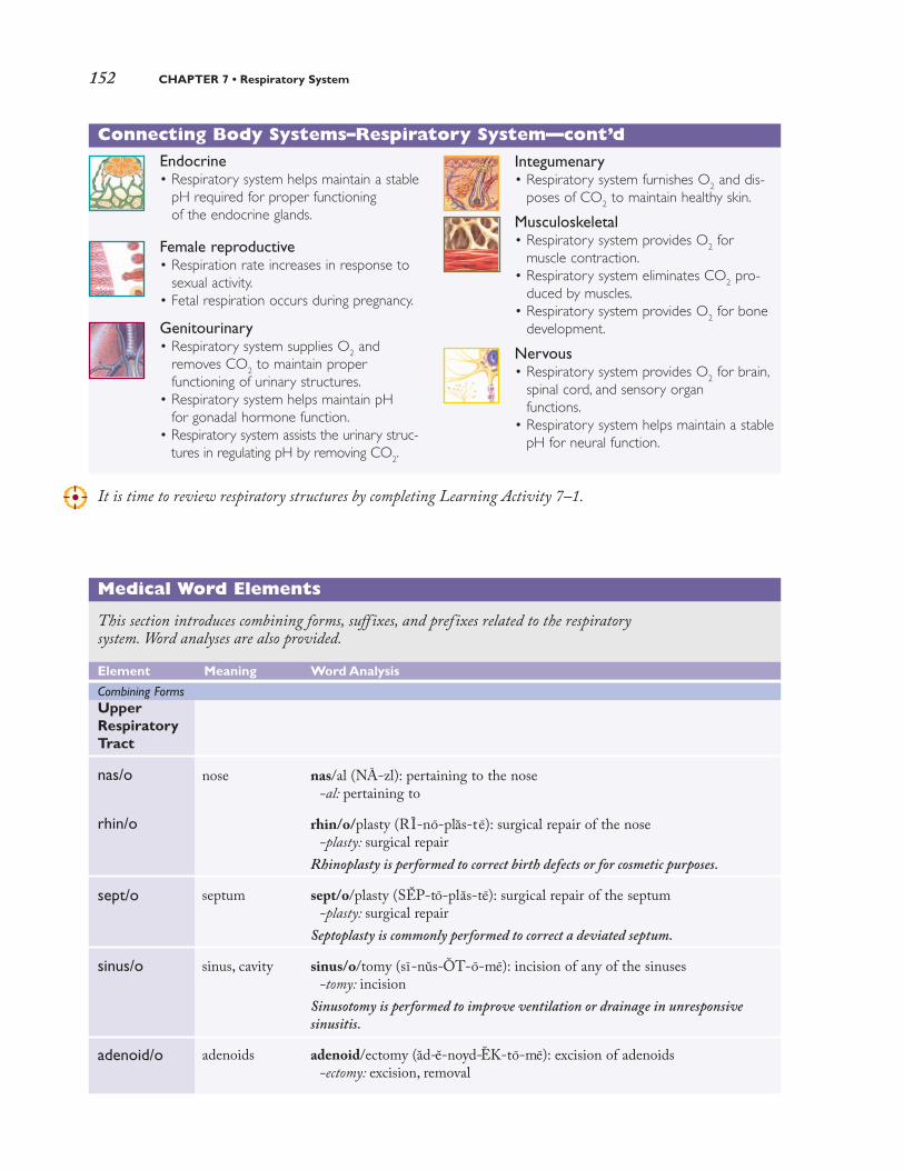
152 CHAPTER 7 • Respiratory System
It is time to review respiratory structures by completing Learning Activity 7–1.
Medical Word Elements
This section introduces combining forms, suff ixes, and prefixes related to the respiratory system. Word analyses are also provided.
Element Meaning Word Analysis
UpperRespiratoryTract
nas/o
rhin/o
sept/o
sinus/o
adenoid/o
nose
septum
sinus, cavity
adenoids
nas/al (NA-zl): pertaining to the nose-al: pertaining to
rhin/o/plasty (RI-no-plas-t e): surgical repair of the nose-plasty: surgical repair
Rhinoplasty is performed to correct birth defects or for cosmetic purposes.
sept/o/plasty (SEP-to-plas-te): surgical repair of the septum-plasty: surgical repair
Septoplasty is commonly performed to correct a deviated septum.
sinus/o/tomy (sı-nus-OT-o-me): incision of any of the sinuses-tomy: incision
Sinusotomy is performed to improve ventilation or drainage in unresponsive
sinusitis.
adenoid/ectomy (ad-e-noyd-EK-to-me): excision of adenoids-ectomy: excision, removal
Combining Forms
Connecting Body Systems–Respiratory System—cont’d
Endocrine• Respiratory system helps maintain a stable
pH required for proper functioning of the endocrine glands.
Female reproductive• Respiration rate increases in response to
sexual activity.• Fetal respiration occurs during pregnancy.
Genitourinary• Respiratory system supplies O2 and
removes CO2 to maintain proper functioning of urinary structures.
• Respiratory system helps maintain pH for gonadal hormone function.
• Respiratory system assists the urinary struc-tures in regulating pH by removing CO2.
Integumenary• Respiratory system furnishes O2 and dis-
poses of CO2 to maintain healthy skin.
Musculoskeletal• Respiratory system provides O2 for
muscle contraction.• Respiratory system eliminates CO2 pro-
duced by muscles.• Respiratory system provides O2 for bone
development.
Nervous• Respiratory system provides O2 for brain,
spinal cord, and sensory organ functions.
• Respiratory system helps maintain a stablepH for neural function.
2090_Ch07_147-184.qxd 3/5/09 10:00 PM Page 152

Anatomy and Physiology 153
Medical Word Elements—cont’d
Element Meaning Word Analysis
tonsill/o
pharyng/o
epiglott/o
laryng/o
trache/o
LowerRespiratoryTract
bronchi/o
bronch/o
bronchiol/o
alveol/o
pleur/o
pneum/o
pneumon/o
tonsils
pharynx (throat)
epiglottis
larynx (voicebox)
trachea (windpipe)
bronchus (plural, bronchi)
bronchiole
alveolus; air sac
pleura
air; lung
peri/tonsill/ar (per-ı-TON-sı-lar): pertaining to (the area) around the tonsilsperi-: around-ar: pertaining to
pharyng/o/scope (f ar-IN-go-skop): instrument for examining the pharynx-scope: instrument for examining
epiglott/itis (ep-ı-glot-I-t ıs): inflammation of the epiglottis-itis: inflammation
Because the epiglottis seals the passageway traveled by air to and from the lungs,
inflammation can lead to severe airway obstruction and death. Epiglottitis is
treated as a medical emergency.
laryng/o/plegia (la-rın-go-PLE-je-a): paralysis of the (vocal cords and) larynx-plegia: paralysis
trache/o/plasty (TRA-ke-o-plas-te): surgical repair of the trachea-plasty: surgical repair
Tracheoplasty is performed to correct a narrow or stenotic trachea.
bronchi/ectasis (brong-ke-EK-ta-s ıs): dilation of (one or more) bronchi-ectasis: dilation, expansion
Bronchiectasis is associated with various lung conditions and is commonly accom-
panied by chronic infection.
bronch/o/scope (BRONG-ko-skop): instrument for examining the bronchusor bronchi
-scope: instrument for examining
A bronchoscope is a flexible tube that is passed through the nose or mouth and enables
inspection of the lungs and collection of tissue biopsies and secretions for analysis.
bronchiol/itis (brong-ke-o-LI-t ıs): inflammation of the bronchioles-itis: inflammation
alveol/ar (al-VE-o-lar): pertaining to the alveoli-ar: pertaining to
pleur/o/centesis (ploo-ro-sen-TE-sıs): surgical puncture of the pleural cavi-ty; also called thoracocentesis or thoracentesis
-centesis: surgical puncture
pneum/ectomy (num-EK-to-me): excision of (all or part of ) a lung-ectomy: excision
pneumon/ia (nu-MO-ne-a): condition of inflammation of the lungs-ia: condition
The usual causes of pneumonia are infections due to bacteria, viruses, or other
pathogenic organisms. (continued)
2090_Ch07_147-184.qxd 3/5/09 10:00 PM Page 153

154 CHAPTER 7 • Respiratory System
Medical Word Elements—cont’d
Element Meaning Word Analysis
pulmon/o
Other
anthrac/o
atel/o
coni/o
cyan/o
lob/o
orth/o
ox/i
ox/o
pector/o
steth/o
lung
coal, coal dust
incomplete;imperfect
dust
blue
lobe
straight
oxygen
chest
pulmon/o/logist (pul-mo-NOL-o-j ıst): specialist in the study (and treatment) of lungs (and respiratory diseases)
-logist: specialist in the study of
anthrac/osis (an-thra-KO-sıs): abnormal condition of coal dust (in the lungs)-osis: abnormal condition; increase (used primarily with blood cells)
Anthracosis is a chronic occupational disease found in coal miners and those associ-
ated with the coal industry.
atel/ectasis (at-e-LEK-ta-s ıs): incomplete expansion of the lung; also calledairless lung or collapsed lung
-ectasis: dilation, expansion
pneum/o/coni/osis (nu-mo-ko-ne-O-sıs): condition of dust in the lungspneum/o: air; lung-osis: abnormal condition; increase (used primarily with blood cells)
Pneumoconiosis is usually caused by mineral dusts of occupational or environmen-
tal origin. Forms of pneumoconiosis include silicosis, asbestosis, and anthracosis.
cyan/osis (sı-a-NO-sıs): abnormal condition of blueness-osis: abnormal condition; increase (used primarily with blood cells)
Cold temperatures, heart failure, lung diseases, and smothering cause unusual blueness
of the skin and mucous membranes due to the build-up of carbon dioxide in the blood.
lob/ectomy (lo-BEK-to-me): excision of a lobe-ectomy: excision
Lobectomies are performed when a malignancy is conf ined to a single lobe of any
lobed organ, such as the lungs, liver, brain, and thyroid gland.
orth/o/pnea (or-THOP-ne-a): breathing in a straight (or upright position)-pnea: breathing
Various lung disorders cause a patient to experience diff iculty breathing in any
position other than sitting or standing erect.
ox/i/meter (ok-SIM-e-ter): instrument used for measuring oxygen-meter: instrument for measuring
An oximeter is usually attached to the tip of a f inger but may also be placed on a toe
or ear lobe. It provides a measurement of the oxygen saturation level of the blood.
hyp/ox/emia (hı-poks-E-me-a): deficiency of oxygen in bloodhyp-: under, below, deficient-emia: blood condition
pector/algia (pek-to-RAL-je-a): pain in the chest; also called thoracalgia,thoracodynia, and pectorodynia
-algia: pain
steth/o/scope (STETH-o-skop): instrument used for examining the chest-scope: instrument for examining
A stethoscope enables evaluation of sounds in the chest as well as the abdomen.
2090_Ch07_147-184.qxd 3/5/09 10:00 PM Page 154

Anatomy and Physiology 155
Medical Word Elements—cont’d
Element Meaning Word Analysis
thorac/o
phren/o
spir/o
-capnia
-osmia
-phonia
-pnea
-ptysis
-thorax
brady-
dys-
eu-
tachy-
diaphragm;mind
breathe
carbon dioxide(CO
2)
smell
voice
breathing
spitting
chest
slow
bad; painful;difficult
good, normal
rapid
thorac/o/pathy (tho-rak-OP-a-the): disease of the chest-pathy: disease
phren/o/spasm (FREN-o-spazm): involuntary contraction of the diaphragm-spasm: involuntary contraction, twitching
spir/o/meter (spı-ROM-et-er): instrument for measuring breathing-meter: instrument for measuring
A spirometer measures how much air the lungs can hold (vital capacity) as well as
how much and how quickly air can be exhaled.
hyper/capnia (hı-per-KAP-ne-a): excessive CO2
hyper-: excessive, above normal
an/osmia (an-OZ-me-a): without (the sense of ) smellan-: without, not
dys/phonia (dıs-FO-ne-a): bad (impaired) voice qualitydys-: bad; painful; difficult
Dysphonia includes hoarseness, voice fatigue, or decreased projection.
a/pnea (ap-NE-a): not breathinga-: without, not
Apnea is a temporary loss of breathing and includes sleep apnea, cardiac apnea,
and apnea of the newborn.
hem/o/ptysis (he-MOP-tı-s ıs): (coughing up or) spitting of bloodhem/o: blood
Bloody sputum is usually a sign of a serious condition of the lungs.
py/o/thorax (pı-o-THO-raks): pus in the chest (cavity); also called empyema
py/o: pus
Pyothorax is usually caused by a penetrating chest wound or spreading of infection
from another part of the body.
brady/pnea (brad-ıp-NE-a): slow breathing-pnea: breathing
dys/pnea (dısp-NE-a): difficult breathing-pnea: breathing
Dyspnea includes any discomfort or signif icant breathlessness.
eu/pnea (up-NE-a): normal breathing-pnea: breathing
The normal range for a resting adult respiratory rate is 12 to 20 breaths/minute.
tachy/pnea (tak-ıp-NE-a): rapid breathing-pnea: breathing
Suffixes
Prefixes
It is time to review word elements by completing Learning Activity 7–2. For audio pronunciations of theabove-listed key terms, you can visit www.davisplus.fadavis.com/gylys/systems to download this chapter’sListen and Learn! exercises or use the book’s audio CD (if included).
2090_Ch07_147-184.qxd 3/5/09 10:00 PM Page 155

156 CHAPTER 7 • Respiratory System
A. Chronic bronchitis
B. Emphysema C. Asthma
Distended bronchiole
Enlargedalveoli
Excess mucusproduction
Constrictedsmoothmuscle
Extra mucus
Inflamedairway
Figure 7-3. COPD.
(A) Chronic bronchitis with
inflamed airways and exces-
sive mucus. (B) Emphysema
with distended bronchioles
and alveoli. (C) Asthma with
narrowed bronchial tubes
and swollen mucous
membranes.
Pathology
Common signs and symptoms of many respirato-ry disorders include cough (dry or productive),chest pain, altered breathing patterns, shortness ofbreath (SOB), cyanosis, and fever. Many disordersof the respiratory system, including bronchitis andemphysema, begin as an acute problem butbecome chronic over time. Chronic respiratorydiseases are usually difficult to treat. Their damag-ing effects are commonly irreversible.
For diagnosis, treatment, and management ofrespiratory disorders, the medical services of a spe-cialist may be warranted. Pulmonology is themedical specialty concerned with disorders of the
respiratory system. The physician who treats thesedisorders is called a pulmonologist.
Chronic ObstructivePulmonary DiseaseChronic obstructive pulmonary disease(COPD) includes respiratory disorders that pro-duce a chronic partial obstruction of the air pas-sages. The patient finds it difficult to breath (dys-pnea) especially upon exertion and usuallyexhibits a chronic cough. The three major disor-ders included in COPD are asthma, chronicbronchitis, and emphysema. (See Figure 7–3.)
2090_Ch07_147-184.qxd 3/5/09 10:00 PM Page 156

Pathology 157
Asthma
Asthma produces spasms in the bronchial pas-sages (bronchospasms) that may be sudden andviolent (paroxysmal) and lead to dyspnea. Asthmais commonly caused by exposure to allergens orirritants. Other causes include stress, cold, andexercise. During recovery, coughing episodes pro-duce large amounts of mucus (productive cough).Over time, the epithelium of the bronchial pas-sages thickens, and breathing becomes more diffi-cult. Treatment includes agents that loosen andbreak down mucus (mucolytics) and medicationsthat expand the bronchi (bronchodilators) byrelaxing their smooth muscles. If usual measuresdo not reverse the bronchospasms, the conditionis referred to as status asthmaticus.
Chronic Bronchitis
Chronic bronchitis is an inflammation of thebronchi caused mainly by smoking and air pollu-tion. However, other agents, such as viruses andbacteria may also cause the disorder. Bronchitis ischaracterized by swelling of the mucosa and aheavy, productive cough, commonly accompaniedby chest pain. Patients usually seek medical helpwhen they suffer exercise intolerance, wheezing,and shortness of breath (SOB). Bronchodilatorsand medications that aid in the removal of mucus(expectorants) help to widen air passages. Steroidsmay be prescribed if the disease progresses orbecomes chronic.
Emphysema
Emphysema is characterized by decreased elastic-ity of the alveoli. The alveoli expand (dilate) butare unable to contract to their original size.The airthat remains trapped in the chest results in a char-acteristic “barrel-chested” appearance. This diseasecommonly occurs with another respiratory disor-der, such as asthma, tuberculosis, or chronic bron-chitis. It is also found in long-term heavy smokers.Most emphysema sufferers find it easier to breathewhen sitting upright or standing erect (orthop-nea). As the disease progresses, relief even in theorthopneic position is not possible. Treatment foremphysema is similar to that of chronic bronchitis.
InfluenzaInfluenza (flu) is an acute infectious respiratoryviral disease. Three major viral types are responsi-ble: type A, type B, and type C. Type A is of pri-mary concern because it is associated with world-wide epidemics (pandemics) and its causative
organism is highly infectious (virulent). Influenzatype A epidemics occur about every 2 to 3 years.Type B is usually limited geographically and tendsto be less severe than type A. Both viruses undergoantigenic changes; consequently, new vaccines mustbe developed in anticipation of outbreaks. Type Cis a mild flu and is not associated with epidemics.
The onset of the flu is usually rapid. Symptomsinclude fever, chills, headache, generalized musclepain (myalgia), and loss of appetite, but recoveryoccurs in about 7 to 10 days. The flu virus rarelycauses death. If death occurs, it is usually the resultof a secondary pneumonia caused by bacteria orviruses that invade the lungs. Children should notuse aspirin for relief of symptoms caused by virus-es because there appears to be a relationshipbetween Reye syndrome and the use of aspirin bychildren 2 to 15 years of age.
Pleural EffusionsAny abnormal fluid in the pleural cavity, the spacebetween the visceral and parietal pleura, is called apleural effusion. Normally, the pleural cavity con-tains only a small amount of lubricating fluid.However, some disorders may cause excessive fluidto collect in the pleural cavity. Two initial tech-niques used to diagnose pleural effusion are aus-cultation and percussion. Auscultation is the lis-tening of sounds made by organs of the body usinga stethoscope. Percussion is the gentle tapping thechest with the fingers and listening to the resultantsounds to determine the position, size, or consis-tency of the underlying structures. Chest x-ray(CXR) or magnetic resonance imaging (MRI)confirms the diagnosis.
Effusions are classified as transudates and exu-dates. A transudate is a noninflammatory fluidthat resembles serum but with slightly less pro-tein. It results from an imbalance in venous-arterial pressure or decrease of protein in blood.Both of these conditions allow serum to leakfrom the vascular system and collect in the pleu-ral space. Common causes include left ventricularheart failure and liver disorders. An exudate isusually high in protein and often contains bloodand immune cells. Common causes includetumors, infections, and inflammation. Various typesof pleural effusions include serum (hydrothorax),pus (empyema or pyothorax), and blood (hemo-thorax). Although not considered a pleural effusion, air can enter the pleural space (pneu-mothorax), resulting in a partial or complete collapse of a lung. (See Figure 7–4.)
2090_Ch07_147-184.qxd 3/5/09 10:00 PM Page 157

158 CHAPTER 7 • Respiratory System
Treatment consists of correcting the underlyingcause of the effusion. Often a surgical puncture ofthe chest using a hollow-bore needle (thoracocen-tesis, thoracentesis) is undertaken to removeexcess fluid for diagnostic or therapeutic purposes.(See Figure 7–5.) Sometimes chest tubes are insert-ed to drain fluid or remove air in pneumothorax.
TTuubbeerrccuulloossiissTuberculosis (TB) is a communicable diseasecaused by the bacterium Mycobacterium tuberculosis.TB spreads by droplets of respiratory secretions(droplet nuclei) from an infected individual whenhe/she coughs, laughs, or sneezes. The waxy coat ofthe TB organism keeps it alive (viable) and infec-
tious for 6 to 8 months outside the body. It alsomakes laboratory staining of this organism morechallenging. Hence TB is also known as the acid-fastbacillus (AFB), a reference to its more complexmethod of laboratory staining.
The first time the TB organism enters the body(primary tuberculosis), the disease develops slow-ly. It eventually produces typical inflammatorynodules (granulomas) called tubercles. Thesegranulomas usually remain dormant for years, dur-ing which time the patient is asymptomatic. Whenthe immune system becomes impaired (immuno-compromised) or when the patient is reexposed tothe bacterium, a full-blown disease may develop.
Although primarily a lung disease, TB can infectthe bones, genital tract, meninges, and peritoneum.
Normal lung
A.
B. C.
Pneumothorax oninspiration
Pneumothorax onexpiration
Visceral pleura
Parietalpleura
Pleuralcavity
Visceral pleura
Parietalpleura
Air Air
Figure 7-4. Pneumothorax. (A) Normal. (B) Open pneumothorax during inspiration. (C) Open pneumothorax during
expiration.
2090_Ch07_147-184.qxd 3/5/09 10:00 PM Page 158

Pathology 159
Some TB strains that infect AIDS patients havebecome resistant and do not respond to standardmedications. Treatment may include using severalantibiotics (combination therapy) at the same time.
PneumoniaPneumonia is any inflammatory disease of thelungs that may be caused by bacteria, viruses, orfungi. Chemicals or other agents can cause thelungs to become inflamed. A type of pneumoniaassociated with influenza is sometimes fatal.Other potentially fatal pneumonias may resultfrom food or liquid inhalation (aspiration pneu-monias). Some pneumonias affect only a lobe ofthe lung (lobar pneumonia), but some are morediffuse (bronchopneumonia). Chest pain, muco-purulent sputum, and spitting of blood (hemop-tysis) are common signs and symptoms of the disease. If the air in the lungs is replaced by fluidand inflammatory debris, the lung tissue looses its spongy texture and become swollen andengorged (consolidation). Consolidation is asso-ciated primarily with bacterial pneumonias, notviral pneumonias.
Pneumocystis carinii pneumonia (PCP) is atype of pneumonia closely associated withAIDS. Recent evidence suggests that it is caused
by a fungus that resides in or on most people(normal flora) but causes no harm as long as theindividual remains healthy. When the immunesystem begins to fail, this organism becomesinfectious (opportunistic). Diagnosis relies onexamination of biopsied lung tissue or bronchialwashings (lavage).
Cystic FibrosisCystic fibrosis is a hereditary disorder of theexocrine glands that causes the body to secreteextremely thick (viscous) mucus. This thickenedmucus clogs ducts of the pancreas and digestivetract. As a result, digestion is impaired and thepatient may suffer from malnutrition. It also blocksducts of the sweat glands, causing the skin tobecome highly “salty.” In the lungs, mucus blocksairways and impedes natural disease-fighting mech-anisms, causing repeated infections. Medication inthe form of mists (aerosols) along with posturaldrainage provide relief.
An important diagnostic test called the sweattest measures the amount of salt excreted in sweat.When elevated, it indicates cystic fibrosis.Although the disease is fatal, improved methods oftreatment have extended life expectancy, andpatient survival is approximately 30 years.
Figure 7-5. Thoracentesis.
Syringe with catheterremoving pleural fluid from around lung
Collecting bottle
Ribs
Parietal pleura
Visceral pleura
Lung
Pleural effusion
2090_Ch07_147-184.qxd 3/5/09 10:00 PM Page 159

160 CHAPTER 7 • Respiratory System
Acute Respiratory DistressSyndromeAcute respiratory distress syndrome (ARDS) is acondition in which the lungs no longer functioneffectively, threatening the life of the patient. Itusually occurs as a result of very serious lung con-ditions, such as trauma, severe pneumonia, andother major infections that affect the entire body(systemic infections) or blood (sepsis). In ARDS,the alveoli fill with fluid (edema) caused byinflammation, and then collapse, making oxygenexchange impossible. Mechanical ventilation iscommonly required to save the life of the patient.
Hyaline membrane disease (HMD), sometimescalled infant respiratory distress syndrome (IRDS), isa form of respiratory distress syndrome. It is mostcommonly seen in preterm infants or infants bornto diabetic mothers. It is caused by insufficientsurfactant, a phospholipid substance that helpskeep alveoli open. With insufficient surfactant, thealveoli collapse and breathing becomes labored.Clinical signs may include blueness (cyanosis) ofthe extremities. Flaring of the nostrils (nares) and central cyanosis are typically present. Othersigns include rapid breathing (tachypnea), inter-costal retraction, and a characteristic grunt audible
during exhalation. Radiography shows a mem-brane that has a ground-glass appearance (hyalinemembrane), bilateral decrease in volume, and alve-olar consolidation. Although severe cases of HMDresult in death, some forms of therapy are effective.
OncologyThe most common form of lung cancer is bron-chogenic carcinoma; also called primary pul-monary cancer. This cancer is usually associatedwith tobacco use. Cells of the bronchial epitheliumdivide repeatedly until the entire epithelium isinvolved. Within a short time, the epitheliumbegins to invade underlying tissues. As massesform, they block air passages and alveoli.Bronchogenic carcinoma spreads (metastasizes)rapidly to other areas of the body, including thelymph nodes, liver, bones, brain, and kidneys. Onlyabout 10% of lung cancers are found in the earlystages when the cure rate is high. Treatment oflung cancer includes surgery, radiation, andchemotherapy or a combination of these methodsdepending on specific cell type, how far the diseasehas spread, and the general health of the patient.Nevertheless, lung cancer is difficult to control andsurvival rates are very low.
Diagnostic, Symptomatic, and Related Terms
This section introduces diagnostic, symptomatic, and related terms and their meanings. Wordanalyses for selected terms are also provided.
Term Definition
acidosisas- ı-DO-sıs
anosmiaan-OZ-me-aan-: without, not-osmia: smell
Excessive acidity of body fluids
Respiratory acidosis is commonly associated with pulmonary insuff iciency and
the subsequent retention of carbon dioxide
Absence of the sense of smell
Anosmia usually occurs as a temporary condition resulting from an upper respi-
ratory infection or a condition that causes intranasal swelling.
2090_Ch07_147-184.qxd 3/5/09 10:00 PM Page 160

Pathology 161
Diagnostic, Symptomatic, and Related Terms—cont’d
Term Definition
Enlargedtonsil causing
obstructivesleep apnea
Uvula
Epiglottis
Trachea
A.
B.
Positive pressureprovided by a fan
Nasal mask (pillows)
Figure 7-6. Sleep apnea. (A) Airway obstruction caused by enlarged tonsils, eventually leads to obstructive
sleep apnea. (B) Continuous positive airway pressure (CPAP) machine used to treat sleep apnea.(continued)
apneaap-NE-a
a-: without, not-pnea: breathing
sleep
Temporary loss of breathing
There are three types of apnea: obstructive (enlarged tonsils and adenoids),
central (failure of the brain to transmit impulses for breathing), and mixed
(combination of obstructive and central apnea).
Sleeping disorder in which breathing stops repeatedly for more than 10 seconds, causing measurable blood deoxygenation. (See Figure 7-6.)
2090_Ch07_147-184.qxd 3/5/09 10:00 PM Page 161

162 CHAPTER 7 • Respiratory System
Diagnostic, Symptomatic, and Related Terms—cont’d
Term Definition
asphyxiaas-FIK-se-aa-: without, not-sphyxia: pulse
atelectasisat-e-LEK-ta-s ısatel: incomplete; imperfect-ectasis: dilation, expansion
cheyne-Stokes respirationchan-stoks
compliancekom-PLI-ans
coryzako-RI-za
crackleKRAK-el
croupcroop
deviated nasal septumDE-ve-at-ed NA-zl SEP-tum
epiglottitisep-ı-glot-I-t ısepiglott: epiglottis-itis: inflammation
epistaxisep-ı-STAK-sıs
finger clubbingKLUB-ıng
hypoxemiahı-poks-E-me-a
hyp-: under, below, deficientox: oxygen-emia: blood condition
Condition caused by insufficient intake of oxygen
Some common causes of asphyxia are drowning, electric shock, lodging of a for-
eign body in the respiratory tract, inhalation of toxic smoke, and poisoning.
Collapsed or airless state of the lung, which may be acute or chronic andaffect all or part of a lung
Atelectasis is a potential complication of some surgical procedures, especially
those of the chest because breathing is commonly shallow after surgery to avoid
pain from the surgical incision. In fetal atelectasis, the lungs fail to expand
normally at birth.
Repeated breathing pattern characterized by fluctuation in the depth ofrespiration, first deeply, then shallow, then not at all
Cheyne-Stokes respirations are usually caused by diseases that affect the
respiratory centers of the brain (such as heart failure and brain damage).
Ease with which lung tissue can be stretched
Low compliance means lungs are less elastic; therefore, more effort is required
to inflate the lungs.
Head cold; upper respiratory infection (URI)
Abnormal respiratory sound heard on auscultation, caused by exudates,spasms, hyperplasia, or when air enters moisture-filled alveoli; also called rale
Common childhood condition involving inflammation of the larynx, tra-chea, bronchial passages and, sometimes, lungs
Signs and symptoms include a resonant, barking cough with suffocative, diffi-
cult breathing; laryngeal spasms, and, sometimes, the narrowing of the top of
the air passages.
Displacement of cartilage dividing the nostrils
Severe, life-threatening infection of the epiglottis and supraglottic structuresthat occurs most commonly in children between 2 and 12 years of age
Signs and symptoms of epiglottitis include fever, dysphagia, inspiratory stridor,
and severe respiratory distress. Intubation or tracheostomy may be required to
open the obstructed airway.
Nosebleed; nasal hemorrhage
Enlargement of the terminal phalanges of the fingers and toes, commonlyassociated with pulmonary disease
Deficiency of oxygen in the blood
Hypoxemia is usually a sign of respiratory impairment.
2090_Ch07_147-184.qxd 3/5/09 10:00 PM Page 162

Pathology 163
Diagnostic, Symptomatic, and Related Terms—cont’d
Term Definition
hypoxiahı-POKS-e-a
hyp-: under, below, deficient-oxia: oxygen
pertussisper-TUS-ıs
pleurisyPLOO-r ıs-e
pleur: pleura-isy: state of; condition
pneumoconiosisnu-mo-ko-ne-O-sıs
pneum/o: air; lungconi: dust-osis: abnormal condition; increase
(used primarily with blood cells)
pulmonary edemaPUL-mo-ne-re e-DE-ma
pulmon: lung-ary: pertaining to
pulmonary embolusPUL-mo-ne-re EM-bo-lus
pulmon: lung-ary: pertaining toembol: plug-us: condition, structure
rhonchusRONG-kus
stridorSTRI-dor
sudden infant death syndrome(SIDS)
wheezeHWEZ
Deficiency of oxygen in tissues
Hypoxia is usually a sign of respiratory impairment.
Acute infectious disease characterized by a cough that has a “whoop”sound; also called whooping cough
Immunization of infants as part of the diphtheria-pertussis-tetanus (DPT)
vaccination is effective in the prevention of pertussis.
Inflammation of the pleural membrane characterized by a stabbing painthat is intensified by coughing or deep breathing; also called pleuritis
Disease caused by inhaling dust particles, including coal dust (anthraco-sis), stone dust (chalicosis), iron dust (siderosis), and asbestos particles(asbestosis)
Accumulation of extravascular fluid in lung tissues and alveoli, causedmost commonly by heart failure
Excessive fluid in the lungs induces coughing and dyspnea.
Blockage in an artery of the lungs caused by a mass of undissolved matter(such as a blood clot, tissue, air bubbles, and bacteria)
Abnormal breath sound heard on auscultation
A rhonchus is described as a course, rattling noise that resembles snoring, com-
monly suggesting secretions in the larger airways.
High-pitched, harsh, adventitious breath sound caused by a spasm orswelling of the larynx or an obstruction in the upper airway
The presence of stridor requires immediate intervention.
Completely unexpected and unexplained death of an apparently normal,healthy infant, usually less than 12 months of age; also called crib death
The rate of SIDS has decreased more than 30% since parents have been instruct-
ed to place babies on their backs for sleeping rather than on their stomachs.
Whistling or sighing sound heard on auscultation that results from narrowing of the lumen of the respiratory passageway
Wheezing is a characteristic of asthma, croup, hay fever, obstructive emphysema,
and other obstructive respiratory conditions.
It is time to review pathological, diagnostic, symptomatic, and related terms by completing Learning Activity 7–3.
2090_Ch07_147-184.qxd 3/5/09 10:00 PM Page 163

164 CHAPTER 7 • Respiratory System
Diagnostic and Therapeutic Procedures
This section introduces procedures used to diagnose and treat respiratory disorders. Descriptions areprovided as well as pronunciations and word analyses for selected terms.
Procedure Descripton
Clinical
Mantoux testman-TU
oximetryok-SIM-e-treox/i: oxygen-metry: act of measuring
polysomnographypol-e-som-NOG-ra-fe
poly-: many, muchsomn/o: sleep-graphy: process of recording
pulmonary function tests(PFTs)PUL-mo-ne-re
pulmon: lung-ary: pertaining to
spirometryspı-ROM-e-tre
spir/o: breathe-metry: act of measuring
Endoscopic
bronchoscopybrong-KOS-ko-pe
bronch/o: bronchus-scopy: visual examination
laryngoscopylar- ın-GOS-ko-pe
laryng/o: larynx (voice box)-scopy: visual examination
mediastinoscopyme-de-as-t ı-NOS-ko-pe
mediastin/o: mediastinum-scopy: visual examination
Intradermal test to determine tuberculin sensitivity based on a positivereaction where the area around the test site becomes red and swollen
A positive test suggests a past or present exposure to TB or past TB vaccination. How-
ever, the Mantoux test does not differentiate between active and inactive infection.
Noninvasive method of monitoring the percentage of hemoglobin (Hb)saturated with oxygen; also called pulse oximetry
In oximetry, a probe is attached to the patient’s f inger or ear lobe and linked to
a computer that displays the percentage of hemoglobin saturated with oxygen.
Test of sleep cycles and stages using continuous recordings of brain waves(EEGs), electrical activity of muscles, eye movement (electro-oculogram),respiratory rate, blood pressure, blood oxygen saturation, heart rhythmand, sometimes, direct observation of the person during sleep using avideo camera
Multiple tests used to evaluate the ability of the lungs to take in and expelair as well as perform gas exchange across the alveolocapillary membrane
Measurement of ventilatory ability by assessing lung capacity and flow,including the time necessary for exhaling the total volume of inhaled air
A spirometer produces a graphic record for placement in the patient’s chart.
Visual examination of the bronchi using an endoscope (flexible fiberopticor rigid) inserted through the mouth and trachea for direct viewing ofstructures or for projection on a monitor (See Figure 7-7.)
Attachments on the bronchoscope can be used to suction mucus, remove foreign
bodies, collect sputum, or perform biopsy.
Visual examination of the larynx to detect tumors, foreign bodies, nerveor structural injury, or other abnormalities
Visual examination of the mediastinal structures including the heart, tra-chea, esophagus, bronchus, thymus, and lymph nodes
The mediastinoscope is inserted through a small incision made above the sternum.
The attached camera projects images on a monitor. Additional incisions may be made
if nodes are removed or other diagnostic or therapeutic procedures are performed.
Diagnostic Procedures
2090_Ch07_147-184.qxd 3/5/09 10:00 PM Page 164

Pathology 165
Diagnostic and Therapeutic Procedures—cont’d
Procedure Descripton
Viewingpiece
Bronchoscope
Channel in the flexible tube toaccommodate biopsy forceps
and instruments
Leftbronchus
Visualexamination
of left bronchus
Figure 7-7. Bronchoscopy of the left bronchus. (continued)
2090_Ch07_147-184.qxd 3/5/09 10:00 PM Page 165

166 CHAPTER 7 • Respiratory System
Diagnostic and Therapeutic Procedures—cont’d
Procedure Descripton
Laboratory
arterial blood gas (ABG)ar-TE-re-al
sputum cultureSPU-tum
sweat test
throat culture
Radiographic
radiographyr a-de-OG-ra-fe
radi/o: radiation, x-ray; radius(lower arm bone on thumbside)
-graphy: process of recording
thoracic (chest)tho-RAS-ık
thorac: chest-ic: pertaining to, relating to
scan
lung
Test that measures partial pressure of oxygen (Po2), carbon dioxide (Pco
2),
pH (acidity or alkalinity), and bicarbonate level of an arterial blood sample
ABG analysis evaluates pulmonary gas exchange and helps guide treatment of
acid-base imbalances.
Microbial test used to identify disease-causing organisms of the lower res-piratory tract, especially those that cause pneumonias
Measurement of the amount of salt (sodium chloride) in sweat
A sweat test is used almost exclusively in children to conf irm cystic f ibrosis.
Test used to identify pathogens, especially group A streptococci
Untreated streptococcal infections may lead to serious secondary complications,
including kidney and heart disease.
Process of producing images using an x-ray passed through the body orarea and captured on a film
Images of the chest taken from anteroposterior (AP) projection, posteroante-rior (PA) projection, lateral projection, or a combination of these projections
Chest radiography is used to diagnose rib fractures and lung diseases, including
atelectasis, masses, pneumonia, and emphysema.
Imaging procedure that gathers information about a specific organ orstructure of the body. In some cases, small amounts of injected radionu-clide (tracer) are used to enhance images
Nuclear scanning test primarily used to detect pulmonary emboli
Lung scan is commonly performed to detect the presence of a blood clot that may
be interfering with blood flow in or to the lung.
2090_Ch07_147-184.qxd 3/5/09 10:00 PM Page 166

Pathology 167
Diagnostic and Therapeutic Procedures—cont’d
Procedure Descripton
Clinical
aerosol therapyAR-o-sol THER-a-pe
Lung treatment using various techniques to deliver medication in mistform directly to the lungs or air passageways. Techniques include nebuliz-ers, metered-dose inhalers (MDIs), and dry powder inhalers (DPIs)
Nebulizers change liquid medications into droplets to be inhaled through a mouth-
piece. (See Figure 7-8.) MDIs deliver a specif ic amount when activated. Children
and the elderly can use a spacer to synchronize inhalation with medication release.
(See Figure 7-9.) A DPI is activated by a quick inhalation by the user.
Nebulizer
Spacer
Metered-doseinhaler
Metered-doseinhaler without
a spacer
Figure 7-8. Nebulizer.
Figure 7-9. Metered-dose inhaler. (continued)
Therapeutic Procedures
2090_Ch07_147-184.qxd 3/5/09 10:00 PM Page 167

168 CHAPTER 7 • Respiratory System
Diagnostic and Therapeutic Procedures—cont’d
Procedure Descripton
lavagela-VAZH
antralAN-tral
postural drainagePOS-tu-ral
Surgical
pleurectomyploor-EK-to-me
pleur: pleura-ectomy: excision, removal
pneumectomynum-EK-to-me
pneum: air; lung-ectomy: excision, removal
rhinoplastyRI-no-plas-te
rhin/o: nose-plasty: surgical repair
septoplastysep-to-PLAS-te
sept/o: septum-plasty: surgical repair
thoracentesistho-ra-sen-TE-sıs
tracheostomytra-ke-OS-to-me
Irrigating or washing out of an organ, stomach, bladder, bowel, or bodycavity with a stream of water or other fluid
Lavage of the paranasal sinuses is usually performed to remove mucopurulent
material in an immunosuppressed patient or one with known sinusitis that has
failed medical management.
Irrigation of the antrum (maxillary sinus) in chronic or nonresponsive sinusitis
Positioning a patient so that gravity aids in the drainage of secretionsfrom the bronchi and lobes of the lungs
Excision of part of the pleura, usually parietal
Pleurectomy is performed to reduce pain caused by a tumor mass or to prevent
the recurrence of pleural effusion but is generally ineffective in the treatment of
malignancy of the pleura.
Excision of a lung
The removal of a lobe of the lung is called a lobectomy
Reconstructive surgery of the nose to correct deformities or for cosmeticpurposes
Surgical repair of a deviated nasal septum usually performed when theseptum is encroaching on the breathing passages or nasal structures
Common complications of a deviated septum include interference with breath-
ing and a predisposition to sinus infections.
Surgical puncture and drainage of the pleural cavity; also called pleurocen-
tesis or thoracocentesis
Thoracentesis is performed as a diagnostic procedure to determine the nature
and cause of an effusion or as a therapeutic procedure to relieve the discomfort
caused by the effusion. (See Figure 7–5.)
Surgical procedure in which an opening is made in the neck and into thetrachea into which a breathing tube may be inserted (See Figure 7–10.)
2090_Ch07_147-184.qxd 3/5/09 10:00 PM Page 168

Pharmacology 169
PPhhaarrmmaaccoollooggyy
In addition to antibiotics used to treat respiratoryinfections, there are several classes of drugs thattreat pulmonary disorders. (See Table 7–1.)Bronchodilators are especially significant in thetreatment of COPD and exercise-induced asth-ma. They relax smooth muscles of the bronchi,thus increasing airflow. Some bronchodilators are
delivered as a fine mist directly to the airways viaaerosol delivery devices, including nebulizers andmetered-dose inhalers (MDIs). Another methodof delivering medications directly to the lungs isdry-powder inhalers (DPIs) that dispense med-ications in the form of a powder. Steroidal andnonsteroidal anti-inflammatory drugs are impor-tant in the control and management of many pul-monary disorders.
Diagnostic and Therapeutic Procedures—cont’d
Procedure Descripton
Epiglottis
Tracheostomytube
Expandingballoon
Trachea
Thyroidgland
A. B.
Figure 7-10. Tracheostomy. (A) Lateral view with tracheostomy tube in place. (B) Frontal view.
2090_Ch07_147-184.qxd 3/5/09 10:00 PM Page 169

170 CHAPTER 7 • Respiratory System
Drugs Used to Treat Respiratory DisordersTable 7-1
This table lists common drug classif ications used to treat respiratory disorders, their therapeutic actions,and selected generic and trade names.
Classification Therapeutic Action Generic and Trade Names
Block histamines from binding with histamine
receptor sites in tissues
Histamines cause sneezing, runny nose, itchiness,and rashes.
Relieve or suppress coughing by blocking the
cough reflex in the medulla of the brain
Antitussives alleviate nonproductive dry coughs andshould not be used with productive coughs.
Stimulate bronchial muscles to relax, thereby
expanding air passages, resulting in increased
air flow
Bronchodilators are used to treat chronic symp-toms and prevent acute attacks in respiratory dis-eases, such as asthma and COPD. Pharmacologicalagents may be delivered by an inhaler either orallyor intravenously.
Act on the immune system by blocking pro-
duction of substances that trigger allergic and
inflammatory actions
Corticosteroids are available as nasal sprays, inmetered-dose-inhalers (inhaled steroids) and in oralforms (pills or syrups) to treat chronic lung condi-tions such as asthma and COPD.
Constrict blood vessels of nasal passages and
limit blood flow, which causes swollen tissues to
shrink so that air can pass more freely through
the passageways
Decongestants are commonly prescribed for aller-gies and colds and are usually combined with anti-histamines in cold remedies.They can be adminis-tered orally or topically as nasal sprays and nasaldrops.
Liquify respiratory secretions so that they are
more easily dislodged during coughing episodes
Expectorants are prescribed for productive coughs.
fexofenadinefeks-o-FEN-a-den
Allegra
loratadinelor-AH-ta-den
Claritin
hydrocodonehı-dro-KO-don
Hycodan
dextromethorphandek-stro-METH-or-fan
Vicks Formula 44
albuterolal-BU-ter-al
Proventil,Ventolin
salmeterolsal-me-TER-ol
Serevent
beclomethasone dipropionatebe-klo-METH-a-son
dı-PRO-pe-o-nat
Vanceril, Beclovent
triamcinolonetrı-am-SIN-o-lon
Azmacort
oxymetazolineoks-e-met-AZ-o-len
Dristan
pseudoephedrinesoo-do-e-FED-r ın
Drixoral, Sudafed
guaifenesingwı-FEN-e-s ın
Robitussin, Organidin
antihistamines
antitussives
bronchodilators
corticosteroids
decongestants
expectorants
2090_Ch07_147-184.qxd 3/5/09 10:00 PM Page 170

Pharmacology 171
Abbreviations
This section introduces respiratory-related abbreviations and their meanings.
Abbreviation Meaning Abbreviation Meaning
ABG
AFB
AP
ARDS
CO2
COPD
CPAP
CPR
CT
CXR
DPI
DPT
EEG
FVC
Hb, Hgb
HMD
Hx
IPPB
IRDS
MDI
arterial blood gas(es)
acid-fast bacillus (TB organism)
anteroposterior
acute respiratory distress syndrome
carbon dioxide
chronic obstructive pul-monary disease
continuous positive airway pressure
cardiopulmonary resuscitation
computed tomography
chest x-ray, chest radiograph
dry power inhaler
diphtheria, pertussis, tetanus
encephalogram, encephalog-raphy
forced vital capacity
hemoglobin
hyaline membrane disease
history
intermittent positive-pressurebreathing
infant respiratory distress syndrome
metered dose inhaler
MRI
NMT
O2
PA
PCO2
PCP
PFT
pH
PND
PO2
RD
RDS
SaO2
SIDS
SOB
T&A
TB
TPR
URI
VC
magnetic resonance imaging
nebulized mist treatment
oxygen
posteroanterior; pernicious anemia
partial pressure of carbon dioxide
Pneumocystis carinii pneumonia;primary care physician; phencyclidine(hallucinogen)
pulmonary function test
symbol for degree of acidity or alkalinity
paroxysmal nocturnal dyspnea
partial pressure of oxygen
respiratory distress
respiratory distress syndrome
arterial oxygen saturation
sudden infant death syndrome
shortness of breath
tonsillectomy and adenoidectomy
tuberculosis
temperature, pulse, and respiration
upper respiratory infection
vital capacity
It is time to review procedures, pharmacology, and abbreviations by completing Learning Activity 7–4.
2090_Ch07_147-184.qxd 3/5/09 10:00 PM Page 171

172 CHAPTER 7 • Respiratory System
The following activities provide review of the respiratory system terms introduced in thischapter. Complete each activity and review your answers to evaluate your understanding ofthe chapter.
Learning Activity 7-1
Identifying Respiratory Structures
Label the following illustration using the terms listed below.
adenoids larynx parietal pleura
alveoli left lung pleural cavity
bronchi mediastinum pulmonary capillaries
bronchiole nasal cavity right lung
diaphragm nasopharynx trachea
epiglottis oropharynx visceral pleura
laryngopharynx palatine tonsils
LEARNING ACTIVITIES
2090_Ch07_147-184.qxd 3/5/09 10:00 PM Page 172

Learning Activities 173
Check your answers by referring to Figure 7–1 on page 150. Review material that you didnot answer correctly.✓
Nose
Thyroid cartilageGlottis and vocal cords
Base of lung
Deoxygenatedblood from heart
Oxygenatedblood to heart
Apex of lung
CO2
Alveolus O2
Exchange of gases between analveolus and a pulmonary capillary
Pulmonary capillary
Enhance your study and reinforcement of word elements with the power of DavisPlus. Visit www.davisplus.fadavis.com/gylys/systems for this chapter’s flash-card activity. We recommend youcomplete the flash-card activity before completing activity 7–2 below.
2090_Ch07_147-184.qxd 3/5/09 10:00 PM Page 173

174 CHAPTER 7 • Respiratory System
Check your answers in Appendix A. Review material that you did not answer correctly.✓
Correct Answers �� 5 �� % Score
Learning Activity 7-2
Building Medical Words
Use rhin/o (nose) to build words that mean:
1. discharge from the nose ______________________________________________________________
2. inflammation of (mucous membranes of the) nose _________________________________________
Use laryng/o (larynx [voice box]) to build words that mean:
3. visual examination of larynx ___________________________________________________________
4. inflammation of larynx ________________________________________________________________
5. stricture or narrowing of the larynx _____________________________________________________
Use bronch/o or bronchi/o (bronchus) to build words that mean:
6. dilation or expansion of the bronchus ___________________________________________________
7. disease of the bronchus ______________________________________________________________
8. spasm of the bronchus _______________________________________________________________
Use pneumon/o or pneum/o (air; lung) to build words that mean:
9. air in the chest (pleural space) _________________________________________________________
10. inflammation of lungs ________________________________________________________________
Use pulmon/o (lung) to build words that mean:
11. specialist in lung (diseases) ____________________________________________________________
12. pertaining to the lung ________________________________________________________________
Use -pnea (breathing) to build words that mean:
13. difficult breathing ____________________________________________________________________
14. slow breathing ______________________________________________________________________
15. rapid breathing _____________________________________________________________________
16. absence of breathing _________________________________________________________________
Build surgical words that mean:
17. surgical repair of the nose _____________________________________________________________
18. surgical puncture of the chest __________________________________________________________
19. removal of a lung ___________________________________________________________________
20. forming an opening (mouth) in the trachea _______________________________________________
2090_Ch07_147-184.qxd 3/5/09 10:01 PM Page 174

Learning Activities 175
Learning Activity 7-3
Matching Pathological, Diagnostic, Symptomatic, and Related Terms
Match the following terms with the definitions in the numbered list.
anosmia consolidation empyema pneumoconiosis
apnea coryza epistaxis pulmonary edema
atelectasis crackle hypoxemia stridor
auscultation deviated septum pertussis surfactant
compliance emphysema pleurisy tubercles
1. collapsed or airless lung
2. pus in the pleural cavity
3. phospholipid that allows the lungs to expand with ease
4. loss of sponginess of lungs due to engorgement
5. listening to the chest sounds using a stethoscope
6. absence or decrease in the sense of smell
7. deficiency of oxygen in the blood
8. granulomas associated with tuberculosis
9. temporary loss of breathing
10. disease characterized by a decrease in alveolar elasticity
11. ease with which lung tissue can be stretched
12. nosebleed; nasal hemorrhage
13. excessive fluid in the lungs that induces cough and dyspnea
14. abnormal respiratory sound associated with exudates, spasms, orhyperplasia
15. displacement of the cartilage dividing the nostrils
16. head cold; upper respiratory infection
17. condition in which dust particles are found in the lungs
18. inflammation of the pleural membrane
19. abnormal sound caused by spasms or swelling of larynx
20. whooping cough
Check your answers in Appendix A. Review material that you did not answer correctly.✓
Correct Answers �� 5 �� % Score
2090_Ch07_147-184.qxd 3/5/09 10:01 PM Page 175

176 CHAPTER 7 • Respiratory System
Check your answers in Appendix A. Review any material that you did not answer correctly.✓
Correct Answers �� 5 �� % Score
Learning Activity 7-4
Matching Procedures, Pharmacology, and Abbreviations
Match the following terms with the definitions in the numbered list.
ABGs antral lavage Mantoux test radiography
aerosol therapy decongestant oximetry rhinoplasty
AFB expectorant pneumectomy septoplasty
antihistamine laryngoscopy polysomnography sweat test
antitussive lung scan pulmonary function tests throat culture
1. imaging procedure that uses radionuclide to evaluate blood flow inthe lungs
2. test of sleep cycles and stages
3. producing images using an x-ray machine
4. washing or irrigating sinuses
5. sneezing, runny nose, itchiness, and rashes
6. relieves or suppresses coughing
7. used primarily in children to confirm cystic fibrosis
8. noninvasive test used to monitor percentage of hemoglobin saturat-ed with oxygen
9. TB organism
10. inhalation of medication directly into the respiratory system via anebulizer
11. decreases mucous membrane swelling by constricting blood vessels
12. intradermal test to determine tuberculin sensitivity
13. laboratory tests to assess gases and pH of arterial blood
14. reduces the viscosity of sputum to facilitate productive coughing
15. used to identify pathogens, especially group A streptococci
16. multiple tests used to determine the ability of lungs and capillarymembranes to exchange oxygen
17. visual examination of the voice box to detect tumors and otherabnormalities
18. surgery to correct a deviated nasal septum
19. excision of the entire lung
20. reconstructive surgery of the nose, commonly for cosmetic purposes
2090_Ch07_147-184.qxd 3/5/09 10:01 PM Page 176

The two medical records included in the following activities use common clinical scenariosto show how medical terminology is used to document patient care. Complete the termi-nology and analysis sections for each activity to help you recognize and understand termsrelated to body structure.
Medical Record Activity 7-1
SOAP Note: Respiratory Evaluation
TerminologyTerms listed below come from the SOAP Note: Respiratory Evaluation that follows. Use amedical dictionary such as Taber’s Cyclopedic Medical Dictionary, the appendices of thisbook, or other resources to define each term. Then review the pronunciations for each termand practice by reading the medical record aloud.
MEDICAL RECORD ACTIVITIES
Term Definition
anteriorlyan-TER-e-or-le
bilateralbı-LAT-er-al
COPD
exacerbationeks-as-er-BA-shun
heart failure
Hx
hypertensionhı-per-TEN-shun
interstitialın-ter-STISH-al
PE
peripheral vascular diseaseper-IF-er-al VAS-
ku-lar
pleuralPLOO-ral
posteriorlypos-TER-e-or-le
rhonchiRONG-kı
Medical Record Activities 177
2090_Ch07_147-184.qxd 3/5/09 10:01 PM Page 177

178 CHAPTER 7 • Respiratory System
Term Definition
SOB
wheezesHWEZ-ez
Listen and Learn Online! will help you master the pronunciation of selected medical words from thismedical record activity. Visit www.davisplus.com/gylys/systems to f ind instructions on completing theListen and Learn Online! exercise for this section and to practice pronunciations.
(( ))
2090_Ch07_147-184.qxd 3/5/09 10:01 PM Page 178

SOAP Note: Respiratory Evaluation 179
Emergency Department Record
Date: February 1, 20xx Time Registered: 1345 hoursPatient: Flowers, Richard Physician: Samara Batichara, MD
Chief Complaint: SOB
Medications: Vytorin 10/20 mg daily; Toprol-XL 50 mg daily; Azmacort 2 puffs t.i.d; Proventil 2puffs q.6 h.
S: This 49-year-old man with Hx of COPD is admitted because of exacerbation of SOB over the past few days. Patient was a heavy smoker and states that he quit smoking for ashort while but now smokes 3-4 cigarettes a day. He has a Hx of difficult breathing, hyperten-sion, COPD, and peripheral vascular disease. He underwent triple bypass surgery in 19xx.
O: T: 98.9 F. BP: 180/90. Pulse: 80 and regular. R: 20 and shallow. PE indicates scattered bilateralwheezes and rhonchi heard anteriorly and posteriorly. When compared with a portable chest filmtaken 22 months earlier, the current study most likely indicates interstitial vascular congestion.Some superimposed inflammatory change cannot be excluded. There may also be some pleuralreactive change.
A: 1. Acute exacerbation of chronic obstructive pulmonary disease.2. Heart failure.3. Hypertension.4. Peripheral vascular disease.
P: Admit to hospital.
SSaammaarraa BBaattiicchhaarraa,, MMDDSamara Batichara, MD
SB:icc
D: 2/1/20xxT: 2/1/20xx
SOAP NOTE: RESPIRATORY EVALUATION
2090_Ch07_147-184.qxd 3/5/09 10:01 PM Page 179

180 CHAPTER 7 • Respiratory System
AnalysisReview the medical record SOAP Note: Respiratory Evaluation to answer the followingquestions.
1. What symptom caused the patient to seek medical help?
2. What was the patient’s previous history?
3. What were the abnormal findings of the physical examination?
4. What changes were noted from the previous film?
5. What are the present assessments?
6. What new diagnosis was made that did not appear in the previous medical history?
Medical Record Activity 7-2
SOAP Note: Chronic Interstitial Lung Disease
TerminologyTerms listed below come from the SOAP Note: Chronic Interstitial Lung Disease that follows.Use a medical dictionary such as Taber’s Cyclopedic Medical Dictionary, the appendices of thisbook, or other resources to define each term. Then review the pronunciations for each termand practice by reading the medical record aloud.
Term Definition
ABG
adenopathyad-e-NOP-a-the
basilar cracklesBAS-ı-lar KRAK-elz
cardiomyopathykar-de-o-mı-OP-a-the
chronicKRON-ık
2090_Ch07_147-184.qxd 3/5/09 10:01 PM Page 180

Term Definition
diuresisdı-u-RE-sıs
dyspneadısp-NE-a
fibrosisfı-BRO-sıs
interstitialın-ter-STISH-al
kyphosiskı-FO-sıs
LasixLA-sıks
neuropathynu-ROP-a-the
PCO2
pedal edemaPED-el e-DE-ma
pH
PO2
pulmonary fibrosisPUL-mo-ne-re
fı-BRO-sıs
renal insufficiencyRE-nal
ın-su-FISH-en-se
rhonchiRONG-kı
silicosiss ıl- ı-KO-sıs
thyromegalythı-ro-MEG-a-le
Listen and Learn Online! will help you master the pronunciation of selected medical words from thismedical record activity. Visit www.davisplus.com/gylys/systems to f ind instructions on completing theListen and Learn Online! exercise for this section and to practice pronunciations.
(( ))
SOAP Note: Respiratory Evaluation 181
2090_Ch07_147-184.qxd 3/5/09 10:01 PM Page 181

182 CHAPTER 7 • Respiratory System
09/01/20xx
O’Malley, Robert
SUBJECTIVE: Patient is an 84-year-old male with chief complaint of dyspnea with activity andpedal edema. He carries the dx cardiomyopathy, renal insufficiency, COPD, and pulmonary fibrosis.He also has peripheral neuropathy, which has improved with Elavil therapy.
OBJECTIVE: BP: 140/70. Pulse: 76. Neck is supple without thyromegaly or adenopathy. Mildkyphosis without scoliosis is present. Chest reveals basilar crackles without wheezing or rhonchi.Cardiac examination shows trace edema without clubbing or murmur. Abdomen is soft and non-tender. ABGs on room air demonstrate a Po
2of 55, Pco
2of 45, and pH of 7.42.
ASSESSMENT: Chronic interstitial lung disease, likely a combination of pulmonary fibrosis andheart failure. We do believe he would benefit from further diuresis, which was implemented by Dr. Lu. Should there continue to be concerns about his volume status or lack of response to Lasixtherapy, then he might benefit from right heart catheterization.
PLAN: Supplemental oxygen will be continued. We plan no change in his pulmonary medication atthis time and will see him in return visit in 4 months. He has been told to contact us should he wors-en in the interim.
SSaammaarraa BBaattiicchhaarraa,, MMDDSamara Batichara, MD
SB:icc
SOAP NOTE: CHRONIC INTERSTITIAL LUNG DISEASE
2090_Ch07_147-184.qxd 3/5/09 10:01 PM Page 182

AnalysisReview the medical record SOAP Note: Chronic Interstitial Lung Disease to answer the following questions.
1. When did the patient notice dyspnea?
2. Other than the respiratory system, what other body systems are identified in the history of present illness?
3. What were the findings regarding the neck?
4. What was the finding regarding the chest?
5. What appears to be the likely cause of the chronic interstitial lung disease?
6. What did the cardiac examination reveal?
SOAP Note: Chronic Interstitial Lung Disease 183
2090_Ch07_147-184.qxd 3/5/09 10:01 PM Page 183

2090_Ch07_147-184.qxd 3/5/09 10:01 PM Page 184

CardiovascularSystem
Chapter Outline
Objectives
Anatomy and PhysiologyAnatomy and Physiology Key TermsVascular System
ArteriesCapillariesVeins
HeartConduction System of the Heart
Blood PressureFetal CirculationConnecting Body Systems–Cardiovascular System
Medical Word Elements
PathologyArteriosclerosisCoronary Artery DiseaseEndocarditisVaricose VeinsOncology
Diagnostic, Symptomatic, and Related Terms
Diagnostic and Therapeutic Procedures
Pharmacology
Abbreviations
Learning Activities
Medical Record ActivitiesChart note: Acute myocardial infarctionOperative report: Right temporal artery biopsy
ObjectivesUpon completion of this chapter, you will be able to:
• Locate and describe the structures of the cardiovas-
cular system.
• Describe the functional relationship between the
cardiovascular system and other body systems.
• Identify, pronounce, spell, and build words related
to the cardiovascular system.
• Describe pathological conditions, diagnostic and
therapeutic procedures, and other terms related
to the cardiovascular system.
• Explain pharmacology related to the treatment
of cardiovascular disorders.
• Demonstrate your knowledge of this chapter by
completing the learning and medical record
activities.
C H A P T E R
8
2090_Ch08_185-226.qxd 3/5/09 10:01 PM Page 185

Anatomy and Physiology
The cardiovascular (CV) system is composed ofthe heart and blood vessels. The heart is a hollow,muscular organ lying in the mediastinum, thecenter of the thoracic cavity between the lungs.The pumping action of the heart propels bloodcontaining oxygen, nutrients, and other vital prod-ucts from the heart to body cells through a vastnetwork of blood vessels called arteries. Arteriesbranch into smaller vessels until they becomemicroscopic vessels called capillaries. It is at thecapillary level that exchange of products occursbetween body cells and blood. Capillaries mergeto form larger blood vessels called venules, whichthen combine to form veins, the vessels thatreturn blood to the heart to begin the cycle again.Millions of body cells rely on the cardiovascularsystem for their survival. When this transporta-tion system fails, life at the cellular level is notpossible and, ultimately, the organism will die.
Vascular SystemThree major types of vessels—(1) artery, (2) capil-lary, and (3) vein—carry blood throughout thebody. (See Figure 8–1.) Each type of vessel differsin structure depending on its function.
Arteries
Arteries carry blood from the heart to all cells ofthe body. Because blood is propelled thorough thearteries by the pumping action of the heart, thewalls of the arteries must be strong and flexibleenough to withstand the surge of blood that resultsfrom each contraction of the heart.
The walls of large arteries have three layers toprovide toughness and elasticity. The (4) tunicaexterna is the outer coat composed of connectivetissue that provides strength and flexibility. The (5) tunica media is the middle layer composed ofsmooth muscle. Depending on the needs of thebody, this muscle can alter the size of the (7) lumen
186 CHAPTER 8 • Cardiovascular System
Anatomy and Physiology Key Terms
This section introduces important terms and their definitions and pronunciation.
Term Definition
autonomic nervous system(ANS)aw-to-NOM-ık NER-ves
leaflet
lumenLU-men
regurgitationre-gur-j ı-TA-shun
sphincterSFINGK-ter
vasoconstrictionvas-o-kon-STRIK-shun
vasodilationvas-o-dı-LA-shun
viscosityvıs-KOS-ı-te
Portion of the nervous system that regulates involuntary actions, such asheart rate, digestion, and peristalsis
Thin, flattened structure; term used to describe the leaf-shaped structuresthat compose a heart valve
Tubular space or channel within any organ or structure of the body; spacewithin an artery, vein, intestine, or tube
Backflow or ejecting of contents through an opening
Circular muscle found in a tubular structure or hollow organ that constricts or dilates to regulate passage of substances through its opening
Narrowing of the lumen of a blood vessel that limits blood flow, usually asa result of diseases, medications, or physiological processes
Widening of the lumen of a blood vessel caused by the relaxing of themuscles of the vascular walls
State of being sticky or gummy
A solution that has high viscosity is relatively thick and flows slowly.
Pronunciation Help Long Sound a—rate e—rebirth ı—isle o—over u—uniteShort Sound a—alone e—ever ı—it o—not u—cut
2090_Ch08_185-226.qxd 3/5/09 10:01 PM Page 186

of the vessel. When it contracts, it causes vasocon-striction, resulting in decreased blood flow. Whenit relaxes, it causes vasodilation, resulting inincrease blood flow. The (6) tunica intima is thethin, inner lining of the lumen of the vessel, com-posed of endothelial cells that provide a smoothsurface on the inside of the vessel.
The surge of blood felt in the arteries whenblood is pumped from the heart is referred to as apulse. Because of the pressure against arterial wallsassociated with the pumping action of the heart, acut or severed artery may lead to profuse bleeding.
Arterial blood (except for that found in the pul-monary artery) contains a high concentration ofoxygen (oxygenated) and appears bright red incolor. Oxygenated blood travels to smaller arteries
called (8) arterioles and, finally, to the smallestvessels, the capillaries.
Capillaries
Capillaries are microscopic vessels that join thearterial system with the venous system. Althoughthey seem like the most insignificant of the threevessel types owing to their microscopic size, theyare actually the most important because of theirfunction. Because capillary walls are composed ofonly a single layer of endothelial cells, they are verythin. This thinness enables the exchange of water,respiratory gases, macromolecules, metabolites,and wastes between the blood and adjacent cells. The vast number of capillaries branchingfrom arterioles causes blood to flow very slowly,
Anatomy and Physiology 187
Heart
Aorta
Vena cava
(3) Vein (1) Artery
(8) Arteriole
(9) Venule
(2) Capillary
Endothelial cell
Precapillarysphincter
Smoothmuscle
(4) Tunica externa
(6) Tunica intima
(6) Tunica intima
(5) Tunica media (5) Tunica
media
(4) Tunica externa
Elasticlayer
(10) Valve
(7) Lumen (7) Lumen
Endothelium
Endothelium
Figure 8-1. Vascular structures.
2090_Ch08_185-226.qxd 3/5/09 10:01 PM Page 187

providing sufficient time for exchange of necessarysubstances.
Blood flow through the highly branched capil-lary system is partially regulated by the contractionof smooth muscle precapillary sphincters that leadinto the capillary bed. When tissues require moreblood, these sphincters open; when less blood isrequired, they close. Once the exchange of prod-ucts is complete, blood enters the venous systemfor its return cycle to the heart.
Veins
Veins return blood to the heart. They are formedfrom smaller vessels called (9) venules that devel-op from the union of capillaries. Because the exten-sive network of capillaries absorbs the propellingpressure exerted by the heart, veins use other meth-ods to return blood to the heart, including:
• skeletal muscle contraction• gravity• respiratory activity• valves.
The (10) valves are small structures within veinsthat prevent the backflow of blood. Valves arefound mainly in the extremities and are especiallyimportant for returning blood from the legs to theheart because blood must travel a long distanceagainst the force of gravity to reach the heart fromthe legs. Large veins, especially in the abdomen,contain smooth muscle that propels blood towardthe heart by peristalsis.
Blood carried in veins (except for the blood in thepulmonary veins) contains a low concentration ofoxygen (deoxygenated) with a corresponding highconcentration of carbon dioxide. Deoxygenatedblood takes on a characteristic purple color. Bloodcontinuously circulates from the heart to the lungsso that carbon dioxide can be exchanged for oxygen.
HeartThe heart is a muscular pump that propels bloodto entire body through a closed vascular system. Itis found in a sac called the pericardium. The heartis composed of three distinct tissue layers:
• endocardium, a serous membrane that linesthe four chambers of the heart and its valvesand is continuous with the endothelium ofthe arteries and veins
• myocardium, the muscular layer of the heart• epicardium, the outermost layer of the heart.
The heart is divided into four chambers. (SeeFigure 8–2.) The two upper chambers, the (1) right
atrium and (2) left atrium, collect blood. The twolower chambers, the (3) right ventricle and (4) leftventricle, pump blood from the heart. The rightventricle pumps blood to the lungs (pulmonary circulation) for oxygenation, and the left ventriclepumps oxygenated blood to the entire body (systemic circulation).
Deoxygenated blood from the body returns tothe right atrium by way of two large veins: the (5) superior vena cava, which collects and carriesblood from the upper body; and the (6) inferiorvena cava, which collects and carries blood from thelower body. From the right atrium, blood passesthrough the (7) tricuspid valve, consisting of threeleaflets, to the right ventricle. When the heart con-tracts, blood leaves the right ventricle by way of the (8) left pulmonary artery and (9) right pul-monary artery and travels to the lungs. During con-traction of the ventricle, the tricuspid valve closes toprevent a backflow of blood to the right atrium.The (10) pulmonic valve (or pulmonary semilunar valve)prevents regurgitation of blood into the right ven-tricle from the pulmonary artery. In the lungs, thepulmonary artery branches into millions of capillar-ies, each lying close to an alveolus. Here, carbondioxide in the blood is exchanged for oxygen thathas been drawn into the lungs during inhalation.
Pulmonary capillaries unite to form four pul-monary veins—two (11) right pulmonary veinsand two (12) left pulmonary veins. These vesselscarry oxygenated blood back to the heart. Theydeposit blood in the left atrium. From there, bloodpasses through the (13) mitral (bicuspid) valve,consisting of two leaflets to the left ventricle. Uponcontraction of the ventricles, the oxygenated bloodleaves the left ventricle through the largest artery ofthe body, the (14) aorta. The aorta contains the(15) aortic semilunar valve (aortic valve) that per-mits blood to flow in only one direction—from theleft ventricle to the aorta. The aorta branches intomany smaller arteries that carry blood to all parts ofthe body. Some arteries derive their names from theorgans or areas of the body they vascularize. Forexample, the splenic artery vascularizes the spleenand the renal arteries vascularize the kidneys.
It is important to understand that oxygen in theblood passing through the chambers of the heartcannot be used by the myocardium as a source ofoxygen and nutrients. Instead, an arterial systemcomposed of the coronary arteries branches fromthe aorta and provides the heart with its own bloodsupply. The artery vascularizing the right side ofthe heart is the (16) right coronary artery. Theartery vascularizing the left side of the heart is the(17) left coronary artery. The left coronary artery
188 CHAPTER 8 • Cardiovascular System
2090_Ch08_185-226.qxd 3/5/09 10:01 PM Page 188

Anatomy and Physiology 189
Brachiocephalictrunk
(5) Superior vena cava
(5) Superiorvena cava
(9) Right pulmonary artery
(8) Left pulmonary artery
(8) Left pulmonary artery
(11) Right pulmonary veins
(11) Right pulmonary veins
(12) Left pulmonary veins
(12) Left pulmonary veins
(1) Right atrium
(1) Right atrium
(2) Left atrium
(2) Left atrium
(16) Right coronary artery
(17) Left coronary artery
(6) Inferior vena cava
(6) Inferior vena cava
(3) Right ventricle
(3) Right ventricle
(4) Left ventricle
(4) Left ventricle
(14) Aorta
Left subclavianartery
Left subclavianartery
Left internaljugular vein
Left commoncarotid artery
Left commoncarotid artery
Aortic arch
Aortic arch
(19) Circumflex artery
Left coronaryvein
(18) Left anterior descending artery
(7) Tricuspid valve
Apex
(13) Mitral (bicuspid) valve
(15) Aortic semilunar valve
Interventricularseptum
Papillarymuscles
Chordaetendineae
(10) Pulmonic valve
Apex
A.
B.
Brachiocephalictrunk
(14) Aorta
Figure 8-2. Structures of the heart. (A) Anterior view of the heart. (B) Frontal section of the heart.
2090_Ch08_185-226.qxd 3/5/09 10:01 PM Page 189

divides into the (18) left anterior descendingartery and the (19) circumflex artery. If blood flowin the coronary arteries is diminished, damage tothe heart muscle may result. When severe damageoccurs, part of the heart muscle may die.
Conduction System of the Heart
Within the heart, a specialized cardiac tissueknown as conduction tissue has the sole function ofinitiating and spreading contraction impulses. (SeeFigure 8–3.) This tissue consists of four masses ofhighly specialized cells that possess characteristicsof nervous and cardiac tissue:
• sinoatrial (SA) node• atrioventricular (AV) node• bundle of His (AV bundle)• Purkinje fibers.
The (1) sinoatrial (SA) node is located in theupper portion of the right atrium and possesses itsown intrinsic rhythm. Without being stimulatedby external nerves, it has the ability to initiate andpropagate each heartbeat, thereby setting the basicpace for the cardiac rate. For this reason, the SAnode is commonly known as the pacemaker of theheart. Cardiac rate may be altered by impulsesfrom the autonomic nervous system. Such anarrangement allows outside influences to acceler-ate or decelerate heart rate. For example, the heartbeats more quickly during physical exertion andmore slowly during rest. Each electrical impulsedischarged by the SA node is transmitted to the(2) atrioventricular (AV) node, causing the atriato contract. The AV node is located at the base ofthe right atrium. From this point, a tract of con-
190 CHAPTER 8 • Cardiovascular System
P
R
S
T
Q
P wave T wave
ST segment
P–R interval QRS complex Q–T interval(1) Sinoatrial(SA) node
Right atrium
Left atrium
(2) Atrioventricular(AV) node
Rightventricle
Leftventricle
(3) Bundle of His(AV bundle)
(4) Purkinje fibers
Left bundlebranch
Right bundlebranch
Figure 8-3. Conduction system.
2090_Ch08_185-226.qxd 3/5/09 10:01 PM Page 190

duction fibers called the (3) bundle of His (AVbundle), composed of a right and left branch,relays the impulse to the (4) Purkinje fibers. Thesefibers extend up the ventricle walls. The Purkinjefibers transmit the impulse to the right and leftventricles, causing them to contract. Blood is nowforced from the heart through the pulmonaryartery and aorta. Thus, the sequence of the fourstructures responsible for conduction of a contrac-tion impulse is:
SA node →→ AV node →→ bundle of His →→Purkinje fibers
Impulse transmission through the conductionsystem generates weak electrical currents that canbe detected on the surface of the body. An instru-ment called an electrocardiograph records theseelectrical impulses, using a needle, or stylus, thatrecords the activity on graph paper. The needledeflection of the electrocardiograph produceswaves or peaks designated by the letters P, Q, R, S,and T, each of which is associated with a specificelectrical event:
• The P wave is the depolarization (contraction)of the atria.
• The QRS complex is the depolarization(contraction) of the ventricles.
• The T wave, which appears a short timelater, is the repolarization (recovery) of theventricles.
Blood PressureBlood pressure (BP) measures the force of bloodagainst the arterial walls during two phases of aheartbeat: the contraction phase (systole) whenthe blood is forced out of the heart, and therelaxation phase (diastole) when the ventriclesare filling with blood. Systole produces the max-imum force; diastole, the weakest. These meas-urements are recorded as two figures separatedby a diagonal line. Systolic pressure is given first,followed by diastolic pressure. For instance, ablood pressure of 120/80 mm Hg means a sys-tolic pressure of 120 with a diastolic pressure of80. A consistently elevated blood pressure is
called hypertension; decreased blood pressure iscalled hypotension.
Several factors influence blood pressure:
• resistance of blood flow in blood vessels• pumping action of the heart• viscosity, or thickness, of blood• elasticity of arteries• quantity of blood in the vascular system.
Fetal CirculationBlood circulation through a fetus is, by necessity,different from that of a newborn infant. Theprocess of gas exchange, the procurement of nutri-ents, and the elimination of metabolic wastes occurin the placenta. This remarkable structure deliversnutrients and oxygen from the mother to the fetusand removes waste products from the fetus anddelivers them to the mother for disposal. The pla-centa develops during pregnancy and is expelledafter the delivery of the infant.
There are several important structures involvedin fetal circulation. (See Figure 8–4.) The (1) um-bilical cord, containing (2) two arteries, carriesdeoxygenated blood from the fetus to the (3) pla-centa. After oxygenation in the placenta, bloodreturns to the fetus via the (4) umbilical vein.Most of the blood in the umbilical vein enters the(5) inferior vena cava through the (6) ductusvenosus, where it is delivered to the (7) right atri-um. Some of this blood passes to the (8) right ven-tricle; however, most of it passes to the (9) leftatrium through a small opening in the atrial sep-tum called the (10) foramen ovale, which closesshortly after birth. From the left atrium, bloodenters the (11) left ventricle and finally exits theheart through the aorta, where it travels to thehead and upper extremities. Because fetal lungs arenonfunctional, most of the blood in the pulmonaryarteries is shunted through a connecting vesselcalled the (12) ductus arteriosus to the aorta.Immediately after birth, the ductus arteriosuswithers and closes off. As circulation increases inthe neonate, the increase of blood flow to the rightatrium forces the foramen ovale to close. Normalcirculation is now fully established.
Anatomy and Physiology 191
2090_Ch08_185-226.qxd 3/5/09 10:01 PM Page 191

192 CHAPTER 8 • Cardiovascular System
(10) Foramen ovale
(7) Right atrium
(8) Right ventricle
(5) Inferior vena cava
Aortic arch
(12) Ductus arteriosus
Pulmonary artery
(9) Left atrium
(11) Left ventricle
Descendingaorta
(6) Ductus venosus
(1) Umbilical cord
(2) Umbilical arteries
(4) Umbilical vein
(3) Placenta
Lung
Liver
Kidney
Figure 8-4. Fetal circulation.
Connecting Body Systems-Cardiovascular System
The main function of the cardiovascular system is to provide a network of vessels though whichblood is pumped by the heart to all body cells. Specif ic functional relationships between the cardio-vascular system and other body systems are discussed below.
Blood, lymph, and immune• Cardiovascular system transports the
products of the immune system.
Digestive• Cardiovascular system delivers hormones
that affect glandular activity of the diges-tive tract.
• Cardiovascular system provides vascula-ture to the walls of the small intestine forabsorption of nutrients.
Endocrine• Cardiovascular system delivers oxygen and
nutrients to endocrine glands.• Cardiovascular system transports hor-
mones from glands to target organs.
Female reproductive• Cardiovascular system transports hor-
mones that regulate the menstrual cycle.• Cardiovascular system influences the nor-
mal function of sex organs, especiallyerectile tissue.
2090_Ch08_185-226.qxd 3/5/09 10:01 PM Page 192

Anatomy and Physiology 193
It is time to review cardiovascular structures by completing Learning Activity 8–1.
Medical Word Elements
This section introduces combining forms, suff ixes, and prefixes related to the cardiovascular system.Word analyses are also provided.
Element Meaning Word Analysis
aneurysm/o
angi/o
vascul/o
aort/o
arteri/o
widened bloodvessel
vessel (usuallyblood orlymph)
aorta
artery
aneurysm/o/rrhaphy (an-u-r ız-MOR-a-fe): suture of an aneurysm-rrhaphy: suture
angi/o/plasty (AN-je-o-plas-te): surgical repair of a vessel-plasty: surgical repair
Angioplasty includes any endovascular procedure that reopens narrowed blood
vessels and restores blood flow.
vascul/itis (vas-ku-LI-t ıs): inflammation of (blood) vessels-itis: inflammation
aort/o/stenosis (a-or-to-ste-NO-sıs): narrowing of the aorta-stenosis: narrowing, stricture
arteri/o/rrhexis (ar-te-re-o-REK-sıs): rupture of an artery-rrhexis: rupture
Combining Forms
(continued)
Connecting Body Systems-Cardiovascular System—cont’d
• Cardiovascular system provides the ves-sels of the placenta during pregnancy forthe exchange of nutrients and wasteproducts.
Genitourinary• Cardiovascular system transports repro-
ductive hormones.• Cardiovascular system influences the nor-
mal function of sex organs, especiallyerectile tissue.
• Cardiovascular system delivers dissolvedwastes to the kidneys for excretion inurine.
Integumentary• Cardiovascular system provides blood
vessels in the skin to regulate body tem-perature
• Cardiovascular system transports clottingfactors to the skin to control bleeding.
• Cardiovascular system carries immuneagents to sites of skin wounds and infections.
Musculoskeletal• Cardiovascular system removes heat and
waste products generated by muscle con-traction.
• Cardiovascular system delivers oxygen forenergy to sustain muscle contraction.
• Cardiovascular system delivers calciumand nutrients and removes metabolicwastes from skeletal structures.
• Cardiovascular system delivers hormonesthat regulate skeletal growth.
Nervous• Cardiovascular system carries electrolytes
for transmission of electrical impulses.
Respiratory• Cardiovascular system transports oxygen
and carbon dioxide between lungs andtissues
2090_Ch08_185-226.qxd 3/5/09 10:01 PM Page 193

194 CHAPTER 8 • Cardiovascular System
Medical Word Elements—cont’d
Element Meaning Word Analysis
arteriol/o
atri/o
ather/o
cardi/o
electr/o
embol/o
hemangi/o
my/o
phleb/o
ven/o
scler/o
arteriole
atrium
fatty plaque
heart
electricity
embolus (plug)
blood vessel
muscle
vein
hardening;sclera (white ofeye)
arteriol/itis (ar-ter-e-o-LI-t ıs): inflammation of an arteriole-itis: inflammation
atri/o/megaly (a-tre-o-MEG-a-le): enlargement of the atrium-megaly: enlargement
ather/oma (ath-er-O-ma): tumor of fatty plaque-oma: tumor
Atheromas are formed when fatty plaque builds up on the inner lining of arterial
walls. As calcium and other minerals are absorbed by plaque, the vessel hardens.
cardi/o/megaly (kar-de-o-MEG-a-le): enlargement of the heart-megaly: enlargement
electr/o/cardi/o/gram (e-lek-tro-KAR-de-o-gram): record of the electrical(impulses) of the heart
cardi/o: heart-gram: record, recording
An electrocardiogram is commonly used to diagnose abnormalities of the heart.
embol/ectomy (em-bo-LEK-to-me): removal of an embolus-ectomy: excision, removal
An embolectomy is the removal of a clot or other foreign material from a blood ves-
sel. Most emboli are blood clots (thrombi) that have been transported from a
distant vessel by the blood.
hemangi/oma (he-man-je-O-ma): tumor of blood vessels-oma: tumor
Infantile hemangiomas are also called birthmarks. They are not considered malig-
nant and usually disappear over time.
my/o/cardi/al (mı-o-KAR-de-al): pertaining to heart musclecardi: heart-al: pertaining to, relating to
phleb/ectasis (fle-BEK-ta-s ıs): expansion of a vein-ectasis: dilation, expansion
ven/o/stasis (ve-no-STA-sıs): standing still of (blood in a) vein; also calledphlebostasis
-stasis: standing still
arteri/o/scler/osis (ar-te-re-o-skle-RO-sıs): abnormal condition of hardeningof the artery
arteri/o: artery-osis: abnormal condition; increase (used primarily with blood cells)
The most common cause of arteriosclerosis is the presence of an atheroma in the
vessel. Other common causes include smoking, diabetes, high blood pressure, obesity,
and familial tendency.
2090_Ch08_185-226.qxd 3/5/09 10:01 PM Page 194

Anatomy and Physiology 195
Medical Word Elements—cont’d
Element Meaning Word Analysis
sept/o
sphygm/o
sten/o
thromb/o
ventricul/o
-gram
-graph
-graphy
-sphyxia
-stenosis
brady-
endo-
septum
pulse
narrowing,stricture
blood clot
ventricle (of theheart or brain)
record, writing
instrument forrecording
process ofrecording
pulse
narrowing,stricture
slow
in, within
sept/o/stomy (sep-TOS-to-me): forming an opening in a septum-stomy: forming an opening (mouth)
Septostomy is a temporary procedure performed to increase systemic oxygenation in
infants with congenital heart defects until corrective surgery can be performed.
sphygm/oid (SFIG-moyd): resembling a pulse-oid: resembling
sten/o/tic (ste-NOT-ık): pertaining to a narrowing or stricture-tic: pertaining to
thromb/o/lysis (throm-BOL-ı-s ıs): destruction of a blood clot-lysis: separation; destruction; loosening
In thrombolysis, enzymes that destroy blood clots are infused into the occluded vessel.
ventricul/ar (ven-TRIK-u-lar): pertaining to a ventricle (chamber of theheart or brain)
-ar: pertaining to
arteri/o/gram (ar-TE-re-o-gram): record of an arteryarteri/o: artery
An arteriogram is used to visualize almost any artery, including those of the heart,
head, kidneys, lungs, and other organs.
electr/o/cardi/o/graph (e-lek-tro-KAR-de-o-graf ): instrument for recordingelectrical (activity) of the heart
electr/o: electricitycardi/o: heart
angi/o/graphy (an-je-OG-ra-fe): process of recording (an image of ) a vesselangi/o: vessel (usually blood or lymph)
Angiography is commonly used to identify atherosclerosis and diagnose heart and
peripheral vascular disease.
a/sphyxia (as-FIK-se-a): without a pulse; also called suffocation
a-: without, not
The term asphyxia usually refers to a death caused by anoxia.
aort/o/stenosis (a-or-to-ste-NO-sıs): narrowing of the aortaaort/o: aorta
brady/cardia (brad-e-KAR-de-a): slow heart (beat)-cardia: heart condition
Bradycardia is usually def ined as a heart rate less than 55 beats per minute when
resting.
endo/vascul/ar (en-do-VAS-ku-lar): relating to (the area) within a vesselvascul: vessel-ar: pertaining to
Suffixes
(continued)
Prefixes
2090_Ch08_185-226.qxd 3/5/09 10:01 PM Page 195

196 CHAPTER 8 • Cardiovascular System
Medical Word Elements—cont’d
Element Meaning Word Analysis
extra-
peri-
tachy
trans-
outside
around
rapid
across
extra/vascul/ar (eks-tra-VAS-ku-lar): relating to the (area) outside a vesselvascul: vessel-ar: pertaining to
peri/cardi/al (per- ı-KAR-de-a l): pertaining to (the area) around the heartcardi: heart-al: pertaining to, relating to
tachy/cardia (tak-e-KAR-de-a): rapid heart (beat)-cardia: heart condition
Tachycardia is def ined as a heart rate greater than 100 beats per minute.
trans/sept/al (trans-SEP-tal): across the septumsept: septum-al: pertaining to
It is time to review medical word elements by completing Learning Activity 8–2. For audio pronunciations ofthe above-listed key terms, you can visit www.davisplus.fadavis.com/gylys/systems to download this chapter’sListen and Learn! exercises or use the book’s audio CD (if included).
Pathology
Many cardiac disorders, especially coronary arterydisease, and valvular disorders are associated witha genetic predisposition. Thus, a complete histo-ry as well as a physical examination is essential inthe diagnosis of cardiovascular disease. Althoughsome of the most serious cardiovascular diseaseshave few signs and symptoms, when they occurthey may include chest pain (angina), palpita-tions, breathing difficulties (dyspnea), cardiacirregularities (arrhythmias) and loss of conscious-ness (syncope). The location, duration, pattern ofradiation, and severity of pain are important qual-ities indifferentiating the various forms of cardio-vascular disease and are sometimes characteristicof specific disorders. Because of the generalnature of the signs and symptoms of cardiovascu-lar disorders, invasive and noninvasive tests are usually required to confirm or rule out a sus-pected disease.
For diagnosis, treatment, and management ofcardiovascular disorders, the medical services of aspecialist may be warranted. Cardiology is themedical specialty concerned with disorders of thecardiovascular system. The physician who treatsthese disorders is called a cardiologist.
ArteriosclerosisArteriosclerosis is a hardening of arterial wallsthat causes them to become thickened and brit-tle. This hardening results from a build-up of aplaquelike substance composed of cholesterol,lipids, and cellular debris (atheroma). Over time,it builds up on the inside lining (tunica intima)of the arterial walls. Eventually, the plaque hard-ens (atherosclerosis), causing the vessel to loseelasticity. (See Figure 8–5.) The lumen narrowsas the plaque becomes larger. After a while, itbecomes difficult for blood to pass through theblocked areas. Tissues distal to the occlusionbecome ischemic. In many instances, blood hem-orrhages into the plaque and forms a clot(thrombus) that may dislodge. When a throm-bus travels though the vascular system it is calledan embolus (plural, emboli). Emboli in venous cir-culation may cause death. Emboli in arterial cir-culation commonly lodge in a capillary bed andcause localized tissue death (infarct). Sometimesplaque weakens the vessel wall to such an extentthat it forms a bulge (aneurysm) that may rupture. (See Figure 8–6.)
Arteriosclerosis usually affects large- or medium-sized arteries, including the abdominal aorta; the
2090_Ch08_185-226.qxd 3/5/09 10:01 PM Page 196

Pathology 197
coronary, cerebral, and renal arteries; and majorarteries of the legs (femoral arteries). One of themajor risk factors for developing arteriosclerosis isan elevated cholesterol level (hypercholesterolemia).Other major risk factors include age, family history,smoking, hypertension, and diabetes.
Treatment for arthrosclerosis varies dependingon the location and symptoms. In one method,occluding material and plaque are removed fromthe innermost layer of the artery (endarterecto-my). (See Figure 8–7.) In this procedure, the sur-geon opens the site and removes the plaque, there-
Brain
Piece ofplaquethat hasbrokenaway
Plaque ininternal
carotid artery
Internalcarotidartery
Externalcarotidartery
Commoncarotidartery
DissectingSaccularFusiform
Figure 8-5. Atherosclerosis of the internal carotid artery.
Figure 8-6. Aneurysms.
by resuming normal blood flow. Physicians com-monly use endarterectomy to treat carotid arterydisease, peripheral arterial disease, and diseases ofthe renal artery and aortic arch.
CCoorroonnaarryy AArrtteerryy DDiisseeaasseeIn order for the heart to function effectively, itmust receive an uninterrupted supply of blood.This blood is delivered to the heart muscle by wayof the coronary arteries. Failure of the coronaryarteries to deliver an adequate supply of blood to
2090_Ch08_185-226.qxd 3/5/09 10:01 PM Page 197

198 CHAPTER 8 • Cardiovascular System
Neck incision
Incision siteof carotid
artery
Removalof plaque
Arteriorrhaphy
Figure 8-7. Endarterectomy of the common carotid artery.
Figure 8-8. Occlusions. (A) Partial occlusion. (B) Total occlusion.
the myocardium is called coronary artery disease(CAD). Its major cause is the accumulation ofplaque which causes the walls of the artery toharden (arteriosclerosis). With partial occlusion,localized areas of the heart experience oxygen defi-ciency (ischemia). When the occlusion is total oralmost total, the affected area of the heart muscledies (infarction). (See Figure 8–8.) The clinicalsigns and symptoms of myocardial infarction (MI)typically include intense chest pain (angina), pro-fuse sweating (diaphoresis), paleness (pallor), andlabored breathing (dyspnea). Arrhythmia with anabnormally rapid heart rate (tachycardia) or anabnormally slow heart rate (bradycardia) may alsoaccompany an MI.
As the heart muscle undergoes necrotic changes,it releases several highly specific cardiac enzymes,including troponin T, troponin I, and creatinine kin-ase (CK). The rapid elevation of these enzymes atpredictable times following MI helps differentiateMI from pericarditis, abdominal aortic aneurysm(AAA), and acute pulmonary embolism.
When angina cannot be controlled with med-ication, surgical intervention may be necessary. Inpercutaneous transluminal coronary angioplasty(PTCA), a deflated balloon is passed through asmall incision in the skin and into the diseasedblood vessel. When the balloon inflates, it pressesthe occluding material against the lumen walls toforce open the channel. (See Figure 8–9.) After the
Plaque reducesblood flow
Area ofischemia
Blood flowis blocked
Area ofinfarct
2090_Ch08_185-226.qxd 3/5/09 10:01 PM Page 198

Pathology 199
Figure 8-9. Balloon angioplasty.
Guidewire
Plaque
A. Uninflated balloon catheter is inserted into artery.
B. Balloon catheter is inflated, opening the blockage.
Inflatedballoon
C. Balloon catheter is deflated and then removed.
D. After the procedure, lumen is unblocked.
procedure, the physician deflates and removes theballoon. Sometimes, the physician will place a hol-low, thin mesh tube (stent) on the balloon andposition it against the artery wall. It remains inplace after the balloon catheter is removed andkeeps the artery opened.
A more invasive procedure involves reroutingblood around the occluded area using a vein graftthat bypasses the obstruction (coronary arterybypass graft [CABG]). One end of the graft ves-sel is sutured to the aorta and the other end issutured to the coronary artery below the blockedarea. This graft reestablishes blood flow to theheart muscle. (See Figure 8–10.)
EEnnddooccaarrddiittiissEndocarditis is an inflammation of the inner liningof the heart and its valves. It may be noninfective innature, caused by thrombi formation, or infective,caused by various microorganisms. Although theinfecting organism can be viral or fungal, the usualculprit is a bacterium. Congenital valvular defects,scarlet fever, rheumatic fever, calcified bicuspid oraortic valves, mitral valve prolapse, and prosthetic
Aorta
Bypass graft
Left anteriordescendingartery (LAD)
Area ofblockage
Figure 8-10. Coronary artery bypass graft.
2090_Ch08_185-226.qxd 3/5/09 10:01 PM Page 199

200 CHAPTER 8 • Cardiovascular System
valves are predisposing factors. Bacteria traveling inthe bloodstream (bacteremia) may lodge in theweakened heart tissue and form small masses calledvegetations composed of fibrin and platelets.Vegetations usually collect on the leaflets of thevalves and their cords, causing a backflow of blood(regurgitation) or scarring. Vegetations may dis-lodge (embolize) and travel to the brain, lungs, kid-neys, or spleen. Scaring of the valves may causethem to narrow (stenosis) or not close properly(insufficiency). Although medications may provehelpful, if heart failure develops as a result of dam-aged heart valves, surgery may be the only option.Whenever possible, the original valve is repaired.When the damage is extensive, a mechanical orbioprosthetic valve may be used.
Patients who are susceptible to endocarditis aregiven antibiotic treatment to protect against infec-tion prior to invasive procedures (prophylactic treat-ment). Because many bacteria normally found in themouth are also responsible for endocarditis, prophy-lactic treatment is essential for tooth removal, rootcanal procedures, and even routine cleaning.
Varicose VeinsVaricose veins are enlarged, twisted, superficialveins. They develop when the valves of the veinsdo not function properly (incompetent) and fail toprevent the backflow of blood. (See Figure 8–11.)Blood accumulates and the vein becomes engorgedand distended. Excess fluid eventually seeps from
the vein, causing swelling in surrounding tissues(edema). Varicose veins may develop in almost anypart of the body, including the esophagus (varices)and rectum (hemorrhoids), but occur most com-monly in the greater and lesser saphenous veins ofthe lower legs. Types of varicose veins includereticular veins, which appear as small blue veinsseen through the skin, and “spider” veins (teleang-iectases), which look like short, fine lines, starburstclusters, or weblike mazes.
Varicose veins of the legs are not typically painfulbut may be unsightly in appearance. However, ifopen lesions or pain is present, treatment includeslaser ablation, microphlebectomies, sclerotherapy,and occasionally, ligation and stripping for heavilydamaged or diseased veins. The same methods areused as an elective procedure to improve the appear-ance of the legs.Treatment of mild cases of varicoseveins includes use of elastic stockings and rest peri-ods during which the legs are elevated.
OncologyAlthough rare, the most common primary tumorof the heart is composed of mucous connective tis-sue (myxoma); however, these tumors tend to bebenign. Although some myxomas originate in theendocardium of the heart chambers, most arise inthe left atrium. Occasionally, they impede mitralvalve function and cause a decrease in exercise tol-erance, dyspnea, fluid in the lungs (pulmonaryedema), and systemic problems, including joint
Normal vein withcompetent valves
Varicose vein withincompetent valves
Blood
Venous blood flow
back to the heart
Backflow and poolingof blood in superficial
vein
Figure 8-11. Healthy and unhealthy veins and valves. (A) Valve function in competent and incompetent valves. (B) Varicose veins.
2090_Ch08_185-226.qxd 3/5/09 10:01 PM Page 200

Pathology 201
pain (arthralgia), malaise, and anemia. Thesetumors are usually identified and located by two-dimensional echocardiography. When present,they should be excised surgically.
Most malignant tumors of the heart are theresult of a malignancy originating in another areaof the body (primary tumor) that has spread
(metastasized) to the heart. The most commontype originates in a darkly pigmented mole ortumor (malignant melanoma) of the skin. Otherprimary sites of malignancy that metastasize to theheart are bone marrow and lymphatic tissue.Treatment of the metastatic tumor of the heartinvolves treating the primary tumor.
Diagnostic, Symptomatic, and Related Terms
This section introduces diagnostic, symptomatic, and related terms and their meanings. Wordanalyses for selected terms are also provided
Term Definition
aneurysmAN-u-r ızm
arrest
cardiac KAR-de-ak
cardi: heart-ac: pertaining to, relating to
circulatorySER-ku-la-tor-e
arrhythmiaa-RITH-me-a
bruitbrwe
cardiomyopathykar-de-o-mı-OP-a-the
cardi/o: heartmy/o: muscle-pathy: disease
catheterKATH-e-ter
coarctationko-ark-TA-shun
deep vein thrombosis (DVT)throm-BO-sıs
thromb: blood clot-osis: abnormal condition;
increased (used primarilywith blood cells)
ejection fraction (EF)
Localized abnormal dilation of a vessel, usually an artery
Condition of being stopped or bringing to a stop
Loss of effective cardiac function, which results in cessation of circulation
Cardiac arrest (CA) may be due to ventricular f ibrillation or asystole in which
there is no observable myocardial activity.
Cessation of the circulation of blood due to ventricular standstill or fibrillation
Inability of the heart to maintain a normal sinus rhythm, possibly includ-ing a rapid or slow beat or “skipping” a beat; also called dysrhythmia
Soft blowing sound heard on auscultation, possibly due to vibrations associat-ed with the movement of blood, valvular action, or both; also called murmur
Any disease or weakening of heart muscle that diminishes cardiac function
Causes of cardiomyopathy include viral or bacterial infections, metabolic disor-
ders, or general systemic disease.
Thin, flexible, hollow plastic tube that is small enough to be threadedthrough a vein, artery, or tubular structure
Narrowing of a vessel, especially the aorta
Blood clot that forms in the deep veins of the body, especially those in thelegs or thighs
In DVT, blood clots may break away from the vein wall and travel in the body.
If they lodge in the lung, the condition is called pulmonary embolism. Pulmonary
embolism may be life threatening if a large portion of the lung is damaged.
Calculation of how much blood a ventricle can eject with one contraction
The left ventricular EF averages 50% to 70% in healthy hearts but can be
markedly reduced if part of the heart muscle dies, as evident after an MI or in
cardiomyopathy or valvular heart disease. (continued)
2090_Ch08_185-226.qxd 3/5/09 10:01 PM Page 201

202 CHAPTER 8 • Cardiovascular System
Diagnostic, Symptomatic, and Related Terms—cont’d
Term Definition
heart failure (HF)
embolusEM-bo-lusembol: embolus (plug)-us: condition, structure
fibrillationf ı-brıl-A-shun
hemostasishe-mo-STA-sıs
hem/o: blood-stasis: standing still
hyperlipidemiahı-per-l ıp-ı-DE-me-a
hyper-: excessive, above normallipid: fat-emia: blood condition
hypertension (HTN)hı-per-TEN-shun
hyper-: excessive, above normal-tension: to stretch
primary
secondary
hypertensive heart diseasehı-per-TEN-sıv
implantable cardioverter-defibrillator (ICD)KAR-de-o-ver-ter-de-FIB-
rı-la-tor
infarctIN-farkt
Failure of the heart to supply an adequate amount of blood to tissues andorgans
HF is commonly caused by impaired coronary blood flow, cardiomyopathies,
and heart valve disease.
Mass of undissolved matter (foreign object, air, gas, tissue, thrombus) circu-lating in blood or lymphatic channels until it becomes lodged in a vessel
Quivering or spontaneous muscle contractions, especially of the heart,causing ineffectual contractions
Fibrillation is commonly corrected with a def ibrillator.
Arrest of bleeding or circulation
Excessive amounts of lipids (cholesterol, phospholipids, and triglycerides)in the blood
The term hypercholesterolemia refers to elevation of cholesterol in the blood. It
has been associated with an increased risk of atherosclerosis.
Common disorder characterized by elevated blood pressure persistentlyexceeding 140 mm Hg systolic or 90 mm Hg diastolic
HTN in which there is no identifiable cause; also called essential
hypertension
Primary hypertension is the most common form of hypertension and is associated
with obesity, a high serum sodium level, hypercholesterolemia, or family history.
HTN that results from an underlying, identifiable, commonly correctablecause
Any heart disorder caused by prolonged hypertension, including left ven-tricular hypertrophy, coronary artery disease, cardiac arrhythmias, andheart failure
Implantable battery-powered device that monitors and automatically cor-rects ventricular tachycardia or fibrillation by sending electrical impulsesto the heart (See Figure 8-12.)
In ventricular f ibrillation, the heart quivers rather than beats, and blood is not
pumped to the brain. Unless treatment is received within 5 to 10 minutes, ven-
tricular f ibrillation causes death.
Area of tissue that undergoes necrosis following cessation of blood supply
2090_Ch08_185-226.qxd 3/5/09 10:01 PM Page 202

Pathology 203
Diagnostic, Symptomatic, and Related Terms—cont’d
Term Definition
ischemiaıs-KE-me-a
mitral valve prolapse (MVP)MI-tral, PRO-laps
radioisotopera-de-o-I-so-top
Local and temporary deficiency of blood supply due to circulatoryobstruction
Common and occasionally serious condition in which the leaflets of themitral valve prolapse into the left atrium during systole causing a charac-teristic murmur heard on auscultation
Common signs and symptoms of MVP include palpitations of the heart and,
occasionally, panic attacks with pounding heartbeat. Because of the possibility
of valve infection, prophylactic treatment with antibiotics is suggested before
undergoing invasive procedures such as dental work.
Chemical radioactive material used as a tracer to follow a substancethrough the body or a structure
Electrodes insertedinto cephalic vein
leading to the heart
ICD implantedunder the skin
Electrodesin heart
Rightatrium
Rightventricle
Lead deliveringelectrical shock
Electrical charge
Figure 8-12. Implantable cardioverter-defibrillator (ICD).
(continued)
2090_Ch08_185-226.qxd 3/5/09 10:01 PM Page 203

204 CHAPTER 8 • Cardiovascular System
Diagnostic, Symptomatic, and Related Terms—cont’d
Term Definition
palpitationpal-pı-TA-shun
patent ductus arteriosusPAT-ent DUK-tus ar-te-re-O-sus
perfusionper-FU-zhun
tetralogy of Fallotte-TRAL-o-je, f al-O
stentstent
thrombusTHROM-bus
thromb: blood clot-us: condition; structure
Sensation that the heart is not beating normally, possibly including“thumping,” “fluttering,” “skipped beats,” or a pounding feeling in the chest
Although most palpitations are harmless, those caused by arrhythmias may be
serious. Medical attention should be sought if palpitations are accompanied by
pain, dizziness, overall weakness, or shortness of breath.
Failure of the ductus arteriosus to close after birth, allowing blood to flowfrom the aorta into the pulmonary (lung) artery
Circulation of blood through tissues or the passage of fluids through ves-sels of an organ
Congenital anomaly consisting of four elements: (1) pulmonary arterystenosis; (2) interventricular septal defect; (3) transposition of the aorta, sothat both ventricles empty into the aorta; (4) right ventricular hypertro-phy caused by increased workload of the right ventricle
Slender or threadlike device used to hold open vessels, tubes, or obstructed arteries
Stents are used to support tubular structures that are being anastomosed or to
induce or maintain patency within these tubular structures.
Blood clot that obstructs a vessel
It is time to review pathological, diagnostic, symptomatic, and related terms by completing Learning Activity 8–3.
Diagnostic and Therapeutic Procedures
This section introduces procedures used to diagnose and treat cardiovascular disorders. Descriptionsare provided as well as pronunciations and word analyses for selected terms
Procedure Description
Clinical
cardiac catheterization (CC)KAR-de-ak kath-e-ter-ı-ZA-shun
cardi: heart-ac: pertaining to, relating to
electrocardiogram (ECG, EKG)e-lek-tro-KAR-de-o-gramelectr/o: electricitycardi/o: heart-gram: record, writing
Passage of a catheter into the heart through a vein or artery to provide acomprehensive evaluation of the heart (See Figure 8-13.)
CC gathers information about the heart, such as blood supply through the coro-
nary arteries and blood flow and pressure in the chambers of the heart as well as
enabling blood sample collection and x-rays of the heart.
Graphic line recording that shows the spread of electrical excitation todifferent parts of the heart using small metal electrodes applied to thechest, arms, and legs
ECGs help diagnose abnormal heart rhythms and myocardial damage.
Diagnostic Procedures
2090_Ch08_185-226.qxd 3/5/09 10:01 PM Page 204
Pathology 205
Diagnostic and Therapeutic Procedures—cont’d
Procedure Description
Catheterin aortic arch
Catheter inabdominal
artery
Catheterinserted into
femoral artery
Femoralvein
A.
Radialartery
Alternativecatheterentry site
Brachialartery
B.
Figure 8-13. Cardiac catheterization. (A) Catheter insertion in femoral vein or
artery. (B) Catheter insertion in brachial or radial artery.(continued)
2090_Ch08_185-226.qxd 3/5/09 10:01 PM Page 205

206 CHAPTER 8 • Cardiovascular System
Diagnostic and Therapeutic Procedures—cont’d
Procedure Description
Holter monitor testHOL-ter MON-ı-tor
stress test
nuclear
Laboratory
cardiac enzyme studiesKAR-de-ak EN-zım
lipid panelLIP-ıd
ECG taken with a small portable recording system capable of storing upto 24 hours of ECG tracings (See Figure 8-14.)
Holter monitoring is particularly useful in obtaining a cardiac arrhythmia
record that would be missed during an ECG of only a few minutes’ duration.
ECG taken under controlled exercise stress conditions
A stress test may show abnormal ECG tracings that do not appear during an
ECG taken when the patient is resting.
ECG that utilizes a radioisotope to evaluate coronary blood flow
In a nuclear stress test, the radioisotope is injected at the height of exercise.
The area not receiving suff icient oxygen is visualized by decreased uptake of the
isotope.
Blood test that measures troponin T, troponin I, and creatinine kinase(CK-MB)
Cardiac enzymes are released into the bloodstream from damaged heart muscle
tissue. Their presence in a blood specimen is consistent with myocardial damage.
Series of tests (total cholesterol, high density lipoprotein, low densitylipoprotein, and triglycerides) used to assess risk factors of ischemic heartdisease
Figure 8-14. Holter monitor.
2090_Ch08_185-226.qxd 3/5/09 10:01 PM Page 206

Pathology 207
Radiographic
angiographyan-je-OG-ra-feangi/o: vessel (usually blood or
lymph)-graphy: process of recording
coronaryKOR-o-na-re an-je-OG-ra-fe
digital subtractionan-je-OG-ra-fe
aortographya-or-TOG-ra-feaort/o: aorta-graphy: process of recording
echocardiography (ECHO)ek-o-kar-de-OG-ra-feecho-: repeated soundcardi/o: heart-graphy: process of recording
Doppler ultrasoundDOP-ler
magnetic resonance imaging(MRI)mag-NET-ık REZ-en-ans
IM-ıj- ıng
multiple-gated acquisition(MUGA)
Radiographic imaging of the heart and blood vessels after injection of acontrast dye
Angiography to determine the degree of obstruction of the arteries thatsupply blood to the heart
In coronary angiography, a catheter is inserted into the femoral artery and
threaded to the aorta. The contrast dye outlines the coronary arteries and shows
narrowing, stenosis, or blockage.
Angiography in which two radiographic images are obtained, the first onewithout contrast material and the second one after a contrast material hasbeen injected, and then compared by a computer that digitally subtracts(removes) the images of soft tissues, bones, and muscles, leaving only theimage of vessels with contrast
Radiological examination of the aorta and its branches following injectionof a contrast medium via a catheter
Noninvasive diagnostic method that uses ultrasound to visualize internalcardiac structures and produce images of the heart
A transducer is placed on the chest to direct ultra-high-frequency sound waves
toward cardiac structures. Reflected echoes are then converted to electrical
impulses and displayed on a screen.
Noninvasive adaptation of ultrasound technology in which blood flowvelocity is assessed in different areas of the heart
Sound waves strike moving red blood cells and are reflected back to a recording
device that graphically records blood flow through cardiac structures.
Noninvasive technique that uses radiowaves and a strong magnetic field,rather than an x-ray beam, to produce multiplanar cross-sectional imagesof blood vessels
MRI provide information about aneurysms, cardiac structures, and cardiac
output. Magnetic resonance angiography (MRA) is a type of MRI that pro-
vides highly detailed images of the blood vessels. MRA is used to view arteries
and blockages within the arteries. A radiopaque contrast dye can be injected to
provide greater detail of body structures.
Nuclear procedure that uses radioactive tracers to produce movie-likeimages of the structures of the heart, including the myocardium and themitral and tricuspid valves
The MUGA scan shows the motion of the heart wall muscle and the ventricle’s
ability to eject blood (ejection fraction).
Diagnostic and Therapeutic Procedures—cont’d
Procedure Description
(continued)
2090_Ch08_185-226.qxd 3/5/09 10:01 PM Page 207

208 CHAPTER 8 • Cardiovascular System
phonocardiographyfo-no-kar-de-OG-ra-fe
phon/o: voice, soundcardi/o: heart-graphy: process of recording
scintigraphys ın-TIG-ra-fe
thallium study (resting)THAL-e-um
sclerotherapyskler-o-THER-a-pe
scler/o: hardening; sclera (whiteof the eye)
-therapy: treatment
venographyve-NOG-ra-fe
ven/o: vein-graphy: process of recording
Clinical
cardioversionKAR-de-o-ver-zhun
cardi/o: heart-version: turning
embolizationem-bo-l ı-ZA-shunembol: plug-izaton: process (of )
Surgical
angioplastyAN-je-o-plas-te
angi/o: vessel (usually blood orlymph)
-plasty: surgical repair
coronary artery bypass graft(CABG)
KOR-o-na-re AR-ter-e
Imaging technique that provides a graphic display of heart sounds andmurmurs during the cardiac cycle
In phonocardiography, a transducer sends ultrasonic pulses through the chest
wall and the echoes are converted into images on a monitor to assess overall
cardiac performance.
Diagnostic test that uses radiation emitted by the body after an injectionof radioactive substances to create images of various organs or identifybody functions and diseases
Scintigraphy identif ies infarcted or scarred areas of the heart that show up as
“cold spots” (areas of reduced radioactivity), taken when the patient is at rest.
Scintigraphy procedure that uses injected radioactive thallium and recordsthe uptake of the isotope with a gamma camera to produce an image
A stress thallium study is commonly performed at the same time as a resting
study, and the two images are compared to further identify abnormalities.
Injection of a chemical irritant (sclerosing agent) into a vein to produceinflammation and fibrosis that destroys the lumen of the vein
Sclerotherapy is commonly performed to treat varicose veins and sometimes
telangiectasias.
Radiography of a vein after injection of a contrast medium to detectincomplete filling of a vein, which indicates obstruction
Venography is used primarily to locate blood clots in veins of the leg.
Procedure to restore normal rhythm of the heart by applying a controlledelectrical shock to the exterior of the chest
Technique used to block blood flow to a site by passing a catheter to thearea and injecting a synthetic material or medication specially designed toocclude the blood vessel
Embolization may serve to eliminate an abnormal communication between an
artery and a vein, stop bleeding, or close vessels that are supporting tumor growth.
Procedure that alters a vessel through surgery or dilation of the vesselusing a balloon catheter
Surgical procedure that uses a vessel graft from another part of the bodyto bypass the blocked part of a coronary artery and restore blood supplyto the heart muscle
Diagnostic and Therapeutic Procedures—cont’d
Procedure Description
Therapeutic Procedures
2090_Ch08_185-226.qxd 3/5/09 10:01 PM Page 208

Pathology 209
percutaneous transluminal coro-nary angioplasty (PTCA)
per-ku-TA-ne-ustrans-LU-mı-nalKOR-o-na-re AN-je-o-plas-te
per-: throughcutane: skin-ous: pertaining to, relating to
atherectomyath-er-EK-to-meather: fatty plaque-ectomy: excision, removal
biopsyBI-op-se
arterialar-TE-re-alarteri: artery-al: pertaining to, relating to
catheter ablationKATH-e-ter ab-LA-shun
commissurotomykom-ı-shur-OT-o-me
laser ablationLA-zer ab-LA-shun
ligation and strippingl ı-GA-shun, STRIP-ıng
open heart surgery
Dilation of an occluded vessel using a balloon catheter under fluoroscopicguidance
In PTCA, the physician inserts a catheter transcutaneously, inflates the balloon
thereby dilating the narrowed vessel, and commonly positions a stent to hold the
vessel open.
Removal of material from an occluded vessel using a specially designedcatheter fitted with a cutting or grinding device
Removal and examination of a small piece of tissue for diagnostic purposes
Removal and examination of a segment of an arterial vessel wall to con-firm inflammation of the wall or arteritis, a type of vasculitis
Destruction of conduction tissue of the heart to interrupt the abnormalconduction pathway causing the arrhythmia, thus allowing normal heartrhythm to resume
Catheter ablation is usually performed under fluoroscopic guidance.
Surgical separation of the leaflets of the mitral valve, which have fusedtogether at their “commissures” (points of touching)
Many candidates for commissurotomy are now treated with balloon mitral
valvuloplasty.
Procedure used to remove or treat varicose veins
In laser ablation, the laser’s heat coagulates blood inside the vessel, causing it to
collapse and seal. Later, the vessels dissolve within the body, becoming less visi-
ble, or disappear altogether.
Tying a varicose vein (ligation) followed by removal (stripping) of theaffected segment
Ligation and stripping are procedures performed for heavily damaged or dis-
eased veins. Usual treatment for varicose veins is laser ablation in combination
with microphlebectomies and sclerotherapy.
Surgical procedure performed on or within the exposed heart, usuallywith the assistance of a heart-lung machine
During the operation, the heart-lung machine takes over circulation to allow
surgery on the resting (nonbeating) heart. After the heart has been restarted
and is beating, the patient is disconnected from the heart-lung machine. Types
of open heart surgery include coronary artery bypass graft, valve replacement,
and heart transplant.
Diagnostic and Therapeutic Procedures—cont’d
Procedure Description
(continued)
2090_Ch08_185-226.qxd 3/5/09 10:01 PM Page 209

210 CHAPTER 8 • Cardiovascular System
Pharmacology
A healthy, functional cardiovascular system isneeded to ensure adequate blood circulation andefficient delivery of oxygen and nutrients to allparts of the body. When any part of the cardio-vascular system malfunctions or becomes dis-eased, drug therapy plays an integral role in
establishing and maintaining perfusion andhomeostasis.
Medications are used to treat a variety of cardio-vascular conditions, including angina pectoris,myocardial infarction, heart failure (HF), arrhyth-mias, hypertension, hyperlipidemia, and vasculardisorders. (See Table 8–1.) Many of the cardiovas-cular drugs treat multiple problems simultaneously.
pericardiocentesisper-ı-kar-de-o-sen-TE-sıs
peri-: aroundcardi/o: heart-centesis: surgical puncture
thrombolysisthrom-BOL-ı-s ıs
thromb/o: blood clot-lysis: separation; destruction;
looseningintravascular
ın-tra-VAS-ku-larintra-: in, withinvascul/o: vessel-ar: pertaining to, relating to
valvotomyval-VOT-o-me
valv/o: valve-tomy: incision
venipunctureVEN-ı-punk-chur
Puncturing of the pericardium to remove excess fluid from the pericardialsac or to test for protein, sugar, and enzymes or determine the causativeorganism of pericarditis
Destruction of a blood clot using anticlotting agents called clot-busters,
such as tissue plasminogen activator
Prompt thrombolysis can restore blood flow to tissue before serious irreversible dam-
age occurs. However, many thrombolytic agents also pose the risk of hemorrhage.
Infusion of a thrombolytic agent into a vessel to dissolve a blood clot
Incision of a valve to increase the size of the opening; used in treatingmitral stenosis
Puncture of a vein by a needle attached to a syringe or catheter to with-draw a specimen of blood; also called phlebotomy
Diagnostic and Therapeutic Procedures—cont’d
Procedure Description
Drugs Used to Treat Cardiovascular DisordersTable 8-1
This table lists common drug classif ications used to treat cardiovascular disorders, their therapeuticactions, and selected generic and trade names.
Classification Therapeutic Action Generic and Trade Names
angiotensin-convertingenzyme inhibitors
Lower blood pressure by inhibiting the
conversion of angiotensin I (an inactive
enzyme) to angiotensin II (a potent vaso-
constrictor).
Angiotensin-converting enzyme (ACE)inhibitors are used to treat hypertensionalone or with other agents and aid in themanagement of heart failure.
benazeprilben-A-za-pr ıl
Lotensin
captoprilKAP-to-pr ıl
Capoten
2090_Ch08_185-226.qxd 3/5/09 10:01 PM Page 210

Pharmacology 211
Drugs Used To Treat Cardiovascular Disorders—cont’dTable 8-1
Classification Therapeutic Action Generic and Trade Names
antiarrhythmics
beta-blockers
calcium channelblockers
diuretics
nitrates
statins
Prevent, alleviate, or correct cardiac arrhyth-
mias (dysrhythmias) by stabilizing the electri-
cal conduction of the heart.
Anitarrhythmics are used to treat atrial and ven-tricular dysrhythmias.
Block the effect of adrenaline on beta recep-
tors, which slow nerve pulses that pass
through the heart, thereby causing a decrease
heart rate and contractility.
Beta-blockers are prescribed for hypertension, angi-na, and arrhythmias (dysrhythmias).
Block movement of calcium (required for
blood vessel contraction) into myocardial
cells and arterial walls, causing heart rate and
blood pressure to decrease
Calcium channel blockers are used to treat angi-na pectoris, hypertension, arrhythmias, and heartfailure.
Act on kidneys to increase excretion of water
and sodium.
Diuretics reduce fluid build-up in the body, includ-ing fluid in the lungs, a common symptom ofheart failure. Diuretics are also used to treathypertension.
Dilate blood vessels of the heart, causing an
increase in the amount of oxygen delivered to
the myocardium, and decrease venous return
and arterial resistance, which decreases
myocardial oxygen demand and relieves angina.
Nitrates can be administered in several ways: sub-lingually as a spray or tablet, orally as a tablet,transdermally as a patch, topically as an oint-ment, or intravenously in an emergency setting.
Lower cholesterol in the blood and reduce its
production in the liver by blocking the
enzyme that produces it.
Vitorin, a statin drug, combined with a cholesterolabsorption inhibitor not only lowers cholesterol inthe blood and reduces its production in the liver,but also decreases absorption of dietary choles-terol from the intestine. Hypercholesterolemia is amajor factor in development of heart disease.Dilate arteries in skeletal muscles, thus
improving peripheral blood flow.
flecainidefle-KA-n ıd
Tambocor
atenolola-TEN-o-lol
Tenormin
metoprololme-TO-pro-lol
Lopressor,Toprol-XL
amlodipineam-LO-dı-pen
Norvasc
diltiazemdıl-TI-a-zem
Cardizem CD
nifedipinenı-FED- ı-pen
Adalat CC, Procardia
furosemidefu-RO-se-mıd
Lasix
nitroglycerinnı-tro-GLIS-er- ın
Nitrolingual, Nitrogard, Nitrostat
atorvastatinah-tor-va-STA-t ın
Lipitor
simvastatinSIM-va-sta-t ın
Zocor
simvastatin and ezetimibeSIM-va-sta-t ın, e-ZE-tı-mıb
Vytorin
(continued)
2090_Ch08_185-226.qxd 3/5/09 10:01 PM Page 211

212 CHAPTER 8 • Cardiovascular System
coronary heart disease
cholesterol
creatine kinase (cardiac enzyme);conductive keratoplasty
cardiopulmonary resuscitation
cardiovascular
drug-eluting stent
dyspnea on exertion
digital subtraction angiography
deep vein thrombosis, deep venousthrombosis
electrocardiogram; electrocardiography
echocardiogram echocardiography; echo-encephalogram, echoencephalography
ejection fraction
exercise tolerance test
high-density lipoprotein
heart failure
Drugs Used to Treat Cardiovascular Disorders—cont’dTable 8-1
Classification Therapeutic Action Generic and Trade Names
peripheralvasodilators
Peripheral vasodilators treat peripheral vasculardiseases, diabetic peripheral vascular insufficiency,and Raynaud disease.
cyclandelates ı-KLAN-de-lat
Cyclan
isoxsuprineı -SOK-su-pren
Vasodilan
Abbreviations
This section introduces cardiovascular-related abbreviations and their meanings.
Abbreviation Meaning Abbreviation Meaning
AAA
ACE
AF
AS
ASD
ASHD
AST
AV
BBB
BP, B/P
CA
CABG
CAD
CC
CCU
abdominal aortic aneurysm
angiotensin-convertingenzyme (inhibitor)
atrial fibrillation
aortic stenosis
atrial septal defect
arteriosclerotic heart disease
angiotensin sensitivity test
atrioventricular; arteriovenous
bundle-branch block
blood pressure
cancer; chronological age;cardiac arrest
coronary artery bypass graft
coronary artery disease
cardiac catheterization
coronary care unit
CHD
Chol
CK
CPR
CV
DES
DOE
DSA
DVT
ECG, EKG
ECHO
EF
ETT
HDL
HF
2090_Ch08_185-226.qxd 3/5/09 10:01 PM Page 212

MUGA
MVP
NSR
PAC
PTCA
PVC
RA
RV
SA, S-A
SOB
VSD
VT
Abbreviations—cont’dAbbreviation Meaning Abbreviation Meaning
Multiple-gated acquisition (scan)
mitral valve prolapse
normal sinus rhythm
premature atrial contraction
percutaneous transluminal coronaryangioplasty
premature ventricular contraction
right atrium
residual volume; right ventricle
sinoatrial
shortness of breath
ventricular septal defect
ventricular tachycardia
It is time to review procedures, pharmacology, and abbreviations by completing Learning Activity 8–4.
hypertension
implantable cardioverterdefibrillator
intravenous
left atrium
lactate dehydrogenase; lacticacid dehydrogenase (cardiacenzyme)
low-density lipoprotein
left ventricle
myocardial infarction
mitral regurgitation
magnetic resonanceangiogram; magnetic resonance angiography
magnetic resonance imaging
musculoskeletal; multiplesclerosis; mitral stenosis;mental status
Cardiovascular System 213
HTN
ICD
IV
LA
LD
LDL
LV
MI
MR
MRA
MRI
MS
2090_Ch08_185-226.qxd 3/5/09 10:01 PM Page 213

214 CHAPTER 8 • Cardiovascular System
Check your answers by referring to Figure 8–2A on page 189. Review material that you didnot answer correctly.✓
Left subclavianartery
Left internaljugular vein
Left commoncarotid artery
Aortic arch
Left coronaryvein
Apex
Brachiocephalictrunk
The following activities provide review of the cardiovascular system terms introduced inthis chapter. Complete each activity and review your answers to evaluate your understand-ing of the chapter.
Learning Activity 8-1
Identifying Cardiovascular Structures
Label the following illustration using the terms listed below.
aorta left coronary artery right coronary artery
circumflex artery left pulmonary artery right pulmonary artery
inferior vena cava left pulmonary veins right pulmonary veins
left anterior descending artery left ventricle right ventricle
left atrium right atrium superior vena cava
LEARNING ACTIVITIES
2090_Ch08_185-226.qxd 3/5/09 10:01 PM Page 214

Learning Activities 215
Check your answers by referring to Figure 8–2B on page 189. Review material that you didnot answer correctly.
✓
Enhance your study and reinforcement of word elements with the power of DavisPlus. Visit www.davisplus.fadavis.com/gylys/systems for this chapter’s flash-card activity. We recommend youcomplete the flash-card activity before completing Activity 8–2 below.
Label the following illustration using the terms listed below.
aorta left pulmonary veins right pulmonary veins
aortic semilunalr valve left ventricle right ventricle
inferior vena cava mitral (bicuspid) valve superior vena cava
left atrium pulmonic valve tricuspid valve
left pulmonary artery right atrium
Brachiocephalictrunk
Left subclavianartery
Left commoncarotid artery
Aortic arch
Apex
Interventricularseptum
Papillarymuscles
Chordaetendineae
2090_Ch08_185-226.qxd 3/5/09 10:01 PM Page 215

216 CHAPTER 8 • Cardiovascular System
Learning Activity 8-2
Building Medical Words
Use ather/o (fatty plaque) to build words that mean:
1. tumor of fatty plaque _______________________________________________________________
2. hardening of fatty plaque _____________________________________________________________
Use phleb/o (vein) to build words that mean:
3. inflammation of a vein (wall) __________________________________________________________
4. abnormal condition of a blood clot in a vein _____________________________________________
Use ven/o (vein) to build words that mean:
5. pertaining to a vein _________________________________________________________________
6. spasm of a vein ____________________________________________________________________
Use cardi/o (heart) to build words that mean:
7. specialist in the study of the heart _____________________________________________________
8. rupture of the heart ________________________________________________________________
9. poisonous to the heart ______________________________________________________________
10. enlargement of the heart ____________________________________________________________
Use angi/o (vessel) to build words that mean:
11. softening of a vessel (wall) ____________________________________________________________
12. tumor of a vessel ___________________________________________________________________
Use thromb/o (blood clot) to build words that mean:
13. beginning or formation of a blood clot __________________________________________________
14. abnormal condition of a blood clot _____________________________________________________
Use aort/o (heart) to build words that mean:
15. abnormal condition of narrowing or stricture of the aorta ___________________________________
16. process of recording the aorta ________________________________________________________
Build surgical words that mean:
17. puncture of the heart _______________________________________________________________
18. suture of an artery _________________________________________________________________
19. removal of an embolus ______________________________________________________________
20. separation, destruction, or loosening of a blood clot ________________________________________
Check your answers in Appendix A. Review material that you did not answer correctly.✓Correct Answers �� 5 �� % Score
2090_Ch08_185-226.qxd 3/5/09 10:01 PM Page 216

Learning Activities 217
Learning Activity 8-3
Matching Pathological, Diagnostic, Symptomatic, and Related Terms
Match the following terms with the definitions in the numbered list.
Check your answers in Appendix A. Review any material that you did not answer correctly.✓Correct Answers �� 5 �� % Score
aneurysm
angina
arrest
arrhythmia
bruit
catheter
coarctation
deep vein thrombosis
diaphoresis
embolus
hyperlipidemia
hypertension
incompetent
infarct
ischemia
palpitation
perfusion
stent
varices
vegetations
1. area of tissue that undergoes necrosis __________________________________________________
2. chest pain ________________________________________________________________________
3. inability of a valve to close completely __________________________________________________
4. small masses of inflammatory material found on the leaflets of valves __________________________
5. varicose veins of the esophagus _______________________________________________________
6. soft, blowing sound heard on auscultation; murmur ________________________________________
7. thin, flexible, hollow tube that can be inserted into a vessel or cavity (vein or artery) of the body _________________________________________________________________________________
8. sensation of the heart not beating normally ______________________________________________
9. blood clot that often forms in the legs and thighs and may lead to pulmonary thrombosis_________________________________________________________________________________
10. localized abnormal dilation of a vessel ___________________________________________________
11. mass of undissolved matter circulating in blood or lymph channels ____________________________
12. inability of the heart to maintain a steady beat ____________________________________________
13. condition of being stopped or bringing to a stop __________________________________________
14. profuse sweating ___________________________________________________________________
15. slender or threadlike device used to support tubular structures or hold arteries open during and afterangioplasty ________________________________________________________________________
16. common disorder characterized by persistent elevated blood pressure _________________________
17. excessive amounts of lipids in the blood _________________________________________________
18. narrowing of a vessel, especially the aorta _______________________________________________
19. local and temporary deficiency of blood supply due to circulatory obstruction ___________________
20. circulation of blood through tissues ____________________________________________________
2090_Ch08_185-226.qxd 3/5/09 10:01 PM Page 217

218 CHAPTER 8 • Cardiovascular System
Check your answers in Appendix A. Review any material that you did not answer correctly.✓Correct Answers �� 5 �� % Score
Learning Activity 8-4
Matching Procedures, Pharmacology, and Abbreviations
Match the following terms with the definitions in the numbered list.
angioplasty catheter ablation embolization scintigraphy
arterial biopsy commissurotomy Holter monitor test statins
atherectomy coronary angiography ligation and stripping stress test
CABG diuretics nitrates thrombolysis
cardiac enzyme studies echocardiography PTCA venipuncture
1. 24-hour ECG tracing taken with a small, portable recording system ___________________________
2. noninvasive ultrasound diagnostic test used to visualize internal cardiac structures ________________
3. radiological examination of the blood vessels of and around the heart _________________________
4. agents used to treat angina __________________________________________________________
5. drugs that have powerful lipid-lowering properties _________________________________________
6. management of edema associated with heart failure and hypertension _________________________
7. include troponin T, troponin I, and creatinine kinase ________________________________________
8. injection and detection of radioactive isotopes to create images and identify function and disease_________________________________________________________________________________
9. ECG taken under controlled exercise stress conditions _____________________________________
10. tying of a varicose vein and subsequent removal __________________________________________
11. surgical separation of the leaflets of the mitral valve ________________________________________
12. removal of a small segment of an artery for diagnostic purposes ______________________________
13. destruction of conductive tissue of the heart to interrupt abnormal contractions ________________
14. technique used to block flow to a site by injecting an occluding agent _________________________
15. procedure that alters a vessel through surgery or dilation ___________________________________
16. dilation of an occluded vessel using a balloon catheter ______________________________________
17. surgery that creates a bypass around a blocked segment of a coronary artery ___________________
18. removal of occluding material using a cutting or grinding device ______________________________
19. incision or puncture of a vein to remove blood or introduce fluids ____________________________
20. destruction of a blood clot ___________________________________________________________
2090_Ch08_185-226.qxd 3/5/09 10:01 PM Page 218

Medical Record Activities 219
The two medical records included in the following activities use common clinical scenariosto show how medical terminology is used to document patient care. Complete the termi-nology and analysis sections for each activity to help you recognize and understand termsrelated to the cardiovascular system.
Medical Record Activity 8-1
Chart Note:Acute Myocardial Infarction
TerminologyTerms listed in the following table are taken from Chart Note: Acute Myocardial Infarctionthat follows. Use a medical dictionary such as Taber’s Cyclopedic Medical Dictionary, theappendices of this book, or other resources to define each term. Then review the pronuncia-tions for each term and practice by reading the medical record aloud.
MEDICAL RECORD ACTIVITIES
Term Definition
acute
cardiac enzymesKAR-de-ak EN-zımz
CCU
ECG
heparinHEP-a-r ın
infarctionın-FARK-shun
inferior
ischemiaıs-KE-me-a
lateralLAT-er-al
MI
myocardialmı-o-KAR-de-al
partial thromboplastin timethrom-bo-PLAS-tın
2090_Ch08_185-226.qxd 3/5/09 10:01 PM Page 219

220 CHAPTER 8 • Cardiovascular System
Term Definition
streptokinasestrep-to-KI-nas
substernalsub-STER-nal
Listen and Learn Online! will help you master the pronunciation of selected medical words from thismedical record activity. Visit www.davisplus.com/gylys/systems to f ind instructions on completing theListen and Learn Online! exercise for this section and to practice pronunciations.
(( ))
2090_Ch08_185-226.qxd 3/5/09 10:01 PM Page 220

Chart Note: Acute Myocardial Infarction 221
CHART NOTE: ACUTE MYOCARDIAL INFARCTION
March 15, 20xx
Gately, Mary
PRESENT ILLNESS: Patient is a 68–year-old woman hospitalized for acute anterior myocardialinfarction. She has a history of sudden onset of chest pain. Approximately 2 hours before hospitaliza-tion, she had severe substernal pain with radiation to the back. ECG showed evidence of abnormali-ties. She was given streptokinase and treated with heparin at 800 units per hour. She will be evaluatedwith a partial thromboplastin time and cardiac enzymes in the morning.
PAST HISTORY: Patient was seen in 20xx, with history of an inferior MI in 19xx, but she was sta-ble and underwent a treadmill test. Test results showed no ischemia and she had no chest pain. Herrecords confirmed an MI with enzyme elevation and evidence of a previous inferior MI.
IMPRESSION: Acute lateral anterior myocardial infarction and a previous healed inferior myocardialinfarction. At this time patient is stable, is in coronary care unit, and will be given appropriate fol-lowup and supportive care.
PLAN: At this time the patient is stable, is in CCU and will be given appropriate followup and sup-portive care.
JJuuaann PPeerreezz,, MMDDJuan Perez, MD
D: 03-15-20xxT: 03-15-20xx
Ibg
AnalysisReview the medical record Chart Note: Acute Myocardial Infarction to answer the followingquestions.
1. How long had the patient experienced chest pain before she was seen in the hospital?
2. Did the patient have a previous history of chest pain?
2090_Ch08_185-226.qxd 3/5/09 10:01 PM Page 221

222 CHAPTER 8 • Cardiovascular System
3. Initially, what medications were administered to stabilize the patient?
4.What two laboratory tests will be used to evaluate the patient?
5. During the current admission, what part of the heart was damaged?
6. Was the location of damage to the heart for this admission the same as for the initial MI?
Medical Record Activity 8-2
Operative Report: Right Temporal Artery Biopsy
Terms listed in the following table are taken from Operative Report: Right Temporal ArteryBiopsy that follows. Use a medical dictionary such as Taber’s Cyclopedic Medical Dictionary,the appendices of this book, or other resources to define each term. Then review the pro-nunciations for each term and practice by reading the medical record aloud.
Term Definition
arteritisar-te-RI-t ıs
BetadineBA-ta-dın
biopsyBI-op-se
dissecteddı-SEKT-ed
distallyDIS-ta-le
incisedın-SIZD
IV
ligatedLI-ga-ted
palpablePAL-pa-b’l
2090_Ch08_185-226.qxd 3/5/09 10:01 PM Page 222

Chart Note: Acute Myocardial Infarction 223
Term Definition
preauricularpre-aw-RIK-u-lar
proximallyPROK-sı-ma-le
superficial fasciasoo-per-FISH-al
FASH-e-a
supinesu-PIN
temporalTEM-por-al
XylocaineZI-lo-kan
Listen and Learn Online! will help you master the pronunciation of selected medical words from thismedical record activity. Visit www.davisplus.com/gylys/systems to f ind instructions on completing theListen and Learn Online! exercise for this section and to practice pronunciations.
(( ))
2090_Ch08_185-226.qxd 3/5/09 10:01 PM Page 223

224 CHAPTER 8 • Cardiovascular System
OPERATIVE REPORT: RIGHT TEMPORAL ARTERY BIOPSY
General Hospital
1511 Ninth Avenue ■■ Sun City, USA 12345 ■■ (555) 8022-1887
OPERATIVE REPORT
Date: May 14, 20xx Physician: Dante Riox, MD
Patient: Gonzolez, Roberto Room: 703
PREOPERATIVE DIAGNOSIS: Rule out right temporal arteritis.
POSTOPERATIVE DIAGNOSIS: Rule out right temporal arteritis.
PROCEDURE: Right temporal artery biopsy.
SPECIMEN: 1.5-cm segment of right temporal artery.
ESTIMATED BLOOD LOSS: Nil.
COMPLICATIONS: None.
TIME UNDER SEDATION: 25 minutes.
PROCEDURE AND FINDINGS: Informed consent was obtained. Patient was taken to the surgicalsuite and placed in the supine position. IV sedation was administered. Patient was turned to his left sideand the preauricular area was prepped for surgery using Betadine. Having been draped in sterile fashion,1% Xylocaine was infiltrated along the palpable temporal artery and a vertical incision was made.Dissection was carried down through the subcutaneous tissue and superficial fascia, which was incised.Thetemporal artery was located and dissected proximally and distally.Then the artery was ligated with 6–0Vicryl proximally and distally and a large segment of approximately 1.5 cm was removed.The specimenwas sent to the pathology laboratory and then superficial fascia was closed with interrupted stitches of 6–0Vicryl and skin was closed with interrupted stitches of 6–0 Prolene. A sterile dressing was applied. Patienttolerated the procedure well and was transferred to the postanesthesia care unit in stable condition.
DDaannttee RRiiooxx,, MMDDDante Riox, MD
dr:bg
D: 5-14-20xxT: 5-14-20xx
2090_Ch08_185-226.qxd 3/5/09 10:01 PM Page 224
AnalysisReview the medical record Operative Report: Right Temporal Artery Biopsy to answer the following questions.
1.Why was the right temporal artery biopsied?
2. In what position was the patient placed?
3.What was the incision area?
4. How was the temporal artery located for administration of Xylocaine?
5. How was the dissection carried out?
6.What was the size of the specimen?
Operative Report: Right Temporal Artery Biopsy 225
2090_Ch08_185-226.qxd 3/5/09 10:01 PM Page 225

2090_Ch08_185-226.qxd 3/5/09 10:01 PM Page 226

Blood, Lymph,and ImmuneSystems
Chapter Outline
Objectives
Anatomy and PhysiologyAnatomy and Physiology Key TermsBlood
Red Blood CellsWhite Blood cellsPlateletsPlasmaBlood Groups
Lymph SystemImmune System
MonocytesLymphocytes
Connecting Body Systems–Blood, Lymph, andImmune Systems
Medical Word Elements
PathologyAnemiasAcquired Immune Deficiency Syndrome (AIDS)AllergyAutoimmune DiseaseEdemaHemophiliaInfectious MononucleosisOncology
Diagnostic, Symptomatic, and Related Terms
Diagnostic and Therapeutic Procedures
Pharmacology
Abbreviations
Learning Activities
Medical Record ActivitiesDischarge summary: Sickle cell crisisDischarge summary: PCP and HIV
ObjectivesUpon completion of this chapter, you will be able to:
• Identify and describe the components of blood.
• Locate and identify the structures associated with
the lymphatic system.
• List the cells associated with the acquired immune
response and describe their function.
• Describe the functional relationship between the
blood, lymph, and immune systems and other body
systems.
• Recognize, pronounce, spell, and build words related
to the blood, lymph, and immune systems.
• Describe pathological conditions, diagnostic and
therapeutic procedures, and other terms related to
the blood, lymph, and immune systems.
• Explain pharmacology related to the treatment of
blood, lymph, and immune disorders.
• Demonstrate your knowledge of this chapter
by completing the learning and medical record
activities.
C H A P T E R
9
2090_Ch09_227-264.qxd 3/5/09 10:02 PM Page 227

Anatomy and Physiology
The blood, lymph, and immune systems sharecommon cells, structures, and functions. Bloodprovides immune cells that locate, identify, anddestroy disease-causing agents. These immunecells actively engage in the destruction of theinvading agent or produce substances that seekout and tag the agent for destruction. Immunecells rely on lymph vessels and blood vessels todeliver their protective devices to the entire body.Furthermore, immune cells use lymph structures(the spleen and lymph nodes) for permanent ortemporary lodging sites.They also use these struc-tures to monitor the extracellular fluid of thebody as it filters through the nodes. Whenimmune cells identify disease-causing agentspassing through the nodes, they destroy them
before they cause disease in the host. The lymphsystem returns extracellular fluid, lymph, andimmune substances back to the circulatory systemas plasma to be ready once again for redelivery tothe entire body. Although blood, lymph, andimmune systems are discussed separately, theirfunctions and structures overlap.
BloodBlood is connective tissue composed of a liquidmedium called plasma in which solid componentsare suspended. It accounts for approximately 8% ofthe total weight of the body.The solid componentsof blood include:
• red blood cells (erythrocytes)• white blood cells (leukocytes)• platelets (thrombocytes). (See Figure 9–1.)
228 CHAPTER 9 • Blood, Lymph, and Immune Systems
Anatomy and Physiology Key Terms
This section introduces important blood, lymph, and immune system terms and their definitions.Word analyses for selected terms are also provided.
Term Definition
antibodyAN-tı-bod-e
antigenAN-tı-jen
bile pigmentsBIL
cytokinesSI-to-kınz
extracellular fluideks-tra-SEL-u-lar
host
immunocompetentım-u-no-KOM-pe-tent
natural killer cells
Protective protein produced by B lymphocytes in response to presence ofa foreign substance called an antigen
Substance recognized as harmful to the host and stimulates formation ofantibodies in an immunocompetent individual
Substances derived from the breakdown of hemoglobin, produced by theliver, and excreted in the form of bile
Interference with the excretion of bile may lead to jaundice.
Chemical substances produced by certain cells that initiate, inhibit,increase, or decrease activity in other cells
Cytokines are important chemical communicators in the immune response,
regulating many activities associated with immunity and inflammation.
All body fluids found outside cells, including interstitial fluid, plasma,lymph, and cerebrospinal fluid
Extracellular fluid provides a stable external environment for body cells.
Organism that maintains or harbors another organism
Ability to develop an immune response, or the ability to recognize anti-gens and respond to them
Specialized lymphocytes that kill abnormal cells by releasing chemicalsthat destroy the cell membrane causing its intercellular fluids to leak out
Natural killer (NK) cells destroy virally infected cells and tumor cells.
Pronunciation Help Long Sound a—rate e—rebirth ı—isle o—over u—uniteShort Sound a—alone e—ever ı—it o—not u—cut
2090_Ch09_227-264.qxd 3/5/09 10:02 PM Page 228

In adults, blood cells are formed in the bonemarrow of the skull, ribs, sternum, vertebrae, pelvisand the ends of the long bones of the arms andlegs. Blood cells develop from an undifferentiatedcell called a stem cell. The development and matu-ration of blood cells is called hematopoiesis, orhemopoiesis. (See Figure 9–2.) Red blood celldevelopment is called erythropoiesis; white bloodcell development, leukopoiesis; and platelet devel-opment, thrombopoiesis. After blood cells mature,they leave the bone marrow and enter the circula-tory system.
Red blood cells transport oxygen and carbondioxide. White blood cells provide defenses againstdiseases and other harmful substances and aid intissue repair. Platelets provide mechanisms forblood coagulation. Although blood makes up onlyabout 8% of all body tissues, it is essential to life.
Red Blood Cells
Red blood cells (RBCs), or erythrocytes, are themost numerous of the circulating blood cells.During erythropoiesis, RBCs decrease in size and,just before reaching maturity, the nucleus isextruded. Small fragments of nuclear material mayremain in the immature RBC and appear as a fine,lacy net. This immature RBC is called a reticulo-cyte. Although some reticulocytes are found in cir-culation, most lose their nuclear material prior toentering the circulatory system as mature erythro-cytes. During erythropoiesis, RBCs develop a spe-cialized iron-containing compound called hemo-
globin that gives them their red color. Hemoglobincarries oxygen to body tissues and exchanges it forcarbon dioxide. When mature, RBCs are shapedlike biconcave disks.
RBCs live about 120 days and then rupture,releasing hemoglobin and cell fragments. Hemo-globin breaks down into an iron compound calledhemosiderin and several bile pigments. Mosthemosiderin returns to the bone marrow and isreused in a different form to manufacture new bloodcells. The liver eventually excretes bile pigments.
White Blood Cells
White blood cells (WBCs), or leukocytes, protectthe body against invasion by pathogens and for-eign substances, remove debris from injured tissue,and aid in the healing process. While RBCsremain in the bloodstream, WBCs migratethrough endothelial walls of capillaries andvenules and enter tissue spaces by a process calleddiapedesis. (See Figure 9–3.) There they initiateinflammation and the immune response if theyencounter sites of injury or infection. WBCs aredivided into two groups: granulocytes and agranu-locytes depending on the presence or absence ofgranules in the cytoplasm.
GranulocytesThere are three types of granulocytes: neu-trophils, eosinophils, and basophils. Their namesare derived from the type of dye that stains their
Anatomy and Physiology 229
Plasma(55% of
totalvolume)
Formedelements(45% of
total volume)
Thrombocytes(platelets)
Erythrocytes
Monocyte Lymphocyte
Neutrophil Eosinophil Basophil
Leukocytes
Figure 9-1. Blood composition.
2090_Ch09_227-264.qxd 3/5/09 10:02 PM Page 229

230 CHAPTER 9 • Blood, Lymph, and Immune Systems
Erythrocytes Monocyte
Lymphocytein thymus
Neutrophil
Neutrophil
EosinophilBasophil
T Lymphocyte
T LymphocyteB Lymphocyte
Band cells
Platelets
Platelets
Metamyelocytes
Promonocyte
Megakaryocyte
Megakaryoblast
Myelocytes
Normoblast
Erythroblast
Eosinophil
MonoblastMyeloblast Lymphoblast
Stem cell
B Lymphocyte
NK cell
Mast cell Macrophage Plasma cell
Bon
e m
arro
wC
ircul
atin
g in
blo
odst
ream
Tis
sues
Reticulocyte
Figure 9-2. Hematopoiesis.
Neutrophil
Bacteria
Erythrocytes
Endothelial cell
Tissue space
Lymphocytes
Figure 9-3. Diapedesis.
2090_Ch09_227-264.qxd 3/5/09 10:02 PM Page 230

cytoplasmic granules when a blood smear is pre-pared in the laboratory for examination:
• Neutrophils are the most numerous circulat-ing leukocyte. Their granules stain with aneutral dye, giving them their lilac color.Neutrophils are motile and highly phagocytic,permitting them to ingest and devour bacte-ria and other particulate matter. They are thefirst cell to appear at a site of injury or infec-tion to begin the work of phagocytizing for-eign material. (See Figure 9–4.) Their impor-tance in body protection cannot be underesti-mated. A person with a serious deficiency ofthis blood cell type will die despite protectiveattempts by other body defences.
• Eosinophils contain granules that stain witha red acidic dye called eosin. Eosinophils pro-tect the body by releasing many substancesthat neutralize toxic compounds, especially ofa chemical nature. Eosinophils increase innumber during allergic reactions and animalparasite infestations.
• Basophils contain granules that readily stainwith a purple alkaline (basic) dye. Basophilsrelease histamines and heparin when tissue is
damaged. Histamines initiate the inflammatoryprocess by increasing blood flow. As more bloodflows to the damaged area, it carries with it ad-ditional nutrients, immune substances, and im-mune cells that help in damage containment andtissue repair. Heparin is an anticoagulant and actsto prevent blood from clotting at the injury site.
In their mature forms, all three types of granu-locytes commonly exhibit a nucleus with at leasttwo lobes, and in neutrophils, sometimes as manyas six lobes (polymorphonuclear).
AgranulocytesAgranulocytes arise in the bone marrow fromstem cells. Their nuclei do not form lobes. Thus,they are commonly called mononuclear leukocytes.There are two types of agranulocytes: monocytesand lymphocytes.
• Monocytes are mildly phagocytic whenfound within blood vessels. However,they remain in the vascular channels only a short time. When they exit, they trans-form into macrophages, avid phagocytes
Anatomy and Physiology 231
Phagocyte
BacteriaLysosomes
Nucleus
Engulfment byendocytosis
Phagosomeformation
Phagosome
Lysosomesmerge
PhagolysosomeKilling anddigestion
Postdigestion
Debris
Figure 9-4. Phagocytosis.
2090_Ch09_227-264.qxd 3/5/09 10:02 PM Page 231

capable of ingesting pathogens, dead cells,and other debris found at sites of inflamma-tion. Macrophages play a chief role in manyactivities associated with specific immunity.
• Lymphocytes include B cells, T cells, andnatural killer cells. B cells and T cells pro-vide a specialized type of defence calledthe specif ic immune response. This mode ofprotection is custom-made and aimed at aspecific antigen. Its dual action includeshumoral immunity and cellular immunity.Natural killer (NK) cells provide a gener-alized defence and respond whenever apotentially dangerous or abnormal cell isencountered. They “kill” by releasingpotent chemicals that rupture the cellmembrane of abnormal cells. NK cells arehighly effective against cancer cells andcells harboring pathogens.These cells havethe ability to kill over and over againbefore they die. (See Table 9-1.)
Platelets
Platelets are the smallest formed elements foundin blood. Although they are sometimes calledthrombocytes, they are not true cells, as this termerroneously suggests, but merely fragments ofcells. Platelets initiate blood clotting (hemostasis)when injury occurs. Hemostasis is not a singlereaction, but a series of interlinked reactions, eachrequiring a specific factor. If any one of the factorsis absent, a clot will not form. Initially, the dam-aged blood vessel constricts and platelets become“sticky.” They aggregate at the injury site and pro-
vide a barrier to contain blood loss. Clotting factors in platelets and injured tissue releasethromboplastin, a substance that initiates clot for-mation. In the final step of coagulation, fibrinogen(a soluble blood protein) becomes insoluble andforms fibrin strands that act as a net, entrappingblood cells. This jellylike mass of blood cells andfibrin is called a thrombus or blood clot.
Plasma
Plasma is the liquid portion of blood in which bloodcells are suspended. When blood cells are removed,plasma appears as a thin, almost colorless fluid. It iscomposed of about 92% water and contains suchproducts as plasma proteins (albumins, globulins,and fibrinogen), gases, nutrients, salts, hormones,and waste materials. Plasma makes possible thechemical communication between body cells bytransporting body products throughout the body.
Blood serum is a product of blood plasma. Iffibrinogen and clotting elements are removedfrom plasma, the resulting fluid is called serum. Ifa blood sample clots in a test tube, the resultingfluid that remains after the clot is removed isserum, because fibrinogen and other clotting ele-ments have been expended to form the clot.
Blood Groups
Human blood is divided into four groups, A, B,AB, and O, based on the presence or absence ofspecific antigens on the surface of RBCs. (SeeTable 9–2.) In each of these four blood groups, theplasma does not contain the antibody against the antigen that is present on the RBCs. Rather,the plasma contains the opposite antibodies.These
232 CHAPTER 9 • Blood, Lymph, and Immune Systems
Protective Actions of White BloodTable 9-1
This chart lists the two main categories of white blood cells along with their cellular components andtheir protective actions.
Cell Type Protective Action
GranulocytesNeutrophils Phagocytosis
Eosinophils Allergy, animal parasites
Basophils Inflammation mediators, anticoagulant properties
AgranulocytesMonocytes Phagocytosis
Lymphocytes
• B cells Humoral immunity
• T cells Cellular immunity
• Natural killer cells Destruction without specificity
2090_Ch09_227-264.qxd 3/5/09 10:02 PM Page 232

antibodies occur naturally; that is, they are presentor develop shortly after birth even though therehas been no previous exposure to the antigen.
In addition to antigens of the four bloodgroups, there are numerous other antigens thatmay be present on RBCs. One such antigen groupincludes the Rh blood factor. This particular factoris implicated in hemolytic disease of the newborn(HDN), caused by an incompatibility betweenmaternal and fetal blood.
Blood groups are medically important in trans-fusions, transplants, and maternal-fetal incompati-bilities. Although hematologists have identifiedmore than 300 different blood antigens, most ofthese are not of medical concern.
Table 9–2
Lymph SystemThe lymph system consists of a fluid called lymph(in which lymphocytes and monocytes are sus-pended), a network of transporting vessels calledlymph vessels, and a multiplicity of other struc-tures, including nodes, spleen, thymus, and tonsils.Functions of the lymph system include:
• maintaining fluid balance of the body bydraining extracellular fluid from tissue spacesand returning it to the blood
• transporting lipids away from the digestiveorgans for use by body tissues
• filtering and removing unwanted or infectious products in lymph nodes.(See Figure 9–5.)
Lymph vessels begin as closed-ended capillariesin tissue spaces and terminate at the right lym-phatic duct and the thoracic duct in the chest cav-ity. As whole blood circulates, a small amount ofplasma seeps from (1) blood capillaries. Thisfluid, now called extracellular (interstitial or tissue)fluid, resembles plasma but contains slightly lessprotein. Extracellular fluid carries needed products
to tissue cells while removing their wastes. As extracellular fluid moves through tissues, it also collectscellular debris, bacteria, and particulate matter.Extracellular fluid returns to blood capillaries tobecome plasma or enters (2) lymph capillaries tobecome lymph. Lymph passes into larger and larg-er vessels on its return trip to the bloodstream.Before it reaches its final destination, it first enters(3) lymph nodes. These nodes serve as deposito-ries for cellular debris. In the node, macrophagesphagocytize bacteria and other harmful materialwhile T cells and B cells exert their protectiveinfluence. When a local infection exists, the num-ber of bacteria entering a node is so great and thedestruction by T cells and B cells so powerful thatthe node commonly enlarges and becomes tender.
Lymph vessels from the right chest and armjoin the (4) right lymphatic duct. This duct drainsinto the (5) right subclavian vein, a major vesselin the cardiovascular system. Lymph from all otherareas of the body enters the (6) thoracic duct anddrains into the (7) left subclavian vein. Lymph isredeposited into the circulating blood and becomesplasma. This cycle continually repeats itself.
The (8) spleen resembles lymph nodes because itacts like a filter removing cellular debris, bacteria,parasites, and other infectious agents. However, thespleen also destroys old RBCs and serves as a repos-itory for healthy blood cells.The (9) thymus is locat-ed in the upper part of the chest (mediastinum). Itpartially controls the immune system by transform-ing certain lymphocytes into T cells, the lymp-hocytes responsible for cellular immunity. The (10) tonsils are masses of lymphatic tissue located inthe pharynx. They act as filters to protect the upperrespiratory structures from invasion by pathogens.
Immune SystemAlthough exposed to a vast number of harmfulsubstances, most people suffer relatively few dis-eases throughout their lifetime. Numerous body
Anatomy and Physiology 233
ABO SystemTable 9-2
The table below lists the four blood types along with their respective antigens and antibodies and thepercentage of the population that have each type.
Blood Type Antigen (RBC) Antibody (plasma) % Population
A A anti-B 41
B B anti-A 10
AB A and B none 4
O neither A nor B anti-A and anti-B 45
2090_Ch09_227-264.qxd 3/5/09 10:02 PM Page 233

234 CHAPTER 9 • Blood, Lymph, and Immune Systems
(10) Tonsil
Submandibular node
Cervical node
(4) Right lymphatic duct
(5) Right subclavian vein
(7) Left subclavian vein
(9) Thymus
Lymphatic vessel
(6) Thoracic duct
Cisterna chyli
Intestinal node
Large intestine
Red bone marrow
(6) Thoracic duct
Axillary node
(8) Spleen
Small intestine
Aggregated lymphaticfollicle (Peyer patch)
Iliac node
Inguinal node
Area drained byright lymphatic duct
Area drained bythoracic duct
(1) Blood capillary
Arteriole
Venule
(2) Lymph capillaryLymph
Extracellular fluid
(3) Lymph node
Arteriole
Venule
Afferent vessels
Efferent vessel
Cortex
Valves
Valve
T cells
Capsule
Densely packed B and T cells,macrophages, and plasma cells
Artery
Vein
B cells
A
B
Figure 9-5. Lymph system. (A) Capillary. (B) Lymph node.
2090_Ch09_227-264.qxd 3/5/09 10:02 PM Page 234

defenses called resistance work together to protectagainst disease. Resistance includes physical barri-ers (skin and mucous membranes) and chemicaland cellular barriers (tears, saliva, gastric juices, andneutrophils). Because these barriers are present atbirth, they are said to be innate barriers. Anotherform of resistance called the acquired immuneresponse develops after birth. This form of resist-ance is by far the most complex in structure andfunction. It continuously develops throughout lifeas a result of exposure to one disease after another.With each exposure, the immune system of animmunocompetent individual identifies theinvading antigen, musters a unique response todestroy it, and then retreats with a memory of boththe invader and the method of destruction. In theevent of a second encounter by the same invader,the immune system is armed and ready to destroyit before it can cause disease. The WBCs responsi-ble for the specific immune response includemonocytes and lymphocytes.
Monocytes
After a brief stay in the vascular system, monocytesenter tissue spaces and become highly phagocyticmacrophages. In this form, they consume largenumbers of pathogens, including bacteria andviruses. After macrophages engulf a pathogen, theyprocess it in such a way that the highly specificantigenic properties of the pathogen are placed onthe cell surface of the macrophage. Thus, themacrophage becomes an antigen-presenting cell(APC). The APC awaits an encounter with a lym-phocyte capable of responding to that specificantigen. When this occurs, the specific immunesystem begins the operations required for the sys-tematic destruction of the antigen.
Lymphocytes
Two types of lymphocytes, T cells and B cells, arethe active cells of the acquired immune response.Each cell type mediates a specific type of immuni-ty, either humoral or cellular.
Humoral ImmunityHumoral immunity is the component of the spe-cific immune system that protects primarily againstextracellular antigens, such as bacteria and virusesthat have not yet entered a cell. Humoral immuni-ty is mediated by B cells, which originate andmature in the bone marrow. During maturation,each B cell develops receptors for a specific antigenand then enters the circulatory system. Upon an
encounter with its specific antigen, the B cell pro-duces a clone of cells called plasma cells. Plasmacells produce highly specific proteins called anti-bodies. Antibodies travel throughout the body inplasma, tissue fluid, and lymph. When an antibodyencounters its specific antigen, it attaches to it andforms an antigen-antibody complex. Once theantigen-antibody complex is formed, the antigen isinactivated, neutralized, or tagged for destruction.After all antigens have been destroyed, memory B cells migrate to lymph tissue and remain avail-able for immediate recall if that same antigen isencountered again.
Cellular ImmunityCellular immunity is the component of the specificimmune system that protects primarily against intra-cellular antigens such as viruses and cancer cells.Cellular immunity is mediated by T cells.These cellsoriginate in the bone marrow but migrate andmature in the thymus. The four types of T cellsinclude the cytotoxic T cell (TC), helper T cell (TH),suppressor T cell (TS), and memory T cell (TM).Thecytotoxic T cell is the cell that actually destroys theinvading antigen. It determines the antigen’s specificweakness and uses this weakness as a point of attackto destroy it. The helper T cell is essential to theproper functioning of both humoral and cellularimmunity. It uses chemical messengers calledcytokines to activate, direct, and regulate the activi-ty of most of the other components of the immunesystem, especially B cells. If the number of helper T cells is deficient, the immune system essentiallyshuts down and the patient becomes a victim of eventhe most harmless organisms.The suppressor T cellmonitors the progression of infection. When infec-tion resolves, the suppressor T cell “shuts down” theimmune response. Finally, like the humoral response,the cellular response also produces memory cells.These memory T cells find their way to the lymphsystem and remain there long after the encounterwith the antigen, ready for combat if the antigenreappears. (See Table 9–3.)
The memory component is unique to theacquired immune response. Memory B and T cellsare able to “recall” how they previously disposed ofa particular antigen and are able to repeat theprocess. The repeat performance is immediate,powerful, and sustained. Disposing of the antigenduring the second and all subsequent exposures isextremely rapid and much more effective than itwas during the first exposure. This “repeat per-formance” is called the anamnestic response.
Anatomy and Physiology 235
2090_Ch09_227-264.qxd 3/5/09 10:02 PM Page 235

236 CHAPTER 9 • Blood, Lymph, and Immune Systems
Lymphocytes and Immune ResponseTable 9-3
The chart below lists the lymphocytes involved in humoral and cellular immunity along with theirfunctions and sites of origin and maturation.
Lymphocyte Function Origin Maturation
Humoral immunityB lymphocytes Bone marrow Bone marrow
• Plasma cells • Antibody formation for destruction
of extracellular antigens
• Memory cells • Provides active immunity
Cellular immunityT lymphocytes Bone marrow Thymus, immune system
• Cytotoxic T cell (TC) • Destruction of infected cells
and cancer cells
• Helper T cell (TH) • Assistance for B cells, cytotoxic
T cells and other components of the
immune system
• Suppressor T cell (TS) • Suppression (shutting down) of
humoral and cellular response
when infection resolves
• Memory T cell (TM) • Active immunity
Connecting Body Systems-Blood, Lymph, and Immune Systems
The main functions of the blood, lymph, and immune systems are to provide a medium for thetransport and exchange of products throughout the body and to protect and repair cells that aredamaged by disease or trauma. Specif ic functional relationships between the blood, lymph, andimmune systems and other body systems are summarized below.
Cardiovascular• Blood delivers oxygen to the heart need-
ed for contraction.• Lymphatic system returns interstitial fluid
to the vascular system to maintain bloodvolume.
• Immune system protects against infections.
Digestive• Blood transports products of digestion to
nourish body cells.• Immune system provides surveillance
mechanisms to detect and destroy cancercells in the digestive tract.
• An innate component of the immune sys-tem, the acidic environment of the stom-ach helps control pathogens of the diges-tive tract.
(continued)
Endocrine• Blood and lymph systems transport hor-
mones to target organs.• Immune system protects against infection
in endocrine glands.
Female reproductive• Blood, lymph, and immune systems trans-
port nourishing and defensive productsacross the placental barrier for the devel-oping fetus.
• Immune system provides specific defenseagainst pathogens that enter the bodythrough the reproductive tract.
• Immune system supplies antibodies forbreast milk that protect the baby until itsimmune system is established.
2090_Ch09_227-264.qxd 3/5/09 10:02 PM Page 236

Anatomy and Physiology 237
Connecting Body Systems-Blood, Lymph, and Immune Systems—cont’d
Musculoskeletal• Blood removes lactic acid that accumu-
lates in muscles during strenuous exercise.• Blood transports calcium to bones for
strength and healing.• Lymph system maintains interstitial fluid
balance in muscle tissue.• Immune system aids in repair of muscle
tissue following trauma.
Nervous• Immune system responds to nervous stim-
uli in order to identify injury or infectionsites and initiate tissue defense and repair.
• Plasma and lymph provide the medium inwhich nervous stimuli cross from oneneuron to another.
• Lymph system removes excess interstitialfluid from tissues surrounding nerves.
Respiratory• Red blood cells transport respiratory
gases to and from the lungs.
It is time to review lymph structures by completing Learning Activity 9–1.
• Tonsils harbor immune cells that combatpathogens that enter through the noseand mouth.
Genitourinary• Immune system provides surveillance
against cancer cells.• Blood transports waste products, espe-
cially urea, to the kidneys for removal viathe production of urine.
• Blood in peritubular capillaries recapturesessential products that have been filteredby the nephron.
Integumentary• Blood provides leukocytes, especially neu-
trophils, to the integumentary systemwhen breaches or injury occur in the skin.
• Lymph system supplies antibodies to thedermis for defense against pathogens.
• Blood found in the skin, the largest organof the body, helps maintain temperaturehomeostasis.
Medical Word Elements
This section introduces combining forms, suff ixes, and prefixes related to the blood, lymph, andimmune systems. Word analyses are also provided.
Element Meaning Word Analysis
aden/o gland aden/oid (AD-e-noyd): resembling a gland-oid: resembling
agglutin/o clumping, agglutin/ation (a-gloo-t ı-NA-shun): process of clumpinggluing -ation: process (of )
bas/o base (alkaline, bas/o/phil (BA-so-f ıl): attraction to baseopposite (alkaline dyes)of acid) -phil: attraction for
The granules of the basophil appear dark blue when stained with a dye used in
hematology.
blast/o embryonic cell erythr/o/blast/osis (e-r ıth-ro-blas-TO-sıs): abnormal increase of embryonicred (cells)
erythr/o: red-osis: abnormal condition; increase (used primarily with blood cells)
Erythroblastosis fetalis is a potentially fatal disease of newborns occurring when a
blood incompatibility exists between mother and fetus.
Combining Forms
(continued)
2090_Ch09_227-264.qxd 3/5/09 10:02 PM Page 237

Medical Word Elements—cont’d
Element Meaning Word Analysis
chrom/o color hypo/chrom/ic (hı-po-KROM-ık): under colorationhypo-: under, below-ic: pertaining to
Hypochromic cells are erythrocytes that contain inadequate hemoglobin. These cells
are commonly associated with iron-def iciency anemia.
eosin/o dawn eosin/o/phil (e-o-SIN-o-f ıl): attraction for rose colored (dye)(rose-colored) -phil: attraction for
The granules of an eosinophil appear rose-colored when stained with eosin, a dye
used in hematology.
erythr/o red erythr/o/cyte (e-RITH-ro-sıt): red cell-cyte: cell
An erythrocyte is a red blood cell.
granul/o granule granul/o/cyte (GRAN-u-lo-sıt): cell (containing) granulocytes (in the cyto-plasm)
-cyte: cell
hem/o blood hem/o/phobia (he-mo-FO-be-a): fear of blood-phobia: fear
People who suffer from hemophobia commonly faint at the sight of blood.
hemat/o hemat/oma (he-ma-TO-ma): blood tumor-oma: tumor
A hematoma is a mass of extravasated, usually clotted blood caused by a break or
leak in a blood vessel. It may be found in any organ, tissue, or space within the
body.
immun/o immune, immun/o/logy ( ım-u-NOL-o-je): study of immunityimmunity, safe -logy: study of
Immunology includes the study of autoimmune diseases, hypersensitivities, and
immune def iciencies.
kary/o nucleus kary/o/lysis (kar-e-OL-ı-s ıs): destruction of the nucleus-lysis: separation; destruction; loosening
Karyolysis results in cell death.
nucle/o mono/nucle/ar (mon-o-NU-kl e-ar): pertaining to a single nucleusmono-: one-ar: pertaining to
leuk/o white leuk/emia (loo-KE-me-a): white blood condition-emia: blood condition
Leukemia causes a profoundly elevated white blood cell count and a very low red
blood cell count.
lymphaden/o lymph gland lymphaden/o/pathy (l ım-fad-e-NOP-a-the): disease of lymph nodes(node) -pathy: disease
Lymphadenopathy is characterized by changes in the size, consistency, or number
of lymph nodes.
238 CHAPTER 9 • Blood, Lymph, and Immune Systems
2090_Ch09_227-264.qxd 3/5/09 10:02 PM Page 238

Anatomy and Physiology 239
Medical Word Elements—cont’d
Element Meaning Word Analysis
lymph/o lymph lymph/oid (LIM-foyd): resembling lymph-oid: resembling
lymphangi/o lymph vessel lymphangi/oma (l ım-fan-je-O-ma): tumor (composed of ) lymph vessels-oma: tumor
morph/o form, shape, morph/o/logy (mor-FOL-o-je): study of form, shape, and structurestructure -logy: study of
myel/o bone marrow; myel/o/gen/ic (mı-e-lo-JEN-ık): relating to the origin in bone marrowspinal cord gen: forming, producing, origin
-ic: pertaining to
Granulocytes are formed in the bone marrow and are thus considered myelogenic.
neutr/o neutral, neutr/o/phil/ic (nu-tro-FIL-ık): pertaining to an attraction for neutral dyesneither -phil: attraction for
-ic: pertaining to, relating to
A neutrophil is a leukocyte whose granules stain easily with neutral dyes.
phag/o swallowing, phag/o/cyte (FAG-o-sıt): cell that eats (foreign material)eating -cyte: cell
The neutrophil is phagocytic and protects the body by consuming foreign substances
that may cause disease or injury.
plas/o formation, a/plas/tic (a-PLAS-t ık): pertaining to a failure to formgrowth a-: without, not
-tic: pertaining to
Aplastic anemia is a failure of the bone marrow to produce adequate blood cells.
poikil/o varied, poikil/o/cyte (POY-kıl-o-sıt): cell that is irregular or varied (in shape)irregular cyte: cell
reticul/o net, mesh reticul/o/cyte (re-TIK-u-lo-sıt): cell (that contains a) net or meshwork-cyte: cell
A reticulocyte is an immature erythrocyte that contains strands of nuclear material.
This material appears as a tiny net when observed microscopically.
ser/o serum ser/o/logy (se-ROL-o-je): study of serum-logy: study of
Serology includes the study of antigens and antibodies in serum as well as sources
other than serum, including plasma, saliva, and urine.
sider/o iron sider/o/penia (s ıd-er-o-PE-ne-a): deficiency of iron-penia: decrease, deficiency
Sideropenia usually results from inadequate iron uptake or from hemorrhage.
splen/o spleen splen/o/rrhagia (sple-no-RA-je-a): bursting forth of the spleen-rrhagia: bursting forth
Splenorrhagia is a hemorrhage from a ruptured spleen.(continued)
2090_Ch09_227-264.qxd 3/5/09 10:02 PM Page 239

240 CHAPTER 9 • Blood, Lymph, and Immune Systems
Medical Word Elements—cont’d
Element Meaning Word Analysis
thromb/o blood clot thromb/osis (throm-BO-sıs): abnormal condition of a blood clot-osis: abnormal condition; increase (used primarily with blood cells)
Thrombosis is the formation of blood clots in the blood vessels.
thym/o thymus gland thym/o/pathy (thı-MOP-a-the): disease of the thymus gland-pathy: disease
xen/o foreign, xen/o/graft (ZEN-o-graft): foreign transplantation, also called heterograft
strange -graft: transplantation
A xenograft is a cross-species transplant, such as a pig heart valve to a human
recipient. A xenograft is used as a temporary measure when there is insuff icient
tissue available from the patient or other human donors.
-blast embryonic cell erythr/o/blast (e-RITH-ro-blast): embryonic red cellerythr/o: red
-emia blood an/emia (a-NE-me-a): without bloodcondition an-: without, not
Anemia is any condition characterized by a reduction in the number of red blood
cells or a def iciency in their hemoglobin.
-globin protein hem/o/globin (HE-mo-glo-bın): blood proteinhem/o: blood
Hemoglobin is an iron-containing protein found in RBCs that transports oxygen
and gives blood its red color.
-graft transplantation auto/graft (AW-to-graft): self transplantationauto-: self, own
An autograft is a surgical transplantation of tissue from one location of the body to
another in the same individual.
-osis abnormal leuk/o/cyt/osis (loo-ko-sı-TO-sıs): abnormal increase in white (blood) cellscondition; leuk/o: whiteincrease (used cyt: cellprimarily with blood cells)
-penia decrease, erythr/o/penia (e-r ıth-ro-PE-ne-a): abnormal decrease in red (blood cells)deficiency erythr/o: red
-phil attraction for neutr/o/phil (NU-tro-f ıl): attraction for a neutral (dye)neutr/o: neutral, neither
Neutrophils are the most numerous type of leukocyte. They provide phagocytic pro-
tection for the body.
-phoresis carrying, electr/o/phoresis (e-lek-tro-fo-RE-sıs): carrying an electric (charge)transmission electr/o: electricity
Electrophoresis is a laboratory technique used to separate proteins based on their
electrical charge, size, and shape. It is a commonly employed technique used in
deoxyribonucleic acid (DNA) testing.
Combining Forms
Suffixes
2090_Ch09_227-264.qxd 3/5/09 10:02 PM Page 240

Anatomy and Physiology 241
Medical Word Elements—cont’d
Element Meaning Word Analysis
-phylaxis protection ana/phylaxis (an-a-f ı-LAK-sıs): against protectionana-: against, up, back
Anaphylaxis is an exaggerated, life-threatening hypersensitivity reaction to a pre-
viously encountered antigen. It is treated as a medical emergency.
-poiesis formation, hem/o/poiesis (he-mo-poy-E-sıs): formation of bloodproduction hem/o: blood
-stasis standing still hem/o/stasis (he-mo-STA-sıs): standing still of bloodhem/o: blood
Hemostasis is the control or arrest of bleeding, commonly using chemical agents.
a- without, not a/morph/ic (a-MOR-f ık): without a (definite) formmorph: form, shape, structureic: pertaining to
allo- other, differing allo/graft (AL-o-graft): transplantation differing from the normal;from the also called homograft
normal -graft: transplantation
An allograft is a transplant between two individuals who are not identical twins
but are genetically compatible.
aniso- unequal, aniso/cyt/osis (an-ı-so-sı-TO-sıs): abnormal increase in cells that are unequaldissimilar cyt: cell
-osis: abnormal condition; increase (used primarily with blood cells)
Anisocytosis generally refers to red blood cells that vary is size from normal
(normocytic) to abnormally large (macrocytic) or abnormally small (microcytic).
iso- same, equal iso/chrom/ic (ı-so-KROM-ık): pertaining to the same colorchrom: coloric: pertaining to
macro- large macro/cyte (MAK-ro-sıt): large (red) cell-cyte: cell
micro- small micro/cyte (MI-kro-sıt): small (red) cell-cyte: cell
mono- one mono/nucle/osis (mon-o-nu-kle-O-s ıs): abnormal increase of mononuclear(cells)
nucle: nucleus-osis: abnormal condition; increase (used primarily with blood cells)
In infectious mononucleosis, there is an increase in monocytes and lymphocytes.
poly- many, much poly/morph/ic (pol-e-MOR-fık): pertaining to many forms or shapesmorph: form, shape, structure
-ic: pertaining to
Prefixes
It is time to review medical word elements by completing Learning Activity 9–2. For audio pronunciations ofthe above-listed key terms, you can visit www.davisplus.fadavis.com/gylys/systems to download this chapter’sListen and Learn! exercises or use the book’s audio CD (if included).
2090_Ch09_227-264.qxd 3/5/09 10:02 PM Page 241

242 CHAPTER 9 • Blood, Lymph, and Immune Systems
Pathology
Pathology associated with blood includes anemias,leukemias, and coagulation disorders. These groupsof disorders typically share common signs andsymptoms that generally include paleness, weak-ness, shortness of breath, and heart palpitations.Lymphatic disorders are commonly associatedwith edema and lymphadenopathy. In these disor-ders, tissues are swollen with enlarged, tendernodes. Immunopathies include abnormally height-ened immune responses to antigens (allergies,hypersensitivities, and autoimmune disorders) orabnormally depressed responses (immunodeficien-cies and cancers). Many immunological disordersare manifested in other body systems. For example,asthma and hay fever are immunological disordersthat affect the respiratory system; atopic dermati-tis and eczema are immunological disorders thataffect the integumentary system. Some of the mostdevastating diseases, such as rheumatoid arthritis,and AIDS, are caused by disordered immunity.
For diagnosis, treatment, and management ofdiseases that affect blood and blood-formingorgans, the medical services of a specialist may bewarranted. Hematology is the branch of medicinethat studies blood cells, blood-clotting mecha-nisms, bone marrow, and lymph nodes. The physi-cian who specializes in this branch of medicine iscalled a hematologist. Allergy and immunology isthe branch of medicine involving disorders of the immune system, including asthma and ana-phylaxis, adverse reactions to drugs, autoimmune diseases, organ transplantations, and malignanciesof the immune system. The physician who special-izes in this combined branch of medicine is calledan allergist and immunologist.
AnemiasAnemia is any condition in which the oxygen-carrying capacity of blood is deficient. It is not adisease but rather a symptom of various diseases. Itresults when there is a decrease in the number ofcirculating RBCs (erythropenia), the amount ofhemoglobin (hypochromasia) within them, or inthe volume of packed erythrocytes (hematocrit).Some of the causes of anemias include excessiveblood loss, excessive blood-cell destruction,decreased blood formation, and faulty hemoglobinproduction.
Anemia commonly causes changes in theappearance of RBCs when observed microscopi-cally. In healthy individuals, RBCs fall within anormal range for size (normocytic) and amount
of hemoglobin (normochromic). Variations inthese normal values include RBCs that are exces-sively large (macrocytic), are excessively small(microcytic), or have decreased amounts of hemo-globin (hypochromic). Signs and symptoms asso-ciated with most anemias include difficultybreathing (dyspnea), weakness, rapid heartbeat(tachycardia), paleness (pallor), low blood pres-sure (hypotension) and, commonly, a slight fever.(See Table 9–4.)
Table 9–4
Acquired Immune DeficiencySyndrome (AIDS)Acquired immune deficiency syndrome (AIDS) isan infectious disease caused by the human immun-odeficiency virus (HIV), which slowly destroys theimmune system. The immune system becomes soweak (immunocompromised) that, in the finalstage of the disease, the patient falls victim to infec-tions that usually do not affect healthy individuals(opportunistic infections). Symptoms of AIDSbegin to appear gradually, and include swollenlymph glands (lymphadenopathy), malaise, fever,night sweats, and weight loss. Kaposi sarcoma,a neoplastic disorder, and Pneumocystis pneumo-nia (PCP) are two diseases closely associated with AIDS.
Transmission of HIV occurs primarily throughbody fluids—mostly blood, semen, and vaginalsecretions. The virus attacks the most importantcell in the immune system, the helper T cell. Onceinfected by HIV, the helper T cell becomes a“mini-factory” for the replication of the virus.More importantly, the virus destroys the helper T cell, which impacts the effective functioning ofthe humoral and cellular arms of the immune sys-tem, ultimately causing the patient’s death.
Although there is no cure for HIV, treatmentsare available that can slow the development of thevirus and the progression of the disease. Thesemedications have serious adverse effects; however,once the decision for medical management ismade, the patient should continue treatment.Failure to do so causes the virus to become highlyresistant to current treatment options.
AllergyAn allergy is an acquired abnormal immuneresponse. It requires initial exposure (sensitization)to an allergen (antigen). Subsequent exposures tothe allergen produce increasing allergic reactionsthat cause a broad range of inflammatory changes.
2090_Ch09_227-264.qxd 3/5/09 10:02 PM Page 242

Pathology 243
Common AnemiasTable 9-4
This table lists various types of anemia along with descriptions and causes for each.
Type of Anemia
Aplastic (hypoplastic)
Folic-acid deficiencyanemia
Hemolytic
Hemorrhagic
Iron-deficiency anemia
Pernicious anemia
Sickle cell anemia
Description
• Associated with bone marrow failure
• Diminished numbers of red blood cells
(RBCs), white blood cells (WBCs),
and platelets due to bone marrow
suppression
• Serious form of anemia that may be
fatal
• RBCs are large and deformed with a
diminished production rate and life span
• Associated with premature destruction
of RBCs
• Usually accompanied by jaundice
• Associated with loss of blood volume
• Normal levels achieved with correction
of the underlying disorder
• Most common type of anemia world-
wide
• Chronic, progressive disorder found
mostly in people older than age 50
• Treated with B12 injections
• Most common genetic disorder in peo-
ple of African descent
• Characterized by RBCs that become
crescent and irregularly shaped when
oxygen levels are low, thus preventing
cells from entering capillaries and
resulting in severe pain and internal
bleeding
Causes
Commonly caused by exposure to
cytotoxic agents, radiation, hepatitis virus,
and certain medications
Caused by insufficient folic acid intake due
to poor diet, impaired absorption, pro-
longed drug therapy, or increased require-
ments (pregnancy or rapid growth as seen
in children)
Caused by the excessive destruction of red
blood cells or such disorders as erythrob-
lastosis and sickle cell anemia
Commonly caused by acute blood loss (as
in trauma), childbirth, or chronic blood loss
(as in bleeding ulcers)
Caused by a greater demand on stored iron
than can be supplied, commonly as a result
of inadequate dietary iron intake or malab-
sorption of iron
Caused by low levels of vitamin B12 in
peripheral red blood cells that may be the
result of a lack of intrinsic factor in the
stomach, which then inhibits absorption of
vitamin B12
Caused by a defect in the gene responsible
for hemoglobin synthesis (A person must
have both genes for the disease to manifest.
Those with only one gene for the trait are
carriers of the disease.)
Common signs and symptoms include hives(urticaria), eczema, allergic rhinitis, asthma and, inthe extreme, anaphylactic shock, a life-threateningcondition.
The offending allergens are identified by allergysensitivity tests. In one such test, small scratchesare made on the patient’s back and a liquid suspen-sion of the allergen is introduced into the scratch.If antibodies to the allergen are present in the
patient, the scratch becomes red, swollen, andhardened (indurated).
A treatment called desensitization reduces thesensitivity of the patient to the offending allergen.This treatment involves repeated injections of high-ly diluted solutions containing the allergen.The ini-tial concentration of the solution is too weak tocause symptoms. Additional exposure to higherconcentrations promotes tolerance of the allergen.
2090_Ch09_227-264.qxd 3/5/09 10:02 PM Page 243

244 CHAPTER 9 • Blood, Lymph, and Immune Systems
Autoimmune DiseaseAutoimmunity is the failure of the body to distin-guish accurately between “self ” and “nonself.” Inthis abnormal response, the immune systemattacks the antigens found on its own cells to suchan extent that tissue injury results. Types ofautoimmune disorders range from those that affectonly a single organ to those that affect manyorgans and tissues (multisystemic).
Myasthenia gravis is an autoimmune disorderthat affects the neuromuscular junction. Muscles ofthe limbs and eyes and those affecting speech andswallowing are usually involved. Other autoim-mune diseases include rheumatoid arthritis (RA),idiopathic thrombocytopenic purpura (ITP), vas-culitis, and systemic lupus erythematosus (SLE).
Treatment consists of attempting to reach a bal-ance between suppressing the immune response toavoid tissue damage, while still maintaining theimmune mechanism sufficiently to protect againstdisease. Most autoimmune diseases have periods offlare-up (exacerbations) and latency (remissions).Autoimmune diseases are usually chronic, requiringlifelong care and monitoring, even when the personmay look or feel well. Currently, few autoimmunediseases can be cured; however, with treatment,those afflicted can live relatively normal lives.
EdemaEdema is an abnormal accumulation of fluids inthe intercellular spaces of the body. A major causeof edema is a decrease in the blood protein level(hypoproteinemia), especially albumin, whichcontrols the amount of plasma leaving the vascularchannels. Other causes of edema include poorlymph drainage, high sodium intake, increasedcapillary permeability, and heart failure.
Edema limited to a specific area (localized)may be relieved by elevation of that body part andapplication of cold packs. Systemic edema may betreated with medications that promote urination(diuretics).
Closely associated with edema is a conditioncalled ascites, in which fluid collects within theperitoneal or pleural cavity. The chief causes ofascites are interference in venous return in cardiacdisease, obstruction of lymphatic flow, distur-bances in electrolyte balance, and liver disease.
HemophiliaHemophilia is a hereditary disorder in which theblood-clotting mechanism is impaired. There aretwo main types of hemophilia: hemophilia A,
a deficiency in clotting factor VIII, and hemophil-ia B, a deficiency in clotting factor IX. The degreeof deficiency varies from mild to severe. The dis-ease is sex-linked and found most commonly inmen. Women are carriers of the trait but generallydo not have symptoms of the disease.
Mild symptoms include nosebleeds, easy bruis-ing, and bleeding from the gums. Severe symp-toms produce areas of blood seepage (hematomas)deep within muscles. If blood enters joints(hemarthrosis), it is associated with pain and,possibly, permanent deformity. Uncontrolledbleeding in the body may lead to shock and death.Treatment consists of intravenous administrationof the deficient factor. The amount of factorreplaced depends on the seriousness of the hemor-rhage and the amount of blood lost.
Infectious MononucleosisInfectious mononucleosis is one of the acuteinfections caused by the Epstein-Barr virus(EBV). It is usually found in young adults andtends to appear in early spring and fall. Salivaand respiratory secretions have been implicatedas significant infectious agents, hence the name“kissing disease.” Sore throat, fever, and enlargedcervical lymph nodes characterize this disease.Other signs and symptoms include gum infec-tion (gingivitis), headache, tiredness, loss ofappetite (anorexia), and general malaise. In mostcases, the disease resolves spontaneously andwithout complications. In some cases, however,the liver and spleen enlarge (hepatomegaly/splenomegaly). Less common clinical findingsinclude hemolytic anemia with jaundice andthrombocytopenia. Recovery usually ensures alasting immunity.
OncologyOncological disorders associated with the blood,lymph, and immune systems include leukemia,Hodgkin disease, and Kaposi sarcoma.
Leukemia
Leukemia is an oncological disorder of the blood-forming organs, characterized by an overgrowth(proliferation) of blood cells. With this condition,malignant cells replace healthy bone marrow cells.The disease is generally categorized by the type ofleukocyte population affected: granulocytic (myel-ogenous) or lymphocytic.
The various types of leukemia may be furtherclassified as chronic or acute. In the acute form,the cells are highly embryonic (blastic) with few
2090_Ch09_227-264.qxd 3/5/09 10:02 PM Page 244

Pathology 245
mature forms, resulting in severe anemia, infec-tions, and bleeding disorders. This form ofleukemia is life threatening. Although there is aproliferation of blastic cells in chronic forms ofleukemia, there are usually enough mature cells tocarry on the functions of the various cell types.
Although the causes of leukemia are unknown,viruses, environmental conditions, high-dose radi-ation, and genetic factors have been implicated.Bone marrow aspiration and bone marrow biopsyare used to diagnose leukemia. Treatment includeschemotherapy, radiation, biological therapy, bonemarrow transplant, or a combination of thesemodalities. Left untreated, leukemias are fatal.
Hodgkin Disease
Hodgkin disease, also called Hodgkin lymphoma,is a malignant disease of the lymph system, prima-rily the lymph nodes. Although malignancy usual-ly remains only in neighboring nodes, it may spreadto the spleen, GI tract, liver, or bone marrow.
Hodgkin disease usually begins with a painlessenlargement of lymph nodes, typically on one sideof the neck, chest, or underarm. Other symptomsinclude severe itching (pruritus), weight loss,progressive anemia, and fever. If nodes in the neck
become excessively large, they may press on thetrachea, causing difficulty in breathing (dyspnea),or on the esophagus, causing difficulty in swallow-ing (dysphagia).
Radiation and chemotherapy are importantmethods of controlling the disease. Newer meth-ods of treatment include bone marrow transplants.Treatment is highly effective.
Kaposi Sarcoma
Kaposi sarcoma is a malignancy of connective tis-sue, including bone, fat, muscle, and fibrous tissue.It is closely associated with AIDS and is common-ly fatal because the tumors readily metastasize toother organs. Its close association with HIV hasresulted in this disorder being classified as one ofseveral “AIDS-defining conditions.” The lesionsemerge as purplish brown macules and developinto plaques and nodules. The lesions initiallyappear over the lower extremities and tend tospread symmetrically over the upper body, particu-larly the face and oral mucosa. Treatment forAIDS-related Kaposi sarcoma is usually palliative,relieving the pain and discomfort that accompanythe lesions, but there is little evidence that it pro-longs life.
Diagnostic, Symptomatic, and Related Terms
This section introduces diagnostic, symptomatic, and related terms and their meanings. Wordanalyses for selected terms are also provided.
Term Definition
anisocytosisan-ı-so-sı-TO-sıs
an-: without, notiso-: same, equalcyt: cell-osis: abnormal condition;
increase (used primarilywith blood cells)
ascitesa-SI-tez
bacteremiabak-ter-E-me-a
bacter: bacteria-emia: blood condition
graft rejectiongraft
Condition of marked variation in the size of erythrocytes when observedon a blood smear
With anisocytosis, the blood smear shows macrocytes (large RBCs) and micro-
cytes (small RBCs) as well as normocytes (normal-size RBCs).
Accumulation of serous fluid in the peritoneal or pleural cavity
Presence of viable bacteria circulating in the bloodstream usually transientin nature
Destruction of a transplanted organ or tissue by the recipient’s immunesystem
2090_Ch09_227-264.qxd 3/5/09 10:02 PM Page 245

246 CHAPTER 9 • Blood, Lymph, and Immune Systems
Diagnostic, Symptomatic, and Related Terms—cont’d
Term Definition
graft-versus-host disease(GVHD)GRAFT
hematomahem-a-TO-ma
hemat: blood-oma: tumor
hemoglobinopathyhe-mo-glo-bı-NOP-a-the
hem/o: bloodglobin/o: protein-pathy: disease
hemolysishe-MOL-ı-s ıs
hem/o: blood-lysis: separation; destruction;
loosening
hemostasishe-mo-STA-sıs
hem/o: blood-stasis: standing still
immunityı-MU-nı-te
active
passive
lymphadenopathyl ım-f ad-e-NOP-a-the
lymph: lymphaden/o: gland-pathy: disease
Condition that occurs following bone marrow transplant in which theimmune cells in the transplanted marrow produce antibodies against thehost’s tissues
GVHD can be acute or chronic. The acute form appears within 2 months of the
transplant; the chronic form usually appears within 3 months. GVHD may also
occur as a reaction to blood transfusion.
Localized accumulation of blood, usually clotted, in an organ, space, ortissue due to a break in or severing of a blood vessel
Any disorder caused by abnormalities in the hemoglobin molecule
One of the most common hemoglobinopathies is sickle cell anemia.
Destruction of RBCs with a release of hemoglobin that diffuses into thesurrounding fluid
Arrest of bleeding or circulation
State of being protected against infectious diseases
Immunity produced by the person’s own immune system
Active immunity is generally long lived because memory cells are formed. Its two
types include natural active immunity, resulting from recovery from a disease,
and artif icial active immunity, resulting from an immunizing vaccination.
Immunity in which antibodies or other immune substances formed in oneindividual are transferred to another individual to provide immediate,temporary immunity
Passive immunity is short lived because memory cells are not transferred to the
recipient. Two types of passive immunity include natural passive immunity,
where medical intervention is not required (infant receiving antibodies through
breast milk) and artif icial passive immunity, where antibodies, antitoxins, or
toxoids (generally produced in sheep or horses) are transfused or injected into the
patient to provide immediate protection.
Any disease of the lymph nodes
In localized lymphadenopathy, only one area of the body is affected. In systemic
lymphadenopathy, two or more noncontiguous areas of the body are affected.
2090_Ch09_227-264.qxd 3/5/09 10:02 PM Page 246

Pathology 247
Diagnostic, Symptomatic, and Related Terms—cont’d
Term Definition
lymphosarcomal ım-fo-sar-KO-ma
lymph/o: lymphsarc: flesh (connective tissue)-oma: tumor
septicemiasep-t ı-SE-me-a
serologyse-ROL-o-je
ser/o: serum-logy: study of
titerTI-ter
Malignant neoplastic disorder of lymphatic tissue (not related to Hodgkindisease)
Serious, life-threatening bloodstream infection that may arise from otherinfections throughout the body, such as pneumonia, urinary tract infec-tion, meningitis, or infections of the bone or GI tract; also called blood
infection or blood poisoning
Septicemia is characterized by chills, fever, tachycardia, tachypnea, confusion,
hypotension, and ecchymoses. If left untreated, it may lead to shock and death.
Laboratory test to detect the presence of antibodies, antigens, or immunesubstances
Blood test that measures the amount of antibodies in blood; commonlyused as an indicator of immune status
It is time to review pathological, diagnostic, symptomatic, and related terms by completing Learning Activity 9–3.
Diagnostic and Therapeutic Procedures
Procedure Description
Laboratory
blood culture
complete blood count (CBC)
differential count (diff)dıf-er-EN-shal
erythrocyte sedimentation rate(ESR)e-RITH-ro-sıt sed-ı-men-
TA-shunerythr/o: red-cyte: cell
Test to determine the presence of pathogens in the bloodstream
Series of tests that includes hemoglobin; hematocrit; RBC, WBC, andplatelet counts; differential WBC count; RBC indices; and RBC andWBC morphology
Test that enumerates the distribution of WBCs in a stained blood smearby counting the different kinds of WBCs and reporting each as a percent-age of the total examined
Because differential values change considerably in pathology, this test is com-
monly used as a f irst step in diagnosing a disease.
Measurement of the distance RBCs settle to the bottom of a test tubeunder standardized condition; also called sed rate
Elevated ESR is associated with inflammatory diseases, cancer, and pregnancy,
but decreases in liver disease. The more elevated the sed rate, the more severe is
the inflammation.
This section introduces procedures used to diagnose and treat blood, lymph, and immune disorders.Descriptions are provided as well as pronunciations and word analyses for selected terms.
Diagnostic Procedures
(continued)
2090_Ch09_227-264.qxd 3/5/09 10:02 PM Page 247

248 CHAPTER 9 • Blood, Lymph, and Immune Systems
Diagnostic and Therapeutic Procedures—cont’d
Procedure Description
hemoglobin (Hgb) valueHE-mo-glo-bın
hem/o: blood-globin: protein
hematocrit (Hct)he-MAT-o-krıt
Monospot
partial thromboplastin time(PTT)throm-bo-PLAS-tın
prothrombin time (PT)pro-THROM-bın
red blood cell (RBC) indices
Schilling test
Radiographic
lymphadenographyl ım-f ad-e-NOG-ra-fe
lymph: lymphaden/o: gland-graphy: process of recording
lymphangiographyl ım-fan-je-OG-ra-fe
lymph: lymphangi/o: vessel-graphy: process of recording
Surgical
aspirationas-pı-RA-shunbone marrow
bon MAR-o
Measurement of the amount of hemoglobin found in a whole blood sample
Hgb values decrease in anemia and increase in dehydration, polycythemia vera,
and thrombocytopenia purpura.
Measurement of the percentage of RBCs in a whole blood sample
Non specific rapid serological test for infectious mononucleosis; alsocalled the heterophile antibody test
Test that measures the length of time it takes blood to clot. It screens fordeficiencies of some clotting factors and monitors the effectiveness ofanticoagulant (heparin) therapy; also called activated partial thromboplastin
time (APTT)
PTT is valuable for preoperative screening of bleeding tendencies.
Test that measures the time it takes for the plasma portion of blood toclot. It is used to evaluate portions of the coagulation system; also calledpro time
PT is commonly used to manage patients receiving the anticoagulant warfarin
(Coumadin).
Mathematical calculation of the size, volume, and concentration of hemo-globin for an RBC
Test used to assess the absorption of radioactive vitamin B12 by the diges-tive system
Schilling test is the def initive test for diagnosing pernicious anemia because vita-
min B12 is not absorbed in this disorder and passes from the body by way of stool.
Radiographic examination of lymph nodes after injection of a contrastmedium
Radiographic examination of lymph vessels or tissues after injection ofcontrast medium
Drawing in or out by suction
Procedure using a syringe with a thin aspirating needle inserted (usuallyin the pelvic bone and rarely the sternum) to withdraw a small sample ofbone marrow fluid for microscopic evaluation (See Figure 9–6.)
2090_Ch09_227-264.qxd 3/5/09 10:02 PM Page 248

Pathology 249
Diagnostic and Therapeutic Procedures—cont’d
Procedure Description
biopsy (bx)BI-op-se
bone marrowbon MAR-o
sentinel nodeSENT-ı-nel NOD
Surgical
lymphangiectomyl ım-fan-je-EK-to-me
lymph: lymphangi: vessel-ectomy: excision
transfusiontrans-FU-zhun
autologousaw-TOL-o-gus
homologousho-MOL-o-gus
transplantation
autologous bone marrowaw-TOL-o-gus bon MAR-o
homologous bone marrowho-MOL-o-gus bon MAR-o
Representative tissue sample removed from a body site for microscopicexamination, usually to establish a diagnosis
Removal of a small core sample of tissue from bone marrow for examina-tion under a microscope and, possibly, for analysis using other tests
Removal of the first lymph node (the sentinel node) that receives drainagefrom cancer-containing areas and the one most likely to contain malignant cells
If the sentinel node does not contain malignant cells, there may be no need to
remove additional lymph nodes.
Removal of a lymph vessel
Infusion of blood or blood components into the bloodstream
Transfusion prepared from the recipient’s own blood
Transfusion prepared from another individual whose blood is compatiblewith that of the recipient
Grafting of living tissue from its normal position to another site or fromone person to another
Harvesting, freezing (cryopreserving), and reinfusing the patient’s ownbone marrow to treat bone marrow hypoplasia following cancer therapy
Transplantation of bone marrow from one individual to another to treataplastic anemia, leukemia, and immunodeficiency disorders
Therapeutic Procedures
Figure 9-6. Bone marrow aspiration.
2090_Ch09_227-264.qxd 3/5/09 10:02 PM Page 249

250 CHAPTER 9 • Blood, Lymph, and Immune Systems
Pharmacology
Various drugs are prescribed to treat blood, lymph,and immune systems disorders. (See Table 9–5.)These drugs act directly on individual componentsof each system. For example, anticoagulants areused to prevent clot formation but are ineffectivein destroying formed clots. Instead, thrombolyticsare used to dissolve clots that obstruct coronary,
cerebral, or pulmonary arteries and, conversely,hemostatics are used to prevent or control hemor-rhage. In addition, chemotherapy and radiation arecommonly used to treat diseases of the blood andimmune system. For example, antineoplastics pre-vent cellular replication to halt the spread of can-cer in the body; antivirals prevent viral replicationwithin cells and have been effective in slowing theprogression of HIV and AIDS.
Drugs used to Treat Blood, Lymph, and Immune DisordersTable 9-5
This table lists common drug classif ications used to treat blood, lymph, and immune disorders, theirtherapeutic actions, and selected generic and trade names.
Classification
anticoagulants
antifibrinolytics
antimicrobials
antivirals
Therapeutic Action
Prevent blood clot formation by inhibiting the syn-
thesis or inactivating one or more clotting factors
These drugs prevent deep vein thrombosis (DVT) andpostoperative clot formation and decrease the risk ofstroke.
Neutralize fibrinolytic chemicals in the mucous
membranes of the mouth, nose, and urinary tract
to prevent the breakdown of blood clots
Antifibrinolytics are commonly used to treat seriousbleeding following certain surgeries and dental proce-dures especially in patients with medical problemssuch as hemophilia
Destroy bacteria, fungi, and protozoa, depending
on the particular drug, generally by interfering
with the functions of their cell membrane or
their reproductive cycle
HIV patients are commonly treated prophylacticallywith antimicrobials to prevent development ofPneumocystis carinii pneumonia (PCP).
Prevent replication of viruses within host cells
Antivirals are used in treatment of HIV infection andAIDS.
Generic and Trade Names
heparinHEP-a-r ın
heparin sodium
warfarinWAR-far- ın
Coumadin
aminocaproic acida-me-no-ka-PRO-ık AS- ıd
Amicar
trimethoprim, sulfamethoxa-zole
tr ı-METH-o-pr ım,
sul-f a-meth-OK-sa-zo l
Bactrim, Septra
pentamidinepen-TAM-ı-den
NebuPent, Pentam-300
nelfinavirnel-F IN- a-ver
Viracept
lamivudine/zidovudinel a-MIV-u-den-
zı-DO-vu-den
Combivir
2090_Ch09_227-264.qxd 3/5/09 10:02 PM Page 250

Pharmacology 251
Drugs used to Treat Blood, Lymph, and Immune Disorders—cont’dTable 9-5
Classification
fat-soluble vitamins
thrombolytics
Therapeutic Action
Prevent and treat bleeding disorders resulting
from a lack of prothrombin, which is commonly
caused by vitamin K deficiency
Dissolve blood clots by destroying their fibrin
strands
Thrombolytics are used to break apart, or lyse, throm-bi, especially those that obstruct coronary, pulmonary,and cerebral arteries.
Generic and Trade Names
phytonadionef ı-to-na-DI-on
Vitamin K1 Mephyton
alteplaseAL-te-plas
Activase, t-PA
streptokinasestrep-to-KI-nas
Streptase
Abbreviations
This section introduces blood, lymph, and immune system abbreviations and their meanings.
AB, Ab, ab
A, B, AB, O
AIDS
ALL
AML
APC
APTT
baso
CBC
CLL
CML
diff
antibody, abortion
blood types in ABO bloodgroup
acquired immune deficiencysyndrome
acute lymphocytic leukemia
acute myelogenous leukemia
Antigen-presenting cell
activated partial thrombo-plastin time
basophil (type of white bloodcell)
complete blood count
chronic lymphocyticleukemia
chronic myelogenousleukemia
differential count (whiteblood cells)
Abbreviation Meaning Abbreviation Meaning
Epstein-Barr virus
eosinophil (type of white blood cell)
erythrocyte sedimentation rate
hemoglobin
hematocrit
hemolytic disease of the newborn
human immunodeficiency virus
immunoglobulins
idiopathic thrombocytopenic purpura
intravenous
lymphocytes
mean cell hemoglobin (averageamount of hemoglobin per cell)
EBV
eos
ESR
Hb, Hgb
HCT, Hct
HDN
HIV
Igs
ITP
IV
lymphos
MCH
2090_Ch09_227-264.qxd 3/5/09 10:02 PM Page 251

PT
PTT
RA
RBC, rbc
sed
segs
SLE
WBC, wbc
prothrombin time, physical therapy
partial thromboplastin time
right atrium; rheumatoid arthritis
red blood cell
sedimentation
segmented neutrophils
systemic lupus erythematosus
white blood cell
252 CHAPTER 9 • Blood, Lymph, and Immune Systems
It is time to review procedures, pharmacology, and abbreviations by completing LearningActivity 9–4.
Abbreviations—cont’d
Abbreviation Meaning Abbreviation Meaning
MCHC
MCV
ml, mL
NK cell
PA
PCP
PCV
poly, PMN,PMNL
mean cell hemoglobin con-centration (average concentra-tion of hemoglobin in a singlered cell)
mean cell volume (averagevolume or size of a single redblood cell)
milliliter (1/1000 of a liter)
natural killer cell
posteroanterior; perniciousanemia; pulmonary artery
Pneumocystis pneumonia; pri-mary care physician; phency-clidine (hallucinogen)
packed cell volume
polymorphonuclear leukocyte
2090_Ch09_227-264.qxd 3/5/09 10:02 PM Page 252

Learning Activities 253
The following activities provide review of the blood, lymph, and immune system termsintroduced in this chapter. Complete each activity and review your answers to evaluate yourunderstanding of the chapter.
Learning Activity 9-1
Identifying Lymph Structures
Label the following illustration using the terms listed below.
LEARNING ACTIVITIES
blood capillary
left subclavian vein
lymph capillary
lymph node
right lymphatic duct
right subclavian vein
spleen
thoracic duct
thymus
tonsil
2090_Ch09_227-264.qxd 3/5/09 10:02 PM Page 253

254 CHAPTER 9 • Blood, Lymph, and Immune Systems
Check your answers by referring to Figure 9–5 on page 234. Review material that you didnot answer correctly.✓
Enhance your study and reinforcement of word elements with the power of DavisPlus. Visit www.davisplus.fadavis.com/gylys/systems for this chapter’s flash-card activity. We recommend youcomplete the flash-card activity before completing activity 9–2 below.
Submandibular node
Cervical node
Lymphatic vessel
Cisterna chyli
Intestinal node
Large intestine
Red bone marrow
Axillary node
Small intestine
Aggregated lymphaticfollicle (Peyer patch)
Iliac node
Inguinal node
Area drained byright lymphatic duct
Area drained bythoracic duct
Arteriole
Venule
Lymph
Extracellular fluid
Arteriole
Venule
Afferent vessels
Efferent vessel
Cortex
Valves
Valve
T cells
Capsule
Densely packed B and T cells,macrophages, and plasma cells
Artery
Vein
B cells
A
B
2090_Ch09_227-264.qxd 3/5/09 10:02 PM Page 254

Learning Activities 255
Learning Activity 9-2
Building Medical Words
Use -osis (abnormal condition; increase [used primarily with blood cells]) to build wordsthat mean:
1. abnormal increase in erythrocytes
2. abnormal increase in leukocytes
3. abnormal increase in lymphocytes
4. abnormal increase in reticulocytes
Use -penia (deficiency, decrease) to build words that mean:
5. decrease in leukocytes
6. decrease in erythrocytes
7. decrease in thrombocytes
8. decrease in lymphocytes
Use -poiesis (formation, production) to build words that mean:
9. production of blood
10. production of white cells
11. production of thormbocytes
Use immun/o (immune, immunity, safe) to build words that mean:
12. specialist in study of immunity
13. study of immunity
Use splen/o (spleen) to build words that mean:
14. herniation of the spleen
15. destruction of the spleen
Build surgical words that mean:
16. excision of the spleen
17. removal of the thymus
18. destruction of the thymus
19. incision of the spleen
20. fixation of (a displaced) spleen
Check your answers in Appendix A. Review any material that you did not answer correctly.✓Correct Answers � 5 � % Score
2090_Ch09_227-264.qxd 3/5/09 10:02 PM Page 255

256 CHAPTER 9 • Blood, Lymph, and Immune Systems
Check your answers in Appendix A. Review any material that you did not answer correctly.✓
Correct Answers � 5 � % Score
Learning Activity 9-3
Matching Pathological, Diagnostic, Symptomatic, and Related Terms
Match the following terms with the definitions in the numbered list.
active exacerbations hemophilia myelogenous
anisocytosis graft rejection immunocompromised normocytic
aplastic anemia hematoma infectious mononucleosis opportunistic infection
artificial hemoglobinopathy Kaposi sarcoma passive
bacteremia hemolysis lymphadenopathy septicemia
1. ___________________ periods of flare-up
2. ___________________ any disorder due to abnormalities in the hemoglobin molecule
3. ___________________ presence of bacteria in blood
4. ___________________ associated with bone marrow failure
5. ___________________ type of immunity where memory cells are formed
6. ___________________ malignancy of connective tissue commonly associated with HIV
7. ___________________ used to denote an erythrocyte that is normal in size
8. ___________________ swollen or diseased lymph glands
9. ___________________ term that denotes a weakened immune system
10. ___________________ blood-clotting disorder
11. ___________________ common viral disorder caused by the Epstein-Barr virus
12. ___________________ leukemia that affects granulocytes
13. ___________________ type of immunity where memory cells are not transferred to the recipient
14. ___________________ type of passive immunity where medical intervention is required
15. ___________________ destruction of erythrocytes with the release of hemoglobin
16. ___________________ localized accumulation of blood in tissue; blood clot
17. ___________________ destruction of a transplanted organ or tissue by the recipient’s immune system
18. ___________________ condition of marked variation in the size of erythrocytes
19. ___________________ disease that normally does not infect a healthy individual
20. ___________________ blood infection
2090_Ch09_227-264.qxd 3/5/09 10:02 PM Page 256

Learning Activities 257
Learning Activity 9-4
Matching Procedures, Pharmacology, and Abbreviations
Match the following terms with the definitions in the numbered list.
anticoagulants homologous RBC indices
aspiration lymphadenography sentinel
autologous lymphangiectomy Shilling
differential Monospot thrombolytics
hematocrit RBC WBC
1. ___________________ drawing in or out by suction
2. ___________________ measurement of erythrocytes expressed as a percentage in a whole bloodsample
3. ___________________ serologic test for infectious mononucleosis
4. ___________________ used to prevent blood clot formation
5. ___________________ leukocyte
6. ___________________ term used to describe a transplantation from another individual
7. ___________________ removal of a lymph vessel
8. ___________________ mathematical calculation of the size, volume, and concentration of hemoglobinfor an average RBC
9. ___________________ definitive test for pernicious anemia
10. ___________________ radiographic examination of lymph nodes
11. ___________________ term used to describe a transfusion from the recipient’s own blood
12. ___________________ first lymph node that receives drainage from cancer containing areas
13. ___________________ erythrocyte
14. ___________________ used to dissolve blood clots
15. ___________________ test to enumerate the distribution of WBCs in a stained blood smear
Check your answers in Appendix A. Review any material that you did not answer correctly.✓
Correct Answers � 6.67 � % Score
2090_Ch09_227-264.qxd 3/5/09 10:02 PM Page 257

258 CHAPTER 9 • Blood, Lymph, and Immune Systems
The two medical records included in the following activities use common clinical scenariosto show how medical terminology is used to document patient care. Complete the termi-nology and analysis sections for each activity to help you recognize and understand termsrelated to the blood, lymph, and immune systems.
Medical Record Activity 9-1
Discharge Summary: Sickle Cell Crisis
Terms listed in the following table are taken from Discharge Summary: Sickle Cell Crisis that follows. Usea medical dictionary such as Taber’s Cyclopedic Medical Dictionary, the appendices of this book,or other resources to define each term. Then review the pronunciations for each term and practice byreading the medical record aloud.
MEDICAL RECORD ACTIVITIES
Term Definition
ambulatingAM-bu-lat- ıng
analgesiaan-al-JE-ze-a
anemiaa-NE-me-a
crisisKRI-sıs
CT
hemoglobinHE-mo-glo-bın
ileusIL-e-us
infarctionın-FARK-shun
morphineMOR-fen
sickle cellSIK-al SEL
splenectomysple-NEK-to-me
VicodinVI-ko-dın
Listen and Learn Online! will help you master the pronunciation of selected medical words from thismedical record activity. Visit www.davisplus.com/gylys/systems to f ind instructions on completing theListen and Learn Online! exercise for this section and to practice pronunciations.
(( ))
2090_Ch09_227-264.qxd 3/5/09 10:02 PM Page 258

Discharge Summary: Sickle Cell Crisis 259
General Hospital
1511 Ninth Avenue ■■ Sun City, USA 12345 ■■ (555) 802-1887
DISCHARGE SUMMARY
July 6, 20xx
ADMISSION DATE: June 21, 20xx DISCHARGE DATE: June 23, 20xx
ADMITTING AND DISCHARGE DIAGNOSES:1. Sickle cell crisis.2. Abdominal pain.
PROCEDURES: Two units of packed red blood cells and CT scan of the abdomen.
REASON FOR ADMISSION: This is a 46-year-old African American man who reports a historyof sickle cell anemia, which results in abdominal cramping when he is in crisis. His hemoglobin was 6 upon admission. He says his baseline runs 7 to 8. The patient states that he has not had a splenecto-my. He describes the pain as mid abdominal and cramplike. He denied any chills, fevers, or sweats.
HOSPITAL COURSE BY PROBLEM:Problem 1. Sickle cell crisis. Patient was admitted to a medical/surgical bed, and placed on oxygen andIV fluids. He received morphine for analgesia, as well as Vicodin. At discharge, his abdominal painhad resolved; however, he reported weakness. He was kept for an additional day for observation.
Problem 2. CT scan was performed on the belly and showed evidence of ileus in the small bowel withsomewhat dilated small-bowel loops and also an abnormal enhancement pattern in the kidney. Thepatient has had no nausea or vomiting. He is moving his bowels without any difficulty. He is ambulat-ing. He even goes outside to smoke cigarettes, which he has been advised not to do. Certainly, weshould obtain some information on his renal function and have his regular doctor assess this problem.
DISCHARGE INSTRUCTIONS: Patient advised to stop smoking and to see his regular doctor forfollow-up on renal function.
MMiicchhaaeell RR.. SSaaaaddii,, MMDDMichael R. Saadi, MD
MRS:dp
D: 6-21-20xxT: 6-21-20xx
Patient: Evans, Joshua Physician: Michael R. Saadi, MDRoom #: 609 P Patient ID#: 532657
DISCHARGE SUMMARY: SICKLE CELL CRISIS
2090_Ch09_227-264.qxd 3/5/09 10:02 PM Page 259

260 CHAPTER 9 • Blood, Lymph, and Immune Systems
AnalysisReview the medical record Discharge Summary: Sickle Cell Crisis to answer the following questions.
1. What blood product was administered to the patient?
2.Why was this blood product given to the patient?
3.Why was a CT scan performed on the patient?
4.What were the three findings of the CT scan?
5.Why should the patient see his regular doctor?
Medical Record Activity 9-2
Discharge Summary: PCP and HIV
TerminologyTerms listed in the following table are taken from Discharge Summary: PCP and HIV thatfollows. Use a medical dictionary such as Taber’s Cyclopedic Medical Dictionary, the appen-dices of this book, or other resources to define each term. Then review the pronunciationsfor each term and practice by reading the medical record aloud.
Term Definition
alveolar lavageal-VE-o-lar la-VAZH
BactrimBAK-trım
bronchoscopybrong-KOS-ko-pe
diffusedı-FUS
HIV
human immuno-deficiency virusım-u-no-de-FISH-en-se
infiltrateIN-f ıl-trat
2090_Ch09_227-264.qxd 3/5/09 10:02 PM Page 260

Discharge Summary: Sickle Cell Crisis 261
Term Definition
Kaposi sarcomaKAP-o-se sar-KO-ma
leukoencephalopathyloo-ko-en-sef-a-LOP-
a-the
multifocalmul-t ı-FO-kal
PCP
PMN
Pneumocystispneumonianu-mo-SIS-tıs
nu-MO-ne-a
thrushTHRUSH
vaginal candidiasisVAJ-ın-al kan-dı-DI-
a-s ıs
Listen and Learn Online! will help you master the pronunciation of selected medical words from thismedical record activity. Visit www.davisplus.com/gylys/systems to f ind instructions on completing theListen and Learn Online! exercise for this section and to practice pronunciations.
(( ))
2090_Ch09_227-264.qxd 3/5/09 10:02 PM Page 261

262 CHAPTER 9 • Blood, Lymph, and Immune Systems
General Hospital
1511 Ninth Avenue ■■ Sun City, USA 12345 ■■ (544) 802-1887
DISCHARGE SUMMARY
March 5, 20xx Age: 31
ADMISSION DATE: March 5, 20xx DISCHARGE DATE: March 6, 20xx
ADMITTING AND DISCHARGE DIAGNOSES:1. Pneumocystis pneumonia.2. Human immunodeficiency virus infection.3. Wasting.
SOCIAL HISTORY: Patient’s husband is deceased from AIDS 1 year ago with progressive multifo-cal leukoencephalopathy and Kaposi sarcoma. She denies any history of intravenous drug use, transfu-sion, and identifies three lifetime sexual partners.
PAST MEDICAL HISTORY: Patient’s past medical history is significant for HIV and severalepisodes of diarrhea, sinusitis, thrush, and vaginal candidiasis. She gave a history of a 10-poundweight loss. The chest x-ray showed diffuse lower lobe infiltrates, and she was diagnosed with pre-sumptive Pneumocystis pneumonia and placed on Bactrim. She was admitted for a bronchoscopy withalveolar lavage to confirm the diagnosis.
PROCEDURE: The antiretroviral treatment was reinitiated, and she was counseled as to the need tostrictly adhere to her therapeutic regimen.
DISCHARGE INSTRUCTIONS: Complete medication regimen. Patient discharged to the care ofDr. Amid Shaheen.
MMiicchhaaeell RR.. SSaaaaddii,, MMDDMichael R. Saadi, MD
MRS:dp
D: 3-05-20xxT: 3-06-20xx
Patient: Smart, Joann Physician: Michael R. Saadi, MDRoom #: 540 Patient ID#: 532850
DISCHARGE SUMMARY: PCP AND HIV
2090_Ch09_227-264.qxd 3/5/09 10:02 PM Page 262

Discharge Summary: PCP and HIV 263
AnalysisReview the medical record Discharge Summary: PCP and HIV to answer the following questions.
1. How do you think the patient acquired the HIV infection?
2.What were the two diagnoses of the husband?
3.What four disorders in the medical history are significant for HIV?
4.What was the x-ray finding?
5.What two procedures are going to be performed to confirm the diagnosis of PCP pneumonia?
2090_Ch09_227-264.qxd 3/5/09 10:02 PM Page 263

2090_Ch09_227-264.qxd 3/5/09 10:02 PM Page 264

MusculoskeletalSystem
Chapter Outline
Objectives
Anatomy and PhysiologyAnatomy and Physiology Key TermsMuscles
AttachmentsBones
Bone TypesSurface Features of BonesDivisions of the Skeletal System
Joints or ArticulationsConnecting Body Systems–Musculoskeletal System
Medical Word Elements
PathologyBone Disorders
FracturesInfectionsOsteoporosisSpinal Curvatures
Joint DisordersMuscle Disorders
Muscular DystrophyMyasthenia Gravis
OncologyDiagnostic, Symptomatic, and Related Terms
Diagnostic and Therapeutic Procedures
Pharmacology
Abbreviations
Learning Activities
Medical Record ActivitiesOperative report: Right knee arthroscopy and medial meniscectomy Radiographic consultation:Tibial diaphysis nuclear scan
ObjectivesUpon completion of this chapter, you will be able to:
• Locate and describe the structures of the muscu-
loskeletal system.
• Recognize, pronounce, spell, and build words related
to the musculoskeletal system.
• Describe pathological conditions, diagnostic and
therapeutic procedures, and other terms related to
the musculoskeletal system.
• Explain pharmacology related to the treatment of
musculoskeletal disorders.
• Demonstrate your knowledge of this chapter by
completing the learning and medical record
activities.
C H A P T E R
10
2090_Ch10_265-308.qxd 3/5/09 10:03 PM Page 265

Anatomy and Physiology
The musculoskeletal system includes muscles,bones, joints, and related structures, such as thetendons and connective tissue that function insupport and movement of body parts and organs.
MusclesMuscle tissue is composed of contractile cells orfibers that provide movement of an organ or bodypart. Muscles contribute to posture, produce bodyheat, and act as a protective covering for internalorgans. Muscles make up the bulk of the body. Theyhave the ability to be excited by a stimulus, contract,relax, and return to their original size and shape.Whether muscles are attached to bones or to internalorgans and blood vessels, their primary responsibilityis movement. (See Table 10–1.) Apparent motionprovided by muscles include walking and talking.Less apparent motions include the passage and elim-ination of food through the digestive system, propul-sion of blood through the arteries, and contraction ofthe bladder to eliminate urine. (See Figure 10–1.)
Table 10–1
There are three types of muscle tissue in thebody:
• Skeletal muscles, also called voluntary orstriated muscles, are muscles whose action isunder voluntary control. Some examples ofvoluntary muscles are muscles that move theeyeballs, tongue, and bones.
• Cardiac muscle is found only in the heart.It is unique for its branched interconnections,and makes up most of the wall of the heart.Cardiac muscle shares similarities with both skeletal and smooth muscles. Like skeletal muscle, it is striated, but it producesrhythmic involuntary contractions likesmooth muscle.
• Smooth muscles, also called involuntary orvisceral muscles, are muscles whose actionsare involuntary. They are found principally inthe visceral organs, walls of arteries and respi-ratory passages, and urinary and reproductiveducts. The contraction of smooth muscle iscontrolled by the autonomic (involuntary)nervous system. (See Figure 10–2.)
266 CHAPTER 10 • Musculoskeletal System
Anatomy and Physiology Key Terms
This section introduces important terms along with their definitions and pronunciations. Wordanalyses are also provided.
Term Definition
appendagea-PEN-dıj
articulationar-t ık-u-LA-shun
cancellous
cruciate ligamentsKROO-she-at
cruci: cross-ate: having the form of;
possessing
hematopoiesishem-a-to-poy-E-sıs
hemat/o: blood-poiesis: formation, production
Any body part attached to a main structure
Examples of appendages include the arms and legs.
Place of union between two or more bones; also called joint
Spongy or porous structure, as found at the ends of long bones
Ligaments that cross each other forming an X within the notch betweenthe femoral condyles
Along with other structures, the cruciate ligaments help secure and stabilize
the knee.
Production and development of blood cells, normally in the bone marrow
Pronunciation Help Long Sound a—rate e—rebirth ı—isle o—over u—uniteShort Sound a—alone e—ever ı—it o—not u—cut
2090_Ch10_265-308.qxd 3/5/09 10:03 PM Page 266

Anatomy and Physiology 267
Body Movements Produced by Muscle ActionTable 10-1
This chart lists body movements and the resulting muscle action. With the exception of rotation, thesemovements are in pairs of opposing functions.
Motion Action
Adduction Moves closer to the midline
Abduction Moves away from the midline
Flexion Decreases the angle of a joint
Extension Increases the angle of a joint
Rotation Moves a bone around its own axis
Pronation Turns the palm down
Supination Turns the palm up
Inversion Moves the sole of the foot inward
Eversion Moves the sole of the foot outward
Dorsiflexion Elevates the foot
Plantar flexion Lowers the foot (points the toes)
Brachioradialis
Brachioradialis
Trapezius
Triceps brachiiTriceps brachii
Gluteus maximus
Biceps femoris
Soleus
Achilles tendon
Gastrocnemius
Masseter
SternocleidomastoidDeltoid
Pectoralis major
Brachialis
Biceps brachii
Rectus abdominus
Orbicularis oculi
Figure 10-1. Selected muscles of the body.
2090_Ch10_265-308.qxd 3/5/09 10:03 PM Page 267

Attachments
Muscles attach to bones by fleshy or fibrous attach-ments. In fleshy attachments, muscle fibers arisedirectly from bone. Although these fibers distributeforce over wide areas, they are weaker than a fibrousattachment. In fibrous attachments, the connec-tive tissue converges at the end of the muscle tobecome continuous and indistinguishable from theperiosteum. When the fibrous attachment spans a
large area of a particular bone, the attachment iscalled an aponeurosis. Such attachments are foundin the lumbar region of the back. In some instances,this connective tissue penetrates the bone itself.When connective tissue fibers form a cord or strap,it is referred to as a tendon.This arrangement local-izes a great deal of force in a small area of bone.Ligaments are flexible bands of fibrous tissue thatare highly adapted for resisting strains and are one
268 CHAPTER 10 • Musculoskeletal System
Flexion Extension AbductionAdduction
Rotation
Eversion Inversion
Dorsiflexion
Plantar flexion
Pronation Supination
Figure 10-2. Body movements produced by muscle action.
2090_Ch10_265-308.qxd 3/5/09 10:03 PM Page 268

of the principal mechanical factors that hold bonesclose together in a synovial joint. An example arethe cruciate ligaments of the knee that help to
prevent anterior-posterior displacement of thearticular surfaces and to secure articulating boneswhen we stand.
BonesBones provide the framework of the body, pro-tect internal organs, store calcium and otherminerals, and produce blood cells within bonemarrow (hematopoiesis). Together with soft tis-sue, most vital organs are enclosed and protectedby bones. For example, bones of the skull protectthe brain; the rib cage protects the heart andlungs. In addition to support and protection, theskeletal system carries out a number of otherimportant functions. Movement is possiblebecause bones provide points of attachment formuscles, tendons, and ligaments. As musclescontract, tendons and ligaments pull on bonesand cause skeletal movement. Bone marrow,found within the larger bones, is responsible forhematopoiesis, continuously producing millionsof blood cells to replace those that have beendestroyed. Bones serve as a storehouse for miner-als, particularly phosphorus and calcium. Whenthe body experiences a need for a certain miner-al, such as calcium during pregnancy, and a suffi-cient dietary supply is not available, calcium iswithdrawn from the bones.
Bone types
There are four principal types of bone:
• Short bones are somewhat cube shaped.They consist of a core of spongy bone, alsoknown as cancellous bone, enclosed in a thinsurface layer of compact bone. Examples ofshort bones include the bones of the ankles,wrists, and toes.
• Irregular bones include the bones that can-not be classified as short or long because oftheir complex shapes. Examples of irregularbones include vertebrae and the bones of themiddle ear.
• Flat bones are exactly what their name sug-gests. They provide broad surfaces for muscu-lar attachment or protection for internalorgans. Examples of flat bones include bonesof the skull, shoulder blades, and sternum.
• Long bones are found in the appendages(extremities) of the body, such as the legs,
arms, and fingers. (See Figure 10–3.) Theparts of a long bone include:– The (1) diaphysis is the shaft or long, main
portion of a bone. It consists of (2) com-pact bone that forms a cylinder and sur-rounds a central canal called the (3)medullary cavity. The medullary cavity,also called marrow cavity, contains fatty yel-low marrow in adults and consists primarilyof fat cells and a few scattered blood cells.
– The (4) distal epiphysis and (5) proximalepiphysis (plural, epiphyses) are the twoends of the bones. Both ends have a some-what bulbous shape to provide space formuscle and ligament attachments near thejoints. The epiphyses are covered with (6) articular cartilage, a type of elasticconnective tissue that provides a smoothsurface for movement of joints. It alsoreduces friction and absorbs shock at thefreely movable joints. In addition, the epi-physes are made up largely of a porouschamber of (7) spongy bone surroundedby a layer of compact bone. Within spongybone is red bone marrow, which is richlysupplied with blood and consists of imma-ture and mature blood cells in variousstages of development. In an adult, pro-duction of red blood cells (erythropoiesis)occurs in red bone marrow. Red bone marrow is also responsible for the forma-tion of white blood cells (leukopoiesis)and platelets.
– The (8) periosteum, a dense, white, fibrousmembrane, covers the remaining surface ofthe bone. It contains numerous blood andlymph vessels and nerves. In growing bones,the inner layer contains the bone-formingcells known as osteoblasts. Because bloodvessels and osteoblasts are located here, theperiosteum provides a means for bone repairand general bone nutrition. Bones that loseperiosteum through injury or disease usuallyscale or die. The periosteum also serves as apoint of attachment for muscles, ligaments,and tendons.
Anatomy and Physiology 269
It is time to review muscle structures by completing Learning Activity 10–1.
It is time to review bone structures by completing Learning Activity 10–2.
2090_Ch10_265-308.qxd 3/5/09 10:03 PM Page 269

Surface Features of Bones
Surfaces of bones are rarely smooth but ratherconsist of projections, depressions, and openingsthat provide sites for muscle and ligamentattachment. They also provide pathways andopenings for blood vessels, nerves, and ducts.Various types of projections are evident in bones,some of which serve as points of articulation.Surfaces of bones may be rounded, sharp, or narrow or have a large ridge. Depressions andopenings are cavities and holes in a bone. Theyprovide pathways and openings for blood vessels,nerves, and ducts. (See Table 10–2.)
Table 10–2
Divisions of the Skeletal System
The skeletal system of a human adult consists of206 individual bones. However, only the majorbones are discussed. For anatomical purposes, thehuman skeleton is divided into the axial skeletonand appendicular skeleton. (See Figure 10–4.)
Axial Skeleton
The axial skeleton is divided into three majorregions: skull, rib cage, and vertebral column. Itcontributes to the formation of body cavities andprovides protection for internal organs, such as thebrain, spinal cord, and organs enclosed in the tho-rax. The axial skeleton is distinguished with bonecolor in Figure 10–4.
Skull
The bony structure of the skull consists of cranialbones and facial bones. (See Figure 10–5.) Withthe exception of one facial bone, all other bones ofthe skull are joined together by sutures. Sutures are
270 CHAPTER 10 • Musculoskeletal System
(6) Articular cartilage
(5) Proximal epiphysis
(4) Distal epiphysis
(2) Compact bone
(3) Medullary cavity (contains yellow marrow)
(8) Periosteum
(7) Spongy bone (contains red marrow)
(1) Diaphysis
Figure 10-3. Longitudinal structure of a long bone.
Surface Features of BonesTable 10-2
This chart lists the most common types of projections, depressions, and openings along with the bonesinvolved, descriptions, and examples for each. Becoming familiar with these terms will help you identi-fy parts of individual bones described in medical reports related to orthopedics.
Surface Type Bone Marking Description Example
Projections
• Nonarticulating • Trochanter • Very large, irregularly • Greater trochanter of the femur
surfaces shaped process found only
on the femur
• Sites of muscle and • Tubercle • Small, rounded process • Tubercle of the femur
ligament attachment
• Tuberosity • Large, rounded process • Tuberosity of the humerus
2090_Ch10_265-308.qxd 3/5/09 10:03 PM Page 270

the lines of junction between two bones, especiallyof the skull, and are usually immovable.
Cranial BonesEight bones, collectively known as the cranium(skull), enclose and protect the brain and theorgans of hearing and equilibrium. Cranial bonesare connected to muscles to provide head move-ments, chewing motions, and facial expressions.
An infant’s skull contains an unossified mem-brane, or soft spot (incomplete bone formation),lying between the cranial bones called a fontanel.The pulse of blood vessels can be felt under theskin in those areas. The chief function of thefontanels is to allow the bones to move as the fetuspasses through the birth canal during the deliveryprocess. With age, the fontanels begin to fusetogether and become immobile in early childhood.
The (1) frontal bone forms the anterior portionof the skull (forehead) and the roof of the bonycavities that contain the eyeballs. One (2) parietalbone is situated on each side of the skull justbehind the frontal bone. Together they form theupper sides and roof of the cranium. Each parietalbone meets the frontal bone along the (3) coronalsuture. A single (4) occipital bone forms the backand base of the skull. It contains an opening in itsbase through which the spinal cord passes. Two (5) temporal bone(s), one on each side of theskull, form part of the lower cranium. Each tempo-ral bone has a complicated shape that contains
various cavities and recesses associated with theinternal ear, the essential part of the organ of hear-ing. The temporal bone projects downward toform the mastoid process, which provides a pointof attachment for several neck muscles. The (6) sphenoid bone, located at the middle part ofthe base of the skull, forms a central wedge thatjoins with all other cranial bones, holding themtogether. A very light and spongy bone, the (7) eth-moid bone, forms most of the bony area betweenthe nasal cavity and parts of the orbits of the eyes.
Facial BonesAll facial bones, with the exception of the (8) mandible (lower jaw bone), are joined togetherby sutures and are immovable. Movement of themandible is needed for speaking and chewing(mastication). The (9) maxillae, paired upper jaw-bones, are fused in the midline by a suture. Theyform the upper jaw and hard palate (roof of themouth). If the maxillary bones do not fuse proper-ly before birth, a congenital defect called cleft palateresults. The maxillae (singular, maxilla) and themandible contain sockets for the roots of the teeth.Two thin, nearly rectangular bones, the (10) nasalbones, lie side-by-side and are fused medially,forming the shape and the bridge of the nose. Twopaired (11) lacrimal bones are located at the cornerof each eye. These thin, small bones unite to formthe groove for the lacrimal sac and canals throughwhich the tear ducts pass into the nasal cavity. The
Anatomy and Physiology 271
Surface Features of Bones—cont’dTable 10-2
Surface Type Bone Marking Description Example
Articulating surfaces
• Projections that • Condyle • Rounded, articulating knob • Condyle of the humerus
form joints
• Head • Prominent, rounded, • Head of the femur
articulating end of a bone
Depressions and openings
• Sites for blood • Foramen • Rounded opening through • Foramen of the skull through
vessel, nerve, and nerves a bone to which cranial nerves pass
and duct passage accommodate blood vessels
• Fissure • Narrow, slitlike opening • Fissure of the sphenoid bone
• Meatus • Opening or passage into • External auditory meatus of the
a bone temporal bone
• Sinus • Cavity or hollow space • Cavity of the frontal sinus con
in a bone taining a duct that carries secre-
tions to the upper part of the
nasal cavity
2090_Ch10_265-308.qxd 3/5/09 10:03 PM Page 271

paired (12) zygomatic bones are located on theside of the face below the eyes and form the high-er portion of the cheeks below and to the sides ofthe eyes.The zygomatic bone is commonly referredto as the cheekbone. The (13) vomer is a single,thin bone that forms the lower part of the nasalseptum.
Other important structures, the paranasalsinuses, are cavities located within the cranial andfacial bones. As their name implies, the frontal,ethmoidal, sphenoidal, and maxillary sinuses arenamed after the bones in which they are located.
(See Figure 10–6.) The paranasal sinuses open intothe nasal cavities and are lined with ciliary epithe-lium that is continuous with the mucosa of thenasal cavities. When sinuses are unable to drainproperly, a feeling of being “stuffed up” ensues.This commonly occurs during upper respiratoryinfections (URI) or with allergies.
Thorax
The internal organs of the chest (thorax),including the heart and lungs, are enclosed andprotected by a bony rib cage. The thorax consists
272 CHAPTER 10 • Musculoskeletal System
Figure 10-4. Anterior view of the axial (bone colored) and appendicular (blue colored)
skeleton.
Maxilla
(1) Clavicle
(2) Scapula
Pectoralgirdle
Mandible
(3) Humerus
(4) Radius
(5) Ulna
Ribs
Sternum
(10) Ilium
(15) Sacrum
(11) Ischium
(9) Pelvic girdle(12) Pubis
(16) Coccyx
(14) Symphysis pubis
Patella
(18) Tibia
(19) Fibula
(17) Femur
(6) Carpals
(7) Metacarpals
(8) Phalanges
(22) Phalanges
(20) Tarsals
(21) Metatarsals
Skull
Vertebral column
(13) Acetabulum
2090_Ch10_265-308.qxd 3/5/09 10:03 PM Page 272

of 12 pairs of ribs, all attached to the spine. (SeeFigure 10–7.) The first seven pairs, the (1) trueribs, are attached directly to the (2) sternum bya strip of (3) costal cartilage. The costal carti-lage of the next five pairs of ribs is not fasteneddirectly to the sternum, so these ribs are knownas (4) false ribs. The last two pairs of false ribsare not joined, even indirectly, to the sternum butattach posteriorly to the thoracic vertebrae.These last two pairs of false ribs are known as(5) floating ribs.
Vertebral Column
The vertebral column of the adult is composed of26 bones called vertebrae (singular, vertebra). Thevertebral column supports the body and provides aprotective bony canal for the spinal cord. A healthy,normal spine has four curves that help make itresilient and maintain balance. The cervical andlumbar regions curve forward, whereas the thoracicand sacral regions curve backward. Abnormalcurves may be due to a congenital defect, poor pos-ture, or bone disease. (See Figure 10–8.)
Anatomy and Physiology 273
Figure 10-5. Bony structures of the skull. (A) Cranial bones. (B) Facial bones.
(1) Frontal bone
(2) Parietal bone
(3) Coronal suture
(5) Temporal bone
(6) Sphenoid bone
Squamosal suture
(7) Ethmoid bone
(11) Lacrimal bone
(12) Zygomatic bone
(10) Nasal bones
(9) Maxilla
(13) Vomer
(8) Mandible
(4) Occipital bone
A.
B.
2090_Ch10_265-308.qxd 3/5/09 10:03 PM Page 273

The vertebral column consists of five regions ofbones, each deriving its name from its locationwithin the spinal column. The seven (1) cervicalvertebrae form the skeletal framework of theneck. The first cervical vertebra, the (2) atlas, sup-ports the skull. The second cervical vertebra, the(3) axis, makes possible the rotation of the skull onthe neck. Under the seventh cervical vertebra are12 (4) thoracic vertebrae, which support the chestand serve as a point of articulation for the ribs.Thenext five vertebrae, the (5) lumbar vertebrae, aresituated in the lower back area and carry most ofthe weight of the torso. Below this area are fivesacral vertebrae, which are fused into a single bonein the adult and are referred to as the (6) sacrum.The tail of the vertebral column consists of four orfive fragmented fused vertebrae referred to as the(7) coccyx.
Vertebrae are separated by flat, round structures,the (8) intervertebral disks, which are composedof a fibrocartilaginous substance with a gelatinousmass in the center (nucleus pulposus). When diskmaterial protrudes into the neural canal, pressureon the adjacent spinal root nerve causes pain. Thiscondition occurs most commonly in the lower
274 CHAPTER 10 • Musculoskeletal System
Frontal
sinuses
Ethmoidal
sinuses
Maxillary
sinusSphenoidal
sinus
(3) Costal cartilage
Lung
(1) True ribs (ribs 1–7)
Liver
Stomach
Manubrium
Body
Xiphoidprocess
(2) Sternum
Heart
(4) False ribs (ribs 8–12)
(5) Floating ribs
Figure 10-6. Paranasal sinuses.
Figure 10-7. Thorax.
2090_Ch10_265-308.qxd 3/5/09 10:03 PM Page 274

spine and is referred to as herniation of an inter-vertebral disk, herniated nucleus pulposus (HNP),ruptured disk, prolapsed disk, or slipped disk. (SeeFigure 10–9.)
Appendicular Skeleton
The appendicular skeleton consists of bones of theupper and lower limbs and their girdles that attachthe limbs to the axial skeleton. The appendicularskeleton is distinguished with a blue color in Figure10–4. The difference between the axial and appen-dicular skeletons is that the axial skeleton protectsinternal organs and provides central support for thebody; the appendicular skeleton enables the body
to move. The ability to walk, run, or catch a ball ispossible because of the movable joints of the limbsthat make up the appendicular skeleton.
Pectoral (Shoulder) Girdle
The pectoral girdle consists of two bones, the ante-rior (1) clavicle (collar bone) and the posterior (2) scapula (triangular shoulder blade). The pri-mary function of the pectoral girdle is to attach thebones of the upper limbs to the axial skeleton andprovide attachments for muscles that aid upperlimb movements. The paired pectoral structuresand their associated muscles form the shoulders ofthe body.
Anatomy and Physiology 275
(1) Cervical vertebrae
(4) Thoracic vertebrae
(5) Lumbar vertebrae
Sacralvertebrae
Coccygealvertebrae
(6) Sacrum
(7) Coccyx
Foramen forspinal nerve
Spinousprocess
Spinousprocess
(8) Intervertebral disk
Transverseprocess
Transverseprocess
(2) Atlas (1st cervical)
(3) Axis (2nd cervical)
Superior viewof vertebra
Body
Canal forspinal cord
Lamina
Figure 10-8. Lateral view of the vertebral column.
2090_Ch10_265-308.qxd 3/5/09 10:03 PM Page 275

Upper Limbs
The skeletal framework of each upper limb includesthe arm, forearm, and hand. Anatomically speaking,the arm is only that part of the upper limb betweenthe shoulder and elbow. Each appendage consists ofa (3) humerus (upper arm bone), which articulateswith the (4) radius and (5) ulna at the elbow. Theradius and ulna form the skeleton of the forearm.The bones of the hand include eight (6) carpals(wrist); five radiating (7) metacarpals (palm); andten radiating (8) phalanges (fingers).
Pelvic (Hip) Girdle
The (9) pelvic girdle is a basin-shaped structurethat attaches the lower limbs to the axial skeleton.Along with its associated ligaments, it supports thetrunk of the body and provides protection for thevisceral organs of the pelvis (lower organs of diges-tion and urinary and reproductive structures).
Male and female pelves (singular, pelvis) differconsiderably in size and shape but share the samebasic structures. Some of the differences are attribut-able to the function of the female pelvis duringchildbearing.The female pelvis is shallower than themale pelvis but wider in all directions. The femalepelvis not only supports the enlarged uterus as thefetus matures but also provides a large opening toallow the infant to pass through during birth. Evenso, female and male pelves are divided into the (10) ilium, (11) ischium, and (12) pubis. Thesethree bones are fused together in the adult to form a single bone called the innominate (hip) bone.The ilium travels inferiorly to form part of the (13) acetabulum (the deep socket of the hip joint)and medially to join the pubis. The bladder is locat-
ed behind the (14) symphysis pubis; the rectum is inthe curve of the (15) sacrum and (16) coccyx. In thefemale, the uterus, fallopian tubes, ovaries, and vagi-na are located between the bladder and the rectum.
Lower Limbs
The lower limbs support the complete weight of theerect body and are subjected to exceptional stresses,especially in running or jumping. To accommodatefor these types of forces, the lower limb bones arestronger and thicker than comparable bones of theupper limbs. The difference between the upper andlower limb bones is that the lighter bones of theupper limbs are adapted for mobility and flexibility;the massive bones of the lower limbs are specializedfor stability and weight bearing.
There are three parts of each lower limb: thethigh, the leg, and the foot. The thigh consists of asingle bone called the (17) femur. It is the largest,longest, and strongest bone in the body. The leg isformed by two parallel bones: the (18) tibia andthe (19) fibula. The seven (20) tarsals (anklebones) resemble metacarpals (wrist bones) instructure. Lastly, the bones of the foot include the(21) metatarsals, which consists of five small longbones numbered 1 to 5 beginning with the greattoe on the medial side of the foot, and the muchsmaller (22) phalanges (toes).
Joints or ArticulationsTo allow for body movements, bones must havepoints where they meet (articulate). These articu-lating points form joints that have various degreesof mobility. Some are freely movable (diarthroses),
276 CHAPTER 10 • Musculoskeletal System
Nerve root
Lamina
Vertebra
Spinous process
Nucleus pulposus herniates and compresses nerve root
Intervertebral disk
Figure 10-9. Herniated disk.
2090_Ch10_265-308.qxd 3/5/09 10:03 PM Page 276

others are only slightly movable (amphiarthroses),and the remaining are immovable (synarthroses).All three types are necessary for smooth, coordinat-ed body movements.
Joints that allow movement are called synovialjoints. The ends of the bones that comprise thesejoints are encased in a sleevelike extension of theperiosteum called the joint capsule. This capsulebinds the articulating bones to each other. In
most synovial joints, the capsule is strengthenedby ligaments that lash the bones together,providing additional strength to the joint cap-sule. A membrane called the synovial membranesurrounds the inside of the capsule. It secretes alubricating fluid (synovial fluid) within theentire joint capsule. The ends of each of thebones are covered with a smooth layer of carti-lage that serves as a cushion.
Anatomy and Physiology 277
It is time to review skeletal structures by completing Learning Activity 10–3.
Connecting Body Systems–Musculoskeletal System
The main function of the musculoskeletal system is to provide support, protection, and movement of body parts. Specif ic functional relationships between the musculoskeletal system and other bodysystems are summarized below.
Blood, lymph, and immune• Muscle action pumps lymph through lym-
phatic vessels.• Bone marrow provides a place for cells of
the immune system to develop.
Cardiovascular• Bone helps regulate blood calcium levels,
important to heart function.
Digestive• Muscles play an important role in swal-
lowing and propelling food through thedigestive tract.
• Muscles of the stomach mechanicallybreak down food to prepare it for chemi-cal digestion.
Endocrine• Exercising skeletal muscles stimulate
release of hormones to increase bloodflow.
Female reproductive• Skeletal muscles are important in sexual
activity and during delivery of the fetus.
• Bones provide a source of calcium duringpregnancy and lactation if dietary intake islacking or insufficient.
• Pelvis helps support the enlarged uterusduring pregnancy.
Genitourinary• Skeletal muscles are important in sexual
activity.• Bones work in conjunction with the kid-
neys to help regulate blood calcium levels.• Skeletal muscles help control urine elimi-
nation.
Integumentary• Involuntary muscle contractions (shivering)
help regulate body temperature.
Nervous• Bones protect the brain and spinal cord.
Respiratory• Muscles and ribs work together in the
breathing process.
2090_Ch10_265-308.qxd 3/5/09 10:03 PM Page 277

278 CHAPTER 10 • Musculoskeletal System
Medical Word Elements
This section introduces combining forms, suff ixes, and prefixes related to the musculoskeletal system. Word analyses are also provided.
Element Meaning Word Analysis
SkeletalSystem
General
ankyl/o
arthr/o
kyph/o
lamin/o
lord/o
myel/o
orth/o
oste/o
ped/o
ped/i
scoli/o
thorac/o
stiffness; bent,crooked
joint
humpback
lamina (part ofvertebral arch)
curve, swayback
bone marrow;spinal cord
straight
bone
foot; child
crooked, bent
chest
ankyl/osis (ang-kı-LO-sıs): abnormal condition of stiffness-osis: abnormal condition; increase (used primarily with blood cells)
Ankylosis results in immobility and stiffness of a joint. It may be the result of trau-
ma, surgery, or disease and most commonly occurs in rheumatoid arthritis.
arthr/itis (ar-THRI-tıs): inflammation of a joint-itis: inflammation
kyph/osis (kı-FO-sıs): abnormal condition of a humpback posture-osis: abnormal condition; increase (used primarily with blood cells)
lamin/ectomy (lam-ı-NEK-to-me): excision of the lamina-ectomy: excision, removal
Laminectomy is usually performed to relieve compression of the spinal cord or to
remove a lesion or herniated disk.
lord/osis (lor-DO-sıs): abnormal condition of a swayback posture-osis: abnormal condition; increase (used primarily with blood cells)
myel/o/cyte (MI-el-o-sıt): bone marrow cell-cyte: cell
orth/o/ped/ist (or-tho-PE-dıst): specialist in treatment of musculoskeletaldisorders
ped: foot; child-ist: specialist
Initially, an orthopedist corrected deformities and straightened children’s bones. In
today’s medical practice, however, the orthopedist treats musculoskeletal disorders
and associated structures in persons of all ages.
oste/oma (os-te-O-ma): tumor composed of bone-oma: tumor
Osteomas are benign bony tumors.
ped/o/graph (PED-o-graf ): instrument for recording the foot-graph: instrument for recording
A pedograph is an instrument for recording an imprint of the foot on paper, and
the gait (manner of walking).
ped/i/cure (PED-ı-kur): care of feet
scoli/osis (sko-le-O-sıs):abnormal bending of the spine
-osis: abnormal condition; increase (used primarily with blood cells)
thorac/o/dynia (tho-rak-o-DIN-e-a): pain in the chest-dynia: pain
Combining Forms
2090_Ch10_265-308.qxd 3/5/09 10:03 PM Page 278

Anatomy and Physiology 279
Medical Word Elements—cont’d
Element Meaning Word Analysis
Specific Bones
acromi/o
brachi/o
calcane/o
carp/o
cephal/o
cervic/o
clavicul/o
cost/o
crani/o
dactyl/o
femor/o
fibul/o
humer/o
ili/o
ischi/o
acromion (projection ofscapula)
arm
calcaneum (heel bone)
carpus (wristbone)
head
neck; cervixuteri (neck ofuterus)
clavicle (collarbone)
Ribs
cranium (skull)
fingers; toes
femur (thighbone)
fibula (smallerbone of lowerleg)
humerus (upperarm bone)
ilium (lateral,flaring portionof hip bone)
ischium (lowerportion of hipbone)
acromi/al (ak-RO-me-al): pertaining to the acromion-al: pertaining to
brachi/algia (bra-ke-AL-je-a): pain in the arm-algia: pain
calcane/o/dynia (kal-kan-o-DIN-e-a): pain in the heel-dynia: pain
carp/o/ptosis (kar-pop-TO-sıs): wrist drop-ptosis: prolapse, downward displacement
cephal/ad (SEF-a-lad): toward the head-ad: toward
cervic/o/dynia (ser-vı-ko-DIN-e-a): pain in the neck; also called cervical
neuralgia-dynia: pain
clavicul/ar (kla-VIK-u-lar): pertaining to the clavicle-ar: pertaining to
cost/ectomy (kos-TEK-to-me): excision of a rib-ectomy: excision, removal
crani/o/tomy (kra-ne-OT-o-me): incision of the cranium-tomy: incision
dactyl/itis (dak-t ıl-I-t ıs): inflammation of fingers or toes-itis: inflammation
femor/al (FEM-or-al): pertaining to the femur-al: pertaining to
fibul/o/calcane/al (f ıb-u-lo-kal-KA-ne-al): pertaining to the fibula and calcaneus
calcane: calcaneum (heel bone)-al: pertaining to
humer/o/scapul/ar (hu-mer-o-SKAP-u-lar): relating to the humerus andscapula
scapul: scapula (shoulder blade)-ar: pertaining to
ili/o/pelv/ic ( ıl-e-o-PEL-vık): pertaining to the iliac area of the pelvispelv: pelvis-ic: pertaining to
ischi/o/dynia (ıs-ke-o-DIN-e-a): pain in the ischium-dynia: pain
(continued)
2090_Ch10_265-308.qxd 3/5/09 10:03 PM Page 279

280 CHAPTER 10 • Musculoskeletal System
Medical Word Elements—cont’d
Element Meaning Word Analysis
lumb/o
metacarp/o
metatars/o
patell/o
pelv/i
pelv/o
phalang/o
pod/o
pub/o
radi/o
spondyl/o
vertebr/o
stern/o
loins (lowerback)
metacarpus(hand bones)
metatarsus(foot bones)
patella(kneecap)
pelvis
phalanges(bones of thefingers andtoes)
foot
pelvis bone(anterior part ofpelvic bone)
radiation, x-ray;radius (lowerarm bone onthumb side)
vertebrae (backbone)
sternum(breastbone)
lumb/o/dynia (lum-bo-DIN-e-a): pain in lumbar region of the back; alsocalled lumbago
-dynia: pain
metacarp/ectomy (met-a-kar-PEK-to-me): excision of metacarpal bone(s)-ectomy: excision, removal
metatars/algia (met-a-tar-SAL-je-a): pain in the metatarsus-algia: pain
Metatarsalgia emanates from the heads of the metatarsus and worsens with weight
bearing or palpation.
patell/ectomy (pat-e-LEK-to-me): removal of the patella-ectomy: excision, removal
pelv/i/metry* (pel-VIM-et-re): act of measuring the pelvis-metry: act of measuring
Pelvimetry is routinely performed in obstetrical management.
pelv/ic (PEL-vık): pertaining to the pelvis-ic: petaining to
phalang/ectomy (fal-an-JEK-to-me): excision of phalanges-ectomy: excision, removal
pod/iatry (po-DI-a-tre): treatment of the feet-iatry: medicine, treatment
pub/o/coccyg/eal (pu-bo-kok-SIJ-e-al): pertaining to the pubis and the coccyxcoccyg: coccyx (tailbone)-eal: pertaining to
radi/al (RA-de-al): pertaining to the radius-al: pertaining to
spondyl/itis (spon-dıl-I-t ıs): inflammation of the vertebrae-itis: inflammation
The combining form spondyl/o is used to describe diseases and conditions.
inter/vertebr/al ( ın-ter-VERT-e-brel): relating to the area between two vertebrae
inter-: between-al: pertaining to
The combining form vertebr/o is used to indicate anatomical terms.
stern/ad (STER-nad): toward the sternum-ad: toward
*The i in pelv/i/metry is an exception to the rule of using the connecting vowel o.
2090_Ch10_265-308.qxd 3/5/09 10:03 PM Page 280

Anatomy and Physiology 281
Medical Word Elements—cont’d
Element Meaning Word Analysis
tibi/o
MuscularSystem
leiomy/o
muscul/o
my/o
rhabd/o
rhabdomy/o
RelatedStructures
chondr/o
fasci/o
fibr/o
synov/o
ten/o
tend/o
tendin/o
-asthenia
tibia (largerbone of lowerleg)
smooth muscle(visceral)
muscle
rod-shaped(striated)
rod-shaped(striated) muscle
cartilage
band, fascia(fibrous mem-brane support-ing and sepa-rating muscles)
fiber, fibroustissue
synovial mem-brane, synovialfluid
tendon
weakness,debility
tibi/o/femor/al (t ıb-e-o-FEM-or-al) pertaining to the tibia and femurfemor: femur-al: pertaining to
leiomy/oma (lı-o-mı-O-ma): tumor of smooth muscle-oma: tumor
muscul/ar (MUS-ku-lar): pertaining to muscles-ar: pertaining to
my/oma (mı-O-ma): tumor of muscle (tissue)-oma: tumor
rhabd/oid (RAB-doyd): resembling a rod-oid: resembling
rhabdomy/oma (rab-do-mı-O-ma): tumor composed of striated muscular tissue
-oma: tumor
chondr/itis (kon-DRI-t ıs): inflammation of cartilage-itis: inflammation
fasci/o/plasty (FASH-e-o-plas-te): surgical repair of fascia-plasty: surgical repair
fibr/oma (fı-BRO-ma): tumor of fibrous tissue-oma: tumor
synov/ectomy (s ın-o-VEK-to-me): removal of a synovial membrane-ectomy: excision, removal
ten/o/desis (ten-OD-e-s ıs): surgical binding or fixation of a tendon-desis: binding, fixation (of a bone or joint)
tend/o/plasty (TEN-do-plas-te): surgical repair of a tendon-plasty: surgical repair
tendin/itis (ten-dın-I-t ıs): inflammation of a tendon-itis: inflammation
my/asthenia (mı-as-THE-ne-a): weakness of muscle (and abnormal fatigue)my: muscle
Suffixes
(continued)
2090_Ch10_265-308.qxd 3/5/09 10:03 PM Page 281

282 CHAPTER 10 • Musculoskeletal System
Medical Word Elements—cont’d
Element Meaning Word Analysis
-blast
-clasia
-clast
-desis
-malacia
-physis
-porosis
-scopy
a-
dys-
sub-
embryonic cell
to break; surgi-cal fracture
to break
binding, fixa-tion (of a boneor joint)
softening
growth
porous
visual examina-tion
without, not
bad; painful;difficult
under, below
my/o/blast (MI-o-blast): embryonic cell that develops into musclemy/o: muscle
oste/o/clasia (os-te-o-K LA-ze-a): surgical fracture of a boneoste/o: bone
Osteoclasia is the intentional fracture of a bone to correct a deformity and is also
called osteoclasis.
oste/o/clast (OS-te-o-klast): cell that breaks down boneoste/o: bone
An osteoclast destroys the matrix of bone. Osteoblasts and osteoclasts work together
to maintain a constant bone size in adults.
arthr/o/desis (ar-thro-DE-sıs): binding together of a jointarthr/o: joint
chondr/o/malacia (kon-dro-mal-A-she-a): softening of cartilagechondr/o: cartilage
Chondromalacia is a softening of the articular cartilage, usually involving the
patella.
epi/physis (e-PIF-ı-s ıs): growth upon (the end of a long bone)epi-: above, upon
The epiphyses are the enlarged proximal and distal ends of a long bone.
oste/o/porosis (os-te-o-po-RO-sıs): porous boneoste/o: bone
Osteoporosis is a disorder characterized by loss of bone density. It may cause pain,
especially in the lower back; pathological fractures; loss of stature; and hairline
fractures.
arthr/o/scopy (ar-THROS-ko-pe): visual examination of a jointarthr/o: joint
Arthroscopy is an endoscopic examination of the interior of a joint. It is performed
by inserting small surgical instruments to remove and repair damaged tissue, such
as cartilage fragments or torn ligaments.
a/trophy (AT-ro-fe): without nourishment-trophy: development, nourishment
Atrophy is a wasting or decrease in size or physiological activity of a part of the
body because of disease or other influences.
dys/trophy (DIS-tro-f e): disorder caused by defective nutrition or metabolism-trophy: development, nourishment
sub/patell/ar (sub-pa-TEL-ar): pertaining to below the patellapatell: patella (kneecap)-ar: pertaining to
Prefixes
2090_Ch10_265-308.qxd 3/5/09 10:03 PM Page 282

Pathology
Joints are especially vulnerable to constant wearand tear. Repeated motion, disease, trauma, andaging affect joints as well as muscles and tendons.Overall, disorders of the musculoskeletal systemare more likely to be caused by injury than disease.Other disorders of structure and bone strength—such as osteoporosis, which occurs primarily inelderly women—affect the health of the muscu-loskeletal system.
For diagnosis, treatment, and management ofmusculoskeletal disorders, the medical services of aspecialist may be warranted. Orthopedics is thebranch of medicine concerned with prevention,diagnosis, care, and treatment of musculoskeletaldisorders. The physician who specializes in thediagnoses and treatment of musculoskeletal disor-ders is known as an orthopedist. These physiciansemploy medical, physical, and surgical methods torestore function that has been lost as a result ofmusculoskeletal injury or disease. Another physi-cian who specializes in treating joint disease is therheumatologist. Still another physician, a Doctorof Osteopathy (DO), maintains that good healthrequires proper alignment of bones, muscles, liga-ments, and nerves. Like the medical doctor, osteo-pathic physicians combine manipulative proce-dures with state-of-the-art methods of medicaltreatment, including prescribing drugs and per-forming surgeries.
Bone DisordersDisorders involving the bones include fractures,infections, osteoporosis, and spinal curvatures.
Fractures
A broken bone is called a fracture. The differenttypes of fractures are classified by extent of damage.(See Figure 10–10.) A (1) closed (simple) fractureis one in which the bone is broken but no externalwound exists. An (2) open (compound) fractureinvolves a broken bone and an external wound thatleads to the site of fracture. Fragments of bonecommonly protrude through the skin. A (3) com-plicated fracture is one in which a broken bone hasinjured an internal organ, such as when a broken ribpierces a lung. In a (4) comminuted fracture,the bone has broken or splintered into pieces. An(5) impacted fracture occurs when the bone is bro-ken and one end is wedged into the interior ofanother bone. An (6) incomplete fracture occurswhen the line of fracture does not completelytransverse the entire bone. A (7) greenstick frac-ture is when the broken bone does not extendthrough the entire thickness of the bone; that is,one side of the bone is broken and one side of thebone is bent. It occurs most often in children aspart of the bone is still composed of flexible carti-lage. The term greenstick refers to new branches ona tree that bend rather than break. A greenstickfracture is also known as an incomplete fracture. A(8) Colles fracture, a break at the lower end of theradius, occurs just above the wrist. It causes dis-placement of the hand and usually occurs as a resultof flexing a hand to cushion a fall. A hairline frac-ture is a minor fracture in which all portions of thebone are in perfect alignment. The fracture is seenon radiographic examination as a very thin hairlinebetween the two segments but not extending entire-ly through the bone. Pathological (spontaneous)
Pathology 283
Medical Word Elements—cont’d
Element Meaning Word Analysis
supra-
syn-
above; exces-sive; superior
union, together,joined
supra/cost/al (soo-pra-KOS-tal): pertaining to above the ribscost: ribs-al: pertaining to
syn/dactyl/ism (s ın-DAK-tıl- ızm): condition of joined fingers or toesdactyl: fingers, toes-ism: condition
Syndactylism is a fusion of two or more f ingers or toes.
It is time to review medical word elements by completing Learning Activity 10–4. For audio pronunciations ofthe above-listed key terms, you can visit www.davisplus.fadavis.com/gylys/systems to download this chapter’sListen and Learn! exercises or use the book’s audio CD (if included).
2090_Ch10_265-308.qxd 3/5/09 10:03 PM Page 283

fractures are usually caused by a disease processsuch as a neoplasm or osteoporosis.
Unlike other repairs of the body, bones some-times require months to heal. Several factors influ-ence the rate at which fractures heal. Some fracturesneed to be immobilized to ensure that bones unitesoundly in their proper position. In most cases, thisis achieved with bandages, casts, traction, or a fixa-tion device. Certain fractures, particularly thosewith bone fragments, require surgery to repositionand fix bones securely, so that surrounding tissuesheal. In addition to promoting healing, immobiliza-tion prevents further injury and reduces pain.
Some bones have a natural tendency to heal morerapidly than others. For instance, the long bones ofthe arms usually mend twice as fast as those of thelegs. Age also plays an important role in bone frac-ture healing rate; older patients require more timefor healing. In addition, an adequate blood supply tothe injured area and the nutritive state of the individ-ual are crucial to the healing process.
Infections
Infection of the bone and bone marrow is calledosteomyelitis. It may be acute or chronic. Boneinfections are primarily caused by pus-forming
284 CHAPTER 10 • Musculoskeletal System
(1) Closed (2) Open (3) Complicated (4) Comminuted
(5) Impacted (6) Incomplete (7) Greenstick (8) Colles fracture
Figure 10-10. Types of fractures.
2090_Ch10_265-308.qxd 3/5/09 10:03 PM Page 284

(pyogenic) bacteria. The disease usually beginswith local trauma to the bone causing a blood clot(hematoma). Bacteria from an acute infection inanother area of the body find their way to theinjured bone and establish the infection.
Most bone infections are more difficult to treateffectively than soft tissue infections. Eventually,some bone infections result in destruction (necro-sis) of the bone and stiffening or freezing of thejoints (ankylosis). Osteomyelitis may be acute orchronic. With early treatment, prognosis for acuteosteomyelitis is good; prognosis for the chronicform of the disease is poor.
Paget disease, also known as osteitis deformans,is a chronic inflammation of bones resulting inthickening and softening of bones. It can occur inany bone but most commonly affects the longbones of the legs, the lower spine, the pelvis, andthe skull. This disease is found in persons over age40. Although a variety of causes have been pro-posed, a slow virus (not yet isolated) is currentlythought to be the most likely cause.
Osteoporosis
Osteoporosis is a common metabolic bone disorderin the elderly, particularly in postmenopausalwomen and especially women older than age 60. Itis characterized by decreased bone density thatoccurs when the rate of bone resorption (loss of
substance) exceeds the rate of bone formation.Among the many causes of osteoporosis are distur-bances of protein metabolism, protein deficiency, dis-use of bones due to prolonged periods of immobiliza-tion,estrogen deficiencies associated with menopause,a diet lacking vitamins or calcium, and long-termadministration of high doses of corticosteroids.
Patients with osteoporosis commonly complainof bone pain, typically in the back, which may becaused by repeated microscopic fractures. Thinareas of porous bone are also evident. Deformityassociated with osteoporosis is usually the result ofpathological fractures.
Spinal Curvatures
Any persistent, abnormal deviation of the vertebralcolumn from its normal position may cause anabnormal spinal curvature. Three common devia-tions are scoliosis, kyphosis, and lordosis. (SeeFigure 10–11.)
An abnormal lateral curvature of the spine,either to the right or left, is called scoliosis. Somerotation of a portion of the vertebral column mayalso occur. Scoliosis, or C-shaped curvature of thespine, may be congenital, caused by chronic poorposture during childhood while the vertebrae arestill growing, or the result of one leg being longerthan the other. Treatment depends on the severityof the curvature and may vary from exercises,
Pathology 285
Figure 10-11. Spinal curvatures.
Normal Scoliosis Kyphosis Lordosis
2090_Ch10_265-308.qxd 3/5/09 10:03 PM Page 285

physical therapy, and back braces to surgical inter-vention. Untreated scoliosis may result in pul-monary insufficiency (curvature may decrease lungcapacity), back pain, sciatica, disk disease, or evendegenerative arthritis.
An abnormal curvature of the upper portion ofthe spine is called kyphosis, more commonly knownas humpback or hunchback. Rheumatoid arthritis,rickets, poor posture, or chronic respiratory diseasesmay cause kyphosis. Treatment consists of spine-stretching exercises, sleeping with a board underthe mattress, and wearing a brace to straighten thekyphotic curve; surgery is rarely required.
An abnormal, inward curvature of a portion ofthe lower portion of the spine is called lordosis,more commonly known as swayback. It may becaused by increased weight of the abdominal con-tents, resulting from obesity or excessive weightgain during pregnancy. Kyphosis and lordosis alsooccur in combination with scoliosis.
Joint DisordersArthritis, a general term for many joint diseases, isan inflammation of a joint usually accompanied bypain, swelling and, commonly, changes in structure.Because of their location and constant use, jointsare prone to stress injuries and inflammation. Themain types of arthritis include rheumatoid arthri-tis, osteoarthritis, and gouty arthritis, or gout.
Rheumatoid arthritis (RA), a systemic diseasecharacterized by inflammatory changes in joints andtheir related structures, results in crippling deformi-ties. (See Figure 10–12.) This form of arthritis isbelieved to be caused by an autoimmune reaction of
joint tissue. It occurs most commonly in womenbetween ages 23 and 35 but can affect people of anyage group. Intensified aggravations (exacerbations)of this disease are commonly associated with peri-ods of increased physical or emotional stress. Inaddition to joint changes, muscles, bones, and skinadjacent to the affected joint atrophy. There is nospecific cure, but nonsteroidal anti-inflammatorydrugs (NSAIDs), physical therapy, and orthopedicmeasures are used in treatment of less severe cases.
Osteoarthritis, also called degenerative jointdisease (DJD), is the most common type of con-nective tissue disease. Cartilage destruction andnew bone formation at the edges of joints (spurs)are the most common pathologies seen withosteoarthritis. Even though osteoarthritis is lesscrippling than rheumatoid arthritis, it may resultin fusion of two bone surfaces, thereby completelyimmobilizing the joint. In addition, small, hardnodules may form at the distal interphalangealjoints of the fingers (Heberden nodes).
Gouty arthritis, also called gout, is a metabol-ic disease caused by the accumulation of uric acidcrystals in the blood. These crystals may becomedeposited in joints and soft tissue near joints, caus-ing painful swelling and inflammation. Althoughthe joint chiefly affected is the big toe, any jointmay be involved. Sometimes, renal calculi(nephroliths) form because of uric acid crystalscollecting in the kidney.
Muscle DisordersDisorders involving the muscles include musculardystrophy and myasthenia gravis.
286 CHAPTER 10 • Musculoskeletal System
Wrist bones
Metacarpophalangealjoints
Proximalinterphalangeal
joints
Figure 10-12. Rheumatoid arthritis.
2090_Ch10_265-308.qxd 3/5/09 10:03 PM Page 286

Muscular Dystrophy
Muscular dystrophy, a genetic disease, is charac-terized by gradual atrophy and weakening of mus-cle tissue. There are several types of muscular dys-trophy. The most common type, Duchenne dys-trophy, affects children; boys more commonly thangirls. It is transmitted as a sex-linked disease passedfrom mother to son. As muscular dystrophy pro-gresses, the loss of muscle function affects not onlyskeletal muscle but also cardiac muscle. At present,there is no cure for this disease, and most childrenwith muscular dystrophy die before age 30.
Myasthenia Gravis
Myasthenia gravis (MG), a neuromuscular disorder,causes fluctuating weakness of certain skeletal musclegroups (of the eyes, face and, sometimes, limbs). It ischaracterized by destruction of the receptors in thesynaptic region that respond to acetylcholine, a sub-stance that transmits nerve impulses (neurotransmit-ter). As the disease progresses, the muscle becomesincreasingly weak and may eventually cease to functionaltogether. Women tend to be affected more oftenthan men. Initial symptoms include a weakness of theeye muscles and difficulty swallowing (dysphagia).Later, the individual has difficulty chewing and talk-ing. Eventually, the muscles of the limbs may becomeinvolved. Myasthenia gravis can be controlled, andmedical management is the usual form of treatment.
OncologyThe two major types of malignancies that affectbone are those that arise directly from bone orbone tissue, called primary bone cancer, and those
that arise in another region of the body and spread(metastasize) to bone, called secondary bone can-cer. Primary bone cancers are rare, but secondarybone cancers are quite prevalent. They are usuallycaused by malignant cells that have metastasizedto the bone from the lungs, breast, or prostate.
Malignancies that originate from bone, fat, mus-cle, cartilage, bone marrow, and cells of the lym-phatic system are called sarcomas. Three majortypes of sarcomas include fibrosarcoma, osteosarco-ma, and Ewing sarcoma. Fibrosarcoma developsin cartilage and generally affects the pelvis, upperlegs, and shoulders. Patients with fibrosarcoma areusually between ages 50 and 60. Osteosarcomadevelops from bone tissue and generally affects theknees, upper arms, and upper legs. Patients withosteosarcoma are usually between ages 20 and 25.Ewing sarcoma develops from primitive nerve cellsin bone marrow. It usually affects the shaft of longbones but may occur in the pelvis or other bones ofthe arms or legs. This disease usually affects youngboys between ages 10 and 20.
Signs and symptoms of sarcoma include swellingand tenderness, with a tendency toward fractures inthe affected area. Magnetic resonance imaging(MRI), bone scan, and computed tomography (CT)scan are diagnostic tests that assist in identifyingbone malignancies. All malignancies, includingEwing sarcoma, are staged and graded to determinethe extent and degree of malignancy. This staginghelps the physician determine an appropriate treat-ment modality. Generally, combination therapy isused, including chemotherapy for management ofmetastasis and radiation when the tumor is radi-osensitive. In some cases, amputation is required.
Pathology 287
Diagnostic, Symptomatic, and Related Terms
This section introduces diagnostic, symptomatic, and related terms and their meanings. Wordanalyses for selected terms are also provided.
Term Definition
ankylosisang-kı-LO-sıs
ankyl: stiffness, bent, crooked-osis: abnormal condition,
increase (used primarilywith blood cells)
carpal tunnel syndrome (CTS)KAR-pal
Stiffening and immobility of a joint as a result of disease, trauma, surgery,or abnormal bone fusion
Painful condition resulting from compression of the median nerve withinthe carpal tunnel (wrist canal through which the flexor tendons and themedian nerve pass)
(continued)
2090_Ch10_265-308.qxd 3/5/09 10:03 PM Page 287

288 CHAPTER 10 • Musculoskeletal System
Diagnostic, Symptomatic, and Related Terms—cont’d
Term Definition
claudicationklaw-dı-KA-shun
contracturekon-TRAK-chur
crepitationkrep-ı-TA-shun
electromyographye-lek-tro-mı-OG-ra-fe
electr/o: electricmy/o: muscle-graphy: process of recording
exacerbationeks-as-er-BA-shun
ganglion cystGANG-le-on SIST
hemarthrosishem-ar-THRO-sıs
hem: bloodarthr: joint-osis: abnormal condition;
increase (used primarilywith blood cells)
hypotoniahı-po-TO-ne-a
hypo-: under, below, deficientton: tension-ia: conditon
multiple myelomamı-e-LO-ma
myel: bone marrow; spinal cord-oma: tumor
osteophyteOS-te-o-fıt
phantom limbFAN-tum
prosthesispros-THE-sıs
ricketsRIK-ets
Lameness, limping
Fibrosis of connective tissue in the skin, fascia, muscle, or joint capsulethat prevents normal mobility of the related tissue or joint
Dry, grating sound or sensation caused by bone ends rubbing together,indicating a fracture or joint destruction
Use of electrical stimulation to record the strength of muscle contraction
Increase in severity of a disease or any of its symptoms
Tumor of tendon sheath or joint capsule, commonly found in the wrist
To treat a ganglion cyst, the cyst is aspirated and injected with an anti-inflam-
matory agent.
Effusion of blood into a joint cavity
Loss of muscular tone or a diminished resistance to passive stretching
Primary malignant tumor that infiltrates the bone and red bone marrow
Multiple myeloma is a progressive, typically fatal disease that causes multiple
tumor masses and bone fractures.
Bony outgrowth that occasionally develops on the vertebra and may exertpressure on the spinal cord also called bone spur.
Perceived sensation, following amputation of a limb, that the limb still exists
The sensation that pain exists in the removed part is known as phantom limb pain.
Replacement of a missing part by an artificial substitute, such as an artificial extremity
Form of osteomalacia in children caused by vitamin D deficiency; alsocalled rachitis
2090_Ch10_265-308.qxd 3/5/09 10:03 PM Page 288

Pathology 289
Diagnostic, Symptomatic, and Related Terms—cont’d
Term Definition
sequestrumse-KWES-trum
sequestr: separation-um: structure, thing
spondylolisthesisspon-dı-lo-l ıs-THE-sıs
spondyl/o: vertebrae (backbone)-listhesis: slipping
spondylosisspon-dı-LO-sıs
spondyl: vertebrae (backbone)-osis: abnormal condition;
increase (used primarilywith blood cells)
sprainspran
strainstran
subluxationsub-luk-SA-shun
talipes equinovarusTAL-ı-pez e-kw ı-no-VA-rus
Fragment of necrosed bone that has become separated from surroundingtissue
Any slipping (subluxation) of a vertebra from its normal position in rela-tionship to the one beneath it
Degeneration of the cervical, thoracic, and lumbar vertebrae and relatedtissues
Spondylosis may cause pressure on nerve roots with subsequent pain or pares-
thesia in the extremities.
Tearing of ligament tissue that may be slight, moderate, or complete
A complete tear of a major ligament is especially painful and disabling.
Ligamentous tissue does not heal well because of poor blood supply. Treatment
usually consists of surgical reconstruction of the severed ligament.
To exert physical force in a manner that may result in injury, usually muscular
Partial or incomplete dislocation
Congenital deformity of one or both feet in which the foot is pulleddownward and laterally to the side; also called clubfoot (See Figure 10–13.)
In talipes, the heel never rests on the ground. Treatment consists of applying
casts to progressively straighten the foot and surgical correction for severe cases.
Figure 10-13. Talipes equinovarus.
It is time to review pathological, diagnostic, symptomatic, and related terms by completing Learning Activity 10–5.
2090_Ch10_265-308.qxd 3/5/09 10:03 PM Page 289

290 CHAPTER 10 • Musculoskeletal System
Diagnostic and Therapeutic Procedures
This section introduces procedures used to diagnose and treat musculoskeletal disorders. Descriptionsare provided as well as pronunciations and word analyses for selected terms.
Procedure Description
Radiographic
arthrographyar-THROG-ra-fe
arthr/o: joint-graphy: process of recording
bone density tests
computed tomography (CT) kom-PU-ted to-MOG-ra-f e
tom/o: to cut-graphy: process of recording
discographydıs-KOG-ra-fe
lumbosacral spinal radiographyLUM-bo-sa-kral SPI-nal
ra-de-OG-ra-felumb/o: loins (lower back)sacr: sacrum-al: pertaining to, relating toradi/o: radiation, x-ray, radius (lower
arm bone on thumb side)-graphy: process of recording
myelographymı-e-LOG-ra-fe
myel/o: bone marrow, spinal cord-graphy: process of recording
scintigraphys ın-TIG-ra-fe
Series of radiographs taken after injection of contrast material into a jointcavity, especially the knee or shoulder, to outline the contour of the joint
Radiographic procedures that use low-energy x-ray absorption to measurebone mineral density (BMD)
Bone density tests are used to determine if demineralization from osteoporosis
has occurred. The areas of decreased density indicate osteopenia and osteoporosis.
There are two types of bone density tests, dual-energy x-ray absorptiometry
(DEXA or DXA) scan, also known as bone densitometry, and computed
tomography (CT).
Imaging technique that uses an x-ray beam and a computer to make aseries of cross-sectional images of a body part, which together make up athree-dimensional view of the area scanned; formerly called computerized
axial tomography (CAT)
The CT scan identif ies various types of tissue abnormalities, but bony struc-
tures appear particularly clear. Thus, CT scan is one of the most sensitive studies
for early detection of joint disease.
Radiological examination of the intervertebral disk structures by injectinga contrast medium
Discography is used to diagnose suspected cases of herniated disk.
Radiography of the five lumbar vertebrae and the fused sacral vertebrae,including anteroposterior, lateral, and oblique views of the lower spine
The most common indication for lumbosacral (LS) spinal radiography is lower
back pain. It is used to identify or differentiate traumatic fractures, spondylosis,
spondylolisthesis, and metastatic tumor.
Radiography of the spinal cord after injection of a contrast medium toidentify and study spinal distortions caused by tumors, cysts, herniatedintervertebral disks, or other lesions
Nuclear medicine procedure that visualizes various tissues and organsafter administration of a radionuclide
After absorption of the radioactive substance, a scanner detects the radioactive
tracer and makes a photographic recording (scintigram) of radionuclide distri-
bution using a gamma camera to detect areas of uptake, called hotspots.
Diagnostic Procedures
2090_Ch10_265-308.qxd 3/5/09 10:03 PM Page 290

Pathology 291
Diagnostic and Therapeutic Procedures—cont’d
Procedure Description
bone
reduction
closed
open
casting
splinting
traction
Surgical
amputationam-pu-TA-shun
arthrocentesisar-thro-sen-TE-sıs
arthr/o: joint-centesis: surgical puncture
arthroclasiaar-thro-KLA-ze-a
arthr/o: joint-clasia: to break; surgical
fracture
Scintigraphy procedure in which radionuclide is injected intravenouslyand taken up into the bone
Bone scintigraphy is used to detect bone disorders, especially arthritis, fractures,
osteomyelitis, bone cancers, or areas of bony metastases. Areas of increased
uptake (hot spots) are abnormal and may be infection or cancer.
Procedure that restores a bone to its normal position
Following reduction, the bone is immobilized with an external device to main-
tain proper alignment during the healing process.
Reduction procedure where fractured bones are realigned by manipulationrather than surgery.
Reduction procedure that treats bone fractures by placing the bones intheir proper position using surgery
In open reduction of a complicated fracture, an incision is made at the fracture
site and the fracture is reduced. Often internal f ixation devices such as nails,
screws, or plates are required to f ix the fracture fragments in their correct
anatomical position.
Application of a solid, stiff dressing formed with plaster of Paris or othermaterial to a body part to immobilize it during the healing process
Application of an orthopedic device to an injured body part for immobi-lization, stabilization, and protection during the healing process
A splint is constructed from wood, metal, or plaster of Paris and may be move-
able or immovable.
Use of weights and pulleys to align or immobilize a fracture and facilitatethe healing process
Partial or complete removal of an extremity due to trauma or circulatorydisease
After the extremity is removed, the surgeon cuts a shaped flap from muscle and
cutaneous tissue to cover the end of the bone and provide cushion and support for
a prosthesis. The most common reason for limb loss is peripheral vascular disease
caused by a blood flow blockage from cigarette smoking, physical inactivity, or
uncontrolled diabetes mellitus.
Puncture of a joint space using a needle to remove accumulated fluid
Surgical breaking of an ankylosed joint to provide movement
Therapeutic Procedures
(continued)
2090_Ch10_265-308.qxd 3/5/09 10:03 PM Page 291
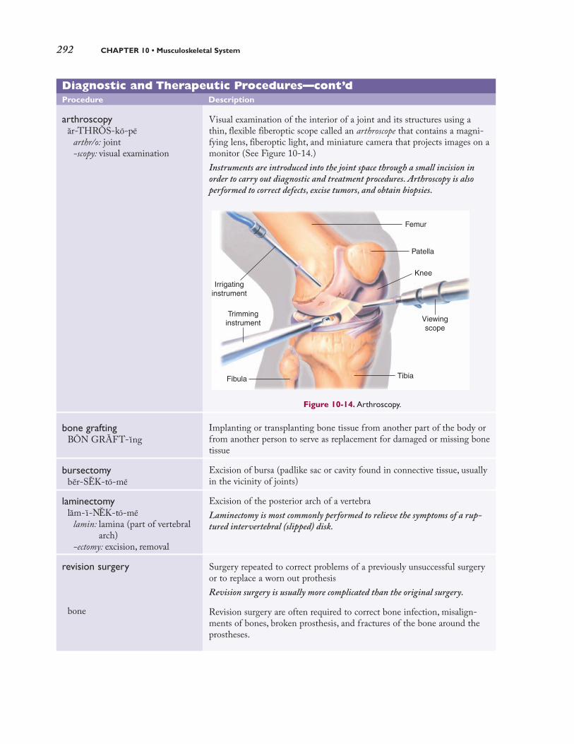
292 CHAPTER 10 • Musculoskeletal System
Diagnostic and Therapeutic Procedures—cont’d
Procedure Description
arthroscopyar-THROS-ko-pe
arthr/o: joint-scopy: visual examination
bone graftingBON GRAFT-ıng
bursectomy ber-SEK-to-me
laminectomylam-ı-NEK-to-me
lamin: lamina (part of vertebralarch)
-ectomy: excision, removal
revision surgery
bone
Visual examination of the interior of a joint and its structures using athin, flexible fiberoptic scope called an arthroscope that contains a magni-fying lens, fiberoptic light, and miniature camera that projects images on amonitor (See Figure 10-14.)
Instruments are introduced into the joint space through a small incision in
order to carry out diagnostic and treatment procedures. Arthroscopy is also
performed to correct defects, excise tumors, and obtain biopsies.
Implanting or transplanting bone tissue from another part of the body orfrom another person to serve as replacement for damaged or missing bonetissue
Excision of bursa (padlike sac or cavity found in connective tissue, usuallyin the vicinity of joints)
Excision of the posterior arch of a vertebra
Laminectomy is most commonly performed to relieve the symptoms of a rup-
tured intervertebral (slipped) disk.
Surgery repeated to correct problems of a previously unsuccessful surgeryor to replace a worn out prothesis
Revision surgery is usually more complicated than the original surgery.
Revision surgery are often required to correct bone infection, misalign-ments of bones, broken prosthesis, and fractures of the bone around theprostheses.
Viewingscope
Irrigatinginstrument
Trimminginstrument
Knee
Fibula Tibia
Femur
Patella
Figure 10-14. Arthroscopy.
2090_Ch10_265-308.qxd 3/5/09 10:03 PM Page 292

Pathology 293
Diagnostic and Therapeutic Procedures—cont’d
Procedure Description
sequestrectomyse-kwes-TREK-to-me
sequestr: separation-ectomy: excision, removal
synovectomys ın-o-VEK-to-me
synov: synovial membrane,synovial fluid
-ectomy: excision, removal
total hip replacement
Excision of a sequestrum (segment of necrosed bone)
Excision of a synovial membrane
Surgical procedure to replace a hip joint damaged by a degenerative dis-ease, commonly arthritis (See Figure 10-15.)
In a total hip replacement (THR), the femoral head and the acetabulum are
replaced with a metal ball and stem (prosthesis). The acetabulum is plastic
coated to avoid metal-to-metal contact on articulating surfaces; the stem is
anchored into the central core of the femur to achieve a secure f it.
Pelvis
Acetabulum
FemurFemur
Femoralneck and head
Acetabulumcap
Femoralshaft
A.
(continued)
Figure 10-15. Total hip replacement. (A) Right total hip replacement. (B) Radiograph showing total
hip replacement of arthritic hip. From McKinnis: Fundamentals of Musculoskeletal Imaging. 2nd. Ed. FA
Davis, Philadelphia, 2005, p. 314, with permission.
2090_Ch10_265-308.qxd 3/5/09 10:03 PM Page 293

PPhhaarrmmaaccoollooggyy
Unlike other medications that treat specific dis-ease, most pharmacological agents for muscu-loskeletal disorders are used to treat symptoms.(See Table 10–3.) Acute musculoskeletal condi-tions, such as strains, sprains, and “pulled” muscles,are treated with analgesics and anti-inflammatory
drugs. Nonsteroidal anti-inflammatory drugs(NSAIDs), salicylates, muscle relaxants, opioidanalgesics, or narcotics are commonly used to treatpain by anesthetizing (numbing) the area ordecreasing the inflammation. NSAIDs and salicy-lates are also used to treat arthritis, in addition togold salts. Calcium supplements are used to treathypocalcemia.
294 CHAPTER 10 • Musculoskeletal System
Diagnostic and Therapeutic Procedures—cont’d
Procedure Description
Drugs Used to Treat Musculoskeletal Disorders Table 10-3
This table lists common drug classif ications used to treat musculoskeletal disorders, their therapeuticactions, and selected generic and trade names.
Classification Therapeutic Action Generic and Trade Names
calcium supplements Treat and prevent hypocalcemia.
Over-the-counter calcium supplements are numer-ous and are contained in many antacids as a sec-ondary therapeutic effect.They are used to preventosteoporosis when normal diet is lacking adequateamounts of calcium.
calcium carbonateKAL-se-um KAR-bon-at
Calci-Mix,Tums
calcium citrateKAL-se-um SIT-rat
Cal-Citrate 250, Citracal
Figure 10-15 cont’d
2090_Ch10_265-308.qxd 3/5/09 10:03 PM Page 294

Drugs Used to Treat Musculoskeletal Disorders—cont’dTable 10-3
Classification Therapeutic Action Generic and Trade Names
gold salts
nonsteroidal anti-inflammatorydrugs
salicylates
skeletal muscle relaxants
Treat rheumatoid arthritis by inhibiting activity
within the immune system.
Gold salts contain actual gold in capsules or in solu-tion for injection.This agent prevents further diseaseprogression but cannot reverse past damage.
Decrease pain and suppress inflammation
Nonsteroidal anti-inflammatory drugs (NSAIDs) areused to treat acute musculoskeletal conditions,such as sprains and strains, and inflammatory dis-orders, including rheumatoid arthritis, osteoarthri-tis, bursitis, gout, and tendinitis.
Relieve mild to moderate pain and reduce
inflammation
Salicylates have anti-inflammatory abilities andalleviate pain. Aspirin (acetylsalicylic acid) is theoldest drug in this classification that is used totreat arthritis.
Relieve muscle spasms and stiffness
These drugs are also prescribed for muscle spasmsdue to multiple sclerosis, spinal cord injury, cerebralpalsy, and stroke.
auranofinaw-RAN-o-f ın
Ridaura
aurothioglucoseaw-ro-thı-o-GLOO-kos
Solganal
ibuprofenı -bu-PRO-fen
Advil, Motrin
naproxenna-PROK-sen
Aleve, Naprosyn
aspirinAS-per- ın
Acuprin,Aspergum, Bayer Aspirin
magnesium salicylatemag-NE-ze-um sa-LIS- ı-lat
Magan, Mobidin
cyclobenzaprinesı-klo-BEN-za-pren
Flexeril
methocarbamol and aspirinmeth-o-KAR-ba-mol
Robaxin
Abbreviations
This section introduces musculoskeletal-related abbreviations and their meanings.
Abbreviation Meaning Abbreviation Meaning
ACL
AE
AK
BE
BK
C1, C2, andso on
anterior cruciate ligament
above the elbow
above the knee
barium enema; below theelbow
below the knee
first cervical vertebra, secondcervical vertebra, and so on
calcium; cancer
congenital dislocation of the hip
carpal tunnel syndrome
computed tomography
dual energy x-ray absorptiometry
degenerative joint disease
(continued)
Pharmacology 295
Ca
CDH
CTS
CT
DEXA, DXA
DJD
2090_Ch10_265-308.qxd 3/5/09 10:03 PM Page 295

296 CHAPTER 10 • Musculoskeletal System
Abbreviations—cont’d
Abbreviation Meaning Abbreviation Meaning
EMG
Fx
MG
HD
HNP
HP
IS
IM
IV
KD
L1, L2, andso on
LS
MG
MRI
electromyography
fracture
myasthenia gravis
hemodialysis; hip disarticulation; hearing distance
herniated nucleus pulposus(herniated disk)
hemipelvectomy
intracostal space
intramuscular; infectiousmononucleosis
intravenous
knee disarticulation
first lumbar vertebra, secondlumbar vertebra, and so on
lumbosacral spine
myasthenia gravis
magnetic resonance imaging
musculoskeletal; multiple sclerosis; men-tal status; mitral stenosis
nonsteroidal anti-inflammatory drugs
orthopedics
phosphorus; pulse
posterior cruciate ligament
rheumatoid arthritis; right atrium
rheumatoid factor; radio frequency
range of motion
shoulder disarticulation
total hip arthroplasty
total hip replacement
total knee arthroplasty
total knee replacement
transverse rectus abdominis muscle
It is time to review procedures, pharmacology, and abbreviations by completing LearningActivity 10–6.
MS
NSAIDs
ORTH, ortho
P
PCL
RA
RF
ROM
SD
THA
THR
TKA
TKR
TRAM
2090_Ch10_265-308.qxd 3/5/09 10:03 PM Page 296

Learning Activities 297
The following activities provide review of the musculoskeletal system terms introduced inthis chapter. Complete each activity and review your answers to evaluate your understand-ing of the chapter.
Learning Activity 10-1
Identifying Muscle Structures
Label the following illustration using the terms listed below.
Achilles tendon gastrocnemius rectus abdominus
biceps brachii gluteus maximus soleus
biceps femoris masseter sternocleidomastoid
brachioradialis orbicularis oculi trapezius
deltoid pectoralis major
LEARNING ACTIVITIES
Triceps brachiiTriceps brachii
Brachialis
Check your answers by referring to Figure 10–1 on page 267. Review material that you didnot answer correctly.✓
2090_Ch10_265-308.qxd 3/5/09 10:03 PM Page 297

298 CHAPTER 10 • Musculoskeletal System
Learning Activity 10-2
Identifying Sections of a Typical Long Bone (Femur)
Label the following illustration using the terms listed below.
articular cartilage distal epiphysis proximal epiphysis
compact bone medullary cavity spongy bone
diaphysis periosteum
Check your answers by referring to Figure 10–3 on page 270. Review material that you did not answercorrectly.✓
2090_Ch10_265-308.qxd 3/5/09 10:03 PM Page 298

Learning Activity 10-3
Identifying Skeletal Structures
Label the following illustration using the terms listed below.
acetabulum
carpals
clavicle
coccyx
femur
fibula
humerus
ilium
ischium
metacarpals
metatarsals
pectoral girdle
pelvic girdle
phalanges
pubis
radius
sacrum
scapula
sternum
symphysis pubis
tarsals
tibia
ulna
Maxilla
Mandible
Ribs
Patella
Skull
Vertebral column
Check your answers by referring to Figure 10–4 on page 272. Review material that you didnot answer correctly.✓
Enhance your study and reinforcement of word elements with the power of DavisPlus. Visitwww.davisplus.fadavis.com/gylys/systems for this chapter’s flash-card activity. We recommendyou complete the flash-card activity before completing activity 10–4 below.
2090_Ch10_265-308.qxd 3/5/09 10:03 PM Page 299

300 CHAPTER 10 • Musculoskeletal System
Check your answers in Appendix A. Review material that you did not answer correctly.✓Correct Answers �� 4 �� % Score
Learning Activity 10-4
Building Medical Words
Use oste/o (bone) to build words that mean:
1. bone cells _________________________________________________________________________
2. pain in bones _______________________________________________________________________
3. disease of bones and joints ____________________________________________________________
4. beginning or formation of bones ________________________________________________________
Use cervic/o (neck) to build words that mean:
5. pertaining to the neck ________________________________________________________________
6. pertaining to the neck and arm ________________________________________________________
7. pertaining to the neck and face ________________________________________________________
Use myel/o (bone marrow; spinal cord) to build words that mean:
8. tumor of bone marrow _______________________________________________________________
9. sarcoma of bone marrow (cells) ________________________________________________________
10. bone marrow cell ____________________________________________________________________
11. resembling bone marrow _____________________________________________________________
Use stern/o (sternum) to build words that mean:
12. pertaining to above the sternum ________________________________________________________
13. resembling the breastbone ____________________________________________________________
Use arthr/o (joint) or chondr/o (cartilage) to build words that mean:
14. embryonic cell that forms cartilage _____________________________________________________
15. inflammation of a joint _______________________________________________________________
16. inflammation of bones and joints _______________________________________________________
Use pelv/i (pelvis) to build a word that means:
17. instrument for measuring the pelvis _____________________________________________________
Use my/o (muscle) to build words that mean:
18. twitching of a muscle _________________________________________________________________
19. any disease of muscle ________________________________________________________________
20. rupture of a muscle __________________________________________________________________
Build surgical words that mean:
21. excision of one or more of the phalanges (bones of a finger or toe) ____________________________
22. incision of the thorax (chest wall) _______________________________________________________
23. excision of a vertebra ________________________________________________________________
24. binding of a joint ____________________________________________________________________
25. repair of muscle (tissue) ______________________________________________________________
2090_Ch10_265-308.qxd 3/5/09 10:03 PM Page 300

Learning Activities 301
Learning Activity 10-5
Matching Pathological, Diagnostic, Symptomatic, and Related Terms
Match the following terms with the definitions in the numbered list.
ankylosis
chondrosarcoma
claudication
comminuted fracture
Ewing sarcoma
ganglion cyst
gout
greenstick fracture
hematopoiesis
hypotonia
kyphosis
muscular dystrophy
myasthenia gravis
necrosis
osteoporosis
phantom limb
prosthesis
pyogenic
rickets
scoliosis
sequestrum
spondylitis
spondylolisthesis
subluxation
talipes
1. incomplete or partial dislocation
2. softening of the bones caused by vitamin D deficiency
3. slipped vertebrae
4. limping
5. disease causing degeneration of muscles
6. congenital deformity of the foot, which is twisted out of shape or position
7. part of dead or necrosed bone that has become separated from surrounding tissue
8. chronic neuromuscular disorder characterized by weakness manifested in ocular muscles
9. artificial part used for replacement of a missing limb
10. tendon sheath or joint capsule tumor, commonly found in the wrist
11. loss of muscular tonicity; diminished resistance of muscles to passive stretching
12. type of sarcoma that attacks the shafts rather than the ends of long bones
13. bone that is partially bent and partially broken; occurs in children
14. exaggeration of the thoracic curve of the vertebral column; hunpback
15. disease caused by a decrease in bone density; occurs in the elderly
16. deviation of the spine to the right or left
17. cartilaginous sarcoma
18. describes a bone that has splintered into pieces
19. inflammation of the vertebrae
20. metabolic disease caused by accumulation of uric acid, usually in the big toe
21. development and production of blood cells, normally in the bone marrow
22. formation of pus
23. death of cells, tissues, or organs
24. stiffening and immobility of a joint
25. perceived sensation, following amputation, that the limb still exists
Check your answers in Appendix A. Review material that you did not answer correctly.✓Correct Answers �� 4 �� % Score
2090_Ch10_265-308.qxd 3/5/09 10:03 PM Page 301

302 CHAPTER 10 • Musculoskeletal System
Learning Activity 10-6Matching Procedures, Pharmacology, and Abbreviations
Match the following terms with the definitions in the numbered list.
ACL closed reduction myelography
amputation CTS open reduction
arthrodesis gold salts relaxants
arthrography HNP salicylates
arthroscopy laminectomy sequestrectomy
1. radiograph of spinal cord after injection of a contrast medium
2. treatment of bone fractures by use of surgery to place bones in normal position
3. used to treat rheumatoid arthritis by inhibiting activity with the immune system
4. painful disorder of the wrist and hand due to compression of the median nerve as itpasses through the carpal tunnel
5. excision of the posterior arch of a vertebra
6. series of joint radiographs preceded by injection of a radiopaque substance or airinto the joint cavity
7. surgical binding or immobilizing of a joint
8. partial or complete removal of a limb
9. herniated nucleus pulposus
10. relieve mild to moderate pain and reduce inflammation
11. visual examination of a joint’s interior, especially the knee
12. excising a segment of necrosed bone
13. anterior cruciate ligament
14. relieve muscle spasms and stiffness
15. manipulative treatment of bone fractures by placing the bones in normal positionwithout incision
Check your answers in Appendix A. Review any material that you did not answer correctly.✓Correct Answers �� 6.67 �� % Score
2090_Ch10_265-308.qxd 3/5/09 10:03 PM Page 302

Medical Record Activities 303
The two medical records included in the activities that follow use common clinical scenar-ios to show how medical terminology is used to document patient care. Complete the ter-minology and analysis sections for each activity to help you recognize and understand termsrelated to the musculoskeletal system.
Medical Record Activity 10-1
Operative Report: Right Knee Arthroscopy and Medial Meniscectomy
TerminologyTerms listed below come from the medical record Operative Report: Right Knee Arthroscopyand Medial Meniscectomy that follows. Use a medical dictionary such as Taber’s CyclopedicMedical Dictionary, the appendices of this book, or other resources to define each term. Thenreview the pronunciations for each term and practice by reading the medical record aloud.
MEDICAL RECORD ACTIVITIES
Term Definition
ACL
arthroscopyar-THROS-ko-pe
effusionse-FU-zhunz
intracondyların-tra-KON-dı-lar
Lachman test
McMurray sign test
meniscectomymen-ı-SEK-to-me
MRI
PCL
synovitiss ın-o-VI-t ıs
Listen and Learn Online! will help you master the pronunciation of selected medical words from thismedical record activity. Visit www.davisplus.com/gylys/systems to f ind instructions on completing theListen and Learn Online! exercise for this section and to practice pronunciations.
(( ))
2090_Ch10_265-308.qxd 3/5/09 10:03 PM Page 303

304 CHAPTER 10 • Musculoskeletal System
General Hospital
1511 Ninth Avenue ■■ Sun City, USA 12345 ■■ (555) 8022-1887
OPERATIVE REPORT
Date: August 14, 20xx Physician: Robert L. Mead, MD
Patient: Jay, Elizabeth Patient ID#: 20798
PREOPERATIVE DIAGNOSIS: Tear, medial meniscus, right knee.
POSTOPERATIVE DIAGNOSIS: Tear, medial meniscus, right knee.
CLINICAL HISTORY: This 42-year-old woman has jogged for the past 10 years, an average of 25 miles each week. She has persistent posteromedial right knee pain with occasional effusions. Thepatient has MRI-documented medial meniscal tear.
PROCEDURE: Right knee arthroscopy and medial meniscectomy.
ANESTHESIA: General.
COMPLICATIONS: None.
OPERATIVE SUMMARY: Examination of the knee under anesthesia showed a full range ofmotion, no effusion, no instability, and negative Lachman and negative McMurray sign tests.Arthroscopic evaluation showed a normal patellofemoral groove and normal intracondylar notch withnormal ACL and PCL, some anterior synovitis, and a normal lateral meniscus and lateral compart-ment to the knee. The medial compartment of the knee showed an inferior surface, posterior andmid-medial meniscal tear that was flipped up on top of itself. This was resected, and then the remain-ing meniscus contoured back to a stable rim. A sterile dressing was applied.
Patient was taken to the post anesthesia care unit in stable condition.
RRoobbeerrtt LL.. MMeeaadd,, MMDDRobert L. Mead, MD
rlm:bg
D: 8-14-20xxT: 8-14-20xx
OPERATIVE REPORT: RIGHT KNEE ARTHROSCOPYAND MEDIAL MENISCECTOMY
2090_Ch10_265-308.qxd 3/5/09 10:03 PM Page 304

Operative Report: Right Knee Arthroscopy and Medial Meniscectomy 305
AnalysisReview the medical record Operative report: Right knee arthroscopy and medial meniscectomy toanswer the following questions.
1. Describe the meniscus and identify its location.
2.What is the probable cause of the tear in the patient’s meniscus?
3.What does normal ACL and PCL refer to in the report?
4. Explain the McMurray sign test.
5. Because Lachman and McMurray tests were negative (normal), why was the surgery performed?
2090_Ch10_265-308.qxd 3/5/09 10:03 PM Page 305

306 CHAPTER 10 • Musculoskeletal System
NSAIDs
nuclear scanNU-kle-ar
periostealper-e-OS-te-al
resorptionre-SORP-shun
tibialTIB-e-al
Listen and Learn Online! will help you master the pronunciation of selected medical words from thismedical record activity. Visit www.davisplus.com/gylys/systems to f ind instructions on completing theListen and Learn Online! exercise for this section and to practice pronunciations.
(( ))
Term Definition
buttressingBU-tres- ıng
corticalKOR-tı-kal
diaphysisdı-AF-ı-s ıs
endostealen-DOS-te-al
focalFO-kal
fusiformFU-zı-form
Medical Record Activity 10-2
Radiographic Consultation:Tibial Diaphysis Nuclear Scan
TerminologyTerms listed below come from the medical record Radiographic Consultation: TibialDiaphysis Nuclear Scan. Use a medical dictionary such as Taber’s Cyclopedic MedicalDictionary, the appendices of this book, or other resources to define each term. Then reviewthe pronunciations for each term and practice by reading the medical record aloud.
2090_Ch10_265-308.qxd 3/5/09 10:03 PM Page 306

Radiographic Consultation:Tibial Diaphysis Nuclear Scan 307
Physician Center
2422 Rodeo Drive ■■ Sun City, USA 12345 ■■ (555)333-2427
September 3, 20xx
Grant Hammuda, MD1115 Forest AveSun City, USA 12345
Dear Doctor Hammuda:
We are pleased to provide the following in response to your request for consultation.
This is an 18-year-old male cross-country runner. He complains of pain of more than 1 month’s duration, with persistent symptoms over middle one third of left tibia withresting. He finds no relief with NSAIDs.
FINDINGS: Nuclear scan reveals the following: There is focal increased blood flow,blood pool, and delayed radiotracer accumulation within the left mid posterior tibial diaphysis. The delayed spot planar images demonstrate focal fusiform uptake involving50% to 75% of the tibial diaphysis width.
It is our opinion that with continued excessive, repetitive stress, the rate of resorptionwill exceed the rate of bone replacement. This will lead to weakened cortical bone withbuttressing by periosteal and endosteal new bone deposition. If resorption continues toexceed replacement, a stress fracture will occur.
Please let me know if I can be of any further assistance.
Sincerely yours,
AAddrriiaann JJoonneess,, MMDDAdrian Jones, MD
aj:bg
RADIOGRAPHIC CONSULTATION:TIBIALDIAPHYSIS NUCLEAR SCAN
2090_Ch10_265-308.qxd 3/5/09 10:03 PM Page 307

308 CHAPTER 10 • Musculoskeletal System
AnalysisReview the medical record Radiographic Consultation: Tibial Diaphysis Nuclear Scan toanswer the following questions.
1.Where was the pain located?
2.What medication was the patient taking for pain and did it provide relief?
3. How was the blood flow to the affected area described by the radiologist?
4. How was the radiotracer accumulation described?
5.What will be the probable outcome with continued excessive repetitive stress?
6.What will happen if resorption continues to exceed replacement?
2090_Ch10_265-308.qxd 3/5/09 10:03 PM Page 308

GenitourinarySystem
Chapter Outline
Objectives
Anatomy and PhysiologyAnatomy and Physiology Key TermsUrinary System
NephronMale Reproductive SystemConnecting Body Systems–Genitourinary System
Medical Word Elements
PathologyPyelonephritisGlomerulonephritisNephrolithiasisBenign Prostatic HyperplasiaCryptorchidismAcute Tubular NecrosisOncology
Diagnostic, Symptomatic, and Related Terms
Diagnostic and Therapeutic Procedures
Pharmacology
Abbreviations
Learning Activities
Medical Record ActivitiesOperative report: Ureterocele and ureterocele calculusOperative report: Extracorporeal shock-wavelithotripsy
ObjectivesUpon completion of this chapter, you will be able to:
• Locate and describe the urinary structures as well
as the structures of the male reproductive system.
• Describe the functional relationship between the
genitourinary system and other body systems.
• Identify, pronounce, spell, and build words related to
the genitourinary system.
• Describe pathological conditions, diagnostic and
therapeutic procedures, and other terms related to
the genitourinary system.
• Explain pharmacology related to the treatment of
urinary disorders as well as male reproductive dis-
orders.
• Demonstrate your knowledge of this chapter by
completing the learning and medical record
activities.
C H A P T E R
11
2090_Ch11_309-348.qxd 3/5/09 9:48 PM Page 309

Anatomy and Physiology
The male and female urinary systems have similarstructures. In the male, however, some of the uri-nary structures also have reproductive functions.Thus, the genitourinary system includes the uri-nary system of both the male and female as well asthe reproductive system of the male.
Urinary SystemThe purpose of the urinary system is to regulate thecomposition of the extracellular fluids of the bodyby removing their harmful substances in the form ofurine, while retaining beneficial products. Harmfulsubstances, including nitrogenous wastes andexcess electrolytes, are removed by the kidneys and
excreted from the body in urine. Nitrogenous wastesare toxic to the body, and must be continuouslyeliminated or death will occur. Electrolyte concen-tration must remain fairly constant for proper func-tioning of nerves, heart, and muscles. An excess ordeficiency of electrolytes can have devastatingeffects. Besides regulating the composition of extra-cellular fluids, kidneys also secrete the hormone erythropoietin. This hormone acts on bone mar-row to stimulate production of red blood cells whenblood oxygen levels are low.The macroscopic struc-tures that make up the urinary system include:
• two kidneys• two ureters• bladder• urethra. (See Figure 11–1.)
310 CHAPTER 11 • Genitourinary System
Anatomy and Physiology Key Terms
This section introduces important genitourinary system terms along with their definitions andpronunciations. Word analyses are provided for selected terms.
Term Definition
electrolytese-LEK-tro-lıts
filtrateFIL-trat
nitrogenous wastesnı-TROJ-en-us
peristaltic wavesper-ı-STAL-tık
peritoneumper-ı-to-NE-um
plasmaPLAZ-ma
semenSE-men
testosteronetes-TOS-ter-on
Mineral salts (sodium, potassium, and calcium) that carry an electricalcharge in solution
A proper balance of electrolytes is essential to the normal functioning of the
entire body but especially nerves, muscles, and heart.
Fluid that passes from the blood through the capillary walls of theglomeruli of the kidney
Filtrate is similar to plasma but with far less protein. Urine is formed from f iltrate.
Products of cellular metabolism that contain nitrogen
Nitrogenous wastes include urea, uric acid, creatine, creatinine, and ammonia.
Sequence of rhythmic contraction of smooth muscles of a hollow organ toforce material forward and prevent backflow
Serous membrane that lines the abdominopelvic cavity and covers most ofthe organs within the cavity
Liquid portion of blood, composed primarily of water (90%), and contain-ing dissolved proteins, nutrients, lipids, and various waste products
Fluid containing sperm and secretions from the prostate and other struc-tures of the male reproductive system; also called seminal fluid
Androgenic hormone responsible for the development of the male sexorgans, including the penis, testicles, scrotum, and prostate
Testosterone is also responsible for the development of secondary sex characteris-
tics (musculature, hair patterns, thickened vocal cords, and so forth).
Pronunciation Help Long Sound a—rate e—rebirth ı—isle o—over u—uniteShort Sound a—alone e—ever ı—it o—not u—cut
2090_Ch11_309-348.qxd 3/5/09 9:48 PM Page 310

Anatomy and Physiology 311
Figure 11-1. Urinary structures, including a cross section of the kidney.
Adrenal(suprarenal)
glands
(1) Right kidney
Inferiorvena cava
(9) Ureteral orifice
(11) Urethra
(1) Left kidney
Abdominal aorta
(8) Ureters
(10) Urinary bladder
Prostate gland(in males)
(12) Urinary meatus
(5) Renal artery
(6) Renal vein
Inferiorvena cava
(2) Renal cortex
(7) Renal pelvis
(4) Hilum
(8) Ureter
(3) Renal medulla
Renal pyramid
Cross section of kidney
2090_Ch11_309-348.qxd 3/5/09 9:48 PM Page 311

The (1) left and right kidneys, each about the sizeof a fist, are located in the abdominal cavity slight-ly above the waistline. Because they lie outside ofthe peritoneum, their location is said to beretroperitoneal. A concave medial border gives thekidney its beanlike shape. In a frontal section, twodistinct areas are visible: an outer section, the (2) renal cortex, and a middle area, the (3) renalmedulla, which contain portions of the microscop-ic filtering units of the kidney called nephrons.Near the medial border is the (4) hilum (also calledhilus), an opening through which the (5) renalartery enters and the (6) renal vein exits the kid-ney. The renal artery carries blood that containswaste products to the nephrons for filtering. Afterwaste products are removed, blood leaves the kid-ney by way of the renal vein.
Waste material, now in the form of urine, passesto a hollow chamber, the (7) renal pelvis. Thiscavity is formed where the (8) ureter merges with the kidney. Each ureter is a slender tube about10 inches to 12 inches long. They each carry urinein peristaltic waves to the bladder. Urine entersthe bladder at the (9) ureteral orifice. The (10) urinary bladder, an expandable hollow organ,
acts as a temporary reservoir for urine.The bladderhas small folds called rugae that expand as thebladder fills. A triangular area at the base of thebladder called the trigone is delineated by theopenings of the ureters and the urethra.
The base of the trigone forms the (11) urethra,a tube that discharges urine from the bladder. Thelength of the urethra is approximately 1.5 inches inwomen and about 7 to 8 inches in men. In themale, the urethra passes through the prostate glandand the penis. During urination (micturition),urine is expelled from the body through the ure-thral opening, the (12) urinary meatus.
Nephron
Microscopic examination of kidney tissue revealsthe presence of approximately 1 million nephrons.(See Figure 11–2.) These microscopic structuresare responsible for maintaining homeostasis bycontinually adjusting and regulating the contentsof blood plasma. Substances removed by nephronsare nitrogenous wastes, the end products of pro-tein metabolism, excess electrolytes, and manyother products that exceed the amount toleratedby the body.
312 CHAPTER 11 • Genitourinary System
(6) Proximal convoluted
tubule
(8) Distal tubule
(1) Glomerulus
(2) Bowman capsule
(4) Efferent arteriole
(3) Afferent arteriole
(5) Peritubular capillary
(9) Collecting tubule
(7) Loop of Henle
Descendinglimb
Ascendinglimb
To renal vein
From renal artery
Ren
al c
orte
xR
enal
med
ulla
Figure 11-2. Nephron with its associated blood vessels.
2090_Ch11_309-348.qxd 3/5/09 9:48 PM Page 312

Each nephron includes a renal corpuscle and arenal tubule. The renal corpuscle is composed of a tuft of capillaries called the (1) glomerulus and amodified, enlarged extension of the renal tubuleknown as (2) Bowman capsule that encapsulates theglomerulus. A larger (3) afferent arteriole carriesblood to the glomerulus, and a smaller (4) efferentarteriole carries blood from the glomerulus.The dif-ference in the size of these vessels provides the need-ed pressure to force blood plasma into Bowman capsule. Once this happens, the fluid is no longerplasma but is called filtrate. As the efferent arteriolepasses behind the renal corpuscle, it forms the (5) peritubular capillaries. Each renal tubule consistsof four sections: the (6) proximal convoluted tubule,followed by the narrow (7) loop of Henle, then thelarger (8) distal tubule and, finally, the (9) collectingtubule. The collecting tubule transports newly formedurine to the renal pelvis for excretion by the kidneys.
The nephron performs three physiologicalfunctions as it produces urine:
• Filtration occurs in the renal corpuscle,where plasma containing water, electrolytes,sugar, and other small molecules is forcedfrom the blood within the glomerulus intoBowman capsule to form filtrate.
• Reabsorption begins as filtrate travelsthrough the long, twisted pathway of thetubule. Most of the water and some of theelectrolytes and amino acids are returned tothe peritubular capillaries and reenter the circulating blood.
• Secretion is the final stage of urine forma-tion. Substances are actively secreted from theblood in the peritubular capillaries into thefiltrate in the renal tubules. Waste products,such as ammonia, uric acid, and metabolicproducts of medications are secreted into thefiltrate to be eliminated in the urine.
Urine leaves the collecting tubule and enters therenal pelvis. From here it passes to the bladderuntil urination takes place.
Male Reproductive SystemThe purpose of the male reproductive system is toproduce, maintain, and transport sperm, the malesex cell required for fertilization of the female egg.It also produces the male hormone testosterone,which is essential to the development of spermand male secondary sex characteristics. (SeeFigure 11–3.)
The primary male reproductive organ consistsof two (1) testes (singular, testis) located in anexternal sac called the (2) scrotum. Within thetestes are numerous small tubes that twist andcoil to form (3) seminiferous tubules, whichproduce sperm. The testes also secrete testos-terone, a hormone that develops and maintainssecondary sex characteristics. Lying over thesuperior surface of each testis is a single, tightlycoiled tube, the (4) epididymis. This structurestores sperm after it leaves the seminiferoustubules. The epididymis is the first duct throughwhich sperm passes after its production in thetestes. Tracing the duct upward, the epididymisforms the (5) vas deferens (also called the semi-nal duct or ductus deferens), a narrow tube thatpasses through the inguinal canal into theabdominal cavity. The vas deferens extends overthe top and down the posterior surface of thebladder, where it joins the (6) seminal vesicle.
The union of the vas deferens with the duct fromthe seminal vesicle forms the (7) ejaculatoryduct. The seminal vesicle contains nutrients thatsupport sperm viability and produces approxi-mately 60% of the seminal fluid that is ultimate-ly ejaculated during sexual intercourse (coitus).The ejaculatory duct passes at an angle throughthe (8) prostate gland, a triple-lobed organ fusedto the base of the bladder. The prostate glandsecretes a thin, alkaline substance that accountsfor about 30% of seminal fluid. Its alkalinity helpsprotect sperm from the acidic environments ofthe male urethra and the female vagina. Two pea-shaped structures, the (9) bulbourethral (Cowper)glands, are located below the prostate and areconnected by a small duct to the urethra. Thebulbourethral glands provide the alkaline fluidnecessary for sperm viability. The (10) penis isthe male organ of copulation. It is cylindrical and composed of erectile tissue that encloses the(11) urethra. The urethra expels semen and urinefrom the body. During ejaculation, the sphincterat the base of the bladder closes, which not onlystops the urine from being expelled with thesemen, but also prevents semen from entering the bladder. The enlarged tip of the penis, the(12) glans penis, contains the (13) urethral ori-fice (meatus). A movable hood of skin, called the(14) prepuce (foreskin) covers the glans penis.
Anatomy and Physiology 313
It is time to review urinary system anatomy by completing Learning Activity 11–1.
2090_Ch11_309-348.qxd 3/5/09 9:48 PM Page 313

314 CHAPTER 11 • Genitourinary System
Symphysis pubis
(5) Vas deferens
Urinary bladder
(10) Penis
(11) Urethra
(12) Glans penis
(14) Prepuce
(13) Urethral orifice
(2) Scrotum
(1) Testis
(4) Epididymis
Sacrum
Opening of ureter
Rectum
(6) Seminal vesicle
(7) Ejaculatory duct
(8) Prostate gland
(9) Bulbourethral gland
Anus(3) Seminiferous tubules
Figure 11-3. Midsagittal section of male reproductive structures shown through the pelvic cavity.
Connecting Body Systems–Genitourinary System
The main function of the genitourinary system is to enable sexual reproduction and to regulateextracellular fluids of the body. Specif ic functional relationships between the genitourinary systemand other body systems are summarized below.
Blood, lymph, and immune• Male reproductive system transports
testosterone throughout the body inblood and lymph.
• Kidneys regulate composition and qualityof blood plasma and lymph.
• Kidneys retain needed products andremove those that are excessive or toxicto the body.
Cardiovascular• Kidneys help regulate essential electrolytes
needed for contraction of the heart.
Digestive• Kidneys aid in removing glucose from the
blood when excessive amounts are con-sumed.
• Kidneys remove excessive fluids absorbedfrom the gastrointestinal (GI) tract.
Endocrine• Kidneys regulate sodium and water bal-
ance, which is essential for hormone trans-port in the blood.
• Kidneys produce erythropoietin, a hor-mone synthesized mainly in the kidneysand released into the bloodstream.
• Gonads produce hormones that providefeedback to influence pituitary function.
Female reproductive• Male organs of reproduction work in con-
junction with the female reproductive sys-tem to enable fertilization of the ovum.
• Kidneys aid in removing waste productsproduced by the fetus in the pregnantwoman.
2090_Ch11_309-348.qxd 3/5/09 9:48 PM Page 314

Anatomy and Physiology 315
Connecting Body Systems–Genitourinary System
Integumentary• Kidneys compensate for extracellular fluid
loss due to hyperhidrosis.• Kidneys adjust electrolytes, especially
potassium and sodium, in response totheir loss through the dermis.
Musculoskeletal• Kidneys work in conjunction with bone
tissue to maintain a constant calcium level.
Nervous• Kidneys regulate sodium, potassium, and
calcium, which are the electrolytes respon-sible for the transmission of nervous stimuli.
Respiratory• Kidneys and lungs assist in regulating acid-
base balance of the body.
It is time to review male reproductive anatomy by completing Learning Activity 11–2.
Medical Word Elements
This section introduces combining forms, suff ixes, and prefixes related to the genitourinary system.Word analyses are also provided.
Element Meaning Word Analysis
UrinarySystem
cyst/o
vesic/o
glomerul/o
lith/o
meat/o
nephr/o
ren/o
bladder
glomerulus
stone, calculus
opening,meatus
kidney
cyst/o/scope (SIST-o-skop): instrument for examining the bladder-scope: instrument for examining
vesic/o/cele (VES-ı-ko-sel): hernia of the bladder; also called cystocele
-cele: hernia, swelling
With a vesicocele, the bladder herniates into the vaginal wall, which may lead to
incomplete emptying of the bladder.
glomerul/o/pathy (glo-mer-u-LOP-a-the): disease of the glomerulus-pathy: disease
lith/o/tripsy (LITH-o-tr ıp-se): crushing of a stone-tripsy: crushing
The most common method of lithotripsy is extracorporeal shock wave lithotripsy
(ESWL). When stones are large or ESWL is not recommended, percutaneous
nephrolithotomy or ureteroscopic stone removal are alternate methods of treatment.
meat/o/tomy (me-a-TOT-o-me): incision of the meatus-tomy: incision
A meatotomy is performed to relieve stenosis of the urethra, which may inhibit the
proper passage of urine or semen.
nephr/o/pexy (NEF-ro-peks-e): fixation of kidney-pexy: fixation (of an organ)
ren/al (RE-nal): pertaining to the kidney-al: pertaining to (continued)
Combining Forms
2090_Ch11_309-348.qxd 3/5/09 9:48 PM Page 315

316 CHAPTER 11 • Genitourinary System
Medical Word Elements—cont’d
Element Meaning Word Analysis
pyel/o
ur/o
ureter/o
urethr/o
MaleReproductiveSystem
andr/o
balan/o
epididym/o
orch/o
orchi/o
orchid/o
test/o
perine/o
prostat/o
spermat/o
renal pelvis
urine, urinarytract
ureter
urethra
male
glans penis
epididymis
testis (plural,testes)
perineum (areabetween scro-tum [or vulvain the female]and anus)
prostate gland
spermatozoa,sperm cells
pyel/o/plasty (PI-e-lo-plas-te): surgical repair of the renal pelvis-plasty: surgical repair
ur/o/lith (U-ro-l ıth): stone in urinary tract-lith: stone, calculus
ureter/ectasis (u-re-ter-EK-ta-s ıs): dilation of the ureter-ectasis: dilation, expansion
urethr/o/stenosis (u-re-thro-st en-O-sıs): narrowing or stricture of the urethra-stenosis: narrowing, stricture
andr/o/gen/ic (an-dro-JEN-ık): pertaining to malenessgen: forming, producing, origin-ic: pertaining to
Androgenic hormones include all natural or synthetic compounds that stimulate or
maintain male characteristics. The most common androgenic hormone is testosterone.
balan/o/plasty (BAL-a-no-plas-te): surgical repair of the glans penis-plasty: surgical repair
epididym/o/tomy (ep-ı-dıd-ı-MOT-o-me): incision of the epididymis-tomy: incision
orch/itis (or-KI-t ıs): inflammation of testes-itis: inflammation
A common cause of orchitis in young boys is a mumps infection.
orchi/algia (or-ke-AL-je-a): pain in the testes-algia: pain
orchid/o/ptosis (or-kıd-op-TO-sıs): downward displacement of the testes-ptosis: prolapse, downward displacement
test/ectomy (tes-TEK-to-me): excision of a testis-ectomy: excision, removal
perine/al (per- ı-NE-al): pertaining to the perineum-al: pertaining to
prostat/o/megaly (pros-ta-to-MEG-a-le): enlargement of the prostate gland-megaly: enlargement
spermat/o/cele (sper-MAT-o-sel): swelling containing spermatozoa-cele: hernia, swelling
A spermatocele is usually an epididymal cyst commonly containing sperm.
2090_Ch11_309-348.qxd 3/5/09 9:48 PM Page 316

Anatomy and Physiology 317
Medical Word Elements—cont’d
Element Meaning Word Analysis
sperm/o
varic/o
vas/o
vesicul/o
Other
albumin/o
azot/o
bacteri/o
crypt/o
gonad/o
kal/i
keton/o
dilated vein
vessel; vas deferens; duct
seminal vesicle
albumin,protein
nitrogenouscompounds
bacteria (singu-lar, bacterium)
hidden
gonads, sexglands
potassium (an electrolyte)
ketone bodies(acids and ace-tones)
sperm/ic (SPER-mık): pertaining to sperm cells-ic: pertaining to
varic/o/cele (VAR-ı-ko-sel): swelling of a dilated vein-cele: hernia, swelling
Varicocele is a dilation of the veins of the spermatic cord, the structure that
supports the testicles.
vas/ectomy (vas-EK-to-me): removal of (all or part of ) the vas deferens-ectomy: excision, removal
Bilateral vasectomy is a surgical procedure to produce sterility in the male.
vesicul/itis (ve-s ık-u-LI-t ıs): inflammation of the seminal vesicle-itis: inflammation
albumin/oid (al-BU-mı-noyd): resembling albumin-oid: resembling
azot/emia (az-o-TE-me-a): nitrogenous compounds in the blood-emia: blood condition
Nitrogenous products, especially urea, are toxic. If they are not removed from the
body, death will result.
bacteri/uria (bak-te-re-U-re-a): bacteria in urine-uria: urine
crypt/orchid/ism (krıpt-OR-kıd-ızm): condition of hidden testes; alsocalled cryptorchism
orchid: testis (plural, testes)-ism: condition
Cryptorchidism is the failure of the testes to descend into the scrotum; usually a
congenital disorder.
gonad/o/pathy (gon-a-DOP-a-the): disease of the sex glands-pathy: disease
hypo/kal/emia (hı-po-k a-LE-me-a): abnormally low concentration of potas-sium in the blood
hypo-: under, below-emia: blood condition
Hypokalemia may result from excessive urination, which depletes potassium from
the body.
keton/uria (ke-to-NU-re-a): presence of ketone bodies in the urine-uria: urine
Ketonuria is commonly found in diabetes mellitus, starvation, and excessive
dieting. (continued)
2090_Ch11_309-348.qxd 3/5/09 9:48 PM Page 317

318 CHAPTER 11 • Genitourinary System
Medical Word Elements—cont’d
Element Meaning Word Analysis
noct/o
olig/o
py/o
-cide
-genesis
-iasis
-ism
-spadias
-uria
dia-
retro-
night
scanty
pus
killing
forming, pro-ducing, origin
abnormal con-dition (pro-duced by some-thing specified)
condition
slit, fissure
urine
through,across
backward,behind
noct/uria (nok-TU-re-a): excessive and frequent urination after going to bed-uria: urine
Nocturia is associated with prostate disease, urinary tract infection, and uncon-
trolled diabetes.
olig/o/sperm/ia (ol-ı-go-SPER-me-a): scanty (decreased production) of spermsperm: spermatozoa, sperm cells-ia: condition
py/o/rrhea (pı-o-RE-a): flow or discharge of pus-rrhea: discharge, flow
sperm/i/cide (SPER-mı-sıd): (agents that) kill sperm; also called spermaticide
sperm/i: spermatozoa, sperm cells
lith/o/genesis (l ıth-o-JEN-e-s ıs): forming or producing stoneslith/o: stone, calculus
lith/iasis (l ıth-I-a-s ıs): abnormal condition of stones or calculilith/o: stone, calculus
an/orch/ism (an-OR-kızm): condition without testesan-: without, notorch: testis (plural, testes)
Anorchism is the congenital or acquired absence of one or both testes.
hypo/spadias (hı-po-SPA-de-as): a fissure under (the penis)hypo-: under, below
Hypospadias is a congenital defect in which the urethra opens on the underside of
the glans penis instead of the tip.
poly/uria (pol-e-U-re-a): much (excretion of ) urinepoly-: many, much
Polyuria is generally considered to be the excretion of over 2.5 liters per 24 hours.
dia/lysis (dı-AL-ı-s ıs): separation across-lysis: separation; destruction; loosening
Renal dialysis is a procedure that uses a membrane to separate and selectively remove
waste products from blood when kidneys are unable to complete this function.
retro/peritone/al (ret-ro-per- ı-to-NE-al): pertaining to (the area) behind theperitoneum
peritone: peritoneum-al: pertaining to
Suffixes
Prefixes
It is time to review medical word elements by completing Learning Activity 11–3. For audio pronunciations ofthe above-listed key terms, you can visit www.davisplus.fadavis.com/gylys/systems to download this chapter’sListen and Learn! exercises or use the book’s audio CD (if included).
2090_Ch11_309-348.qxd 3/5/09 9:48 PM Page 318

Pathology 319
Pathology
Pathology of the urinary system includes a range ofdisorders from those that are asymptomatic tothose that manifest an array of signs and symp-toms. Causes for these disorders include congenitalanomalies, infectious diseases, trauma, or condi-tions that secondarily involve the urinary struc-tures. Many times, asymptomatic urinary diseasesare first diagnosed when a routine urinalysis showsabnormalities. Forms of glomerulonephritis andchronic urinary tract infection are two such dis-orders. Symptoms specific to urinary disordersinclude changes in urination pattern, output, ordysuria. Endoscopic tests, radiological evaluations,and laboratory tests that evaluate renal function aretypically employed to diagnose disorders of the uri-nary system.
Signs and symptoms of male reproductive dis-orders include pain, swelling, erectile dysfunction,and loss of normal sexual drive (libido). Char-acteristics of infectious diseases, especially thosetransmitted through sexual activity, commonlyinclude pain, discharge, or lesions as well as a vaguefeeling of fullness or discomfort in the perineal orrectal area. A complete evaluation of the genitalia,reproductive history, and past and present geni-tourinary infections and disorders is necessary toidentify disorders associated with male reproduc-tive structures.
For diagnosis, treatment, and management ofgenitourinary disorders, the medical services of aspecialist may be warranted. Urology is the branchof medicine concerned with male and female urinarydisorders and diseases of the male reproductive sys-tem.The physician who specializes in diagnoses andtreatment of genitourinary disorders is known as aurologist. However, the branch of medicine con-cerned specifically with diseases of the kidney, elec-trolyte imbalance, renal transplantation, and dialysistherapy is known as nephrology. Physicians whopractice in this specialty are called nephrologists.
PyelonephritisOne of the most common forms of kidney diseaseis pyelonephritis, (also called kidney infection orcomplicated urinary tract infection). In this disor-der, bacteria invade the renal pelvis and kidney tis-sue, commonly as a result of a bladder infection thathas ascended to the kidney via the ureters. Whenthe infection is severe, lesions form in the renalpelvis, causing bleeding. The microscopic examina-tion of urine shows large quantities of bacteria(bacteriuria), white blood cells (pyuria), and, when
lesions are present, red blood cells (hematuria).The onset of the disease is usually acute, withsymptoms including pain around the kidney,dysuria, fatigue, urinary urgency and frequency,chills, fever, nausea, and vomiting. Treatment withantibiotics is usually successful. However, in somecases, organisms may have developed a resistanceand alternative antibiotics may be required.
GlomerulonephritisAny condition that causes the glomerular walls to become inflamed is referred to as glomeru-lonephritis. One of the most common causes ofglomerular inflammation is a reaction to the toxinsgiven off by pathogenic bacteria, especially strepto-cocci that have recently infected another part ofthe body, usually the throat. Glomerulonephritis is also associated with diabetes and autoimmunediseases such as systemic lupus erythematosus,polyarthritis, and scleroderma.
When the glomerular membrane is inflamed, itbecomes “leaky” (permeable). Red blood cells andprotein pass through the glomerulus and enter thetubule.. In some cases, protein solidifies in thenephron tubules and forms solid masses that takethe shape of the tubules in which they develop.These masses are called casts. They commonly passout of the kidney by way of the urine and may bevisible when urine is examined microscopically.The clinical picture for glomerulonephritisincludes blood and protein in the urine (hema-turia and proteinuria) and red cell casts, alongwith high blood pressure (hypertension (HTN),edema, and impaired renal function. Most patientswith acute glomerulonephritis associated with astreptococcal infection recover with no lasting kid-ney damage.
NephrolithiasisStones (calculi) may form in any part of the uri-nary tract (urolithiasis), but most arise in the kid-ney, a condition called nephrolithiasis. (See Figure11–4.) They commonly form when dissolvedurine salts begin to solidify. These stones mayincrease in size and obstruct urinary structures. Ifthey lodge in the ureters, they cause intense throb-bing pain called colic. Because urine is hinderedfrom passing into the bladder, it flows backward(refluxes) into the renal pelvis and the tubules,causing them to dilate.
In one method of treatment called extracorpo-real shock wave lithotripsy (ESWL), calculi arepulverized using concentrated ultrasound waves,
2090_Ch11_309-348.qxd 3/5/09 9:48 PM Page 319
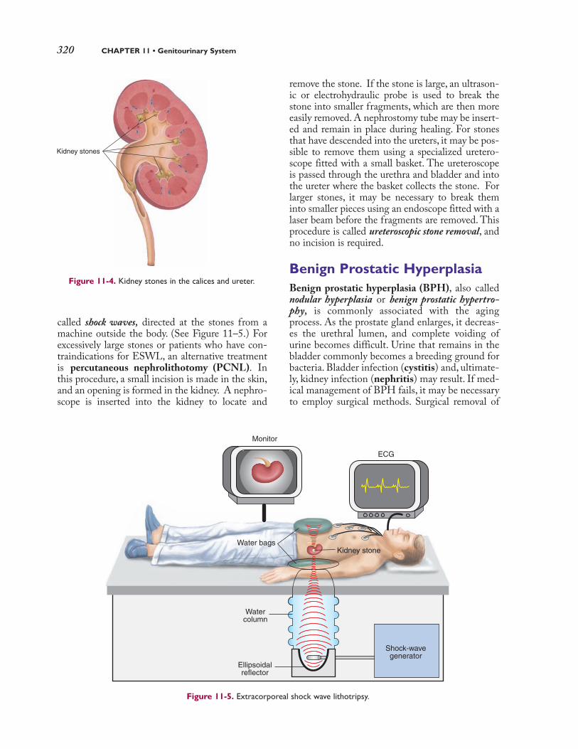
320 CHAPTER 11 • Genitourinary System
called shock waves, directed at the stones from amachine outside the body. (See Figure 11–5.) Forexcessively large stones or patients who have con-traindications for ESWL, an alternative treatmentis percutaneous nephrolithotomy (PCNL). Inthis procedure, a small incision is made in the skin,and an opening is formed in the kidney. A nephro-scope is inserted into the kidney to locate and
remove the stone. If the stone is large, an ultrason-ic or electrohydraulic probe is used to break thestone into smaller fragments, which are then moreeasily removed. A nephrostomy tube may be insert-ed and remain in place during healing. For stonesthat have descended into the ureters, it may be pos-sible to remove them using a specialized uretero-scope fitted with a small basket. The ureteroscopeis passed through the urethra and bladder and intothe ureter where the basket collects the stone. Forlarger stones, it may be necessary to break theminto smaller pieces using an endoscope fitted with alaser beam before the fragments are removed. Thisprocedure is called ureteroscopic stone removal, andno incision is required.
Benign Prostatic HyperplasiaBenign prostatic hyperplasia (BPH), also callednodular hyperplasia or benign prostatic hypertro-phy, is commonly associated with the agingprocess. As the prostate gland enlarges, it decreas-es the urethral lumen, and complete voiding ofurine becomes difficult. Urine that remains in thebladder commonly becomes a breeding ground forbacteria. Bladder infection (cystitis) and, ultimate-ly, kidney infection (nephritis) may result. If med-ical management of BPH fails, it may be necessaryto employ surgical methods. Surgical removal of
Kidney stones
Figure 11-4. Kidney stones in the calices and ureter.
Figure 11-5. Extracorporeal shock wave lithotripsy.
ECG
Monitor
Water bagsKidney stone
Shock-wavegenerator
Ellipsoidalreflector
Watercolumn
2090_Ch11_309-348.qxd 3/5/09 9:48 PM Page 320

Pathology 321
the entire prostate may be done through the per-ineum (perineal prostatectomy) or an abdominalopening above the pubis and directly over thebladder (suprapubic prostatectomy). These pro-cedures are invasive and are performed when theentire prostate must be removed, such as whencancer is identified. These methods also enable theremoval of lymph nodes for examination. Whenonly a portion of the prostate must be removed, theless invasive transurethral resection of theprostate (TURP) is usually performed. In TURP,a resectoscope is inserted through the urethra, anda small loop “chips away” some of the obstructingtissue. At the conclusion of the surgery, the urethraand bladder are irrigated to remove the smallchips. (See Figure 11–6.)
CryptorchidismFailure of the testes to descend into the scrotal sacprior to birth is called cryptorchidism. In manyinfants born with this condition, the testesdescend spontaneously by the end of the first year. If this does not occur, correction of the
disorder involves surgical suspension of the testes(orchiopexy) in the scrotum. This procedure isusually done before the child reaches age 2.Because an inguinal hernia commonly accompa-nies cryptorchidism, the hernia may be sutured(herniorrhaphy) at the same time.
Acute Tubular NecrosisIn acute tubular necrosis (ATN), the tubular por-tion of the nephron is injured by a decrease in bloodsupply (ischemic ATN) or after the ingestion oftoxic chemicals (nephrotoxic ATN). Ischemia may occur because of circulatory collapse, severehypotension, hemorrhage, dehydration, or otherdisorders that affect blood supply. ATN does notproduce specific signs and symptoms, and diagno-sis relies on a positive history of risk factors.General signs and symptoms of ATN includescanty urine production (oliguria) fluid retention,mental apathy, nausea, vomiting, and increasedblood levels of calcium (hypercalcemia). Whentubular damage is not severe, the disorder is usuallyreversible.
Testis
Urinarybladder
Benign prostatichyperplasia
Resectocopein urethra
Rectum
Figure 11-6. Transurethral resection of the prostate (TURP).
2090_Ch11_309-348.qxd 3/5/09 9:48 PM Page 321

322 CHAPTER 11 • Genitourinary System
OncologyThe most common form of cancer in men is carci-noma of the prostate. In the United States, the dis-ease is rarely found in men younger than age 50;however, the incidence dramatically increases withage. Symptoms include difficulty starting urina-tion (hesitancy) and stopping the urinary stream,dysuria, urinary frequency, and hematuria. By thetime these symptoms develop and the patientseeks treatment, the disease is quite advanced and long-term survival is not likely. Early pre-symptomatic tests include a blood test forprostate-specific antigen (PSA) and periodic digi-tal rectal examination (DRE). (See Figure 11–7.)
Like other forms of cancer, prostatic carcinomasare staged and graded to determine metastaticpotential, response to treatment, chances of sur-vival, and appropriate forms of therapy. Surgery andradiation therapy are usual treatment modalities,but other forms of treatment may also be em-ployed. Surgical treatment includes the removal of
the entire prostate (radical prostatectomy). Twoforms of radiation oncology include brachytherapyand external beam radiation. In brachytherapy(also called internal radiation therapy), radioactive“seeds” are placed directly in the malignant tissue.They remain in place for long or short periods oftime depending on the type of malignancy, its loca-tion, and other diagnostic criteria. (See Figure11–8.) In external beam radiation (EBR), high-energy x-ray beams are generated by a machine anddirected at the tumor from outside the body todestroy prostate tissue. Another treatment modalityis the application of extreme cold (cryosurgery),which results in the destruction of prostate tissue.(See Figure 11–9.) Administering antiandrogenicagents as well as hormones that deplete the body of testicular hormones (combined hormonal ther-apy) has been effective in treatment at the earlystages of the disease. Because prostatic cancer isstimulated by testosterone, surgical removal of thetestes (bilateral orchiectomy) may be necessary.
Figure 11-7. Digital rectal examination.
Prostate withnodule
Urinarybladder
Rectum
Digitalrectal examination
2090_Ch11_309-348.qxd 3/5/09 9:48 PM Page 322

Pathology 323
Bladder
Urethral catheter
Urethra
Needle that transmitsseeds into prostate
Rectum
Ultrasound probefor needle guidanceProstate Template to aid accurate
placement of needlesdelivering radioactive seeds
Figure 11-8. Brachytherapy.
Bladder
RectumUltrasound probe
for needle guidanceProstate
Cryoprobeneedles
Ice ballsforming
Urethralcatheter
Figure 11-9. Cryosurgery of the prostate.
2090_Ch11_309-348.qxd 3/5/09 9:48 PM Page 323

324 CHAPTER 11 • Genitourinary System
Diagnostic, Symptomatic, and Related Terms
This section introduces diagnostic, symptomatic, and related terms and their meanings. Wordanalyses for selected terms are also provided.
Term Definition
anuriaan-U-re-a
an-: without, noturia: urine
azotemiaaz-o-TE-me-aazot: nitrogenous compounds-emia: blood condition
bladder neck obstruction(BNO)
chronic renal failureKRO-nık RE-nal
dysuriadıs-U-re-a
dys-: bad; painful; difficulturia: urine
end-stage renal disease (ESRD)dı-AL-ı-s ıs
dia-: through, across-lysis: separation; destruction;
loosening
enuresisen-u-RE-sısen-: in, withinur: urine-esis: condition
fistulaFIS-tu-la
frequencyFRE-kwen-se
Absence of urine production or urinary output
Anuria may be obstructive, in which there is blockage proximal to the bladder, or
unobstructive, which is caused by severe damage to the nephrons of the kidneys.
Retention of excessive amounts of nitrogenous compounds (urea, creati-nine, and uric acid) in the blood; also called uremia
Blockage at base of the bladder that reduces or prevents urine from pass-ing into the urethra
BNO can be caused by benign prostatic hyperplasia, bladder stones, bladder
tumors, or tumors in the pelvic cavity.
Renal failure that occurs over a period of years, in which the kidneys losetheir ability to maintain volume and composition of body fluids with nor-mal dietary intake
Chronic renal failure is the result of decreased numbers of functioning nephrons in
the kidneys.
Painful or difficult urination, commonly described as a “burning sensa-tion” while urinating
Dysuria is a symptom of numerous conditions but, most commonly, urinary tract
infection (UTI).
Condition in which kidney function is permanently lost
Involuntary discharge of urine; also called incontinence
Enuresis that occurs during the night is called nocturnal enuresis; during the day,
diurnal enuresis.
Abnormal passage from a hollow organ to the surface or from one organto another
The most common type of urinary f istula is the vesicovaginal f istula where com-
munication occurs between the bladder and vagina. Its causes include previous
pelvic surgery such as hysterectomy, diff icult and prolonged labor, or reduced blood
supply to the area.
Voiding urine at frequent intervals
Urinary System
2090_Ch11_309-348.qxd 3/5/09 9:48 PM Page 324

Pathology 325
Diagnostic, Symptomatic, and Related Terms—cont’d
Term Definition
hesitancyHEZ-ı-ten-se
hydronephrosishı-dro-nef-RO-sıs
hydr/o: waternephr: kidney-osis: abnormal condition;
increase (used primarilywith blood cells
nephrotic syndromene-FROT-ık
nephr/o: kidney-tic: pertaining to, relating to
nocturianok-TU-re-a
noct: night-uria: urine
Involuntary delay in initiating urination
Abnormal dilation of the renal pelvis and the calyces of one or both kid-neys due to pressure from accumulated urine that cannot flow past anobstruction in the urinary tract
The most common causes of hydronephrosis are BPH, urethral strictures, and cal-
culi that lodge in the ureter and cause an obstruction. The pressure impairs, and
may eventually interrupt, kidney function. (See Figure 11–10.)
Loss of large amounts of plasma protein, usually albumin by way of urinedue to increased permeability of the glomerular membrane
Hypoproteinemia, edema, and hyperlipidemia are commonly associated with
nephrotic syndrome.
Excessive or frequent urination after going to bed
Nocturia is typically caused by excessive fluid intake, uncontrolled diabetes mellitus,
urinary tract infection, prostate disease, impaired renal function, or the use of
diuretics. (continued)
Hydronephrosis
Hydroureter
Stricture
Stone
Stone
Normalureter
Normalkidney
Urinarybladder
Figure 11-10. Hydronephrosis and hydroureter.
2090_Ch11_309-348.qxd 3/5/09 9:48 PM Page 325

326 CHAPTER 11 • Genitourinary System
Diagnostic, Symptomatic, and Related Terms—cont’d
Term Definition
oliguriaol- ıg-U-re-aolig: scanty-uria: urine
polycystic kidney disease (PKD)pol-e-SIS-tık
poly: many, muchcyst: bladder-ic: pertaining to
urgencyUR-jen-se
vesicoureteral reflux (VUR)ves- ı-ko-u-RE-ter-al
vesic/o: bladderureter: ureter-al: pertaining to
Wilms tumorVILMZ TOO-mer
Male Reproductive System
anorchidisman-OR-kı-dızman-: without, notorchid: testis (plural, testes)-ism: condition
aspermiaa-SPER-me-a
a-: without, notsperm: spermatozoa, sperm cells-ia: condition
balanitisbal-a-NI-t ıs
balan: glans penis-itis: inflammation
epispadiasep-ı-SPA-de-asepi-: above, upon-spadias: slit, fissure
Diminished capacity to form and pass urine, resulting in inefficient excre-tion of the end products of metabolism
Oliguria is usually caused by fluid and electrolyte imbalances, renal lesions, or uri-
nary tract obstruction.
Inherited disease in which sacs of fluid called cysts develop in the kidneys
If cysts increase in number or size or if they become infected, kidney failure may result.
Dialysis or kidney transplant may be necessary for renal failure caused by PKD.
Feeling of the need to void immediately
Urinary urgency commonly occurs in urinary tract infection (UTI).
Disorder caused by the failure of urine to pass through the ureters to thebladder, usually due to impairment of the valve between the ureter andbladder or obstruction in the ureter
VUR may result in the enlargement of the kidney (hydronephrosis) if the obstruc-
tion is in the proximal portion of the ureter or enlargement of the ureter
(hydroureter) and the kidney if the obstruction is in the distal portion of the ureter.
Rapidly developing malignant neoplasm of the kidney that usually occursin children
Diagnosis of Wilms tumor is established by an excretory urogram (EU) with
tomography. The tumor is well encapsulated in the early stage but may metastasize
to other sites, such as lymph nodes and lungs, at later stages.
Congenital absence of one or both testes; also called anorchia or anorchism
Treatment for anorchidism requires replacement of the male hormone testosterone.
Boys affected with anorchidism will need testosterone for puberty to occur.
Failure to form or ejaculate semen
Aspermia should not be confused with azoospermia, which is absence of sperm in
the ejaculate.
Inflammation of the skin covering the glans penis
Uncircumcised men with poor personal hygiene are prone to this disorder.
Malformation in which the urethra opens on the dorsum of the penis
2090_Ch11_309-348.qxd 3/5/09 9:48 PM Page 326

Pathology 327
Diagnostic, Symptomatic, and Related Terms—cont’d
Term Definition
erectile dysfunction (ED)e-REK-tıl
hydroceleHI-dro-sel
hydr/o: water-cele: hernia, swelling
hypospadiashı-po-SPA-de-as
hypo-: under, below, deficient-spadias: slit, fissure
phimosisfı-MO-sıs
phim: muzzle-osis: abnormal condition;
increase (used primarilywith blood cells)
sterilityster-IL-ı-te
varicoceleVAR-ı-ko-sel
varic/o: dilated vein-cele: hernia, swelling
Repeated inability to initiate or maintain an erection sufficient for sexualintercourse
Any disorder that causes injury to the nerves or impairs blood flow in the penis has
the potential to cause ED.
Accumulation of serous fluid in a saclike cavity, especially the testes andassociated structures
Hydrocele is common in male newborns but usually resolves within the f irst year.
Developmental anomaly in which the urethra opens on the underside ofthe penis or, in extreme cases, on the perineum
Stenosis or narrowing of preputial orifice so that the foreskin cannot beretracted over the glans penis
Inability to produce offspring; in the male, inability to fertilize the ovum
Swelling and distention of veins of the spermatic cord
It is time to review pathological, diagnostic, symptomatic, and related terms by completingLearning Activity 11–4.
This section introduces procedures used to diagnose and treat genitourinary system disorders.Descriptions are provided as well as pronunciations and word analyses for selected terms.
Procedure Description
Clinical
digital rectal examination (DRE) Screening test that assesses the rectal wall surface for lesions or abnormal-ly firm areas that might indicate cancer
In DRE, the physician inserts a gloved, lubricated f inger into the rectum.
In males, the physician also evaluates the size and consistency of the prostate.
(See Figure 11-7.)
Diagnostic and Therapeutic Procedures
Diagnostic Procedures
(continued)
2090_Ch11_309-348.qxd 3/5/09 9:48 PM Page 327

328 CHAPTER 11 • Genitourinary System
Procedure Description
electromyography (EMG)e-lek-tro-mı-OG-ra-fe
electr/o: electricitymy/o: muscle-graphy: process of recording
testicular self-examination(TSE)
Endoscopic
cystoscopy (cysto)s ıs-TOS-ko-pecyst/o: bladder-scopy: examination
Measures the contraction of muscles that control urination using elec-trodes placed in the rectum and urethra
EMG determines whether incontinence is due to weak muscles or other causes.
Self-examination of the testes for abnormal lumps or swellings in thescrotal sac
TSE is increasingly recommended by physicians to detect abnormalities, especially
cancer, when the disease is easily treatable. Testicular cancer is the number one
cancer killer in men ages 20 to 30.
Endoscopy of the urinary bladder for evidence of pathology, obtainingbiopsies of tumors or other growths, and removal of polyps
In cystoscopy, a catheter can be inserted thought the hollow channel in the
cystoscope to collect tissue samples or introduce contrast media during radiography.
(See Figure 11–11.)
Diagnostic and Therapeutic Procedures—cont’d
Figure 11-11. Cystoscopy.
Eyepiece
Cystoscope
Urethra
Urinarybladder
Videoconnection
Prostategland
Rectum
2090_Ch11_309-348.qxd 3/5/09 9:48 PM Page 328

Procedure Description
nephroscopyne-FROS-ko-pe
nephr/o: kidney-scopy: examination
urethroscopyu-re-THROS-ko-pe
urethr/o: urethrascopy: examination
Laboratory
blood urea nitrogen (BUN)u-RE-a NI-tro-jen
culture and sensitivity (C&S)KUL-tur, sen-sı-TI-vı-te
prostate-specific antigen (PSA)PROS-tat spe-SIF-ık AN-tı-jen
semen analysisSE-men a-NAL-ı-s ıs
urinalysis (UA)u-r ı-NAL-ı-s ıs
Radiographic
computed tomography (CT) kom-PU-ted to-MOG-ra-fe
tom/o: to cut-graphy: process of recording
cystographys ıs-TOG-ra-fe
cyst/o: bladder-graphy: process of recording
Endoscopy of the kidney(s) using a specialized, three-channel endoscopethat enables visualization and irrigation of the kidney
The nephroscope is passed through a small incision made in the renal pelvis.
Kidney pathology and congenital deformities may be observed.
Endoscopy of the urethra using a specialized endoscope, typically forlithotripsy or TURP
Test that determines the amount of urea nitrogen, a waste product of pro-tein metabolism, present in a blood sample
Because urea is cleared from the bloodstream by the kidneys, the BUN test
is used as an indicator of kidney function.
Test that determines the causative organism of a disease and how theorganism responds to various antibiotics
C&S tests are performed on urine, blood, and body secretions.
Blood test used to detect prostatic disorders, especially prostatic cancer
PSA is a substance produced by the prostate and is normally found in a blood
sample in small quantities. The level is elevated in prostatitis, benign prostatic
hyperplasia, and tumors of the prostate.
Test that analyzes a semen sample for volume, sperm count, motility, andmorphology to evaluate fertility or verify sterilization after a vasectomy
Battery of tests performed on a urine specimen, including physical obser-vation, chemical tests, and microscopic evaluation
UA not only provides information on the urinary structures but may also
be the f irst indicator of such system disorders as diabetes and liver and
gallbladder disease.
Imaging technique that rotates an x-ray emitter around the area to bescanned and measures the intensity of transmitted rays from different angles
In the genitourinary system, CTs are used to diagnose tumors, cysts, inflamma-
tion, abscesses, perforation, bleeding, and obstructions of the kidneys, ureters,
and bladder.)
Radiographic examination of the urinary bladder using a contrast medium
Cystography is used to diagnose tumors or defects in the bladder wall, vesi-
coureteral reflux, stones, or other pathological conditions of the bladder.
Diagnostic and Therapeutic Procedures—cont’d
Pathology 329
(continued)
2090_Ch11_309-348.qxd 3/5/09 9:48 PM Page 329

330 CHAPTER 11 • Genitourinary System
Procedure Description
cystometrographys ıs-to-me-TROG-ra-fe
cyst/o: bladdermetr/o: uterus (womb); measure-graphy: process of recording
intravenous pyelography (IVP)ın-tra-VE-nus
pı-e-LOG-ra-feintra-: in, withinven: vein-ous: pertaining topyel/o: renal pelvis-graphy: process of recording
kidney, ureter, bladder (KUB)radiography
radi/o: radiation, x-ray; radius(lower arm bone onthumb side)
-graphy: process of recording
nuclear scan
renalren: kidney-al: pertaining to,
ultrasound (US)UL-tra-sownd
scrotalSKRO-tal
voiding cystourethrography(VCUG)s ıs-to-u-re-THROG-ra-fe
cyst/o: bladderurethr/o: urethra-graphy: process of recording
Clinical
dialysisdı-AL-ı-s ıs
dia-: through, across-lysis: separation; destruction;
loosening
Procedure that assesses volume and pressure in the bladder at variousstages of filling using saline and a contrast medium introduced into thebladder through a catheter
Cystometrography is the primary test used to investigate stress incontinence
and urge incontinence.
Radiographic examination of the kidneys, and urinary tract after IV injection of a contrast medium; also called excretory urography (EU)
IVP detects kidney stones, enlarged prostate, internal injuries after an accident
or trauma, and tumors in the kidneys, ureters, and bladder.
Radiographic examination to determine the location, size, and shape ofthe kidneys in relationship to other organs in the abdominopelvic cavityand to identify abnormalities of the urinary system
KUB radiography identif ies stones and calcif ied areas and does not require a
contrast medium.
Radiology test in which radioactive materials called tracers are introducedinto the patient and a specialized camera, which acts as a radiation detec-tor, produces images by recording the emitted tracers
Imaging test where a monitor is used to track a radioactive substance as itpasses through the kidney
Radiograph that uses high-frequency sound waves (ultrasound) and displaysthe reflected echoes on a monitor; also called sonography, echography, or echo
US used to assess scrotal structures and patency of the vas deferens
Radiological examination of the bladder and urethra performed before,during, and after voiding using a contrast medium to enhance imaging
VCUG is performed to determine the cause of repeated bladder infections or
stress incontinence and to identify congenital or acquired structural abnormali-
ties of the bladder and urethra.
Medical procedure used to filter toxic substances from the patient’sbloodstream, such as excess electrolytes and nitrogenous wastes
Dialysis provides a means of removing waste products from the blood when
kidneys no longer function. Nitrogenous waste products are collected in a solu-
tion called dialysate, which is discarded at the end of the procedure. There are
two primary methods of dialysis: hemodialysis and peritoneal dialysis.
Diagnostic and Therapeutic Procedures—cont’d
Therapeutic Procedures
2090_Ch11_309-348.qxd 3/5/09 9:48 PM Page 330

Pathology 331
Procedure Description
hemodialysishe-mo-dı-AL-ı-s ıs
hem/o: blooddia: through, across-lysis: separation; destruction;
loosening
peritonealper- ı-to-NE-al
peritone: peritoneum-al: pertaining to
Method of removing waste substances from the blood by shunting it from thebody, passing it through an artificial kidney machine where it is filtered, andthen returning the dialyzed blood to the patient’s body (See Figure 11–12.)
Removal of toxic substances from the body by perfusing the peritonealcavity with a warm, sterile chemical solution (See Figure 11–13.)
In peritoneal dialysis, the lining of the peritoneal cavity is used as the dialyzing
membrane. Dialyzing fluid remains in the peritoneal cavity for 1 to 2 hours
and then is removed. The procedure is repeated as often as necessary.
Diagnostic and Therapeutic Procedures—cont’d
Figure 11-12. Hemodialysis
Dialyzer
Dialysatelines
Access
Bloodlines
Hemodialysismachine
Figure 11-13. Peritoneal dialysis. (A) Introducing dialysis fluid into the peritoneal
cavity. (B) Draining dialysate with waste products from the peritoneal cavity.
A. B.
Peritonealcavity
Freshdialysatesolution
Useddialysatesolution
(continued)
2090_Ch11_309-348.qxd 3/5/09 9:48 PM Page 331

Procedure Description
Surgical
circumcisionser-kum-SI-zhun
nephropexyNEF-ro-peks-e
nephr/o: kidney-pexy: fixation (of an organ)
orchidectomyor-kı-DEK-to-meorchid: testis (plural, testes)-ectomy: excision, removal
transurethral resection of theprostate (TURP)
trans-u-RE-thral re-SEK-shunPROS-tat
trans: across, throughurethr/o: urethra-al: pertaining to
urethrotomyu-re-THROT-o-me
urethr/o: urethra-tomy: incision
vasectomyvas-EK-to-me
vas: vessel; vas deferens; duct-ectomy: excision, removal
Removal of all or part of the foreskin, or prepuce, of the penis
Fixation of a floating or mobile kidney
Removal of one or both testes; also called orchiectomy
Orchidectomy may be indicated for serious disease or injury to the testis or to
control cancer of the prostate by removing a source of androgenic hormones.
Surgical procedure that involves inserting a resectoscope into the urethrato “chip away” at the prostate gland to remove the obstruction and flush-ing out the chips and sending them for analysis to detect possible evi-dence of cancer
TURP is used most commonly to relieve obstruction caused by benign prostatic
hyperplasia. Because the prostate gland is not completely removed, remaining
tissue eventually grows back and may cause obstruction again at a later time.
Incision of a urethral stricture
Urethrotomy corrects constrictions of the urethra that make voiding diff icult.
Excision of all or a segment of the vas deferens (See Figure 11–14.)
Bilateral vasectomy is the most successful method of male contraception.
Although the procedure is considered permanent, with advances in micro-
surgery, vasectomy is sometimes reversible.
Diagnostic and Therapeutic Procedures—cont’d
Figure 11-14. Vasectomy and reversal.
Vas deferens
Skin incisionVas deferens pulled
through incision and cut
Vasectomy reversalwith ends of vas deferens
sutured together
Each end tied offwith suture before
incision is closed
2090_Ch11_309-348.qxd 3/5/09 9:48 PM Page 332

Pharmacology 333
Drugs Used to Treat Genitourinary DisordersTable 11-1
This table lists common drug classifications used to treat urinary and male reproductive disorders, their therapeutic
actions, and selected generic and trade names.
Classification Therapeutic Action Generic and Trade Names
Urinary System
antibiotics
antispasmodics
diuretics
potassium supplements
androgens
Treat bacterial infections of the urinary tract
by acting on the bacterial membrane or one of
its metabolic processes.
The type of antibiotic prescribed depends on theinfecting organism and the type and extent ofinfection.
Decrease spasms in the urethra and bladder by
relaxing the smooth muscles lining their walls,
thus allowing normal emptying of the bladder.
Bladder spasms can result from such conditions asurinary tract infections and catheterization.
Promote and increase the excretion of urine.
Diuretics are grouped by their action and are usedto treat edema, hypertension, heart failure, andvarious renal and hepatic diseases.
Replace potassium due to depletion caused by
diuretics.
Dietary sources of potassium are usually not suffi-cient to replace potassium loss caused by diuretics.
Increase testosterone levels.
Androgens are used to correct hormone deficiencyin hypogonadism and treat delayed puberty inmales.
ciprofloxacins ıp-ro-FLOX-a-s ın
Cipro
sulfamethoxazole/trimethoprimsul-f a-meth-OX-a-zol
trı-METH-o-pr ım
Bactrim
oxybutyninok-se-BU-t ı-n ın
Ditropan
furosemidefu-RO-se-mıd
Lasix
spironolactonespı-ro-no-LAK-ton
Aldactone
potassium chloridepo-TAS-e-um KLO-rıd
K-Tab, Kaon Cl
testosterone basetes-TOS-ter-on
Androderm,Testim
testosterone cypionatetes-TOS-ter-on SIP-e-o-nat
Depo-testosterone
Male ReproductiveSystem
Pharmacology
Pharmacological agents used to treat urinary tractdisorders include antibiotics, diuretics, antidi-uretics, urinary antispasmodics, and potassiumsupplements, which are commonly taken concur-
rently with many diuretics to counteract potassi-um depletion. (See Table 11–1.) Pharmacologicagents are used to treat conditions of the malereproductive system including hypogonadism,erectile disfunction, and reproductive concernsand disorders.
2090_Ch11_309-348.qxd 3/5/09 9:48 PM Page 333

334 CHAPTER 11 • Genitourinary System
Abbreviations
This section introduces genitourinary-related abbreviations and their meanings.
Abbreviation Meaning Abbreviation Meaning
AGN acute glomerulonephritis ESRD end-stage renal disease
ARF acute renal failure ESWL extracorporeal shock-wave lithotripsy
ATN acute tubular necrosis EU excretory urography
BNO bladder neck obstruction GU genitourinary
BPH benign prostatic hyperplasia; HD hemodialysis; hipbenign prostatic hypertrophy disarticulation; hearing distance
BUN blood urea nitrogen HTN hypertension
C&S culture and sensitivity IVP intravenous pyelogram, intravenouspyelography
Cath catheterization; catheter K potassium (an electrolyte)
CT computed tomography KUB kidney, ureter, bladder
cysto cystoscopy Na sodium (an electrolyte)
DRE digital rectal examination PCNL percutaneous nephrolithotomy
EBT external beam therapy pH symbol for degree of acidity or alkalinity
ED erectile dysfunction; PKD polycystic kidney diseaseemergency department
EMG electromyogram, PSA prostate-specific antigenelectromyography
Drugs Used to Treat Genitourinary Disorders—cont’dTable 11-1
Classification Therapeutic Action Generic and Trade Names
anti-impotenceagents
Treat erectile dysfunction (impotence) by
increasing blood flow to the penis, resulting in
an erection.
Anti-impotence drugs should not be used by patientswith coronary artery disease or hypertension.
sildenafil citrates ıl-DEN-a-f ıl SIT-rat
Viagra
vardenafilvar-DEN-a-f ıl
Levitra
2090_Ch11_309-348.qxd 3/5/09 9:48 PM Page 334

Pharmacology 335
Abbreviations
Abbreviation Meaning Abbreviation Meaning
RP retrograde pyelogram, UA urinalysisretrograde pyelography
sp. gr. specific gravity UTI urinary tract infection
TSE testicular self-examination VCUG voiding cystourethrography prostate
TURP transurethral resection of the VUR vesicoureteral reflux
It is time to review procedures, pharmacology, and abbreviations by completing LearningActivity 11–5.
2090_Ch11_309-348.qxd 3/5/09 9:48 PM Page 335

336 CHAPTER 11 • Genitourinary System
The activities that follow provide review of the genitourinary system terms introduced inthis chapter. Complete each activity and review your answers to evaluate your understand-ing of the chapter.
Learning Activity 11-1
Identifying Urinary Structures
Label the following illustration using the terms listed below.
hilum renal cortex renal vein urethra urinary meatus
left kidney renal medulla right kidney ureteral orifice
renal artery renal pelvis ureters urinary bladder
LEARNING ACTIVITIES
Adrenal(suprarenal)
glands
Inferiorvena cava
Abdominal aorta
Prostate gland(in males)
Inferiorvena cava
Renal pyramid
Check your answers by referring to Figure 11–1 on page 311. Review material that you didnot answer correctly.✓
2090_Ch11_309-348.qxd 3/5/09 9:48 PM Page 336

Learning Activities 337
Learning Activity 11-2
Identifying Male Reproductive Structures
Label the following illustration using the terms listed below.
bulbourethral gland prepuce testis
ejaculatory duct prostate gland urethra
epididymis scrotum urethral orifice
glans penis seminal vesicle vas deferens
penis seminiferous tubules
Symphysis pubis
Urinary bladder
Sacrum
Opening of ureter
Rectum
Anus
Check your answers by referring to Figure 11–3 on page 314. Review material that you did not answercorrectly.✓
Enhance your study and reinforcement of word elements with the power of DavisPlus. Visitwww.davisplus.fadavis.com/gylys/systems for this chapter’s flash-card activity. We recommendyou complete the flash-card activity before completing Activity 11–3 below.
2090_Ch11_309-348.qxd 3/5/09 9:48 PM Page 337

338 CHAPTER 11 • Genitourinary System
Learning Activity 11-3
Building Medical Words
Use nephr/o (kidney) to build words that mean:
1. stone in the kidney
2. abnormal condition of pus in the kidney
3. abnormal condition of water in the kidney
Use pyel/o (renal pelvis) to build words that mean:
4. dilation of renal pelvis
5. disease of renal pelvis
Use ureter/o (ureter) to build words that mean:
6. dilation of ureter
7. calculus in ureter
Use cyst/o (bladder) to build words that mean:
8. inflammation of bladder
9. instrument to view the bladder
Use vesic/o (bladder) to build words that mean:
10. herniation of bladder
11. pertaining to bladder and prostate
Use urethr/o (urethra) to build words that mean:
12. narrowing or stricture of urethra
13. instrument used to incise urethra
Use ur/o (urine, urinary tract) to build words that mean:
14. radiography of urinary tract
15. disease of urinary tract
Use the suffix -uria (urine) to build words that mean:
16. difficult or painful urination
17. scanty urination
Use orchid/o or orchi/o (testes) to build words that mean:
18. disease of testes
19. pain in testes
2090_Ch11_309-348.qxd 3/5/09 9:48 PM Page 338

Learning Activities 339
Use balan/o (glans penis) to build a word that means:
20. discharge from glans penis
Build surgical words that mean:
21. excision of testes
22. surgical repair of glans penis
23. excision of vas deferens
24. incision of renal pelvis
25. fixation of bladder
Check your answers in Appendix A. Review material that you did not answer correctly.✓
Correct Answers �� 4 �� % Score
2090_Ch11_309-348.qxd 3/5/09 9:48 PM Page 339

340 CHAPTER 11 • Genitourinary System
Learning Activity 11-4
Matching Pathological, Diagnostic, Symptomatic, and Related Terms
Match the following terms with the definitions in the numbered list.
anorchidism benign prostatic hyperplasia hesitancy oliguria
anuria enuresis hydrocele phimosis
aspermia epispadias hydronephrosis pyuria
azotemia fistula nephrotic syndrome sterility
balanitis herniorrhaphy nocturia urgency
1. need to void immediately
2. abnormal passage from a hollow organ to the surface or between organs
3. complete absence of one or both testes
4. absence of urine production
5. nitrogenous wastes in blood
6. dilation of kidneys and calices, usually due to reflux
7. nonmalignant enlargement of the prostate gland
8. difficulty starting urination
9. scanty urine production
10. loss of large amounts of plasma protein, resulting in systemic edema
11. stenosis of the preputial orifice
12. inability to produce offspring
13. malformation in which the urethra opens on the dorsum of the penis
14. lack of or failure to ejaculate semen
15. pus in urine
16. suture of a hernia
17. excessive urination at night
18. involuntary passage of urine
19. accumulation of fluid in a saclike cavity
20. inflammation of skin covering the penis
Check your answers in Appendix A. Review any material that you did not answer correctly.✓
Correct Answers �� 5 �� % Score
2090_Ch11_309-348.qxd 3/5/09 9:48 PM Page 340

Learning Activities 341
Learning Activity 11-5
Matching Procedures, Pharmacology, and Abbreviations
Match the following terms with the definitions in the numbered list.
androgens diuretics potassium supplements
antibiotics ESWL PSA
C&S KUB semen analysis
circumcision orchidectomy urethrotomy
cystoscopy peritoneal dialysis vasectomy
1. radiograph that shows the size, shape, and location of the kidneys
2. test used to verify sterility after vasectomy
3. visual examination of the urinary bladder
4. inhibit or kill bacterial microorganisms
5. laboratory test that evaluates effect of an antibiotic on an organism
6. drugs used to promote the excretion of urine
7. incision of a urethral stricture
8. noninvasive test used to pulverize urinary or bile stones
9. removal of toxic substances by perfusing the peritoneal cavity
10. blood test to detect prostatic disorders, especially cancer
11. most effective form of male contraception
12. surgical removal of the testes
13. surgical removal of all or part of the foreskin
14. used to increase testosterone levels
15. used to treat or prevent the hypokalemia commonly associated with theuse of diuretics
Check your answers in Appendix A. Review material that you did not answer correctly.✓
Correct Answers �� 6.67 �� % Score
2090_Ch11_309-348.qxd 3/5/09 9:48 PM Page 341

342 CHAPTER 11 • Genitourinary System
The two medical records included in the following activities use common clinical scenariosto show how medical terminology is used to document patient care. Complete the termi-nology and analysis sections for each activity to help you recognize and understand termsrelated to the genitourinary system.
Medical Record Activity 11-1
Operative Report: Ureterocele and Ureterocele Calculus
TerminologyTerms listed in the following table are taken from Operative report: Ureterocele and ureterocelecalculus that follows. Use a medical dictionary such as Taber’s Cyclopedic Medical Dictionary,the appendices of this book, or other resources to define each term. Then review the pro-nunciations for each term and practice by reading the medical record aloud.
MEDICAL RECORD ACTIVITIES
Term Definition
calculusKAL-ku-lus
cystolithotripsys ıs-to-LITH-o-tr ıp-se
cystoscopeSIST-o-skop
fulgurationf ul-gu-RA-shun
hematuriahe-ma-TU-re-a
resectoscopere-SEK-to-skop
transurethraltrans-u-RE-thral
ureteroceleu-RE-ter-o-sel
urethral soundu-RE-thral
Listen and Learn Online! will help you master the pronunciation of selected medical words from thismedical record activity. Visit www.davisplus.com/gylys/systems to f ind instructions on completing theListen and Learn Online! exercise for this section and to practice pronunciations.
(( ))
2090_Ch11_309-348.qxd 3/5/09 9:48 PM Page 342

Operative Report: Ureterocele and Ureterocele Calculus 343
General Hospital
1511 Ninth Avenue ■■ Sun City, USA 12345 ■■ (555) 802-1887
OPERATIVE REPORT
Date: May 14, 20xx Physician: Elmer Augustino, MDPatient: Motch, Edwin Patient: ID#: 48778
PREOPERATIVE DIAGNOSIS: Hematuria with left ureterocele and ureterocele calculus.
POSTOPERATIVE DIAGNOSIS: Hematuria with left ureterocele and ureterocele calculus.
OPERATION: Cystoscopy, transurethral incision of ureterocele, extraction of stone, and cys-tolithotripsy.
ANESTHESIA: General.
COMPLICATIONS: None.
PROCEDURE: Patient was prepped and draped and placed in the lithotomy position. The urethrawas calibrated with ease using a #26 French Van Buren urethral sound. A #24 resectoscope was insert-ed with ease. The prostate and bladder appeared normal, except for the presence of a left ureterocele,which was incised longitudinally; a large calculus was extracted from the ureterocele. There was mini-mal bleeding and no need for fulguration. The stone was crushed with the Storz stone-crushing instru-ment, and the fragments were evacuated. The bladder was emptied and the procedure terminated.
Patient tolerated the procedure well and was transferred to the postanesthesia care unit.
EEllmmeerr AAuugguussttiinnoo,, MMDDElmer Augustino, MD
ea:bg
D: 5-14-20xxT: 5-14-20xx
OPERATIVE REPORT: URETEROCELE AND URETEROCELE CALCULUS
2090_Ch11_309-348.qxd 3/5/09 9:48 PM Page 343

344 CHAPTER 11 • Genitourinary System
AnalysisReview the medical record Operative Report: Ureterocele and Ureterocele Calculus to answerthe following questions.
1.What were the findings from the resectoscopy?
2.What was the name and size of the urethral sound used in the procedure?
3.What is the function of the urethral sound?
4. In what direction was the ureterocele incised?
5.Was fulguration required? Why or why not?
Medical Record Activity 11-2
Operative Report: Extracorporeal Shock-Wave Lithotripsy
TerminologyTerms listed in the following table are taken from the Operative Report: Extracorporeal Shock-Wave Lithotripsy that follows. Use a medical dictionary such as Taber’s CyclopedicMedical Dictionary, the appendices of this book, or other resources to define each term. Thenreview the pronunciations for each term and practice by reading the medical record aloud.
Term Definition
calculusKAL-ku-lus
calyxKA-l ıx
cystoscopeSIST-o-skop
cystoscopys ıs-TOS-ko-pe
dorsal lithotomyDOR-sal l ıth-OT-o-me
ESWL
extracorporealeks-tra-kor-POR-e-al
fluoroscopyfloo-or-OS-ko-pe
2090_Ch11_309-348.qxd 3/5/09 9:48 PM Page 344

Operative Report: Ureterocele and Ureterocele Calculus 345
Term Definition
lithotripsyLITH-o-tr ıp-se
Lt
shock-wave
staghorn calculusSTAG-horn
KAL-ku-lus
stentstent
Listen and Learn Online! will help you master the pronunciation of selected medical words from thismedical record activity. Visit www.davisplus.com/gylys/systems to f ind instructions on completing theListen and Learn Online! exercise for this section and to practice pronunciations.
(( ))
2090_Ch11_309-348.qxd 3/5/09 9:48 PM Page 345

346 CHAPTER 11 • Genitourinary System
General Hospital
1511 Ninth Avenue ■■ Sun City, USA 12345 ■■ (555) 802-1887
OPERATIVE REPORT
Date: April 1, 20xx Physician: Elmer Augustino, MDPatient: Marino, Julius Room: 7201
PREOPERATIVE DIAGNOSIS: Left renal calculus.
POSTOPERATIVE DIAGNOSIS: Left renal calculus.
PROCEDURE: Extracorporeal shock-wave lithotripsy, cystoscopy with double-J stent removal
INDICATION FOR PROCEDURE: This 69-year-old male had undergone ESWL on 5/15/xx,with double-J stent placement to allow stone fragments to pass from the calyx to the bladder. At thattime, approximately 50% of a partial staghorn calculus was fragmented. He now presents for the frag-menting of the remainder of the calculus and removal of the double-J stent.
ANESTHESIA: General.
COMPLICATIONS: None.
OPERATIVE TECHNIQUE: Patient was brought to the Lithotripsy Unit and placed in the supineposition on the lithotripsy table. After induction of anesthesia, fluoroscopy was used to position thepatient in the focal point of the shock waves. Being well positioned, he was given a total of 4,000shocks with a maximum power setting of 3.0. After confirming complete fragmentation via fluo-roscopy, the patient was transferred to the cystoscopy suite.
Patient was placed in the dorsal lithotomy position and draped and prepped in the usual manner. Acystoscope was inserted into the bladder through the urethra. Once the stent was visualized, it wasgrasped with the grasping forceps and removed as the scope was withdrawn.
Patient tolerated the procedure well and was transferred to recovery.
EEllmmeerr AAuugguussttiinnoo,, MMDDElmer Augustino, MD
ea:bg
D: 5-14-20xxT: 5-14-20xx
OPERATIVE REPORT: EXTRACORPOREAL SHOCK-WAVE LITHOTRIPSY
2090_Ch11_309-348.qxd 3/5/09 9:48 PM Page 346

Operative Report: Extracorporeal Shock-Wave Lithotripsy 347
AnalysisReview the medical record Operative Report: Extracorporeal Shock-Wave Lithotripsy toanswer the following questions.
1.What previous procedures were performed on the patient?
2.Why is this current procedure being performed?
3.What imaging technique was used for positioning the patient to ensure that the shock waves wouldstrike the calculus?
4. In what position was the patient placed in the cystoscopy suite?
5. How was the double-J stent removed?
2090_Ch11_309-348.qxd 3/5/09 9:48 PM Page 347

2090_Ch11_309-348.qxd 3/5/09 9:48 PM Page 348

FemaleReproductiveSystem
Chapter Outline
Objectives
Anatomy and PhysiologyAnatomy and Physiology Key TermsFemale Reproductive Organs
OvariesFallopian TubesUterus and Vagina
Mammary GlandsMenstrual CyclePregnancyLabor and ChildbirthMenopauseConnecting Body Systems–Female ReproductiveSystem
Medical Word Elements
PathologyMenstrual DisordersEndometriosisPelvic and Vaginal InfectionsVaginitisSexually Transmitted Disease
GonorrheaSyphilisChlamydiaGenital HerpesGenital WartsTrichomoniasis
Uterine FibroidsOncology
Breast CancerCervical Cancer
Diagnostic, Symptomatic, and Related Terms
Diagnostic and Therapeutic Procedures
Pharmacology
Abbreviations
Learning Activities
Medical Record Activities
SOAP note: Primary herpes 1infectionPreoperative consultation: Menometrorrhagia
ObjectivesUpon completion of this chapter, you will be able to:
• Locate and describe the structures of the female
reproductive system.
• Describe the functional relationship between the
female reproductive system and other body
systems.
• Recognize, pronounce, spell, and build words related
to the female reproductive system.
• Describe pathological conditions, diagnostic and
therapeutic procedures, and other terms related to
the female reproductive system.
• Explain pharmacology related to the treatment of
female reproductive disorders.
• Demonstrate your knowledge of this chapter by
completing the learning and medical record
activities.
C H A P T E R
12
2090_Ch12_349-392.qxd 3/5/09 10:28 PM Page 349

Anatomy and Physiology
The female reproductive system is composed ofinternal and external organs of reproduction.(See Figure 12–1.) The internal organs include the (1) ovaries, (2) fallopian tubes, (3) uterus,(4) vagina, and external genitalia. The external
genitalia are collectively known as the vulva.Included in these structures are the (5) labia mino-ra, (6) labia majora, (7) clitoris, (8) Bartholinglands, and mons pubis, an elevation of adiposetissue covered by skin and coarse pubic hair thatcushions the pubis (pubic bone).
350 CHAPTER 12 • Female Reproductive System
(2) Fallopian tube (oviduct)
(2) Fallopian tube (oviduct)
(1) Ovary
Peritonealcavity
(3) Uterus
(3) Uterus
Pubis
(7) Clitoris
(5) Labia minora
(6) Labia majora
Urinary bladder
Urethra
(4) Vagina
(4) Vagina
Perineum
Anus
Rectum
Cervix
B. Sagittal section
Sacrum
(8) Bartholin gland
(1) Ovary
Nipple
Breast
A. Anterior view
Figure 12-1. Female reproductive system. (A) Anterior view. (B) Sagittal section showing organs within the pelvic cavity.
2090_Ch12_349-392.qxd 3/5/09 10:28 PM Page 350

Female Reproductive OrgansThe female reproductive organs include the ovaries,fallopian tubes, uterus, and vagina. They aredesigned to produce ova (female reproductive cells),transport the cells to the site of fertilization, providea favorable environment for a developing fetusthrough pregnancy and childbirth, and producefemale sex hormones. Hormones play an importantrole in the reproductive process, providing theirinfluence at critical times during preconception, fer-tilization, and gestation. (See Figure 12–2.)
Ovaries
The (1) ovaries are almond-shaped glands locatedin the pelvic cavity, one on each side of the uterus.Each ovary contains thousands of tiny, saclike structures called (2) graafian follicles, each contain-ing an ovum. When an ovum ripens, the (3) maturefollicle moves to the surface of the ovary, ruptures,and releases the ovum; a process called ovulation.After ovulation, the empty follicle is transformed
into a structure called the (4) corpus luteum, a smallyellow mass that secretes estrogen and progesterone.The corpus luteum degenerates at the end of a non-fertile cycle. Estrogen and progesterone influencethe menstrual cycle and menopause. They also pre-pare the uterus for implantation of the fertilizedegg, help maintain pregnancy, promote growth ofthe placenta, and play an important role in develop-ment of secondary sex characteristics. (See Chapter13, Endocrine System.)
Fallopian Tubes
Two (5) fallopian tubes (oviducts, uterine tubes)extend laterally from superior angles of the uterus.The (6) fimbriae are fingerlike projections thatcreate wavelike currents in fluid surrounding theovary to move the ovum into the uterine tube. Ifthe egg unites with a spermatozoon, the malereproductive cell, fertilization or conception takesplace. If conception does not occur, the ovum dis-integrates within 48 hours and is dischargedthrough the vagina.
Anatomy and Physiology 351
Anatomy and Physiology
This section introduces important female reproductive system terms and their definitions.Word analyses for selected terms are also provided.
Term Definition
external genitaliajen-ı-TAL-e-a
gestationjes-TA-shun
gest: pregnancy-ation: process (of )
lactationlak-TA-shun
lact: milk-ation: process (of )
orificeOR-ı-f ıs
puerperiumpu-er-PE-re-um
The sex, or reproductive, organs visible on the outside of the body; alsocalled genitals
The external female genitalia, also known as the vulva. Male genitalia include
the penis, scrotum, and testicles.
Length of time from conception to birth
The human gestational period typically extends approximately 280 days from
the last menstrual period. Gestation (pregnancy) of less than 36 weeks is
regarded as premature.
Production and release of milk by mammary glands
Mouth; entrance or outlet of any anatomical structure
Time after childbirth that lasts approximately 6 weeks, during which theanatomical and physiological changes brought about by pregnancy resolveand a woman adjusts to the new or expanded responsibilities of mother-hood and nonpregnant life
Pronunciation Help Long Sound a—rate e—rebirth ı—isle o—over u—uniteShort Sound a—alone e—ever ı—it o—not u—cut
2090_Ch12_349-392.qxd 3/5/09 10:28 PM Page 351

Uterus and Vagina
The (7) uterus contains and nourishes the embryofrom the time the fertilized egg is implanted untilthe fetus is born. It is a muscular, hollow, invert-ed–pear-shaped structure located in the pelvic areabetween the bladder and rectum. The uterus isnormally in a position of anteflexion (bent for-ward) and consists of three parts: the (8) fundus,the upper, rounded part; the (9) body, the centralpart; and the (10) cervix, also called the neck of theuterus or cervix uteri, the inferior constricted por-tion that opens into the vagina.
The (11) vagina is a muscular tube that extendsfrom the cervix to the exterior of the body. Its lin-ing consists of folds of mucous membrane thatgive the organ an elastic quality. During sexualexcitement, the vaginal orifice is lubricated bysecretions from (12) Bartholin glands. In addi-tion to serving as the organ of sexual intercourseand receptor of semen, the vagina discharges men-strual flow. It also acts as a passageway for thedelivery of the fetus. The clitoris, located anteriorto the vaginal orifice, is composed of erectile tissuethat is richly innervated with sensory endings. Theclitoris is similar in structure to the penis in the
male, but is smaller and has no urethra. The areabetween the vaginal orifice and the anus is knownas the perineum. During childbirth, this area maybe surgically incised to enlarge the vaginal openingfor delivery. If the incision is made, the procedureis called an episiotomy.
Mammary GlandsAlthough mammary glands (breasts) are presentin both sexes, they function only in females. (SeeFigure 12–3.) The breasts are not directlyinvolved in reproduction but become importantafter delivery. Their biological role is to secretemilk for the nourishment of the newborn; aprocess called lactation. Breasts begin to developduring puberty as a result of periodic stimulationof the ovarian hormones estrogen and proges-terone and are fully developed by age 16. Es-trogen is responsible for the development of (1) adipose tissue, which enlarges the size of thebreasts until they reach full maturity. Breast sizeis primarily determined by the amount of fataround the glandular tissue but is not indicativeof functional ability. Each breast is composed of
352 CHAPTER 12 • Female Reproductive System
(4) Corpus luteum
Fertilization of ovum(5) Fallopian tube
(7) Uterus
(8) Fundus of uterus
(6) Fimbriae
(1) Ovary
Ovarian ligament
(9) Body of uterusEndometrium
Myometrium
Sperm
(10) Cervix
(11) Vagina
Rugae
(12) Bartholin glands
ArteryVein
(3) Mature follicle
(2) Graafian follicles
Figure 12-2. Anterior view of the female reproductive system.The developing follicles are shown in the sectioned left cavity.
2090_Ch12_349-392.qxd 3/5/09 10:28 PM Page 352

15 to 20 lobules of milk-producing glands thatare drained by a (2) lactiferous duct, whichopens on the tip of the raised (3) nipple. Circlingthe nipple is a border of slightly darker skin calledthe (4) areola. During pregnancy, the breastsenlarge and remain so until lactation ceases. Atmenopause, breast tissue begins to atrophy.
Menstrual CycleMenarche, the initial menstrual period, occurs atpuberty (about age 12) and continues approxi-mately 40 years, except during pregnancy. Theduration of the menstrual cycle is approximately28 days, during which time several phases occur.(See Table 12–1.)
Anatomy and Physiology 353
Pectoral muscle
Mammarylobule
(3) Nipple
(3) Nipple
(2) Lactiferousduct
Rib
Internal mammarylymph node
(4) Areola
Axillarylymph node
Lymphvessel
Breasttissue
(1) Adipose tissue
Supraclavicularlymph node (1) Adipose
tissue
Figure 12-3. Structure of mammary glands.
2090_Ch12_349-392.qxd 3/5/09 10:28 PM Page 353

PregnancyDuring pregnancy, the uterus changes its shape,size, and consistency. It increases greatly in sizeand muscle mass; houses the growing placenta,which nourishes the embryo-fetus; and expels thefetus after gestation. To prepare and serve as thebirth canal at the end of pregnancy, the vaginalcanal elongates as the uterus rises in the pelvis.Themucosa thickens, secretions increase, and vascular-ity and elasticity of the cervix and vagina becomemore pronounced.
The average pregnancy (gestation) lasts approx-imately 9 months and is followed by childbirth(parturition). Up to the third month of pregnancy,the product of conception is referred to as theembryo. From the third month to the time of birth,the unborn offspring is referred to as the fetus.
Pregnancy also causes enlargement of thebreasts, sometimes to the point of pain. Manyother changes occur throughout the body toaccommodate the development and birth of thefetus. Toward the end of gestation, the myometri-um begins to contract weakly at irregular intervals.
At this time, the full-term fetus is usually posi-tioned head down within the uterus.
Labor and ChildbirthLabor is the physiological process by which thefetus is expelled from the uterus. Labor occurs inthree stages. The first is the stage of dilation,which begins with uterine contractions and termi-nates when there is complete dilation of the cervix(10 cm). The second is the stage of expulsion, thetime from complete cervical dilation to birth ofthe baby. The last stage is the placental stage, orafterbirth. This stage begins shortly after child-birth when the uterine contractions discharge theplacenta from the uterus. (See Figure 12–5.)
MenopauseMenopause is cessation of ovarian activity anddiminished hormone production that occurs atabout age 50. Menopause is usually diagnosed ifabsence of menses (amenorrhea) has persisted for 1 year.The period in which symptoms of approaching
354 CHAPTER 12 • Female Reproductive System
Changes in the Menstrual CycleTable 12-1
The menstrual cycle consists of a series of phases, during which the uterine endometrium changes as itresponds to changing levels of ovarian hormones. These changes are outlined in the table below. Inaddition, see Figure 12–4 for a graphic representation of these changes.
Phase Description
Menstrual
Days 1 to 5 Uterine endometrium sloughs off because of hormonal stimulation; a process that is accompa-
nied by bleeding.The detached tissue and blood are discharged through the vagina as menstrual
flow.
Ovulatory
Days 6 to 14 When menstruation ceases, the endometrium begins to thicken as new tissue is rebuilt.As
estrogen level rises, several ova begin to mature in the graafian follicles with only one ovum
reaching full maturity.At about the 14th day of the cycle, the graafian follicle ruptures, releasing
the egg, a process called ovulation. The egg then leaves the ovary and travels down the fallopian
tube toward the uterus.
Postovulatory
Days 15 to 28 The empty graafian follicle fills with a yellow material and is now called the corpus luteum.
Secretions of estrogen and progesterone by the corpus luteum stimulate the building of the
endometrium in preparation for implantation of an embryo. If fertilization does not occur, the
corpus luteum begins to degenerate as estrogen and progesterone levels decline.* With
decreased hormone levels, the uterine lining begins to shed, the menstrual cycle starts over
again, and the first day of menstruation starts.
* Some women experience a loose grouping of symptoms called premenstrual syndrome (PMS). These symptoms usually occurabout 5 days after the decline in hormones and include nervous tension, irritability, headaches, breast tenderness, and a feeling of depression.
2090_Ch12_349-392.qxd 3/5/09 10:28 PM Page 354

menopause occur is also known as change of life orthe climacteric.
Many women experience hot flashes and vagi-nal drying and thinning (vaginal atrophy) asestrogen levels fall. Although hormone replace-ment therapy (HRT) has become more contro-versial, it is still used to treat vaginal atrophy and
porous bones (osteoporosis), and is believed toplay a role in heart attack prevention. Restraint inprescribing estrogens for long periods in allmenopausal women arises from concern thatthere is an increased risk that long-term usage willinduce neoplastic changes in estrogen-sensitiveaging tissue.
Anatomy and Physiology 355
Figure 12-4. Menstrual cycle.
Ova
rian
activ
ityU
terin
e ac
tivity
Developing ovum Ovulation Breaking down of ovum
Graafianfollicles
Corpusluteum
Hormonelevels
High
Low
Menstrual Menstrual
Endometrium repairs
Ovulatory
Endometrium builds up Endometrium breaks down
DAYS 1–5 6–14 15–28 DAY 1Pha
se Postovulatory
Pathology
Many female reproductive disorders are caused byinfection, injury, or hormonal dysfunction. Althoughsome disorders may be mild and correct themselvesover time, others, such as those caused by infection,may require medical attention. Pain, itching, lesions,and discharge are signs and symptoms commonlyassociated with sexually transmitted diseases andmust not be ignored.Other common problems of thefemale reproductive system are related to hormonaldysfunction that may cause menstrual disorders.
As a preventive measure, a pelvic examinationshould be performed regularly throughout life.
This diagnostic procedure helps identify manypelvic abnormalities and diseases. Cytological andbacteriological specimens are usually obtained atthe time of examination.
Gynecology is the branch of medicine con-cerned with diseases of the female reproductiveorgans and the breast. Obstetrics is the branch ofmedicine that manages the health of a woman andher fetus during pregnancy, childbirth, and thepuerperium. Because of the obvious overlapbetween gynecology and obstetrics, many practicesinclude both specialties. The physician who simul-taneously practices these specialties is called anobstetrician/gynecologist.
It is time to review anatomy by completing Learning Activities 12–1 and 12–2.
2090_Ch12_349-392.qxd 3/5/09 10:28 PM Page 355

356 CHAPTER 12 • Female Reproductive System
(1) Labor begins, membranes intact (2) Effacement of cervix, which is now partially dilated
(3) When head reaches floor of pelvis, it rotates
(4) Extension of the cervix allows head to pass through
(5) Delivery of head, head rotates torealign itself with body
(6) Delivery of shoulders
(7) Delivery of infant is complete,uterus begins to contract
(8) Umbilical cord is cut, external massage to uterus continues to stimulate
contractions, and placenta is delivered
Figure 12-5. Sequence of labor and childbirth.
2090_Ch12_349-392.qxd 3/5/09 10:28 PM Page 356

Pathology 357
Connecting Body Systems–Female Reproductive System
The main function of the female reproductive system is to provide structures that support fertil-ization and development of offspring. Should these structures be excised, childbearing would nolonger be possible and the female production system would lose important functions. Other bodysystems, however, would continue to function normally. In other words, the female reproductivesystem depends on the other systems to support its functions, but only provides very limited sup-port to the functions of other body systems. These limited functional relationships are summa-rized below.
Blood, lymph, and immune• Female immune system has special mecha-
nisms to inhibit its attack on sperm cells.• Female reproductive tract secretes
enzymes and acids that provide defenseagainst entry of pathogens into the inter-nal reproductive structures.
Cardiovascular• Estrogens lower blood cholesterol levels
and promote cardiovascular health in pre-menopausal women.
Digestive • Estrogens have an effect on the metabolic
rate.
Endocrine• Estrogens produce hormones that provide
a feedback mechanism which influencespituitary function.
• Estrogens assist in the production ofhuman chorion gonadotropin hormone(HCG).
Genitourinary• The female reproductive system provides
the ovum needed to make fertilization bysperm possible.
Integumentary• Female hormones affect growth and distri-
bution of body hair.• Female hormones influence the activity of
sebaceous glands.• Female hormones influence skin texture
and fat distribution.
Musculoskeletal• Estrogen influences muscle development
and size.• Estrogen influences bone growth, mainte-
nance, and closure of epiphyseal plates.
Nervous• Estrogen affects central nervous system
development and sexual behavior.• Estrogens provide antioxidants that have a
neuroprotective function.
Respiratory• Sexual arousal and pregnancy produce
changes in rate and depth of breathing.• Estrogen is believed to provide a beneficial
effect on alveoli of the lungs.
Menstrual DisordersMenstrual disorders are usually caused by hor-monal dysfunction or pathological conditions ofthe uterus and may produce a variety of symptoms.Here are commmon disorder:
• Menstrual pain and tension (dysmenorrhea)may be the result of uterine contractions,pathological growths, or such chronic disor-ders as anemia, fatigue, diabetes, and tubercu-
losis. The female hormone estrogen is used totreat dysmenorrhea and also to regulate men-strual abnormalities.
• Irregular uterine bleeding between menstrualperiods (metrorrhagia) or after menopauseis usually symptomatic of disease, includingbenign or malignant uterine tumors.Consequently, early diagnosis and treatmentare warranted. Metrorrhagia is probably themost significant form of menstrual disorder.
2090_Ch12_349-392.qxd 3/5/09 10:28 PM Page 357

358 CHAPTER 12 • Female Reproductive System
Medical Word Elements
This section introduces combining forms, suff ixes, and prefixes related to the female reproductivesystem. Word analyses are also provided.
Element Meaning Word Analysis
amni/o
cervic/o
colp/o
vagin/o
galact/o
lact/o
gynec/o
hyster/o
metri/o
uter/o
mamm/o
mast/o
men/o
amnion (amni-otic sac)
neck; cervixuteri (neck ofuterus)
vagina
milk
woman, female
uterus (womb)
breast
menses, men-struation
amni/o/centesis (am-ne-o-sen-T¯E-sıs): surgical puncture of the amniotic sac-centesis: surgical puncture
Amniocentesis is a transabdominal puncture performed under ultrasound guidance
using a needle and syringe to remove amniotic fluid.
cervic/itis (ser-vı-SI-t ıs): inflammation of the cervix-itis: inflammation
colp/o/scopy (kol-POS-ko-pe): visual examination of the vagina-scopy: visual examination
vagin/o/cele (VAJ-ın-o-sel): vaginal hernia; also called colpocele
-cele: hernia, swelling
galact/o/poiesis (ga-lak-to-poy-E-sıs): production of milk-poiesis: formation, production
lact/o/gen (LAK-to-jen): forming or producing milk-gen: forming, producing, origin
Lactogen refers to any substance that stimulates milk production, such as a
hormone.
gynec/o/logist (gı-ne-KOL-o-j ıst): physician specializing in treating disor-ders of the female reproductive system
-logist: specialist in study of
hyster/ectomy (hıs-ter-EK-to-me): excision of the uterus-ectomy: excision, removal
endo/metri/al (en-do-ME-tr e-al): pertaining to the lining of the uterusendo-: in, within-al: pertaining to, relating to
uter/o/vagin/al (u-ter-o-VAJ-ı-nal): relating to the uterus and vaginavagin/o: vagina
-al: pertaining to
mamm/o/gram (MAM-o-gram): radiograph of the breast-gram: record, writing
mast/o/pexy (MAS-to-peks-e): surgical fixation of the breast(s)-pexy: fixation (of an organ)
Mastopexy is reconstructive, cosmetic surgery performed to aff ix sagging breasts
in a more elevated position, commonly improving their shape.
men/o/rrhagia (men-o-RA-je-a): bursting forth of the menses-rrhagia: bursting forth (of )
Menorrhagia is an excessive amount of menstrual flow over a longer duration than
normal.
Combining Forms
2090_Ch12_349-392.qxd 3/5/09 10:28 PM Page 358

Pathology 359
Medical Word Elements—cont’d
Element Meaning Word Analysis
metr/o
nat/o
oophor/o
ovari/o
perine/o
salping/o
-arche
-cyesis
-gravida
-para
-salpinx
-tocia
-version
uterus (womb);measure
birth
ovary
perineum
tube (usuallyfallopian oreustachian[auditory]tubes)
beginning
pregnancy
pregnantwoman
to bear (offspring)
tube (usuallyfallopian oreustachian[auditory]tubes)
childbirth, labor
turning
metr/o/ptosis (me-tro-TO-sıs): prolapse or downward displacement of theuterus
-ptosis: prolapse, downward displacement
pre/nat/al (pre-NA-tal): pertaining to (time period) before birthpre-: before, in front-al: pertaining to
oophor/oma (o-of-o-RO-ma): ovarian tumor-oma: tumor
ovari/o/rrhexis (o-va-re-o-REK-sıs): rupture of an ovary-rrhexis: rupture
perine/o/rrhaphy (per- ı-ne-OR-a-fe): suture of the perineum-rrhaphy: suture
Perineorrhaphy is used to repair an episiotomy or a laceration that occurs during
delivery of the fetus.
salping/o/plasty (sal-PING-go-plas-te): surgical repair of a fallopian tube-plasty: surgical repair
men/arche (men-AR-ke): beginning of menstruationmen: menses, menstruation
pseudo/cyesis (soo-do-sı-E-s ıs): false pregnancypseudo-: false
Pseudocyesis, also called false pregnancy, is a condition in which a woman develops
bodily changes consistent with pregnancy when she is not pregnant.
multi/gravida (mul-t ı-GRAV-ı-da): woman who has been pregnant morethan once
multi-: many, much
nulli/para (nul-IP-a-ra): woman who has never produced a viable offspringnulli-: none
hem/o/salpinx (he-mo-SAL-pınks): blood in a fallopian tube; also calledhematosalpinx
hem/o: blood
Hemosalpinx refers to a collection of blood in a fallopian tube, commonly associated
with a tubal pregnancy.
dys/tocia (dıs-TO-se-a): difficult childbirthdys-: bad; painful; difficult
retro/version (ret-ro-VER-shun): tipping or turning back (of an organ)retro-: backward, behind
Retroversion of the uterus occurs in one of every four otherwise healthy women.
Suffixes
(continued)
2090_Ch12_349-392.qxd 3/5/09 10:28 PM Page 359

• Profuse or prolonged bleeding during regu-lar menstruation (menorrhagia or hyperme-norrhea) may, during early life, be caused by endocrine disturbances. However, in later life, it is usually due to inflammatorydiseases, fibroids, tumors, or emotional disturbances.
• Premenstrual syndrome (PMS) is a disor-der with signs and symptoms that rangefrom complaints of headache and fatigue tomood changes, anxiety, depression, uncon-trolled crying spells, and water retention.Signs and symptoms involving almost every organ have been attributed to PMS.This syndrome occurs several days beforethe onset of menstruation and ends whenmenses begins or a short time after andappears to be related to hormonal changes.The reason most individuals with PMS seek medical assistance is related to moodchange. Simple changes in behavior, such as an increase in exercise and a reductionin caffeine, salt, and alcohol use, may be beneficial.
EndometriosisEndometriosis is the presence of functionalendometrial tissue in areas outside the uterus.(See Figure 12–6.) The endometrial tissuedevelops into what are called implants, lesions,or growths and can cause pain, infertility, andother problems. The ectopic tissue is usuallyconfined to the pelvic area but may appear any-where in the abdominopelvic cavity. Like normalendometrial tissue, the ectopic endometriumresponds to hormonal fluctuations of the men-strual cycle.
Pelvic and Vaginal InfectionsPelvic inflammatory disease (PID) is a generalterm for inflammation of the uterus, fallopiantubes, ovaries, and adjacent pelvic structures and isusually caused by bacterial infection. The infectionmay be confined to a single organ or it may involveall the internal reproductive organs. The disease-producing organisms (pathogens) generally enter through the vagina during coitus, induced
360 CHAPTER 12 • Female Reproductive System
Medical Word Elements—cont’d
Element Meaning Word Analysis
ante-
dys-
endo-
multi-
post-
primi-
before, in frontof
bad; painful;difficult
in, within
many, much
after
first
ante/version (an-te-VER-zhun): tipping or turning forward of an organ-version: turning
dys/men/o/rrhea (dıs-men-o-RE-a): painful menstruationmen/o: menses, menstruation-rrhea: discharge, flow
endo/metr/itis (en-do-me-TRI-t ıs): inflammation of (tissue) within theuterus
metr: uterus (womb); measure-itis: inflammation
multi/para (mul-TIP-a-ra): woman who has delivered more than one viableinfant
-para: to bear (offspring)
post/nat/al (post-NA-tal): occurring after birthnat: birth-al: pertaining to
primi/gravida (prı-mı-GRAV-ı-da): woman during her first pregnancy-gravida: pregnant woman
Prefixes
It is time to review medical word elements by completing Learning Activity 12–3. For audio pronunciations ofthe above-listed key terms, you can visit www.davisplus.fadavis.com/gylys/systems to download this chapter’sListen and Learn! exercises or use the book’s audio CD (if included).
2090_Ch12_349-392.qxd 3/5/09 10:28 PM Page 360

Candidiasis, also called moniliasis, is caused byCandida albicans, a yeast that is present as part ofthe normal flora of the vagina. Steroid therapy,diabetes, or pregnancy may cause a change in thevaginal environment that disrupts the normal floraand promotes the overgrowth of this organism,resulting in a yeast (fungal) infection. The use ofantibiotics may also disrupt the normal balance ofmicroorganisms in the vagina by destroying“friendly bacteria,” thus allowing the overpopula-tion of yeast. Antifungal agents (mycostatics) thatsuppress the growth of fungi are used to treat thisdisease.
Trichomoniasis, caused by the protozoanTrichomonas vaginalis, is now known to be one ofthe most common causes of sexually transmittedlower genital tract infections. Trichomoniasis isdiscussed more fully in the sexually transmitteddisease section below.
Sexually Transmitted DiseaseSexually transmitted disease (STD), also calledvenereal disease, is any of several contagious dis-eases acquired as a result of sexual activity with aninfected partner. As many as 20 different STDshave been identified, of which the newest andmost serious is acquired immune deficiency syn-drome (AIDS). (For a full description of AIDS,see Chapter 9, Blood, Lymph, and ImmuneSystems.) In the United States, the widespreadoccurrence of STDs is regarded as an epidemic. As
abortion, childbirth, or the postpartum period. Asan ascending infection, the pathogens spread fromthe vagina and cervix to the upper structures of thefemale reproductive tract. Two of the most com-mon causes of PID are gonorrhea and chlamydialinfection, which are sexually transmitted diseases(STDs). Unless treated promptly, PID may resultin scarring of the narrow fallopian tubes and theovaries, causing sterility. The widespread infectionof the reproductive structures can also lead to fatalsepticemia. Because regions of the uterine tubeshave an internal diameter slightly larger than thewidth of a human hair, the scarring and closure ofthe tubes is one of the major causes of femaleinfertility.
VaginitisThe vagina is generally resistant to infectionbecause of the acidity of vaginal secretions.Occasionally, however, localized infections andinflammations occur from viruses, bacteria, oryeast. If confined to the vagina, these infections arecalled vaginitis. Although symptoms may benumerous and varied, the most common symp-toms are genital itching, painful intercourse, andfoul-smelling vaginal discharge. It is not uncom-mon for vaginitis to be accompanied by urethralinflammation (urethritis) because of the proximi-ty of the urethra to the vagina. Two of the mostcommon types of vaginitis are candidiasis and trichomoniasis.
Figure 12-6. Endometriosis.
Scar on abdominal wall
Anterior cul-de-sacand bladder
Umbilicus
Ovary
Uterine wall
VulvaPerineum
Pelvic colon
Posterior cul-de-sac
Rectovaginalseptum
Posterior surface of uterusand uterosacral ligaments
Pathology 361
2090_Ch12_349-392.qxd 3/5/09 10:28 PM Page 361

362 CHAPTER 12 • Female Reproductive System
a group, STDs are the single most important causeof reproductive disorders. Until recently, gonorrheaand syphilis were the most common STDs.However, over the past few decades, chlamydia has become the most widespread STD. Viral dis-eases, such as genital herpes and genital warts,are also increasing in prevalence. The currentSTDs of medical concern include gonorrhea,syphilis, chlamydia, genital herpes, genital warts,and trichomoniasis.
Gonorrhea
Gonorrhea is caused by the bacterium Neisseriagonorrhoeae. It involves the mucosal surface of thegenitourinary tract and, possibly, the rectum andpharynx. This disease may be acquired throughsexual intercourse and through orogenital andanogenital contact. Some women do not experi-ence pain or manifest overt clinical symptoms(asymptomatic) until the disease has spread to theovaries (oophoritis) and fallopian tubes (salpingi-tis), causing PID. The most common symptom ofgonorrhea in women is a greenish yellow cervicaldischarge. The organism may infect the eyes of thenewborn during vaginal delivery, which may resultin blindness. As a precaution, silver nitrate isinstilled in the eyes of newborns immediately afterdelivery as a preventive measure to ensure that thisinfection does not occur. The most common signof gonorrhea in males is a discharge of pus fromthe penis. Other signs and symptoms includeinflammation of the urethra (urethritis), whichmay cause painful urination (dysuria). If leftuntreated, the disease may infect the bladder (cys-titis) and inflame the joints (arthritis). In addi-tion, sterility may result from formation of scarsthat close the reproductive tubes of both sexes.Both sex partners must be treated because theinfection can recur.
Syphilis
Although less common than gonorrhea, syphilis isthe more serious of the two diseases. It is caused byinfection with the bacterium Treponema pallidum.If left untreated, syphilis may become a chronic,infectious, multisystemic disease. Syphilis is charac-terized by three distinct phases. In the first phase,a primary sore (chancre) develops at the pointwhere the organism enters the body.The chancre isan ulcerated sore with hard edges that containscontagious organisms. The second phase producesa variety of symptoms that make diagnosis of thedisease difficult. The third phase is the latent phasewhereby the disease may remain dormant for years.
Although there may be no symptoms of the diseaseduring this time, the patient is nevertheless infec-tious. Symptoms may include blindness, insanity,and eventual death.Treatment with antibiotic ther-apy is effective.
Chlamydia
Chlamydia, caused by infection with the bacteri-um Chlamydia trachomatis, is the most prevalentand one of the most damaging STDs in theUnited States. In women, chlamydial infectionsare associated with mucopurulent discharge andinflammation of the cervix uteri (cervicitis) thatmay lead to PID. Chlamydia can be transmittedto the newborn baby during the birth processand cause a form of conjunctivitis or pneumonia.In men, chlamydial infections are associated witha whitish discharge from the penis that may leadto urethritis or epididymitis. Chlamydia in men,women, and babies can be successfully treated withantibiotics. However, many cases are asympto-matic, especially in women, and the disease com-monly remains untreated until irreversible damageto the reproductive structures has occurred.
Genital Herpes
Genital herpes causes red, blisterlike, painfullesions that closely resemble the common feverblister or cold sore that appears on the lips andaround the mouth. Although both diseases arecaused by the herpes simplex virus (HSV), genitalherpes is associated with type 2 (HSV-2), and oralherpes is associated with type 1 (HSV-1).Regardless, both forms can cause oral and genitalinfections through oral-genital sexual activity.Fluid in the blisters is highly infectious and con-tains the active virus. However, this disease is asso-ciated with a phenomenon called viral shedding.During viral shedding, the virus is present on theskin of the infected patient, and can be transmittedto sexual partners, even when no lesions are pres-ent. Individuals with herpes infection may haveonly one episode or may have repeated attacks thatusually lessen in severity over the years. The dis-ease may be transmitted to a baby during the birthprocess and, although rare, may lead to death of the infant. In females, lesions appear in the vagi-nal area, buttocks, and thighs. In men, lesionsappear on the glans, foreskin, or penile shaft.
Genital Warts
Genital warts (condylomas) are caused by thehuman papillomavirus (HPV). Of the 100 iden-tified types of HPV, only about 30 are spread
2090_Ch12_349-392.qxd 3/5/09 10:28 PM Page 362

Pathology 363
through sexual contact. The warts may be verysmall and almost unnoticeable or may be largeand appear in clusters. In females, the lesionsmay be found on the vulva, in the vagina, or onthe cervix. In males, the lesions commonlyappear on the penis or around the rectum. Manywarts disappear without treatment, but there isno way to determine which ones will resolve.When treatment is required, surgical excision orfreezing the wart is the usual method. HPVinfection has been found to increase the risk ofcertain cancers, including penile, vaginal, cervi-cal, and anal cancer. The virus is linked to 80% ofall cases of invasive cervical cancer. Thus, womenwho have been diagnosed with HPV infectionare urged to have Papanicolaou (Pap) tests every6 months after diagnosis. There is also a muchgreater incidence of miscarriages in individualswith HPV disease.
Trichomoniasis
Trichomoniasis, caused by the protozoan Tric-homonas vaginalis, affects males and females, butsymptoms are more common in females. Inwomen, it causes vaginitis, urethritis, and cystitis.Signs and symptoms include a frothy, yellow-greenvaginal discharge with a strong odor.The infectionmay also cause discomfort during intercourse andurination. Irritation and itching in the female gen-ital area and, in rare cases, lower abdominal paincan also occur. When symptoms are present inmales, they include irritation inside the penis, milddischarge, or slight burning after urination or ejac-ulation. Treatment is generally very effective butreinfection is common if sexual partners are nottreated simultaneously.
Uterine FibroidsAbout 30% to 40% of all women develop benigntumors called f ibroids (also called leiomyomas or,more commonly, myomas). These benign tumorsdevelop slowly between ages 25 and 40 and com-monly enlarge in response to fluctuating endocrinestimulation after this period. Although some indi-viduals are asymptomatic with these types oftumors, when present they include menorrhagia,backache, constipation, and urinary symptoms. Inaddition, such tumors commonly cause metrorrha-gia and even sterility.
Treatment of uterine fibroid tumors depends ontheir size and location. If the patient plans to havechildren, treatment is as conservative as possible.As a rule, large tumors that produce symptoms,
such as pelvic pain and pressure accompanied byheavy menstrual periods (menorrhagia) or bleed-ing in between periods (metrorrhagia), should beremoved. Usually, the uterus is removed (hysterec-tomy), but the ovaries are preserved. If the tumoris small, a myomectomy may be performed.However, when the tumor is producing excessivebleeding, the uterus and the tumor are excised.
OncologyThe two most common forms of cancer (CA)involving the female reproductive system are breastcancer and cervical cancer.
Breast Cancer
Breast cancer, also called carcinoma of the breast, isthe most common malignancy of women in theUnited States. This disease appears to be associat-ed with ovarian hormonal function. In addition, adiet high in fats appears to increase the incidenceof breast cancer. Other contributing factors includea family history of the disease and, possibly, the useof hormone replacement therapy (HRT). Womenwho have not borne children (nulliparous) orthose who have had an early onset of menstruation(menarche) or late onset of menopause are alsomore likely to develop breast cancer. Because thistype of malignancy is highly responsive to treat-ment when detected early, women are urged topractice breast self-examination monthly and toreceive periodic mammograms after age 40. Manybreast malignancies are detected by the patient.
Cervical Cancer
Cancer of the cervix most commonly affects womenbetween ages 40 and 49. Statistics indicate thatinfection associated with sexual activity has somerelationship to the incidence of cervical cancer. Firstcoitus at a young age, large number of sex partners,infection with certain sexually transmitted viruses,and frequent intercourse with men whose previouspartners had cervical cancer are all associated withincreased risk of developing cervical cancer.
The Pap test, a cytological examination, candetect cervical cancer before the disease becomesclinically evident. Abnormal cervical cytologyroutinely calls for colposcopy, which can detectthe presence and extent of preclinical lesionsrequiring biopsy and histological examination.Treatment of cervical cancer consists of surgery,radiation, and chemotherapy. If left untreated,the cancer will eventually metastasize and lead to death.
2090_Ch12_349-392.qxd 3/5/09 10:28 PM Page 363

364 CHAPTER 12 • Female Reproductive System
Diagnostic, Symptomatic, and Related Terms
This section introduces diagnostic, symptomatic, and related terms and their meanings. Word analyses for selected terms are also provided.
Term Definition
adnexaad-NEK-sa
atresiaa-TRE-ze-a
choriocarcinomako-re-o-kar-s ı-NO-ma
chori/o: chorioncarcin: cancer-oma: tumor
contraceptive diaphragmkon-tra-SEP-tıv DI-a-fram
corpus luteumKOR-pus LU-te-um
dyspareuniadıs-pa-RU-ne-a
endocervicitisen-do-ser-vı-SI-t ısendo-: in, withincervic: neck; cervix uteri (neck of
the uterus)-itis: inflammation
fibroidsFI-broyds
f ibr: fiber, fibrous tissue-oids: resembling
infertilityın-fer-TIL-ı-te
hormonal contraceptionhor-MO-nal kon-tra-SEP-shun
oral contraceptive pillsOR-al kon-tra-SEP-tıv
Accessory parts of a structure
Adnexa uteri are the ovaries and fallopian tubes.
Congenital absence or closure of a normal body opening, such as the vagina
Malignant neoplasm of the uterus or at the site of an ectopic pregnancy
Although its actual cause is unknown, choriocarcinoma is a rare tumor that may
occur after pregnancy or abortion.
Contraceptive device consisting of a hemisphere of thin rubber bonded toa flexible ring; inserted into the vagina together with spermicidal jelly orcream up to 2 hours before coitus so that spermatozoa cannot enter theuterus, thus preventing conception
Ovarian scar tissue that results from rupturing of a follicle during ovula-tion and becomes a small yellow body that produces progesterone afterovulation
Occurrence of pain during sexual intercourse
Inflammation of the mucous lining of the cervix uteri
Endocervicitis is usually chronic, commonly due to infection, and accompanied
by cervical erosion.
Benign uterine tumors composed of muscle and fibrous tissue; also calledleiomyomas (myomas) and f ibromyomata uteri
Myomectomy or hysterectomy may be indicated if the f ibroids grow too large,
causing such symptoms as metrorrhagia, pelvic pain, and menorrhagia.
Inability or diminished ability to produce offspring
Use of hormones to suppress ovulation and prevent conception
Birth control pills containing estrogen and progesterone in varying proportions
When taken according to schedule, oral contraceptive pills (OCPs) are about
98% effective.
Female Reproductive System
2090_Ch12_349-392.qxd 3/5/09 10:28 PM Page 364

Pathology 365
Diagnostic, Symptomatic, and Related Terms—cont’d
Term Definition
menarchemen-AR-ke
men: menses, menstruation-arche: beginning
oligomenorrheaol- ı-go-men-o-RE-a
olig/o: scantymen/o: menses, menstruation-rrhea: discharge, flow
perineumper-ı-NE-um
pubertyPU-ber-t e
pyosalpinxpı-o-SAL-pınks
py/o: pus-salpinx: tube (usually fallopian
or eustachian [auditory] tubes)
retroversionr et-ro-VER-shun
retro-: backward, behind-version: turning
sterilityst e r-IL- ı-te
vaginismusvaj- ın-IZ-mus
viableVI-a-bl
abortiona-BOR-shun
abruptio placentaea-BRUP-she-o pla-SEN-te
Beginning of menstrual function
Scanty or infrequent menstrual flow
Region between the vulva and anus that constitutes the pelvic floor
Period during which secondary sex characteristics begin to develop andthe capability of sexual reproduction is attained
Pus in the fallopian tube
Turning or state of being turned back, especially an entire organ, such asthe uterus, being tipped from its normal position
Inability of the female to become pregnant or the male to impregnate thefemale
Painful spasm of the vagina from contraction of its surrounding muscles
Capable of sustaining life; denotes a fetus sufficiently developed to liveoutside of the uterus
A viable infant is one who at birth weighs at least 500 g or is 24 weeks or more
of gestational age. Because an infant is determined viable does not mean the
baby is born alive.
Termination of pregnancy before the embryo or fetus is capable of surviv-ing outside the uterus
Premature separation of a normally situated placenta
(continued)
Obstetrics
2090_Ch12_349-392.qxd 3/5/09 10:28 PM Page 365

366 CHAPTER 12 • Female Reproductive System
Diagnostic, Symptomatic, and Related Terms—cont’d
Term Definition
amnionAM-ne-on
breech presentation
Down syndrome, trisomy 21SIN-drom, TRI-so-me
dystociadıs-TO-se-a
dys-: bad; painful; difficult-tocia: childbirth, labor
eclampsiae-KLAMP-se-a
ectopic pregnancyek-TOP-ık PREG-nan-se
gravidaGRAV-ı-da
multigravidamul-t ı-GRAV-ı-da
multi-: many, much-gravida: pregnant woman
multiparamul-TIP-a-ra
multi-: many, much-para: to bear (offspring)
paraPAR-a
Membrane, continuous with and covering the fetal side of the placenta,that forms the outer surface of the umbilical cord
The fetus is suspended in amniotic fluid.
Common abnormality of delivery in which the fetal buttocks or feet pres-ent first rather than the head
Congenital condition characterized by physical malformations and somedegree of mental retardation
Trisomy 21 is the occurrence of three copies of chromosome 21 rather than two
copies and occurs in about 1 of 700 live births. The terms Down syndrome and
trisomy 21 are preferred to the term mongolism.
Difficult labor, which may be produced by the large size of the fetus orthe small size of the pelvic outlet
Most serious form of toxemia during pregnancySigns of eclampsia include high blood pressure, edema, convulsions, renal dys-
function, proteinuria, and, in severe cases, coma.
Pregnancy in which the fertilized ovum does not reach the uterine cavity butbecomes implanted on any tissue other than the lining of the uterine cavity,such as a fallopian tube, an ovary, the abdomen, or even the cervix uteri
Kinds of ectopic pregnancy include abdominal pregnancy, ovarian pregnancy,
and tubal pregnancy. (See Figure 12–7.)
Pregnant woman
The term gravida may be followed by numbers, indicating number of
pregnancies, such as gravida 1, 2, 3, 4 or I, II, III, IV, and so forth.
Woman who has been pregnant more than once
Woman who has delivered more than one viable infant
Woman who has given birth to one or more viable infants
Para followed by a Roman numeral or preceded by a Latin pref ix (primi-,quadri-, and so forth) designates the number of times a pregnancy has culminated in
a single or multiple birth. For example, para I and primipara refer to a woman who
has given birth for the f irst time. Para II refers to a woman who has given birth a
second time. Whether the births were multiple (twins, triplets) is irrelevant.
2090_Ch12_349-392.qxd 3/5/09 10:28 PM Page 366

Pathology 367
Diagnostic, Symptomatic, and Related Terms—cont’d
Term Definition
parturitionpar-tu-RISH-un
pelvimetrypel-VIM-e-tre
pelv/i: pelvis-metry: act of measuring
placenta previapla-SEN-ta PRE-ve-a
Process of giving birth
Measurement of pelvic dimensions to determine whether the head of thefetus will be able to pass through the bony pelvis during the deliveryprocess
Measurement of the pelvis is usually determined by ultrasound during the early
part of pregnancy. X-ray pelvimetry may be performed late in the pregnancy or
during labor if more precise measurements are needed. The size of the pelvic outlet
determines whether or not the baby is delivered vaginally or by cesarean section.
Condition in which the placenta is attached near the cervix and rupturesprematurely, with spotting as the early symptom
Prevention of hemorrhage may necessitate a cesarean delivery.
Uterus
Ovary
Villi invadingtubule wallChorionAmnion Fetus
Lumen of fallopian tube
IsthmicAmpullar
Infundibular
Fimbrial
Ovarian
Cervical
Abdominal
Interstitial
Intramural
Intraligamentous
A.
B.
Figure 12-7. Ectopic pregnancy (A) and sites of ectopic pregnancy (B).
(Continued)
2090_Ch12_349-392.qxd 3/5/09 10:28 PM Page 367

368 CHAPTER 12 • Female Reproductive System
Diagnostic, Symptomatic, and Related Terms—cont’d
Term Definition
primigravidaprı-mı-GRAV-ı-da
primi-: first-gravida: pregnant woman
primiparaprı-MIP-a-ra
primi-: first-para: to bear (offspring)
puerperiumpu-er-PE-re-um
Woman pregnant for the first time
Woman who has given birth to one viable infant, her first child, indicatedby the notation para I on the patient’s chart
Period of 42 days after childbirth and expulsion of the placenta and mem-branes, during which the reproductive organs usually return to normal
It is time to review pathological, diagnostic, symptomatic, and related terms by completing Learning Activity 12–4.
Diagnostic and Therapeutic Procedures
This section introduces procedures used to diagnose and treat female reproductive disorders. Descriptions areprovided as well as pronunciations and word analyses for selected terms.
Procedure Description
Clinical
amniocentesisam-ne-o-sen-TE-sısamni/o: amnion (amniotic sac)-centesis: surgical puncture
insufflationın-su-FLA-shun
tubalTU-bal
Endoscopic
colposcopykol-POS-ko-pe
colp/o: vagina-scopy: visual examination
Transabdominal puncture of the amniotic sac under ultrasound guidanceusing a needle and syringe to remove amniotic fluid (See Figure 12—8.)
The sample obtained in amniocentesis is chemically and cytologically studied to
detect genetic and biochemical disorders and fetal maturity. The procedure also
enables transfusion of platelets or blood to the fetus and instillation of drugs for
treating the fetus.
Delivery of pressurized air or gas into a cavity, chamber, or organ to allowvisual examination, remove an obstruction, or apply medication
Insufflation is performed to increase the distance between structures so the
physician can see more clearly and better diagnose possible disorders.
Test for patency of the uterine tubes made by transuterine insufflationwith carbon dioxide; also called Rubin test
Visual examination of the vagina and cervix with an optical magnifyinginstrument (colposcope)
Colposcopy is used chiefly to identify areas of cervical dysplasia in women with
abnormal Papanicolaou tests and as an aid in biopsy or excision procedures,
including cautery, cryotherapy, and loop electrosurgical excision.
Diagnostic Procedures
2090_Ch12_349-392.qxd 3/5/09 10:28 PM Page 368

Pathology 369
Diagnostic and Therapeutic Procedures—cont’d
Procedure Description
laparoscopylap-ar-OS-ko-pe
lapar/o: abdomen-scopy: visual examination
Laboratory
chorionic villus sampling (CVS)kor-e-ON-ık VIL-us SAM-plıng
Visual examination of the abdominal cavity with a laparoscope throughone or more small incisions in the abdominal wall, usually at the umbili-cus (See Figure 12–9.)
Laparoscopy has become a standard technique for many routine surgical proce-
dures, including gynecological sterilization by fulguration of the oviducts and
tubal ligation.
Sampling of placental tissues for prenatal diagnosis of potential geneticdefects
In CVS, the sample is obtained through a catheter inserted into the uterus. The
advantage of CVS over amniocentesis is that it can be undertaken in the f irst
trimester of pregnancy.
DNA andchromosome
analysis
Cell culture
Biochemicalanalysis
Amnioticfluid
Fetal cells
Centrifuge
Amniotic fluidUltrasoundmonitor
Placenta
Amniotic fluid
Urinary bladder
Vagina
Figure 12-8. Amniocentesis.
(continued)
2090_Ch12_349-392.qxd 3/5/09 10:28 PM Page 369

370 CHAPTER 12 • Female Reproductive System
Diagnostic and Therapeutic Procedures—cont’d
Procedure Description
endometrial biopsyen-do-ME-tre-al BI-op-seendo-: in, withinmetri: uterus (womb); measure-al: pertaining to, relating to
Papanicolaou (Pap) testpa-pa-NI-ko-la-oo
Radiographic
mammographymam-OG-ra-fe
mamm/o: breast-graphy: process of recording
Removal of a sample of uterine endometrium for microscopic study
Endometrial biopsy is commonly used in fertility assessment to conf irm ovula-
tion and as a diagnostic tool to determine the cause of dysfunctional and post-
menopausal bleeding.
Cytological study used to detect abnormal cells sloughed from the cervixand vagina, usually obtained during routine pelvic examination
A Pap test is commonly used to screen for and diagnose cervical cancer. It may
also be used to evaluate cells from any organ, such as the pleura and peritoneum,
to detect changes that indicate malignancy.
Radiographic examination of the breast to screen for breast cancer
Mammography is used to detect tumors, cysts, and microcalcif ictions and may
help locate a malignant lesion.
Uterus Fallopian tube
Gas-filledarea
Laparoscope
Forceps
Eyepiece
Ovary
Figure 12-9. Laparoscopy.
2090_Ch12_349-392.qxd 3/5/09 10:28 PM Page 370

Pathology 371
Diagnostic and Therapeutic Procedures—cont’d
Procedure Description
hysterosalpingographyhıs-ter-o-sal-pın-GOG-ra-fe
hyster/o: uterus (womb)salping/o: tube (usually fallopian
or eustachian [auditory] tube)
-graphy: process of recording
ultrasonography (US)ul-tra-son-OG-ra-feultra-: excess, beyondson/o: sound-graphy: process of recording
pelvicPEL-vık
transvaginaltranz-VAJ-ı-nal
trans-: through, acrossvagin: vagina-al: pertaining to
Surgical
breast implant revision
cerclageser-KLAZH
cesarean birthse-SAR-e-an
colpocleisiskol-po-KLI-sıs
colp/o: vagina-cleisis: closure
conizationkon-ı-ZA-shun
cordocentesiskor-do-sen-TE-sıs
Radiography of the uterus and uterine tubes (oviducts) following injectionof a contrast medium
Hysterosalpingography is used to determine pathology in the uterine cavity,
evaluate tubal patency, and determine the cause of infertility.
Process by which high-frequency sound waves (ultrasound) produce anddisplay an image from reflected “echoes” on a monitor; also called ultra-
sound, sonography, and echo
US of the pelvic region used to evaluate abnormalities in the femalereproductive system as well as the fetus in the obstetric patient
US of the pelvic area performed with a probe inserted into the vagina,which provides sharper images of pathological and normal structureswithin the pelvis
Surgery designed to correct an unsuccessful procedure that has created acosmetic problem or poses a health risk
Breast implant revision is commonly performed to replace older silicone
implants with new saline-f illed implants.
Suturing the cervix to prevent it from dilating prematurely during preg-nancy, thus decreasing the chance of a spontaneous abortion. The suturesare removed prior to delivery
Cerclage is sometimes referred to as a “purse-string” procedure.
Incision of the abdomen and uterus to remove the fetus; also called C-section
Cesarean birth is most commonly used in the event of cephalopelvic disproportion,
presence of sexually transmitted disease, fetal distress, and breech presentation.
Surgical closure of the vaginal canal
Excision of a cone-shaped piece of tissue, such as mucosa of the cervix,for histological examination
Sampling of fetal blood drawn from the umbilical vein and performedunder ultrasound guidance
Cord blood is evaluated in the laboratory to identify hemolytic diseases or
genetic abnormalities.
Therapeutic Procedures
(continued)
2090_Ch12_349-392.qxd 3/5/09 10:28 PM Page 371

372 CHAPTER 12 • Female Reproductive System
Diagnostic and Therapeutic Procedures—cont’d
Procedure Description
cryosurgerykrı-o-SER-jer-e
dilatation and curettage (D&C)dıl-a-TA-shun, ku-re-TAZH
Process of freezing tissue to destroy cells; also called cryocautery
Cryosurgery is used for chronic cervical infections and erosions because offending
organisms may be entrenched in cervical cells and glands. The process destroys
these infected areas and, in the healing process, normal cells are replenished.
Widening of the cervical canal with a dilator and scraping of the uterineendometrium with a curette
D&C is used to obtain a sample for cytological examination of tissue, control
abnormal uterine bleeding, and treat incomplete abortion. (See Figure 12-10.)
Uterine soundUterus
A.
Cervix Speculum
Cervical dilator
B.
2090_Ch12_349-392.qxd 3/5/09 10:28 PM Page 372

Pathology 373
Diagnostic and Therapeutic Procedures—cont’d
Procedure Description
episiorrhaphye-pıs-e-OR-a-feepisi/o: vulva-rrhaphy: suture
episiotomye-pıs-e-OT-o-meepisi/o: vulva-tomy: incision
hysterectomyhıs-ter-EK-to-me
hyster: uterus (womb)-ectomy: excision, removal
subtotal
total
total plus bilateral salpingo-oophorectomy
bı-LAT-er-al sal-pıng-go-o-of-o-REK-to-me
intrauterine deviceın-tra-U-ter- ın
Repair of a lacerated vulva or an episiotomy
Incision of the perineum from the vaginal orifice usually done to preventtearing of the tissue and to facilitate childbirth
Excision of the uterus (See Figure 12–11.)
Indications for hysterectomy include abnormalities of the uterus and cervix
(cancer, severe dysfunctional bleeding, large or bleeding f ibroid tumors, prolapse
of the uterus, or severe endometriosis). The approach to excision may be abdomi-
nal or vaginal.
Hysterectomy where the cervix, ovaries, and fallopian tubes remain
Hysterectomy where the cervix is removed but the ovaries and fallopiantubes remain; also called complete hysterectomy
Total (complete) hysterectomy, including uterus, cervix, fallopian tubes,and ovaries
Plastic or metal object placed inside the uterus to prevent implantation ofa fertilized egg in the uterine lining
Serrated curet
C.
Figure 12-10. Dilatation and curettage. (A) Examination of the uterine cavity
with a uterine sound. (B) Dilatation of the cervix with a series of cervical dilators.
(C) Curettage (scraping) of the uterine lining with a serrated uterine curet.
(continued)
2090_Ch12_349-392.qxd 3/5/09 10:28 PM Page 373

374 CHAPTER 12 • Female Reproductive System
Diagnostic and Therapeutic Procedures—cont’d
Procedure Description
lumpectomylum-PEK-to-me
mammoplastyMAM-o-plas-te
mamm/o: breast-plasty: surgical repair
augmentation
reduction
Excision of a small primary breast tumor (a “lump”) and some of the nor-mal tissue that surrounds it (See Figure 12-12A.)
In lumpectomy, lymph nodes may also be removed because they are located with-
in the breast tissue taken during surgery. Typically, the patient will
undergo radiation therapy after lumptectomy.
Surgical reconstruction of the breast(s) to change the size, shape, or position
Breast enlargement to increase breast size or to replace one that has beensurgically removed
Augmentation is performed by inserting a breast prosthesis (f illed with silicone
gel or saline) beneath the skin or beneath the pectoralis major muscle.
Breast reduction to reduce the size of a large, pendulous breast
This procedure may be performed in conjunction with mastopexy, a surgery to
uplift a sagging breast.
Figure 12-11. Hysterectomy.
Uterus Fallopian tube
Cervix
Fundus
Ovary
Vagina
Subtotal hysterectomy (cervix not removed)
Total hysterectomy(cervix removed)
Total hysterectomyplus bilateral salpingo-
oopherectomy
2090_Ch12_349-392.qxd 3/5/09 10:28 PM Page 374

Pathology 375
Diagnostic and Therapeutic Procedures—cont’d
Procedure Description
Surroundingtissue removed
Tumor
A.Entire breast andunderarm lymphnodes removed,chest muscles leftintact
B.
mastectomymas-TEK-to-me
mast: breast-ectomy: excision, removal
total (simple)
modified radical
radical
Excision of the entire breast
Excision of the entire breast, nipple, areola, and the involved overlyingskin; also called simple mastectomy
In total mastectomy, lymph nodes are removed only if they are included in the
breast tissue being removed.
Excision of the entire breast, including the lymph nodes in the underarm(axillary dissection) (See Figure 12–12B.)
Most women who have mastectomies today have modif ied radical mastectomies.
Excision of the entire breast, all underarm lymph nodes, and chest wallmuscles under the breast
Figure 12-12.Lumpectomy and
mastectomy.
(A) Lumpectomy with
primary tumor in red
and surrounding tissue
removed in pink.
(B) Modified radical
mastectomy.
(continued)
2090_Ch12_349-392.qxd 3/5/09 10:28 PM Page 375

376 CHAPTER 12 • Female Reproductive System
Diagnostic and Therapeutic Procedures—cont’d
Procedure Description
myomectomymı-o-MEK-to-me
my/o: muscle-ectomy: excision, removal
reconstructive breast surgery
tissue (skin) expansion
Excision of a myomatous tumor, generally uterine
Reconstruction of a breast that has been removed because of cancer orother disease
Reconstruction is commonly possible immediately following mastectomy so the
patient awakes from anesthesia with a breast mound already in place.
Common breast reconstruction technique in which a balloon expander isinserted beneath the skin and chest muscle, saline solution is graduallyinjected to increase size, and the expander is then replaced with a morepermanent implant (See Figure 12–13.)
Figure 12-13. Tissue expander for breast reconstruction.
Catheter
Pectoralismajor muscle (cut)
Pectoralisminor muscle
Syringe
Filled tissueexpander
Muscles ofchest wall
2090_Ch12_349-392.qxd 3/5/09 10:28 PM Page 376

Pathology 377
Diagnostic and Therapeutic Procedures—cont’d
Procedure Description
transverse rectus abdominismuscle (TRAM) flap
salpingo-oophorectomysal-pıng-go-o-of-o-REK-to-me
salping/o: tube (usually fallopianor eustachian [auditory] tubes)
oophor: ovary-ectomy: excision, removal
tubal ligationTU-bal lı-GA-shun
Surgical creation of a skin flap using skin and fat from the lower half ofthe abdomen which is passed under the skin to the breast area. Theabdominal tissue (flap) is shaped into a natural-looking breast and suturedinto place (See Figure 12–4.)
The TRAM flap procedure is a popular reconstruction option.
Excision of an ovary and fallopian tube
A salpingo-oophorectomy is usually identif ied as right, left, or bilateral.
Procedure that ties (ligates) the fallopian tubes to prevent pregnancy
Tubal ligation is a form of sterilization surgery that is usually performed dur-
ing laparoscopy.
Incision forreceiving
reconstructedbreast
Transverserectus abdominis
muscle
Incision forTRAM flap
A.
Lines ofreconstructed
breast incisions
Left TRAMmuscle is swungover to recreate
new breast
Left transverserectus abdominis
muscle
Incision circleof repositioned“belly button”
Line ofabdominal
surgery
B.
Figure 12-14. TRAM flap. (A) After mastectomy. (B) Process of TRAM reconstruction.
2090_Ch12_349-392.qxd 3/5/09 10:28 PM Page 377

378 CHAPTER 12 • Female Reproductive System
Drugs Used to Treat Obstetrical and Gynecological DisordersTable 12-2
Classification Therapeutic Action Generic and Trade Names
antifungals
estrogens
oral contraceptives
oxytocics
prostaglandins
Treat vaginal yeast infection by altering the
yeast cell membrane or interfering with a
metabolic process.
Most antifungals used to treat yeast infections areapplied topically as ointments, suppositories, orvaginal tablets.
Treat symptoms of menopause (hot flashes,
vaginal dryness, fatigue) through hormone
replacement therapy (HRT).
Long-term use of estrogen has been linked with anincreased risk of thrombophlebitis and breast andendometrial cancer.
Prevent ovulation.
Oral contraceptives, or birth control pills, contain acombination of estrogen and progestin and arehighly effective in preventing pregnancy if taken asdirected.
Induce labor at term by increasing the strength
and frequency of uterine contractions.
Oxytocics are also used during the postpartumperiod to control bleeding after the expulsion ofthe placenta.
Terminate pregnancy.
Large doses of certain prostaglandins can causethe uterus to contract strongly enough to sponta-neously abort a fetus.
miconazolemı-KON-a-zol
Monistat
nystatinNIS-ta-t ın
Mycostatin, Nilstat
conjugated estrogensKON-ju-ga-ted ES-tro-jenz
Cenestin, Premarin
desogestrel/ethinyl estradioldez-o-JES-tral/ETH-ı-n ıl es-tra-DI-ol
Desogen, Ortho-Cept
ethinyl estradiol/norgestrelETH-ı-n ıl es-tra-DI-ol/nor-JES-trel
Lo/Ovral-28
oxytocinok-se-TO-s ın
Pitocin
dinoprostonedı-no-PROS-ton
Prostin E2, Cervidil
mifepristonemı-fe-PRIS-ton
Mifeprex
Pharmacology
Hormones perform a vital role in reproduction andsexual development of the female. Hormonereplacement therapy (HRT) is the use of synthet-ic or natural estrogens or a combination of estro-gen and progestin to replace the decline or lack ofnatural hormones, a condition that accompanieshysterectomy and menopause. (See Table 12–2.)Such symptoms as vaginal dryness, hot flashes, andfatigue are commonly relieved or lessened usingHRT. The medical profession is currently rethink-ing the use of hormone replacement in menopausebecause of an apparent increased risk of some
disorders with extended use of the combinationtherapy. Use of estrogen alone for HRT is still inclinical trials but has not exhibited the strong con-traindications of the combination form of HRT.Estrogen may be administered orally, transdermal-ly, by injection, or as a topical cream (to treat vagi-nal symptoms only). Other hormones, includingoxytocins and prostaglandins, are used for obstet-rical applications. In addition, pharmacologicalagents are available for birth control and familyplanning. These include oral contraceptives,implants, morning-after pills (abortifacients), andspermicides.
2090_Ch12_349-392.qxd 3/5/09 10:28 PM Page 378

Pharmacology 379
Drugs Used to Treat Obstetrical and Gynecological Disorders—cont’dTable 12-2
Classification Therapeutic Action Generic and Trade Names
spermicides Chemically destroy sperm by creating a highly
acidic environment in the uterus.
Spermicides are available in foam, jelly, gel, andsuppositories.They are used within the female vagi-na for contraception. Spermicides have a higherfailure rate than other methods of birth control.
nonoxynol 9, octoxynol 9non-OK-s ı-nol,
ok-TOK-s ı-nol
Semicid, Koromex, Ortho-Gynol
Abbreviations
Abbreviation Meaning Abbreviation Meaning
(continued)
Gynecologic
This section introduces female reproductive-related abbreviations and their meanings.
AB; Ab, ab antibody; abortion LH luteinizing hormone
AI artificial insemination LMP last menstrual period
BSE breast self-examination LSO left salpingo-oophorectomy
CA cancer; chronological age; OCPs oral contraceptive pillscardiac arrest
D&C dilatation (dilation) and curettage Pap Papanicolaou (test)
DUB dysfunctional uterine bleeding PID pelvic inflammatory disease
FSH follicle-stimulating hormone PMP previous menstrual period
G gravida (pregnant) PMS premenstrual syndrome
GC gonococcus (Neisseria gonorrhoeae) RSO right salpingo-oophorectomy
GYN gynecology STD sexually transmitted disease
HRT hormone replacement therapy TAH total abdominal hysterectomy
HSG hysterosalpingography TRAM transverse rectus abdominis muscle
HSV herpes simplex virus TVH total vaginal hysterectomy
IUD intrauterine device VD venereal disease
CPD cephalopelvic disproportion CVS chorionic villus sampling
CS, C-section cesarean section CWP childbirth without pain
Fetal-Obstetrical
2090_Ch12_349-392.qxd 3/5/09 10:28 PM Page 379

380 CHAPTER 12 • Female Reproductive System
It is time to review procedures, pharmacology, and abbreviations by completing Learning Activity 12–5.
Abbreviations—cont’d
Abbreviation Meaning Abbreviation Meaning
FECG, FEKG fetal electrocardiogram LBW low birth weight
FHR fetal heart rate NB newborn
FHT fetal heart tone OB obstetrics
FTND full-term normal delivery para 1, 2, 3 unipara, bipara, tripara (number of and so on viable births)
IUGR intrauterine growth rate; UC uterine contractionsintrauterine growth retardation
IVF-ET in vitro fertilization and embryo transfer
2090_Ch12_349-392.qxd 3/5/09 10:28 PM Page 380

Learning Activities 381
The activities that follow provide review of the female reproductive system terms intro-duced in this chapter. Complete each activity and review your answers to evaluate yourunderstanding of the chapter.
Learning Activity 12-1
Identifying Female Reproductive Structures (Lateral View)
Label the following illustration using the terms listed below.
Bartholin gland labia majora uterus
clitoris labia minora vagina
fallopian tube ovary
LEARNING ACTIVITIES
Peritonealcavity
Pubis
Urinary bladder
Urethra
Perineum
Anus
Rectum
Cervix
Sacrum
Check your answers by referring to Figure 12–1 on page 350. Review material that you did not answercorrectly.✓
2090_Ch12_349-392.qxd 3/5/09 10:28 PM Page 381

382 CHAPTER 12 • Female Reproductive System
Learning Activity 12-2
Identifying Female Reproductive Structures (Anterior View)
Label the following illustration using the terms listed below.
Bartholin glands fertilization of ovum ovarian ligament
body of the uterus fimbriae ovary
cervix fundus of uterus uterus
corpus luteum graafian follicles vagina
fallopian tube mature follicle
Endometrium
Myometrium
Rugae
Sperm
ArteryVein
Check your answers by referring to Figure 12–2 on page 352. Review material that you did not answercorrectly.✓
Enhance your study and reinforcement of word elements with the power of DavisPlus. Visitwww.davisplus.fadavis.com/gylys/systems for this chapter’s flash-card activity. We recommendyou complete the flash-card activity before completing Activity 12–3 below.
2090_Ch12_349-392.qxd 3/5/09 10:28 PM Page 382

Learning Activities 383
Learning Activity 12-3
Building Medical Words
Use gynec/o (woman, female) to build words that mean:
1. disease (specific to) women ____________________________________________________________
2. physician who specializes in diseases of the female __________________________________________
Use cervic/o (neck; cervix uteri) to build words that mean:
3. inflammation of cervix uteri and vagina __________________________________________________
4. pertaining to cervix uteri and bladder ___________________________________________________
Use colp/o (vagina) to build words that mean:
5. instrument used to examine the vagina __________________________________________________
6. visual examination of the vagina ________________________________________________________
Use vagin/o (vagina) to build words that mean:
7. inflammation of the vagina ____________________________________________________________
8. herniation of the vagina _______________________________________________________________
Use hyster/o (uterus) to build words that mean:
9. myoma of uterus ____________________________________________________________________
10. disease of uterus ____________________________________________________________________
11. radiography of uterus and oviducts ______________________________________________________
Use metr/o (uterus) to build words that mean:
12. hemorrhage from uterus ______________________________________________________________
13. inflammation around the uterus ________________________________________________________
Use uter/o (uterus) to build words that mean:
14. herniation of the uterus ______________________________________________________________
15. relating to uterus and cervix ___________________________________________________________
16. pertaining to uterus and bladder ________________________________________________________
Use oophor/o (ovary) to build words that mean:
17. inflammation of an ovary ______________________________________________________________
18. inflammation of an ovary and oviduct ____________________________________________________
Use salping/o (fallopian tube) to build words that mean:
19. herniation of a fallopian tube __________________________________________________________
20. radiography of uterine tubes ___________________________________________________________
2090_Ch12_349-392.qxd 3/5/09 10:28 PM Page 383

384 CHAPTER 12 • Female Reproductive System
Build surgical words that mean:
21. fixation of (a displaced) ovary __________________________________________________________
22. excision of uterus and ovaries __________________________________________________________
23. suturing the perineum ________________________________________________________________
24. excision of uterus, oviducts, and ovaries __________________________________________________
25. puncture of the amnion (amniotic sac) ___________________________________________________
Check your answers in Appendix A. Review material that you did not answer correctly.✓
Correct Answers �� 4 �� % Score
2090_Ch12_349-392.qxd 3/5/09 10:28 PM Page 384

Learning Activities 385
1. accumulation of pus in a uterine tube
2. woman who has had one pregnancy that has resulted in a viable offspring
3. pregnancy; 40 weeks in human beings
4. primary syphilitic sore
5. entire organ, such as the uterus, that is tipped backward from its normalposition
6. present at birth
7. difficult labor or childbirth
8. congenital absence of a normal body opening, such as the vagina
9. trisomy 21
10. intense itching of the external female genitalia
11. without symptoms
12. irregular uterine bleeding between menstrual periods
13. beginning of menstrual function
14. benign uterine tumor composed of muscle and fibrous tissue
15. infrequent menstrual flow
16. process of giving birth
17. most serious form of toxemia during pregnancy
18. capable of living outside the uterus
19. genital warts
20. woman during her first pregnancy
Check your answers in Appendix A. Review material that you did not answer correctly.✓
Correct Answers �� 5 �� % Score
Learning Activity 12-4
Matching Pathological, Diagnostic, Symptomatic, and Related Terms
Match the following terms with the definitions in the numbered list.
asymptomatic
atresia
candidiasis
chancre
condylomas
congenital
Down syndrome
dystocia
eclampsia
gestation
leiomyoma
menarche
metrorrhagia
oligomenorrhea
parturition
primigravida
primipara
pruritus vulvae
pyosalpinx
retroversion
viable
2090_Ch12_349-392.qxd 3/5/09 10:28 PM Page 385

386 CHAPTER 12 • Female Reproductive System
Learning Activity 12-5
Matching Procedures, Pharmacology, and Abbreviations
Match the following terms with the definitions in the numbered list.
amniocentesis
antifungals
chorionic villussampling
colpocleisis
cordocentesis
cryocautery
D&C
episiotomy
estrogens
hysterosalpingogra-phy
IUD
laparoscopy
lumpectomy
OCPs
oxytocins
Pap test
prostaglandins
TAH
tubal ligation
ultrasonography
1. cytological study to detect cancer in cells that an organ has shed
2. radiography of uterus and oviducts after injection of a contrast medium
3. transabdominal puncture of the amniotic sac to remove amniotic fluid forbiochemical and cytological study
4. class of drugs used to treat vaginal yeast infections
5. surgical closure of the vaginal canal
6. procedure that widens the cervical canal with a dilator and scrapes theuterine endometrium with a curette
7. excision of entire uterus, including the cervix, through an abdominal incision
8. tying uterine tubes to prevent pregnancy
9. birth control pills taken orally
10. examination of the abdominal cavity using an endoscope
11. incision of the perineum to facilitate childbirth
12. noninvasive technique using echoes to produce images of internal struc-tures in the body
13. test to detect chromosomal abnormalities that can be done earlier thanamniocentesis
14. hormone replacement to reduce adverse symptoms of menopause
15. agents used to induce labor and to rid the uterus of an unexpelled placen-ta or a fetus that has died
16. freezing tissue to destroy cells
17. birth control method in which an object is placed inside the uterus to pre-vent pregnancy
18. sampling of fetal blood drawn from the umbilical vein
19. excision of a small primary breast tumor
20. agents used to terminate pregnancy
Check your answers in Appendix A. Review any material that you did not answer correctly.✓
Correct Answers �� 5 �� % Score
2090_Ch12_349-392.qxd 3/5/09 10:28 PM Page 386

The two medical records included in the activities that follow use common clinical scenar-ios to show how medical terminology is used to document patient care. Complete the ter-minology and analysis sections for each activity to help you recognize and understand termsrelated to the female reproductive system.
Medical Record Activity 12-1
SOAP Note: Primary Herpes 1 Infection
TerminologyTerms listed below come from SOAP Note: Primary Herpes 1 Infection that follows. Use amedical dictionary such as Taber’s Cyclopedic Medical Dictionary, the appendices of thisbook, or other resources to define each term. Then review the pronunciations for each termand practice by reading the medical record aloud.
MEDICAL RECORD ACTIVITIES
Term Definition
adenopathyad-e-NOP-a-the
chlamydiakla-MID-e-a
GC screen
herpes lesionsHER-pez LE-zhunz
introitusın-TRO-ı-tus
labiaLA-be-a
LMP
monilialmo-NIL-e-al
OCPs
pruritusproo-RI-tus
R/O
vulvarVUL-var
Wet prepWET PREP
Medical Record Activities 387
Listen and Learn Online! will help you master the pronunciation of selected medical words from thismedical record activity. Visit www.davisplus.com/gylys/systems to f ind instructions on completing theListen and Learn Online! exercise for this section and to practice pronunciations.
(( ))
2090_Ch12_349-392.qxd 3/5/09 10:28 PM Page 387

388 CHAPTER 12 • Female Reproductive System
PROGRESS NOTES
O’Malley, Roberta
09/01/xx
S: This 24-year-old patient started having some sore areas around the labia, both rtand lt side. She stated that the last few days she started having a brownish dis-charge. She has pruritus and pain of her vulvar area with adenopathy, p.m. fever,and blisters. Apparently, her partner had a cold sore and they had oral-genitalsex. Patient has been using condoms since last seen in April. She has not missedany OCPs. LMP 5/15/xx.
O: Patient has what looks like herpes lesions and ulcers all over vulva and introitusarea. Rt labia appears as an ulcerlike lesion; it appears to be almost like aninfected follicle. Speculum inserted, a brown discharge noted. GC screen,chlamydia screen, and genital culture obtained from that. Wet prep revealedmonilial forms. Viral culture obtained from the ulcerlike lesion on the rightlabia.
A: Primary herpes 1 infection; will rule out other infectious etiologies.
P: Patient advised to return next week for consultation with Dr. Abdu.
JJooaannnnaa MMaasstteerrss,, MMDDJoanna Masters, MD
JM:st
SOAP NOTE: PRIMARY HERPES 1 INFECTION
AnalysisReview the medical record SOAP Note: Primary Herpes 1 Infection to answer the followingquestions.
1. Did the patient have any discharge? If so, describe it.
2.What type of discomfort did the patient experience around the vulvar area?
3. Has the patient been taking her oral contraceptive pills regularly?
4.Where was the viral culture obtained?
5. Even though the patient’s partner used a condom, how do you think the patient became infected with herpes?
2090_Ch12_349-392.qxd 3/5/09 10:28 PM Page 388

SOAP Note: Primary Herpes 1 Infection 389
Term Definition
ablationab-LA-shun
benignbe-NIN
cesarean sectionse-SAR-e-an
cholecystectomyko-le-s ıs-TEK-to-me
dysmenorrheadıs-men-o-RE-a
endometrial biopsyen-do-ME-tre-al
BI-op-se
fibroidsFI-broyds
gravida 2GRAV-ı-da
hysterectomyhıs-ter-EK-to-me
laparoscopiclap-a-ro-SKOP-ık
mammogramMAM-o-gram
menometrorrhagiamen-o-met-ro-RA-je-a
palliativePAL-e-a-t ıv
para IPAR-a
postoperativepost-OP-er-a-t ıv
Medical Record Activity 12-2
Preoperative Consultation: Menometrorrhagia
TerminologyTerms listed below come from Preoperative Consultation: Menometrorrhagia that follows.Use a medical dictionary such as Taber’s Cyclopedic Medical Dictionary, the appendices ofthis book, or other resources to define each term. Then review the pronunciations for eachterm and practice by reading the medical record aloud.
2090_Ch12_349-392.qxd 3/5/09 10:28 PM Page 389

390 CHAPTER 12 • Female Reproductive System
Term Definition
PremarinPREM-a-r ın
salpingo-oophorectomysal-pıng-go-o-of-o-REK-
to-me
therapeutic abortionther-a-PU-t ık
a-BOR-shun
thyroid function testTHI-royd FUNG-shun
Listen and Learn Online! will help you master the pronunciation of selected medical words from thismedical record activity. Visit www.davisplus.com/gylys/systems to f ind instructions on completing theListen and Learn Online! exercise for this section and to practice pronunciations.
(( ))
2090_Ch12_349-392.qxd 3/5/09 10:28 PM Page 390

Physician Center2422 Rodeo Drive ■■ Sun City, USA 12345 ■■ (555)7888-2427
PREOPERATIVE CONSULTATION
July 2, 20xxMazza, Rosemary
CHIEF COMPLAINT: Dysmenorrhea and night sweats
HISTORY OF PRESENT ILLNESS: Patient is a 43-year-old gravida 2, para 1 with multiple smalluterine fibroids, irregular menses twice a month, family history of ovarian cancer, benign endometrialbiopsy, normal Pap, normal mammogram, and normal thyroid function tests. Negative cervical cul-tures. She has completed childbearing and desires definitive treatment of endometrial ablation, hor-monal regulation.
SURGICAL HISTORY: Cesarean section, therapeutic abortion, and cholecystectomy.
ASSESSMENT: This is a patient with menometrorrhagia who declines palliative treatment anddesires definitive treatment in the form of a hysterectomy.
PLAN: The plan is to perform a laparoscopic-assisted vaginal hysterectomy, as the patient has essen-tially no uterine prolapse, and she desires her ovaries to be taken out. She desires to be started onPremarin in the postoperative period. She has been counseled concerning the risks of surgery, includ-ing injury to bowel or bladder, infection, and bleeding. She voices understanding and agrees to theplan to perform a laparoscopic-assisted vaginal hysterectomy and bilateral salpingo-oophorectomy.
JJuulliiaa MMaasstteerrss,, MMDDJulia Masters, MD
JM:st
PREOPERATIVE CONSULTATION:MENOMETRORRHAGIA
Preoperative Consultation: Menometrorrhagia 391
2090_Ch12_349-392.qxd 3/5/09 10:28 PM Page 391

392 CHAPTER 12 • Female Reproductive System
AnalysisReview the medical record Preoperative consultation: Menometrorrhagia to answer the following questions.
1. How many pregnancies did this patient have? How many viable infants did she deliver?
2.What is a therapeutic abortion?
3.Why did the physician propose to perform a hysterectomy?
4.What is a vaginal hysterectomy?
5. Does the surgeon plan to remove one or both ovaries and fallopian tubes?
6.Why do you think the physician will use the laparoscope to perform the hysterectomy?
2090_Ch12_349-392.qxd 3/5/09 10:28 PM Page 392

Endocrine System
Chapter Outline
Objectives
Anatomy and PhysiologyAnatomy and Physiology Key TermsPituitary GlandThyroid GlandParathyroid GlandsAdrenal Glands
Adrenal CortexAdrenal Medulla
PancreasPineal GlandConnecting Body Systems–Endocrine System
Medical Word Elements
PathologyPituitary DisordersThyroid DisordersParathyroid DisordersDisorders of the Adrenal Glands
Adrenal CortexAdrenal Medulla
Pancreatic DisordersComplications
OncologyPancreatic CancerPituitary TumorsThyroid Carcinoma
Diagnostic, Symptomatic, and Related Terms
Diagnostic and Therapeutic Procedures
Pharmacology
Abbreviations
Learning Activities
Medical Record ActivitiesConsultation note: HyperparathyroidismSOAP note: Diabetes mellitus
ObjectivesUpon completion of this chapter, you will be able to:
• Locate and describe the structures of the endocrine
system.
• Describe the functional relationship between the
endocrine system and other body systems.
• Recognize, pronounce, spell, and build words related
to the endocrine system.
• Describe pathological conditions, diagnostic and
therapeutic procedures, and other terms related to
the endocrine system.
• Explain pharmacology related to the treatment of
endocrine disorders.
• Demonstrate your knowledge of this chapter by
completing the learning and medical record
activities.
C H A P T E R
13
2090_Ch13_393-424.qxd 3/5/09 9:52 PM Page 393

Anatomy and Physiology
The primary function of the endocrine system is tokeep the body in homeostasis, the body’s internalstate of equilibrium that is maintained so all bodysystems can function most effectively. Thus, theendocrine system comprises a network of ductlessglands, which have a rich blood supply that enablesthe hormones they produce to enter the blood-stream and influence body functions.
Hormones are chemicals produced by glandsthat cause a specific effect at a target. A target, alsoknown as a target cell, is programmed with recep-tors to respond to a unique hormone. Althoughhormones travel throughout the entire body inblood and lymph, they affect only targets that havespecific receptors for the hormone. Once bound tothe receptor, the hormone initiates a specific bio-logical effect. Hormones control diverse activities.such as growth, metabolism, reproduction, energylevel, and sexual characteristics.
Although the nervous system provides many ofthe same functions as the endocrine system, it isdesigned to act instantaneously by transmittingelectrical impulses to specific body locations. It isone of the most complicated systems of the body.Nonetheless, the endocrine and nervous systemswork together like an interlocking supersystem tocontrol many intricate activities of the body.
This chapter discusses the structure and func-tions of hormones and the pituitary, thyroid,parathyroid, adrenal, pancreatic, and pinealglands. (See Figure 13–1.) (See Chapter 9, Blood,Lymph, and Immune Systems, for information on the function of the thymus; Chapter 11,Genitourinary System, for information on thetestes; and Chapter 12, Female ReproductiveSystem, for information on the ovaries.)
Endocrine System
The endocrine system includes glands that secretehormones directly into the bloodstream ratherthan though a duct (exocrine glands).
Although a given hormone travels anywhere inthe body that blood does, it affects only a specific tar-get. Hormones influence their target cells by chemi-cally binding to specific receptors. Only the targetcells for a given hormone have receptors that bindand recognize that hormone. The receptors initiatespecific biological effects when the hormones bindto them. For example, thyroid-stimulating hormone(TSH) binds to receptors on cells of the thyroidgland, but it does not bind to cells of the ovariesbecause ovarian cells do not have TSH receptors.
The release of a hormone by an endocrine glandto a target is determined by the body’s need for thehormone at any given time and is regulated toavoid overproduction (hypersecretion) or under-production (hyposecretion). Unfortunately, thereare times when the body’s regulating mechanismdoes not operate properly and hormone levelsbecome excessive or deficient, causing various disorders.
Pituitary GlandThe (1) pituitary gland, or hypophysis, is a pea-sized organ located at the base of the brain. It isknown as the master gland because it regulatesmany body activities and stimulates other glands tosecrete their own specific hormones. (See Figure13–2.) The pituitary gland consists of two distinctportions, an anterior lobe (adenohypophysis) anda posterior lobe (neurohypophysis). The anteriorlobe, triggered by the action of the hypothalamus,produces at least six hormones. The posterior lobestores and secretes two hormones produced by thehypothalamus: antidiuretic hormone (ADH) andoxytocin. These hormones are released into thebloodstream as needed. (See Table 13–1.)
Thyroid GlandThe (2) thyroid gland is the largest gland of theendocrine system. An H-shaped organ located inthe neck just below the larynx, this gland is com-posed of two large lobes that are separated by astrip of tissue called an isthmus.Thyroid hormone(TH) is the body’s major metabolic hormone. THincreases the rate of oxygen consumption and thusthe rate at which carbohydrates, proteins, and fatsare metabolized. TH is actually two active iodine-containing hormones, thyroxine (T4) and tri-iodothyronine (T3). Thyroxine is the major hormone secreted by the thyroid; most triiodothy-ronine is formed at the target tissues by conversionof T4 to T3. Except for the adult brain, spleen,testes, uterus, and the thyroid gland itself, thyroidhormone affects virtually every cell in the body.TH also influences growth hormone and plays animportant role in maintaining blood pressure.(See Table 13–2.)
Parathyroid GlandsThe (3) parathyroid glands consist of at leastfour separate glands located on the posterior sur-face of the lobes of the thyroid gland. The onlyhormone known to be secreted by the parathyroid
394 CHAPTER 13 • Endocrine System
2090_Ch13_393-424.qxd 3/5/09 9:52 PM Page 394

glands is parathyroid hormone (PTH). PTHhelps to regulate calcium balance by stimulatingthree target organs: bones, kidneys, and intes-tines. (See Table 13–3.) Because of PTH stimu-lation, calcium and phosphates are released frombones, increasing concentration of these sub-stances in blood. Thus, calcium that is necessaryfor the proper functioning of body tissues isavailable in the bloodstream. At the same time,PTH enhances the absorption of calcium andphosphates from foods in the intestine, causing a
rise in blood levels of calcium and phosphates.PTH causes the kidneys to conserve blood calci-um and to increase the excretion of phosphatesin the urine.
Adrenal GlandsThe (4) adrenal glands are paired organs coveringthe superior surface of the kidneys. Because oftheir location, the adrenal glands are also known assuprarenal glands. Each adrenal gland is divided
Anatomy and Physiology 395
Hypothalamus Releasing hormones for anterior pituitary
(1) Pituitary (hypophysis) gland Anterior: ACTH, FSH, GH, LH,
Prolactin, TSH Posterior: ADH, Oxytocin
(6) Pineal gland Melatonin
(3) Parathyroid glands PTH
(2) Thyroid gland Calcitonin, T4, T3
Thymus gland(4) Adrenal (suprarenal) glands Cortex: Cortisol, Aldosterone, Sex hormones Medulla: Epinephrine, Norepinephrine
(5) Pancreas Glucagon, Insulin
Ovaries Estrogen, Progesterone Inhibin Testes
Testosterone Inhibin
Figure 13-1. Locations of major endocrine glands.
2090_Ch13_393-424.qxd 3/5/09 9:52 PM Page 395

396 CHAPTER 13 • Endocrine System
Anatomy and Physiology Key Terms
This section introduces important endocrine system terms and their definitions. Word analyses forselected terms are also provided.
Term Definition
antagonistican-tag-o-NIST-ık
electrolytese-LEK-tro-lıts
glucagonGLOO-ka-gon
glucoseGLOO-kos
homeostasisho-me-o-STA-sıs
homeo-: same, alike-stasis: standing still
hormonesHOR-monz
insulinIN-su-l ın
sympathomimetics ım-pa-tho-mım-ET-ık
target
Acting in opposition; mutually opposing
Mineral salts (sodium, potassium, and calcium) that carry an electricalcharge in solution
A proper balance of electrolytes is essential to the normal functioning of the
entire body.
Hormone produced by pancreatic alpha cells that increases the blood glu-cose level by stimulating the liver to change stored glycogen (a starch formof sugar) to glucose
Glucagon opposes the action of insulin and is used to reverse hypoglycemic reac-
tions in insulin shock.
Simple sugar that is the end product of carbohydrate digestion
Glucose is the primary source of energy for living organisms.
Relative constancy or balance in the internal environment of the body,maintained by processes of feedback and adjustment in response to exter-nal or internal changes
Chemical substances produced by specialized cells of the body that arereleased slowly in minute amounts directly into the bloodstream
Hormones are produced primarily by endocrine glands and are carried through
the bloodstream to the target organ.
Hormone produced by pancreatic beta cells that acts to remove sugar(glucose) from the blood by promoting its storage in tissues as carbohy-drates (glycogen)
Agent that mimics the effects of the sympathetic nervous system
Epinephrine and norepinephrine are sympathomimetic hormones because they
produce effects that mimic those brought about by the sympathetic nervous
system.
Structure, organ, or tissue to which something is directed
In the endocrine system, a target is the structure, organ, or tissue on which a
hormone exerts its specif ic effect.
Pronunciation Help Long Sound a—rate e—rebirth ı—isle o—over u—uniteShort Sound a—alone e—ever ı—it o—not u—cut
into two sections, each of which has its own struc-ture and function. The outer adrenal cortex makesup the bulk of the gland and the adrenal medullamakes up the inner portion. Although theseregions are not sharply divided, they represent dis-tinct glands that secrete different hormones.
Adrenal Cortex
The adrenal cortex secretes three types of steroidhormones:
1. Mineralocorticoids, mainly aldosterone, areessential to life. These hormones act mainlythrough the kidneys to maintain the balance
2090_Ch13_393-424.qxd 3/5/09 9:52 PM Page 396

Anatomy and Physiology 397
FSHLH
TSH
ACTH
GH
ADH
Oxytocin
Prolactin
Bones and general growth
Adrenal cortex
Thyroid
Testes
OvariesMammary
glands
Uterus
Kidneys
Anterior pituitary
Anterior pituitary hormones
Posterior pituitary
Posterior pituitary hormones
Hypothalamus
Figure 13-2. Hormones secreted by the anterior and posterior pituitary gland, along with target organs.
Pituitary HormonesTable 13-1
This table identif ies pituitary hormones, their target organs and functions, and associated disorders.
Hormone Target Organ and Functions Disorders
Anterior Pituitary Hormones (Adenohypophysis)Adrenocorticotropic • Adrenal cortex—promotes secretions • Hyposecretion is rare.
hormone (ACTH) of some hormones by adrenal cortex, • Hypersecretion causes Cushing disease.
especially cortisol
Follicle-stimulating • Ovaries—in females, stimulates egg • Hyposecretion causes failure of
hormone (FSH) production; increases secretion sexual maturation.
of estrogen
• Testes—in males, stimulates sperm • Hypersecretion has no known
production significant effects.(continued)
2090_Ch13_393-424.qxd 3/5/09 9:52 PM Page 397

398 CHAPTER 13 • Endocrine System
Pituitary Hormones—cont’dTable 13-1
Hormone Target Organ and Functions Disorders
Growth hormone • Bone, cartilage, liver, muscle, and • Hyposecretion in children causes
(GH) or somatotropin other tissues—stimulates somatic pituitary dwarfism.
growth; increases use of fats for energy • Hypersecretion in children causes
gigantism; hypersecretion in adults caus-
es acromegaly.
Luteinizing hormone • Ovaries—in females, promotes • Hyposecretion causes failure of sexual
(LH) ovulation; stimulates production maturation.
of estrogen and progesterone • Hypersecretion has no known signifi-
• Testes—in males, promotes secretion cant effects.
of testosterone • Hyposecretion in nursing mothers
Prolactin • Breast—in conjunction with other causes poor lactation.
hormones, promotes lactation • Hypersecretion in nursing mothers
causes galactorrhea.
Thyroid-stimulating • Thyroid gland—stimulates secretion • Hyposecretion in infants causes cretinism;
hormone (TSH) of thyroid hormone hyposecretion in adults causes myxedema.
• Hypersecretion causes Graves disease,
indicated by exophthalmos. (See
Figure 13–3.
Posterior Pituitary Hormones (Neurohypophysis)Antidiuretic • Kidney—increases water reabsorption • Hyposecretion causes diabetes insipidus.
hormone (ADH) (water returns to the blood) • Hypersecretion causes syndrome of
inappropriate antidiuretic hormone
(SIADH).
Oxytocin • Uterus—stimulates uterine contractions; • Unknown
initiates labor
• Breast—promotes milk secretion from
the mammary glands
Figure 13-3. Exophthalmos caused by
Graves disease.
2090_Ch13_393-424.qxd 3/5/09 9:52 PM Page 398

Anatomy and Physiology 399
Thyroid HormonesTable 13-2
This table identif ies thyroid hormones, their functions, and associated disorders.
Hormone Functions Disorders
Calcitonin • Regulates calcium levels in the blood • The most significant effects are exerted
in conjunction with parathyroid in childhood when bones are growing
hormone and changing dramatically in mass, size,
• Secreted when calcium levels in the and shape.
blood are high in order to maintain • At best, calcitonin is a weak hypocal-
homeostasis cemic agent in adults.
Thyroxine (T4) and • Increases energy production from • Hyposecretion in infants causes cre
triiodothyronine (T3) all food types tinism; hyposecretion in adults causes
• Increases rate of protein synthesis myxedema.
• Hypersecretion causes Graves disease,
indicated by exophthalmos. (See
Figure 13–3.)
Parathyroid HormonesTable 13-3
This table identif ies parathyroid hormones, their target organs and functions, and associated disorders.
Hormone Target Organ and Functions Disorders
Parathyroid • Bones—increases the reabsorption • Hyposecretion causes tetany.
hormone (PTH) of calcium and phosphate from bone • Hypersecretion causes osteitis fibrosa
to blood cystica
• Kidneys—increases calcium absorption
and phosphate excretion
• Small intestine—increases absorption of
calcium and phosphate
of sodium and potassium (electrolytes) inthe body. More specifically, aldosterone causesthe kidneys to conserve sodium and excretepotassium. At the same time, it promoteswater conservation and reduces urine output.
2. Glucocorticoids, mainly cortisol, influencethe metabolism of carbohydrates, fats, andproteins. The glucocorticoid with the great-est activity is cortisol. It helps regulate theconcentration of glucose in the blood, pro-tecting against low blood sugar levelsbetween meals. Cortisol also stimulates thebreakdown of fats in adipose tissue andreleases fatty acids into the blood. The
increase in fatty acids causes many cells touse relatively less glucose.
3. Sex hormones, including androgens, estro-gens, and progestins, help maintain second-ary sex characteristics, such as developmentof the breasts and adult distribution of hair.
Adrenal Medulla
The adrenal medulla cells secrete two closely relatedhormones, epinephrine (adrenaline) and norepi-nephrine (noradrenaline). Both hormones are acti-vated when the body responds to crisis situations,and are considered sympathomimetic agentsbecause they produce effects that mimic those
2090_Ch13_393-424.qxd 3/5/09 9:52 PM Page 399

brought about by the sympathetic nervous system.Because hormones of the adrenal medulla merelyintensify activities set into motion by the sympathet-ic nervous system, their deficiency is not a problem.
Of the two hormones, epinephrine is secreted inlarger amounts. In the physiological response tostress, epinephrine is responsible for maintaining
blood pressure and cardiac output, keeping airwaysopen wide, and raising blood glucose levels. Allthese functions are useful for frightened, trauma-tized, injured, or sick persons. Norepinephrinereduces the diameter of blood vessels in theperiphery (vasoconstriction), thereby raising bloodpressure. (See Table 13–4.)
Table 13–4
400 CHAPTER 13 • Endocrine System
Adrenal HormonesTable 13-4
This table identif ies adrenal hormones, their target organs and functions, and associated disorders.
Hormone Target Organ and Functions Disorders
Adrenal Cortex HormonesGlucocorticoids • Body cells—promote gluconeogenesis; • Hyposecretion causes Addison disease.
(mainly cortisol) regulate metabolism of carbohydrates, • Hypersecretion causes Cushing syn-
proteins, and fats; and help depress drome. (See Figure 13–4.)
inflammatory and immune responses
Mineralocorticoids • Kidneys—increase blood levels of • Hyposecretion causes Addison disease.
(mainly aldosterone) sodium and decrease blood levels • Hypersecretion causes aldosteronism.
of potassium in the kidneys
Thinning hair
Buffalo hump
Supraclavicularfat pad
Easy bruising
Thin extremities
Slow woundhealing
Pendulous abdomen
Purple striae
Moon face
Increased bodyand facial hair
Figure 13-4. Physical manifestations of Cushing
syndrome.
2090_Ch13_393-424.qxd 3/5/09 9:52 PM Page 400

PancreasThe (5) pancreas lies inferior to the stomach in abend of the duodenum. It functions as an exocrineand endocrine gland. A large pancreatic duct runsthrough the gland, carrying enzymes and otherexocrine digestive secretions from the pancreas tothe small intestine. The endocrine portion of thepancreas consists of groups of cells called islets ofLangerhans. The islets secrete two distinct types ofhormones: alpha cells that produce glucagon andbeta cells that produce insulin. Both hormonesplay important roles in carbohydrate metabolism.
When blood glucose levels are low (hypo-glycemia), glucagon stimulates the release of
glucose from storage sites in the liver. Because the liver converts stored glycogen to glucose(glycogenolysis), the blood glucose level rises.Theoverall effect, therefore, is a rise in the blood glu-cose level. When blood glucose levels are high(hyperglycemia), the pancreatic beta cells arestimulated to produce insulin.This insulin produc-tion causes glucose to enter body cells to be usedfor energy and acts to clear glucose from the bloodby promoting its storage as glycogen. Insulin andglucagon function antagonistically so that normalsecretion of both hormones ensures a blood glu-cose level that fluctuates within normal limits.(See Table 13–5.)
Anatomy and Physiology 401
Adrenal Hormones—cont’dTable 13-4
Hormone Target Organ and Functions Disorders
Sex hormones • In females, possibly responsible for • Hypersecretion of adrenal androgen
(any of the androgens, female libido and source of estrogen in females leads to virilism (develop-
estrogens, or related after menopause (Otherwise, effects ment of male characteristics).
steroid hormones) in adults are insignificant.) • Hypersecretion of adrenal estrogen
produced by the and progestin secretion in males leads
ovaries, testes, and to feminization (development of femi
adrenal cortices nine characteristics).
• Hyposecretion has no known signifi-
cant effects.
Adrenal Medullary HormonesEpinephrine and • Sympathetic nervous system target • Hyposecretion has no known significant
norepinephrine organs—hormone effects mimic effects.
sympathetic nervous system activation • Hypersecretion causes prolonged
(sympathomimetic), increase metabolic “fight-or-flight” reaction and
rate and heart rate, and raise blood hypertension.
pressure by promoting vasoconstriction
Pancreatic HormonesTable 13-5
This table identif ies pancreatic hormones, their target organs and functions, and associated disorders.
Hormone Target Organ and Functions Disorders
Glucagon • Liver and blood—raises blood glucose • Persistently low blood glucose levels
level by accelerating conversion of glycogen (hypoglycemia) may be caused by
into glucose in the liver (glycogenolysis) deficiency in glucagon.
and other nutrients into glucose in the
liver (gluconeogenesis) and releasing glucose
into blood (glycogen to glucose)
Insulin • Tissue cells—lowers blood glucose level by • Hyposecretion of insulin causes
accelerating glucose transport into cells diabetes mellitus.
and the use of that glucose for energy • Hypersecretion of insulin causes
production (glucose to glycogen) hyperinsulinism.
2090_Ch13_393-424.qxd 3/5/09 9:52 PM Page 401

402 CHAPTER 13 • Endocrine System
Pineal GlandThe (6) pineal gland, which is shaped like a pinecone, is attached to the posterior part of the thirdventricle of the brain. Although the exact func-tions of this gland have not been established, there
is evidence that it secretes the hormone melatonin.It is believed that melatonin may inhibit the activ-ities of the ovaries. When melatonin production ishigh, ovulation is blocked, and there may be adelay in puberty.
It is time to review anatomy by completing Learning Activity 13–1.
Connecting Body Systems–Endocrine System
The main function of the endocrine system is to secrete hormones that have a diverse effect on cells,tissues, organs, and organ systems. Specif ic functional relationships between the endocrine systemand other body systems are summarized below.
Blood, lymph, and immune• Hormones from the thymus stimulate
lymphocyte production.• Glucocorticoids depress the immune
response and inflammation.
Cardiovascular• Hormones influence heart rate, contrac-
tion strength, blood volume, and bloodpressure.
• Estrogen helps maintain vascular health inwomen.
Digestive• Hormones help control digestive system
activity.• Hormones influence motility and glandu-
lar activity of the digestive tract, gallblad-der secretion, and secretion of enzymesfrom the pancreas.
• Insulin and glucagon adjust glucose metab-olism in the liver.
Female reproductive• Hormones play a major role in the devel-
opment and function of the reproductiveorgans.
• Hormones influence the menstrual cycle,pregnancy, parturition, and lactation.
• Sex hormones play a major role in thedevelopment of secondary sexcharacteristics.
• Hormone oxytocin triggers contraction ofthe pregnant uterus and then later stimu-lates the release of breast milk.
Genitourinary• Hormones play a major role in the devel-
opment and function of the reproductiveorgans.
• Hormones play a role in sexual develop-ment, sex drive, and gamete production.
Integumentary• Hormones regulate activity of the seba-
ceous glands, distribution of subcutaneoustissue, and growth of hair.
• Hormones stimulate melanocytes to produce skin pigment.
• Estrogen hormone increases skin hydration.
Musculoskeletal• Hormone secretions influence blood flow
to muscles during exercise.• Hormones influence muscle metabolism,
mass, and strength.• Hormones from the pituitary and thyroid
glands and the gonads stimulate bonegrowth.
• Hormones govern blood calcium balance.
Nervous• Several hormones play an important role
in the normal maturation and function ofthe nervous system.
Respiratory• Hormones stimulate red blood cell pro-
duction when the body experiences adecrease in oxygen.
• Epinephrine influences ventilation by dilat-ing the bronchioles; epinephrine and thy-roxine stimulate cell respiration.
2090_Ch13_393-424.qxd 3/5/09 9:52 PM Page 402

Medical Word Elements
This section introduces combining forms, suffixes, and prefixes related to the endocrine system. Wordanalyses are also provided.
Element Meaning Word Analysis
adren/o
adrenal/o
calc/o
crin/o
gluc/o
glyc/o
glycos/o
home/o
kal/i
pancreat/o
parathyroid/o
thym/o
adrenal glands
calcium
secrete
sugar, sweetness
same, alike
potassium (anelectrolyte)
pancreas
parathyroidglands
thymus gland
adren/o/megaly (ad-ren-o-MEG-a-le): enlargement of adrenal glands-megaly: enlargement
adrenal/ectomy (ad-re-nal- EK-to-me): excision of (one or both) adrenalglands
-ectomy: excision, removal
hyper/calc/emia (hı-per-kal-SE-me-a): excessive calcium in the bloodhyper-: excessive, above normal-emia: blood condition
endo/crin/o/logy (en-do-krın-OL-o-je): study of endocrine glands (and theirfunctions)
endo-: in, within-logy: study of
gluc/o/genesis (gloo-ko-JEN- e-s ıs): forming or producing glucose-genesis: forming, producing, origin
hypo/glyc/emia (hı-po-glı-SE-me-a): abnormally low level of glucose in theblood
hypo-: under, below-emia: blood condition
Hypoglycemia is usually caused by administration of too much insulin, excessive
secretion of insulin by the islet cells of the pancreas, or dietary def iciency.
glycos/uria (glı-ko-SU-re-a): abnormal amount of glucose, in the urine-uria: urine
home/o/stasis (ho-me-o-STA-sıs): state of equilibrium in the internal envi-ronment of the body
-stasis: standing still
kal/emia (ka-LE-me-a): potassium in the blood-emia: blood condition
pancreat/o/tomy (pan-kre-a-TOT-o-me): incision of the pancreas-tomy: incision
parathyroid/ectomy (par-a-thı-royd-EK-to-me): excision of (one or more ofthe) parathyroid glands
-ectomy: excision, removal
thym/oma (thı-MO-ma): tumor of the thymus gland-oma: tumor
A thymoma is a rare neoplasm of the thymus gland. Treatment includes surgical
removal, radiation therapy, or chemotherapy.
Combining Forms
(continued)
Anatomy and Physiology 403
2090_Ch13_393-424.qxd 3/5/09 9:52 PM Page 403

404 CHAPTER 13 • Endocrine System
Medical Word Elements—cont’d
Element Meaning Word Analysis
thyr/o
thyroid/o
toxic/o
-crine
-dipsia
-gen
-toxic
-uria
eu-
exo-
hyper-
thyroid gland
poison
secrete
thirst
forming, pro-ducing, origin
poison
urine
good, normal
outside, outward
excessive, abovenormal
thyr/o/megaly (thı-ro-MEG-a-le): enlargement of the thyroid gland-megaly: enlargement
hyper/thyroid/ism (hı-per-THI-royd-ızm): condition of excessive thyroid gland(function)
hyper-: excessive, above normal-ism: condition
toxic/o/logist (toks-ı-KOL-o-j ıst): specialist in the study of poisons-logist: specialist in study of
Toxicologists also study the effects of toxins and antidotes used for treatment of
toxic disorders.
endo/crine (EN-do-krın): secrete internally or withinendo-: in, within
poly/dipsia (pol-e-DIP-se-a): excessive thirstpoly-: many, much
Polydipsia is one of the three “polys” (polyphagia and polyuria) associated with
diabetes.
andr/o/gen (AN-dro-jen): any steroid hormone that increases masculinizationandr/o: male
thyr/o/toxic (thı-ro-TOKS-ık): pertaining to toxic activity of the thyroidgland
thyr/o: thyroid gland
glycos/uria (glı-ko-SU-re-a): glucose in the urineglycos: sugar, sweetness
eu/thyr/oid (u-THI-royd): resembling a normal thyroid glandthyr/o: thyroid gland-oid: resembling
exo/crine (EKS-o-krın): secrete outwardly-crine: secrete
Exocrine glands secrete their products outwardly through excretory ducts
hyper/glyc/emia (hı-per-glı-SE-me-a): excessive glucose in the bloodglyc: sugar, sweetness-emia: blood condition
Abnormally high blood glucose levels are found in patients with diabetes mellitus
or those treated with drugs such as prednisone.
Suffixes
Prefixes
2090_Ch13_393-424.qxd 3/5/09 9:52 PM Page 404

Pathology 405
Medical Word Elements—cont’d
Element Meaning Word Analysis
hypo-
poly-
under, below
many, much
hypo/insulin/ism (hı-po-IN-su-l ın-ızm): condition of deficiency of insulin-ism: condition
Hypoinsulinism is a characteristic of type 1 diabetes mellitus.
poly/uria (pol-e-U-re-a): excessive urination-uria: urine
Some causes of polyuria are diabetes, use of diuretics, excessive fluid intake, and
hypercalcemia.
It is time to review medical word elements by completing Learning Activity 13–2. For audio pronunciations ofthe above-listed key terms, you can visit www.davisplus.fadavis.com/gylys/systems to download this chapter’sListen and Learn! exercises or use the book’s audio CD (if included).
Pathology
Disorders of the endocrine system are caused byunderproduction (hyposecretion) or overproduc-tion (hypersecretion) of hormones. In general,hyposecretion is treated with drug therapy in theform of hormone replacement. Hypersecretion isgenerally treated by surgery. Most hormone defi-ciencies result from genetic defects in the glands,surgical removal of the glands, or production ofpoor-quality hormones.
Pituitary DisordersPituitary disorders are related to hypersecretion orhyposecretion of growth hormone, which leads tobody-size abnormalities. Abnormal variations ofADH secretion lead to disorders in the composi-tion of blood and marked electrolyte imbalance.
Thyroid DisordersThyroid gland disorders are common and maydevelop at any time during life. They may be theresult of a developmental problem, injury, disease,or dietary deficiency. One form of hypothyroidismthat develops in infants is called cretinism. If nottreated, this disorder leads to mental retardation,impaired growth, low body temperatures, andabnormal bone formation. Usually these symptomsdo not appear at birth because the infant hasreceived thyroid hormones from the mother’sblood during fetal development. When hypothy-roidism develops during adulthood, it is known as
myxedema. The characteristics of this disease areedema, low blood levels of T3 and T4, weight gain,cold intolerance, fatigue, depression, muscle orjoint pain, and sluggishness.
Hyperthyroidism results from excessive secre-tions of T3, T4, or both. Two of the most commondisorders of hyperthyroidism are Graves diseaseand toxic goiter. Graves disease is considerablymore prevalent and is characterized by an elevatedmetabolic rate, abnormal weight loss, excessiveperspiration, muscle weakness, and emotionalinstability. Also, the eyes are likely to protrude(exophthalmos) because of edematous swelling inthe tissues behind them. (See Figure 13–3.) At thesame time, the thyroid gland is likely to enlarge,producing goiter. (See Figure 13–5.)
Figure 13-5. Enlargement of the thyroid gland in goiter.
2090_Ch13_393-424.qxd 3/5/09 9:52 PM Page 405

406 CHAPTER 13 • Endocrine System
It is believed that toxic goiter may occurbecause of excessive release of thyroid-stimulatinghormone (TSH) from the anterior lobe of thepituitary gland. Overstimulation by TSH causesthyroid cells to enlarge and secrete extra amountsof hormones. Treatment for hyperthyroidismmay involve drug therapy to block the productionof thyroid hormones or surgical removal of all orpart of the thyroid gland. Another method fortreating this disorder is to administer a sufficientamount of radioactive iodine to destroy the thy-roid secretory cells.
Parathyroid DisordersAs with the thyroid gland, dysfunction of theparathyroids is usually characterized by inadequateor excessive hormone secretion.
Insufficient production of parathyroid hormone(PTH), called hypoparathyroidism, can be causedby primary parathyroid dysfunction or elevatedblood calcium levels. This condition can resultfrom an injury or from surgical removal of theglands, sometimes in conjunction with thyroid sur-gery. The primary effect of hypoparathyroidism isa decreased blood calcium level (hypocalcemia).Decreased calcium lowers the electrical threshold,causing neurons to depolarize more easily, andincreases the number of nerve impulses, resultingin muscle twitches and spasms (tetany).
Excessive production of PTH, called hyper-parathyroidism, is commonly caused by a benigntumor. The increase in PTH secretion leads todemineralization of bones (osteitis fibrosa cysti-ca), making them porous (osteoporosis) andhighly susceptible to fracture and deformity.When this condition is the result of a benignglandular tumor (adenoma) of the parathyroid,the tumor is removed. Treatment may also includeorthopedic surgery to correct severe bone defor-mities. Excess PTH also causes calcium to bedeposited in the kidneys. When the disease isgeneralized and all bones are affected, this disor-der is known as von Recklinghausen disease.Renal symptoms and kidney stones (nephrolithi-asis) may also develop.
Disorders of the AdrenalGlandsAs discussed, the adrenal glands consist of theadrenal cortex and adrenal medulla. Each has itsown structure and function as well as its own set ofassociated disorders.
Adrenal Cortex
The adrenal cortex is mainly associated withAddison disease and Cushing syndrome.
Addison disease
Addison disease, a relatively uncommon chronicdisorder caused by a deficiency of cortical hor-mones, results when the adrenal cortex is damagedor atrophied. Atrophy of the adrenal glands isprobably the result of an autoimmune process in which circulating adrenal antibodies slowlydestroy the gland. The gland usually suffers 90%destruction before clinical signs of adrenal insuffi-ciency appear. Hypofunction of the adrenal cortexinterferes with the body’s ability to handle inter-nal and external stress. In severe cases, the distur-bance of sodium and potassium metabolism maybe marked by depletion of sodium and waterthrough urination, resulting in severe chronicdehydration. Other clinical manifestations includemuscle weakness, anorexia, gastrointestinal symp-toms, fatigue, hypoglycemia, hypotension, lowblood sodium (hyponatremia), and high serumpotassium (hyperkalemia). If treatment for thiscondition begins early, usually with adrenocorticalhormone therapy, the prognosis is excellent. Ifuntreated, the disease will continue a chronic coursewith progressive but relatively slow deterioration. Insome patients, the deterioration may be rapid.
Cushing syndrome
Cushing syndrome is a cluster of symptoms pro-duced by excessive amounts of cortisol, adrenocor-ticotropic hormone (ACTH), or both circulatingin the blood. (See Figure 13–4.) Causes of thisexcess secretion include:
• long-term administration of steroid drugs(glucocorticoids) in treating such diseases asrheumatoid arthritis, lupus erythematosus,and asthma
• adrenal tumor resulting in excessive produc-tion of cortisol
• Cushing disease, a pituitary disorder causedby hypersecretion of ACTH from an adeno-ma in the anterior pituitary gland
Regardless of the cause, Cushing syndrome alterscarbohydrate and protein metabolism and electrolytebalance. Overproduction of mineralocorticoids andglucocorticoids causes blood glucose concentrationto remain high, depleting tissue protein. In addition,sodium retention causes increased fluid in tissue thatleads to edema. These metabolic changes produceweight gain and may cause structural changes, such
2090_Ch13_393-424.qxd 3/5/09 9:52 PM Page 406

Pathology 407
as a moon-shaped face, grossly exaggerated head andtrunk, and pencil-thin arms and legs. Other symp-toms include fatigue, high blood pressure, and exces-sive hair growth in unusual places (hirsutism), espe-cially in women. The treatment goal for this diseaseis to restore serum cortisol to normal levels. Ne-vertheless, treatment varies with the cause and maynecessitate radiation, drug therapy, surgery, or a com-bination of these methods.
Adrenal Medulla
No specific diseases can be traced directly to adeficiency of hormones from the adrenal medulla.However, medullary tumors sometimes causeexcess secretions. The most common disorder is aneoplasm known as pheochromocytoma, whichproduces excessive amounts of epinephrine andnorepinephrine. Most of these tumors are encap-sulated and benign.These hypersecretions producehigh blood pressure, rapid heart rate, stress, fear,palpitations, headaches, visual blurring, musclespasms, and sweating.Typical treatment consists ofantihypertensive drugs and surgery.
Pancreatic DisordersDiabetes is a general term that, when used alone,refers to diabetes mellitus (DM). It is by far themost common pancreatic disorder. DM is a chronicmetabolic disorder of impaired carbohydrate, pro-tein, and fat metabolism due to insufficient pro-duction of insulin or the body’s inability to utilizeinsulin properly. When body cells are deprived ofglucose, their principal energy fuel, they begin tometabolize proteins and fats. As fat is metabolized,ketones are produced and enter the blood, causinga condition called ketosis. Hyperglycemia andketosis are responsible for the host of troublingand commonly life-threatening symptoms of dia-betes mellitus. Insulin is an essential hormone thatprepares body cells to absorb and use glucose as anenergy source. When insulin is lacking, sugar doesnot enter cells but returns to the bloodstream witha subsequent rise in its concentration in the blood(hyperglycemia). When blood glucose levels reacha certain concentration, sugar “spills” into the urineand is expelled from the body (glucosuria), alongwith electrolytes, particularly sodium. Sodium andpotassium losses result in muscle weakness andfatigue. Because glucose is unavailable to cells, cel-lular starvation results and leads to hunger and anincreased appetite (polyphagia.)
Although genetics and environmental factors,such as obesity and lack of exercise, seem signifi-cant in the development of this disease, the cause
of diabetes is not always clear. (See Table 13–5.)Diabetes mellitus occurs in two primary forms:
• Type 1 diabetes is usually diagnosed in chil-dren and young adults and was previouslycalled juvenile diabetes. In type 1 diabetes,the body does not produce sufficient insulin.Treatment includes injection of insulin tomaintain a normal level of glucose in theblood. (See Table 13–6.)
• Type 2 diabetes is the most common form andis distinctively different from type 1. Its onsetwas typically later in life but it has becomemore prevalent in children as the incidence ofobesity has increased. Risk factors include afamily history of diabetes and obesity. In type 2 diabetes, the body is deficient in producingsufficient insulin or the body’s cells are resist-ant to insulin action in target tissues. Hyper-glycemia that results may cause cell starvationand, over time, may damage the kidneys, eyes,nerves, or heart. Treatment for type 2 diabetesincludes exercise, diet, weight loss, and, ifneeded, insulin or oral antidiabetic agents.Oral antidiabetic agents activate the release ofpancreatic insulin and improve the body’s sen-sitivity to insulin. (See Table 13–6.)
Complications
Diabetes is associated with a number of primary andsecondary complications. Patients with type 1 dia-betes usually report rapidly developing symptoms.With type 2 diabetes, the patient’s symptoms areusually vague, long standing, and develop gradually.
Primary complications of type 1 diabetesinclude diabetic ketoacidosis (DKA). DKA, alsoreferred to as diabetic acidosis or diabetic coma,may develop over several days or weeks. It can becaused by too little insulin, failure to follow a pre-scribed diet, physical or emotional stress, or undi-agnosed diabetes.
Secondary complications due to long-standingdiabetes emerge years after the initial diagnosis(Dx). Common chronic complications include dia-betic retinopathy and diabetic nephropathy. In dia-betic retinopathy, the retina’s blood vessels aredestroyed, causing visual loss and, eventually, blind-ness. In diabetic nephropathy, destruction of thekidneys causes renal insufficiency and commonlyrequires hemodialysis or renal transplantation.
Gestational diabetes may occur in women whoare not diabetic, but develop diabetes during preg-nancy. That is, they develop an inability to metab-olize carbohydrates (glucose intolerance) withresultant hyperglycemia. Gestational diabetes most
2090_Ch13_393-424.qxd 3/5/09 9:52 PM Page 407

408 CHAPTER 13 • Endocrine System
often resolves after childbirth (parturition); how-ever, this places women at risk of the developmentof type 2 diabetes later in life.
OncologyOncological disorders of the endocrine system varybased on the organ involved and include pancreat-ic cancer, pituitary tumors, and thyroid carcinoma.
Pancreatic Cancer
Most carcinomas of the pancreas arise as epithelialtumors (adenocarcinomas) and make their pres-ence known by obstruction and local invasion.Because the pancreas is richly supplied withnerves, pain is a prominent feature of pancreaticcancer, whether it arises in the head, body, or tail ofthe organ.
The prognosis in pancreatic cancer is poor, withonly a 2% survival rate in 5 years. Pancreatic can-cer is the fourth leading cause of cancer death inthe United States. The highest incidence is amongpeople ages 60 to 70.The etiology is unknown, butcigarette smoking, exposure to occupational chem-icals, a diet high in fats, and heavy coffee intake areassociated with an increased incidence of pancreat-ic cancer.
Pituitary Tumors
Pituitary tumors are generally not malignant; howev-er, because their growth is invasive, they are consid-ered neoplastic and are usually treated as such. Initialsigns and symptoms include weight changes, intoler-ance to heat or cold, headache, blurred vision, and,commonly, personality changes, dementia, and
Clinical Manifestations of DiabetesTable 13-6
According to the American Diabetes Association, the following signs and symptoms are manifestationsof type 1 and type 2 diabetes.
Type 1 Diabetes
Type 1 diabetes may be suspected if any one of the associated signs and symptoms appears. Children usually exhibit
dramatic, sudden symptoms and must receive prompt treatment. Signs and symptoms that signal type 1 diabetes can
be remembered using the mnemonic CAUTION.Type 1 diabetes is characterized by the sudden appearance of:
• Constant urination (polyuria) and glycosuria
• Abnormal thirst (polydipsia)
• Unusual hunger (polyphagia)
• The rapid loss of weight
• Irritability
• Obvious weakness and fatigue
• Nausea and vomiting.
Type 2 Diabetes
Many adults may have type 2 diabetes with none of the associated signs or symptoms.The disease is commonly dis-
covered during a routine physical examination. In addition to any of the signs and symptoms associated with type 1
diabetes, those for type 2 diabetes can be remembered using the acronym DIABETES:
• Drowsiness
• Itching
• A family history of diabetes
• Blurred vision
• Excessive weight
• Tingling, numbness, and pain in the extremities
• Easily fatigued
• Skin infections and slow healing of cuts and scratches, especially of the feet.
2090_Ch13_393-424.qxd 3/5/09 9:52 PM Page 408

Pathology 409
seizures. Tomography, skull radiographs, pneumoen-cephalography, angiography, and computed tomogra-phy scans assist in diagnosis. Depending on the sizeof the tumor and its location, different treatmentmodalities are employed.Treatments include surgicalremoval, radiation, or both.
Thyroid Carcinoma
Cancer of the thyroid gland, or thyroid carcinoma, isclassified according to the specific tissue that isaffected. In general, however, all types share many
predisposing factors, including radiation, prolongedTSH stimulation, familial disposition, and chronicgoiter. The malignancy usually begins with a pain-less, commonly hard nodule or a nodule in the adjacent lymph nodes accompanied with an enlargedthyroid. When the tumor is large, it typicallydestroys thyroid tissue, which results in symptoms ofhypothyroidism. Sometimes the tumor stimulatesthe production of thyroid hormone, resulting insymptoms of hyperthyroidism. Treatment includessurgical removal, radiation, or both.
Diagnostic, Symptomatic, and Related Terms
This section introduces diagnostic, symptomatic, and related terms and their meanings. Wordanalyses for selected terms are also provided.
Term Definition
acromegalyak-ro-MEG-a-le
acr/o: extremity-megaly: enlargement
diuresisdı-u-RE-sıs
di-: doubleur: urine-esis: condition
glucagonGLOO-ka-gon
glucoseGLOO-kos
glycosuriaglı-ko-SU-re-a
glycos: sugar, sweetness-uria: urine
Graves disease
hirsutismHER-soot-ızm
Chronic metabolic disorder characterized by a gradual, marked enlarge-ment and thickening of the bones of the face and jaw
Acromegaly afflicts middle-aged and older persons and is caused by overproduction
of growth hormone (GH). Treatment includes radiation, pharmacological agents,
or surgery, which commonly involves partial resection of the pituitary gland.
Increased formation and secretion of urine
Diuresis occurs in such conditions as diabetes mellitus, diabetes insipidus, and
acute renal failure. Alcohol and coffee are common diuretics that increase
formation and secretion of urine.
Hormone secreted by the pancreatic alpha cells
Glucagon increases the blood glucose level by stimulating the liver to change
stored glycogen to glucose. Glucagon opposes the action of insulin. It is used as
an injection in diabetes to reverse hypoglycemic reactions and insulin shock.
Simple sugar that is the end product of carbohydrate digestion
Glucose is found in many foods, especially fruits, and is a major source of energy.
The determination of blood glucose levels is an important diagnostic test in dia-
betes and other disorders.
Presence of glucose in the urine or abnormal amount of sugar in the urine
Multisystem autoimmune disorder characterized by pronounced hyper-thyroidism usually associated with enlarged thyroid gland and exophthal-mos (abnormal protrusion of the eyeball) (See Figure 13-3.)
Excessive distribution of body hair, especially in women
Hirsutism in women is usually caused by abnormalities of androgen production
or metabolism. (continued)
2090_Ch13_393-424.qxd 3/5/09 9:52 PM Page 409

410 CHAPTER 13 • Endocrine System
Diagnostic, Symptomatic, and Related Terms—cont’d
Term Definition
hypercalcemiahı-per-kal-SE-me-a
hyper-: excessive, above normalcalc: calcium-emia: blood
hyperkalemiahı-per-ka-LE-me-a
hyper-: excessive, above normalkal: potassium (an electrolyte)-emia: blood
hypervolemiahı-per-vol-E-me-a
hyper-: excessive, above normalvol: volume-emia: blood
hyponatremiahı-po-na-TRE-me-a
hypo-: under, below, deficientnatr: sodium (an electrolyte)-emia: blood
insulinomaın-su-l ın-O-ma
insulin: insulin-oma: tumor
obesityo-BE-sı-te
morbid obesityo-BE-sı-te
panhypopituitarismpan-hı-po-pı-TU-ı-tar- ızm
pan-: allhyp/o: under, below, deficientpituitar: pituitary gland-ism: condition
pheochromocytomafe-o-kro-mo-sı-TO-ma
Excessive amount of calcium in the blood
Excessive amount of potassium in the blood
Hyperkalemia is most commonly a result of defective renal excretion of potassium.
Abnormal increase in the volume of circulating fluid (plasma) in the body
Hypervolemia commonly results from retention of large amounts of sodium
and water by the kidneys. Signs and symptoms of hypervolemia include weight
gain, edema, dyspnea, tachycardia, and pulmonary congestion.
Abnormal condition of low sodium in the blood
Tumor of the islets of Langerhans of the pancreas
Excessive accumulation of fat that exceeds the body’s skeletal and physicalstandards, usually an increase of 20 percent or more above ideal bodyweight
Obesity may be due to excessive intake of food (exogenous) or metabolic or
endocrine abnormalities (endogenous).
Body mass index (BMI) of 40 or greater, which is generally 100 or morepounds over ideal body weight
Morbid obesity is a disease with serious psychological, social, and medical ramif i-
cations and one that threatens necessary body functions such as respiration.
Total pituitary impairment that brings about a progressive and generalloss of hormonal activity
Small chromaffin cell tumor, usually located in the adrenal medulla
2090_Ch13_393-424.qxd 3/5/09 9:52 PM Page 410

Pathology 411
Diagnostic, Symptomatic, and Related Terms—cont’d
Term Definition
thyroid stormTHI-royd
thyr: thyroid gland-oid: resembling
virileVIR-ıl
virilismVIR-ıl- ızm
Crisis of uncontrolled hyperthyroidism caused by the release into thebloodstream of increased amount of thyroid hormone; also called thyroid
crisis or thyrotoxic crisis
Thyroid storm may occur spontaneously or be precipitated by infection, stress, or
thyroidectomy performed on a patient who is inadequately prepared with
antithyroid drugs. Thyroid storm is considered a medical emergency and, if left
untreated, may be fatal.
Masculine or having characteristics of a man
Masculinization in a woman or development of male secondary sex char-acteristics in the woman
It is time to review pathological, diagnostic, symptomatic, and related terms by completingLearning Activity 13–3.
Diagnostic and Therapeutic Procedures
This section introduces procedures used to diagnose and treat endocrine disorders. Descriptions areprovided as well as pronunciations and word analyses for selected terms.
Procedure Description
Clinical
exophthalmometryek-sof-thal-MOM-e-tre
ex-: out, out fromophthalm/o: eye-metry: act of measuring
Laboratory
fasting blood glucoseGLOO-kos
glucose tolerance test (GTT)GLOO-kos
insulin tolerance testIN-su-l ın
Test that measures the degree of forward displacement of the eyeball(exophthalmos) as seen in Graves disease (See Figure 13-3.)
The test is administered with an instrument called an exophthalmometer, which
allows measurement of the distance from the center of the cornea to the lateral
orbital rim.
Test that measures blood glucose levels after a 12-hour fast
Test that measures the body’s ability to metabolize carbohydrates byadministering a standard dose of glucose and measuring glucose levels inthe blood and urine at regular intervals
GTT is commonly used to help diagnose diabetes or other disorders that affect
carbohydrate metabolism.
Test that determines insulin levels in serum (blood) by administeringinsulin and measuring blood glucose levels in blood at regular intervals
In hypoglycemia, glucose levels may be lower and return to normal more slowly.
Diagnostic Procedures
(continued)
2090_Ch13_393-424.qxd 3/5/09 9:52 PM Page 411

412 CHAPTER 13 • Endocrine System
Diagnostic and Therapeutic Procedures—cont’d
Procedure Description
protein-bound iodine (PBI)I-o-dın
thyroid function test (TFT)THI-royd
total calciumKAL-se-um
Radiographic
computed tomography (CT) kom-PU-ted to-MOG-ra-fe
tom/o: to cut-graphy: process of recording
magnetic resonance imaging(MRI)mag-NET-ık REZ-en-ans
IM-ıj- ıng
radioactive iodine uptake(RAIU)ra-de-o-AK-tıv I-o-dın
thyroid scanTHI-royd
thyr: thyroid gland-oid: resembling
Surgical
microneurosurgery of the pituitary glandmı-kro-nu-ro-SER-jer-e, pı-TU-
ı-tar-e
parathyroidectomypar-a-thı-royd-EK-to-me
para-: near, beside; beyondthyroid: thyroid gland-ectomy: excision, removal
Test that measures the concentration of thyroxine in a blood sample
The PBI test provides an index of thyroid activity.
Test that detects an increase or decrease in thyroid function
The TFT measures levels of thyroid-stimulating hormone (TSH),
triiodothyronine (T3 ), and thyroxine (T4 ).
Test that measures calcium to detect bone and parathyroid disorders
Hypercalcemia can indicate primary hyperparathyroidism; hypocalcemia can
indicate hypoparathyroidism.
Imaging technique that rotates an x-ray emitter around the area to bescanned and measures the intensity of transmitted rays from different angles
In a CT scan, the computer generates a detailed cross-sectional image that
appears as a slice. CT scan is used to detect disease and tumors in soft body tis-
sues, such as the pancreas, thyroid, and adrenal glands, and may be used with
or without a contrast medium.
Noninvasive imaging technique that uses radio waves and a strong mag-netic field rather than an x-ray beam to produce multiplanar cross-sectional images
MRI is the method of choice for diagnosing a growing number of diseases
because it provides superior soft-tissue contrast, allows multiple plane views,
and avoids the hazards of ionizing radiation. MRI is used to identify abnor-
malities of pituitary, pancreatic, adrenal, and thyroid glands.
Administration of radioactive iodine (RAI) orally or intravenously (IV) asa tracer to test how quickly the thyroid gland takes up (uptake) iodinefrom the blood
Results of the radioactive iodine uptake (RAIU) test are used to determine
thyroid function.
After injection of a radioactive substance, a scanner detects radioactivityand visualizes the thyroid gland
Thyroid scanning is used to identify pathological formations such as nodules
and tumors.
Microdissection of a tumor using a binocular surgical microscope formagnification
Excision of one or more of the parathyroid glands, usually to controlhyperparathyroidism
Therapeutic Procedures
2090_Ch13_393-424.qxd 3/5/09 9:52 PM Page 412

Pharmacology 413
Diagnostic and Therapeutic Procedures—cont’d
Procedure Description
pinealectomypın-e-al-EK-to-me
thymectomythı-MEK-to-me
thym: thymus gland-ectomy: excision, removal
thyroidectomythı-royd-EK-to-me
thyroid: thyroid gland-ectomy: excision, removal
partial
subtotal
Removal of the pineal body
Excision of the thymus gland
Excision of the thyroid gland
Thyroidectomy is performed for goiter, tumors, or hyperthyroidism that does
not respond to iodine therapy and antithyroid drugs.
Method of choice for removing a fibrous, nodular thyroid
Removal of most of the thyroid to relieve hyperthyroidism
Drugs Used to Treat Endocrine DisordersTable 13-7
Classification Therapeutic Action Generic and Trade Names
antidiuretics
antithyroids
Reduce or control excretion of urine.
Treat hyperthyroidism by impeding the forma-
tion of T3 and T4 hormone.
Antithyroids are administered in preparation for athyroidectomy and in thyrotoxic crisis.
vasopressinvas-o-PRES- ın
Pitressin, Pressyn
methimazolemeth-IM-a-zol
Tapazole
strong iodine solutionI-o-dın
Lugol’s solution
This table lists common drug classif ications used to treat endocrine disorders, their therapeutic actions,and selected generic and trade names.
(continued)
Pharmacology
Common disorders associated with endocrineglands include hyposecretion and hypersecretionof hormones. When deficiencies of this type occur,natural and synthetic hormones, such as insulinand thyroid agents, are prescribed. These agentsnormalize hormone levels to maintain properfunctioning and homeostasis. Therapeutic agents
are also available to regulate various substances inthe body, such as glucose levels in diabetic patients.Hormone replacement therapy (HRT), such assynthetic thyroid and estrogen, treat these hor-monal deficiencies. Although specific drugs arenot covered in this section, hormonal chemothera-py drugs are used to treat certain cancers, such astesticular, ovarian, breast, and endometrial cancer.(See Table 13–7.)Table 13–7
2090_Ch13_393-424.qxd 3/5/09 9:52 PM Page 413

414 CHAPTER 13 • Endocrine System
Drugs Used to Treat Endocrine Disorders—cont’dTable 13-7
Classification Therapeutic Action Generic and Trade Names
corticosteroids
growth hormonereplacements
insulins
oral antidiabetics
thyroid supplements
Replace hormones lost in adrenal insufficiency
(Addison disease).
Corticosteroids are also widely used to suppressinflammation, control allergic reactions, reducerejection in transplantation, and treat some cancer.
Increase skeletal growth in children and growth
hormone deficiencies in adults
Growth hormones increase spinal bone density andhelp manage growth failure in children.
Lower blood glucose by promoting its
entrance into body cells and converting
glucose to glycogen (a starch-storage form
of glucose).
Insulin links with an insulin receptor on the cellmembrane, and transports glucose inside the cellwhere it is metabolized.Type 1 diabetes mustalways be treated with insulin. Insulin can also beadministered through an implanted pump whichinfuses the drug continuously.Type 2 diabetes thatcannot be controlled with oral antidiabetics mayrequire insulin to maintain a normal level of glu-cose in the blood.
Treat type 2 diabetes mellitus by stimulating
the pancreas to produce more insulin and
decrease peripheral resistance to insulin.
Antidiabetic drugs are not insulin and they are notused in treating type I diabetes mellitus.
Replace or supplement thyroid hormones
Each thyroid supplement contains T3,T4, or a combi-nation of both.Thyroid supplements are also usedto treat some types of thyroid cancer.
cortisoneKOR-t ı-son
Cortisone acetate
hydrocortisonehı-dro-KOR-t ı-son
A-Hydrocort, Cortef
somatropin (recombinant)s ¯o-ma-TRO-pın
Humatrope, Norditropin
regular insulinIN-su-l ın
Humulin R*, Novolin R
NPH insulinIN-su-l ın
Humulin N, Novolin N, Humulin
glipizideGLIP- ı-zıd
Glucotrol, Glucotrol XL
glyburideGLI-bu-rıd
DiaBeta, Glynase
levothyroxinel e-vo-thı-ROK-sen
Levo-T, Levoxyl, Synthroid
liothyroninel ı -o-THI-ro-nen
Cytomel,Triostat
*The trade name for all human genetically produced insulins is Humulin.Traditionally, insulin has been derived from beef or porkpancreas. Human insulin is genetically produced using recombinant DNA techniques to avoid the potential for allergic reaction.
2090_Ch13_393-424.qxd 3/5/09 9:52 PM Page 414

It is time to review procedures, pharmacology, and abbreviations by completing Learning Activity 13–4.
Pharmacology 415
Abbreviations
This section introduces endocrine-related abbreviations and their meanings.
Abbreviation Meaning Abbreviation Meaning
ACTH adrenocorticotropic hormone MSH melanocyte-stimulating hormone
ADH antidiuretic hormone (vasopressin) NPH neutral protamine Hagedorn(insulin)
BMI body mass index PBI protein-bound iodine
BMR basal metabolic rate PRL prolactin
DI diabetes insipidus; diagnostic imaging PGH pituitary growth hormone
DKA diabetic ketoacidosis PTH parathyroid hormone; also calledparathormone
DM diabetes mellitus RAI radioactive iodine
FSH follicle-stimulating hormone RAIU radioactive iodine uptake
GH growth hormone T3 triiodothyronine (thyroidhormone)
HRT hormone replacement therapy T4 thyroxine (thyroid hormone)
K potassium (an electrolyte) TFT thyroid function test
LH luteinizing hormone TSH thyroid-stimulating hormone
mg/dl, mg/dL milligrams per deciliter
2090_Ch13_393-424.qxd 3/5/09 9:52 PM Page 415

416 CHAPTER 13 • Endocrine System
The activities that follow provide review of the endocrine system terms introduced in thischapter. Complete each activity and review your answers to evaluate your understanding ofthe chapter.
Learning Activity 13-1
Identifying Endocrine Structures
Label the following illustration using the terms listed below.
adrenal (suprarenal) glands parathyroid glands testes
ovaries pineal gland thymus gland
pancreas pituitary (hypophysis) gland thyroid gland
LEARNING ACTIVITIES
Check your answers by referring to Figure 13–1 on page 395. Review material that you didnot answer correctly.✓
Enhance your study and reinforcement of word elements with the power of Davis Plus. Visitwww.davisplus.fadavis.com/gylys/systems for this chapter’s flash-card activity. We recommendyou complete the flash-card activity before completing activity 13–2 below.
2090_Ch13_393-424.qxd 3/5/09 9:52 PM Page 416

Learning Activities 417
Learning Activity 13-2
Building Medical Words
Use glyc/o (sugar) to build words that mean:
1. blood condition of excessive glucose
2. blood condition of deficiency of glucose
3. formation of glycogen
Use pancreat/o (pancreas) to build words that mean:
4. inflammation of the pancreas
5. destruction of the pancreas
6. disease of the pancreas
Use thyr/o or thyroid/o (thyroid gland) to build words that mean:
7. inflammation of the thyroid gland
8. enlargement of the thyroid
Build surgical words that mean:
9. excision of a parathyroid gland
10. removal of the adrenal gland
Check your answers in Appendix A. Review material that you did not answer correctly.✓
Correct Answers �� 10 �� Score
2090_Ch13_393-424.qxd 3/5/09 9:52 PM Page 417

418 CHAPTER 13 • Endocrine System
Learning Activity 13-3
Matching Pathological, Diagnostic, Symptomatic, and Related Terms
Match the following terms with the definitions in the numbered list.
Addison disease glycosuria myxedema
cretinism hirsutism pheochromocytoma
Cushing syndrome hyperkalemia type 1 diabetes
diuresis hyponatremia type 2 diabetes
exophthalmic goiter insulin virile
1. having characteristics of a man; masculine
2. hypothyroidism acquired in adulthood
3. increased excretion of urine
4. excessive growth of hair in unusual places, especially in women
5. hypothyroidism that appears as a congenital condition and is commonlyassociated with other endocrine abnormalities
6. hormone produced by beta cells of the pancreas
7. caused by deficiency in the secretion of adrenocortical hormones
8. characterized by protrusion of the eyeballs, increased heart action, enlarge-ment of the thyroid gland, weight loss, and nervousness
9. excessive amount of potassium in the blood
10. small chromaffin cell tumor, usually located in the adrenal medulla
11. insulin-dependent diabetes mellitus; occurs most commonly in children andadolescents (juvenile onset)
12. decreased concentration of sodium in the blood
13. abnormal presence of glucose in the urine
14. metabolic disorder caused by hypersecretion of the adrenal cortex result-ing in excessive production of glucocorticoids, mainly cortisol
15. non-insulin-dependent diabetes mellitus; occurs later in life (maturity onset)
Check your answers in Appendix A. Review material that you did not answer correctly.✓
Correct Answers �� 6.67 �� Score
2090_Ch13_393-424.qxd 3/5/09 9:52 PM Page 418

Learning Activities 419
Learning Activity 13-4
Matching Procedures, Pharmacology, and Abbreviations
Match the following terms with the definitions in the numbered list.
antithyroids growth hormone protein-bound iodine
CT scan GTT RAIU
corticosteroids Humulin T3
exophthalmometry MRI T4
FBS oral antidiabetics thyroid scan
1. measures circulating glucose level after a 12-hour fast
2. measures thyroid function and monitors how quickly ingested iodine istaken into the thyroid gland
3. replacement hormones for adrenal insufficiency (Addison disease)
4. increases skeletal growth in children
5. radioactive compound is administered and localizes in the thyroid gland;used to detect thyroid abnormalities
6. thyroxine
7. used to treat type 2 diabetes
8. diagnostic test used to determine hypoglycemia, hyperglycemia, and adjust-ments in insulin dosage
9. used to treat hyperthyroidism by impeding the formation of T3 and T4
hormone
10. test to measure the concentration of thyroxine in a blood sample
11. triiodothyronine
12. noninvasive imaging technique that uses radio waves and a strong magneticfield to produce multiplanar cross-sectional images
13. test that measures the degree of forward displacement of the eyeball asseen in Graves disease
14. imaging technique achieved by rotating an x-ray emitter around the area tobe scanned and measuring the intensity of transmitted rays from differentangles; used to detect disease in soft body tissues, such as the pancreas,thyroid, and adrenal glands
15. trade name for all human genetically produced insulins
Check your answers in Appendix A. Review any material that you did not answer correctly.✓
Correct Answers �� 6.67 �� Score
2090_Ch13_393-424.qxd 3/5/09 9:52 PM Page 419

420 CHAPTER 13 • Endocrine System
The two medical records included in the activities that follow use common clinical scenar-ios to show how medical terminology is used to document patient care. Complete the ter-minology and analysis sections for each activity to help you recognize and understand termsrelated to the endocrine system.
Medical Record Activity 13-1
Consultation Note: Hyperparathyroidism
TerminologyTerms listed below come from Consultation Note: Hyperparathyroidism that follows. Use amedical dictionary such as Taber’s Cyclopedic Medical Dictionary, the appendices of thisbook, or other resources to define each term. Then review the pronunciations for each termand practice by reading the medical record aloud.
MEDICAL RECORD ACTIVITIES
Term Definition
adenomaad-e-NO-ma
claudicationklaw-dı-KA-shun
diabetes mellitusdı-a-BE-tez ME-l ı-tus
endocrinologisten-do-krın-OL-o-j ıst
hypercalciuriahı-per-kal-se-U-re-a
hyperparathyroidismhı-per-par-a-THI-
roy-dızm
impressionım-PRESH-un
osteoarthritisos-te-o-ar-THRI-tıs
parathyroidpar-a-THI-royd
peripheral vascular diseaseper-IF-er-al
VAS-ku-lar
Listen and Learn Online! will help you master the pronunciation of selected medical words from thismedical record activity. Visit www.davisplus.com/gylys/systems to f ind instructions on completing theListen and Learn Online! exercise for this section and to practice pronunciations.
(( ))
2090_Ch13_393-424.qxd 3/5/09 9:52 PM Page 420

Consultation Note: Hyperparathyroidism 421
CONSULTATION NOTE: HYPERPARATHYROIDISM
Consultation Note
Day, Phyllis 5/25/xxMed Record: P25882
HISTORY OF PRESENT ILLNESS: This 66-year-old former blackjack dealer isunder evaluation for hyperparathyroidism. Surgery evidently has been recommended, butthere is confusion as to how urgent this is. She has a 13-year history of type 1 diabetesmellitus, a history of shoulder pain, osteoarthritis of the spine, and peripheral vasculardisease with claudication. She states her 548-pack/year smoking history ended 3-1/2 yearsago. Her first knowledge of parathyroid disease was about 3 years ago when laboratoryfindings revealed an elevated calcium level. This subsequently led to the diagnosis ofhyperparathyroidism. She was further evaluated by an endocrinologist in the Lake Tahoearea, who determined that she also had hypercalciuria, although there is nothing to sug-gest a history of kidney stones.
IMPRESSION: Hyperparathyroidism and hypercalciuria, probably a parathyroid adenoma
PLAN: Patient advised to make a follow-up appointment with her endocrinologist.
JJuuaann PPeerreezz,, MMDDJuan Perez, MD
D: 05-25-xxT: 05-25-xxjp:lg
AnalysisReview the medical record Consultation Note: Hyperparathyroidism to answer the followingquestions.
1.What is an adenoma?
2.What does the physician suspect caused the patient’s hyperparathyroidism?
3.What type of laboratory findings revealed parathyroid disease?
4.What is hypercalciuria?
5. If the patient smoked 548 packs of cigarettes per year, how many packs did she smoke in an average day?
2090_Ch13_393-424.qxd 3/5/09 9:52 PM Page 421

422 CHAPTER 13 • Endocrine System
Medical Record Activity 13-2
SOAP Note: Diabetes Mellitus
TerminologyTerms listed below come from SOAP Note: Diabetes Mellitus that follows. Use a medical dictionary such as Taber’s Cyclopedic Medical Dictionary, the appendices of this book, orother resources to define each term. Then review the pronunciations for each term andpractice by reading the medical record aloud.
Term Definition
Accu-chekAK-u-chek
morbid obesityMOR-bıd o-BE-sı-te
obesity, exogenouso-BE-sı-te, eks-OJ-
e-nus
polydipsiapol-e-DIP-se-a
polyphagiapol-e-FA-je-a
polyuriapol-e-U-re-a
Listen and Learn Online! will help you master the pronunciation of selected medical words from this medical record activity. Visit www.davisplus.com/gylys/systems to f ind instructions on completing theListen and Learn Online! exercise for this section and to practice pronunciations.
(( ))
2090_Ch13_393-424.qxd 3/5/09 9:52 PM Page 422

SOAP Note: Diabetes Mellitus 423
AnalysisReview the medical record SOAP Note: Diabetes Mellitus to answer the following questions.
1. How long has this patient been experiencing voracious eating?
2.Was the patient’s obesity due to overeating or metabolic imbalance?
3.Why did the doctor experience difficulty in examining the patient’s abdomen?
4.Was the patient’s blood glucose above or below normal on admission?
5.What is the reference range for fasting blood glucose?
SOAP NOTE: DIABETES MELLITUS
Emergency Department Record
Date: 2/4/xx Time Registered: 1445 hoursPatient: Pleume, Roberta Physician: Samara Batichara, MDAge: 68 Patient ID#: 22258
Chief Complaint: Frequent urination, increased hunger and thirst.
S: This 200-pound patient was admitted to the hospital because of a 10-day histo-ry of polyuria, polydipsia, and polyphagia. She has been very nervous, irritable,and very sensitive emotionally and cries easily. During this period, she has hadheadaches and has become very sleepy and tired after eating. On admission, herAccu-Chek was 540 mg/dL. Family history is significant in that both parentsand two sisters have type 1 diabetes.
O: Physical examination was essentially negative. The abdomen was difficult toevaluate because of morbid obesity.
A: Diabetes mellitus; obesity, exogenous.
P: Patient admitted to the hospital for further evaluation.
SSaammaarraa BBaattiicchhaarraa,, MMDDSamara Batichara, MD
D: 02-04-xxT: 02-04-xx
sb:lb
2090_Ch13_393-424.qxd 3/5/09 9:52 PM Page 423

2090_Ch13_393-424.qxd 3/5/09 9:52 PM Page 424

Nervous System
Chapter Outline
Objectives
Anatomy and Physiology
Anatomy and Physiology Key TermsCellular Structure of the Nervous System
NeuronsNeuroglia
Nervous System DivisionsCentral Nervous SystemPeripheral Nervous System
Connecting Body Systems–Nervous SystemMedical Word Elements
PathologyRadiculopathyCerebrovascular DiseaseSeizure DisordersParkinson DiseaseMultiple SclerosisAlzheimer DiseaseMental IllnessOncology
Diagnostic, Symptomatic, and Related Terms
Diagnostic and Therapeutic Procedures
Pharmacology
Abbreviations
Learning Activities
Medical Record ActivitiesDischarge summary: Subarachnoid hemorrhageConsultation report: Acute onset paraplegia
ObjectivesUpon completion of this chapter, you will be able to:
• Locate and describe the structures of the nervous
system.
• Describe the functional relationship between the
nervous system and other body systems.
• Recognize, pronounce, spell, and build words related
to the nervous system.
• Describe pathological conditions, diagnostic and
therapeutic procedures, and other terms related to
the nervous system.
• Explain pharmacology related to the treatment of
nervous disorders.
• Demonstrate your knowledge of this chapter by
completing the learning and medical record
activities.
C H A P T E R
14
2090_Ch14_425-464.qxd 3/5/09 10:31 PM Page 425

Anatomy and Physiology
The nervous system is one of the most complicat-ed systems of the body in both structure and func-tion. It senses physical and chemical changes inthe internal and external environments, processesthem, and then responds to maintain homeostasis.Voluntary activities, such as walking and talking,and involuntary activities, such as digestion and circulation, are coordinated, regulated, and inte-grated by the nervous system. The entire neuralnetwork of the body relies on the transmission of nervous impulses. Nervous impulses are elec-trochemical stimuli that travel from cell to cell asthey send information from one area of the bodyto another. The speed at which this occurs isalmost instantaneous, thus providing an immedi-ate response to change.
Cellular Structure of the Nervous SystemDespite its complexity, the nervous system is com-posed of only two principal types of cells: neuronsand neuroglia. Neurons are cells that transmitimpulses. They are commonly identified by thedirection the impulse travels as afferent when thedirection is toward the brain or spinal cord or effer-ent when the direction is away from the brain orspinal cord. Neuroglia are cells that support neu-rons and bind them to other neurons or other tis-sues of the body. Although they do not transmitimpulses, they provide a variety activities essentialto the proper functioning of neurons. Along withneurons, neuroglia contitute the nervous tissue ofthe body.
426 CHAPTER 14 • Nervous System
Anatomy and Physiology Key Terms
This section introduces important nervous system terms and their definitions. Word analyses forselected terms are also provided.
Term Definition
afferentAF-er-ent
blood-brain barrier
central nervous system (CNS)NER-ves
efferentEF-e-rent
nerve fiber
neurilemmanu-rı-LEM-a
ventricleVEN-trık-l
ventr: belly, belly side-ical: pertaining to
Carry or move inward or toward a central structure
The term afferent refers to certain arteries, veins, lymphatic vessels, and nerves.
Protective mechanism that blocks specific substances found in the blood-stream from entering delicate brain tissue
Network of nervous tissue found in the brain and spinal cord
Carry or move away from a central structure
The term efferent refers to certain arteries, veins, lymphatic vessels, and nerves.
Projection of a neuron, especially the axon that transmits impulses
Additional sheath external to myelin that is formed by Schwann cells andfound only on axons in the peripheral nervous system
Because neurilemma does not disintegrate after injury to the axon, its enclosed
hollow tube provides an avenue for regeneration of injured axons.
Chamber or cavity of an organ that receives or holds a fluid
Pronunciation Help Long Sound a—rate e—rebirth ı—isle o—over u—uniteShort Sound a—alone e—ever ı—it o—not u—cut
2090_Ch14_425-464.qxd 3/5/09 10:31 PM Page 426

Neurons
The three major structures of the neuron are thecell body, axon, and dendrites. (See Figure 14–1.)The (1) cell body is the enlarged structure of theneuron that contains the (2) nucleus of the cell andvarious organelles. Its branching cytoplasmic pro-jections are (3) dendrites that carry impulses to thecell body and (4) axons that carry impulses fromthe cell body. Dendrites resemble tiny branches on
a tree, providing additional surface area for receiv-ing impulses from other neurons. Axons are long,single projections ranging from a few millimetersto more than a meter in length. Axons transmitimpulses to dendrites of other neurons as well asmuscles and glands.
Axons in the peripheral nervous system and thecentral nervous system possess a white, lipoid cov-ering called (5) myelin sheath. This covering acts as an electrical insulator that reduces the possibility
Anatomy and Physiology 427
(1) Cell body
(2) Nucleus
(3) Dendrites
(4) Axon
(8) Node of Ranvier(10) Axon terminal
(10) Axon terminal
(9) Synapse
(11) Neurotransmitter
Mitochondrion
Receptor sites
Synaptic bulb
Dendrite ofreceiving neuron
(5) Myelin sheath
(6) Schwann cell
(7) Neurilemma
Schwann cell nucleus
(4) Axon
A.
B.
Figure 14-1. Neuron. (A) Schwann cell. (B) Axon terminal synapse.
2090_Ch14_425-464.qxd 3/5/09 10:31 PM Page 427

of an impulse stimulating adjacent nerves. It alsoaccelerates impulse transmission through the axon.On nerves in the peripheral nervous system,myelin sheath is formed by a neuroglial cell called a (6) Schwann cell that wraps tightly around the axon.Its exterior surface forms a thin tube called (7) neurilemma, or neurolemma. The neurilemmadoes not disintegrate after an axon has been crushedor severed, as does the axon and myelin sheath, butremains intact.This intact sheath provides a pathwayfor possible neuron regeneration after injury.
The myelin sheath covering the axons in the cen-tral nervous system is formed by oligodendrocytesrather than Schwann cells. Oligodendrocytes do notproduce neurilemma, thus injury or damage to neu-rons located in the central nervous system is irrepara-ble.The short unmyelinated spaces between adjacentsegments of myelin sheath are called (8) nodes ofRanvier. These nodes help speed the transmission ofimpulses down the axon because an impulse jumpsacross the nodes at a faster rate than it is able to trav-el through the myelinated axon.
The functional connection between two neuronsor between a neuron and its target (muscle orgland) is a gap or space called a (9) synapse.Impulses must travel from the (10) axon terminalof one neuron to the dendrite of the next neuron or
to its target by crossing this synapse. The impulsewithin the transmitting axon causes a chemicalsubstance called a (11) neurotransmitter to bereleased at the end of its axon. The neurotransmit-ter diffuses across the synapse and attaches to thereceiving neuron at specialized receptor sites.When sufficient receptor sites are occupied, it sig-nals an acceptance “message” and the impulse pass-es to the receiving neuron. The receiving neuronimmediately inactivates the neurotransmitter, andprepares the site for receiving another stimulus.
Neuroglia
The term neuroglia literally means nerve gluebecause these cells were originally believed to serveonly one function: to bind neurons to each otherand to other structures. They are now known tosupply nutrients and oxygen to neurons and assistin other metabolic activities. They also play animportant role when the nervous system suffersinjury or infection. The four major types of neuroglia include astrocytes, oligodendrocytes,microglia, and ependyma. (See Figure 14–2.)
Astrocytes, as their name suggests, are star-shaped neuroglia. They provide three-dimensionalmechanical support for neurons and form tightsheaths around the capillaries of the brain. These
428 CHAPTER 14 • Nervous System
AstrocyteEpendymocytes
Oligodendrocytes Microglia Figure 14-2. Four types of neuroglia.
2090_Ch14_425-464.qxd 3/5/09 10:31 PM Page 428

sheaths provide an obstruction, called the blood-brain barrier, that keeps large molecular substancesfrom entering the delicate tissue of the brain. Evenso, small molecules, such as water, carbon dioxide,oxygen, and alcohol, readily pass from blood ves-sels through the barrier and enter the interstitialspaces of the brain. Researchers must take theblood-brain barrier into consideration when developing drugs for treatment of brain disorders.Astrocytes also perform mildly phagocytic func-tions in the brain and spinal cord. Oligo-dendrocytes, also called oligodendroglia, are re-sponsible for developing myelin on neurons of thecentral nervous system. Microglia, the smallest ofthe neuroglia, possess phagocytic properties andmay become very active during times of infection.Ependyma are ciliated cells that line fluid-filledcavities of the central nervous system, especiallythe ventricles of the brain. They assist in the cir-culation of cerebrospinal fluid (CSF).
Nervous System DivisionsThe nervous system consists of two main divisions:the central nervous system and the peripheral nerv-ous system. The central nervous system (CNS)consists of the brain and spinal cord. The periph-eral nervous system (PNS) includes all other nerv-ous tissues of the body. (See Table 14–1.)
Central Nervous System
The central nervous system (CNS) consists of thebrain and spinal cord. Its nervous tissue is classi-fied as white matter or gray matter. Bundles ofaxons with their white lipoid myelin sheath consti-tutes white matter and unmyelinated fibers, den-drites, and nerve cell bodies make up gray matter
of the brain and spinal cord. The brain is protect-ed by the bony skull and the spinal cord is protect-ed by vertebrae.
Brain
In addition to being one of the largest organs ofthe body, the brain is highly complex in structureand function. (See Figure 14–3.) It integratesalmost every physical and mental activity of thebody and is the center for memory, emotion,thought, judgment, reasoning, and consciousness.The four major structures of the brain are:
• cerebrum• cerebellum• diencephalon• brainstem.
Cerebrum
The (1) cerebrum is the largest and uppermostportion of the brain. It consists of two hemispheresdivided by a deep longitudinal fissure, or groove.The fissure does not completely separate thehemispheres. A structure called the (2) corpus cal-losum joins these hemispheres, permitting com-munication between the right and left sides of thebrain. Each hemisphere is divided into five lobes.Four of these lobes are named for the bones that lie directly above them: (3) frontal, (4) parietal,(5) temporal, and (6) occipital. The fifth lobe, theinsula (not shown in Figure 14–3), is hidden fromview and can be seen only upon dissection.
The cerebral surface consists of numerous folds,or convolutions, called gyri. The gyri are separatedby furrows or fissures called sulci. A thin layercalled the cerebral cortex covers the entire cere-brum and is composed of gray matter.
Anatomy and Physiology 429
Nervous System Structures and FunctionsTable 14-1
This table lists the structures of the nervous system along with their functions.
Structures Function
CentralBrain Center for thought and emotion, interpretation of sensory stimuli, and coordination of
body functions
Spinal cord Main pathway for transmission of information between the brain and body
PeripheralCranial nerves Includes 12 pairs of nerves that emerge from the base of the skull and may act in either a
motor capacity, sensory capacity, or both
Spinal nerves Includes 31 pairs of nerves that emerge from the spine and act in both motor and sensory
capacities
2090_Ch14_425-464.qxd 3/5/09 10:31 PM Page 429

The remainder of the cerebrum is primarilycomposed of white matter (myelinated axons).Major functions of the cerebrum include sensoryperception and interpretation, language, voluntarymovement, memory, and the emotional aspects ofbehavior.
Cerebellum
The second largest structure of the brain, the (7) cerebellum, occupies the posterior portion ofthe skull. All functions of the cerebellum involve
movement. When the cerebrum initiates muscularmovement, the cerebellum coordinates and refinesit.The cerebellum also aids in maintaining equilib-rium and balance.
Diencephalon
The (8) diencephalon (also called interbrain) is composed of many smaller structures, includ-ing the thalamus and the hypothalamus. The (9) thalamus receives all sensory stimuli exceptolfactory and processes and transmits them to
430 CHAPTER 14 • Nervous System
(1) Cerebrum
(2) Corpus callosum
(3) Frontal lobe
(4) Parietal lobe
(6) Occipital lobe
Choroid plexus inthird ventricle
(8) Diencephalon(interbrain)
(9) Thalamus
(10) Hypothalamus
Pituitary gland (13) Pons
(12) Medulla
(11) Midbrain (mesencephalon)
(7) Cerebellum
(7) Cerebellum
Spinal cord
(5) Temporal lobe
Figure 14-3. Brain structures.
2090_Ch14_425-464.qxd 3/5/09 10:31 PM Page 430

the appropriate centers in the cerebral cortex. Inaddition, the thalamus receives impulses fromthe cerebrum and relays them to efferent nerves.The (10) hypothalamus regulates activities ofthe autonomic nervous system (ANS), includ-ing impulses that regulate heartbeat, body tem-perature, and fluid balance. It also controls manyendocrine functions.
Brainstem
The brainstem completes the last major section ofthe brain. It is composed of three structures: the (11) midbrain (also called mesencephalon), separatingthe cerebrum from the brainstem; the (12) medulla,which attaches to the spinal cord; and (13) the pons,or “bridge,” connecting the midbrain to the medulla.In general, the brainstem is a pathway for impulseconduction between the brain and spinal cord. Thebrainstem is the origin of 10 of the 12 pairs of cranialnerves and controls respiration, blood pressure, andheart rate. Because the brainstem is the site that con-trols the beginning of life (the initiation of the beat-ing heart in a fetus) and the end of life (the cessationof respiration and heart activity) it is sometimescalled the primary brain.
Spinal cord
The spinal cord transmits sensory impulses fromthe body to the brain and motor impulses from thebrain to muscles and organs of the body. The sen-sory nerve tracts are called ascending tracts becausethe direction of the impulse is upward. Conversely,motor nerve tracts are called descending tractsbecause they carry impulses in a downward direc-tion to muscles and organs. A cross-section of thespinal cord reveals an inner gray matter composedof cell bodies and dendrites and an outer whitematter area composed of myelinated tissue of theascending and descending tracts.
The entire spinal cord is located within thespinal cavity of the vertebral column, with spinalnerves exiting between the intervertebral spacesthroughout almost the entire length of the spinalcolumn. Unlike the cranial nerves, which havespecific names, the spinal nerves are identified bythe region of the vertebral column from whichthey exit.
Meninges
The brain and spinal cord receive limited protec-tion from three coverings called meninges (singu-lar, meninx). These coverings include the duramater, arachnoid, and pia mater.
The dura mater is the outermost covering ofthe brain and spinal cord. It is tough, fibrous, and
dense, and composed primarily of connective tis-sue. Because of its thickness, this membrane is alsocalled the pachymeninges. Beneath the dura materis a cavity called the subdural space, which is filledwith serous fluid.
The arachnoid is the middle covering and, as itsname suggests, has a spider-web appearance. It fits loosely over the underlying structures. A sub-arachnoid space contains cerebrospinal fluid, acolorless fluid that contains proteins, glucose, urea,salts, and some white blood cells. This fluid circu-lates around the spinal cord and brain and throughventricles located within the inner portion of thebrain. It provides nutritive substances to the cen-tral nervous system and adds additional protectionfor the brain and spinal cord by acting as a shockabsorber. Normally, cerebrospinal fluid is absorbedas rapidly as it is formed, maintaining a constantfluid volume. Any interference with its absorptionresults in a collection of fluid in the brain; a condi-tion called hydrocephalus.
The pia mater is the innermost meninx. Thismembrane directly adheres to the brain and spinalcord. As it passes over the brain, it follows the con-tours of the gyri and sulci. It contains numerousblood vessels and lymphatics that nourish theunderlying tissues. Because of the thinness anddelicacy of the arachnoid and pia mater, these twomeninges are collectively called the leptomeninges.
Peripheral Nervous System
The peripheral nervous system (PNS) is com-posed of all nervous tissue located outside of thespinal column and skull. Its anatomical structuresconsists of 12 pairs of cranial nerves and 31 pairsof spinal nerves. Functionally, the PNS is subdivid-ed into the somatic nervous system(SNS) and theautonomic nervous system (ANS).
The somatic nervous system consists of nervefibers that transmit sensory information to thebrain and spinal cord, and nerve fibers that trans-mit impulses from the brain and spinal cord tomuscles under conscious or voluntary control, suchas those required for walking and talking. Theautonomic nervous system consists of nerves thatcontrol involuntary movement, such as digestion,heart contraction, and vasoconstriction. It also reg-ulates secretion by glands.
The ANS is subdivided into the sympatheticand parasympathetic divisions. To a large extent,these subdivisions oppose the action of the other,although in certain instances, they may exhibitindependent or complimentary action. In general,the sympathetic subdivision produces responsesevident in “fight-or-flight” situations. It responds
Anatomy and Physiology 431
2090_Ch14_425-464.qxd 3/5/09 10:31 PM Page 431

when immediate actions are required. Blood flowincreases in skeletal muscles to prepare an individ-ual to either fight or retreat from a threateningsituation. The parasympathetic subdivision generally responds when immediate action is not required or a threatening situation subsides.This subdivision is sometimes called the “rest and relax” or “rest and digest” condition. (SeeTable 14–2.)
Cranial Nerves
The cranial nerves originate in the base of thebrain and emerge though openings in the base ofthe skull. They are designated by name or number.(See Figure 14–4.) Cranial nerves may be sensory,motor, or a mixture of both. Sensory nerves areafferent, and receive impulses from the senseorgans, including the eyes, ears, nose, tongue, andskin and transmit them to the CNS. Motor nervesconduct impulses to muscles and glands. Somecranial nerves are composed of both sensory and
motor fibers. They are called mixed nerves. Anexample of a mixed nerve is the facial nerve. It actsin a motor capacity by transmitting impulses forsmiling or frowning. However, it also acts in a sen-sory capacity by transmitting taste impulses fromthe tongue to the brain.
Spinal Nerves
The spinal nerves emerge from the intervertebralspaces in the spinal column. All 31 pairs of spinalnerves are mixed nerves. (See Figure 14–5.) Theyexit from the spinal canal between the vertebraeand extend to various parts of the body. Each ofthem is identified according to the vertebra fromwhich they exit. Each of them has two points ofattachment to the spinal cord: an anterior (ven-tral) root and a posterior (dorsal) root. The ante-rior root contains motor fibers and the posteriorroot contains sensory fibers.These two roots uniteto form the spinal nerve that has both afferent andefferent qualities.
432 CHAPTER 14 • Nervous System
Actions Regulated by Sympathetic and Parasympathetic SystemsTable 14-2
This table summarizes some of the responses regulated by the sympathetic and parasympathetic divi-sions of the peripheral nervous system.
Sympathetic Division Parasympathetic Division
Dilates pupils Constricts pupils
Inhibits the flow of saliva Increases the flow of saliva
Relaxes bronchi Constricts bronchi
Accelerates heart rate Slows heart rate
Slows digestive activities Accelerates digestive activities
Constricts visceral blood vessels Dilates visceral blood vessels
2090_Ch14_425-464.qxd 3/5/09 10:31 PM Page 432

Anatomy and Physiology 433
Connecting Body Systems–Nervous System
The main function of the nervous system is to identify and respond to internal and external changes in theenvironment to maintain homeostasis. Specif ic functional relationships between the nervous system and otherbody systems are discussed below.
Blood, Lymph, and Immune• Nervous system identifies changes in
blood and lymph composition and pro-vides the stimuli to maintain homeostasis.
• Nervous system identifies pathologicallyaltered tissue and assists the immune system in containing injury and promotinghealing.
Cardiovascular• Nervous tissue, especially the conduction
system of the heart, transmits a contrac-tion impulse.
• Nervous system identifies pressurechanges on vascular walls and responds toregulate blood pressure.
Digestive• Nervous stimuli of digestive organs propel
food by peristalsis.• Nerve receptors in the lower colon iden-
tify the need to defecate.
Endocrine• The hypothalamus regulates hormone pro-
duction.
Female reproductive• Nervous system transmits contraction
impulses needed for delivery of a fetus.• Nervous system provides stimuli needed
for lactation.• Nervous system regulates hormones
needed for the menstrual cycle.
It is time to review nervous system structures by completing Learning Activity 14–1.
Genitourinary• Nervous tissue in reproductive organs
receives pleasure responses.• Nervous system responds to pressure
changes in bladder walls that indicate theneed to void.
• Nervous system stimulates the thirstreflex when body fluid levels are low.
Integumentary• Sensory nervous system supplies recep-
tors in the skin that respond to environ-mental stimuli.
• Autonomic nervous system regulates bodytemperature by controlling shivering andsweating.
Musculoskeletal• Nervous system provides impulses for
contraction resulting in voluntary andinvoluntary movement of muscles.
• Autonomic nervous tissue responds topositional changes.
Respiratory• Nervous system stimulates muscle con-
tractions that create pressure changesnecessary for ventilation.
• Nervous system regulates rate and depthof breathing.
2090_Ch14_425-464.qxd 3/5/09 10:31 PM Page 433

434 CHAPTER 14 • Nervous System
I Olfactory
II Optic
III Oculomotor
IV Trochlear
V Trigeminal
VI Abducens
VII Facial
VIII Vestibulocochlear
IX Glossopharyngeal
X Vagus
XI Accessory
XII Hypoglossal
Vestibular
Cochlear
Ophthalmic
Maxillary
Mandibular
Efferent (motor) fibers
Afferent (sensory) fibers
Figure 14-4. Cranial nerve distribution.
2090_Ch14_425-464.qxd 3/5/09 10:31 PM Page 434

Anatomy and Physiology 435
C2C1
C4C3
C6C7C8
C5
T1T2T3
T4T5
T6T7
T8T9
T10
T11
T12
L1
L2
L3L4
L5
S1S2
S3S4
S5Co
Cervical nerves
Thoracic nerves
Lumbar nerves
Sacral nerves
Coccygeal nerve
Cauda equina
Whitematter Gray
matter
Spinalnerve
Posterior rootganglion
Anteriorroots
Pia mater
Arachnoidmembrane
Dura mater
Subarachnoidspace
Centralcanal
A.
Posterior root
Figure 14-5. Spinal nerves. (A) Spinal cord enlargement.
2090_Ch14_425-464.qxd 3/5/09 10:31 PM Page 435

436 CHAPTER 14 • Nervous System
Medical Word Elements
This section introduces combining forms, suff ixes, and prefixes related to the nervous system. Wordanalyses are also provided.
Element Meaning Word Analysis
cerebr/o
crani/o
dendr/o
encephal/o
gangli/o
gli/o
kinesi/o
lept/o
lex/o
cerebrum
cranium (skull)
tree
brain
ganglion (knotor knotlikemass)
glue; neuroglialtissue
movement
thin, slender
word, phrase
cerebr/o/tomy (ser-e-BROT-o-me): incision of the cerebrum-tomy: incision
crani/o/malacia (kra-ne-o-ma-LA-she-a): softening of the cranium-malacia: softening
dendr/oid (DEN-droyd): resembling a (branching) tree-oid: resembling
Dendrons, the highly branched portion of the neuron, conduct nerve impulses
toward the cell body.
encephal/o/cele (en-SEF-a-lo-sel): herniation of the brain-cele: hernia, swelling
Encephalocele is a condition in which portions of the brain and meninges protrude
through a bony midline defect in the skull. It is usually associated with a neural
tube defect.
gangli/ectomy (gang-gle-EK-to-me): excision of a ganglion-ectomy: excision, removal
A ganglion is a mass of nerve cell bodies (gray matter) in the peripheral nervous
system.
gli/oma (glı-O-ma): tumor (composed of ) neuroglial tissue-oma: tumor
A glioma is a tumor composed of neuroglial or supporting tissue of the nervous system.
brady/kines/ia (brad-e-kı-NE-se-a): condition of slow movementbrady-: slow-ia: condition
lept/o/mening/o/pathy (lep-to-men-ın-GOP-a-the): disease of the meninges-mening/o: meninges (membranes covering brain and spinal cord)-pathy: disease
The leptomeninges include the pia mater and arachnoid, both of which are thin
and delicate in structure, as opposed to the dura mater.
dys/lex/ia (dıs-LEK-se-a): difficulty using wordsdys-: bad; painful; difficult-ia: condition
Dyslexia is diff iculty or inability with reading, including the tendency to reverse
letters or words when reading or writing.
Combining Forms
2090_Ch14_425-464.qxd 3/5/09 10:31 PM Page 436

Medical Word Elements—cont’d
Element Meaning Word Analysis
mening/o
meningi/o
myel/o
narc/o
neur/o
radicul/o
sthen/o
thalam/o
thec/o
ton/o
ventricul/o
-algesia
-algia
meninges(membranescovering brainand spinal cord)
bone marrow;spinal cord
stupor; numb-ness; sleep
nerve
nerve root
strength
thalamus
sheath (usuallyrefers tomeninges)
tension
ventricle (ofheart or brain)
pain
mening/o/cele (men-IN-go-sel): herniation of the meninges -cele: hernia, swelling
meningi/oma (men-ın-je-O-ma): tumor in the meninges-oma: tumor
poli/o/myel/itis (pol-e-o-mı-el-I-t ıs): inflammation of the gray matter of thespinal cord
poli/o: gray; gray matter (of brain or spinal cord)-itis: inflammation
narc/o/tic (nar-KOT-ık): relating to sleep-tic: pertaining to
Narcotics depress the central nervous system, thus relieving pain and producing sleep.
neur/o/lysis (nu-ROL-ıs- ıs): destruction of a nerve-lysis: separation; destruction; loosening
Neurolysis is sometimes performed using cryoablation or radio-frequency tech-
niques to relieve intractable pain as a temporary or permanent measure.
radicul/algia (ra-dık-u-LAL-je-a): pain in the nerve root-algia: pain
hyper/sthen/ia (hı-per-STHE-ne-a): condition of excessive strengthhyper-: excessive, above normal-ia: condition
Hypersthenia is a condition of excessive strength or tonicity of the body or a body part.
thalam/o/tomy (thal-a-MOT-o-me): incision of the thalamus-tomy: vincision
Thalamotomy is performed to treat intractable pain or psychoses.
intra/thec/al ( ın-tra-THE-kal): pertaining to the space within a sheathintra-: in, within
-al: pertaining to
dys/ton/ia (dıs-TO-ne-a): bad or poor (muscle) tonedys-: bad; painful; diff icult
-ia: condition
Dystonia usually refers to a movement disorder characterized by sustained muscle
contractions resulting in a persistently abnormal posture.
ventricul/o/metry (ven-tr ık-u-LOM-e-tre): measurement of ventricle (pressure)
-metry: act of measuring
an/algesia (an-al-JE-ze-a): absence of (a normal sense of ) painan-: without, not
syn/algia (s ın-AL-je-a): joined (referred) painsyn-: union, together, joined
Synalgia is pain experienced in a part of the body other than the place of pathology.
For example, right shoulder pain is commonly associated with gallstones.
Suffixes
(continued)
2090_Ch14_425-464.qxd 3/5/09 10:31 PM Page 437

438 CHAPTER 14 • Nervous System
Medical Word Elements—cont’d
Element Meaning Word Analysis
-asthenia
-esthesia
-kinesia
-lepsy
-paresis
-phasia
-plegia
-taxia
pachy-
para-
syn-
uni-
weakness,debility
feeling
movement
seizure
partial paralysis
speech
paralysis
order, coordina-tion
thick
near, beside;beyond
union, together,joined
one
my/asthenia (mı-as-THE-ne-a): muscle weaknessmy: muscle
hyper/esthesia (hı-per-es-THE-ze-a): increased feelinghyper-: excessive, above normal
Hyperesthesia involves a marked sensitivity to touch, pain, or other sensory stimuli.
hyper/kinesia (hı-per-kı-NE-ze-a): excessive movement; also called hyperactivity
hyper-: excessive, above normal
narc/o/lepsy (NAR-ko-lep-se): seizure of sleepnarc/o: sleep
In narcolepsy, the individual has a sudden and uncontrollable urge to sleep at an
inappropriate time, such as when driving.
hemi/paresis (hem-e-PAR-e-s ıs): paralysis of one-half (of the body); alsocalled hemiplegia
hemi-: one-half
When used alone, the term paresis means partial paralysis or motor weakness.
a/phasia (a-FA-ze-a): without speecha-: without, not
quadri/plegia (kwod-rı-PLE-je-a): paralysis of four (extremities)quadri-: four
a/taxia (a-TAK-se-a): without coordinationa-: without, not
Ataxia refers to poor muscle coordination, especially when voluntary movements
are attempted.
pachy/mening/itis (pak-e-men-ın-JI-t ıs): inflammation of the dura matermening: meninges (membranes covering brain and spinal cord)
-itis: inflammation
The dura mater is a thick membrane that provides protection for the brain and
spinal cord.
para/plegia (par-a-PLE-je-a): paralysis of lower body and limbs-plegia: paralysis
Paraplegia is the paralysis of the lower limbs of the body.
syn/algia (s ın-AL-je-a): referred painalgia: pain
Pain in a deteriorated hip commonly causes referred pain in a healthy knee.
uni/later/al (u-nı-LAT-er-al): pertaining to one sidelater: side, to one side-al: pertaining to
Prefixes
It is time to review medical word elements by completing Learning Activity 14–2. For audio pronunciations ofthe above-listed key terms, you can visit www.davisplus.fadavis.com/gylys/systems to download this chapter’sListen and Learn! exercises or use the book’s audio CD (if included).
2090_Ch14_425-464.qxd 3/5/09 10:31 PM Page 438

Pathology
Damage to the brain and spinal cord invariablycauses signs and symptoms in other parts of thebody. Common signs and symptoms for many neu-rological disorders include headache, insomnia, backor neck pain, weakness, and involuntary movement(dyskinesia). Careful observation of the patientduring the history and physical examination mayprovide valuable clues about mental status and cog-nitive and motor ability. Muscle strength, coordina-tion, gait, balance, and reflexes provide additionaldiagnostic clues. Lumbar puncture provides a sam-ple of CSF for analysis and helps identify varioustypes of meningitis and encephalitis. Radiology—especially computed tomography (CT) and mag-netic resonance imaging (MRI) scans—providedetailed images that can locate cerebrovascularirregularities, lesions, and tumors.
RadiculopathyRadiculopathy, also called radiculitis, is aninflammation of the nerve root associated withthe spinal column. Spinal nerves exit the spinalcolumn at each level along the length of thespine. When pressure is applied to the nerve root(compression), the patient experiences tingling,numbness, weakness, or a radiating pain startingin the spine and moving outward. Pressure can bethe result of a herniated disc, degenerativechanges, arthritis, fractures, bone spurs, ortumors. The areas most commonly affected arethe neck (cervical radiculopathy) and the lowerback (lumbar radiculopathy, sciatica). Theoffending nerve root is commonly identified dur-ing the history and physical examination by eval-uating the area of the skin known to be served bya specific nerve. MRIs and CT scans help tolocalize the site and nature of compression. Restand anti-inflammatory medications are usuallyrecommended. However, for disabling pain thatlasts for several months or is accompanied by lossof bowel or bladder control, surgery to remove thecause of the pressure (decompression surgery)may be the only option.
Cerebrovascular DiseaseCerebrovascular disease refers to any functionalabnormality of the cerebrum caused by disorders ofthe blood vessels of the brain. It is most commonlyassociated with a stroke, also called cerebrovascularaccident (CVA).The three major types of strokes areischemic stroke, intracerebral hemorrhage, and
subarachnoid hemorrhage. The most commontype, which accounts for about 80% of all strokes, isischemic stroke. Ischemic stroke is caused by anarrowing of the arteries of the brain or the arter-ies of the neck (carotid), generally due to athero-sclerosis. (See Chapter 8, Figure 8–5.) This nar-rowing causes insufficient oxygen delivery to thebrain tissue and, within a few minutes, the tissuebegins to die. Occasionally, pieces of plaque breakloose and travel to the narrower vessels of the brain,causing occlusion, also resulting in ischemia. Anintracerebral hemorrhage is caused by the suddenrupture of an artery within the brain. After the rup-ture, released blood compresses brain structures anddestroys them. In a subarachnoid hemorrhage,blood is released into the space surrounding thebrain.This condition is commonly caused by a rup-tured aneurysm and is usually fatal.
Signs and symptoms of stroke include weaknessin one half of the body (hemiparesis), paralysis inone half of the body (hemiplegia), inability tospeak (aphasia), lack of muscle coordination (atax-ia), stupor, loss of consciousness (LOC), coma, oreven death. If the CVA is mild, the patient mayexperience a brief “blackout,” blurred vision, ordizziness and may be unaware of the “minorstroke.” Stroke symptoms that resolve within 24 hours are known as a transient ischemic attack(TIA). About one third of all strokes are precededby a TIA. A family history of cerebrovascular dis-ease and high blood pressure appears to be a con-tributing factor to stroke. Computed tomography(CT) is usually performed to determine the type ofstroke. “Clot buster” (thrombolytic) medication isusually administered within 3 hours of symptomonset when ischemic stroke is diagnosed. Anti-hypertensives may also be administered to controlblood pressure. Treatment involves speech, physi-cal, and occupational therapy and various medica-tions, depending on the type of stroke
Seizure DisordersSeizure disorders include any medical conditioncharacterized by sudden changes in behavior orconsciousness as a result of uncontrolled electricalactivity in the brain. However, chronic or recurringseizure disorders are called epilepsies. Causes ofepilepsy include brain injury, congenital anomalies,metabolic disorders, brain tumors, vascular distur-bances, and genetic disorders.
Seizures are characterized by sudden bursts ofabnormal electrical activity in neurons, resultingin temporary changes in brain function. Twomajor types of seizures are partial and generalized.
Pathology 439
2090_Ch14_425-464.qxd 3/5/09 10:31 PM Page 439

440 CHAPTER 14 • Nervous System
In partial seizures, only a portion of the brain isinvolved. There is a short alteration of conscious-ness of about 10 to 30 seconds with repetitive,unusual movements and confusion. In a general-ized seizure, the entire brain is involved. Themost common type of generalized seizure is thetonic-clonic seizure; also called grand malseizure. In the tonic phase of a tonic-clonicseizure, the entire body becomes rigid; in theclonic phase there is uncontrolled jerking.Recovery may take minutes to hours and usuallyleaves the patient weak. In status epilepticus,tonic-clonic seizures follow one after anotherwithout an intervening period of recovery. It is alife-threatening emergency that involves thewhole cortex and emergency medical attention isessential. Diagnosis and evaluation commonly rely on electroencephalography and magnetoen-cephalography to locate the affected area of thebrain. Epilepsy can usually be controlled byantiepileptic medications.
Parkinson DiseaseParkinson disease, also called shaking palsy, is aprogressive neurological disorder affecting theportion of the brain responsible for controllingmovement. As neurons degenerate, the patientdevelops uncontrollable nodding of the head,decreased speed of movement (bradykinesia,hypokinesia), tremors, large joint stiffness, and ashuffling gait. Muscle rigidity causes facial expres-sions to appear fixed and masklike with unblinkingeyes. Sometimes the patient exhibits “pill rolling,”in which he or she inadvertently rubs the thumbagainst the index finger.
In patients with Parkinson disease, dopamine (a neurotransmitter that facilitates the transmissionof impulses at synapses) is lacking in the brain.Management involves the administration of L-dopa,which can cross the blood-brain barrier. L-dopa isconverted in the brain to dopamine. Even so, thistreatment only reduces symptoms; it is not a cure forParkinson disease.
Multiple SclerosisMultiple sclerosis (MS) is a progressive, degenera-tive disease of the central nervous system. MS ischaracterized by inflammation, hardening and, finally,loss of myelin (demyelination) throughout the spinalcord and brain. As myelin deteriorates, the trans-mission of electrical impulses from one neuron toanother is impeded. In effect, the conduction path-way develops “short circuits.”
Signs and symptoms of MS include tremors,muscle weakness, and bradykinesia. Occasionally,visual disturbances exist. During remissions,symptoms temporarily disappear, but progressivehardening of myelin areas leads to other attacks.Ultimately, most voluntary motor control is lostand the patient becomes bedridden. Death occursanywhere from 7 to 30 years after the onset of thedisease. Young adults, usually women, betweenages 20 and 40 are the most common victims ofMS. The etiology of the disease is unclear, butautoimmune disease or a slow viral infection isbelieved to be the most probable cause.
Alzheimer DiseaseAlzheimer disease (AD) is a progressive neuro-logical disorder that causes memory loss and seri-ous mental deterioration. Small lesions calledplaques develop in the cerebral cortex and disruptthe passage of electrochemical signals betweencells.The clinical manifestations of Alzheimer dis-ease include memory loss and cognitive decline.There is also a decline in social skills and ability tocarry out activities of daily living. Most patientsundergo personality, emotional, and behavioralchanges. As the disease progresses, loss of concen-tration and increased fatigue, restlessness, and anx-iety are common. Alzheimer disease was once con-sidered rare but is now identified as a leading causeof senile dementia. Although there is no specifictreatment, moderate relief has been associatedwith medications that prevent a breakdown ofbrain chemicals required for neurotransmission.
Mental IllnessMental illness includes an array of psychologicaldisorders, syndromes, and behavioral patterns thatcause alterations in mood, behavior, and thinking.(See Table 14–3.) Its forms range from mild toserious. For example, anxiety may manifest as aslight apprehension or uneasiness lasting a fewdays to a more severe form involving intense fearslasting for months and even years.
Psychosis refers to a serious mental disorder com-monly characterized by false beliefs despite over-whelming evidence to the contrary (delusions).Thepsychotic patient typically “hears voices” and “seesvisions” in the absence of an actual stimulus (hallu-cinations).The patient’s speech is usually incoherentand disorganized and behavior is erratic.
Neurosis is a mental disorder caused by an emo-tion experienced in the past that overwhelminglyinterferes or affects a present emotion. For example,
2090_Ch14_425-464.qxd 3/5/09 10:31 PM Page 440

Pathology 441
Common Terms Associated with Mental IllnessTable 14-3
This table lists common terms or disorders associated with mental illness along with their definitions.
Term Definition
affective disorder Psychological disorder in which the major characteristic is an abnormal mood, usu-
ally mania or depression
anorexia nervosa Eating disorder characterized by a refusal to maintain adequate weight for age and
height and an all-consuming desire to remain thin
anxiety Psychological “worry” disorder characterized by excessive pondering or thinking
“what if.…”
Feelings of worry, dread, lack of energy, and a loss of interest in life are common signsassociated with anxiety.
attention deficit Disorder affecting children and adults characterized by impulsiveness, overactivity,
hyperactivity disorder and the inability to remain focused on a task
Behavioral modification, medical management, or a combination of both are commonly used in the treatment of ADHD.
bipolar disorder Mental disorder that causes unusual shifts in mood, emotion, energy, and ability to
function; also called manic-depressive illness
bulimia nervosa Eating disorder characterized by binging (overeating) and purging (vomiting or use
of laxatives)
depression Mood disorder associated with sadness, despair, discouragement, and, commonly,
feelings of low self-esteem, guilt, and withdrawal
mania Mood disorder characterized by mental and physical hyperactivity, disorganized
behavior, and excessively elevated mood
panic attack Sudden, intense, overwhelming feeling of fear that comes without warning and is not
attributable to any immediate danger
A key symptom of a panic attack is the fear of its recurrence.
a child bitten by a dog may show irrational fear ofanimals as an adult. Many mental disorders areforms of neuroses, including irrational fears (pho-bias), exaggerated emotional and reflexive behav-iors (hysterias), or irrational, uncontrolled per-formance of ritualistic actions for fear of a dire con-sequence (obsessive compulsive disorders).
Research and education have removed much ofthe stigma attached to mental illness. Today, mentalillness is becoming a more recognizable and treat-able disorder. Many psychological disorders can beeffectively treated or managed by family physicians,school psychologists, marriage counselors, familycounselors, and even support groups such as griefsupport groups and Alcoholics Anonymous.
Diagnosis and treatment of serious mental disor-ders usually require the skills of a medical specialistcalled a psychiatrist. In the capacity of a physician,the psychiatrist is licensed to prescribe medicationsand perform medical procedures not available
(ADHD)
to those who do not hold a medical license.Psychiatrists commonly work in association withclinical psychologists, who are individuals trainedin evaluating human behavior, intelligence, and personality.
OncologyIntracranial tumors that originate directly inbrain tissue are called primary intracranialtumors. They are commonly classified accord-ing to histological type and include those thatoriginate in neurons and those that develop inglial tissue. Signs and symptoms of intracranialtumors include headaches, especially upon aris-ing in the morning, during coughing episodes,and upon bending or sudden movement.Occasionally, the optic disc in the back of the eyeball swells (papilledema) because ofincreased intracranial pressure. Personality
2090_Ch14_425-464.qxd 3/5/09 10:31 PM Page 441

442 CHAPTER 14 • Nervous System
Diagnostic, Symptomatic, and Related Terms
This section introduces diagnostic, symptomatic, and related terms and their meanings. Word analyses forselected terms are also provided.
Term Definition
agnosiaag-NO-ze-a
a-: without, notgnos: knowing-ia: condition
astheniaas-THE-ne-aa-: without, notsthen: strength-ia: condition
ataxiaa-TAK-se-aa-: without, nottax: order, coordination-ia: condition
auraAW-ra
autismAW-tızm
closed head traumaTRAW-ma
Inability to comprehend auditory, visual, spatial, olfactory, or other sensa-tions even though the sensory sphere is intact
The type of agnosia is usually identif ied by the sense or senses affected, such as
visual agnosia. Agnosia is common in parietal lobe tumors.
Weakness, debility, or loss of strength
Asthenia is a characteristic of multiple sclerosis (MS).
Lack of muscle coordination in the execution of voluntary movement
Ataxia may be the result of head injury, stroke, MS, alcoholism, or a variety of
hereditary disorders.
Premonitory awareness of an approaching physical or mental disorder;peculiar sensation that precedes seizures
Developmental disorder characterized by extreme withdrawal and anabnormal absorption in fantasy, usually accompanied by an inability tocommunicate even on a basic level
A person with autism may engage in repetitive behavior, such as rocking or
repeating words.
Injury to the head in which the dura mater remains intact and brain tissueis not exposed
In closed head trauma, the injury site may occur at the impact site, where the
brain hits the inside of the skull (coup) or at the rebound site, where the opposite
side of the brain strikes the skull (contrecoup).
changes are common and include depression,anxiety, and irritability.
Intracranial tumors can arise from any struc-ture within the cranial cavity, including the pitu-itary and pineal glands, cranial nerves, and thearachnoid and pia mater (leptomeninges). Inaddition, all of these tissues may be the sites ofmetastatic spread from primary malignancies thatoccur outside of the nervous system. Metastatictumors of the cranial cavity tend to exhibit growthcharacteristics similar to those of the primarymalignancy but tend to grow more slowly than the
parent tumor. Metastatic tumors of the cranialcavity are usually easier to remove than primaryintracranial tumors.
Computed tomography (CT) scans and magnet-ic resonance imaging (MRI) help establish a diag-nosis but are not definitive. Surgical removal relievespressure and confirms or rules out malignancy. Evenafter surgery, most intracranial tumors require radi-ation therapy as a second line of treatment.Chemotherapy combined with radiation therapyusually provides the best chance for survival andquality of life.
2090_Ch14_425-464.qxd 3/5/09 10:31 PM Page 442

Pathology 443
Diagnostic, Symptomatic, and Related Terms—cont’d
Term Definition
comaKO-ma
concussionkon-KUSH-un
convulsionkon-VUL-shun
dementiadı-MEN-she-a
de-: cessationment: mind-ia: condition
dyslexiadıs-LEK-se-a
dys-: bad; painful; difficultlex: word, phrase-ia: condition
Guillain-Barré syndromege-YA bar-RA SIN-drom
herpes zosterHER-pez ZOS-ter
Huntington choreaHUNT-ıng-tun ko-RE-a
hydrocephalushı-dro-SEF-a-lus
lethargyLETH-ar-je
neurosisnu-RO-sıs
neur: nerve-osis: abnormal condition;
increase (used primarilywith blood cells)
Abnormally deep unconsciousness with absence of voluntary response tostimuli
Injury to the brain, occasionally with transient loss of consciousness as aresult of injury or trauma to the head
Delayed symptoms of concussion may include headache, nausea, vomiting, and
blurred vision.
Any sudden and violent contraction of one or more muscles
Broad term that refers to cognitive deficit, including memory impairment
Inability to learn and process written language despite adequate intelli-gence, sensory ability, and exposure
Autoimmune condition that causes acute inflammation of the peripheralnerves in which myelin sheaths on the axons are destroyed, resulting indecreased nerve impulses, loss of reflex response, and sudden muscle weakness
This disease usually follows a viral gastrointestinal or respiratory infection,
stress, or trauma. The muscle weakness involves the entire body and the patient
may temporarily require respiratory support until the inflammation subsides.
Painful, acute infectious disease of the posterior root ganglia of only a fewsegments of the spinal or cranial nerves; also called shingles
Herpes zoster is caused by the same organism (varicella-zoster) that causes
chickenpox in children. The disease is self-limiting and usually resolves in
10 days to 5 weeks.
Inherited disease of the CNS characterized by quick, involuntary move-ments, speech disturbances, and mental deterioration.
Onset of Huntington chorea is commonly between ages 30 and 50.
Accumulation of fluid in the ventricles of the brain, causing increased intra-cranial pressure (ICP), thinning of brain tissue, and separation of cranial bones
Abnormal inactivity or lack of response to normal stimuli; also calledsluggishness
Nonpsychotic mental illness that triggers feelings of distress and anxietyand impairs normal behavior
A child who has been consistently been warned of “germs” by an over protective
parent may later develop an irrational fear of using public restrooms, for exam-
ple, or touching doorknobs or phones.(continued)
2090_Ch14_425-464.qxd 3/5/09 10:31 PM Page 443

444 CHAPTER 14 • Nervous System
Diagnostic, Symptomatic, and Related Terms—cont’d
Term Definition
palsyPAWL-ze
Bell
cerebralse-RE-bral
cerebr: cerebrum-al: pertaining to
paralysispa-RAL-ı-s ıs
para-: near, beside; beyond-lysis: separation, destruction,
loosening
hemiplegiahem-e-PLE-je-a
hemi-: one-half-plegia: paralysis
paraplegiapar-a-PLE-je-a
para-: near, beside; beyond-plegia: paralysis
quadriplegiakwod-rı-PLE-je-a
quadri-: four-plegia: paralysis
psychosissı-KO-sıs
psych: mind-osis: abnormal condition;
increase (used primarilywith blood cells)
spina bifidaSPI-na BI-f ı-da
meningocelemen-IN-go-sel
mening/o: meninges (mem-branes covering brainand spinal cord)
-cele: hernia, swelling
Paralysis, usually partial, and commonly characterized by weakness andshaking or uncontrolled tremor
Facial paralysis caused by a functional disorder of the seventh cranialnerve, associated with herpes virus
Bell palsy is self-limiting and usually spontaneously resolves in 3 to 5 weeks.
Type of paralysis that affects movement and body position and, some-times, speech and learning ability
Cerebral palsy (CP) commonly occurs as a result of trauma to the brain during
the birthing process.
Loss of voluntary motion in one or more muscle groups with or withoutloss of sensation
Strokes and spinal cord injuries are the common causes of paralysis. Strokes
usually affect only one side of the body. Spinal cord injuries result in paralysis
below the site of the injury. (See Figure 14-6.)
Paralysis of one side of the body, typically as the result of a stroke; alsocalled unilateral paralysis
Paralysis of both lower limbs, typically as a result of trauma or disease ofthe lower spinal cord
Paralysis of both arms and legs, typically as a result of trauma or disease ofthe upper spinal cord
Major emotional disorder in which contact with reality is lost to the pointthat the individual is incapable of meeting challenges of daily life
Defect in which the neural tube (tissue that forms the brain and spinalcord in the fetus) fails to close during embryogenesis
Spina bif ida is a birth defect that includes meningocele, meningomyelocele, and
occulta. (See Figure 14–7.)
Form of spina bifida in which the spinal cord develops properly but themeninges protrude through the spine
2090_Ch14_425-464.qxd 3/5/09 10:32 PM Page 444

Pathology 445
Diagnostic, Symptomatic, and Related Terms—cont’d
Term Definition
(continued)
Cervical(neck)
Thoracic(upper back)
Lumbar(lower back)
Sacral
Coccygeal
C4injury
(quadriplegia)
C6injury
(quadriplegia)
T6injury
(paraplegia)
L1injury
(paraplegia)
C1
C2
C3
C4
C5
C6
C7
T1
L1
L2
L3
L4
L5
T2
T3
T4
T5
T6
T7
T8
T9
T10
T11
T12
Figure 14-6. Spinal cord injuries showing extent of paralysis.
2090_Ch14_425-464.qxd 3/5/09 10:32 PM Page 445

446 CHAPTER 14 • Nervous System
Diagnostic, Symptomatic, and Related Terms—cont’d
Term Definition
myelomeningocelemı-e-lo-men-IN-go-sel
myel/o: bone marrow; spinalcord
mening/o: meninges (mem-branes covering brainand spinal cord)
-cele: hernia, swelling
occultao-KUL-ta
paresthesiapar-es-THE-ze-a
poliomyelitispol-e-o-mı-el-I-t ıs
poli/o: gray; gray matter (of brain or spinal cord)
myel: bone marrow; spinal cord-itis: inflammation
Reye syndromeRI SIN-drom
sciaticası-AT-ı-ka
Most severe form of spina bifida in which the spinal cord and meningesprotrude through the spine
Form of spina bifida in which one or more vertebrae are malformed andthe spinal cord is covered with a layer of skin
Sensation of numbness, prickling, tingling, or heightened sensitivity
Paresthesia can be caused by disorders affecting the central nervous system, such
as stroke, transient ischemic attack, multiple sclerosis, transverse myelitis, and
encephalitis.
Inflammation of the gray matter of the spinal cord caused by a virus, com-monly resulting in spinal and muscle deformity and paralysis
Polio is preventable with standard vaccinations administered to children.
Acute encephalopathy and fatty infiltration of the brain, liver and, possi-bly, the pancreas, heart, kidney, spleen, and lymph nodes
Reye syndrome is usually seen in children younger than age 15 who had an
acute viral infection. Mortality in Reye syndrome may be as high as 80%. The
use of aspirin by children experiencing chickenpox or influenza may induce
Reye syndrome.
Severe pain in the leg along the course of the sciatic nerve felt at the baseof the spine, down the thigh, and radiating down the leg due to a com-pressed nerve
Normal spine Spina bifida occulta Spina bifida withmeningocele
Spina bifida withmeningomyelocele
Abnormal openingin bone
Meninges
Meninges
Spinalcord
Spinalcord
Figure 14-7. Spina bifida.
2090_Ch14_425-464.qxd 3/5/09 10:32 PM Page 446

Pathology 447
It is time to review pathological, diagnostic, symptomatic, and related terms by completing Learning Activity 14–3.
Diagnostic, Symptomatic, and Related Terms—cont’d
Term Definition
syncopeSIN-ko-pe
vasovagalvas-o-VA-gal
tonic-clonic seizure
transient ischemic attack (TIA)TRAN-ze-ent ıs-KE-mık
Temporary loss of consciousness due to the sudden decline of blood flowto the brain; also called fainting
Syncope due to a drop in blood pressure brought on by the response ofthe nervous system to abrupt emotional stress, pain, or trauma
General type of seizure characterized by the loss of consciousness andstiffening of the body (tonic phase) followed by rhythmic, jerking move-ments (clonic phase)
Temporary interference with blood supply to the brain lasting from a fewminutes to a few hours
Symptoms of TIA may include numbness or weakness in the extremities, especially
on one side of the body; confusion or diff iculty in talking or understanding speech;
visual impairment; dizziness; loss of balance; and diff iculty walking.
Diagnostic and Therapeutic Procedures
This section introduces procedures used to diagnose and treat nervous disorders. Descriptions are provided aswell as pronunciations and word analyses for selected terms.
Procedure Description
Clinical
electroencephalography (EEG)e-lek-tro-en-sef-a-LOG-ra-fe
electr/o: electricityencephal/o: brain-graphy: process of recording
electromyography (EMG)e-lek-tro-mı-OG-ra-fe
electr/o: electricitymy/o: muscle-graphy: process of recording
lumbar punctureLUM-bar PUNK-chur
Recording of electrical activity in the brain, whose cells emit distinct pat-terns of rhythmic electrical impulses
Different wave patterns in the EEG are associated with normal and abnormal
waking and sleeping states. They help diagnose such conditions as tumors,
infections, and seizure disorders.
Recording of electrical signals (action potentials) that occur in a musclewhen it is at rest and during contraction to assess nerve damage
In the EMG, an electrode inserted into a muscle records impulses and displays
them on a monitor called an oscilloscope.
Needle puncture of the spinal cavity to extract spinal fluid for diagnosticpurposes, introduce anesthetic agents into the spinal canal, or remove fluidto allow other fluids (such as radiopaque substances) to be injected; alsocalled spinal puncture and spinal tap (See Figure 14–8.)
Diagnostic Procedures
(continued)
2090_Ch14_425-464.qxd 3/5/09 10:32 PM Page 447

448 CHAPTER 14 • Nervous System
Diagnostic and Therapeutic Procedures—cont’d
Procedure Description
nerve conduction velocity(NCV)NERV kon-DUK-shun
ve-LO-sı-te
Laboratory
cerebrospinal fluid (CSF) analysisser-e-bro-SPI-nal, a-NAL-ı-s ıs
cerebr/o: cerebrumspin: spine-al: pertaining to
Test that measures the speed at which impulses travel through a nerve
In NCV, one electrode stimulates a nerve while other electrodes, placed over dif-
ferent areas of the nerve record an electrical signal (action potential) as it trav-
els through the nerve. This test is used for diagnosing muscular dystrophy and
neurological disorders that destroy myelin.
Series of chemical, microscopic, and microbial tests used to diagnose dis-orders of the central nervous system, including viral and bacterial infec-tions, tumors, and hemorrhage
L3 vertebra
L4 vertebra
Subarachnoidspace containing
cerebrospinalfluid
1 2 3 4 5 6 7 8 9 10
Figure 14-8. Lumbar puncture.
2090_Ch14_425-464.qxd 3/5/09 10:32 PM Page 448

Pathology 449
Diagnostic and Therapeutic Procedures—cont’dProcedure Description
Radiographic
angiographyan-je-OG-ra-fe
angi/o: vessel (usually blood orlymph)
-graphy: process of recording
cerebralser-E-bral
cerebr/o: cerebrum-al: pertaining to
computed tomography (CT)kom-PU-ted to-MOG-ra-fe
tom/o: to cut-graphy: process of recording
myelographymı-e-LOG-ra-f e
myel/o: bone marrow; spinalcord
-graphy: process of recording
positron emission tomography(PET)POZ-ı-tron e-MISH-un
to-MOG-ra-fe
ultrasonography (US)ul-tra-son-OG-ra-fe
ultra-: excess, beyondson/o: sound-graphy: process of recording
echoencephalographyek-o-en-sef-a-LOG-ra-fe
echo-: repeated soundencephal/o: brain-graphy: process of recording
Surgical
cryosurgerykrı-o-SER-jer-e
Radiography of the blood vessels after introduction of a contrast medium
Angiography is used to visualize vascular abnormalities. The contrast medi-
um may be injected into an artery or vein or administered through a catheter
inserted in a peripheral artery, run through the vessel, and positioned at a
visceral site.
Angiography of blood vessels of the brain after injection of a contrastmedium; also called cerebral arteriography
Vascular tumors, aneurysms, and occlusions are identif ied using cerebral
angiography, which is usually performed when intracranial procedures are
being considered.
Imaging technique achieved by rotating an x-ray emitter around the areato be scanned and measuring the intensity of transmitted rays from differ-ent angles
CT of the brain can be performed with or without contrast media. It is effective
in visualizing tumors, abscesses, hemorrhage, trauma and fractures.
Diagnostic radiological examination of the spinal canal, nerve roots, andspinal cord after injection of contrast medium into the spinal canal
Myelography is usually performed in conjunction with CT and when an MRI
is not possible because the patient has a pacemaker or other implantable device.
Scan using computed tomography to record the positrons (positivelycharged particles) emitted from a radiopharmaceutical and produce across-sectional image of metabolic activity in body tissues to determinethe presence of disease
PET is especially useful in scanning the brain and nervous system to diagnose
disorders that involve abnormal tissue metabolism, such as schizophrenia, brain
tumors, epilepsy, stroke, and Alzheimer disease.
Imaging procedure using high-frequency sound waves (ultrasound) thatdisplay the reflected “echoes” on a monitor; also called ultrasound, sonogra-
phy, echo, and echogram
Ultrasound technique used to study intracranial structures of the brainand, especially, diagnose conditions that cause a shift in the midline struc-tures of the brain
Technique that exposes abnormal tissue to extreme cold to destroy it
Cryosurgery is sometimes used to destroy malignant tumors of the brain.
Therapeutic Procedures
(continued)
2090_Ch14_425-464.qxd 3/5/09 10:32 PM Page 449

450 CHAPTER 14 • Nervous System
Drugs Used to Treat Neurological and Psychiatric DisordersTable 14-4
Classification Therapeutic Action Generic and Trade Names
Neurologicalanesthetics
general
Produce partial or complete loss of sensation,
with or without loss of consciousness.
Anesthetics may be classified as general or local.
Act upon the brain to produce complete loss
of feeling with loss of consciousness.
General anesthetics are delivered by the bloodstream to all areas of the body, including the brain.Since they suppress all reflexes including coughing,and swallowing, breathing tubes are usually required.
propofolPRO-po-fol
Diprivan
This table lists common drug classif ications used to treat neurological and psychiatric disorders, theirtherapeutic actions, and selected generic and trade names.
Diagnostic and Therapeutic Procedures—cont’dProcedure Description
stereotaxic radiosurgeryster-e-o-TAK-sık
ra-de-o-SUR-jer-e
thalamotomythal-a-MOT-o-me
thalam/o: thalamus-tomy: incision
tractotomytrak-TOT-o-me
trephinationtref- ın-A-shun
vagotomyva-GOT-o-me
Precise method of locating and destroying sharply circumscribed lesionson specific, tiny areas of pathological tissue in deep-seated structures ofthe central nervous system; also called stereotaxy or stereotactic surgery
Stereotaxic radiosurgery is used in the treatment of seizure disorders,
aneurysms, brain tumors, and many other neuropathological conditions and is
performed without a surgical incision. The pathological site is localized with
three-dimensional coordinates, and high doses of radiation are used to destroy it.
Partial destruction of the thalamus to treat intractable pain, involuntarymovements, or emotional disturbances
Thalamotomy produces few neurological def icits or changes in personality.
Transection of a nerve tract in the brainstem or spinal cord
Tractotomy is sometimes used to relieve intractable pain.
Technique that cuts a circular opening into the skull to reveal brain tissueand decrease intracranial pressure
Interruption of the function of the vagus nerve to relieve peptic ulcer
Vagotomy is performed when ulcers in the stomach and duodenum do not
respond to medication or changes in diet.
Pharmacology
Neurological agents are used to relieve or elimi-nate pain, suppress seizures, control tremors, andreduce muscle rigidity. (See Table 14–4.)Hypnotics, a class of drugs used as sedatives,depress CNS function to relieve agitation and
induce sleep. Anesthetics are capable of producinga complete or partial loss of feeling and are usedfor surgery. Psychotherapeutic agents alter brainchemistry to treat mental illness. These drugs areused as mood stabilizers in various mental disor-ders. They also reduce symptoms of depressionand treat ADHD and narcolepsy.Table 14–4.
2090_Ch14_425-464.qxd 3/5/09 10:32 PM Page 450

Pharmacology 451
Drugs Used to Treat Neurological and Psychiatric Disorders—cont’dTable 14-4
Classification Therapeutic Action Generic and Trade Names
local
anticonvulsants
antiparkinsonian agents
Psychiatricantipsychotics
antidepressants
hypnotics
psychostimulants
Act upon nerves or nerve tracts to affect a
local area only.
Local anesthetics are injected directly into the areainvolved in the local surgery. Patients may remainfully alert unless additional medications to inducesleep are given.
Prevent uncontrolled neuron activity associated
with seizures by altering electrical transmission
along neurons or altering the chemical compo-
sition of neurotransmitters; also called
antiepileptics
Many anticonvulsants are also used as mood stabi-lizers.
Control tremors and muscle rigidity associated
with Parkinson disease by increasing dopamine
in the brain
Treat psychosis, paranoia, and schizophrenia by
altering chemicals in the brain, including the
limbic system (group of brain structures),
which controls emotions
Treat multiple symptoms of depression by
increasing levels of specific neurotransmitters
Antidepressants fall under different classificationsand some are also used to treat anxiety and pain.
Depress central nervous system (CNS) func-
tions, promote sedation and sleep, and relieve
agitation, anxiousness, and restlessness
Hypnotics may be nonbarbiturates or barbiturates.Barbiturate hypnotics carry a risk of addiction.
Reduce impulsive behavior by increasing the
level of neurotransmitters
Psychostimulants have a calming effect on peoplewith attention deficit hyperactivity disorder (ADHD)and are also used to treat narcolepsy.
procainePRO-kan
Novocain
lidocaineLI-do-kan
Xylocaine
carbamazepinekar-ba-MAZ-e-pen
Tegretol
valproateval-PRO-at
Depacon
levodopale-vo-DO-pa
L-dopa, Larodopa
levodopa/carbidopakar-b ı-DO-pa
Sinemet, Sinemet CR
clozapineCLO-za-pen
Clozaril
risperidoner ıs-PER- ı-don
Risperdal
paroxetinepa-ROK-se-ten
Paxil
fluoxetinefloo-OK-se-ten
Prozac
secobarbitalse-ko-BAR-bı-tol
Seconal
temazepamte-MAZ-e-pam
Restoril
dextroamphetaminedeks-tro-am-FET-a-men
Dexedrine
methylphenidatemeth- ıl-FEN- ı-dat
Ritalin
2090_Ch14_425-464.qxd 3/5/09 10:32 PM Page 451

452 CHAPTER 14 • Nervous System
It is time to review procedures, pharmacology, and abbreviations by completing Learning Activity 14–4.
Abbreviations
This section introduces nervous system–related abbreviations and their meanings.
Abbreviation Meaning Abbreviation Meaning
ADAD Alzheimer disease
ADHD attention-deficit hyperactivitydisorder
ALS amyotrophic lateral sclerosis;also called Lou Gehrig disease
ANS autonomic nervous system
BEAM brain electrical activity mapping
CNS central nervous system
CP cerebral palsy
CSF cerebrospinal fluid
CT computed tomography
CVA cerebrovascular accident
EEG ectroencephalography
EMG electromyography
ICP intracranial pressure
LOC loss of consciousness
LP lumbar puncture
MRA magnetic resonance angiogram; mag-netic resonance angiography
MRI magnetic resonance imaging
MS musculoskeletal; multiple sclerosis;mental status; mitral stenosis
NCV nerve conduction velocity
PET positron emission tomography
PNS peripheral nervous system
SNS sympathetic nervous system; somaticnervous system
TIA transient ischemic attack
2090_Ch14_425-464.qxd 3/5/09 10:32 PM Page 452

Nervous System 453453 CHAPTER 14 • Nervous System453 CHAPTER 14 • Nervous System453 CHAPTER 14 • Nervous System
The activities that follow provide a review of the nervous system terms introduced in this chapter.Complete each activity and review your answers to evaluate your understanding of the chapter.
Learning Activity 14-1
Identifying Structures of the Brain
Label the following illustration using the terms listed below.
LEARNING ACTIVITIES
Choroid plexus inthird ventricle
Pituitary glandSpinal cord
cerebellum
cerebrum
corpus callosum
diencephalon (interbrain)
frontal lobe
hypothalamus
medulla
midbrain (mesencephalon)
occipital lobe
parietal lobe
pons
temporal lobe
thalamus
Check your answers by referring to Figure 14–3 on page 430. Review material that you didnot answer correctly.✓
Enhance your study and reinforcement of word elements with the power of DavisPlus. Visitwww.davisplus.fadavis.com/gylys/systems for this chapter’s flash-card activity. We recommendyou complete the flash-card activity before completing activity 14–2 below.
2090_Ch14_425-464.qxd 3/5/09 10:32 PM Page 453

454 CHAPTER 14 • Nervous System
Learning Activity 14-2
Building Medical Words
Use encephal/o (brain) to build words that mean:
1. disease of the brain __________________________________________________________________
2. herniation of the brain _______________________________________________________________
3. radiography of the brain ______________________________________________________________
Use cerebr/o (cerebrum) to build words that mean:
4. disease of the cerebrum ______________________________________________________________
5. inflammation of the cerebrum _________________________________________________________
Use crani/o (cranium [skull]) to build words that mean:
6. herniation (through the) cranium _______________________________________________________
7. instrument for measuring the skull ______________________________________________________
Use neur/o (nerve) to build words that mean:
8. pain in a nerve _____________________________________________________________________
9. specialist in the study of the nervous system _______________________________________________
10. crushing a nerve ____________________________________________________________________
Use myel/o (bone marrow; spinal cord) to build words that mean:
11. herniation of the spinal cord ___________________________________________________________
12. paralysis of the spinal cord ____________________________________________________________
Use psych/o (mind) to build words that mean:
13. pertaining to the mind _______________________________________________________________
14. abnormal condition of the mind ________________________________________________________
Use the suffix -kinesia (movement) to build words that mean:
15. movement that is slow _______________________________________________________________
16. painful or difficult movement ___________________________________________________________
Use the suffix -plegia (paralysis) to build words that mean:
17. paralysis of one half (of the body) ______________________________________________________
18. paralysis of four (limbs) _______________________________________________________________
Use the suffix -phasia (speech) to build words that mean:
19. difficult speech ______________________________________________________________________
20. lacking or without speech _____________________________________________________________
2090_Ch14_425-464.qxd 3/5/09 10:32 PM Page 454

Learning Activities 455
Build surgical terms that mean:
21. destruction of a nerve ________________________________________________________________
22. incision of the skull __________________________________________________________________
23. surgical repair of the skull _____________________________________________________________
24. suture of a nerve ____________________________________________________________________
25. incision of the brain __________________________________________________________________
Check your answers in Appendix A. Review material that you did not answer correctly.✓
Correct Answers �� 4 �� % Score
2090_Ch14_425-464.qxd 3/5/09 10:32 PM Page 455

456 CHAPTER 14 • Nervous System
1. weakness in one half of the body
2. inability to speak
3. pathological condition associated with formation of small plaques in thecerebral cortex
4. eating disorder characterized by binging and purging
5. part of the grand mal seizure characterized by uncontrolled jerking of thebody
6. autoimmune condition that causes acute inflammation of peripheral nerves
7. type of neurosis characterized by irrational fears
8. mental disorder that causes unusual shifts in mood, emotion, and energy
9. chronic or recurring seizure disorders
10. commonly caused by narrowing of the carotid arteries
11. disease caused by the same organism that causes chickenpox in children
12. disease of the nerve root associated with the spinal cord
13. paralysis of the lower portion of the trunk and both legs
14. disease that causes inflammation of the gray matter of the spinal cord
15. abnormal inactivity or lack of response to normal stimuli
16. most severe form of spina bifida where the spinal cord and meninges pro-trude though the spine
17. mental disorder characterized by extreme withdrawal and abnormalabsorption in fantasy
18. disease characterized by head nodding, bradykinesia, tremors, and shufflinggait
19. disease characterized by demyelination in the spinal cord and brain
20. loss of consciousness caused by trauma to the head
Check your answers in Appendix A. Review any material that you did not answer correctly.✓
Correct Answers �� 5 �� % Score
Learning Activity 14-3
Matching Pathological, Diagnostic, Symptomatic, and Related Terms
Match the following terms with the definitions in the numbered list.
Alzheimer disease
aphasia
autism
bipolar disorder
bulimia nervosa
clonic phase
concussion
epilepsies
Guillain-Barré syndrome
hemiparesis
ischemic stroke
lethargy
multiple sclerosis
myelomeningocele
paraplegia
Parkinson disease
phobias
poliomyelitis
radiculopathy
shingles
2090_Ch14_425-464.qxd 3/5/09 10:32 PM Page 456

Learning Activities 457
1. tests the speed at which impulses travel through a nerve
2. reduce impulsive behavior by increasing the level of neurotransmitters;treat ADHD and narcolepsy
3. treat psychosis, paranoia, and schizophrenia by altering chemicals in thebrain, including the limbic system, which controls emotions
4. act upon the brain to produce complete loss of feeling with loss of con-sciousness
5. ultrasound technique used to study the intracranial structures of the brain
6. technique that employs extreme cold to destroy tissue
7. radiological examination of the spinal canal, nerve roots, and spinal cord
8. visualization of the cerebrovascular system after injection of radiopaquedye
9. laboratory test used to diagnose viral and bacterial infections, tumors, andhemorrhage
10. recording of electrical signals when a muscle is at rest and during contrac-tion to assess nerve damage
11. procedure to extract spinal fluid for diagnostic purposes, introduce anes-thetic agents, or remove fluid to allow other fluids to be injected
12. scan using computed tomography to record the positrons emitted from aradiopharmaceutical
13. transection of a nerve tract in the brainstem or spinal cord
14. agents that depress central nervous system (CNS) functions, promotesedation and sleep, and relieve agitation, anxiousness, and restlessness
15. cutting a circular opening into the skull to reveal brain tissue and decreaseintracranial pressureMEDIC
Learning Activity 14-4
Matching Procedures, Pharmacology, and Abbreviations
Match the following terms with the definitions in the numbered list.
antipsychotics
cerebral angiography
cryosurgery
CSF analysis
echoencephalography
electromyography
general anesthetics
hypnotics
lumbar puncture
myelography
NCV
PET
psychostimulants
tractotomy
trephination
Check your answers in Appendix A. Review any material that you did not answer correctly.✓
Correct Answers �� 6.67 �� % Score
2090_Ch14_425-464.qxd 3/5/09 10:32 PM Page 457

458 CHAPTER 14 • Nervous System
The two medical records included in the following activities use common clinical scenariosto show how medical terminology is used to document patient care. Complete the termi-nology and analysis sections for each activity to help you recognize and understand termsrelated to the nervous system.
Medical Record Activity 14-1
Discharge Summary: Subarachnoid Hemorrhage
TerminologyTerms listed below come from Discharge Summary: Subarachnoid Hemorrhage that follows.Use a medical dictionary such as Taber’s Cyclopedic Medical Dictionary, the appendices of thisbook, or other resources to define each term. Then review the pronunciations for each termand practice by reading the medical record aloud.
MEDICAL RECORD ACTIVITIES
Term Definition
aneurysmAN-u-r ızm
cerebral MRI
cisterna subarach-noidaliss ıs-TER-na sub-a-rak-
NOYD-a-l ıs
CSF
CT
hydrocephalushı-dro-SEF-a-lus
lumbar punctureLUM-bar PUNK-chur
meningismusmen-ın-JIS-mus
occipitalok-SIP-ı-tal
R/O
subarachnoidsub-a-RAK-noyd
Listen and Learn Online! will help you master the pronunciation of selected medical words from this medical record activity. Visit www.davisplus.com/gylys/systems to f ind instructions on completing theListen and Learn Online! exercise for this section and to practice pronunciations.
(( ))
2090_Ch14_425-464.qxd 3/5/09 10:32 PM Page 458

Discharge Summary: Subarachnoid Hemorrhage 459
General Hospital
1511 Ninth Avenue ■■ Sun City, USA 12345 ■■ (555) 8022-1887
DISCHARGE SUMMARY
ADMISSION DATE: July 5, 20xx DISCHARGE DATE: July 16, 20xx
ADMITTING DIAGNOSIS: Severe headaches associated with nausea and vomiting.
DISCHARGE DIAGNOSIS: Subarachnoid hemorrhage.
HISTORY OF PRESENT ILLNESS: Patient is a 61-year-old woman who presents at this time complaining of an “extreme severe headache while swimming.” She alsocomplains of associated neck pain, occipital pain, nausea, and vomiting.
A CT scan was obtained that showed blood in the cisterna subarachnoidalis consistentwith subarachnoid hemorrhage. The patient also had mild acute hydrocephalus.Neurologically, the patient was found to be within normal limits. A cerebral MRI wasperformed and no aneurysm was noted.
HOSPITAL COURSE: The patient was hospitalized on 7/5/xx. On 7/7/xx, she hadsudden worsening of her headache, associated with nausea and vomiting. Also, she wasnoted to have meningismus on examination. A lumbar puncture was performed to R/Opossible rebleed. At the time of the lumbar puncture, CSF in four tubes was read as con-sistent with recurrent subarachnoid hemorrhage. A repeat MRI was performed withoutevidence of an aneurysm.
PROCEDURE: On 7/9/xx, the patient underwent repeat MRI, which again showed noaneurysm. The patient was deemed stable for discharge on 7/10/xx.
ACTIVITY: Patient instructed to avoid any type of activity that could result in raisedpressure in the head. The patient was advised that she should undergo no activity morevigorous than walking.
MMiicchhaaeell RR.. SSaaaaddii,, MMDDMichael R. Saadi, MD
MRS:dp
D: 7-16-20xxT: 7-16-20xx
Patient: Gomez, Anna Physician: Michael R. Saadi, MDRoom #: 609 P Patient ID#: 920276
DISCHARGE SUMMARY: SUBARACHNOIDHEMORRHAGE
2090_Ch14_425-464.qxd 3/5/09 10:32 PM Page 459

460 CHAPTER 14 • Nervous System
AnalysisReview the medical record Discharge Summary: Subarachnoid Hemorrhage to answer the fol-lowing questions.
1. In what part of the head did the patient feel pain?
2.What imaging tests were performed, and what was the finding in each test?
3.What was the result of the lumbar puncture?
4.What was the result of the repeat MRI?
5. Regarding activity, what limitations were placed upon the patient?
Medical Record Activity 14-2
Consultation Report: Acute Onset Paraplegia
TerminologyTerms listed below come from Consultation Report: Acute Onset Paraplegia that follows. Usea medical dictionary such as Taber’s Cyclopedic Medical Dictionary, the appendices of thisbook, or other resources to define each term. Then review the pronunciations for each termand practice by reading the medical record aloud.
Term Definition
abscessAB-ses
acutea-KUT
clonidineKLO-nı-den
epiduralep-ı-DOO-ral
fluoroscopyfloo-or-OS-ko-pe
infarctIN-farkt
2090_Ch14_425-464.qxd 3/5/09 10:32 PM Page 460

Discharge Summary: Subarachnoid Hemorrhage 461
Term Definition
L2–3
lumbarLUM-bar
methadoneMETH-a-don
myelitismı-e-LI-t ıs
paraplegiapar-a-PLE-je-a
paresthesiapar-es-THE-ze-a
subarachnoidsub-a-RAK-noyd
T10–11
transversetrans-VERS
Listen and Learn Online! will help you master the pronunciation of selected medical words from thismedical record activity. Visit www.davisplus.com/gylys/systems to f ind instructions on completing theListen and Learn Online! exercise for this section and to practice pronunciations.
(( ))
2090_Ch14_425-464.qxd 3/5/09 10:32 PM Page 461

462 CHAPTER 14 • Nervous System
Physician Center
2422 Rodeo Drive ■■ Sun City, USA 12345 ■■ (555)788-2427
CONSULTATION
August 15, 20xxJacobs, Elaine
CHIEF COMPLAINT: Low back pain and lower extremity weakness.
HISTORY OF PRESENT ILLNESS: This is a 41-year-old right-handed whitefemale with a history of low back pain for the past 15 to 20 years after falling at work.She has had four subsequent lumbar surgeries, with the most recent in 7/20/xx. She wasadmitted to the hospital for pain management. The patient had a subarachnoid catheterplacement for pain control and management on 7/28/xx, at the L10–11 level. This wasfollowed by trials of clonidine for hypertension and methadone for pain control, withbladder retention noted after clonidine administration. Upon catheter removal, thepatient noted the subacute onset of paresis, paresthesias, and pain in the legs approxi-mately 2 1⁄2 to 3 hours later. We were consulted neurologically for assessment of thelower extremity weakness.
IMPRESSION: Patient has symptoms of acute-onset paraplegia. Differential diagnosesinclude a subarachnoid hemorrhage, epidural abscess, and transverse myelitis.
PLAN: Patient will be placed on IV steroids with compression stockings for lymphede-ma should physical therapy be cleared by cardiology for manipulation of that region.Documentation of spinal fluid will be obtained under fluoroscopy. Her glucose andblood pressures must be carefully monitored.
JJaakkee SS.. DDoommeerr,, MMDDJake S. Domer, MD
JSD:st
CONSULTATION REPORT:ACUTE ONSETPARAPLEGIA
2090_Ch14_425-464.qxd 3/5/09 10:32 PM Page 462

Consultation Report:Acute Onset Paraplegia 463
AnalysisReview the medical record Consultation report: acute onset paraplegia to answer the followingquestions.
1.What was the original cause of the patient’s current problems and what treatments were provided?
2.Why was the patient admitted to the hospital?
3.What medications did the patient receive and why was each given?
4.What was the cause of bladder retention?
5.What occurred after the catheter was removed?
6.What three disorders were listed in the differential diagnosis?
7. How will lymphedema be controlled should physical therapy be undertaken?
2090_Ch14_425-464.qxd 3/5/09 10:32 PM Page 463

2090_Ch14_425-464.qxd 3/5/09 10:32 PM Page 464

Special Senses
Chapter Outline
Objectives
Anatomy and PhysiologyAnatomy and Physiology Key TermsEye
Fibrous TunicVascular TunicSensory TunicOther Structures
EarHearingEquilibrium
Medical Word Elements
PathologyEye Disorders
Errors of RefractionCataractsGlaucomaStrabismusMacular Degeneration
Ear DisordersOtitis MediaOtosclerosis
OncologyDiagnostic, Symptomatic, and Related Terms
Diagnostic and Therapeutic Procedures
Pharmacology
Abbreviations
Learning Activities
Medical Record ActivitiesOperative report: Retained foreign bodiesOperative report: Phacoemulsification and lens implant
ObjectivesUpon completion of this chapter, you will be able to:
• Locate and describe the main structures of the eye
and ear.
• Recognize, pronounce, spell, and build words related
to the special senses.
• Describe pathological conditions, diagnostic and
therapeutic procedures, and other terms related to
the special senses.
• Explain pharmacology related to the treatment of
eye and ear disorders.
• Demonstrate your knowledge of this chapter
by completing the learning and medical record
activities.
C H A P T E R
15
2090_Ch15_465-502.qxd 3/5/09 9:56 PM Page 465

Anatomy and Physiology
General sensations perceived by the body includetouch, pressure, pain, and temperature. These sen-sations are not identified with any specific site of the body. Specific sensations include smell(olfaction), taste (gustation), vision, hearing (audi-tion), and equilibrium. Each specific sensation isconnected to a specific organ or structure in thebody. (For a discussion of olfaction, see Chapter 7,Respiratory System; for a discussion of gustation,
see Chapter 6, Digestive System.) This chapterpresents information on the sense of vision provid-ed by the eye and senses of hearing and equilibriumprovided by the ear.
EyeThe eye is a globe-shaped organ composed ofthree distinct tunics, or layers: the fibrous tunic,the vascular tunic, and the sensory tunic. (SeeFigure 15–1.)
466 CHAPTER 15 • Special Senses
Anatomy and Physiology Key Terms
This section introduces important terms associated with the special senses and their definitions.Word analyses for selected terms are also provided.
Term Definition
accommodationa-kom-o-DA-shun
acuitya-KU-ı-te
adnexaad-NEK-sa
articulatingar-TIK-u-lat-ing
humor
labyrinthLAB-ı-r ınth
opaqueo-PAK
perilymphPER-ı-l ımf
photopigmentfo-to-PIG-ment
refractiver ı-FRAK-tıv
tunicTU-nık
Adjustment of the eye for various distances so that images fall on the reti-na of the eye
Clearness or sharpness of a sensory function
Tissues or structures in the body adjacent to or near a related structure
The adnexa of the eye include the extraocular muscles, orbits, eyelids, conjuncti-
va, and lacrimal apparatus.
Being loosely joined or connected together to allow motion between the parts
Any fluid or semifluid of the body
Series of intricate communicating passages
The labyrinth of the ear includes the cochlea, semicircular canals, and vestibule.
Substance that does not allow the passage of light; not transparent
Fluid that very closely resembles spinal fluid but found in the cochlea
Light-sensitive pigment in the retinal cones and rods that absorbs lightand initiates the visual process; also called visual pigment
Ability to bend light rays as they pass from one medium to another
Layer or coat of tissue; also called membrane layer
The f ibrous, vascular, and sensory tunics are the three tunics of the eyeball.
Pronunciation Help Long Sound a—rate e—rebirth ı—isle o—over u—uniteShort Sound a—alone e—ever ı—it o—not u—cut
2090_Ch15_465-502.qxd 3/5/09 9:56 PM Page 466

Fibrous Tunic
The outermost layer of the eyeball, the fibroustunic, serves as a protective coat for the more sen-sitive structures beneath. It includes the (1) scleraand the (2) cornea. The sclera, or “white of theeye,” provides strength, shape, and structure to theeye. As the sclera passes in front of the eye, itbulges forward to become the cornea. Rather thanbeing opaque, the cornea is transparent, allowinglight to enter the interior of the eye. The cornea isone of the few body structures that does not con-tain capillaries and must rely on eye fluids fornourishment. A thin membrane, the (3) conjunc-tiva, covers the outer surface of the eye and linesthe eyelids.
Vascular Tunic
The middle layer of the eyeball, the vascular tunic,is also known as the uvea. The uvea consists of thechoroid, iris, and ciliary body.The (4) choroid pro-vides the blood supply for the entire eye. It containspigmented cells that prevent extraneous light fromentering the inside of the eye. An opening in the
choroid allows the optic nerve to enter the inside ofthe eyeball. The anterior portion of the choroidcontains two modified structures, the (5) iris andthe (6) ciliary body. The iris is a colored, contrac-tile membrane whose perforated center is called the(7) pupil. The iris regulates the amount of lightpassing through the pupil to the interior of the eye.As environmental light increases, the pupil con-stricts; as light decreases, the pupil dilates. The cil-iary body is a circular muscle that produces aqueoushumor. The ciliary body is attached to a capsularbag that holds the (8) lens between the (9) suspen-sory ligaments. As the ciliary muscle contracts andrelaxes, it alters the shape of the lens making itthicker or thinner. These changes in shape allowthe eye to focus on an image, a process calledaccommodation.
Sensory Tunic
The innermost sensory tunic is the delicate,double-layered (10) retina. It consists of a thin,outer pigmented layer lying over the choroid and athick, inner nervous layer, or visual portion. The
Anatomy and Physiology 467
(8) Lens
(3) Conjunctiva
(2) Cornea(5) Iris
(7) Pupil
(15) Anterior chamber
(16) Canal of Schlemm
(6) Ciliary body
(9) Suspensoryligament
Inferior rectusmuscle
(1) Sclera
(4) Choroid
(10) Retina
(13) Optic disc
(12) Optic nerve
Retinal arteryand vein
(11) Fovea (in macula)
(17) Vitreous chamber (14) Posterior chamber
Figure 15-1. Eye structures.
2090_Ch15_465-502.qxd 3/5/09 9:56 PM Page 467

retina is responsible for the reception and transmis-sion of visual impulses to the brain. It has two typesof visual receptors: rods and cones. Rods functionin dim light and produce black-and-white vision.Cones function in bright light and produce colorvision. In the central portion of the retina is a high-ly sensitive structure called the macula. In the cen-ter of the macula is the (11) fovea. When the eyefocuses on an object, light rays from that object aredirected to the fovea. Because the fovea is com-posed of only cones that lie very close to each other,it provides the greatest acuity for color vision.
Other Structures
Rods and cones contain a chemical called photopig-ment, or visual pigment. As light strikes the photopigment, a chemical change occurs that stimu-lates rods and cones. The chemical changes produceimpulses that are transmitted through the (12) opticnerve to the brain, where they are interpreted asvision. The optic nerve and blood vessels of the eyeenter at the (13) optic disc. Its center is referred toas the blind spot because the area has neither rodsnor cones for vision.
One of two major fluids (humors) of the eye isaqueous humor. It is found in the (14) posteriorchamber and (15) anterior chamber of the anteriorsegment and provides nourishment for the lens andthe cornea. Aqueous humor is continually producedby the ciliary body and is drained from the eyethrough a small opening called the (16) canal ofSchlemm. If aqueous humor fails to drain from theeye at the rate at which it is produced, a conditioncalled glaucoma results.The second major humor ofthe eye is vitreous humor, a jellylike substance thatfills the interior of eye, the (17) vitreous chamber.
The vitreous humor, lens, and aqueous humor arethe refractive structures of the eye, focusing lightrays sharply on the retina. If any one of these struc-tures does not function properly, vision is impaired.
The adnexa of the eye include all supportingstructures of the eye globe. Six extraocular musclescontrol the movement of the eye: the superior,inferior, lateral, and medial rectus muscles and thesuperior and inferior oblique muscles. These mus-cles coordinate the eyes so that they move in a syn-chronized manner.
Two movable folds of skin constitute the eyelids,each with eyelashes that protect the front of theeye. (See Figure 15–2.) The (1) conjunctiva linesthe inner surface of the eyelids and the cornea.Lying superior and to the outer edge of each eye arethe (2) lacrimal glands, which produce tears thatbathe and lubricate the eyes.The tears collect at theinner edges of the eyes, the canthi (singular, can-thus), and pass through pinpoint openings, the (3) lacrimal canals, to the mucous membranes thatline the inside of the (4) nasal cavity.
EarThe ear is the sense receptor organ for hearing andequilibrium. Hearing is a function of the cochlea;the semicircular canals and vestibule control equi-librium.
Hearing
The ear consists of three major sections: the outerear, or external ear; the middle ear, or tympaniccavity; and the inner ear, or labyrinth. (See Figure15–3.) The external ear conducts sound wavesthrough air; the middle ear, through bone; and the
468 CHAPTER 15 • Special Senses
(1) Conjunctiva
(2) Lacrimal gland
(3) Lacrimal canals
(4) Nasal cavity
Upper punctum
Lacrimal sac
Lower punctum
Nasolacrimal duct
Figure 15-2. Lacrimal apparatus.
2090_Ch15_465-502.qxd 3/5/09 9:56 PM Page 468

inner ear, through fluid. This series of transmis-sions ultimately generates impulses that are sent tothe brain and interpreted as sound.
An (1) auricle (or pinna) collects waves travelingthrough air and channels them to the (2) externalauditory canal, also called the ear canal. The earcanal is a slender tube lined with glands that pro-duce a waxy secretion called cerumen. Its stickinesstraps tiny foreign particles and prevents them fromentering the deeper areas of the canal. The (3) tympanic membrane (also called the tympanumor eardrum) is a flat, membranous structure drawnover the end of the ear canal. Sound waves enteringthe ear canal strike against the tympanic membrane,causing it to vibrate. These vibrations cause move-ment of the three smallest bones of the body, collec-tively called the ossicles. These tiny articulatingbones, the (4) malleus (or hammer), the (5) incus(or anvil), and the (6) stapes (or stirrups), are located within the tympanic cavity and form a cou-pling between the tympanic membrane and the (7) cochlea, the first structure of the inner ear. Thecochlea is a snail-shaped structure filled with a fluid
called perilymph. Its inner surfaces are lined with ahighly sensitive hearing structure called the organ ofCorti, which contains tiny nerve endings called thehair cells. A membrane-covered opening on theexternal surface of the cochlea called the (8) ovalwindow provides a place for attachment of thestapes. The movement of the ossicles in the middleear causes the stapes to exert a gentle pumpingaction against the oval window.The pumping actionforces the perilymph to disturb the hair cells, gener-ating impulses that are transmitted to the brain byway of the auditory nerve, where they are interpret-ed as sound. The (9) eustachian tube connects themiddle ear to the pharynx. It equalizes pressure onthe outer and inner surfaces of the eardrum. Whensudden pressure changes occur, pressure can beequalized on either side of the tympanic membraneby a deliberate swallow.
Equilibrium
The inner ear consists of a system of fluid-filledtubes and sacs as well as nerves that connect thesestructures to the brain. Because of its mazelike
Anatomy and Physiology 469
(1) Auricle
Temporal bone
(4) Malleus
(5) Incus
(6) Stapes (10) Semicircular canals
Vestibular branch
Cochlear branch
(7) Cochlea
Vestibulocochlearnerve
(8) Oval window(3) Tympanic membrane
(11) Vestibule
(2) External auditory canal
External ear Middle ear Inner ear
(9) Eustachian tube
Figure 15-3. Ear structures.
2090_Ch15_465-502.qxd 3/5/09 9:56 PM Page 469

design, it is referred to as the labyrinth. Thelabyrinth, which rests inside the skull bones,includes not only the cochlea (the organ devotedto hearing) but also the vestibular system, whichis devoted to the control of balance and eye movements. The vestibular system contains the(10) semicircular canals and the (11) vestibule.The vestibule joins the cochlea and the semicircu-
lar canals. Many complex structures located in thismaze are responsible for maintaining both staticand dynamic equilibrium. Static equilibriumrefers to the orientation of the body relative togravity. It allows an individual to maintain postureand orientation while at rest. Dynamic equilibri-um refers to maintaining body position inresponse to movement.
470 CHAPTER 15 • Special Senses
It is time to review eye and ear anatomy by completing Learning Activities 15–1 and 15–2.
Medical Word Elements
This section introduces combining forms, suff ixes, and prefixes related to the special senses. Wordanalyses are also provided.
Element Meaning Word Analysis
Eye
ambly/o
aque/o
blephar/o
choroid/o
conjunctiv/o
core/o
pupill/o
corne/o
cycl/o
dacry/o
dull, dim
water
eyelid
choroid
conjunctiva
pupil
cornea
ciliary body ofeye; circular;cycle
tear; lacrimalapparatus (duct,sac, or gland)
ambly/opia (am-ble-O-pe-a): dimness of vision-opia: vision
In amblyopia, visual stimulation through the optic nerve of one eye (lazy eye) is
impaired, thus resulting in poor or dim vision.
aque/ous (A-kwe-us): pertaining to water-ous: pertaining to
blephar/o/ptosis (blef-a-ro-TO-sıs): prolapse or downward displacement ofthe eyelid
-ptosis: prolapse, downward displacement
choroid/o/pathy (ko-roy-DOP-a-the): disease of the choroid-pathy: disease
conjunctiv/al (kon-junk-TI-val): pertaining to the conjunctiva-al: pertaining to
core/o/meter (ko-re-OM-e-ter): instrument for measuring the pupil-meter: instrument for measuring
pupill/o/graphy (pu-pı-LOG-ra-fe): process of recording (movement of )the pupil
-graphy: process of recording
corne/al (KOR-ne-al): pertaining to the cornea-al: pertaining to
cycl/o/plegia (sı-klo-PLE-je-a): paralysis of the ciliary body-plegia: paralysis
dacry/oma (dak-re-O-ma): tumorlike swelling of the lacrimal duct-oma: tumor
Combining Forms
2090_Ch15_465-502.qxd 3/5/09 9:56 PM Page 470

Anatomy and Physiology 471
Medical Word Elements—cont’d
Element Meaning Word Analysis
lacrim/o
dacryocyst/o
glauc/o
goni/o
irid/o
kerat/o
ocul/o
ophthalm/o
opt/o
optic/o
phac/o
phot/o
presby/o
lacrimal sac
gray
angle
iris
horny tissue;hard; cornea
eye
eye, vision
lens
light
old age
lacrim/o/tomy (lak-r ı-MOT-o-me): incision of the lacrimal duct or sac-tomy: incision
dacryocyst/o/ptosis (dak-re-o-s ıs-top-TO-sıs): prolapse of the lacrimal sac-ptosis: prolapse, downward displacement
glauc/oma (glaw-KO-ma): gray tumor-oma: tumor
If not treated, glaucoma results in increased intraocular pressure (IOP) that destroys
the retina and optic nerve. Because of diminished blood flow to the back of the eye, the
optic nerve appears pale gray, hence the name glaucoma.
goni/o/scopy (go-ne-OS-ko-pe): visual examination of the irideocorneal angle-scopy: visual examination
Gonioscopy is used to differentiate the two forms of glaucoma (open- and closed-angle).
irid/o/plegia ( ır- ıd-o-PLE-je-a): paralysis of (the sphincter of ) the iris-plegia: paralysis
kerat/o/tomy (ker-a-TOT-o-me): incision of the cornea-tomy: incision
ocul/o/myc/osis (ok-¯u-lo-mı-KO-sıs): fungal infection of the eye (or its parts)myc: fungus
-osis: abnormal condition; increase (used primarily with blood cells)
opthalm/o/logist (of-thal-MOL-o-j ıst): specialist in the study of the eye-logist: specialist in study of
Ophthalmologists are physicians who specialize in the medical and surgical man-
agement of diseases and disorders of the eyes.
opt/o/metry (op-TOM-e-tre): act of measuring vision-metry: act of measuring
Optometry is the science of diagnosing, managing, and treating nonsurgical condi-
tions and diseases of the eye and visual system.
optic/al (OP-tı-kal): pertaining to the eye or vision-al: pertaining to
phac/o/cele (FAK-o-sel): herniation (displacement) of the lens into the inte-rior chamber of the eye
-cele: hernia, swelling
The usual cause of phacocele is blunt trauma to the eye.
phot/o/phobia (fo-to-FO-be-a): abnormal fear of (intolerance to) light-phobia: fear
Intolerance to light is associated with people who suffer from migraines or have light-
colored eyes or glaucoma. Some medications also cause a marked intolerance to light.
presby/opia (prez-be-O-pe-a): (poor) vision (associated with) old age-opia: vision
Presbyopia is the loss of accommodation due to weakening of the ciliary muscles as
a result of the aging process. (continued)
2090_Ch15_465-502.qxd 3/5/09 9:56 PM Page 471

472 CHAPTER 15 • Special Senses
Medical Word Elements—cont’d
Element Meaning Word Analysis
retin/o
scler/o
scot/o
vitr/o
Ear
audi/o
labyrinth/o
mastoid/o
ot/o
salping/o
staped/o
myring/o
tympan/o
retina
hardening; scle-ra (white of eye)
darkness
vitreous body(of eye)
hearing
labyrinth (innerear)
mastoid process
ear
tubes (usuallyfallopian oreustachian[auditory]tubes)
stapes
tympanic mem-brane (eardrum)
retin/o/sis (ret- ı-NO-sıs): abnormal condition of the retina-osis: abnormal condition; increase (used primarily with blood cells)
Retinosis includes any degenerative process of the retina not associated with
inflammation.
scler/o/malacia (skle-ro-ma-LA-she-a): softening of the sclera-malacia: softening
scot/o/ma (sko-TO-ma): dark, tumorlike spot-oma: tumor
Scotoma is an area of diminished vision in the visual f ield.
vitr/ectomy (vı-TREK-to-me): removal of the (contents of the) vitreouschamber
-ectomy: excision, removal
The removal of the vitreous allows surgical procedures that would otherwise be
impossible, including repair of macular holes and tears in the retina.
audi/o/meter (aw-de-OM-e-ter): instrument to measure hearing-meter: instrument for measuring
labyrinth/o/tomy (lab-ı-r ın-THOT-o-me): incision of the labyrinth-tomy: incision
mastoid/ectomy (mas-toyd-EK-to-me): removal of the mastoid process-ectomy: excision, removal
ot/o/py/o/rrhea (o-to-pı-o-RE-a): discharge of pus from the earpy/o: pus-rrhea: discharge, flow
salping/o/scope (sal-PING-go-skop): instrument to examine the eustachiantubes
-scope: instrument to view or examine
staped/ectomy (sta-pe-DEK-to-me): excision of the stapes-ectomy: excision, removal
Stapedectomy is performed to improve hearing, especially in cases of otosclerosis.
myring/o/myc/osis (mır-ın-go-mı-KO-sıs): abnormal condition due to fun-gal infection of the tympanic membrane
myc: fungus-osis: abnormal condition; increase (used primarily with blood cells)
tympan/o/stomy (t ım-pa-NOS-to-me): forming an opening in the tympan-ic membrane
-stomy: forming an opening (mouth)
This procedure is usually performed to insert small pressure-equalizing (PE) tubes
through the tympanum.
2090_Ch15_465-502.qxd 3/5/09 9:56 PM Page 472

Pathology 473
Medical Word Elements—cont’d
Element Meaning Word Analysis
-acusia
-cusis
-opia
-opsia
-tropia
exo-
hyper-
hearing
vision
turning
outside, out-ward
excessive, abovenormal
an/acusia (an-a-KU-se-a): not hearing (deafness)an-: without, not
presby/cusis (prez-bı-KU-sıs): hearing (loss) associated with old agepresby: old age
Presbycusis generally occurs in both ears and primarily affects high-pitched tones.
dipl/opia (dıp-LO-pe-a): double visiondipl-: double, twofold
heter/opsia (het-er-OP-se-a): inequality of vision (in the two eyes)heter-: different
eso/tropia (es-o-TRO-pe-a): turning inward (of the eyes); also called conver-
gent strabismus or crossed eyes
eso-: inward
exo/tropia (eks-o-TRO-pe-a): abnormal turning outward of (one or botheyes); also called divergent strabismus
-tropia: turning
hyper/opia (hı-per-O-pe-a): excess (farsighted) vision-opia: vision
Suffixes
Prefixes
It is time to review medical word elements by completing Learning Activity 15–3. For audio pronunciations ofthe above-listed key terms, you can visit www.davisplus.fadavis.com/gylys/systems to download this chapter’sListen and Learn! exercises or use the book’s audio CD (if included).
Pathology
Common signs and symptoms of eye disordersinclude decrease in visual acuity,headaches,and pain inthe eye or adnexa. However, many disorders of the eyeare serious but asymptomatic; therefore, regular eyecheckups are necessary. For diagnosis, treatment, andmanagement of visual disorders, the medical servicesof a specialist may be warranted. Ophthalmology isthe medical specialty concerned with disorders of theeye. The physician who treats these disorders is calledan ophthalmologist. Optometrists work with ophthal-mologists in a medical practice or practice independ-ently. Optometrists are not medical doctors, but aredoctors of optometry (OD). They diagnose visionproblems and eye disease,prescribe eyeglasses and con-tact lenses, and prescribe drugs to treat eye disorders.Although they cannot perform surgery, they common-ly provide preoperative and postoperative care.
Common signs and symptoms of ear disordersinclude hearing impairment, ringing in the ears, painor drainage from the ears, loss of balance, dizziness,
or nausea.Young children are especially vulnerable tomiddle ear infections that, if not treated, may causehearing loss. For diagnosis, treatment, and manage-ment of hearing disorders, the medical services of aspecialist may be warranted. Otolaryngology is themedical specialty concerned with disorders of theear, nose, and throat. The physician who treats thesedisorders is called an otolaryngologist. Many oto-laryngologists employ audiologists. Audiologists areallied health-care professionals who work withpatients with hearing, balance, and related problems.They perform hearing examinations, evaluate hear-ing loss, clean and irrigate the ear canal, fit and dis-pense hearing aids or other assistive devices, andprovide audiological rehabilitation, including audi-tory training and instruction in speech or lip reading.
Eye DisordersEye disorders include not only visual deficienciesassociated with refractive errors, but also disordersof associated structures, such as the eye muscles,
2090_Ch15_465-502.qxd 3/5/09 9:56 PM Page 473

nerves, and blood vessels. A complete examinationof the eye and its adnexa is necessary to identifythe source of any disorder. Most ocular examina-tions begin by recording visual acuity (VA) andvisual field (VF). Then the eyelids, pupils, cornea,and lacrimal structures are examined and intraoc-ular pressure is assessed as well. If infection isdetected, it must be located and identified by cul-turing eye and nasal discharges and performingcomputed tomography (CT) of the sinuses.Occasionally, the patient may be referred for den-tal examination to determine if abscesses in themouth are the source of infection. Family history isimportant because many eye disorders have a genet-ic predisposition, including glaucoma. Common eyedisorders include errors of refraction, cataracts,glaucoma, strabismus, and macular degeneration.
Errors of Refraction
An error of refraction (ametropia) exists whenlight rays fail to focus sharply on the retina. Thismay be due to a defect in the lens, cornea, or theshape of the eyeball. If the eyeball is too long, theimage falls in front of the retina, causing near-sightedness. (See Figure 15–4.) In farsightedness(hyperopia, hypermetropia), the opposite of
myopia, the eyeball is too short and the image fallsbehind the retina. A form of farsightedness ispresbyopia, a defect associated with the agingprocess. The onset of presbyopia usually occursbetween ages 40 and 45. Distant objects are seenclearly, but near objects are not in proper focus. Inanother form of ametropia called astigmatism(Ast), the cornea or lens has a defective curvature.This curvature causes light rays to diffuse over alarge area of the retina rather than being sharplyfocused.
Corrective lenses usually compensate for thevarious types of ametropia. An alternative to cor-rective lenses is laser-assisted in situ ker-atomileusis (LASIK) surgery. This procedurechanges the shape of the cornea and, in mostinstances, the change is permanent. A small inci-sion is made in the cornea to produce a flap.The flapis lifted to the side while a laser reshapes theunderlying corneal tissue. At the completion of theprocedure, the corneal flap is replaced. The proce-dure usually takes less than 15 minutes. However,not all people are candidates for this surgery. Somemedical conditions, certain medications, or theshape and structure of the eye may preclude thisprocedure as a viable alternative to corrective lenses.
474 CHAPTER 15 • Special Senses
Emmetropia (normal) Myopia
Hyperopia Astigmatism
Figure 15-4. Refraction of the eye.
2090_Ch15_465-502.qxd 3/5/09 9:56 PM Page 474

Cataracts
Cataracts are opacities that form on the lens andimpair vision. These opacities are commonly pro-duced by protein that slowly builds up over time untilvision is affected.The most common form of cataractis age related. More than one half of Americans olderthan age 65 have cataracts to a greater or lesser extent.Congenital cataracts found in children are usually aresult of genetic defects or maternal rubella duringthe first trimester of pregnancy. This rare form ofcataract is treated in the same manner as age-relatedcataract.The usual treatment is removal of the cloud-ed lens by emulsifying it using ultrasound or a laserprobe (phacoemulsification). This method is typi-cally referred to as small incision cataract surgery(SICS) because the cataract is broken into very smallparticles that can be removed through the tiny inci-sion. (See Figure 15–5.) An artificial, bendableintraocular lens (IOL) is then inserted into the cap-sule. Once in position, the lens unfolds. The surgeryis usually performed using a topical anesthetic, andthe incision normally does not require stitches. Thisis one of the safest and most effective surgical proce-dures performed in medicine.
Glaucoma
Glaucoma is characterized by increased intraocu-lar pressure (IOP) caused by the failure of aqueoushumor to drain from the eye through a tiny ductcalled the canal of Schlemm. (See Fig. 15–6.) Theincreased pressure on the optic nerve destroys it,and vision is permanently lost.
Although there are various forms of glaucoma,all of them eventually lead to blindness unless thecondition is detected and treated in its early
Pathology 475
stages. Glaucoma may occur as a primary or con-genital disease or secondary to other causes, suchas injury, infection, surgery, or prolonged topicalcorticosteroid use. Primary glaucoma can bechronic or acute. The chronic form is also calledopen-angle, simple or wide-angle glaucoma. Theacute form is called angle-closure or narrow-angle glaucoma. Chronic glaucoma may produceno symptoms except gradual loss of peripheralvision over a period of years. Headaches, blurredvision, and dull pain in the eye may also be pres-ent. Cupping of the optic discs may be noted onophthalmoscopic examination. Acute glaucoma isaccompanied by extreme ocular pain, blurredvision, redness of the eye, and dilation of thepupil. Nausea and vomiting may also occur. Ifuntreated, acute glaucoma causes complete andpermanent blindness within 2 to 5 days.
Glaucoma is diagnosed by tonometry, a screen-ing test that measures intraocular pressure bydetermining the resistance of the eyeball to inden-tation by an applied force. A slit lamp examination(SLE) with a high-intensity beam is used to assessthe external surface and internal segments of theeye after administration of a local anesthetic.Devices such as a tonometer, which measuresintraocular pressure, and a gonioscope, whichvisualizes the anterior chamber angle, expand thescope of the examination. Several methods oftonometry are available, but the one that is consid-ered most accurate is applanation tonometry.(See Figure 15–7.) Numbing drops are used andthe test is pain free. Treatment for glaucomaincludes medications that cause the pupils to con-strict (miotics), which permits aqueous humor to
Artificial lens
Lens capsule
Cataract removal Artificial lens insertion
Figure 15-5. Phacoemulsification.
2090_Ch15_465-502.qxd 3/5/09 9:56 PM Page 475

476 CHAPTER 15 • Special Senses
Angle
Angle
Lens
Anteriorchamber
Posteriorchamber
Aqueoushumor
Cornea
Iris
Canal ofSchlemm
Vitreous chambercontaining
vitreous humor
Opticnerve
Fovea
Opticdisc
Normal optic disc Increased pressure on head of optic disccausing destruction of the nerve fiber
Figure 15-6. Glaucoma. Eye showing normal flow of aqueous humor (yellow arrows) and abnormal flow of aqueous humor
(red arrow) causing destruction of optic nerve.
escape from the eye, thereby relieving pressure. Ifmiotics are ineffective, surgery may be necessary.
Strabismus
Strabismus, also called heterotropia or tropia, is acondition in which one eye is misaligned with theother and the eyes do not focus simultaneously
when viewing an object. This misalignment maybe in any direction—inward (esotropia), outward(exotropia), up, down, or a combination.The devi-ation may be a constant condition or may ariseintermittently with stress, exhaustion, or illness.(See Figure 15–8.) In normal vision, each eye viewsan image from a somewhat different vantage point,
2090_Ch15_465-502.qxd 3/5/09 9:56 PM Page 476

Pathology 477
thus transmitting a slightly different image to thebrain. The result is binocular perception of depthor three-dimensional space, a phenomenon knownas stereopsis. Strabismus commonly causes a loss ofstereopsis. In children, strabismus is commonly,but not always, associated with “lazy-eye syn-drome” (amblyopia). Vision is suppressed in the“lazy” eye so that the child uses only the “good” eyefor vision. The vision pathway fails to develop inthe “lazy” eye.
There is a critical period during which ambly-opia must be corrected, usually before age 6. If notdetected and treated early in life, amblyopia cancause a permanent loss of vision in the affectedeye, with associated loss of stereopsis. Treatmentfor strabismus depends on the cause. It commonlyconsists of covering the normal eye, forcing thechild to use the deviated, or lazy, eye. Eye exercis-es and corrective lenses may be prescribed, or sur-gical correction may be necessary.
Macular Degeneration
Macular degeneration is a deterioration of the mac-ula, the most sensitive portion of the retina. Themacula is responsible for central, or “straight-ahead,”vision required for reading, driving, detail work, andrecognizing faces. (See Figure 15–9.) Althoughdeterioration of the macula is associated with toxiceffects of some drugs, the most common type is age-related macular degeneration (ARMD, AMD).ARMD is a leading cause of visual loss in the UnitedStates. The disease is unpredictable and progressesdifferently in each individual.
So far, two forms of ARMD have been identified:wet and dry.The less common, but more severe, formis wet, or neovascular ARMD. It affects about 10%of those afflicted with the disease.Small blood vesselsform under the macula. Blood and other fluids leakfrom these vessels and destroy the visual cells, leadingto severe loss of central vision and permanent visualimpairment. If identified in its early stages, laser sur-gery can be employed to destroy the newly formingvessels.This treatment is called laser photocoagulation.It is successful in about one half of the patients withwet ARMD. However, the effects of the procedurecommonly do not last and new vessels begin to form.
The more common form of the macular degener-ation is dry ARMD. Small yellowish deposits calleddrusen develop on the macula and interfere withcentral vision. Drusen are dried retinal pigmentepithelial cells that form granules on the macula.Although some vision is lost, this form of the diseaserarely leads to total blindness. Patients with dryARMD are encouraged to see their ophthalmologistfrequently and perform a simple at-home test thatidentifies visual changes that may indicate the devel-opment of the more serious neovascular ARMD.
Ear DisordersCommon signs and symptoms of ear disordersinclude hearing loss, earache, vertigo, and tinnitus.Hearing tests are important in diagnosing hearingloss as well as aiding in localizing the source andnature of the hearing deficiency. In addition, manyinfections of the nose and throat refer pain (synalgia)
Figure 15-7. Applanation
tonometry using a slit
lamp to measure intraoc-
ular pressure (courtesy of
Richard H. Koop, MD).
2090_Ch15_465-502.qxd 3/5/09 9:56 PM Page 477

478 CHAPTER 15 • Special Senses
Normal macula Macular degeneration
Normal vision Central vision loss
Figure 15-8. Types of strabismus.
Figure 15-9. Macular degeneration.
2090_Ch15_465-502.qxd 3/5/09 9:56 PM Page 478

to the ear. Therefore, an examination of the noseand throat is usually essential in identifying thecause of ear pain. Common ear disorders includeotitis media and otosclerosis.
Otitis Media
Otitis media (OM) is an inflammation of the mid-dle ear. This infection may be caused by a virus orbacterium. However, the most common culprit isStreptococcus pneumoniae. Otitis media is foundmost commonly in infants and young children,especially in the presence of an upper respiratoryinfection (URI). Symptoms may include earacheand draining of pus from the ear (otopyorrhea). Inits most severe form, otitis media may lead to infec-tion of the mastoid process (mastoiditis) or inflam-mation of brain tissue near the middle ear (otoen-cephalitis). Recurrent episodes of otitis media maycause scarring of the tympanic membrane, leadingto hearing loss. Treatment consists of bed rest,medications to relieve pain (analgesics), andantibiotics. Occasionally, an incision of the eardrum(myringotomy, tympanotomy) may be necessaryto relieve pressure and promote drainage.
The usual treatment for children with recurrentinfection is the use of pressure-equalizing tubes
(PE tubes) that are passed through the tympanicmembrane. These tubes help drain fluid from themiddle ear. (See Figure 15–10.)
Otosclerosis
Otosclerosis is a disorder characterized by anabnormal hardening (ankylosis) of bones of themiddle ear that causes hearing loss. The ossiclemost commonly affected is the stapes, the bonethat attaches to the oval window of the cochlea.The formation of a spongy growth at the footplateof the stapes decreases its ability to move the ovalwindow, resulting in hearing loss. Occasionally,the patient perceives a ringing sound (tinnitus)within the ear, along with dizziness and a progres-sive loss of hearing, especially of low tones. De-velopment of otosclerosis is typically closely tiedto genetic factors; if one or both parents have thedisorder, the child is at high risk for developingthe disease. Surgical correction involves removingpart of the stapes (stapedectomy or, more com-monly, stapedotomy) and implanting a prostheticdevice that allows sound waves to pass to the innerear. The procedure requires only a local anestheticand usually lasts only 45 minutes. Hearing isimmediately restored.
Pathology 479
Figure 15-10. Placement of
pressure-equalizing (PE) tube.
Cochlea
Eustachian tube
Externalauditory canal
Tympanicmembrane
PE tube Middle ear
2090_Ch15_465-502.qxd 3/5/09 9:56 PM Page 479

OncologyTwo major neoplastic diseases account for morethan 90% of all primary intraocular diseases:retinoblastoma, found primarily in children, andmelanoma, found primarily in adults. Most re-tinoblastomas tend to be familial. The cell involvedis the retinal neuron. Vision is impaired and, inabout 30% of patients, the disease is found in botheyes (bilateral). Melanoma may occur in the orbit,the bony cavity of the eyeball, the iris, or the ciliarybody, but it arises most commonly in the pigment-ed cells of the choroid.The disease is usually asymp-tomatic until there is a hemorrhage into the anteri-or chamber. Any discrete, fleshy mass on the irisshould be examined by an ophthalmologist. Ifmalignancy occurs in the choroid, it usually appearsas a brown or gray mushroom-shaped lesion.
Treatment for retinoblastoma usually involvesthe removal of the affected eye(s) (enucleation),followed by radiation. In melanoma where thelesion is on the iris, an iridectomy is performed.For melanoma of the choroid, enucleation is nec-essary. Many eye tumors are noninvasive and arenot necessarily life threatening.
Both malignant and nonmalignant tumors canarise in the external ear, the canal, or the middleear. Malignant tumors of the ear include basal cellcarcinoma and squamous cell tumors. The mostcommon ear malignancy is basal cell carcinoma,which usually occurs on the top of the pinna asthe result of sun exposure. It is found more com-monly in elderly patients or those with fair skin. Small, craterlike ulcers form as the diseaseprogresses. Basal cell carcinoma does not readilymetastasize; however, failure to treat it in a time-ly manner may result in the need for extensivesurgery to remove the tumor. Squamous cell car-cinoma, on the other hand, is much more inva-sive. However, it is a very rare type of ear tumor.In appearance, it closely resembles basal cell carcinoma, and biopsy is required to make adefinitive diagnosis. Squamous cell carcinomagrows more slowly than basal cell carcinoma;however, because of its tendency to metastasize tothe surrounding nodes and the nodes of the neck,it must be removed. Surgery combined with radi-ation therapy is the most effective treatment forsquamous cell carcinoma.
480 CHAPTER 15 • Special Senses
Diagnostic, Symptomatic, and Related Terms
This section introduces diagnostic, symptomatic, and related terms and their meanings. Wordanalyses for selected terms are also provided.
Term Definition
achromatopsiaa-kro-ma-TOP-se-aa-: without, not
chromat: color-opsia: vision
chalazionka-LA-ze-on
conjunctivitiskon-junk-tı-VI-t ıs
conjunctiv: conjunctiva-itis: inflammation
convergencekon-VER-jents
diopter (D)dı-OP-ter
Severe congenital deficiency in color perception; also called complete color
blindness
Small, hard tumor developing on the eyelid, somewhat similar to a seba-ceous cyst
Inflammation of the conjunctiva with vascular congestion, producing a red orpink eye; may be secondary to viral, bacterial, or fungal infections or allergy
Medial movement of the two eyeballs so that they are both directed at theobject being viewed
Measurement of refractive error
When the D value is negative, it signif ies an eye with myopia. When the
D value is positive, it signif ies an eye with hyperopia.
Eye
2090_Ch15_465-502.qxd 3/5/09 9:56 PM Page 480

Pathology 481
Diagnostic, Symptomatic, and Related Terms—cont’d
Term Definition
ectropionek-TRO-pe-on
emmetropia (Em)em-e-TRO-pe-a
entropionen-TRO-pe-on
epiphorae-PIF-o-ra
exophthalmoseks-of-THAL-mos
hordeolumhor-DE-o-lum
metamorphopsiamet-a-mor-FOP-se-a
meta-: change; beyondmorph: form, shape, structure-opsia: vision
nyctalopianık-ta-LO-pe-a
nyctal: night-opia: vision
nystagmusnıs-TAG-mus
papilledemapap-ıl-e-DE-ma
photophobiafo-to-FO-be-a
phot/o: light-phobia: fear
presbyopiaprez-be-O-pe-a
presby: old age-opia: vision
retinopathyret- ın-OP-a-the
retin/o: retina-pathy: disease
diabeticdı-a-BET-ık
Eversion, or outward turning, of the edge of the lower eyelid
State of normal vision
In emmetropia, when the eye is at rest, the image is focused directly on the retina.
Inversion or inward turning of the edge of the lower eyelid
Abnormal overflow of tears
Epiphora is sometimes caused by obstruction of the tear ducts.
Protrusion of one or both eyeballs
Common causes of exophthalmos include hyperactive thyroid, trauma, and
tumor.
Localized, circumscribed, inflammatory swelling of one of the severalsebaceous glands of the eyelid, generally caused by a bacterial infection;also called stye
Visual distortion of objects
Metamorphopsia is commonly associated with errors of refraction, retinal dis-
ease, choroiditis, detachment of the retina, and tumors of the retina or choroid.
Impaired vision in dim light; also called night blindness
Common causes of nyctalopia include cataracts, vitamin A def iciency, certain
medications, and hereditary causes.
Involuntary eye movements that appear jerky and may reduce vision or beassociated with other, more serious conditions that limit vision
Edema and hyperemia of the optic disc usually associated with increasedintracranial pressure; also called choked disc
Unusual intolerance and sensitivity to light
Photophobia commonly occurs in such diseases as meningitis, inflammation of
the eyes, measles, and rubella.
Loss of accommodation of the crystalline lens associated with the agingprocess
During the aging process, proteins in the lens become harder and less elastic
and muscle f ibers surrounding the lens lose strength. These changes cause a
decreased ability to focus, especially at close range.
Any disorder of retinal blood vessels
Disorder that occurs in patients with diabetes and is manifested by smallhemorrhages, edema, and formation of new vessels on the retina, leadingto scarring and eventual loss of vision (continued)
2090_Ch15_465-502.qxd 3/5/09 9:56 PM Page 481

482 CHAPTER 15 • Special Senses
Diagnostic, Symptomatic, and Related Terms—cont’d
Term Definition
trachomatra-KO-ma
visual field
anacusisan-a-KU-sıs
an-: without, not-acusis: hearing
conduction impairmentkon-DUK-shun
labyrinthitislab-ı-r ın-THI-tıs
labyrinth: labyrinth (inner ear)-itis: inflammation
Ménière diseasemen-e-AR
noise-induced hearing loss(NIHL)
otitis externao-TI-t ıs eks-TER-naot: ear
-itis: inflammation
presbyacusisprez-be-a-KU-sıs
presby: old age-acusis: hearing
pressure-equalizing (PE) tubes
Chronic, contagious form of conjunctivitis common in the southwesternUnited States that typically leads to blindness
Area within which objects may be seen when the eye is in a fixed position
Complete deafness; also called anacusia
Anacusis may be unilateral or bilateral. Anacusis should not be confused with
hearing loss. Hearing loss refers to impairment in hearing and the individual
may be able to respond to auditory stimuli including speech.
Blocking of sound waves as they pass through the external and middle ear(conduction pathway)
Inflammation of the inner ear that usually results from an acute febrileprocess
Labyrinthitis may lead to progressive vertigo.
Disorder of the labyrinth that leads to progressive loss of hearing
Ménière disease is characterized by vertigo, sensorineural hearing loss, and tinnitus.
Condition caused by the destruction of hair cells, the organs responsiblefor hearing, caused by sounds that are “too long, too loud, or too close”
Target shooting, leaf blowing, motorcycle engines, rock concerts, woodworking,
and other such environmental noises all produce sounds that may, over time,
cause NIHL.
Infection of the external auditory canal
Common causes of otitis externa include exposure to water when swimming
(swimmer’s ear), bacterial or fungal infections, seborrhea, eczema, and chronic
conditions such as allergies.
Impairment of hearing resulting from old age; also called presbyacusia
In presbyacusis, patients are generally able to hear low tones but lose the ability
to hear higher tones. This condition usually affects speech perception, especially
in the presence of background noise, as in a restaurant or a large crowd. This
type of hearing loss is irreversible.
Tubes that are inserted through the tympanic membrane, commonly totreat chronic otitis media; also called tympanostomy tubes or ventilation tubes
PE tubes remain in the ear several months, and then fall out on their own or
are removed surgically. (See Figure 15-10.)
Ear
2090_Ch15_465-502.qxd 3/5/09 9:56 PM Page 482

Pathology 483
Diagnostic, Symptomatic, and Related Terms—cont’d
Term Definition
tinnitust ın-I-tus
vertigoVER-tı-go
Perception of ringing, hissing, or other sounds in the ears or head whenno external sound is present
Tinnitus may be caused by a blow to the head, ingestion of large doses of aspirin,
anemia, noise exposure, stress, impacted wax, hypertension, and certain types of
medications and tumors.
Hallucination of movement, or a feeling of spinning or dizziness
Vertigo may be caused by a variety of disorders, including Ménière disease and
labyrinthitis.
Diagnostic and Therapeutic Procedures
This section introduces procedures used to diagnose and treat eye and ear disorders. Descriptions areprovided as well as pronunciations and word analyses for selected terms.
Procedure Description
Clinical
audiometryaw-de-OM-e-tre
audi/o: hearing-metry: act of measuring
caloric stimulation test
electronystagmography (ENG)e-lek-tro-nıs-tag-MOG-ra-fe
ophthalmodynamometryof-thal-mo-dı-na-MOM-e-tre
tonometryton-OM-e-tr e
ton/o: tension-metry: act of measuring
Measurement of hearing acuity at various sound wave frequencies
In audiometry, pure tones of controlled intensity are delivered through ear-
phones to one ear at a time while the patient indicates if the tone was heard.
The minimum intensity (volume) required to hear each tone is graphed.
Test that uses different water temperatures to assess the vestibular portionof the nerve of the inner ear (acoustic nerve) to determine if nerve dam-age is the cause of vertigo
In the caloric stimulation test, cold and warm water are separately introduced
into each ear while electrodes placed around the eye record nystagmus. Eyes
move in a predictable pattern when the water is introduced, except with
acoustic nerve damage.
Method of assessing and recording eye movements by measuring the elec-trical activity of the extraocular muscles
In ENG, electrodes are placed above, below, and to the side of each eye. A
ground electrode is placed on the forehead. The electrodes record eye movement
relative to the position of the ground electrode.
Measurement of the blood pressure of the retinal vessels
Ophthalmodynamometry is a screening test used to determine reduction of
blood flow in the carotid artery.
Evaluation of intraocular pressure by measuring the resistance of the eye-ball to indentation by an applied force
Tonometry is used to detect glaucoma. Several kinds of tonometers can be used.
The applanation method of tonometry uses a sensor to depress the cornea and is
considered the most accurate method of tonometry. (See Figure 15-7.)
Diagnostic Procedures
It is time to review pathological, diagnostic, symptomatic, and related terms by completing Learning Activity 15–4.
(continued)
2090_Ch15_465-502.qxd 3/5/09 9:56 PM Page 483

484 CHAPTER 15 • Special Senses
Diagnostic and Therapeutic Procedures—cont’d
Procedure Description
visual acuity (VA) testa-KU-ı-te
Endoscopic
gonioscopygo-ne-OS-ko-pe
goni/o: angle-scopy: visual examination
ophthalmoscopyof-thal-MOS-ko-pe
ophthalm/o: eye-scopy: visual examination
otoscopyo-TOS-ko-pe
ot/o: ear-scopy: visual examination
pneumaticnu-MAT-ık
retinoscopyret- ın-OS-ko-pe
retin/o: retina-scopy: visual examination
Radiographic
dacryocystographydak-re-o-s ıs-TOG-ra-fe
dacryocyst/o: lacrimal sac-graphy: process of recording
fluorescein angiographyfloo-RES-en an-je-OG-ra-fe
angio: vessel (usually blood orlymph)
-graphy: process of recording
Part of an eye examination that determines the smallest letters that can beread on a standardized chart at a distance of 20 feet
Visual acuity (VA) is expressed as a fraction. The top number refers to the dis-
tance from the chart and the bottom number indicates the distance at which a
person with normal eyesight could read the same line. For example 20/40 indi-
cates that the patient correctly read letters at 20 feet that could be read by a per-
son with normal vision at 40 feet.
Examination of the angle of the anterior chamber of the eye to determineocular motility and rotation and diagnose and manage glaucoma
Visual examination of the interior of the eye using a hand-held instru-ment called an ophthalmoscope, which has various adjustable lenses formagnification and a light source to illuminate the interior of the eye
Ophthalmoscopy is used to detect eye disorders as well as other disorders that
cause changes in the eye.
Visual examination of the external auditory canal and the tympanic mem-brane using an otoscope
Procedure that assesses the ability of the tympanic membrane to move inresponse to a change in air pressure
In pneumatic otoscopy, a tight seal is created in the ear canal and then a very
slight positive pressure and then a negative pressure is applied by squeezing
and releasing a rubber bulb attached to the pneumatic otoscope. The fluctuation
in air pressure causes movement of a normal tympanic membrane.
Evaluation of refractive errors of the eye by projecting a light into theeyes and determining the movement of reflected light rays
Retinoscopy is especially important in determining errors of refraction in babies
and small children who cannot be refracted by traditional methods.
Radiographic imaging procedures of the nasolacrimal (tear) glands andducts
Dacryocystography is performed for excessive tearing (epiphora) to determine
the cause of hypersecretion of the lacrimal gland or obstruction in the lacrimal
passages.
Assesses blood vessels and their leakage in and beneath the retina afterinjection of fluorescein dye. The dye circulates while photographs of thevessels within the eye are obtained.
Fluorescein angiography facilitates the in vivo study of the retinal blood flow
circulation and is particularly useful in the management of diabetic retinopathy
and macular degeneration, two leading causes of blindness.
2090_Ch15_465-502.qxd 3/5/09 9:56 PM Page 484

Pathology 485
Diagnostic and Therapeutic Procedures—cont’d
Procedure Description
Clinical
orthoptic trainingor-THOP-tık TRA-nıng
orth: straightopt: eye, vision-ic: pertaining to
Surgical
blepharoplastyBLEF-a-ro-plas-te
blephar/o: eyelid-plasty: surgical repair
cochlear implantKOK-le-ar IM-plant
cochle: cochlea-ar: pertaining to
cyclodialysissı-klo-dı-AL-ı-s ıs
cycl/o: ciliary body of eye;circular, cycle
dia: through, across-lysis: separation; destruction;
loosening
enucleatione-nu-kle-A-shun
evisceratione-vıs-er-A-shun
mastoid antrotomyMAS-toyd an-TROT-o-me
otoplastyO-to-plas-te
ot/o: ear-plasty: surgical repair
phacoemulsificationf a-ko-e-mul-s ı-f ı-
KA-shun
Exercises intended to improve eye movements or visual tracking that usetraining glasses, prism glasses, or tinted or colored lenses
Cosmetic surgery that removes fatty tissue above and below the eyes thatcommonly form as a result of the aging process or excessive exposure tothe sun
Artificial hearing device that produces useful hearing sensations by elec-trically stimulating nerves inside the inner ear; also called bionic ear
Formation of an opening between the anterior chamber and the supra-choroidal space for the draining of aqueous humor in glaucoma
Removal of the eyeball from the orbit
Enucleation is performed to treat cancer of the eye when the tumor is large and
f ills most of the structure.
Removal of the contents of the eye while leaving the sclera and corneaintact
Evisceration is performed when the blind eye is painful or unsightly. The eye
muscles are left intact, and a thin prosthesis called a cover shell is f itted over
the sclera and cornea.
Surgical opening of a cavity within the mastoid process
Corrective surgery for a deformed or excessively large or small pinna
Otoplasty is also performed to rebuild new ears for those who lost them through
burns or other trauma or were born without them.
Method of treating cataracts by using ultrasonic waves to disintegrate acloudy lens, which is then aspirated and removed
Therapeutic Procedures
(continued)
2090_Ch15_465-502.qxd 3/5/09 9:56 PM Page 485

486 CHAPTER 15 • Special Senses
Diagnostic and Therapeutic Procedures—cont’d
Procedure Description
radial keratotomy (RK)ker-a-TOT-o-me
kerat/o: horny tissue; hard;cornea
-tomy: incision
sclerostomyskle-ROS-to-me
scler/o: hardening; sclera (whiteof eye)
-stomy: forming an opening(mouth)
tuning fork test
Rinne
Weber
tympanoplastyt ım-pan-o-PLAS-te
tympan/o: tympanic membrane(eardrum)
-plasty: surgical repair
Incision of the cornea for treatment of nearsightedness or astigmatism.
In RK, hairline radial incisions are made on the outer portion of the cornea
that allow the cornea to be flatten, to correct nearsightedness, or to reshape an
irregular curvature of the cornea in astigmatism.
Surgical formation of an opening in the sclera
Sclerostomy is commonly performed in conjunction with surgery for glaucoma.
Method use to evaluate sound conduction using a vibrating tuning fork
Tuning fork test that evaluates bone conduction (BC) versus air conduc-tion (AC) of sound
In the Rinne test, the base of a vibrating fork is placed against the mastoid bone
(bone conduction) and in front of the auditory meatus (air conduction). If the
sound is louder when the tuning fork is next to the ear, hearing in that ear is
normal. If the sound is louder when the tuning fork touches the mastoid process,
it is an indication of conductive hearing loss.
Tuning fork test that evaluates bone conduction of sound in both ears atthe same time
In the Weber test, the vibrating tuning fork is placed on the center of the fore-
head. If sound perception is equal in both ears, hearing is normal.
Reconstruction of the eardrum, commonly due to perforation; also calledmyringoplasty
Pharmacology
Disorders of the eyes and ears are commonly treat-ed with instillation of drops onto the surface of theeye or into the cavity of the ear. The eyes and earsare typically irrigated with liquid solution toremove foreign objects and to provide topical appli-cation of medications. Pharmacological agents usedto treat eye disorders include antibiotics for bacter-ial eye infections, beta blockers and carbonic
anhydrase inhibitors for glaucoma, and ophthalmicdecongestants and moisturizers for irritated eyes.Mydriatics and miotics are used not only to treateye disorders but also to dilate (mydriatics) andcontract (miotics) the pupil during eye examina-tions. Ear medications include antiemetics torelieve nausea associated with inner ear infections,products to loosen and remove wax buildup in theear canal, and local anesthetics to relieve pain asso-ciated with ear infections. (See Table 15–1.)
2090_Ch15_465-502.qxd 3/5/09 9:56 PM Page 486

Pharmacology 487
Drugs Used to Treat Sensory DisordersTable 15-1
Classification Therapeutic Action Generic and Trade Names
Eyeantibiotics,ophthalmic
antiglaucoma agents
mydriatics
ophthalmic decongestants
ophthalmic moisturizers
Earantiemetics
Inhibit growth of microorganisms that infect
the eye.
Ophthalmic antibiotics are dispensed as topicalointments and solutions to treat various bacterialeye infections such as conjunctivitis (pinkeye).
Decrease aqueous humor production by con-
stricting the pupil to open the angle between
the iris and cornea.
Drugs that disrupt parasympathetic nerve sup-
ply to the eye or stimulate the sympathetic
nervous system, causing the pupil to dilate.
Mydriatics are commonly used to dilate the pupilto treat inflammatory conditions or in preparationfor internal examinations of the eye.
Constrict the small arterioles of the eye,
decreasing redness and relieving conjunctival
congestion.
Ophthalmic decongestants are over-the-counterproducts that temporarily relieve the itching andminor irritation commonly associated with allergy.
Soothe dry eyes due to environmental irritants
and allergens
Ophthalmic moisturizers are administered topicallyand may also be used to facilitate ophthalmoscopicexamination in gonioscopy and ophthalmoscopy.
Treat and prevent nausea, vomiting, dizziness,
and vertigo by reducing the sensitivity of the
inner ear to motion or inhibiting stimuli from
reaching the part of the brain that triggers
nausea and vomiting
Antiemetics are commonly used to treat vertigo.
erythromycin basee-r ıth-ro-MI-s ın
timololTI-mo-lol
Betimol
acetazolamideas-et-a-ZOL-a-mıd
Diamox
atropine sulfateAT-ro-pen SUL-f at
tetrahydrozolinetet-ra-hı-DROZ-o-len
Murine,Visine
buffered isotonic solutionsBU-ferd ı-so-TON-ık
so-LU-shunz
Akwa Tears, Moisture Eyes
meclizineMEK-l ı-zen
Antrizine, Bonine, Meni-D
This table lists common drug classif ications used to treat eye and ear disorders, their therapeuticactions, and selected generic and trade names.
(continued)
2090_Ch15_465-502.qxd 3/5/09 9:56 PM Page 487

488 CHAPTER 15 • Special Senses
Drugs Used to Treat Sensory Disorders—cont’dTable 15-1
Classification Therapeutic Action Generic and Trade Names
otic analgesics
wax emulsifiers
Provide temporary relief from pain and inflam-
mation associated with otic disorders
Otic analgesics may be prescribed for otitis media,otitis externa, and swimmer’s ear. Some otic anal-gesics are also wax emulsifiers.
Loosen and help remove impacted cerumen
(ear wax)
Excessive wax may be washed out, vacuumed out,or removed using special instruments.
antipyrine and benzocainean-t ı-PI-ren, BEN-zo-kan
Allergan Ear Drops,A/B Otic
carbamide peroxideKAR-ba-mıd per-OK-sıd
Debrox Drops, Murine Ear Drops
Abbreviations
This section introduces abbreviations related to the eye and ear along with their meanings.
Abbreviation Meaning Abbreviation Meaning
Eye
Ear
Acc accommodation OD right eye
ARMD, AMD age-related macular degeneration O.D. Doctor of Optometry
Ast astigmatism OS left eye
CK conductive keratoplasty; creatine OU both eyeskinase (cardiac enzyme)
CT computed tomography PERRLA pupils equal, round, and reactiveto light and accommodation
D diopter (lens strength) RK radial keratotomy
Em emmetropia SICS small incision cataract surgery
EOM extraocular movement SLE slit lamp examination; systemic lupus erythematosus
IOL intraocular lens ST esotropia
IOP intraocular pressure VA visual acuity
LASIK laser-assisted in situ keratomileusis VF visual field
mix astig mixed astigmatism XT exotropia
Myop myopia (nearsightedness)
AC air conduction AS left ear
AD right ear AU both ears
2090_Ch15_465-502.qxd 3/5/09 9:56 PM Page 488
Pharmacology 489
Abbreviations—cont’d
Abbreviation Meaning Abbreviation Meaning
BC bone conduction OM otitis media
ENT ears, nose, and throat PE physical examination; pulmonary embolism; pressure-equalizing (tube)
NIHL noise-induced hearing loss URI upper respiratory infection
It is time to review procedures, pharmacology, and abbreviations by completing LearningActivity 15–5.
2090_Ch15_465-502.qxd 3/5/09 9:56 PM Page 489

490 CHAPTER 15 • Special Senses
The following activities provide review of the eye and ear terms introduced in this chapter.Complete each activity and review your answers to evaluate your understanding of thechapter.
Learning Activity 15-1
Identifying Eye Structures
Label the following illustration using the terms listed below.
anterior chamber conjunctiva lens pupil suspensory ligaments
canal of Schlemm cornea optic disc retina vitreous chamber
choroid fovea optic nerve sclera
ciliary body iris posterior chamber
LEARNING ACTIVITIES
Inferior rectusmuscle
Retinal arteryand vein
Check your answers by referring to Figure 15–1 on page 467. Review material that you did not answercorrectly.✓
2090_Ch15_465-502.qxd 3/5/09 9:56 PM Page 490

Learning Activities 491
Learning Activity 15-2
Identifying Ear Structures
Label the following illustration using the terms listed below.
auricle incus stapes
cochlea malleus tympanic membrane
eustachian tube oval window vestibule
external auditory canal semicircular canals
Check your answers by referring to Figure 15–3 on page 469. Review material that you didnot answer correctly.✓
Enhance your study and reinforcement of word elements with the power of DavisPlus. Visitwww.davisplus.fadavis.com/gylys/systems for this chapter’s flash-card activity. We recommendyou complete the flash-card activity before completing Activity 15–3 below.
Temporal bone
Vestibular branch
Cochlear branch
Vestibulocochlearnerve
External ear Middle ear Inner ear
2090_Ch15_465-502.qxd 3/5/09 9:56 PM Page 491

492 CHAPTER 15 • Special Senses
Learning Activity 15-3
Building Medical Words
Use ophthalm/o (eye) to build words that mean:
1. paralysis of the eye
2. study of the eye
Use pupill/o (pupil) to build a word that means:
3. examination of the pupil
Use kerat/o (cornea) to build words that mean:
4. softening of the cornea
5. instrument for measuring the cornea
Use scler/o (sclera) to build words that mean:
6. inflammation of the sclera
7. softening of the sclera
Use irid/o (iris) to build words that mean:
8. paralysis of the iris
9. herniation of the iris
Use retin/o (retina) to build words that mean:
10. disease of the retina
11. inflammation of the retina
Use blephar/o (eyelid) to build words that mean:
12. paralysis of the eyelid
13. prolapse of the eyelid
Use ot/o (ear) to build a word that means:
14. flow of pus from the ear
Use audi/o (hearing) to build a word that means:
15. instrument for measuring hearing
Use myring/o (tympanic membrane [eardrum]) to build a word that means:
16. instrument for cutting the eardrum
Use the suffix -opia (vision) to build words that mean:
17. dim or dull vision
18. excessive (far-sighted) vision
2090_Ch15_465-502.qxd 3/5/09 9:56 PM Page 492

Learning Activities 493
Use the suffix -acusis (hearing) to build words that mean:
19. without hearing
20. excessive (sensitivity to) hearing
Build surgical words that mean:
21. removal of the stapes
22. incision of the labyrinth
23. removal of the mastoid process
24. surgical repair of the eardrum
25. incision of the cornea
Check your answers in Appendix A. Review material that you did not answer correctly.✓
Correct Answers �� 4 �� % Score
2090_Ch15_465-502.qxd 3/5/09 9:56 PM Page 493

494 CHAPTER 15 • Special Senses
Learning Activity 15-4
Matching Pathological, Diagnostic, Symptomatic, and Related Terms
Match the following terms with the definitions in the numbered list.
achromatopsia chalazion exotropia nyctalopia tinnitus
amblyopia diopter gonioscope otitis externa tonometer
anacusis enucleation Mèniëre disease retinoblastoma vertigo
cataract epiphora neovascular strabismus visual field
1. opacity that forms on the lens and impairs vision
2. complete color blindness
3. inability to see well in dim light
4. instrument for examining the angle of the anterior chamber of the eye
5. complete deafness
6. infection of the external auditory canal
7. measurement of refractive errors
8. instrument that measures the internal pressure of the eye
9. area in which objects are seen when the eye is in a fixed position
10. abnormal overflow of tears
11. a condition in which one eye is misaligned with the other eye; also calledheterotropia
12. disorder of the labyrinth that leads to progressive hearing loss
13. refers to the wet form of macular degeneration
14. feeling of dizziness or spinning
15. outward deviation of the eye
16. removal of the eye
17. tumor of the eyelid
18. “lazy-eye” syndrome
19. neoplastic disease of the eye found primarily in children
20. perception of ringing in the ears with no external stimuli
Check your answers in Appendix A. Review any material that you did not answer correctly.✓
Correct Answers �� 5 �� % Score
2090_Ch15_465-502.qxd 3/5/09 9:56 PM Page 494

Learning Activities 495
Learning Activity 15-5
Matching Procedures, Pharmacology, and Abbreviations
Match the following terms with the definitions in the numbered list.
antiemetics evisceration ophthalmoscopy ST
audiometry fluorescein angiography otic analgesics tonometry
caloric stimulation test gonioscopy otoplasty visual acuity test
cochlear implant mydriatics otoscopy wax emulsifiers
enucleation ophthalmic decongestants radial keratotomy XT
1. test that uses different temperatures to assess the vestibular portion of thenerve
2. visual examination of the interior of the eye
3. artificial device that produces hearing sensations by electrically stimulatingnerves inside the inner ear
4. assesses blood vessels and retinal circulation using a colored dye whilephotographs are taken
5. corrective surgery for large, small, or deformed ears
6. agents that dilate the pupils and paralyze the eye muscles of accommodation
7. measurement of the intraocular pressure for detecting glaucoma
8. determines the smallest letters that can be read on a standardized chart
9. removal of the contents of the eyeball, leaving the sclera and cornea
10. treat and prevent nausea, vomiting, dizziness, and vertigo
11. loosen and help remove impacted cerumen
12. removal of the entire eyeball from its orbit
13. esotropia
14. constrict small arterioles of the eye to decrease redness and conjunctivalcongestion
15. exotropia
16. visual examination of the angle of the anterior chamber of the eye
17. visual examination of the external auditory canal
18. measurement of hearing acuity at various frequencies
19. surgical treatment for nearsightedness that uses small incisions to flattenthe cornea
20. provide temporary relief from earache
Check your answers in Appendix A. Review any material that you did not answer correctly.✓
Correct Answers �� 5 �� % Score
2090_Ch15_465-502.qxd 3/5/09 9:56 PM Page 495

496 CHAPTER 15 • Special Senses
The two medical records included in the following activities use common clinical scenariosto show how medical terminology is used to document patient care. Complete the termi-nology and analysis sections for each activity to help you recognize and understand termsrelated to the special senses.
Medical Record Activity 15-1
Operative Report: Retained Foreign Bodies
TerminologyTerms listed below come from the Operative Report: Retained Foreign Bodies that follows.Use a medical dictionary such as Taber’s Cyclopedic Medical Dictionary, the appendices of thisbook, or other resources to define each term. Then review the pronunciations for each termand practice by reading the medical record aloud.
MEDICAL RECORD ACTIVITIES
Term Definition
bilateralbı-LAT-er-al
cerumense-ROO-men
perforationper-fo-RA-shun
supinesu-PIN
tympanostomyt ım-pa-NOS-to-me
Listen and Learn Online! will help you master the pronunciation of selected medical words from thismedical record activity. Visit www.davisplus.com/gylys/systems to f ind instructions on completing theListen and Learn Online! exercise for this section and to practice pronunciations.
(( ))
2090_Ch15_465-502.qxd 3/5/09 9:56 PM Page 496

Operative Report: Retained Foreign Bodies 497
Physicians Day Surgery
1514 Ninth Avenue ■■ Sun City, USA 12345 ■■ (555) 936-1933
OPERATIVE REPORT
Date: 5/13/xx Surgeon: Richard Roake, MDPatient: Hirsch, Annie Patient ID#: 33328
PREOPERATIVE DIAGNOSIS: Foreign body, ears.
POSTOPERATIVE DIAGNOSIS: Foreign body, ears.
OPERATIVE INDICATIONS: Patient is a 9-year-old girl who presents with bilateralretained tympanostomy tubes. The tubes had been placed for more than 2-1/2 years.
ANESTHESIA: General.
COMPLICATIONS: None.
OPERATIVE FINDINGS: Retained tympanostomy tubes, bilateral.
PROCEDURE: Removal of foreign bodies from ears with placement of paper patches.
INFORMED CONSENT: The risks and alternatives were explained to the mother,and she consented to the surgery.
In the supine position under satisfactory general anesthesia via mask, the patient wasdraped in a routine fashion.
The operating microscope was used to inspect the right ear. A previously placed tympa-nostomy tube was found to be in position and was surrounded with hard cerumen. Thecerumen and the tube were removed, resulting in a very large perforation. The edges ofthe perforation were freshened sharply with a pick, and a paper patch was applied.
Patient tolerated the surgery very well, and was sent to recovery in stable condition.
RRiicchhaarrdd RRooaakkee,, MMDDRichard Roake, MD
rk:bg
D: 5-14-20xxT: 5-14-20xx
OPERATIVE REPORT: RETAINED FOREIGN BODIES
2090_Ch15_465-502.qxd 3/5/09 9:56 PM Page 497

498 CHAPTER 15 • Special Senses
AnalysisReview the medical record Operative Report: Retained Foreign Bodies to answer the followingquestions.
1. Did the surgery involve one or both ears?
2.What was the nature of the foreign body in the patient’s ears?
3.What ear structure was involved?
4.What instrument was used to locate the tubes?
5.What was the material in which the tubes were embedded?
6.What occurred when the cerumen and tubes were removed?
7. How was the perforation treated?
2090_Ch15_465-502.qxd 3/5/09 9:56 PM Page 498

Operative Report: Retained Foreign Bodies 499
Listen and Learn Online! will help you master the pronunciation of selected medical words from thismedical record activity. Visit www.davisplus.com/gylys/systems to f ind instructions on completing theListen and Learn Online! exercise for this section and to practice pronunciations.
(( ))
Medical Record Activity 15-2
Operative Report: Phacoemulsification and Lens Implant
TerminologyTerms listed below come from Operative report: Phacoemulsif ication and lens implant that fol-lows. Use a medical dictionary such as Taber’s Cyclopedic Medical Dictionary, the appendicesof this book, or other resources to define each term. Then review the pronunciations foreach term and practice by reading the medical record aloud.
Term Definition
anesthesiaan-es-THE-ze-a
blepharostatBLEF-a-ro-stat
capsulorrhexiskap-su-lo-REK-sıs
cataractKAT-a-rakt
conjunctivalkon-junk-TI-val
diopterdı-OP-ter
intravenousın-tra-VE-nus
keratomeKER-a-tom
peritomyper-IT-o-me
phacoemulsificationf a-ko-e-mul-s ı-f ı-
KA-shun
posterior chamberpos-TER-e-or
CHAM-ber
retrobulbar blockret-ro-BUL-bar
suturesSU-churz
TobraDexTO-bra-deks
2090_Ch15_465-502.qxd 3/5/09 9:56 PM Page 499

500 CHAPTER 15 • Special Senses
Physicians Day Surgery
1514 Ninth Avenue ■■■■ Sun City, USA 12345 ■■■■ (555) 936-1933
OPERATIVE REPORT
Date: 5/14/xx Surgeon: Lewis Sloope, MDPatient: Deetrick, Douglas Patient ID#: 33422
PREOPERATIVE DIAGNOSIS: Right eye cataract.
POSTOPERATIVE DIAGNOSIS: Right eye cataract.
OPERATION: Phacoemulsification, right eye, with posterior chamber lensimplantation.
COMPLICATIONS: None.
PROCEDURE: This 68-year-old male was brought to the operating suite on 8/4/xxas an outpatient. Intravenous anesthesia and retrobulbar block to the right eye wereadministered. The right eye was prepped in the usual manner. A blepharostat wasinserted and a surgical microscope was positioned. Conjunctival peritomy was performed. Using a keratome, the anterior chamber was entered at the 12 o’clock position. A capsulorrhexis was performed. The cataract was removed by pha-coemulsification.
After confirming the 20.5 diopters on the package, the implant was easily inserted intothe capsular bag. The wound was observed and shown to be fluid tight. The incisionrequired no sutures. TobraDex ointment was applied and a sterile patch was taped intoplace.
Patient was monitored until stable. Postoperative care was reviewed, and patient wasreleased with instructions to return to the office the following day.
LLeewwiiss SSllooooppee,, MMDDLewis Sloope, MD
rk:bg
D: 5-14-20xxT: 5-14-20xx
OPERATIVE REPORT: PHACOEMULSIFICATIONAND LENS IMPLANT
2090_Ch15_465-502.qxd 3/5/09 9:56 PM Page 500

Operative Report: Phacoemulsification and Lens Implant 501
AnalysisReview the medical record Operative Report: Phacoemulsif ication and Lens Implant to answerthe following questions.
1.What technique was used to destroy the cataract?
2. In what portion of the eye was the implant placed?
3.What anesthetics were used for surgery?
4.What was the function of the blepharostat?
5.What is a keratome?
6.Where was the implant inserted?
2090_Ch15_465-502.qxd 3/5/09 9:56 PM Page 501

2090_Ch15_465-502.qxd 3/5/09 9:56 PM Page 502

Answer Key A P P E N D I X
A
2090_App-A_503-528.qxd 3/6/09 7:45 PM Page 503

504 APPENDIX A • Answer Key
Learning Activity 1-1
Understanding Medical Word Elements
Fill in the following blanks to complete the sentences correctly.
1. root, combining form, suffix, and prefix.
2. arthr
Identify the following statements aseither true or false. If false, rewritethe statement correctly in the spaceprovided.
3. False—A combining vowel is usually an “o.”
4. False—A word root links a suffix that beginswith a vowel.
5.True
6.True
7. False—To define a medical word, first definethe suffix or the end of the word. Second,define the first part of the word.Third, definethe middle of the word.
8.True
Underline the word root in each offollowing combining forms.
9. splen/o
10. hyster/o
11. enter/o
12. neur/o
13. ot/o
14. dermat/o
15. hydr/o
Learning Activity 1-2
Identifying Word Roots and Combining Forms
Underline the word roots in the following terms.
1. nephritis
2. arthrodesis
3. dermatitis
4. dentist
5. gastrectomy
6. chondritis
7. hepatoma
8. muscular
9. gastria
10. osteoma
Underline the following elementsthat are combining forms.
11. nephr
12. hepat/o
13. arthr
14. oste/o/arthr
15. cholangi/o
Learning Activity 1-3
Understanding Pronunciations
1. macron
2. breve
3. long
4. short
5. k
6. n
7. is
8. eye
9. second
10. separate
CHAPTER 1—Basic Elements of a Medical Word
2090_App-A_503-528.qxd 3/6/09 7:45 PM Page 504

Answer Key 505
Learning Activity 1-5
Defining and Building Medical Words
Use the three basic steps to define the following words.
Write the number for the rule that applies to each listed term as well as a short summary ofthe rule.
Term Definition
1. gastritis inflammation of the stomachgas-TRI-t ıs
2. nephritis inflammation of the kidney(s)nef-RI-t ıs
3. gastrectomy excision of the stomachgas-TREK-to-me
4. osteoma tumor of boneos-te- O-ma
5. hepatoma tumor of the liverhep-a-TO-ma
6. hepatitis inflammation of the liverhep-a-TI-t ıs
Term Rule Summary of the Rule
7. arthr/itis 1 WR links a suffix that begins with a vowel.ar-THRI-tıs
8. scler/osis 1 WR links a suffix that begins with a vowel.skle-RO-sıs
9. arthr/o/centesis 2 CF links a suffix that begins with a consonant.ar-thro-sen-TE-sıs
10. colon/o/scope 2 CF links a suffix that begins with a consonant.ko-LON-o-skop
Learning Activity 1-4
Identifying Suffixes and Prefixes
Analyze each term and write theelement from each that is a suffix.
1. -tomy
2. -scope
3. -itis
4. -oma
5. -ectomy
Analyze each term and write theelement from each that is a prefix.
6. an-
7. hyper-
8. intra-
9. para-
10. poly-
2090_App-A_503-528.qxd 3/6/09 7:45 PM Page 505

506 APPENDIX A • Answer Key
Term Rule Summary of the Rule
11. chondr/itis 1 WR links a suffix that begins with a vowel.kon-DRI-t ıs
12. chondr/oma 1 WR links a suffix that begins with a vowel.kon-DRO-ma
13. oste/o/chondr/itis 3,1 CF links multiple roots to each other. This rule holds true even if theos-te-o-kon-DRI-t ıs next word root begins with a vowel.
WR links a suffix that begins with a vowel.
14. muscul/ar 1 WR links a suffix that begins with a vowel.MUS-ku-lar
15. oste/o/arthr/itis 3,1 CF links multiple roots to each other. This rule holds true even ifos-te-o-ar-THRI-tıs the next word root begins with a vowel.
WR links a suffix that begins with a vowel.
Chapter 2—SuffixesLearning Activity 2-1
Building Surgical Words
1. episiotomy
2. colectomy
3. arthrocentesis
4. splenectomy
5. colostomy
6. osteotome
7. tympanotomy
8. tracheostomy
9. mastectomy
10. lithotomy
11. hemorrhoidectomy
12. colostomy
13. colectomy
14. osteotome
15. arthrocentesis
16. lithotomy
17. mastectomy
18. tympanotomy
19. tracheostomy
20. splenectomy
Learning Activity 2-2
Building More Surgical Words
1. arthrodesis
2. rhinoplasty
3. tenoplasty
4. myorrhaphy
5. mastopexy
6. cystorrhaphy
7. osteoclasis
8. lithotripsy
9. enterolysis
10. neurotripsy
11. rhinoplasty
12. arthrodesis
13. myorrhaphy
14. mastopexy
15. cystorrhaphy
16. tenoplasty
17. osteoclasis
18. lithotripsy
19. enterolysis
20. neurotripsy
2090_App-A_503-528.qxd 3/6/09 7:45 PM Page 506

Answer Key 507
Learning Activity 2-3
Selecting a Surgical Suffix
1. lithotripsy
2. arthrocentesis
3. splenectomy
4. colostomy
5. dermatome
6. tracheostomy
7. lithotomy
8. mastectomy
9. hemorrhoidectomy
10. tracheotomy
11. mastopexy
12. colectomy
13. gastrorrhaphy
14. hysteropexy
15. rhinoplasty
16. arthrodesis
17. osteoclasis
18. neurolysis
19. myorrhaphy
20. tympanotomy
Learning Activity 2-4
Selecting Diagnostic,Pathological, and Related Suffixes
1. hepatoma
2. neuralgia
3. bronchiectasis
4. carcinogenesis
5. dermatosis
6. nephromegaly
7. otorrhea
8. hysterorrhexis
9. blepharospasm
10. cystocele
11. hemorrhage
12. lithiasis
13. hemiplegia
14. myopathy
15. dysphagia
16. osteomalacia
17. aphasia
18. leukemia
19. erythropenia
20. pelvimetry
Learning Activity 2-5
Building Pathological and RelatedWords
1. bronchiectasis
2. cholelith
3. carcinogenesis
4. osteomalacia
5. hepatomegaly
6. cholelithiasis
7. hepatocele
8. neuropathy
9. dermatosis
10. hemiplegia
11. dysphagia
12. aphasia
13. cephalalgia
14. blepharospasm
15. hyperplasia or hypertrophy
2090_App-A_503-528.qxd 3/6/09 7:45 PM Page 507

508 APPENDIX A • Answer Key
Learning Activity 2-6
Selecting Adjective, Noun, andDiminutive Suffixes
1. thoracic
2. gastric, gastral
3. bacterial
4. aquatic
5. axillary
6. cardiac or cardial
7. spinal or spinous
8. membranous
9. internist
10. leukemia
11. sigmoidoscopy
12. alcoholism
13. podiatry
14. allergist or allergy
15. mania
16. arteriole
17. ventricle
18. venule
Learning Activity 2-7
Forming Plural Words
Singular Plural Rule Singular Plural Rule1. diagnosis diagnoses Drop is and add es.
2. fornix fornices Drop ix and add ices.
3. vertebra vertebrae Retain a and add e.
4. keratosis keratoses Drop is and add es.
5. bronchus bronchi Drop us and add i.
6. spermatozoon spermatozoa Drop on and add a.
7. septum septa Drop um and add a.
8. coccus cocci Drop us and add i.
9. ganglion ganglia Drop on and add a.
10. prognosis prognoses Drop is and add es.
11. thrombus thrombi Drop us and add i.
12. appendix appendices Drop ix and add ices.
13. bacterium bacteria Drop um and add a.
14. testis testes Drop is and add es.
15. nevus nevi Drop us and add i.
Word Definition of Prefix1. inter/dental between
2. hypo/dermic under, below, deficient
3. epi/dermis above, upon
4. retro/version backward, behind
5. sub/lingual under, below
6. trans/vaginal through, across
7. infra/costal under, below
8. post/natal after, behind
9. quadri/plegia four
10. hyper/calcemia excessive, above normal
11. primi/gravida first
12. micro/scope small
Word Definition of Prefix13. tri/ceps three
14. poly/dipsia many, much
15. ab/duction from, away from
16. an/esthesia without, not
17. macro/cyte large
18. intra/muscular in, within
19. supra/pelvic above, excessive, superior
20. dia/rrhea through, across
21. circum/duction around
22. ad/duction toward
23. peri/odontal around
24. brady/cardia slow
Chapter 3—Prefixes
Learning Activity 3-1
Identifying and Defining Pefixes
2090_App-A_503-528.qxd 3/6/09 7:45 PM Page 508

Answer Key 509
Word Definition of Prefix25. tachy/pnea rapid
26. dys/tocia bad, painful, difficult
27. eu/pnea good, normal
Word Definition of Prefix28. hetero/graft different
29. mal/nutrition bad
30. pseudo/cyesis false
Learning Activity 3-2
Matching Prefixes of Position,Number and Measurement,and Direction
1. retroversion
2. hypodermic
3. prenatal
4. subnasal
5. postoperative
6. intercostal
7. pseudocyesis
8. periodontal
9. diarrhea
10. ectogenous
11. suprarenal
12. hemiplegia
13. quadriplegia
14. macrocyte
15. polyphobia
Learning Activity 3-3
Matching Other Prefixes
1. dyspepsia
2. heterograft
3. panarthritis
4. antibacterial
5. bradycardia
6. malnutrition
7. amastia
8. anesthesia
9. eupnea
10. syndactylism
11. tachycardia
12. contraception
13. homograft
14. dystocia
15. homeoplasia
Chapter 4—BodyStructure
Learning Activity 4-3
Matching Body Cavity, Spine,and Directional Terms
1. h. ventral cavity that contains digestive, repro-ductive, and excretory structures
2. k. movement toward the median plane
3. j. part of the spine known as the neck
4. b. tailbone
5. m. away from the surface of the body (internal)
6. f. turning outward
7. l. away from the head; toward the tail or lowerpart of a structure
8. i. turning inward or inside out
9. n. part of the spine known as the loin
10. a. pertaining to the sole of the foot
11. o. near the back of the body
12. e. lying horizontal with face downward
13. g. nearer to the center (trunk of the body)
14. d. toward the surface of the body (external)
15. c. ventral cavity that contains heart, lungs, andassociated structures
2090_App-A_503-528.qxd 3/6/09 7:45 PM Page 509

510 APPENDIX A • Answer Key
Learning Activity 4-4
Matching Word Elements
1. kary/o
2. dist/o
3. -graphy
4. -gnosis
5. leuk/o
6. viscer/o
7. jaund/o
8. hist/o
9. -genesis
10. infra-
11. ultra-
12. caud/o
13. dors/o
14. poli/o
15. eti/o
16. morph/o
17. xer/o
18. idi/o
19. ad-
20. somat/o
Learning Activity 4-5
Matching Diagnostic and Therapeutic Terms and Procedures
1. radiology
2. Doppler
3. ultrasonography
4. thoracoscopy
5. punch biopsy
6. endoscopy
7. nuclear scan
8. fluoroscopy
9. morbid
10. radionuclide
11. febrile
12. resection
13. suppurative
14. cauterize
15. ablation
Medical Record Activity 4-1
Radiological Consultation Letter:Cervical and Lumbar Spine
1.What was the presenting problem?
The patient had neck and lower back pain for morethan 2 years’ duration.
2.What were the three views of the radiologicexamination of June 14, 20xx?
Anterior posterior (AP), lateral, and odontoid
3.Was there evidence of recent bony disease orinjury?
There was no evidence of recent bony disease orinjury.
4.Which cervical vertebrae form the atlantoaxialjoint?
The first cervical vertebra (atlas) and the second cer-vical vertebra (axis)
5.Was the odontoid fractured?
No, the odontoid was intact.
6.What did the AP and lateral films of the lumbarspine demonstrate?
Apparent minimal spina bifida occulta of the firstsacral segment.
2090_App-A_503-528.qxd 3/6/09 7:45 PM Page 510

Answer Key 511
Medical Record Activity 4-2
Radiology Report: Injury of LeftWrist, Elbow, and Humerus
1.Where are the fractures located?
Distal shafts of the radius and ulna
2.What caused the soft-tissue deformity?
A fracture caused deformity to surrounding soft tissue.
3. Did the radiologist take any side views of theleft elbow?
A single view of the left elbow was obtained in thelateral projection.
4. In the AP view of the humerus, what structurewas also visualized?
A portion of the elbow
5.What findings are causes for concern to theradiologist?
Lucency through the distal humerus on the AP viewalong its medial aspect and elevation of the anteriorand posterior fat pads
CHAPTER 5—INTEGUMENTARYSYSTEM
Chapter 5—Integumentary System
Learning Activity 5-2
Building Medical Words
1. adipoma, lipoma
2. adipocele, lipocele
3. adipoid, lipoid
4. adipocyte, lipocyte
5. dermatitis
6. dermatotome
7. onychoma
8. onychomalacia
9. onychosis
10. onychomycosis
11. onychocryptosis
12. onychopathy
13. trichopathy
14. trichomycosis
15. dermatology
16. dermatologist
17. adipectomy, lipectomy
18. onychectomy
19. onychotomy
20. dermatoplasty, dermoplasty
Learning Activity 5-4
Matching Burn and OncologyTerms
1. i. redness of skin
2. e.no evidence of primary tumor
3. h. cancerous; may be life threatening
4. g. heals without scar formation
5. f. determines degree of abnormal cancer cellscompared with normal cells
6. a. develops from keratinizing epidermal cells
7. b. noncancerous
8. j. primary tumor size, small with minimal invasion
9. c. no evidence of metastasis
10. d. extensive damage to underlying connectivetissue
2090_App-A_503-528.qxd 3/6/09 7:45 PM Page 511

512 APPENDIX A • Answer Key
Learning Activity 5-5
Matching Diagnostic,Symptomatic, Procedural, andPharmacological Terms
1. pediculosis
2. vitiligo
3. tinea
4. scabies
5. impetigo
6. urticaria
7. chloasma
8. ecchymosis
9. petechiae
10. alopecia
11. antifungals
12. fulguration
13. corticosteroids
14. dermabrasion
15. parasiticides
16. keratolytics
17. intradermal test
18. patch test
19. autograft
20. xenograft
Medical Record Activity 5-1
Pathology Report: Skin Lesion
1. In the specimen section, what does “skin ondorsum left wrist” mean?
Skin was obtained from the back, or posterior, surfaceof the left wrist.
2.What was the inflammatory infiltrate?
Lymphocytic inflammatory infiltrate in the papillarydermis
3.What was the pathologist’s diagnosis for the leftforearm?
Nodular and infiltrating basal cell carcinoma near theelbow
4. Provide a brief description of Bowen disease,the pathologist’s diagnosis for the left wrist.
Bowen disease is a form of intraepidermal carcinoma(squamous cell) characterized by red-brown scaly orcrusted lesions that resemble a patch of psoriasis ordermatitis.
Medical record Activity 5-2
Patient Referral Letter:Onychomycosis
1.What pertinent disorders were identified in thepast medical history?
History of hypertension and breast cancer
2.What pertinent surgery was identified in thepast surgical history?
Mastectomy
3. Did the doctor identify any problems in the vas-cular system or nervous system?
Vascular and neurological systems were intact.
4.What was the significant finding in the laborato-ry results?
Alkaline phosphatase was elevated.
5.What treatment did the doctor employ for theonychomycosis?
Debridement and medication or Sporanox PulsePak
6.What did the doctor recommend regarding theabnormal laboratory finding?
The doctor recommended a repeat of the liverenzymes in approximately 4 weeks.
2090_App-A_503-528.qxd 3/6/09 7:45 PM Page 512

Answer Key 513
Chapter 6—DigestiveSystem
Learning Activity 6-3
Building Medical Words
1. esophagodynia, esophagalgia
2. esophagospasm
3. esophagostenosis
4. gastritis
5. gastrodynia, gastralgia
6. gastropathy
7. jejunectomy
8. duodenal
9. ileitis
10. jejunoileal
11. enteritis
12. enteropathy
13. enterocolitis
14. colitis
15. colorectal
16. coloptosis
17. colopathy
18. proctostenosis, rectostenosis
19. rectocele, proctocele
20. proctoplegia, proctoparalysis
21. cholecystitis
22. cholelithiasis
23. hepatoma
24. hepatomegaly
25. pancreatitis
Learning Activity 6-4
Building Surgical Words
1. gingivectomy
2. glossectomy
3. esophagoplasty
4. gastrectomy
5. gastrojejunostomy
6. esophagectomy
7. gastroenterocolostomy
8. enteroplasty
9. enteropexy
10. choledochorrhaphy
11. colostomy
12. hepatopexy
13. proctoplasty, rectoplasty
14. cholecystectomy
15. choledochoplasty
Learning Activity 6-5
Matching Pathological, Diagnostic,Symptomatic, and Related Terms
1. hematemesis
2. dysphagia
3. fecalith
4. halitosis
5. anorexia
6. dyspepsia
7. cirrhosis
8. cachexia
9. obstipation
10. lesion
Learning Activity 6-6
Matching Procedures,Pharmacology, and Abbreviations
1. PTHC
2. bilirubin
3. emetics
4. antispasmodics
5. choledochoplasty
6. lower GI series
7. gastroscopy
8. stomatoplasty
9. intubation
2090_App-A_503-528.qxd 3/6/09 7:45 PM Page 513

514 APPENDIX A • Answer Key
10. anastomosis
11. stool guaiac
12. endoscopy
13. laxatives
14. antacids
15. ultrasonography
16. liver function tests
17. bariatric
18. stat.
19. proctosigmoidoscopy
20. upper GI series
Medical Record Activity 6-1
Chart Note: GI Evaluation
1.While referring to Figure 6–3, describe the loca-tion of the gallbladder in relation to the liver.
Posterior and inferior portion of the right lobe of theliver
2.Why did the patient undergo the cholecy-stectomy?
To treat cholecystitis and cholelithiasis
3. List the patient’s prior surgeries.
Tonsillectomy, appendectomy, and cholecystectomy
4. How does the patient’s most recent postopera-tive episode of discomfort (pain) differ from theinitial pain she described?
The continuous, deep right-sided pain took a crescen-do pattern and then a decrescendo pattern. Initially, itwas intermittent and sharp epigastric pain.
Medical Record Activity 6-2
Operative Report:Esophagogastroduodenoscopywith Biopsy
1.What caused the hematemesis?
Etiology was unknown. Inflammation of the stomachand duodenum was noted.
2.What procedures were carried out to deter-mine the cause of bleeding?
During x-ray tomography using the videoendoscope,biopsies were taken of the stomach and duodenum.It was also noted that previously the patient hadesophageal varices.
3. How much blood did the patient lose duringthe procedure?
None
4.Were there any ulcerations or erosions foundduring the exploratory procedure that mightaccount for the bleeding?
No
5.What type of sedation was used during the pro-cedure?
Demerol and Versed administered intravenously
6.What did the doctors find when they examinedthe stomach and duodenum?
Diffuse and punctate erythema
Chapter 7—RespiratorySystem
Learning Activity 7-2
Building Medical Words
1. rhinorrhea
2. rhinitis
3. laryngoscopy
4. laryngitis
5. laryngostenosis
6. bronchiectasis
7. bronchopathy
8. bronchospasm
9. pneumothorax
10. pneumonitis
11. pulmonologist
12. pulmonary, pulmonic
13. dyspnea
14. bradypnea
15. tachypnea
2090_App-A_503-528.qxd 3/6/09 7:45 PM Page 514

Answer Key 515
16. apnea
17. rhinoplasty
18. thoracocentesis, thoracentesis
19. pulmonectomy, pneumonectomy
20. tracheostomy
Learning Activity 7-3
Matching Pathological, Diagnostic,Symptomatic, and Related Terms
1. atelectasis
2. empyema
3. surfactant
4. consolidation
5. auscultation
6. anosmia
7. hypoxemia
8. tubercles
9. apnea
10. emphysema
11. compliance
12. epistaxis
13. pulmonary edema
14. crackle
15. deviated septum
16. coryza
17. pneumonoconiosis
18. pleurisy
19. stridor
20. pertussis
Learning Activity 7-4
Matching Procedures,Pharmacology, and Abbreviations
1. lung scan
2. polysomnography
3. radiography
4. antral lavage
5. antihistamine
6. antitussive
7. sweat test
8. oximetry
9. AFB
10. aerosol therapy
11. decongestant
12. Mantoux test
13. ABGs
14. expectorant
15. throat culture
16. pulmonary function tests
17. laryngoscopy
18. septoplasty
19. pneumectomy
20. rhinoplasty
Medical Record Activity 7-1
SOAP Note: RespiratoryEvaluation
1.What symptom caused the patient to seek med-ical help?
Shortness of breath
2.What was the patient’s previous history?
Difficult breathing, high blood pressure, chronic obstruc-tive pulmonary disease, and peripheral vascular disease
3.What were the abnormal findings of the physicalexamination?
Bilateral wheezes and rhonchi heard anteriorly andposteriorly
4.What changes were noted from the previousfilm?
Interstitial vascular congestion with possible superim-posed inflammatory change and some pleural reac-tive change
5.What are the present assessments?
Acute exacerbation of chronic obstructive pulmonarydisease, heart failure, hypertension, peripheral vasculardisease
6.What new diagnosis was made that did notappear in the previous medical history?
Heart failure
2090_App-A_503-528.qxd 3/6/09 7:45 PM Page 515

516 APPENDIX A • Answer Key
Medical Record Activity 7-2
SOAP Note: Chronic InterstitialLung Disease
1.When did the patient notice dyspnea?
With activity
2. Other than the respiratory system, what otherbody systems are identified in the history ofpresent illness?
Cardiovascular, urinary, and nervous system
3.What were the findings regarding the neck?
Supple and no evidence of thyromegaly or ade-nomegaly
4.What was the finding regarding the chest?
Basilar crackles without wheezing or rhonchi
5.What appears to be the likely cause of thechronic interstitial lung disease?
Combination of pulmonary fibrosis and heart failure
6.What did the cardiac examination reveal?
Trace of edema without clubbing or murmur
Chapter 8—Cardiovascular System
Learning Activity 8-2
Building Medical Words
1. atheroma
2. atherosclerosis
3. phlebitis
4. phlebothrombosis
5. venous
6. venospasm
7. cardiologist
8. cardiorrhexis
9. cardiotoxic
10. cardiomegaly
11. angiomalacia
12. angioma
13. thrombogen, thrombogenesis
14. thrombosis
15. aortostenosis
16. aortography
17. cardiocentesis
18. arteriorrhaphy
19. embolectomy
20. thrombolysis
Learning Activity 8-3
Matching Pathological, Diagnostic,Symptomatic, and Related Terms
1. infarct
2. angina
3. incompetent
4. vegetations
5. varices
6. bruit
7. catheter
8. palpitation
9. deep vein thrombosis
10. aneurysm
11. embolus
12. arrhythmia
13. arrest
14. diaphoresis
15. stent
16. hypertension
17. hyperlipidemia
18. coarctation
19. ischemia
20. perfusion
2090_App-A_503-528.qxd 3/6/09 7:45 PM Page 516

Answer Key 517
Learning Activity 8-4
Matching Procedures,Pharmacology, and Abbreviations
1. Holter monitor test
2. echocardiography
3. coronary angiography
4. nitrates
5. statins
6. diuretics
7. cardiac enzyme studies
8. scintigraphy
9. stress test
10. ligation and stripping
11. commissurotomy
12. arterial biopsy
13. catheter ablation
14. embolization
15. angioplasty
16. PTCA
17. CABG
18. atherectomy
19. venipuncture
20. thrombolysis
Medical Record Activity 8-1
Chart Note:Acute MyocardialInfarction
1. How long had the patient experienced chestpain before she was seen in the hospital?
Approximately 2 hours
2. Did the patient have a previous history of chestpain?
Yes
3. Initially, what medications were administered tostabilize the patient?
Streptokinase and heparin
4.What two laboratory tests will be used to eval-uate the patient?
Partial thromboplastin time and cardiac enzymes
5. During the current admission, what part of theheart was damaged?
The lateral front side of the heart (anterior of heart)
6.Was the location of damage to the heart forthis admission the same as for the initial MI?
No, in the earlier admission, the damage was to thelower part of the heart.
Medical Record Activity 8-2
Operative Report: RightTemporal Artery Biopsy
1.Why was the right temporal artery biopsied?
Rule out arteritis
2. In what position was the patient placed?
Supine
3.What was the incision area?
Right preauricular area
4. How was the temporal artery located foradministration of Xylocaine?
By palpation
5. How was the dissection carried out?
Down through the subcutaneous tissue and superfi-cial fascia
6.What was the size of the specimen?
Approximately 1.5-cm segment
2090_App-A_503-528.qxd 3/6/09 7:45 PM Page 517

518 APPENDIX A • Answer Key
Chapter 9—Blood,Lymph, and ImmuneSystems
Learning Activity 9-2
Building Medical Words
1. erythrocytosis
2. leukocytosis
3. lymphocytosis
4. reticulocytosis
5. erythropenia
6. leukopenia
7. thrombocytopenia, thrombopenia
8. lymphocytopenia
9. hemopoiesis, hematopoiesis
10. leukopoiesis, leukocytopoiesis
11. thrombocytopoiesis
12. immunologist
13. immunology
14. splenocele
15. splenolysis
16. splenectomy
17. thymectomy
18. thymolysis
19. splenotomy
20. splenopexy
Learning Activity 9-3
Matching Pathological, Diagnostic,Symptomatic, and Related Terms
1. exacerbations
2. hemoglobinopathy
3. bacteremia
4. aplastic anemia
5. active
6. Kaposi sarcoma
7. normocytic
8. lymphadenopathy
9. immunocompromised
10. hemophilia
11. infectious mononucleosis
12. myelogenous
13. passive
14. artificial
15. hemolysis
16. hematoma
17. graft rejection
18. anisocytosis
19. opportunistic infection
20. septicemia
Learning Activity 9-4
Matching Procedures,Pharmacology, and Abbreviations
1. aspiration
2. hematocrit
3. Monospot
4. anticoagulants
5.WBC
6. homologous
7. lymphangiectomy
8. RBC indices
9. Shilling
10. lymphadenography
11. autologous
12. sentinel
13. RBC
14. thrombolytics
15. differential
2090_App-A_503-528.qxd 3/6/09 7:45 PM Page 518

Answer Key 519
Medical Record Activity 9-1
Discharge Summary: Sickle CellCrisis
1.What blood product was administered to thepatient?
Two units of packed red blood cells
2.Why was this blood product given to thepatient?
The patient was anemic due to sickle cell anemia.
3.Why was a CT scan performed on the patient?
To determine the cause of abdominal pain
4.What were the three findings of the CT scan?
Ileus in the small bowel, dilated small bowel loops,and abnormal enhancement pattern in the kidney
5.Why should the patient see his regular doctor?
To follow up on the renal abnormality
Medical Record Activity 9-2
Discharge Summary: PCP and HIV
1. How do you think the patient acquired the HIVinfection?
From her husband, who died of HIV
2.What were the two diagnoses of the husband?
Multifocal leukoencephalopathy (PMN) and Kaposisarcoma
3.What four disorders in the medical history aresignificant for HIV?
Several episodes of diarrhea, sinusitis, thrush, andvaginal candidiasis
4.What was the x-ray finding?
Diffuse lower lobe infiltrates
5.What two procedures are going to be per-formed to confirm the diagnosis of PCP pneu-monia?
Bronchoscopy and alveolar lavage
Chapter 10—Musculoskeletal System
Learning Activity 10-4
Building Medical Words
1. osteocytes
2. ostealgia, osteodynia
3. osteoarthropathy
4. osteogenesis
5. cervical
6. cervicobrachial
7. cervicofacial
8. myeloma
9. myelosarcoma
10. myelocyte
11. myeloid
12. suprasternal
13. sternoid
14. chondroblast
15. arthritis
16. osteoarthritis
17. pelvimeter
18. myospasm
19. myopathy
20. myorrhexis
21. phalangectomy
22. thoracotomy
23. vertebrectomy
24. arthrodesis
25. myoplasty
2090_App-A_503-528.qxd 3/6/09 7:45 PM Page 519

520 APPENDIX A • Answer Key
Learning Activity 10-5
Matching Pathological, Diagnostic,Symptomatic, and Related Terms
1. subluxation
2. rickets
3. spondylolisthesis
4. claudication
5. muscular dystrophy
6. talipes
7. sequestrum
8. myasthenia gravis
9. prosthesis
10. ganglion cyst
11. hypotonia
12. Ewing sarcoma
13. greenstick fracture
14. kyphosis
15. osteoporosis
16. scoliosis
17. chondrosarcoma
18. comminuted fracture
19. spondylitis
20. gout
21. hematopoiesis
22. pyogenic
23. necrosis
24. ankylosis
25. phantom limb
Learning Activity 10-6
Matching Procedures,Pharmacology, and Abbreviations
1. myelography
2. open reduction
3. gold salts
4. CTS
5. laminectomy
6. arthrography
7. arthrodesis
8. amputation
9. HNP
10. salicylates
11. arthroscopy
12. sequestrectomy
13. ACL
14. relaxants
15. closed reduction
Medical Record Activity 10-1
Operative Report: Right KneeArthroscopy and MedialMeniscectomy
1. Describe the meniscus and identify its location.
The meniscus is the curved, fibrous cartilage in theknees and other joints.
2.What is the probable cause of the tear in thepatient’s meniscus?
The continuous pressure on the knees from joggingon a hard surface, such as the pavement
3.What does normal ACL and PCL refer to in thereport?
The anterior and posterior cruciate ligamentsappeared to be normal.
4. Explain the McMurray sign test.
Rotation of the tibia on the femur is used to deter-mine injury to meniscal structures. An audible clickduring manipulation of the tibia with the leg flexed isan indication that the meniscus has been injured.
5. Because Lachman and McMurray tests werenegative (normal), why was the surgery per-formed?
The medial compartment of the knee showed aninferior surface posterior and mid medial meniscaltear that was flipped up on top of itself.The surgeonresected the tear, and the remaining meniscus wascontoured back to a stable rim.
2090_App-A_503-528.qxd 3/6/09 7:45 PM Page 520

Answer Key 521
Medical Record Activity 10-2
Radiographic Consultation:TibialDiaphysis Nuclear Scan
1.Where was the pain located?
Middle one third of the left tibia
2.What medication was the patient taking for painand did it provide relief?
He finds no relief with NSAIDs.
3. How was the blood flow to the affected areadescribed by the radiologist?
There is focal, increased blood flow and blood pooling.
4. How was the radiotracer accumulationdescribed?
The radiotracer accumulated within the left mid pos-terior tibial diaphysis was delayed.
5.What will be the probable outcome with con-tinued excessive repetitive stress?
The rate of resorption will exceed the rate of bonereplacement.
6.What will happen if resorption continues toexceed replacement?
A stress fracture will occur.
Chapter 11—Genitourinary System
Learning Activity 11-3
Building Medical Words
1. nephrolith
2. nephropyosis, pyonephrosis
3. hydronephrosis, nephrohydrosis
4. pyelectasis, pyelectasia
5. pyelopathy
6. ureterectasis, ureterectasia
7. ureterolith
8. cystitis
9. cystoscope
10. vesicocele
11. vesicoprostatic
12. urethrostenosis
13. urethrotome
14. urography
15. uropathy
16. dysuria
17. oliguria
18. orchidopathy, orchiopathy
19. orchialgia, orchiodynia, orchidalgia
20. balanorrhea
21. orchidectomy, orchiectomy
22. balanoplasty
23. vasectomy
24. pyelotomy
25. cystopexy
Learning Activity 11-4
Matching Pathological, Diagnostic,Symptomatic, and Related Terms
1. urgency
2. fistula
3. anorchidism
4. anuria
5. azotemia
6. hydronephrosis
7. benign prostatic hypertrophy
8. hesitancy
9. oliguria
10. nephrotic syndrome
11. phimosis
12. sterility
13. epispadias
14. aspermia
15. pyuria
16. herniorrhaphy
17. nocturia
18. enuresis
19. hydrocele
20. balanitis
2090_App-A_503-528.qxd 3/6/09 7:45 PM Page 521

522 APPENDIX A • Answer Key
Learning Activity 11-5
Matching Procedures,Pharmacology, and Abbreviations
1. KUB
2. semen analysis
3. cystoscopy
4. antibiotics
5. C&S
6. diuretics
7. urethrotomy
8. ESWL
9. peritoneal dialysis
10. PSA
11. vasectomy
12. orchidectomy
13. circumcision
14. androgens
15. potassium supplements
Medical Record Activity 11-1
Operative Report: Ureteroceleand Ureterocele Calculus
1.What were the findings from the resectoscopy?
The prostate and bladder appeared normal but therewas a left ureterocele.
2.What was the name and size of the urethralsound used in the procedure?
#26 French Van Buren
3.What is the function of the urethral sound?
Dilate the urethra
4. In what direction was the ureterocele incised?
Longitudinally
5.Was fulguration required? Why or why not?
Fulguration was not required because there was nobleeding.
Medical Record Activity 11-2
Operative Report: ExtracorporealShock-Wave Lithotripsy
1.What previous procedures were performed onthe patient?
ESWL and double-J stent placement
2.Why is this current procedure being performed?
To fragment the remaining calculus and remove thedouble-J stent
3.What imaging technique was used for position-ing the patient to ensure that the shock waveswould strike the calculus?
Fluoroscopy
4. In what position was the patient placed in thecystoscopy suite?
Dorsal lithotomy
5. How was the double-J stent removed?
Using grasping forceps and removing it as the scopewas withdrawn
Chapter 12—FemaleReproductive System
Learning Activity 12-3
Building Medical Words
1. gynecopathy
2. gynecologist
3. cervicovaginitis
4. cervicovesical
5. colposcope
6. colposcopy
7. vaginitis
8. vaginocele
9. hysteromyoma
10. hysteropathy
11. hysterosalpingography
12. metrorrhagia
13. parametritis
14. uterocele
15. uterocervical
2090_App-A_503-528.qxd 3/6/09 7:45 PM Page 522

Answer Key 523
16. uterovesical
17. oophoritis
18. oophorosalpingitis
19. salpingocele
20. salpingography
21. oophoropexy, ovariopexy
22. hystero-oophorectomy
23. episiorrhaphy, perineorrhaphy
24. hysterosalpingo-oophorectomy
25. amniocentesis
Learning Activity 12-4
Matching Pathological, Diagnostic,Symptomatic, and Related Terms
1. pyosalpinx
2. primipara
3. gestation
4. chancre
5. retroversion
6. congenital
7. dystocia
8. atresia
9. Down syndrome
10. pruritus vulvae
11. asymptomatic
12. metrorrhagia
13. menarche
14. leiomyoma
15. oligomenorrhea
16. parturition
17. eclampsia
18. viable
19. condylomas
20. primigravida
Learning Activity 12-5
Matching Procedures,Pharmacology, and Abbreviations
1. Pap test
2. hysterosalpingography
3. amniocentesis
4. antifungals
5. colpocleisis
6. D&C
7.TAH
8. tubal ligation
9. OCPs
10. laparoscopy
11. episiotomy
12. ultrasonography
13. chorionic villus sampling
14. estrogens
15. oxytocins
16. cryocautery
17. IUD
18. cordocentesis
19. lumpectomy
20. prostaglandins
Medical Record Activity 12-1
SOAP Note: Primary Herpes 1Infection
1. Did the patient have any discharge? If so,describe it.
A brownish discharge
2.What type of discomfort did the patient experi-ence around the vulvar area?
She was experiencing severe itching (pruritus), fever,and blisters.
3. Has the patient been taking her oral contracep-tive pills regularly?
Yes
2090_App-A_503-528.qxd 3/6/09 7:45 PM Page 523

524 APPENDIX A • Answer Key
4.Where was the viral culture obtained?
Ulcerlike lesion on the right labia
5. Even though her partner used a condom, howdo you think the patient became infected withherpes?
She probably got infected from the cold sore whenhaving oral-genital sex.
Medical Record Activity 12-2
Postoperative Consultation:Menometrorrhagia
1. How many pregnancies did this woman have?How many viable infants did she deliver?
Two pregnancies and one viable birth
2.What is a therapeutic abortion?
An abortion performed when the pregnancy endan-gers the mother’s mental or physical health or whenthe fetus has a known condition incompatible with life
3.Why did the physician propose to perform ahysterectomy?
Patient desires definitive treatment for menometror-rhagia and has declined palliative treatment
4.What is a vaginal hysterectomy?
Surgical removal of the uterus through the vagina
5. Does the surgeon plan to remove one or bothovaries and fallopian tubes?
The surgeon plans to perform a bilateral (relates totwo sides) salpingo-oophorectomy.
6.Why do you think the physician will use thelaparoscope to perform the hysterectomy?
To permit visualization of the abdominal cavity as theovaries and fallopian tubes are removed through thevagina
Chapter 13—EndocrineSystem
Learning Activity 13-2
Building Medical Words
1. hyperglycemia
2. hypoglycemia
3. glycogenesis
4. pancreatitis
5. pancreatolysis
6. pancreatopathy
7. thyroiditis
8. thyromegaly
9. parathyroidectomy
10. adrenalectomy
Learning Activity 13-3
Matching Pathological, Diagnostic,Symptomatic, and Related Terms
1. virile
2. myxedema
3. diuresis
4. hirsutism
5. cretinism
6. insulin
7. Addison disease
8. exophthalmic goiter
9. hyperkalemia
10. pheochromocytoma
11. type 1 diabetes
12. hyponatremia
13. glycosuria
14. Cushing syndrome
15. type 2 diabetes
2090_App-A_503-528.qxd 3/6/09 7:45 PM Page 524

Answer Key 525
Learning Activity 13-4
Matching Procedures,Pharmacology, and Abbreviations
1. FBS
2. RAIU
3. corticosteroids
4. growth hormone
5. thyroid scan
6.T4
7. oral antidiabetics
8. GTT
9. antithyroids
10. protein-bound iodine
11.T3
12. MRI
13. exophthalmometry
14. CT scan
15. Humulin
Medical Record Activity 13-1
Consultation Note:Hyperparathyroidism
1.What is an adenoma?
Benign tumor of a gland
2.What does the physician suspect caused thepatient’s hyperparathyroidism?
Possible parathyroid adenoma
3.What type of laboratory findings revealedparathyroid disease?
Elevated calcium level
4.What is hypercalciuria?
Excessive amount of calcium in the urine
5. If the patient smoked 548 packs of cigarettesper year, how many packs did she smoke in anaverage day?
Approximately 1.5 packs per day (365 days peryr/548 pks = 1.5)
Medical Record Activity 13-2
SOAP Note: Diabetes Mellitus
1. How long has this patient been experiencingvoracious eating?
For the past 10 days
2.Was the patient’s obesity due to overeating ormetabolic imbalance?
Overeating
3.Why did the doctor experience difficulty inexamining the patient’s abdomen?
Because she was so obese
4.Was the patient’s blood glucose above or belownormal on admission?
Above normal
5.What is the reference range for fasting bloodglucose?
The range for fasting blood glucose is 70/110 mg/dL.
Chapter 14—NervousSystem
Learning Activity 14-2
Building Medical Words
1. encephalopathy
2. encephalocele
3. encephalography
4. cerebropathy
5. cerebritis
6. craniocele
7. craniometer
8. neuralgia, neurodynia
9. neurologist
10. neurotripsy
11. myelocele
12. myeloplegia
13. psychotic, psychic
14. psychosis
15. bradykinesia
2090_App-A_503-528.qxd 3/6/09 7:45 PM Page 525

526 APPENDIX A • Answer Key
16. dyskinesia
17. hemiplegia
18. quadriplegia
19. dysphasia
20. aphasia
21. neurolysis
22. craniotomy
23. cranioplasty
24. neurorrhaphy
25. encephalotomy
Learning Activity 14-3
Matching Pathological, Diagnostic,Symptomatic, and Related Terms
1. hemiparesis
2. aphasia
3. Alzheimer disease
4. bulimia nervosa
5. clonic phase
6. Guillain-Barrè syndrome
7. phobias
8. bipolar disorder
9. epilepsies
10. ischemic stroke
11. shingles
12. radiculopathy
13. paraplegia
14. poliomyelitis
15. lethargy
16. myelomeningocele
17. autism
18. Parkinson disease
19. multiple sclerosis
20. concussion
Learning Activity 14-4
Matching Procedures,Pharmacology, and Abbreviations
1. NCV
2. psychostimulants
3. antipsychotics
4. general anesthetics
5. echoencephalography
6. cryosurgery
7. myelography
8. cerebral angiography
9. CSF analysis
10. electromyography
11. lumbar puncture
12. PET
13. tractotomy
14. hypnotics
15. trephination
Medical Record Activity 14-1
Discharge Summary:Subarachnoid Hemorrhage
1. In what part of the head did the patient feelpain?
Occipital, the back part of the head
2.What imaging tests were performed, and whatwas the finding in each test?
CT scan showed blood in the cisterna subarach-noidalis and mild acute hydrocephalus. Cerebralangiogram and MRI showed no aneurysm.
3.What was the result of the lumbar puncture?
The results were consistent with recurrent subarach-noid hemorrhage.
4.What was the result of the repeat MRI?
It again showed no evidence of an aneurysm.
5. Regarding activity, what limitations were placedupon the patient?
Avoid activity that could raise the pressure in thehead, and perform no activity more vigorous thanwalking.
2090_App-A_503-528.qxd 3/6/09 7:45 PM Page 526

Answer Key 527
Medical Record Activity 14-2
Consultation Report:AcuteOnset Paraplegia
1.What was the original cause of the patient’s current problems and what treatments were provided?
Fall at work about 15 to 20 years ago and four sub-sequent lumbar surgeries
2.Why was the patient admitted to the hospital?
Pain management
3.What medications did the patient receive andwhy was each given?
Clonidine for hypertension and methadone for pain
4.What was the cause of bladder retention?
Administration of clonidine
5.What occurred after the catheter was removed?
Subacute onset of paresis, paresthesias, and pain inthe legs, approximately 2.5 to 3.0 hours later
6.What three disorders were listed in the differen-tial diagnosis?
Subarachnoid hemorrhage, epidural abscess, andtransverse myelitis
7. How will lymphedema be controlled shouldphysical therapy be undertaken?
Compression stockings
Chapter 15—SpecialSenses
Learning Activity 15-3
Building Medical Words
1. ophthalmoplegia, ophthalmoparalysis
2. ophthalmology
3. pupilloscopy
4. keratomalacia
5. keratometer
6. scleritis
7. scleromalacia
8. iridoplegia, iridoparalysis
9. iridocele
10. retinopathy
11. retinitis
12. blepharoplegia
13. blepharoptosis
14. otopyorrhea
15. audiometer
16. myringotome
17. amblyopia
18. hyperopia
19. anacusis
20. hyperacusis
21. stapedectomy
22. labyrinthotomy
23. mastoidectomy
24. myringoplasty, tympanoplasty
25. keratotomy
Learning Activity 15-4
Matching Pathological, Diagnostic,Symptomatic, and Related Terms
1. cataract
2. achromatopsia
3. nyctalopia
4. gonioscope
5. anacusis
6. otitis externa
7. diopter
8. tonometer
9. visual field
10. epiphora
11. strabismus
12. Mèniëre disease
13. neovascular
14. vertigo
15. exotropia
16. enucleation
2090_App-A_503-528.qxd 3/6/09 7:45 PM Page 527

528 APPENDIX A • Answer Key
17. chalazion
18. amblyopia
19. retinoblastoma
20. tinnitus
Learning Activity 15-5
Matching Procedures,Pharmacology, and Abbreviations
1. caloric stimulation test
2. ophthalmoscopy
3. cochlear implant
4. fluorescein angiography
5. otoplasty
6. mydriatics
7. tonometry
8. visual acuity test
9. evisceration
10. antiemetics
11. wax emulsifiers
12. enucleation
13. ST
14. ophthalmic decongestants
15. XT
16. gonioscopy
17. otoscopy
18. audiometry
19. radial keratotomy
20. otic analgesics
Medical Record Activity 15-1
Operative Report: Retained Foreign Bodies
1. Did the surgery involve one or both ears?
It was bilateral, involving both ears.
2.What was the nature of the foreign body in thepatient’s ears?
Retained tympanostomy tubes
3.What ear structure was involved?
Eardrum, or tympanum
4.What instrument was used to locate the tubes?
Operating microscope
5.What was the material in which the tubes wereembedded?
Earwax, or cerumen
6.What occurred when the cerumen and tubeswere removed?
It resulted in a large perforation.
7. How was the perforation treated?
The edges were freshened sharply with a pick, and apaper patch was applied.
Medical Record Activity 15-2
Operative Report:Phacoemulsification and Lens Implant
1.What technique was used to destroy thecataract?
Phacoemulsification, an ultrasound technique
2. In what portion of the eye was the implantplaced?
Posterior chamber
3.What anesthetics were used for surgery?
Intravenous and retrobulbar block
4.What was the function of the blepharostat?
To separate the eyelids during surgery
5.What is a keratome?
A knife used to incise the cornea
6.Where was the implant inserted?
In the capsular bag
2090_App-A_503-528.qxd 3/6/09 7:45 PM Page 528

CommonAbbreviations and Symbols
A P P E N D I X
B
Common Abbreviations 530
Summary of DiscontinuedAbbreviations 536
Common Symbols 537
2090_App-B_529-538.qxd 3/6/09 7:46 PM Page 529

530 APPENDIX B • Common Abbreviations and Symbols
Common Abbreviations
The table below lists common abbreviations used in health care and related f ields along with theirmeanings.
Abbreviation Meaning Abbreviation Meaning
AAA abdominal aortic aneurysmA&P auscultation and percussionA, B, AB, O blood types in ABO blood groupAB, Ab, ab antibody; abortionABC aspiration biopsy cytologyABG arterial blood gas(es)AC air conductiona.c.* before mealsAcc accommodationACE angiotensin-converting enzyme
(inhibitor)ACL anterior cruciate ligamentACS acute coronary syndromeACTH adrenocorticotropic hormoneAD Alzheimer diseaseAD* right earADH antidiuretic hormone (vasopressin)ADHD attention-deficit hyperactivity
disorderad lib. as desiredADLs activities of daily livingAE above the elbowAED automatic external defibrillatorAF atrial fibrillationAFB acid-fast bacillus (TB organism)AGN acute glomerulonephritisAI artificial inseminationAICD automatic implantable
cardioverter defibrillatorAIDS acquired immune deficiency
syndromeAK above the kneealk phos alkaline phosphataseALL acute lymphocytic leukemiaALS amyotrophic lateral sclerosis; also
called Lou Gehrig disease
ALT alanine aminotransferase AM, a.m. in the morning or before noonAML acute myelogenous leukemiaANS autonomic nervous systemant anteriorAOM acute otitis mediaAP anteroposteriorAPC antigen-presenting cellAPTT activated partial thromboplastin
time
ARDS acute respiratory distress syndromeARF acute renal failureARMD, AMD age-related macular degenerationAS aortic stenosisAS* left earASD atrial septal defectASHD arteriosclerotic heart diseaseAST angiotensin sensitivity test; aspar-
tate aminotransferase Ast astigmatismATN acute tubular necrosisAU* both earsAV atrioventricular; arteriovenous
Ba bariumbaso basophil (type of white blood cell)BBB bundle branch blockBC bone conductionBCC basal cell carcinomaBE barium enema; below the elbowBEAM brain electrical activity mappingb.i.d.* twice a dayBK below the kneeBKA below-knee amputationBM bowel movementBMI body mass indexBMR basal metabolic rateBNO bladder neck obstructionBP, B/P blood pressureBPH benign prostatic hyperplasia;
benign prostatic hypertrophyBS blood sugarBSE breast self-examinationBUN blood urea nitrogenBx, bx biopsy
c with
C&S culture and sensitivityc/o complains of, complaintsC1, C2, first cervical vertebra, second
and so on cervical vertebra, and so onCA cancer; chronological age; cardiac
arrestCa calcium; cancerCABG coronary artery bypass graftCAD coronary artery disease
A
B
C
2090_App-B_529-538.qxd 3/6/09 7:46 PM Page 530

Common Abbreviations and Symbols 531
Common Abbreviations—cont’d
Abbreviation Meaning Abbreviation Meaning
CAH chronic active hepatitis; congenital adrenal hyperplasia
CAT computed axial tomographyCath catheterization; catheterCBC complete blood countCC cardiac catheterization; chief com-
plaintcc* cubic centimeters; same as mL
(1/1000 of a liter)CCU coronary care unitCDH congenital dislocation of the hipCF cystic fibrosisCHD coronary heart diseasechemo chemotherapyCHF congestive heart failureChol cholesterolCIS carcinoma in situCK creatine kinase (cardiac enzyme);
conductive keratoplastyCLL chronic lymphocytic leukemiacm centimeter (1/100 of a meter)CML chronic myelogenous leukemiaCNS central nervous systemCO coccygeal nerves c/o complains ofCO
2carbon dioxide
COLD chronic obstructive lung diseaseCOPD chronic obstructive pulmonary
diseaseCP cerebral palsyCPAP continuous positive airway pressureCPD cephalopelvic disproportionCPK creatine phosphokinase (enzyme
released into bloodstream after a heart attack)
CPR cardiopulmonary resuscitationCRF chronic renal failureCRRT continuous renal replacement
therapyCS, C-section cesarean sectionCSF cerebrospinal fluidCT computed tomography
computed tomography CTS carpal tunnel syndromeCV cardiovascularCVA cerebrovascular accidentCVD cardiovascular diseaseCVS chorionic villus samplingCWP childbirth without painCXR chest x-ray, chest radiographcysto cystoscopy
D diopter (lens strength)D&C dilatation (dilation) and curettagedc, DC, D/C* discharge; discontinueDecub. decubitus (lying down)D.O. Doctor of OsteopathyD.P.M. Doctor of Podiatric MedicineDerm dermatologyDES diffuse esophageal spasm; drug-
eluting stentDEXA, DXA dual energy x-ray absorptiometry DI diabetes insipidus; diagnostic
imagingdiff differential count (white blood
cells)DJD degenerative joint diseaseDKA diabetic ketoacidosisDM diabetes mellitusDNA deoxyribonucleic acidDOE dyspnea on exertionDPI dry powder inhalerDPT diphtheria, pertussis, tetanusDO Doctor of OsteopathyDRE digital rectal examinationDSA digital subtraction angiographyDUB dysfunctional uterine bleedingDVT deep vein thrombosis; deep venous
thrombosisDx diagnosis
EBR external beam radiationEBT external beam therapyEBV Epstein-Barr virusECCE extracapsular cataract extractionECG, EKG electrocardiogram; electrocardiog-
raphyECHO echocardiogram; echocardiogra-
phy; echoencephalogram;echoencephalography
ECRB extensor carpi radialis brevis (muscleor tendon)
ED erectile dysfunction; emergency department
EEG electroencephalographyEENT eyes, ears, nose, throatEF ejection fractionEGD esophagogastroduodenoscopyELT endovenous laser ablation; endolu-
mina laser ablationEm emmetropia
(continued)
D
E
2090_App-B_529-538.qxd 3/6/09 7:46 PM Page 531

532 APPENDIX B • Common Abbreviations and Symbols
Common Abbreviations—cont’d
Abbreviation Meaning Abbreviation Meaning
EMG electromyographyENT ears, nose, and throatEOM extraocular movementeos eosinophil (type of white blood
cell)ERCP endoscopic retrograde cholan-
giopancreatographyESR erythrocyte sedimentation rateESRD end-stage renal diseaseESWL extracorporeal shock-wave
lithotripsyETT exercise tolerance testETT-MIBI exercise tolerance test combined
with a radioactive tracer (ses-tamibi) scan
EU excretory urography
FBS fasting blood sugarFECG, FEKG fetal electrocardiogramFH family historyFHR fetal heart rateFHT fetal heart toneFS frozen sectionFSH follicle-stimulating hormoneFTND full-term normal deliveryFVC forced vital capacityFx fracture
G gravida (pregnant)GB gallbladderGBP gastric bypassGBS gallbladder series (x-ray studies)GC gonococcus (Neisseria gonorrhoeae)
G-CSF granulocyte-colony-stimulating factor
GER gastroesophageal refluxGERD gastroesophageal reflux diseaseGH growth hormoneGI gastrointestinalGTT glucose tolerance testGU genitourinaryGVHD graft-versus-host diseaseGVHR graft-versus-host reactionGYN gynecology
HAV hepatitis A virusHb, Hgb hemoglobinHBV hepatitis B virus
HCG human chorionic gonadotropinHCl hydrochloric acidHCT, Hct hematocritHCV hepatitis C virusHD hemodialysis; hip disarticulation;
hearing distanceHDL high-density lipoproteinHDN hemolytic disease of the newbornHDV hepatitis D virusHEV hepatitis E virusHF heart failureHIV human immunodeficiency virusH
2O water
HMD hyaline membrane diseaseHNP herniated nucleus pulposus
(herniated disk)HP hemipelvectomyHPV human papillomavirusHRT hormone replacement therapyhs* half strengthh.s.* at bedtimeHSG hysterosalpingographyHSV herpes simplex virusHTN hypertensionHx history
IAS interatrial septumIBD irritable bowel diseaseI&D incision and drainageIBS irritable bowel syndromeICD implantable cardioverter-
defibrillatorICP intracranial pressureICU intensive care unitID intradermalIDDM insulin-dependent diabetes mellitusIgs immunoglobulinsIM intramuscular; infectious mononu
cleosisIMP impression (synonymous with
diagnosis)INR international normalized ratioIVP instravenous pyelogram; intra-
venous pyelographyIOL intraocular lensIOP intraocular pressureIPPB intermittent positive-pressure
breathingIRDS infant respiratory distress syndrome
F
G
H
I, J
2090_App-B_529-538.qxd 3/6/09 7:46 PM Page 532

Common Abbreviations and Symbols 533
Common Abbreviations—cont’d
Abbreviation Meaning Abbreviation Meaning
IS intracostal spaceITP idiopathic thrombocytopenic
purpuraIU* international unitIUD intrauterine deviceIUGR intrauterine growth rate; intrauter-
ine growth retardationIV intravenousIVC intravenous cholangiogram; intra-
venous cholangiographyIVF in vitro fertilizationIVF-ET in vitro fertilization and embryo
transferIVP intravenous pyelogram; intra-
venous pyelography
K potassium (an electrolyte)KD knee disarticulationKUB kidney, ureter, bladder
L1, L2, first lumbar vertebra, second and so on lumbar vertebra, and so on
LA left atriumLASIK laser-assisted in situ keratomileusisLAT, lat lateralLBBB left bundle branch blockLBW low birth weightLD lactate dehydrogenase; lactic acid
dehydrogenase (cardiac enzyme)LDL low-density lipoproteinLES lower esophageal sphincterLFT liver function testLH luteinizing hormoneLLQ left lower quadrantLMP last menstrual periodLOC loss of consciousnessLP lumbar punctureLPR laryngopharyngeal refluxLS lumbosacral spineLSO left salpingo-oophorectomylt leftLUQ left upper quadrantLV left ventriclelymphos lymphocytes
MCH mean cell hemoglobin (average amount of hemoglobin per red cell)
MCHC mean cell hemoglobin concentra-tion (average concentration of hemoglobin per red cell)
MCV mean cell volume (average volume or size per red cell
MDI metered-dose inhalerMEG magnetoencephalographyMG myasthenia gravismg milligram (1/1000 of a gram)mg/dl, mg/dL milligram per deciliterMI myocardial infarctionmix astig mixed astigmatismml, mL milliliters (1/1000 of a liter)mm millimeter (1/1000 of a meter)mm Hg millimeters of mercuryMR mitral regurgitationMRA magnetic resonance angiogram;
magnetic resonance angiographyMRI magnetic resonance imagingMS mitral stenosis; musculoskeletal;
multiple sclerosis; mental status;magnesium sulfate
MSH melanocyte-stimulating hormoneMUGA multiple-gated acquisition (scan)MVP mitral valve prolapseMVR mitral valve replacement; massive
vitreous retraction (blade);microvitreoretinal
Myop myopia (nearsightedness)
Na sodium (an electrolyte)NB newbornNCV nerve conduction velocityNG nasogastricNIDDM non–insulin-dependent diabetes
mellitusNIHL noise-induced hearing lossNK cell natural killer cellNMT nebulized mist treatmentNPH neutral protamine Hagedorn
(insulin)NPO, n.p.o.* nothing by mouthNSAIDs nonsteroidal anti-inflammatory
drugsNSR normal sinus rhythm
O2
oxygenOB obstetricsOCPs oral contraceptive pillsOD overdose
(continued)
K
L
M
N
O
2090_App-B_529-538.qxd 3/6/09 7:46 PM Page 533

534 APPENDIX B • Common Abbreviations and Symbols
Common Abbreviations—cont’d
Abbreviation Meaning Abbreviation Meaning
O.D. Doctor of OptometryOD* right eyeOM otitis mediaOP outpatient; operative procedureORTH, ortho orthopedicsOS* left eye; by mouth (pharmacology)OU* both eyesOSA obstructive sleep apnea
P phosphorus; pulsep afterPBI protein-bound iodinePCO
2partial pressure of carbon dioxide
PID pelvic inflammatory diseasePA posteroanterior; pernicious ane-
mia; pulmonary arteryPAC premature atrial contractionPap Papanicolaou (test)para 1, 2, 3 unipara, bipara, tripara and so on
and so on (number of viable births)PAT paroxysmal atrial tachycardiaPBI protein-bound iodinepc, p.c.* after meals PCL posterior cruciate ligamentPCNL percutaneous nephrolithotomyPCO
2partial pressure of carbon dioxide
PCP Pneumocystis pneumonia; primarycare physician; phencyclidine
(hallucinogen)PCV packed cell volumePE physical examination; pulmonary
embolism; pressure-equalizing (tube)
PERRLA pupils equal, round, and reactive to light and accommodation
PET positron emission tomographyPFT pulmonary function testsPGH pituitary growth hormonepH symbol for degree of acidity or
alkalinityPID pelvic inflammatory diseasePIH pregnancy induced hypertensionPKD polycystic kidney diseasePMH past medical historyPMI point of maximum impulsePMP previous menstrual periodPMS premenstrual syndromePND paroxysmal nocturnal dyspneaPNS peripheral nervous systemp.o.* by mouth
PO2
partial pressure of oxygenpoly, PMN, polymorphonuclear leukocyte
PMNLpost posteriorPPV pars plana vitrectomyPRL prolactinp.r.n.* as requiredPSA prostate-specific antigenPT prothrombin time, physical therapypt patientPTHC percutaneous transhepatic cholan-
geographyPTCA percutaneous transluminal coro-
nary angioplastyPTH parathyroid hormone; also called
parathormone
PTT partial thromboplastin timePUD peptic ulcer diseasePVC premature ventricular contraction
qAM* every morningq.2h.* every 2 hoursq.d.* every dayqEEG quantitative electroencephalographyq.h.* every hourq.i.d.* four times a dayq.o.d.* every other day qPM* every evening
RA right atrium; rheumatoid arthritis
RAI radioactive iodineRAIU radioactive iodine uptakeRBC, rbc red blood cellRD respiratory distressRDS respiratory distress syndromeRF rheumatoid factor; radio
frequencyRGB Roux-en-Y gastric bypassRK radial keratotomyRLQ right lower quadrantR/O rule outROM range of motionRP retrograde pyelogram; retrograde
pyelographyRSO right salpingo-oophorectomyrt rightRUQ right upper quadrantRV residual volume; right ventricle
P
Q
R
2090_App-B_529-538.qxd 3/6/09 7:46 PM Page 534

Common Abbreviations and Symbols 535
Common Abbreviations—cont’d
Abbreviation Meaning Abbreviation Meaning
s without
S1, S2, first sacral vertebra, second sacral and so on vertebra, and so on
SA, S-A sinoatrialSaO
2arterial oxygen saturation
SD shoulder disarticulationsegs segmented neutrophilsSIADH syndrome of inappropriate antidi-
uretic hormoneSICS small incision cataract surgerySIDS sudden infant death syndromeSLE systemic lupus erythematosus;
slit-lamp examinationSNS sympathetic nervous systemSOB shortness of breathsono sonogramSPECT single photon emission computed
tomographysp. gr. specific gravityST esotropiastat., STAT immediatelySTD sexually transmitted diseasesubcu*, Sub-Q, subcutaneous (injection)
subQSx symptom
T&A tonsillectomy and adenoidectomyT1, T2, first thoracic vertebra, second
and so on thoracic vertebra, and so onT
3triiodothyronine (thyroid
hormone)T
4thyroxine (thyroid hormone)
TAH total abdominal hysterectomyTB tuberculosisTFT thyroid function testTHA total hip arthroplastyTHR total hip replacementther therapyTIA transient ischemic attackt.i.d.* three times a dayTKA total knee arthroplasty
TKR total knee replacementTPPV trans pars plana vitretomyTPR temperature, pulse, and respirationTRAM transverse rectus abdominis muscleTSE testicular self-examinationTSH thyroid-stimulating hormoneTURP transurethral resection of the
prostate TVH total vaginal hysterectomyTx treatment
U* unitU&L, U/L upper and lowerUA urinalysisUC uterine contractionsUGI upper gastrointestinalUGIS upper gastrointestinal seriesung ointmentUPP uvulopalatopharyngoplastyURI upper respiratory infectionUS ultrasound; ultrasonographyUTI urinary tract infection
VA visual acuityVC vital capacityVCUG voiding cystourethrographyVD venereal diseaseVF visual fieldVSD ventricular septal defectVT ventricular tachycardiaVUR vesicoureteral reflux
WBC, wbc white blood cellWD well-developedWN well-nourishedWNL within normal limits
XP, XDP xeroderma pigmentosumXT exotropia
*Although these abbreviations are currently found in medical records and clinical notes, they are easily misinterpreted. Thus, the JointCommission (formerly JCAHO) requires their discontinuance. Instead, they recommend to write out their meanings. For a summary of theseabbreviations, see the table below.
S
T
X,Y, Z
V
W
U
2090_App-B_529-538.qxd 3/6/09 7:46 PM Page 535

536 APPENDIX B • Common Abbreviations and Symbols
Summary of Discontinued Abbreviations
Abbreviation Meaning
a.c. before mealsb.i.d. twice a dayhs half strengthh.s. at bedtimeNPO, n.p.o. nothing by mouthp.c. after mealsp.o. by mouthp.r.n. as requiredqAM every morningq.d. every dayq.h. every hourq.2h. every 2 hoursq.i.d. four times a dayq.o.d. every other dayqPM every eveningt.i.d. three times a day
AD right earAS left earAU both earscc cubic centimeters; same as mL, ml (1/1000 of a liter)
Use ml for milliliters or write out the meaning.
dc, DC, discharge; discontinueOD right eyeOS left eyeOU both eyessubcu, Sub-Q, subcutaneous (injection)
subQU unit
As noted above, the Joint Commission has recommended the discontinuance of certain abbrevia-tions that are easily misinterpreted in medical records. The table below lists these abbreviationsalong with their meanings.
Medication and Therapy Time Schedule
Other Related Abbreviations
2090_App-B_529-538.qxd 3/6/09 7:46 PM Page 536

Common Abbreviations and Symbols 537
Common Symbols
Symbol Meaning Symbol Meaning
@ at � minus, negativeaa of each � plus or minus; either positive or
negative; indefinite' foot � no′′ inch # number; following a number;
pounds� divided by
Δ change; heat / divided by� multiplied by; magnification
� prescription, treatment, therapy � equals⊕ approximately equal
→ to, in the direction of � degreeincrease(d), up % percent
↓ decrease(d), down �� female plus, positive �� male
↓
2090_App-B_529-538.qxd 3/6/09 7:46 PM Page 537

2090_App-B_529-538.qxd 3/6/09 7:46 PM Page 538

Glossary ofMedical WordElements
Medical Word Elements 540
English Terms 548
A P P E N D I X
C
2090_App-C_539-558.qxd 3/6/09 7:47 PM Page 539

540 APPENDIX C • Glossary of Medical Word Elements
Element Meaning Element Meaning
a- without, notab- from, away fromabdomin/o abdomenabort/o to miscarry-ac pertaining toacid/o acidacous/o hearingacr/o extremityacromi/o acromion (projection of scapula)-acusis hearing-ad towardad- towardaden/o glandadenoid/o adenoidsadip/o fatadren/o adrenal glandsadrenal/o adrenal glandsaer/o airaf- towardagglutin/o clumping, gluingagora- marketplace-al pertaining toalbin/o whitealbumin/o albumin (protein)-algesia pain-algia painallo- other, differing from the normalalveol/o alveolus; air sacambly/o dull, dimamni/o amnion (amniotic sac)an- without, notan/o anusana- against; up; backandr/o maleaneurysm/o widened blood vesselangi/o vessel (usually blood or lymph)aniso- unequal, dissimilarankyl/o stiffness; bent, crookedante- before, in front ofanter/o anterior, frontanthrac/o coal, coal dustanti- againstaort/o aortaappend/o appendixappendic/o appendixaque/o water-ar pertaining to-arche beginningarteri/o arteryarteriol/o arteriolearthr/o joint
-ary pertaining toasbest/o asbestos-asthenia weakness, debilityastr/o star-ate having the form of, possessingatel/o incomplete; imperfectather/o fatty plaque-ation process (of )atri/o atriumaudi/o hearingaudit/o hearingaur/o earauricul/o earauto- self, ownax/o axis, axonazot/o nitrogenous compounds
bacteri/o bacteria (singular, bacterium)balan/o glans penisbas/o base (alkaline, opposite of acid)bi- twobil/i bile, gallbi/o life-blast embryonic cellblast/o embryonic cellblephar/o eyelidbrachi/o armbrachy- shortbrady- slowbronch/o bronchus (plural, bronchi)bronchi/o bronchus (plural, bronchi)bronchiol/o bronchiolebucc/o cheek
calc/o calciumcalcane/o calcaneum (heel bone)-capnia carbon dioxide (CO
2)
carcin/o cancercardi/o heart-cardia heart conditioncarp/o carpus (wrist bones)cata- downcaud/o tailcauter/o heat, burncec/o cecum-cele hernia, swelling-centesis surgical puncturecephal/o head-ceps head-ception conceivingcerebell/o cerebellum
A
B
C
Medical Word Elements
2090_App-C_539-558.qxd 3/6/09 7:47 PM Page 540

Glossary of Medical Word Elements 541
Element Meaning Element Meaning
cerebr/o cerebrumcervic/o neck; cervix uteri (neck of uterus)chalic/o limestonecheil/o lipchem/o chemical; drugchlor/o greenchol/e bile, gallcholangi/o bile vesselcholecyst/o gallbladdercholedoch/o bile ductchondr/o cartilagechori/o chorionchoroid/o choroidchrom/o colorchromat/o color-cide killingcine- movementcinemat/o things that movecircum- aroundcirrh/o yellow-cision a cutting-clasia to break; surgical fracture-clasis to break; surgical fracture-clast to breakclavicul/o clavicle (collar bone)-cleisis closureclon/o clonus (turmoil)-clysis irrigation, washingcoccyg/o coccyx (tailbone)cochle/o cochleacol/o coloncolon/o coloncolp/o vaginacondyl/o condyleconi/o dustconjunctiv/o conjunctiva-continence to hold backcontra- against, oppositecor/o pupilcore/o pupilcorne/o corneacoron/o heartcorp/o bodycorpor/o bodycortic/o cortexcost/o ribscrani/o cranium (skull)crin/o secrete-crine secretecruci/o crosscry/o coldcrypt/o hidden
culd/o cul-de-sac-cusia hearing-cusis hearingcutane/o skincyan/o bluecycl/o ciliary body of eye; circular; cycle-cyesis pregnancycyst/o bladdercyt/o cell-cyte cell
dacry/o tear; lacrimal apparatus (duct, sac,or gland)
dacryocyst/o lacrimal sacdactyl/o fingers; toesde- cessationdendr/o treedent/o teethderm/o skin-derma skindermat/o skin-desis binding, fixation (of a bone or joint)di- doubledia- through, acrossdipl- doubledipl/o doubledips/o thirst-dipsia thirstdist/o far, farthestdors/o back (of body)duct/o to lead; carry-duction act of leading, bringing, conductingduoden/o duodenum (first part of small
intestine)dur/o dura mater; hard-dynia paindys- bad; painful; difficult
-eal pertaining toec- out, out fromecho- a repeated sound-ectasis dilation, expansionecto- outside, outward-ectomy excision, removal-edema swellingef- away fromelectr/o electricity-ema state of; conditionembol/o embolus (plug)-emesis vomiting
D
E
(continued)
2090_App-C_539-558.qxd 3/6/09 7:47 PM Page 541

542 APPENDIX C • Glossary of Medical Word Elements
Element Meaning Element Meaning
-emia blood conditionemphys/o to inflateen- in, withinencephal/o brainend- in, withinendo- in, withinenter/o intestine (usually small intestine)eosin/o dawn (rose-colored)epi- above, uponepididym/o epididymisepiglott/o epiglottisepisi/o vulvaerythem/o rederythemat/o rederythr/o redeschar/o scab-esis conditioneso- inwardesophag/o esophagusesthes/o feeling-esthesia feelingeti/o causeeu- good, normalex- out, out fromexo- outside, outwardextra- outside
faci/o facefasci/o band, fascia (fibrous membrane
supporting and separating muscles)
femor/o femur (thigh bone)-ferent to carryfibr/o fiber, fibrous tissuefibul/o fibula (smaller bone of lower leg)fluor/o luminous, fluorescence
galact/o milkgangli/o ganglion (knot or knotlike mass)gastr/o stomach-gen forming, producing, origingen/o forming, producing, origin-genesis forming, producing, origingenit/o genitaliagest/o pregnancygingiv/o gum(s)glauc/o graygli/o glue; neuroglial tissue-glia glue; neuroglial tissue-globin protein
glomerul/o glomerulusgloss/o tongueglott/o glottisgluc/o sugar, sweetnessglucos/o sugar, sweetnessglyc/o sugar, sweetnessglycos/o sugar, sweetnessgnos/o knowing-gnosis knowinggonad/o gonads, sex glandsgoni/o anglegon/o seed (ovum or spermatozoon)-grade to go-graft transplantation-gram record, writinggranul/o granule-graph instrument for recording-graphy process of recording-gravida pregnant womangyn/o woman, femalegynec/o woman, female
hallucin/o hallucinationhedon/o pleasurehem/o bloodhemangi/o blood vesselhemat/o bloodhemi- one halfhepat/o liverhetero- differenthidr/o sweathist/o tissuehisti/o tissuehome/o same, alikehomeo- same, alikehomo- samehumer/o humerus (upper arm bone)hydr/o waterhyp- under, below, deficienthyper- excessive, above normalhyp/o under, below, deficienthypn/o sleephypo- under, below, deficienthyster/o uterus (womb)
-ia condition-iac pertaining to-iasis abnormal condition (produced by
something specified)iatr/o physician; medicine; treatment
F
H
I
G
2090_App-C_539-558.qxd 3/6/09 7:47 PM Page 542

Glossary of Medical Word Elements 543
Element Meaning Element Meaning
-iatry medicine; treatment-ic pertaining to-ical pertaining to-ice noun endingichthy/o dry, scaly-ician specialist-icle small, minute-icterus jaundiceidi/o unknown, peculiar-ile pertaining toile/o ileum (third part of small intestine)ili/o ilium (lateral, flaring portion of
hip bone)im- notimmun/o immune, immunity, safein- in, not-ine pertaining toinfer/o lower, belowinfra- below, underinguin/o groininsulin/o insulininter- betweenintra- in, within-ion the act of-ior pertaining toirid/o iris-is noun endingisch/o to hold back; blockischi/o ischium (lower portion of hip
bone)-ism conditioniso- same, equal-ist specialist-isy state of; condition-itic pertaining to-itis inflammation-ive pertaining to-ization process (of )
jaund/o yellowjejun/o jejunum (second part of small
intestine)kal/i potassium (an electrolyte)kary/o nucleuskerat/o horny tissue; hard; corneakern/o kernel (nucleus)ket/o ketone bodies (acids and acetones)keton/o ketone bodies (acids and acetones)kinesi/o movement-kinesia movementkinet/o movement
klept/o to stealkyph/o humpback
labi/o liplabyrinth/o labyrinth (inner ear)lacrim/o tear; lacrimal apparatus (duct, sac,
or gland)lact/o milk-lalia speech, babblelamin/o lamina (part of vertebral arch)lapar/o abdomenlaryng/o larynx (voice box)later/o side, to one sidelei/o smoothleiomy/o smooth muscle (visceral)-lepsy seizurelept/o thin, slenderleuk/o whitelex/o word, phraselingu/o tonguelip/o fatlipid/o fat-listhesis slipping-lith stone, calculuslith/o stone, calculuslob/o lobelog/o study of-logist specialist in the study of-logy study oflord/o curve, swayback-lucent to shine; clearlumb/o loins (lower back)lymph/o lymphlymphaden/o lymph gland (node)lymphangi/o lymph vessel-lysis separation; destruction; loosening
macro- largemal- bad-malacia softeningmamm/o breast-mania state of mental disorder, frenzymast/o breastmastoid/o mastoid processmaxill/o maxilla (upper jaw bone)meat/o opening, meatusmedi- middlemedi/o middlemediastin/o mediastinummedull/o medulla
J, K
L
M
(continued)
2090_App-C_539-558.qxd 3/6/09 7:47 PM Page 543

544 APPENDIX C • Glossary of Medical Word Elements
Element Meaning Element Meaning
mega- enlargementmegal/o enlargement-megaly enlargementmelan/o blackmen/o menses, menstruationmening/o meninges (membranes covering
brain and spinal cord)meningi/o meninges (membranes covering
brain and spinal cord)ment/o mindmeso- middlemeta- change, beyondmetacarp/o metacarpus (hand bones)metatars/o metatarsus (foot bones)-meter instrument for measuringmetr/o uterus (womb); measuremetri/o uterus (womb)-metry act of measuringmi/o smaller, lessmicr/o smallmicro- smallmono- onemorph/o form, shape, structuremuc/o mucusmulti- many, muchmuscul/o musclemut/a genetic changemy/o musclemyc/o fungus (plural, fungi)mydr/o widen, enlargemyel/o bone marrow; spinal cordmyos/o musclemyring/o tympanic membrane (eardrum)myx/o mucus
narc/o stupor; numbness; sleepnas/o nosenat/o birthnatr/o sodium (an electrolyte)necr/o death, necrosisneo- newnephr/o kidneyneur/o nerveneutr/o neutral; neithernid/o nestnoct/o nightnucle/o nucleusnulli- nonenyctal/o night
obstetr/o midwifeocul/o eyeodont/o teeth-oid resembling-ole small, minuteolig/o scanty-oma tumoromphal/o navel (umbilicus)onc/o tumoronych/o nailoophor/o ovary-opaque obscureophthalm/o eye-opia vision-opsia vision-opsy view ofopt/o eye, visionoptic/o eye, visionor/o mouthorch/o testis (plural, testes)orchi/o testis (plural, testes)orchid/o testis (plural, testes)-orexia appetiteorth/o straight-ory pertaining to-ose pertaining to; sugar-osis abnormal condition; increase (used
primarily with blood cells)-osmia smelloste/o boneot/o ear-ous pertaining toovari/o ovaryox/i oxygenox/o oxygen-oxia oxygenoxy- quick, sharp
pachy- thickpalat/o palate (roof of mouth)pan- allpancreat/o pancreas-para to bear (offspring)para- near, beside; beyondparathyroid/o parathyroid glands-paresis partial paralysispatell/o patella (kneecap)path/o disease
N
O
P
2090_App-C_539-558.qxd 3/6/09 7:47 PM Page 544

Glossary of Medical Word Elements 545
Element Meaning Element Meaning
-pathy diseasepector/o chestped/i foot; childped/o foot; childpedicul/o licepelv/i pelvispelv/o pelvispen/o penis-penia decrease, deficiency-pepsia digestionper- throughperi- aroundperine/o perineum (area between scrotum
[or vulva in the female] and anus)
peritone/o peritoneum-pexy fixation (of an organ)phac/o lensphag/o swallowing, eating-phage swallowing, eating-phagia swallowing, eatingphalang/o phalanges (bones of fingers and
toes)pharmaceutic/o drug, medicinepharyng/o pharynx (throat)-phasia speechphe/o dusky, dark-phil attraction forphil/o attraction for-philia attraction forphim/o muzzlephleb/o vein-phobia fear-phonia voice-phoresis carrying, transmission-phoria feeling (mental state)phot/o lightphren/o diaphragm; mind-phylaxis protection-physis growthpil/o hairpituitar/o pituitary gland-plakia plaqueplas/o formation, growth-plasia formation, growth-plasm formation, growth-plasty surgical repair-plegia paralysispleur/o pleura-plexy stroke
-pnea breathingpneum/o air; lungpneumon/o air; lungpod/o foot-poiesis formation, productionpoikil/o varied, irregularpoli/o gray; gray matter (of brain or
spinal cord)poly- many, muchpolyp/o small growth-porosis porouspost- after, behindposter/o back (of body), behind, posterior-potence power-prandial mealpre- before, in front ofpresby/o old ageprimi- firstpro- before, in front ofproct/o anus, rectumprostat/o prostate glandproxim/o near, nearestpseudo- falsepsych/o mind-ptosis prolapse, downward displacementptyal/o saliva-ptysis spittingpub/o pelvis bone (anterior part of pelvic
bone)pulmon/o lungpupill/o pupilpy/o puspyel/o renal pelvispylor/o pyloruspyr/o fire
quadri- fourrachi/o spineradi/o radiation, x-ray; radius (lower arm
bone on thumb side)radicul/o nerve rootrect/o rectumren/o kidneyreticul/o net, meshretin/o retinaretro- backward, behindrhabd/o rod-shaped (striated)rhabdomy/o rod-shaped (striated) musclerhin/o nose
Q, R
(continued)
2090_App-C_539-558.qxd 3/6/09 7:47 PM Page 545

546 APPENDIX C • Glossary of Medical Word Elements
Element Meaning Element Meaning
rhytid/o wrinkleroentgen/o x-rays-rrhage bursting forth (of )-rrhagia bursting forth (of )-rrhaphy suture-rrhea discharge, flow-rrhexis rupture-rrhythm/o rhythmrube/o red
sacr/o sacrumsalping/o tube (usually fallopian or
eustachian [auditory] tube)-salpinx tube (usually fallopian or
eustachian [auditory] tube)sarc/o flesh (connective tissue)-sarcoma malignant tumor of connective
tissuescapul/o scapula (shoulder blade)-schisis a splittingschiz/o splitscler/o hardening; sclera (white of eye)scoli/o crooked, bent-scope instrument for examining-scopy visual examinationscot/o darknessseb/o sebum, sebaceoussemi- one halfsemin/o semen; seedsemin/i semen; seedsept/o septumsequestr/o separationser/o serumsial/o saliva, salivary glandsider/o ironsigmoid/o sigmoid colonsilic/o flintsin/o sinus, cavitysinus/o sinus, cavity-sis state of; condition-social societysomat/o bodysomn/o sleepson/o sound-spadias slit, fissure-spasm involuntary contraction, twitchingsperm/i spermatozoa, sperm cellssperm/o spermatozoa, sperm cellsspermat/o spermatozoa, sperm cellssphygm/o pulse-sphyxia pulse
spin/o spinespir/o breathesplen/o spleenspondyl/o vertebrae (backbone)squam/o scalestaped/o stapes-stasis standing stillsteat/o fatsten/o narrowing, stricture-stenosis narrowing, stricturestern/o sternum (breastbone)steth/o cheststhen/o strengthstigmat/o point, markstomat/o mouth-stomy forming an opening (mouth)sub- under, belowsudor/o sweatsuper- upper, abovesuper/o upper, abovesupra- above; excessive; superiorsym- union, together, joinedsyn- union, together, joinedsynapt/o synapsis, point of contactsynov/o synovial membrane, synovial fluid
tachy- rapidtax/o order, coordination-taxia order, coordinationten/o tendontend/o tendontendin/o tendon-tension to stretchtest/o testis (plural, testes)thalam/o thalamusthalass/o seathec/o sheath (usually refers to meninges)thel/o nippletherapeut/o treatment-therapy treatmenttherm/o heatthorac/o chest-thorax chestthromb/o blood clotthym/o thymus gland-thymia mind; emotionthyr/o thyroid glandthyroid/o thyroid glandtibi/o tibia (larger bone of lower leg)-tic pertaining to,ill/o to pull
S
T
2090_App-C_539-558.qxd 3/6/09 7:47 PM Page 546

Glossary of Medical Word Elements 547
Element Meaning Element Meaning
-tocia childbirth, labortom/o to cut-tome instrument to cut-tomy incisionton/o tensiontonsill/o tonsilstox/o poison-toxic poisontoxic/o poisontrabecul/o trabecula (supporting bundles of
fibers)trache/o trachea (windpipe)trans- across, throughtri- threetrich/o hairtrigon/o trigone (triangular region at base
of bladder)-tripsy crushing-trophy development, nourishment-tropia turning-tropin stimulatetubercul/o a little swellingtympan/o tympanic membrane (eardrum)
-ula small, minute-ule small, minuteuln/o ulna (lower arm bone on opposite
side of thumb)ultra- excess, beyond-um structure, thingumbilic/o umbilicus, navelungu/o nailuni- oneur/o urine, urinary tract
ureter/o ureterurethr/o urethra-uria urineurin/o urine, urinary tract-us condition; structureuter/o uterus (womb)uvul/o uvula
vagin/o vaginavalv/o valvevaric/o dilated veinvas/o vessel; vas deferens; ductvascul/o vessel (usually blood or lymph)ven/o veinventr/o belly, belly sideventricul/o ventricle (of heart or brain)-version turningvertebr/o vertebrae (backbone)vesic/o bladdervesicul/o seminal vesiclevest/o clothesviscer/o internal organsvitr/o vitreous body (of eye)vitre/o glassyvol/o volumevoyeur/o to seevulv/o vulva
xanth/o yellowxen/o foreign, strangexer/o dryxiph/o sword-y condition; process
U
V, W
X,Y, Z
2090_App-C_539-558.qxd 3/6/09 7:47 PM Page 547

548 APPENDIX C • Glossary of Medical Word Elements
Meaning Element Meaning Element
abdomen abdomin/olapar/o
abnormal condition(produced by something specified) -iasis
abnormal condition;increase (used primarily with blood cells) -osis
above normal, excessive hyper-above, upon epi-above, upper super-above; excessive; superior supra-acromion (projection acromi/o
of scapula)across, through dia-
trans-the act of -ionact of leading, bringing, -duction
conductingact of measuring -metryadenoids adenoid/oadrenal glands adren/o
adrenal/oafter, behind post-against anti-against, opposite contra-against; up; back ana-air aer/oair; lung pneum/o
pneumon/oair sac; alveolus alveol/oalbumin (protein) albumin/oalike, same home/o
homeo-all pan-alveolus; air sac alveol/oamnion (amniotic sac) amni/oangle goni/oanterior, front anter/oanus an/oanus, rectum proct/oaorta aort/oappendix append/o
appendic/oappetite -orexiaarm brachi/oaround circum-
peri-arteriole arteriol/oartery arteri/oasbestos asbest/o
atrium atri/oattraction for -phil
phil/o-philia
away from ef-away from, from ab-axis, axon ax/oaxon, axis ax/o
babble, speech -laliaback (of body) dors/oback (of body), poster/o
behind, posteriorback; against; up ana-backward, behind retro-bacteria (singular, bacteri/o
bacterium)bad mal-bad; painful; difficult dys-band, fascia (fibrous
membrane supportingand separating muscles) fasci/o
base (alkaline, opposite of acid) bas/o
to bear (offspring) -parabefore, in front of ante-
pre-pro-
beginning -archebehind, after post-behind, backward retro-belly side, belly ventr/obelow, deficient, under hyp-
hyp/obelow, lower infer/obelow, under infra-
sub-bent, crooked scoli/obent, crooked; stiffness ankyl/obeside, near; beyond para-between inter-beyond, change meta-beyond, excess ultra-beyond; near, beside para-bile, gall bil/i, chol/ebile duct choledoch/obile vessel cholangi/obinding, fixation -desis
(of a bone or joint)birth nat/oblack melan/o
B
A
English Terms
2090_App-C_539-558.qxd 3/6/09 7:47 PM Page 548

Glossary of Medical Word Elements 549
Meaning Element Meaning Element
bladder cyst/ovesic/o
blood hem/ohemat/o
blood clot thromb/oblood condition -emiablood vessel hemangi/oblue cyan/obody corp/o
corpor/osomat/o
bone oste/obone marrow; spinal cord myel/obrain encephal/oto break -clastto break; surgical fracture -clasia
-clasisbreast mamm/o
mast/obreathe spir/obreathing -pneabronchiole bronchiol/obronchus (plural, bronchi) bronch/o
bronchi/oburn, heat cauter/obursting forth (of ) -rrhage
-rrhagia
calcaneum (heel bone) calcane/ocalcium calc/ocalculus, stone -lith
lith/ocancer carcin/ocarbon dioxide (CO
2) -capnia
carpus (wrist bones) carp/oto carry -ferentcarry; to lead duct/ocarrying, transmission -phoresiscartilage chondr/ocause eti/ocavity, sinus sin/o
sinus/ocecum cec/ocell cyt/o
-cytecerebellum cerebell/ocerebrum cerebr/ocervix uteri (neck of uterus); cervic/o
neckcessation de-change, beyond meta-cheek bucc/o
chemical; drug chem/ochest pector/o
steth/othorac/o-thorax
child; foot ped/iped/o
childbirth, labor -tociachorion chori/ochoroid choroid/ociliary body of eye; cycl/o
circular; cycleclavicle (collar bone) clavicul/oclear; to shine -lucentclonus (turmoil) clon/oclosure -cleisisclothes vest/oclumping, gluing agglutin/ocoal dust, coal anthrac/ococcyx (tailbone) coccyg/ocochlea cochle/ocold cry/ocolon col/o
colon/ocolor chrom/o
chromat/oconceiving -ceptioncondition -esis
-ia-ism
condition; process -ycondition; state of -ema
-isy-sis
condition; structure -usconducting, act of leading, -duction
bringingcondyle condyl/oconjunctiva conjunctiv/ocoordination, order tax/o
-taxiacornea corne/ocornea; horny tissue; hard kerat/ocortex cortic/ocranium (skull) crani/ocrooked, bent scoli/ocrooked, bent; stiffness ankyl/ocross cruci/ocrushing -tripsycul-de-sac culd/ocurve, swayback lord/oto cut tom/ocutting -cision
C
(continued)
2090_App-C_539-558.qxd 3/6/09 7:47 PM Page 549

550 APPENDIX C • Glossary of Medical Word Elements
Meaning Element Meaning Element
cycle; ciliary body of eye; cycl/ocircular
dark, dusky phe/odarkness scot/odawn (rose-colored) eosin/odeath, necrosis necr/odebility, weakness -astheniadecrease, deficiency -peniadeficiency, decrease -peniadeficient, under, below hyp-
hyp/ohyp/o
development; nourishment -trophydiaphragm; mind phren/odifferent hetero-differing from the normal, allo-
otherdifficult; bad; painful dys-digestion -pepsiadilated vein varic/odilation, expansion -ectasisdim, dull ambly/odischarge, flow -rrheadisease path/o
-pathydissimilar, unequal aniso-double di-
dipl-dipl/o
down cata-downward displacement, -ptosis
prolapsedrug, medicine pharmaceutic/odrug; chemical chem/odry xer/odry, scaly ichthy/oduct; vessel; vas deferens vas/odull, dim ambly/oduodenum duoden/o
(first part of small intestine)
dura mater; hard dur/odusky, dark phe/odust coni/o
ear aur/oauricul/oot/o
eating, swallowing phag/o-phage-phagia
electricity electr/oembolus (plug) embol/oembryonic cell -blast
blast/oemotion; mind -thymiaenlarge, widen mydr/oenlargement mega-
megal/o-megaly
epididymis epididym/oepiglottis epiglott/oequal, same iso-esophagus esophag/oexcess, beyond ultra-excessive, above normal hyper-excision, removal -ectomyexpansion, dilation -ectasisextremity acr/oeye ocul/o
ophthalm/oeye, vision opt/o
optic/oeyelid blephar/o
face faci/ofalse pseudo-far, farthest dist/ofascia (fibrous membrane fasci/o
supporting and separatingmuscles), band
fat adip/olip/olipid/osteat/o
fatty plaque ather/ofear -phobiafeeling esthes/o
-esthesiafeeling (mental state) -phoriafemale, woman gyn/o
gynec/ofemur (thigh bone) femor/ofiber, fibrous tissue fibr/ofibula (smaller bone of fibul/o
lower leg)fingers; toes dactyl/ofire pyr/o
E
F
D
2090_App-C_539-558.qxd 3/6/09 7:47 PM Page 550

Glossary of Medical Word Elements 551
Meaning Element Meaning Element
first primi-fissure, slit -spadiasfixation (of a bone or joint), -desis
bindingfixation (of an organ) -pexyflesh (connective tissue) sarc/oflint silic/oflow, discharge -rrheafluorescence, luminous fluor/ofoot pod/ofoot; child ped/i
ped/oforeign, strange xen/oform, shape, structure morph/oformation, growth plas/o
-plasia-plasm
formation, production -poiesisforming an opening (mouth) -stomyforming, producing, origin -gen
gen/o-genesis
four quadri-frenzy, state of mental disorder -maniafrom, away from ab-front, anterior anter/ofungus (plural, fungi) myc/o
gall, bile chol/egallbladder cholecyst/oganglion (knot or knotlike gangli/o
mass)genetic change mut/agland aden/oglans penis balan/oglassy vitre/oglomerulus glomerul/oglottis glott/oglue; neuroglial tissue gli/o
-gliagluing, clumping agglutin/ogonads, sex glands gonad/ogood, normal eu-granule granul/ogray glauc/ogray matter (of brain or poli/o
spinal cord); graygray; gray matter
(of brain or spinal cord) poli/o
green chlor/ogroin inguin/ogrowth -physisgrowth, formation plas/o
-plasia-plasm
gum(s) gingiv/o
hair pil/otrich/o
hallucination hallucin/ohard; dura mater dur/ohardening; sclera (white of eye) scler/ohaving the form of, possessing -atehead cephal/o
-cepshearing acous/o
-acusisaudi/oaudit/o-cusia-cusis
heart cardi/oheart condition -cardiaheat therm/oheat, burn cauter/ohernia, swelling -celehidden crypt/oto hold back -continence
isch/ohorny tissue; hard; cornea kerat/ohumerus (upper arm bone) humer/ohumpback kyph/o
ileum (third part of small ile/ointestine)
ilium (lateral,flaring portion of hip bone) ili/o
immune, immunity, safe immun/oimperfect; incomplete atel/oin, not in-in, within en-
end-endo-intra-
in front of, before ante-pre-pro-
G
I
H
(continued)
2090_App-C_539-558.qxd 3/6/09 7:47 PM Page 551

552 APPENDIX C • Glossary of Medical Word Elements
Meaning Element Meaning Element
incision -tomyincomplete; imperfect atel/oincrease (used primarily -osis
with blood cells);abnormal condition
inflammation -itisto inflate emphys/oinstrument for examining -scopeinstrument for measuring -meterinstrument for recording -graphinstrument to cut -tomeinsulin insulin/ointernal organs viscer/ointestine (usually small enter/o
intestine)involuntary contraction, -spasm
twitchinginward eso-iris irid/oiron sider/oirregular, varied poikil/oirrigation, washing -clysisischium (lower portion ischi/o
of hip bone)
jaundice -icterusjejunum (second part of
small intestine) jejun/ojoined, union, together sym-
syn-joint arthr/o
kernel (nucleus) kern/oketone bodies (acids ket/o
and acetones)keton/o
kidney nephr/oren/o
killing -cideknowing gnos/o
-gnosis
labor, childbirth -tocialabyrinth (inner ear) labyrinth/olacrimal apparatus dacry/o
(duct, sac, or gland); tear lacrim/olacrimal sac dacryocyst/olamina (part of vertebral arch) lamin/olarge macro-
larynx (voice box) laryng/oto lead; carry duct/olens phac/oless, smaller mi/olice pedicul/olife bi/olight phot/olimestone chalic/olip cheil/o
labi/oliver hepat/olobe lob/oloins (lower back) lumb/oloosening; separation; -lysis
destructionlower, below infer/oluminous, fluorescence fluor/olung pulmon/olung; air pneum/o
pneumon/olymph lymph/olymph gland (node) lymphaden/olymph vessel lymphangi/o
male andr/omalignant tumor of connective -sarcoma
tissuemany, much multi-
poly-mark, point stigmat/omarketplace agora-mastoid process mastoid/omaxilla (upper jaw bone) maxill/omeal -prandialmeasure; uterus (womb) metr/omeatus, opening meat/omediastinum mediastin/omedicine, drug pharmaceutic/omedicine; treatment -iatrymedulla medull/omeninges (membranes mening/o
covering brain and spinal cord)
meningi/omenses, menstruation men/omesh, net reticul/ometacarpus (hand bones) metacarp/ometatarsus (foot bones) metatars/omiddle medi-
medi/omeso-
J
K
L
M
2090_App-C_539-558.qxd 3/6/09 7:47 PM Page 552

Glossary of Medical Word Elements 553
Meaning Element Meaning Element
midwife obstetr/omilk galact/o
lact/omind ment/o
psych/omind; diaphragm phren/omind; emotion -thymiaminute, small -icle
-ole-ula-ule
miscarry abort/omouth or/o
stomat/omovement cine-
kinesi/o-kinesiakinet/o
much, many multi-poly-
mucus muc/omyx/o
muscle muscul/omy/omyos/o
muzzle phim/o
nail onych/oungu/o
narrowing, stricture sten/o-stenosis
navel, umbilicus omphal/oumbilic/o
near, beside; beyond para-near, nearest proxim/oneck; cervix uteri (neck cervic/o
of uterus)necrosis, death necr/oneither; neutral neutr/onerve neur/onerve root radicul/onest nid/onet, mesh reticul/oneuroglial tissue; glue gli/o
-glianeutral; neither neutr/onew neo-night noct/o
nyctal/onipple thel/onitrogenous compounds azot/o
none nulli-normal, good eu-nose nas/o
rhin/onot im-not, in in-not, without a-
an-noun ending -ice
-isnourishment, development -trophynucleus kary/o
nucle/o
obscure -opaqueold age presby/oone mono-
uni-one half hemi-
semi-opening, meatus meat/oopposite, against contra-order, coordination tax/o
-taxiaorigin, forming, producing -gen
gen/o-genesis
other, differing from allo-the normal
out from, out ec-ex-
outside extra-outside, outward ecto-
exo-outward, outside ecto-
exo-ovary oophor/o
ovari/oown, self auto-oxygen ox/i
ox/o-oxia
pain -algesia-algia-dynia
palate (roof of mouth) palat/opancreas pancreat/oparalysis -plegia
N
O
P, Q
(continued)
2090_App-C_539-558.qxd 3/6/09 7:47 PM Page 553

554 APPENDIX C • Glossary of Medical Word Elements
Meaning Element Meaning Element
parathyroid glands parathyroid/opartial paralysis -paresispatella (kneecap) patell/opeculiar, unknown idi/opelvis pelv/i
pelv/opelvis bone (anterior part of pub/o
pelvic bone)penis pen/operineum (area between perine/o
scrotum [or vulva in the female] and anus
peritoneum peritone/opertaining to -ac
-al-ar-ary-eal-iac-ic-ical-ile-ine-ior-itic-ive-ory-ous-tic
pertaining to; sugar -osephalanges (bones of fingers phalang/o
and toes)pharynx (throat) pharyng/ophrase, word lex/ophysician; medicine; treatment iatr/opituitary gland pituitar/oplaque -plakiapleasure hedon/opleura pleur/opoint, mark stigmat/opoint of contact, synapsis synapt/opoison tox/o
-toxictoxic/o
porous -porosispossessing, having the form of -ateposterior, back (of body), poster/o
behindpotassium (an electrolyte) kal/ipower -potencepregnancy -cyesis
gest/opregnant woman -gravida
process (of ) -ation-ization
process; condition -yprocess of recording -graphyproduction, formation -poiesisprolapse, downward -ptosis
displacementprostate gland prostat/oprotection -phylaxisprotein -globinto pull till/opulse sphygm/o
-sphyxiapupil cor/o
core/opupill/o
pus py/opylorus pylor/oquick, sharp oxy-
radiation, x-ray; radius (lower radi/oarm bone on thumb side)
radius (lower arm bone on radi/ohumb side); radiation, x-ray
rapid tachy-record, writing -gramrectum rect/orectum, anus proct/ored erythem/o
erythemat/oerythr/orube/o
removal, excision -ectomyrenal pelvis pyel/orepeated sound echo-resembling -oidretina retin/oribs cost/orod-shaped (striated) rhabd/orod-shaped (striated) muscle rhabdomy/orupture -rrhexis
sacrum sacr/osafe, immune, immunity immun/osaliva ptyal/osaliva, salivary gland sial/osalivary gland, saliva sial/osame homo-same, alike home/o
homeo-same, equal iso-
R
S
2090_App-C_539-558.qxd 3/6/09 7:47 PM Page 554

Glossary of Medical Word Elements 555
Meaning Element Meaning Element
scab eschar/oscale squam/oscaly, dry ichthy/oscanty olig/oscapula (shoulder blade) scapul/osclera (white of eye); hardening scler/osea thalass/osebaceous, sebum seb/osebum, sebaceous seb/osecrete crin/o
-crineto see voyeur/oseizure -lepsyself, own auto-seminal vesicle vesicul/oseparation sequestr/oseparation; destruction; -lysis
looseningseptum sept/oserum ser/osex glands, gonads gonad/osharp, quick oxy-sheath (usually refers to thec/o
meninges)to shine; clear -lucentshort brachy-side, to one side later/osigmoid colon sigmoid/osinus, cavity sin/o
sinus/oskin cutane/o
derm/o-dermadermat/o
sleep hypn/osomn/o
sleep; stupor; numbness narc/oslender, thin lept/oslipping -listhesisslit, fissure -spadiasslow brady-small micr/o
micro-small, minute -icle
-ole-ula-ule
small growth polyp/osmaller, less mi/osmell -osmiasmooth lei/osmooth muscle (visceral) leiomy/o
society -socialsodium (an electrolyte) natr/osoftening -malaciasound son/ospecialist -ician
-istspecialist in the study of -logistspeech -phasiaspeech, babble -laliasperm cells, spermatozoa semin/o
sperm/isperm/ospermat/o
spinal cord; bone marrow myel/ospine rachi/o
spin/ospitting -ptysisspleen splen/osplit schiz/osplitting -schisisstanding still -stasisstapes staped/ostar astr/ostate of mental disorder, frenzy -maniastate of; condition -ema
-isy-sis
to steal klept/osternum (breastbone) stern/ostiffness; bent, crooked ankyl/ostimulate -tropinstomach gastr/ostone, calculus -lith
lith/ostraight orth/ostrange, foreign xen/ostrength sthen/oto stretch -tensionstricture, narrowing sten/o
-stenosisstroke -plexystructure, form, shape morph/ostructure, thing -umstructure; condition -usstudy of log/o
-logystupor; numbness; sleep narc/osugar, sweetness gluc/o
glucos/oglyc/oglycos/o
(continued)
2090_App-C_539-558.qxd 3/6/09 7:47 PM Page 555

556 APPENDIX C • Glossary of Medical Word Elements
Meaning Element Meaning Element
sugar; pertaining to -osesuperior; above; excessive supra-surgical fracture; to break -clasia
-clasissurgical puncture -centesissurgical repair -plastysuture -rrhaphyswallowing, eating phag/o
-phage-phagia
swayback, curve lord/osweat hidr/o
sudor/osweetness, sugar gluc/o
glucos/oglyc/oglycos/o
swelling -edemaswelling (a little) tubercul/oswelling, hernia -celesword xiph/osynapsis, point of contact synapt/osynovial fluid, synovial synov/o
membrane
tail caud/otear; lacrimal apparatus dacry/o
(duct, sac, or gland)lacrim/o
teeth dent/oodont/o
tendon ten/otend/otendin/o
tension ton/otestis (plural, testes) orch/o
orchi/oorchid/otest/o
thalamus thalam/othick pachy-thin, slender lept/othing, structure -umthings that move cinemat/othirst dips/o
-dipsiathree tri-through per-through, across dia-
trans-thymus gland thym/o
thyroid gland thyr/othyroid/o
tibia (larger bone of lower leg) tibi/otissue hist/o
histi/oto one side, side later/otoes; fingers dactyl/otongue gloss/o
lingu/otonsils tonsill/otoward -ad
ad-af-
trabecula (supporting bundlesof fibers) trabecul/o
trachea (windpipe) trache/otransmission, carrying -phoresistransplantation -grafttreatment therapeut/o
-therapytreatment; medicine -iatrytreatment; physician; medicine iatr/otree dendr/otrigone (triangular region trigon/o
at base of bladder)tube (usually fallopian or salping/o
eustachian [auditory] tubes)
-salpinxtumor -oma
onc/oturning -tropia
-versiontwitching, involuntary -spasm
contractiontwo bas/otympanic membrane (eardrum) myring/o
tympan/o
ulna (lower arm bone uln/oon opposite side of thumb)
umbilicus, navel umbilic/ounder, below infra-
sub-under, below, deficient hyp-
hyp/ohyp/o
unequal, dissimilar aniso-union, together, joined sym-
syn-unknown, peculiar idi/o
T
U
2090_App-C_539-558.qxd 3/6/09 7:47 PM Page 556

Glossary of Medical Word Elements 557
vomiting -emesisvulva episi/o
vulv/o
washing, irrigation -clysiswater aque/o
hydr/oweakness, debility -astheniawhite albin/o
leuk/owiden, enlarge mydr/owidened blood vessel, aneurysm/o
a wideningwithin, in en-
end-endo-intra-
without, not a-an-
woman, female gyn/ogynec/o
word, phrase lex/owrinkle rhytid/owriting, record -gram
x-ray, radiation; radius (lowerarm bone on thumb side),radiation radi/o
x-rays roentgen/oyellow cirrh/o
jaund/oxanth/o
V
X,Y, Z
W
Meaning Element Meaning Element
upon, above epi-upper, above super-ureter ureter/ourethra urethr/ourine ur/o
-uriaurin/o
uterus (womb) hyster/ometri/outer/o
uterus (womb); measure metr/ouvula uvul/o
vagina colp/ovagin/o
valve valv/ovaried, irregular poikil/ovein phleb/o
ven/oventricle (of heart or brain) ventricul/overtebrae (backbone) spondyl/o
vertebr/ovessel (usually blood or lymph) angi/o
vascul/ovessel; vas deferens; duct vas/oview of -opsyvision -opia
-opsiavision, eye opt/o
optic/ovisual examination -scopyvitreous body (of eye) vitr/ovoice -phoniavolume vol/o
2090_App-C_539-558.qxd 3/6/09 7:47 PM Page 557

2090_App-C_539-558.qxd 3/6/09 7:47 PM Page 558

Index of GeneticDisorders
A P P E N D I X
D
2090_App-D_559-560.qxd 3/6/09 7:48 PM Page 559

A
Albinism, Chapter 5, Integumentary System, 82
Asthma, Chapter 7, Respiratory System, 156f, 157
B
Breast cancer, Chapter 12, Female Reproductive System, 363
C
Cretinism, Chapter 13, Endocrine System, 405
Cystic fibrosis, Chapter 7, Respiratory System, 159
D
Diabetes mellitus, Chapter 13, Endocrine System, 407, 408t
Down syndrome, Chapter 12, Female Reproductive System,366
G
Glaucoma, Chapter 15, Special Senses, 468, 475, 476f
H
Hemophilia, Chapter 9, Blood, Lymph, and ImmuneSystems, 244
Huntington chorea, Chapter 14, Nervous System, 443
M
Muscular dystrophy, Chapter 10, Musculoskeletal System,287
R
Retinoblastoma, Chapter 15, Special Senses, 480
Rheumatoid arthritis, Chapter 10, Musculoskeletal System,286, 286f
S
Sickle cell anemia, Chapter 9, Blood, Lymph, and ImmuneSystems, 244t
Spina bifida, Chapter 14, Nervous System, 444, 446f
Spina bifida occulta, Chapter 14, Nervous System, 444, 446f
Spina bifida with meningocele, Chapter 14, Nervous System,444, 446f
Spina bifida with myelomeningocele, Chapter 14, NervousSystem, 446, 446f
T
Trisomy 21, Chapter 12, Female Reproductive System, 366
560 APPENDIX D • Index of Genetic Disorders
2090_App-D_559-560.qxd 3/6/09 7:48 PM Page 560

Index of DiagnosticImagingProcedures
A P P E N D I X
E
2090_App-E_561-564.qxd 3/6/09 7:49 PM Page 561

A
Angiography, Chapter 14, Nervous System, 448
Aortography, Chapter 8, Cardiovascular System, 207
Arthroscopy, Chapter 10, Musculoskeletal System, 292, 292f
B
Barium enema, Chapter 6, Digestive System, 124, 124f
Barium swallow, Chapter 6, Digestive System, 125
Bone density tests, Chapter 10, Musculoskeletal System, 290
Bone scintigraphy, Chapter 10, Musculoskeletal System, 291
Bronchoscopy, Chapter 7, Respiratory System, 164, 165f
C
Cerebral angiography, Chapter 14, Nervous System, 448
Cholescystography, Chapter 6, Digestive System, 125
Computed tomography (CT) scans
Chapter 4, Body Structure, 56
Chapter 6, Digestive System, 125
Chapter 13, Endocrine System, 412
Chapter 11, Genitourinary System, 329
Chapter 10, Musculoskeletal System, 290
Chapter 14, Nervous System, 448
Coronary angiography, Chapter 8, Cardiovascular System,207
Cystography, Chapter 11, Genitourinary System, 329
Cystometrography, Chapter 11, Genitourinary System, 330
Cystourethrography, Chapter 11, Genitourinary System, 330
D
Dacryocystography, Chapter 15, Special Senses, 484
Discography, Chapter 10, Musculoskeletal System, 290
Doppler ultrasound
Chapter 4, Body Structure, 56
Chapter 8, Cardiovascular System, 207
Dual energy x-ray absorptiometry, Chapter 10,Musculoskeletal System, 290
E
Echocardiography, Chapter 8, Cardiovascular System, 207
Echoencephalography, Chapter 14, Nervous System, 449
Electromyography, Chapter 14, Nervous System, 447
Endoscopic retrograde cholangiopancreatography (ERCP),Chapter 6, Digetive System, 125
Endoscopy
Chapter 4, Body Structure, 55, 56f
Chapter 6, Digestive System, 123
Chapter 7, Respiratory System, 164
F
Fluorescein angiography, Chapter 15, Special Senses, 484
Fluoroscopy, Chapter 4, Body Structure, 57
I
Interventional radiology, Chapter 4, Body Structure, 55
Intravenous pyelography (IVP), Chapter 11, GenitourinarySystem, 330
K
Kidney, ureter, bladder (KUB) radiography, Chapter 11,Genitourinary System, 330
L
Laryngoscopy, Chapter 7, Respiratory System, 164
Lumbosacral spinal radiography (LS spine), Chapter 10,Musculoskeletal System, 290
Lung scans, Chapter 7, Respiratory System, 166
Lymphadenography, Chapter 9, Blood, Lymph, and ImmuneSystems, 250
Lymphangiography, Chapter 9, Blood, Lymph, and ImmuneSystems, 250
M
Magnetic resonance angiography (MRA), Chapter 8,Cardiovascular System, 207
Magnetic resonance imaging (MRI)
Chapter 4, Body Structure, 57
Chapter 8, Cardiovascular System, 207
Chapter 13, Endocrine System, 412
Mammography, Chapter 12, Female Reproductive System,272
Medical imaging, Chapter 4, Body Structure, 54f
Myelography
Chapter 10, Musculoskeletal System, 290
Chapter 14, Nervous System, 448
N
Nuclear scans, Chapter 4, Body Structure, 57
Nuclear stress test, Chapter 8, Cardiovascular System, 206
P
Pelvic ultrasonography, Chapter 12, Female ReproductiveSystem, 371
Percutaneous transhepatic cholangiography, Chapter 6,Digestive System, 125
Phonocardiography, Chapter 8, Cardiovascular System, 208
562 APPENDIX E • Index of Diagnostic Imaging Procedures
2090_App-E_561-564.qxd 3/6/09 7:49 PM Page 562

Positron emission tomography (PET)
Chapter 4, Body Structure, 57
Chapter 14, Nervous System, 448
Pyelography, Chapter 11, Genitourinary System, 330
R
Radioactive iodine uptake (RAIU), Chapter 13, EndocrineSystem, 412
Radiographic film, Chapter 4, Body Structure, 54f, 57
Radiographic procedures
Chapter 4, Body Structure, 54f, 54–55
Chapter 6, Digestive System, 125–126
Chapter 14, Nervous System, 452–453
Chapter 7, Respiratory System, 166
Radioisotope, Chapter 8, Cardiovascular System, 203
Renal nuclear scan, Chapter 11, Genitourinary System, 330
S
Scans
Chapter 4, Body Structure, 55
Chapter 14, Nervous System, 432
Chapter 7, Respiratory System, 166
Scintigraphy
Chapter 8, Cardiovascular System, 208
Chapter 10, Musculoskeletal System, 290
Scrotal ultrasound, Chapter 11, Genitourinary System, 331
Sialography, Chapter 6, Digestive System, 125
Single photon emission computed tomography (SPECT),Chapter 4, Body Structure, 57
Subtraction angiography, Chapter 8, Cardiovascular System,207
T
Thallium study (resting), Chapter 8, Cardiovascular System,208
Thyroid scan, Chapter 13, Endocrine System, 412
Transvaginal ultrasonography, Chapter 12, FemaleReproductive System, 370
U
Ultrasonography
Chapter 4, Body Structure, 58
Chapter 6, Digestive System, 126
Chapter 12, Female Reproductive System, 391
Chapter 11, Genitourinary System, 331
Chapter 14, Nervous System, 449
V
Venography, Chapter 8, Cardiovascular System, 208
Voiding cystourethrography (VCUG), Chapter 11,Genitourinary System, 331
Index of Diagnostic Imaging Procedures 563
2090_App-E_561-564.qxd 3/6/09 7:49 PM Page 563

2090_App-E_561-564.qxd 3/6/09 7:49 PM Page 564

Index ofPharmacology
A P P E N D I X
F
2090_App-F_565-568.qxd 3/6/09 7:50 PM Page 565

A
Abortifacients, Chapter 12, Female Reproductive System,378
Analgesics, Chapter 15, Special Senses, 479, 498
Androgens, Chapter 11, Genitourinary System, 334
Anesthetics
Chapter 5, Integumentary System, 91t
Chapter 14, Nervous System, 450t
Angiotensin converting enzymes (ACE) inhibitors,Chapter 8, Cardiovascular System, 210
Antacids, Chapter 6, Digestive System, 129t
Antiarrhythmics, Chapter 8, Cardiovascular System, 211t
Antibiotics
Chapter 9, Blood, Lymph, and Immune Systems, 252
Chapter 11, Genitourinary System, 334
Chapter 15, Special Senses, 487
Anticoagulants, Chapter 9, Blood, Lymph, and ImmuneSystems, 251
Anticonvulsants, Chapter 14, Nervous System, 450
Antidepressants, Chapter 14, Nervous System, 451
Antidiarrheals, Chapter 6, Digestive System, 129t
Antidiuretics, Chapter 11, Genitourinary System, 321, 324
Antiemetics
Chapter 6, Digestive System, 129t
Chapter 15, Special Senses, 487
Antifungals
Chapter 12, Female Reproductive System, 378
Chapter 5, Integumentary System, 90t
Antiglaucoma agents, Chapter 15, Special Senses, 487
Antihistamines
Chapter 5, Integumentary System, 90t
Chapter 7, Respiratory System, 170t
Anti-impotence agents, Chapter 11, Genitourinary System,334
Antiparkinsonian agents, Chapter 14, Nervous System, 450
Antipsychotics, Chapter 14, Nervous System, 450
Antiseptics, Chapter 5, Integumentary System, 90t
Antispasmotics
Chapter 6, Digestive System, 129t
Chapter 11, Genitourinary System, 333
Antithyroids, Chapter 13, Endocrine System, 413
Antitussives, Chapter 7, Respiratory System, 170
Antivirals, Chapter 9, Blood, Lymph, and Immune Systems,252
B
Beta blockers, Chapter 8, Cardiovascular System, 211t
Bronchodilators, Chapter 7, Respiratory System, 170t
C
Calcium, Chapter 10, Musculoskeletal System, 294t
Calcium channel blockers, Chapter 8, CardiovascularSystem, 211t
Corticosteroids
Chapter 13, Endocrine System, 413
Chapter 5, Integumentary System, 91t
Chapter 7, Respiratory System, 170t
D
Decongestants, Chapter 7, Respiratory System, 170t
Diuretics, Chapter 8, Cardiovascular System, 211t
E
Estrogens, Chapter 12, Female Reproductive System, 378
Expectorants, Chapter 7, Respiratory System, 170t
F
Fat-soluble vitamins, Chapter 9, Blood, Lymph, andImmune Systems, 252
G
Gold Salts, Chapter 10, Musculoskeletal System, 295t
Growth hormone replacements, Chapter 13, EndocrineSystem, 413
H
Hormone replacement therapy (HRT), Chapter 12, FemaleReproductive System, 355
Hypnotics, Chapter 14, Nervous System, 451
I
Insulins, Chapter 13, Endocrine System, 414
K
Keratolytics, Chapter 5, Integumentary System, 91t
L
Laxatives, Chapter 6, Digestive System, 130t
M
Metered-dose inhalers (MDIs), Chapter 7, RespiratorySystem, 166, 167f, 169
Miotics, Chapter 15, Special Senses, 475–476
Mycostatics, Chapter 12, Female Reproductive System, 361
Mydratics, Chapter 15, Special Senses, 487
N
Nebulizers, Chapter 7, Respiratory System, 166, 167f
Nitrates, Chapter 8, Cardiovascular System, 211t
Nonsteroidal anti-inflammatory drugs (NSAIDs),Chapter 10, Musculoskeletal System, 295t
566 APPENDIX F • Index of Pharmacology
2090_App-F_565-568.qxd 3/6/09 7:50 PM Page 566

O
Ophthalmic decongestants, Chapter 15, Special Senses, 487
Ophthalmic moisturizers, Chapter 15, Special Senses, 487
Oral antidiabetics, Chapter 13, Endocrine, 415
Oral contraceptives, Chapter 12, Female ReproductiveSystem, 365, 378
Otic analgesics, Chapter 15, Special Senses, 488
P
Parasiticides, Chapter 5, Integumentary System, 91t
Peripheral vasodilators, Chapter 11, Genitourinary System,334
Prostaglandins, Chapter 12, Female Reproductive System,379
Protectives, Chapter 5, Integumentary System, 91t
Psychostimulants, Chapter 14, Nervous System, 451
R
Radiopharmaceuticals, Chapter 4, Body Structure, 55
S
Salicylates, Chapter 10, Musculoskeletal System, 295
Skeletal muscle relaxants, Chapter 10, Musculoskeletal system, 295
Spermicides, Chapter 11, Genitourinary System, 379
Statins, Chapter 8, Cardiovascular System, 211t
T
Thrombolytics, Chapter 9, Blood, Lymph, and ImmuneSystems, 252
Thyroid supplements, Chapter 13, Endocrine System, 252
W
Wax emulsifiers, Chapter 15, Special Senses, 488
Index of Pharmacology 567
2090_App-F_565-568.qxd 3/6/09 7:50 PM Page 567

2090_App-F_565-568.qxd 3/6/09 7:50 PM Page 568

Index ofOncologicalTerms
A P P E N D I X
G
2090_App-G_569-570.qxd 3/6/09 7:51 PM Page 569

A
Adenocarcinomas, Chapter 13, Endocrine System, 408
B
Basal cell carcinoma, Chapter 5, Integumentary System, 82,83f
Breast carcinomas, Chapter 12, Female ReproductiveSystem, 363
Bronchogenic carcinoma, Chapter 7, Respiratory System,160
C
Cervical cancer, Chapter 12, Female Reproductive System,364
Choriocarcinoma, Chapter 12, Female Reproductive System,364
Colorectal cancer, Chapter 6, Digestive System, 118
E
Esophageal carcinoma, Chapter 6, Digestive System, 118
Ewing sarcoma, Chapter 10, Musculoskeletal System, 287
Eye melanomas, Chapter 15, Special Senses, 480
F
Fibrosarcoma, Chapter 10, Musculoskeletal System, 287
G
Gastric adenocarcinomas, Chapter 6, Digestive System, 118
H
Hepatocellular carcinomas, Chapter 6, Digestive System, 118
Hodgkin disease, Chapter 9, Blood, Lymph, and ImmuneSystems, 246
I
Intercranial tumors, Chapter 14, Nervous System, 440
K
Kaposi sarcoma, Chapter 9, Blood, Lymph, and ImmuneSystems, 243, 246
L
Leukemias, Chapter 9, Blood, Lymph, and ImmuneSystems, 240, 246
Lymphosarcoma, Chapter 9, Blood, Lymph, and ImmuneSystems, 248
M
Malignant melanoma
Chapter 8, Cardiovascular System, 201
Chapter 5, Integumentary System, 82-83
Metastasis
Chapter 8, Cardiovascular System, 201
Chapter 5, Integumentary System, 81-82
Chapter 7, Respiratory System, 160
Metastatic tumor of heart, Chapter 8, CardiovascularSystem, 201
Multiple myeloma, Chapter 10, Musculoskeletal System, 288
N
Neoplasms, Chapter 5, Integumentary System, 82-83
O
Osteosarcomas, Chapter 10, Musculoskeletal System, 287
P
Pancreatic carcinomas
Chapter 6, Digestive System, 118
Chapter 13, Endocrine System, 408
Pituitary tumors, Chapter 13, Endocrine System, 409
Primary bone cancer, Chapter 10, Musculoskeletal System,287
Primary pulmonary cancer, Chapter 10, Respiratory System,160
Primary tumors, Chapter 8, Cardiovascular System, 201
Prostate carcinomas, Chapter 11, Genitourinary System, 321
R
Retinoblastoma, Chapter 15, Special Senses, 480
S
Secondary bone cancer, Chapter 10, Musculoskeletal System,287
Squamous cell carcinoma, Chapter 3, Integumentary System,82
Stomach cancer, Chapter 6, Digestive System, 118
T
Thyroid carcinoma, Chapter 13, Endocrine System, 409
TNM system of staging, Chapter 5, Integumentary System,81-82, 83t
Tumor grading, Chapter 5, Integumentary System, 81, 82t
570 APPENDIX G • Index of Oncological Terms
2090_App-G_569-570.qxd 3/6/09 7:51 PM Page 570

Index
2090_Index_571-587.qxd 3/6/09 8:10 PM Page 571

A
Abbreviations (health care), 532–536
discontinued, 536
Abdominopelvic divisions, 43–44, 46t
Ablation, 58
ABO system (blood groups), 233, 233t
Abortifacients, 378
Abortion, 369
Abruptio placentae, 365
Abscesses, 84
Accessory organs of digestion,109–110, 110f
Accessory organs of skin, 74
Accommodation, 466–467
Acetabulum, 272f, 276
Achromatopsia, 486
Acidosis, 160
Acne, 84
Acquired immune response, 234
Acquired immunodeficiency syndrome(AIDS), 242, 246
Acromegaly, 409
Actinic keratosis, 82
Activated partial thromboplastic time(APTT), 409
Active immunity, 247–248
Acuity, 476
Acute myocardial infarction, 198
Acute respiratory distress syndrome(ARDS), 160
Acute tubular necrosis (ATN), 321
Addison disease, 406
Adenocarcinomas, 408
Adenohypophysis, 394
Adenoids, 149, 150f
Adenoma, 406
Adhesion, 53, 117
Adipose tissue, 73, 352, 353f
Adnexa, 364, 466, 468
Adrenal cortex hormones, 400t
Adrenal gland disorders, 400f, 406–407
Adrenal glands, 395f, 396–400
Adrenaline, 399
Adrenocorticotropin hormone(ACTH), 397t
Aerosol therapy, 159, 166, 167f
Affective disorder, 441
Afferent arteriole, 313
Afferent nerves, 426
Afterbirth, 354
Age-related macular degeneration(ARMD), 477
Agnosia, 442
Agranulocytes, 231
Albinism, 82
Albinos, 73
Aldosterone, 400
Alimentary canal, 106
Allergies, 244–245
Allergy and immunology, 242
Allograft, 89, 242
Alopecia (baldness), 74, 84
Alveoli, 149, 150f
Alzheimer disease, 440
Amblyopia, 477
Amenorrhea, 356
Ametropia, 474
Amniocentesis, 360, 370, 371f
Amnion, 368
Amphiarthroses, 276
Amputation, 291
Anacusis, 482
Analgesics, 479, 488
Analyte, 53
Anaphylactic shock, 245
Anastomosis, 58, 58f, 126
Anatomical position, 43
Androgens, 76, 334
Anemias, 242, 243t
Anesthetics, 91t
Aneurysms, 196, 197f, 201
Angina, 191, 198
Angiography, 207, 448
Angioplasty, 198, 199f, 208
Angiotensin converting enzyme (ACE)inhibitors, 210
Anisocytosis, 242, 247
Ankylosis, 285, 287, 479
Anorchidism, 326
Anorexia, 119, 245
Anorexia nervosa, 441
Anosmia, 160
Antacids, 129t
Antagonistic, 396, 401
Anteflexion, 392
Anterior chamber, 467f, 468
Antiarrhythmics, 211t
Antibiotics, 334, 487
Antibodies, 228, 233, 236
Anticoagulants, 250
Anticonvulsants, 450
Antidepressants, 451
Antidiarrheals, 129t
Antidiuretic hormone, 398t
Antidiuretics, 413
Antiemetics, 129t, 487
Antifibrinolytics, 250
Antifungals, 90t, 378
Antigen, 228, 233, 244
Antigen-antibody complex, 236
Antigen-presenting cells (APCs), 236
Antiglaucoma agents, 487
Antihistamines, 90t, 170t
Anti-impotence agents, 333
Antimicrobials, 250
Antiparkinsonian agents, 450
Antipsychotics, 450
Antiseptics, 90t
Antispasmodics, 129t, 333
Antithyroids, 413
Antitussives, 170t
Antivirals, 250
Antral lavage, 167
Anuria, 324
Anus, 108, 109f
Anvil, 469
Anxiety, 441
Aorta, 188
Aortic semilunar valve, 188, 189f
Aortography, 207
Aphagia, 121
Aphasia, 439
Aplastic anemia, 244t
Apnea, 160
Aponeurosis, 268
Appendage, 266
Appendectomy, 119f
Appendicitis, 119
Appendicular skeleton, 272f,275–276
Appendix, 108, 109f
Applanation tonometry, 475
Aqueous humor, 468
Arachnoid, 431
Areola, 353, 353f
Arrhythmia, 191, 201
Arterial biopsy, 209
Arterial blood gases (ABCs), 166
Arteries, 186–187, 187f
Arterioles, 187
Arteriosclerosis, 196–197, 197f, 198
Arthralgia, 200–201
Arthritis, 286, 362
Arthrocentesis, 291
Arthroclasia, 291
Arthrography, 290
Arthroscopy, 292, 292f
Articular cartilage, 269, 270f
Articulate, 276, 466
Articulations, 266, 276–277
Ascending colon, 108, 109f
Ascending nerve tracts, 432
Ascites, 119, 245, 247
Aspermia, 326
Asphyxia, 162
572 Index
2090_Index_571-587.qxd 3/6/09 8:10 PM Page 572

Aspiration, 248
Aspiration pneumonia, 159
Asthenia, 442
Asthma, 156f, 157
Astigmatism, 474
Astrocytes, 428
Asymptomatic, 362
Ataxia, 439, 442
Atelectasis, 162
Atherectomy, 209
Atheroma, 196
Atherosclerosis, 196, 197f
Atlas, 274, 275f
Atresia, 364
Atrioventricular (AV) node, 190, 190f
Atrophy, 287
Attention deficit hyperactivity disorder(ADHD), 441
Audiologists, 473
Audiometry, 483
Audition, 466
Aura, 442
Auricle (pinna), 469, 469f
Auscultation, 157
Autism, 442
Autograft, 90, 241
Autoimmune disease, 244
Autologous bone marrow transplanta-tions, 251
Autologous transfusions, 251
Autonomic nervous system (ANS),186, 190, 431–432
Axial skeleton, 270–275, 272f
Axillae, 74
Axis, 274, 275f
Axon terminal, 427f, 428
Axons, 427f, 428
Azotemia, 324
B
B cells, 236
Bacteremia, 200, 247
Bacteriuria, 319
Balanitis, 326
Bariatric surgery, 126, 127f
Barium enema, 124, 124f
Barium swallow, 125
Bartholin glands, 350, 350f, 352
Basal cell carcinoma, 82, 83f
Basal cells, 160
Basal layer of skin, 72
Basophils, 232
Bell palsy, 443
Benign prostatic hyperplasia (BNP), 320
Benign prostatic hypertrophy, 320
Beta blockers, 211t
Bilateral, 480
Bilateral orchiectomy, 321
Bile pigments, 228, 231
Bilirubin, 107, 110
Biopsy, 58, 83, 89, 126, 209, 249
Biotherapy for cancer, 81
Bipolar disorder, 441
Birth control, 378
Bladder neck obstruction, 324
Blastic cells, 246
Blepharoplasty, 485
Blind spot, 468
Blood, 228–231, 230f
Blood, lymph, and immune systems
abbreviations, 252–253
anatomy and physiology, 228–237,230f
blood, 228–231, 229f
blood groups, 232, 234t
composition, 228, 229f, 230f
diapedesis, 231, 231f
erythrocytes (RBCs), 228
hematopoiesis, 229, 230f
leukocytes, 228, 231
phagocytosis, 231, 232f
plasma, 228
platelets, 231
white blood cell protectiveactions, 231, 233t
immune system, 234–236
lymphocytes, 236
monocytes, 236
lymph system, 233–234, 235f
connecting body systems, 237–238
diagnostic and symptomatic terms,247–248
diagnostic procedures, 249–251
key terms, 228
pathology, 236–246
acquired immunodeficiency syndrome (AIDS), 243–244
allergy, 244–245
anemias, 243, 244t
autoimmune disease, 245
edema, 245
hemophilia, 245
infectious mononucleosis,245–246
oncology, 246
pharmacology, 246, 251t–252t
therapeutic procedures, 251
word elements, 238–243
combining forms, 238–241
prefixes, 242–243
suffixes, 241–242
Blood-brain barrier, 426
Blood capillaries, 234, 235f
Blood cells proliferation, 246
Blood clot, 233
Blood count, 56
Blood culture, 249
Blood groups, 233, 234t
Blood pressure, 191
Blood urea nitrogen (BUN), 329
Body cavities, 43, 45f, 45t
Body movements produced by muscleaction, 266, 267t, 268f
Body structure, 39–70
abbreviations, 59
abdominopelvic divisions, 43–44
quadrants, 43–44, 45t, 46f
regions, 44, 46f, 46t
anatomical position, 43
body cavities, 43, 45f, 45t
diagnostic and symptomatic terms,53–55
diagnostic procedures, 55–58
endoscopic procedures, 55–56, 56f
laboratory procedures, 56
radiographic procedures, 56–58
surgical procedures, 58–59
directional terms, 44, 46t
key terms, 40–41
levels of organization, 41–43, 42f
cells, 41
organisms, 43
organs, 41–43
systems, 43
tissues, 41
pathology, 53
planes, 43, 43t, 44f
spine, 44
surgical procedures, 58
therapeutic procedures, 58f, 58–59
word elements, 47–52
color, 49–50
combining forms, 47
position and direction, 48
prefixes, 52
regions of body, 48–49
suffixes, 51–52
Bone density tests, 290
Bone disorders, 283–286
Bone grafting, 292
Bone marrow aspiration, 250, 250f
Bone marrow biopsy, 250
Bone spur, 288
Bones, 269–276, 270t–271t, 273f
Index 573
2090_Index_571-587.qxd 3/6/09 8:10 PM Page 573

Borborygmus, 120
Bowel obstructions, 117
Bowen disease, 82, 84
Bowman capsule, 313
Brachytherapy, 323, 323f
Bradycardia, 198
Bradykinesia, 439
Brain, 428–431, 431f
Brain scans, 432
Brainstem, 431
Breast cancer, 363
Breast carcinoma, 363
Breast implant revision, 371
Breast reconstruction, 376, 376f
Breathing, 148, 151
Breech presentation, 366
Bronchi, 149, 150f
Bronchioles, 149, 150f
Bronchodilators, 157, 170t
Bronchopneumonia, 159
Bronchoscopy, 164, 165f
Bronchospasms, 157
Bruit, 201
Buccal cavity, 106, 106f
Bulbourethral (Cowper) glands, 313,314f
Bulimia nervosa, 441
Bullae, 80
Bundle of His (AV bundle), 190f, 191
Burns, 78–80, 79f, 81f, 191
Bursectomy, 292
C
Cachexia, 120
Calcitonin, 399
Calcium channel blockers, 211t
Calcium supplements, 294t
Caloric stimulation test, 483
Canal of Schlemm, 467f, 468, 475,476f
Cancellous, 266
Candida albicans, 361
Candidiasis, 361
Canthi, 468
Capillaries, 186, 187f, 190
Carbon dioxide, 148
Carbuncles, 84
Carcinogens, 82
Cardiac arrest, 201
Cardiac catheterization, 204, 205f
Cardiac enzyme studies, 206
Cardiac muscle, 266
Cardiologist, 196
Cardiology, 196
Cardiomyopathy, 201
Cardiovascular system, 185–226
abbreviations, 212–213
anatomy and physiology, 186–191
blood pressure, 191
fetal circulation, 191, 192f
heart, 188–191, 189f
conduction system, 190f,190–191
vascular system, 186–188, 187f
arteries, 186–187
capillaries, 187–188
veins, 188
connecting body systems, 192–193
diagnostic and symptomatic terms,201–204
diagnostic procedures, 204–208
key terms, 186
pathology, 191–197
arteriosclerosis, 196–197, 197f,198
coronary artery disease (CAD),197–199
endocarditis, 199–200
oncology, 200–201
varicose veins, 200, 200f
pharmacology, 210t–212t, 210–212
therapeutic procedures, 208–210
word elements, 193–196
combining forms, 193–195
prefixes, 196
suffixes, 195
Cardioversion, 208
Carotid arteries, 439
Carpal tunnel syndrome, 287
Carpals, 272f, 276
Cartilage, 148
Casting, 291
Casts, 321
Cataracts, 475
Catheter, 201
Catheter ablation, 209
Cauterize, 59
Cecum, 108, 109f
Cells, 41
Cellular immunity (T cells), 235, 236t
Cellular structure of nervous system,426–428
Cellulitis, 84
Central nervous system (CNS), 426,428–431
Cerclage, 371
Cerebellum, 430f, 430–431
Cerebral angiography, 448
Cerebral cortex, 430
Cerebral palsy, 443
Cerebrospinal fluid (CFS), 431
Cerebrospinal fluid (CFS) analysis, 448
Cerebrovascular accident (CVA),435–439
Cerebrovascular disease (CVD),435–439
Cerebrum, 429
Cerumen, 469
Cervical cancer, 364
Cervical radiculopathy, 434
Cervical vertebrae, 274, 275f
Cervicitis, 362
Cervix, 352, 352f
Cesarean birth, 371
Chalazion, 480
Chancre, 362
Cheekbone, 272, 273f
Chemical peel, 89
Cheyne-Stokes respiration, 162
Childbirth, 354, 355f
Chlamydia, 362
Chlamydia trachomatis, 362
Chloasma, 84
Cholecystography, 125
Cholelithiasis, 120
Choriocarcinoma, 364
Chorionic villus sampling (CVS), 369
Choroid, 467, 467f
Chromatin, 40–41
Chromosomes, 40–41
Chronic bronchitis, 156f, 157
Chronic obstructive pulmonary disease(COPD), 156–157, 156f
Chronic renal failure, 324
Chyme, 107–108
Cilia, 148, 149
Ciliary body, 467, 467f
Ciliary epithelium, 272
Circulatory arrest, 201
Circumcision, 332
Circumflex artery, 189f, 190
Cirrhosis, 120
Claudication, 288
Clavicle, 275
Cleft palate, 271
Climacteric, 355
Clinical psychologists, 440
Clitoris, 350, 352f, 352
Closed head trauma, 442
Closed reduction, 291
Closed (simple) fracture, 283, 284f
“Clot buster,” 439
Clotting factor VIII, 245
Coarctation, 201
574 Index
2090_Index_571-587.qxd 3/6/09 8:10 PM Page 574

Coccyx, 274, 275f, 276
Cochlea, 469, 469f
Cochlear implant, 485
Colic, 120, 319
Collecting tubule, 313
Colles fracture, 283, 284f
Colonscopy, 123, 123f
Color in word elements, 49–50
Colorectal cancer, 118
Colostomy, 128, 128f
Colpocleisis, 371
Colposcopy, 368
Coma, 443
Combined hormonal therapy, 321
Combined modality treatment, 81
Comedo, 84
Comminuted fracture, 283, 284f
Commissurotomy, 209
Common bile duct, 110, 110f
Compact bone, 269, 270f
Complete blood count (CBC), 56, 247
Compliance, 162
Complicated fracture, 283, 284f
Complicated urinary tract infections,319
Compression, 434
Computed tomography (CT) scans, 56,125, 290, 329, 412, 448
Concussion, 443
Conduction impairment, 482
Conduction system of heart, 190f,190–191
Conduction tissue, 190
Condylomas, 363
Cones, 468
Conization, 371
Conjunctiva, 467, 467f, 468, 468f
Conjunctivitis, 480
Connective tissue, 41
Consolidation, 159
Contraceptive diaphragm, 364
Contracture, 288
Contrast medium, 53
Convergence, 480
Cordocentesis, 371
Corium, 73
Cornea, 467, 467f
Coronal suture, 271, 273f
Coronary angiography, 207
Coronary artery bypass graft (CABG),199, 199f, 208
Coronary artery disease (CAD),197–199
Corpus callosum, 429–430
Corpus luteum, 351, 352f, 364
Corticosteroids, 91t, 170t, 413
Cortisol, 399, 403t
Coryza, 162
Costal cartilage, 273, 274f
Cranial bones, 271, 273f
Cranial nerves, 436, 434f
Cranium, 270–272, 272f, 273f
Creatinine kinase (CK-MB), 198
Crepitation, 288
Cretinism, 405
Crib death, 163
Crohn disease, 120
Croup, 162
Cruciate ligaments, 266, 269
Cryocautery, 372
Cryosurgery, 89, 323, 323f, 372, 449
C-shaped curvature of spine, 285f,285–286
Culture and sensitivity (C&S), 329
Curettage, 59
Cushing syndrome, 400f, 406–407
Cyanosis, 160
Cyclodialysis, 485
Cystic duct, 110, 110f
Cystic fibrosis, 159
Cystitis, 322, 362
Cystography, 329
Cystometrography, 330
Cystoscopy, 328, 329f
Cystourethrography, 330
Cytokines, 228
Cytology, 41
Cytoplasm, 41
D
Dacryocystography, 484
Debridement, 89
Decompression surgery, 435
Decongestants, 170t, 487
Decubitus ulcers, 86
Deep vein thrombosis (DVT), 201
Degenerative joint disease (DJD), 286
Deglutition, 106, 120
Dehiscence, 53
Delusions, 444
Dementia, 443
Demyelination, 439
Dendrites, 428
Dentin, 106
Deoxygenated blood, 188
Deoxyribonucleic acid (DNA), 41
Depression, 441
Dermabrasion, 89
Dermatologist, 75
Dermatology, 75
Dermatomycosis, 85
Dermatoplasty, 80
Dermis (corium), 73
Descending colon, 108, 109f
Descending nerve tracts, 432
Desensitization, 245
Deviated nasal septum, 162
Diabetes, 407, 408t
Diabetic ketoacidosis (DKA), 407
Diabetic retinopathy, 482
Diagnosis, 53
Diagnostic imaging procedures, 55–56,56f
Dialysis, 330
Diaphoresis, 198
Diaphragm, 40, 149–151, 150f
Diaphragmatic hernias, 116f, 117
Diaphysis, 269, 270f
Diarthroses, 276
Diastole, 191
Diastolic pressure, 191
Diencephalon (interbrain), 430f, 431
Differential count, 249
Diffuse, 148
Digestive system, 105–145, 106f
abbreviations, 130–131
anatomy and physiology, 106–111
accessory organs of digestion,109–110, 110f
gallbladder, 110
liver, 109–110, 110f
pancreas, 110, 110f
esophagus, 106f, 107, 108f
large intestine, 108, 109f
mouth, 106f, 106–107
pharynx, 106f, 107
small intestine, 108, 109f
stomach, 107–108, 108f
connecting body systems, 111
diagnostic and symptomatic terms,119–122
diagnostic procedures, 123–126
endoscopy, 123
laboratory, 123–124
radiographic, 124–126
surgical, 126
key terms, 107
medical record activities, 139–140
medication time schedule, 131
pathology, 111–118
bowel obstructions, 117
diverticulosis, 118
hemorrhoids, 117
hernias, 116, 116f
Index 575
2090_Index_571-587.qxd 3/6/09 8:10 PM Page 575

liver disorders, 117–118
oncology, 118
ulcers, 116
peptic ulcer disease (PUD), 116
ulcerative colitis, 116
pharmacology, 119, 129t–130t
therapeutic procedures, 126–129
word elements, 112–115
combining forms, 112–114
prefixes, 115
suffixes, 114–115
Digital rectal examination (DRE), 322322f, 324
Digital subtraction angiography, 207
Dilatation and curettage (D&C), 372,372f–373f
Dilation stage, 354
Diopter, 480
Direction prefixes, 30, 32t–33t
Directional terms, 44, 46t
Discography, 290
Disease, 53
Distal epiphysis, 269, 270f
Distal tubule, 312f, 313
Diuresis, 409
Diuretics, 211t, 245, 333
Diverticula, 118, 118f
Diverticulectomy, 118
Diverticulitis, 118
Diverticulosis, 118
DO (Doctor of Osteopathy), 283
Doppler ultrasound, 56, 207
Down syndrome/trisomy 21, 366
Droplet nuclei, 158
Drusen, 477
Dry-powder inhalers (DPIs), 169
Dual energy x-ray absorptiometry(DEXA), 290
Duchenne dystrophy, 287
Ductules, 72
Ductus arteriosus, 191, 192f
Ductus venosus, 191, 192f
Duodenum, 107, 108f, 110, 110f
Dura mater, 431
Dynamic equilibrium, 470
Dysentery, 121
Dyskinesia, 432
Dyslexia, 443
Dysmenorrhea, 357
Dyspareunia, 364
Dyspepsia, 121
Dysphagia, 121, 246, 287
Dyspnea, 151, 191, 198, 243, 246
Dystocia, 366
Dysuria, 324, 362
E
Ear, 468–470, 469f
diagnostic and symptomatic terms,482–483
medical word elements, 472–473
Ear canal, 469
Ear disorders, 477–479
Eardrum, 469
Ecchymosis, 85, 85f
Echocardiography, 207
Echoencephalography, 449
Eclampsia, 366
Ectopic pregnancy, 366, 367f
Ectropion, 481
Eczema, 85
Edema, 160, 200, 244
Efferent arteriole, 313
Efferent nerves, 426
Ejaculatory duct, 313, 314f
Ejection fraction, 201
Electrocardiogram (ECG/EKG), 204
Electrocardiograph, 191
Electrodesiccation, 89
Electroencephalography (EEG), 447
Electrolytes, 312, 396
Electromyography (EMG), 288, 328,447
Electronystagmography (ENG), 483
Electrophoresis, 242
Embolization, 200, 208
Embolus, 196, 202
Embryo, 354
Emmetropia, 481
Emphysema, 156f, 157
Empyema, 157
End-stage renal disease (ESRD), 325
Endarterectomy, 197, 198f
Endocarditis, 199–200
Endocardium, 188
Endocervicitis, 364
Endocrine glands, 110, 394–400, 395f
Endocrine system, 393–424
abbreviations, 414–415
anatomy and physiology, 394
adrenal glands, 395–400
adrenal cortex, 396–398
adrenal hormones, 400t–401t
adrenal medulla, 399–400, 407
endocrine glands, 394, 395f
pancreas (islets of Langerhans),401
pancreatic hormones, 401t
parathyroid glands, 394–395
parathyroid hormones, 399t
pineal gland, 402
pituitary gland, 394, 397, 397t
pituitary hormones, 397f–398f
thyroid gland, 394, 395f
thyroid hormones, 399t
connecting body systems, 402–403
diagnostic and symptomatic terms,409–411
diagnostic procedures, 411–412
key terms, 396
pathology, 405–409
adrenal gland disorders, 400f,406–407
oncology, 408–409
pancreatic cancer, 118, 408
pituitary tumors, 409
thyroid carcinoma, 409
pancreatic disorders, 407–408
clinical manifestations of dia-betes, 408t
complications, 407–408
type 1 diabetes, 407
type 2 diabetes, 407
parathyroid disorders, 406
pituitary disorders, 405
thyroid disorders, 405–406
pharmacology, 409, 413t–414t
therapeutic procedures, 412–413
word elements, 403–405
combining forms, 403–404
prefixes, 404–405
suffixes, 404
Endometrial biopsy, 370
Endometriosis, 360, 361f
Endoscopic retrograde cholangiopan-creatography (ERCP), 125
Endoscopy, 55, 56f, 123, 484
Enucleation, 480, 485
Enuresis, 324
Eosinophils, 231
Ependyma, 428
Epicardium, 188
Epidermis, 72–73
Epididymis, 313, 314f
Epiglottis, 106f, 107, 149, 150f, 162
Epilepsies, 439
Epinephrine, 399–400
Epiphora, 481
Episiorrhaphy, 373
Episiotomy, 352, 373
Epispadias, 326
Epithelial tissue, 41
Equilibrium, 469–470
Erectile dysfunction, 327
Eructation, 121
576 Index
2090_Index_571-587.qxd 3/6/09 8:10 PM Page 576

Erythema, 79, 85
Erythroblastosis fetalis, 239
Erythrocyte sedimentation rate (ESR; sed rate), 247
Erythrocytes (RBCs), 110, 228, 239
Erythropenia, 243
Erythropoiesis, 229, 269
Erythropoietin (EPO), 310
Eschar, 85
Esophageal carcinomas, 118
Esophagus, 106f, 107, 108f, 112–113
Esotropia, 476
Essential hypertension, 202
Estrogen, 378
Ethmoid bone, 271, 273f
Etiology, 53
Eustachian tube, 469, 469f
Evisceration, 485
Ewing sarcoma, 287
Exacerbations, 116, 245, 286, 288
Exocrine, 107, 110
Exocrine glands, 74, 394
Exophthalmometry, 411
Exophthalmos, 398f, 398t, 405, 481
Exotropia, 476
Expectorants, 170t
Expiration, 151, 151f
Expulsion stage, 354
External auditory canal, 469, 469f
External beam radiation (EBR), 321
External ear, 468, 469f
Extracellular fluid, 228, 234, 235f
Extracorporeal shock wave, 128
Extracorporeal shockwave lithotripsy(ESWI), 128, 320f, 321–322
Eye, 466–468, 467f
diagnostic and symptomatic terms,480–482
medical word elements, 470–472
refraction, 474f
Eye disorders, 473–477
F
Facial bones, 271–272, 273f
Fallopian tubes, 350, 350f
False ribs, 273, 274f
Fasting blood glucose, 411
Fat-soluble vitamins, 251
Febrile, 53
Fecalith, 121
Female reproductive organs, 350f,351–352
Female reproductive system, 350–392
abbreviations, 378–380
anatomy and physiology, 350–355
female reproductive organs
fallopian tubes, 350, 350f
ovaries, 350, 350f, 351
uterus and vagina, 350, 352,352f
labor and childbirth, 354, 356f
mammary glands, 352–353, 353f
menopause, 354–355
menstrual cycle, 353, 354t
pregnancy, 354
connecting body systems, 357
diagnostic and symptomatic terms,364–368
diagnostic procedures, 368–371
key terms, 351
pathology, 355–378
endometriosis, 358, 361f
menstrual disorders, 357–360
oncology, 363–364
breast cancer, 363–364
cervical cancer, 364
pelvic and vaginal infections, 361
sexually transmitted diseases(STDs), 362–363
chlamydia, 362
genital herpes, 362–363
genital warts, 363
gonorrhea, 362
syphilis, 362
trichomoniasis, 363
uterine fibroids, 364
vaginitis, 361
pharmacology, 378, 378t–379t
therapeutic procedures, 371–378
word elements, 358–360
combining forms, 358–359
prefixes, 360
suffixes, 359
Femoral arteries, 196
Femur
196, 272f, 276
Fetal circulation, 191, 192f
Fetus, 354
Fibrillation, 202
Fibrinogen, 233
Fibroids, 363, 364
Fibrosarcomas, 287
Fibrous attachments, 268
Fibrous tunic, 467
Fibula, 272f, 276
Filtrate, 310
Filtration by nephron, 313
Fimbriae, 351
Finger clubbing, 162
Fingernail, 74, 74f
First-degree burns, 79
Fistula, 324
Flat bones, 269
Flatus, 121
Fleshy attachments, 268
Floating ribs, 273, 274f
Fluorescein angiography, 484
Fluoroscopy, 57
Folic-acid deficiency anemia, 244t
Follicle-stimulating hormone, 397t
Fontanel, 271
Foramen ovale, 191, 192f
Forehead, 271, 273f
Fovea, 467f, 468
Fractures, 283–284, 284f
Frequency of urination, 324
Frontal bone, 271, 273f
Frontal lobe, 434, 430f
Frozen section, 58
Frozen section biopsy, 89
Fulguration, 89
Fundus, 107, 108f, 352, 352f
Fungal infection of vagina, 361
Furuncles, 84, 84f
G
Gallbladder, 110
Gallstones, 120f
Ganglion cyst, 288
Gastric adenocarcinoma, 118
Gastroenterologist, 115–116
Gastroenterology, 115
Gastroesophageal reflux disease(GERD), 117, 121
Gastrointestinal (GI) system. SeeDigestive system
Gastroscopy, 55, 56f
Generalized seizures, 439
Genital herpes, 362–363
Genital warts, 363
Genitalia, 351
Genitourinary system, 309–348
abbreviations, 335
anatomy and physiology, 310–318
male reproductive system, 314,314f
nephron, 312f, 312–313
urinary system, 310–313, 311f–312f, 314f
connecting body systems, 314–315
diagnostic and symptomatic terms,324–328
diagnostic procedures, 328–331
key terms, 310
Index 577
2090_Index_571-587.qxd 3/6/09 8:10 PM Page 577

pathology, 319–321
acute tubular necrosis (ATN), 320
benign prostatic hyperplasia(BNP), 320
cryptorchidism, 320
glomerulonephritis, 319
nephrolithiasis, 318 320f
oncology, 321
pyelonephritis, 319
pharmacology, 321, 334t
therapeutic procedures, 328–333
word elements, 315–318
combining forms, 315–318
prefixes, 318
suffixes, 318
Gestation, 351, 354
Gestational diabetes, 408
Gingiva, 106
Gingivitis, 245
Glands of skin, 74
Glans penis, 314, 314f
Glaucoma, 468, 475, 476f
Glomerulonephritis, 319
Glomerulus, 313
Glucagon, 396, 401t, 409
Glucocorticoids, 399, 400t
Glucose, 110, 396, 409
Glucose tolerance test (GTT), 110,396, 409
Glycogen, 110
Glycogenolysis, 401
Glycosuria, 410
Goiter, 405, 405f
Gold salts, 295t
Gonioscope, 475
Gonioscopy, 484
Gonorrhea, 362
Gouty arthritis, 286
Graafian follicles, 351, 352f
Grading and staging systems, 81
Graft rejection, 247
Graft-versus-host disease (GVHD),247
Grand mal seizure, 439
Granulocytes, 229, 240
Granulomas, 158
Graves disease, 398, 405, 400, 416t
Gravida, 366
Greenstick fracture, 283, 284f
Growth hormone, 398t
Growth hormone replacements, 413
Guillain-Barré syndrome, 443
Gustation, 467
Gynecology, 355
Gyri, 430
H
Hair, 74
Hair follicle, 74
Hair shaft, 74
Hairline fracture, 283
Halitosis, 121
Hallucinations, 440
Hard palate, 107, 271
Health care abbreviations, 530–536
Health care symbols, 537
Hearing, 466
Hearing loss
noise-induced, 482
Heart, 188–191, 189f–190f
Heart failure, 202
Heberden nodes, 286
Helicobacter pylori, 116
Hemarthrosis, 245, 288
Hematemesis, 121
Hematocrit (Hct), 243, 248
Hematology, 239, 243
Hematoma, 245, 246, 285
Hematomegaly, 245
Hematopoiesis, 229, 230f, 266, 269
Hematuria, 319
Hemiparesis, 439
Hemiplegia, 439, 445
Hemodialysis, 331, 331f
Hemoglobin (Hgb; Hb), 231, 248
Hemoglobinopathy, 246
Hemolysis, 246
Hemolytic anemia, 244t
Hemolytic disease of newborn (HDN),233
Hemophilia, 244
Hemoptysis, 159
Hemorrhagic anemia, 244t
Hemorrhoidectomy, 117
Hemorrhoids, 117, 200
Hemosiderin, 231
Hemostasis, 202, 233, 246
Hemothorax, 157
Heparin, 232
Hepatic duct, 110, 110f
Hepatic flexure, 108, 109f
Hepatitis, 117–118
Hepatitis panel, 123
Hepatocellular carcinomas, 118
Hernias, 116, 116f
Herniated nucleus pulosus (HNP),274–275, 276f
Hernioplasty, 117
Herniorrhaphy, 117, 322
Herpes zoster, 443
Hesitancy in urination, 321, 325
Heterotropia, 476
Hiatal hernias, 116f, 117
Hilium (hilus), 311f, 312
Hip replacement (total) (THR),293f–294f, 293–294
Hirsutism, 407, 410
Histamines, 232
Histology, 41
Hodgkin disease, 245
Holter monitor test, 206, 206f
Homeostasis, 53, 72, 148, 396
Homograft, 242
Homologous bone marrow transplanta-tion, 251
Homologous transfusions, 251
Hordeolum, 481
Hormonal contraception, 365
Hormone replacement therapy (HRT),355
Hormones, 394, 396, 397t–398t
Host, 228
Human immunodeficiency virus(HIV), 243
Humerus, 276
Humor, 466, 468
Humoral immunity (B cells), 235
Humpback (hunchback), 286
Huntington chorea, 443
Hyaline membrane, 160
Hyaline membrane disease (HMD),160
Hydrocele, 327
Hydrocephalus, 431, 443
Hydronephrosis, 325, 326f
Hydrothorax, 157
Hydroureter, 326f
Hypercalcemia, 318, 410
Hypercholesterolemia, 197
Hyperesthesia, 80
Hyperglycemia, 401, 407
Hyperkalemia, 410
Hyperlipidemia, 202
Hypermenorrhea, 360
Hypermetropia, 474
Hyperopia, 474
Hyperparathyroidism, 405
Hypersecretion, 394
Hypertension, 191, 202, 319
Hypertensive heart disease, 202
Hypervolemia, 410
Hypnotics, 451
Hypocalcemia, 406
Hypochromasia, 243
Hypochromic RBCs, 243
578 Index
2090_Index_571-587.qxd 3/6/09 8:10 PM Page 578

Hypodermis, 73
Hypoglycemia, 401
Hypokinesia, 439
Hyponatremia, 410
Hypoparathyroidism, 406
Hypophysis, 394
Hypoproteinemia, 245
Hyposecretion, 394
Hypospadias, 327
Hypotension, 191, 243
Hypothalamus, 430f, 431
Hypotonia, 288
Hypoxemia, 162
Hypoxia, 162
Hysterectomy, 363, 373, 374f
Hysterias, 440
Hysterosalpingography, 371
I
Icterus, 117
Idiopathic, 53
Ileorectal anastomosis, 126
Ilium, 272f, 276
Immune system, 233–235
Immunity, 236, 247–248
Immunocompetent, 228, 234
Immunocompromised, 158, 243
Immunology, 243
Immunopathies, 236–243
Immunotherapy for cancer, 81
Impacted fracture, 283, 284f
Impetigo, 85
Implantable cardioverter-defibrillator(ICD), 202
In situ squamous cell carcinoma, 82
Incision and drainage (I&D), 59, 89
Incompetent, 200
Incomplete fracture, 283, 284f
Incus (anvil), 469, 469f
Indurated, 245
Infant respiratory distress syndrome(IRDS), 160
Infarct, 196, 202
Infarction, 198
Infectious hepatitis, 117
Infectious mononucleosis, 243, 244–246
Inferior vena cava, 188, 189f, 191
Infertility, 365
Inflammation, 54
Influenza, 157
Inguinal hernias, 116, 116f
Innominate (hip) bone, 276
Inspiration, 151, 151f
Insufficiency, 200
Insufflation, 368
Insulin-dependent diabetes mellitus(IDDM), 407
Insulin tolerance test, 411
Insulinoma, 410
Insulins, 396, 401t, 416
Integumentary system, 71–104
abbreviations, 92
anatomy and physiology, 72–74
accessory organs of skin, 74
glands, 74
hair, 74
nails, 74, 75f
skin, 72–74
dermis (corium), 73–74
epidermis, 72–73
connecting body systems, 74
diagnostic and symptomatic terms,84–90
diagnostic procedures, 88–89
key terms, 73
pathology, 74–75
burns, 78–80, 80f
oncology, 80–83
basal cell carcinoma, 82, 83f
malignant melanoma, 82–83
squamous cell carcinoma, 82
TNM system of staging, 81–82,83t
tumor grading, 81, 82t
skin lesions, 78
pharmacology, 90t–91t, 90–92
therapeutic procedures, 89–90
word elements, 76–78
combining forms, 76–77
prefixes, 78
suffixes, 78–79
Internal ear, 468, 469f
Interstitial fluid, 228, 234, 235f
Interventional radiology, 55
Intervertebral disks, 274, 275f
Intestinal anastomosis, 126
Intestinal obstruction, 117
Intracerebral hemorrhage, 439
Intracranial tumors, 440
Intradermal skin tests, 88
Intrauterine device (IUD), 373
Intravascular thrombosis, 210
Intravenous pyelography (IVP), 330
Intussusception, 117
Invasive squamous cell carcinoma, 82
Involunatry muscles, 266
Iris, 471, 471f
Iron-deficiency anemia, 244t
Irregular bones, 269
Irritable bowel syndrome (IBS), 121
Ischemia, 198, 203
Ischemic acute tubular necrosis, 320
Ischemic stroke, 439
Ischium, 272f, 276
Islets of Langerhans (pancreas), 401
Isthmus, 394
J
Jaundice, 117
Joint disorders, 286, 286f
Joints or articulations, 266, 276–277
Juvenile diabetes, 407
K
Kaposi sarcoma, 243, 245
Karolysis, 239
Keratin, 72
Keratinizing, 82
Keratosis, 85
Ketosis, 407
Kidney, ureter, bladder (KUB) radiography, 330
Kidney infections, 318
Kidney stones, 319, 320f
Kidneys, 311f, 312
Kyphosis, 285, 285f, 286
L
Labia majora, 350, 350f
Labia minora, 350, 350f
Labor and childbirth, 354, 356f
Labyrinth, 466, 469–470
Labyrinthitis, 482
Laciferous duct, 353, 353f
Lacrimal bones, 271, 273f
Lacrimal canals, 468, 468f
Lacrimal glands, 468, 468f
Lactation, 351, 353f
Lactogen, 358
Laminectomy, 292
Laparoscopy, 55, 369, 370f
Large intestine, 108, 109f, 113–114
Laryngopharynx, 149, 150f
Laryngoscopy, 164
Larynx, 149, 150f
Laser ablation, 209
Laser-assisted in situ keratomileusis(LASIK) surgery, 474–475
Laser photocoagulation, 477
Laser surgery, 59
Lavage, 159, 167
Laxatives, 130t
Index 579
2090_Index_571-587.qxd 3/6/09 8:10 PM Page 579

Lazy-eye syndrome, 477
Leaflet, 186
Left anterior descending artery, 190
Left atrium, 188, 189f, 191, 192f
Left coronary artery, 188–190
Left hepatic duct, 110, 110f
Left pulmonary artery, 188, 189f
Left pulmonary veins, 188, 189f
Left subclavian vein, 234, 235f
Left ventricle, 188, 189f, 191, 192f
Leiomyomas, 363
Lens, 467, 467f
Lentigo, 85
Leptomeninges, 431, 441
Lethargy, 443
Leukemias, 240, 244
Leukocytes, 228, 231
Leukopoiesis, 229, 269
Libido, 319
Ligaments, 268–269
Ligation and stripping, 209
Lipid panel, 206
Lithotripsy, 128
Liver, 108, 109f
Liver function tests (LFTs), 124
Lobar pneumonia, 159
Lobectomy, 168
Localized edema, 245
Long bones, 269, 270f
Loop of Henle, 313
Lordosis, 285f, 286
Lower esophageal (cardiac) sphincter,107, 108f
Lower limb bones, 276
Lower respiratory tract, 149, 150f,153–156
Lumbar puncture, 448, 445f
Lumbar radiculopathy, 434
Lumbar vertebrae, 274, 275f
Lumbosacral spinal radiography (LS spine), 290
Lumen, 186
Lumpectomy, 374, 375f
Lung scans, 166
Lungs, 149, 150f
Lunula, 74
Luteinizing hormone, 398t
Lymph capillaries, 234, 235f
Lymph nodes, 234, 235f
Lymph system, 233–234, 234f
Lymphadenography, 248
Lymphadenopathy, 240, 243, 246
Lymphangiectomy, 249
Lymphocytes, 232–234, 234f, 235
Lymphosarcoma, 247
M
Macrocytic RBCs, 243
Macrophages, 232
Macula, 468
Macular degeneration, 477, 478f
Magnetic resonance angiography(MRA), 412
Magnetic resonance imaging (MRI),57, 207
Malabsorption syndrome, 121
Male reproductive system, 313, 314f
Malignant melanoma, 82–83, 201
Malleus (hammer), 469, 469f
Mammary glands, 352–353, 353f
Mammography, 370
Mammoplasty, 374
Mandible, 271, 271f
Mania, 441
Mantoux test, 164
Marrow cavity, 269
Mastectomy, 375, 375f
Mastication, 106, 271
Mastoid antrotomy, 485
Mastoid process, 271
Mastoiditis, 479
Mastopexy, 358
Mature follicles, 351, 352f
Maxillae, 271, 273f
Mediastinoscopy, 164
Mediastinum, 149, 150f, 234
Medical imaging, 54f
Medical record activities, 65
Medical words, 1–11
basic rules for, 4–5, 5t
building rules, 5–6
defining rules, 4, 4t
elements of, 2–4, 47–52, 76–78
combining forms, 4t
glossary, 500–557
prefixes, 4, 4t
suffixes, 3t
word roots, 2, 2t, 14t
pronunciation guidelines, 6
Medulla, 430f, 431
Medullary cavity, 269, 270f
Melanin, 73, 74
Melanocytes, 73
Melanoma of eye, 480
Melena, 122
Menarche, 353, 364, 365
Ménière disease, 482
Meninges, 431
Meningitis, 438
Meningocele spina bifida, 445, 445f
Menopause, 354–355
Menorrhagia, 358, 360, 363
Menstrual cycle, 353, 354t, 355f
Menstrual disorders, 357–360
Mental illness, 440, 441t
Mesencephalon, 431
Metabolism, 40
Metacarpals, 272f, 276
Metamorphopsia, 481
Metastasis, 81–82, 160, 201, 287
Metatarsals, 272f, 276
Metered-dose inhalers (MDIs), 166,167f, 169
Metrorrhagia, 357–360, 363
Microcytic RBCs, 243
Microglia, 428, 429f
Microneurosurgery of pituitary gland,412
Micturition, 312
Mineralocorticoids, 396–399, 400t
Miotics, 475–476
Mitral (bicuspid) valve, 188, 189f
Mitral valve prolapse (MVP), 203
Mixed nerves, 432
Moniliasis, 361
Monocytes, 232, 235
Mononuclear leukocytes, 232
Monospot, 248
Morbid, 54
Morbid condition, 53
Morbid obesity, 122, 410
Motor nerves, 432
Mouth, 106f, 106–107, 112
Mucolytics, 157
Mucosa, 107, 149
Mucous membrane, 148
Multigravida, 366
Multipara, 366
Multiple-gated acquisition (MUGA),207
Multiple myeloma, 288
Multiple sclerosis (MS), 440
Multisystemic disorders, 245
Muscle tissue, 41
Muscles, 266–269, 267f, 267t, 268f
Muscular dystrophy, 287
Musculoskeletal system, 265–309
abbreviations, 295–296
anatomy and physiology,266–283
bones, 269–276
skeletal system divisions,270–276, 272f–276f
surface features of bones, 270,270t–271t
580 Index
2090_Index_571-587.qxd 3/6/09 8:10 PM Page 580

types of, 269
joints or articulations, 278–277
muscles, 266–269, 267f, 267t, 268f
attachments, 268–269
body movements produced bymuscle action, 266–267t, 268f
connecting body systems, 277
diagnostic and symptomatic terms,287–289
diagnostic procedures, 290–291
key terms, 266
pathology, 283–287
bone disorders, 283–286
fractures, 283–284, 284f
infections, 284–285
osteoporosis, 285
spinal curvatures, 285f, 285–286
joint disorders, 286, 286f
muscle disorders, 286–287
oncology, 287
pharmacology, 294, 294t–295t
therapeutic procedures, 291–294
word elements, 278–283
combining forms, 278–281
prefixes, 282–283
suffixes, 281–282
Myalgia, 157
Myasthenia gravis (MG), 245, 287
Mycostatics, 361
Mydriatics, 487
Myelin sheath, 427f, 428
Myelogenous leukocytes, 246
Myelography, 290, 448
Myelomeningocele spina bifida, 445f,446
Myocardial infarction (MI), 198
Myocardium, 188
Myomas, 363
Myomectomy, 376
Myopia, 474
Myringoplasty, 486
Myringotomy, 479
Myxedema, 405
N
Nail bed, 74, 75f
Nail body, 74
Nail root, 74
Nails, 74, 75f
Nares (naris), 160
Nasal cavity, 149, 150f, 468, 468f
Nasal septum, 149
Nasogastric intubation, 126
Nasopharynx, 149, 150f
Natural killer (NK) cells, 228, 232
Nebulizers, 166, 167f
Necrosis, 285
Needle biopsy, 58, 89
Neoplasms, 480
Nephritis, 320
Nephrolithiasis, 286, 406
Nephron, 314–315, 314f
Nephropexy, 332
Nephroscopy, 329
Nephrotic syndrome, 325
Nephrotoxic acute tubular necrosis, 320
Nerve conducting velocity (NCV),448
Nerve fiber, 426
Nervous impulses, 426
Nervous layer, 467
Nervous system, 425–464
abbreviations, 456–452
anatomy and physiology, 426–432
cellular structure of nervous sys-tem, 426–428
neuroglia, 427
neurons, 426–427, 427f,
nervous system divisions,428–432, 429t
central nervous system (CNS),428–431
peripheral nervous system(PNS), 431–432
connecting body systems, 435
diagnostic and symptomatic terms,442–446
diagnostic procedures, 447
key terms, 426
pathology, 432–441
Alzheimer disease, 440
Bell palsy, 443
cerebrovascular disease (CVD),435–439
mental illness, 440, 441t
multiple sclerosis (MS), 439–440
oncology, 440–441
Parkinson disease, 439
seizure disorders, 439
pharmacology, 441, 450t–451t
therapeutic procedures, 449
word elements, 436–438
combining forms, 436–437
prefixes, 438
suffixes, 437–438
Nervous system divisions, 428–432,429t
Nervous tissue, 41
Neurilemma, 426, 427f, 428
Neuroglia, 427, 428, 429f
Neurohypophysis, 396, 398t
Neurological drugs, 450t
Neurons, 426–427, 427f
Neurosis, 440, 443
Neurotransmitters, 287, 427f, 428
Neutrophils, 231, 240
Nipple, 353, 353f
Nitrates, 211t
Nitrogenous wastes, 310
Nocturia, 325
Nodes of Ranvier, 427f, 428
Nonsteroidal anti-inflammatory drugs(NSAIDs), 295t
Noradrenaline, 399
Norepinephrine, 399–400
Normal flora, 159
Normochromic RBCs, 243
Normocytic RBCs, 243
Nuclear medicine, 54
Nuclear scan, 57, 330
Nuclear stress test, 206
Nucleus, 41, 427, 427f
Nucleus pulposus, 274–275, 276f
Nulliparous, 363
Number and measurement prefixes, 30,31t–32t
Nyctalopia, 481
Nystagmus, 481
O
Obesity, 122, 470
Obsessive compulsive disorders, 440
Obstetrician/gynecologist, 355
Obstetrics, 355
Obstipation, 117, 118, 122
Occipital bone, 271, 273f
Occipital lobe, 430, 430f
Occlusions, 198, 198f
Occulta spina bifida, 445f, 446
Olfaction, 466
Olfactory neurons, 149
Oligodendrocytes, 428, 429f
Oligomenorrhea, 365
Oliguria, 320, 325
Oncologist, 81
Oncology
blood, lymph, and immune systems,246
cardiovascular system, 200–201
digestive system, 118
endocrine system, 408–409
female reproductive system, 363–364
genitourinary system, 321
integumentary system, 80–83
Index 581
2090_Index_571-587.qxd 3/6/09 8:10 PM Page 581

musculoskeletal system, 287
nervous system, 441–445
respiratory system, 160
Oophoritis, 362
Opaque, 466
Open (compound) fracture, 283, 284f
Open heart surgery, 209
Open reduction, 291
Ophthalmic decongestants, 487
Ophthalmic moisturizers, 487
Ophthalmodynamometry, 483
Ophthalmologist, 473
Ophthalmology, 473
Ophthalmoscopy, 484
Opportunistic infections, 159, 243
Optic disc, 467f, 468
Optic nerve, 467f, 468
Optometrists, 473
Oral antidiabetics, 414
Oral cavity, 106, 106f
Oral contraceptive pills (OCPs), 365,378
Oral leukoplakia, 122
Orchidectomy, 332
Orchiopexy, 320
Organ of Corti, 469
Organelles, 40, 41
Organisms, 43
Organs, 41–43
Orifice, 353
Oropharynx, 149, 150f
Orthopedics, 283
Orthopedist, 283
Orthopnea, 157
Orthoptic training, 485
Ossicles, 469
Osteitis deformans, 285
Osteitis fibrosa cystica, 406
Osteoarthritis, 286
Osteoblasts, 269
Osteomyelitis, 284–285
Osteophyte, 288
Osteoporosis, 285, 355, 406
Osteosarcoma, 287
Otic analgesics, 488
Otitis externa, 482
Otitis media, 479
Otoencephalitis, 479
Otolaryngologist, 473
Otolaryngology, 473
Otoplasty, 485
Otopyorrhea, 479
Otosclerosis, 479
Otoscopy, 484
Ova, 351
Oval window, 273, 273f
Ovaries, 350, 350f, 351
Oviducts, 351
Ovulation, 351
Oximetry, 164
Oxygen, 148
Oxygenated blood, 187
Oxytocics, 379
Oxytocin, 398t
P
P wave, 191
Pacemaker, 190
Pachymeninges, 431
Paget disease, 285
Palatine tonsils, 149, 150f
Pallor, 85, 198, 243
Palpitation, 204
Pancreas, 108, 109f, 110, 405
Pancreas (islets of Langerhans), 401
Pancreatic carcinomas, 118, 408
Pancreatic disorders, 407–408
Pancreatic duct, 110, 110f
Pancreatic hormones, 401t
Pandemics, 157
Panhypopituitarism, 411
Panic attack, 441
Papanicolaou (Pap) test, 363, 364, 370
Papilla(ae), 74, 106
Papilledema, 440, 481
Para, 367
Paralysis, 444, 444f
Paranasal sinuses, 272, 274f
Paraplegia, 445
Parasiticides, 91t
Parasympathetic system, 432, 432t
Parathyroid disorders, 406
Parathyroid glands, 394–395, 395f
Parathyroid hormones (PTH), 399t
Parathyroidectomy, 413
Paresthesia, 446
Parietal bone, 271
Parietal pleura, 149, 150f
Parkinson disease, 440
Partial seizures, 439
Partial-thickness burns, 80
Partial thromboplastin time (PTT),248
Parturition, 354, 367, 408
Passive immunity, 247
Patch skin tests, 88
Patent ductus arteriosus, 204
Pathogens, 361
Pathological condition, 53
Pathological (spontaneous) fractures,283–284
Pathologists, 81
Pathology, 40
Pectoral (shoulder) girdle, 275
Pediculosis, 86
Pelvic and vaginal infections, 361
Pelvic (hip) girdle, 272f, 276
Pelvic inflammatory disease (PID), 361
Pelvic ultrasonography, 371
Pelvimetry, 367
Pelvis, 276
Penis, 313, 314f
Peptic ulcer disease (PUD), 116
Percussion, 157
Percutaneous nephrolithotomy(PCNL), 320
Percutaneous transhepatic cholangiog-raphy (PTCP), 125
Percutaneous transluminal coronaryangioplasty (PTCA), 198–199, 199f,209
Perfusion, 204
Pericardiocentesis, 210
Pericardium, 188
Perilymph, 466, 469
Perineal prostatectomy, 320
Perineum, 350, 352, 365
Periosteum, 269, 270f, 365
Peripheral nervous system (PNS), 426,428, 431–432
Peripheral vasodilators, 212t
Peristalsis, 41, 108, 122
Peristaltic waves, 310, 312
Peritoneal dialysis, 331, 331f
Peritoneum, 310, 312
Peritubular capillaries, 313
Permeable, 319
Pernicious anemia, 244t
Pertussis, 163
Petechia, 86
pH, 148
Phacoemulsification, 475, 475f, 485
Phacoemulsification and lens implant,475, 475f
Phagocytosis, 231, 232f
Phanlanges, 272f, 276
Phantom limb, 288
Pharyngeal tonsils, 149, 150f
Pharynx, 106f, 107, 112–113, 149
Pheochromocytoma, 407, 411
Phimosis, 327
Phobias, 440
Phonocardiography, 208
Photophobia, 481
Photopigment, 466, 468
582 Index
2090_Index_571-587.qxd 3/6/09 8:10 PM Page 582

Pia mater, 431, 441
Pigmented layer, 467
Pineal gland, 402
Pinealectomy, 413
Pituitary gland, 394, 393, 397t
Pituitary hormones, 397f–398f
Pituitary tumors, 408
Placenta, 191
Placenta previa, 368
Placental stage, 350
Planes of body, 43, 43t, 44f
Plaques, 440
Plasma, 228, 232, 310
Plasma cells, 236
Plasma proteins, 233
Platelets, 232
Pleura, 149, 150f
Pleural cavity, 149, 150f
Pleural effusions, 157–158
Pleurectomy, 168
Pleurisy, 163
Pleurocentesis, 168
Pneumatic otoscopy, 484
Pneumectomy, 168
Pneumoconiosis, 163
Pneumocystis pneumonia (PCP), 159,243
Pneumonia, 159
Pneumothorax, 157–158, 158f
Poliomyelitis, 446
Polycystic kidney disease (PKD), 326
Polymorphonuclear, 232
Polypectomy, 232
Polysomnography, 164
Pons, 430f, 431
Position prefixes, 30, 30t–31t
Positron emission tomography (PET),57, 448
Posterior chamber, 467f, 468
Postural drainage, 168
Potassium supplements, 334
Prefixes, 4, 4t
blood, lymph, and immune systems,242–243
cardiovascular system, 196
digestive system, 115
endocrine system, 404–405
female reproductive system, 360
genitourinary system, 318
in body structure, 52
integumentary system, 78
linking of, 30, 30t
musculoskeletal system, 282–283
nervous system, 438
respiratory system, 155–156
special senses, 473
types of, 30
commonly used, 34t–35t
direction, 30, 32t–33t
number and measurement, 30,31t–32t
position, 30, 31t–32t
Pregnancy, 354
Premenstrual syndrome (PMS), 360
Prepuce (foreskin), 313, 314f
Presbyacusis, 482
Presbyopia, 474, 481
Pressure-equalizing (PE) tubes, 479,479f, 486
Pressure ulcer, 86
Primary bone cancer, 287
Primary hypertension, 202
Primary pulmonary cancer, 160
Primary skin lesions, 78, 79f
Primary tuberculosis, 158
Primary tumors, 201
Primigravida, 368
Primipara, 368
Prognosis, 53
Prolactin, 398
Prophylactic treatment, 200
Prostaglandins, 379
Prostate carcinomas, 321
Prostate gland, 313, 314f
Prostate-specific antigen (PSA), 329
Prosthesis, 288
Protectives, 91t
Protein-bound iodine (PBI), 412
Proteinuria, 319
Prothrombin time (PT), 249
Proximal convoluted tubule, 313
Proximal epiphysis, 269, 270f
Pruritus, 86, 246f
Psoriasis, 86, 86f
Psychiatric drugs, 450t–451t
Psychiatrists, 440
Psychosis, 440, 445
Psychostimulants, 451
Puberty, 365
Pubis, 272f, 276
Puerperium, 351, 368
Pulmonary capillaries, 149, 150f
Pulmonary circulation, 188
Pulmonary edema, 163, 200
Pulmonary embolus, 163
Pulmonary function tests (PFTs), 164
Pulmonic valve, 188, 189f
Pulmonology, 151
Pulp, 106
Pulse, 187
Punch biopsy, 58
Pupil, 467, 467f
Purkinje fibers, 190f, 191
Purpura, 86
Pyelography, 330
Pyelonephritis, 319
Pyloric sphincter, 108, 108f
Pyloric stenosis, 122
Pyloromyotomy, 129
Pylorus, 107, 108f
Pyogenic bacteria, 284–285
Pyosalpinx, 365
Pyothorax, 157
Pyuria, 319
Q
QRS complex, 191
Quadriplegia, 445
R
Radial keratotomy, 486
Radical dissection, 59
Radical mastectomy, 376
Radiculitis, 432–433
Radiculopathy, 432–435
Radioactive iodine uptake (RAIU), 412
Radiography, 57, 166
Radioisotope, 57, 166
Radiology, 54–55
Radionuclides, 55
Radiopharmaceuticals, 55
Radius, 272f, 276
Rale, 163
Reabsorption by nephron, 313
Reconstructive breast surgery, 376, 376f
Rectum, 108, 109f
Red blood cells (RBCs), 229, 248
Reduction, 291
Reflux, 319
Refraction errors, 474f, 474–475
Refractive, 466
Regurgitation, 122, 186, 188, 200
Remissions, 245
Renal artery, 311f, 312
Renal corpuscle, 313
Renal cortex, 311f, 312
Renal medulla, 311f, 312
Renal nuclear scan, 330
Renal pelvis, 311f, 312
Renal vein, 311f, 312
Resection, 59
Resistance, 234
Respiration, 151
Index 583
2090_Index_571-587.qxd 3/6/09 8:10 PM Page 583

Respiratory distress syndrome, 160
Respiratory gas transport, 151
Respiratory system, 147–184
abbreviations, 171
anatomy and physiology, 148–151
lower respiratory tract, 149, 150f
respiration, 151
upper respiratory tract, 149, 150f
connecting body systems, 152
diagnostic and symptomatic terms,160–163
diagnostic procedures, 164–166
clinical, 164
endoscopic, 164
laboratory, 166
radiographic, 166
key terms, 148–149
pathology, 151–168
chronic obstructive pulmonarydisease (COPD), 151–157, 156f
cystic fibrosis, 159
influenza, 157
oncology, 160
pleural effusions, 157–158
pneumonia, 159
respiratory distress syndrome, 160
tuberculosis, 158–159
pharmacology, 169, 170t
therapeutic procedures, 166–168,169f
word elements, 152–156
combining forms, 152–155
prefixes, 155–156
suffixes, 155
Reticulocyte, 231, 240
Retina, 467, 467f
Retinoblastoma, 480
Retinopathy, 481
Retinoscopy, 484
Retroperitoneal, 312
Retroversion, 365
Revision surgery, 292
Reye syndrome, 446
Rheumatoid arthritis, 286, 286f
Rheumatologist, 283
Rhinoplasty, 168
Rhonchus, 163
Rickets, 288
Right atrium, 188, 189f, 191, 192f
Right coronary artery, 188
Right hepatic duct, 110, 110f
Right lymphatic duct, 234, 235f
Right pulmonary artery, 188, 189f
Right pulmonary veins, 188, 189f
Right subclavian vein, 234, 235f
Right ventricle, 188, 189f, 191, 192f
Rinne test, 486
Rods, 468
Root
hair, 74
nail, 74, 75f
spinal nerves, 432
Roux-en-Y gastric bypass, 127f, 128
Rubin test, 368
Rugae, 107, 312
Rule of Nines (burn damage), 80, 81f
Rules for medical words, 4–5, 5t
Ruptured disk, 275
S
Sacrum, 274, 275f, 276
Salicylates, 295
Salpingitis, 362
Salpingo-oophorectomy, 373, 377
Sarcomas, 287
Scabies, 87
Scans, 55, 432
Scapula, 275
Schilling test, 248
Schwann cell, 427f, 428
Sciatica, 434, 446
Scintigraphy, 208, 290–291
Sclera, 467, 467f
Sclerostomy, 486
Sclerotherapy, 208
Scoliosis, 285f, 285–286
Scratch skin tests, 88
Scrotal ultrasound, 331
Scrotum, 72, 313, 314f
Sebaceous glands, 73, 74
Sebum, 74
Second-degree burns, 80, 80f
Secondary bone cancer, 287
Secondary hypertension, 202
Secondary skin lesions, 78, 79f
Secretion by nephron, 313
Sed rate, 249
Seizure disorders, 439
Semen, 310
Semen analysis, 329
Semicircular canals, 469f, 470
Seminal vesicle, 313, 314f
Seminiferous tubules, 313, 314f
Senile dementia, 440
Senses, special, 465–501
abbreviations, 488–489
anatomy and physiology, 466–473
ears, 468–469, 469f
eyes, 466–468
diagnostic and symptomatic terms,480–483
diagnostic procedures, 483–484
key terms, 466
pathology, 473–479
ear disorders, 477–479
otitis media, 479
otosclerosis, 479
eye disorders, 473–477
cataracts, 475
glaucoma, 475–476, 476f
macular degeneration, 477,488f
refraction errors, 474–475
strabismus, 476–477, 488f
oncology, 480
pharmacology, 486, 487t, 488t
therapeutic procedures, 485–486
word elements, 470–473
combining forms, 470–472
prefixes, 473
suffixes, 473
Sensitization, 244
Sensory neurons, 427
Sensory tunic, 467
Sentinel node biopsy, 249
Sepsis, 55, 160
Septicemia, 247
Septoplasty, 168
Septum, 148
Sequestrectomy, 292
Sequestrum, 289
Serology, 240, 247
Serum, 253
Serum bilirubin, 124
Serum hepatitis, 117
Sex hormones, 399, 401t
Sexally transmitted diseases (STDs),399, 401t
Shaking palsy, 439
Shave biopsy, 58, 89
Shock waves, 319–320
Short bones, 269
Shoulder girdle, 275
Sialography, 125
Sickle cell anemia, 244t
Sideropenia, 241
Sigmoid colon, 108, 109f
Sigmoidoscopy, 123, 123f
Signs, 53
Single photon emission computedtomography (SPECT), 57
Sinoatrial (SA) node, 190, 190f
Skeletal muscle relaxants, 295
Skeletal muscles, 266
584 Index
2090_Index_571-587.qxd 3/6/09 8:10 PM Page 584

Skeletal system divisions, 270–276,272f, 276f
Skin, 72–74, 73f
Skin lesions, 78, 79f
pathology report, 66
Skin tests, 88f, 88–89
Skull, 270–272, 272f–273f
Sleep apnea, 160, 161f
Small incision cataract surgery (SICS),475, 475f
Small intestine, 108, 109f
Smell, sense of, 466
Smooth muscles, 266
Soft palate, 107
Somatic nervous system (SNS), 426431
Specific immune response, 232
Specific sensations, 466
Spermicides, 379
Sphenoid bone, 271, 273f
Sphincter, 107, 186
Spina bifida, 445, 445f
Spinal cord, 430f, 431, 444f
Spinal curvatures, 285f, 285–286
Spinal nerves, 432, 435f
Spine, 44
Spirometry, 164
Spleen, 108, 109f, 234
Splenic flexure, 108, 109f
Splenomegaly, 245
Splenorrhagia, 241
Splinting, 291
Spondylolisthesis, 289
Spongy bone, 269, 270f
Sprain, 289
Spurs, 286
Sputum culture, 166
Squamous cell carcinoma, 82
Stapedectomy, 479
Stapedotomy, 479
Stapes (stirrups), 496, 469f
Static equilibrium, 470
Statins, 211t
Status asthmaticus, 157
Status epilepticus, 439
Steatorrhea, 122
Stem cells, 229
Stenosis, 200
Stent, 204
Stereopsis, 477
Stereotaxic radiosurgery, 449
Sterility, 327, 365
Sternum, 273, 274f
Stomach, 107–108, 108f, 112–113
Stool culture, 124
Stool guaiac, 124
Strabismus, 476–477, 478f
Strain, 289
Strangulated hernias, 116, 116f
Stratum corneum, 72
Stratum germinativum (basal layer), 72
Stress test, 206
Striated muscles, 266
Stridor, 163
Stroke, 435
Stye, 481
Subarachnoid hemorrhage, 439
Subarachnoid space, 431
Subcutaneous tissue, 73
Subdural space, 431
Subluxation, 289
Sudden infant death syndrome (SIDS),163
Sudoriferous glands, 73, 74
Suffixes, 3, 4t
adjective, noun, and diminutive,20t–21t
blood, lymph, and immune systems,241–242
body structure, 51–52
cardiovascular system, 195
digestive system, 114–155
endocrine system, 404
female reproductive system, 359
genitourinary system, 318
integumentary system, 77–78
linking of, 14, 14t
musculoskeletal system, 281–282
nervous system, 437–438
respiratory system, 155
special senses, 473
types of, 15
diagnostic suffixes, 15, 16t–20t
grammatical suffixes, 20, 20t–21t
pathological and related suffixes,15, 16t–20t
plural suffixes, 21
surgical suffixes, 15, 15t–16t
Sulci, 430
Sunburn, 79
Superior vena cava, 188, 189f
Suppurative, 55
Suprarenal glands, 395
Surface features of bones, 270,270t–271t
Surfactant, 160
Suspensory ligaments, 467, 467f
Swayback, 285f, 286
Sweat test, 159, 166
Symbols (health care), 538
Sympathetic system, 432, 432t
Sympathomimetic agents, 396,399–400
Symphysis pubis, 272f, 276
Symptoms, 53
Synalgia, 477–479
Synapse, 423f, 428
Synarthroses, 276
Syncope, 191, 446
Synovectomy, 293
Synovial fluid, 277
Synovial joints, 277
Synovial membrane, 277
Synthesis, 72
Synthetic skin, 90
Syphilis, 362
Systemic circulation, 188
Systems in body structure, 43
Systole, 191
Systolic pressure, 191
T
T cells, 236, 237t
T wave, 191
Tachycardia, 198, 243
Tachypnea, 160
Talipes equinovarus, 289, 289f
Target organs of tissues, 394, 396
Tarsals, 276
Taste, sense of, 106, 476
Teeth, 106, 106f
Teleangiectases, 200
Temporal bones, 271, 273f
Temporal lobe, 430f
Tendons, 268
Testes, 313, 314f
Testicular self-examination (TSE), 328
Testosterone, 310
Tetany, 406
Tetralogy of Fallot, 204
Thalamotomy, 449
Thalamus, 430f, 431
Thallium study (resting), 208
Therapeutic procedures, 58
Therapeutic radiology, 55
Thermal burn, 79
Third-degree burns, 80
Thoracentesis, 158, 159f, 168
Thoracic duct, 234, 235f
Thoracic scans, 166
Thoracic vertebrae, 274, 275f
Thoracocentesis, 158
Thoracoscopy, 55
Thorax, 272–273, 274f
Index 585
2090_Index_571-587.qxd 3/6/09 8:10 PM Page 585

Throat culture, 166
Thrombocytes, 228, 230f, 233
Thrombolysis, 210
Thrombolytics, 251, 438
Thromboplastin, 233
Thrombopoiesis, 229
Thrombus, 196, 204, 233
Thymectomy, 409
Thymus, 234, 235f
Thyroid carcinoma, 409
Thyroid disorders, 398f, 405–406, 406f
Thyroid function test (TET), 412
Thyroid gland, 394, 395f
Thyroid hormones, 399t
Thyroid scan, 412
Thyroid-stimulating hormone, 398t
Thyroid storm, 411
Thyroid supplements, 414
Thyroidectomy, 413
Thyroxine, 394, 399t
Tibia, 272f, 276
Tinea, 87
Tinnitus, 479, 483
Tissue fluid, 228, 234, 235f
Tissue (skin) expansion, 376, 376f
Tissues, 41
Titer, 247
TNM (tumor/node/metastasis) systemof staging, 81–82, 83t
Tongue, 106f, 106–107
Tonic-clonic seizure, 438, 446
Tonometer, 475
Tonometry, 475, 477f, 483
Tonsils, 149, 150f, 234
Total calcium, 412
Total hip replacement (THR),293–294, 293f–294f
Toxic goiter, 405, 406f
Trachea, 106f, 107, 149, 150f
Tracheostomy, 168, 169f
Trachoma, 482
Traction, 291
Tractotomy, 449
Transfusions, 249
Transient ischemic attack (TIA),439, 446
Transurethral resection of prostate(TURP), 329, 322f, 332
Transvaginal ultrasonography, 370
Transverse colon, 108, 109f
Transverse rectus abdominis muscle(TRAM) flap, 376, 377f
Trephination, 449
Treponema pallidum, 362
Trichomonas vaginalis, 361, 363
Trichomoniasis, 361, 363
Tricuspid valve, 188
Trigone, 312
Triiodothyronine, 394, 398t
Tropia, 476
True ribs, 273, 274f
Tubal insufflation, 368
Tubal ligation, 378
Tubercles, 158
Tuberculosis, 158–159
Tumor grading, 81, 82t
Tunic, 468
Tunica externa, 186
Tunica intima, 187, 196
Tunica media, 186
Tuning fork test, 486
Tympanic cavity, 468
Tympanic membrane, 469, 469f
Tympanoplasty, 486
Tympanotomy, 479
Tympanum, 469
U
Ulcerative colitis, 116
Ulcers, 116
Ulna, 272f, 276
Ultrasonography (US), 58, 126, 331,371, 449
Umbilical cord, 191, 192f
Umbilical hernias, 116f, 116–117
Umbilical vein, 191, 192f
Upper limb bones, 276
Upper respiratory tract, 149, 150f,152–153
Ureter, 311, 312f
Ureteroscopic stone removal, 320
Urethra, 311f, 312–313, 314f
Urethral orifice (meatus), 311f, 312
Urethritis, 361, 364
Urethroscopy, 329
Urethrotomy, 332
Urgency in urination, 326
Urinalysis (UA), 56, 329
Urinary bladder, 311f, 312
Urinary meatus 311f, 312
Urinary system 310–313, 311f–312f
Urolithiasis, 319, 320f
Urologist, 319
Urology, 319
Urticaria, 87, 87f, 245
Uterine fibroids, 363
Uterine tubes, 351
Uterus, 350, 352, 352f
Uvea, 467
Uvula, 106f, 107
V
Vagina, 350, 352, 352f
Vaginal atrophy, 355
Vaginismus, 365
Vaginitis, 361
Vagotomy, 351
Valvotomy, 210
Varices, 200
Varicocele, 327
Varicose veins, 200, 200f
Vas deferens (seminal duct or ductusdeferens), 313, 314f
Vascular system, 186, 187f
Vascular tunic, 467
Vasectomy, 332, 332f
Vasoconstriction, 186
Vasodilation, 186
Vasovagal syncope, 456
Vegetations, 200
Veins, 186, 188, 189f
Venereal disease, 362–363
Venipuncture, 210
Venography, 208
Ventilation, 148
Ventricles, 426
Venules, 186, 188
Verruca, 87
Vertebral column, 273–274, 275f
Vertical banded gastroplasty,126, 127f
Vertigo, 483
Vesicles, 80
Vesicoureteral reflux (VUR), 326
Vestibule, 469f, 470
Viable, 365
Viral shedding, 363
Virile, 411
Virilism, 411
Virulent, 157
Visceral muscles, 266
Visceral pleura, 149, 150f
Viscosity, 186
Visual acuity test, 484
586 Index
2090_Index_571-587.qxd 3/6/09 8:10 PM Page 586

Visual field, 482
Visual pigment, 468
Vitamins, 252
Vitiligo, 87, 87f
Vitreous chamber, 467f, 488
Vitreous humor, 468
Voiding cystourethrography (VCUG),331
Voluntary muscles, 266
Volvulus, 117
Vomer, 272, 273f
von Recklinghausen disease, 406
W
Wax emulsifiers, 488
Weber test, 486
Wet macular degeneration, 477
Wheeze, 163
White blood cells (WBCS), 229,230f, 231
Whooping cough (pertussis), 163
Wilms tumor, 326
Windpipe, 149, 150f
Words (medical). See Medical words
X
Xenograft, 90, 241
Xeroderma pigmentosum, 82
Z
Zygomatic bones, 272, 273f
Index 587
2090_Index_571-587.qxd 3/6/09 8:10 PM Page 587

Rules for Singular and Plural Suffixes
This table presents common singular suff ixes, the rules for forming plurals, and examples of each.
Rule Example
-a Retain a and add e. pleura pleurae
-ax Drop x and add ces. thorax thoraces
-en Drop en and add ina. lumen lumina
-is Drop is and add es. diagnosis diagnoses
-ix Drop ix and add ices. appendix appendices
-ex Drop ex and add ices. apex apices
-ma Retain ma and add ta. carcinoma carcinomata
-on Drop on and add a. ganglion ganglia
-um Drop um and add a. bacterium bacteria
-us Drop us and add i. bronchus bronchi
-y Drop y and add ies. deformity deformities
Singular Plural Singular Plural
2090_Back_cover.qxd 3/6/09 7:52 PM Page 1