Massive Protein Structural Property Explorations Using New Indexing Mechanism
10
J.-L. Hainaut et al. (Eds.): ER Workshops 2007, LNCS 4802, pp. 14–23, 2007. © Springer-Verlag Berlin Heidelberg 2007 Massive Protein Structural Property Explorations Using New Indexing Mechanism Yu-Feng Huang 1 , Chia-Chen Chang 2 , and Chien-Kang Huang 2,* 1 Department of Computer Science and Information Engineering, National Taiwan University, Taipei, Taiwan 106 2 Department of Engineering Science and Ocean Engineering, National Taiwan University, Taipei, Taiwan 106 * Tel.: +886 2 3366 5736, Fax: +886 2 2932 9885 [email protected], {r95525051, ckhuang}@ntu.edu.tw Abstract. In order to comprehend residue environment, we use residue environmental sphere which is a sphere with 10 Å of radius, to describe environment information surrounding a residue. For the purpose of detecting residue-residue contacts more quickly and efficiently, we decompose a protein structure into lots of spheres, and it is a great challenge to store protein structure and sphere information in database. Therefore, we build a database for protein structure, ligand/substrate, and DNA/RNA information for quick search and mining to observe residue environment of protein structure. In each residue environmental sphere, we can easily identify neighbor residues and their properties, including secondary structure, physicochemical property, and b- factor, could be considered. In this paper, we focus on disulfide bond which stabilizes protein folding. Furthermore, we detect all possible residue contacts of cysteine pairs in three-dimensional space and disulfide bonds between two cysteines annotated in Protein Data Bank to analyze how disulfide bond affects protein structures. We use a sphere to represent a protein structure and build a database for protein structure and structure representation for further analysis. Keywords: Residue environmental sphere (RES), protein structural property mining, residue contact, disulfide bond. 1 Introduction As of July 3, 2007, there are 44,476 determined protein structures examined by X-ray or nuclear magnetic resonance (NMR) in Protein Data Bank (PDB) [4]. They include proteins, protein complexes, nucleic acids and protein nucleic acid complexes. Applying mining technique on protein structures is an interesting issue to discover residue environmental information inside protein structure [12, 13, 14]. Residue environment has been studied for many years and applied on protein threading and protein binding site characterization [2, 15]. In the protein structure, a residue is the essential element for conformation, and residue-residue contacts will affect the overall * Corresponding author.
Transcript of Massive Protein Structural Property Explorations Using New Indexing Mechanism
J.-L. Hainaut et al. (Eds.): ER Workshops 2007, LNCS 4802, pp. 14–23, 2007. © Springer-Verlag Berlin Heidelberg 2007
Massive Protein Structural Property Explorations Using New Indexing Mechanism
Yu-Feng Huang1, Chia-Chen Chang2, and Chien-Kang Huang2,*
1 Department of Computer Science and Information Engineering, National Taiwan University, Taipei, Taiwan 106
2 Department of Engineering Science and Ocean Engineering, National Taiwan University, Taipei, Taiwan 106
* Tel.: +886 2 3366 5736, Fax: +886 2 2932 9885 [email protected], {r95525051, ckhuang}@ntu.edu.tw
Abstract. In order to comprehend residue environment, we use residue environmental sphere which is a sphere with 10 Å of radius, to describe environment information surrounding a residue. For the purpose of detecting residue-residue contacts more quickly and efficiently, we decompose a protein structure into lots of spheres, and it is a great challenge to store protein structure and sphere information in database. Therefore, we build a database for protein structure, ligand/substrate, and DNA/RNA information for quick search and mining to observe residue environment of protein structure. In each residue environmental sphere, we can easily identify neighbor residues and their properties, including secondary structure, physicochemical property, and b-factor, could be considered. In this paper, we focus on disulfide bond which stabilizes protein folding. Furthermore, we detect all possible residue contacts of cysteine pairs in three-dimensional space and disulfide bonds between two cysteines annotated in Protein Data Bank to analyze how disulfide bond affects protein structures. We use a sphere to represent a protein structure and build a database for protein structure and structure representation for further analysis.
Keywords: Residue environmental sphere (RES), protein structural property mining, residue contact, disulfide bond.
1 Introduction
As of July 3, 2007, there are 44,476 determined protein structures examined by X-ray or nuclear magnetic resonance (NMR) in Protein Data Bank (PDB) [4]. They include proteins, protein complexes, nucleic acids and protein nucleic acid complexes. Applying mining technique on protein structures is an interesting issue to discover residue environmental information inside protein structure [12, 13, 14]. Residue environment has been studied for many years and applied on protein threading and protein binding site characterization [2, 15]. In the protein structure, a residue is the essential element for conformation, and residue-residue contacts will affect the overall * Corresponding author.
Massive Protein Structural Property Explorations Using New Indexing Mechanism 15
framework of a protein structure. Therefore, residue environment can help us to comprehend protein structure conformation. In addition, binding site environment analysis is also a good starting point to understand how residue contacts affect protein binding and protein function [6, 18].
In previous researches, residue-residue contact is an important issue to be investigated for protein structure fold, protein structure conservation, and protein function [3, 7, 9, 17, 21]. With the fast growth of protein structure, it provides more materials on the study of discovering local residue environment with/without chemical bond information. Furthermore, protein conformation is highly correlated to residue contact with chemical bonds such as covalent bonds, ionic bonds, hydrogen bonds, Van der Waals attractions, or disulfide bonds. For quick searching of residue environment, we use residue environmental sphere to describe environment information surrounding a residue. On the purpose of protein structural property exploration, we have to trace residue neighborhood on whole protein structure collection. Furthermore, to handle huge protein structure collection is also a great challenge to store entire structure and sphere information in database.
2 Review of Protein Structural Property Exploration
In sequence based prediction, the position-specific scoring matrix (PSSM) is used to improve their prediction accuracy for protein sequence analysis. The PSSM gives the log-odds score for finding a particular matching amino acid against to a target sequence. Therefore, the prediction tools treat PSSM as sequence property for each amino acid. In protein structure prediction, amino acid property, secondary structure information, b-factor, accessible surface area (ASA), or relative solvent accessibility (RSA) are structural properties. In 1992, Singh and Thornton [19] discovered the atlas of protein side-chain interaction to understand sidechain-sidechain interactions. In this research, they revealed interactions for 20 * 20 amino acids, and counted the frequency for each amino acid pairs. In addition, Glaser et. al. [10] also studied structural property of residues at protein-protein interfaces. In order to realize the inside of protein structure conformation, protein structural property exploration is very important such as amino acid interactions or residue-residue contact.
3 Proposed Indexing Mechanism for Massive Structural Property Exploration
3.1 Residue Environmental Sphere and Indexing Mechanism
In order to describe residue environment of protein local structure, our original idea comes from the neighbor string (NSr,) developed by Jonassen et al. for mining structure motif [11]. This string encodes all residues in the structure that are with a distance of d Å from r (d=10, as default), including r itself from N-terminal to C-terminal. The protein structure is folded by the interactions between amino acids to connect with each other; therefore, amino acid plays an important role on protein folding. Therefore, each 10 Å sphere representation, residue environmental sphere (RES), can describe environmental information inside a protein. This distance cut-off
16 Y.-F. Huang, C.-C. Chang, and C.-K. Huang
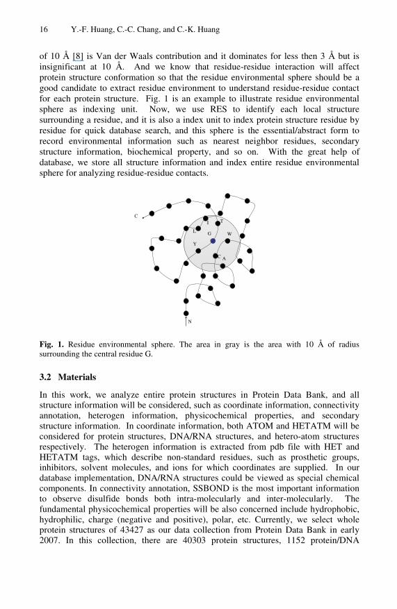
of 10 Å [8] is Van der Waals contribution and it dominates for less then 3 Å but is insignificant at 10 Å. And we know that residue-residue interaction will affect protein structure conformation so that the residue environmental sphere should be a good candidate to extract residue environment to understand residue-residue contact for each protein structure. Fig. 1 is an example to illustrate residue environmental sphere as indexing unit. Now, we use RES to identify each local structure surrounding a residue, and it is also a index unit to index protein structure residue by residue for quick database search, and this sphere is the essential/abstract form to record environmental information such as nearest neighbor residues, secondary structure information, biochemical property, and so on. With the great help of database, we store all structure information and index entire residue environmental sphere for analyzing residue-residue contacts.
N
C
Y
L
C
T
A
WG
I
Fig. 1. Residue environmental sphere. The area in gray is the area with 10 Å of radius surrounding the central residue G.
3.2 Materials
In this work, we analyze entire protein structures in Protein Data Bank, and all structure information will be considered, such as coordinate information, connectivity annotation, heterogen information, physicochemical properties, and secondary structure information. In coordinate information, both ATOM and HETATM will be considered for protein structures, DNA/RNA structures, and hetero-atom structures respectively. The heterogen information is extracted from pdb file with HET and HETATM tags, which describe non-standard residues, such as prosthetic groups, inhibitors, solvent molecules, and ions for which coordinates are supplied. In our database implementation, DNA/RNA structures could be viewed as special chemical components. In connectivity annotation, SSBOND is the most important information to observe disulfide bonds both intra-molecularly and inter-molecularly. The fundamental physicochemical properties will be also concerned include hydrophobic, hydrophilic, charge (negative and positive), polar, etc. Currently, we select whole protein structures of 43427 as our data collection from Protein Data Bank in early 2007. In this collection, there are 40303 protein structures, 1152 protein/DNA
Massive Protein Structural Property Explorations Using New Indexing Mechanism 17
LEVEL (*=ATOM, CA, SG)
CROSS_CHAIN
CO
SEQ
RADIUS
UNORG_#
UNORG_IDX_SEQ
ORIGIN_IDXFK
UNORIGIN_TYPE
ORIGIN_TYPE
SEQ
PDBID
IDXPK
tbl_SPHERE
SSE
Z
Y
X
ATOM_NAME
RES_NAME
RES_SEQ
CHAIN_ID
PDBID
IDXPK
tbl_ATOM
HETATM#
SEQ(res_id)
CHAIN_ID
HETID
PDBID
IDXPK
tbl_HET
MIN_DIS
AVG_DIS
MAX_DIS
APPEAR_PDB_LIST
APPEAR_PDB_#
SIZE
TYPE_NAME
IDXPK
tbl_Ligang
Z
Y
X
HET_IDXFK
RES_NAME(ligand_type)
ATOM_NAME
SEQ
PDBID
IDXPK
tbl_HETATM
M
1
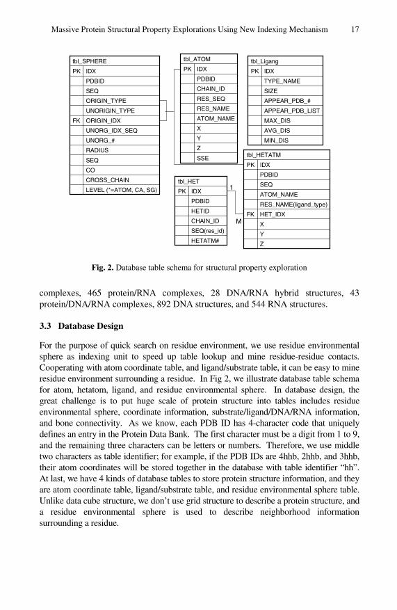
Fig. 2. Database table schema for structural property exploration
complexes, 465 protein/RNA complexes, 28 DNA/RNA hybrid structures, 43 protein/DNA/RNA complexes, 892 DNA structures, and 544 RNA structures.
3.3 Database Design
For the purpose of quick search on residue environment, we use residue environmental sphere as indexing unit to speed up table lookup and mine residue-residue contacts. Cooperating with atom coordinate table, and ligand/substrate table, it can be easy to mine residue environment surrounding a residue. In Fig 2, we illustrate database table schema for atom, hetatom, ligand, and residue environmental sphere. In database design, the great challenge is to put huge scale of protein structure into tables includes residue environmental sphere, coordinate information, substrate/ligand/DNA/RNA information, and bone connectivity. As we know, each PDB ID has 4-character code that uniquely defines an entry in the Protein Data Bank. The first character must be a digit from 1 to 9, and the remaining three characters can be letters or numbers. Therefore, we use middle two characters as table identifier; for example, if the PDB IDs are 4hhb, 2hhb, and 3hhb, their atom coordinates will be stored together in the database with table identifier “hh”. At last, we have 4 kinds of database tables to store protein structure information, and they are atom coordinate table, ligand/substrate table, and residue environmental sphere table. Unlike data cube structure, we don’t use grid structure to describe a protein structure, and a residue environmental sphere is used to describe neighborhood information surrounding a residue.

18 Y.-F. Huang, C.-C. Chang, and C.-K. Huang
4 Statistical Analysis of Structural Properties on Protein Data Bank
4.1 Residue-Residue Contacts
In protein structure, residue-residue interactions make a protein to fold as a stable conformation. If two residues are considered to be in contact with each other provided the distance between their alpha carbon atom (Cα) below a certain cutoff. Therefore, we collect residue-residue contacts from whole protein structures and extract all residue pairs and its neighbor residues to understand how interactions help protein folding. Moreover, each residue can have multiple properties on it such as biochemical property (hydrophobic, hydrophilic, charge, etc), physicochemical property, and secondary structure element type (α-helix, β-sheet, or coil). Inside the residue environmental sphere, we first use Cα in backbone to represent geometry information, but in order to describe detail residue contact with chemical bond, therefore, atom level residue-residue contacts will be also considered.
4.2 Chemical Component Contacts
In this sub-section, we try to observe residue environment surrounding a chemical component to understand the interaction environment between protein and ligand or substrate. We also use residue environmental sphere to observe chemical component close to a residue contacts. According to PDB format, HET records are used to describe chemical components or non-standard residues, such as prosthetic groups, inhibitors, solvent molecules, and ions for which coordinates are supplied. Groups are considered HET if they are not part of a biological polymer described in SEQRES and considered to be a molecule bound to the polymer, or they are a chemical species that constitutes part of a biological polymer that is not one of the following: (a) not one of the standard amino acids, and (b) not one of the nucleic acids (C, G, A, T, U, and I), and (c) not an unknown amino acid or nucleic acid where UNK is used to indicate the unknown residue name. Because we focus on residue-residue contacts to realize how they interacts with chemical component, and chemical component information is used to understand how interaction begins.
4.3 Property Analysis on Disulfide Bond
4.3.1 Disulfide Bond In general, disulfide bonds are suggested to stabilize protein folding which has been reviewed [1, 5, 16, 20]. In biochemistry, disulfide bond or disulfide bridge is connected between Cβ-Sγ-Sγ-Cβ (Sγ is a SG atom in PDB, and Cβ is a beta carbon) which can occur intra-molecularly (i.e within a single polypeptide chain) and inter-molecularly (i.e. between two polypeptide chains). Disulfide bond in intra-molecular stabilize the tertiary structures of proteins while those that occur inter-molecularly are involved in stabilizing quaternary structure. In this paper, we focus on SSBOND section which identifies each disulfide bond in protein and polypeptide structures by identifying the two residues involved in the bond. Furthermore, we also use residue environmental sphere to detect residue-residue contacts of cysteine pairs intra-molecularly.

Massive Protein Structural Property Explorations Using New Indexing Mechanism 19
4.3.2 SSBOND In PDB, the connectivity annotation section is used to allow the depositors to specify the existence and location of disulfide bonds and other linkages. The bond between two Sγ atoms is disulfide bond annotated as SSBOND by Protein Data Bank. We separate this collection into two groups, intra-molecular and inter-molecular; therefore, we have 48152 pairs in intra-molecular group and 2115 pairs in inter-molecular group. While applying secondary structure information, we observe that SSBOND tends to grasp at β-sheets and coils.
4.3.3 Residue-Residue Contacts of Cysteine Pairs Unlike SSBOND discovery, not all protein structures contain disulfide bonds; therefore, we observe all cysteine pairs in whole PDB to distinguish the difference between SSBOND and residue-residue contacts of cysteine pair. In this work, we only collect all cysteine pairs in both Cα and atom level (Sγ) intra-molecularly to observe their environment. The reason to use atom level discovery is that we will miss some cysteine pairs if we only count Cα atom level. Therefore, we have 114,777 residue-residue contacts intra-molecularly for further analysis.
4.4 Results
Although we detect all possible residue-residue contacts among whole protein structures in PDB; according to previous studies, we select SSBOND annotation in PDB and residue-residue contacts of cysteine pair as example to explore protein structural property because of well-studied topic on disulfide bond.
4.4.1 Residue-Residue Contacts and Chemical Component Contacts We detect all pairs of amino acid combination to discuss relationship among residue interaction and secondary structure property. In our experimental result, the top-10 residue-residue contacts contain Glycine, and the pairs are Gly-Gly, Gly-Ala, Gly-Ser, Gly-Pro, Gly-Asp, Gly-Glu, Gly-Lys, Gly-Leu, Gly-Thr, and Gly-Val ranked by their occurrence frequency. According to amino acid property, the amino acid glycine tends to contact with small or tiny amino acids such as Ala, Ser, Asp, Thr, and Pro. Focusing on cysteine paris, we observe that Cys-Cys occurs in β-sheet and loop frequently. Moreover, the chemical component is defined as hetID in PDB; thus we totally extract about 6827 different hetIDs from PDB. The top-5 hetIDs are SO4, _CA, _ZN, _MG, and MSE.
Table 1. Statistical result of SSBOND and Cysteine pair
Intra-molecular Inter-molecular Total (A) 48152 2115 50267
SSBOND (B) 3333 95 3429 (A) 114777 - 114777
Cysteine Pairs (B) 12847 - 12847
(A) Number of pairs; (B) Chemical component contacts.
20 Y.-F. Huang, C.-C. Chang, and C.-K. Huang
Residue contact of SSBOND and Cysteine pairs
0
0.1
0.2
0.3
0.4
0.5
0.6
0.7
0.8
0.9
1
0-1 1-2 2-3 3-4 4-5 5-6 6-7 7-8 8-9 9-10
Distance between two SG atoms
Freq
uenc
y (%
)
Cysteine pairsSSBOND (Intra-molecular)SSBOND (Inter-molecular)SSBOND (All)
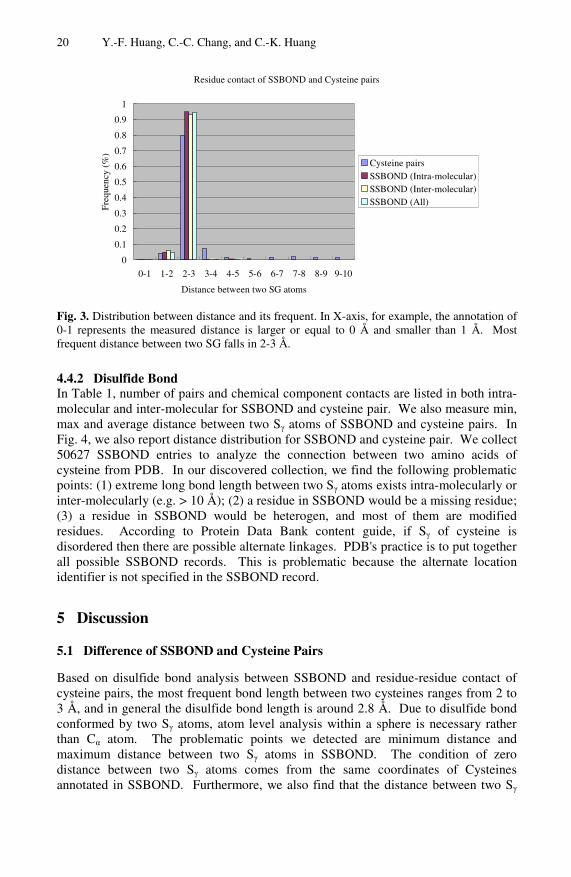
Fig. 3. Distribution between distance and its frequent. In X-axis, for example, the annotation of 0-1 represents the measured distance is larger or equal to 0 Å and smaller than 1 Å. Most frequent distance between two SG falls in 2-3 Å.
4.4.2 Disulfide Bond In Table 1, number of pairs and chemical component contacts are listed in both intra-molecular and inter-molecular for SSBOND and cysteine pair. We also measure min, max and average distance between two Sγ atoms of SSBOND and cysteine pairs. In Fig. 4, we also report distance distribution for SSBOND and cysteine pair. We collect 50627 SSBOND entries to analyze the connection between two amino acids of cysteine from PDB. In our discovered collection, we find the following problematic points: (1) extreme long bond length between two Sγ atoms exists intra-molecularly or inter-molecularly (e.g. > 10 Å); (2) a residue in SSBOND would be a missing residue; (3) a residue in SSBOND would be heterogen, and most of them are modified residues. According to Protein Data Bank content guide, if Sγ of cysteine is disordered then there are possible alternate linkages. PDB's practice is to put together all possible SSBOND records. This is problematic because the alternate location identifier is not specified in the SSBOND record.
5 Discussion
5.1 Difference of SSBOND and Cysteine Pairs
Based on disulfide bond analysis between SSBOND and residue-residue contact of cysteine pairs, the most frequent bond length between two cysteines ranges from 2 to 3 Å, and in general the disulfide bond length is around 2.8 Å. Due to disulfide bond conformed by two Sγ atoms, atom level analysis within a sphere is necessary rather than Cα atom. The problematic points we detected are minimum distance and maximum distance between two Sγ atoms in SSBOND. The condition of zero distance between two Sγ atoms comes from the same coordinates of Cysteines annotated in SSBOND. Furthermore, we also find that the distance between two Sγ

Massive Protein Structural Property Explorations Using New Indexing Mechanism 21
atoms larger than 10 Å (e.g. 149.663 Å in intra-molecular of 1RHG:C 64 and 74, and 77.881 Å in inter-molecular of 1uMR:B 135 and 1UMR:D 203), and it’s might be incorrect annotation of bond connectivity. In addition, because the size of chemical component will affect the result, we only select large-size chemical component for structure similarity evaluation. In whole residue environmental spheres containing SSBOND, we have 16 residue environmental spheres containing BEN and 66 residue environmental spheres containing FAD. We find that residue environmental spheres of SSBOND surrounded by two chemical components of FAD and BEN respectively have highly conserved region of spheres. In Fig. 4, atoms in yellow are Sγ atoms and the chemical component in CPK mode is FAD. Unlike previous researches, we try to index whole PDB dataset to analysis all residue-residue contacts and bond connectivity inside a protein structure while previous researches focus on only analyze special pair preference and residue frequencies [10].
5.2 File Parsing and Efficiency of Database Query
To extract atom coordinate information, we have to parse pdb file to obtain structure information, but file parsing is the worst way for data mining because of information reusable. Besides, the use of sphere can gain the advantage of neighborhood information. Thus, for the purpose of structure mining on PDB, we try to simplify our mining procedure, and then we parse PDB raw files, index whole protein structures with residue environmental sphere and deposit all information into database. To avoid database connection I/O, we use database dump technique to prepare dump file for database restore instead of row-by-row insertion. Comparing with file parsing and database query, without consideration of preprocessing, we spend about 1 hour to select sphere information from database for detect residue-residue contact of cysteine pairs via database query while spending 17 hours via file parsing without database utilization. Therefore, we can gain more benefit from indexing mechanism and database query.
Fig. 4. Atoms in yellow are Sγ atoms that build the disulfide bond annotated as SSBOND in PDB ID 1BHY and the chemical component is FAD (FLAVIN-ADENINE DINUCLEOTIDE) in CPK mode
22 Y.-F. Huang, C.-C. Chang, and C.-K. Huang
6 Conclusions
In summary, sphere-based neighborhood searching is an appropriate local structure representation for structure mining on PDB. Consequently, we obtain huge scale collection of residue environmental sphere for describing protein local environment based on the believed assumption of protein function interacted with local structure. In order to searching and mining among this collection, indexing mechanism is very important; therefore, the residue environmental sphere is local structure representation and indexing unit for the reason of information reuse. Focusing on disulfide bond, the observation can be put on both SSBOND and cysteine pairs. Although there is some problematic information in SSBOND, they still provide useful information to compare with SSBOND and cysteine pair. In the future, further analysis on different residue-residue contacts and discussion on structural property should be scrutinized.
Reference
1. Aitken, A., Learmonth, M.: Quantification and location of disulfide bonds in proteins. Methods in molecular biology 64, 317–328 (1997)
2. Alton, D., Adab, P., Roberts, L., Barrett, T.: Relationship between walking levels and perceptions of the local neighbourhood environment. Archives of disease in childhood 92, 29–33 (2007)
3. Bagley, S.C., Altman, R.B.: Characterizing the microenvironment surrounding protein sites. Protein Sci. 4, 622–635 (1995)
4. Berman, H.M., Battistuz, T., Bhat, T.N., Bluhm, W.F., Bourne, P.E., Burkhardt, K., Feng, Z., Gilliland, G.L., Iype, L., Jain, S., Fagan, P., Marvin, J., Padilla, D., Ravichandran, V., Schneider, B., Thanki, N., Weissig, H., Westbrook, J.D., Zardecki, C.: The Protein Data Bank. Acta crystallographica 58, 899–907 (2002)
5. Betz, S.F.: Disulfide bonds and the stability of globular proteins. Protein Sci. 2, 1551–1558 (1993)
6. Chalk, A.J., Worth, C.L., Overington, J.P., Chan, A.W.: PDBLIG: classification of small molecular protein binding in the Protein Data Bank. Journal of medicinal chemistry 47, 3807–3816 (2004)
7. Cheng, J., Baldi, P.: Improved residue contact prediction using support vector machines and a large feature set. BMC bioinformatics 8, 113 (2007)
8. Crowley, M., Darden, T., Cheatham, T., Deerfield, D.: Adventures in Improving the Scaling and Accuracy of a Parallel Molecular Dynamics Program. The Journal of Supercomputing 11, 255–278 (1997)
9. Fan, S.C., Zhang, X.G.: Characterizing the microenvironment surrounding phosphorylated protein sites. Genomics, proteomics & bioinformatics / Beijing Genomics Institute 3, 213–217 (2005)
10. Glaser, F., Steinberg, D.M., Vakser, I.A., Ben-Tal, N.: Residue frequencies and pairing preferences at protein-protein interfaces. Proteins 43, 89–102 (2001)
11. Jonassen, I., Eidhammer, I., Conklin, D., Taylor, W.R.: Structure motif discovery and mining the PDB. Bioinformatics 18, 362–367 (2002)
12. Lutteke, T., Frank, M., von der Lieth, C.W.: Data mining the protein data bank: automatic detection and assignment of carbohydrate structures. Carbohydrate research 339, 1015–1020 (2004)
Massive Protein Structural Property Explorations Using New Indexing Mechanism 23
13. Oldfield, T.J.: Creating structure features by data mining the PDB to use as molecular-replacement models. Acta crystallographica 57, 1421–1427 (2001)
14. Oldfield, T.J.: Data mining the protein data bank: residue interactions. Proteins 49, 510–528 (2002)
15. Plochocka, D., Kosinski, J., Rabczenko, A.: Formation of the local secondary structure of proteins: local sequence or environment. Acta biochimica Polonica 33, 109–118 (1986)
16. Raina, S., Missiakas, D.: Making and breaking disulfide bonds. Annual review of microbiology 51, 179–202 (1997)
17. Rodionov, M.A., Johnson, M.S.: Residue-residue contact substitution probabilities derived from aligned three-dimensional structures and the identification of common folds. Protein Sci. 3, 2366–2377 (1994)
18. Shin, J.M., Cho, D.H.: PDB-Ligand: a ligand database based on PDB for the automated and customized classification of ligand-binding structures. Nucleic acids research 33, D238–241 (2005)
19. Singh, J., Thornton, J.M.: Atlas of Protein Side-Chain Interactions, vol. I, II. IRL press, Oxford (1992)
20. Wedemeyer, W.J., Welker, E., Narayan, M., Scheraga, H.A.: Disulfide bonds and protein folding. Biochemistry 39, 4207–4216 (2000)
21. Zhang, C., Kim, S.H.: Environment-dependent residue contact energies for proteins. Proceedings of the National Academy of Sciences of the United States of America 97, 2550–2555 (2000)