Integrin binding angiopoietin-1 monomers reduce cardiac hypertrophy
Upload
manchester-usCategory
view
3download
0
The Rockefeller University Press, 0021-9525/2001/12/1319/14 $5.00The Journal of Cell Biology, Volume 155, Number 7, December 24, 2001 1319–1332http://www.jcb.org/cgi/doi/10.1083/jcb.200107107
JCB
Article
1319
Marching at the front and dragging behind:
differential
�
V
�
3-integrin turnover regulates focal adhesion behavior
Christoph Ballestrem, Boris Hinz, Beat A. Imhof, and Bernhard Wehrle-Haller
Department of Pathology, Centre Médical Universitaire, Geneva, Switzerland
ntegrins are cell–substrate adhesion molecules thatprovide the essential link between the actin cytoskeletonand the extracellular matrix during cell migration. We
have analyzed
�
V
�
3-integrin dynamics in migrating cellsusing a green fluorescent protein–tagged
�
3-integrin chain.At the cell front, adhesion sites containing
�
V
�
3-integrinremain stationary, whereas at the rear of the cell they slideinward. The integrin fluorescence intensity within thesedifferent focal adhesions, and hence the relative integrindensity, is directly related to their mobility. Integrin densityis as much as threefold higher in sliding compared withstationary focal adhesions. High intracellular tension underthe control of RhoA induced the formation of high-density
I
contacts. Low-density adhesion sites were induced by Rac1and low intracellular tension. Photobleaching experiments
demonstrated a slow turnover of
�
3-integrins in low-densitycontacts, which may account for their stationary nature. Incontrast, the fast
�
3-integrin turnover observed in high-density contacts suggests that their apparent sliding may becaused by a polarized renewal of focal contacts. Therefore,differential acto-myosin–dependent integrin turnover andfocal adhesion densities may explain the mechanical andbehavioral differences between cell adhesion sites formedat the front, and those that move in the retracting rear ofmigrating cells.
Introduction
Modulation of cell–substrate adhesion plays a crucial role incellular processes such as migration, spreading, or contraction.These morphological changes result from the coordinatedreorganization of the actin cytoskeleton induced by intra- orextracellular stimuli (Lauffenburger and Horwitz, 1996).Cell migration is sustained by the continuous growth ofactin filaments at the leading edge, and the controlled retractionof adhesive contacts at the rear of the cell (Palecek et al.,1998; Horwitz and Parsons, 1999; Ballestrem et al., 2000).Integrin
��
heterodimers provide the physical link betweenthe continuously reorganizing actin cytoskeleton andcomponents of the extracellular matrix (ECM)* during cell mi-
gration (Hynes, 1992). Different types of integrin-containingcell–substrate contacts have been described, of which focalcomplexes and contacts are the best studied. These two typesof contacts have been distinguished according to several fea-tures including size, the site where they are formed in thecell, their age, their appearance in interference reflection mi-croscopy, and their regulation by small GTPases (Geiger andBershadsky, 2001). In fibroblasts, small point-like focalcomplexes form at sites of Rac1-dependent lamellipodiainduction (Ridley et al., 1992; Nobes and Hall, 1995; Rottneret al., 1999), whereas large and elongated focal contactslocalize to the ends of actin stress fibers upon RhoA activation(Ridley and Hall, 1992; Nobes and Hall, 1995; Amano etal., 1997; Rottner et al., 1999). The mechanical influence ofacto-myosin–induced intracellular contractility and extracel-lular tension was suggested as a major factor converting focalcomplexes into focal contacts (Chrzanowska-Wodnicka andBurridge, 1996; Pelham and Wang, 1997; Riveline et al.,2001). In this study, we will use the general term focaladhesion, and will classify them according to their differentbehavior and localization in migrating cells as well as theirintegrin dynamics.
Although the pathways leading to the changes in the actincytoskeleton are well understood, it is not known how the
The online version of this article contains supplemental material.
Address correspondence to Bernhard Wehrle-Haller, Dept. of Pathol-ogy, Centre Médical Universitaire, 1, Rue Michel-Servet, 1211Geneva 4, Switzerland. Tel.: 0041-22-702-57-35. Fax: 0041-22-702-57-46. E-mail: [email protected] C. Ballestrem’s current address is Dept. of Molecular Cell Biology, TheWeizmann Institute of Science, Rehovot 76100 Israel.*Abbreviations used in this paper: EGFP, enhanced GFP; GFP, greenfluorescent protein; LPA, lysophosphatidic acid, MF, mobile fraction,ECM, extracellular matrix Key words: cell migration; cell adhesion; green fluorescent protein; RhoGTPases; integrin density
on June 22, 2015jcb.rupress.org
Dow
nloaded from
Published December 24, 2001
http://jcb.rupress.org/content/suppl/2001/12/20/jcb.200107107.DC1.html Supplemental Material can be found at:

1320 The Journal of Cell Biology
|
Volume 155, Number 7, 2001
strength of the integrin-mediated link between the actin cy-toskeleton and the ECM is controlled to promote eitherfirm adhesion or detachment. Nonaggregated integrins ex-
hibit a high lateral diffusion within the plasma membrane(Duband et al., 1988). However, upon extracellular ligandbinding, integrins become anchored to the actin cytoskele-
Figure 1. The GFP-tagged �3-integrin chain forms functional heterodimers with endogenous �V. (A) Scheme of the �V�3–GFP-integrin heterodimer. The GFP protein is tagged COOH-terminally to the cytoplasmic domain of the �3 subunit. (B) Immunoprecipitations of cell extracts from surface biotinylated B16 �3–GFP cells. Extracts were precipitated with the indicated antibodies (c, control) and separated under reducing conditions by PAGE followed by transfer onto nitrocellulose membranes. Revelation with either streptavidin coupled horseradish peroxidase (SA-HPO, top) or anti-GFP antibodies (GFP, bottom) demonstrated the typical double-band pattern for integrin heterodimers and the coprecipitated GFP-tagged �3-integrin subunit, respectively. The position of the molecular mass markers is indicated to the left of the blots. (C) Substrate-specific clustering of the �V�3–GFP-integrin into adhesions sites. B16 �3–GFP cells were plated overnight on glass coverslips, previously coated with 5 �g ml�1 laminin-1 (LN), 5 �g ml�1 fibronectin (FN), or 1 �g ml�1 vitronectin (VN). Cells were subsequently fixed and substrate adhesion sites were revealed by immunohistochemical detection of vinculin. Note that �3–GFP-integrin–positive adhesion sites were only found on fibronectin and vitronectin, which are ligands for �V�3-integrin. In contrast, �3–GFP-integrin did not cluster on laminin-1, for which it is not a ligand. Because B16 cells use a different type of integrin receptor (�6�1) to adhere to LN than to FN or VN (�5�1, �V�3), their morphology and migration behavior is different between these substrates (Ballestrem et al., 1998). (D) FACS analysis of nontransfected, �3-, and �3–GFP-transfected CHO cells with a Kistrin–CD31 fusion construct (SKI-7) (Legler et al., 2001). Note that the �3–GFP-transfected CHO clone is not homogeneous, exhibiting cells that lost �3-GFP expression, which reduces their SKI-7 reactivity to endogenous �V�3-integrin levels (gate 1) (ctr; SKI-7, unpublished data). Bar, 20 �m.
on June 22, 2015jcb.rupress.org
Dow
nloaded from
Published December 24, 2001

Dynamics of
�
3-integrin clustering |
Ballestrem at al. 1321
ton by a large set of structural and regulatory proteins(Miyamoto et al., 1995), thereby forming cell–ECM adhe-sion sites. A “sliding” of
�
1-integrin–containing focal con-tacts has recently been demonstrated, and was suggested torepresent weak attachment of stationary cells (Smilenov etal., 1999). In addition, the movement of
�
5
�
1-integrins onthe ventral side of fibroblasts has been related to ECM reor-ganization by fibrillar adhesions (Katz et al., 2000; Pankovet al., 2000; Zamir et al., 2000).
To analyze the dynamics of individual integrin het-erodimers within adhesion sites of migrating cells, we fo-cused on the integrin
�
V
�
3. Integrin
�
V
�
3 is expressed onvarious motile cells such as neural crest cells (Delannet et al.,1994) and plays an important role in tumor metastasis (Al-belda et al., 1990; Felding-Habermann et al., 2001), angio-genesis (Brooks et al., 1994), leukocyte transmigration(Weerasinghe et al., 1998), and osteoclast function (Mc-Hugh et al., 2000). Its ECM ligands include fibronectin,vitronectin, and fibrinogen (Cheresh and Spiro, 1987).
The use of a directly green fluorescent protein (GFP)-labeled
�
3-integrin chain that was coexpressed with the en-dogenous
�
V-integrin subunit on the cell surface allowed usto follow clustering and dispersal, and to perform quantita-tive analysis of
�
V
�
3-integrins within adhesion sites of liv-ing cells. We specifically asked whether the organization of
�
3-integrins in focal adhesions differed according to theirsubcellular localization, and whether distinct organizationpatterns could be attributed to the activities of members ofthe Rho family of small GTPases. In particular, we studiedthe influence of intracellular tension, analyzed with the helpof elastic silicon substrata, on the organization of
�
3-inte-grins within focal adhesion sites. Moreover, using FRAP, weanalyzed the turnover rates of
�
3-integrins within differentfocal adhesions, in order to understand whether the motilebehavior and function of a given focal adhesion site could becorrelated to the temporal stability of the embedded inte-grins. We found differential densities of integrins within fo-cal adhesion sites that correlated with the degree of acto-myosin–dependent intracellular contraction, and an inversecorrelation to the temporal stability of integrins within thesesites. Our data reveal casual connections between the behav-ior of integrins and the state of the actin cytoskeleton thatprovides the base for a detailed mechanical model of cell mi-gration.
Results
Dimerization of
�
3–GFP-integrin chain with
�
V
To study and quantify
�
V
�
3-integrin dynamics in livingcells, we generated a fusion protein of the
�
3 integrin sub-unit with GFP (Fig. 1 A). To determine whether this
�
3–GFP-integrin chain formed heterodimers with the en-dogenous
�
V subunit, we surface biotinylated stable
�
3–GFP-integrin–transfected cells (B16 F1 melanoma and 3T3fibroblasts), and performed immunoprecipitations with an-tibodies against either the
�
V- or the
�
3-integrin subunits(Fig. 1 B). After precipitation of the integrin and subsequentWestern blotting, both
�
- and
�
-integrin subunits could bedetected with avidin-peroxidase (Fig. 1 B). Bands for the
�
3–GFP-integrin fusion protein were only detected in pre-
cipitations with anti–
�
V- or –
�
3-integrin subunits, but notwith control rat serum nor with anti–
�
6-integrin subunitwhich forms heterodimers with the
�
1 and
�
4 chains (Fig. 1B, bottom). These experiments clearly demonstrated that
�
3–GFP-integrin was expressed on the cell surface as a het-erodimeric complex in association with the endogenous
�
Vchain.
Ligand-specific clustering of
�
V
�
3–GFP-integrin
To further test whether the
�
V
�
3–GFP-integrin hetero-dimer was functional and did not unspecifically associatewith cytoskeletal elements of focal adhesions, we plated
�
3–GFP-transfected cells on the
�
V
�
3 ligands fibronectin andvitronectin, and on laminin-1, which is not a ligand for
�
V
�
3. Clustering of GFP was observed on fibronectin andvitronectin, but not on laminin-1 (Fig. 1 C). Vinculin andpaxillin are present in, and used as markers for, cell–sub-strate adhesion sites. Both localized to focal adhesion sites onall three substrates (Fig. 1 C, paxillin, unpublished data). Incontrast,
�
V
�
3-integrin–containing focal adhesions wereonly detected in cells cultured on fibronectin or vitronectinsubstrata (Fig. 1 C). To demonstrate that the transfected
�
3–GFP-integrin engaged in ECM binding was comparableto wild-type
�
3 chains, we analyzed stable
�
3–GFP- and
�
3-transfected CHO cells for their binding to a
�
V
�
3-inte-grin–specific snake venom disintegrin (Kistrin). A FACSprofile using a Kistrin–CD31 fusion protein (SKI-7) re-vealed only low levels of endogenous
�
V
�
3-integrin in non-transfected CHO cells (Legler et al., 2001). In contrast, both
�
3- and
�
3–GFP-transfected cells displayed extensive SKI-7reactivity (Fig. 1 D). These results demonstrate that
�
V
�
3–GFP-integrin behaves like endogenous
�
V
�
3, indicatingthat ligand binding, integrin signaling, and substrate speci-ficity are not perturbed by the fusion of GFP to the
�3-inte-grin chain. Moreover, these data suggest that the associated�V-integrin integrin chain specifically protects the cytoplas-mic domain of �3–GFP from matrix-independent engage-ment with cytoskeletal elements of focal adhesions (Yauch etal., 1997). Therefore, direct labeling of the �3-integrin withGFP allowed us to follow and quantify �3-containing inte-grins in living cells.
Dynamics of �3-integrinThis �V�3–GFP tool permitted now the direct observationof integrin clustering and turnover in adhesion sites ofmigrating or stationary cells. Therefore, we performed time-lapse experiments with stably �3–GFP-transfected, fast-migrating B16 F1 melanoma cells, or stationary 3T3 fibro-blasts (B16 �3–GFP or 3T3 �3–GFP, respectively). In B16�3–GFP cells, we observed the formation of small integrinclusters just behind the leading edge of the advancing lamel-lipodia (Fig. 2, A and B). These clusters remained stationarywith respect to the substratum, whereas the cell moved for-ward. When GFP-containing focal adhesions reached a dis-tance of 10 �m from the leading edge, they began to shrinkand finally disappeared (Fig. 2, A, circled, and B, boxed;Video1, available at http://www.jcb.org/cgi/content/full/jcb.200107107/DC1). We noted that some of the focaladhesions in the smoothly protruding lamellipodia assumedan elongated shape. Although we never observed actin stress
on June 22, 2015jcb.rupress.org
Dow
nloaded from
Published December 24, 2001

1322 The Journal of Cell Biology | Volume 155, Number 7, 2001
fibers in actively protruding lamellipodia, the presence of ra-dially oriented actin ribs within the lamellipodia was fre-quent (Ballestrem et al., 1998). Continuous appearance anddisappearance of stationary �3-integrin focal adhesions oc-curred within a restricted area in the advancing lamella. Werefer to this area as the zone of transient integrin clustering.In posterior regions of the cell, integrin-containing focaladhesions moved in relation to the substratum during re-traction (Fig. 2 A, arrow; Video2, available at http://www.jcb.org/cgi/content/full/jcb.200107107/DC1). To ex-amine the possibility that integrin contacts in highly migra-tory melanoma cells might behave differently from station-ary or slow moving fibroblasts (3T3 �3–GFP cells), wecompared the appearance of GFP fluorescence in transfectedB16 and 3T3 cells. 3T3 cells displayed continuous cycles oflamellipodia formation followed by retraction, and theyshowed comparable integrin cluster dynamics to what hadbeen seen in B16 �3–GFP cells (Fig. 3; Video3, available athttp://www.jcb.org/cgi/content/full/jcb.200107107/DC1).However, during collapse of lamellipodia, small focal adhe-sions in 3T3 cells transformed into larger, fluorescentlybrighter focal adhesions (Fig. 3 B). During retraction, thesefocal adhesions began to move in relation to the substratum(Fig. 3, B and C). In conclusion, our data show that �V�3-integrins aggregate into stationary focal adhesions within the
zone of transient integrin clustering during the protrusion oflamellipodia. After the collapse of lamellipodia and subse-quent retraction, small stationary focal adhesions transforminto inwards sliding larger focal adhesions.
Induction of differential �3-integrin cluster densities upon transfection with dominant Rac1, Cdc42, and RhoALamellipodia formation, as well as the retraction of celledges, depends on the reorganization of the actin cytoskele-ton. In addition, the transition from small and stationary tolarger, retracting focal adhesions was associated with an in-crease in fluorescence intensity, and hence increased integrindensity (Fig. 3). Because signaling through members of theRho family of small GTPases is known to cause changes inthe actin cytoskeleton (Ridley and Hall, 1992; Ridley et al.,1992; Nobes and Hall, 1995), we asked if changes in activa-tion of these GTPases would influence the organization anddensity of �V�3-integrin in focal adhesion sites. To answerthis question, we transfected dominant active forms of Rac1,Cdc42, and RhoA into B16 �3–GFP cells, and quantifiedintegrin fluorescence and focal adhesion morphology (Fig.4). Control cells typically displayed a leading lamella withsmall �3-integrin– positive focal adhesions and larger, fluo-rescently brighter �3-integrin focal adhesions at the side and
Figure 2. Dynamics of �3–GFP-integrin in stable transfected B16 F1 cells. Time-lapse analysis of a B16 �3–GFP cell plated overnight on vitronectin (1 �g ml�1) revealed the transient �3–GFP-integrin clustering and subsequent dispersal in the advancing lamellipodium. A typical �3-integrin cluster (A, circled) appeared close to the leading edge (8�) and remained stationary (12�) until it began to gradually disappear (16�–24�). In retracting parts of the cells, integrin clusters began to slide inward (arrow). To appreciate the relative movement of the different integrin clusters during this time-lapse, an overlay revealed the stationary nature of focal adhesions in the lamellipodia (arrowhead) and the streak-like pattern of sliding focal adhesions in retracting parts of the cell (arrow). In B, a higher temporal and spatial resolution of the boxed area in A (12�) revealed the polymorphic appearance of the stationary integrin clusters (small box as reverence). Although shapes were variable, the fate of the clusters were identical. Arrowheads in B mark the smoothly advancing leading edge of the lamellipodium. Bar, 18 �m.
on June 22, 2015jcb.rupress.org
Dow
nloaded from
Published December 24, 2001

Dynamics of �3-integrin clustering | Ballestrem at al. 1323
rear of the cell (Fig. 4 A). Expression of dominant activeCdc42 (V12) and Rac1 (L61) that are known to inducefilopodia and lamellipodia, respectively (Ridley et al., 1992;Nobes and Hall, 1995), led to the appearance of flat andwell-spread B16 �3–GFP cells. Compared with control cellsthe surface area increased by 188 and 248%, respectively(see Materials and methods), and cells exhibited actin-richfilopodia and lamellipodia and many small caliber actin fila-ments (unpublished data). This phenotype is apparent onlyafter prolonged exposure to dominant active Rac1, and is as-sociated with the formation of many small caliber actin fila-ments as previously reported (Ridley et al., 1992). In thesecells, �3–GFP fluorescence resulted in a streak-like patternof integrin clusters associated with filopodia (cdc42) orlamellipodia (Rac1) covering large areas of the substratum.These extensive clusters exhibited a granular pattern that re-sembled assemblies of numerous small focal adhesions (Fig.4, B and C). The fluorescence intensity profiles indicated inFig. 4, A–C, revealed that the density of �3-integrin in smallfocal adhesions in control cells (Fig. 4 E, profile a) corre-sponded to the densities measured across the integrin clus-
ters of dominant Cdc42- and Rac1-transfected cells (Fig. 4E, profile da-Cdc42 and da-Rac1). In contrast, measure-ments of GFP intensity (and hence, integrin densities) in fo-cal adhesions that were localized in retracting cell edges atthe rear of control cells were consistently higher (Fig. 4 E,profile b). Moreover, expression of dominant active RhoA(V14) induced robust stress fiber formation and the cells ap-peared contracted (64% of control cell surface area) withlarge, even brighter fluorescent �3-integrin focal adhesions(Fig. 4, D and E, profile da-RhoA). From these data we cal-culated (see Materials and methods) that the relative �V�3-integrin densities compared with nonclustered integrin inthe plasma membrane increased by three- to fivefold inlamellipodial and Rac1- or Cdc42-induced low-density focaladhesions, by five- to eightfold in lateral and rear high-den-sity focal adhesions of control cells, and by 9–14-fold in fo-cal adhesions of dominant RhoA-stimulated cells (Fig. 4 F).Similar to the raise in integrin densities, we also observed aRhoA-dependent increase in anti-vinculin labeling of focaladhesions (unpublished data). These results demonstratethat the Rac1- and RhoA-induced changes in the bundling
Figure 3. Dynamics of �3–GFP-integrin in stable transfected 3T3 cells. Time-lapse analysis of a 3T3 �3–GFP cell plated overnight on vitronectin (1 �g ml�1), exhibiting cycles of lamellipodia extension (B) followed by cell edge retraction (B and C). During lamellipodia extension, transient stationary small focal adhesions formed behind the leading edge (B, circled focal adhesion). During cessation of the extension phase, small peripheral focal adhesions grew in size and were transformed into inward sliding focal adhesions (B, boxed area). Note that the start of the time lapse in B corresponds to 54� in A. Continuous inward sliding of large focal adhesions occurred in parallel with cell edge retraction (C). A fiduciary mark on the substrate (C, white crosses) can be used to gauge the speed and position of retracting focal adhesions. Bar, 24 �m.
on June 22, 2015jcb.rupress.org
Dow
nloaded from
Published December 24, 2001

1324 The Journal of Cell Biology | Volume 155, Number 7, 2001
state of the actin filament network led to changes in the clus-tering behavior of �3 integrin, i.e., the generation of integrinlow- and high-density focal adhesions, respectively.
Intracellular tension controls integrin density transition within focal adhesionsWhat causes this RhoA-induced increase in integrin density?
Figure 4. Members of the Rho family of small GTPases regulate �3-integrin clustering differentially. B16 �3–GFP cells transfected with myc-epitope–tagged dominant active forms of Cdc42, Rac1, and RhoA were plated overnight on vitronectin (1 �g ml�1)-coated glass cover-slips. Cells were fixed and stained for the expression of the myc-epitope (inserts), and GFP fluorescence images were recorded with identical camera settings in order to appreciate qualitative as well as quantitative differences in the integrin localization pattern. (A) Nontransfected control cells displayed the typical pattern of small low-fluorescent focal adhesions in the lamellipodium (profile a) and larger high-fluorescent focal adhesions at lateral borders and rear of the cell (profile b). (B) Dominant active Cdc42 (da-Cdc42) induced the formation of long, streak-like arrays of low-fluorescent �3 integrin focal adhesions mainly localized in the lamella or periphery of the cell. Similarly, dominant active Rac1 (da-Rac1) induced extensive �3-integrin clustering into low-fluorescent adhesion sites at the periphery of the cell (C). In contrast, dominant active RhoA (da-RhoA) induced a retracted cellular morphology with intensively fluorescent �3-integrin focal adhesions at the cell periphery (D). Fluorescence intensity profiles of the indicated traces in A–D are shown in E. Note that the intensity profiles are similar between focal adhesions in the lamellipodium of control cells and cells transfected with dominant active Cdc42 and Rac1. Peak fluorescent intensities of lateral and rear focal adhesions in control cells are consistently higher compared with lamellipodial focal adhesions, but can increase even more after dominant active RhoA induction. A quantification of the �3-integrin density (fluorescence intensity increase over membrane) is shown in F. Bar, 15 �m.
on June 22, 2015jcb.rupress.org
Dow
nloaded from
Published December 24, 2001

Dynamics of �3-integrin clustering | Ballestrem at al. 1325
Previously it has been shown that activated RhoA increasesmyosin-dependent contraction of the actin cytoskeleton,leading to intracellular tension (Chrzanowska-Wodnicka andBurridge, 1996; Amano et al., 1997). Integrins in focal adhe-sion sites are anchored within the actin cytoskeleton, suchthat contraction of this actin filament backbone induced bymyosin activity may lead to increased integrin density, i.e.,the transition of lamellipodial to lateral focal adhesions. Totest this, we determined whether intracellular tension in-duced by RhoA was correlated with this transition. Intracel-lular tension was measured by plating 3T3 fibroblasts onflexible silicone rubber that formed wrinkles in response tocellular contraction (Harris et al., 1980). Activation of RhoAby lysophosphatidic acid (LPA) (10 �M) within these cellsincreased the number of wrinkles (Fig. 5 A; Video4, avail-able at http://www.jcb.org/cgi/content/full/jcb.200107107/DC1) (Amano et al., 1997). RhoA activates Rho-kinase,which blocks myosin light chain phosphatase, resulting inmyosin-dependent actin contraction (Kimura et al., 1996).Treatment of cells with the Rho-kinase inhibitor Y-27632(10 �M) removed the wrinkles (Fig. 5 B; Video5, availableat http://www.jcb.org/cgi/content/full/jcb.200107107/DC1)
(Uehata et al., 1997). Similarly, blocking of myosin lightchain kinase with the wide-spectrum protein kinase inhibitorstaurosporine (50 nM) resulted also in the disappearance ofwrinkles (Fig. 5 C; Video6, available at http://www.jcb.org/cgi/content/full/jcb.200107107/DC1). Using these modula-tors of intracellular tension, the formation of high- and low-density focal adhesions was analyzed. Treatment of 3T3 fi-broblasts with LPA increased �3-integrin compaction andformed high-density focal adhesions (Fig. 5 E). In contrast,cells treated with Y-27632 or staurosporine displayed the dis-appearance of high-density focal adhesions, whereas low-density focal adhesions remained in the periphery of the cellsin the lamellipodia (Fig. 5, F and G). To correlate thechanges in intracellular tension with that of the integrin den-sity and focal adhesion size, we displayed the relative peak in-tegrin density (compared with the integrin density in themembrane) and the respective area of focal adhesions (seeMaterials and methods). For each experimental condition,we analyzed �500 focal adhesions from different cells. Con-trol cells displayed a significant number of small-sized, low-density focal adhesions that were mainly associated with pro-truding lamellipodia (Fig. 5 H). In addition, a considerable
Figure 5. Intracellular tension correlates with integrin density in focal adhesion sites. 3T3 �3–GFP cells were grown on flexible silicone rubber substrates in order to visualize intracellular contractile forces by the appearance of substrate wrinkles. (A–C) Cells were recorded for1 h under control conditions to confirm stability of the wrinkles (nt). After addition of drugs, the increase in substrate wrinkles (LPA, 10 �M) or their disappearance (Y 27632, 10 �M, or staurosporine, 50 nM) were recorded for the indicated times (A�–C�) (Videos 4–6). In a parallel experiment, 3T3 �3–GFP cells were grown overnight on serum-coated glass coverslips (D, nt), and changes in �3–GFP-integrin localization in response to the above mentioned drugs was determined after 60 min of treatment (E, LPA; F, Y27632; G, staurosporine). The peak �3-integrin density/focal adhesion area relationship was plotted for untreated (H), LPA- (I), Y 27632- (J), and staurosporine- (K)treated cells. Note the shift in the focal adhesion population after addition of agonist. Bar: (A–C), 80 �m; (D–G), 40 �m.
on June 22, 2015jcb.rupress.org
Dow
nloaded from
Published December 24, 2001
1326 The Journal of Cell Biology | Volume 155, Number 7, 2001
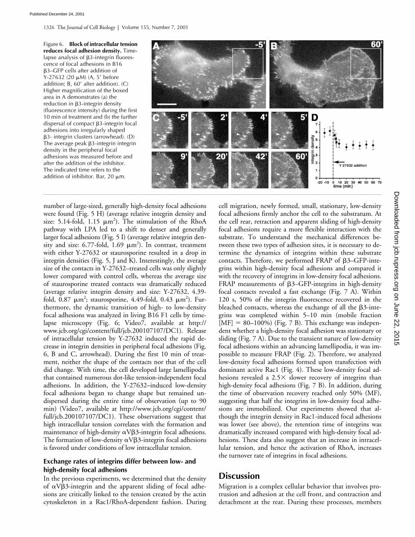
number of large-sized, generally high-density focal adhesionswere found (Fig. 5 H) (average relative integrin density andsize: 5.14-fold, 1.15 �m2). The stimulation of the RhoApathway with LPA led to a shift to denser and generallylarger focal adhesions (Fig. 5 I) (average relative integrin den-sity and size: 6.77-fold, 1.69 �m2). In contrast, treatmentwith either Y-27632 or staurosporine resulted in a drop inintegrin densities (Fig. 5, J and K). Interestingly, the averagesize of the contacts in Y-27632–treated cells was only slightlylower compared with control cells, whereas the average sizeof staurosporine treated contacts was dramatically reduced(average relative integrin density and size: Y-27632, 4.39-fold, 0.87 �m2; staurosporine, 4.49-fold, 0.43 �m2). Fur-thermore, the dynamic transition of high- to low-densityfocal adhesions was analyzed in living B16 F1 cells by time-lapse microscopy (Fig. 6; Video7, available at http://www.jcb.org/cgi/content/full/jcb.200107107/DC1). Releaseof intracellular tension by Y-27632 induced the rapid de-crease in integrin densities in peripheral focal adhesions (Fig.6, B and C, arrowhead). During the first 10 min of treat-ment, neither the shape of the contacts nor that of the celldid change. With time, the cell developed large lamellipodiathat contained numerous dot-like tension-independent focaladhesions. In addition, the Y-27632–induced low-densityfocal adhesions began to change shape but remained un-dispersed during the entire time of observation (up to 90min) (Video7, available at http://www.jcb.org/cgi/content/full/jcb.200107107/DC1). These observations suggest thathigh intracellular tension correlates with the formation andmaintenance of high-density �V�3-integrin focal adhesions.The formation of low-density �V�3-integrin focal adhesionsis favored under conditions of low intracellular tension.
Exchange rates of integrins differ between low- and high-density focal adhesionsIn the previous experiments, we determined that the densityof �V�3-integrin and the apparent sliding of focal adhe-sions are critically linked to the tension created by the actincytoskeleton in a Rac1/RhoA-dependent fashion. During
cell migration, newly formed, small, stationary, low-densityfocal adhesions firmly anchor the cell to the substratum. Atthe cell rear, retraction and apparent sliding of high-densityfocal adhesions require a more flexible interaction with thesubstrate. To understand the mechanical differences be-tween these two types of adhesion sites, it is necessary to de-termine the dynamics of integrins within these substratecontacts. Therefore, we performed FRAP of �3–GFP-inte-grins within high-density focal adhesions and compared itwith the recovery of integrins in low-density focal adhesions.FRAP measurements of �3–GFP-integrins in high-densityfocal contacts revealed a fast exchange (Fig. 7 A). Within120 s, 50% of the integrin fluorescence recovered in thebleached contacts, whereas the exchange of all the �3-inte-grins was completed within 5–10 min (mobile fraction[MF] � 80–100%) (Fig. 7 B). This exchange was indepen-dent whether a high-density focal adhesion was stationary orsliding (Fig. 7 A). Due to the transient nature of low-densityfocal adhesions within an advancing lamellipodia, it was im-possible to measure FRAP (Fig. 2). Therefore, we analyzedlow-density focal adhesions formed upon transfection withdominant active Rac1 (Fig. 4). These low-density focal ad-hesions revealed a 2.5� slower recovery of integrins thanhigh-density focal adhesions (Fig. 7 B). In addition, duringthe time of observation recovery reached only 50% (MF),suggesting that half the integrins in low-density focal adhe-sions are immobilized. Our experiments showed that al-though the integrin density in Rac1-induced focal adhesionswas lower (see above), the retention time of integrins wasdramatically increased compared with high-density focal ad-hesions. These data also suggest that an increase in intracel-lular tension, and hence the activation of RhoA, increasesthe turnover rate of integrins in focal adhesions.
Discussion Migration is a complex cellular behavior that involves pro-trusion and adhesion at the cell front, and contraction anddetachment at the rear. During these processes, members
Figure 6. Block of intracellular tension reduces focal adhesion density. Time-lapse analysis of �3-integrin fluores-cence of focal adhesions in B16 �3–GFP cells after addition of Y-27632 (20 �M) (A, 5� before addition; B, 60� after addition). (C) Higher magnification of the boxed area in A demonstrates (a) the reduction in �3-integrin density (fluorescence intensity) during the first 10 min of treatment and (b) the further dispersal of compact �3-integrin focal adhesions into irregularly shaped �3- integrin clusters (arrowhead). (D) The average peak �3-integrin integrin density in the peripheral focal adhesions was measured before and after the addition of the inhibitor. The indicated time refers to the addition of inhibitor. Bar, 20 �m.
on June 22, 2015jcb.rupress.org
Dow
nloaded from
Published December 24, 2001

Dynamics of �3-integrin clustering | Ballestrem at al. 1327
of the integrin family provide the physical link betweenthe actin cytoskeleton and the extracellular environment(Hynes, 1992). Here, we developed a new tool to fol-low and quantify �V�3-integrins within focal adhesionsformed in living cells by GFP labeling of the �3-integrinsubunit (Plancon et al., 2001) for a similar construct). Wehave chosen the �3-integrin subunit for GFP tagging, as itforms heterodimers uniquely with the V and the platelet-specific IIb � chains. Therefore, GFP–�3-integrin trans-fected into cells (except platelets), will pair exclusively with�V to create a single species of labeled integrins. Most im-portantly, for quantitative studies of integrins, the amountof GFP fluorescence correlates directly with the numberof �V�3 heterodimers, and represents a direct measure ofthe relative integrin density within the two-dimensionalplasma membrane and focal adhesion sites. In addition,the �3–GFP-integrin subunit, like normal �-integrinchains, requires heterodimerization with the �V chain forER export and for an ECM-dependent engagement intofocal adhesions (Heino et al., 1989; Lenter and Vestweber,1994; Yauch et al., 1997). This is in contrast to mono-meric chimeric �-integrin constructs that associate with fo-cal adhesions in an ECM-independent manner, and there-fore are not suited for quantitative measurements ofintegrin behavior (LaFlamme et al., 1994; Smilenov et al.,1999). Our analysis of �V�3-integrin–containing contactsin migrating and stationary cells revealed two differentlybehaving types of cell adhesion sites. At the cell front, sta-tionary focal adhesions formed within a zone of transient
integrin clustering, whereas focal adhesions in retractingcell processes moved in relation to the substratum. The rel-ative abundance of these two types of contacts may deter-mine the migratory behavior of a cell. In the short-livedlamellipodia of slow-migrating fibroblasts, the zone oftransient integrin clustering was difficult to define. In con-trast, extremely fast-moving cells such as fish keratocytesexhibit stationary focal adhesions throughout the entirewidth of the lamellipodium that represent a major part oftheir total cell area (Lee and Jacobson, 1997; Anderson andCross, 2000). Thus, the size of the area occupied by sta-tionary focal adhesions, in respect to the total cell area,may determine the stability and persistence of lamellipo-dial protrusion and hence the overall speed of cell locomo-tion. In these different cell types, the zone of transient inte-grin clustering is equivalent to the area that is occupied byactin filaments originating at the edge of the lamellipo-dium (Svitkina et al., 1997; Ballestrem et al., 1998). It isconceivable that the actin filament turnover within thelamellipodium determines the half-lives of these stationaryfocal adhesions. In contrast to the stationary focal adhe-sions at the front of migrating cells, focal adhesions in re-tracting cell edges move relative to the substratum. Thismobility of focal adhesions, previously described as sliding,has been suggested important for cell edge retraction andmigration (Smilenov et al., 1999; Anderson and Cross,2000; Zamir et al., 2000). Because the efficiency of cell mi-gration is determined by the speed and ability of rear re-traction (Palecek et al., 1998; Ballestrem et al., 2000), the
Figure 7. FRAP reveals different �3-integrin exchange rates in high- versus low-density focal adhesions. (A) Nontransfected or dominant active Rac1 transfected B16 �3–GFP cells were cultured overnight on serum-coated glass coverslips and FRAP was performed on focal adhesions localized to the edge of cells. The bleached area of each series is circled in the first frame and the recovery time (seconds after completion of bleach) indicated to the left. In control cells, immobile (first series) and inward sliding (second series) high-density focal adhesions show almost complete recovery (MF 80%). In cells transfected with dominant active Rac1 (third series) in which low-density focal adhesions were formed, fluorescence recovery was only partial (50% MF), reaching fluorescent levels just slightly above fluorescence intensities of nonclustered �3–GFP-integrin present in the plasma membrane (visible on the right hand side of the frame). Qualitative FRAP curves from several cells (5–8) are displayed in B. Each data point is the median of three to five individual focal adhesions. Bar, 10 �m.
on June 22, 2015jcb.rupress.org
Dow
nloaded from
Published December 24, 2001

1328 The Journal of Cell Biology | Volume 155, Number 7, 2001
degree of focal adhesion sliding may limit the maximalspeed of cell migration.
The quantitative analysis of GFP-labeled �V�3-integrinallowed us to determine the changes in integrin densitieswithin different types of focal adhesions. Hence, we ob-served a low �V�3-integrin density in small, stationary focaladhesions, and a high �V�3-integrin density in large, slid-ing focal adhesions. The formation of small dot-like focaladhesions (named focal complexes) in response to Rac1 andCdc42 activation was first described by Nobes and Hall(1995) in fibroblasts, and they were distinguished fromlarger RhoA induced focal adhesions (named focal contacts)by their size (Nobes and Hall, 1995; Rottner et al., 1999). Itis likely that these focal complexes and focal contacts are ho-mologous to the low-density, respectively high-density focaladhesions observed by us. However, in contrast to focal ad-hesion size, integrin density represents a parameter that en-ables one to distinguish focal complexes from focal contactsin many different cell types, where the size differences areless accentuated as in fibroblasts, and is more specific thanthe recently questioned interference reflection microscopy(Iwanaga et al., 2001). Because activation of RhoA inducesthe bundling of actin filaments into stress fibers (Chrza-nowska-Wodnicka and Burridge, 1996; Machesky and Hall,1997), we suggest that the acto-myosin–dependent contrac-tion of the actin scaffold increases integrin density in focaladhesions. This idea is supported by the observation that thegradual raise in acto-myosin–dependent intracellular tensionstrictly correlates with the amount of stress fiber formationand the increase in size of focal adhesions (Balaban et al.,2001). The increase in focal adhesion size has been recentlyattributed to RhoA activated mDia1, resulting in de novoactin polymerization at sites of focal complexes (Riveline etal., 2001). In contrast to RhoA, Rac1 or Cdc42 induces theformation of a looser actin filament lattice (Machesky andHall, 1997) that could form the initial scaffold for low-den-sity focal adhesions. The fate of these low-density focal ad-hesions, and hence their respective size and density, is thenindependently controlled by mDia1-stimulated actin poly-merization and RhoA/Rac1-regulated acto-myosin contrac-tion, respectively (Ridley et al., 1992; Sanders et al., 1999;van Leeuwen et al., 1999; Riveline et al., 2001). The impor-tance of mechanical tension for focal adhesion compactionwas corroborated by studies analyzing fields of intracellulartension during cell migration (Dembo and Wang, 1999; Ol-iver et al., 1999). In the lamellipodia of fish keratocytes, ten-sion was low and isometric, predicting stationary low-den-sity focal adhesions, whereas tension was high and vectorialat the lateral edges of the cell consistent with the formationof sliding high-density focal adhesions (Oliver et al., 1999;Anderson and Cross, 2000).
We have concluded that the integrin density within focaladhesion sites is determined by the degree of intracellulartension. Accordingly, changes in the elasticity of the ECMmust similarly influence integrin density. We propose thefollowing model. During cell migration, newly formed ad-hesion sites at the cell front are always in a low-density con-figuration. Subsequently, these sites mature into high-den-sity contacts upon acto-myosin–dependent contraction. Arigid substratum such as glass resists the intracellular con-
traction resulting in a distortion of the actin integrin link-age. This mechanical stress within the focal adhesion sitegenerates a signal for actin polymerization and growth of thefocal adhesion. In contrast, an elastic ECM substratum willnot resist the acto-myosin–dependent focal adhesion con-traction failing to generate a distortion signal that would en-force the focal adhesion. Therefore, cells will favor a rigidover an elastic substrate for adhesion, a behavior consistentwith the observed increase in cell motility and reducedspreading of fibroblasts on elastic substrates (Pelham andWang, 1997). Furthermore, we propose that integrin den-sity within a focal adhesion may act as a relay to exchangeinformation about the degree of intracellular tension and ex-tracellular elasticity, hence allowing cells to respond to gradi-ents of extracellular elasticity and to adapt to mechanical dis-tortions of the extracellular environment (Lo et al., 2000;Jalali et al., 2001; Riveline et al., 2001).
Induction of intracellular tension leads to the apparentsliding of focal adhesions in the rear of the cells. What is themechanism of contacts sliding and how is �V�3-integrin in-volved in this process? Focal adhesion have been consideredas stable anchor points of the cell, supported by the observa-tion that integrin containing fragments are left on the tracksof migrating cells (Chen, 1981). However, more recently ithas become clear that fast-migrating cells recover integrinsfrom the retracting, trailing portion of their body (Palecek etal., 1998; Pierini et al., 2000). Our FRAP analysis demon-strated a complete exchange of �V�3-integrins in high-den-sity focal adhesions within 5–10 min. This fast integrinturnover may provide a mechanistic explanation for focaladhesion sliding, resulting from a polarized renewal of inte-grins. We propose that the continuous loss of integrins fromthe distal edge, and recruitment of new integrins at the prox-imal edge of focal contacts, creates the illusion of sliding. Inaddition, high integrin turnover would increase the plastic-ity of focal adhesions, permitting rapid responses to localchanges in intra- or extracellular tension. Because integrinturnover requires the loss of intracellular as well as extracel-lular links, the apparent turnover rates of integrins dependon the respective rate-limiting binding reaction. Measure-ment of fibrinogen to �IIb/�3 integrin affinity revealed adissociation constant in the mM range (Rivas et al., 1996).Due to this low binding affinity, the rate-limiting step forintegrin turnover is likely to be determined by the interac-tion of the integrin with the actin cytoskeleton.
Focal adhesions that are formed at sites of Rac1 activity atthe cell front exhibit a surprisingly slow turnover and hightemporal stability. Recently, it has been reported that Rac1 ac-tivation induces the high-affinity state of �V�3-integrins,preferentially located within the leading edge of the cell(Kiosses et al., 2001). We propose that the slow and fast turn-over rates of �V�3-integrin in different focal adhesions di-rectly represent its respective high- and low-affinity state.Hence, the high-affinity state of �V�3-integrin in low-den-sity focal adhesions may result in their stationary nature,whereas the low-affinity state in high-density focal adhesionsmay lead to their sliding. The RhoA signaling pathway leadsto accumulation of phosphorylated myosin light chains,which in turn results in focal adhesions exhibiting high�V�3-integrin turnover rates. Activation of the RAS/MAPK/
on June 22, 2015jcb.rupress.org
Dow
nloaded from
Published December 24, 2001

Dynamics of �3-integrin clustering | Ballestrem at al. 1329
ERK signaling pathway also results in myosin light chainphosphorylation. This pathway induces the low-affinity statesof integrins and enhances rear retraction (Hughes et al., 1997;Klemke et al., 1997; Nobes and Hall, 1999; Fincham et al.,2000). These data suggest that myosin light chain phosphory-lation might be the key step to the generation of low-affinityintegrins in high-density focal adhesions that results in fast in-tegrin turnover and is required to generate the sliding pheno-type of these focal adhesions. Consistent with this hypothesis,it has recently been demonstrated that the RhoA signalingpathway, involving Rho-kinase and acto-myosin contraction,is required for integrin recycling and tail retracting of migrat-ing leukocytes (Niggli, 1999; Pierini et al., 2000; Worthylakeet al., 2001). Moreover, it has been demonstrated that nascentfocal adhesions at the front of migrating fibroblasts generatethe strongest propulsive forces (Beningo et al., 2001). Thesecontacts display the typical phenotype of low-density andslow-turnover integrin contacts defined by this study.
To conclude, we propose a model of dynamic integrinclustering and dispersal in motile cells (Fig. 8). Rac1 andCdc42 become activated and induce filopodia and lamelli-poda that exhibit many stationary, low-density focal adhe-sions. Maximal cell motility is maintained by their ability toform firm adhesions due to slow integrin turnover and rapiddispersal at the rear of the zone of “transient integrin cluster-ing,” possibly caused by the depolymerization of lamellipo-dial actin filaments. Low-density focal adhesions form inde-pendent of intracellular tension, but transform into integrindense focal adhesions at the lateral edge of the lamellipo-dium in response to RhoA induced acto-myosin–dependentintracellular tension. During this contraction, the actin inte-
grin linkage senses the mechanical condition of the con-tacted ECM substrate resulting in the enforcement of focaladhesions on rigid substrates. High-density focal adhesionsmaintained by high traction forces at the lateral borders, be-gin to slide and subsequently detach due to their fast inte-grin turnover, a prerequisite for cell migration. Therefore,acto-myosin induced integrin turnover would offer a crucialtherapeutic target to control migratory behavior of many celltypes and might be relevant for pathological situations in-volving excessive migration of cells.
Materials and methods�3–GFP-integrin fusion proteinFull-length mouse �3-integrin cDNA was provided by Dr. Patrick Ross(Washington University School of Medicine, St. Louis, MO) (Weerasinghe etal., 1998; Legler et al., 2001). Fusion of the enhanced GFP (EGFP) coding se-quence (CLONTECH Laboratories, Inc.) with �3-integrin cDNA was per-formed in two steps. First, a COOH-terminal fragment of �3-integrin,containing a unique EcoRV site (underlined), was amplified with a 5�-ATG-GATCCAAGGGTCCTGATATCCTG-3� forward and 5�-AATACCGGTGA-AGTCCCCCGGTAGGTGATA-3� reverse primer pair, in order to remove thestop codon. The amplified sequence was digested with BamHI (5�) and AgeI(3�) restriction enzymes and cloned into pcDNA3/EGFP, containing theEGFP cDNA sequence 3� to an AgeI site. pcDNA3/EGFP was prepared byinsertion of the HindIII/NotI EGFP containing fragment from pEGFP-N1(CLONTECH Laboratories, Inc.) into pcDNA3 at these sites (Invitrogen). Theremaining NH2-terminal part of the �3-integrin cDNA sequence (5� to theEcoRV site) was cut out of the original vector with BamHI and EcoRV, andinserted at the respective sites into pcDNA3/EGFP, resulting in full-length�3–GFP-integrin joined by a short spacer (SerProValAlaThr).
Cells and transfectionsNIH 3T3 fibroblasts and mouse B16F1 melanoma cells were cultured inDME and CHO cells in F12 medium, both supplemented with antibioticsand 10% FCS as described (Ballestrem et al., 1998). Superfect (QIAGEN)
Figure 8. Model of cell migration based on differential integrin turnover. Cell migration is driven by Rac1- and Cdc42-dependent actin polymerization in the advancing lamellipodium. Integrin �V�3 is incorporated in the lamellipodial actin filament lattice (gray shading) to form low-density integrin focal adhesions (focal complexes) (large and irregular shaped dots). These low-density focal adhesions remain stationary in respect to the substrate, firmly anchored in the cytoskeletal scaffold due to their slow turnover rate. These stabilized focal adhesions support the advancing lamellipodium. At the rear of the zone of transient clustering (circumferenced by dotted line), station-ary focal adhesions rapidly disperse due to the depolymerization of the lamellipodial actin filament lattice (gray shading). While the cell moves forward, low-density focal adhesions at the lateral edges of the lamellipodium transform into high-density integrin focal adhesions (focal contacts) (accumulation of small dots). This transformation is directed by the acto-myosin–driven local collapse of the lamellipodial actin filaments into stress fibers and provides a means to sense the rigidity of the sub-strate. Integrins localized in high-density focal adhesions loose their firm cytoskeletal anchor and begin to show fast turnover, creating a great degree of plasticity for modulation of the contact. Thisplasticity can lead to polarized renewal of focal adhesions, the loss of integrins from the distal edge and their addition at the proximal edge of the con-tact, giving the illusion of sliding (small arrows).
on June 22, 2015jcb.rupress.org
Dow
nloaded from
Published December 24, 2001

1330 The Journal of Cell Biology | Volume 155, Number 7, 2001
or Fugen 6 (Roche) were used according to the manufacturers’ recommen-dation for stable and transient transfections. Cells were cultured in thepresence of 1 mg ml�1 G418 (GIBCO BRL) to select for stable �3–GFP-integrin–expressing clones (B16 �3–GFP; 3T3 �3–GFP).
NH2 terminally myc-tagged (9E-10 epitope) (Evans et al., 1985) cDNA’sfor dominant active (L61) Rac, dominant active (V12) Cdc42, and domi-nant active (V14) RhoA, all in pRK5, were provided by Dr. Kurt Ballmer-Hofer (Paul Scherrer Institute, Villigen, Switzerland). The DS Red expres-sion vector was obtained from CLONTECH Laboratories, Inc.
Antibodies and immunofluorescenceAnti–human vinculin were obtained from Sigma-Aldrich (Cat # V-9131).Mouse anti–chicken paxillin (clone 349) was from Transduction Laborato-ries, and mouse anti-myc (9E-10) was from American Type Culture Collec-tion. The Kistrin–CD31 fusion protein (SKI-7) and the rat anti-CD31 mono-clonal antibody (CG51) were used for FACS analysis as described (Legleret al., 2001).
B16 �3–GFP and 3T3 �3–GFP cells were cultured overnight in completeculture medium on Lab-Tek chambers (Nunc), previously coated with 5 �gml�1 laminin-1, a gift from M. Chiquet (Morris Mueller Institute, Bern, Swit-zerland), 5 �g ml�1 fibronectin (Biomedical Products), or vitronectin 1 �gml�1 (Sigma-Aldrich). Cells were fixed for 10 min with 4% paraformalde-hyde in PBS and permeabilized with 0.5% Triton X-100 in PBS, washed, andblocked with PBS supplemented with 1% BSA. Cells were incubated 1 hwith the respective monoclonal antibody diluted in PBS supplemented with1% BSA. After rinsing twice with PBS, cells were incubated in the presenceof goat anti–mouse antibodies conjugated to Texas red diluted in PBS sup-plemented with 1% BSA (Jackson Laboratories Inc.) and washed three timeswith PBS. Stained cells were examined and photographed using a Zeiss Ax-iovert 100 TV microscope equipped with a digital CCD camera (HamamatsuPhotonics) controlled by the Openlab software (Improvisions).
ImmunoprecipitationsB16 �3–GFP were harvested by trypsinization and washed twice in ice-cold PBS before surface biotinylation with 0.5 mg ml�1 sulfo NHS-biotin(Pierce Chemical Co.) in PBS. Biotinylation was stopped by washing thecells in 2 ml ice-cold FCS. After three more washes in ice-cold PBS, cellswere lysed in extraction buffer (1% Triton X-100, 0.5% deoxycholate,0.1% SDS, 120 mM NaCl, and protease inhibitors) for 20 min at 4C. Celllysates were centrifuged at maximal speed for 15 min in a precooled mi-crofuge. Supernatants were precleared with protein G beads (AmershamPharmacia Biotech), and subsequently incubated overnight at 4C with ratserum as control, a rat monoclonal against the integrin �6 subunit (EA-1)(Ruiz et al., 1993), a rat monoclonal against the �V subunit (RMV-7) (Taka-hashi et al., 1990), and a hamster anti-�3 subunit (anti-�V�3, Cat #01522D; PharMingen), coupled to protein G beads. Beads were washedfour times in extraction buffer and then boiled in SDS sample buffer. Anequivalent of 106 cells per lane was run under reducing conditions on 7%SDS PAGE gel. Proteins were transferred to nitrocellulose membranes andnonspecific binding sites were blocked with TBS containing 1% BSA and0.1% Tween-20. After incubation with streptavidin–horse radish peroxi-dase or polyclonal rabbit anti-GFP antibodies (CLONTECH Laboratories,Inc.) followed by peroxidase-conjugated anti–rabbit immunglobulin anti-bodies (Sigma-Aldrich), peroxidase activity was visualized by chemilumi-nescence (ECL; Amersham).
Flexible silicone rubber contraction assayFlexible rubber silicone substrata were prepared as described previously(Harris et al., 1980) with some modifications in order to obtain even andthin substrata. 5 �l of silicone fluid (poly dimethyl siloxane; 30,000 centi-stokes; Dow Corning) were deposited onto glass coverslips and centrifugedat 1,000 rpm for 1 min. The silicone surface was then crosslinked by pass-ing the coverslip through a very low Bunsen flame for �0.5 s. An incuba-tion chamber was created by placing a silicone ring onto the coverslip. Sil-icone substrates were equilibrated with 0.1% gelatin in Tris-HCl buffer, pH8.4 to facilitate cell adhesion, sterilized by UV light exposure for 3 h andleft overnight in the incubator at 37C. 3T3 fibroblasts were then seeded inDME/10%FCS and allowed to spread and to deform the silicone substratumfor 2 d. Live cells were observed at 37C on a Zeiss Axiovert microscopeusing a 32� Ph2 objective. Cells were recorded (KS400, 1 frame/15 s) for60 min under control conditions before agonists were added to the culturemedium. Three experiments were performed per experimental condition.
Time-lapse and inhibitor studiesTime-lapse studies were performed as described (Ballestrem et al., 1998,2000). Briefly, B16 �3–GFP and 3T3 �3–GFP were cultured overnight on
Lab-Tek chambers previously coated overnight at 4C with indicated con-centrations of ECM proteins. Cells were visualized on an Axiovert 100 TVinverted microscope (Zeiss), equipped with an incubation chamber, a stan-dard GFP filter set (Omega), and a Hamamatsu C4742–95–10 digitalcharge coupled device camera. Images were recorded in intervals of 1 or 2min and processed using Openlab software (Improvision).
LPA and staurosporine were obtained from Sigma-Aldrich and used at10 �M and 50 nM, respectively. Rho kinase inhibitor Y-27632 (Uehata etal., 1997) was obtained from Yoshitomi Pharmaceutical Industries andused at 10 �M for 3T3 �3–GFP cells, and at 20 �M for B16 �3–GFP cells.
Measurement of fluorescence intensities of integrin clusters and cell surface areasStable and homogeneously �3-integrin–expressing B16 �3–GFP and 3T3�3–GFP cells derived from clonal selection were either transiently trans-fected with dominant active Rac1, Cdc42, and RhoA or treated with vari-ous drugs (see above). 24 h after transfection, or 1 h after the addition ofdrugs, cells were fixed and, where appropriate, were counterstained withanti-myc (9E-10) anti–mouse Texas red, in order to detect the transfectedGTPases. GFP fluorescence images were recorded with identical exposuresettings for all different experimental conditions on an Axiovert 100TVequipped with a CCD-camera (see above). Cell surface and focal adhesionareas and their respective mean and peak fluorescence were measured us-ing the Openlab software (Improvision). For the GTPase transfection exper-iments, 20 or more cells were measured per experimental condition, andmean surface areas were calculated (nontransfected, 1127 � 283 �m2; da-Rac1, 2800 � 1072 �m2; da-Cdc42, 2117 � 500 �m2; da-RhoA, 724 �315 �m2; data from one out of three qualitatively similar experiments).Fluorescence intensity profiles were obtained with the software of theLSM510 confocal microscope (Zeiss). The range of fluorescence values(expressed in 8-bit gray levels) were derived from several intensity profiles(three to five profiles per cell, from three to five different cells per condi-tion) by determining the local minima and maxima: 48–52 (backgroundfluorescence outside of cells), 65–75 (nonclustered integrin fluorescencein the cell periphery corresponding to two sheets of plasma membrane),90–110 (peak fluorescence intensities of focal complexes in control, da-Rac1-, or da-Cdc42-transfected cells), 110–140 (peak fluorescence intensi-ties of focal contacts in control cells) and 150–200 (peak fluorescence in-tensities of focal contacts in RhoA transfected cells). To calculate therelative increase in integrin densities in respect to the density of nonclus-tered integrins in one sheet of plasma membrane, we subtracted the back-ground fluorescence (50) and the fluorescence of one of the plasma mem-brane sheets (10) from each value and divided it by the fluorescence ofone plasma membrane sheet (10).
The same type of calculation was used to determine the x-fold fluores-cence increase between the relative integrin density of membranes andthat of individual focal adhesions in normal and drug treated cells. To de-velop an integrin density/contact area profile, we analyzed between 400–600 contacts from four to six cells from one out of three similar experi-ments. To follow the integrin densities in Y-27632–treated cells (see Fig.6), the average peak fluorescence intensities of 40–60 peripheral focal ad-hesions were measured.
FRAPControl or dominant active Rac1/Ds red–transfected B16 �3–GFP cellswere plated and grown on serum-coated glass coverslips in DMM/10%FCSfor 24 h. For photo-bleaching and fluorescence recovery, the culture me-dium was changed to F12/10%FCS medium and cells were mounted on aninverted confocal microscope equipped with an incubation chamber(LSM510; Zeiss). Confocal images of focal adhesion sites were recordedwith 2–3% of the intensity of the 488-nm line from living Mock and Rac1/Ds red–transfected cells. GFP fluorescence was eliminated using fivebleach cycles at 100% intensity of the 488 line. Control bleach experi-ments performed over the entire cell surface demonstrated that the GFPchromophore was completely inactivated by this treatment and that recov-ery of fluorescence due to newly synthesized GFP proteins was not detect-able during the period of recovery (15–20 min). Qualitative recoverycurves (R[t]) were obtained per cell, by comparing fluorescence intensitiesof three to five bleached contacts (Ibleached contact[t]) with neighboring un-bleached (Iunbleached contacts[t]) contacts after background (Ibackground[t]) subtrac-tion: R(t) � (Ibleached contact[t] � Ibackground[t])/(Iunbleached contacts[t] � Ibackground[t]); t,time of recovery (White and Stelzer, 1999). This internal calibration com-pensated for intrinsic changes in fluorescence due to small focus changesand or the gradual loss of cellular GFP fluorescence during the observationperiod due to fluorophore inactivation by the laser. For comparison of sev-eral recovery curves, the fractional recovery (Rfrac[t]) of each curve was
on June 22, 2015jcb.rupress.org
Dow
nloaded from
Published December 24, 2001

Dynamics of �3-integrin clustering | Ballestrem at al. 1331
plotted against time. The analysis of the fractional recovery corrected fordifferences between cells due to incomplete inactivation of the chro-mophores during bleaching. Typically, the fluorescence intensity just afterbleaching (Ibleach) was between 5 and 20% of the prebleach fluorescence(100%). The fractional recovery (Rfrac[t]) (all curves start at 0) was thereforeRfrac(t) � (R[t] � Ibleach)/(1 � Ibleach) (Axelrod et al., 1976). A single logarith-mic regression curve was calculated from the datapoints (Excel; Microsoft),in order to determine the half-maximal recovery time (T1/2) and the MF(MF � Rfrac[tend]); tend corresponds to the endpoint of the recovery period(10–15 min). Due to the variable size of the focal contacts analyzed, and alikely second order logarithmic recovery of integrins in focal contacts, adiffusion coefficient was not calculated. In addition, we did not correct theerror that is due to the integrin fluorescence of the contact-overlayingmembrane sheet in which the recovery is much faster (unpublished data).This error is more important for low- than high-density contacts, as the dif-ferences in the fluorescence between the membrane and high-density con-tacts is bigger than that for low-density contacts.
Online supplemental materialVideo sequences of untreated and Rho-kinase inhibitor–treated �3–GFP-integrin–transfected cells reveal the dynamics of �3–GFP-containing adhe-sion sites. In addition, video sequences of cells cultured on elastic siliconsubstrata reveal the change in intracellular tension caused by the applica-tion of drugs. Video 1 demonstrates the stationary and transient nature offocal complexes appearing within an advancing lamellipodium. Video 2reveals the apparent sliding of focal contacts localized within the rear of amigrating cell. Video 3 illustrates lamellipodia extension, collapse, and re-traction in a nonmigratory cell, and follows the cycling of integrin fluores-cence intensity as well as mobility between small and large focal adhe-sions. Video 4 demonstrates the increase in intracellular tensionmanifested by an increase in wrinkling of the flexible silicon substrate afterLPA addition. Videos 5 and 6 illustrate the respective loss of intracellulartension after addition of Rho-kinase inhibitor (Y 27632 or staurospo-rine, respectively. Video 7 shows the behavioral and structural changesof �3–GFP-containing focal adhesion sites upon release of intracellu-lar tension after inhibition of Rho-kinase. All videos are available athttp://www.jcb.org/cgi/content/full/jcb.200107107/DC1.
We thank Dr. Caroline Johnson-Léger and Michel Aurrands-Lions for criti-cal reading and discussion of this manuscript. We are grateful to Marie-Claude Jacquier for excellent technical support, and to Jacqueline Ntah forsecretarial assistance. We would like to thank Drs. Giulio Gabbiani (Cen-tre Médical Universitaire, Geneva, Switzerland), Matthias Chiquet, KurtBallmer-Hofer, and Patrick Ross for providing us with reagents.
This work has been supported by grants from the Schweizerischen Kreb-sliga (KFS 412-1-1997), the Swiss National Science Foundation (31-49241-96, 31-052727.97, 31.059173.99, and 31-64000.00), the Fondation Gab-rielle Giorgi-Cavaglieri, and Helmut Horten Stiftung.
Submitted: 25 July 2001Revised: 14 November 2001Accepted: 14 November 2001
ReferencesAlbelda, S.M., S.A. Mette, D.E. Elder, R. Stewart, L. Damjanovich, M. Herlyn,
and C.A. Buck. 1990. Integrin distribution in malignant melanoma: associa-tion of the beta 3 subunit with tumor progression. Cancer Res. 50:6757–6764.
Amano, M., K. Chihara, K. Kimura, Y. Fukata, N. Nakamura, Y. Matsuura, andK. Kaibuchi. 1997. Formation of actin stress fibers and focal adhesions en-hanced by Rho-kinase. Science. 275:1308–1311.
Anderson, K.I., and R. Cross. 2000. Contact dynamics during keratocyte motility.Curr. Biol. 10:253–260.
Axelrod, D., D.E. Koppel, J. Schlessinger, E. Elson, and W.W. Webb. 1976. Mo-bility measurement by analysis of fluorescence photobleaching recovery ki-netics. Biophys. J. 16:1055–1069.
Balaban, N.Q., U.S. Schwarz, D. Riveline, P. Goichberg, G. Tzur, I. Sabanay, D.Mahalu, S. Safran, A. Bershadsky, L. Addadi, and B. Geiger. 2001. Forceand focal adhesion assembly: a close relationship studied using elastic micro-patterned substrates. Nat. Cell Biol. 3:466–472.
Ballestrem, C., B. Wehrle-Haller, and B.A. Imhof. 1998. Actin dynamics in livingmammalian cells. J. Cell Sci. 111:1649–1658.
Ballestrem, C., B. Wehrle-Haller, B. Hinz, and B.A. Imhof. 2000. Actin-depen-
dent lamellipodia formation and microtubule-dependent tail retraction con-trol-directed cell migration. Mol. Biol. Cell. 11:2999–3012.
Beningo, K.A., M. Dembo, I. Kaverina, J.V. Small, and Y.L. Wang. 2001. Nascentfocal adhesions are responsible for the generation of strong propulsive forcesin migrating fibroblasts. J. Cell Biol. 153:881–888.
Brooks, P.C., R.A. Clark, and D.A. Cheresh. 1994. Requirement of vascular inte-grin alpha v beta 3 for angiogenesis. Science. 264:569–571.
Chen, W.T. 1981. Mechanism of retraction of the trailing edge during fibroblastmovement. J. Cell Biol. 90:187–200.
Cheresh, D.A., and R.C. Spiro. 1987. Biosynthetic and functional properties of anArg-Gly-Asp-directed receptor involved in human melanoma cell attach-ment to vitronectin, fibrinogen, and von Willebrand factor. J. Biol. Chem.262:17703–17711.
Chrzanowska-Wodnicka, M., and K. Burridge. 1996. Rho-stimulated contractilitydrives the formation of stress fibers and focal adhesions. J. Cell Biol. 133:1403–1415.
Delannet, M., F. Martin, B. Bossy, D.A. Cheresh, L.F. Reichardt, and J.L.Duband. 1994. Specific roles of the alpha V beta 1, alpha V beta 3 and alphaV beta 5 integrins in avian neural crest cell adhesion and migration on vi-tronectin. Development. 120:2687–2702.
Dembo, M., and Y.L. Wang. 1999. Stresses at the cell-to-substrate interface duringlocomotion of fibroblasts. Biophys. J. 76:2307–2316.
Duband, J.L., G.H. Nuckolls, A. Ishihara, T. Hasegawa, K.M. Yamada, J.P. Thi-ery, and K. Jacobson. 1988. Fibronectin receptor exhibits high lateral mobil-ity in embryonic locomoting cells but is immobile in focal contacts andfibrillar streaks in stationary cells. J. Cell Biol. 107:1385–1396.
Evans, G.I., G.K. Lewis, G. Ramsey, and M.J. Bishop. 1985. Isolation of mono-clonal antibodies specific for human c-myc proto-oncogene product. Mol.Cell Biol. 5:3610–3616.
Felding-Habermann, B., T.E. O’Toole, J.W. Smith, E. Fransvea, Z.M. Ruggeri,M.H. Ginsberg, P.E. Hughes, N. Pampori, S.J. Shattil, A. Saven, and B.M.Mueller. 2001. Integrin activation controls metastasis in human breast can-cer. Proc. Natl. Acad. Sci. USA. 98:1853–1858.
Fincham, V.J., M. James, M.C. Frame, and S.J. Winder. 2000. Active ERK/MAPkinase is targeted to newly forming cell-matrix adhesions by integrin engage-ment and v-Src. EMBO J. 19:2911–2923.
Geiger, B., and A. Bershadsky. 2001. Assembly and mechanosensory function offocal contacts. Curr. Opin. Cell Biol. 13:584–592.
Harris, A.K., P. Wild, and D. Stopak. 1980. Silicone rubber substrata: a new wrin-kle in the study of cell locomotion. Science. 208:177–179.
Heino, J., R.A. Ignotz, M.E. Hemler, C. Crouse, and J. Massague. 1989. Regula-tion of cell adhesion receptors by transforming growth factor-beta. Concom-itant regulation of integrins that share a common beta 1 subunit. J. Biol.Chem. 264:380–388.
Horwitz, A.R., and J.T. Parsons. 1999. Cell migration—movin’ on. Science. 286:1102–1103.
Hughes, P.E., M.W. Renshaw, M. Pfaff, J. Forsyth, V.M. Keivens, M.A. Schwartz,and M.H. Ginsberg. 1997. Suppression of integrin activation: a novel func-tion of a Ras/Raf-initiated MAP kinase pathway. Cell. 88:521–530.
Hynes, R.O. 1992. Integrins: versatility, modulation, and signaling in cell adhe-sion. Cell. 69:11–25.
Iwanaga, Y., D. Braun, and P. Fromherz. 2001. No correlation of focal contactsand close adhesion by comparing GFP-vinculin and fluorescence interfer-ence of Dil. Eur. Biophys. J. 30:17–26.
Jalali, S., M.A. del Pozo, K. Chen, H. Miao, Y. Li, M.A. Schwartz, J.Y. Shyy, andS. Chien. 2001. Integrin-mediated mechanotransduction requires its dy-namic interaction with specific extracellular matrix (ECM) ligands. Proc.Natl. Acad. Sci. USA. 98:1042–1046.
Katz, B.Z., E. Zamir, A. Bershadsky, Z. Kam, K.M. Yamada, and B. Geiger. 2000.Physical state of the extracellular matrix regulates the structure and molecu-lar composition of cell-matrix adhesions. Mol. Biol. Cell. 11:1047–1060.
Kimura, K., M. Ito, M. Amano, K. Chihara, Y. Fukata, M. Nakafuku, B.Yamamori, J. Feng, T. Nakano, K. Okawa, et al. 1996. Regulation of myo-sin phosphatase by Rho and Rho-associated kinase (Rho-kinase). Science.273:245–248.
Kiosses, W.B., S.J. Shattil, N. Pampori, and M.A. Schwartz. 2001. Rac recruitshigh-affinity integrin alphavbeta3 to lamellipodia in endothelial cell migra-tion. Nat. Cell. Biol. 3:316–320.
Klemke, R.L., S. Cai, A.L. Giannini, P.J. Gallagher, P. de Lanerolle, and D.A.Cheresh. 1997. Regulation of cell motility by mitogen-activated protein ki-nase. J. Cell Biol. 137:481–492.
LaFlamme, S.E., L.A. Thomas, S.S. Yamada, and K.M. Yamada. 1994. Single sub-unit chimeric integrins as mimics and inhibitors of endogenous integrin
on June 22, 2015jcb.rupress.org
Dow
nloaded from
Published December 24, 2001

1332 The Journal of Cell Biology | Volume 155, Number 7, 2001
functions in receptor localization, cell spreading and migration, and matrixassembly. J. Cell Biol. 126:1287–1298.
Lauffenburger, D.A., and A.F. Horwitz. 1996. Cell migration: a physically inte-grated molecular process. Cell. 84:359–369.
Lee, J., and K. Jacobson. 1997. The composition and dynamics of cell-substratumadhesions in locomoting fish keratocytes. J. Cell Sci. 110:2833–2844.
Legler, D.F., G. Wiedle, F.P. Ross, and B.A. Imhof. 2001. Superactivation of inte-grin alphavbeta3 by low antagonist concentrations. J. Cell Sci. 114:1545–1553.
Lenter, M., and D. Vestweber. 1994. The integrin chains beta 1 and alpha 6 associ-ate with the chaperone calnexin prior to integrin assembly. J. Biol. Chem.269:12263–12268.
Lo, C.M., H.B. Wang, M. Dembo, and Y.L. Wang. 2000. Cell movement isguided by the rigidity of the substrate. Biophys. J. 79:144–152.
Machesky, L.M., and A. Hall. 1997. Role of actin polymerization and adhesion toextracellular matrix in Rac- and Rho-induced cytoskeletal reorganization. J.Cell Biol. 138:913–926.
McHugh, K.P., K. Hodivala-Dilke, M.H. Zheng, N. Namba, J. Lam, D. Novack,X. Feng, F.P. Ross, R.O. Hynes, and S.L. Teitelbaum. 2000. Mice lackingbeta3 integrins are osteosclerotic because of dysfunctional osteoclasts. J.Clin. Invest. 105:433–440.
Miyamoto, S., H. Teramoto, O.A. Coso, J.S. Gutkind, P.D. Burbelo, S.K. Aki-yama, and K.M. Yamada. 1995. Integrin function: molecular hierarchies ofcytoskeletal and signaling molecules. J. Cell Biol. 131:791–805.
Niggli, V. 1999. Rho-kinase in human neutrophils: a role in signalling for myosinlight chain phosphorylation and cell migration. FEBS Lett. 445:69–72.
Nobes, C.D., and A. Hall. 1995. Rho, rac, and cdc42 GTPases regulate the assem-bly of multimolecular focal complexes associated with actin stress fibers,lamellipodia, and filopodia. Cell. 81:53–62.
Nobes, C.D., and A. Hall. 1999. Rho GTPases control polarity, protrusion, andadhesion during cell movement. J. Cell Biol. 144:1235–1244.
Oliver, T., M. Dembo, and K. Jacobson. 1999. Separation of propulsive and adhe-sive traction stresses in locomoting keratocytes. J. Cell Biol. 145:589–604.
Palecek, S.P., A. Huttenlocher, A.F. Horwitz, and D.A. Lauffenburger. 1998.Physical and biochemical regulation of integrin release during rear detach-ment of migrating cells. J. Cell Sci. 111:929–940.
Pankov, R., E. Cukierman, B.Z. Katz, K. Matsumoto, D.C. Lin, S. Lin, C. Hahn,and K.M. Yamada. 2000. Integrin dynamics and matrix assembly: tensin-dependent translocation of alpha(5)beta(1) integrins promotes early fi-bronectin fibrillogenesis. J. Cell Biol. 148:1075–1090.
Pelham, R.J., Jr., and Y. Wang. 1997. Cell locomotion and focal adhesions are reg-ulated by substrate flexibility. Proc. Natl. Acad. Sci. USA. 94:13661–13665.
Pierini, L.M., M.A. Lawson, R.J. Eddy, B. Hendey, and F.R. Maxfield. 2000. Ori-ented endocytic recycling of alpha5beta1 in motile neutrophils. Blood. 95:2471–2480.
Plancon, S., M.C. Morel-Kopp, E. Schaffner-Reckinger, P. Chen, and N. Kieffer.2001. Green fluorescent protein (GFP) tagged to the cytoplasmic tail of al-phaIIb or beta3 allows the expression of a fully functional integrin al-phaIIb(beta3): effect of beta3GFP on alphaIIb(beta3) ligand binding. Bio-chem. J. 357:529–536.
Ridley, A.J., and A. Hall. 1992. The small GTP-binding protein rho regulates theassembly of focal adhesions and actin stress fibers in response to growth fac-tors. Cell. 70:389–399.
Ridley, A.J., H.F. Paterson, C.L. Johnston, D. Diekmann, and A. Hall. 1992. Thesmall GTP-binding protein rac regulates growth factor-induced membraneruffling. Cell. 70:401–410.
Rivas, G., K. Tangemann, A.P. Minton, and J. Engel. 1996. Binding of fibrinogento platelet integrin alpha IIb beta 3 in solution as monitored by tracer sedi-mentation equilibrium. J. Mol. Recognit. 9:31–38.
Riveline, D., E. Zamir, N.Q. Balaban, U.S. Schwarz, T. Ishizaki, S. Narumiya, Z.Kam, B. Geiger, and A.D. Bershadsky. 2001. Focal contacts as mechanosen-sors: externally applied local mechanical force induces growth of focal con-tacts by an mDia1-dependent and ROCK-independent mechanism. J. CellBiol. 153:1175-86.
Rottner, K., A. Hall, and J.V. Small. 1999. Interplay between Rac and Rho in thecontrol of substrate contact dynamics. Curr. Biol. 9:640–648.
Ruiz, P., D. Dunon, A. Sonnenberg, and B.A. Imhof. 1993. Suppression of mousemelanoma metastasis by EA-1, a monoclonal antibody specific for alpha 6integrins. Cell. Adhes. Commun. 1:67–81 (published erratum appears in Cell.Adhes. Commun. 1993. 2:190).
Sanders, L.C., F. Matsumura, G.M. Bokoch, and P. de Lanerolle. 1999. Inhibitionof myosin light chain kinase by p21-activated kinase. Science. 283:2083–2085.
Smilenov, L.B., A. Mikhailov, R.J. Pelham, E.E. Marcantonio, and G.G. Gunder-sen. 1999. Focal adhesion motility revealed in stationary fibroblasts. Science.286:1172–1174.
Svitkina, T.M., A.B. Verkhovsky, K.M. McQuade, and G.G. Borisy. 1997. Analy-sis of the actin-myosin II system in fish epidermal keratocytes: mechanism ofcell body translocation. J. Cell Biol. 139:397–415.
Takahashi, K., T. Nakamura, M. Koyanagi, K. Kato, Y. Hashimoto, H. Yagita,and K. Okumura. 1990. A murine very late activation antigen-like extracel-lular matrix receptor involved in CD2- and lymphocyte function-associatedantigen-1-independent killer-target cell interaction. J. Immunol. 145:4371–4379.
Uehata, M., T. Ishizaki, H. Satoh, T. Ono, T. Kawahara, T. Morishita, H. Ta-makawa, K. Yamagami, J. Inui, M. Maekawa, and S. Narumiya. 1997. Cal-cium sensitization of smooth muscle mediated by a Rho-associated proteinkinase in hypertension. Nature. 389:990–994.
van Leeuwen, F.N., S. van Delft, H.E. Kain, R.A. van der Kammen, and J.G. Col-lard. 1999. Rac regulates phosphorylation of the myosin-II heavy chain, act-inomyosin disassembly and cell spreading. Nat. Cell Biol. 1:242–248.
Weerasinghe, D., K.P. McHugh, F.P. Ross, E.J. Brown, R.H. Gisler, and B.A. Im-hof. 1998. A role for the alphavbeta3 integrin in the transmigration ofmonocytes. J. Cell Biol. 142:595–607.
White, J., and E. Stelzer. 1999. Photobleaching GFP reveals protein dynamics in-side live cells. Trends Cell Biol. 9:61–65.
Worthylake, R.A., S. Lemoine, J.M. Watson, and K. Burridge. 2001. RhoA is re-quired for monocyte tail retraction during transendothelial migration. J. CellBiol. 154:147–160.
Yauch, R.L., D.P. Felsenfeld, S.K. Kraeft, L.B. Chen, M.P. Sheetz, and M.E.Hemler. 1997. Mutational evidence for control of cell adhesion through in-tegrin diffusion/clustering, independent of ligand binding. J. Exp. Med. 186:1347–1355.
Zamir, E., M. Katz, Y. Posen, N. Erez, K.M. Yamada, B.Z. Katz, S. Lin, D.C. Lin,A. Bershadsky, Z. Kam, and B. Geiger. 2000. Dynamics and segregation ofcell-matrix adhesions in cultured fibroblasts. Nat. Cell. Biol. 2:191–196.
on June 22, 2015jcb.rupress.org
Dow
nloaded from
Published December 24, 2001
Copyright © 2022 FDOKUMEN