Emergency treatment of eye burns: which rinsing solution ...
Management of BurnsDr Imran Javed.
Associate Professor Surgery.
Fiji National University.
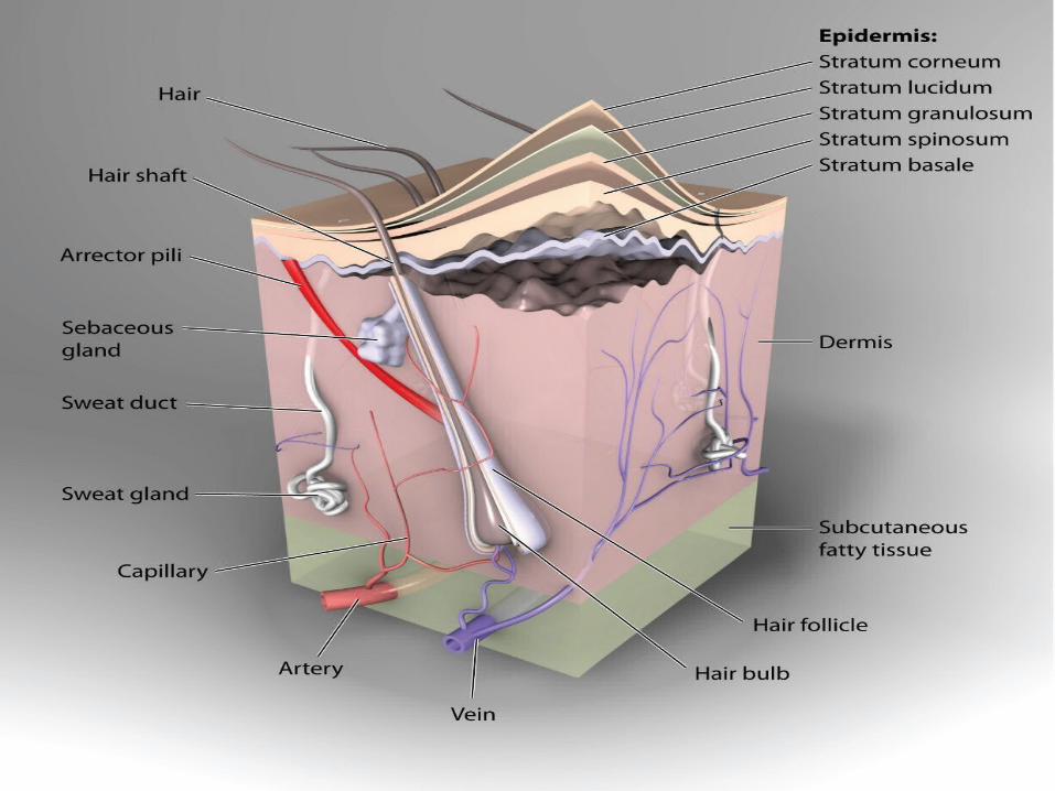
Functions of the Skin• Skin is the largest organ of the body
• Essential for:• - Thermoregulation• - Prevention of fluid loss by evaporation
• - Barrier against infection• - Protection against environment provided by sensory information

Types of burn injuries• A burn is a type of injury to flesh or skin caused by heat, electricity, chemicals, friction, or radiation.
• Thermal: direct contact with heat• (flame, scald, contact)• Electrical• A.C. – alternating current (residential)• D.C. – direct current (industrial/lightening)
• Chemical• Frostbite

Classification of Burns• Burns are classified by depth, type and extent of injury
• Every aspect of burn treatment depends on assessment of the depth and extent
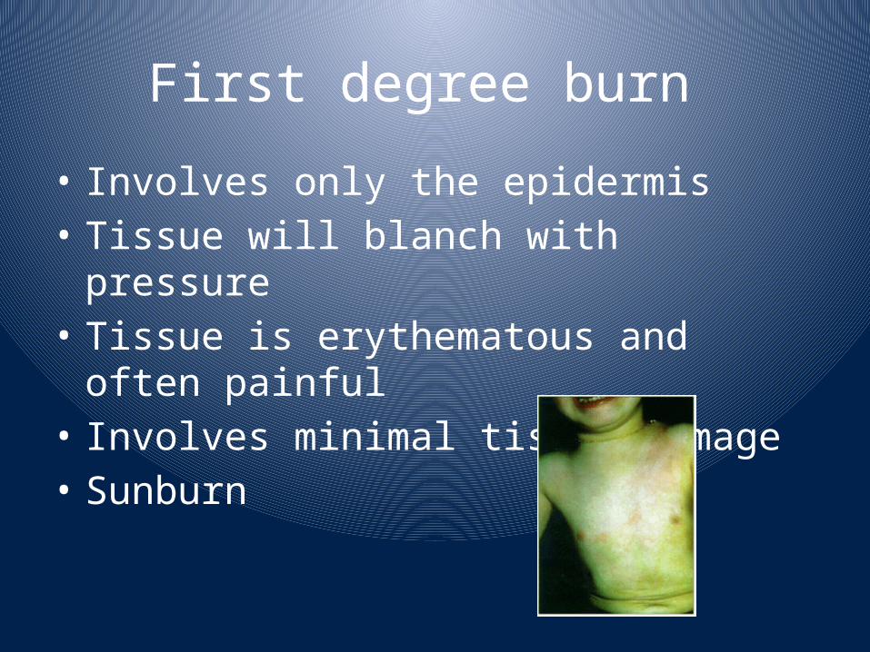
First degree burn • Involves only the epidermis• Tissue will blanch with pressure
• Tissue is erythematous and often painful
• Involves minimal tissue damage• Sunburn
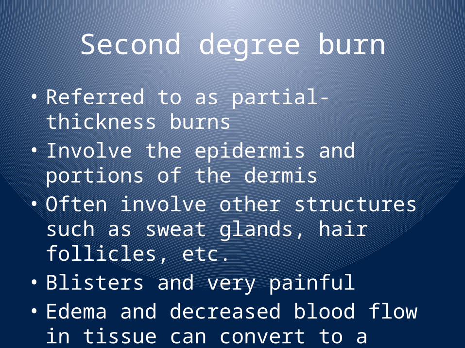
Second degree burn• Referred to as partial-thickness burns
• Involve the epidermis and portions of the dermis
• Often involve other structures such as sweat glands, hair follicles, etc.
• Blisters and very painful• Edema and decreased blood flow in tissue can convert to a full-thickness burn

Second Degree Burns
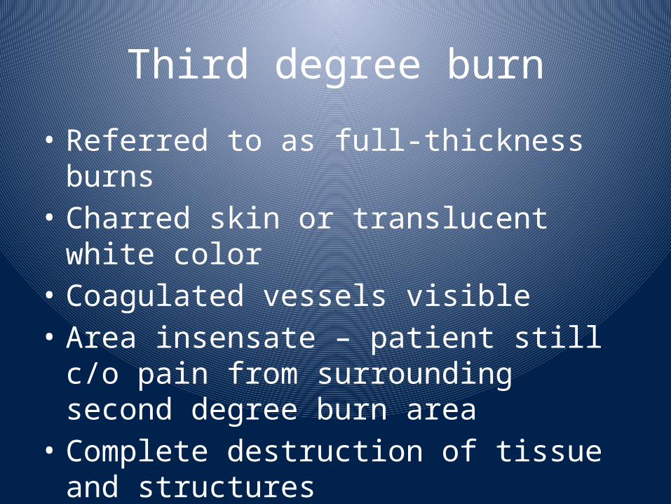
Third degree burn• Referred to as full-thickness burns
• Charred skin or translucent white color
• Coagulated vessels visible• Area insensate – patient still c/o pain from surrounding second degree burn area
• Complete destruction of tissue and structures
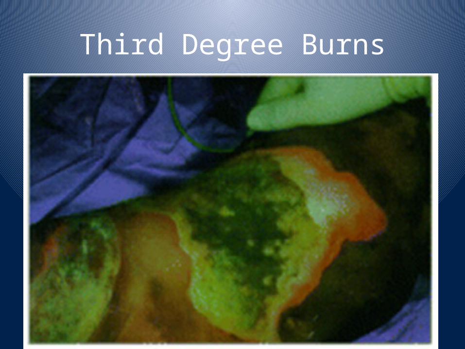
Third Degree Burns

Fourth degree burn• Involves subcutaneous tissue, tendons and bone
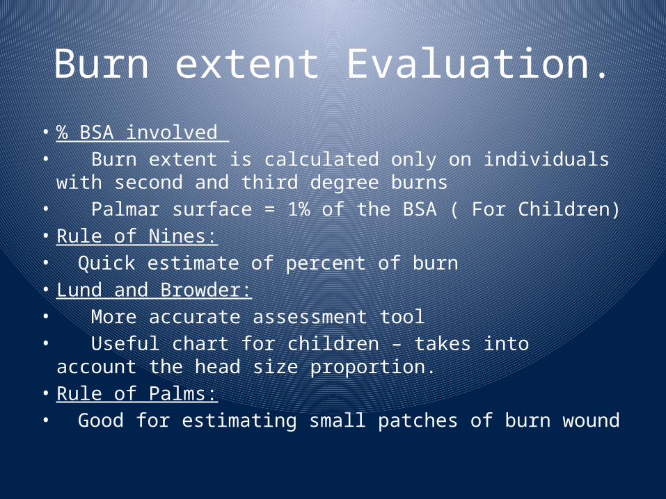
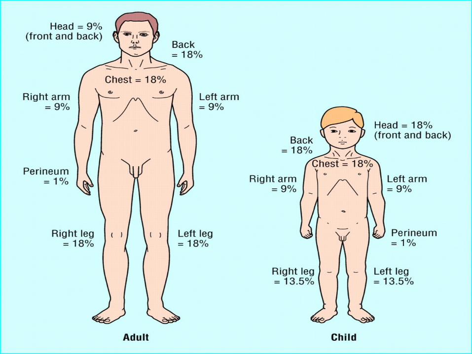
Burn extent Evaluation.• % BSA involved • Burn extent is calculated only on individuals with second and third degree burns
• Palmar surface = 1% of the BSA ( For Children)• Rule of Nines:• Quick estimate of percent of burn• Lund and Browder:• More accurate assessment tool• Useful chart for children – takes into account the head size proportion.
• Rule of Palms:• Good for estimating small patches of burn wound
History & Physical Examinations.
• Cause of the burn• Time of injury• Place of the occurrence (closed space, presence of chemicals, noxious fumes)
• Likelihood of associated trauma (explosion,…)
• Pre-hospital interventions
Lab Investigations• Severe burns:• CBC• Chemistry profile• ABG with carboxyhemoglobin• Coagulation profile• U/A• CPK and urine myoglobin (with electrical injuries)
• 12 Lead EKG
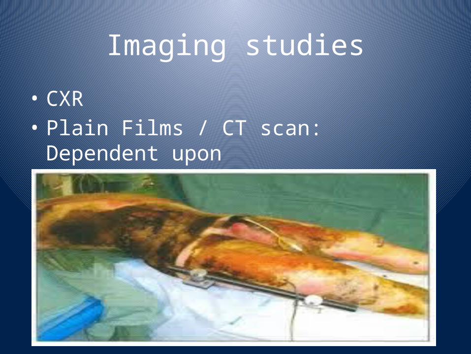
Imaging studies• CXR• Plain Films / CT scan: Dependent upon
history and physical findings
Initial patient treatment
• Stop the burning process
• Consider burn patient as a multiple trauma patient until determined otherwise
• Perform ABCDE assessment
• Avoid hypothermia!
• Remove constricting clothing and jewelry
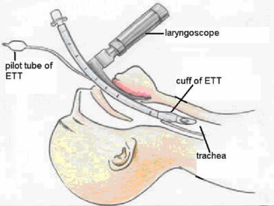
Airway Management• Maintain low threshold for intubation and high index of suspicion for airway injury
• Swelling is rapid and progressive first 24 hours
• Intubation – cautious use of succinylcholine hours after burn due to K+ increase
• Prior to intubation attempt:• have smaller sizes of ETT available
• Prepare for cricothyrotomy or tracheostomy• Utilize ETCO2 monitoring – pulse oximetry may be inaccurate or difficult to apply to patient.
Airway considerations• Upper airway injury (above the glottis): Area buffers the heat of smoke – thermal injury is usually confined to the larynx and upper trachea.
• IS THERE ANY ROLE FOR STERIODS ???????????• Lower airway/alveolar injury (below the glottis):
• - Caused by the inhalation of steam or chemical smoke.
• - Presents as ARDS often after 24-72 hours
Criteria for intubation• Changes in voice• Wheezing / labored respirations • Excessive, continuous coughing• Altered mental status• Carbonaceous sputum• Singed facial or nasal hairs• Facial burns• Oro-pharyngeal edema / stridor• Assume inhalation injury in any patient confined in a fire environment
• Extensive burns of the face / neck• Eyes swollen shut• Burns of 50% TBSA or greater
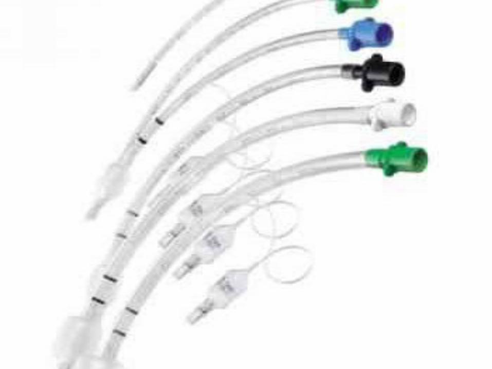

Pediatric intubation• Normally have smaller airways than adults• Small margin for error• If intubation is required, an uncuffed ETT should be placed
• Intubation should be performed by experienced individual – failed attempts can create edema and further obstruct the airway
• AGE/4+ 4 = ETT size
Criteria for burn center admission
• Full-thickness > 5% BSA• Partial-thickness > 10% BSA• Any full-thickness or partial-thickness burn involving critical areas (face, hands, feet, genitals, perineum, skin over major joint)
• Children with severe burns• Circumferential burns of thorax or extremities• Significant chemical injury, electrical burns, lightening injury, co-existing major trauma or significant pre-existing medical conditions
• Presence of inhalation injury
Ventilator therapies• Rapid Sequence Intubation• Pain Management, Sedation and Paralysis• PEEP• High concentration oxygen• Avoid barotrauma• Hyperbaric oxygen• Burn patients with ARDS requiring • PEEP > 14 cm for adequate ventilation should receive prophylactic tube thoracostomy.

Circumferential burns of the chest
• Eschar - burned, inflexible, necrotic tissue
• Compromises ventilatory motion
• Escharotomy may be necessary
• Performed through non-sensitive, full-thickness eschar
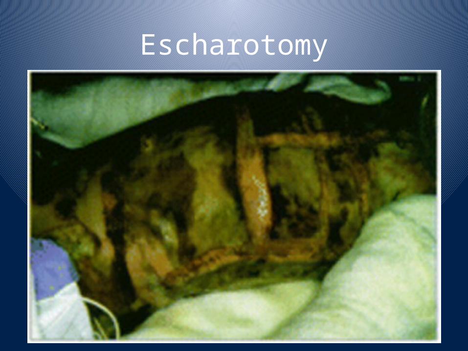
Escharotomy
Carbon Monoxide Intoxication
• Carbon monoxide has a binding affinity for hemoglobin which is 210-240 times greater than that of oxygen.
• Results in decreased oxygen delivery to tissues, leading to cerebral and myocardial hypoxia.
• Cardiac arrhythmias are the most common fatal occurrence.
• Usually symptoms not present until 15% of the hemoglobin is bound to carbon monoxide rather than to oxygen.
• Early symptoms are neurological in nature due to impairment in cerebral oxygenation
Signs and Symptoms of Carbon Monoxide Intoxication
• Confused, irritable, restless• Headache• Tachycardia, arrhythmias or infarction• Vomiting / incontinence• Dilated pupils• Bounding pulse• Pale or cyanotic complexion • Seizures• Overall cherry red color – rarely seen
Management of Carbon Monoxide Intoxication
• Remove patient from source of exposure.
• Administer 100% high flow oxygen
• Half life of Carboxyhemoglobin in patients:
• Breathing room air 120-200 minutes
• Breathing 100% O2 30 minutes
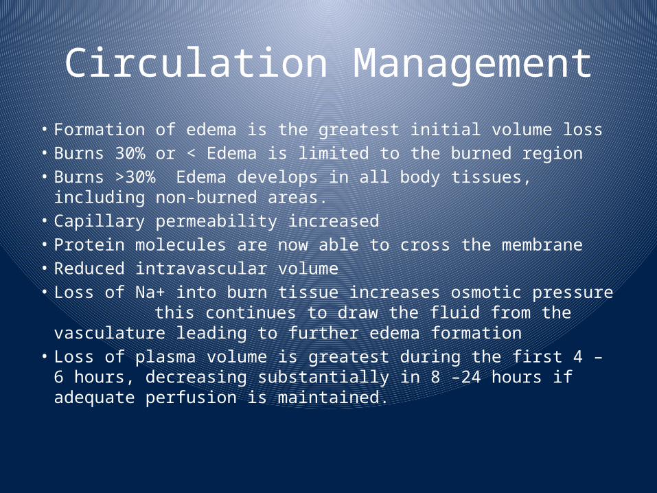
Circulation Management• Formation of edema is the greatest initial volume loss• Burns 30% or < Edema is limited to the burned region• Burns >30% Edema develops in all body tissues, including non-burned areas.
• Capillary permeability increased• Protein molecules are now able to cross the membrane • Reduced intravascular volume• Loss of Na+ into burn tissue increases osmotic pressure
this continues to draw the fluid from the vasculature leading to further edema formation
• Loss of plasma volume is greatest during the first 4 – 6 hours, decreasing substantially in 8 –24 hours if adequate perfusion is maintained.

Impaired peripheral perfusion
• May be caused by mechanical compression, vasospasm or destruction of vessels
• Escharotomy indicated when muscle compartment pressures > 30 mmHg
• Compartment pressures best obtained via ultrasound to avoid potential risk of microbial seeding by using slit or wick catheter
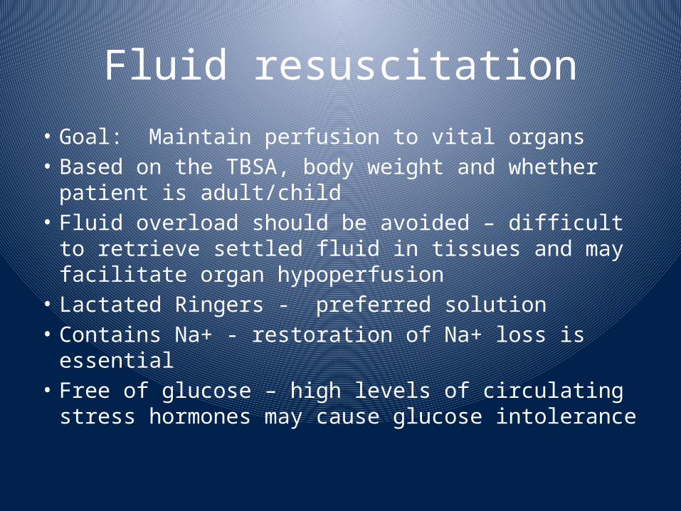
Fluid resuscitation• Goal: Maintain perfusion to vital organs• Based on the TBSA, body weight and whether patient is adult/child
• Fluid overload should be avoided – difficult to retrieve settled fluid in tissues and may facilitate organ hypoperfusion
• Lactated Ringers - preferred solution• Contains Na+ - restoration of Na+ loss is essential
• Free of glucose – high levels of circulating stress hormones may cause glucose intolerance

Fluid resuscitation• Burned patients have large insensible fluid losses
• Fluid volumes may increase in patients with co-existing trauma
• Vascular access: Two large bore peripheral lines (if possible) or central line.
• Fluid requirement calculations for infusion rates are based on the time from injury, not from the time fluid resuscitation is initiated.

Assessing adequacy of resuscitation
• Peripheral blood pressure: may be difficult to obtain – often misleading
• Urine Output: Best indicator unless ARF occurs• A-line: May be inaccurate due to vasospasm • CVP: Better indicator of fluid status• Heart rate: Valuable in early post burn period – should be around 120/min.
• > HR indicates need for > fluids or pain control
• Invasive cardiac monitoring: Indicated in a minority of patients (elderly or pre-existing cardiac disease)
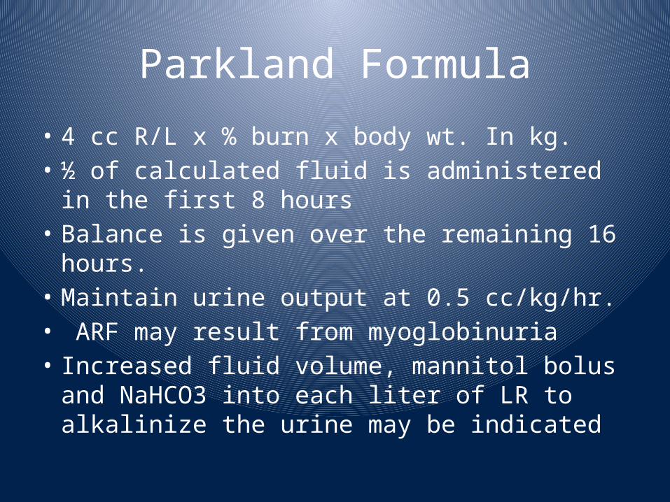
Parkland Formula• 4 cc R/L x % burn x body wt. In kg.• ½ of calculated fluid is administered in the first 8 hours
• Balance is given over the remaining 16 hours.
• Maintain urine output at 0.5 cc/kg/hr.• ARF may result from myoglobinuria • Increased fluid volume, mannitol bolus and NaHCO3 into each liter of LR to alkalinize the urine may be indicated

Galveston Formula• Used for pediatric patients • Based on body surface area rather than weight
• More time consuming• L/R is used at 5000cc/m2 x % BSA burn plus 2000cc/M2/24 hours maintenance.
• ½ of total fluid is given in the first 8 hrs and balance over 16 hrs.
• Urine output in pediatric patients should be maintained at 1 cc/kg/hr.
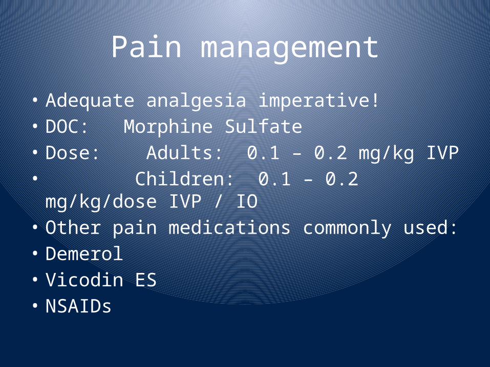
Pain management• Adequate analgesia imperative!• DOC: Morphine Sulfate• Dose: Adults: 0.1 – 0.2 mg/kg IVP • Children: 0.1 – 0.2 mg/kg/dose IVP / IO
• Other pain medications commonly used:• Demerol• Vicodin ES• NSAIDs
Prevention of hypothermia
• Cover patients with a dry sheet – keep head covered
• Pre-warm trauma room • Administer warmed IV solutions• Avoid application of saline-soaked dressings• Avoid prolonged irrigation• Remove wet / bloody clothing and sheets• Paralytics – unable to shiver and generate heat• Avoid application of antimicrobial creams• Continual monitoring of core temperature via foley or SCG temperature probe

Antibiotics & Local Treatment
• Prophylactic antibiotics are not indicated
in the early post burn period. -Topical Antibiotics are controversial in the management of Burns like Flamazine Cream & Polymyxin B for Face to prevent staining are still in use.- Simple Vaseline may be a simple option.
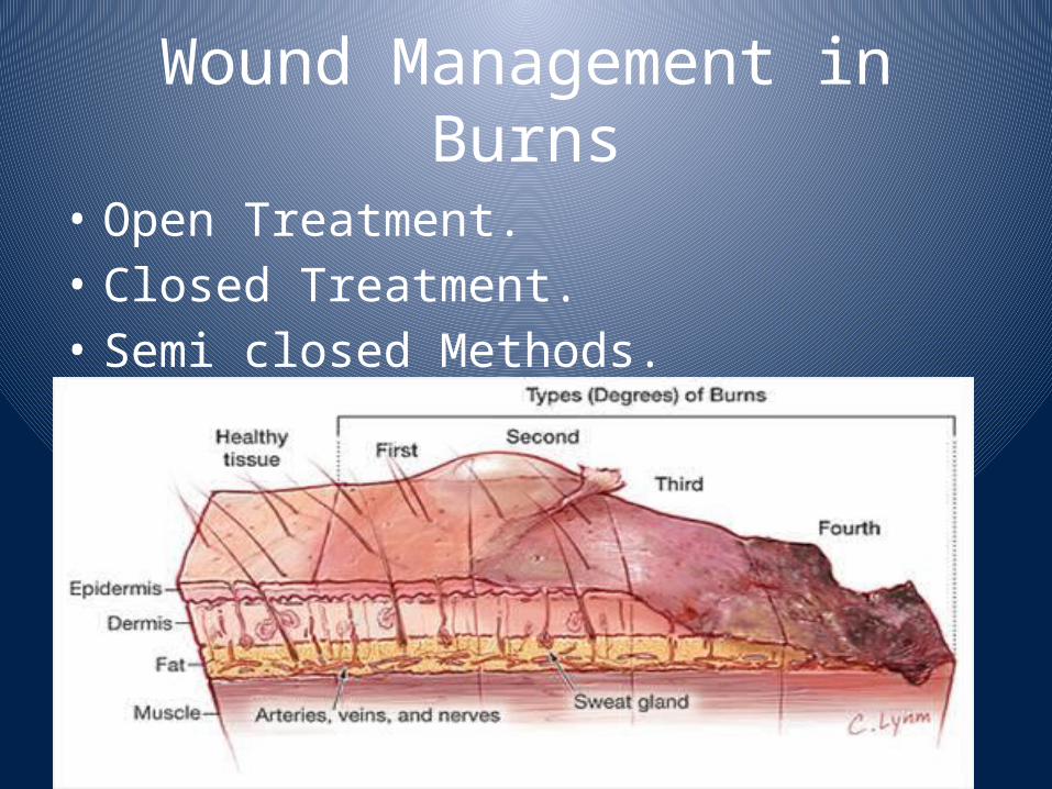
Wound Management in Burns
• Open Treatment.• Closed Treatment.• Semi closed Methods.
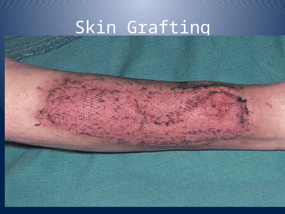
Skin Grafting
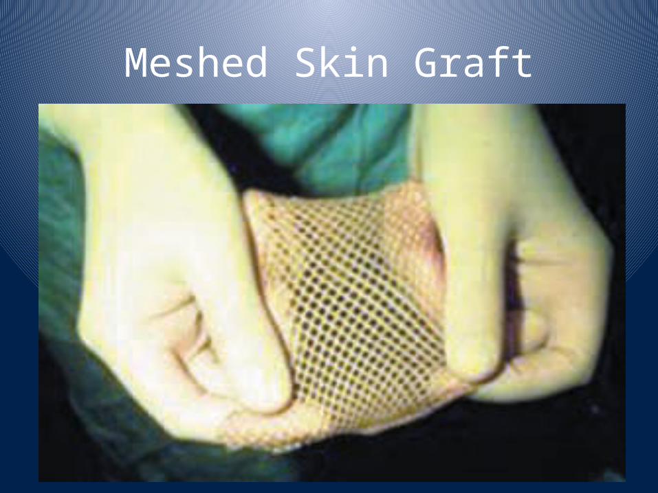
Meshed Skin Graft
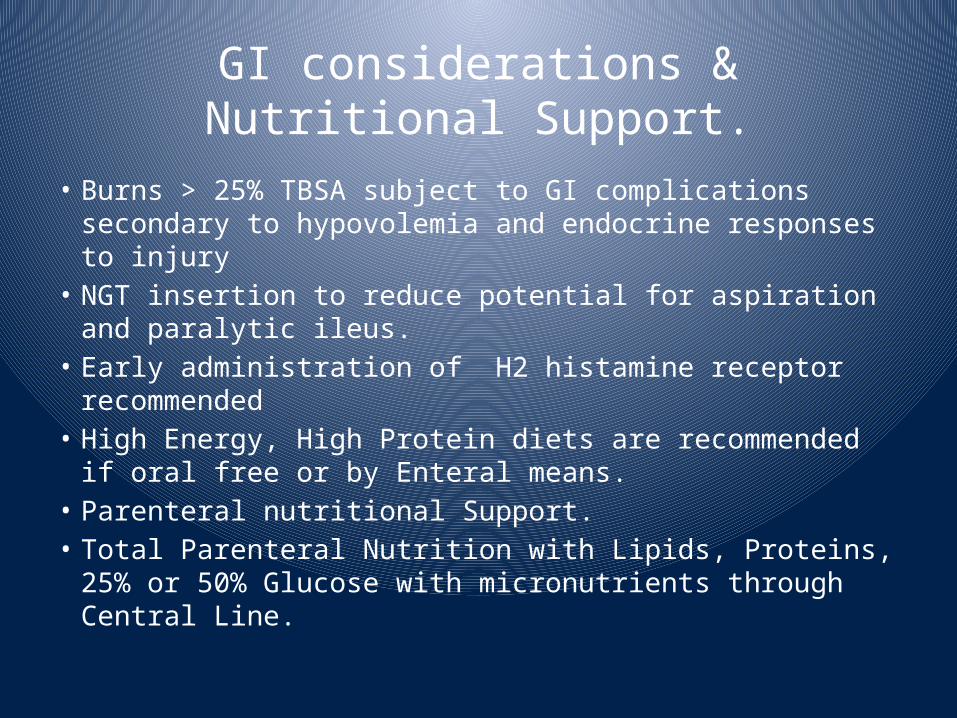
GI considerations & Nutritional Support.
• Burns > 25% TBSA subject to GI complications secondary to hypovolemia and endocrine responses to injury
• NGT insertion to reduce potential for aspiration and paralytic ileus.
• Early administration of H2 histamine receptor recommended
• High Energy, High Protein diets are recommended if oral free or by Enteral means.
• Parenteral nutritional Support.• Total Parenteral Nutrition with Lipids, Proteins, 25% or 50% Glucose with micronutrients through Central Line.
Other considerations• Check tetanus status – administer Td as appropriate
• Debride and treat open blisters or blisters located in areas that are likely to rupture
• Debridement of intact blisters is controversial
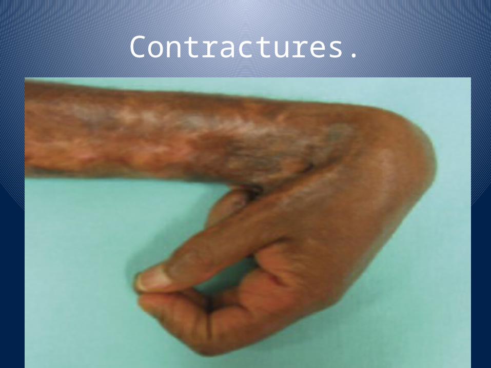
Contractures.
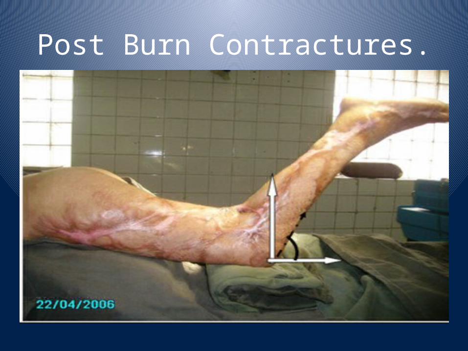
Post Burn Contractures.

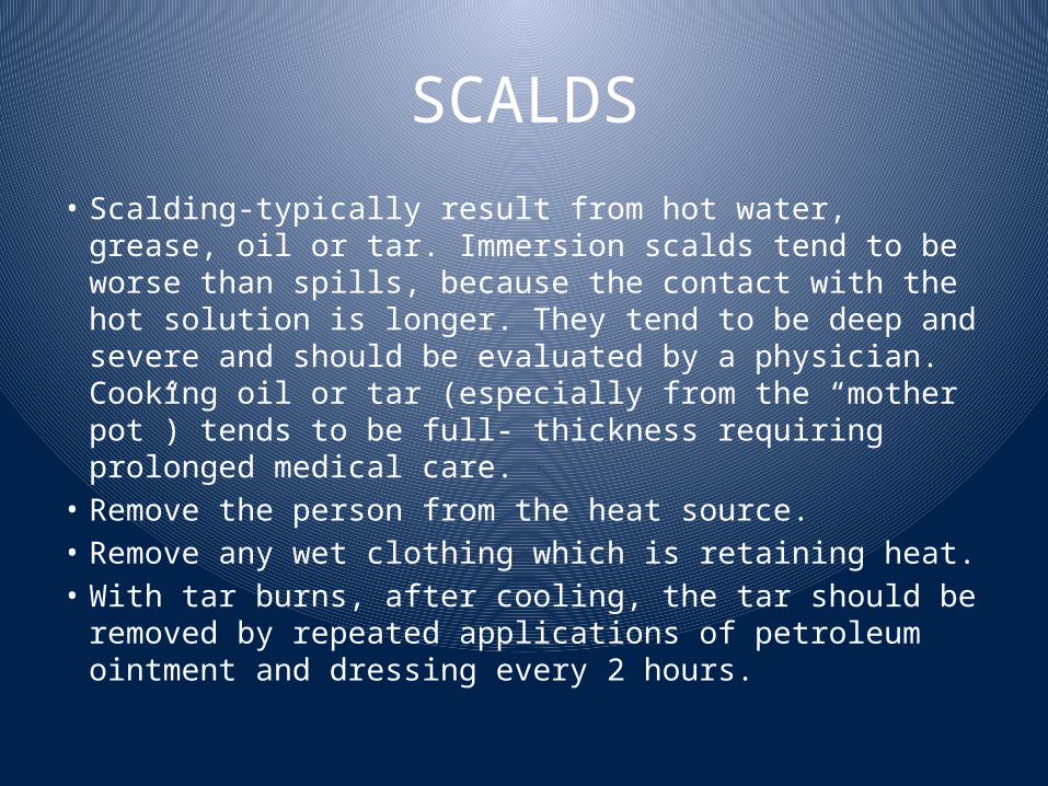
SCALDS• Scalding-typically result from hot water, grease, oil or tar. Immersion scalds tend to be worse than spills, because the contact with the hot solution is longer. They tend to be deep and severe and should be evaluated by a physician. Cooking oil or tar (especially from the “mother pot”) tends to be full- thickness requiring prolonged medical care.
• Remove the person from the heat source.• Remove any wet clothing which is retaining heat.• With tar burns, after cooling, the tar should be removed by repeated applications of petroleum ointment and dressing every 2 hours.

Scald Injury to Child Foot
Flame Burns• a. Remove the person from the source of the heat.
• b. If clothes are burning, make the person lie down to keep smoke away from their face.
• c. Use water, blanket or roll the person on the ground to smother the flames.
• d. Once the burning has stopped, remove the clothing.
• e. Manage the persons airway, as anyone with a flame burn should be considered to have an inhalation injury.
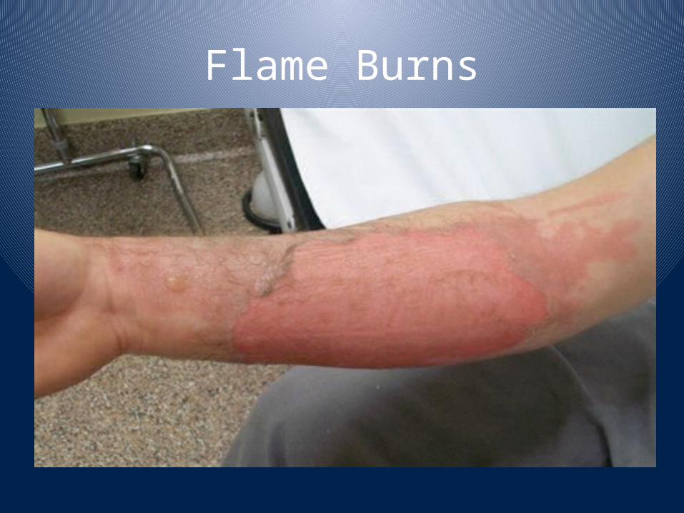
Flame Burns
Electrical Burns• thermal injuries resulting from high intensity heat. The skin injury area may appear small, but the underlying tissue damage may be extensive. Additionally, there may be brain or heart damage or musculoskeletal injuries associated with the electrical injuries.
• a. Safely remove the person from the source of the electricity. Do not become a victim.
• b. Check their Airway, Breathing and Circulation and if necessary begin CPR using an AED (Automatic External Defibrillator) if available and EMS is not present. If the victim is breathing, place them on their side to prevent airway obstruction.
• c. Due to the possibility of vertebrae injury secondary to intense muscle contraction, you should use spinal injury precautions during resuscitation.
• d. Elevate legs to 45 degrees if possible.• e. Keep the victim warm until EMS arrives.
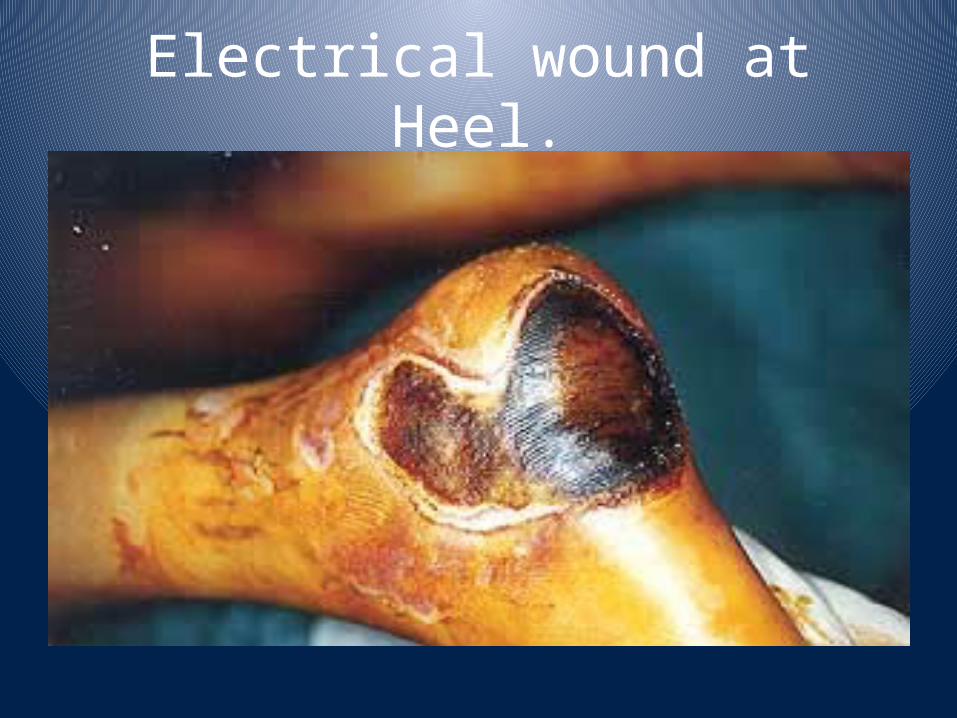
Electrical wound at Heel.
Acid Burns.• Most often caused by strong acids or alkalis. Unlike thermal burns, they can cause progressive injury until the agent is inactivated.
• Flush the injured area with a copious amount of water while at the scene of the incident.
• Don’t delay or waste time looking for or using a neutralizing agent. These may in fact worsen the injury by producing heat or causing direct injury themselves.


Psychological Impact.

After Successful Treatment.
Copyright © 2022 FDOKUMEN