Mating competitiveness of sterile male Anopheles coluzzii in large cages
Upload
independentCategory
view
0download
0
10.1128/IAI.71.6.3000-3009.2003.
2003, 71(6):3000. DOI:Infect. Immun. Yoram VodovotzPeterson, Nicole Emmith, Junghwa Lim, David A. Wink andMatthew J. Lieber, Patricia C. Dos Santos, Tina M. L. Shirley Luckhart, Andrea L. Crampton, Ruben Zamora,
Modulates Mosquito ImmunitystephensiAnophelesActivated after Ingestion by
1βMammalian Transforming Growth Factor
http://iai.asm.org/content/71/6/3000Updated information and services can be found at:
These include:
REFERENCEShttp://iai.asm.org/content/71/6/3000#ref-list-1at:
This article cites 58 articles, 13 of which can be accessed free
CONTENT ALERTS more»articles cite this article),
Receive: RSS Feeds, eTOCs, free email alerts (when new
http://journals.asm.org/site/misc/reprints.xhtmlInformation about commercial reprint orders: http://journals.asm.org/site/subscriptions/To subscribe to to another ASM Journal go to:
on February 12, 2014 by guest
http://iai.asm.org/
Dow
nloaded from
on February 12, 2014 by guest
http://iai.asm.org/
Dow
nloaded from
INFECTION AND IMMUNITY, June 2003, p. 3000–3009 Vol. 71, No. 60019-9567/03/$08.00�0 DOI: 10.1128/IAI.71.6.3000–3009.2003Copyright © 2003, American Society for Microbiology. All Rights Reserved.
Mammalian Transforming Growth Factor �1 Activated after Ingestionby Anopheles stephensi Modulates Mosquito Immunity
Shirley Luckhart,1* Andrea L. Crampton,1† Ruben Zamora,2 Matthew J. Lieber,1Patricia C. Dos Santos,1 Tina M. L. Peterson,1 Nicole Emmith,1 Junghwa Lim,1
David A. Wink,3 and Yoram Vodovotz2
Department of Biochemistry, Virginia Tech, Blacksburg, Virginia 240611; Department of Surgery,University of Pittsburgh, Pittsburgh, Pennsylvania 152602; and Radiation Biology Branch,
National Cancer Institute, Bethesda, Maryland 208923
Received 7 January 2003/Returned for modification 15 February 2003/Accepted 12 March 2003
During the process of bloodfeeding by Anopheles stephensi, mammalian latent transforming growth factor �1(TGF-�1) is ingested and activated rapidly in the mosquito midgut. Activation may involve heme and nitricoxide (NO), agents released in the midgut during blood digestion and catalysis of L-arginine oxidation by A.stephensi NO synthase (AsNOS). Active TGF-�1 persists in the mosquito midgut to extended times posting-estion and is recognized by mosquito cells as a cytokine. In a manner analogous to the regulation of vertebrateinducible NO synthase and malaria parasite (Plasmodium) infection in mammals by TGF-�1, TGF-�1 regu-lates AsNOS expression and Plasmodium development in A. stephensi. Together, these observations indicatethat, through conserved immunological cross talk, mammalian and mosquito immune systems interface witheach other to influence the cycle of Plasmodium development.
In Anopheles, malaria parasite (Plasmodium spp.) develop-ment begins with the ingestion of blood containing sexual-stagegametocytes. Parasite fertilization commences within minutesof ingestion. Zygotes penetrate the midgut epithelium 24 to36 h later and transform into vegetative oocysts under the basallamina in the hemolymph-filled, open circulatory system of themosquito. As blood digestion proceeds to completion at� 48 h, parasite development continues. Oocysts grow anddevelop for 10 to 12 days and then release thousands of hap-loid sporozoites into the hemolymph. Sporozoites invade thesalivary glands, where they are released into the saliva duringsubsequent blood feeding.
Anopheles stephensi, a primary vector of human malaria inIndia and the Middle East, limits malaria parasite develop-ment with the inducible synthesis of nitric oxide (NO [34])catalyzed by A. stephensi NO synthase (AsNOS [33, 34]). In-duction of AsNOS is proportional to the intensity of parasiteinfection and is detectable in the midgut by 6 h postinfection(14). Early induction is critical to inhibition of parasite devel-opment: dietary provision of the pan-NOS inhibitor L-N G-nitroarginine methyl ester (L-NAME), with a half-life in bloodof 3 to 6 h (13), resulted in significantly higher Plasmodiuminfection intensities than did the inactive enantiomer D-NAME(34).
The NO-mediated defense of A. stephensi is analogous tomammalian NO-mediated inactivation of liver-invading sporo-zoites and blood-stage gametocytes (36, 41) and indicates thatmosquitos and vertebrates share a conserved anti-Plasmodium
defense. Limited information, however, is available on theregulation of AsNOS (32). In contrast, iNOS, the inducibleNOS of primary importance in the mammalian arsenal againstmany pathogens (10), is regulated by numerous cytokines andother agents in a wide variety of mammalian cells, therebyensuring homeostasis (10, 42). Recent studies suggest thatamong this diverse array of cytokines, TGF-�1 is perhaps themost potent physiological regulator of iNOS (54).
In primary cells of the vertebrate immune system (e.g., mac-rophages), TGF-�1 reduces iNOS expression (54), iNOSmRNA stability and translation (54), and iNOS stability andenzyme activity (42, 54). In contrast to this role as a negativeregulator, TGF-�1 can upregulate iNOS expression and en-zyme activity in “sentinel” cells associated with tissue immu-nity, including fibroblasts, chondrocytes, and epithelial cells (9,23, 25). These latter cell types, although commonly implicatedas first-line defenders of host integrity against infection (31,50), have received less attention than cell types more com-monly associated with adaptive immunity.
During parasite infection in mammalian hosts, the role ofTGF-�1 in regulation of the immune response is largely de-pendent on timing and level of synthesis (43). Early in parasiteinfection, low levels of TGF-�1 appear to promote proinflam-matory responses (e.g., induction of iNOS) that inhibit parasitegrowth, whereas later in infection, elevated levels of TGF-�1are anti-inflammatory and serve to limit host autoimmune pa-thology.
In A. stephensi, the maintenance of immunological balance(e.g., regulation of AsNOS and other immune effectors) duringparasite infection is poorly understood. The A. stephensi ge-nome encodes a TGF-� superfamily member, As60A, whoseexpression is induced in the midgut epithelium as early as 3 hafter infection with Plasmodium spp. (14, 15). Induction ofAs60A is correlated with periods of parasite motility and re-production. Furthermore, expression of As60A in the A. ste-
* Corresponding author. Mailing address: Department of Biochem-istry, Mail Stop 0308, 306 Engel Hall, Virginia Tech, Blacksburg,VA 24061. Phone: (540) 231-5729. Fax: (540) 231-9070. E-mail:[email protected].
† Present address: CRC for Diagnostics, School of Life Sciences,Queensland University of Technology, Brisbane 4002, Australia.
3000
on February 12, 2014 by guest
http://iai.asm.org/
Dow
nloaded from

phensi midgut, like that of AsNOS, depends on the intensity ofparasite infection (14). This finding suggests that As60A maybe involved in the regulation of parasite development. Al-though we have not yet established that As60A regulatesAsNOS in a manner similar to the regulation of iNOS byTGF-�1, As60A is the first inflammatory cytokine gene to bedescribed from an insect.
Signaling by TGF-� superfamily members is mediated by theSmad family of proteins (3). Previous studies have demon-strated extensive cross talk among vertebrate and invertebrate(Drosophila melanogaster) TGF-� superfamily proteins andSmad signaling pathways (35). In general, activated TGF-�/activin and BMP receptor complexes recruit and phosphory-late relevant receptor Smads, which are then released to formmultimeric complexes with co-Smads. Receptor Smad/co-Smad complexes translocate to the nucleus and interact withother DNA-binding proteins to regulate transcription of down-stream target genes (45).
TGF-�1 is made by most mammalian cell types, especiallyplatelets, in a biologically inactive form that can reach levels of5,000 pg/ml in the circulation (49). Based on our understandingof the A. stephensi NO-mediated defense against Plasmodium,the regulation of iNOS and parasite infection by TGF-�1, andthe demonstrated cross talk among Drosophila and vertebrateTGF-� superfamily ligands and Smads, we sought to answerthe following questions. Is mammalian TGF-�1 ingested by A.stephensi during blood feeding? Could heme and NO, agentscommon to the mosquito midgut after feeding, participate inthe activation of latent TGF-�1? Can ingested TGF-�1 regu-late Plasmodium development in A. stephensi? We show herethat mammalian TGF-�1 is ingested and activated in the mid-gut of A. stephensi and that activation may proceed through amechanism involving heme and NO. Further, in a manneranalogous to TGF-�1 regulation of human iNOS and Plasmo-dium development, human TGF-�1 can regulate both AsNOSexpression in A. stephensi cells and parasite development in themosquito.
MATERIALS AND METHODS
Mosquito rearing, blood feeding, midgut analyses, and Plasmodium infection.A. stephensi Liston (Indo-Pakistan) malaria mosquitos were reared and main-tained at 27°C and 75% relative humidity under a 12-h light-dark cycle. Forpreparation of midgut lysates, 4- to 7-day-old A. stephensi mosquitos were al-lowed to feed to repletion on anesthetized naive or Plasmodium berghei NK65-infected Institute of Cancer Research (ICR) mice. This protocol was reviewedand approved by the Virginia Tech Animal Care Committee in accordance withall relevant federal guidelines. Quantitative reverse transcription-PCR (RT-PCR) analysis of induction of AsNOS expression in the A. stephensi midgutepithelium was performed by using Taqman technology and the sequence de-tection system 7700 (PE Applied Biosystems, Foster City, Calif.) as describedpreviously (14); six separate cohorts of blood-fed uninfected and P. berghei-infected A. stephensi were used for these assays. Differences between uninfectedand P. berghei-infected A. stephensi at each time point were analyzed by using theStudent t test. These and all subsequent Student t tests were performed by usingMicrosoft Excel 2000. To determine the concentration of nitrogen oxides (NOx)and active mammalian TGF-�1 in A. stephensi midgut lysates and whethermidgut lysate components could activate latent human TGF-�1 in vitro, 50midguts were dissected into phosphate-buffered saline (pH 7.4) [PBS; with 1%Nonidet P-40, 4 mM potassium ferricyanide or K3Fe(CN)6, 10 mM N-ethylma-leimide, and 0.1 mM diethylamine triamine pentaacetic acid [DTPA] for NOx
analysis (24)], sonicated, and immediately frozen in a dry ice-ethanol bath atvaried times postfeeding.
The concentration of NOx was determined essentially as described previously(37). Briefly, protein concentration was reduced in diluted midgut lysate samples
by microfiltration (10- or 100-kDa Microcon; Millipore Corp., Bedford, Mass.)prior to reduction with vanadium (III)-hydrochloride at 95°C in a helium-purged,anaerobic reaction vessel; this treatment was essential to minimize sample foam-ing during analysis. The resulting NO was reacted with ozone for chemilumines-cent detection and integration (Nitric Oxide Analyzer; Sievers Instruments,Boulder, Colo.). All values were compared to a standard curve of sodium nitrate(NaNO3) and normalized against the heme concentrations of paired samplealiquots to account for differences in blood meal volumes among mosquitoes.Heme concentrations were determined from the absorbance of the Soret band at410 to 414 nm (2). Four separate cohorts of uninfected and P. berghei-infected A.stephensi were used for these analyses. The levels of NOx induction in midguts ofP. berghei-infected A. stephensi relative to those in uninfected A. stephensi at eachtime point were analyzed by using the Student t test.
Concentration of active TGF-�1 in the midgut lysates was determined by usingthe Quantikine enzyme-linked immunosorbent assay (ELISA; R&D Systems,Inc., Minneapolis, Minn.), which detects binding to immobilized TGF-�1 type IIreceptors. The concentration of active TGF-�1 present in each lysate was as-sessed after PBS treatment for 3 h in vitro according to the manufacturer’srecommendation, whereas the concentration of latent TGF-�1 in each lysate wasassessed after transient acidification with 1 mM HCl (11, 56). Activation ofexogenous latent TGF-�1 was assessed by ELISA after incubation of lysates with5,000 pg or 1 �g of latent human TGF-�1/ml for 3 h at room temperature. Theimpact of NO on activation of exogenous latent TGF-�1 was assessed aftertreatment of lysates with the NO donor S-nitroso-N-acetyl-D,L-penicillamine(SNAP) alone as a control (1 mM for 3 h) or with SNAP and latent TGF-�1. Twoseparate cohorts of A. stephensi were used for these analyses.
To confirm that mosquito proteins did not cross-react with the anti-TGF-�1antisera used in the Quantikine ELISA, a lysate of total proteins from immor-talized ASE A. stephensi cells, which express As60A, was blotted and probed withanti-human TGF-�1. Briefly, ASE cells were washed with PBS, resuspended,lysed in loading buffer (62.5 mM Tris-HCl, pH 6.8; 2% sodium dodecyl sulfate;25% glycerol; 0.01% bromophenol blue), and incubated at 100°C for 5 min.Approximately 22 �g of ASE cell proteins was loaded onto a 12% Tris-HClReady-Gel (Bio-Rad, Hercules, Calif.); 10 ng of recombinant human TGF-�1(R&D Systems) was loaded as a positive control. After electrophoresis, sepa-rated proteins were transferred to a nitrocellulose membrane by using a semidryapparatus (Bio-Rad); poststaining of the transferred gel with Coomassie brilliantblue (48) indicated complete transfer. The nitrocellulose membrane was blockedand then incubated overnight with 1 �g of chicken anti-human TGF-�1 (R&DSystems)/ml at 4°C in blocking buffer (Tris-buffered saline [pH 7.4] with 0.1%Tween 20, 0.1% bovine serum albumin [BSA], and 10% nonfat dry milk),washed, and incubated with a 1:15,000 dilution of anti-chicken immunoglobulinG-horseradish peroxidase in blocking buffer. Antibody binding was detected byusing luminol enhancer as recommended by the manufacturer (Pierce, Rockford,Ill.). To detect cross-reacting mosquito proteins, the membrane was overexposedto film for 4 h at room temperature.
For infection with Plasmodium falciparum (NF54 strain), 4- to 7-day-old fe-male A. stephensi were allowed to feed through a 37°C water-jacketed membraneon identical aliquots of a mixture of parasites cultured in human erythrocyteswith added uninfected erythrocytes (washed twice with RPMI 1640 medium) andhuman serum. The use of human blood components in this procedure wasreviewed by the Virginia Tech Institutional Review Board and declared anexempt protocol in accordance with federal guidelines. Human serum used formosquito feeding was stored at �70°C, thawed, and then stored at 4°C forapproximately 1 week during experimental feedings. ELISA analysis of thesesera revealed no detectable active human TGF-�1; latent TGF-�1 levels rangedfrom 3,000 to 5,000 pg/ml. For analysis of the effects of human recombinantTGF-�1 on parasite development, equivalent volumes of sterile PBS or TGF-�1to a final concentration of 2, 200, or 2,000 pg/ml were added to the bloodimmediately before membrane feeding. At 7 days postinfection, A. stephensimidguts were dissected to count mature P. falciparum oocysts on the midgutepithelium. Two separate cohorts of A. stephensi were used for these assays;oocysts from 60 to 75 mosquitos from each treatment group and the PBS controlswere counted. Differences between each treatment group and the PBS controlgroup were analyzed by using the Student t test.
Activation of latent human TGF-�1 in vitro. All experiments were conductedin siliconized plastic tubes. Carrier-free latent TGF-�1 (10 �g/ml in 1 mMHEPES; R&D Systems) was treated with Angeli’s Salt (a nitroxyl anion donor;10 �M), peroxynitrite (100 �M), or hemin (10 �M) for 30 to 60 min. Log dosesof latent human TGF-�1 (6 to 2,000 pg/ml) were then prepared and assessed forTGF-�1 activity in replicated bioassays of mink lung epithelial cell (CCL-64)proliferation as described previously (55). As controls, paired plates of CCL-64cells were treated with 10 �M hemin, 10 �M Angeli’s salt, or 100 �M peroxyni-
VOL. 71, 2003 MAMMALIAN TGF-�1 MODULATES MOSQUITO IMMUNITY 3001
on February 12, 2014 by guest
http://iai.asm.org/
Dow
nloaded from
trite alone and assayed as described earlier. Proliferation rates of CCL-64 cellstreated with oxidants alone or with TGF-�1 prepared as described were normal-ized to proliferation of positive control CCL-64 cells treated with heat-activatedlatent TGF-�1 (11, 22). Data were analyzed by one-way analysis of variance,followed by application of the Bonferroni post hoc test by using commerciallyavailable software (SPSS, Inc., Chicago, Ill.).
Modulation of the neutralizing capacity of LAP by hemin or nitroxyl anion.Recombinant human active TGF-�1 was added to CCL-64 cells for 20 h. Alter-natively, recombinant human latency-associated peptide (LAP) was untreated ortreated with 10 �M hemin or 10 �M Angeli’s salt, incubated with active TGF-�1,and subsequently added to CCL-64 cells for 20 h. The cells were then assessedfor proliferative capacity as described above.
Analyses of A. stephensi cell responses to human TGF-�1 in vitro. For theseassays, immortalized MSQ43 and ASE A. stephensi cell lines were maintained inmodified Eagle minimum essential medium supplemented with 5% heat-inacti-vated fetal calf serum as described previously (E5 [20]). For analysis of change inmorphology, MSQ43 cells were plated in E0 medium (no serum) and allowed torecover overnight. Duplicate plates of cells were treated with diluent (4 mM HCl,1 mg of BSA/ml) or human TGF-�1 in diluent at 0.6, 6.0, or 30 pg/ml for 20 minor 60 min. Cells were then fixed and coverslipped for microscopic examination.At least 100 cells per treatment were scored as “round” or “spread”; the lengths(in centimeters) of the spread cells were measured on enlarged photomicro-graphs along the axis perpendicular to the cell body. Differences among percent-ages of round cells from assay replicates were analyzed by �2 analysis withcommercially available software (SPSS), while differences between mean celllengths were analyzed by using the Student t test. For analysis of DNA synthesis,duplicate plates of ASE cells were treated with PBS or 0.08, 0.8, or 8 pg of humanTGF-�1/ml in PBS for 48 h. [3H]thymidine was added to each plate to 5 �Ci/mlfor 30 min at room temperature. After incubation, DNA was precipitated fromwashed cells with 10% trichloroacetic acid and incorporated counts were mea-sured by liquid scintillation as described previously (52). Data from three sepa-rate assays were analyzed by using the Student t test. For analysis of AsNOSexpression in cultured cells, duplicate plates of ASE cells were treated with PBSor 6 pg of human TGF-�1/ml for 6, 24, or 48 h. In a second assay, ASE cells werepretreated for 1 h with PBS or with 6 pg of human TGF-�1/ml. After pretreat-ment, cells were exposed for 48 h to 1,000 heat-killed Micrococcus luteus orEscherichia coli organisms per ASE cell as an immune stimulus (21). Total RNAwas isolated from cells postassay by using TRIzol (Invitrogen Life Technologies,Carlsbad, Calif.) reagent. Quantitative RT-PCR of AsNOS expression was per-formed as described previously (14). Data from replicated assays were analyzedby using the Student t test.
RESULTS
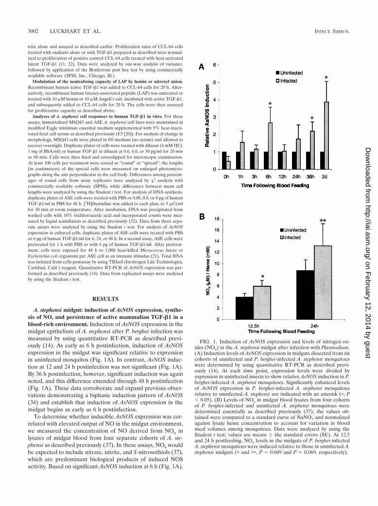
A. stephensi midgut: induction of AsNOS expression, synthe-sis of NO, and persistence of active mammalian TGF-�1 in ablood-rich environment. Induction of AsNOS expression in themidgut epithelium of A. stephensi after P. berghei infection wasmeasured by using quantitative RT-PCR as described previ-ously (14). As early as 6 h postinfection, induction of AsNOSexpression in the midgut was significant relative to expressionin uninfected mosquitos (Fig. 1A). In contrast, AsNOS induc-tion at 12 and 24 h postinfection was not significant (Fig. 1A).By 36 h postinfection, however, significant induction was againnoted, and this difference extended through 48 h postinfection(Fig. 1A). These data corroborate and expand previous obser-vations demonstrating a biphasic induction pattern of AsNOS(34) and establish that induction of AsNOS expression in themidgut begins as early as 6 h postinfection.
To determine whether inducible AsNOS expression was cor-related with elevated output of NO in the midgut environment,we measured the concentration of NO derived from NOx inlysates of midgut blood from four separate cohorts of A. ste-phensi as described previously (37). In these assays, NOx wouldbe expected to include nitrate, nitrite, and S-nitrosothiols (37),which are predominant biological products of induced NOSactivity. Based on significant AsNOS induction at 6 h (Fig. 1A),
FIG. 1. Induction of AsNOS expression and levels of nitrogen ox-ides (NOx) in the A. stephensi midgut after infection with Plasmodium.(A) Induction levels of AsNOS expression in midguts dissected from sixcohorts of uninfected and P. berghei-infected A. stephensi mosquitoeswere determined by using quantitative RT-PCR as described previ-ously (14). At each time point, expression levels were divided byexpression in uninfected insects to show relative AsNOS induction in P.berghei-infected A. stephensi mosquitoes. Significantly enhanced levelsof AsNOS expression in P. berghei-infected A. stephensi mosquitoesrelative to uninfected A. stephensi are indicated with an asterisk (�, P� 0.05). (B) Levels of NOx in midgut blood lysates from four cohortsof P. berghei-infected and uninfected A. stephensi mosquitoes weredetermined essentially as described previously (37); the values ob-tained were compared to a standard curve of NaNO3 and normalizedagainst lysate heme concentration to account for variation in bloodmeal volumes among mosquitoes. Data were analyzed by using theStudent t test; values are means � the standard errors (SE). At 12.5and 24 h postfeeding, NOx levels in the midguts of P. berghei-infectedA. stephensi mosquitoes were induced relative to those in uninfected A.stephensi midguts (� and ��, P 0.049 and P 0.069, respectively).
3002 LUCKHART ET AL. INFECT. IMMUN.
on February 12, 2014 by guest
http://iai.asm.org/
Dow
nloaded from

we selected 12.5- and 24-h time points for these analyses. Atboth 12.5 and 24 h postinfection, heme-normalized NOx levelswere elevated in midgut blood lysates from infected comparedto uninfected mosquitos (P 0.049 and 0.069, respectively;Fig. 1B). The mean percentage increase at 12.5 h was 59.5%(95% confidence interval [CI] �0.8 to 119.8), and at 24 h it was50.8% (95% CI �56.6 to 158.2). Sample variation was, there-fore, quite high, but less than the sample variation reported forhuman plasma (51, 58). Based on a mean midgut heme con-centration of 12.7 � 0.4 mM (uninfected and infected midgutlysates were not significantly different), total NOx levels inuninfected midguts would be equivalent to ca. 51 �M at 12.5 hand 110 �M at 24 h, whereas infected midgut levels would beequivalent to ca. 75 �M at 12.5 h and 168 �M at 24 h. Valuesin excess of 75 �M are consistent with inflammatory levels ofnitrite and/or nitrate reported in human serum under condi-
tions of sepsis (51, 58) and, therefore, suggest that the NO-mediated response of A. stephensi to Plasmodium infection isindeed an inflammatory response. These data are also consis-tent with our detection of elevated NADPH-dependent diaph-orase activity in infected versus uninfected A. stephensi midgutsat 24 h and extend our observations demonstrating elevatedcirculating nitrite or nitrate levels in infected A. stephensi at 7to 14 days postinfection (34). Further, when taken togetherwith data indicating that the host ICR mouse strain does notsynthesize inducible NO in response to parasite infection (40),our data suggest that AsNOS induction beginning at 6 h drivesthe synthesis of significantly elevated levels of NOx in themidgut of P. berghei-infected A. stephensi within hours of in-fection.
We hypothesized that, in addition to NO, mammalianTGF-�1 may be present in the A. stephensi midgut. Specifically,we predicted that A. stephensi ingests circulating mammalianTGF-�1 during blood feeding and that this cytokine may beactive in the insect midgut environment. To determine theconcentration of active mammalian TGF-�1 in A. stephensimidgut lysates and whether midgut lysate components couldactivate added latent human TGF-�1 in vitro, midguts fromblood-fed mosquitoes at varied times postfeeding were as-sessed for the presence of active mammalian TGF-�1 by usingQuantikine ELISA. Mosquito proteins do not cross-react withthe anti-TGF-�1 antisera used in the ELISA (Fig. 2A) and,therefore, would not be expected to contribute to signal outputfrom these assays.
Midgut lysates prepared from mosquitoes 1 to 48 h afterblood feeding and treated ex vivo with PBS (control) appearedto contain active TGF-�1 at levels that ranged from 100 to 530pg/ml (Fig. 2B). Active TGF-�1 was almost undetectable at 7days (Fig. 2B). In comparison, TGF-�1 is present at 3,000 to5,000 pg/ml, with 100% in the latent form, in platelet-poorplasma (26) from both healthy and P. berghei-infected hostICR mice (data not shown). Accordingly, recombinant humanlatent TGF-�1 was added to midgut lysates to a final concen-tration of 5,000 pg/ml in order to determine whether latentTGF-�1 could be activated in vitro by midgut lysates. Littleactive TGF-�1 above that already present in the midgut lysateswas detected (Fig. 2B), suggesting that activating substanceshad been depleted.
Nitric oxide can activate latent TGF-�1 (55) and is presentat high levels in the Anopheles midgut (Fig. 1B) (34). Accord-ingly, we examined whether NO provided by a chemical donorwould enhance activation of latent TGF-�1 by midgut lysates.To test this hypothesis, lysates were treated with SNAP as acontrol or with SNAP and latent human TGF-�1. Based on ourexperience with SNAP (61, 62), we expect that the effectivedose of NO delivered in 3 h was 100 �M, within the range oftotal NOx concentrations measured in the midgut lysates (seeabove). The levels of active TGF-�1 in lysates treated withSNAP only were similar to or lower than those in the lysatestreated only with PBS (Fig. 2B). These findings with SNAPwere consistent with earlier observations that NO does notalter antiproliferative effects of active TGF-�1 on CCL-64 cells(55) and, hence, the receptor-binding properties of activeTGF-�1.
Simultaneous treatment of A. stephensi midgut lysates withboth latent TGF-�1 and SNAP (Fig. 2B) led to the nearly
FIG. 2. Presence of active mammalian TGF-�1, as well as activa-tion of latent TGF-�1 in the presence of NO, in A. stephensi midguts.(A) Mosquito cell proteins did not cross-react with anti-TGF-�1 usedin the Quantikine ELISA. In the left panel, 22 �g of mosquito cellproteins (lane A) and 10 ng of human TGF-�1 (lane B) are shownafter polyacrylamide gel electrophoresis and Coomassie brilliant bluestaining. Identical protein samples transferred to nitrocellulose (rightpanel) were incubated with anti-human TGF-�1 as described in thetext. Note the strong detection of human TGF-�1 in lane B (arrow)and the lack of cross-reacting mosquito proteins in lane A. (B) Lysatesof mosquito midguts (50 per lysate) were prepared at various timesafter blood feeding (“incubation time”) indicated on the x axis. Thelysates were treated as described in the boxed legend and in Materialsand Methods. Active TGF-�1 was quantified in the samples by usingthe Quantikine ELISA.
VOL. 71, 2003 MAMMALIAN TGF-�1 MODULATES MOSQUITO IMMUNITY 3003
on February 12, 2014 by guest
http://iai.asm.org/
Dow
nloaded from

uniform presence of higher levels of active TGF-�1 than inlysates treated with latent TGF-�1 alone; this effect was mostpronounced in the 1- to 12-h lysates. At the 7-day time point,the presence of active TGF-�1 after treatment with latentTGF-�1 and SNAP verified that substance(s) with the capacityto activate latent TGF-�1 in the presence of NO were stillpresent in the lysate, despite the complete lack of digestate atthis time and the almost undetectable active TGF-�1 in thePBS-treated lysate (Fig. 2B).
As a final control, lysates of A. stephensi midgut blood (pH7.5 to 7.8 in vivo [8]) were transiently acidified with 1 mM HCl(11, 56) in order to examine whether any residual latentTGF-�1 was present in the lysates (Fig. 2B). Acidification,however, did not result in any increase in detectable activeTGF-�1. Thus, all TGF-�1 present in A. stephensi midgutswithin 1 h after blood feeding was active.
To verify that the capacity of the midgut lysates to activateadded latent TGF-�1 was not restricted by availability of thegrowth factor, midgut lysates from a second cohort of blood-fed A. stephensi were subjected to the same analyses exceptthat latent TGF-�1 was added to a final concentration of 1
FIG. 3. Activation of latent TGF-�1 by hemin. CCL-64 cells wereincubated with the indicated concentrations of latent TGF-�1 that wasuntreated, treated with 10 �M hemin, or heated. Alternatively,CCL-64 cells were incubated either with the indicated concentrationsof active TGF-�1 or with dilutions of the hemin stock solution identicalto those used to treat latent TGF-�1. Proliferation of CCL-64 cells wasassessed by uptake of [3H]thymidine. Values are the means � the SEof three to seven separate experiments.
FIG. 4. Activation of latent TGF-�1 by nitroxyl anion. (A) CCL-64cells were incubated with the indicated concentrations of latentTGF-�1 that was treated with 10 �M Angeli’s salt (nitroxyl anionchemical donor) or 100 �M peroxynitrite. Alternatively, CCL-64 cellswere incubated with dilutions of the Angeli’s salt stock solution iden-tical to those used to treat latent TGF-�1. These assays were per-formed simultaneously with those represented in Fig. 3. Hence, datafrom active and latent TGF-�1 only controls are identical to thoserepresented in Fig. 3. (B) Recombinant active human TGF-�1 (�) wasadded to CCL-64 cells for 20 h. Alternatively, recombinant humanLAP was either left untreated (o) or treated with 10 �M Angeli’s salt(AS) (1), or 10 �M hemin (■); incubated with active TGF-�1; andsubsequently added to CCL-64 cells for 20 h. �, P � 0.05 versusincubation of active TGF-�1 with untreated LAP. The proliferation ofCCL-64 cells was assessed by measuring the uptake of [3H]thymidine.Values are means � the SE of two to seven separate experiments.
3004 LUCKHART ET AL. INFECT. IMMUN.
on February 12, 2014 by guest
http://iai.asm.org/
Dow
nloaded from

�g/ml, a level 200 times that provided in the previous assays.Results from this second set of assays showed similar levels ofactive TGF-�1 (330 to 900 pg/ml) from 1 to 48 h after PBStreatment (data not shown). In addition, we observed similartrends of activation in the presence of added latent TGF-�1and SNAP, at levels that ranged from three to five times thatobserved in the presence of 5,000 pg of added latent TGF-�1/ml under identical conditions. Because active TGF-�1 lev-els were not 200-fold greater than the levels observed in pre-
FIG. 5. Human TGF-�1 alters the morphology of A. stephensiMSQ43 cells in vitro. (A) Typical morphology of MSQ43 cells treatedwith diluent (4 mM HCl, 1 mgof BSA/ml) as a control for 20 or 60 min.Note the predominance of rounded cells (arrow) compared to spreadcells with filopodia. (B) Typical morphology of MSQ43 cells treatedwith 6 or 30 pg of human TGF-�1/ml in diluent for 20 or 60 min. Notethe predominance of spread cells with filopodia (arrowhead) com-pared to rounded cells. (C) Lengths of spread MSQ43 cells and per-centages of round cells were determined from enlarged photomicro-graphs of treated and control cells from three trials. In trial 1, cellswere treated as shown for 20 min; cell lengths are means � the SE forn 10 cells. In trials 2 and 3, cells were treated as shown for 60 min.Cell lengths for trial 2 are means � the SE for n 10 cells, whereascell lengths for trial 3 means � the SE for n 40 to 80 cells. Differ-ences between mean cell lengths within a single trial were analyzed byusing the Student t test; significant differences among treatmentswithin a trial are indicated by different lowercase letters. Differencesamong percentages of round cells within a single trial were analyzed by�2; significant differences among treatments within a trial are indicatedby different lowercase letters.
FIG. 6. A. stephensi cells recognize human TGF-�1 as an immuno-modulatory cytokine in vitro. (A) At 48 h, human TGF-�1 at 8 pg/mlsignificantly reduced [3H]thymidine incorporation into A. stephensi ASEcells compared to PBS-treated controls (P 0.03). Data were analyzed byusing the Student t test; values are means � the SE of three separateexperiments. (B) At 24 h, AsNOS expression in ASE cells was significantlyinduced by human TGF-�1 at 6 pg/ml compared to PBS-treated controls(�, P 0.05). Induction levels of AsNOS expression were determined byusing quantitative RT-PCR as described previously (14). PBS andTGF-�1 mean valuess within each time point were divided by the PBSmean value to show relative AsNOS induction in TGF-�1-treated cells.Data were analyzed by using the Student t test; values are means � the SEof nine separate experiments. (C) Pretreatment with human TGF-�1 at 6pg/ml significantly enhanced AsNOS induction in response to heat-killedE. coli (�, P 0.05). ASE cells were pretreated with PBS or TGF-�1 for1 h prior to a 48-h exposure to heat-killed E. coli or M. luteus. AsNOSexpression was analyzed, and mean values are illustrated as described inpanel B. Data from five separate experiments were analyzed by using theStudent t test.
VOL. 71, 2003 MAMMALIAN TGF-�1 MODULATES MOSQUITO IMMUNITY 3005
on February 12, 2014 by guest
http://iai.asm.org/
Dow
nloaded from

vious assays, we concluded that our results accurately reflectedthe activation capacity of A. stephensi midgut lysates.
Activation of latent TGF-�1 in vitro by the prooxidantsheme and nitroxyl anion but not by peroxynitrite. The studiesdescribed above showed that factor(s) present in the midgut ofA. stephensi can activate latent TGF-�1 in synergy with NO.Heme, which can catalyze the synthesis of oxygen free radicals(4), is released into the midgut and polymerized into hematinduring blood digestion. However, polymerization is not instan-taneous, nor does it completely abolish radical chemistry (19).Elevated NO levels can lead to the activation of latent TGF-�1by nitrosation of the LAP portion of latent TGF-�1 (55),whereas oxygen free radicals can activate cell-free latentTGF-�1 directly (5). Subsequently, we hypothesized that, inthe redox-active Anopheles midgut, heme and NO may partic-ipate in activating latent TGF-�1.
To test this hypothesis, as well as to examine the role ofreaction products of NO on the function of TGF-�1, we com-pleted a series of in vitro assays. The mink lung epithelial cellline CCL-64 is highly sensitive to growth suppression byTGF-�1 (17). Latent human TGF-�1 at doses ranging from 6to 2,000 pg/ml did not suppress the proliferation of CCL-64cells, in contrast to the suppression of CCL-64 proliferationupon treatment with the same concentrations of activeTGF-�1 (Fig. 3). When latent TGF-�1 was activated with heat,the 50% inhibitory concentration for the suppression of pro-liferation of CCL-64 cells was 0.4 � 0.1 ng/ml (n 6) (Fig. 3).The addition of 10 �M hemin to latent TGF-�1 for 30 min
activated this cytokine (50% inhibitory concentration 0.4 �0.2 ng/ml [n 5]) to levels comparable to that observed withheat; hemin alone did not affect the proliferation of CCL-64cells (Fig. 3).
In the redox-active mosquito midgut, the effects of endog-enously produced NO may be enhanced through the formationof redox products of NO, such as nitroxyl anion and peroxyni-trite, two reactants commonly associated with the cellular syn-thesis of NO (57). In the environment of the midgut, nitroxylanion and/or peroxynitrite could function to activate latentTGF-�1 directly. To test this hypothesis, latent TGF-�1 wastreated with Angeli’s salt, a nitroxyl anion chemical donor, orreagent peroxynitrite prior to analysis in CCL-64 cell prolifer-ation assays. Our results demonstrated that, whereas Angeli’ssalt activated latent TGF-�1 directly, peroxynitrite did not(Fig. 4A). Angeli’s salt alone, as a control, did not affect theproliferation of CCL-64 cells (Fig. 4A).
We next sought to examine the mechanism by which heminactivated latent TGF-�1. One possibility was that LAP, whichbinds noncovalently to the active TGF-�1 dimer and maintainsTGF-�1 in the latent state, was modified by redox-active heminsuch that it could no longer bind to active TGF-�1, reminiscentof inactivation of LAP through S nitrosation (55). Preincuba-tion of LAP with 10 �M hemin significantly abrogated thecapacity of LAP to neutralize active TGF-�1 (P � 0.05 versusincubation of active TGF-�1 with untreated LAP; Fig. 4B);however, nitroxyl anion did not appear to cause such an effect(Fig. 4B). The presence of an agent such as heme may disruptthe interaction between LAP and active TGF-�1, permittingthe S nitrosation of LAP. At high levels of NO, such as thosepresent in the mosquito midgut (Fig. 1), S nitrosation of dis-sociated LAP should occur readily and prevent rebinding toand inactivation of TGF-�1 (55). Nitroxyl anion appears toactivate latent TGF-�1 by a mechanism distinct from that ofNO (55) or hemin (Fig. 3). These observations suggest that NOin the presence of heme may lead to sustained activation oflatent TGF-�1 in the Anopheles midgut.
A. stephensi cells recognize human TGF-�1 as an immuno-modulatory cytokine in vitro. Based on our findings that mam-malian TGF-�1 is activated and persists in the A. stephensimidgut to extended times after blood feeding, we sought todetermine whether mammalian TGF-�1 could be recognizedby A. stephensi cells in vitro. Three assays based on cell mor-phology, cell proliferation, and gene expression were used toassess the responses of A. stephensi cells to active TGF-�1. Inthe first assay, A. stephensi MSQ43 cells treated with humanTGF-�1 compared to control cells showed significantly en-hanced spreading (Fig. 5A and B), a behavior consistent withactin-dependent immune responsiveness of immortalized mos-quito cells (30) and other insect cells (29).
At doses of 6 or 30 pg of TGF-�1/ml, the mean spread celllength was significantly increased (n 3) compared to control,diluent-treated cells (Fig. 5C). Significant decreases in the per-centages of round MSQ43 cells were observed at 20 min at adose of 6 pg/ml of TGF-�1 (trial 1; Fig. 5C) or at 60 min at adose of 0.6 pg/ml (trial 3; Fig. 5C), indicating that 0.6 pg/ml wassufficient to cause the observed changes. These effective dosesare similar to those reported for both vertebrate and inverte-brate cells (38, 44, 60).
At a dose of 8 pg of human TGF-�1/ml, [3H]thymidine
FIG. 7. Human TGF-�1 significantly alters P. falciparum develop-ment in A. stephensi. Two cohorts of mosquitos were fed P. falciparum-infected blood with PBS or TGF-�1 at 2, 200, or 2,000 pg/ml in PBS.To determine the intensity of infection, parasite oocysts were countedfrom 60 to 75 mosquitos per group; data from cohorts 1 and 2 were notcombined because mean infections were not equivalent. Data wereanalyzed by using the Student t test; values are means � the SE. Forcohorts 1 and 2, human TGF-�1 at 2 pg/ml reduced the number of P.falciparum oocysts by 46 and 26%, respectively, compared to the con-trols (P 0.004 for cohort 1 and P 0.03 for cohort 2). Similarly, 200pg of TGF-�1/ml reduced oocyst numbers by 37 and 27%, respectively,compared to controls (P 0.02 for cohort 1 and P 0.03 for cohort2). In contrast, the highest concentration of human TGF-�1 (2,000pg/ml) had no effect on oocyst numbers in either cohort compared tothe controls.
3006 LUCKHART ET AL. INFECT. IMMUN.
on February 12, 2014 by guest
http://iai.asm.org/
Dow
nloaded from

incorporation into A. stephensi cell DNA was reduced by nearly25% compared to PBS-treated controls (P 0.03; Fig. 6A). Nosignificant differences were observed between controls andcells treated with 0.08 or 0.8 pg of human TGF-�1/ml (data notshown). Although a 25% change may be modest, similar pat-terns of [3H]thymidine incorporation have been observed afterTGF-�1 treatment of human cardiac fibroblasts (20% decrease[1]) and rat costochondral chondrocytes (30% increase [47]).
Finally, we sought to determine whether human TGF-�1could modulate AsNOS expression in a manner analogous toits effect on iNOS expression. To address this question, we usedtwo assays. In the first assay, duplicate plates of ASE cells weretreated with PBS or with 6 pg of human TGF-�1/ml for 6, 24,or 48 h. In the second assay, cells were pretreated for 1 h withPBS or 6 pg of human TGF-�1/ml. After pretreatment, cellswere exposed for 48 h to 1,000 heat-killed M. luteus or E. coli,immune stimuli (21) that induce AsNOS expression.
In the first assay, no significant effects of human TGF-�1 onAsNOS expression were observed at 6 and 48 h (Fig. 6B).However, AsNOS expression was induced �3.7-fold (P 0.05)in ASE cells at 24 h after exposure to human TGF-�1 (95% CI 0.13 to 7.67; Fig. 6B). Further, pretreatment of ASE cellswith human TGF-�1 increased AsNOS induction in the pres-ence of immune stimuli, with a statistically significant effectnoted for E. coli (Fig. 6C). Taken together, our data indicatethat A. stephensi cells recognize human TGF-�1 as an immu-nomodulatory cytokine and that, in contrast to iNOS (54),AsNOS is induced by TGF-�1.
Human TGF-�1 alters Plasmodium development in A. ste-phensi. We next examined whether human TGF-�1 could alterPlasmodium development in vivo in the mosquito host. Toaddress this question, two cohorts of A. stephensi were allowedto feed on an artificial meal containing P. falciparum-infectederythrocytes, human serum, and additional washed uninfectederythrocytes that was supplemented with equivalent volumes ofPBS or 2, 200, or 2,000 pg of human TGF-�1/ml. Native 2-macroglobulin is the primary carrier of TGF-�1 in circulation(16). However, TGF-�1 is likely to be complexed exclusively toa conformationally altered form of 2-macroglobulin in storedserum (A. Kurdowska, personal communication), such as thatused in our feeding assays. Consistent with the negative effectsof conformationally altered 2-macroglobulin on bioavailabil-ity (28) and physiological function of TGF-�1 (59), latentTGF-�1 in the artificial meal did not appear to be activated inthe A. stephensi midgut (results not shown). In contrast, re-combinant active TGF-�1 added immediately prior to feedingat doses that bracket the levels of active growth factor detectedin midgut lysates (Fig. 2B) would be freely available to exert adistinguishable biological effect. At 7 days postfeeding, mos-quitos from each treatment were dissected to count the matureoocysts as an indicator of intensity of parasite infection.
Our results clearly demonstrated that active human TGF-�1alters Plasmodium development in A. stephensi (Fig. 7). A doseof 2 pg of TGF-�1/ml reduced the number of P. falciparumoocysts by 46 and 26%, respectively, compared to the controls(P 0.004 for cohort 1 and P 0.03 for cohort 2). Similarly,200 pg of TGF-�1/ml reduced oocyst numbers by 37 and 27%,respectively, compared to controls (P 0.02 for cohort 1 andP 0.03 for cohort 2). In contrast, the highest concentration of
TGF-�1 (2,000 pg/ml) had no effect on the oocyst numbers ineither cohort compared to the controls.
DISCUSSION
Although the impacts of TGF-�1 on the host immune re-sponse to parasite infection have not yet been fully elucidated,this cytokine is crucial for maintaining an immunological bal-ance between parasite clearance and inflammation (43). This isthe first study, however, to document the transfer of TGF-�1from a mammalian host to an invertebrate, with consequentbiological effects. Our data demonstrate that (i) mammalianTGF-�1 ingested by A. stephensi is activated in the midgut andpersists during the 48 h process of blood digestion, (ii) theblood-filled A. stephensi midgut possesses factor(s) that canactivate latent mammalian TGF-�1 in synergy with NO, (iii)products associated with blood digestion and NO synthesis thatare either known or presumed present in the redox-activemosquito midgut can activate latent TGF-�1 in vitro, (iv) A.stephensi cells recognize mammalian TGF-�1 in vitro as animmunomodulatory cytokine, and (v) human TGF-�1 can alterdevelopment of P. falciparum in A. stephensi.
Thus, we have demonstrated that ingested mammalianblood is not only a source of nutrients for mosquito reproduc-tion but also a source of cell signaling factors that can com-municate with immunocompetent cells of the insect host. Fur-ther, these observations establish that a cytokine of criticalimportance to Plasmodium development in the vertebrate hostcan also influence parasite development in the mosquito host.Based on cross talk among TGF-�/activin and BMP ligandsand Smad signaling pathways of Drosophila and human cells,we propose that mammalian TGF-�1 signals Anopheles cellsthrough a conserved, endogenous TGF-�/activin pathway.
Intriguingly, our data indicate that murine TGF-�1, whichcirculates exclusively in the latent form, is activated in themosquito midgut within 1 h of feeding (Fig. 2B). If we assumea concentration of 3,000 to 5,000 pg of latent TGF-�1/ml iningested blood, at least 10% is activated in the midgut in thistime period. What factors could contribute to this rapid andsignificant activation? Our in vitro data indicate that bothheme (hemin) and nitroxyl anion are possible candidates (Fig.3 and 4). Within 20 min of feeding, erythrocyte hemolysis frees1 to 10% of the total ingested hemoglobin in the midgut lumen(12). Activation of at least one Anopheles trypsin occurs im-mediately after fluid ingestion (39) and contributes to a peak ofproteolytic activity at 24 h that decreases the protein contentby 80% at 36 h (7). The globins are dissociated from heme anddigested, whereas the heme groups are polymerized to insol-uble hematin under oxygenated, slightly alkaline conditions(6). Digestion, therefore, could liberate heme from hemoglo-bin soon after feeding in sufficient quantities to activate latentTGF-�1. Activation could be potentiated by redox products ofNO: induction of AsNOS expression at 6 h postfeeding (Fig.1A) appears to yield enhanced levels of NO by as early as12.5 h postfeeding in the midguts of Plasmodium-infected A.stephensi (Fig. 1B). Although we do not yet know the stimulusfor early AsNOS induction, which occurs when parasites areactive in the blood bolus, later induction of AsNOS likelyoccurs in response to parasite invasion of the midgut epithe-
VOL. 71, 2003 MAMMALIAN TGF-�1 MODULATES MOSQUITO IMMUNITY 3007
on February 12, 2014 by guest
http://iai.asm.org/
Dow
nloaded from

lium (34). This, in turn, could serve to maintain or augmentlevels of redox products of NO in the A. stephensi midgut.
Levels of active TGF-�1 decline with time in the A. stephensimidgut (Fig. 2B), presumably due in part to the physical pro-cess of blood digestion. We argue, however, that activeTGF-�1 is present in sufficient quantities throughout digestionto impact the physiology of A. stephensi. Further, we proposethat the physiological effects of active TGF-�1 persist through48 h after feeding, a critical period of Plasmodium develop-ment that spans fertilization, ookinete development, and mid-gut invasion.
Based on our observations with cultured cells, we hypothe-size that the effects of mammalian TGF-�1 on Plasmodiumdevelopment occur, in part, through induction of AsNOS ex-pression in the A. stephensi midgut. Although this hypothesisremains to be tested, we propose that the induction of AsNOSexpression in A. stephensi cells by human TGF-�1 is analogousto induction of iNOS by TGF-�1 in mammalian “sentinel”immune cells, including fibroblasts, chondrocytes, and immu-nocompetent retinal epithelial cells (9, 23, 25). These cell typesare commonly implicated as first-line defenders of host integ-rity against infection (31, 50), functioning as critical compo-nents of the mammalian innate immune system. By compari-son, insect immunity is based solely on cellular and humoraldefenses that are innate (53), with tissue barriers such as themidgut serving an important role in defense against ingestedand invading microorganisms. Hence, it is not unexpected tofind that immortalized Anopheles cells, which possess geneexpression patterns similar to circulating hemocytes of theinsect cellular immune system (18), exhibit changes in prolif-eration and gene expression after TGF-�1 treatment that arereminiscent of those exhibited by mammalian innate immunecells.
Our data clearly demonstrate that mammalian TGF-�1 al-ters the development of P. falciparum in A. stephensi, a naturaland important vector of this parasite in India and parts of theMiddle East. Although the magnitude of the effect of TGF-�1appears to be dependent on parasite infection intensity, whichcan vary significantly from cohort to cohort, the qualitativeeffects on Plasmodium development are nonetheless consis-tent. The phenomenon of inhibition of parasite growth atlower TGF-�1 doses and the lack of inhibition at the highestdose of TGF-�1 is reminiscent of the pro- and anti-inflamma-tory roles of TGF-�1 in balancing parasite infection and in-flammation in the mammalian host. We propose that othermammalian cytokines important to Plasmodium infection, in-cluding interleukin-1, tumor necrosis factor alpha, and alphaand gamma interferon (46), may function in this capacity aswell. Experiments to confirm that AsNOS is induced in vivo inresponse to low doses of TGF-�1 and repressed at high dosesof TGF-�1, as well as examining the roles of these othercytokines, are currently under way.
The impact of mammalian immunity on parasite transmis-sion by Anopheles has potentially significant practical implica-tions for malaria control. A current global effort is directedtoward the development and release of transgenic Anophelesthat are resistant to parasite infection (27). Expression oftransgenes and activity of transgene products targeted to theAnopheles midgut may be influenced by mammalian factorsthat remain active in the midgut environment after ingestion.
There exists a panoply of growth factors, cytokines, and othersignaling factors whose plasma concentrations are influencedby nutritional and diseased states of the mammalian host.Accordingly, prudent experimental designs would include as-sessments of parasite development in transgenic Anophelesafter exposure to a variety of hosts, maintained under thevariety of physiological conditions and stresses typical of re-gions where this type of infection is endemic.
ACKNOWLEDGMENTS
This work was supported by Public Health Service grants AI41027and AI50663 from the National Institute of Allergy and InfectiousDiseases and by a grant from the Thomas F. Jeffress and Kate MillerJeffress Memorial Trust.
S.L. and T.M.L.P. acknowledge the generous support and guidanceprovided by Andrew J. Gow (Children’s Hospital of Philadelphia,University of Pennsylvania) for NOx assays and data analysis. We arealso grateful to Megan Dowler and Jackie L. Williams (Walter ReedArmy Institute of Research) for assistance with infections of A. ste-phensi with P. falciparum and to Binnie Betton (University of Pitts-burgh) for assistance with the TGF-�1 Quantikine ELISAs (R&DSystems).
REFERENCES
1. Agocha, A., H. W. Lee, and M. Eghbali-Webb. 1997. Hypoxia regulates basaland induced DNA synthesis and collagen type I production in human cardiacfibroblasts: effects of transforming growth factor-�1, thyroid hormone, an-giotensin II, and basic fibroblast growth factor. J. Mol. Cell Cardiol. 29:2233–2244.
2. Antonini, E., and M. Brunori. 1971. Hemoglobin and myoglobin in theirreactions with ligands. Elsevier Press, New York, N.Y.
3. Attisano, L., and S. Tuen Lee-Hoeflich. 2001. The Smads. Genome Biol.2:3010.1–3010.8.
4. Balla, G., G. M. Vercellotti, U. Muller-Eberhard, J. Eaton, and H. S. Jacob.1991. Exposure of endothelial cells to free heme potentiates damage medi-ated by granulocytes and toxic oxygen species. Lab. Investig. 64:648–655.
5. Barcellos-Hoff, M. H., and T. A. Dix. 1996. Redox mediated activation oflatent transforming growth factor-�1. Mol. Endocrinol. 10:1077–1083.
6. Berner, R., W. Rudin, and H. Hecker. 1983. Peritrophic membranes andproteolytic activtity in the midgut of the malaria mosquito, Anopheles ste-phensi (Liston) (Insecta:Diptera) under normal and experimental conditions.J. Ultrastruct. Res. 83:195–204.
7. Billingsley, P. F., and H. Hecker. 1991. Blood digestion in the mosquitoAnopheles stephensi (Liston) (Diptera:Culicidae): activity and distribution oftrypsin, aminopeptidase, and -glucosidase in the midgut. J. Med. Entomol.28:865–887.
8. Billker, O., A. J. Miller, and R. E. Sinden. 2000. Determination of mosquitobloodmeal pH in situ by ion-selective microelectrode measurement: impli-cations for the regulation of malarial gametogenesis. Parasitology 120:547–551.
9. Blanco, F. J., Y. Geng, and M. Lotz, M. 1995. Differentiation-dependenteffects of IL-1 and TGF-beta on human articular chondrocyte proliferationare related to inducible nitric oxide synthase expression. J. Immunol. 154:4018–4026.
10. Bogdan, C. 2001. Nitric oxide and the immune response. Nat. Immunol.2:907–916.
11. Brown, P. D., L. M. Wakefield, A. D. Levinson, and M. B. Sporn. 1990.Physiocochemical activation of recombinant latent transforming growth fac-tor-betas 1, 2, and 3. Growth Factors 3:35–43.
12. Chege, G. M. M., and J. C. Beier. 1998. Blood acquisition and processing bythree Anopheles (Diptera:Culicidae) species with different innate suscepti-bilities to Plasmodium falciparum. J. Med. Entomol. 35:319–323.
13. Conner, E. M., S. Aiko, M. Fernandez, H. D. Battarbee, L. Gray, and M. B.Grisham. 2000. Duration of the hemodynamic effects of NG-nitro-L-argininemethyl ester in vivo. Nitric Oxide 4:85–93.
14. Crampton, A. L., and S. Luckhart. 2001. The role of As60A, a TGF-�homolog, in Anopheles stephensi innate immunity against Plasmodium infec-tion. Infect. Genet. Evol. 1:131–141.
15. Crampton, A. L., and S. Luckhart. 2001. Isolation and characterization ofAs60A, a transforming growth factor-� gene, from the malaria vector Anoph-eles stephensi. Cytokine 13:65–74.
16. Crookston, K. P., D. J. Webb, J. Lamarre, and S. L. Gonias. 1993. Bindingof platelet-derived growth factor-BB and transforming growth factor-�1 to2-macroglobulin in vitro and in vivo: comparison of receptor-recognizedand non-recognized 2-macroglobulin conformations. Biochem. J. 293:443–450.
3008 LUCKHART ET AL. INFECT. IMMUN.
on February 12, 2014 by guest
http://iai.asm.org/
Dow
nloaded from

17. Danielpour, D., and M. B. Sporn. 1990. Differential inhibition of transform-ing growth factor-�1 and -�2 activity by 2-macroglobulin. J. Biol. Chem.265:6973–6977.
18. Dimopoulos, G., G. K. Christophides, S. Meister, J. Schultz, K. P. White, C.Barillas-Mury, and F. C. Kafatos. 2002. Genome expression analysis ofAnopheles gambiae: responses to injury, bacterial challenge, and malariainfection. Proc. Natl. Acad. Sci. USA 99:8814–8819.
19. Dix, T. A., R. Fontana, A. Panthani, and L. J. Marnett, L. J. 1985. Hematin-catalyzed epoxidation of 7,8-dihydroxy-7,8-dihydrobenzo[a]pyrene by poly-unsaturated fatty acid hydroperoxides. J. Biol. Chem. 260:5358–5365.
20. Fallon, A. M., and V. Stollar. 1987. The biochemistry and genetics of mos-quito cells in culture. Adv. Cell Culture 5:97–137.
21. Fallon, A. M., and D. Sun. 2001. Exploration of mosquito immunity usingcells in culture. Insect Biochem. Mol. Biol. 31:263–278.
22. Flaumenhaft, R., S. Kojima, M. Abe, and D. B. Rifkin. 1993. Activation oflatent transforming growth factor �. Adv. Pharmacol. 24:51–76.
23. Gilbert, R. S., and H. R. Herschman. 1993. Transforming growth factor betadifferentially modulates the inducible nitric oxide synthase gene in distinctcell types. Biochem. Biophys. Res. Commun. 195:380–384.
24. Gladwin, M. T., X. Wang, C. D. Reiter, B. K. Yang, E. X. Vivas, C. Bonaven-tura, and A. N. Schechter. 2002. S-Nitrosohemoglobin is unstable in thereductive erythrocyte environment and lacks O2/NO-linked allosteric func-tion. J. Biol. Chem. 277:27818–27828.
25. Goureau, O., D. Hicks, and Y. Courtois. 1994. Human retinal pigmentedepithelial cells produce nitric oxide in response to cytokines. Biochem. Bio-phys. Res. Commun. 198:120–126.
26. Grainger, D. J., D. E. Mosedale, J. C. Metcalfe, P. L. Weissberg, and P. R.Kemp. 1995. Active and acid-activatable TGF-beta in human sera, plateletsand plasma. Clin. Chim. Acta 235:11–31.
27. Hoffman, S. L., G. M. Subramanian, F. H. Collins, and J. C. Venter. 2002.Plasmodium, human and Anopheles genomics and malaria. Nature 415:702–709.
28. LaMarre, J., M. A. Hayes, G. K. Wollenberg, I. Hussaini, S. W. Hall, andS. L. Gonias. 1991. An 2-macroglobulin receptor-dependent mechanism forthe plasma clearance of transforming growth factor-�1 in mice. J. Clin.Investig. 87:39–44.
29. Lavine, M., and M. Strand, M. 2002. Insect hemocytes and their role inimmunity. Insect Biochem. Mol. Biol. 32:1295–1309.
30. Levashina, E. A., L. F. Moita, S. Blandin, G. Vriend, M. Lagueux, and F. C.Kafatos. 2001. Conserved role of a complement-like protein in phagocytosisrevealed by dsRNA knockout in cultured cells of the mosquito, Anophelesgambiae. Cell 104:709–718.
31. Lo, D., L. Feng, L. Li, M. J. Carson, M. Crowley, M. Pauza, A. Nguyen, andC. R. Reilly. 1999. Integrating innate and adaptive immunity in the wholeanimal. Immunol. Rev. 169:225–239.
32. Luckhart, S., and K. Li. 2001. Transcriptional complexity of the Anophelesstephensi nitric oxide synthase gene. Insect Biochem. Mol. Biol. 31:249–256.
33. Luckhart, S., and R. Rosenberg. 1999. Gene structure and polymorphism ofan invertebrate nitric oxide synthase gene. Gene 232:25–34.
34. Luckhart, S., Y. Vodovotz, L. Cui, and R. Rosenberg. 1998. The mosquitoAnopheles stephensi limits malaria parasite development with inducible syn-thesis of nitric oxide. Proc. Natl. Acad. Sci. USA 95:5700–5705.
35. Massague, J. 1998. TGF-beta signal transduction. Annu. Rev. Biochem.67:753–791.
36. Mellouk, S., S. L. Hoffman, Z. Liu, P. de la Vega, T. R. Billiar, and A. K.Nussler. 1994. Nitric oxide-mediated antiplasmodial activity in human andmurine hepatocytes induced by gamma interferon and the parasite itself:enhancement by exogenous tetrahydrobiopterin. Infect. Immun. 62:4043–4046.
37. Miranda, K. M., M. G. Espey, and D. A. Wink. 2001. A rapid, simplespectrophotometric method for simultaneous detection of nitrate and nitrite.Nitric Oxide 5:62–71.
38. Moulin, V., G. Castilloux, F. A. Auger, D. Garrel, M. D. O’Connor-McCourt,and L. Germain. 1998. Modulated response to cytokines of human woundhealing myofibroblasts compared to dermal fibroblasts. Exp. Cell Res. 238:283–293.
39. Muller, H. M., F. Catteruccia, J. Vizioli, A. della Torre, and A. Crisanti.1995. Constitutive and blood meal-induced trypsin genes in Anopheles gam-biae. Exp. Parasitol. 81:371–385.
40. Murata, K., F. Takano, S. Fushiya, and Y. Oshima. 1999. Potentiation byfebrifugine of host defense in mice against Plasmodium berghei NK65. Bio-chem. Pharmacol. 58:1593–1601.
41. Naotunne, T. S., N. D. Karunaweera, K. N. Mendis, and R. Carter. 1993.
Cytokine-mediated inactivation of malarial gametocytes is dependent on thepresence of white blood cells and involves reactive nitrogen intermediates.Immunology 78:555–562.
42. Nathan, C., and Q.-W. Xie. 1994. Regulation of biosynthesis of nitric oxide.J. Biol. Chem. 269:13725–13728.
43. Omer, F. M., J. A. L. Kurtzhals, and E. M. Riley. 2000. Maintaining theimmunological balance in parasitic infections: a role for TGF-�? Parasitol.Today 16:18–23.
44. Ottaviani, E., D. Sassi, and D. Kletsas. 1997. PDGF- and TGF-�-inducedchanges in cell shape of invertebrate immunocytes: effect of calcium entryblockers. Eur. J. Cell Biol. 74:336–341.
45. Piek, E., C. H. Heldin, and P. ten Dijke. 1999. Specificity, diversity, andregulation in TGF-beta superfamily signaling. FASEB J. 13:2105–2124.
46. Richards, A. L. 1997. Tumour necrosis factor and associated cytokines in thehost’s response to malaria. Int. J. Parasitol. 27:1251–1263.
47. Rosado, E., Z. Schwartz, V. L. Sylvia, D. D. Dean, and B. D. Boyan. 2002.Transforming growth factor-�1 regulation of growth zone chondrocytes ismediated by multiple interacting pathways. Biochim. Biophys. Acta 1590:1–15.
48. Sambrook, J., and D. W. Russell. (ed.). 2002. Molecular cloning: a laboratorymanual, 3rd ed. Cold Spring Harbor Laboratory Press, Cold Spring Harbor,N.Y.
49. Sanderson, N., V. Factor, P. Nagy, J. Kopp, P. Kondaiah, L. Wakefield, A. B.Roberts, M. B. Sporn, and S. S. Thorgeirsson. 1995. Hepatic expression ofmature transforming growth factor-beta1 in transgenic mice results in mul-tiple tissue lesions. Proc. Natl. Acad. Sci. USA 92:2572–2576.
50. Smith, R. S., T. J. Smith, T. M. Blieden, and R. P. Phipps. 1997. Fibroblastsas sentinel cells: synthesis of chemokines and regulation of inflammation.Am. J. Pathol. 151:317–322.
51. Spack, L., P. L. Havens, and O. W. Griffith. 1997. Measurements of totalplasma nitrite and nitrate in pediatric patients with the systemic inflamma-tory response syndrome. Crit. Care Med. 25:1071–1078.
52. Stein, G. S., J. L. Stein, J. B. Lian, T. J. Last, T. Owen, and L. McCabe. 1994.Synchronization of normal diploid and transformed cells, p. 282–287. In J. E.Celis (ed.), Cell biology: a laboratory handbook. Academic Press, Inc., SanDiego, Calif.
53. Tzou, P., E. De Gregorio, and B. Lemaitre. 2002. How Drosophila combatsmicrobial infection: a model to study innate immunity and host-pathogeninteractions. Curr. Opin. Microbiol. 5:102–110.
54. Vodovotz, Y. 1997. Control of nitric oxide production by transforming growthfactor-�1: mechanistic insights and potential relevance to human disease.Nitric Oxide Biol. Chem. 1:3–17.
55. Vodovotz, Y., L. Chesler, H. Chong, S. J. Kim, J. T. Simpson, W. DeGraff,G. W. Cox, A. B. Roberts, D. A. Wink, and M. H. Barcellos-Hoff, M. H. 1999.Regulation of transforming growth factor-�1 by nitric oxide. Cancer Res.59:2142–2149.
56. Wakefield, L. M., D. M. Smith, S. Broz, M. Jackson, A. D. Levinson, andM. B. Sporn. 1989. Recombinant TGF-�1 is synthesized as a two-componentlatent complex that shares some structural features with the native plateletlatent TGF-�1 complex. Growth Factors 1:203–218.
57. Wink, D. A., M. Feelisch, Y. Vodovotz, J. Fukuto, and M. B. Grisham. 1999.The chemical biology of nitric oxide, p. 245–291. In C. A. Colton and D. L.Gilbert (ed.), Reactive oxygen species in biological systems: an interdiscipli-nary approach. Kluwer Academic/Plenum Publishing, New York, N.Y.
58. Wong, H. R., J. A. Carcillo, G. Burckart, N. Shah, and J. E. Janosky. 1995.Increased serum nitrite and nitrate concentrations in children with the sepsissyndrome. Crit. Care Med. 23:835–842.
59. Wu, S. M., D. D. Patel, and S. V. Pizzo. 1998. Oxidized 2-macroglobulin(2M) differentially regulates receptor binding by cytokines/growth factors:implications for tissue injury and repair mechanisms in inflammation. J. Im-munol. 161:4356–4365.
60. Yokozeki, M., K. Moriyama, H. Shimokawa, and T. Kuroda. 1997. Trans-forming growth factor-�1 modulates myofibroblastic phenotype of rat palatalfibroblasts in vitro. Exp. Cell Res. 231:328–336.
61. Zamora, R., L. Alarcon, Y. Vodovotz, B. Betten, P. K. Kim, K. F. Gibson, andT. R. Billiar. 2001. Nitric oxide suppresses the expression of Bcl-2 bindingprotein BNIP3 in hepatocytes. J. Biol. Chem. 276:46887–46895.
62. Zamora, R., Y. Vodovotz, L. Alarcon, B. Betten, P. A. Loughran, K. S. Aulak,D. J. Stuehr, K. F. Gibson, and T. R. Billiar. 2001. Nitric oxide from theinducible nitric oxide synthase (iNOS) increases the expression of cyto-chrome P450 2E1 in iNOS-null hepatocytes in the absence of inflammatorystimuli. Arch. Biochem. Biophys. 390:287–294.
Editor: W. A. Petri, Jr.
VOL. 71, 2003 MAMMALIAN TGF-�1 MODULATES MOSQUITO IMMUNITY 3009
on February 12, 2014 by guest
http://iai.asm.org/
Dow
nloaded from
Copyright © 2022 FDOKUMEN