Chalcolithic pig remains from Çamlıbel Tarlası, Central Anatolia
Upload
independentCategory
view
6download
0
BioMed Central
Journal of Occupational Medicine and Toxicology
ss
Open AcceResearchLung responses to secondary endotoxin challenge in rats exposed to pig barn airChandrashekhar Charavaryamath1,2, Taryn Keet2, Gurpreet K Aulakh2, Hugh GG Townsend3 and Baljit Singh*1,2Address: 1Immunology and Infectious Disease Research Group, University of Saskatchewan, Saskatoon, SK S7N 5B4, Canada, 2Department of Veterinary Biomedical Sciences, University of Saskatchewan, Saskatoon, SK S7N 5B4, Canada and 3Department of Large Animal Clinical Sciences, University of Saskatchewan, Saskatoon, SK S7N 5B4, Canada
Email: Chandrashekhar Charavaryamath - [email protected]; Taryn Keet - [email protected]; Gurpreet K Aulakh - [email protected]; Hugh GG Townsend - [email protected]; Baljit Singh* - [email protected]
* Corresponding author
AbstractBackground: Swine barn air contains endotoxin and many other noxious agents. Single ormultiple exposures to pig barn air induces lung inflammation and loss of lung function. However,we do not know the effect of exposure to pig barn air on inflammatory response in the lungsfollowing a secondary infection. Therefore, we tested a hypothesis that single or multipleexposures to barn air will result in exaggerated lung inflammation in response to a secondary insultwith Escherichia coli LPS (E. coli LPS).
Methods: We exposed Sprague-Dawley rats to ambient (N = 12) or swine barn air (N = 24) forone or five days and then half (N = 6/group) of these rats received intravenous E. coli LPS challenge,observed for six hours and then euthanized to collect lung tissues for histology,immunohistochemistry and ELISA to assess lung inflammation.
Results: Compared to controls, histological signs of lung inflammation were evident in barnexposed rat lungs. Rats exposed to barn air for one or five days and challenged with E. coli LPSshowed increased recruitment of granulocytes compared to those exposed only to the barn.Control, one and five day barn exposed rats that were challenged with E. coli LPS showed higherlevels of IL-1β in the lungs compared to respective groups not challenged with E. coli LPS. The levelsof TNF-α in the lungs did not differ among any of the groups. Control rats without E. coli LPSchallenge showed higher levels of TGF-β2 compared to controls challenged with E. coli LPS.
Conclusion: These results show that lungs of rats exposed to pig barn air retain the ability torespond to E. coli LPS challenge.
BackgroundSwine production is a major agricultural industry in Can-ada and employs many fulltime workers who may work inshifts of 8 hours/day and 5 days/week inside the confined
barns (reviewed in [1]). Full-time barn workers experiencemultiple-interrupted exposures to complex swine barnenvironment [2-5]. Swine barn environment is a hetero-geneous mixture containing organic dust, various
Published: 30 October 2008
Journal of Occupational Medicine and Toxicology 2008, 3:24 doi:10.1186/1745-6673-3-24
Received: 29 August 2008Accepted: 30 October 2008
This article is available from: http://www.occup-med.com/content/3/1/24
© 2008 Charavaryamath et al; licensee BioMed Central Ltd. This is an Open Access article distributed under the terms of the Creative Commons Attribution License (http://creativecommons.org/licenses/by/2.0), which permits unrestricted use, distribution, and reproduction in any medium, provided the original work is properly cited.
Page 1 of 12(page number not for citation purposes)

Journal of Occupational Medicine and Toxicology 2008, 3:24 http://www.occup-med.com/content/3/1/24
microbes, endotoxin and a number of gases such asammonia, carbon dioxide, hydrogen sulphide and meth-ane [2,6,7]. Therefore, despite clean appearance, the mod-ern large scale barns pose greater health risk to swine barnworkers [8].
Exposure to barn air causes respiratory symptoms, loss oflung function, increased airway hyperresponsivneess(AHR) and airway inflammation (reviewed in [1]). Single(2–5 hour) experimental exposure of naïve human volun-teers to barn environment induces fever, malaise, drowsi-ness [9], bronchial responsiveness [10] and lunginflammation with increased influx of neutrophils, lym-phocytes, eosinophils and macrophages in broncholav-elolar lavage fluid (BALF) as well as chemoattractants suchas IL-8 [8,9,11]. When compared to naïve volunteers,repeatedly exposed swine farmers demonstrate accentu-ated inflammatory and airway responses following a sin-gle experimental barn exposure [9,12,13] to indicate apossible adaptation response.
Recently, we have used rat and mouse models to mimicoccupational exposures of full-time barn workers anddemonstrate that single or five exposures to the barn airinduce lung inflammation and AHR. Interestingly, theresponses were attenuated after 20 exposures to the barn[14,15]. We have also reported that barn air induced lunginflammation but not AHR is dependent on TLR4 activa-tion [15]. Recently, we showed transient recruitment ofpulmonary intravascular monocytes/macrophages(PIMMs) in rats at 48 hours after a single 8-hour exposureto the barn air and that treatment of these rats withEscherichia coli LPS (E. coli LPS) at 48 hours after the barnexposure resulted in robust lung inflammation [16].Taken together, these data showed that single exposure tobarn air induced recruitment of PIMMs and recruitedPIMMs may mediate exacerbation of lung inflammationin response to a secondary challenge with E. coli LPS.
To date, we do not know lung responses of barn exposedanimals to secondary challenge with LPS prior to PIMMrecruitment. Because there is potential that a barn workermay be exposed to a bacterial infection within a few hoursof finishing a work shift, it is important to understand thislung response. Since our previous work has shown signif-icant recruitment of PIMMs at 48 hours post-single barnexposure [16], it is important to understand the hostresponse to a secondary LPS challenge prior to this timepoint.
Therefore, in the current study, we used a recently charac-terized rat model of barn air induced lung inflammationto test a hypothesis that lungs of rats exposed to single ormultiple times to pig barn air will be competent torespond to a E. coli LPS challenge at 18 hour post barn
exposure. Our data show that lungs of rats exposed to thebarn air remain capable of mounting an effective innateinflammatory response to a secondary E. coli LPS chal-lenge.
Materials and methodsRats and treatment groupsThe animal experiment protocols were approved by Ani-mal Research Ethics Board, University of Saskatchewan,Saskatoon, Canada and were conducted according to theCanadian Council on Animal Care Guidelines. Specificpathogen-free, six-week-old, male, Sprague-Dawley rats(Charles River Laboratories, Canada) were maintained inthe animal care unit of Western College of VeterinaryMedicine. Rats were randomly assigned to six groups (n =6 each). All the personnel involved in collection and anal-yses of samples were blinded to the treatment groups.
Exposure to swine barn air and E. coli LPS challengeThe barn exposure procedure has been described previ-ously [14]. Briefly, the rats were placed in the cages andthe cages were hung from the barn ceiling approximatelyat a height of two meters from the floor. Rats were exposedeither to ambient air (N = 12) or to the barn air (N = 24).Barn exposure was for a period of eight hours per day forone (N = 12) or five days (N = 12). Immediately followingexposure to the barn or ambient air, one half of these rats(n = 6/group) were euthanized and lung tissues were col-lected. The remaining half of the rats received a secondarychallenge with E. coli LPS intravenously (1.5 μg/kg ofbody weight, Sigma-Aldrich, MD) 18 hours after comple-tion of the barn exposure, observed for six-hours and theneuthanized prior to collection of lung tissues for histol-ogy, immunohistochemistry and ELISA. Previously, wedemonstrated induction of lung inflammation followingintravenous administration of E. coli LPS (1.5 μg/kg)[16,17].
Tissue collection and processingLung tissues were collected and processed as describedpreviously [14,17]. Briefly, following euthanasia, threepieces from each lung lobe (left and right) were taken andfixed in 4% buffered-paraformaldehyde for 16–18 hoursand embedded in paraffin. Haematoxylin and eosinstained, five micron thick sections, were used for his-topathological evaluation of lung inflammation. Remain-ing lung tissue was snap frozen in liquid nitrogen andstored at -80°C until used.
Semi-quantitative evaluation of lung inflammation wasperformed as described before [15]. Briefly, histologicalsigns of lung inflammation, such as perivascular and per-ibronchiolar inflammation as well as perivascular edema,were evaluated by an observer blinded to the study design.Stained slides were coded and randomly selected fields
Page 2 of 12(page number not for citation purposes)

Journal of Occupational Medicine and Toxicology 2008, 3:24 http://www.occup-med.com/content/3/1/24
(40 × objective covering an area of 0.096 mm2/field) wereused for subjective grading of histological changes.Absence of inflammation and edema was recorded as, "-",minimal inflammation as, "+", moderate as, "+ +",intense as, "+ + +" and very intense as, "+ + + +". Whenintensity of inflammation was intermediate between twosuccessive grades such as "-"and "+", a range, "- to +" wasassigned.
ImmunohistochemistryLung sections were processed for immunohistochemistryas described [18]. Briefly, the sections were deparaffin-ized, hydrated and incubated with 5% hydrogen peroxidefor 30 minutes to quench endogenous peroxidase, treatedwith pepsin (2 mg/ml in 0.01 N HCl) for 45 minutes tounmask the antigens and blocked with 1% bovine serumalbumin for 30 minutes. Sections were incubated with pri-mary antibodies against TNF-α(1:50), IL-1β(1:25), TGF-β2 (1:100) (all from Santa Cruz Biotechnology, Inc., CA),ED-1 (1:150, mouse anti rat CD68, AbD Serotec, NC) andanti-granulocytes (1:50, BD Biosciences, Mississauga, ON,Canada) followed by horseradish peroxidase (HRP)-con-jugated respective secondary antibodies (1:150; DAKO A/S, Denmark). The reaction was visualized using a colourdevelopment kit (VECTOR -VIP, Vector laboratories,USA). Controls consisted of staining without primaryantibody or with isotype matched immunoglobulininstead of primary antibody.
We used ED-1 and anti-granulocyte antibodies to detectand quantify septal macrophages and granulocytes in thelungs respectively. Previously, ED-1 antibody has beenshown to recognize a lysosomal protein in rat monocytes/macrophages [19,20], while anti-granulocyte antibodyrecognizes all types of granulocytes [21] and has previ-ously been used by our group [16]. Following immuno-histochemistry, stained slides (n = 3/group) were codedand twenty randomly selected fields (High Power Field(HPF) covered by 40 × objective) were used for countingED-1 and anti-granulocyte positive cells in the lung sep-tae.
Enzyme-Linked Immunosorbent Assay (ELISA)We followed sandwich ELISA protocols to measure theconcentrations of TNF-α, IL-1β and TGF-β2 using com-mercially available capture/detection antibody pairs andrecombinant protein standards (TNF-α, BD Biosciences,ON, Canada and IL-1β and TGF-β2, R&D Systems, MN,USA) as described before [15,17,22]. Briefly, lung sampleswere homogenized in Hanks balanced salt solution(HBSS) (100 mg lung tissue/ml of HBSS) containing pro-tease inhibitor cocktail (100 μl/10 ml; Sigma-Aldrich, St.Louis, MO, USA). ELISA plates were coated with captureantibody (over night at 4°C), blocked with 1% bovineserum albumin (Sigma Aldrich, Canada) followed byaddition of standards and samples (n = 3,100 μl each induplicates) and incubation over night at 4°C. The plateswere washed with PBS-Tween and incubated with detec-tion antibody (60 minutes at 37°C) followed by colordetection reagents and reading at 450 nm.
Statistical analysesAll data were expressed as mean ± SD. Group differenceswere examined for significance using two-way analysis ofvariance with Tukey Test as post hoc test (SigmaStat forWindows Version 3.11, San Jose, CA). Significance wasestablished at P < 0.05.
ResultsHistopathology of lung sectionsSemi quantitative evaluation of histological signs of lunginflammation is summarized in Table 1. Control rat lungsshowed no signs of inflammation (Figure 1A) while ratstreated with intravenous E. coli LPS alone and one or fiveday barn exposed rats with or without E. coli LPS treat-ment showed lung inflammation characterized by peri-brochiolar infiltration of neutrophils (Figure 1B),perivascular and peribrochiolar infiltration of inflamma-tory cells (Figure 1C–F) and perivascular edema (picturenot shown).
Immunohistochemistical identification and quantification of macrophages and granulocytesThere was no significant difference in the mean number ofED-1 positive cells in the lung septae among all the groups
Table 1: Semi-quantitative evaluation of histological inflammation in lung sections.
Treatment groups Peri-vascular inflammation Peri-bronchiolar inflammation Peri-vascular edema
Control - to + - to + - to+Control+LPS + + to + + + + + to+ + + + to+ +
1 day exposure + + to+ + + + + to + + + + + + + +1 day exposure+LPS + + to + + + + + + to + + + + + + to + + + +
5 day exposure + to + + + + to + + + + + to + + +5 day exposure + LPS + + to + + + + + + + to + + +
Page 3 of 12(page number not for citation purposes)

Journal of Occupational Medicine and Toxicology 2008, 3:24 http://www.occup-med.com/content/3/1/24
Page 4 of 12(page number not for citation purposes)
Histopathology of lung sectionsFigure 1Histopathology of lung sections. Histopathological changes in the lungs of rats exposed either to ambient (control) or swine barn air with or without E. coli LPS challenge were evaluated using hematoxylin and eosin stained sections. Control rat lung tissues showed no inflammation and normal architecture of the organ (A) while rats challenged with E. coli LPS (B), one day barn exposed rats without E. coli LPS (C) and with E. coli LPS (D), five day barn exposed rats with or without E. coli LPS (E and F respectively) showed peribronchiolar (arrows and inset, B) and septal neutophilic infiltration (arrows and inset, C), perivascular infiltration of leukocytes (arrowheads and insets, C-E), and peribronchiolar accumulation of leukocytes (arrow-head and inset, F). Original magnification A-B: ×400, C-F: ×100 and micrometer bar = 50 μm.

Journal of Occupational Medicine and Toxicology 2008, 3:24 http://www.occup-med.com/content/3/1/24
Page 5 of 12(page number not for citation purposes)
Immunohistochemical quantification of monocytes/macrophages in the lungFigure 2Immunohistochemical quantification of monocytes/macrophages in the lung. Monocytes/macrophages were stained using ED-1 antibody in the lung sections from control (A), E. coli LPS (B), one day barn exposed rats without and with E. coli LPS challenge (C-D) and five day barn exposed rats without (E) (arrows) and with E. coli LPS challenge respectively (picture not shown) F: Quantification of septal monocytes/macrophages revealed no significant difference among any of the groups (P > 0.05).Original magnification A-F: ×400 and micrometer bar = 50 μm.

Journal of Occupational Medicine and Toxicology 2008, 3:24 http://www.occup-med.com/content/3/1/24
Page 6 of 12(page number not for citation purposes)
Immunohistochemical quantification of granulocytes in the lungFigure 3Immunohistochemical quantification of granulocytes in the lung. Granulocytes in the lung sections were stained using anti-granulocyte antibody from control (A, arrows and inset), E. coli LPS (B), one day (C-D) exposed rats without and with E. coli LPS challenge and five day (E) barn exposed rats without (arrows, B-E) and with E. coli LPS challenge (picture not shown) respectively. F: Quantification of septal granulocytes showed increased numbers in one day exposed rats with E. coli LPS chal-lenge compared to one day exposed rats without E. coli LPS challenge (P = 0.029). Five day exposed rats with E. coli LPS chal-lenge show a trend towards significant increase when compared to respective five day exposed rats without E. coli LPS challenge (F, P = 0.051). Original magnification A-F: ×400 and micrometer bar = 50 μm.

Journal of Occupational Medicine and Toxicology 2008, 3:24 http://www.occup-med.com/content/3/1/24
(Figure 2F, P > 0.05). The mean number of granulocyteswas increased in the lung septae of one (P = 0.029) or five(P = 0.051) day exposed rats challenged with E. coli LPSwhen compared to one or five day exposed rats not treatedwith the LPS (Figure 3F).
Expression and quantification of IL-1βImmunohistochemistry detected staining for IL-1β in air-way epithelium (Figure 4, A–E), blood vessel wall, lungsepta and occasionally in alveolar macrophages (AMs)(data not shown). Quantification with ELISA revealed sig-nificantly higher IL-1β concentrations in the lungs ofambient air or barn exposed rats (one or five exposures)that received E. coli LPS challenge compared to respectivegroups without E. coli LPS challenge (Figure 4F, P <0.001).
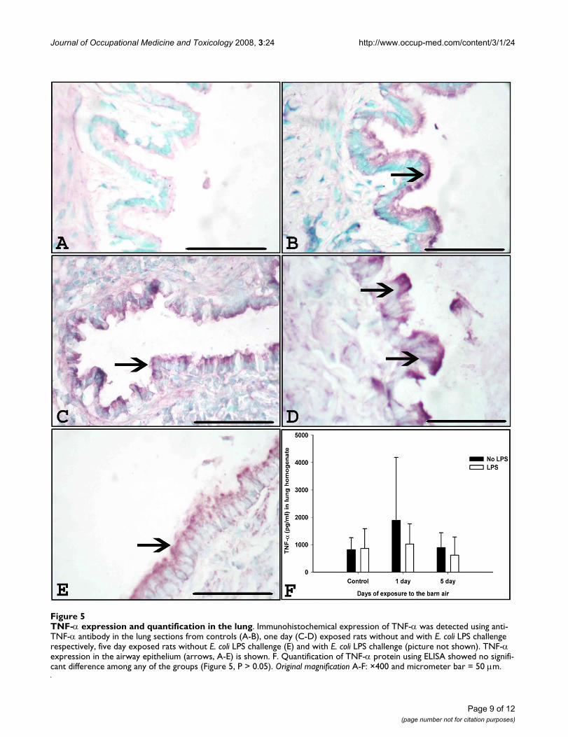
Expression and quantification of TNF-αImmunohistochemistry detected TNF-α in airway epithe-lium (Figure 5A–E), blood vessel wall, lung septa andoccasionally in AMs and quantification using ELISArevealed no difference among any of the groups (Figure5F, P > 0.05).
Expression and quantification of TGF-β2Immunohistochemistry detected TGF-β2 in airway epithe-lium (Figure 6A–E), blood vessel wall, lung septa andoccasionally in AMs and quantification using ELISArevealed that control rats without E. coli LPS challengeshowed higher levels of TGF-β2 compared to controls ratschallenged with E. coli LPS (Figure 6F, P = 0.001).
DiscussionWe conducted this study to investigate lung responses toa secondary LPS challenge in rats exposed to barn air. Ourdata show that lungs of rats exposed to barn air becamemore inflamed following challenge with LPS when com-pared to those exposed to barn air only. These data suggestthat lungs of animals exposed to pig barn air are capableof responding to microbial challenges.
We have previously demonstrated that significant PIMMrecruitment is observed at 48 hours after a single exposureto pig barn air and these recruited PIMMs exacerbatedlung inflammation in response to a secondary LPS chal-lenge [16]. Now, we studied secondary LPS-induced lunginflammation in rats exposed to the barn air prior toPIMM recruitment. We chose to do an E. coli LPS chal-lenge at 18 hours after single or five day barn exposureand confirmed the lack of significant recruitment ofPIMMs at these time points by immunohistochemicalstaining using ED-1 antibody which is a known markerfor rat monocytes/macrophages.
In the current study we have demonstrated induction oflung inflammation following one or five days of barnexposure as before [14-16]. The E. coli LPS challenge ofone day barn exposed but not control rats at 18 hour post-exposure showed an increase in granulocyte recruitmentcompared to respective control groups. However, therewere no differences in granulocyte numbers between LPS-treated barn exposed and control animals. Neutrophilsare the predominant granulocytes recruited into theinflamed lung [23] and are considered central to develop-ment of acute lung inflammation [24,25]. As expected wedid not observe an increase in lung monocyte/macro-phage numbers at 6 hour post-LPS challenge, as this earlytime point in acute lung inflammation is characterized byan early recruitment of neutrophils followed by mono-cytes and macrophages in the later periods [26,27]. How-ever, our previous work in rat endotoxin-induced lunginflammation model has also shown increased mono-cytes recruitment at 3 and 24 hour time points, bothdependent on neutrophils [28]. Our current data showthat lungs of animals, that had been exposed to barn air,experienced an inflammatory response following chal-lenge with LPS that was equal to, if not greater than thatexperienced by animal lungs that had not been exposed tobarn air prior to LPS challenge. Endotoxin or LPS in thebarn or intravenously administered LPS alone used in thisstudy will likely induce different host responses. Intrave-nously administered LPS very likely induce systemic vas-cular activation including lung vascular inflammationand exacerbate lung inflammation [16,17]. In contrast tothis, LPS in the barn air is associated with other contami-nants in the barn and primarily induces lung inflamma-tion and associated respiratory symptoms [1]. Some of thedifferences in the host responses following intravenousLPS challenge or the barn exposure may be due to differ-ences in dose/exposure, duration of exposure and pres-ence of other agents in the barn will all account fordifferences in host responses.
Barn exposed as well as control rats challenged with LPSshowed increased concentrations of IL-1β but not TNF-αin lung homogenates compared to their respective con-trols. Again, there were no differences in IL-1β or TNF-αexpression between barn exposed and control rats chal-lenged with LPS. IL-1β is produced by monocytes/macro-phages and neutrophils as well as endothelial cells andfibroblasts, and is a known early response cytokine inacute lung inflammation and induces expression of adhe-sion molecules to regulate neutrophil migration [29-33].IL-1β also directly activates neutrophils through stimula-tion of mitogen activated protein kinases to result inincreased superoxide anion production and respiratoryburst in neutrophils [34,35]. IL-1β also induces fever,increases vascular permeability, production of IL-6 andleukocyte adherence to endothelium [36,37]. On the
Page 7 of 12(page number not for citation purposes)

Journal of Occupational Medicine and Toxicology 2008, 3:24 http://www.occup-med.com/content/3/1/24
Page 8 of 12(page number not for citation purposes)
IL-1β expression and quantification in the lungFigure 4IL-1β expression and quantification in the lung. Immunohistochemical expression of IL-1β was detected using anti-IL-1β antibody in the lung sections from controls (A-B), one day (C-D) exposed rats without and with E. coli LPS challenge respec-tively, five day exposed rats without E. coli LPS challenge (E) and with E. coli LPS challenge (picture not shown). IL-1β expession in the airway epithelium (arrows, A-E) is shown. F. Quantification of IL-1β protein using ELISA shows increased concentrations in rats that received E. coli LPS compared to respective groups of rats that did not receive E. coli LPS (Figure 4, P < 0.001). Orig-inal magnification A-F: ×400 and micrometer bar = 50 μm.
Journal of Occupational Medicine and Toxicology 2008, 3:24 http://www.occup-med.com/content/3/1/24
Page 9 of 12(page number not for citation purposes)
TNF-α expression and quantification in the lungFigure 5TNF-α expression and quantification in the lung. Immunohistochemical expression of TNF-α was detected using anti-TNF-α antibody in the lung sections from controls (A-B), one day (C-D) exposed rats without and with E. coli LPS challenge respectively, five day exposed rats without E. coli LPS challenge (E) and with E. coli LPS challenge (picture not shown). TNF-α expression in the airway epithelium (arrows, A-E) is shown. F. Quantification of TNF-α protein using ELISA showed no signifi-cant difference among any of the groups (Figure 5, P > 0.05). Original magnification A-F: ×400 and micrometer bar = 50 μm.

Journal of Occupational Medicine and Toxicology 2008, 3:24 http://www.occup-med.com/content/3/1/24
Page 10 of 12(page number not for citation purposes)
TGF-β2 expression and quantification in the lungFigure 6TGF-β2 expression and quantification in the lung. Immunohistochemical expression of TGF-β2 was detected using anti- TGF-β2 antibody in the lung sections from controls (A-B), one day (C-D) exposed rats without and with E. coli LPS challenge respectively, five day exposed rats without E. coli LPS challenge (E) and with E. coli LPS challenge (picture not shown). TGF-β2 expression in the airway epithelium (arrows, A-E) is shown. F. Quantification of TGF-β2 protein using ELISA showed increased levels in control rats without E. coli LPS challenge compared to control rats with E. coli LPS challenge (*, P = 0.001).Original mag-nification A-F: ×400 and micrometer bar = 50 μm.

Journal of Occupational Medicine and Toxicology 2008, 3:24 http://www.occup-med.com/content/3/1/24
other hand, neutralization of IL-1β has proven protectiveand beneficial to the host [38]. Based on the expression ofthese two proinflammatory cytokines, it appears that sin-gle or multiple exposures to barn air do not dampen theinflammatory response of lungs to LPS challenge.
Because lung inflammation is controlled by a complexnetwork of both pro and anti-inflammatory cytokines[39], we examined the tissue expression and quantifica-tion of TGF-β2, a known anti-inflammatory cytokine withimportant roles in tissue repair and remodeling [40,41].The data show reduced expression of TGF-β2 in LPS chal-lenged control rats compared to control rats without E.coli LPS challenge. This observation indicates that the sup-pression of TGF-β2 in inflamed lungs may be due to anactive inflammatory reaction which is similar to previousreports of suppression of TGF-β2 expression in lungs ofLPS challenged rats [42]. We have reported similar datafrom rats challenged with E. coli LPS at 48 hours after sin-gle barn exposure which contain significant numbers ofPIMMs [16]. In contrast to LPS challenge 48 hours after anexposure to barn air or of control rats, we did not observeany change in the expression of TGF-β2 when rats werechallenged with LPS at 18 hours after one or five expo-sures to barn air. It seems that exposure to barn air mayhave suppressed expression of TGF-β2 to LPS challenge.Considering well established roles of TGF-β2 in lunginjury and repair, it may be useful to undertake furtherstudies to explore the role of this cytokine in lung inflam-mation and repair following exposure to the barn air. Fur-thermore, because of the involvement of TLR4, it may alsobe useful to conduct in vitro studies to clarify the cell sig-naling pathways activated by the barn air.
ConclusionOur data show that exposure to swine barn environmentfor one or five days induces lung inflammation, while asecondary challenge with E. coli LPS exacerbates the lunginflammation in rats exposed to pig barn air. These datasuggest that exposure to barn air does not suppress lung'sability to respond to a secondary LPS challenge.
Competing interestsThe authors declare that they have no competing interests.
Authors' contributionsCC carried out the experiment, did the histopathologicalevaluation, statistical analyses and drafted the manu-script. TK did the immunohistochemistry and GKA partic-ipated in the quantification of immunohistochemistrydata. HT participated in statistical analyses and manu-script preparation. BS conceived of the study, participatedin its design and coordination as well as manuscript prep-aration. All the authors have read and approved the finalmanuscript.
AcknowledgementsThe work was supported through a grant from Lung Association of Sas-katchewan to Dr. B. Singh. Dr. C. Charavaryamath is a recipient of a Grad-uate Merit Scholarship from College of Graduate Studies and Research, Founding Chairs Graduate Fellowship from Canadian Centre for Health and Safety in Agriculture, University of Saskatchewan and a scholarship from the CIHR Strategic Training Program in Public Health and the Agricul-tural Rural Ecosystem and Partner Institutes including the Institute of Can-cer Research, Institute of Circulatory and Respiratory Health, Institute of Infection and Immunity, Institute of Population and Public Health and the University of Saskatchewan. Taryn Keet was a recipient of an Undergradu-ate Summer Research Award from the Natural Sciences and Engineering Research Council of Canada. Gurpreet K. Aulakh is a recipient of Graduate Teaching Fellowship from the Department of Veterinary Biomedical Sci-ences, University of Saskatchewan.
References1. Charavaryamath C, Singh B: Pulmonary effects of exposure to
pig barn air. J Occup Med Toxicol 2006, 1:10.2. Donham KJ, Popendorf W, Palmgren U, Larsson L: Characteriza-
tion of dusts collected from swine confinement buildings. AmJ Ind Med 1986, 10:294-297.
3. Cole D, Todd L, Wing S: Concentrated swine feeding opera-tions and public health: a review of occupational and com-munity health effects. Environ Health Perspect 2000, 108:685-699.
4. Wenger I: Air Quality and Health of Career Pig Barn Work-ers. Advances in Pork Production 1999, 10:93-101.
5. Wenger II, Ouellette CA, Feddes JJ, Hrudey SE: The design and useof the Personal Environmental Sampling Backpack (PESB II)for activity-specific exposure monitoring of career pig barnworkers. J Agric Saf Health 2005, 11:315-324.
6. Asmar S, Pickrell JA, Oehme FW: Pulmonary diseases caused byairborne contaminants in swine confinement buildings. VetHum Toxicol 2001, 43:48-53.
7. Donham KJ, Popendorf WJ: Ambient levels of selected gasesinside swine confinement buildings. Am Ind Hyg Assoc J 1985,46:658-661.
8. Cormier Y, Israel-Assayag E, Racine G, Duchaine C: Farming prac-tices and the respiratory health risks of swine confinementbuildings. Eur Respir J 2000, 15:560-565.
9. Larsson K, Eklund AG, Hansson LO, Isaksson BM, Malmberg PO:Swine dust causes intense airways inflammation in healthysubjects. Am J Respir Crit Care Med 1994, 150:973-977.
10. Malmberg P, Larsson K: Acute exposure to swine dust causesbronchial hyperresponsiveness in healthy subjects. Eur RespirJ 1993, 6:400-404.
11. Larsson BM, Palmberg L, Malmberg PO, Larsson K: Effects of expo-sure to swine dust on levels of IL-8 in airway lavage fluid. Tho-rax 1997, 52:638-642.
12. Palmberg L, Larssson BM, Malmberg P, Larsson K: Airwayresponses of healthy farmers and nonfarmers to exposure ina swine confinement building. Scand J Work Environ Health 2002,28:256-263.
13. Israel-Assayag E, Cormier Y: Adaptation to organic dust expo-sure: a potential role of l-selectin shedding? Eur Respir J 2002,19:833-837.
14. Charavaryamath C, Janardhan KS, Townsend HG, Willson P, Singh B:Multiple exposures to swine barn air induce lung inflamma-tion and airway hyper-responsiveness. Respir Res 2005, 6:50.
15. Charavaryamath C, Juneau V, Suri SS, Janardhan KS, Townsend H,Singh B: Role of toll-like receptor 4 in lung inflammation fol-lowing exposure to Swine barn air. Exp Lung Res 2008, 34:19-35.
16. Gamage LN, Charavaryamath C, Swift TL, Singh B: Lung inflamma-tion following a single exposure to swine barn air. J Occup MedToxicol 2007, 2:18.
17. Charavaryamath C, Janardhan KS, Caldwell S, Singh B: Pulmonaryintravascular monocytes/macrophages in a rat model of sep-sis. Anat Rec A Discov Mol Cell Evol Biol 2006, 288:1259-1271.
18. Singh B, Rawlings N, Kaur A: Expression of integrin αvβ3 in pig,dog and cattle. Histol Histopathol 2001, 16(4):1037-1046.
19. Dijkstra CD, Dopp EA, Joling P, Kraal G: The heterogeneity ofmononuclear phagocytes in lymphoid organs: distinct mac-
Page 11 of 12(page number not for citation purposes)

Journal of Occupational Medicine and Toxicology 2008, 3:24 http://www.occup-med.com/content/3/1/24
Publish with BioMed Central and every scientist can read your work free of charge
"BioMed Central will be the most significant development for disseminating the results of biomedical research in our lifetime."
Sir Paul Nurse, Cancer Research UK
Your research papers will be:
available free of charge to the entire biomedical community
peer reviewed and published immediately upon acceptance
cited in PubMed and archived on PubMed Central
yours — you keep the copyright
Submit your manuscript here:http://www.biomedcentral.com/info/publishing_adv.asp
BioMedcentral
rophage subpopulations in rat recognized by monoclonalantibodies ED1, ED2 and ED3. Adv Exp Med Biol 1985,186:409-419.
20. Damoiseaux JG, Dopp EA, Calame W, Chao D, MacPherson GG,Dijkstra CD: Rat macrophage lysosomal membrane antigenrecognized by monoclonal antibody ED1. Immunology 1994,83:140-147.
21. van Goor H, Fidler V, Weening JJ, Grond J: Determinants of focaland segmental glomerulosclerosis in the rat after renal abla-tion. Evidence for involvement of macrophages and lipids.Lab Invest 1991, 64:754-765.
22. Gordon JR, Zhang X, Stevenson K, Cosford K: Thrombin inducesIL-6 but not TNFalpha secretion by mouse mast cells:threshold-level thrombin receptor and very low level Fcepsi-lonRI signaling synergistically enhance IL-6 secretion. CellImmunol 2000, 205:128-135.
23. Thorn J: The inflammatory response in humans after inhala-tion of bacterial endotoxin: a review. Inflamm Res 2001,50:254-261.
24. Kinoshita M, Ono S, Mochizuki H: Neutrophils mediate acutelung injury in rabbits: role of neutrophil elastase. Eur Surg Res2000, 32:337-346.
25. Abraham E: Neutrophils and acute lung injury. Crit Care Med2003, 31:S195-S199.
26. Larsen GL, Holt PG: The concept of airway inflammation. Am JRespir Crit Care Med 2000, 162:S2-S6.
27. Kaplanski G, Marin V, Montero-Julian F, Mantovani A, Farnarier C: IL-6: a regulator of the transition from neutrophil to monocyterecruitment during inflammation. Trends Immunol 2003,24:25-29.
28. Janardhan KS, Sandhu SK, Singh B: Neutrophil depletion inhibitsearly and late monocyte/macrophage increase in lunginflammation. Front Biosci 2006, 11:1569-1576.
29. Dinarello CA: Proinflammatory cytokines. Chest 2000,118:503-508.
30. Kasama T, Miwa Y, Isozaki T, Odai T, Adachi M, Kunkel SL: Neu-trophil-derived cytokines: potential therapeutic targets ininflammation. Curr Drug Targets Inflamm Allergy 2005, 4:273-279.
31. Tosi MF: Innate immune responses to infection. J Allergy ClinImmunol 2005, 116:241-249.
32. Williams JH Jr, Patel SK, Hatakeyama D, Arian R, Guo K, Hickey TJ,et al.: Activated pulmonary vascular neutrophils as earlymediators of endotoxin-induced lung inflammation. Am JRespir Cell Mol Biol 1993, 8:134-144.
33. Parsey MV, Tuder RM, Abraham E: Neutrophils Are Major Con-tributors to Intraparenchymal Lung IL-1{beta} ExpressionAfter Hemorrhage and Endotoxemia. J Immunol 1998,160:1007-1013.
34. Yagisawa M, Yuo A, Kitagawa S, Yazaki Y, Togawa A, Takaku F: Stim-ulation and priming of human neutrophils by IL-1 alpha andIL-1 beta: complete inhibition by IL-1 receptor antagonistand no interaction with other cytokines. Exp Hematol 1995,23:603-608.
35. Suzuki K, Hino M, Kutsuna H, Hato F, Sakamoto C, Takahashi T, et al.:Selective Activation of p38 Mitogen-Activated ProteinKinase Cascade in Human Neutrophils Stimulated by IL-1{beta}. J Immunol 2001, 167:5940-5947.
36. Strieter RM, Belperio JA, Keane MP: Cytokines in innate hostdefense in the lung. J Clin Invest 2002, 109:699-705.
37. Feghali CA, Wright TM: Cytokines in acute and chronic inflam-mation. Front Biosci 1997, 2:d12-d26.
38. Standiford TJ: Anti-inflammatory cytokines and cytokineantagonists. Curr Pharm Des 2000, 6:633-649.
39. Thacker EL: Lung inflammatory responses. Vet Res 2006,37:469-486.
40. Letterio JJ, Roberts AB: TGF-beta: a critical modulator ofimmune cell function. Clin Immunol Immunopathol 1997,84:244-250.
41. Bartram U, Speer CP: The role of transforming growth factorbeta in lung development and disease. Chest 2004,125:754-765.
42. Ayache N, Boumediene K, Mathy-Hartert M, Reginster JY, HenrotinY, Pujol JP: Expression of TGF-betas and their receptors is dif-ferentially modulated by reactive oxygen species and nitricoxide in human articular chondrocytes. Osteoarthritis Cartilage2002, 10:344-352.
Page 12 of 12(page number not for citation purposes)
Copyright © 2022 FDOKUMEN