Low Birth Weight in Perinatally HIV-Exposed Uninfected Infants: Observations in Urban Settings in...
10
Low Birth Weight in Perinatally HIV-Exposed Uninfected Infants: Observations in Urban Settings in Cameroon Casimir Ledoux Sofeu 1,2,3 , Josiane Warszawski 4,5,6 , Francis Ateba Ndongo 7 , Ida Calixte Penda 8,9 , Suzie Tetang Ndiang 10 , Georgette Guemkam 7,11 , Nicaise Makwet 8 , Fe ´ licite ´ Owona 1 , Anfumbom Kfutwah 12 , Patrice Tchendjou 1 , Gae ¨ tan Texier 1,13 , Maurice Tchuente 2,3 , Albert Faye 14,15 , Mathurin Cyrille Tejiokem 1,4 * " and the ANRS-PEDIACAM study group " 1 Service d’Epide ´ miologie et de Sante ´ Publique, Centre Pasteur du Cameroun, Membre du Re ´ seau International des Instituts Pasteur, Yaounde ´ , Cameroun, 2 Universite ´ de Yaounde ´ I, IRD UMI 209 UMMISCO, Yaounde ´, Cameroun, 3 Laboratoire International en Recherche Informatique et Mathe ´ matiques Applique ´es, Equipe Idasco, Yaounde ´, Cameroun, 4 Equipe 4 (VIH et IST) - INSERM U1018 (CESP), Le Kremlin Bice ˆ tre, France, 5 Assistance Publique des Ho ˆ pitaux de Paris, Service d’Epide ´miologie et de Sante ´ Publique, Ho ˆ pital de Bice ˆtre, Le Kremlin Bice ˆtre, France, 6 Universite ´ de Paris Sud 11, Paris, France, 7 Centre Me `re et Enfant de la Fondation Chantal Biya, Yaounde ´, Cameroun, 8 Ho ˆ pital de Jour, Ho ˆ pital Laquintinie, Douala, Cameroun, 9 Faculte ´ de Me ´decine et des Sciences Pharmaceutiques, Universite ´ de Douala, Douala, Cameroun, 10 Service de Pe ´diatrie, Centre Hospitalier d’Essos, Yaounde ´, Cameroun, 11 Maternite ´ Principale, Ho ˆ pital Central, Yaounde ´, Cameroun, 12 Service de Virologie, Centre Pasteur du Cameroun, Membre du Re ´ seau International des Instituts Pasteur, Yaounde ´, Cameroun, 13 SESSTIM (UMR 912), Universite ´ Aix-Marseille, Marseille, France, 14 Assistance Publique des Ho ˆ pitaux de Paris, Pe ´diatrie Ge ´ne ´rale, Ho ˆ pital Robert Debre ´, Paris, France, 15 Universite ´ Paris 7 Denis Diderot, Paris Sorbonne Cite ´, Paris, France Abstract Background: The consequences of maternal HIV infection for fetal growth are controversial. Here, we estimated the frequency of small for gestational age and gender (SGAG) among neonates born to HIV-infected or uninfected mothers and assessed the contribution, if any, of maternal HIV to the risk of SGAG. Methods: The data used were obtained from the ANRS-Pediacam cohort in Cameroon. Pairs of newborns, one to a HIV- infected mother and the other to an uninfected mother, were identified during the first week of life, and matched on gender and recruitment site from 2007–2010. SGAG was defined in line with international recommendations as a birth weight Z-score adjusted for gestational age at delivery and gender more than two standard deviations below the mean (2 2SD). Considering the matched design, logistic regression modeling was adjusted on site and gender to explore the effect of perinatal HIV exposure on SGAG. Results: Among the 4104 mother-infant pairs originally enrolled, no data on birth weight and/or gestational age were available for 108; also, 259 were twins and were excluded. Of the remaining 3737 mother-infant pairs, the frequency of SGAG was 5.3% (95%CI: 4.6–6.0), and was significantly higher among HIV-infected infants (22.4% vs. 6.3%; p,.001) and lower among HIV-unexposed uninfected infants (3.5% vs. 6.3%; p,.001) than among HIV-exposed uninfected infants. Similarly, SGAG was significantly more frequent among HIV-infected infants (aOR: 4.1; 2.0–8.1) and less frequent among HIV- unexposed uninfected infants (aOR: 0.5; 0.4–0.8) than among HIV-exposed uninfected infants. Primiparity (aOR: 1.9; 1.3–2.7) and the presence of any disease during pregnancy (aOR: 1.4; 1.0–2.0) were identified as other contributors to SGAG. Conclusion: Maternal HIV infection was independently associated with SGAG for HIV-exposed uninfected infants. This provides further evidence of the need for adapted monitoring of pregnancy in HIV-infected women, especially if they are symptomatic, to minimize additional risk factors for SGAG. Citation: Sofeu CL, Warszawski J, Ateba Ndongo F, Penda IC, Tetang Ndiang S, et al. (2014) Low Birth Weight in Perinatally HIV-Exposed Uninfected Infants: Observations in Urban Settings in Cameroon. PLoS ONE 9(4): e93554. doi:10.1371/journal.pone.0093554 Editor: Soren Gantt, University of British Columbia, Canada Received October 24, 2013; Accepted March 5, 2014; Published April 3, 2014 Copyright: ß 2014 Sofeu et al. This is an open-access article distributed under the terms of the Creative Commons Attribution License, which permits unrestricted use, distribution, and reproduction in any medium, provided the original author and source are credited. Funding: This study is sponsored by the French National Agency for Research on AIDS and Viral Hepatitis (ANRS). The authors also acknowledge the material support of the ‘‘Fondation Total’’ to this project. The funders had no role in study design, data collection and analysis, decision to publish, or preparation of the manuscript. Competing Interests: The authors have declared that no competing interests exist. * E-mail: [email protected] " Membership of the ANRS-PEDIACAM study group is provided in the Acknowledgments. Introduction Impaired fetal growth can have devastating effects on subse- quent infant growth, survival [1–9] and neurocognitive develop- ment [10]. Fetal growth is influenced by diverse medical, obstetrical, socio-economic and behavioral factors [11–16]. Studies in both industrialized and developing countries report inconsistent results concerning the consequences of maternal HIV infection on fetal growth. Some authors report that anthropomet- ric measures for both HIV-infected and uninfected infants born to PLOS ONE | www.plosone.org 1 April 2014 | Volume 9 | Issue 4 | e93554
Transcript of Low Birth Weight in Perinatally HIV-Exposed Uninfected Infants: Observations in Urban Settings in...

Low Birth Weight in Perinatally HIV-Exposed UninfectedInfants: Observations in Urban Settings in CameroonCasimir Ledoux Sofeu1,2,3, Josiane Warszawski4,5,6, Francis Ateba Ndongo7, Ida Calixte Penda8,9,
Suzie Tetang Ndiang10, Georgette Guemkam7,11, Nicaise Makwet8, Felicite Owona1,
Anfumbom Kfutwah12, Patrice Tchendjou1, Gaetan Texier1,13, Maurice Tchuente2,3, Albert Faye14,15,
Mathurin Cyrille Tejiokem1,4*" and the ANRS-PEDIACAM study group"
1 Service d’Epidemiologie et de Sante Publique, Centre Pasteur du Cameroun, Membre du Reseau International des Instituts Pasteur, Yaounde, Cameroun, 2 Universite de
Yaounde I, IRD UMI 209 UMMISCO, Yaounde, Cameroun, 3 Laboratoire International en Recherche Informatique et Mathematiques Appliquees, Equipe Idasco, Yaounde,
Cameroun, 4 Equipe 4 (VIH et IST) - INSERM U1018 (CESP), Le Kremlin Bicetre, France, 5 Assistance Publique des Hopitaux de Paris, Service d’Epidemiologie et de Sante
Publique, Hopital de Bicetre, Le Kremlin Bicetre, France, 6 Universite de Paris Sud 11, Paris, France, 7 Centre Mere et Enfant de la Fondation Chantal Biya, Yaounde,
Cameroun, 8 Hopital de Jour, Hopital Laquintinie, Douala, Cameroun, 9 Faculte de Medecine et des Sciences Pharmaceutiques, Universite de Douala, Douala, Cameroun,
10 Service de Pediatrie, Centre Hospitalier d’Essos, Yaounde, Cameroun, 11 Maternite Principale, Hopital Central, Yaounde, Cameroun, 12 Service de Virologie, Centre
Pasteur du Cameroun, Membre du Reseau International des Instituts Pasteur, Yaounde, Cameroun, 13 SESSTIM (UMR 912), Universite Aix-Marseille, Marseille, France,
14 Assistance Publique des Hopitaux de Paris, Pediatrie Generale, Hopital Robert Debre, Paris, France, 15 Universite Paris 7 Denis Diderot, Paris Sorbonne Cite, Paris,
France
Abstract
Background: The consequences of maternal HIV infection for fetal growth are controversial. Here, we estimated thefrequency of small for gestational age and gender (SGAG) among neonates born to HIV-infected or uninfected mothers andassessed the contribution, if any, of maternal HIV to the risk of SGAG.
Methods: The data used were obtained from the ANRS-Pediacam cohort in Cameroon. Pairs of newborns, one to a HIV-infected mother and the other to an uninfected mother, were identified during the first week of life, and matched ongender and recruitment site from 2007–2010. SGAG was defined in line with international recommendations as a birthweight Z-score adjusted for gestational age at delivery and gender more than two standard deviations below the mean (22SD). Considering the matched design, logistic regression modeling was adjusted on site and gender to explore the effectof perinatal HIV exposure on SGAG.
Results: Among the 4104 mother-infant pairs originally enrolled, no data on birth weight and/or gestational age wereavailable for 108; also, 259 were twins and were excluded. Of the remaining 3737 mother-infant pairs, the frequency ofSGAG was 5.3% (95%CI: 4.6–6.0), and was significantly higher among HIV-infected infants (22.4% vs. 6.3%; p,.001) andlower among HIV-unexposed uninfected infants (3.5% vs. 6.3%; p,.001) than among HIV-exposed uninfected infants.Similarly, SGAG was significantly more frequent among HIV-infected infants (aOR: 4.1; 2.0–8.1) and less frequent among HIV-unexposed uninfected infants (aOR: 0.5; 0.4–0.8) than among HIV-exposed uninfected infants. Primiparity (aOR: 1.9; 1.3–2.7)and the presence of any disease during pregnancy (aOR: 1.4; 1.0–2.0) were identified as other contributors to SGAG.
Conclusion: Maternal HIV infection was independently associated with SGAG for HIV-exposed uninfected infants. Thisprovides further evidence of the need for adapted monitoring of pregnancy in HIV-infected women, especially if they aresymptomatic, to minimize additional risk factors for SGAG.
Citation: Sofeu CL, Warszawski J, Ateba Ndongo F, Penda IC, Tetang Ndiang S, et al. (2014) Low Birth Weight in Perinatally HIV-Exposed Uninfected Infants:Observations in Urban Settings in Cameroon. PLoS ONE 9(4): e93554. doi:10.1371/journal.pone.0093554
Editor: Soren Gantt, University of British Columbia, Canada
Received October 24, 2013; Accepted March 5, 2014; Published April 3, 2014
Copyright: � 2014 Sofeu et al. This is an open-access article distributed under the terms of the Creative Commons Attribution License, which permitsunrestricted use, distribution, and reproduction in any medium, provided the original author and source are credited.
Funding: This study is sponsored by the French National Agency for Research on AIDS and Viral Hepatitis (ANRS). The authors also acknowledge the materialsupport of the ‘‘Fondation Total’’ to this project. The funders had no role in study design, data collection and analysis, decision to publish, or preparation of themanuscript.
Competing Interests: The authors have declared that no competing interests exist.
* E-mail: [email protected]
" Membership of the ANRS-PEDIACAM study group is provided in the Acknowledgments.
Introduction
Impaired fetal growth can have devastating effects on subse-
quent infant growth, survival [1–9] and neurocognitive develop-
ment [10]. Fetal growth is influenced by diverse medical,
obstetrical, socio-economic and behavioral factors [11–16].
Studies in both industrialized and developing countries report
inconsistent results concerning the consequences of maternal HIV
infection on fetal growth. Some authors report that anthropomet-
ric measures for both HIV-infected and uninfected infants born to
PLOS ONE | www.plosone.org 1 April 2014 | Volume 9 | Issue 4 | e93554

HIV-infected mothers are significantly lower than those for infants
born to HIV-negative mothers [1,4,13,17]. Other studies failed to
detect any maternal HIV-related effects on growth
[3,15,16,18,19]. The reported effects on fetal growth of antiret-
roviral therapy used during pregnancy are similarly contradictory
[20–23]. There is also conflicting data regarding perinatal HIV-
exposure and post natal infant growth. Some authors report no
differences whereas others have observed growth faltering in HIV-
exposed uninfected infants [4,10,18,19,24,25]. These discrepan-
cies may be the consequence of methodological issues, and in
particular the use in most of these studies of anthropometric
statistics that do not take into account both gestational age at
delivery and gender.
Programs to prevent mother-to-child HIV transmission have
been implemented and are generally successful in sub-Saharan
Africa: they include improved healthcare coverage and access to
antiretroviral therapy for HIV-infected women and their infants.
As a result, most infants now escape HIV infection and the
number of HIV-exposed uninfected infants will increase such that
any problems specifically associated with this group will become of
major public health importance. Therefore, data on HIV-exposed
uninfected infant growth are needed as a basis for preventive
strategies to ensure appropriate care and to evaluate the impact of
HIV and antiretroviral drug use on infant life [4,24,25].
We estimated the frequency of SGAG among HIV-infected and
uninfected newborns to HIV-infected mothers, and compared
these values to those for newborns to uninfected mothers, in urban
settings of Cameroon. We used data from the ongoing ANRS-
Pediacam study, in which infants were enrolled at birth or before
the 8th day of life. We studied birth weight z-scores adjusted on
gestational age at delivery and gender to appreciate the effects of
maternal HIV infection on the risk of SGAG in HIV-exposed
uninfected infants.
Methods
Ethics StatementThe ANRS-Pediacam study was granted ethical approval in
Cameroon by the National Ethics Committee and in France by
the Biomedical Research Committee of the Pasteur Institute of
Paris. The Cameroon Ministry of Public Health gave administra-
tive authorization to start the study. Written informed consent was
obtained from parents or guardians prior to inclusion of infants
into the research project.
Data SourceData used in this analysis were obtained from the ANRS-
Pediacam cohort based in three referral hospitals in Cameroon
(The Maternity of the Central hospital/Mother and Child Center
of the Chantal Biya Foundation (MCH/MCC-CBF), Essos
Hospital Center in Yaounde (EHC) and the Laquintinie Hospital
in Douala (LH)) and coordinated by the Centre Pasteur of
Cameroon. The ANRS-Pediacam cohort study is observational
and was designed to assess the feasibility of early infant diagnosis of
HIV and early antiretroviral multitherapy in HIV-infected infants,
and to evaluate the humoral response of these children to vaccines
of the Expanded Program on Immunization [26]. All HIV-
infected mothers who were seen from November 2007 to October
2010 at one of the participating maternity or pediatric wards
during the first week after delivery were invited to participate in
the study. Infants born to uninfected mothers matched on study
site and sex were also identified and enrolled during the first week
of life as controls. The newborn pairs were followed, according to
the Cameroon National EPI calendar, at 6, 10 and 14 weeks.
Samples for HIV virological testing were collected from HIV-
exposed infants at the first follow-up visit planned at 6 weeks, as
previously described [26]. Incentives, including free medical
support for consultation, biological analysis, additional vaccines
and reimbursement of transport costs, were provided to parents/
caregivers by the project during follow up visits.
ParticipantsAmong the 4104 mother-infant pairs initially enrolled in the
ANRS-Pediacam study, birth weight and/or gestational age data
were missing for 108; these infants were excluded because of the
lack of adequate outcome data. An additional 259 cases involved
twin, and because multiple birth increases the risk of low birth
weight, these cases were also excluded from the study [7]. Our
analyses were thus conducted on the remaining 3737 mother-
infant pairs (Figure 1).
Data CollectionData were collected by interviewing the mothers/guardians and
completed using the mothers’ medical records (in their hospital
books). Socioeconomic and demographic characteristics collected
included: mothers’ age, education level, employment and marital
status, electricity and water supply at home, existence of a
functional fridge at home. The obstetrical history recorded
included: gravidity, use of malarial drugs and pathologies during
pregnancy, date and mode of delivery, gestational age at delivery,
infant birth weight, length, head circumference and gender, and
any neonatal or post partum complications. The medical data
included: HIV status according to the Cameroonian national
algorithm based on rapid tests, and the use of any ART for
PMTCT or for the mothers’ own health during pregnancy (ART
initiated only at delivery was not considered). During the
recruitment period of this study, the national PMTCT policy in
Cameroon was based on the 2006 WHO guidelines: pregnant
HIV-infected women who had CD4 cell counts of 200 cells/mm3
or less were advised to start ART for their own health, and to
continue lifelong ART using a triple combination regimen;
however, women with higher CD4 cell counts were not considered
medically eligible for such treatment, and were advised to take
zidovudine (AZT) from 28 weeks of gestation plus a single-dose
nevirapine at the onset of labor, a combination of AZT+lamivudine (3TC) during delivery and one week postpartum, with
infant prophylaxis for one week after birth [27].
The obstetrical estimate of gestational age was mainly based on
the last menstrual period unless adjusted by the midwife on the
basis of clinical assessment in conformity with routine practice.
These estimations were sometimes confirmed by obstetrical
ultrasound scans. Infant birth weight (in grams) was measured in
the recumbent position using a Salter scale. For babies delivered at
home or on the way to the maternity, weight measured at arrival
was recorded. Birth length was measured with the baby supine and
the crown of the head touching a vertical headboard. The legs
were extended gently and the length recorded using a tape
graduated in centimeters from the headboard to the infant’s heel.
Head circumference (occipitofrontal) was also measured using a
tape positioned just above the eyebrows and placed posteriorly.
Outcome and Main Exposure DefinitionsThe studied outcome was SGAG (small for gestational age and
gender) defined as a birth weight Z-score adjusted for gestational
age at delivery and gender that is more than two standard
deviations below the mean (22SD), in line with international
recommendations [28]. Birth weight Z-scores were calculated
using French standards, calculated from AUDIPOG [29]. The
Low Birth Weight in HIV-Exposed Uninfected Infants
PLOS ONE | www.plosone.org 2 April 2014 | Volume 9 | Issue 4 | e93554
AUDIPOG database include data from 211,337 infants born in
209 French maternities between 1999 and 2005. Based on these
data, different model equations with high order polynomials have
been fitted to calculate predicted mean births weight and standard
deviations for male and female infants born at any gestational age.
In this study, we used these equations to generate Z-scores using
the following formula: Z score = (WO–WP)/SDP, where WO:
weight observed, WP: weight predicted, SDP: standard deviation
predicted. The main exposure investigated was the HIV status of
the neonate, classified into three categories: HIV-infected, HIV-
uninfected exposed to maternal HIV, HIV-uninfected born to a
HIV seronegative mother. The HIV status of infants born to HIV-
infected mothers was determined at age 6 weeks by a polymerase
chain reaction (PCR)-based test. If HIV was not detected, the
infant was considered to be uninfected with HIV. Infants were
considered to be HIV-infected if the first and the second sample
were positive. Infants infected by HIV subsequently through
breastfeeding were considered to be HIV-uninfected but exposed
at the time of this analysis.
Statistical AnalysisMaternal and infant characteristics were described using
frequencies for categorical variables, medians and interquartile
ranges for continuous variables, and were compared between the
three groups of infants defined according to their HIV status. The
first step was to analyze the association between the risk of SGAG
and the infant HIV status (three main categories), with each
sociodemographic, medical and obstetrical characteristic separate-
ly, to identify potential confounders. The independent association
of SGAG with maternal and infant HIV status, was studied in the
second step, taking into account potential confounders. Logistic
regression models, all adjusted for matching variables (study site
and infant’s gender), were used for both these steps. In the second
step, the initial model included variables known to be associated
with SGAG, or found to be significant at the 0.25 level in first step
of the analysis. The final model was obtained by successively
removing variables not associated at a p-value ,0.05 only if the
odds ratios for remaining variables were unchanged and taking
interactions into account. Known risk factors were maintained in
the final model [30]. The above analyses were also conducted
separately for HIV-uninfected infants born to HIV-negative
mothers and HIV-exposed uninfected infants. All analyses were
performed using R version 2.12.1 software and Epicalc package. A
p-value of 0.05 was considered as the cutoff for statistical
significance.
Results
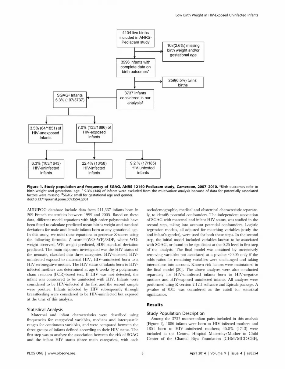
Study Population DescriptionAmong the 3737 mother-infant pairs included in this analysis
(Figure 1), 1886 infants were born to HIV-infected mothers and
1851 born to HIV-uninfected mothers; 45.8% (1713) were
included at the Central Hospital Maternity/Mother to Child
Center of the Chantal Biya Foundation (CHM/MCC-CBF),
Figure 1. Study population and frequency of SGAG, ANRS 12140-Pediacam study, Cameroon, 2007–2010. *Birth outcomes refer tobirth weight and gestational age. { 9.3% (346) of infants were excluded from the multivariate analysis because of data for potentially associatedfactors were missing. `SGAG: small for gestational age and gender.doi:10.1371/journal.pone.0093554.g001
Low Birth Weight in HIV-Exposed Uninfected Infants
PLOS ONE | www.plosone.org 3 April 2014 | Volume 9 | Issue 4 | e93554

25.4% (951) at the Hospital Center Essos (HCE) in Yaounde and
28.7% (1073) at the Laquintinie Hospital (LH) in Douala. About
85.5% (3196) of all the infants enrolled were born in the three
maternities participating in the study, 13.8% (514) in other
maternities of the cities and 0.7% (27) at home or on their way to a
healthcare facility.
The median age at inclusion of infants was 2 days (IQR: 1–5)
for infants born at hospital, and 4 days for those born at home or
on their way to hospital (IQR: 2–6). The two groups of infants
were comparable regarding anthropometric characteristics but
there were other significant differences: the mothers of infants
born at home had a lower level of education (40.7% vs. 13.6%,
p = .001), and more of the infants were born before term (,37
weeks) (37.0% vs. 11.3%, p,.001). The median age of mothers
was 29.0 years (25.0–33.0), and this value was significantly lower
for HIV-uninfected mothers than HIV-infected mothers: (28.0
years (24.0–32.0) vs. 29.5 years (26.0–33.0); p,.001). The median
gestational age at delivery was 39.0 (IQR: 38.0–40.0) weeks.
About 89.3% (1685/1886) of HIV-infected mothers received
ART before or during pregnancy for PMTCT and/or for their
own health. Complete data on ART regimen and time at first
exposure was available for 1553 of these: 792 (51.0%) took ART
for prophylaxis, 331 (21.3%) started HAART during pregnancy
and 430 (27.7%) before pregnancy. Nearly half of the HIV-
infected mothers (44.9% (846/1886)) knew their HIV status before
pregnancy. Only 928 mothers reported CD4 counts determined
during pregnancy. Of the 1886 infants born to HIV-infected
mothers, 1701 were tested for HIV at a median age of 6.0 weeks
(IQR: 6.0–7.0) and 58 were infected.
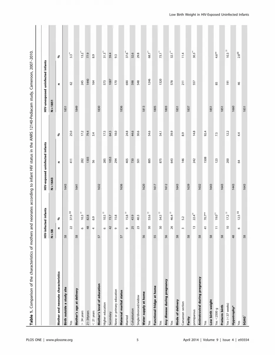
Maternal and infant characteristics at delivery for the HIV-
exposed uninfected infant group were significantly different from
those for the HIV-unexposed uninfected infant group but similar
to those for the HIV-infected infant group (Table 1). Relative to
the HIV-unexposed uninfected infant group, mothers of the HIV-
exposed uninfected infant group were mostly of low socio-
demographic and economic background (low level of education,
absence of running water at home, and no functional fridge at
home). The proportion of primiparous women was lower among
mothers of HIV-exposed uninfected infants than mothers of HIV-
unexposed uninfected infants (14.8 % vs. 30.3%, p,0.001) and
similar for the HIV-infected group. The proportion of premature
births was similar in the three groups.
Infants Anthropometric Characteristics and Frequency ofSGAG
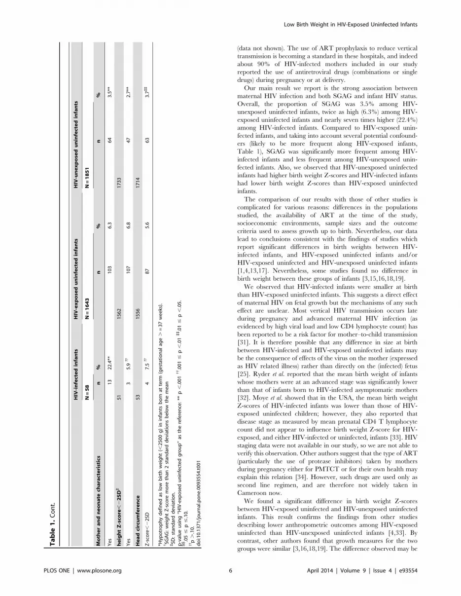
The median birth weight was 3,250 g (IQR: 2,910–3,500),
significantly higher in infants born to HIV-uninfected mothers
than HIV-exposed uninfected infants (3,300 g vs. 3,200 g; p,.001)
and lower in HIV-infected infants than HIV-exposed uninfected
infants (2,960 g vs. 3,200 g; p,.001). The length at birth was 50
cm (IQR: 49–52) and head circumference 34 cm (IQR: 33–35).
HIV-exposed uninfected infants had lower mean birth weight
Z-score (20.13 vs. 0.11; p,.001), lower mean length Z-score (0.39
vs. 0.78; p,.001) and lower mean head circumference Z-score
(0.07 vs. 0.16; p = .002) than HIV-unexposed uninfected infants.
HIV-exposed uninfected infants had higher mean birth weight Z-
score (20.13 vs. 20.67; p,.04) than HIV-infected infants, and
comparable mean length Z-score (0.39 vs. 0.10; p,.16) and mean
head circumference Z-score (0.07 vs. 0.44; p = .60).
The overall proportion of low birth weight was 7.5% among
HIV-exposed uninfected infants and 4.4% of them were hypo-
trophic. These proportions were significantly higher than for the
HIV-unexposed uninfected infant group (4.6% and 2.8%,
respectively) and lower than for the HIV-infected infant group
(19.0% and 12.5%, respectively) (Table 1).
The overall frequency of SGAG was 5.3% [CI: 4.6–6.0], and
SGAG was significantly more frequent among HIV-exposed
infants than HIV-unexposed uninfected infants (7.0% vs 3.5%;
p = ,.001, Figure 1). The frequency of SGAG was 22.4% [11.7–
33.1] among the HIV-infected infants, 6.3% [5.1–7.5] among the
HIV-exposed uninfected infants, 3.5% [2.7–4.3] among the HIV-
unexposed uninfected infants, and 9.2% [5.0–13.4] among HIV-
exposed untested infants (Figure 1 and Table 1).
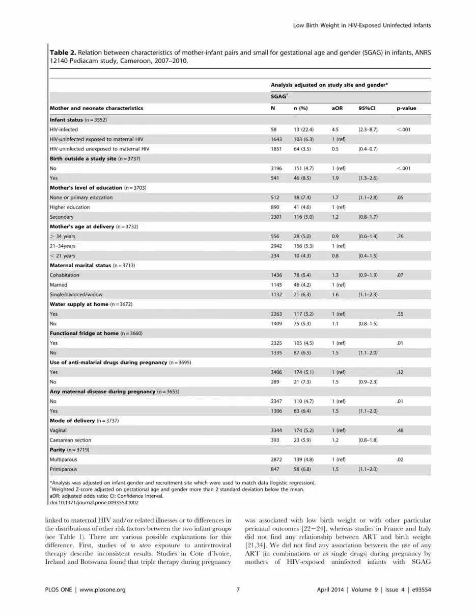
Factors Associated with SGAGIn the first step of the analysis, the association between
covariates and SGAG adjusted for study site and infant’s gender
were investigated (Table 2): exposure to maternal HIV infection,
primiparity, birth outside of a study site, any maternal illness
during pregnancy, low maternal education level, and lack of a
functional fridge at home were each significantly associated with
SGAG. The use of anti-malaria drugs and the marital status of
mothers tended to be associated with SGAG.
In multivariate modeling (Table 3), some of the independent
predictors of SGAG were similar to those suggested in the first step
analysis; in particular, exposure to maternal HIV infection
appeared to be an important factor. Relative to HIV-exposed
uninfected infants (reference group), the odds of SGAG were
fourfold higher for infants who were HIV-infected (aOR: 4.0; 2.0–
8.1) and half for infants who were HIV-unexposed uninfected
(aOR: 0.5; 0.4–0.8). Other significant contributors to SGAG
identified were primiparity (aOR: 1.9; 1.3–2.7), and the existence
of any maternal disease during pregnancy (aOR: 1.4; 1.022.0);
the odds of SGAG also tended to be higher for those infants with
no functional fridge at home (aOR: 1.3; 0.9–1.9). No significant
interactions were identified between the principal variable of
interest (mother-infant HIV status) and other independent
variables included in the model or between certain independent
variables.
Subgroup analyses (not shown) were conducted and indicated
that within the HIV-unexposed uninfected infant group, primi-
parity (aOR 2.1, 1.1–3.7, p = .02) was associated with SGAG.
Such association was also found for the HIV-exposed uninfected
infant group (aOR 2.1, 1.124.3, p = .05). Among HIV-exposed
uninfected infants, the use of any antiretroviral drugs during
pregnancy (aOR 0.8, 0.126.5, p = .82) was not associated with
SGAG. Using the absence of a functional fridge at home as a
proxy of socioeconomic status, no independent relation was found
either in HIV-exposed uninfected or in HIV-unexposed uninfect-
ed infant groups.
Univariate analysis was conducted for HIV-exposed infant
group, taking various measures of HIV severity into account, and
in particular CD4 lymphocyte count and ART status of HIV-
infected mothers; no relation between ART started before or
during pregnancy and SGAG was found; however, despite the
small sample size (n = 845) in this multivariate analysis, severe
immunosuppression in HIV-infected pregnant women (CD4 count
,200 cells/mm3) was associated with SGAG (aOR: 2.5; IC: 1.32
4.8).
Discussion
We investigated the effect of maternal HIV infection on
standardized birth weight using data from the ANRS-PEDIA-
CAM study collected in three referral hospitals in the two biggest
cities of Cameroon. Almost all HIV-infected mothers at these
hospitals were contacted, and the study enrollment rate was 85%
Low Birth Weight in HIV-Exposed Uninfected Infants
PLOS ONE | www.plosone.org 4 April 2014 | Volume 9 | Issue 4 | e93554
Ta
ble
1.
Co
mp
aris
on
of
the
char
acte
rist
ics
of
mo
the
rsan
dn
eo
nat
es
acco
rdin
gto
infa
nt
HIV
stat
us
inth
eA
NR
S1
21
40
-Pe
dia
cam
stu
dy,
Cam
ero
on
,2
00
7–
20
10
.
HIV
-in
fect
ed
infa
nts
HIV
-ex
po
sed
un
infe
cte
din
fan
tsH
IV-u
ne
xp
ose
du
nin
fect
ed
infa
nts
N=
58
N=
16
43
N=
18
51
Mo
the
ra
nd
ne
on
ate
cha
ract
eri
stic
sn
%n
%n
%
Bir
tho
uts
ide
ast
ud
ysi
te5
81
64
31
85
1
Ye
s2
23
7.9
``
41
12
5.0
62
3.3
**
Mo
the
r’s
ag
ea
td
eli
ve
ry5
81
64
11
84
9
.3
4ye
ars
61
0.3
??2
82
17
.22
45
13
.2**
21
–3
4ye
ars
48
82
.81
30
37
9.4
14
40
77
.9
,2
1ye
ars
46
.95
63
.41
64
8.9
Mo
the
r’s
lev
el
of
ed
uca
tio
n5
71
63
21
83
0
Hig
he
re
du
cati
on
61
0.5
??2
85
17
.55
73
31
.3**
Seco
nd
ary
42
73
.71
05
36
4.5
10
87
59
.4
No
ne
or
pri
mar
ye
du
cati
on
91
5.8
29
41
8.0
17
09
.3
Ma
tern
al
ma
rita
lst
atu
s5
71
63
61
83
6
Mar
rie
d9
15
.8??
40
52
4.8
69
03
7.6
**
Co
hab
itat
ion
25
43
.97
30
44
.65
98
32
.6
Sin
gle
/div
orc
ed
/wid
ow
23
40
.35
01
30
.65
48
29
.8
Wa
ter
sup
ply
at
ho
me
56
16
20
18
13
Ye
s3
05
3.6
??8
85
54
.61
24
66
8.7
**
Fu
nct
ion
al
frid
ge
at
ho
me
55
16
17
18
05
Ye
s3
05
4.5
??8
75
54
.11
32
07
3.1
**
An
yd
ise
ase
du
rin
gp
reg
na
ncy
56
16
12
18
03
Ye
s2
64
6.4
??6
43
39
.95
78
32
.1**
Mo
de
of
de
liv
ery
58
16
43
18
51
Cae
sare
anse
ctio
n3
5.2
14
68
.92
11
11
.4
Pa
rity
58
16
39
18
37
pri
mip
aro
us
13
22
.4??
24
21
4.8
55
73
0.3
**
An
tire
tro
vir
al
du
rin
gp
reg
na
ncy
58
16
32
Ye
s4
17
0.7
**1
50
89
2.4
Lo
wb
irth
we
igh
t5
81
64
31
85
1
Ye
s,
25
00
g1
11
9.0{{
12
37
.58
54
.6**
Pre
term
bir
th5
81
64
31
85
1
Ye
s(,
37
we
eks
)1
01
7.2
??2
00
12
.21
91
10
.3??
Hy
po
tro
ph
y*
48
14
43
16
60
Ye
s6
12
.5``
64
4.4
46
2.8``
SG
AG
{5
81
64
31
85
1
Low Birth Weight in HIV-Exposed Uninfected Infants
PLOS ONE | www.plosone.org 5 April 2014 | Volume 9 | Issue 4 | e93554
(data not shown). The use of ART prophylaxis to reduce vertical
transmission is becoming a standard in these hospitals, and indeed
about 90% of HIV-infected mothers included in our study
reported the use of antiretroviral drugs (combinations or single
drugs) during pregnancy or at delivery.
Our main result we report is the strong association between
maternal HIV infection and both SGAG and infant HIV status.
Overall, the proportion of SGAG was 3.5% among HIV-
unexposed uninfected infants, twice as high (6.3%) among HIV-
exposed uninfected infants and nearly seven times higher (22.4%)
among HIV-infected infants. Compared to HIV-exposed unin-
fected infants, and taking into account several potential confound-
ers (likely to be more frequent along HIV-exposed infants,
Table 1), SGAG was significantly more frequent among HIV-
infected infants and less frequent among HIV-unexposed unin-
fected infants. Also, we observed that HIV-unexposed uninfected
infants had higher birth weight Z-scores and HIV-infected infants
had lower birth weight Z-scores than HIV-exposed uninfected
infants.
The comparison of our results with those of other studies is
complicated for various reasons: differences in the populations
studied, the availability of ART at the time of the study,
socioeconomic environments, sample sizes and the outcome
criteria used to assess growth up to birth. Nevertheless, our data
lead to conclusions consistent with the findings of studies which
report significant differences in birth weights between HIV-
infected infants, and HIV-exposed uninfected infants and/or
HIV-exposed uninfected and HIV-unexposed uninfected infants
[1,4,13,17]. Nevertheless, some studies found no difference in
birth weight between these groups of infants [3,15,16,18,19].
We observed that HIV-infected infants were smaller at birth
than HIV-exposed uninfected infants. This suggests a direct effect
of maternal HIV on fetal growth but the mechanisms of any such
effect are unclear. Most vertical HIV transmission occurs late
during pregnancy and advanced maternal HIV infection (as
evidenced by high viral load and low CD4 lymphocyte count) has
been reported to be a risk factor for mother–to-child transmission
[31]. It is therefore possible that any difference in size at birth
between HIV-infected and HIV-exposed uninfected infants may
be the consequence of effects of the virus on the mother (expressed
as HIV related illness) rather than directly on the (infected) fetus
[25]. Ryder et al. reported that the mean birth weight of infants
whose mothers were at an advanced stage was significantly lower
than that of infants born to HIV-infected asymptomatic mothers
[32]. Moye et al. showed that in the USA, the mean birth weight
Z-scores of HIV-infected infants was lower than those of HIV-
exposed uninfected children; however, they also reported that
disease stage as measured by mean prenatal CD4 T lymphocyte
count did not appear to influence birth weight Z-score for HIV-
exposed, and either HIV-infected or uninfected, infants [33]. HIV
staging data were not available in our study, so we are not able to
verify this observation. Other authors suggest that the type of ART
(particularly the use of protease inhibitors) taken by mothers
during pregnancy either for PMTCT or for their own health may
explain this relation [34]. However, such drugs are used only as
second line regimen, and are therefore not widely taken in
Cameroon now.
We found a significant difference in birth weight Z-scores
between HIV-exposed uninfected and HIV-unexposed uninfected
infants. This result confirms the findings from other studies
describing lower anthropometric outcomes among HIV-exposed
uninfected than HIV-unexposed uninfected infants [4,33]. By
contrast, other authors found that growth measures for the two
groups were similar [3,16,18,19]. The difference observed may be
Ta
ble
1.
Co
nt.
HIV
-in
fect
ed
infa
nts
HIV
-ex
po
sed
un
infe
cte
din
fan
tsH
IV-u
ne
xp
ose
du
nin
fect
ed
infa
nts
N=
58
N=
16
43
N=
18
51
Mo
the
ra
nd
ne
on
ate
cha
ract
eri
stic
sn
%n
%n
%
Ye
s1
32
2.4
**1
03
6.3
64
3.5
**
he
igh
tZ
-sco
re,
22
SD
`5
11
56
21
73
3
Ye
s3
5.9
??1
07
6.8
47
2.7
**
He
ad
circ
um
fere
nce
53
15
56
17
14
Z-s
core
,2
2SD
47
.5??
87
5.6
63
3.7``
*Hyp
otr
op
hy
de
fin
ed
aslo
wb
irth
we
igh
t(,
25
00
g)
inin
fan
tsb
orn
atte
rm(g
est
atio
nal
age
.=
37
we
eks
).{ SG
AG
:w
eig
ht
Z-s
core
mo
reth
an2
stan
dar
dd
evi
atio
ns
be
low
the
me
an`SD
:st
and
ard
de
viat
ion
.p
-val
ue
usi
ng
‘‘HIV
-exp
ose
du
nin
fect
ed
gro
up
’’as
the
refe
ren
ce:
**p
,.0
01
{{.0
01
#p
,.0
1``.0
1#
p,
.05
.$$.0
5#
p#
.10
.??
p.
.10
.d
oi:1
0.1
37
1/j
ou
rnal
.po
ne
.00
93
55
4.t
00
1
Low Birth Weight in HIV-Exposed Uninfected Infants
PLOS ONE | www.plosone.org 6 April 2014 | Volume 9 | Issue 4 | e93554
linked to maternal HIV and/or related illnesses or to differences in
the distributions of other risk factors between the two infant groups
(see Table 1). There are various possible explanations for this
difference. First, studies of in utero exposure to antiretroviral
therapy describe inconsistent results. Studies in Cote d’Ivoire,
Ireland and Botswana found that triple therapy during pregnancy
was associated with low birth weight or with other particular
perinatal outcomes [22224], whereas studies in France and Italy
did not find any relationship between ART and birth weight
[21,34]. We did not find any association between the use of any
ART (in combinations or as single drugs) during pregnancy by
mothers of HIV-exposed uninfected infants with SGAG
Table 2. Relation between characteristics of mother-infant pairs and small for gestational age and gender (SGAG) in infants, ANRS12140-Pediacam study, Cameroon, 2007–2010.
Analysis adjusted on study site and gender*
SGAG{
Mother and neonate characteristics N n (%) aOR 95%CI p-value
Infant status (n = 3552)
HIV-infected 58 13 (22.4) 4.5 (2.3–8.7) ,.001
HIV-uninfected exposed to maternal HIV 1643 103 (6.3) 1 (ref)
HIV-uninfected unexposed to maternal HIV 1851 64 (3.5) 0.5 (0.4–0.7)
Birth outside a study site (n = 3737)
No 3196 151 (4.7) 1 (ref) ,.001
Yes 541 46 (8.5) 1.9 (1.3–2.6)
Mother’s level of education (n = 3703)
None or primary education 512 38 (7.4) 1.7 (1.1–2.8) .05
Higher education 890 41 (4.6) 1 (ref)
Secondary 2301 116 (5.0) 1.2 (0.8–1.7)
Mother’s age at delivery (n = 3732)
. 34 years 556 28 (5.0) 0.9 (0.6–1.4) .76
21–34years 2942 156 (5.3) 1 (ref)
, 21 years 234 10 (4.3) 0.8 (0.4–1.5)
Maternal marital status (n = 3713)
Cohabitation 1436 78 (5.4) 1.3 (0.9–1.9) .07
Married 1145 48 (4.2) 1 (ref)
Single/divorced/widow 1132 71 (6.3) 1.6 (1.1–2.3)
Water supply at home (n = 3672)
Yes 2263 117 (5.2) 1 (ref) .55
No 1409 75 (5.3) 1.1 (0.8–1.5)
Functional fridge at home (n = 3660)
Yes 2325 105 (4.5) 1 (ref) .01
No 1335 87 (6.5) 1.5 (1.1–2.0)
Use of anti-malarial drugs during pregnancy (n = 3695)
Yes 3406 174 (5.1) 1 (ref) .12
No 289 21 (7.3) 1.5 (0.9–2.3)
Any maternal disease during pregnancy (n = 3653)
No 2347 110 (4.7) 1 (ref) .01
Yes 1306 83 (6.4) 1.5 (1.1–2.0)
Mode of delivery (n = 3737)
Vaginal 3344 174 (5.2) 1 (ref) .48
Caesarean section 393 23 (5.9) 1.2 (0.8–1.8)
Parity (n = 3719)
Multiparous 2872 139 (4.8) 1 (ref) .02
Primiparous 847 58 (6.8) 1.5 (1.1–2.0)
*Analysis was adjusted on infant gender and recruitment site which were used to match data (logistic regression).{Weighted Z-score adjusted on gestational age and gender more than 2 standard deviation below the mean.aOR: adjusted odds ratio; CI: Confidence Interval.doi:10.1371/journal.pone.0093554.t002
Low Birth Weight in HIV-Exposed Uninfected Infants
PLOS ONE | www.plosone.org 7 April 2014 | Volume 9 | Issue 4 | e93554

Table 3. Maternal HIV status and small for gestational age and gender (SGAG) in infants considering other maternal and infantcharacteristics, multivariate analysis; ANRS-Pediacam study, Cameroon, 200722010.
N = 3342 IUGR{
Maternal and neonate characteristics Adj. OR 95%CI p-value
Infant status
HIV-infected 4.0 (2.0–8.1) ,.001
HIV-uninfected exposed to maternal HIV 1 (ref)
HIV-uninfected not exposed to maternal HIV 0.5 (0.4–0.8)
Clinical site*
EHC$ 1.4 (0.9–2.1) .1
MCH/MCC-CBF ` 1 (ref)
LH ? 0.9 (0.6–1.3)
Sex*
Female 1 (ref) .02
Male 1.4 (1.0–2.0)
Birth outside a study site
No 1 (ref) .12
Yes 1.4 (0.9–2.1)
Mother’s level of education
None or primary education 1.5 (0.9–2.6) .16
Higher education 1 (ref)
Secondary education 1.0 (0.7–1.6)
Mother’s age at delivery
. 34 years 1.1 (0.7–1.7) .59
21234years 1 (ref)
, 21 years 0.8 (0.4–1.6)
Maternal marital status
Cohabitation 0.9 (0.6–1.3) .77
Married 1 (ref)
Single/divorced/widow 1.1 (0.8–1.7)
Functional fridge at home
Yes 1 (ref) .08
No 1.3 (0.9–1.9)
Using of anti-malarial drugs during pregnancy
Yes 1 (ref) .58
No 1.2 (0.7–2.0)
Any disease during pregnancy
No 1 (ref) .03
Yes 1.4 (1.0–2.0)
Mode of delivery
Vaginal 1 (ref) .15
Caesarean section 1.5 (0.9–2.4)
Parity
Multiparous 1 (ref) ,.001
primiparous 1.9 (1.3–2.7)
*Weighted Z-score adjusted on gestational age and sex of newborn more than 2 standard deviations below the mean.{CHM/MCC-CBF: Central hospital Maternity/Mother and Child Center of the Chantal Biya Foundation, Yaounde.`EHC: Essos Hospital Center, Yaounde.$LH: Laquintinie Hospital, Douala.
Adj. OR: adjusted Odd ratio; CI: Confidence Interval.doi:10.1371/journal.pone.0093554.t003
Low Birth Weight in HIV-Exposed Uninfected Infants
PLOS ONE | www.plosone.org 8 April 2014 | Volume 9 | Issue 4 | e93554
(aOR = 0.8; 0.126.5). Second, exposure of the fetus to maternal
HIV and related illnesses and/or to ART may lead to immune
system abnormalities [35]; the cause of any such immune
abnormality is unclear. Possibly, there is an unusually strong
maternal placental response in HIV-infected and ART-treated
pregnant women, including substantial placental production of the
pro-inflammatory cytokines such as tumor necrosis factor-a and
interleukin-8 [36,37]; which could in association with parasitic
infestation (for example malaria) affect fetal growth [24].
We found that primiparity was associated with SGAG. This
observation was confirmed by a subgroup analysis of HIV-
unexposed uninfected infants and of the HIV-exposed uninfected
subgroup. This is consistent with previous reports [7,17,38]. In our
study, 71 % of primiparous mothers were under 21 years old,
corroborating previous findings that primiparity and maternal age
are important contributors to fetal growth restriction [39,40].
Primigravidae are prone to malaria in endemic areas, and
placental infections with plasmodium parasites may affect placen-
tal function and thereby restrict fetal growth [12].
We also found that ‘any infectious pathology’ during pregnancy
was significantly associated with SGAG. Although the evidence is
not detailed, other maternal factors such as anemia and malaria,
which are frequent among HIV-positive mothers [41] during
pregnancy, have been reported to be associated with fetal growth
restriction [12,16,37,41].
Neither maternal age nor socioeconomic status (evaluated here
as ‘‘lack of a functional fridge at home’’ as a proxy) found in other
studies to be associated with SGAG [28] were identified by our
study as factors for SGAG. This could be because our study was
conducted in three referral hospitals in urban areas where most
women were able to afford basic needs.
Our study has several strengths and limitations. One major
strengths is that at inclusion, HIV-exposed and HIV-unexposed
infants were selected independently of their birth weight, and
variables were collected blind to infants’ HIV status. Another is the
standardization of birth anthropometric measurements using Z-
scores and taking gestational age and gender in consideration; this
is more robust than using a single measure alone. We also
controlled for many maternal sociodemographic and obstetrical
characteristics which could act as potential confounders. The
inclusion of a control group of HIV-unexposed uninfected infants
is another strength benefit. There are also various limitations:
gestational age was not collected with sufficient precision,
potentially leading to bias in classification. However, any such
bias is likely to affect the two groups of infants in the same manner,
minimizing any misclassification bias. Some key confounding
factors were not collected, and in particular alcoholism, smoking
and nutritional status of mothers, and parental anthropometric
measures. These factors have been described to influence infants’
birth weight. They are very important in term of prevention
because they represent modifiable risk factors on which action can
be directed.
In summary, we report a clear association between maternal
HIV infection and SGAG among HIV-exposed uninfected infants.
This is a further argument for the need to optimize the provision of
care to pregnant HIV-infected women (especially immune-
suppressed ones) and their infants. Indeed, monitoring growth
and developing strategies to prevent growth faltering in these
infants are required. A better evaluation of the consequences on
fetal growth of ART during pregnancy would be extremely
beneficial. This is now particularly pertinent because the World
Health Organization (WHO) is encouraging countries to engage in
option B+, which is the widespread alternative access to HAART
during pregnancy for the prevention of mother-to-child HIV
transmission.
Acknowledgments
We thank the parents who agreed to inclusion of their children in this
study. We thank the study coordinators, research assistants, laboratory
technicians, nurses, midwives and administrative staff who made this study
possible. We thank Nelly Briand for providing AUDIPOG (French
Association of Users of Computerized Medical Records in Paediatrics,
Obstetrics and Gynaecology Sentinel Network) reference values for the
calculation of Z-scores. This analysis includes work that is part of Sofeu
Casimir Ledoux’s Master dissertation at the epidemiology service of the
Centre Pasteur du Cameroon.
The ANRS-Pediacam Study Team is as follows:
Primary investigators: Prof Albert Faye (Hopital Robert Debre/
Univ. Paris 7, France) and Dr Mathurin Cyrille Tejiokem (Centre Pasteur
du Cameroun, Yaounde).
Co-investigators: Prof Francoise Barre-Sinoussi and Dr Daniel Scott
(Unite de Regulations des Infections Retrovirales, Institut Pasteur de Paris,
France), Dr Frederic Tangy (Unite de Genomique virale et Vaccinantion,
Institut Pasteur de Paris, France), Dr Josiane Warszawski (Equipe 4 (VIH et
IST) - INSERM U1018 (CESP)/Univ. Paris Sud 11, France), Prof
Stephane Blanche (Service d’Immunologie et Hematologie Pediatrique,
Hopital Necker Enfants Malades, Paris, France), Dr Catherine Dollfus
(Hopital Trousseau, Paris, France), Dr Laurence Baril (GSK Bio,
Risenxart, Belgium), Dr Anfumbom Kfutwah (Centre Pasteur du
Cameroun), Dr Ida Penda (Hopital Laquintinie, Douala, Cameroun), Dr
Georgette Guemkam and Dr Ateba Ndongo Francis (Centre Mere et
Enfant de la Fondation Chantal Biya, Yaounde, Cameroun), Dr Suzie
Tetang Ndiang (Centre Hospitalier d’Essos, Yaounde, Cameroun).
Other members of the ANRS-Pediacam team: (by site and
alphabetic order).
Centre Pasteur du Cameroun: Epouner Denise, Mbanzouen William,
Ngoupo Paul Alain, Owona Felicite, Sofeu Casimir Ledoux, Dr Texier
Gaetan, Dr Tchendjou Patrice.
Center Hospital Maternity/Mother and Child care Center in Yaounde: Bossolo
Juste, Ehongo Jean Marie, Dr Evouna Armel, Mbida Patricia, Dr Ndongo
Jean Audrey, Dr Nguefack Felicite, Prof Mbu Robinson, and Prof Koki
Paul.
Essos Hospital Center in Yaounde: Bekono Ernestine, Belinga Marie Louise,
Evoundou Dieudonne, Dr Nga Annie, Nguen Suzanne, Dr Njom Nlend
Anne, Onono Yvette, Dr Wamba Guillaume, Dr Zeudja.
Laquintinie Hospital in Douala: Alibien Michelle, Dr Dissongo Jean II,
Djene Julie, Ewane Valery, Dr Makwet Nicaise, Dr Mbangue Madeleine,
Ngo Sohna Aurore, Dr Ngwa, Obedat Shiro.
Author Contributions
Conceived and designed the experiments: MCT AF ICP AK JW.
Performed the experiments: JW FAN ICP STN GG NM FO AK MT
AF MCT. Analyzed the data: CLS MCT JW GT PT. Contributed
reagents/materials/analysis tools: CLS MCT JW MT. Wrote the paper:
CLS JW FAN ICP STN GG NM FO AK PT GT MT AF MCT.
Coordination of the study: MCT AF JW ICP AK. Recruitment and infants
follow up: GG ICP NM FAN STN FO.
References
1. Bailey RC, Kamenga MC, Nsuami MJ, Nieburg P, St Louis ME (1999). Growth
of children according to maternal and child HIV, immunological and disease
characteristics: a prospective cohort study in Kinshasa, Democratic Republic of
Congo. Int J Epidemiol 28: 532–40.
2. Dreyfuss ML, Msamanga GI, Spiegelman D, Hunter DJ, Urassa EJ, et al.
(2001). Determinants of low birth weight among HIV-infected pregnant women
in Tanzania. Am J Clin Nutr 74: 814–26.
3. Lepage P, Msellati P, Hitimana DG, Bazubagira A, Van Goethem C, et al.
(1996). Growth of human immunodeficiency type 1-infected and uninfected
Low Birth Weight in HIV-Exposed Uninfected Infants
PLOS ONE | www.plosone.org 9 April 2014 | Volume 9 | Issue 4 | e93554
children: a prospective cohort study in Kigali, Rwanda, 1988 to 1993. Pediatr
Infect Dis J 15: 479–85.4. Makasa M, Kasonka L, Chisenga M, Sinkala M, Chintu C, et al. (2007). Early
growth of infants of HIV-infected and uninfected Zambian women. Trop Med
Int Health 12: 594–602.5. Marinda E, Humphrey JH, Iliff PJ, Mutasa K, Nathoo KJ, et al. (2007). Child
mortality according to maternal and infant HIV status in Zimbabwe. PediatrInfect Dis J 26: 519–26.
6. McNally LM, Jeena PM, Gajee K, Thula SA, Sturm AW, et al. (2007). Effect of
age, polymicrobial disease, and maternal HIV status on treatment response andcause of severe pneumonia in South African children: a prospective descriptive
study. Lancet 369: 1440–51.7. Ndirangu J, Newell ML, Bland RM, Thorne C (2012). Maternal HIV infection
associated with small-for-gestational age infants but not preterm births: evidencefrom rural South Africa. Human reproduction 27: 1846–56.
8. Rollins NC, Coovadia HM, Bland RM, Coutsoudis A, Bennish ML, et al.
(2007). Pregnancy outcomes in HIV-infected and uninfected women in rural andurban South Africa. J Acquir Immune Defic Syndr 44: 321–8.
9. Wei R, Msamanga GI, Spiegelman D, Hertzmark E, Baylin A, et al. (2004).Association between low birth weight and infant mortality in children born to
human immunodeficiency virus 1-infected mothers in Tanzania. Pediatr Infect
Dis J 23: 530–5.10. McGrath CJ, Nduati R, Richardson BA, Kristal AR, Mbori-Ngacha D, et al.
(2012) The prevalence of stunting is high in HIV-1-exposed uninfected infants inKenya. J Nutr 142: 757–63.
11. Castetbon K, Ladner J, Leroy V, Chauliac M, Karita E, et al. (1999). Lowbirthweight in infants born to African HIV-infected women: relationship with
maternal body weight during pregnancy: Pregnancy and HIV Study Group
(EGE). J Trop Pediatr 45: 152–7.12. Ebrahim GJ (1996). Malaria during pregnancy. J Trop Pediatr 42: 62–3.
13. Mwanyumba F, Claeys P, Gaillard P, Verhofstede C, Chohan V, et al. (2001).Correlation between maternal and infant HIV infection and low birth weight: a
study in Mombasa, Kenya. J Obstet Gynaecol 21: 27–31.
14. Stratton P, Tuomala RE, Abboud R, Rodriguez E, Rich K, et al. (1999).Obstetric and newborn outcomes in a cohort of HIV-infected pregnant women:
a report of the women and infants transmission study. J Acquir Immune DeficSyndr Hum Retrovirol 20: 179–86.
15. Verhoeff FH, Brabin BJ, van Buuren S, Chimsuku L, Kazembe P, et al. (2001).An analysis of intra-uterine growth retardation in rural MalawiChanges in
haemoglobin levels in infants in Malawi: effect of low birth weight and fetal
anaemia. Eur J Clin Nutr 55: 682–9.16. Watson-Jones D, Weiss HA, Changalucha JM, Todd J, Gumodoka B, et al.
(2007). Adverse birth outcomes in United Republic of Tanzania–impact andprevention of maternal risk factors. Bull World Health Organ 85: 9–18.
17. Kalanda BF, van Buuren S, Verhoeff FH, Brabin BJ (2005). Anthropometry of
fetal growth in rural Malawi in relation to maternal malaria and HIV status.Arch Dis Child Fetal Neonatal Ed 90: F161–5.
18. European Collaborative Study (2003). Exposure to antiretroviral therapy inutero or early life: the health of uninfected children born to HIV-infected
women. J Acquir Immune Defic Syndr 32: 380–7.19. Venkatesh KK, Lurie MN, Triche EW, De Bruyn G, Harwell JI, et al. (2010)
Growth of infants born to HIV-infected women in South Africa according to
maternal and infant characteristics. Trop Med Int Health 15: 1364–74.20. Briand N, Le Coeur S, Traisathit P, Karnchanamayul V, Hansudewechakul R,
et al. (2006). Growth of human immunodeficiency virus-uninfected childrenexposed to perinatal zidovudine for the prevention of mother-to-child human
immunodeficiency virus transmission. Pediatr Infect Dis J 25: 325–32.
21. Briand N, Mandelbrot L, Le Chenadec J, Tubiana R, Teglas JP, et al. (2009).No relation between in-utero exposure to HAART and intrauterine growth
retardation. Aids 23: 1235–43.
22. Ekouevi DK, Coffie PA, Becquet R, Tonwe-Gold B, Horo A, et al. (2008).
Antiretroviral therapy in pregnant women with advanced HIV disease andpregnancy outcomes in Abidjan, Cote d’Ivoire. Aids 22: 1815–20.
23. Townsend CL, Cortina-Borja M, Peckham CS, Tookey PA (2007). Antiretro-
viral therapy and premature delivery in diagnosed HIV-infected women in theUnited Kingdom and Ireland. Aids 21: 1019–26.
24. Filteau S (2009). The HIV-exposed, uninfected African child. Trop Med IntHealth 14: 276–87.
25. Isanaka S, Duggan C, Fawzi WW (2009). Patterns of postnatal growth in HIV-
infected and HIV-exposed children. Nutr Rev 67: 343–59.26. Tejiokem MC, Faye A, Penda IC, Guemkam G, Ateba Ndongo F, et al. (2011).
Feasibility of early infant diagnosis of HIV in resource-limited settings: theANRS 12140-PEDIACAM study in Cameroon. PLoS One 6: e21840.
27. WHO (2006). Medicaments antiretroviraux pour traiter la femme enceinte etprevenir l’infection a VIH chez l’enfant. Vers un acces universel. Recommanda-
tions pour une approche de sante publique 2006. Available: http://www.who.
int/hiv/pub/mtct/arv_guidelines_mtct_fr.pdf. Accessed 22 January 2014.28. Lee PA, Chernausek SD, Hokken-Koelega AC, Czernichow P (2003).
International Small for Gestational Age Advisory Board consensus developmentconference statement: management of short children born small for gestational
age, April 24-October 1, 2001. Pediatrics 111: 1253–61.
29. Association des Utilisateurs de Dossiers Informatises en Pediatrie Obstetrique etGynecologie. AUDIPOG. Available: http://www.audipog.net/courbes_
morpho.php, accessed date 16/01/2014.30. Bouyer J, Hemon D, Cordier S, Derriennic F, Stucker I, et al.(1995)
Epidemiologie Principes et methodes quantitatives. INSERM ed. 499 p.31. Kourtis AP, Bulterys M, Nesheim SR, Lee FK (2001). Understanding the timing
of HIV transmission from mother to infant. Jama 285: 709–12.
32. Ryder RW, Nsa W, Hassig SE, Behets F, Rayfield M, et al. (1989). Perinataltransmission of the human immunodeficiency virus type 1 to infants of
seropositive women in Zaire. N Engl J Med 320: 1637–42.33. Moye J, Jr., Rich KC, Kalish LA, Sheon AR, Diaz C, et al. (1996). Natural
history of somatic growth in infants born to women infected by human
immunodeficiency virus. Women and Infants Transmission Study Group.J Pediatr 128: 58–69.
34. Floridia M, Ravizza M, Bucceri A, Lazier L, Vigano A, et al. (2008). Factorsinfluencing gestational age-adjusted birthweight in a national series of 600
newborns from mothers with HIV. HIV Clin Trials 9: 287–97.35. Clerici M, Saresella M, Colombo F, Fossati S, Sala N, et al. (2000). T-
lymphocyte maturation abnormalities in uninfected newborns and children with
vertical exposure to HIV. Blood 96: 3866–71.36. Faye A, Pornprasert S, Mary JY, Dolcini G, Derrien M, et al. (2007).
Characterization of the main placental cytokine profiles from HIV-1-infectedpregnant women treated with anti-retroviral drugs in France. Clin Exp Immunol
149: 430–9.
37. Kfutwah A, Mary JY, Lemen B, Leke R, Rousset D, et al. (2009). Plasmodiumfalciparum infection significantly impairs placental cytokine profile in HIV
infected Cameroonian women. PLoS One 4: e8114.38. Sombie I, Nacro B, Tiendrebeogo S, Dao B, Cartoux M, et al. (1999). Infection
maternelle par le VIH et parametres anthropometriques de l’enfant a lanaissance au Burkina Faso. Cahiers sante 9: 173–7.
39. BBS/UNICEF (2005). National Low Birth Weight Survey of Bangladesh, 2003–
2004: Bangladesh Bureau of Statistics. Available: http://www.unicef.org/bangladesh/Low_Birth_Weight_report.pdf. Accessed 27 September 2013.
40. Bernstein PS, Divon MY (1997). Etiologies of Fetal Growth Restriction. ClinicalObstetrics and Gynecology 40: 723–729.
41. Naniche D, Bardaji A, Lahuerta M, Berenguera A, Mandomando I, et al.
(2009). Impact of maternal human immunodeficiency virus infection on birthoutcomes and infant survival in rural Mozambique. Am J Trop Med Hyg 80:
870–6.
Low Birth Weight in HIV-Exposed Uninfected Infants
PLOS ONE | www.plosone.org 10 April 2014 | Volume 9 | Issue 4 | e93554