Long-term behavior of oil-based varnishes and paints. Photo and thermooxidation of cured linseed oil
7
ABSTRACT: Thermooxidation at 100°C and photooxidation at wavelengths above 300 nm of dried oil films were evaluated. The chemical modifications of the networks were determined by infrared analysis coupled with gaseous treatments (NO, SF 4 , and NH 3 ). The dried films are rather stable in thermooxidation, whereas in photooxidation, important degradation of the net- work occurs with many chain scissions. This photoinstability results from the presence of crosslinks that are sensitive to radi- cal attack because of the lability of the hydrogen atom on the tertiary carbons. The photooxidation reactions are fully de- scribed in this paper. Yellowing of the cured samples, observed with ultraviolet-visible and fluorescence spectrometries, rapidly is decreased by irradiation because the oil contaminants that are mainly responsible for the yellowness are photooxidized. On the contrary, yellowing slowly but continuously increases during thermooxidation at 100°C. Paper no. J9333 in JAOCS 77, 257–263 (March 2000). KEY WORDS: Drying oil, fluorescence spectroscopy, FTIR spec- troscopy, mechanisms, photooxidation, thermooxidation, thin films, UV spectroscopy. Vegetable oils with important proportions of polyunsaturated fatty acids (PUFA) are called drying oils. Their high drying index (1) allows their use in the manufacture of oil-based paints and varnishes and especially for the creation of works of art. In this last field, oil-based paints and varnishes generate some problems, mainly a tendency to yellowing or cracking. Analysis of the literature concerning the applications of oil-based paints and varnishes shows that most of the research has been focused on the identification of binding media (2–4). The results obtained allow selection of appropriate materials for cleaning paintings and give information to the art histo- rian about artists’ techniques (5). To our knowledge, only two valuable studies dealing with the photo-aging of dried oil films have been published, more than 40 yr ago (6,7). No studies of the reactions involved in photooxidation of these oils have been published. However, it is of great interest to understand the mechanisms of the chemical evolution that oc- curs during long-term aging with light exposure. This is the aim of this paper. The PUFA chains can oxidize quite readily, via a radical chain reaction (8,9). The thermooxidation mechanism of dry- ing oils has been largely reviewed in the literature (10,11). Many oxidation products formed from the decomposition of hydroperoxides have been identified: they consist in alcohols, ketones, aldehydes, or epoxides (12). Oxidation of the PUFA chains leads to crosslinks by formation of C–C, ether, or per- oxy linkages (13,14). The curing step, considered as an “ox- idative polymerization,” leads to a three-dimensional net- work, and when the curing is complete, a stable state is reached (15). Determination of the peroxide value (PV) ap- pears to be the best way to measure the extent of curing (16). When samples are totally dried, the antioxidants in the oil film have been consumed, but the contaminants initially present in the vegetable oil still remain in the film. In the first part of our research (16), we used Fourier trans- form infrared (FTIR) spectroscopy to characterize oxidation products and to elucidate the drying mechanism of drying oils. We found that the addition of peroxy radicals on conju- gated double bonds was responsible for the fast disappear- ance of conjugated double bonds, with the formation of epox- ides as intermediate products. The properties of the hydroper- oxides formed have been studied in the second part (17). Our results have permitted a complete understanding of the dry- ing mechanism on the basis of an induced hydroperoxide de- composition reaction. We have also shown that only low con- centrations of peroxy bridges were formed in the dried oil films. Among drying oils, linseed oil is one of the most com- monly used in paintings. In this paper, we focus on the identi- fication of the various oxidation products formed during photo- and thermooxidation of dried linseed oil films. The products have been identified and quantified by FTIR spec- trometry, sometimes coupled with gaseous treatments. Ultra- violet (UV)-visible spectroscopy and microfluorescence spec- troscopy have been used as complementary methods, mainly to study decomposition of the various contaminants that can contribute to the yellowness of dried film. EXPERIMENTAL PROCEDURES Materials. Analyzed samples were prepared by spreading out linseed oil (Pebeo, Gemenos, France) (linolenic 54%, linoleic Long-Term Behavior of Oil-Based Varnishes and Paints. Photo- and Thermooxidation of Cured Linseed Oil Jacky Mallégol, Jean-Luc Gardette*, and Jacques Lemaire Laboratoire de Photochimie Moléculaire et Macromoléculaire, UMR CNRS 6505, Université Blaise Pascal (Clermont II), Ensemble Scientifique des Cézeaux, F-63177 Aubière Cedex, France Copyright © 2000 by AOCS Press 257 JAOCS, Vol. 77, no. 3 (2000) *To whom correspondence should be addressed. E-mail: [email protected]
Transcript of Long-term behavior of oil-based varnishes and paints. Photo and thermooxidation of cured linseed oil

ABSTRACT: Thermooxidation at 100°C and photooxidationat wavelengths above 300 nm of dried oil films were evaluated.The chemical modifications of the networks were determinedby infrared analysis coupled with gaseous treatments (NO, SF4,and NH3). The dried films are rather stable in thermooxidation,whereas in photooxidation, important degradation of the net-work occurs with many chain scissions. This photoinstabilityresults from the presence of crosslinks that are sensitive to radi-cal attack because of the lability of the hydrogen atom on thetertiary carbons. The photooxidation reactions are fully de-scribed in this paper. Yellowing of the cured samples, observedwith ultraviolet-visible and fluorescence spectrometries, rapidlyis decreased by irradiation because the oil contaminants thatare mainly responsible for the yellowness are photooxidized.On the contrary, yellowing slowly but continuously increasesduring thermooxidation at 100°C.
Paper no. J9333 in JAOCS 77, 257–263 (March 2000).
KEY WORDS: Drying oil, fluorescence spectroscopy, FTIR spec-troscopy, mechanisms, photooxidation, thermooxidation, thinfilms, UV spectroscopy.
Vegetable oils with important proportions of polyunsaturatedfatty acids (PUFA) are called drying oils. Their high dryingindex (1) allows their use in the manufacture of oil-basedpaints and varnishes and especially for the creation of worksof art. In this last field, oil-based paints and varnishes generatesome problems, mainly a tendency to yellowing or cracking.
Analysis of the literature concerning the applications ofoil-based paints and varnishes shows that most of the researchhas been focused on the identification of binding media (2–4).The results obtained allow selection of appropriate materialsfor cleaning paintings and give information to the art histo-rian about artists’ techniques (5). To our knowledge, only twovaluable studies dealing with the photo-aging of dried oilfilms have been published, more than 40 yr ago (6,7). Nostudies of the reactions involved in photooxidation of theseoils have been published. However, it is of great interest tounderstand the mechanisms of the chemical evolution that oc-curs during long-term aging with light exposure. This is theaim of this paper.
The PUFA chains can oxidize quite readily, via a radicalchain reaction (8,9). The thermooxidation mechanism of dry-ing oils has been largely reviewed in the literature (10,11).Many oxidation products formed from the decomposition ofhydroperoxides have been identified: they consist in alcohols,ketones, aldehydes, or epoxides (12). Oxidation of the PUFAchains leads to crosslinks by formation of C–C, ether, or per-oxy linkages (13,14). The curing step, considered as an “ox-idative polymerization,” leads to a three-dimensional net-work, and when the curing is complete, a stable state isreached (15). Determination of the peroxide value (PV) ap-pears to be the best way to measure the extent of curing (16).When samples are totally dried, the antioxidants in the oil filmhave been consumed, but the contaminants initially present inthe vegetable oil still remain in the film.
In the first part of our research (16), we used Fourier trans-form infrared (FTIR) spectroscopy to characterize oxidationproducts and to elucidate the drying mechanism of dryingoils. We found that the addition of peroxy radicals on conju-gated double bonds was responsible for the fast disappear-ance of conjugated double bonds, with the formation of epox-ides as intermediate products. The properties of the hydroper-oxides formed have been studied in the second part (17). Ourresults have permitted a complete understanding of the dry-ing mechanism on the basis of an induced hydroperoxide de-composition reaction. We have also shown that only low con-centrations of peroxy bridges were formed in the dried oilfilms.
Among drying oils, linseed oil is one of the most com-monly used in paintings. In this paper, we focus on the identi-fication of the various oxidation products formed duringphoto- and thermooxidation of dried linseed oil films. Theproducts have been identified and quantified by FTIR spec-trometry, sometimes coupled with gaseous treatments. Ultra-violet (UV)-visible spectroscopy and microfluorescence spec-troscopy have been used as complementary methods, mainlyto study decomposition of the various contaminants that cancontribute to the yellowness of dried film.
EXPERIMENTAL PROCEDURES
Materials. Analyzed samples were prepared by spreading outlinseed oil (Pebeo, Gemenos, France) (linolenic 54%, linoleic
Long-Term Behavior of Oil-Based Varnishes and Paints.Photo- and Thermooxidation of Cured Linseed Oil
Jacky Mallégol, Jean-Luc Gardette*, and Jacques LemaireLaboratoire de Photochimie Moléculaire et Macromoléculaire, UMR CNRS 6505,
Université Blaise Pascal (Clermont II), Ensemble Scientifique des Cézeaux, F-63177 Aubière Cedex, France
Copyright © 2000 by AOCS Press 257 JAOCS, Vol. 77, no. 3 (2000)
*To whom correspondence should be addressed.E-mail: [email protected]
13%, oleic 22%, and saturated 11%) on KBr or CaF2 win-dows with a thickness of about 25 micrometers. IR spectrawere recorded with a Nicolet 510 spectrometer (Madison,WI) (resolution 4 cm−1, 20-scan summation), and UV spectrawere recorded on a PerkinElmer Lambda 5 spectrophotome-ter (Courtabœuf, France), equipped with an integratingsphere. Fluorescence spectroscopy was performed with a Hi-tachi U6000 microspectrofluorimeter (based on an OlympusBHT2 microscope). The excitation wavelength was 395 nm.
Photooxidation, photolysis and thermooxidation experi-ments. Curing of linseed oil was carried out in a ventilatedoven at 60°C in the dark for 25 d, on two series of samples onKBr or CaF2 windows. Dried oil samples were then exposedin a SEPAP 12/24 unit for irradiation at wavelengths longerthan 300 nm (temperature 60°C). This apparatus has been de-scribed in numerous articles (18,19). For photolysis, the sam-ples were individually introduced into Pyrex tubes beforebeing sealed in vacuum (5·10−6 torr). The tubes containingoxidized samples were exposed in the SEPAP 12/24 unit sim-ilar to the photooxidation experiment. Some dried sampleswere also placed in an oven at 100°C to study the influenceof temperature.
Gaseous treatments. Nitrogen monoxide (NO) treatmentand sulfur tetrafluoride (SF4) treatment permit identificationof alcohols and carboxylic acids, respectively. The nitrite bandat 777 cm−1 is characteristic of the presence of alcohol (20,21).In the same manner, after SF4 treatment, the acid fluoridebands at 1843 and 1810 cm−1 allow the determination of thecarboxylic acid concentration (21). Treatment with gaseousammonia (NH3) was also performed on the oxidized film. Lac-tones, esters, and peresters, nonreactive to SF4, are susceptibleto reaction with ammonia. Lactones may be converted intolactams (22). Esters and peresters can form amides with acharacteristic IR absorption band in the range 1660–1680 cm−1
(23). Carboxylic acids are converted into ammonium carboxyl-ates with IR absorption band in the range 1560–1580 cm−1.NO and NH3 treatments were carried out for 6 and 24 h, re-spectively, in a Pyrex container in darkness after a nitrogenpurge. SF4 treatment was carried out in an all-Teflon reactorfor 6 h, the oil samples being on CaF2 windows.
PV. PV of the 60°C samples was determined by iodomet-ric titration (17): refluxing oxidized samples in aceticacid/propan-2-ol with excess sodium iodide and measuringthe I3
− liberated by spectrophotometry (Shimadzu UV-2101PC) (Columbia, MD) at 357 nm.
RESULTS AND DISCUSSION
Photooxidation experiments. Linseed oil samples, oxidized at60°C for 25 d, were considered as “dried” samples because,after this time, oxidative cross-linking is almost complete.The PV of these samples, determined by iodometry, wasaround 100 mmol·kg−1. The 60°C-oxidized samples wereplaced in a SEPAP 12/24 device for irradiation. After variousirradiation times, samples were analyzed by FTIR, UV-visi-ble, and fluorescence spectroscopies, and their PV were de-
termined. IR spectra of irradiated linseed oil after differentexposure durations are represented in Figure 1. The assign-ment of vibration bands for the sample dried at 60°C has beendescribed in a previous article (16). Many changes can be ob-served in the spectrum in Figure 1, such as a decrease in theintensity of the alkyl bands between 3000 and 2820 cm−1 anda marked modification of the ν(O–H) band, due to the forma-tion of bonded carboxylic acids that have a broad absorptionband from 3300 to 2500 cm−1.
In the IR region from 2200 to 400 cm−1 (not represented),important modifications are also observed. The vibrationband at 975 cm−1, assigned to residual unsaturation after cur-ing (24), disappears within 13 h, and the vibrations of alkylgroups at 1464 and 725 cm−1 decrease with irradiation time.Modifications in the range 1300–1100 cm−1 and in the range1900–1500 cm−1 result from changes in the oxidized productconcentrations. The mechanisms of the curing of drying oilswere detailed in our previous publications (16,17). For the60°C-dried linseed oil samples, residual unsaturation at 975cm−1 can be attributed to monounsaturated fatty acid chainswith low reactivity or in-chain oxidation products, such as un-saturated ketones. The residual double bonds may be consid-ered as preferential oxidation sites for photooxidation, whichis confirmed by the fact that they disappear fast in the firsthours of irradiation.
In parallel, the variation of PV in Figure 2 shows thatmany reactions occur during the first hours of irradiation. Thefast increase in the hydroperoxide concentration may bepartly attributed to reactivity of the sites mentioned above.There are great differences between the maximum PV in thecuring step (17) and in the photooxidation step. During irra-diation, the hydroperoxides formed are quickly decomposedinto oxidation products (25) .
The ν(C=O) vibration band of carboxylic acids cannot beobserved in the broad band centered about 1745 cm−1, includ-ing vibration band of ester bonds of triglycerides at 1744 cm−1.The absorption of saturated and unsaturated ketones at 1720
258 J. MALLÉGOL ET AL.
JAOCS, Vol. 77, no. 3 (2000)
FIG. 1. Evolution of the infrared spectra of linseed oil after irradiationfor various times.

and 1696 cm−1 does not permit evaluation of the acid bandaround 1710 cm−1. For this reason, the irradiated samples weretreated with SF4 to observe the presence of carboxylic acids.
As shown in Figure 3, SF4 treatment is informative. In theunexposed oil spectrum, one can observe the decrease of theν(C=O) band of carboxylic acid at 1710 cm−1 and the appear-ance of the ν(C=O) fluoride acid bands at 1843 and 1810 cm−1,which reveals that saturated and unsaturated acids were pro-duced. The disappearance of unsaturated carboxylic acids isnot clearly observed in the spectra, because they could absorbnear 1695 cm−1, as a shoulder of the 1710 cm−1 band. Whenirradiation time increases, many changes occur. The fluorideacid band at 1843 cm−1 on the SF4-treated samples increases,showing that carboxylic acids accumulate in photo-oxidation.An important band around 1775 cm−1 also increases, whichindicates the formation of γ-lactones and peresters. With in-creasing irradiation time, the decrease in the carboxylic acidband at 1710 cm−1 is masked because of the concomitant for-mation, during photooxidation, of products absorbing near
1735 cm−1 (band visible on the 500 h-irradiated spectrum).As proposed for the thermooxidation mechanism (16), hy-droperoxides can be decomposed into alkoxyl and hydroxylradicals. These alkoxyl radicals may then evolve to give ke-tones or aldehydes, rapidly oxidize into acids, or decomposeinto small molecules, such as carbon dioxide.
The IR spectrum of the gas phase was recorded for an oxi-dized sample placed in a gas cell in the SEPAP 12/24 for 40-hirradiation. The gas-phase spectrum of dried linseed oil afterirradiation evidences the formation of CO2, as mentioned inthe literature (6,26).
The ketones formed during the thermal curing step arephoto-unstable and can decompose by Norrish I or II mecha-nisms (27) (Scheme 1). In the presence of oxygen, carboxylicacids are formed.
The IR spectra of photooxidized products do not show anyvinyl vibration band at 909 cm−1. This result suggests that theNorrish II mechanism is only a minor route for the decompo-sition of ketones, if it exists at all.
NO treatment permits quantitation of alcohols in irradiatedsamples. The decrease in the nitrite band at 777 cm−1 in ex-posed and NO-treated samples shows that, during irradiation,alcohols disappear. Usually, tertiary alcohols are consideredas quite stable oxidation products. However, secondary alco-hols can be oxidized when irradiated because of their labilehydrogen atom. The autoxidation mechanism proposed forthe fatty acid chains (9) underwent formation of secondaryalcohols. The disappearance of alcohols may be explained bythe mechanism proposed in Scheme 2.
NH3 treatment carried out on an oxidized sample leads toan important decrease in the 1775 cm−1 absorbance band.This could confirm the presence of peresters. In parallel, ap-pearance of a new band at 1680 cm−1 indicates that amidegroups are formed by NH3 reaction in the photooxidized sam-
PHOTO- AND THERMOOXIDATION OF CURED LINSEED OIL 259
JAOCS, Vol. 77, no. 3 (2000)
FIG. 2. Evolution of the peroxide value vs. irradiation time.
FIG. 3. Subtraction spectra: (dried linseed oil after various irradiationdurations and SF4 treatment) minus (60°C-dried linseed oil).
SCHEME 1

ple. A carboxylate vibration band is also visible at 1570 cm−1.Residual absorption at 1775 cm−1 after NH3 treatment maybe attributed to γ-lactones, which do not react under our ex-perimental conditions (28) or perhaps it indicates an incom-plete reaction of peresters with NH3. γ-Lactones have beenpreviously identified as oxidation products in lipids (29) andmay be formed by a mechanism analogous to that proposedin Scheme 3 (30). Peresters could be obtained by one of thetwo reactions shown in Scheme 4 (28).
Modifications of the carbonyl band of oxidation productsare more easily observed with spectra subtractions shown inFigure 3. The difference between samples before and afterSF4 treatment allows determination of the absorbances at1843 and 1810 cm−1. By subtracting the initial spectrum oflinseed oil from those of the irradiated samples, one can mea-sure the variations of absorbance at 1775 cm−1. Moreover,subtraction of the spectrum of the unexposed and SF4-treatedsample from those of irradiated and SF4-treated samples per-mits observing variations of the 1730 cm−1 band. Figure 4summarizes variations of the oxidation product concentra-tions for irradiated samples.
The β-scission reactions mentioned above lead to a de-crease of the alkyl groups concentration. However, these re-actions cannot justify the notable disappearance of the alkylchains induced by photooxidation. This decrease has been ob-served by Crecelius et al. (7) and is probably the result ofchain breaking after oxidation in the α-position of the reac-
tive sites of the cross-linked oil. After all, the solid film isformed by cross-linking between fatty acid chains that can beof three types: C–C, ether, or peroxy (14). When the films arehighly dried, the peroxy bridge concentration should be muchlower than the ether concentration (17). The residual peroxybonds quickly decompose upon irradiation. Homolytic break-down of the O–O bond of peroxy bridges may occur (31),leading to two alkoxyl radicals. These radicals may evolve togive ketones, alcohols, or aldehydes. Many studies on thephotooxidation of polyethers with a tertiary carbon in the α-position are reported in the literature (32–34). Ethers are eas-ily photooxidized and can lead to esters, following the reac-tions shown in Scheme 5. The formation of esters can also ex-
260 J. MALLÉGOL ET AL.
JAOCS, Vol. 77, no. 3 (2000)
SCHEME 2
SCHEME 3
SCHEME 4
FIG. 4. Absorbance of oxidation product bands (cm−1) as a function ofirradiation time.
SCHEME 5
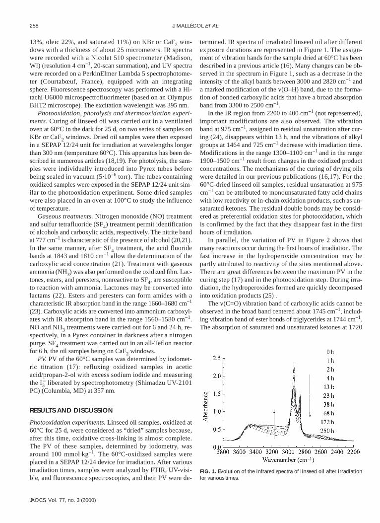
plain the growth of the band at 1735 cm−1 (35). The C–C link-ages are also preferential sites of oxidation because of thepresence of tertiary carbon. The radical attack occurs quitereadily on these sites (36) (Scheme 6). For this reason one canpostulate that cross-linking has two opposite consequences.On one hand, it allows curing and hardening of an oil film.On the other hand, it generates some structures that act as“fragile” sites in terms of durability of the material.
The decomposition of ether, C–C, or peroxy bonds justi-fies the global modification that occurs in the range1000–1300 cm−1. Moreover, the breakdown reactions men-tioned above can explain the formation of most low-molecu-lar-weight oxidation products that have been identified byanalysis of old oil-based paintings media (37,38). In thelinolenate chain, for instance, they mainly consist of car-boxylic acids that result from scission near the three doublebonds (39).
The carbonyl group vibration of ester bonds of triglyc-erides at 1744 cm−1 probably decreases during photooxida-tion. However, the increase in acid bands and in the lactoneand perester bands undoubtedly compensate for the disap-pearance of this ester band. The photodegradation of triglyc-erides can be described by the mechanism shown in Scheme7. The ester bonds in aliphatic polyesters are quite stable inphotooxidation at λ > 300 nm (40). However, in the triglyc-erides constituting the oil, there is a site with a tertiary carbonatom in the α-position of the oxygen atom. As is true for themajority of previous reactions, a radical attack is then likelyto occur.
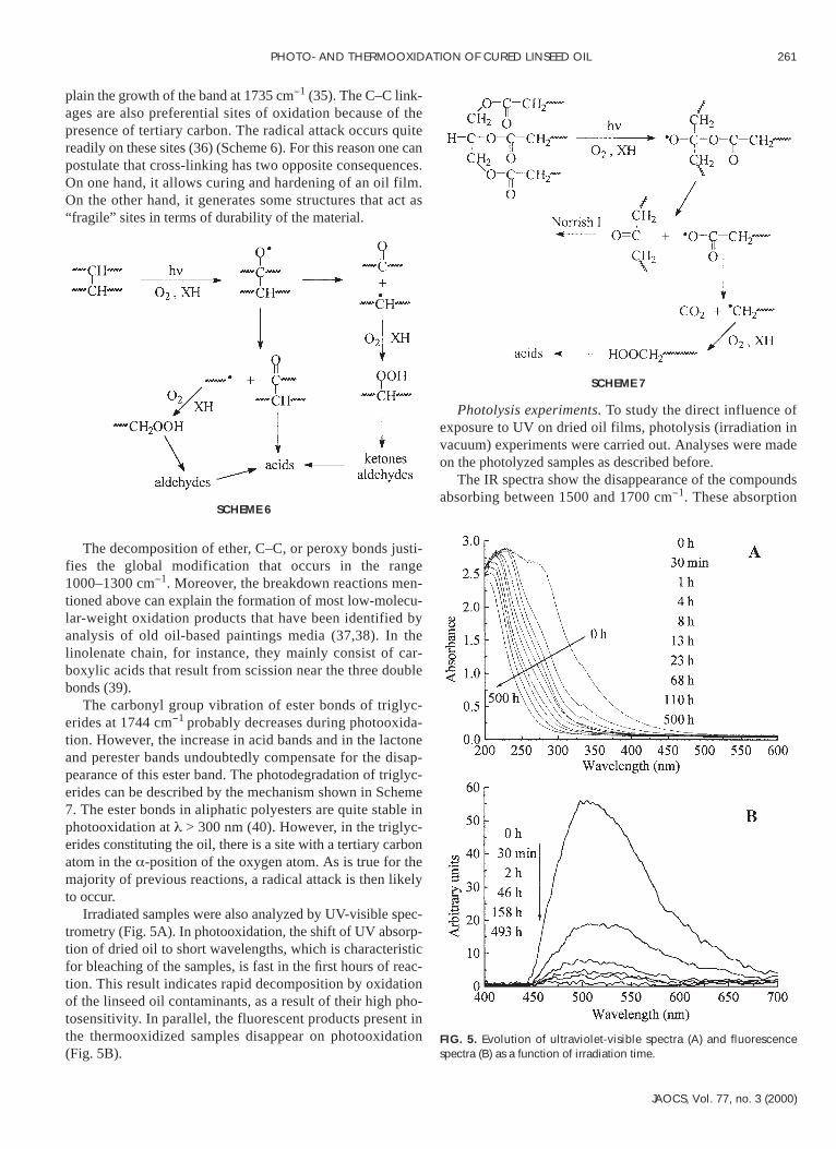
Irradiated samples were also analyzed by UV-visible spec-trometry (Fig. 5A). In photooxidation, the shift of UV absorp-tion of dried oil to short wavelengths, which is characteristicfor bleaching of the samples, is fast in the first hours of reac-tion. This result indicates rapid decomposition by oxidationof the linseed oil contaminants, as a result of their high pho-tosensitivity. In parallel, the fluorescent products present inthe thermooxidized samples disappear on photooxidation(Fig. 5B).
Photolysis experiments. To study the direct influence ofexposure to UV on dried oil films, photolysis (irradiation invacuum) experiments were carried out. Analyses were madeon the photolyzed samples as described before.
The IR spectra show the disappearance of the compoundsabsorbing between 1500 and 1700 cm−1. These absorption
PHOTO- AND THERMOOXIDATION OF CURED LINSEED OIL 261
JAOCS, Vol. 77, no. 3 (2000)
SCHEME 6
SCHEME 7
FIG. 5. Evolution of ultraviolet-visible spectra (A) and fluorescencespectra (B) as a function of irradiation time.

bands indicate the presence of contaminants of linseed oil,which contribute to yellowness and fluorescence of the sam-ples. The ketonic absorption at 1720 cm−1 decreases and, inthe hydroxyl range, one can also observe a decrease in thecontribution of alcohols and acids. NO and SF4 treatments onphotolyzed samples have confirmed this result. Photolysisalso leads to a fast decrease (in 3 h) in UV-visible absorptionof the samples between 400 and 300 nm. Subsequent disap-pearance of the products absorbing below 300 nm is slow.However, products decomposed during irradiation are not flu-orescent, and no notable variation of the fluorescence inten-sity is observed.
The results obtained in photolysis experiments that showonly “minor” modifications of the sample structure confirmthe proposed mechanisms that are involved in the degrada-tion of linseed oil. In all experiments, the reactions includethe fixation of oxygen molecules on radicals.
Thermooxidation experiments. Thermooxidation of a60°C-dried sample of linseed oil has been carried out at100°C. Modifications of the IR spectrum are not importantcompared to those after photooxidation. In the hydroxylrange, one observes the decrease of the broad band mainly at-tributed to bonded carboxylic acids between 3600 and 2300cm−1. This may be due to the formation of esters or anhy-drides by reaction of acids and alcohols. Water that is pro-duced by this reaction is lost by migration from the solid sam-ple. This phenomenon is not visible in a linseed oil sampledried directly at 100°C. This suggests that, at low tempera-tures, the curing step provokes the formation of some hydrox-ylic compounds, such as alcohols or carboxylic acids. After1,000 h at 100°C, the alkyl bands at 2928, 2856, and 725 cm−1
slightly decrease, and in the carbonyl zone there is a weak in-crease in the bands at 1775 cm−1 and about 1720 cm−1. Thesevariations could be attributed to the formation of γ-lactonesor peresters and ketones, respectively. However, there is a de-crease in absorbance in the range 1690–1620 cm−1, owing tothe disappearance of residual unsaturation. This is confirmedby the slight decrease in the 975 cm−1 band. It indicates thatincreasing the temperature allows partial curing to finish. Thisleads to the formation of hydroperoxides in the α-position ofthe double bonds, such as in a classical low-temperature oxi-dation of monounsaturated fatty acid chains.
In thermooxidation at 100°C, the increase in absorption inUV spectra is slow but continuous. The fluorescence spec-trum exhibits minor changes, starting with a slight decreasein the fluorescence, and after 10 h, a new continuous increase.We expected an increase in the fluorescence intensity of lin-seed oil with time because the fluorescence results from thedegradation process (41).
In this study, the thermostability of drying oils has beenclearly demonstrated. In thermooxidation there is no notableevolution of the chemical structure of the dried oil film. Thisresult is not surprising if one considers the state of conserva-tion of paintings that are more than five centuries old. How-ever, these paintings must not be exposed to sunlight [withthe aim of reversing their yellowing (42)]; otherwise the re-
sult would be rapid degradation because of the numerouschain-breaking reactions that are likely to occur in photooxi-dation.
REFERENCES
1. Wold, C.H., and M.D. Soucek, Mixed Metal OxideInorganic/Organic Coatings, J. Coat. Technol. 70:43–51 (1998).
2. Felder-Casagrande, S., and M. Odlyha, Development of Stan-dard Paint Films Based on Artists’ Materials, J. Therm. Anal.49:1585–1591 (1997).
3. Carbini, M., S. Volpin, and P. Traldi, Curie-Point Pyrolysis GasChromatography/Mass Spectrometry in the Identification ofPaint Media, Org. Mass Spectrom. 29:561–565 (1994).
4. Odlyha, M., Characterisation of Aged Paint Films by Differen-tial Scanning Calorimetry, Thermochim. Acta, 134:85–90(1988).
5. Odlyha, M., Investigation of the Binding Media of Paintings byThermoanalytical and Spectroscopic Techniques, Ibid.269/270:705–727 (1995).
6. Elm, A.C., Deterioration of Dried Oil Films, Ind. Eng. Chem.41:319–324 (1949).
7. Crecelius, S.B., R.E. Kagarise, and A.L. Alexander, Drying OilOxidation Mechanism, Film Formation and Degradation, Ibid.47:1643–1649 (1955).
8. Russel, G.A., Fundamental Processes of Autoxidation, J. Chem.Educ. 36:111–118 (1959).
9. Frankel, E.N., Lipid Oxidation, Prog. Lipid Res. 19:1–22(1980).
10. Chan, H.W.S., The Mechanism of Autoxidation, in Autoxida-tion of Unsaturated Lipids, edited by H.W.S. Chan, AcademicPress, London, 1987, pp. 1–16.
11. Frankel, E.N., Lipid Oxidation: Mechanisms, Products, and Bi-ological Significance, J. Am. Oil Chem. Soc. 61:1908–1917(1984).
12. Frankel, E.N., W.E. Neff, and E. Selke, Analysis of AutoxidizedFats by Gas Chromatography–Mass Spectrometry: VII. VolatileThermal Decomposition Products of Pure Hydroperoxides fromAutoxidized and Photosensitized Oxidized Methyl Oleate,Linoleate, and Linolenate, Lipids 16:279–285 (1981).
13. Miyashita, K., N. Hara, K. Fujimoto, and T. Kaneda, DimersFormed in Oxygenated Methyl Linoleate Hydroperoxides, Ibid.20:578–587 (1985).
14. Muizebelt, W.J., J.C. Hubert, and R.A.M. Venderbosch, Mech-anistic Study of Drying of Alkyd Resins Using Ethyl Linoleateas a Model Substance, Prog. Org. Coat. 24:263–279 (1994).
15. Boyatsis, S., E. Ioakimoglou, P. Argitis, A. Fostiridou, and K.Papapanogiotou, Oxidation of Linseed Oil Medium in Paintingsin the Presence of Copper Pigments: Spectroscopic and Chro-matographic Studies of Thin Films, ACS Polymer Preprints37(2):188–189 (1996).
16. Mallégol, J., J.L. Gardette, and J. Lemaire, Long-term Behaviorof Oil-Based Varnishes and Paints. 1. Spectroscopic Analysis ofthe Curing of Drying Oils, J. Am. Oil Chem. Soc. 76:967–976(1999).
17. Mallégol, J., J.L. Gardette, and J. Lemaire, Long-term Behaviorof Oil-Based Varnishes and Paints. 2. Fate of Hydroperoxidesin Drying Oils, Ibid. 77:249–255 (2000).
18. Lemaire, J., R. Arnaud, and J.L. Gardette, Le Vieillissement desPolymères. II-Principes d’Etude du Photovieillissement, Rev.Gén. Caoutch. Plast. 613:87–92 (1981).
19. Tang, L., D. Sallet, and J. Lemaire, Photochemistry of Polyun-decanamides. 1. Mechanisms of Photooxidation at Short andLong Wavelengths, Macromolecules 15:1432–1437 (1982).
20. Carlsson, D.J., R. Brousseau, C. Zhang, and D.M. Wiles, Poly-olefin Oxidation: Quantification of Alcohol and Hydroperoxide
262 J. MALLÉGOL ET AL.
JAOCS, Vol. 77, no. 3 (2000)

Products by Nitric Oxide Reactions, Polym. Degrad. Stab.17:303–318 (1987).
21. Lacoste, J., D. Vaillant, and D.J. Carlsson, Gamma-, Photo-, andThermally-Initiated Oxidation of Isotactic Polypropylene, J.Polym. Sci.: Part A: Polym. Chem. 31:715–722 (1993).
22. March, J., Advanced Organic Chemistry—Reactions, Mecha-nisms, and Structure, 4th edn., John Wiley & Sons, New York,1992 .
23. Avram, M., and G.D. Mateescu, Spectroscopie Infra-Rouge,Dunod, Paris, 1970.
24. Jenden, C.M., A Study of Some Artificially Weathered Paintsby Laser Raman Spectroscopy, Polymer 27:217–224 (1986).
25. Schieberle, P., W. Grosch, and J. Firl, Photolysis of UnsaturatedFatty Acid Hydroperoxides, in Oxygen Radicals in Chemistryand Biology, Walter de Gruyter and Co., Berlin, 1984, pp.257–265.
26. Sullivan, W.F., Weatherability of Titanium Dioxide-ContainingPaints, Progr. Org. Coat. 1:157–203 (1972).
27. Grassie, N., and G. Scott, Photo-degradation, in Polymer Degra-dation and Stabilization, Cambridge University Press, Cam-bridge, 1985, pp. 68–85.
28. Piton, M., and A. Rivaton, Photooxidation of Polybutadiene atLong Wavelengths (λ>300nm), Polym. Degrad. Stab.53:343–359 (1996).
29. Shadidi, F., and R.B. Pegg, Hexanal as an Indicator of the Fla-vor Deterioration of Meat and Meat Products, in Lipids in FoodFlavours, edited by C.T. Ho and T.G. Hartman, ACS Sympo-sium Series 558, 1994, pp. 256–279.
30. McNeill, I.C., and W.T.K. Stevenson, The Structure and Stabil-ity of Oxidised Polybutadiene, Polym. Degrad. Stab.11:123–143 (1985).
31. Ranby, B., and J.F. Rabek, Photodegradation and Photo-oxida-tion of Particular Polymers, in Photodegradation, Photo-oxida-tion and Photostabilization of Polymers—Principles and Appli-cations, John Wiley & Sons, London, 1975, pp. 120–253.
32. Gauvin, P., J. Lemaire, and D. Sallet, Propriétés des Hydroper-oxydes dans les Homopolymères de Polyéthers Correspondants,Makromol. Chem. 188:1815–1824 (1987).
33. Griffiths, P.J.F., J.G. Hughes, and G.S. Park, The Autoxidationof Poly(propylene oxides), Eur. Polym. J. 29:437–442 (1993).
34. Mailhot, B., S. Morel, and J.L. Gardette, Photochemical Behav-iour of Poly(vinylmethylether), Polym. Degrad. Stab.62:117–126 (1998).
35. Posada F., J.L. Philippart, P. Kappler, and J.L. Gardette, Pho-tooxidation of Cured Fluorinated Polymers—II. Comparisonwith Nonfluorinated Polyethers, Ibid. 53:19–31 (1996).
36. Grassie, N., and G. Scott, Oxidation of Polymers, in PolymerDegradation and Stabilization, Cambridge University Press,Cambridge, 1985, pp. 86–118.
37. Nowik, W., Acides Aminés et Acides Gras sur un Même Chro-matogramme—Un Autre Regard sur l’Analyse des Liants enPeinture, Stud. Conserv. 40:120–126 (1995).
38. White, R., and A. Roy, GC–MS and SEM Studies on the Effectsof Solvent Cleaning on Old Master Paintings from the NationalGallery, London, Ibid. 43:159–176 (1998).
39. Helme, J.P., La Science et l’Art: Evolution de la Technique dela Peinture à l’Huile Depuis son Invention Jusqu’à nos Jours,Rev. Fr. Corps Gras 41:13–19 (1994).
40. McKellar, J.F., and N.S. Allen, Photodegradation and Photooxi-dation Processes, in Photochemistry of Man-made Polymers,Applied Science Publishers, London, 1979, pp. 31–181.
41. René De La Rie, E., Fluorescence of Paint and Varnish Layers(part II), Stud. Conserv. 27:65–69 (1982).
42. Rakoff, H., F.L. Thomas, and L.E. Gast, Reversibility of Yel-lowing Phenomenon in Linseed-Based Paints, J. Coat. Technol.51:25–28 (1979).
[Received July 26, 1999; accepted November 2, 1999]
PHOTO- AND THERMOOXIDATION OF CURED LINSEED OIL 263
JAOCS, Vol. 77, no. 3 (2000)