Local Structure-Based Region-of-Interest Retrieval in Brain MR Images
7
IEEE TRANSACTIONS ON INFORMATION TECHNOLOGY IN BIOMEDICINE, VOL. XX, NO. XX, JANUARY 2009 1 Local Structure-based Region-of-Interest Retrieval in Brain MR Images Devrim Unay * , Ahmet Ekin, and Radu Jasinschi Abstract—The aging population and the growing amount of medical data have increased the need for automated tools in the neurology departments. Although the researchers have been de- veloping computerized methods to help the medical expert, these efforts have primarily emphasized improving the effectiveness in single patient data, such as finding a brain lesion and computing its size. However, patient-to-patient comparison that should help improve diagnosis and therapy has not received as much attention. To this effect, this paper introduces a fast and robust region-of-interest retrieval method for brain magnetic resonance (MR) images. We make several contributions to the domains of brain MR image analysis, and search and retrieval: 1)We show the potential and robustness of local structure information in the search and retrieval of brain MR images. 2)We provide analysis of two complementary features, local binary patterns and Kanade-Lucas-Tomasi feature points, and their comparison with a baseline method. 3)We show that incorporating spatial context in the features substantially improves accuracy. 4)We propose to automatically extract dominant local binary patterns and demonstrate that using dominant features improves retrieval relative to the conventional local binary pattern approach. Com- prehensive experiments on real and simulated datasets revealed that dominant local binary patterns with spatial context is robust to geometric deformations and intensity variations and have high accuracy and speed. The proposed method can not only aid the medical expert in disease diagnosis, or be used in scout (localizer) scans for optimization of acquisition parameters, but also support low power devices that we believe will find increasing usage in hospitals. Index Terms—Brain MR image retrieval, local structure analysis, local binary patterns, Kanade-Lucas-Tomasi feature points, spatial descriptors, medical databases, neurodegenerative diseases. I. I NTRODUCTION T HE advances in the medical imaging technology allow for in-vivo visualization and analysis of human body with unprecedented accuracy and resolution. A diagnosis by a specialist often requires a visit to a radiology department to obtain various images that highlight the suspected pathology. Despite the high resolution of the acquired images, image- based diagnosis often utilizes a considerable amount of qual- itative measures. To improve the diagnosis and efficiency, the research in medical image analysis has focused on the computation of quantitative measures by automating some of the error-prone and excruciatingly time-consuming tasks, such as segmentation of a structure. This work was supported in part by the Marie Curie Programme of the European Commission under FP6 IRonDB project MTKI-CT-2006-042717. D. Unay, A. Ekin, and R. Jasinschi are with the Video Process- ing and Analysis Group, Philips Research Europe, 5656 AE Eind- hoven, The Netherlands ([email protected], [email protected], [email protected]). A less emphasized approach for improving diagnosis has been comparison of multiple patients, their pathologies, and progresses by search and retrieval systems. This should es- pecially improve the diagnosis of diseases whose causes and progress have not yet been completely unraveled, and diseases that affect large number of patients. The area of neurology can greatly benefit from such a methodology because the diagnosis of neurodegenerative diseases from one patient data has limitations (A review of 13 neuropathologically confirmed studies show that a clinical diagnosis of Alzheimer’s Disease has an average sensitivity of 81% and specificity of 70% [1]). Accordingly, this paper focuses on region-of-interest retrieval of unregistered brain MR images although the presented methods should be applicable to other organs. In particular, we would like to find similar slices in an unregistered brain MR database given a query slice, automation of which can aid the medical expert in diagnosis of structure-specific diseases, such as hippocampus or basal ganglia disorders. In the medical domain, to obtain clinically useful images imaging system operators typically acquire scout (localizer) and target scans, where the former is visually examined to specify acquisition parameters for the latter. Hence, the pre- sented methods can also be used to automate the examination of the scout images to set parameters for the target scan. The extensive research on search and retrieval in the do- main of multimedia confirms the challenging nature of this problem [2]-[3]. In the domain of medical data, there are domain or even modality-specific differences and challenges. The captured images are usually single channel (gray-scale) that differs from multi-channel (e.g. RGB) nature of consumer image and video data. In some medical modalities, such as CT (Computed Tomography) and X-ray, intensity has an absolute scale. In MR, they vary with the scanning parameters, but also with the age of the device and the patient characteristics. To aggravate the problem, the intensity values of a specific tissue may also be non-stationary within a single MR image (a fact known as intensity non-uniformity or bias field) be- cause of imperfect magnetic field and patient-dependent local perturbations. Furthermore, the characteristics of relevant and irrelevant segments of the data are very close to each other for the databases that are specific to a modality and an organ, such as brain MR database. To the best of our knowledge, only Bucci et al. [4] con- sider a similar search and retrieval problem as ours, that is, retrieving the relevant slice from a brain MR image sequence in response to a given sample slice. Their method performs retrieval of the relevant slice using Principal Component Analysis (or the so-called eigenimages), but requires a com-
-
Upload
independent -
Category
Documents
-
view
3 -
download
0
Transcript of Local Structure-Based Region-of-Interest Retrieval in Brain MR Images
IEEE TRANSACTIONS ON INFORMATION TECHNOLOGY IN BIOMEDICINE, VOL. XX, NO. XX, JANUARY 2009 1
Local Structure-based Region-of-Interest Retrievalin Brain MR Images
Devrim Unay∗, Ahmet Ekin, and Radu Jasinschi
Abstract—The aging population and the growing amount ofmedical data have increased the need for automated tools in theneurology departments. Although the researchers have been de-veloping computerized methods to help the medical expert, theseefforts have primarily emphasized improving the effectiveness insingle patient data, such as finding a brain lesion and computingits size. However, patient-to-patient comparison that shouldhelp improve diagnosis and therapy has not received as muchattention. To this effect, this paper introduces a fast and robustregion-of-interest retrieval method for brain magnetic resonance(MR) images. We make several contributions to the domainsof brain MR image analysis, and search and retrieval: 1)Weshow the potential and robustness of local structure informationin the search and retrieval of brain MR images. 2)We provideanalysis of two complementary features, local binary patternsand Kanade-Lucas-Tomasi feature points, and their comparisonwith a baseline method. 3)We show that incorporating spatialcontext in the features substantially improves accuracy. 4)Wepropose to automatically extract dominant local binary patternsand demonstrate that using dominant features improves retrievalrelative to the conventional local binary pattern approach. Com-prehensive experiments on real and simulated datasets revealedthat dominant local binary patterns with spatial context is robustto geometric deformations and intensity variations and have highaccuracy and speed. The proposed method can not only aid themedical expert in disease diagnosis, or be used in scout (localizer)scans for optimization of acquisition parameters, but also supportlow power devices that we believe will find increasing usage inhospitals.
Index Terms—Brain MR image retrieval, local structureanalysis, local binary patterns, Kanade-Lucas-Tomasi featurepoints, spatial descriptors, medical databases, neurodegenerativediseases.
I. INTRODUCTION
THE advances in the medical imaging technology allowfor in-vivo visualization and analysis of human body
with unprecedented accuracy and resolution. A diagnosis bya specialist often requires a visit to a radiology department toobtain various images that highlight the suspected pathology.Despite the high resolution of the acquired images, image-based diagnosis often utilizes a considerable amount of qual-itative measures. To improve the diagnosis and efficiency,the research in medical image analysis has focused on thecomputation of quantitative measures by automating some ofthe error-prone and excruciatingly time-consuming tasks, suchas segmentation of a structure.
This work was supported in part by the Marie Curie Programme of theEuropean Commission under FP6 IRonDB project MTKI-CT-2006-042717.
D. Unay, A. Ekin, and R. Jasinschi are with the Video Process-ing and Analysis Group, Philips Research Europe, 5656 AE Eind-hoven, The Netherlands ([email protected], [email protected],[email protected]).
A less emphasized approach for improving diagnosis hasbeen comparison of multiple patients, their pathologies, andprogresses by search and retrieval systems. This should es-pecially improve the diagnosis of diseases whose causes andprogress have not yet been completely unraveled, and diseasesthat affect large number of patients. The area of neurologycan greatly benefit from such a methodology because thediagnosis of neurodegenerative diseases from one patient datahas limitations (A review of 13 neuropathologically confirmedstudies show that a clinical diagnosis of Alzheimer’s Diseasehas an average sensitivity of 81% and specificity of 70% [1]).Accordingly, this paper focuses on region-of-interest retrievalof unregistered brain MR images although the presentedmethods should be applicable to other organs. In particular,we would like to find similar slices in an unregistered brainMR database given a query slice, automation of which can aidthe medical expert in diagnosis of structure-specific diseases,such as hippocampus or basal ganglia disorders.
In the medical domain, to obtain clinically useful imagesimaging system operators typically acquire scout (localizer)and target scans, where the former is visually examined tospecify acquisition parameters for the latter. Hence, the pre-sented methods can also be used to automate the examinationof the scout images to set parameters for the target scan.
The extensive research on search and retrieval in the do-main of multimedia confirms the challenging nature of thisproblem [2]-[3]. In the domain of medical data, there aredomain or even modality-specific differences and challenges.The captured images are usually single channel (gray-scale)that differs from multi-channel (e.g. RGB) nature of consumerimage and video data. In some medical modalities, such as CT(Computed Tomography) and X-ray, intensity has an absolutescale. In MR, they vary with the scanning parameters, butalso with the age of the device and the patient characteristics.To aggravate the problem, the intensity values of a specifictissue may also be non-stationary within a single MR image(a fact known as intensity non-uniformity or bias field) be-cause of imperfect magnetic field and patient-dependent localperturbations. Furthermore, the characteristics of relevant andirrelevant segments of the data are very close to each otherfor the databases that are specific to a modality and an organ,such as brain MR database.
To the best of our knowledge, only Bucci et al. [4] con-sider a similar search and retrieval problem as ours, that is,retrieving the relevant slice from a brain MR image sequencein response to a given sample slice. Their method performsretrieval of the relevant slice using Principal ComponentAnalysis (or the so-called eigenimages), but requires a com-
2 IEEE TRANSACTIONS ON INFORMATION TECHNOLOGY IN BIOMEDICINE, VOL. XX, NO. XX, JANUARY 2009
putationally expensive registration and intensity normalizationstep beforehand.
Others working on the search and retrieval of MR imagesmainly focused on the shape information: Hsu et al. [5]introduced a method based on the shape of the brain ventricles.Robinson et al. [6] and Petrakis and Faloutsos [7], bothproposed shape based retrieval methods that require manualdelineation of the anatomical structures in question. Morerecently, Huang et al. [8] used geometric features and Fourierdescriptors to represent brain images of pediatric patients, butrequired registered images.
In CT, because intensity has an absolute scale, many systemshave exploited it together with image structure and shapeinformation in feature extraction, e.g., by using histogram,moment invariants and co-occurrence matrix representations incombination [9], [10]. Traina et al. [11] introduced a structure-based retrieval solution for human brain images by usingwavelets. Ghebraeb et al. [12] focused on shape informationand proposed to use contour curvatures of expert delineatedvertebrae in X-ray images. Retrieval of medical images frommultiple modalities is challenging due to the presence of imagerelated problems specific to different modalities. The systemin [13] uses co-occurrence matrix representations to achieveretrieval of CT and MR images of different tissues, while theone in [14] employs color quantization and wavelet responsesto retrieve images of different body parts acquired with var-ious modalities. Other medical modalities, such as positronemission tomography [15], have been less investigated [16].
The rest of the paper is organized as follows: Section IIprovides an insight into the approach used in this work aswell as our contributions. Section III details the pre-processingapplied to the images. Sections IV and V present the featureextraction and description stages of our solution, where fourlocal structure-based methods and one edge-related baselinemethod are introduced. Section VI explains how the retrievalis performed. Experimental data and results are presented inSection VII. Finally, the conclusions and the related futureworks are given in Section VIII.
II. OUR APPROACH AND CONTRIBUTIONS
In this paper, we focus on a retrieval problem where themost similar slices to the given query slice are searched. Thechallenges for this problem include:• MR intensity variations from one patient to another be-
cause of possible differences in MR settings. This limitsthe use of intensity information in comparing differentdatasets.
• Intensity variations within the same dataset (intensitynon-uniformity or bias field) due to imperfect, inhomo-geneous magnetic field. This requires the use of compu-tational bias field correction algorithms.
• The high similarity between irrelevant and relevant seg-ments in medical images makes their search and retrievalmore challenging.
• Inter and intra-patient misalignment of images, becauseof which anatomical structures are observed at differentspatial positions, orientations and scales in the images.
Although registration could solve this problem, it is toocomputational and impractical for low power mobiledevices or for the scouting application where fast imageacquisition is crucial for the clinical centers.
• Abnormalities of brain structure, such as ventricular en-largement or cerebral atrophy, tumors.
To solve the above problems, this paper introduces the follow-ing contributions:• We introduce a fast and robust search and retrieval
method of brain MR images. Not only the retrievalperformance is invariant to some basic geometric trans-formations and intensity variations, but also the systemavoids the computationally intensive registration, inten-sity normalization, and bias field correction steps.
• We propose to use local structure features instead ofintensities, because intensity is non-standard and sensitiveto MR imaging artifacts. The local structure features arerobust to both inter- and intra-scan intensity variations.
• We show the search and retrieval effectiveness of twolocal structure features: 1) local binary patterns (LBP)proposed by Ojala et al. [17], and 2) Kanade-Lucas-Tomasi (KLT) feature points. The choice of the two isnot arbitrary. LBP operates locally and treats every localregion equally. On the contrary, KLT selects the pre-defined number of features that are the most salient.
• To remedy the high feature space similarity of rele-vant and irrelevant segments, we use a context-baseddescription of features. The spatial description of fea-tures increases the retrieval performance significantly andprovides robustness to large abnormalities seen in severecases.
• We show that using only the most dominant local binarypatterns in the query improves retrieval accuracy whilereducing dimensionality of the data.
III. PRE-PROCESSING
A typical MR image of the head consists of brain tissue aswell as background and non-brain structures, like skull andskin in which we are not interested. Hence, we perform theextraction of the brain tissue by using the Brain ExtractionTool (BET) [18].
Moreover, as misalignment of images is a common problemin magnetic resonance imaging, we benefit from a mid-sagittalplane detection algorithm [19] that indicates orientation, inorder to compensate for rotation in spatial description of thefeatures, which will be explained afterwards.
IV. STRUCTURE-BASED FEATURE EXTRACTION
In this section, we first describe LBP, a computationally veryefficient method to extract local structure and due to its localnature invariant to bias field [20]. Afterwards, we describeKLT feature points that correspond to the most salient localregions of the image.
A. Local Binary PatternsRecently, Ojala et al. [17] introduced LBP, which is a
grayscale invariant local texture descriptor with low computa-tional complexity. Subsequent studies have shown that LBP is

UNAY et al.: LOCAL STRUCTURE-BASED REGION-OF-INTEREST RETRIEVAL IN BRAIN MR IMAGES 3
promising in the computer vision field, including industrial in-spection [21], motion analysis [22], and face recognition [23].However, its application in the medical field has been mainlylimited to adenoma detection in endoscopic images [24] andplaque segmentation in ultrasound images [25].
LBP operator (LBPP,R) describes the local structure patternby thresholding a neighborhood with the gray value of itscenter pixel (gc) and represents the result as a binary code.The neighborhood is formed by a symmetric neighbor set ofP pixels gp(p = 0, . . . , P − 1) on a circle of radius R.
LBPP,R =P−1∑p=0
s(gp − gc)2p, s(x) ={
1, x ≥ 00, x < 0
(1)Extended versions of the original operator are introduced
in [17] as (1) LBP riP,R: rotation invariant, (2) LBPu2
P,R:uniform, and (3) LBP riu2
P,R : rotation invariant and uniformoperators.These extensions provide not only robustness of pat-terns to noise and rotation, but also dimensionality reductionin the pattern space, e.g., structure in a 3 × 3 neighborhoodis represented by 256, 36, 59, and 10 different patternsusing LBP8,1, LBP ri
8,1, LBPu28,1, and LBP riu2
8,1 operators,respectively.
B. KLT Feature Points
LBP computes structure features in all local regions ofthe image. However, one may argue that not all parts ofan image contain valuable information. Consequently, re-searchers proposed to find interesting regions of an imageusing detectors, such as the SIFT descriptor [26]. Anotherexemplary work is the KLT feature tracker [27], [28], whichfocuses on the regions (so-called good features) that can betracked well. KLT method has been applied to various prob-lems, such as registration in augmented reality systems [29],pedestrian tracking [30], and gestures and facial expressionstracking [31], but to the best of our knowledge its applicationin medical field is not yet realized.
Feature point selection in KLT is performed by searchingthe whole image through a window, and selecting the regionsthat have adequate intensity variation in vertical and horizontaldirections, indicating they contain corner. Considering thelocal intensity variation matrix
Z =[
g2x gxgy
gxgy g2y
](2)
where gx and gy are the partial derivatives of an input imageI(x, y), a region is accepted as a candidate feature point if botheigenvalues, λ1 and λ2, of Z exceed a predefined threshold,min(λ1, λ2) > λ. Consequently, the candidate feature pointsof the input image are ranked according to their strengthdefined by min(λ1, λ2), and NF strongest ones are selected,with NF being the user-defined maximum number of featuresto be selected.
V. FEATURE DESCRIPTION
In this section we propose four methods based on eitherLBP or KLT features, and a baseline method for comparison.
Fig. 1. Computation of LBP features.
Fig. 2. A visual example of feature point matching. Among the 50 featurepoints (overlaid as white dots on the images) extracted from a query, 10 arematched on the target.
These five methods are organized depending on whether theirfeatures are described spatially (Sp) or not (nSp).
A. Non-Spatial Feature Description
nSp-LBP As LBP features correspond to all local regionsof an image, we describe them by their statisticaldistribution. Therefore, in nSp-LBP we use the his-togram of the extracted LBP image (Figure 1).
nSp-KLT Salient feature points extracted from an imagewill be successfully matched (and therefore pre-served) on another if the two images are similar.Hence, nSp-KLT method extracts feature points fromthe query and attempts in matching them on thetarget (Figure 2). The number of matched featurepoints are then used as attributes in retrieval.
B. Spatial Feature Description
Sp-LBP This method exploits spatial indexing [32] of theLBP image histogram, where the entries of each
4 IEEE TRANSACTIONS ON INFORMATION TECHNOLOGY IN BIOMEDICINE, VOL. XX, NO. XX, JANUARY 2009
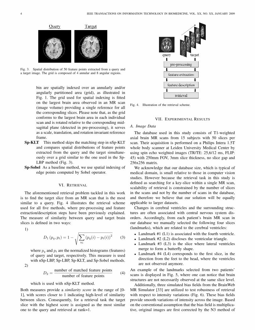
Fig. 3. Spatial distribution of 50 feature points extracted from a query anda target image. The grid is composed of 4 annular and 8 angular regions.
bin are spatially indexed over an annularly and/orangularly partitioned area (grid), as illustrated inFig. 1. The grid used for spatial indexing is fittedon the largest brain area observed in an MR scan(image volume) providing a single reference for allthe corresponding slices. Please note that, as the gridconforms to the largest brain area in each individualscan and is rotated relative to the corresponding mid-sagittal plane (detected in pre-processing), it servesas a scale, translation, and rotation invariant referenceframe.
Sp-KLT This method skips the matching step in nSp-KLTand compares spatial distributions of feature pointsextracted from the query and the target simultane-ously over a grid similar to the one used in the Sp-LBP method (Fig. 3).
Sp-Sobel As a baseline method, we use spatial indexing ofedge points computed by Sobel operator.
VI. RETRIEVAL
The aforementioned retrieval problem tackled in this workis to find the target slice from an MR scan that is the mostsimilar to a query. Fig. 4 illustrates the retrieval schemeused for all five methods, where pre-processing and featureextraction/description steps have been previously explained.The measure of similarity between query and target brainslices is defined in two ways:
1)
D1 (pq, pt) = 1−√∑
∀i
(pq(i)− pt(i))2 (3)
where pq and pt are the normalized histograms (features)of query and target, respectively. This measure is usedwith nSp-LBP, Sp-LBP, Sp-KLT, and Sp-Sobel methods.
2)
D2 =number of matched feature points
number of feature points(4)
which is used with nSp-KLT method.
Both measures provide a similarity score in the range of [0-1], with scores closer to 1 indicating high-level of similaritybetween slices. Consequently, for a retrieval task the targetslice with the highest score is assigned as the most similarone to the query and retrieved at rank=1.
Fig. 4. Illustration of the retrieval scheme.
VII. EXPERIMENTAL RESULTS
A. Image Data
The database used in this study consists of T1-weightedaxial brain MR scans from 15 subjects with 50 slices perscan. Their acquisition is performed on a Philips Intera 1.5Twhole body scanner at Leiden University Medical Center byusing spin echo weighted images (TR/TE: 25,6/12 ms, FLIP:45) with 250mm FOV, 3mm slice thickness, no slice gap and256x256 matrix.
We acknowledge that our database size, which is typical ofmedical domain, is small relative to those in computer visionstudies. However because the retrieval task in this study isdefined as searching for a key-slice within a single MR scan,scalability of retrieval is constrained by the number of slicesin the scans and not by the number of scans in the database,and therefore we believe that our solution will be equallyapplicable to larger datasets.
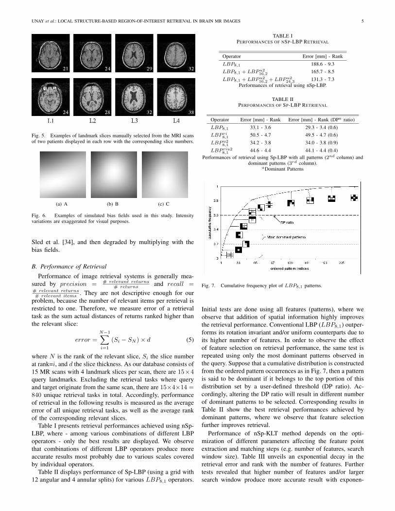
Changes in cerebral ventricles and the surrounding struc-tures are often associated with central nervous system dis-orders. Accordingly, from each patient’s brain MR scan inour database we manually selected the following four slices(landmarks), which are related to the cerebral ventricles:• Landmark #1 (L1) is associated with the fourth ventricle.• Landmark #2 (L2) discloses the ventricular triangle.• Landmark #3 (L3) is the slice where lateral ventricles
merge to form a butterfly shape.• Landmark #4 (L4) corresponds to the first slice, in the
direction from the feet to the head, where the ventriclesare not observed anymore.
An example of the landmarks selected from two patients’scans is displayed in Fig. 5, where one can notice that brainstructures are not necessarily observed at the same slice level.
Additionally, three simulated bias fields from the BrainWebMR Simulator [33] are utilized to test robustness of retrievalwith respect to intensity variations (Fig. 6). These bias fieldsprovide smooth variations of intensity across the image. Basedon the conventional assumption that the bias field is multiplica-tive, original images are first corrected by the N3 method of
UNAY et al.: LOCAL STRUCTURE-BASED REGION-OF-INTEREST RETRIEVAL IN BRAIN MR IMAGES 5
Fig. 5. Examples of landmark slices manually selected from the MRI scansof two patients displayed in each row with the corresponding slice numbers.
(a) A (b) B (c) C
Fig. 6. Examples of simulated bias fields used in this study. Intensityvariations are exaggerated for visual purposes.
Sled et al. [34], and then degraded by multiplying with thebias fields.
B. Performance of Retrieval
Performance of image retrieval systems is generally mea-sured by precision = # relevant returns
# returns and recall =# relevant returns# relevant items . They are not descriptive enough for our
problem, because the number of relevant items per retrieval isrestricted to one. Therefore, we measure error of a retrievaltask as the sum actual distances of returns ranked higher thanthe relevant slice:
error =N−1∑
i=1
(Si − SN )× d (5)
where N is the rank of the relevant slice, Si the slice numberat rank=i, and d the slice thickness. As our database consists of15 MR scans with 4 landmark slices per scan, there are 15×4query landmarks. Excluding the retrieval tasks where queryand target originate from the same scan, there are 15×4×14 =840 unique retrieval tasks in total. Accordingly, performanceof retrieval in the following results is measured as the averageerror of all unique retrieval tasks, as well as the average rankof the corresponding relevant slices.
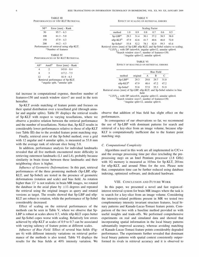
Table I presents retrieval performances achieved using nSp-LBP, where - among various combinations of different LBPoperators - only the best results are displayed. We observethat combinations of different LBP operators produce moreaccurate results most probably due to various scales coveredby individual operators.
Table II displays performance of Sp-LBP (using a grid with12 angular and 4 annular splits) for various LBP8,1 operators.
TABLE IPERFORMANCES OF NSP-LBP RETRIEVAL
Operator Error [mm] - Rank
LBP8,1 188.6 - 9.3LBP8,1 + LBP u2
16,2 165.7 - 8.5LBP8,1 + LBP u2
16,2 + LBP u224,3 131.3 - 7.3
Performances of retrieval using nSp-LBP.
TABLE IIPERFORMANCES OF SP-LBP RETRIEVAL
Operator Error [mm] - Rank Error [mm] - Rank (DPa ratio)
LBP8,1 33.1 - 3.6 29.3 - 3.4 (0.6)LBP ri
8,1 50.5 - 4.7 49.5 - 4.7 (0.6)LBP u2
8,1 34.2 - 3.8 34.0 - 3.8 (0.9)LBP riu2
8,1 44.6 - 4.4 44.1 - 4.4 (0.4)Performances of retrieval using Sp-LBP with all patterns (2nd column) and
dominant patterns (3rd column).aDominant Patterns
Fig. 7. Cumulative frequency plot of LBP8,1 patterns.
Initial tests are done using all features (patterns), where weobserve that addition of spatial information highly improvesthe retrieval performance. Conventional LBP (LBP8,1) outper-forms its rotation invariant and/or uniform counterparts due toits higher number of features. In order to observe the effectof feature selection on retrieval performance, the same test isrepeated using only the most dominant patterns observed inthe query. Suppose that a cumulative distribution is constructedfrom the ordered pattern occurrences as in Fig. 7, then a patternis said to be dominant if it belongs to the top portion of thisdistribution set by a user-defined threshold (DP ratio). Ac-cordingly, altering the DP ratio will result in different numberof dominant patterns to be selected. Corresponding results inTable II show the best retrieval performances achieved bydominant patterns, where we observe that feature selectionfurther improves retrieval.
Performance of nSp-KLT method depends on the opti-mization of different parameters affecting the feature pointextraction and matching steps (e.g. number of features, searchwindow size). Table III unveils an exponential decay in theretrieval error and rank with the number of features. Furthertests revealed that higher number of features and/or largersearch window produce more accurate result with exponen-
6 IEEE TRANSACTIONS ON INFORMATION TECHNOLOGY IN BIOMEDICINE, VOL. XX, NO. XX, JANUARY 2009
TABLE IIIPERFORMANCES OF NSP-KLT RETRIEVAL
NFa Error [mm] - Rank
50 95.7 - 6.3100 61.3 - 5.0150 47.9 - 4.3200 44.2 - 4.3
Performances of retrieval using nSp-KLT.aNumber of features
TABLE IVPERFORMANCES OF SP-KLT RETRIEVAL
ASa AnnSb Error [mm] - Rank
4 2 143.8 - 8.48 3 117.2 - 7.512 4 81.9 - 6.3Retrieval performance of Sp-KLT.
aangular split, bannular split
tial increase in computational expense, therefore number offeatures=150 and search window size=7 are used in the testshereafter.
Sp-KLT avoids matching of feature points and focuses ontheir spatial distribution over a tessellated grid (through annu-lar and angular splits). Table IV displays the retrieval resultsof Sp-KLT with respect to varying tessellations, where weobserve a positive relation between the retrieval performanceand the number of tessellations. Notice that, Sp-KLT results inconsiderably lower performances relative to those of nSp-KLT(see Table III) due to the avoided feature point matching step.
Finally, retrieval error of the Sp-Sobel method, over a gridwith 12 angular and 4 annular splits, is measured as 53.8 mmwith the average rank of relevant slice being 5.0.
In addition, performance analysis for individual landmarksrevealed that all five methods encountered more difficulty inretrieving outermost landmarks (L1 and L4), probably becausesimilarity in brain tissue between these landmarks and theirneighboring slices is higher.
Influence of Geometric Deformations: In this part, retrievalperformances of the three promising methods (Sp-LBP, nSp-KLT, and Sp-Sobel) are tested in the presence of geometricdeformations (rotation and scale) and bias field. As rotationhigher than 15◦ is not realistic in brain MR images, we rotatedthe database in the axial plane by ±15 degrees and repeatedthe retrieval using the original images as query and rotatedversions as target. The results showed that Sp-LBP and nSp-KLT are robust to rotation, while the performance of Sp-Sobelconsiderably decreased.
Effect of scaling on the retrieval performances of themethods can be seen in Table V, where we observe that Sp-LBP is robust at scales above 0.7, while nSp-KLT copes betterand Sp-Sobel copes worse with scaling. Relatively low errorsachieved by nSp-KLT at scales of 0.9 to 0.7 can be associatedwith varying saliency of feature points at different scales.
Influence of Bias Field: Effect of several bias fields (Fig-ure 6) with different intensity variations on retrieval perfor-mance of the methods is also tested. Table VI displays theresults for the bias fields at 40% intensity variation. We
TABLE VEFFECT OF SCALING ON RETRIEVAL ERRORS
Scaling Factor
method 1.0 0.9 0.8 0.7 0.6 0.5
Sp-LBPa 29.3 31.4 30.1 37.2 38.5 38.0nSp-KLTb 47.9 42.6 41.7 40.6 46.0 72.0Sp-Sobelc 53.8 52.2 78.1 82.0 59.3 63.4
Retrieval errors [mm] of Sp-LBP, nSp-KLT, and Sp-Sobel relative to scaling.aLBP8,1 with DP ratio=0.6, angular split=12, annular split=4.
bSearch window size=7, number of features=150.cAngular split=12, annular split=4.
TABLE VIEFFECT OF BIAS FIELD ON RETRIEVAL ERRORS
Bias Fieldmethod original A B C
Sp-LBPa 29.3 29.8 29.7 31.0nSp-KLTb 47.9 48.4 47.2 47.2Sp-Sobelc 53.8 57.9 55.3 51.9
Retrieval errors [mm] of Sp-LBP, nSp-KLT, and Sp-Sobel relative to biasfield.
aLBP8,1 with DP ratio=0.6, angular split=12, annular split=4.bSearch window size=7, number of features=150.
cAngular split=12, annular split=4.
observe that addition of bias field has slight effect on theperformances.
In consequence of our observations so far, we recommendthe use of Sp-LBP with dominant patterns for search andretrieval of a key-slice from an image volume, because nSp-KLT is computationally inefficient due to the feature pointmatching step.
C. Computational Complexity
Algorithms used in this work are all implemented in C/C++and the average processing time per slice (excluding the pre-processing step) on an Intel Pentium processor (2.8 GHz)with 1G memory is measured as 103ms for Sp-KLT, 201msfor nSp-KLT, and around 50ms for the rest. Please notethat, computation time can be further reduced using databaseindexing, optimized software, and dedicated hardware.
VIII. CONCLUSION AND FUTURE WORK
In this paper, we presented a novel and fast region-of-interest retrieval system for brain MR images where the task isto search for a key-slice from an image volume. Consideringthe intensity-related problems present in MR we tested twocomplementary intensity invariant structure features, local bi-nary patterns and Kanade-Lucas-Tomasi feature points. Com-parison of the two with a baseline method provided us withuseful insights and trade-offs. We performed comprehensiveexperiments on real and simulated data and showed thatincorporating spatial information in the local binary patternssubstantially improved accuracy, whereas avoiding matchingof Kanade-Lucas-Tomasi feature points considerably degradedperformance. The experiments further revealed that dominantlocal binary patterns with spatial context consistently outper-formed its rivals in retrieval accuracy and it is observed to
UNAY et al.: LOCAL STRUCTURE-BASED REGION-OF-INTEREST RETRIEVAL IN BRAIN MR IMAGES 7
be robust to common anomalies, such as abnormal cerebralventricles, present in the data. Moreover, this retrieval methodis observed robust to bias fields at 40% intensity variation,rotation up to ±15◦, and downsizing at scales above 0.7. Inconclusion, the proposed retrieval method is fast and robust,has high accuracy, and does not require registration, intensitynormalization or bias field correction.
The retrieval problem addressed in this study has an inherentdifficulty due to the very similar content present in the data,and therefore there is room for improvement. We believe thatincorporating multiresolution analysis and shape informationinto the proposed method may improve its accuracy.
ACKNOWLEDGMENT
The authors thank Prof. Mark van Buchem and Dr. Jeroenvan der Grond from the Department of Radiology, Leiden Uni-versity Medical Center for the discussions and data support.
REFERENCES
[1] D. S. Knopman, S. T. DeKosky, J. L. Cummings, H. Chui, J. Corey-Bloom, N. Relkin, G. W. Small, B. Miller, and J. C. Stevens, “Practiceparameter: Diagnosis of dementia (an evidence-based review): Reportof the Quality Standards Subcommittee of the American Academy ofNeurology,” Neurology, vol. 56, no. 9, pp. 1143–1153, 2001. [Online].Available: http://www.neurology.org/cgi/content/abstract/56/9/1143
[2] Y. Rui, T. Huang, and S. Chang, “Image retrieval: current techniques,promising directions and open issues,” Journal of Visual Communicationand Image Representation, vol. 10, no. 4, pp. 39–62, Apr. 1999.
[3] M. S. Lew, N. Sebe, C. Djerba, and R. Jain, “Content-based multimediainformation retrieval: state of the art and challenges,” ACM Transactionson Multimedia Computing, Communications, and Applications, vol. 2,no. 1, pp. 1–19, Feb. 2006.
[4] G. Bucci, S. Cagnoni, and R. D. Domenicis, “Integrating content-basedretrieval in a medical image reference database,” Computerized MedicalImaging and Graphics, vol. 20, no. 4, pp. 231–241, 1996.
[5] C.-C. Hsu, W. W. Chu, and R. K. Taira, “A knowledge-based approachfor retrieving images by content,” IEEE Transactions on Knowledge andData Engineering, vol. 8, no. 4, pp. 522–532, 1996.
[6] G. P. Robinson, H. D. Tagare, J. S. Duncan, and C. C. Jaffe, “Medicalimage collection indexing: shape-based retrieval using kd-trees,” Com-puterized Medical Imaging and Graphics, vol. 20, no. 4, pp. 209–217,1996.
[7] E. G. M. Petrakis and C. Faloutsos, “Similarity searching in medicalimage databases,” IEEE Transactions on Knowledge and Data Engi-neering, vol. 9, no. 3, pp. 435–447, 1997.
[8] H. K. Huang, J. F. Nielsen, M. D. Nelson, and L. Lui, “Image-matchingas a medical diagnostic support (DST) for brain diseases in children,”Computerized Medical Imaging and Graphics, vol. 29, no. 2-3, pp. 195–202, 2005.
[9] A. Kak and C. Pavlopoulou, “Content-based image retrieval fromlarge medical databases,” in 1st International Symposium on 3D DataProcessing Visualization and Transmission, Padova, Italy, 2002, pp. 138–147.
[10] S.-N. Yu, C.-T. Chiang, and C.-C. Hsieh, “A three-object model for thesimilarity searches of chest CT images,” Computerized Medical Imagingand Graphics, vol. 29, no. 8, pp. 617–630, 2005.
[11] A. J. M. Traina, C. A. B. Castanon, and C. Traina Jr., “Multiwavemed:a system for medical image retrieval through wavelets transformations,”in 16th IEEE Symposium on Computer-Based Medical Systems, NewYork, USA, 2003, pp. 150–155.
[12] S. Ghebraeb, C. C. Jaffe, and A. W. M. Smeulders, “Population-basedincremental interactive concept learning for image retrieval by stochasticstring segmentations,” IEEE Transactions on Medical Imaging, vol. 23,no. 6, pp. 676–689, Jun. 2004.
[13] J. C. Felipe, A. J. M. Traina, and C. Traina Jr., “Retrieval by content ofmedical images using texture for tissue identification,” in 16th IEEESymposium on Computer-Based Medical Systems, New York, USA,2003, pp. 175–180.
[14] H. Muller, A. Rosset, J.-P. Vallet, and A. Geisbuhler, “Comparing featuresets for content-based image retrieval in a medical case database,” inProceedings of SPIE Medical Imaging: PACS and Imaging Informatics,San Diego, USA, 2004, pp. 99–109.
[15] J. Kim, W. Cai, D. Feng, and H. Wu, “A new way for multidimensionalmedical data management: volume of interest (VOI)-based retrieval ofmedical images with visual and functional features,” IEEE Transactionson Information Technology in Biomedicine, vol. 10, no. 3, pp. 598–607,Jul. 1997.
[16] H. Muller, N. Michoux, D. Bandon, and A. Geisbuhler, “A reviewof content-based image retrieval systems in medical applications -clinical benefits and future directions,” International Journal of MedicalInformatics, vol. 73, no. 1, pp. 1–23, Feb. 2004.
[17] T. Ojala, M. Piettikainen, and T. Maenpaa, “Multiresolution gray-scaleand rotation invariant texture classification with local binary patterns,”IEEE Transactions on Pattern Analysis and Machine Intelligence,vol. 24, no. 7, pp. 971–987, Jul. 2002.
[18] S. M. Smith, “Fast robust automated brain extraction,” Human BrainMapping, vol. 17, no. 3, pp. 143–155, Nov. 2002.
[19] A. Ekin, “Feature-based brain mid-sagittal plane detection byRANSAC,” in Proceedings of 14th European Signal Processing Con-ference, Florence, Italy, 2006.
[20] D. Unay, A. Ekin, M. Cetin, R. Jasinschi, and A. Ercil, “Robustnessof local binary patterns in brain MR image analysis,” in 29th AnnualInternational Conference of the IEEE-EMBS, Lyon, France, 2007, pp.2098–2101.
[21] T. Maenpaa, M. Turtinen, and M. Piettikainen, “Real-time surfaceinspection by texture,” Real-Time Imaging, vol. 9, no. 5, pp. 289–296,Oct. 2003.
[22] M. Heikkila and M. Piettikainen, “A texture-based method for modelingthe background and detecting moving objects,” IEEE Transactions onPattern Analysis and Machine Intelligence, vol. 28, no. 4, pp. 657–662,Apr. 2006.
[23] T. Ahonen, A. Hadid, and M. Piettikainen, “Face description with localbinary patterns: application to face recognition,” IEEE Transactions onPattern Analysis and Machine Intelligence, vol. 28, no. 12, pp. 2037–2041, Dec. 2006.
[24] D. K. Iakovidis, D. E. Maroulis, and S. A. Karkanis, “An intelligentsystem for automatic detection of gastrointestinal adenomas in videoendoscopy,” Computers in Biology and Medicine, vol. 36, no. 10, pp.1084–1103, Oct. 2006.
[25] O. Pujol, D. Rotger, P. Radeva, O. Rodriguez, and J. Mauri, “Near real-time plaque segmentation of IVUS,” in Proceedings of Computers inCardiology, Thessaloniki, Greece, 2003, pp. 69–73.
[26] D. Lowe, “Distinctive image features from scale-invariant keypoints,”International Journal of Computer Vision, vol. 60, no. 2, pp. 91–110,Nov. 2004.
[27] C. Tomasi and T. Kanade, “Detection and tracking of point features,”Carnegie Mellon Univ., Pittsburgh, PA, Tech.Rep. CMU-CS-91-132, apr1991.
[28] J. Shi and C. Tomasi, “Good features to track,” in Proceedings of IEEEComputer Vision and Pattern Recognition Conference, Seattle, USA,1994, pp. 593–600.
[29] M. L. Yuan, S. K. Ong, and A. Y. C. Nee, “Registration usingnatural features for augmented reality systems,” IEEE Transactions onVisualization and Computer Graphics, vol. 12, no. 4, pp. 569–580, 2006.
[30] M. Slaughter, “Tracking pedestrians with machine vision,” EvaluationEngineering, vol. 46, no. 2, pp. 56–57, Feb. 2007.
[31] D. Metaxas, G. Tsechpenakis, Z. Li, Y. Huang, and A. Kanaujia,“Dynamically adaptive tracking of gestures and facial expressions,” inProceedings of 6th International Conference on Computational Sci-ence,Part III (Lecture Notes in Computer Science Vol. 3993), Reading,UK, 2006, pp. 554–561.
[32] A. Rao, R. K. Srihari, and Z. Zhang, “Spatial color histograms forcontent-based image retrieval,” in Proceedings of IEEE InternationalConference on Tools with Artificial Intelligence, Chicago, USA, 1999,pp. 183–186.
[33] [online], “http://www.bic.mni.mcgill.ca/brainweb/.”[34] J. G. Sled, A. P. Zijdenbos, and A. C. Evans, “A nonparametric method
for automatic correction of intensity nonuniformity in mri data,” IEEETransactions on Medical Imaging, vol. 17, no. 1, pp. 87–97, Feb. 1998.