Assessment of the constitutive properties from small ball punch test: experiment and modeling
Upload
independentCategory
view
3download
0
Vol. 11, No. 9MOLECULAR AND CELLULAR BIOLOGY, Sept. 1991, p. 4314-43230270-7306/91/094314-10$02.00/0Copyright © 1991, American Society for Microbiology
Liver Cells Contain Constitutive DNase I-Hypersensitive Sites atthe Xenobiotic Response Elements 1 and 2 (XRE1 and -2) of the Rat
Cytochrome P-450IA1 Gene and a Constitutive, Nuclear XRE-Binding Factor That Is Distinct from the Dioxin ReceptorJANET HAPGOOD,1t SCOTT CUTHILL,lt PETER SODERKVIST,1,2 ANNA WILHELMSSON,'
INGEMAR PONGRATZ,1 ROBERT H. TUKEY,3 ERIC F. JOHNSON,4JAN-AKE GUSTAFSSON,1 AND LORENZ POELLINGER1*
Department of Medical Nutrition, Karolinska Institutet, Huddinge University Hospital F60, Novum, S-141 86 Huddinge,'and Department of Occupational Medicine, Faculty of Health Sciences, S-581 852 Sweden; Linkoping; Departments of
Pharmacology and Medicine, Cancer Center, University of California, San Diego, La Jolla, California 920933;and Research Institute of Scripps Clinic, La Jolla, California 920374
Received 1 April 1991/Accepted 3 June 1991
Dioxin stimulates transcription from the cytochrome P-450IA1 promoter by interaction with the intracellulardioxin receptor. Upon binding of ligand, the receptor is converted to a form which specifically interacts in vitrowith two dioxin-responsive positive control elements located in close proximity to each other about 1 kbupstream of the rat cytochrome P-450IA1 gene transcription start point. In rat liver, the cytochrome P-450IA1gene is marked at the chromatin level by two DNase I-hypersensitive sites that map to the location of theresponse elements and exist prior to induction of transcription by the dioxin receptor ligand 0-naphthoflavone.In addition, a DNase I-hypersensitive site is detected near the transcription initiation site and is altered innuclease sensitivity by induction. The presence of the constitutive DNase I-hypersensitive sites at the dioxinresponse elements correlates with the presence of a constitutive, labile factor which specifically recognizes theseelements in vitro. This factor appears to be distinct from the dioxin receptor, which is observed only in nuclearextract from treated cells. In conclusion, these data suggest that a certain protein-DNA architecture may bemaintained at the response elements at different stages of gene expression.
Transcription of the cytochrome P-450IA1 gene is dramat-ically induced in response to the environmental contaminantdioxin (2,3,7,8-tetrachlorodibenzo-p-dioxin) or related com-pounds. The induction process is mediated by the intracel-lular dioxin receptor protein and initiated by binding of theinducing chemical to the receptor, which in its non-ligand-occupied state most probably is located in the cytoplasmiccompartment of target cells (for a review, see reference 29).In the absence of ligand, the cytosolic dioxin receptor isrecovered as a latent, non-DNA-binding species (30). How-ever, upon exposure to ligand in vivo, the dioxin-receptorcomplex undergoes an activation process involving a poorlyunderstood structural alteration that enables it to translocateto the nucleus (29). In vitro, the ligand-activated dioxinreceptor has been shown to represent a DNA-binding pro-tein (references 5 and 47 and references therein) whichspecifically recognizes dioxin-responsive positive controlelements (xenobiotic response elements [XREs]) found inthe promoter and upstream regions of the cytochromeP-450IA1 and glutathione S-transferase Ya genes (9, 16, 17,21, 31, 33). The XREs, in turn, modulate the activity oflinked promoters, thereby serving as dioxin-inducible en-hancers. Thus, the mechanism of action of dioxin is similarto that of steroid hormones (reviewed in references 2, 19,
* Corresponding author.t Present address: Department of Biochemistry, University of
Cape Town, 7700 Rondebosch, Cape Town, Republic of SouthAfrica.
t Present address: The Beatson Institute for Cancer Research,Glasgow G61 1BD, Scotland, United Kingdom.
and 48) in that (i) both categories of receptors directlytransduce an extracellular signal to the target transcriptionunits they regulate and (ii) the dioxin and steroid receptorsrequire ligand for function.A striking property of the dioxin receptor is that the ligand
strictly controls the activation of the receptor from a latentspecies to a DNA-binding form also under cell-free condi-tions (7, 17, 30). In the case of steroid hormone receptors,the glucocorticoid response element of the rat tyrosineaminotransferase gene is protected against methylation bydimethyl sulfate in hepatoma cells only after hormone ad-ministration (3), supporting the hypothesis that the glucocor-ticoid receptor interacts in vivo with its target sequence in aligand-dependent manner. Moreover, glucocorticoids inducethe formation of a DNase I-hypersensitive site at the gluco-corticoid response elements of the rat tyrosine aminotrans-ferase gene in both hepatoma cells and rat liver (4, 25, 32),suggesting that the hormone-activated receptor may alter theDNA or chromatin configuration at these regulatory ele-ments.To assess the function of dioxin-responsive elements
defined by cellular transfection studies (16), we have exam-ined chromatin structural changes associated with activationof the cytochrome P-450IA1 gene in rat liver. Two sequenceelements (XRE1 and -2) mediating dioxin receptor-depen-dent activation of this gene have been defined and arelocalized about 1 kb (extending from positions -1029 to-997 and from positions -1092 to -1069, respectively)upstream of the transcription initiation site (16). Both theXRE1 and -2 elements are recognized by the ligand-activatedform of dioxin receptor in vitro (17, 21). In this study, we
4314
CHROMATIN STRUCTURE OF CYTOCHROME P-450IA1 4315
have observed in untreated rat liver two DNase I-hypersen-sitive sites which map to the positions of the two XREs,respectively. Interestingly, these sites remained unalteredupon induction of gene expression by the dioxin receptorligand P-naphthoflavone. The presence of these constitutiveDNase I-hypersensitive sites could suggest that a specificprotein-DNA architecture is maintained at the XREs,regardless of changes in the transcriptional state of the gene.In agreement with this notion, we have identified and char-acterized an XRE-specific DNA-binding factor in vitro thatis present in nuclear extract from nontreated cells andappears to be distinct from the receptor itself.
MATERIALS AND METHODS
Animals, cell culture, isolation of nuclei, and preparation ofextracts. Male Sprague-Dawley rats weighing 200 to 300 gwere used in animal studies. In indicated cases, the rats weretreated 4 or 20 h prior to sacrifice with one single intraperi-toneal injection of P-naphthoflavone (dissolved in corn oil) ata concentration of 80 mg/kg of body weight. The rats weresacrificed by cervical dislocation. For DNase I hypersensi-tivity studies, liver tissue was homogenized in 15 mMTris-HCI (pH 7.4)-60 mM KCl-0.35 M sucrose-15 mMNaCl-2 mM EDTA-0.5 mM EGTA-0.15 mM spermine-0.5mM spermidine-1 mM phenylmethylsulfonyl fluoride, andnuclei were prepared essentially as described previously(15). Nuclear proteins were extracted from rat liver tissue asdescribed previously (18). Rat Rueber hepatoma cells(H4IIE) were grown in monolayer culture in Dulbecco'smodified Eagle's medium (GIBCO) supplemented with 8%(vol/vol) heat-inactivated fetal calf serum and benzylpenicil-lin-streptomycin (GIBCO). The wild-type Hepa lclc7 cellline and the mutant line c4 derived from it (20) were grown inminimum essential medium as described previously (6, 46).The mutant c4 line expresses a dioxin receptor phenotypewhich is deficient in nuclear translocation (nt-; 28) andDNA-binding activity (5) of ligand-occupied receptor. Inindicated cases, cells were treated with 1 nM dioxin or[3H]dioxin (Chemsyn, Lenexa, Kan.) for 1 h. Nuclear ex-tract was prepared from untreated or treated hepatoma cellsaccording to Dignam et al. (10). This protocol was modifiedin indicated experiments to allow separation of constitutiveXRE-binding factor(s) from the receptor. Briefly, isolatednuclei were preextracted with a low salt concentration (finalconcentration, 0.15 M KCl) prior to a second extraction ofnuclear proteins at a final concentration of 0.42 M KCl.Cytosolic extracts from untreated wild-type Hepa lclc7cells were prepared as described previously (46).Mapping of DNase I-hypersensitive sites in nuclei. DNase I
digestions were performed at 4°C on isolated rat liver nucleiessentially as described previously (14), and protein-freeDNA was recovered as described previously (37). Electro-phoretic separation of DNA, transfer to nitrocellulose, andhybridization to 32P-labeled probes were as described previ-ously (37). A 1-kb ladder (Bethesda Research Laboratories)was used as molecular weight standards. The various DNAfragments used as probes were isolated by restriction diges-tion of various subclones of the rat cytochrome P-450IA1gene (41, 42).
Chromatographic fractionation of nuclear proteins. Hepa-toma cell nuclear extract was fractionated by high-perfor-mance gel permeation chromatography on a prepacked Su-perose 12 (Pharmacia) column (10 by 300 mm) or by anion-exchange chromatography on a prepacked Mono Q HR 5/5column (Pharmacia) as described previously (21, 46). The
anion-exchange column was equilibrated in 20 mM phos-phate (pH 7.2)-i mM EDTA-2 mM 2-mercaptoethanol-1mM phenylmethylsulfonyl fluoride and eluted at a flow rateof 0.5 ml/min with linear salt gradients as indicated.
In vitro DNA-binding assays. DNA-binding activities werecharacterized in rat liver and hepatoma cell nuclear extractsby a gel mobility shift assay as described previously (21,30). DNA-binding reaction mixtures were assembled in10 mM N-2-hydroxyethylpiperazine-N'-2-ethanesulfonic acid(HEPES; pH 7.9)-10% (vol/vol) glycerol-0.5 mM dithiothre-itol-3 mM MgCI2-60 mM KCl-4 mM spermidine in a finalvolume ranging between 20 to 50 ,ul. In these experiments,double-stranded 36-bp-long oligonucleotides spanning thewild-type rat cytochrome P-450IA1 XRE1 or XRE2 (16)sequence motif were used as specific, radiolabeled probes.Oligonucleotides containing point mutations in the XRE1motif (7) were also synthesized and used in either directbinding or DNA-binding competition experiments. For se-quences of the wild-type and mutant oligonucleotides, seeFig. SA. Finally, a 38-bp oligonucleotide spanning an oc-tamer motif from the BCL1 immunoglobulin heavy-chainpromoter (34) was used as an XRE-unrelated sequence in theoligonucleotide competition experiments.
Safety precautions. The experimental use of dioxin re-quired special handling procedures as outlined previously(reference 5 and references therein). Contaminated materialswere disposed of by high-temperature incineration.
RESULTS
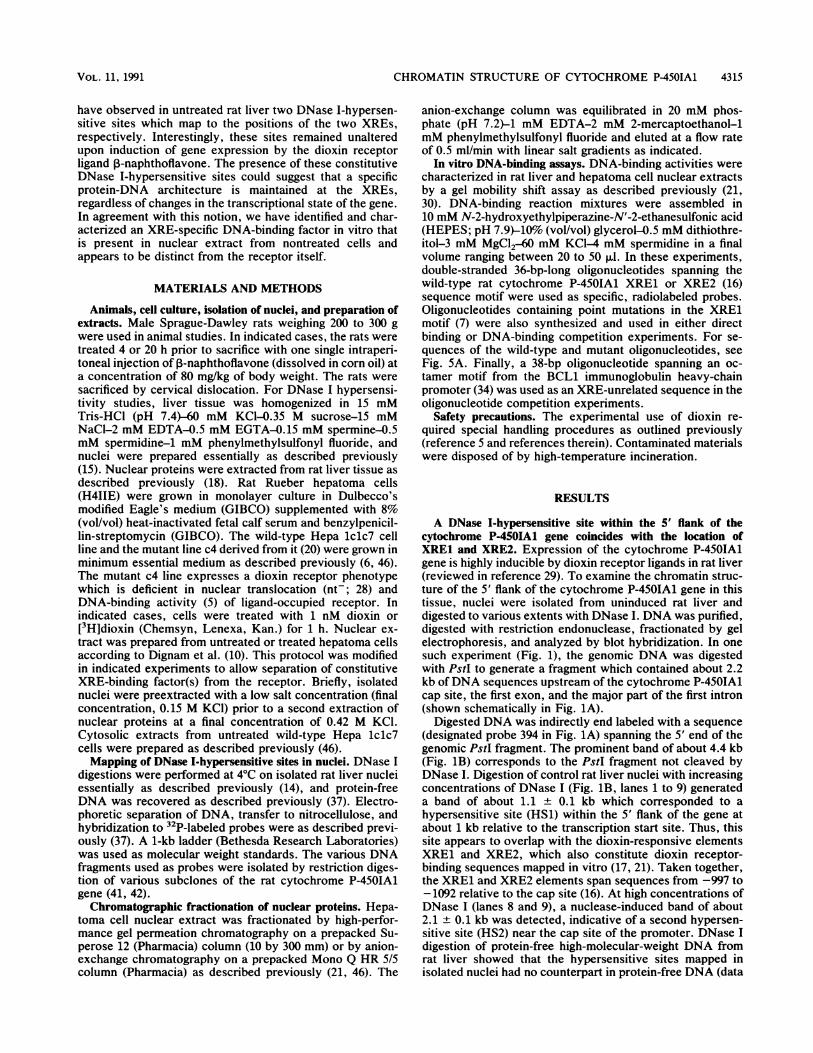
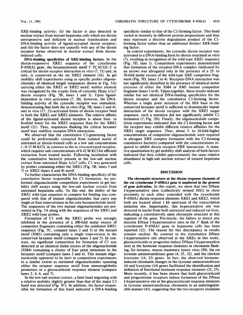
A DNase I-hypersensitive site within the 5' flank of thecytochrome P-450IA1 gene coincides with the location ofXRE1 and XRE2. Expression of the cytochrome P-450IA1gene is highly inducible by dioxin receptor ligands in rat liver(reviewed in reference 29). To examine the chromatin struc-ture of the 5' flank of the cytochrome P-450IA1 gene in thistissue, nuclei were isolated from uninduced rat liver anddigested to various extents with DNase I. DNA was purified,digested with restriction endonuclease, fractionated by gelelectrophoresis, and analyzed by blot hybridization. In onesuch experiment (Fig. 1), the genomic DNA was digestedwith PstI to generate a fragment which contained about 2.2kb ofDNA sequences upstream of the cytochrome P-450IA1cap site, the first exon, and the major part of the first intron(shown schematically in Fig. 1A).
Digested DNA was indirectly end labeled with a sequence(designated probe 394 in Fig. 1A) spanning the 5' end of thegenomic PstI fragment. The prominent band of about 4.4 kb(Fig. 1B) corresponds to the PstI fragment not cleaved byDNase I. Digestion of control rat liver nuclei with increasingconcentrations of DNase I (Fig. 1B, lanes 1 to 9) generateda band of about 1.1 + 0.1 kb which corresponded to ahypersensitive site (HS1) within the 5' flank of the gene atabout 1 kb relative to the transcription start site. Thus, thissite appears to overlap with the dioxin-responsive elementsXRE1 and XRE2, which also constitute dioxin receptor-binding sequences mapped in vitro (17, 21). Taken together,the XRE1 and XRE2 elements span sequences from -997 to-1092 relative to the cap site (16). At high concentrations ofDNase I (lanes 8 and 9), a nuclease-induced band of about2.1 + 0.1 kb was detected, indicative of a second hypersen-sitive site (HS2) near the cap site of the promoter. DNase Idigestion of protein-free high-molecular-weight DNA fromrat liver showed that the hypersensitive sites mapped inisolated nuclei had no counterpart in protein-free DNA (data
VOL . 1 l, 1991
4316 HAPGOOD ET AL.
not shown), indicating that the mapped hypersensitivity is aconsequence of chromatin structure.
Induction of cytochrome P-450IA1 transcription leads to nochange in chromatin structure at the XREs. We have previ-ously shown that transcription from the rat liver cytochromeP-450IA1 promoter is induced by the dioxin receptor ligandP-naphthoflavone (40). Following injection of a single doseof 3-naphthoflavone (80 mg/kg), maximal transcription levelsare observed at 4 h, as assessed by run-on elongationanalyses with isolated rat liver nuclei (40). At 4 h oftreatment, gene activation by ,B-naphthoflavone was notparalleled by any change in sensitivity to DNase I at the HS1site coinciding with the XREs (Fig. iB; compare lanes 1 to 9with lanes 10 to 18). By contrast, the DNase I-hypersensitivesite near the point of transcription initiation (HS2) showed amarked increase in sensitivity to nuclease digestion in nucleifrom P-naphthoflavone-treated liver (lanes 10 to 18) com-pared with control nuclei (lanes 1 to 9).
In a second set of experiments, rats were treated for 20 hwith P-naphthoflavone. During this period of time, 3-naph-thoflavone-induced cytochrome P-450IA1 gene transcriptiondecreases to control levels (40). Blot hybridization analysiswith fragment 394 of DNase I and PstI-digested DNA fromrat liver nuclei after 20 h of in vivo treatment also demon-strated the hypersensitive site at the positions of XRE1 andXRE2. In fact, DNA blot analysis of the HS1 site at highresolution showed that it could be resolved into two bands(HS1A and HS1B) with sizes corresponding to hypersensi-tive sites at about -1 kb + 50 bp and -1.1 kb + 50 bp,respectively, relative to the transcription start site (Fig. 1C).Thus, these two sites map to the locations of XRE1 (at -1kb) and XRE2 (at -1.1 kb; 16), respectively. As in Fig. 1B,the HS1A and HS1B sites were detected in liver nuclei fromuntreated as well as ,B-naphthoflavone-induced rats (Fig. 1C;compare lanes 1 to 9 with lanes 10 to 18).
Consistent with the observations in Fig. 1B, ,-naphtho-flavone treatment dramatically altered the sensitivity toDNase digestion of the HS2 site, which was detected as aband of 2.1 kb + 0.1 kb, positioning this site in closeproximity to the cap site (Fig. 1C). Interestingly, the HS2site showed a sensitivity to nuclease digestion after 20 h of
A
kt -2
Pst-2183
HS1 HS2B A HSi H S
4 E
XRE2 XRE1
Probes: 394 224
B
CONTROL + BNF
DNase : - - ------ --_-
kb:
4.0 -o-
3.0 -*-O
2.0 -O -4-HS2
1.6 -W
-- HS11.0 --*-
1 2 3 4 5 6 7 8 9 10 11 121314 15 16 1718
CCONTROL + BNF--DNase o DNase I- o
kb:
FIG. 1. Mapping of DNase I-hypersensitive sites in the 5' flank-ing region of the cytochrome P-450IA1 gene in untreated and,B-naphthoflavone-treated rat liver. (A) Schematic representation ofthe promoter region of the rat cytochrome P-450IA1 gene. The thickline represents the transcribed region; arrows indicate the DNaseI-hypersensitive sites designated; filled boxes represent the dioxin-responsive elements XRE1 and XRE2; the bent arrow indicates thestart site of transcription. The fragments used as radiolabeled probesfor DNA blot hybridization experiments are indicated below themap. Probes 394 and 224 represent a 394-bp PstI-PvuII (-2183 to-1789) and a 224-bp BamHI-KpnI (-847 to -623) fragment, respec-tively, of the rat cytochrome P-450IA1 gene. (B) Hybridizationanalysis. Nuclei from uninduced (control) and P-naphthoflavone-induced (BNF) rat livers (4 h of treatment) were isolated and treatedin the absence (lanes 1, 2, 10, and 11) or presence (lanes 3 to 9 and12 to 18) of increasing concentrations of DNase 1 (0.03 to 2.1 U/t,l).After purification, the DNA was digested with PstI, and 35 ,ug ofDNA per lane was separated on a 1% agarose gel. The DNA wasblotted to a filter and hybridized to the 394-bp probe. (C) Mapping ofDNase I-hypersensitive sites at higher resolution. Digested DNAwas isolated from untreated rat liver or liver treated for 20 h withP-naphthoflavone and size separated on a 1.5% agarose gel. Seelegend to panel B for details. The parallel mobilities of DNAmolecular weight markers are shown at the left.
4.0 - .-dm.*~'..3.0 -
2.0 -
1.6 -
1,0 !,. a||
0 11.0 - ,gIgg
'I0.5 -
a *X I
S. *Os
HSi
- HS2
- HSii
Pstt21 78.
A-HSlA
iU.- HS1B
I
1 2 3 4 5 6 7 8 9 10 11 12 13 1415 16 17 18
MOL. CELL. BIOL.
P. AL A 40 op 40 0,0*0 o w .0
CHROMATIN STRUCTURE OF CYTOCHROME P-450IA1 4317
CONTROL +BNF
DNase 1: - - - -
kb:
4.0 --*
3.0 --b-
2.0 *
1.6 -0-
1.0 -0
1 2 3 4 5 6 7 8 9 1011 1213 14 151
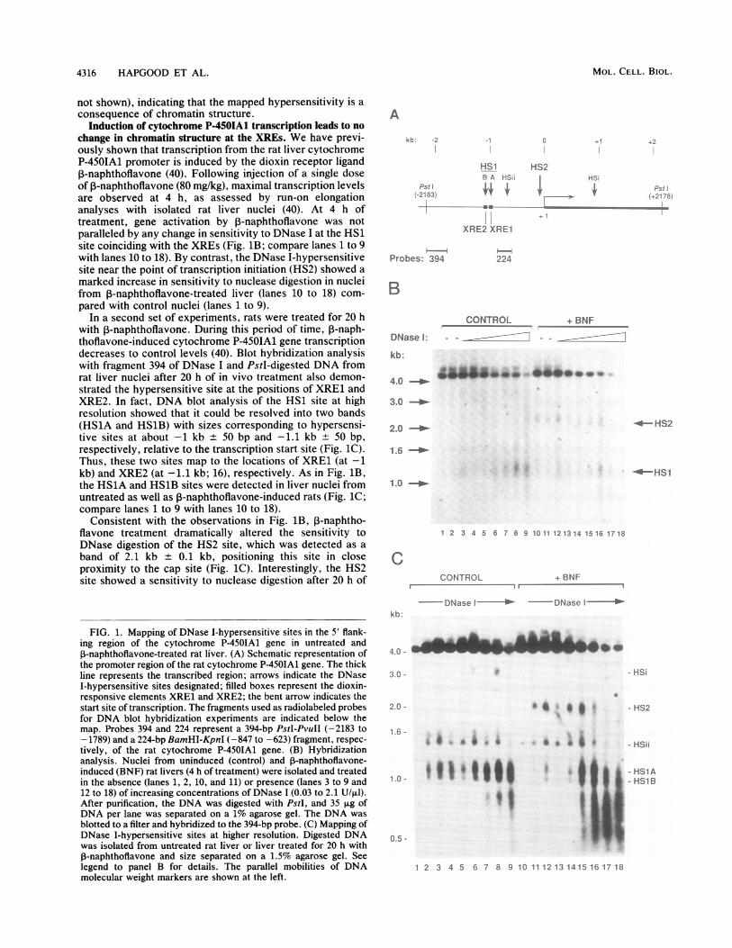
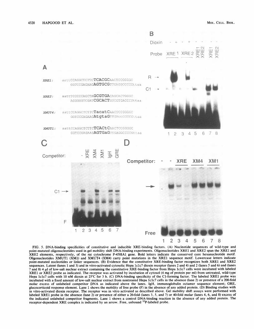
FIG. 2. DNase I-hypersensitive sites in rat cytocchromatin. The autoradiograph shows hypersensitin nuclear chromatin of untreated rat liver (controlrat liver treated for 4 h with P-naphthoflavone (BNIusing probe 224. See legend to Fig. 1B for details. Tsites showed different degrees of sensitivity tocleases in different preparations of nuclei (compalanes 1 and 2 with lanes 10 and 11).
treatment similar to that observed after 4(Compare Fig. lB and C). Thus, the dioxin-iin chromatin structure in the vicinity of thstart site was not reversed after 20 h of treatthe cytochrome P-450IA1 gene is transcribedthis time point. In addition to the two major Isensitive sites HS1 and HS2, this analysi-3.1-kb band corresponding to a hypersensitiabout + 1 kb in the first intron of the gene. Ulflavone induction, the sensitivity of this sidigestion was decreased, in agreement with a
(13). Moreover, a second constitutive hyp4(HSii) was detected at approximately 0.75 kbtranscription start site, and a rather broad relthoflavone-stimulated DNase I hypersensiticated at more than 1.4 kb upstream of the trasite (Fig. 1C; compare lanes 1 to 9 with laneThe chromatin structure of the 4.4-kb PstI
the cytochrome P-450IA1 gene was further clusing an internal probe (designated 224 inhybridizes to sequences in the 3' vicinity of tanalysis did not reveal any DNase-I hyperseaddition to two major bands of 3.1 + 0.1 kb ar
respectively (Fig. 2). The 2.1-kb band shosensitivity to nuclease digestion following 1-'
stimulation and corresponded to the previcHS2 site located in close proximity to the calthe 3.1-kb band was constitutively present (Ilanes 1 to 9 with lanes 10 to 18). Given the bthe intronic DNase I-hypersensitive site atpeared upon 1-naphthoflavone induction (Figstitutive 3.1-kb band most probably corresposite at about -1 kb relative to the start s
analysis confirms the earlier assignment of thsites to the positions of the XREs and the trasite, respectively.The constitutive DNase I-hypersensitive sit
correlates with the presence of a labile, constitutive XRE-specific factor. The constitutive accessibility of the XREs incytochrome P-450IAl chromatin may facilitate interactionwith nuclear proteins also in noninduced cells, or, con-versely, such a factor(s) may play a role in the formation ofthe hypersensitive site. This hypothesis prompted us to
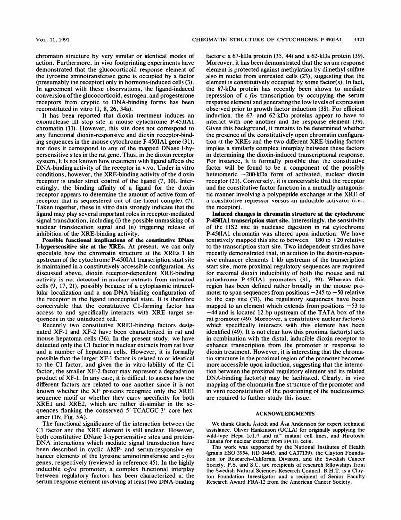
4 HS1 examine whether proteins other than the dioxin receptorcould specifically recognize XRE target sequences in vitro.
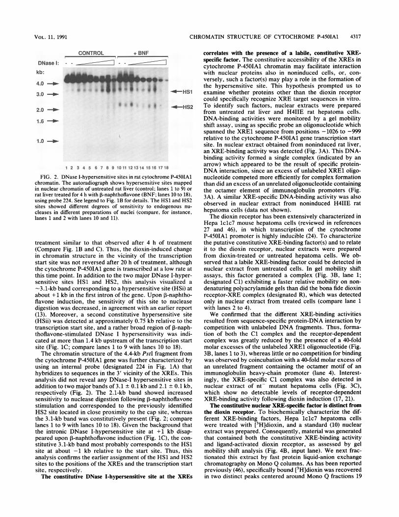
* -*--HS2 To identify such factors, nuclear extracts were preparedfrom untreated rat liver and H4IIE rat hepatoma cells.DNA-binding activities were monitored by a gel mobilityshift assay, using as specific probe an oligonucleotide whichspanned the XRE1 sequence from positions -1026 to -999relative to the cytochrome P-450IAl gene transcription startsite. In nuclear extract obtained from noninduced rat liver,an XRE-binding activity was detected (Fig. 3A). This DNA-binding activity formed a single complex (indicated by an
16 1718 arrow) which appeared to be the result of specific protein-DNA interaction, since an excess of unlabeled XRE1 oligo-
chrome P-450IA1 nucleotide competed more efficiently for complex formation:ive sites mapped than did an excess of an unrelated oligonucleotide containing1; lanes 1 to 9) or the octamer element of immunoglobulin promoters (Fig.F lanes 10 to 18), 3A). A similar XRE-specific DNA-binding activity was alsoendogenous nu- observed in nuclear extract from noninduced H4IIE rat
re, for instance hepatoma cells (data not shown).The dioxin receptor has been extensively characterized in
Hepa lclc7 mouse hepatoma cells (reviewed in references27 and 46), in which transcription of the cytochromeP-450IAl promoter is highly inducible (24). To characterize
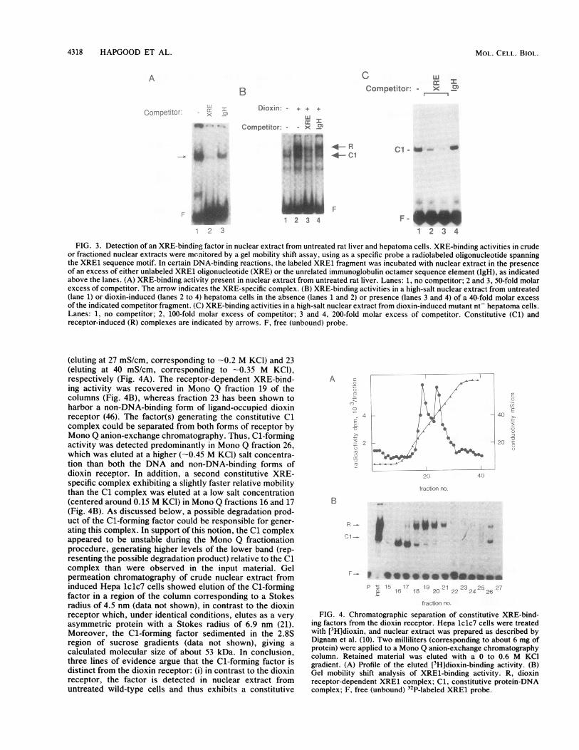
h of treatment the putative constitutive XRE-binding factor(s) and to relateinduced change it to the dioxin receptor, nuclear extracts were preparede transcription from dioxin-treated or untreated hepatoma cells. We ob-ment, although served that a labile XRE-binding factor could be detected inat a low rate at nuclear extract from untreated cells. In gel mobility shiftDNase 1-hyper- assays, this factor generated a complex (Fig. 3B, lane 1;is visualized a designated Cl) exhibiting a faster relative mobility on non-ive site (HSi) at denaturing polyacrylamide gels than did the bona fide dioxinpon 3-naphtho- receptor-XRE complex (designated R), which was detectedite to nuclease only in nuclear extract from treated cells (compare lane 1Ln earlier report with lanes 2 to 4).ersensitive site We confirmed that the different XRE-binding activities) relative to the resulted from sequence-specific protein-DNA interaction bygion of P-naph- competition with unlabeled DNA fragments. Thus, forma-ivity was indi- tion of both the Cl complex and the receptor-dependent,nscription start complex was greatly reduced by the presence of a 40-folds 10 to 18). molar excesses of the unlabeled XRE1 oligonucleotide (Fig.'fragment from 3B, lanes 1 to 3), whereas little or no competition for bindingharacterized by was observed by coincubation with a 40-fold molar excess ofE Fig. lA) that an unrelated fragment containing the octamer motif of anthe XREs. This immunoglobulin heavy-chain promoter (lane 4). Interest-msitive sites in ingly, the XRE-specific Cl complex was also detected inid 2.1 + 0.1 kb, nuclear extract of nt- mutant hepatoma cells (Fig. 3C),)wed increased which show no detectable levels of receptor-dependentnaphthoflavone XRE-binding activity following dioxin induction (17, 21).)usly identified The constitutive nuclear XRE-specific factor is distinct from,p site, whereas the dioxin receptor. To biochemically characterize the dif-Fig. 2; compare ferent XRE-binding factors, Hepa lclc7 hepatoma cellsiackground that were treated with [3H]dioxin, and a standard (10) nuclear+1 kb disap- extract was prepared. Consequently, material was generated
g. iC), the con- that contained both the constitutive XRE-binding activity)nds to the HS1 and ligand-activated dioxin receptor, as assessed by gel;ite. Thus, this mobility shift analysis (Fig. 4B, input lane). We next frac-e HS1 and HS2 tionated this extract by fast protein liquid-anion exchangeLnscription start chromatography on Mono Q columns. As has been reported
previously (46), specifically bound [3H]dioxin was recoveredte at the XREs in two distinct peaks centered around Mono Q fractions 19
lu 4
VOL . 1l, 1991
4318 HAPGOOD ET AL.
A
B
Corl onetILtO-r
Competitor: - >C11Competitor: - xCD
Dioxin: - + + +
u Competitor' - - x C
* W 9.1-RR
C.,-4-C
i F1 F1 2 3 4
0.
2 3
F -
1 2 3 4
FIG. 3. Detection of an XRE-binding factor in nuclear extract from untreated rat liver and hepatoma cells. XRE-binding activities in crudeor fractioned nuclear extracts were monitored by a gel mobility shift assay, using as a specific probe a radiolabeled oligonucleotide spanningthe XRE1 sequence motif. In certain DNA-binding reactions, the labeled XRE1 fragment was incubated with nuclear extract in the presenceof an excess of either unlabeled XRE1 oligonucleotide (XRE) or the unrelated immunoglobulin octamer sequence element (IgH), as indicatedabove the lanes. (A) XRE-binding activity present in nuclear extract from untreated rat liver. Lanes: 1, no competitor; 2 and 3, 50-fold molarexcess of competitor. The arrow indicates the XRE-specific complex. (B) XRE-binding activities in a high-salt nuclear extract from untreated(lane 1) or dioxin-induced (lanes 2 to 4) hepatoma cells in the absence (lanes 1 and 2) or presence (lanes 3 and 4) of a 40-fold molar excessof the indicated competitor fragment. (C) XRE-binding activities in a high-salt nuclear extract from dioxin-induced mutant nt- hepatoma cells.Lanes: 1, no competitor; 2, 100-fold molar excess of competitor; 3 and 4, 200-fold molar excess of competitor. Constitutive (Cl) andreceptor-induced (R) complexes are indicated by arrows. F, free (unbound) probe.
(eluting at 27 mS/cm, corresponding to -0.2 M KCl) and 23(eluting at 40 mS/cm, corresponding to -0.35 M KCl),respectively (Fig. 4A). The receptor-dependent XRE-bind-ing activity was recovered in Mono Q fraction 19 of thecolumns (Fig. 4B), whereas fraction 23 has been shown toharbor a non-DNA-binding form of ligand-occupied dioxinreceptor (46). The factor(s) generating the constitutive Clcomplex could be separated from both forms of receptor byMono Q anion-exchange chromatography. Thus, Cl-formingactivity was detected predominantly in Mono Q fraction 26,which was eluted at a higher (-0.45 M KCl) salt concentra-tion than both the DNA and non-DNA-binding forms ofdioxin receptor. In addition, a second constitutive XRE-specific complex exhibiting a slightly faster relative mobilitythan the Cl complex was eluted at a low salt concentration(centered around 0.15 M KCl) in Mono Q fractions 16 and 17(Fig. 4B). As discussed below, a possible degradation prod-uct of the Cl-forming factor could be responsible for gener-ating this complex. In support of this notion, the Cl complexappeared to be unstable during the Mono Q fractionationprocedure, generating higher levels of the lower band (rep-resenting the possible degradation product) relative to the Clcomplex than were observed in the input material. Gelpermeation chromatography of crude nuclear extract frominduced Hepa 1clc7 cells showed elution of the Cl-formingfactor in a region of the column corresponding to a Stokesradius of 4.5 nm (data not shown), in contrast to the dioxinreceptor which, under identical conditions, elutes as a veryasymmetric protein with a Stokes radius of 6.9 nm (21).Moreover, the Cl-forming factor sedimented in the 2.8Sregion of sucrose gradients (data not shown), giving acalculated molecular size of about 53 kDa. In conclusion,three lines of evidence argue that the Cl-forming factor isdistinct from the dioxin receptor: (i) in contrast to the dioxinreceptor, the factor is detected in nuclear extract fromuntreated wild-type cells and thus exhibits a constitutive
AI
* ~ ~~~ A
*4
.,
i -s
Ttitt)xl iy).
B ~
F-. P *@@@*OSO*eimp 1 7 19 21. 23 25 2-7143 11 2( 22 24 26
fracttrio rio.FIG. 4. Chromatographic separation of constitutive XRE-bind-
ing factors from the dioxin receptor. Hepa 1c1c7 cells were treatedwith [3H]dioxin, and nuclear extract was prepared as described byDignam et al. (10). Two milliliters (corresponding to about 6 mg ofprotein) were applied to a Mono Q anion-exchange chromatographycolumn. Retained material was eluted with a 0 to 0.6 M KCIgradient. (A) Profile of the eluted [3H]dioxin-binding activity. (B)Gel mobility shift analysis of XRE1-binding activity. R, dioxinreceptor-dependent XRE1 complex; Cl, constitutive protein-DNAcomplex; F, free (unbound) 32P-labeled XRE1 probe.
MOL. CELL. BIOL.
,." " ,
A,7
il
"-) ;m
CHROMATIN STRUCTURE OF CYTOCHROME P-450IA1 4319
XRE-binding activity; (ii) the factor is also detected innuclear extract from mutant hepatoma cells which are dioxinunresponsive and deficient in nuclear translocation andDNA-binding activity of ligand-occupied dioxin receptor;and (iii) the factor does not copurify with any of the dioxinreceptor forms observed in nuclear extract from dioxin-induced cells.DNA-binding specificities of XRE-binding factors. In the
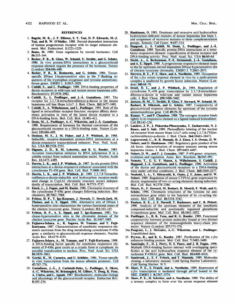
dioxin-responsive XRE1 sequence of the cytochromeP-450IA1 gene, the hexanucleotide motifS'-TCACGC-3' iscritical for dioxin receptor interaction in vitro (7, 31) and, inturn, is conserved in the rat XRE2 element (16). In gelmobility shift experiments using as specific probes oligonu-cleotides of identical length (sequences shown in Fig. SA)carrying either the XRE1 or XRE2 motif, neither elementwas recognized by the cryptic form of cytosolic Hepa lclc7dioxin receptor (Fig. 5B, lanes 1 and 3). Upon ligand-dependent in vitro activation (7, 30), however, the DNA-binding activity of the cytosolic receptor was unmasked,demonstrating that both the in vitro (Fig. 5B, lanes 2 and 4)-and in vivo (17, 21)-activated forms of dioxin receptor bindto both the XRE1 and XRE2 elements. The relative affinityof the ligand-activated dioxin receptor is about four- tofivefold lower for the XRE2 sequence than for the XRE1motif (21). Thus, sequences flanking the critical hexamermotif may stabilize receptor-DNA interaction.We observed that the constitutive Cl-generating factor
could be preferentially extracted from isolated nuclei ofuntreated or dioxin-treated cells at a low salt concentration(-0.15 M KCI), in contrast to the in vivo-activated receptor,which requires salt concentrations of 0.42 M KCI for efficientextraction from treated nuclei. Following incubation withthe constitutive factor(s) present in the low-salt nuclearextract from untreated Hepa 1c1c7 cells, Cl was generatedby probes containing either the XRE1 (Fig. SB, lanes 5 and7) or XRE2 (lanes 6 and 8) motif.To further characterize the DNA-binding specificity of the
constitutive factor responsible for Cl formation, we per-formed oligonucleotide competition experiments in gel mo-bility shift assays using the low-salt nuclear extract fromuntreated hepatoma cells. To this end, the ability of theXRE1 wild-type sequence to compete for binding was com-pared with that of mutant oligonucleotides that carry onesingle or four transversions in the core hexanucleotide motif.The sequences of the two mutant oligonucleotides are pre-sented in Fig. 5A along with the sequences of the XRE1 andXRE2 wild-type probes.
Formation of Cl with the XRE1 probe was stronglyinhibited in the presence of a 200-fold molar excess ofcompetitor fragments containing either the unlabeled XRE1sequence (Fig. 5C, compare lanes 2 and 3) or the mutantmotif (XM1) containing only a single transversion in theconserved hexamer motif (compare lanes 2 and 5). In con-trast, no significant competition for formation of Cl wasdetected at an identical molar excess of the oligonucleotide(XM4) containing a cluster of four point mutations in thehexamer motif (compare lanes 2 and 4). This mutant oligo-nucleotide appeared to be inert in competition experimentsto a similar extent as unrelated oligonucleotides spanningeither the octamer sequence motif from immunoglobulinpromoters or a glucocorticoid response element (comparelanes 2, 4, 6, and 7).
In the low-salt nuclear extract, a faint band migrating witha relative mobility slightly greater than that of the major Clband was detected (Fig. SC). In addition, the factor respon-sible for formation of this band indicated a DNA-binding
specificity similar to that of the Cl-forming factor. This bandvaried in intensity in different protein preparations and thusmay represent a discrete degradation product of the Cl-forming factor rather than an additional distinct XRE-bind-ing factor.
In control experiments, the cytosolic dioxin receptor wasactivated to a DNA-binding form by dioxin treatment in vitro(7), resulting in recognition of the wild-type XRE1 sequence(Fig. SD, lane 1). Competition experiments demonstratedthat formation of the receptor-DNA complex (indicated byan arrow) was abrogated only in the presence of a 25- to50-fold molar excess of the wild-type XRE competitor frag-ment (Fig. SD, lanes 2 to 4). Receptor-DNA interaction wasnot significantly disturbed in the presence of identical molarexcesses of either the XM4 or XM1 mutant competitorfragment (lanes 5 to 8). Taken together, these results indicatesimilar but not identical DNA-binding specificities for thedioxin receptor and the constitutive Cl-forming factor.Whereas a single point mutation of the fifth base in theconserved hexamer motif is sufficient to dramatically impairinteraction of the dioxin receptor with the XRE1 targetsequence, such a mutation did not significantly inhibit Clformation (7; Fig. SD). Finally, the oligonucleotide compe-tition experiments indicated that the Cl-forming factor andthe receptor may exhibit different relative affinities for theXRE1 target sequence. Thus, about 5- to 10-fold-higherconcentrations of competitor oligonucleotide were requiredto abrogate XRE complex formation with the Cl-formingconstitutive factor(s) compared with the concentrations re-quired to inhibit dioxin receptor-XRE interaction. A tenta-tive quantitation by gel mobility shift analysis of both factorsindicated that they exhibit approximately the same relativeabundance in high-salt nuclear extract of treated hepatomacells.
DISCUSSION
The chromatin structure at the dioxin response elements ofthe rat cytochrome P-450IA1 gene is unaltered in the processof gene activation. In this report, we show that two DNaseI-hypersensitive sites (collectively termed HS1) in closeproximity to each other coincided with the cytochromeP-450IA1 dioxin response elements XRE1 and XRE2, whichboth are located about 1 kb upstream of the transcriptioninitiation site. Importantly, this hypersensitive site wasdetected in nuclei from both untreated and induced rat liver,indicating a constitutively open chromatin structure at thissegment of the gene. Previously, the failure to detect anydiscrete DNase I-hypersensitive sites in the 5' flank of thecytochrome P-450IA1 gene in hepatoma cells has beenreported (12). The reason for this discrepancy in resultsremains unclear. By contrast to the constitutive DNaseI-hypersensitive site observed at the XREs in this study,glucocorticoids or progestins induce DNase I-hypersensitivesites at the hormone response elements in chromatin flank-ing, for instance, mouse mammary tumor virus (50), the rattyrosine aminotransferase gene (4, 25, 32), and the chickenlysozyme (14, 22) genes. In fact, the observed hormone-induced chromatin changes in the tyrosine aminotransferase(4) and lysozyme (14) genes facilitated the identification anddefinition of functional hormone response elements (22, 25).More recently, it has been shown that both glucocorticoidand progesterone receptors induce formation of the DNaseI-hypersensitive site at the glucocorticoid response elementsin tyrosine aminotransferase chromatin in an indistinguish-able manner (43), suggesting that the two receptors modulate
VOL . 1 l, 1991
4320 HAPGOOD ET AL.
BDioxinrD'o~~~~~~~~~~~~~~~~~~~~~~~~~~~~~~~~~~~~~~~~~~~~~~~~~~~~~~~~~~~~~~~
..
LLi EL 11WW LbsI-1Probe XRE I XRE 2mc mmr-xx x x
A
XRE-1: - TCACGCSAGTGCG :- -:::X
XRE2: 7 GCGTGA:-:-- -1 CGCACT-:-
XMUT4: zziTacatC-
::. :: -AtgtaG
MT1;: --TCACtC - --7rAGTGaG_;
CLL t L
-X X X P CDCompetitor:
R *--* "
Cl * 1 s of
2 3 t5 o..
D
Competitor: -- XRE XM4 XM1
ci * P *o
.4'.U1_b-to,
Fs
w~f.u<wa -
U__1 2 3 4 5 6 7
Free s*
1 2 3 4 5 6 7 8FIG. 5. DNA-binding specificities of constitutive and inducible XRE-binding factors. (A) Nucleotide sequences of wild-type and
point-mutated oligonucleotides used in gel mobility shift DNA-binding experiments. Oligonucleotides XRE1 and XRE2 span the XRE1 andXRE2 elements, respectively, of the rat cytochrome P-450IA1 gene. Bold letters indicate the conserved core hexanucleotide motif.Oligonucleotides XMUT1 (XM1) and XMUT4 (XM4) carry point mutations in the XRE1 sequence motif. Lowercase letters indicatepoint-mutated nucleotides or linker sequences. (B) Evidence that the constitutive XRE-binding factor recognizes both XRE1 and XRE2sequences. Latent (lanes 1 and 3) and in vitro-activated cytosolic Hepa 1clc7 dioxin receptor (lanes 2 and 4) and 2 (lanes 5 and 6) and (lanes7 and 8) 4 ,ul of low-salt nuclear extract containing the constitutive XRE-binding factor from Hepa 1c1c7 cells were incubated with labeledXRE1 or XRE2 probe as indicated. The receptor was activated by incubation of cytosol (4 mg of protein per ml) from untreated, wild-typeHepa 1c1c7 cells with 10 nM dioxin at 25°C for 3 h. (C) DNA-binding specificity of the Cl-forming factor. The labeled XRE1 probe wasincubated with a fixed amount of low-salt nuclear extract from nontreated Hepa 1c1c7 cells in the absence (lane 2) or presence of a 200-foldmolar excess of unlabeled competitor DNA as indicated above the lanes. IgH, immunoglobulin octamer sequence element; GRE,glucocorticoid response element. Lane 1 shows the mobility of free probe (F) in the absence of any added protein. (D) Binding studies within vitro-activated dioxin receptor. The receptor was in vitro activated as described above. Gel mobility shift assays were performed withlabeled XRE1 probe in the absence (lane 2) or presence of either a 20-fold (lanes 3, 5, and 7) or 40-fold molar (lanes 4, 6, and 8) excess ofthe indicated unlabeled competitor fragments. Lane 1 shows a control DNA-binding reaction in the absence of any added protein. Thereceptor-dependent XRE complex is indicated by an arrow. Free, unbound 32P-labeled probe.
MOL. CELL. BIOL.
CHROMATIN STRUCTURE OF CYTOCHROME P-450IA1 4321
chromatin structure by very similar or identical modes ofaction. Furthermore, in vivo footprinting experiments havedemonstrated that the glucocorticoid response element ofthe tyrosine aminotransferase gene is occupied by a factor(presumably the receptor) only in hormone-induced cells (3).In agreement with these observations, the ligand-inducedconversion of the glucocorticoid, estrogen, and progesteronereceptors from cryptic to DNA-binding forms has beenreconstituted in vitro (1, 8, 26, 34a).
It has been reported that dioxin treatment induces anexonuclease III stop site in mouse cytochrome P-450IA1chromatin (11). However, this site does not correspond toany functional dioxin-responsive and dioxin receptor-bind-ing sequences in the mouse cytochrome P-4501A1 gene (31),nor does it correspond to any of the mapped DNase I-hy-persensitive sites in the rat gene. Thus, in the dioxin receptorsystem, it is not known how treatment with ligand affects theDNA-binding activity of the receptor in vivo. Under in vitroconditions, however, the XRE-binding activity of the dioxinreceptor is under strict control of the ligand (7, 30). Inter-estingly, the binding affinity of a ligand for the dioxinreceptor appears to determine the amount of active form ofreceptor that is sequestered out of the latent complex (7).Taken together, these in vitro data strongly indicate that theligand may play several important roles in receptor-mediatedsignal transduction, including (i) the possible unmasking of anuclear translocation signal and (ii) triggering release ofinhibition of the XRE-binding activity.
Possible functional implications of the constitutive DNaseI-hypersensitive site at the XREs. At present, we can onlyspeculate how the chromatin structure at the XREs 1 kbupstream of the cytochrome P-450IA1 transcription start siteis maintained in a constitutively accessible configuration. Asdiscussed above, dioxin receptor-dependent XRE-bindingactivity is not detected in nuclear extracts from untreatedcells (9, 17, 21), possibly because of a cytoplasmic intracel-lular localization and a non-DNA-binding configuration ofthe receptor in the ligand unoccupied state. It is thereforeconceivable that the constitutive Cl-forming factor hasaccess to and specifically interacts with XRE target se-quences in the uninduced cell.
Recently two constitutive XRE1-binding factors desig-nated XF-1 and XF-2 have been characterized in rat andmouse hepatoma cells (36). In the present study, we havedetected only the Cl factor in nuclear extracts from rat liverand a number of hepatoma cells. However, it is formallypossible that the larger XF-1 factor is related to or identicalto the Cl factor, and given the in vitro lability of the Clfactor, the smaller XF-2 factor may represent a degradationproduct of XF-1. In any case, it is difficult to assess how thedifferent factors are related to one another since it is notknown whether the XF proteins recognize only the XRE1sequence motif or whether they carry specificity for bothXRE1 and XRE2, which are rather dissimilar in the se-quences flanking the conserved 5'-TCACGC-3' core hex-amer (16; Fig. 5A).The functional significance of the interaction between the
Cl factor and the XRE element is still unclear. However,both constitutive DNase I-hypersensitive sites and protein-DNA interactions which mediate signal transduction havebeen described in cyclic AMP- and serum-responsive en-hancer elements of the tyrosine aminotransferase and c-fosgenes, respectively (reviewed in reference 45). In the highlyinducible c-fos promoter, a complex functional interplaybetween regulatory factors has been characterized at theserum response element involving at least two DNA-binding
factors: a 67-kDa protein (35, 44) and a 62-kDa protein (39).Moreover, it has been demonstrated that the serum responseelement is protected against methylation by dimethyl sulfatealso in nuclei from untreated cells (23), suggesting that theelement is constitutively occupied by some factor(s). In fact,the 67-kDa protein has recently been shown to mediaterepression of c-fos transcription by occupying the serumresponse element and generating the low levels of expressionobserved prior to growth factor induction (38). For efficientinduction, the 67- and 62-kDa proteins appear to have tointeract with one another and the response element (39).Given this background, it remains to be determined whetherthe presence of the constitutively open chromatin configura-tion at the XREs and the two different XRE-binding factorsimplies a similarly complex interplay between these factorsin determining the dioxin-induced transcriptional response.For instance, it is formally possible that the constitutivefactor will be found to be a component of the possiblyheteromeric -200-kDa form of activated, nuclear dioxinreceptor (21). Conversely, it is conceivable that the receptorand the constitutive factor function in a mutually antagonis-tic manner involving a polypeptide exchange at the XRE ofa constitutive repressor versus an inducible activator (i.e.,the receptor).
Induced changes in chromatin structure at the cytochromeP-4501A1 transcription start site. Interestingly, the sensitivityof the HS2 site to nuclease digestion in rat cytochromeP-450IA1 chromatin was altered upon induction. We havetentatively mapped this site to between -180 to +20 relativeto the transcription start site. Two independent studies haverecently demonstrated that, in addition to the dioxin-respon-sive enhancer elements 1 kb upstream of the transcriptionstart site, more proximal regulatory sequences are requiredfor maximal dioxin inducibility of both the mouse and ratcytochrome P-450IA1 promoters (31, 49). Whereas thisregion has been defined rather broadly in the mouse pro-moter to span sequences from positions -245 to -50 relativeto the cap site (31), the regulatory sequences have beenmapped to an element which extends from positions -53 to-44 and is located 12 bp upstream of the TATA box of therat promoter (49). Moreover, a constitutive nuclear factor(s)which specifically interacts with this element has beenidentified (49). It is not clear how this proximal factor(s) actsin combination with the distal, inducible dioxin receptor toenhance transcription from the promoter in response todioxin treatment. However, it is interesting that the chroma-tin structure in the proximal region of the promoter becomesmore accessible upon induction, suggesting that the interac-tion between the proximal regulatory element and its relatedDNA-binding factor(s) may be facilitated. Clearly, in vivomapping of the chromatin fine structure of the promoter andin vitro reconstitution of the positioning of the nucleosomesare required to further study this issue.
ACKNOWLEDGMENTS
We thank Gisela Astedt and Asa Andersson for expert technicalassistance, Oliver Hankinson (UCLA) for originally supplying thewild-type Hepa 1c1c7 and nt- mutant cell lines, and HirotoshiTanaka for nuclear extract from H4IIE cells.
This work was supported by the National Institutes of Health(grants ESO 3954, HD 04445, and CA37139), the Clayton Founda-tion for Research-California Division, and the Swedish CancerSociety. P.S. and S.C. are recipients of research fellowships fromthe Swedish Natural Sciences Research Council. R.H.T. is a Clay-ton Foundation Investigator and a recipient of Senior FacultyResearch Award FRA-12 from the American Cancer Society.
VOL . 1l, 1991
4322 HAPGOOD ET AL.
REFERENCES
1. Bagchi, M. K., J. F. Elliston, S. Y. Tsai, D. P. Edwards, M.-J.Tsai, and B. W. O'Malley. 1988. Steroid-dependent interactionof human progesterone receptor with its target enhancer ele-ment. Mol. Endocrinol. 2:1221-1229.
2. Beato, M. 1989. Gene regulation by steroid hormones. Cell56:335-344.
3. Becker, P. B., B. Gloss, W. Schmid, U. Strahle, and G. Schuitz.1986. In vivo protein-DNA interactions in a glucocorticoidresponse element require the presence of the hormone. Nature(London) 324:686-688.
4. Becker, P. B., R. Renkawitz, and G. Schuitz. 1984. Tissue-specific DNase I-hypersensitive sites in the 5'-flanking se-quences of the tryptophan oxygenase and tyrosine aminotrans-ferase genes. EMBO J. 3:2015-2020.
5. Cuthill, S., and L. Poellinger. 1988. DNA-binding properties ofdioxin receptors in wild-type and mutant mouse hepatoma cells.Biochemistry 27:2978-2982.
6. Cuthill, S., L. Poellinger, and J.-A. Gustafsson. 1987. Thereceptor for 2,3,7,8-tetrachlorodibenzo-p-dioxin in the mousehepatoma cell line Hepa 1c1c7. J. Biol. Chem. 262:3477-3481.
7. Cuthill, S., A. Wilhelmsson, and L. Poellinger. 1991. Role of theligand in intracellular receptor function: receptor affinity deter-mines activation in vitro of the latent dioxin receptor to aDNA-binding form. Mol. Cell. Biol. 11:401-411.
8. Denis, M., L. Poellinger, A.-C. Wikstrom, and J.-A. Gustafsson.1988. Requirement of hormone for thermal conversion of theglucocorticoid receptor to a DNA-binding state. Nature (Lon-don) 333:686-688.
9. Denison, M. S., J. M. Fisher, and J. P. Whitlock, Jr. 1988.Inducible, receptor-dependent protein-DNA interactions at adioxin-responsive transcriptional enhancer. Proc. Natl. Acad.Sci. USA 85:2528-2532.
10. Dignam, J. D., R. M. Lebowitz, and R. G. Roeder. 1983.Accurate transcription initiation by RNA polymerase II in asoluble extract from isolated mammalian nuclei. Nucleic AcidsRes. 11:1475-1489.
11. Durrin, L. K., and J. P. Whitlock, Jr. 1987. In situ protein-DNAinteractions at a dioxin-responsive enhancer associated with thecytochrome P1-450 gene. Mol. Cell. Biol. 7:3008-3011.
12. Durrin, L. K., and J. P. Whitlock, Jr. 1989. 2,3,7,8-Tetrachlo-rodibenzo-p-dioxin-inducible aryl hydrocarbon receptor-medi-ated change in CyplAl chromatin structure occurs indepen-dently of transcription. Mol. Cell. Biol. 9:5733-5737.
13. Einck, L., J. Fagan, and M. Bustin. 1986. Chromatin structure ofthe cytochrome P-450c gene changes following induction. Bio-chemistry 25:7062-7068.
14. Fritton, H. P., T. Igo-Kemenes, J. Nowock, U. Strech-Jurk, M.Theisen, and A. E. Sippel. 1984. Alternative sets of DNase Ihypersensitive sites characterize the various functional states ofthe chicken lysozyme gene. Nature (London) 311:163-165.
15. Fritton, H. P., A. E. Sippel, and T. Igo-Kemenes. 1983. Nu-clease-hypersensitive sites in the chromatin domain of thechicken lysozyme gene. Nucleic Acids Res. 11:3467-3485.
16. Fujisawa-Sehara, A., K. Sogawa, M. Yamane, and Y. Fujii-Kuriyama. 1987. Characterisation of xenobiotic responsive ele-ments upstream from the drug metabolising cytochrome P-450cgene: a similarity to glucocorticoid response elements. NucleicAcids Res. 15:4179-4191.
17. Fujisawa-Sehara, A., M. Yamane, and Y. Fujii-Kuriyama. 1988.A DNA-binding factor specific for xenobiotic responsive ele-ments of P-450c gene exists as a cryptic form in cytoplasm: itspossible translocation to nucleus. Proc. Natl. Acad. Sci. USA85:5859-5863.
18. Gorski, K., M. Carneiro, and U. Schibler. 1986. Tissue-specificin vitro transcription from the mouse albumin promoter. Cell47:767-776.
19. Gustafsson, J.-A., J. Carlstedt-Duke, L. Poellinger, S. Okret,A.-C. Wikstrom, M. Bronnegard, M. Gillner, Y. Dong, K. Fuxe,A. Cintra, and L. Agnati. 1987. Biochemistry, molecular biologyand physiology of the glucocorticoid receptor. Endocrine Rev.8:185-234.
20. Hankinson, 0. 1983. Dominant and recessive aryl hydrocarbonhydroxylase-deficient mutants of mouse hepatoma line hepa 1,and assignment of recessive mutants to three complementationgroups. Somatic Cell Genet. 9:497-514.
21. Hapgood, J., S. Cuthill, M. Denis, L. Poellinger, and J.-A.Gustafsson. 1989. Specific protein-DNA interactions at a xeno-biotic-responsive element: copurification of dioxin receptor andDNA-binding activity. Proc. Natl. Acad. Sci. USA 86:60-64.
22. Hecht, A., A. Berkenstam, P.-E. Stromstedt, J.-A. Gustafsson,and A. E. Sippel. 1988. A progesterone responsive element mapsto the far upstream steroid dependent DNase hypersensitive siteof chicken lysozyme chromatin. EMBO J. 7:2063-2073.
23. Herrera, R. E., P. E. Shaw, and A. Nordheim. 1989. Occupationof the c-fos serum response element in vivo by a multi-proteincomplex is unaltered by growth factor induction. Nature (Lon-don) 340:68-70.
24. Israel, D. I., and J. P. Whitlock, Jr. 1984. Regulation ofcytochrome P1-450 gene transcription by 2,3,7,8-tetrachloro-dibenzo-p-dioxin in wild-type and variant mouse hepatomacells. J. Biol. Chem. 259:5400-5402.
25. Jantzen, H.-M., U. Straihle, B. Gloss, F. Steward, W. Schmid, M.Boshart, R. Miksicek, and G. Schlitz. 1987. Cooperativity ofglucocorticoid response elements far upstream of the tyrosineaminotransferase gene. Cell 49:29-38.
26. Kumar, V., and P. Chambon. 1988. The estrogen receptor bindstightly to its responsive element as a ligand-induced homodimer.Cell 55:145-156.
27. Landers, J. P., J. Piskorska-Pliszczynska, T. Zacharewski, N. J.Bunce, and S. Safe. 1989. Photoaffinity labeling of the nuclearAh receptor from mouse Hepa 1c1c7 cells using 2,3,7,8-[3H]tet-rachlorodibenzo-p-dioxin. J. Biol. Chem. 264:18463-18471.
28. Legraverend, C., R. R. Hannah, H. J. Eisen, I. S. Owens, D. W.Nebert, and 0. Hankinson. 1982. Regulatory gene product of theAh locus: characterisation of receptor mutants among mousehepatoma clones. J. Biol. Chem. 257:6402-6407.
29. Nebert, D. W., and F. J. Gonzalez. 1987. P-450 genes: structure,evolution and regulation. Annu. Rev. Biochem. 56:945-993.
30. Nemoto, T., G. G. F. Mason, A. Wilhelmsson, S. Cuthill, J.Hapgood, J.-i. Gustafsson, and L. Poellinger. 1990. Activationof the dioxin and glucocorticoid receptors to a DNA-bindingstate under cell-free conditions. J. Biol. Chem. 265:2269-2277.
31. Neuhold, L. A., Y. Shirayoshi, K. Ozato, J. E. Jones, and D. W.Nebert. 1989. Regulation of mouse CYPlAl gene expression bydioxin: requirement of two cis-acting elements during induction.Mol. Cell. Biol. 9:2378-2386.
32. Nitsch, D., F. Steward, M. Boshart, R. Mestril, F. Weih, and G.Schlitz. 1990. Chromatin structures of the tyrosine rat ami-notransferase gene relate to the function of its cis-acting ele-ments. Mol. Cell. Biol. 10:3334-3342.
33. Paulson, K. E., J. E. Darnell, T. Rushmore, and C. B. Pickett.1990. Analysis of the upstream elements of the xenobioticcompound-inducible and positionally regulated glutathioneS-transferase gene. Mol. Cell. Biol. 10:1841-1852.
34. Poellinger, L., B. K. Yoza, and R. G. Roeder. 1989. Functionalcooperativity between protein molecules bound at two distinctsequence elements of the immunoglobulin heavy-chain pro-moter. Nature (London) 337:573-576.
34a.Pongratz, I., J. McGuire, A.-C. Wikstrom, and L. Poellinger.Unpublished data.
35. Prywes, R., and R. G. Roeder. 1987. Purification of the c-fosenhancer-binding protein. Mol. Cell. Biol. 7:3482-3489.
36. Saatcioglu, F., D. J. Perry, D. S. Pasco, and J. B. Fagan. 1990.Multiple DNA-binding factors interact with overlapping speci-ficities at the aryl hydrocarbon response element of the cy-tochrome P-450IA1 gene. Mol. Cell. Biol. 10:6408-6416.
37. Sambrook, J., E. F. Fritsch, and T. Maniatis. 1989. Molecularcloning: a laboratory manual. Cold Spring Harbor Laboratory,Cold Spring Harbor, N.Y.
38. Shaw, P. E., S. Frasch, and A. Nordheim. 1989. Repression ofc-fos transcription is mediated through p67srf bound to theSRE. EMBO J. 8:2567-2574.
39. Shaw, P. E., H. Schroter, and A. Nordheim. 1989. The ability ofa ternary complex to form over the serum response element
MOL. CELL. BIOL.
CHROMATIN STRUCTURE OF CYTOCHROME P-450IA1 4323
correlates with serum inducibility of the human c-fos promoter.Cell 56:563-572.
40. Soderkvist, P., L. Poellinger, R. Toftgard, and J.-A. Gustafsson.1988. Differential expression of the cytochrome P-450c andP-450d genes in the rat ventral prostate and liver. Cancer Res.48:3045-3049.
41. Sogawa, K., A. Fujisawa-Sehara, M. Yamane, and Y. Fujii-Kuriyama. 1986. Location of regulatory elements responsiblefor drug induction in the rat cytochrome P-450c gene. Proc.Natl. Acad. Sci. USA 83:8044-8048.
42. Sogawa, K., 0. Gotoh, K. Kawajiri, and Y. Fujii-Kuriyama.1984. Distinct organization of methylcholanthrene- and phe-nobarbital-inducible cytochrome P-450 genes in rat. Proc. Natl.Acad. Sci. USA 81:5066-5070.
43. Strahle, U., M. Boshart, G. Klock, F. Stewart, and G. Schiitz.1989. Glucocorticoid- and progesterone-specific effects are de-termined by differential expression of the respective hormonereceptors. Nature (London) 339:629-632.
44. Treisman, R. 1987. Identification and purification of a polypep-tide that binds to the c-fos serum response element. EMBO J.6:2711-2717.
45. Weih, F., F. Steward, M. Boshart, D. Nitsch, and G. Schiitz.1990. In vivo monitoring of a cAMP-stimulated DNA-bindingactivity. Genes Dev. 4:1437-1449.
46. Wilhelmsson, A., S. Cuthill, M. Denis, A.-C. Wikstrom, J.-A.Gustafsson, and L. Poellinger. 1990. The specific DNA-bindingactivity of the dioxin receptor is modulated by the 9OkD heatshock protein. EMBO J. 9:69-76.
47. Wilhelmsson, A., A.-C. Wikstrom, and L. Poellinger. 1986.Polyanionic-binding properties of the receptor for 2,3,7,8-tetra-chlorodibenzo-p-dioxin: a comparison with the glucocorticoidreceptor. J. Biol. Chem. 261:13456-13463.
48. Yamamoto, K. R. 1985. Steroid receptor regulated transcriptionof specific genes and gene networks. Annu. Rev. Genet. 19:205-252.
49. Yanagida, A., K. Sogawa, K.-I. Yasumoto, and Y. Fujii-Kuriyama. 1990. A novel cis-acting DNA element required for ahigh level of inducible expression of the rat cytochrome P-450cgene. Mol. Cell. Biol. 10:1470-1475.
50. Zaret, K. S., and K. R. Yamamoto. 1984. Reversible andpersistent changes in chromatin structure accompany activationof a glucocorticoid-dependent enhancer element. Cell 38:29-38.
VOL. 11, 1991
Copyright © 2022 FDOKUMEN