Life Sciences: DNA Code of Life and Meiosis
65
pg. 1
-
Upload
khangminh22 -
Category
Documents
-
view
5 -
download
0
Transcript of Life Sciences: DNA Code of Life and Meiosis

pg. 1

pg. 2
TABLE OF CONTENTS PAGE
1. Introduction 3 2. How to use this Self-study Guide 4 3. DNA: The Code of Life 5 3.1 Mind map on DNA - Code of life 5
3.2 Links to prior-knowledge/background knowledge 6 3.3 Practice questions on prior-knowledge 7-8 3.4 Differentiate between related terminologies 9-12 3.5 DNA Replication – exam tips/techniques/notes 13 3.6 Practice questions on DNA Replication 14 3.7 DNA profiling – exam tips/techniques/notes 15 3.8 Practice questions on DNA Profiling 17 3.9 Protein Synthesis – exam tips/techniques/notes 18 3.10 Practice questions on DNA Replication and Transcription 19 3.11 Genetic Coding – exam tips/techniques/notes 20 3.12 The effect of mutation on protein structure (DNA sequence)
exam tips/techniques/notes
22
3.13 Practice questions on mutation and Protein structure 25 3.14 Typical exam questions 28 3.15 Solutions to DNA practice questions 37
4. Meiosis 41 4.1 Mind map on Meiosis 42
4.2 Links to prior-knowledge/background knowledge 43 4.3 Differentiate between related terminologies 44 4.4 Process of meiosis - exam tips/techniques/notes 48 4.5 Practical questions on Meiosis 52 4.6 Typical exam questions 60 4.7 Solutions to DNA practice questions 64
5. References 66 6. Acknowledgement 66

pg. 3
1. INTRODUCTION
The declaration of COVID-19 as a global pandemic by the World Health Organisation
led to the disruption of effective teaching and learning in many schools in South Africa.
The majority of learners in various grades spent less time in class due to the phased-
in approach and rotational/ alternate attendance system that was implemented by
various provinces. Consequently, most schools were not able to complete all the
relevant content designed for specific grades in accordance with the Curriculum and
Assessment Policy Statements in most subjects.
As part of mitigating against the impact of COVID-19 on the current Grade 12, the
Department of Basic Education (DBE) worked in collaboration with subject specialists
from various Provincial Education Departments (PEDs) developed this Self-Study
Guide. The Study Guide covers those topics, skills and concepts that are located in
Grade 12, that are critical to lay the foundation for Grade 12. The main aim is to close
the pre-existing content gaps to strengthen the mastery of subject knowledge in Grade
12. More importantly, the Study Guide will engender the attitudes in the learners to
learning independently while mastering the core cross-cutting concepts.

pg. 4
2. HOW TO USE THIS SELF STUDY GUIDE
o There are five Self-study guides covering all Grade 12 topics:
o Booklet One: DNA: Code of Life and Meiosis o Booklet Two: Reproduction in Vertebrates, Human reproduction, Endocrine System
and Homeostasis
o Booklet Three: Genetics and Inheritance
o Booklet Four: Responding to the Environment: Humans and Plants
o Booklet Five: Evolution: Natural Selection and Human evolution
o You must use this Self-study Guide together with the Life Sciences Mind the Gap Study
Guide, which is a complementary booklet. o You need to study the content from the DBE Grade 12 Textbook, DBE Exam Guideline 2021,
and Mind the Gap for all the topics. o Ensure you understand all the relevant concepts and content. o This Self-study Guide focusses mainly on the skills you will need to answer the questions in
examinations. o There are exam technique and tips for each topic (in italics) o These tips will guide you on how to approach certain types in the Life Sciences Examination
papers and tests: o How to master the relevant terminology o Drawing and interpreting of graphs o Interpreting tables o Interpreting diagrams o Genetics crosses and pedigree diagrams o Doing calculations o Scientific investigation questions
o At the end of each booklet you will find typical examination questions and answers

pg. 5
DNA - CODE OF LIFE
TOPIC: DNA – CODE OF LIFE
TERM 1 PAPER 2
DURATION 8 hours
(2 weeks)
WEIGHTING 27 marks (18%)
PRIOR-KNOWLEDGE/BACKGROUND KNOWLEDGE
Grade 10: Plant and Animal cells, proteins, nucleic acids, location of DNA and chromosome. RESOURCES
Textbooks, Study Guides, MTG, Past NSC, SC & Provincial Question Papers
3.1 MINDMAP ON DNA - CODE OF LIFE
pg. 6
3.2 LINKS TO PRIOR-KNOWLEDGE/BACKGROUND KNOWLEDGE It is important to know the location, composition and function of the ribosome, cytoplasm and the parts
of the nucleus (nuclear membrane, nucleoplasm, nucleolus, chromatin network).
Structure of a cell
CELL STRUCTURE LOCATION COMPOSITION FUNCTION 1.Nucleoplasm/ Nuclear sap
In the cell nucleus The nucleoplasm is a liquid that surrounds the chromosomes and nucleoli.
Many substances such as free nucleotides (necessary for purposes such as the replication of DNA) and enzymes (which direct activities that take place in the nucleus) are dissolved in the nucleoplasm.
2. Cytoplasm Fluid part of cell outside the nucleus and inside the cell membrane. The area between the plasma/cell membrane and nucleus.
Filled with a clear fluid called CYTOSOL. Contains many structures called ORGANELLES
Where most metabolic reactions/activities take place.
3. Nuclear Membrane/ Envelope
Enclosing the nucleus Thin wall double membrane
Controls what goes in and out of nucleus
4. Nuclear pore Tiny holes found in the nuclear envelope
Tiny holes (openings) Help to regulate the exchange of materials (such as mRNA and proteins) between the nucleus and the cytoplasm.
5. Chromatin network
In the cell nucleus Tangled, threadlike material
Forms the chromosomes, the chromosomes are the basis of the hereditary functions of the cell, there are 46 chromosomes in human cells (except mature sex cells in which there are 23)

pg. 7
6. Ribosome Found along the endoplasmic reticulum Some ribosomes are found in the cytoplasm
Ribosomes are made up of some protein and RNA.
Makes protein for the cell (the site of protein synthesis)
7. Nucleolus Small, dense structures within nucleus
Made of proteins and RNA. No membrane
Produces ribosomes
Question 1: The basic structure of the cell and nucleus 1.1 Study the following diagrams and answer the questions:
Diagram A Diagram B
1.1.1 Identify the organelle (number and name) in Diagram A that is represented by Diagram B. 1ü- nucleusü
1.1.2 Give the: (a) Two nucleic acids present in Diagram B.
- DNAü (Deoxyribonucleic acid)
- RNA ü (Ribonucleic acid)
(b) Significance of the organelle represented in Diagram B. The nucleus controls all of the cell’s activities.ü
3.3 PRACTICE QUESTIONS on PRIOR-KNOWLEDGE

pg. 8
(c) Way in which substances get into and out of the organelle represented by diagram B The nuclear envelope has nuclear poresü that allow substances to enter
and exit the nucleus.
1.1.3 Identify label D. Ribosomeü
1.1.4 Complete the table with regard to the location, composition and function of label D.
LOCATION COMPOSITION FUNCTION OF ORGANELLE
Found along the
endoplasmic reticulumü
Some ribosomes are found
in the cytoplasmü
Ribosomes are made up of
some proteinü and RNA.ü
Makes proteinü for the cell (the
site of protein synthesis)
1.1.5 Identify the following labelled organelles and give the function of each.
LABEL NUMBER
NAME OF ORGANELLE FUNCTION OF ORGANELLE
5 Mitochondria ü Make energyü through cellular respiration
6 Vacuole ü Stores water, metabolic waste products and pigmentsü
7 Centrosomeü
Helps in cell divisionü Assures equal distribution of chromosomes in daughter cells. ü
Note: A centrosome is made of two separate centrioles. Centrioles are present in animal cells but not in plant cells.

pg. 9
3.4 DIFFERENTIATE BETWEEN RELATED TERMINOLOGIES
NUCLEOLUS NUCLEOPLASM CYTOPLASM RIBOSOME
Structure in the
nucleus responsible
for forming ribosomal
RNA
That part of the
protoplasm within the nucleus
That part of the
protoplasm outside the nucleus.
Structure that is the
site of protein synthesis
CHROMATIN CHROMATID CENTROMERE CHROMOSOME CHROMATIN NETWORK
The DNA-containing network found
in cells in
interphase
(non-dividing)
The individual threads that form
a chromosome
Structure that
holds two
chromatids
together in a
replicated
chromosome and
which also
attaches the
chromosome to
the spindle fibres
during cell
division
It is a thread like
structure made up
of DNA/that
carries hereditary
information in the
form of genes
Visible as thread-
like structures in
the nucleus of an
inactive cell
DNA (DEOXYRIBONUCLEIC ACID) RNA (RIBONUCLEIC ACID) Forms the chromosomes in the nuclei of all
living cells and carries the hereditary information
of the organism. The DNA molecule is a double helix (twisted) strand.
A single strand, located in the nucleoplasm and
cytoplasm. The RNA molecule is always a single strand of nucleotides. Remember that
the RNA contains Uracil instead of Thymine (A, G, C and U). RNA is responsible for protein
synthesis. HELIX
Coiled (natural) shape of a DNA molecule

pg. 10
MONOMER POLYMER A single unit that makes up a larger molecule A large molecule which is formed from many
small molecules (monomers) NUCLEOTIDE
The building block (monomers) of RNA and DNA. Each nucleotide consists of a pentose sugar, a
phosphate ion and a nitrogenous base.
AMINO ACID The basic building block (monomer) of a protein molecule
ENZYME
A protein that speeds up a chemical reaction / a catalyst
CYTOSINE THYMINE URACIL
The base that pairs off with
guanine The base that pairs off with
adenine The base found in RNA and not DNA
NITROGENOUS BASES These are nitrogen containing molecules viz. Adenine, (A); Thymine (T); Guanine (G); Cytosine (C)
and Uracil (U). BASE PAIRING
Adenine (A) always bonds to thymine (T) and guanine (G) with cytosine (C) in DNA molecule, to
ensure the precision of DNA replication
MITOCHONDRIAL DNA NUCLEAR DNA CHLOROPLAST DNA
The type of DNA found only
in the mitochondrion
Type of DNA found in the nucleus – makes up genes on
chromosomes
Type of DNA found in chloroplasts (plants)
TEMPLATE COMPLEMENTARY STRAND The original strand that provides a framework
upon which a new strand is developed The new strand that is made based on the
sequence of nucleotides on the template DNA REPLICATION
Process involving the formation of two new identical DNA molecules from an original DNA.
TRANSCRIPTION TRANSLATION
1st stage of protein synthesis
The synthesis of mRNA from a DNA template 2nd stage of protein synthesis
The process of converting the information carried
by m-RNA to the correct sequence of amino
acids to form a particular protein
pg. 11
SYNTHESIS Building up of separate parts into a whole
MESSENGER RNA (MRNA): RIBOSOMAL RNA (RRNA) TRANSFER RNA (TRNA)
Responsible for carrying the
genetic code that is transcribed
from DNA, to specialized sites
of the ribosomes where the
information is translated for
protein synthesis
Carries codons
Form the ribosomes and
produce the proteins, based on
the information received from the
tRNA
Lacks codons or anticodons
Has anticodons, which codes
for a specific amino acid. The
anticodons are
complementary to the mRNA
codon, during the production
of proteins.
Carries anticodons
CODON ANTICODON
The three adjacent bases found on a mRNA
molecule.
One mRNA molecule contains a number of
codons.
The three adjacent bases found on a tRNA
molecule that will determine which amino acid
will be brought to the ribosome.
One tRNA molecule contains one anticodon.
HYDROGEN BONDS PEPTIDE BOND The chemical bonds which link base pairs in
the DNA molecule
A link between two adjacent amino acids
GENE GENOME Segment of a chromosome that controls each characteristic/ a unit of
sequenced pieces of DNA that carry the genetic information that will
determine the hereditary characteristics of an organism.
All the genes present in
an organism
HEREDITARY Characteristics that are passed from parents to offspring
MUTATION
A sudden change in the DNA nucleotide sequence

pg. 12
How does DNA replication occur? – The Process of DNA Replication
3.5 DNA REPLICATION – EXAM TIPS/TECHNIQUES/NOTES
1. The DNA double helix unwinds
2. The weak hydrogen bonds between the nitrogenous bases are broken. The DNA strands separate (they unzip)
3. Each original DNA strand serves as a template on which its complement is built
4. Free nucleotides build a DNA strand onto each of the original DNA strands, attaching their complementary nitrogenous bases (A to T and C to G)
5. This result in two identical DNA molecules. Each molecule consists of one original strand and one new strand
ERRORS that occur during DNA replication may
sometimes lead to mutations (a change in the
nitrogenous base sequence)
If the incorrect nitrogen base attaches to the original
strand (i.e., if a nitrogen base is added or deleted:
the sequence or order of the bases changes on the
new DNA molecule
resulting in a change in the gene structure (gene mutation)

pg. 13
3.6 PRACTICE QUESTIONS on DNA REPLICATION Question 2: DNA Replication
2.1 Number the steps of DNA replication in the correct order (1, 2, 3, 4 and 5):
__3__Each original DNA strand serves as a template on which its complement is
built.
__1__The double helix unwinds.
__5__Two identical DNA molecules are formed.
__2__Weak hydrogen bonds between nitrogenous bases break and two DNA
strands unzip (separate).
__4__Free nucleotides build a DNA strand onto each of the original two DNA
strands by attaching to their complementary nitrogenous bases.
2.2 Show the complimentary base pairing that would occur in the replication of the short DNA molecule below. Use two different coloured pencils (or different pens, markers, etc.) to show which strands are the original and which are newly synthesized. Also indicate the nitrogenous base.
Original DNA
strand 1
Original DNA
strand 2
Original DNA strand 1
(copy from left)
New DNA strand
+
New DNA strand
(copy from left)
Original DNA
strand 2
A T A T + A T C G C G + C G C G C G + C G T A T A + T A G C G C + G C A T A T + A T T A T A + T A C G C G + C G G C G C + G C T A T A + T A

pg. 14
(a) When and where does DNA replication take place? This occurs during interphaseü of the cell cycle in the nucleusü.
(b) Why is the process of DNA replication important?
• Doubles the genetic material ü so it can be shared between the resulting
daughter cells during cell division.
• Results in the formation of identical daughter cells ü during mitosis.
(c) Give TWO functions of DNA?
• Sections of DNA forming genes carry hereditary information ü
• DNA contains coded information for protein synthesis ü
3.7 DNA PROFILING – EXAM TIPS/TECHNIQUES/NOTES
What is DNA Profiling? A DNA profile is a pattern produced on X-ray film.
This pattern consists of lines which are of different lengths and thicknesses and in different positions.
All individuals, except identical twins, have a unique DNA profile.
Compare the DNA profiles (bands/bars) of two samples – an unknown or evidence sample, such as semen, saliva, blood, hair strands, skin, finger or toenails, tooth with root material, etc. and a known or reference sample, such as a blood sample from a suspect. If most of the DNA bands/bars from evidence sample is matching that of the reference sample, they’re the same DNA. The analysis of the results of the DNA profiling may lead to various conclusions depending on the aim of the DNA profiling (eg. crime suspect, relatives, compatibility of tissue types and probability or causes of genetic defects). Use a ruler to guide you, move down the column while looking at the spacing of the bands, their thickness. (Remember, the bands are not necessarily even spaced, and some are darker and/or thicker than others).
DNA profiles for three different individuals
When we talk about DNA profiling, we no longer refer to the pattern of bars as a DNA fingerprint.

pg. 15
DNA profiles are used to:
• Prove paternity (father) and maternity (mother) (biological parents)
• Determine the probability or causes of genetic defects
• Establish the compatibility of tissue types for organ transplants
• Identify relatives
• Identify crime suspects in forensic investigations (Forensic Pathologists is a person that performs
DNA tests on biological evidence collected at crime scenes)
The role of DNA profiling in paternity testing
• A child received DNA from both parents
• When working out the possible father in paternity testing, you MUST compare the ‘bands’ of
the DNA profiles of the mother, child and possible father using the following steps:
• Step 1: A comparison of the DNA bands of the mother and the child is made
• Step 2: The remaining DNA bands are compared to the possible father’s DNA bands
• If all the remaining DNA bands in the
child’s profile match the possible father’s
DNA bands
• If all the remaining DNA bands in the child’s
profile does not match the possible father’s
DNA bands
• then the possible father is the biological
father
• then the possible father is not the biological
father
(NB: This section is normally covered under genetics)

pg. 16
3.8 PRACTICE QUESTIONS on DNA PROFILING
Question 3: DNA Profiling
3.1 The diagram below shows the DNA profiles of a child, her mother and four males.
There is uncertainty about who the biological father is. To establish paternity, DNA
profiling was conducted.
3.2 The diagram below shows the DNA profiles of six different people.
(a) Which male is the biological father of this child? Male 3ü
(b) Explain precautions that should be taken when working with DNA samples in a laboratory. - Mark the samples clearlyü to make sure vials are
not swopped.ü
- Wear gloves and a maskü not to contaminate
samplesü with your own DNA
- Use new and clean/sterilised apparatusü not to
contaminate samplesü.
(a) Give the letters of the TWO people who are identical twins. Cü and Fü (b) Give the letters of the parents of person B. A üand Eü (c) Explain whether the collection of DNA from every citizen in South Africa to create a DNA profile database for South Africa is a good idea or not. Noü. DNA profiles may reveal personal information about a person which could be used against them in a prejudicial wayü. OR Yesü. It could be used to identify crime suspects and relatives, assist in organ transplant, determining the causes of genetic defects or prove parenthood.ü
pg. 17
Free amino acids.
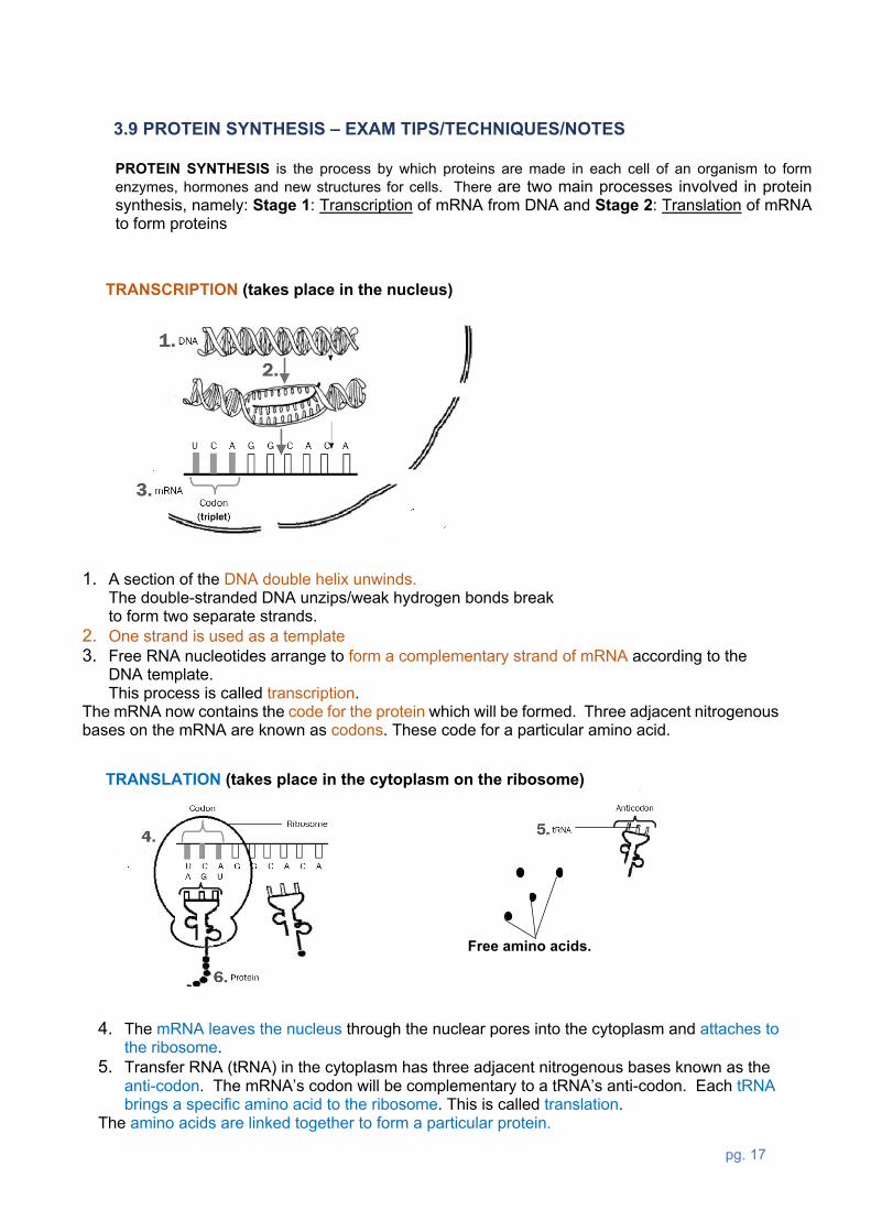
3.9 PROTEIN SYNTHESIS – EXAM TIPS/TECHNIQUES/NOTES
TRANSCRIPTION (takes place in the nucleus)
TRANSLATION (takes place in the cytoplasm on the ribosome)
PROTEIN SYNTHESIS is the process by which proteins are made in each cell of an organism to form enzymes, hormones and new structures for cells. There are two main processes involved in protein synthesis, namely: Stage 1: Transcription of mRNA from DNA and Stage 2: Translation of mRNA to form proteins
1. A section of the DNA double helix unwinds. The double-stranded DNA unzips/weak hydrogen bonds break to form two separate strands.
2. One strand is used as a template 3. Free RNA nucleotides arrange to form a complementary strand of mRNA according to the
DNA template. This process is called transcription.
The mRNA now contains the code for the protein which will be formed. Three adjacent nitrogenous bases on the mRNA are known as codons. These code for a particular amino acid.
4. The mRNA leaves the nucleus through the nuclear pores into the cytoplasm and attaches to the ribosome.
5. Transfer RNA (tRNA) in the cytoplasm has three adjacent nitrogenous bases known as the anti-codon. The mRNA’s codon will be complementary to a tRNA’s anti-codon. Each tRNA brings a specific amino acid to the ribosome. This is called translation.
The amino acids are linked together to form a particular protein.

pg. 18
3.10 PRACTICE QUESTIONS on DNA REPLICATION and TRANSCRIPTION
Question 4: DNA Replication and Transcription
4.1 Complete the following table that shows the differences between DNA replication and Transcription.
DNA REPLICATION TRANSCRIPTION Template (how many) 2 1
Product that is formed DNA mRNA
Bases pairs that are formed G-C and T-A None
4.2 Underline the correct answer.
STATEMENT/QUESTION ANSWER A ANSWER B mRNA is synthesised during __________________ translation transcription
mRNA has a/an ____________________________ codon anticodon
tRNA has a/an _____________________________ codon anticodon
One amino acid is equal to ___________codon(s) 1 3
tRNA carries the amino acids to the _____________ ribosome nucleus
tRNA picks up the amino acids during ___________ translation transcription
A polypeptide is a sequence of _________________ amino acids proteins
Which process is taking place at the ribosomes? translation transcription
4.3 The diagram below shows part of a mRNA (messenger RNA) molecule:
Key cell organelles involve in DNA synthesis: Nucleus Ribosome Key molecules involve in DNA synthesis DNA mRNA tRNA
NOTE: You might not necessarily be asked to explain the entire process of Protein Synthesis but only sections of it, for example:
• Describe the process of transcription or translation, respectively. • Describe the involvement of the different types of RNA in protein synthesis.

pg. 19
(a) How many codons are shown in the diagram? 3ü
(b) Write the complementary base sequence of the DNA strand that formed codon 1 of the mRNA strand in the above diagram. ATGü
(c) Explain the purpose of a specific sequence of codons in a mRNA molecule. A codon codes for a specific amino acidü, and this sequence
of codons codes for a proteinü.
3.11 GENETIC CODING – EXAM TIPS/TECHNIQUES/NOTES
WHAT IS GENETIC CODING? The genetic code is the instructions (sequence of the DNA or mRNA
nucleotides) in a gene that tell the cell how to make a specific protein.
How does Genetic coding occur?
Genes are short sections of DNA made up of
nucleotides and carries coded information
associated with a specific function.
1. Nucleotides are arranged in sets of
three, called triplets. A particular
sequence of nucleotide (bases) in the
DNA determines
2. the sequence set of nitrogenous bases in
mRNA (called codons), which
determines
Double DNA strand
Single DNA strand
mRNA
Remember: Proteins are very important organic molecules because it does most of the work in cells and are required for the structure, function, and regulation of the body’s tissues and organs.
DNA nucleotides = Base Triplets mRNA nucleotides = Codons

pg. 20
3. the order in which the sets on the tRNA
(called ANTI-CODONS) gets attached to
mRNA, which determines
4. the sequence in which amino acids
appear in a protein molecule, this
determines
5. the type of protein formed
mRNA
tRNA
pg. 21
Note: A mutation will NOT always lead to a formation of a different protein
3.12 THE EFFECT OF MUTATION ON PROTEIN STRUCTURE (DNA SEQUENCE) – EXAM TIPS/TECHNIQUES/NOTES
Cell processes that copy genetic material are usually accurate to ensure genetic continuity in both new
cells and offspring but, mistakes/changes (mutation) in the DNA can occur
• Changes in the DNA sequence is referred to as gene mutations
• A gene mutation affects the type/arrangement of a single/a few nitrogen bases.
• this changes the sequence/order of the nitrogen bases/the code
• on the DNA and the RNA.
• the same amino acid may be coded for,
• which causes no change in the amino acid sequence in the protein
• leading to the formation of the same protein
• a different amino acid may be coded for,
• which causes a change in the amino acid sequence in the protein
• leading to the formation of a different protein /alternate form of the required protein
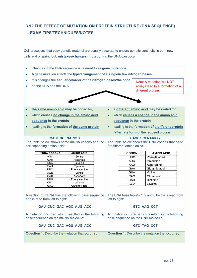
CASE SCENARIO 1 The table below shows some mRNA codons and the corresponding amino acids.
A section of mRNA has the following base sequence and is read from left to right:
GAU CUC GAC AGC AUG ACC A mutation occurred which resulted in the following base sequence on the mRNA molecule:
GAU CUC GAC AGU AUG ACC
CASE SCENARIO 2 The table below shows the RNA codons that code for different amino acids.
The DNA base triplets 1, 2 and 3 below is read from left to right:
GTC AAG CCT A mutation occurred which resulted in the following base sequence on the DNA molecule:
GTC TAG CCT
Question 1: Describe the mutation that occurred.
Question 1: Describe the mutation that occurred.
pg. 22
Steps: Compare the original mRNA to the one that
has undergone mutation.
1. Identify the affected codon or nucleotide
2. Describe which nucleotide has been
replaced/deleted
Answer: C was replaced by U on the 4th codon/AGC
Steps: Compare the original DNA base triplets to
the one that has undergone mutation
1. Identify the affected DNA Base triplet and
nucleotide
2. Describe which nucleotide has been
replaced/deleted
Answer: In DNA base triplet 2 the first adenine
was replaced by T.
Question 2: Explain the effect that the mutation will
have on the resulting protein.
Steps: Use the given table to find out if the new
codon formed after the mutation codes for the same
or a different amino acid.
1. The affected codon (AGC) in the original RNA
codes for the Amino acid SERINE
2. The codon that has undergone mutation (AGU)
also codes for the same Amino acid SERINE
Answer: It codes for the same amino acid/serine
The amino acid sequence will not change
Therefore there will be no effect/same protein formed
Question: Explain how this mutation will affect the
protein that will be formed.
Steps: Use the given table to find out if the new
codon formed after the mutation codes for the same
or a different amino acid.
1. Convert the DNA triplet of bases
(AAG) to the mRNA codon (UUC) and
(TAG) to the mRNA codon (AUC)
before you could read off from the table.
2. The codon (UCC) codes for the amino acid
PHENYLALANINE
3. The codon (AUC) codes for a different amino acid
ISOLEUCINE
Answer: A different amino acid (isoleucine) will be
coded for instead of phenylalanine
The amino acid sequence will change
Therefore, a different protein may form

pg. 23
3.13 PRACTICE QUESTIONS on MUTATION AND PROTEIN STRUCTURE
Question 4: Mutation and Protein structure
4.1 Study the diagram below and complete: (a) Strand 1 and 2,
(b) Anticodons on the tRNA and
(c) The corresponding amino acids
by making use of all the information provided.
Key for amino acids (AA stands for amino acids)
4.2 The diagram below represents a part of protein synthesis.
mRNA Amino acid CAU histidine
AUU isoleucine
GUC valine
GUU leusine
GCU alanine
Note that mRNA was formed on strand 2.

pg. 24
(a) Identify the molecules labelled Y and Z. Y - tRNAü
Z - mRNAü
(b) Name the phase of protein synthesis represented in the diagram. Translationü
(c) Give the name of the group of three bases that are indicated by number 4 on the diagram. Codon ü
(d) Write down the base codes (from left to right) that would be found at point 3 on the diagram. GAAü
(e) The table below shows the DNA base triplets that code for the different amino acids.
Write down the names of the amino acids represented by 1 and 5.
1 – threonineü 5 - valineü
Note: Use the following method to solve similar questions 1. Base triplet in DNA template
2. Codon on mRNA
3. Anti-codon on tRNA
4. Specific Amino acid
GTG CAC GUG Valine

pg. 25
4.3 The diagram below illustrates protein synthesis.
(a) Name the molecule represented by N. mRNAü
(b) Write down the sequence of the FIRST THREE nitrogenous bases on the DNA strand that led to the formation of Z. AGTü
(c) The table below shows the base triplets of DNA and the amino acid each code for.
With reference to the diagram in QUESTION 5.3 and the table
above:
(i) State the anticodon in molecule Q. CCGü (ii) Name the amino acid labelled P. Threonineü
Note: Use the following method to solve similar questions 1. Base triplet in DNA template
2. Codon on mRNA
3. Anti-codon on tRNA
4. Specific Amino acid
TGT ACA UGU Threonine

pg. 26
(e) Describe how the composition of the protein molecule changes if the base sequence at X is UGU instead of UCA. Serine will be replaced by Cysteineü and may lead to the formation of
a different proteinü
3.14 TYPICAL EXAM QUESTIONS
Question 1: DNA REPLICATION – Various sources
1.1 Various options are provided as possible answers to the following questions.
Choose the correct answer.
1.1.1 The phase in which DNA replication takes place is called ...
A Prophase.
B Interphase.
C Metaphase.
D Anaphase.
1.1.2 The list below provides information relating to the replication of DNA:
1. Complementary nucleotides bind to each of the two strands.
2. Sugar phosphate bonds form between the nucleotides.
3. The newly formed DNA molecules are identical to each other.
4. After unwinding, the DNA molecule forms two single strands.
The correct order of these events as they occur in DNA replication is …
A 1, 2, 3 and 4.
B 1, 2, 3 and 2.
C 4, 2, 1 and 3.
D 4, 1, 2 and 3.

pg. 27
1.1.3 The diagram shows the outcomes from four different models of DNA reproduction after one
nuclear division. The parent DNA is shown in black, and the newly synthesized DNA is shown in grey
Which diagram shows traditional DNA replication?
(2 x 3) (6) (DBE, Feb/Mar. 2015, Paper 2); (MP, Sep 2018, Paper 2)
1.2 The diagram below represents DNA replication.
1.2.1 Identify the following:
(a) Molecules W and U (2)
(b) Parts of molecule W labelled X and Y (2)
(c) Bond Z (1)
(d) Nitrogenous base V (1)

pg. 28
1.2.2 Where in the cell does this process take place? (1)
1.2.3 Name the phase of the cell cycle where replication takes place. (1)
1.2.4 Which proteins control this process? (1)
1.2.5 Give ONE biological importance of this process (1)
1.2.6 Describe how this process takes place. (5)
1.2.7 Describe how an error in DNA replication may lead
to a gene mutation. (2)
Question 2: PROTEIN SYNTHESIS and MUTATION - DBE, Nov. 2019, Paper 2 2.1 Various options are provided as possible answers to the following questions. Choose the
correct answer.
The diagram below showing part of a DNA molecule before and after a mutation.
2.1.1 The mutation …
A. will result in an extra chromosome.
B. will produce the same protein if a different amino acid is coded for.
C. will produce a different protein if a different amino acid is coded for.
D. is the result of an extra chromosome.
2.1.2 Which ONE of the following best describes the mutation?
A. More than one nitrogenous base was changed.
B. Adenine was changed to cytosine.
C. Adenine was changed to thymine.
D. Cytosine was changed to adenine.
(2 x 2) (4)

pg. 29
FS, Sep. 2019, Paper 2 2.2 The following sequence represents three m-RNA codons.
AGA AUA GGA
The table below shows the amino acids that correspond with different DNA-triplets.
2.2.1 Write down the correct sequence of amino acids for the three m-RNA
codons from left to right. (2)
2.2.2 A mutation caused codon AUA to change to AUU.
Describe how this mutation will influence the formation of the protein. (3)
DBE, Jun 2017, Paper 2
2.3 A species of bacteria contains a type of protein, called protein 1. A mutation occurred
which resulted in the formation of a second type of protein called protein 2, instead of protein 1.
Scientists determined the amino acid sequence of each protein. They then used the amino
acid sequence to find the DNA base sequences that coded for portions of these proteins. The results are shown in the tables below.
PORTION OF PROTEIN 1 AMINO ACID SEQUENCE Lysine Serine Proline Cysteine
DNA BASE SEQUENCE TTT TCA GGT ACG
PORTION OF PROTEIN 2
AMINO ACID SEQUENCE Lysine Serine Proline Tryptophan
DNA BASE SEQUENCE TTT TCA GGT ACC

pg. 30
2.3.1 Give the:
(a) DNA triplet for the third amino acid from the left in the sequence
for protein 2 (1)
(b) Codon for lysine (1)
(c) Anticodon for serine (1)
2.3.2 Protein 1 is made up of 66 amino acids.
How many of EACH of the following is involved in the formation of this
protein?
(a) Genes (1)
(b) RNA nucleotides (1)
(c) Codons (1)
2.3.3 Describe how the mutation caused a change in the structure of the
protein. (4)
Question 3 - DNA PROFILING - (DBE, Nov. 2019 & 2020, Paper 2) 3.1 Detectives were investigating a crime scene and found blood on a broken window.
They suspected that the blood was that of the criminal. To identify the criminal, they analysed a
DNA sample from the blood and compared it to that of four suspects.
The diagram below was produced:

pg. 31
3.1.1 Name the technique that was used to identify the criminal. (1)
3.1.2 Who is the possible criminal? (1)
3.1.3 Explain your answer to QUESTION (b) (2)
3.1.4 State ONE other use of the technique identified in QUESTION (a) (1)
3.1.5 Sometimes the paternity of a son or a daughter is disputed.
Describe how DNA profiling are used in paternity testing. (5)
Question 4 - PROTEIN SYNTHESIS - (NW, Sep. 2018, Paper 2) 4.1 The diagram below shows the process of protein synthesis.
4.1.1 Name the part of the protein synthesis indicated by process A. (1) 4.1.2 Identify: (a) Molecule X (1) (b) Molecule Y (1) (c) Organelle Z (1) 4.1.3 Describe the role of molecule W during process A. (4) 4.1.4 Name AND describe process B, which takes place at organelle Z. (3) 4.1.5 Name the type of bond that joins two amino acids together. (1) 4.1.6 The table below shows the triplets of bases on a template of DNA for
some amino acids.

pg. 32
The diagram below shows the base sequence in DNA and mRNA for the first seven amino acids in a
polypeptide of haemoglobin.
Use the table to determine:
(a) A (1)
(b) B (1)
(c) C (1)
(d) D (1)
4.1.7 Explain how a change in a single base of the sixth DNA triplet may lead to
the production of a different protein. (2)
pg. 33
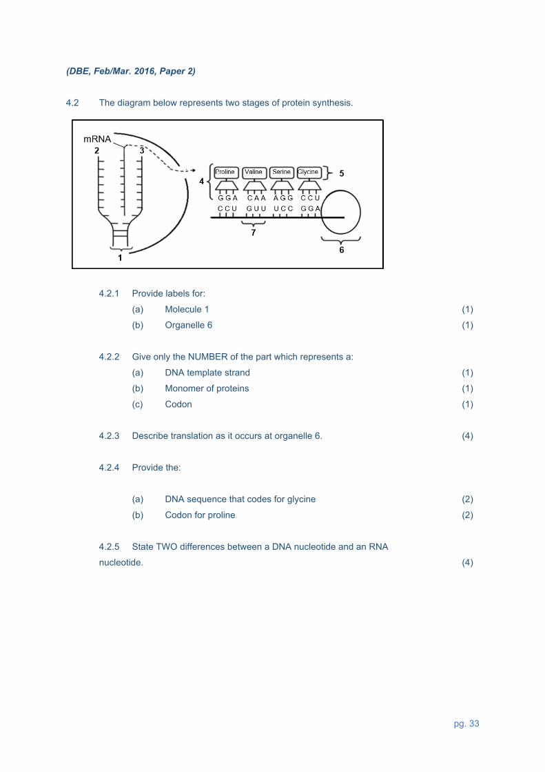
(DBE, Feb/Mar. 2016, Paper 2) 4.2 The diagram below represents two stages of protein synthesis.
4.2.1 Provide labels for:
(a) Molecule 1 (1)
(b) Organelle 6 (1)
4.2.2 Give only the NUMBER of the part which represents a:
(a) DNA template strand (1)
(b) Monomer of proteins (1)
(c) Codon (1)
4.2.3 Describe translation as it occurs at organelle 6. (4)
4.2.4 Provide the:
(a) DNA sequence that codes for glycine (2)
(b) Codon for proline (2)
4.2.5 State TWO differences between a DNA nucleotide and an RNA
nucleotide. (4)

pg. 34
(DBE, Nov. 2019, Paper 2)
4.3 The diagram below shows part of a process involved in the production of a protein.
4.3.1 Identify:
(a) Molecule Y (1)
(b) The group of nitrogenous bases Z (1)
4.3.2 If X is the next amino acid required after W, then identify:
(a) Nitrogenous bases 1, 2 and 3 (2)
(b) The DNA base triplet that codes for X (2)
4.3.3 Describe the process of transcription. (6)

pg. 35
3.15 SOLUTIONS TO DNA PRACTICE QUESTIONS
Question 1
1.1.1 Büü (2)
1.1.2 Cüü (2)
1.1.3 Cüü (2)
1.2.1 (a) W – Nucleotideü U – DNAü (2)
(b) X – Phosphateü/phosphate ion
Y – Deoxyriboseüsugar (2)
(c) Z – Hydrogenü bond (1)
(d) V – Adenineü (1)
1.2.2 Nucleusü (1)
1.2.3 Interphaseü (1)
1.2.4 Enzymesü (1)
1.2.5 - DNA replication ensures that daughter cells in mitosis will have identical genetic
make up as the parent cellü
- ensures that the number of chromosomes in each daughter cell is the same as the
parent cellü
- ensures that genetic properties are transmitted from one generation to the nextü
Any 1 (1)
1.2.6 - DNA unwindsü from one end to appear as a ladderü
- the weak hydrogen bondsü between the nitrogen bases break
- and the two single strands move apartü
- each nucleotides picks up free nucleotidesü from the nucleoplasm
- and become double againü
- the two new double strands are identicalü to each other and the original
- each double strand now become twisted helical structureü
- the process is controlled by enzymesü Any 5 (5)

pg. 36
1.2.7 - If the incorrect nitrogen baseü attaches to the original strand/if a nitrogen base is added or
deleted
- the sequenceü/order of the bases changes on the new DNA molecule
- resulting in a change in the gene structureü (Any 2) (2)
Question 2
2.1.1 Cüü (2)
2.1.2 Büü (2)
2.2.1 Arginine, Isoleucine, Glycineüü (2)
2.2.2 - The mutated codon AUU code for the same amino acid/Isoleucine.ü
- The amino acid sequence will not change ü
- and will therefore code for the same protein.ü (3)
2.3.1 (a) GGTü (1)
(b) AAAü (1)
(c) UCAü (1)
2.3.2 (a) 1ü (1)
(b) 198ü (1)
(c) 66ü (1)
2.3.3 - One of the base triplets on the DNA has changedü
- from ACG to ACCü
- The triplet ACG codes for the amino acid cysteineü
- while the triplet ACC codes for the amino acid tryptophanü
- resulting in a change in the sequenceü of amino acids Any 4 (4)
pg. 37
Question 3
3.1.1 DNA-profilingü (1)
3.1.2 Jennieü (1)
3.1.3 - Jennie’s DNA profileü/bands
- matches the DNA profileü/bands of the sample form the crime scene (2)
3.1.4 - Proof of paternityü
- Tracing missing personsü
- Identification of genetic disordersü
- Matching tissues for organ transplantsü
- Identifying dead personsü /animals (Any 1) (1)
3.1.5 - A child received DNA from both parentsü
- The DNA profiles of the mother, child and the possible father are determinedü
- A comparison of the DNA bands of the mother and the child is madeü
- The remaining DNA bands are compared to the possible father’s DNA bandsü
- If all the remaining DNA bands in the child’s profile match the possible father’s DNA bandsü
- then the possible father is the biological fatherü
- If all the remaining DNA bands in the child’s profile does not match the possible father’s
DNA bandsü
- then the possible father is not the biological fatherü Any 5 (5)
Question 4
4.1.1 Transcriptionü (1)
4.1.2 (a) mRNAü (1)
(b) Amino acidü (1)
(c) Ribosomeü (1)
4.1.3 - The double helix DNA unwinds ü
- The double-stranded DNA molecule unzipsü/ weak hydrogen bonds break
- to form two separate strandsü
- One DNA strand is used as a templateü
- to form mRNAü
- using free RNA nucleotides from the nucleoplasmü
- The mRNA is complementary to the DNAü (Any 4) (4)

pg. 38
4.1.4 - In B translation*ü takes place *Compulsory mark
- mRNA attaches to the ribosomeü
- tRNA picks up amino acids ü
- brings it to the codonsü of mRNA
- the anticodonü determines which amino acid will bind to the tRNA
1*+ Any 2 (3)
4.1.5 Peptideü bond (1)
4.1.6 (a) A- GTCü (1)
(b) B- ACUü (1)
(c) C- Leucine (leu)ü (1)
(d) D- Glutamic acid (glu)ü (1)
4.1.7 - The codon of the mRNA altersü
- This will lead to a different tRNAü picking up a different amino acid. ü
(Any 2) (2)
4.2.1 (a) DNAü (1)
(b) Ribosomeü (1)
4.2.2 (a) 2ü (1)
(b) 5ü (1)
(c) 7ü (1)
4.2.3 - The mRNA attaches to the ribosomeü
- When each codonü of the mRNA
- matches with the anticodon on the tRNAü
- the tRNA brings the required amino acid to the ribosomeü
- When the different amino acids are brought in sequenceü
- adjacent amino acids are linked by peptide bondsü
- to form the required proteinü/polypeptide (Any 4) (4)

pg. 39
4.2.4 (a) CCTüü (2)
(b) CCUüü (2)
4.2.5
(Mark first TWO only) (2 x 2) (4)
TABLE NOT REQUIRED 4.3.1 (a) tRNAü/transfer RNA (1)
(b) Anticodonü (1)
4.3.2 (a) UGGüü (in correct order) (2)
(b) TGGüü (in correct order) (2)
4.3.3 - The double helix DNA unwindsüand
- unzipsü/weak hydrogen bonds break
- to form two separate strandsü
- One strand is used as a templateü
- to form mRNAü
- using free RNA nucleotides from the nucleoplasmü
- The mRNA is complementary to the DNAü
- The coded message for protein synthesis is thus copied onto mRNAü
Any 6 (6)
4. MEIOSIS
TERM 1 PAPER 2
DURATION 8 hours
(2 weeks)
WEIGHTING 21 marks (14%)
LINKS TO PRIOR-KNOWLEDGE/BACKGROUND KNOWLEDGE Mitosis, Chromosomes, DNA replication
RESOURCES Textbooks, Study Guides, MTG, Past NSC, SC & Provincial Question Papers
DNA RNA Has deoxyriboseü sugar Has riboseü sugar
Has nitrogen base thymine (T)ü/ A,
C, G and T
Has nitrogen base uracil(U)ü/ A, C,
G and U

pg. 40
Meiosis & Mitosis differences and
similarities
Where does it occur in plants and
humans?
Causes and consequences of abnormal meiosis Down Syndrome
Genetic Variation
4.1 MINDMAP on MEIOSIS
Importance of Meiosis
Describe the events of each phase
Meiosis: the process of
reduction division.

pg. 41
The process of mitosis - Mitosis is made up of two major divisions: nuclear division (Karyokinesis) and cytoplasm division (Cytokinesis).
PHASES DIAGRAM PROPHASE
• Cell is ready for division.
• Nuclear membrane starts to disintegrate.
• Nucleolus disappears
• Replicated chromosomes become visible
• Spindle fibres are formed from the centrosomes.
• Centrioles move towards the opposite poles. Centrosomes only found in the animal cell.
METAPHASE
• Nuclear membrane has disintegrated.
• Replicated chromosomes line up on the equator.
• Spindle fibre attaches on the centromere of each replicated
chromosome. ANAPHASE
• Centromere of each replicated chromosome splits to form two
unreplicated chromosomes.
• Unreplicated chromosomes from each chromosome are pulled to the opposite poles
TELOPHASE
• Cytokinesis starts by the cell membrane which constricts at the equator.
• Nuclear membrane and nucleolus appear in each daughter
cell.
• Each daughter cell has the same number of unreplicated chromosomes as the parent.
NOTE: Before the process of mitosis starts, DNA replication first occur during Interphase. After DNA
replication the chromatin network in the nucleus becomes visible as chromosomes.
4.2 LINKS TO PRIOR-KNOWLEDGE/BACKGROUND KNOWLEDGE

pg. 42
The significance of DNA replication for mitosis:
• To double the genetic material
• Each daughter cell receives the same amount of DNA
• To ensure genetically identical daughter cells
4.3 DIFFERENTIATE BETWEEN RELATED TERMINOLOGIES
NUCLEAR MEMBRANE
The nuclear membrane is the membrane which
surrounds the nucleus, enclosing the genetic material.
CELL MEMBRANE
The cell membrane is the membrane that separates
the interior of all cells from the outside environment
CENTROSOME CENTRIOLE CENTROMERE Organelle (containing two
centrioles) found only in animal
cells. This structure is
responsible for the formation of
spindle fibres during cell division
in animal cells.
structures formed when the
centrosome divides into two;
they move to opposite ends
of the cell during cell division
The centromere is not a structure as
such but a site where two
chromatids are held together in a
replicated chromosome and also
where the chromosome is attached
to the spindle thread during cell
division.
pg. 43
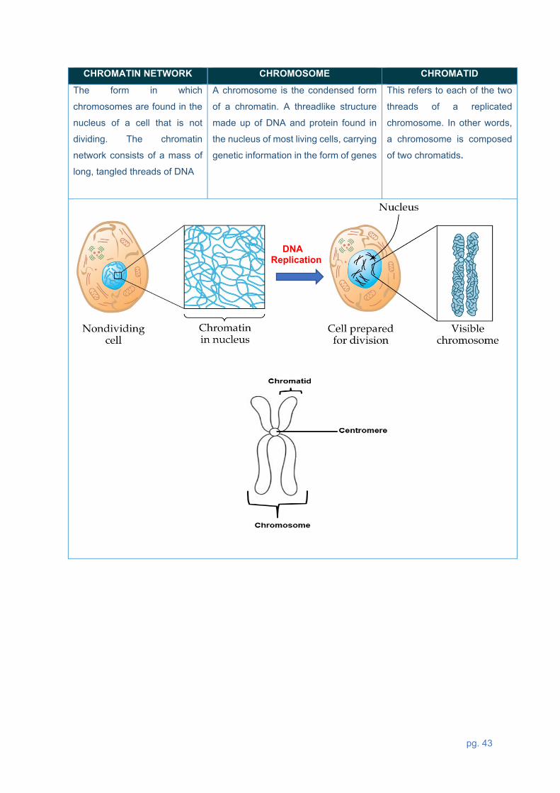
CHROMATIN NETWORK CHROMOSOME CHROMATID The form in which
chromosomes are found in the
nucleus of a cell that is not
dividing. The chromatin
network consists of a mass of
long, tangled threads of DNA
A chromosome is the condensed form
of a chromatin. A threadlike structure
made up of DNA and protein found in
the nucleus of most living cells, carrying
genetic information in the form of genes
This refers to each of the two
threads of a replicated
chromosome. In other words,
a chromosome is composed
of two chromatids.
DNA Replication

pg. 44
Gamete (sex cell)- cells formed by meiosis in male
testis and female ovaries which contain half the
chromosome number.
Somatic cell- Body cells that contain the full set of
chromosomes, 23 inherited from each parent (46 in
total). HAPLOID (N) DIPLOID (2N)
Haploid cells only have one set of chromosomes.
Chromosomes in haploid cells have no homologous
partners.
Diploid cells have two sets of chromosomes, where
each chromosome has a homologous partner.
Karyotype- A diagram that shows the number, size and arrangement of chromosomes within a somatic cell or sex cell
AUTOSOMES GONOSOMES (SEX CHROMOSOMES) The first 22 pairs of chromosomes in a human somatic cell
which control the appearance, structure and functioning of
the body and is not connected with the determination of
sex.
The last pair of chromosomes in a human
somatic cell (XX or XY) responsible for sex
determination
pg. 45
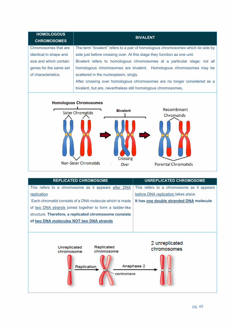
REPLICATED CHROMOSOME UNREPLICATED CHROMOSOME
This refers to a chromosome as it appears after DNA
replication
Each chromatid consists of a DNA molecule which is made
of two DNA strands joined together to form a ladder-like
structure. Therefore, a replicated chromosome consists of two DNA molecules NOT two DNA strands
This refers to a chromosome as it appears
before DNA replication takes place.
It has one double stranded DNA molecule
HOMOLOGOUS CHROMOSOMES
BIVALENT
Chromosomes that are
identical in shape and
size and which contain
genes for the same set
of characteristics.
The term “bivalent” refers to a pair of homologous chromosomes which lie side by
side just before crossing over. At this stage they function as one unit.
Bivalent refers to homologous chromosomes at a particular stage; not all
homologous chromosomes are bivalent. Homologous chromosomes may be
scattered in the nucleoplasm, singly.
After crossing over homologous chromosomes are no longer considered as a
bivalent, but are, nevertheless still homologous chromosomes.

pg. 46
4.4 PROCESS OF MEIOSIS - EXAM TIPS/TECHNIQUES/NOTES
Meiosis topic is linked to Mitosis taught from grade 10. Meiosis can be divided into two parts,
Meiosis I and Meiosis II.
First meiotic division
Prophase I
• Nuclear membrane and nucleolus start to disappear.
• Centrosome splits and the two centrioles move apart
forming spindle fibres.
• Chromatin network condenses into individual
chromosomes and pairs of homologous chromosomes
lie next to each other forming a bivalent.
• Inner chromatids from each homologous chromosomes
overlap and touch each other at a point called the
chiasma (plural: chiasmata) in a process called
crossing over
• Chromatid segments break off and are exchanged,
resulting in the exchange of genetic material.
• This process is called crossing over and it brings
about variation.
Metaphase I
• Homologous chromosomes move to the middle of the cell (the equator).
• The two homologous chromosomes lie on opposite
sides of the equator parallel to each other.
• Which homologous chromosome lies on which side of
the equator is totally up to chance.
• This is called random arrangement and brings about
further variation.
• Each chromosome in the homologous pair becomes
attached to a spindle thread by the centromere.

pg. 47
Anaphase I
• One whole chromosome from each pair is pulled to opposite poles by contraction of the spindle fibres
• This separates the homologous chromosomes – one to
each pole.
Telophase I
• A new nuclear membrane forms around the group of
chromosomes at each pole.
• Nucleolus returns.
• Cytokinesis (division of cytoplasm) splits the mother
cell into two daughter cells.
• Important: Each daughter cell now has half the number of chromosomes (i.e., is haploid) and each
has a slightly different genetic make-up due to
crossing over.
Second meiotic division The second meiotic division takes place in both daughter cells formed during Meiosis I.
Prophase II
• Nuclear membrane and nucleolus start to
disappear.
• Centrosome splits into two centrioles and a
spindle forms.
• Chromosomes are NOT in pairs
Remember: Each chromosome is made of TWO
chromatids

pg. 48
Metaphase II
• Single chromosomes arrange themselves randomly along the equator with the
centromere in line with the equatorial plane.
• Which chromatid faces which pole is totally up
to chance.
• Each chromosome becomes attached to a
spindle fibre.
Anaphase II
• Centromere splits separating each
chromosome into two daughter chromosomes, each pulled to opposite poles.
Telophase II
• A new nuclear membrane forms around the
unreplicated chromosomes at each pole
• Cytokinesis splits the cell into two new cells
Important: As Meiosis II took place in TWO cells, there will now be FOUR daughter cells. These cells will be haploid and genetically different to each other.
pg. 49
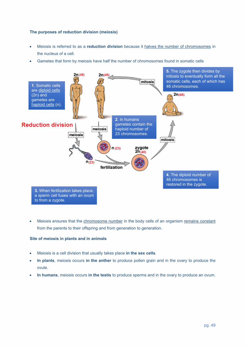
The purposes of reduction division (meiosis)
• Meiosis is referred to as a reduction division because it halves the number of chromosomes in
the nucleus of a cell.
• Gametes that form by meiosis have half the number of chromosomes found in somatic cells
• Meiosis ensures that the chromosome number in the body cells of an organism remains constant
from the parents to their offspring and from generation to generation.
Site of meiosis in plants and in animals
• Meiosis is a cell division that usually takes place in the sex cells.
• In plants, meiosis occurs in the anther to produce pollen grain and in the ovary to produce the
ovule.
• In humans, meiosis occurs in the testis to produce sperms and in the ovary to produce an ovum.
2. In humans gametes contain the haploid number of 23 chromosomes.
4. The diploid number of 46 chromosomes is restored in the zygote.
3. When fertilization takes place, a sperm cell fuses with an ovum to from a zygote.
5. The zygote then divides by mitosis to eventually form all the somatic cells, each of which has 46 chromosomes.
1. Somatic cells are diploid cells (2n) and gametes are haploid cells (n).

pg. 50
Differences between Mitosis and Meiosis
There are two types of cell divisions that takes place in
plants and animals, mitosis
and meiosis.
Mitosis is a process whereby
one cell makes an identical copy
of itself and gives rise to two cells that are genetically identical.
Meiosis produces four sex cells
that have half the number of
chromosomes of the parent cell,
and are genetically different from
the parent cell.
There are two types of cells in
a plant or animal’s body, body
cells (somatic cells) and sex
cells (gametes).
Mitosis deals with the formation
of somatic cells,
while meiosis deals with the
formation of gametes (gametogenesis).
4.5 PRACTICE QUESTIONS on MEIOSIS
1.1 The diagram below shows the karyotypes of two individuals.
(a) State the gender of individual P.
Look at chromosome pair 23, the Gonosomes are of different size and shape (XY)
Maleü

pg. 51
(b) Give ONE reason why the diagram above represents the chromosomes of a human.
Count the chromosome number /pairs in the karyotype
NB different species have different chromosome numbers (This karyotype has 46
chromosomes
It represents a human because it has 46 chromosomesü/ 23 pairs of chromosomes which
is a unique feature in humans
(c)
How many chromosomes will be found in?
(i) A human sperm cell It is a gamete formed by meiosis which is a reduction division,
so it is haploid
23ü chromosomes
(ii) Muscle cell It is a somatic cell - part of the body therefore diploid
46ü chromosomes
(iii) The somatic cells of a normal mother who has a son with Down syndrome
46ü chromosomes (Note: It is the son who will have 47)
2.1 A chemical used in laboratories prevents spindle fibres from forming in cells undergoing meiosis. As a result meiosis cannot start on the completion of interphase. In an investigation, this chemical was added to cells in the anthers of the flowers of rice plants. Each cell in the anther has 24 chromosomes. What is the expected number of chromosomes in each cell at the end of the investigation?
A 12 replicated chromosomes
B 24 replicated chromosomes ü C 24 unreplicated chromosomes
D 48 unreplicated chromosomes
No meiosis, chromosomes will not be halved

pg. 52
2.2 The diagrams below represent various phases of meiosis.
(a) Identify the phase of meiosis in diagram: (i) A – prophase I ü
(ii) B- Anaphase I ü
(iii) C – Metaphase II ü DIAGRAM A DIAGRAM B DIAGRAM C
Know the event that is
unique to a particular
phase
Shows separation of
replicated chromosomes
Single chromosomes are
at the equator
Know the difference
between meiosis I & II
If it was anaphase II
unreplicated chromosomes
would be separating
If it was metaphase I,
homologous
chromosomes would be at
the equator
(b) Draw a labelled diagram to show the cells that will be formed at the end of meiosis from the cell in diagram C.
Step 1: Identify whether the question is based on Meiosis I or II Remember: Each daughter cell in meiosis I will form TWO Gametes. In Meiosis I we have a complete set of chromosomes (diploid) except in Telophase I, but in Meiosis II all the phases show half the number of chromosomes Step 2: Show the effect of crossing over in each gamete using the correct shading Step 3: A complete gamete must have a nucleus surrounded by a nuclear membrane, and a complete cell must also be surrounded by a cell membrane Step 4: The nucleus for a gamete must show an un-replicated chromosome

pg. 53
CRITERIA FOR MARKING Only two cells drawn (D) 1 mark
Each cell contains only two unreplicated chromosomes
(C) 1 mark
Each chromosome is the correct size and correctly
shaded (S) 1 mark
Any TWO correct labels 1 mark
2.3 The diagram below represents TWO phases of meiosis
2.3.1 Identify part A. - centriole ü
C
B
A
Diagram 1 Diagram 2

pg. 54
2.3.2 Describe the events that took place in the phase before the one represented in diagram 2.
First identify diagram 2 as telophase II because cell membrane is starting to
invaginate. So, a phase that occurs before this one is anaphase II. Therefore,
describe the events in anaphase II as follows
- Spindle fibres contractü
- Centromeres splitü
- Each unreplicated chromosome is pulled to the opposite poleü
2.3.3 Name the process that causes the chromosomes to have a combination of genes as shown in the diagrams.
Crossing overü
2.3.4 Give ONE reason why the process named in QUESTION 2.3.3 is important.
Leads to genetic variationü
2.3.5 If this was a human cell, how many chromosomes would be present in the cell during the phase represented in diagram 1 46ü
2.3.6 Structure B and structure C are both chromosomes.
Explain why they are structurally different.
Check terminology, be able to differentiate between replicated and
unreplicated chromosome (picturing a diagram helps in remembering
definitions)
- structure B has two DNA moleculesü
- is a replicated chromosome
- it is made up of TWO chromatidsü
- Structure C has ONE DNA moleculeü, it is an un-replicated chromosome
- Structure C has one chromatid ü

pg. 55
2.4 The diagram below represents a cell during a phase of meiosis.
2.4.1 Name the process taking place at A.
Homologous chromosomes have failed to separate
Non-disjunctionü
2.4.2 State the phase of meiosis illustrated above. Identify what is separating, is it homologous chromosomes, or is it the splitting of
centromere, separating chromatids. What is moving towards the poles? Is it a
replicated chromosome or an unreplicated chromosome?
Anaphase Iü
2.4.3 Name the type of mutation that occurred in the cell.
Check whether it involves a change in the number and size of chromosomes.
Note: if it only involves a change in the number and sequence of nucleotides, it is a
gene mutation
Chromosomal mutation ü
2.4.4 Give the number of chromosomes that will be present in a normal gamete of the
species whose cell is represented above. Identify the diploid cell which represents the chromosome number for somatic cells of
the parent. Then work out half the number of chromosomes, Note this half number of
chromosomes appears at telophase II and is maintained throughout all the stages of
meiosis II Threeü

pg. 56
2.4.5 Give the chromosome number of the four gametes formed at the end of Meiosis
II.
• Determine the number for a full set of chromosomes (in this case (six
• Identify how many pairs chromosomes have been affected by non-disjunction
(one pair)
• Normal separation will be for four chromosomes – to give two chromosomes on
each daughter cell.
• Because of non-disjunction in the third pair, both chromosomes will go to the
same daughter cell, causing it to have four chromosomes.
• The other one will have two chromosomes. Note this number will
• be maintained in all stages of meiosis II
Two cells will have four unreplicated chromosomesü
Two cells will have two unreplicated chromosomesü
2.4.6 Describe the chromosome behaviour in the phase before the one represented
in the diagram.
• PMAT- prophase, metaphase, anaphase, telophase
• Identify whether it is meiosis I or meiosis II
• Identify the stage in the diagram shown- anaphase I
• Work backwards to determine the phase before the one drawn
• Metaphase I.
Homologous chromosomes were randomly arranged at the equatorü
2.4.7 Explain how the new zygote will be affected if a gamete resulting from the error in
meiosis at A is involved in fertilisation with a normal gamete
• Determine the number of chromosomes in the gamete that was affected by non-
disjunction (1 gamete has four, other one has two)
• Work chromosome number expected in a normal gamete which has not
undergone non-disjunction (three)

pg. 57
An ovum with 4 unreplicated chromosomes üwill be fertilized by a normal sperm cell
with 3 unreplicated chromosomeü resulting in
a zygote with 7 chromosomesü instead of 6ü
or An ovum with 2 unreplicated chromosomesü will be fertilized by a
normal sperm cell with 3 unreplicated chromosome resulting in
a zygote with 5 chromosomes üinstead of 6ü
4 chromosomes
2 chromosomes
Gametes formed after non-disjunction (ovum)
Normal sperm cell
3 chromosomes

pg. 58
4.6 TYPICAL EXAM QUESTIONS
QUESTION 1 (DBE, Nov. 2018, Paper 2)
1.1 The diagram below shows the structure of a chromosome
1.1.1 Identify parts D and E. (2)
1.1.2 How many pairs of chromosomes are found in a normal human sperm cell? (1)
1.1.3 Give only the LETTER of the part that:
(a) Attaches to the spindle fibres during cell division
(b) Represents a gene
(1)
(1)
(5)

pg. 59
QUESTION 2 (DBE, May/June 2018, Paper 2)
2.1 Diagrams 1 to 3 below represent some of the phases of meiosis shown in the correct
order.
2.1.1 Identify the phase represented by diagram
(a) 1 (1)
(b) 3 (1)
2.1.2 Give the LETTER only of the part that
(a) Contains DNA (1)
(b) Attaches to the centromeres of chromosomes (1)
(c) Forms the spindle fibres (1)
2.1.3 Name the organ in a human male where meiosis occurs. (1)
(6)

pg. 60
QUESTION 3 (DBE, May/June 2018, Paper 1) 3.1 The diagrams below represent two phases of meiosis in an organism.
3.1 1 Identify the phase of meiosis represented in Diagram 1. (1) 3.1.2 Identify part: (a) A (1) (b) B (1) (c) C (1) 3.1.3 State what happens to structure D in the next phase of meiosis. (1) 3.1.4 Name the process during which genetic material was exchanged, as shown in
the diagrams above.
(1)
3.1.5 State the consequence if the process named in QUESTION 3.1.4 does not
occur
(1)

pg. 61
3.1.6 Give the number of chromosomes present in: (a) The original parent cell in this organism (1) (b) A human cell in the same phase as that shown in
Diagram 2
(1)
(9) QUESTION 4 (DBE, Nov 2013, Paper 1)
4.1 The diagram below represents the distribution of chromosome pair 21 as it
appears in the gametes at the end of meiosis II in the human male
4.1.1 Explain why the gametes represented by diagrams C and D do not have
any chromosomes
(3)
4.1.2 If gamete A is involved in fertilisation, describe how this may result in
down syndrome
(3)
4.1.3 Due to the process of crossing over, the chromosomes in diagram A
and B appear different from each other
(a) Identify the phase of meiosis during which crossing over occurs (1)
(b) Describe the events during crossing over (3)
(10)

pg. 62
4.7 SOLUTIONS TO MEIOSIS PRACTICE QUESTIONS
QUESTION 1 1.1.1
1.1.2
1.1.3
D- chromatid ü
E- centromere ü
23 ü
(a) E ü
(b) C ü
(2)
(1)
(1)
(1)
(5) QUESTION 2
2.1.1 (a) Metaphase Iü (1)
(b) Telophase Iü (1)
2.1.2 (a) Bü (1)
(b) Cü (1)
(c) Dü (1)
2.1.3 Testisü (1)
(6)

pg. 63
QUESTION 3 3.1.1 Anaphase IIü (1)
(a) Centrioleü (1) (b) Centromere (1) (c) Spindle fibreü (1)
3.1.2 The chromatids separate /centromere splitsü (1) 3.1.3 Crossing overü (1) 3.1.4 Reduces genetic variationü (1) 3.1.5 (a) 4ü (1) (b) 23ü (1) (9) QUESTION 4 4.1.1 Due to non – disjunction / non-separation of a chromosome pair during anaphase Iü
Two chromosomes moved to one poleü and none moved to the other poleü
(3)
4.1.2 Gamete A will have 24 chromosomesü / an extra chromosome
When it fertilises a normal ovumü / gamete with 23 chromosomes
The zygote will have 3 chromosomes at position 21ü / 47 chromosomes
(3)
4.1.3 (a) Prophase Iü (1)
(b) Adjacent chromatids of homologous chromosomes crossü
at a point called chiasmaü
There is an exchange of DNA segments ü / genetic material
(3)
(10)
pg. 64
5. REFERENCES
o DBE Exam guidelines for learners
o GDE ATP
o 2015-2020 NSC past papers
o 2014-2020 national diagnostic report on learner performance
o Approved grade 12 national textbooks
o Internet
o Gauteng grade 12 Life Sciences Revision booklet
o Gauteng grade 12 Life Sciences Exam Kit
o NMD grade 12 life sciences workbook
6. ACKNOWLEDGEMENT
The Department of Basic Education (DBE) gratefully acknowledges the following officials for giving up
their valuable time and families and for contributing their knowledge and expertise to develop this
resource booklet for the children of our country, under very stringent conditions of COVID-19:
Writers: Arnold M. Johannes (Eastern Cape) and Phumzile Dlamini (Eastern Cape)
Reviewers: Mpho Mokgotlhoa, Ntombi Dladla, Julia Tladi, Lucas Mothibedi Mfolo, Gezani Phineas
Chavani, Avusiwe Madikane, Mthembi GB, Chauke Magezi Elias, Jacoline Jones
DBE Subject Specialist: Kanthan Naidoo
The development of the Study Guide was managed and coordinated by Ms Cheryl Weston and Dr
Sandy Malapile

pg. 65