:o 'lo - National Security Archive - The George Washington University
Upload
khangminh22Category
view
2download
0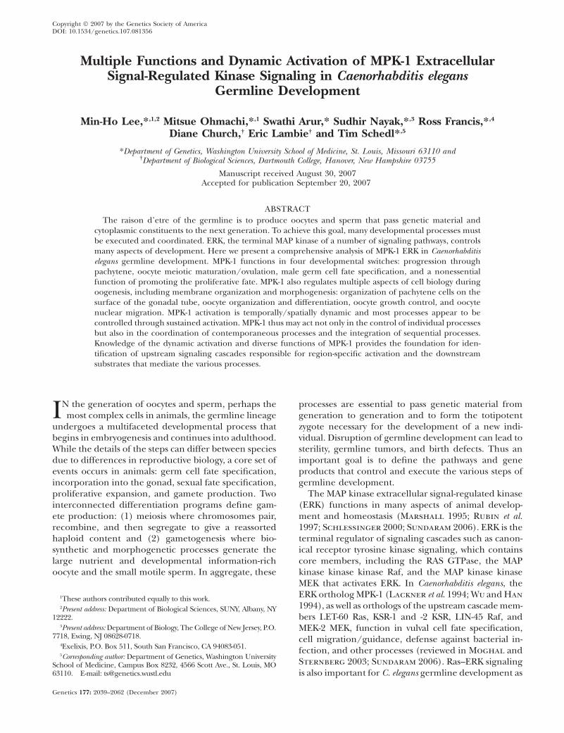
Copyright � 2007 by the Genetics Society of AmericaDOI: 10.1534/genetics.107.081356
Multiple Functions and Dynamic Activation of MPK-1 ExtracellularSignal-Regulated Kinase Signaling in Caenorhabditis elegans
Germline Development
Min-Ho Lee,*,1,2 Mitsue Ohmachi,*,1 Swathi Arur,* Sudhir Nayak,*,3 Ross Francis,*,4
Diane Church,† Eric Lambie† and Tim Schedl*,5
*Department of Genetics, Washington University School of Medicine, St. Louis, Missouri 63110 and†Department of Biological Sciences, Dartmouth College, Hanover, New Hampshire 03755
Manuscript received August 30, 2007Accepted for publication September 20, 2007
ABSTRACT
The raison d’etre of the germline is to produce oocytes and sperm that pass genetic material andcytoplasmic constituents to the next generation. To achieve this goal, many developmental processes mustbe executed and coordinated. ERK, the terminal MAP kinase of a number of signaling pathways, controlsmany aspects of development. Here we present a comprehensive analysis of MPK-1 ERK in Caenorhabditiselegans germline development. MPK-1 functions in four developmental switches: progression throughpachytene, oocyte meiotic maturation/ovulation, male germ cell fate specification, and a nonessentialfunction of promoting the proliferative fate. MPK-1 also regulates multiple aspects of cell biology duringoogenesis, including membrane organization and morphogenesis: organization of pachytene cells on thesurface of the gonadal tube, oocyte organization and differentiation, oocyte growth control, and oocytenuclear migration. MPK-1 activation is temporally/spatially dynamic and most processes appear to becontrolled through sustained activation. MPK-1 thus may act not only in the control of individual processesbut also in the coordination of contemporaneous processes and the integration of sequential processes.Knowledge of the dynamic activation and diverse functions of MPK-1 provides the foundation for iden-tification of upstream signaling cascades responsible for region-specific activation and the downstreamsubstrates that mediate the various processes.
IN the generation of oocytes and sperm, perhaps themost complex cells in animals, the germline lineage
undergoes a multifaceted developmental process thatbegins in embryogenesis and continues into adulthood.While the details of the steps can differ between speciesdue to differences in reproductive biology, a core set ofevents occurs in animals: germ cell fate specification,incorporation into the gonad, sexual fate specification,proliferative expansion, and gamete production. Twointerconnected differentiation programs define gam-ete production: (1) meiosis where chromosomes pair,recombine, and then segregate to give a reassortedhaploid content and (2) gametogenesis where bio-synthetic and morphogenetic processes generate thelarge nutrient and developmental information-richoocyte and the small motile sperm. In aggregate, these
processes are essential to pass genetic material fromgeneration to generation and to form the totipotentzygote necessary for the development of a new indi-vidual. Disruption of germline development can lead tosterility, germline tumors, and birth defects. Thus animportant goal is to define the pathways and geneproducts that control and execute the various steps ofgermline development.
The MAP kinase extracellular signal-regulated kinase(ERK) functions in many aspects of animal develop-ment and homeostasis (Marshall 1995; Rubin et al.1997; Schlessinger 2000; Sundaram 2006). ERK is theterminal regulator of signaling cascades such as canon-ical receptor tyrosine kinase signaling, which containscore members, including the RAS GTPase, the MAPkinase kinase kinase Raf, and the MAP kinase kinaseMEK that activates ERK. In Caenorhabditis elegans, theERK ortholog MPK-1 (Lackner et al. 1994; Wu and Han
1994), as well as orthologs of the upstream cascade mem-bers LET-60 Ras, KSR-1 and -2 KSR, LIN-45 Raf, andMEK-2 MEK, function in vulval cell fate specification,cell migration/guidance, defense against bacterial in-fection, and other processes (reviewed in Moghal andSternberg 2003; Sundaram 2006). Ras–ERK signalingis also important for C. elegans germline development as
1These authors contributed equally to this work.2Present address: Department of Biological Sciences, SUNY, Albany, NY
12222.3Present address: Department of Biology, The College of New Jersey, P.O.
7718, Ewing, NJ 08628-0718.4Exelixis, P.O. Box 511, South San Francisco, CA 94083-051.5Corresponding author: Department of Genetics, Washington University
School of Medicine, Campus Box 8232, 4566 Scott Ave., St. Louis, MO63110. E-mail: [email protected]
Genetics 177: 2039–2062 (December 2007)

loss-of-function (lf), but not null, alleles of let-60, mek-2,and mpk-1, as well as null alleles of ksr-2, result in germcells arrested in pachytene (Church et al. 1995;Ohmachi et al. 2002). The pachytene-arrest phenotype,which is observed in both hermaphrodites and males,was interpreted as an essential function of these genesin the transition from pachytene to diplotene (pachy-tene exit; Church et al. 1995). In vertebrates, ERKfunctions in meiotic maturation of full-grown oocytesand/or arrest at metaphase of meiosis II prior tofertilization (reviewed in Fan and Sun 2004; Liang
et al. 2007); however, little is known about ERK functionin the many earlier steps of germline development.
Since the initial report of Church et al. (1995), newmutations in a number of the core pathway genes havebeen identified: null alleles of mpk-1 and lin-45, atemperature-sensitive (ts) mpk-1 lf allele (ga111) thatallows examination of MPK-1 function in late steps ofgermline development, and a ts let-60 gain-of-function(gf) allele (ga89) that permits examination of the effectof increased MPK-1 activation (Eisenmann and Kim
1997; Lackner and Kim 1998; Hsu et al. 2002).Molecular markers that allow stages of germline devel-opment to be more fully defined and the sex of im-mature germ cells to be determined have also beenidentified. An initial description of the pattern ofMPK-1 activation (the MEK-2 MEK diphosphorylatedform of MPK-1, dpMPK-1) in the hermaphrodite germ-line has been presented, but not analyzed in the contextof core pathway mutant backgrounds (Miller et al.2001; Page et al. 2001). MPK-1 activation was found tocorrelate with oocyte meiotic maturation, consistentwith ERK playing a role in the process as it does in otherorganisms, but experiments have not addressed theimportance of mpk-1 function in the maturation pro-cess. Here we build on previous studies using the newmutations and molecular markers in a comprehensiveanalysis of MPK-1 function and activation in hermaph-rodite and male germlines. We found that MPK-1 hasdiverse germline functions, acting in four developmen-tal switches and in four aspects of cell biology duringoogenesis. MPK-1 activation is temporally/spatially dy-namic compared to relatively constant levels of totalMPK-1, and most processes appear to be controlled bysustained activation. Below we briefly review aspects ofC. elegans germline development relevant to this study.
C. elegans is an excellent system for analysis of germ-line development due to facile genetics and favorableanatomy where most of the processes are ongoing in theadult and can be studied in real time, as live animalsare transparent, and with fixed specimens (reviewed inHubbard and Greenstein 2000). The adult hermaph-rodite has two U-shaped gonads while the male has asingle U-shaped gonad (Figure 1, B and D, linear projec-tion diagrams of single gonads). The hermaphrodite isconsidered to be female in somatic tissues with the germ-line undergoing male development (spermatogenesis)
transiently in the third larval (L3) and L4 stages andfemale development (oogenesis) from L4 throughoutadulthood. The male has a male soma and undergoesspermatogenesis from L3 through adulthood. The germ-line sex determination pathway controls the decisionbetween female and male germline development (re-viewed in Ellis and Schedl 2006). The U-shapedgonads have germ cells arranged in a distal-to-proximalpolarity with respect to the somatic distal tip cell at thedistal end in both sexes and the proximal spermatheca/uterus in the hermaphrodite or the vas deferens in themale. Germ cells progress proximally in an assembly-line-like fashion with the germline divided into regionsbased on chromosome and cellular morphology. At thedistal end, germ cells proliferate (mitotic zone) undercontrol of GLP-1 Notch signaling (reviewed in Hansen
and Schedl 2006; Kimble and Crittenden 2006); inwild type, this region contains �220 cells and extends�20 cell diameters in length proximally from the distaltip. At the proximal end of the mitotic region, germ cellsswitch to meiotic prophase where they are first inleptotene/zygotene (transition zone, or TZ) and thenprogress through an extended pachytene region that isthen followed by diplotene and diakinesis where overtgamete formation occurs. Meiotic prophase of oogen-esis lasts 54–60 hr while meiotic prophase for spermato-genesis in males lasts 20–24 hr ( Jaramillo-Lambert
et al. 2007). Much of the germline is a syncytium; plasmamembranes do not fully surround each nucleus and a‘‘window’’ connects each to a common cytoplasm. Byconvention, each nucleus, surrounding cytoplasm, andmembranes are referred to as a germ cell. From themitotic region through the end of pachytene, germ cellsare arranged on the surface of the gonadal tube withan interior cell/nucleus-free cytoplasmic region calledthe rachis or core. Consistent with the longer time ofmeiotic prophase, oogenesis has complexities that arelacking in spermatogenesis. In oogenesis, the majorityof pachytene cells appear to function as nurse cells,undergoing apoptosis after providing RNAs and pro-teins to the rachis (Gibert et al. 1984; Gumienny et al.1999; Wolke et al. 2007). As germ cells progress frompachytene to diplotene in the loop region, oocyte differ-entiation begins; a single-file row of growing oocytesis on the external surface while the rachis that is sup-plying cytoplasm to growing oocytes is found on theinternal surface (external/internal refer to the loop ofthe U-shaped gonad). The proximal approximately fiveoocytes (-1 through -5) are in diakinesis and are ap-parently fully cellularized as they are no longer connectedto the rachis (Maddox et al. 2005). In the adult herm-aphrodite, sperm are stored in the spermatheca, the siteof fertilization. In the single-file assembly line, the mostproximal oocyte, the -1 oocyte, undergoes meiotic mat-uration (nuclear envelope breakdown, progression tometaphase of meiosis I, and cortical rearrangement), isovulated into the spermatheca and fertilized (McCarter
2040 M.-H. Lee et al.
et al. 1999). The -2 oocyte then moves into the -1 positionwhere it will undergo maturation/ovulation �23 minlater. In hermaphrodites with sperm, the signal thattriggers maturation/ovulation is constitutive: the majorsperm protein (MSP) secreted from sperm (McCarter
et al. 1999; Miller et al. 2001; Kosinski et al. 2005). Bycontrast, in animals without sperm (sex determinationmutant females, adult hermaphrodites that have ex-hausted their self-sperm), oocytes form but are arrestedin diakinesis.
MATERIALS AND METHODS
Strains: The following mutations were used in this study:LGI, mek-2(h294), mek-2(q425), mek-2(q484), mek-2(n2678), mek-2(oz221), mek-2(dx51), mek-2(n1859), rrf-1(pk1417), glp-4(bn2);LGII, tra-2(e1095); LGIII, mpk-1(ga117), mpk-1(ga111ts), mpk-1(oz140), unc-32(e189), glp-1(bn18ts), mrt-2(e2663); LGIV, unc-5(e53), let-60(ga89gf), let-60(n1046), lin-45(oz201), lin-45(dx19),lin-45(dx89), fem-3(e1996), spo-11(ok79), him-8(e1489); and LGV,fog-2(oz40), mre-11(ok179).
Nematode strains and culture: Standard procedures forculture and genetic manipulation of C. elegans strains werefollowed with growth at 20� unless otherwise noted (Sulston
and Hodgkin 1988). Descriptions of genes, alleles, andphenotypes related to this study are in Church et al. (1995),Eisenmann and Kim (1997), Lackner and Kim (1998), andHsu et al. (2002) or are cited in the text as genes, alleles, andphenotypes are mentioned.
Antibodies and reagents: The following antibodies wereused in this study: anti-MAPKYT antibody (Sigma, St. Louis)used at 1:400; anti-SYN-4/PTC-1 antibodies (kind gifts fromMichael Glotzer and Patty Kuwabara) used at 1:400 and 1:50,respectively (these two antibodies were mixed together forvisualizing membrane morphology); anti-AIR-2 antibody(kind gift from Jill Schumacher) used at 1:50 dilution (todetect the chromosomes undergoing maturation/ovulation);anti-REC-8 (kind gift from Josef Loidl) used at 1:100; anti-HIM-3 (kind gift from Monique Zetka) used at 1:100 asdescribed (Hansen et al. 2004); anti-DAZ-1 (kind gift fromMasayuki Yamamoto) used at 1:50; anti-CEP-1 (kind giftfrom Anton Gartner) used at 1:50; anti-CGH-1 (a kind giftfrom David Greenstein) used at 1:400; and anti-GLD-1 usedat 1:200. Anti-NOP-1 (Encor Biotech) was used at 1:200.Rhodamine–phalloidin was used at 1:100 to visualize actincytoskeleton. Secondary antibodies were donkey anti-mouseAlexa 594, goat anti-rabbit Alexa 488, goat anti-rabbit Alexa594, goat anti-rat Alexa 488, and donkey anti-goat Alexa 594,obtained from Molecular Probes (Eugene, OR).
Anti-ERK antibody SC94 (Santa Cruz), while giving a rela-tively specific MPK-1 pattern on Western blots (supplementalFigure 1 at http://www.genetics.org/supplemental/), showedsignificant germline cytological staining in mpk-1(ga117) nullgonads indicative of nonspecific reactivity. The peptide used asthe immunogen for SC94, corresponding to residues 305–327of human ERK1, contains sequences in the N-terminal halfthat may cross-react with C. elegans MAP kinases W06F12.1 (lit-1)and F42G8.4 (pmk-3). Therefore, a peptide (KRITVEEALAPHY)corresponding to the N-terminal portion of the immunogenwas used to subtract antibodies in SC94 that might cross-reactwith other C. elegans MAP kinases. The SC94 antiserum waspassed over a KRITVEEALAPHY-linked column and the flowthrough, designated SC94-C, was collected. SC94-C, used at1:25 dilution, shows no germline staining in mpk-1(ga117)
germlines although it displays some cross-reactivity in thesomatic gonad (supplemental Figure 2B). We also tested anti-ERK2 from Zymed; however, this antibody detected a strongsignal in mpk-1(ga117) germlines and was not used further.
Western analysis: L4 hermaphrodites of given genotypeswere hand picked, grown for 48 hr or the indicated time at theindicated temperature, and then harvested for Western anal-ysis as previously described ( Jones et al. 1996). The extractswere resolved on 10% SDS–PAGE (acrylamide/bis-acrylamideis 100/1), transferred to PVDF membrane, and probed withSC94 (Santa Cruz) at 1:2000 dilution, MAPKYT at 1:10,000dilution, or MH16 (antiparamyosin antibody) at 1:15,000dilution. The Western blots were developed using SuperSignalWest Pico chemiluminescent substrate from Peirce on KodakBioMax MS film.
Antibody staining: For antibody staining, dissected gonads( Jones et al. 1996) of the indicated genotype were fixed in 3%formaldehyde with 100 mm K2HPO4 (pH 7.2) for 1 hr at roomtemperature (or 10 min for DAZ-1, REC-8, and HIM-3) andpostfixed with 100% methanol (�20�) for 5 min (Francis et al.1995). Fixed gonads, in batches, were blocked with 30%normal goat serum (NGS), or 1% BSA for CEP-1, in 13 PBSplus 0.1% Tween-20 (termed blocking buffer) for 1 hr at roomtemperature before incubation with the desired primaryantibody. In all cases, the primary and secondary antibodyincubations were at indicated dilutions in blocking bufferfollowed by washes with blocking buffer ( Jones et al. 1996).
For dpMPK-1 staining, gonads were in fixative within 5 minof beginning the dissection as longer dissection times canresult in reduced dpMPK-1 staining. To verify that the dpMPK-1 staining pattern was not altered through the dissectionprocedure, wild-type adult hermaphrodite whole-mountfreeze-crack preparations were generated ( Jones et al. 1996)and found to show essentially the same staining pattern asdissected gonads (data not shown). When dpMPK-1 levelswere to be compared to genotypes that differed in morphol-ogy, they were dissected, fixed, blocked, stained, and washedtogether in batches and mounted on the same slide, andimages were captured at the same settings and processedidentically. When genotypes had similar morphology, givengenotypes were differentially stained for identification (e.g.,only one stained for SYN-4/PTC-1) and then combined fordpMPK-1 staining and subsequent steps. In many cases, mpk-1(ga117) gonads were included to provide a no-signal baseline.
RNA in situ hybridization: Dissected gonads were fixed in0.25% glutaraldehyde/3% formaldehyde, 100 mm K2HPO4,pH 7.2 ( Jones et al. 1996). Both rme-2 sense and antisenseprobes were synthesized using primer extension and digox-igenin-11-dUTP. The control rme-2 sense probe gave little orno signal (data not shown). Images were captured with a ZeissAxioplan 2 microscope equipped with a SPOT digital CCDcamera (Diagnostic Instruments).
Image capture and processing: Fluorescent images werecaptured with a Zeiss Axioskop microscope equipped withHamamatsu digital CCD camera (Hamamatsu Photonics). Allimages were taken as a montage at 363 and processedidentically with Adobe Photoshop v7. All images for a givenantibody staining were taken with identical exposures, unlessotherwise indicated.
Measurement of oocyte size and dpMPK-1 staining in-tensity: Wild-type and mpk-1(ga111) hermaphrodites weresynchronized at mid-L4, grown at 20� for 16 hr, shifted to25� for 8 hr, and stained with anti-SYN-4/PTC-1 antibodies. Tomeasure the surface area of oocytes, we imported the anti-SYN-4/PTC-1-stained images (as tiff files) into the publicdomain National Institutes of Health Image program (http://rsb.info.nih.gov/nih-image/), traced the membrane surfaceof each oocyte at a given position along the SYN-4/PTC-1
Germline MPK-1 Function and Activation 2041

staining pattern with an ‘‘area tool,’’ and the measured pixelnumber represented as a bar graph.
For dpMPK-1 staining intensity, we synchronized wild-typeand let-60(ga89gf) hermaphrodites and dissected the germlines24 hr after mid-L4 stage at 20� and stained for dpMPK-1. Tomeasure the intensity of dpMPK-1 staining, we imported thedpMPK-1 images (as tiff files) into NIH Image J. For pachyteneintensity, a line was drawn to mark the distal and proximal endsof pachytene, and pixel intensity was graphed relative todistal–proximal cell position in pachytene. For oocyte in-tensity, a line was drawn around each oocyte with the area tooland the pixel intensity was measured. This was then divided bythe area of each oocyte to normalize for the size of the oocyte,and the measure was represented as a bar graph to depict thesignal intensity in the oocytes at each individual position.
For measuring dpMPK-1 accumulation variability betweensibling oocytes in a given germline, we costained with anti-CGH-1 (a uniform cytoplasmic marker) to remove gonads thatcontain damaged oocytes with low/absent CGH-1 from theanalysis. The dpMPK-1 intensity was assigned by increasingthe gain in Adobe Photoshop v7 for each germline until thedpMPK-1 signal in at least one oocyte was saturated. Thisoocyte was assigned a 1111 value. Oocytes with a slightlylower strength (to 1111) were assigned 111 and so on. Anoocyte that showed no signal at all, such as a maturing oocyte,was assigned zero.
Time-lapse video microscopy: To monitor maturation/ovulation, time-lapse video microscopy was used as described(McCarter et al. 1999).
RNA interference: The mpk-1 RNA interference (RNAi)clone was obtained from Open Biosystems. Before analysis, theclone was sequenced to verify its identity. HT115(DE3) strainwith the mpk-1 RNAi clone was thawed from �80� storage andinoculated onto plates containing LB agar with 100 mg/mlampicillin and 10 mg/ml tetracycline and grown overnight at37�. Two to four colonies were inoculated into 2 ml LB with100 mg/ml ampicillin and 10 mg/ml tetracycline and grownovernight at 37� rotator. The next day the culture was diluted1:100 into LB with 100 mg/ml ampicillin and 10 mg/ml tet-racycline and grown for 6 hr at 37�. RNAi bacteria were thenseeded onto plates containing lactose (0.2%) and ampicillin(100 mg/ml) and incubated at room temperature for 3 days togrow bacteria and induce double-strand RNA. We transferredthree to five gravid adults per plate for incubation at 20� (thisplate was marked day 0). The gravid adults were transferred tonew plates the following day (day 1). F1 progeny were picked(from the day 0 and the day 1 plates) as L4 larvae (on freshmpk-1 RNAi plates) and scored 24 hr later or as indicated.
RESULTS
Spatial pattern of total and dpMPK-1 in the wild-typeadult hermaphrodite germline: MPK-1 function is likelycontrolled largely through activation by signaling path-ways but could also be regulated by limiting the amountof total MPK-1 accumulation. To distinguish betweenthese possibilities, we employed the monoclonal anti-body MAPKYT (Sigma; Miller et al. 2001; Page et al.2001; Ohmachi et al. 2002) to detect the diphosphory-lated activated form of MPK-1 (dpMPK-1), while todetect total MPK-1 we used a fraction of the SC94antibody (Santa Cruz) that was affinity purified to showlow cytological cross-reactivity in mpk-1(ga117) nullgermlines (see materials and methods and supple-mental data at http://www.genetics.org/supplemental/
for further antibody characterization). In wild-type her-maphrodites 24 hr post mid-L4, total MPK-1 is foundthroughout the germline, with slightly lower levels inthe distal-most end and slightly higher levels in theproximal region (Figure 1A; supplemental Figure 2A).Total MPK-1 is primarily cytoplasmic except in late-stageoocytes where there is nuclear enrichment. Consistentwith previous studies, we find that dpMPK-1 is undetect-able in the mitotic and transition zone regions, peaks inthe proximal part of pachytene, becomes low but detect-able in the loop region, and peaks again in diakinesisoocytes in the proximal gonad (Figures 1A and 2A;Miller et al. 2001; Page et al. 2001). Thus MPK-1activation appears to be controlled primarily throughsignaling-pathway-mediated phosphorylation/dephos-phorylation, rather than through regulation of MPK-1accumulation, although we have not addressed whetherother core members of the cascade are regulated at thelevel of protein accumulation.
As a first step in understanding the relationshipbetween MPK-1 function and activation, we visuallydetermined the cellular position of dpMPK-1 accumu-lation along the distal–proximal length of the germlinein cell diameters from the distal tip (Figure 3B). In wild-type hermaphrodites 24 hr post mid-L4, the pachyteneregion is 37 (62) cell diameters in length. dpMPK-1 isfirst detected almost midway through pachytene at 45(63) cell diameters from the distal tip, or �46% of thelength of pachytene, with the rise occurring in 3 (61)cell diameters, plateaus for 10 (61) cell diameters, andthen falls for 5 (61) cell diameters, concluding 2 (61)cell diameters prior to the end of pachytene. dpMPK-1 isobserved equivalently in the cytoplasm and nuclei ofsurface proximal pachytene cells and in the correspond-ing cytoplasm of the interior rachis. The valley of lowdpMPK-1 in the loop region from very late pachytenethrough early diakinesis is 7 (61) cell diameters and isfollowed by high, but dynamically variable, levels in late-stage diakinesis oocytes (see below).
MPK-1 is necessary for the progression from distalto proximal pachytene: Previous analysis of nuclearmorphology in lf let-60 RAS, mek-2 MEK, and mpk-1mutants (Church et al. 1995) and null ksr-2 KSRmutants (Ohmachi et al. 2002) indicated that, in thedistal hermaphrodite gonad, germ cells proliferate,enter meiotic prophase, and progress to pachytenenormally but fail to advance to diplotene and diakinesis,resulting in pachytene-arrested nuclei in the proximalgonad. Similarly, we find that germ cells are arrested inpachytene in null mpk-1 and lin-45 RAF mutants andstrong lf mek-2 mutants. dpMPK-1 fails to accumulate inmpk-1 null, strong lf mek-2 mutants, and young adult lin-45 mutants (Table 1; Figure 3D; supplemental Figures 1and 2C at http://www.genetics.org/supplemental/).mpk-1(ga111) is a ts lf mutant (Lackner and Kim 1998).At the restrictive temperature, there is a strong butincompletely penetrant pachytene-arrest phenotype
2042 M.-H. Lee et al.
(Table 2) and dpMPK-1 levels are very low in the gonaddistal to the loop while variably patchy accumulation isobserved in the proximal gonad (data not shown).
The rise in dpMPK-1 levels almost midway throughpachytene suggests that active MPK-1 may promoteprogression through pachytene, possibly functioningto switch germ cells from a distal pachytene subtype to aproximal pachytene subtype. This is analogous to othersystems, where in certain contexts activation of ERKorthologs can function in developmental switches tospecify cell type (Rubin et al. 1997; Sundaram 2006).However, previously the pachytene-arrest phenotype inlet-60, mek-2, and mpk-1 mutants was interpreted as MPK-1promoting the transition from pachytene to diplotene(termed pachytene exit; Church et al. 1995), with theimplication that mutant germ cells are arrested at theend of pachytene just prior to diplotene. To distinguishbetween a role for MPK-1 in promoting pachyteneprogression vs. the transition from pachytene to diplo-
tene, we used antibody markers to assess the stage withinpachytene where germ cells are arrested in mpk-1 nullmutants. Steady-state levels of the RNA-binding proteinDAZ-1 are high in the mitotic and transition zone andfall midway through pachytene in wild-type germlines(Maruyama et al. 2005). Costaining reveals that DAZ-1and dpMPK-1 have reciprocal patterns, where the fall ofDAZ-1 terminates at 52 (63) cell diameters (�67% thelength of pachytene), with a region of staining overlapof �7 cell diameters (Figure 3, A and B). In mpk-1 nullgermlines, DAZ-1 levels remain elevated throughout theextended pachytene region, indicating that the arrestedpachytene cells have distal/mid-pachytene characterrather than proximal/late pachytene character (Figure3D). In wild type, levels of the RNA-binding proteinGLD-1 are high from the mitotic/transition zone bound-ary through distal pachytene and begin falling abouttwo-thirds of the way through the pachytene region( Jones et al. 1996). As GLD-1 levels fall, translation of
Figure 1.—Summary of MPK-1 activa-tion in relation to adult hermaphro-dite and male germline development.Schematics of total MPK-1 and dpMPK-1accumulation relative to the correspond-ing regions of adult hermaphrodite (Aand B) and male (C and D) germline de-velopment. In all panels, distal is to theleft and proximal is to the right. (A)Total MPK-1 levels (dashed line) inoogenesis are derived from supplemen-tal Figure 2A (at http://www.genetics.org/supplemental/) and dpMPK-1 levels(solid line) are from Figure 2A, Figure3, A and B, Figure 4, and supplementalFigures 4A, 7, and 8A. (B) Correspond-ing linear projection diagram of theadult hermaphrodite germline. Surfaceview distal to the loop shows germ cellsorganized in a hexagonal pattern fromthe mitotic region, the TZ throughthe end of pachytene. Below is a crosssection in mid-pachytene showing cellsarranged on the surface of the gonadaltube, the interior cell/nucleus-free cyto-plasm called the rachis, and connectingpassages that allow newly synthesizedmaterials in germ cells to be depositedinto the rachis. Proximal to the loop isan internal view showing growing oo-cytes on the external side, relative tothe U-shaped gonad, and the rachison the internal side, followed by late-stage oocytes that are numbered con-secutively from the most proximal -1oocyte, adjacent to the spermatheca.Below, the -1 oocyte is shown as it under-goes maturation, which includes nu-
clear envelope breakdown, congression of bivalents to the meiosis I metaphase plate, and cortical rearrangement; dpMPK-1falls during maturation in a pattern that is opposite that of chromosomal AIR-2 staining. Neither the somatic distal tip cellnor the somatic gonadal sheath cells that surround the proximal two-thirds of the germline are shown. (C) Total MPK-1 in malespermatogenesis is derived from supplemental Figure 13 while dpMPK-1 is from Figure 7. (D) Corresponding linear projectiondiagram of the adult male germline, with a surface view of hexagonally packed germ cells from the mitotic region through the endof pachytene followed by primary (1�) spermatocytes and then sperm. See text for details.
Germline MPK-1 Function and Activation 2043

one mRNA target, cep-1, begins and CEP-1 p53 is de-tected in late proximal pachytene nuclei (Schumacher
et al. 2005), while in diplotene, when GLD-1 levels arevery low, translation of another target mRNA, rme-2,occurs and the RME-2 yolk receptor is detected (Lee
and Schedl 2001). In mpk-1 null germlines, GLD-1levels remain elevated throughout the extended pachy-tene region and nuclear CEP-1 as well as RME-2 fail toaccumulate (supplemental Figure 3 at http://www.genetics.org/supplemental/; Lee and Schedl 2004;Leacock and Reinke 2006), again indicating that thearrested pachytene cells do not display late proximalpachytene character.
To further investigate the arrest point relative to thedistal pachytene region, we examined polarization ofmeiotic nuclei. In leptotene/zygotene, as homologous
chromosomes pair and synapse, nuclei have a distinctivepolarized appearance with the chromosomes and nu-cleolus displaced toward opposite sides (MacQueen
and Villeneuve 2001). This polarized arrangementcontinues through the distal-most one-fourth/one-third of pachytene in a region termed ‘‘early pachytene’’with later stage pachytene nuclei showing a symmetric/dispersed organization (Carlton et al. 2006). Analysisof mpk-1 null hermaphrodite germlines for DNA mor-phology and nucleolar position, using anti-NOP-1antibody staining (MacQueen and Villeneuve 2001),revealed that mpk-1(ga117)-arrested pachytene nucleihave a symmetric chromosome and nucleolar organiza-tion (data not shown), demonstrating that they haveprogressed beyond the early pachytene polarized orga-nization. Together, the marker staining and nuclear
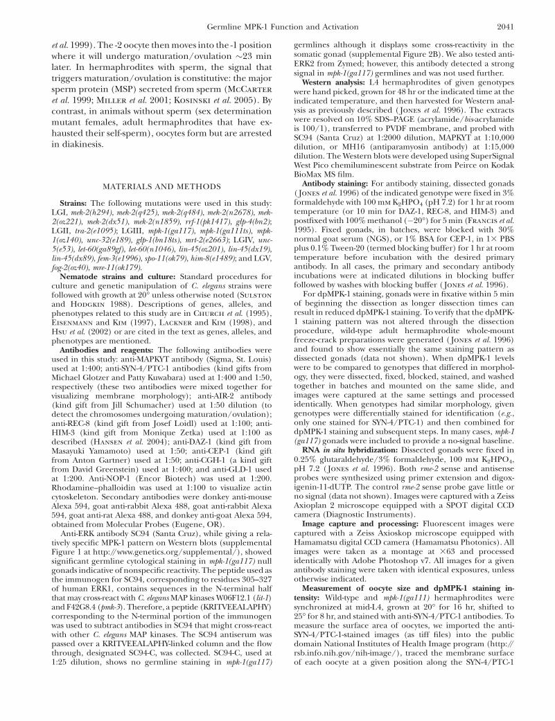
Figure 2.—Dynamic temporal/spatial acti-vation of MPK-1 during oogenesis. Animalsof the indicated genotype were grown at 20�,and the gonads were dissected and stainedfor activated dpMPK-1 (in red) and chromo-some morphology (DAPI in blue). In A–D, dis-tal is to the left and proximal is to the right;germline regions deduced from chromosomemorphology (e.g., pachytene) are indicatedwith the boundaries of the regions, the distaltip and proximal ends of the germline markedby vertical white lines; and panels with com-posite micrographs show a surface view distalto the loop and an interior view at the level ofoocyte nuclei proximal to the loop. Dashedred lines indicate elevated dpMPK-1 staining.(A) Wild-type (WT) hermaphrodite germline24 hr post mid-L4. High dpMPK-1 levels areobserved in proximal pachytene and in the-1 through -5 diakinesis oocytes with a lowbut detectable valley in the loop/diplotene.dpMPK-1 is not detected in the mitotic, tran-sition zone, or distal pachytene regions.(B) let-60(ga89gf) hermaphrodite germline24 hr post mid-L4 at 20�, stained, and photo-graphed together with A; images were pro-cessed identically. dpMPK-1 rises earlier inpachytene, remains elevated in the loop/diplotene, and is further elevated in diakinesisoocytes. dpMPK-1 falls abruptly as the -1 oocyteundergoes maturation in let-60(ga89gf) as itdoes in wild type (Figure 4C). (C) fog-2(oz40)female germline 8 hr after synchronizationat the L4/adult molt, stained, and photo-graphed together with A and B, but with a2.53 exposure time, followed by identical im-age processing. dpMPK-1 is observed in prox-imal pachytene as in wild type and a singlediakinesis oocyte, -6. Similar results were ob-tained with young fog-3(q443) females.(D) fog-2(oz40) female germline 20 hr postL4/adult molt containing .15 oocytes arrestedin diakinesis (distal-most region not shown).
Very low or undetectable dpMPK-1 is observed in proximal pachytene, diplotene, and diakinesis. Not shown is dpMPK-1 accumu-lation (1) in sheath cell nuclei, which is weak in wild type and elevated in let-60(ga89gf), and (2) in a subset of intestinal cells (nucleusand cytoplasm), which is low in wild type and elevated in let-60(ga89gf).
2044 M.-H. Lee et al.

morphology indicate that mpk-1 null germ cells arrestwith mid-pachytene characteristics, at approximatelythe same point at which dpMPK-1 levels would normallyrise. On the basis of these results we propose that MPK-1promotes pachytene progression, with the rise indpMPK-1 triggering a transition from a distal pachyteneto a proximal pachytene subtype (Figure 3B).
dpMPK-1 and pachytene progression in selectmutant backgrounds: To understand the relationshipbetween dpMPK-1 levels and pachytene progression, weexamined mutants in the let-60 RAS–mpk-1 ERK pathway,sex determination mutant females, and mutants inwhich initiation of meiotic recombination is blocked.The canonical let-60 RAS gf allele n1046, which has astrong effect on vulval development (Beitel et al. 1990;Han et al. 1990), is fertile at 15�, 20�, and 25� and has adpMPK-1 accumulation pattern that is indistinguishablefrom wild type (Table 3 legend). By contrast, the let-60 gfallele ga89, which has a relatively mild effect on vulvaldevelopment (Eisenmann and Kim 1997), causes tssterility and alters dpMPK-1 accumulation (Table 3;Figures 2B and 3C; supplemental Figure 4 at http://www.genetics.org/supplemental/). let-60(ga89gf) her-maphrodites are fertile at 20� but show earlier/moredistal MPK-1 activation. Comparison of wild-type andmutant hermaphrodites 24 hr post L4 at 20� reveals thatdpMPK-1 rises at 38 (63) cell diameters from the distaltip in let-60(ga89gf) compared to 45 (63) cell diametersin wild type. However, overall let-60(ga89gf) germlinesare 4 cell diameters shorter than wild type in distal–proximal length (72 6 3 vs. 76 6 2) and the pachyteneregion is 5 cell diameters shorter (32 6 2 vs. 37 6 2).Therefore, we used percentage of length throughpachytene where dpMPK-1 begins to rise as a way tonormalize for differences in cell diameter length ofpachytene and total germline. dpMPK-1 rises at �37%of the length of pachytene in let-60(ga89gf) compared to�46% of length in wild type. Although the rise indpMPK-1 occurs prematurely in let-60(ga89gf), the peaklevel of dpMPK-1 in pachytene, as judged by pixelintensity, is similar to wild type (supplemental Figure4). The premature rise in dpMPK-1 suggests that the
Figure 3.—MPK-1 is necessary for progression from distalto proximal pachytene. Wild-type, let-60(ga89gf), and mpk-1(ga117) hermaphrodites at 20� were dissected 24 hr postmid-L4 and stained for DAZ-1 (green), dpMPK-1 (red), andchromosome morphology (DAPI in blue). (A) In wild type,DAZ-1 is high in the mitotic and transition zone and fallsin mid-pachytene. (B) Representation of the cellular posi-tions for DAZ-1 (green) and dpMPK-1 (red) accumulationin cell diameters from the distal tip from visual inspectionof 13 wild-type germlines. Solid bars indicate high levels ofDAZ-1 or dpMPK-1, diagonal-line bars indicate rising or fall-ing levels, the open bar indicates lower dpMPK-1 accumula-tion at the end of pachytene and in diplotene, and thestippled bar indicates variable high dpMPK-1 accumulationin diakinesis oocytes. The fall of DAZ-1 is reciprocal tothe rise in dpMPK-1 accumulation, with a region of overlapof �7 cell diameters. Stages, separated by a vertical line, aredetermined from DAPI morphology while a diagonal line is
used to separate distal pachytene from proximal pachyteneas it is inferred from the rise in dpMPK-1 staining. (C) Rep-resentation of the cellular positions for DAZ-1 and dpMPK-1 accumulation from nine let-60(ga89gf) germlines (stainedgermline not shown). dpMPK-1 levels rise prematurely,DAZ-1 levels fall prematurely, and the pachytene regionis 5 cell diameters shorter than that of wild type, consistentwith the hypothesis that the rise in dpMPK-1 promotes atransition from distal to proximal pachytene. (D) In mpk-1(ga117) null germlines, pachytene-arrested germ cellsare found in the proximal two-thirds of the gonad and dis-play distal/mid-pachytene character based on a high DAZ-1level. dpMPK-1 is not detected. Due to massive disorganiza-tion, cellular positions were not determined.
Germline MPK-1 Function and Activation 2045
transition from distal to proximal pachytene occursearlier in let-60(ga89gf) germlines and leads to theprediction that DAZ-1 levels should fall prematurely inthe mutant. We found that DAZ-1 levels terminateearlier at 42 (62) cell diameters from the distal tip,corresponding to �51% of the length, in let-60(ga89gf)germlines compared to terminating at 52 (63) celldiameters or 67% of pachytene length in wild type(Figure 3, B and C). The results with let-60(ga89gf) areconsistent with the proposal that the rise in dpMPK-1
promotes the transition of germ cells from a distalpachytene subtype to a proximal pachytene subtype.The decrease in size of the pachytene region in let-60(ga89gf) may be a consequence of the premature risein dpMPK-1, assuming that the distal-to-proximal tran-sition is rate limiting.
In an attempt to further perturb pachytene pro-gression, we examined let-60(ga89gf) mutants shifted tothe restrictive temperature. After a 4-hr shift of let-60(ga89gf) adults to 25�, animals become sterile due to
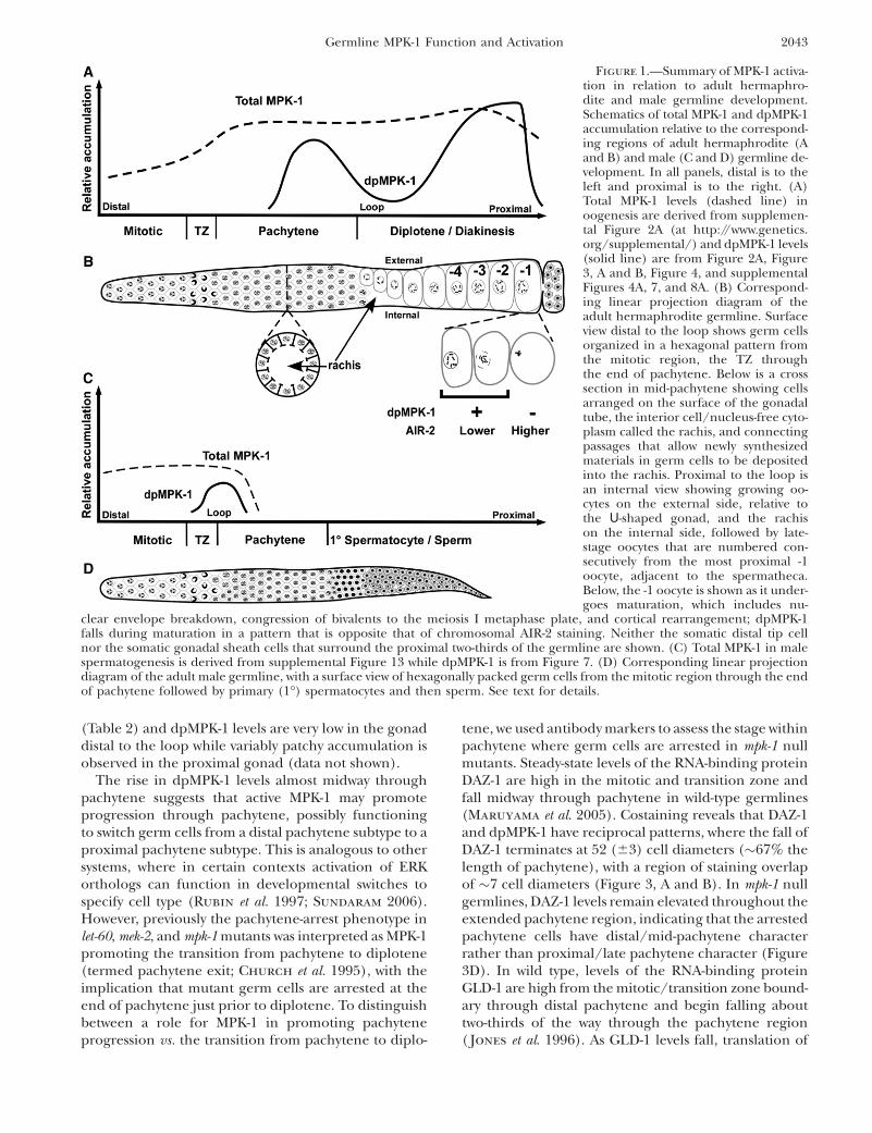
TABLE 2
Phenotypes following shift-up of mpk-1(ga111) to 25�
% hermaphrodite gonad arms with indicated phenotype
Pachytene cellular organization
GenotypeGaps in sheet
of cellsInternal
cells/nucleiPycnoticnucleia
Proximalpachytene
nuclei
Normaldiakinetic
nucleiDisorganized
oocytes EmoNormalsperm
Mitoticzone size n
Embryo shift-upWild type 0 0 0 0 100 0 0 100 18 6 2.1 22mpk-1(ga111) 97 41 84 91 19 9 16 97 18 6 2.3 32
L4 shift-upWild type 0 0 0 0 100 0 0 100 19 6 1.8 22mpk-1(ga111) 94 6 0 66 66 16 41 97 19 6 2.6 32
The temperature-sensitive mpk-1(ga111) is described in Lackner and Kim (1998). Animals were collected for gonad dissection24 hr post L4 at 25�.
a Pycnotic nuclei can contain HIM-3 aggregates.
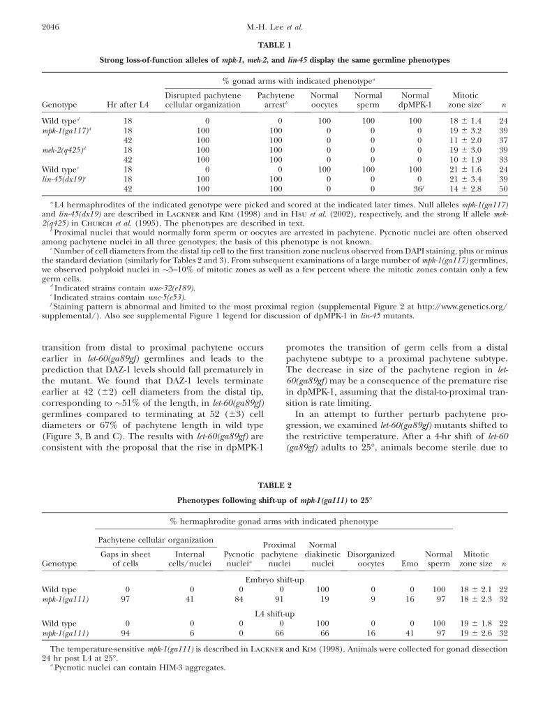
TABLE 1
Strong loss-of-function alleles of mpk-1, mek-2, and lin-45 display the same germline phenotypes
% gonad arms with indicated phenotypea
Genotype Hr after L4Disrupted pachytenecellular organization
Pachytenearrestb
Normaloocytes
Normalsperm
NormaldpMPK-1
Mitoticzone sizec n
Wild typed 18 0 0 100 100 100 18 6 1.4 24mpk-1(ga117)d 18 100 100 0 0 0 19 6 3.2 39
42 100 100 0 0 0 11 6 2.0 37mek-2(q425)d 18 100 100 0 0 0 19 6 3.0 39
42 100 100 0 0 0 10 6 1.9 33Wild typee 18 0 0 100 100 100 21 6 1.6 24lin-45(dx19)e 18 100 100 0 0 0 21 6 3.4 39
42 100 100 0 0 36f 14 6 2.8 50
a L4 hermaphrodites of the indicated genotype were picked and scored at the indicated later times. Null alleles mpk-1(ga117)and lin-45(dx19) are described in Lackner and Kim (1998) and in Hsu et al. (2002), respectively, and the strong lf allele mek-2(q425) in Church et al. (1995). The phenotypes are described in text.
b Proximal nuclei that would normally form sperm or oocytes are arrested in pachytene. Pycnotic nuclei are often observedamong pachytene nuclei in all three genotypes; the basis of this phenotype is not known.
c Number of cell diameters from the distal tip cell to the first transition zone nucleus observed from DAPI staining, plus or minusthe standard deviation (similarly for Tables 2 and 3). From subsequent examinations of a large number of mpk-1(ga117) germlines,we observed polyploid nuclei in �5–10% of mitotic zones as well as a few percent where the mitotic zones contain only a fewgerm cells.
d Indicated strains contain unc-32(e189).e Indicated strains contain unc-5(e53).f Staining pattern is abnormal and limited to the most proximal region (supplemental Figure 2 at http://www.genetics.org/
supplemental/). Also see supplemental Figure 1 legend for discussion of dpMPK-1 in lin-45 mutants.
2046 M.-H. Lee et al.
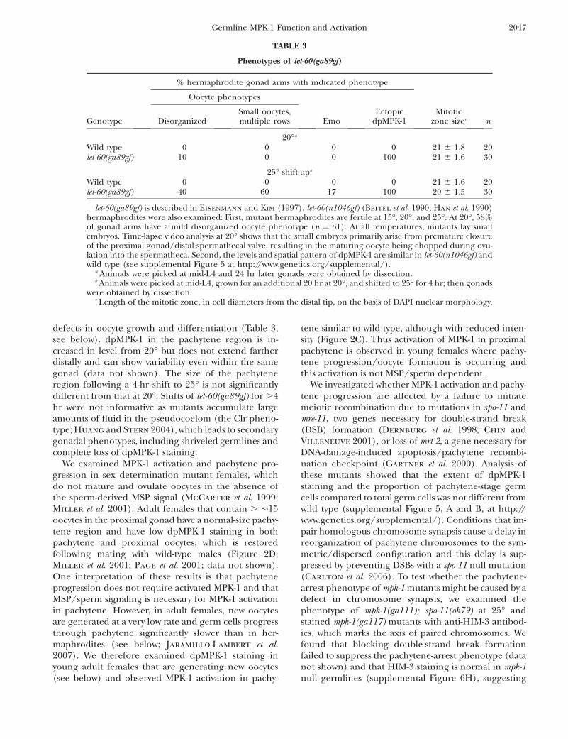
defects in oocyte growth and differentiation (Table 3,see below). dpMPK-1 in the pachytene region is in-creased in level from 20� but does not extend fartherdistally and can show variability even within the samegonad (data not shown). The size of the pachyteneregion following a 4-hr shift to 25� is not significantlydifferent from that at 20�. Shifts of let-60(ga89gf) for .4hr were not informative as mutants accumulate largeamounts of fluid in the pseudocoelom (the Clr pheno-type; Huang and Stern 2004), which leads to secondarygonadal phenotypes, including shriveled germlines andcomplete loss of dpMPK-1 staining.
We examined MPK-1 activation and pachytene pro-gression in sex determination mutant females, whichdo not mature and ovulate oocytes in the absence ofthe sperm-derived MSP signal (McCarter et al. 1999;Miller et al. 2001). Adult females that contain . �15oocytes in the proximal gonad have a normal-size pachy-tene region and have low dpMPK-1 staining in bothpachytene and proximal oocytes, which is restoredfollowing mating with wild-type males (Figure 2D;Miller et al. 2001; Page et al. 2001; data not shown).One interpretation of these results is that pachyteneprogression does not require activated MPK-1 and thatMSP/sperm signaling is necessary for MPK-1 activationin pachytene. However, in adult females, new oocytesare generated at a very low rate and germ cells progressthrough pachytene significantly slower than in her-maphrodites (see below; Jaramillo-Lambert et al.2007). We therefore examined dpMPK-1 staining inyoung adult females that are generating new oocytes(see below) and observed MPK-1 activation in pachy-
tene similar to wild type, although with reduced inten-sity (Figure 2C). Thus activation of MPK-1 in proximalpachytene is observed in young females where pachy-tene progression/oocyte formation is occurring andthis activation is not MSP/sperm dependent.
We investigated whether MPK-1 activation and pachy-tene progression are affected by a failure to initiatemeiotic recombination due to mutations in spo-11 andmre-11, two genes necessary for double-strand break(DSB) formation (Dernburg et al. 1998; Chin andVilleneuve 2001), or loss of mrt-2, a gene necessary forDNA-damage-induced apoptosis/pachytene recombi-nation checkpoint (Gartner et al. 2000). Analysis ofthese mutants showed that the extent of dpMPK-1staining and the proportion of pachytene-stage germcells compared to total germ cells was not different fromwild type (supplemental Figure 5, A and B, at http://www.genetics.org/supplemental/). Conditions that im-pair homologous chromosome synapsis cause a delay inreorganization of pachytene chromosomes to the sym-metric/dispersed configuration and this delay is sup-pressed by preventing DSBs with a spo-11 null mutation(Carlton et al. 2006). To test whether the pachytene-arrest phenotype of mpk-1 mutants might be caused by adefect in chromosome synapsis, we examined thephenotype of mpk-1(ga111); spo-11(ok79) at 25� andstained mpk-1(ga117) mutants with anti-HIM-3 antibod-ies, which marks the axis of paired chromosomes. Wefound that blocking double-strand break formationfailed to suppress the pachytene-arrest phenotype (datanot shown) and that HIM-3 staining is normal in mpk-1null germlines (supplemental Figure 6H), suggesting
TABLE 3
Phenotypes of let-60(ga89gf)
% hermaphrodite gonad arms with indicated phenotype
Oocyte phenotypes
Genotype DisorganizedSmall oocytes,multiple rows Emo
EctopicdpMPK-1
Mitoticzone sizec n
20�a
Wild type 0 0 0 0 21 6 1.8 20let-60(ga89gf) 10 0 0 100 21 6 1.6 30
25� shift-upb
Wild type 0 0 0 0 21 6 1.6 20let-60(ga89gf) 40 60 17 100 20 6 1.5 30
let-60(ga89gf) is described in Eisenmann and Kim (1997). let-60(n1046gf) (Beitel et al. 1990; Han et al. 1990)hermaphrodites were also examined: First, mutant hermaphrodites are fertile at 15�, 20�, and 25�. At 20�, 58%of gonad arms have a mild disorganized oocyte phenotype (n ¼ 31). At all temperatures, mutants lay smallembryos. Time-lapse video analysis at 20� shows that the small embryos primarily arise from premature closureof the proximal gonad/distal spermathecal valve, resulting in the maturing oocyte being chopped during ovu-lation into the spermatheca. Second, the levels and spatial pattern of dpMPK-1 are similar in let-60(n1046gf) andwild type (see supplemental Figure 5 at http://www.genetics.org/supplemental/).
a Animals were picked at mid-L4 and 24 hr later gonads were obtained by dissection.b Animals were picked at mid-L4, grown for an additional 20 hr at 20�, and shifted to 25� for 4 hr; then gonads
were obtained by dissection.c Length of the mitotic zone, in cell diameters from the distal tip, on the basis of DAPI nuclear morphology.
Germline MPK-1 Function and Activation 2047
that the pachytene-arrest phenotype is unlikely to becaused by a defect in meiotic chromosome synapsis.
MPK-1 is necessary for pachytene cell organization:Previous work reported that pachytene-arrested nucleiin mpk-1, mek-2, ksr-2, and let-60 lf mutants are present inclumps on the basis of Nomarski microscopy and DAPIstaining (Church et al. 1995; Ohmachi et al. 2002). Tounderstand the basis of this phenotype, we examinedthe organization of the germline using antibodies toSyntaxin 4 (SYN-4) and Patched-1 (PTC-1) to visualizecell membranes ( Jantsch-Plunger and Glotzer 1999;Kuwabara et al. 2000) and rhodamine–phalloidin(R-ph) to visualize actin filament organization (Strome
1986). In wild type, these cytological reagents show thatfrom the mitotic region through the end of pachytene,germ cells are arranged in a honeycomb pattern on thesurface of the cylindrical gonadal tube, with an interiornucleus/germ-cell-free rachis (Figure 1, B and D; sup-plemental Figure 6, A and E, at http://www.genetics.org/supplemental/; Hirsh et al. 1976; Hall et al. 1999;Maddox et al. 2005). By contrast, SYN-4 and PTC-1staining in mpk-1 and lin-45 null and mek-2 strong lfmutant hermaphrodite germlines reveals a completeloss of the honeycomb organization of surface pachy-tene cells and the interior rachis; the gonadal tube isshrunken and distorted, the majority of the surface ofthe germline is devoid of germ cells, and instead nucleiand associated membranes are found in disorganizedclumps (Table 1; supplemental Figure 6, B–D). Largeregions that are devoid of nuclei and organized mem-branes are apparently cytoplasm as they contain cyto-plasmic DAZ-1 and GLD-1 that are relatively uniformlydistributed (Figure 3D; supplemental Figure 3A). Inwild type, cortical actin is organized in a honeycombpattern that mirrors membranes (Strome 1986), whilein the rachis there is a fine filament network (Wolke
et al. 2007). In strong mpk-1, mek-2, and lin-45 mutanthermaphrodite germlines, the pachytene actin organi-zation is completely disrupted, with areas lackingorganized filaments and cytoplasmic regions that con-tain macroscopic aggregates of actin (supplementalFigure 6F). In these strong mutants, the cellulardisorganization progresses distally in older animalsand can extend into distal pachytene and the transitionzone, regions where dpMPK-1 is not detected. It isunclear whether the disorganization of transition zoneand distal pachytene germ cells is due to a requirementof MPK-1 function in these regions or whether it is asecondary consequence of massive disruption of themore proximal germline that spreads distally. Underconditions of partial mpk-1 lf, either shift-up of ga111 to25� or following mpk-1 RNAi (see below), disruptionsof pachytene germ cell organization are less severe,including gaps in the honeycomb pattern and the pres-ence of internal nuclei in the rachis (Table 2; supple-mental Table 1). Together, these results indicate thatMPK-1 is necessary for the organization of the pachy-
tene region into germ cells on the surface of thegonadal tube and an interior cytoplasmic rachis.
Dynamic dpMPK-1 accumulation in the proximalgermline: As germ cells progress from proximal pachy-tene to growing diplotene oocytes in the loop region,dpMPK-1 falls to low but detectable levels (Figures 1A,2A, and 3, A and B). dpMPK-1 then rises in late-stagediakinesis oocytes, with the most proximal approxi-mately five oocytes (-1 to -5) staining strongly. However,the levels are variable from oocyte to oocyte and fromgonad to gonad, with oocytes at the same positionshowing large differences in dpMPK-1 level. Thisvariability in proximal oocyte dpMPK-1 level is illus-trated qualitatively in Figure 4A, based on a large sampleof gonads scored (supplemental Figure 7 at http://www.genetics.org/supplemental/), and quantitativelyfrom determination of oocyte pixel intensity (supple-mental Figure 8). Although there is not a clear patternof dpMPK-1 level relative to oocyte position fromexamination of fixed specimens, it is possible that thedynamic pattern reflects some yet-to-be-defined stage/process of oocyte development. The exception is the -1oocyte where dpMPK-1 levels are correlated with thestage of meiotic maturation. The -1 oocyte in hermaph-rodites spends �23 min in the most proximal positionand in the final 3–6 min will undergo the meioticmaturation events of nuclear envelope breakdown(NEBD) and cortical rearrangement (McCarter et al.1999; Figure 6). During this period, the initially dis-persed diakinesis bivalents of the -1 oocyte congress tothe metaphase plate, chromosomal AIR-2 aurora kinasestaining increases from low to high and metaphase I(MI) spindle formation begins (Figure 4, B and C;Schumacher et al. 1998; Rogers et al. 2002; Yang et al.2003). We found that in �15% of -1 oocytes dpMPK-1 isvery low/undetectable (Figure 4, A and C). Comparing-1 oocyte stage with dpMPK-1 level, we found that 100%of early -1 oocytes (dispersed diakinesis bivalents, lowchromosomal AIR-2 staining) have high dpMPK-1 while.85% of late -1 oocytes (congressed bivalents, highchromosomal AIR-2 staining) have very low dpMPK-1(Figure 4, B and C; supplemental Table 2). This isconsistent with observations of Page et al. (2001) whoreported that, by completion of MI, dpMPK-1 is notdetectable. Thus, as the -1 oocyte undergoes matura-tion, there is a rapid and dramatic fall in dpMPK-1 level.
Examination of dpMPK-1 levels in let-60(ga89gf) at 20�as germ cells progress from proximal pachytene togrowing diplotene oocytes reveals that levels remainelevated rather than decrease in the loop region asis observed in wild type (Figure 1A; Figure 2, A andB; supplemental Figure 8 at http://www.genetics.org/supplemental/). In the most proximal, approximatelyeight diakinesis oocytes, dpMPK-1 levels are furtherelevated relative to wild type, as shown quantitativelyfrom pixel intensity per oocyte (supplemental Figure 8).Notwithstanding the elevated dpMPK-1 levels in proxi-
2048 M.-H. Lee et al.
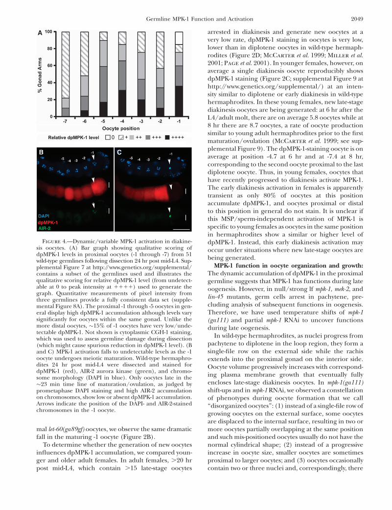
mal let-60(ga89gf) oocytes, we observe the same dramaticfall in the maturing -1 oocyte (Figure 2B).
To determine whether the generation of new oocytesinfluences dpMPK-1 accumulation, we compared youn-ger and older adult females. In adult females, .20 hrpost mid-L4, which contain .15 late-stage oocytes
arrested in diakinesis and generate new oocytes at avery low rate, dpMPK-1 staining in oocytes is very low,lower than in diplotene oocytes in wild-type hermaph-rodites (Figure 2D; McCarter et al. 1999; Miller et al.2001; Page et al. 2001). In younger females, however, onaverage a single diakinesis oocyte reproducibly showsdpMPK-1 staining (Figure 2C; supplemental Figure 9 athttp://www.genetics.org/supplemental/) at an inten-sity similar to diplotene or early diakinesis in wild-typehermaphrodites. In these young females, new late-stagediakinesis oocytes are being generated: at 6 hr after theL4/adult molt, there are on average 5.8 oocytes while at8 hr there are 8.7 oocytes, a rate of oocyte productionsimilar to young adult hermaphrodites prior to the firstmaturation/ovulation (McCarter et al. 1999; see sup-plemental Figure 9). The dpMPK-1-staining oocyte is onaverage at position -4.7 at 6 hr and at -7.4 at 8 hr,corresponding to the second oocyte proximal to the lastdiplotene oocyte. Thus, in young females, oocytes thathave recently progressed to diakinesis activate MPK-1.The early diakinesis activation in females is apparentlytransient as only 80% of oocytes at this positionaccumulate dpMPK-1, and oocytes proximal or distalto this position in general do not stain. It is unclear ifthis MSP/sperm-independent activation of MPK-1 isspecific to young females as oocytes in the same positionin hermaphrodites show a similar or higher level ofdpMPK-1. Instead, this early diakinesis activation mayoccur under situations where new late-stage oocytes arebeing generated.
MPK-1 function in oocyte organization and growth:The dynamic accumulation of dpMPK-1 in the proximalgermline suggests that MPK-1 has functions during lateoogenesis. However, in null/strong lf mpk-1, mek-2, andlin-45 mutants, germ cells arrest in pachytene, pre-cluding analysis of subsequent functions in oogenesis.Therefore, we have used temperature shifts of mpk-1(ga111) and partial mpk-1 RNAi to uncover functionsduring late oogenesis.
In wild-type hermaphrodites, as nuclei progress frompachytene to diplotene in the loop region, they form asingle-file row on the external side while the rachisextends into the proximal gonad on the interior side.Oocyte volume progressively increases with correspond-ing plasma membrane growth that eventually fullyencloses late-stage diakinesis oocytes. In mpk-1(ga111)shift-ups and in mpk-1 RNAi, we observed a constellationof phenotypes during oocyte formation that we call‘‘disorganized oocytes’’: (1) instead of a single-file row ofgrowing oocytes on the external surface, some oocytesare displaced to the internal surface, resulting in two ormore oocytes partially overlapping at the same positionand such mis-positioned oocytes usually do not have thenormal cylindrical shape; (2) instead of a progressiveincrease in oocyte size, smaller oocytes are sometimesproximal to larger oocytes; and (3) oocytes occasionallycontain two or three nuclei and, correspondingly, there
Figure 4.—Dynamic/variable MPK-1 activation in diakine-sis oocytes. (A) Bar graph showing qualitative scoring ofdpMPK-1 levels in proximal oocytes (-1 through -7) from 51wild-type germlines following dissection 24 hr post mid-L4. Sup-plemental Figure 7 at http://www.genetics.org/supplemental/contains a subset of the germlines used and illustrates thequalitative scoring for relative dpMPK-1 level (from undetect-able at 0 to peak intensity at 1111) used to generate thegraph. Quantitative measurements of pixel intensity fromthree germlines provide a fully consistent data set (supple-mental Figure 8A). The proximal -1 through -5 oocytes in gen-eral display high dpMPK-1 accumulation although levels varysignificantly for oocytes within the same gonad. Unlike themore distal oocytes, �15% of -1 oocytes have very low/unde-tectable dpMPK-1. Not shown is cytoplasmic CGH-1 staining,which was used to assess germline damage during dissection(which might cause spurious reduction in dpMPK-1 level). (Band C) MPK-1 activation falls to undetectable levels as the -1oocyte undergoes meiotic maturation. Wild-type hermaphro-dites 24 hr post mid-L4 were dissected and stained fordpMPK-1 (red), AIR-2 aurora kinase (green), and chromo-some morphology (DAPI in blue). Only oocytes late in the�23 min time line of maturation/ovulation, as judged byprometaphase DAPI staining and high AIR-2 accumulationon chromosomes, show low or absent dpMPK-1 accumulation.Arrows indicate the position of the DAPI- and AIR-2-stainedchromosomes in the -1 oocyte.
Germline MPK-1 Function and Activation 2049
are small membrane-enclosed regions that lack nuclei,often on the interior side (Table 2; supplemental Table1 at http://www.genetics.org/supplemental/; data notshown). As the formation of the single-file assembly lineof oocytes occurs after pachytene, we wondered if thedisorganized oocyte phenotype in mpk-1(ga111) andmpk-1 RNAi germlines could be a secondary conse-quence of prior disruption of the organization ofpachytene germ cells. This seems not to be the case aswe find germlines with the disorganized oocyte pheno-type while pachytene cell organization and pachyteneprogression are unaffected. Thus mpk-1 function inoocyte organization and differentiation appears to bedistinct from the distal germline functions in pachytenecellular organization and pachytene progression.
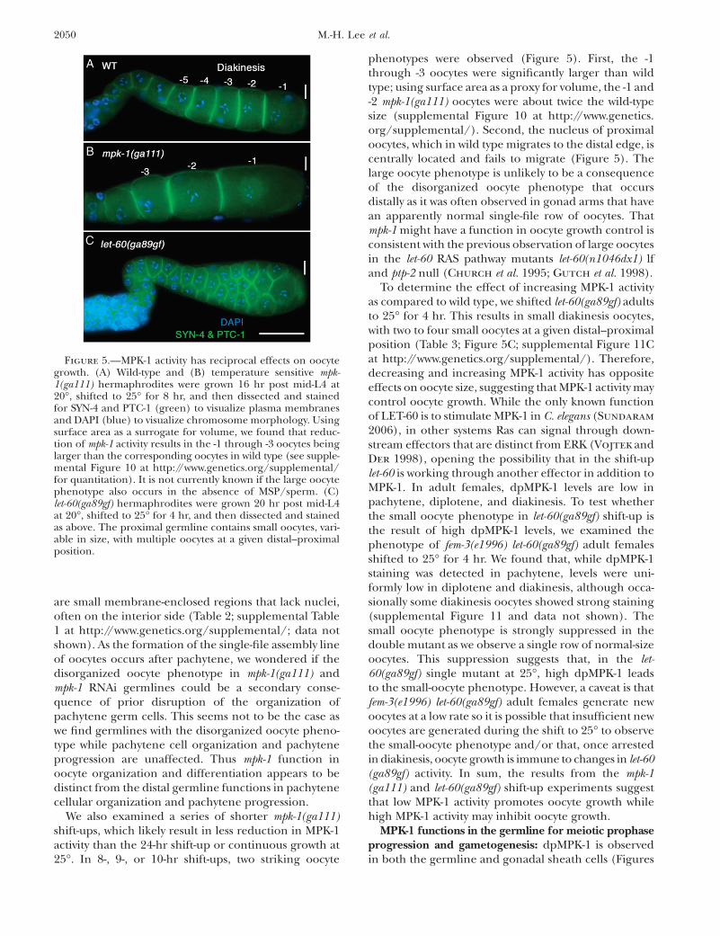
We also examined a series of shorter mpk-1(ga111)shift-ups, which likely result in less reduction in MPK-1activity than the 24-hr shift-up or continuous growth at25�. In 8-, 9-, or 10-hr shift-ups, two striking oocyte
phenotypes were observed (Figure 5). First, the -1through -3 oocytes were significantly larger than wildtype; using surface area as a proxy for volume, the -1 and-2 mpk-1(ga111) oocytes were about twice the wild-typesize (supplemental Figure 10 at http://www.genetics.org/supplemental/). Second, the nucleus of proximaloocytes, which in wild type migrates to the distal edge, iscentrally located and fails to migrate (Figure 5). Thelarge oocyte phenotype is unlikely to be a consequenceof the disorganized oocyte phenotype that occursdistally as it was often observed in gonad arms that havean apparently normal single-file row of oocytes. Thatmpk-1 might have a function in oocyte growth control isconsistent with the previous observation of large oocytesin the let-60 RAS pathway mutants let-60(n1046dx1) lfand ptp-2 null (Church et al. 1995; Gutch et al. 1998).
To determine the effect of increasing MPK-1 activityas compared to wild type, we shifted let-60(ga89gf) adultsto 25� for 4 hr. This results in small diakinesis oocytes,with two to four small oocytes at a given distal–proximalposition (Table 3; Figure 5C; supplemental Figure 11Cat http://www.genetics.org/supplemental/). Therefore,decreasing and increasing MPK-1 activity has oppositeeffects on oocyte size, suggesting that MPK-1 activity maycontrol oocyte growth. While the only known functionof LET-60 is to stimulate MPK-1 in C. elegans (Sundaram
2006), in other systems Ras can signal through down-stream effectors that are distinct from ERK (Vojtek andDer 1998), opening the possibility that in the shift-uplet-60 is working through another effector in addition toMPK-1. In adult females, dpMPK-1 levels are low inpachytene, diplotene, and diakinesis. To test whetherthe small oocyte phenotype in let-60(ga89gf) shift-up isthe result of high dpMPK-1 levels, we examined thephenotype of fem-3(e1996) let-60(ga89gf) adult femalesshifted to 25� for 4 hr. We found that, while dpMPK-1staining was detected in pachytene, levels were uni-formly low in diplotene and diakinesis, although occa-sionally some diakinesis oocytes showed strong staining(supplemental Figure 11 and data not shown). Thesmall oocyte phenotype is strongly suppressed in thedouble mutant as we observe a single row of normal-sizeoocytes. This suppression suggests that, in the let-60(ga89gf) single mutant at 25�, high dpMPK-1 leadsto the small-oocyte phenotype. However, a caveat is thatfem-3(e1996) let-60(ga89gf) adult females generate newoocytes at a low rate so it is possible that insufficient newoocytes are generated during the shift to 25� to observethe small-oocyte phenotype and/or that, once arrestedin diakinesis, oocyte growth is immune to changes in let-60(ga89gf) activity. In sum, the results from the mpk-1(ga111) and let-60(ga89gf) shift-up experiments suggestthat low MPK-1 activity promotes oocyte growth whilehigh MPK-1 activity may inhibit oocyte growth.
MPK-1 functions in the germline for meiotic prophaseprogression and gametogenesis: dpMPK-1 is observedin both the germline and gonadal sheath cells (Figures
Figure 5.—MPK-1 activity has reciprocal effects on oocytegrowth. (A) Wild-type and (B) temperature sensitive mpk-1(ga111) hermaphrodites were grown 16 hr post mid-L4 at20�, shifted to 25� for 8 hr, and then dissected and stainedfor SYN-4 and PTC-1 (green) to visualize plasma membranesand DAPI (blue) to visualize chromosome morphology. Usingsurface area as a surrogate for volume, we found that reduc-tion of mpk-1 activity results in the -1 through -3 oocytes beinglarger than the corresponding oocytes in wild type (see supple-mental Figure 10 at http://www.genetics.org/supplemental/for quantitation). It is not currently known if the large oocytephenotype also occurs in the absence of MSP/sperm. (C)let-60(ga89gf) hermaphrodites were grown 20 hr post mid-L4at 20�, shifted to 25� for 4 hr, and then dissected and stainedas above. The proximal germline contains small oocytes, vari-able in size, with multiple oocytes at a given distal–proximalposition.
2050 M.-H. Lee et al.
1A and 2A). Accordingly, MPK-1 might act in thegermline, the soma, or both to control pachytene pro-gression, pachytene cell organization, oocyte organiza-tion and differentiation, oocyte growth, and oocytenuclear migration. Previously, mosaic analysis with astrong lf mpk-1 mutant indicated that MPK-1 function inthe germline is necessary for pachytene progression andpachytene cell organization (Church et al. 1995). rrf-1encodes an RNA-dependent RNA polymerase that isimportant for the RNAi response in somatic cells but notin germ cells (Sijen et al. 2001). To distinguish betweengermline and somatic function, we compared mpk-1RNAi in wild type with RNAi in the rrf-1 null back-ground. Pachytene arrest, pachytene cellular disorga-nization, disorganized oocytes, large oocytes, anddefective oocyte nuclear migration were phenotypesobserved following RNAi in both rrf-1 null and in wildtype (supplemental Table 1 at http://www.genetics.org/supplemental/). This indicates that mpk-1 function inthe germline is necessary for control of pachytene pro-gression, pachytene cell organization, oocyte organizationand differentiation, oocyte growth, and oocyte nuclearmigration (summarized in the discussion and Table 4).
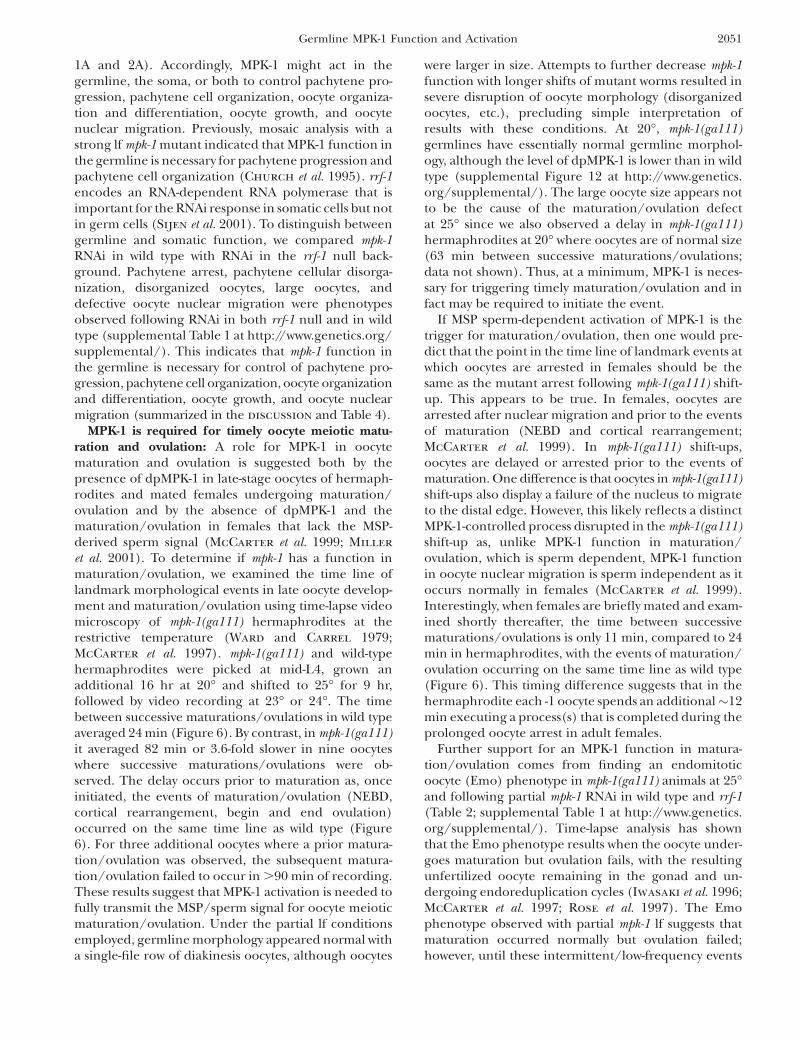
MPK-1 is required for timely oocyte meiotic matu-ration and ovulation: A role for MPK-1 in oocytematuration and ovulation is suggested both by thepresence of dpMPK-1 in late-stage oocytes of hermaph-rodites and mated females undergoing maturation/ovulation and by the absence of dpMPK-1 and thematuration/ovulation in females that lack the MSP-derived sperm signal (McCarter et al. 1999; Miller
et al. 2001). To determine if mpk-1 has a function inmaturation/ovulation, we examined the time line oflandmark morphological events in late oocyte develop-ment and maturation/ovulation using time-lapse videomicroscopy of mpk-1(ga111) hermaphrodites at therestrictive temperature (Ward and Carrel 1979;McCarter et al. 1997). mpk-1(ga111) and wild-typehermaphrodites were picked at mid-L4, grown anadditional 16 hr at 20� and shifted to 25� for 9 hr,followed by video recording at 23� or 24�. The timebetween successive maturations/ovulations in wild typeaveraged 24 min (Figure 6). By contrast, in mpk-1(ga111)it averaged 82 min or 3.6-fold slower in nine oocyteswhere successive maturations/ovulations were ob-served. The delay occurs prior to maturation as, onceinitiated, the events of maturation/ovulation (NEBD,cortical rearrangement, begin and end ovulation)occurred on the same time line as wild type (Figure6). For three additional oocytes where a prior matura-tion/ovulation was observed, the subsequent matura-tion/ovulation failed to occur in .90 min of recording.These results suggest that MPK-1 activation is needed tofully transmit the MSP/sperm signal for oocyte meioticmaturation/ovulation. Under the partial lf conditionsemployed, germline morphology appeared normal witha single-file row of diakinesis oocytes, although oocytes
were larger in size. Attempts to further decrease mpk-1function with longer shifts of mutant worms resulted insevere disruption of oocyte morphology (disorganizedoocytes, etc.), precluding simple interpretation ofresults with these conditions. At 20�, mpk-1(ga111)germlines have essentially normal germline morphol-ogy, although the level of dpMPK-1 is lower than in wildtype (supplemental Figure 12 at http://www.genetics.org/supplemental/). The large oocyte size appears notto be the cause of the maturation/ovulation defectat 25� since we also observed a delay in mpk-1(ga111)hermaphrodites at 20� where oocytes are of normal size(63 min between successive maturations/ovulations;data not shown). Thus, at a minimum, MPK-1 is neces-sary for triggering timely maturation/ovulation and infact may be required to initiate the event.
If MSP sperm-dependent activation of MPK-1 is thetrigger for maturation/ovulation, then one would pre-dict that the point in the time line of landmark events atwhich oocytes are arrested in females should be thesame as the mutant arrest following mpk-1(ga111) shift-up. This appears to be true. In females, oocytes arearrested after nuclear migration and prior to the eventsof maturation (NEBD and cortical rearrangement;McCarter et al. 1999). In mpk-1(ga111) shift-ups,oocytes are delayed or arrested prior to the events ofmaturation. One difference is that oocytes in mpk-1(ga111)shift-ups also display a failure of the nucleus to migrateto the distal edge. However, this likely reflects a distinctMPK-1-controlled process disrupted in the mpk-1(ga111)shift-up as, unlike MPK-1 function in maturation/ovulation, which is sperm dependent, MPK-1 functionin oocyte nuclear migration is sperm independent as itoccurs normally in females (McCarter et al. 1999).Interestingly, when females are briefly mated and exam-ined shortly thereafter, the time between successivematurations/ovulations is only 11 min, compared to 24min in hermaphrodites, with the events of maturation/ovulation occurring on the same time line as wild type(Figure 6). This timing difference suggests that in thehermaphrodite each -1 oocyte spends an additional�12min executing a process(s) that is completed during theprolonged oocyte arrest in adult females.
Further support for an MPK-1 function in matura-tion/ovulation comes from finding an endomitoticoocyte (Emo) phenotype in mpk-1(ga111) animals at 25�and following partial mpk-1 RNAi in wild type and rrf-1(Table 2; supplemental Table 1 at http://www.genetics.org/supplemental/). Time-lapse analysis has shownthat the Emo phenotype results when the oocyte under-goes maturation but ovulation fails, with the resultingunfertilized oocyte remaining in the gonad and un-dergoing endoreduplication cycles (Iwasaki et al. 1996;McCarter et al. 1997; Rose et al. 1997). The Emophenotype observed with partial mpk-1 lf suggests thatmaturation occurred normally but ovulation failed;however, until these intermittent/low-frequency events
Germline MPK-1 Function and Activation 2051

are examined by time-lapse microscopy, this interpreta-tion is uncertain. Interestingly, an Emo phenotype isalso observed in let-60(ga89gf) hermaphrodites shiftedto 25� (Table 3); this presumably represents disruptionof maturation/ovulation at a step separate from thatobserved in mpk-1 lf.
MPK-1 ERK signaling is necessary for the male germcell fate: In wild-type adult males, total MPK-1 is foundin the mitotic region, the transition zone, and earlypachytene but is no longer detectable by mid-pachytene(Figure 1C, supplemental Figure 13). dpMPK-1 is re-stricted to the proximal half of the transition zone andvery early pachytene in an �13-cell-diameter distal–proximal region (Figure 1C; Figure 7A). mpk-1 null XOadult males display phenotypes very similar to nullmutant hermaphrodites: nuclei are arrested in pachy-tene and the honeycomb pattern of germ cells on thesurface of the cylindrical gonadal tube is disrupted, withregions of cytoplasm devoid of nuclei and cell mem-
branes and little or no evidence of gametogenesis(Figure 7C; Church et al. 1995; data not shown). Thesephenotypes suggest that MPK-1 functions to promotepachytene progression and pachytene cellular organi-zation during spermatogenesis as it does in oogenesis.However, the MPK-1 expression pattern seems inconsis-tent with this possibility; dpMPK-1 accumulation inproximal pachytene in the hermaphrodite is likely tomediate pachytene progression and pachytene cellularorganization during oogenesis, but during spermato-genesis dpMPK-1 is limited to the transition zone andvery early distal pachytene and total MPK-1 is notdetected in proximal pachytene.
An understanding of this discrepancy came fromanalysis of hermaphrodites with partial lf for lin-45,mek-2, and mpk-1. In animals that did not displaypachytene arrest or pachytene cellular disorganization,a feminization of the germline phenotype was observedin which germ cells that would normally form sperm
Figure 6.—MPK-1 is required for timely oocytematuration/ovulation. The timing of landmarkevents of maturation/ovulation in the -1 oocytewas determined by Nomarski time-lapse video mi-croscopy (see text and McCarter et al. 1999).(A) Tabulation of the average timing of sevenevents (left column) for wild type (n ¼ 14),mpk-1(ga111) (n¼ 12), and mated fem-3(e1996) fe-males (n¼ 8) at 25�. ‘‘End Ovulation’’ (event 5) isdefined as 0 min, earlier events (e.g., event 1,‘‘End Nuclear Migration’’) are given a negativetime, and the interval between successive matura-tions ovulation is designated ‘‘Previous End Ovu-lation’’ (event 7). B–D are time lines for wild-type,mpk-1(ga111) and mated fem-3(e1996) females,respectively, derived from A, with vertical arrowsshowing the relative timing of each event. (B) Forwild type, nuclear migration and the events ofmaturation/ovulation (arrows 2–5) occur aspreviously reported, with the interval between‘‘Previous End Ovulation’’ (arrow 7) and ‘‘EndOvulation’’ (arrow 5) �24 min. (C) For mpk-1(ga111) (n ¼ 9), there is a long delay between‘‘End Previous Ovulation’’ and the events of mat-uration/ovulation. This delay is prior to theevents of maturation/ovulation as, once initiated,they occur on the same schedule as wild type. Thedelay results in an �83-min interval between suc-cessive maturations/ovulations, 3.6-fold slowerthan wild type. Nuclear migration was not ob-served. For three animals where a maturation/ovulation was observed at the beginning of the re-cording, a subsequent maturation/ovulation wasnot observed for .90 min. (D) Adult females,with oocytes arrested in diakinesis, were brieflymated with CB4855 (‘‘Mr. Vigorous’’) males(Hodgkin and Doniach 1997) and then re-corded. Soon after mating, the interval betweensuccessive maturations/ovulations was reduced�2-fold from that of wild type. The interval short-ening occurs prior to the events of maturation/ovulation as, once initiated, they occur on thesame schedule as wild type.
2052 M.-H. Lee et al.

instead form oocytes (supplemental Table 3 at http://www.genetics.org/supplemental/). On the basis of thispartial lf phenotype, we speculated that, in mpk-1 nullmales, the germline is fully feminized and thus woulddisplay the pachytene-arrest and pachytene cellulardisorganization phenotypes observed in the absenceof MPK-1 activity during oogenesis. We used two mark-ers to test whether pachytene-arrested germ cells inmales were feminized: rme-2 mRNA and GLD-1 protein,which are present at high levels in pachytene of oo-genesis but not of spermatogenesis ( Jones et al. 1996;Grant and Hirsh 1999; Lee and Schedl 2001). Con-sistent with a feminized phenotype, in situ hybridizationshows that rme-2 mRNA is expressed at high levelsthroughout the mpk-1 null pachytene-arrested regioncompared with the near absence in control males(Figure 7, B–E). Hermaphrodites with strong lf muta-tions in lin-45, mek-2, and mpk-1 also fail to make sperm(Table 1). The absence of sperm in these mutanthermaphrodites is also due to feminization. Proximalgerm cells in mid-L4 gonads that would normally formsperm show high levels of rme-2 mRNA in mpk-1 nullgonads in contrast to wild-type hermaphrodite gonads(supplemental Figure 14). We conclude that lin-45,mek-2, and mpk-1 activities are necessary for the malegerm cell fate in both hermaphrodites and males.
MPK-1 might act early to specify the sexual fate of allgerm cells or might act continuously as germ cells transitfrom proliferation to meiotic development in larvae andadults. We used temperature shifts with mpk-1(ga111) toask if there is an ongoing requirement for MPK-1 activityin specification of the male fate in males. mpk-1(ga111)males grown continuously at 15� have a normal malegermline, yet have a feminized germline when growncontinuously at 25�. When mpk-1(ga111) males grown at15� until mid-L4—at which point they have begunspermatogenesis—are shifted to 25� for 24 hr, weobserve feminization of the germline as assessed bysexually dimorphic high GLD-1 levels during pachyteneof oogenesis ( Jones et al. 1996; supplemental Figure 15at http://www.genetics.org/supplemental/). To deter-mine whether MPK-1 acts in germ cells or somatic cellsto control germline sexual fate, we performed mpk-1
Figure 7.—MPK-1 is required for the male germ cell fate.(A) Wild-type adult males at 20� were dissected 24 hr post L4and stained for dpMPK-1 (red) and chromosome morphology(DAPI in blue). dpMPK-1 is observed from the middle of thetransition zone through early pachytene. Proximal dpMPK-1staining in the region indicated with an asterisk correspondsto the vas deferens and likely is nonspecific as it is observed inmpk-1(ga117) null males. (B, D, and F) In situ hybridization ofdissected gonads with yolk receptor rme-2 antisense DNA (sig-
nal in purple) and the corresponding DAPI-stained germlinesin (C, E, and G). (B and C) The mpk-1(ga117) unc-32(e189)adult male germline is feminized, showing a strong rme-2mRNA hybridization signal in the region where germ cellsare disorganized and arrested in pachytene. (D and E) Thewild-type adult male shows very low levels of rme-2 accumula-tion. (F and G) The rrf-1(pk1417); tra-2(e1095) null; mpk-1RNAi adult male germline is feminized, showing a strongrme-2 mRNA hybridization signal in the region where germcells are disorganized and arrested in pachytene. We note thatmpk-1(ga117) male germlines are smaller than those of wildtype, at least in part because mutant males are sickly due todefective defecation; the defecation defect is not observedin tra-2; mpk-1 double null mutant males.
Germline MPK-1 Function and Activation 2053
RNAi in the rrf-1(pk1417) background that is defective insomatic RNAi. The same level of feminization of thehermaphrodite germline was observed following mpk-1RNAi in wild type and rrf-1 null (supplemental Tables 4and S5), suggesting that mpk-1 functions in the germlineto control male fate.
Work from a number of groups has identified apathway for specification of germline sexual fate (re-viewed in Ellis and Schedl 2006). A set of genes thatpromotes the male fate, the genes fem-1, -2, and -3 andfog-1 and -3, act near the end of the pathway and aredownstream of and inhibited by the tra-2 gene, whichpromotes the female fate. We used genetic epistasisanalysis to ask if mpk-1 promotes the male fate by actingupstream or downstream/in parallel of tra-2 in the sexdetermination pathway. tra-2(e1095) null XX mutantshave a male germline and somatic gonad with theremaining soma almost fully masculinized (Hodgkin
1980). In tra-2(e1095); mpk-1 RNAi XX testis, we observefeminization based on high rme-2 mRNA accumulationas well as pachytene arrest and pachytene cellular disor-ganization phenotypes (Figure 7, F and G; supplemen-tal Table 5 at http://www.genetics.org/supplemental/).Germline feminization was observed with tra-2(e1095);mpk-1(ga117) XX mutants, although epistasis was in-complete as 11% of germlines displayed male develop-ment (supplemental Table 5). We have not observedfeminization of somatic tissue either in mpk-1 null XOmales or in the tra-2(e1095); mpk-1(ga117) XX doublemutant, which is a background that is more sensitive topartial feminization, suggesting that MPK-1 does nothave an essential function in somatic sex determination.We conclude that MPK-1 acts downstream or in parallelwith TRA-2 to promote the male germ cell fate. Epistasisanalysis with fem-3(q20gf) is consistent with this interpre-tation; fem-3(q20gf) XX mutant hermaphrodites at 25�have a fully masculinized germline and a female soma(Barton et al. 1987) while the mpk-1(ga117); fem-3(q20gf)mutants at 25� have a feminized germline (data notshown).
MPK-1 ERK has the nonessential function ofpromoting the proliferative germ cell fate: At the distalend of the gonad, GLP-1 Notch signaling promotes thegermline stem cell fate, producing a population of200–230 mitotically cycling cells that extend �20 celldiameters from the distal tip (Kimble and Crittenden
2006; Hansen and Schedl 2006). As proliferative germcells are observed in null/strong lf mutants, LIN-45,MEK-2, and MPK-1 do not have an essential function ingerm cell proliferation (Table 1; Figures 3D and 7C).However, there is a significant reduction in the numberof proliferating germ cells in mpk-1(ga117) young adults(170 vs. 232 for wild type at 18 hr post L4) that furtherdecreases with age (supplemental Table 6 at http://www.genetics.org/supplemental/). This reduction could bebecause LIN-45, MEK-2, and MPK-1 have a nonessentialfunction to promote proliferation in the proliferation
vs. meiotic development decision, have a nonessentialfunction in mitotic cell cycle progression, or both. Mu-tations in glp-1 provide sensitized backgrounds that canbe used to identify genes that function in the controlof the proliferation vs. meiotic development decision,even if the function is nonessential. Mutations (lf) thatenhance the weak ts glp-1 lf allele bn18 at the permissivetemperature, leading to all proliferative germ cells en-tering meiotic prophase, identify genes that promotethe proliferative fate (Qiao et al. 1995), while germlineautonomous lf mutations that enhance the weak glp-1gf allele ar202, leading to overproliferation/tumorousgermlines, identify genes that promote meiotic devel-opment (Hansen et al. 2004).
We found that null mutations in mpk-1 and lin-45 anda strong lf mek-2 allele enhance the premature meioticentry defect of glp-1(bn18) at the permissive temperature(Figure 8; supplemental Table 7 at http://www.genetics.org/supplemental/). Thus MPK-1, MEK-2, and LIN-45function to promote the proliferative fate; however,since the single mutants retain proliferative cells, thisfunction must be nonessential. The observed prema-ture meiotic entry (Glp) phenotype occurred late, inL4’s or in young adults, possibly because of maternalrescue, and while highly penetrant, a low percentage ofgonads retained a small number of proliferative germcells. MPK-1 might function in the germline or thesomatic gonad to promote proliferation. To distinguishbetween these possibilities, we compared enhancementof glp-1(bn18) following mpk-1 RNAi in either an rrf-1null or a wild-type background. We found an equivalentfrequency in wild-type and rrf-1 null backgrounds (sup-plemental Table 7), indicating that MPK-1 functions inthe germline to promote germline proliferation. Weconsidered the possibility that the severe disruption inorganization of meiotic prophase germ cells on thesurface of the gonad tube in lin-45, mek-2, and mpk-1mutants might nonspecifically disrupt trafficking of germ-line regulators, leading to enhancement of glp-1(bn18).However, the extent of disorganization of meiotic pro-phase cells was not correlated with premature meioticentry; germlines that show premature meiotic entry canhave mild disorganization of meiotic prophase cellswhile germlines that retained some proliferative germcells can have strong disorganization of meiotic pro-phase cells.
Thompson et al. (2005) have shown that fog-1, fog-3,and fem-3 genes that promote the male germ cell fatealso have the nonessential function of promoting theproliferative fate. This was based on the finding that nullmutations in these genes enhanced the prematuremeiotic entry phenotype of the fbf-1 fbf-2 null doublemutant (Crittenden et al. 2002). Since mpk-1, like fog-1,fog-3, and fem-3, promote the male germ cell fate, weasked if the enhancement of glp-1(bn18) by mpk-1, mek-2,and lin-45 was a reflection of the LIN-45/MEK-2/MPK-1-signaling module acting at the same step as FOG-1/
2054 M.-H. Lee et al.
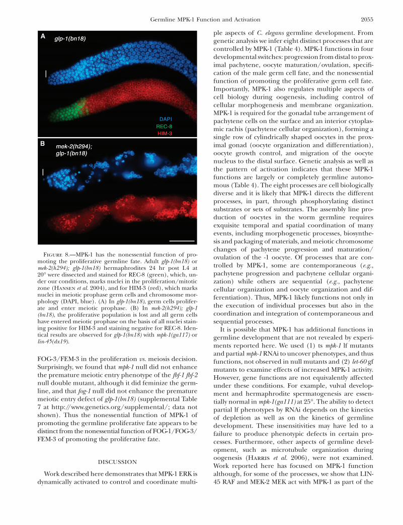
FOG-3/FEM-3 in the proliferation vs. meiosis decision.Surprisingly, we found that mpk-1 null did not enhancethe premature meiotic entry phenotype of the fbf-1 fbf-2null double mutant, although it did feminize the germ-line, and that fog-1 null did not enhance the prematuremeiotic entry defect of glp-1(bn18) (supplemental Table7 at http://www.genetics.org/supplemental/; data notshown). Thus the nonessential function of MPK-1 ofpromoting the germline proliferative fate appears to bedistinct from the nonessential function of FOG-1/FOG-3/FEM-3 of promoting the proliferative fate.
DISCUSSION
Work described here demonstrates that MPK-1 ERK isdynamically activated to control and coordinate multi-
ple aspects of C. elegans germline development. Fromgenetic analysis we infer eight distinct processes that arecontrolled by MPK-1 (Table 4). MPK-1 functions in fourdevelopmental switches: progression from distal to prox-imal pachytene, oocyte maturation/ovulation, specifi-cation of the male germ cell fate, and the nonessentialfunction of promoting the proliferative germ cell fate.Importantly, MPK-1 also regulates multiple aspects ofcell biology during oogenesis, including control ofcellular morphogenesis and membrane organization.MPK-1 is required for the gonadal tube arrangement ofpachytene cells on the surface and an interior cytoplas-mic rachis (pachytene cellular organization), forming asingle row of cylindrically shaped oocytes in the prox-imal gonad (oocyte organization and differentiation),oocyte growth control, and migration of the oocytenucleus to the distal surface. Genetic analysis as well asthe pattern of activation indicates that these MPK-1functions are largely or completely germline autono-mous (Table 4). The eight processes are cell biologicallydiverse and it is likely that MPK-1 directs the differentprocesses, in part, through phosphorylating distinctsubstrates or sets of substrates. The assembly line pro-duction of oocytes in the worm germline requiresexquisite temporal and spatial coordination of manyevents, including morphogenetic processes, biosynthe-sis and packaging of materials, and meiotic chromosomechanges of pachytene progression and maturation/ovulation of the -1 oocyte. Of processes that are con-trolled by MPK-1, some are contemporaneous (e.g.,pachytene progression and pachytene cellular organi-zation) while others are sequential (e.g., pachytenecellular organization and oocyte organization and dif-ferentiation). Thus, MPK-1 likely functions not only inthe execution of individual processes but also in thecoordination and integration of contemporaneous andsequential processes.
It is possible that MPK-1 has additional functions ingermline development that are not revealed by experi-ments reported here. We used (1) ts mpk-1 lf mutantsand partial mpk-1 RNAi to uncover phenotypes, and thusfunctions, not observed in null mutants and (2) let-60 gfmutants to examine effects of increased MPK-1 activity.However, gene functions are not equivalently affectedunder these conditions. For example, vulval develop-ment and hermaphrodite spermatogenesis are essen-tially normal in mpk-1(ga111) at 25�. The ability to detectpartial lf phenotypes by RNAi depends on the kineticsof depletion as well as on the kinetics of germlinedevelopment. These insensitivities may have led to afailure to produce phenotypic defects in certain pro-cesses. Furthermore, other aspects of germline devel-opment, such as microtubule organization duringoogenesis (Harris et al. 2006), were not examined.Work reported here has focused on MPK-1 functionalthough, for some of the processes, we show that LIN-45 RAF and MEK-2 MEK act with MPK-1 as part of the
Figure 8.—MPK-1 has the nonessential function of pro-moting the proliferative germline fate. Adult glp-1(bn18) ormek-2(h294); glp-1(bn18) hermaphrodites 24 hr post L4 at20� were dissected and stained for REC-8 (green), which, un-der our conditions, marks nuclei in the proliferation/mitoticzone (Hansen et al. 2004), and for HIM-3 (red), which marksnuclei in meiotic prophase germ cells and chromosome mor-phology (DAPI, blue). (A) In glp-1(bn18), germ cells prolifer-ate and enter meiotic prophase. (B) In mek-2(h294); glp-1(bn18), the proliferative population is lost and all germ cellshave entered meiotic prophase on the basis of all nuclei stain-ing positive for HIM-3 and staining negative for REC-8. Iden-tical results are observed for glp-1(bn18) with mpk-1(ga117) orlin-45(dx19).
Germline MPK-1 Function and Activation 2055
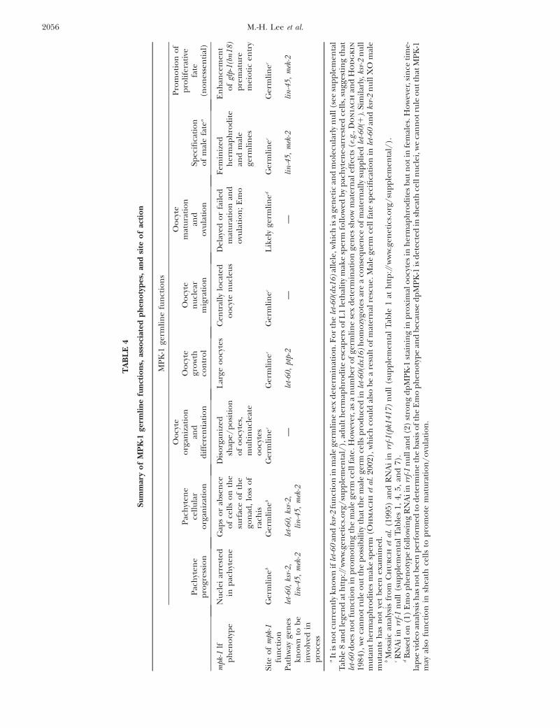
TA
BL
E4
Su
mm
ary
of
MP
K-1
germ
lin
efu
nct
ion
s,as
soci
ated
ph
eno
typ
es,
and
site
of
acti
on
MP
K-1
germ
lin
efu
nct
ion
s
Pac
hyt
ene
pro
gres
sio
n
Pac
hyt
ene
cell
ula
ro
rgan
izat
ion
Oo
cyte
org
aniz
atio
nan
dd
iffe
ren
tiat
ion
Oo
cyte
gro
wth
con
tro
l
Oo
cyte
nu
clea
rm
igra
tio
n
Oo
cyte
mat
ura
tio
nan
do
vula
tio
nSp
ecifi
cati
on
of
mal
efa
tea
Pro
mo
tio
no
fp
roli
fera
tive
fate
(no
nes
sen
tial
)
mpk
-1lf
ph
eno
typ
eN
ucl
eiar
rest
edin
pac
hyt
ene
Gap
so
rab
sen
ceo
fce
lls
on
the
surf
ace
of
the
gon
ad,
loss
of
rach
is
Dis
org
aniz
edsh
ape/
po
siti
on
of
oo
cyte
s,m
ult
inu
clea
teo
ocy
tes
Lar
geo
ocy
tes
Cen
tral
lylo
cate
do
ocy
ten
ucl
eus
Del
ayed
or
fail
edm
atu
rati
on
and
ovu
lati
on
;E
mo
Fem
iniz
edh
erm
aph
rod
ite
and
mal
ege
rmli
nes
En
han
cem
ent
of
glp-
1(b
n1
8)
pre
mat
ure
mei
oti
cen
try
Site
of
mpk
-1fu
nct
ion
Ger
mli
neb
Ger
mli
neb
Ger
mli
nec
Ger
mli
nec
Ger
mli
nec
Lik
ely
germ
lin
edG
erm
lin
ecG
erm
lin
ec
Pat
hw
ayge
nes
kno
wn
tob
ein
volv
edin
pro
cess
let-
60,
ksr-
2,
lin
-45
,m
ek-2
let-
60,
ksr-
2,
lin
-45
,m
ek-2
—le
t-6
0,
ptp-
2—
—li
n-4
5,
mek
-2li
n-4
5,
mek
-2
aIt
isn
ot
curr
entl
ykn
ow
nif
let-
60
and
ksr-
2fu
nct
ion
inm
ale
germ
lin
ese
xd
eter
min
atio
n.F
or
the
let-
60
(dx1
6)
alle
le,w
hic
his
age
net
ican
dm
ole
cula
rly
nu
ll(s
eesu
pp
lem
enta
lT
able
8an
dle
gen
dat
htt
p://
ww
w.g
enet
ics.
org
/su
pp
lem
enta
l/),
adu
lth
erm
aph
rod
ite
esca
per
so
fL
1le
thal
ity
mak
esp
erm
foll
ow
edb
yp
ach
yten
e-ar
rest
edce
lls,
sugg
esti
ng
that
let-
60
do
esn
ot
fun
ctio
nin
pro
mo
tin
gth
em
ale
germ
cell
fate
.Ho
wev
er,a
sa
nu
mb
ero
fge
rmli
ne
sex
det
erm
inat
ion
gen
essh
ow
mat
ern
alef
fect
s(e
.g.,
Do
nia
ch
and
Ho
dg
kin
1984
),w
eca
nn
ot
rule
ou
tth
ep
oss
ibil
ity
that
the
mal
ege
rmce
lls
pro
du
ced
inle
t-6
0(d
x16
)h
om
ozy
gote
sar
ea
con
seq
uen
ceo
fm
ater
nal
lysu
pp
lied
let-
60
(1).
Sim
ilar
ly,k
sr-2
nu
llm
uta
nt
her
map
hro
dit
esm
ake
sper
m(O
hm
ac
hi
etal
.20
02),
wh
ich
cou
ldal
sob
ea
resu
lto
fm
ater
nal
resc
ue.
Mal
ege
rmce
llfa
tesp
ecifi
cati
on
inle
t-6
0an
dks
r-2
nu
llX
Om
ale
mu
tan
tsh
asn
ot
yet
bee
nex
amin
ed.
bM
osa
ican
alys
isfr
om
Ch
ur
ch
etal
.(1
995)
and
RN
Ai
inrr
f-1
(pk1
41
7)
nu
ll(s
up
ple
men
tal
Tab
le1
ath
ttp
://w
ww
.gen
etic
s.o
rg/
sup
ple
men
tal/
).cR
NA
iin
rrf-
1n
ull
(su
pp
lem
enta
lT
able
s1,
4,5,
and
7).
dB
ased
on
(1)
Em
op
hen
oty
pe
foll
ow
ing
RN
Aii
nrr
f-1
nu
llan
d(2
)st
ron
gd
pM
PK
-1st
ain
ing
inp
roxi
mal
oo
cyte
sin
her
map
hro
dit
esb
ut
no
tin
fem
ales
.Ho
wev
er,s
ince
tim
e-la
pse
vid
eoan
alys
ish
asn
ot
bee
np
erfo
rmed
tod
eter
min
eth
eb
asis
of
the
Em
op
hen
oty
pe
and
bec
ause
dp
MP
K-1
isd
etec
ted
insh
eath
cell
nu
clei
,we
can
no
tru
leo
ut
that
MP
K-1
may
also
fun
ctio
nin
shea
thce
lls
top
rom
ote
mat
ura
tio
n/
ovu
lati
on
.
2056 M.-H. Lee et al.
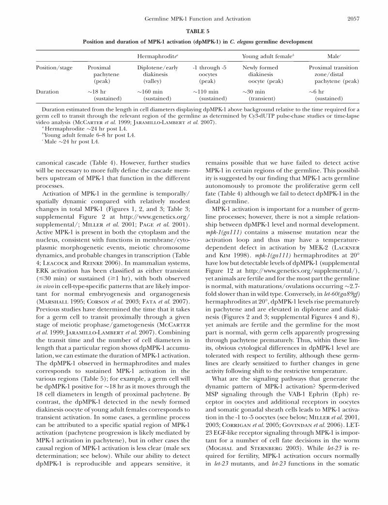
canonical cascade (Table 4). However, further studieswill be necessary to more fully define the cascade mem-bers upstream of MPK-1 that function in the differentprocesses.
Activation of MPK-1 in the germline is temporally/spatially dynamic compared with relatively modestchanges in total MPK-1 (Figures 1, 2, and 3; Table 3;supplemental Figure 2 at http://www.genetics.org/supplemental/; Miller et al. 2001; Page et al. 2001).Active MPK-1 is present in both the cytoplasm and thenucleus, consistent with functions in membrane/cyto-plasmic morphogenetic events, meiotic chromosomedynamics, and probable changes in transcription (Table4; Leacock and Reinke 2006). In mammalian systems,ERK activation has been classified as either transient(#30 min) or sustained ($1 hr), with both observedin vivo in cell-type-specific patterns that are likely impor-tant for normal embryogenesis and organogenesis(Marshall 1995; Corson et al. 2003; Fata et al. 2007).Previous studies have determined the time that it takesfor a germ cell to transit proximally through a givenstage of meiotic prophase/gametogenesis (McCarter
et al. 1999; Jaramillo-Lambert et al. 2007). Combiningthe transit time and the number of cell diameters inlength that a particular region shows dpMPK-1 accumu-lation, we can estimate the duration of MPK-1 activation.The dpMPK-1 observed in hermaphrodites and malescorresponds to sustained MPK-1 activation in thevarious regions (Table 5); for example, a germ cell willbe dpMPK-1 positive for�18 hr as it moves through the18 cell diameters in length of proximal pachytene. Bycontrast, the dpMPK-1 detected in the newly formeddiakinesis oocyte of young adult females corresponds totransient activation. In some cases, a germline processcan be attributed to a specific spatial region of MPK-1activation (pachytene progression is likely mediated byMPK-1 activation in pachytene), but in other cases thecausal region of MPK-1 activation is less clear (male sexdetermination; see below). While our ability to detectdpMPK-1 is reproducible and appears sensitive, it
remains possible that we have failed to detect activeMPK-1 in certain regions of the germline. This possibil-ity is suggested by our finding that MPK-1 acts germlineautonomously to promote the proliferative germ cellfate (Table 4) although we fail to detect dpMPK-1 in thedistal germline.
MPK-1 activation is important for a number of germ-line processes; however, there is not a simple relation-ship between dpMPK-1 level and normal development.mpk-1(ga111) contains a missense mutation near theactivation loop and thus may have a temperature-dependent defect in activation by MEK-2 (Lackner
and Kim 1998). mpk-1(ga111) hermaphrodites at 20�have low but detectable levels of dpMPK-1 (supplementalFigure 12 at http://www.genetics.org/supplemental/),yet animals are fertile and for the most part the germlineis normal, with maturations/ovulations occurring�2.7-fold slower than in wild type. Conversely, in let-60(ga89gf)hermaphrodites at 20�, dpMPK-1 levels rise prematurelyin pachytene and are elevated in diplotene and diaki-nesis (Figures 2 and 3; supplemental Figures 4 and 8),yet animals are fertile and the germline for the mostpart is normal, with germ cells apparently progressingthrough pachytene prematurely. Thus, within these lim-its, obvious cytological differences in dpMPK-1 level aretolerated with respect to fertility, although these germ-lines are clearly sensitized to further changes in geneactivity following shift to the restrictive temperature.
What are the signaling pathways that generate thedynamic pattern of MPK-1 activation? Sperm-derivedMSP signaling through the VAB-1 Ephrin (Eph) re-ceptor in oocytes and additional receptors in oocytesand somatic gonadal sheath cells leads to MPK-1 activa-tion in the -1 to -5 oocytes (see below; Miller et al. 2001,2003; Corrigan et al. 2005; Govindan et al. 2006). LET-23 EGF-like receptor signaling through MPK-1 is impor-tant for a number of cell fate decisions in the worm(Moghal and Sternberg 2003). While let-23 is re-quired for fertility, MPK-1 activation occurs normallyin let-23 mutants, and let-23 functions in the somatic
TABLE 5
Position and duration of MPK-1 activation (dpMPK-1) in C. elegans germline development
Hermaphroditea Young adult femaleb Malec
Position/stage Proximalpachytene(peak)
Diplotene/earlydiakinesis(valley)
-1 through -5oocytes(peak)
Newly formeddiakinesisoocyte (peak)
Proximal transitionzone/distalpachytene (peak)
Duration �18 hr(sustained)
�160 min(sustained)
�110 min(sustained)
�30 min(transient)
�6 hr(sustained)
Duration estimated from the length in cell diameters displaying dpMPK-1 above background relative to the time required for agerm cell to transit through the relevant region of the germline as determined by Cy3-dUTP pulse-chase studies or time-lapsevideo analysis (McCarter et al. 1999; Jaramillo-Lambert et al. 2007).
a Hermaphrodite �24 hr post L4.b Young adult female 6–8 hr post L4.c Male �24 hr post L4.
Germline MPK-1 Function and Activation 2057
gonad, not the germline, to promote oocyte ovulation( J. McCarter, M.-H. Lee and T. Schedl, unpublishedobservations). Thus much remains to be learned aboutthe upstream receptors and the downstream geneproducts that generate the dynamic pattern.
MPK-1 function and activation in pachytene:Reduction or elimination of mpk-1 activity results ingerm cells arrested in pachytene and loss of both thegonadal tube arrangement of pachytene cells on thesurface and the interior cytoplasmic rachis (Table 4). Inour analysis we did not identify a condition wherepachytene arrest and pachytene cellular disorganizationwere separately disrupted, leading to the possibility thatthey represent a single pachytene function. However, inrecent work in identifying phosphorylation substrates ofMPK-1, we have separated the functions by discoveringsubstrates that display only one or the other mutantphenotype, thus indicating that MPK-1 has at least twopachytene functions: one controlling pachytene pro-gression and the other controlling pachytene cellularorganization (S. Arur, M. Ohmachi, S. Nayak andT. Schedl, unpublished observations).
In pachytene, sustained MPK-1 activation is observedin surface pachytene germ cells (nuclei and cytoplasm)and in the interior rachis. The rise in activated MPK-1almost midway through pachytene appears to promote aswitch of germ cells from distal to proximal pachytene.This view is supported by markers demonstrating thatmpk-1(ga117) null germ cells arrest with mid-pachytenecharacteristics, high DAZ-1, absence of CEP-1/highGLD-1, and nuclei displaying a symmetric/dispersedpachytene organization (Figure 3; supplemental Figure3 at http://www.genetics.org/supplemental/). Further-more, in let-60(ga89gf), the earlier rise in dpMPK-1 levelcompared to wild type appears to lead to a prematuretransition from distal to proximal pachytene as judgedby the premature fall in DAZ-1 levels and a shortening inthe total length of pachytene.
It is not known which activities are controlled by MPK-1 for the switch from distal to proximal pachytene.Although the mpk-1 null pachytene-arrest phenotypesuggests an essential function in meiotic recombina-tion/chromosome dynamics (Roeder and Bailis
2000), two lines of reasoning indicate a broader role.First, currently known single-gene mutations that dis-rupt meiotic chromosome pairing, synapsis, double-strand break formation, and recombination do notarrest meiotic prophase progression and do not preventformation of oocytes (e.g., Dernburg et al. 1998;MacQueen and Villeneuve 2001; Rinaldo et al.2002; Colaiacovo et al. 2003). Second, changes inmpk-1 activity alter the spatial pattern of the cytoplasmicproteins DAZ-1 and GLD-1, indicating effects outsidemeiotic recombination/chromosome dynamics. Thus anumber of MPK-1 substrates and effectors are likelyinvolved in coordinating progression from distal toproximal pachytene.
MPK-1 function in pachytene cellular organizationinvolves control of membrane and cytoskeletal organi-zation in the pachytene region. Thus, while progressionfrom distal to proximal pachytene might require onlytransient MPK-1 activation, it is likely that pachytenecellular organization requires continuous function andtherefore sustained activation. Other activities thatoccur in proximal pachytene that may be directly orindirectly controlled by MPK-1 may also require sus-tained activation. These include germ cell apoptosis(Gumienny et al. 1999; Kritikou et al. 2006; S. Arur,C. Schertel, B. Conradt and T. Schedl, unpublishedobservations), cytoplasmic streaming, and changes intranscription and translation that are important forlater aspects of oogenesis in the proximal germline (Lee
and Schedl 2001; Kelly et al. 2002; Leacock andReinke 2006; Wolke et al. 2007). Thus, MPK-1 activationin pachytene may coordinate diverse processes inpachytene as well as integrate pachytene processes withlater oogenesis.
MPK-1 acts as a rheostat in oocyte growth control:Oocytes are unique cells in animals that can achievesizes .1000 times that of diploid somatic cells. Largeoocytes are generated under conditions where MPK-1activity is reduced, employing mpk-1(ga111) shift-ups,partial mpk-1 RNAi, let-60(n1046dx1), or ptp-2 null(Figure 5B; Church et al. 1995; Gutch et al. 1998).Conversely, small oocytes are generated under a condi-tion where MPK-1 activity is increased: let-60(ga89gf)shift-up (Figure 5C; supplemental Figure 11 at http://www.genetics.org/supplemental/). Thus, MPK-1 ap-pears to act as a rheostat to control oocyte size, withlow MPK-1 activity promoting growth, high MPK-1activity inhibiting growth, and, presumably, normal-sizeoocytes produced at intermediate MPK-1 activity. Acti-vated MPK-1 in diplotene and diakinesis appears to beinvolved in oocyte growth control, although it is possiblethat pachytene MPK-1 activity also contributes. Recentwork by Wolke et al. (2007) has shown that oocytegrowth is, in part, accomplished by actin-filament-mediated cytoplasmic streaming of material from therachis into the oocyte through cytoplasmic bridges.MPK-1 could modulate cytoplasmic streaming at anyone of a number of steps to control oocyte growth.
MPK-1 is required for timely oocyte meiotic matu-ration and ovulation: In vertebrates, ERK activationfunctions in promoting events of oocyte meiotic matu-ration and/or the transition to and maintenance ofmeiosis II arrest (Fan and Sun 2004; Liang et al. 2007).In C. elegans, MPK-1 activation and oocyte maturation/ovulation are MSP/sperm dependent (McCarter et al.1999; Miller et al. 2001; Page et al. 2001), but it was notknown if MPK-1 function was required for the process.Here we show that MPK-1, at a minimum, is required fortimely oocyte maturation/ovulation and may be re-quired to initiate the transition. This view is supportedby the observation that the point of delay/arrest when
2058 M.-H. Lee et al.

MPK-1 activity is reduced following mpk-1(ga111) shift-up is the same as the arrest point in females.
The MSP/sperm-dependent strong MPK-1 activationin the most proximal approximately five oocytes is likelyresponsible for MPK-1’s function in maturation/ovulation (Figures 2 and 4; supplemental Figures 7 and8 at http://www.genetics.org/supplemental/). However,in the germline assembly line only the -1 oocyte under-goes maturation. MPK-1 activation thus is not sufficientand additional regulatory mechanisms, such as inhibi-tion of CDK-1 from proximal oocytes and/or inhibitionfrom somatic gonadal sheath cells, must block matura-tion of the -2 through -5 oocytes (McCarter et al. 1999;Burrows et al. 2006; Govindan et al. 2006). We observethat levels of activated MPK-1 vary considerably in the -2through -5 oocytes, even within the same gonad; thesignificance of this variability is unclear and the mech-anism by which the variability is generated is not known.
Activated MPK-1 falls dramatically as the -1 oocyteundergoes maturation, even in let-60(ga89gf) mutantswhere dpMPK-1 levels are significantly elevated in prox-imal oocytes (Figures 1, 2, and 4; supplemental Figure 8at http://www.genetics.org/supplemental/; Page et al.2001). This inactivation of MPK-1 is distinct from thepattern in vertebrates where active ERK is present frommaturation until release of the meiosis II arrest byfertilization. The difference likely reflects differences inreproductive biology. In vertebrates, the meiosis IIarrest is maintained by active ERK and thereby allowsfertilization to be temporally/spatially separate frommaturation, meiosis I, and ovulation. In C. elegans, theevents of maturation, ovulation into the spermatheca,fertilization, and completion of the meiotic divisionsoccur in rapid succession and a meiosis II arrest is notnecessary since maturation/ovulation does not occurunless sperm are available for immediate fertilization.The abrupt fall in dpMPK-1 levels during maturationmay ensure that active MPK-1 does not lead to delay orarrest of meiosis II. Additionally, it has been proposedthat active ERK inhibits sperm centrosome function in avariety of vertebrates and invertebrates (Stephano andGould 2000). In C. elegans, the sperm centrosome maytrigger the completion of female meiosis and appears tofunction in anterior/posterior axis formation at theone-cell stage (Sadler and Shakes 2000; Wallenfang
and Seydoux 2000; Cowan and Hyman 2004;McNally and McNally 2005). The removal of activeMPK-1 during maturation may be important to ensurethat subsequent processes mediated by the sperm cen-trosome are not inhibited.
The effects of MSP/sperm signaling on MPK-1activation and function: From analysis of dpMPK-1levels in wild-type and female backgrounds, we proposethat MSP/sperm signaling has at least three majoreffects. First, as previously described, MSP/sperm sig-naling through the VAB-1 Eph receptor and otherreceptors leads to MPK-1 activation in the -1 through
-5 oocytes to promote maturation/ovulation (see above;Miller et al. 2001, 2003). Second, MSP/sperm signalingleads to MPK-1 activation in diplotene and diakinesisthat likely functions in oocyte growth control (seeabove). Third, MSP/sperm signaling by promotingmaturation/ovulation leads to flux in the germlineassembly line that results in MPK-1 activation that isMSP/sperm independent. This proposal is based ondifferences in MPK-1 activation between young adultfemales that generate new oocytes and older adultfemales that do not. In young females where newoocytes are being generated, cytoplasmic streamingoccurs, and germ cells progress through pachytene, wefound active MPK-1 in pachytene (Figure 2; supplemen-tal Figure 9 at http://www.genetics.org/supplemental/;Wolke et al. 2007). Young adult females also havetransiently activated MPK-1 in a single diakinesis oocyte,usually the second oocyte proximal to the last diploteneoocyte; the function of this activation is not currentlyknown. By contrast, in older adult females with .15diakinesis oocytes, where new oocytes are not beinggenerated, cytoplasmic streaming does not occur andgerm cells progress through pachytene at a slow rate( Jaramillo-Lambert et al. 2007; Wolke et al. 2007), weobserve very low levels of activated MPK-1 in pachytene,diplotene, and diakinesis (Figure 2, supplementalFigure 9). The MPK-1 activation in young adult femalesis, by definition, MSP/sperm independent and occurswhen flux through the germline is generating newoocytes, unlike in older adult females. MSP/sperm thuscan lead to MPK-1 activation in two ways: directly bybinding to receptors such as VAB-1 and indirectly byinducing maturation/ovulation that leads to distal fluxthrough the germline assembly line. How flux of germcells through the oocyte assembly line leads to MSP/sperm-independent MPK-1 activation is unknown.
MPK-1 is necessary for the male germ cell fate:Germline sex determination genes necessary for themale fate have been identified on the basis of lf phe-notype of germ cells that would normally form sperm butinstead form oocytes (Doniach and Hodgkin 1984;Kimble et al. 1984). A role for MPK-1 in germline sex de-termination was not previously recognized since stronglf mutations show pachytene arrest and cellular disorga-nization that preclude the formation of sperm or oocytes.We used sexually dimorphic markers that are expressedprior to overt gametogenesis to assess the sexual identityof the pachytene-arrested and disorganized germ cells.We found that MPK-1 is required for the male germ cellfate in both hermaphrodites and males. This MPK-1function is germline autonomous and appears to berequired continuously, at least in males.
In the C. elegans germline sex determination pathway,the tra-2 gene promotes the female fate by repressingthe downstream fem-1, -2, and -3 and fog-1 and -3 genes,which act near the end of the cascade to specify the malefate. Our genetic epistasis analysis indicates that male
Germline MPK-1 Function and Activation 2059

development in the tra-2 null mutant depends on mpk-1activity, indicating that MPK-1 acts either downstream ofTRA-2 (with the FEMs and FOGs) or in a parallelpathway. The epistasis is not complete (�11% of germ-lines show some male development), suggesting thatanother gene(s) acts redundantly with MPK-1, althoughwith a lesser role. Support for the possibility that MPK-1acts in parallel or separately from the FEMs and FOG-1and FOG-3 comes from our finding that the nonessen-tial function of MPK-1 in promoting the proliferativefate is genetically distinct from the nonessential func-tion of FOG-1, FOG-3, and FEM-3 in promoting theproliferative fate.
Sustained MPK-1 activation is observed from theproximal transition zone through early pachytene inthe male gonad while dpMPK-1 staining is not observedin this region in adult hermaphrodites (Figures 1 and7A; Table 5). This active MPK-1 may be responsible forspecification of the male fate. However, sexually di-morphic FOG-1 expression occurs distal or earlier thandpMPK-1 accumulation; Thompson et al. (2005) foundlow but detectable levels of FOG-1 in the proximal halfof the mitotic region with higher levels in the distaltransition zone through pachytene in adult males whileadult hermaphrodites, which have long completedspermatogenesis, lack FOG-1. One possibility for thespatially distinct patterns in the male germline is thatMPK-1 function in specification of the male fate is inparallel, although somewhat temporally delayed, to thepathway that controls FOG-1 levels. Alternatively, theMPK-1 activity responsible for the male germ cell fateoccurs earlier in the proximal mitotic region/distaltransition zone but is below the limit of dpMPK-1detection. Additionally, it is possible that the dpMPK-1observed in the proximal transition zone through earlypachytene represents activation for an MPK-1 functionin male meiotic prophase and/or spermatogenesis thatis masked by the germline feminization.
Perspectives: The dynamic pattern of MPK-1 activa-tion and the identification of eight germline processescontrolled by MPK-1 lead to a number of questionsrelevant to both C. elegans germline development andMPK-1 signaling. What are the upstream signals thatlead to MPK-1 control of a specific germline pro-cess(es)? For example, is there a systemic signal thatpromotes male germline sex determination in both thehermaphrodite and male that leads to MPK-1 activa-tion? What are the upstream signaling pathways andfeedback loops that generate specific patterns of sus-tained or transient MPK-1 activation? What are thedownstream MPK-1 substrates that execute a givengermline process(es)? How is MPK-1 activation andsubstrate phosphorylation spatially controlled so thatspecific germline processes are executed and coordi-nated? Given the conservation of ERK signaling inanimals, information obtained in C. elegans is likely tobe relevant to ERK function in other systems.
We acknowledge Andrew Godbey for help in the initial stages of thiswork. We thank our many colleagues for helpful discussions through-out the course of the study. We thank Dave Hansen, Associate Editor,and the reviewers for constructive and insightful comments on themanuscript. We are grateful to our colleagues Michael Gloltzer, DavidGreenstein, Patty Kuwabara, Verena Jantsch-Plunger, Josef Loidl, JillSchumacher, Masayuki Yammamoto, and Monique Zetka for anti-bodies. Some of the strains used in this study were supplied by theCaenorhabditis Genetics Center. Work in the Schedl laboratory wasfunded by National Institutes of Health grant GM63310.
LITERATURE CITED
Barton, M. K., T. B. Schedl and J. Kimble, 1987 Gain-of-functionmutations of fem-3, a sex-determination gene in Caenorhabditiselegans. Genetics 115: 107–119.
Beitel, G. J., S. G. Clark and H. R. Horvitz, 1990 Caenorhabditiselegans ras gene let-60 acts as a switch in the pathway of vulvalinduction. Nature 348: 503–509.
Burrows, A. E., B. K. Sceurman, M. E. Kosinski, C. T. Richie, P. L.Sadler et al., 2006 The C. elegans Myt1 ortholog is requiredfor the proper timing of oocyte maturation. Development 133:697–709.
Carlton, P. M., A. P. Farruggio and A. F. Dernburg, 2006 A linkbetween meiotic prophase progression and crossover control.PLoS Genet. 2: e12.
Chin, G. M., and A. M. Villeneuve, 2001 C. elegans mre-11 is re-quired for meiotic recombination and DNA repair but is dispens-able for the meiotic G(2) DNA damage checkpoint. Genes Dev.15: 522–534.
Church, D. L., K. L. Guan and E. J. Lambie, 1995 Three genes ofthe MAP kinase cascade, mek-2, mpk-1/sur-1 and let-60 ras, arerequired for meiotic cell cycle progression in Caenorhabditiselegans. Development 121: 2525–2535.
Colaiacovo, M. P., A. J. MacQueen, E. Martinez-Perez,K. McDonald, A. Adamo et al., 2003 Synaptonemal complexassembly in C. elegans is dispensable for loading strand-exchangeproteins but critical for proper completion of recombination.Dev. Cell 5: 463–474.
Corrigan, C., R. Subramanian and M. A. Miller, 2005 Eph andNMDA receptors control Ca21/calmodulin-dependent proteinkinase II activation during C. elegans oocyte meiotic maturation.Development 132: 5225–5237.
Corson, L. B., Y. Yamanaka, K. M. Lai and J. Rossant, 2003 Spatialand temporal patterns of ERK signaling during mouse embryo-genesis. Development 130: 4527–4537.
Cowan, C. R., and A. A. Hyman, 2004 Centrosomes direct cellpolarity independently of microtubule assembly in C. elegansembryos. Nature 431: 92–96.
Crittenden, S. L., D. S. Bernstein, J. L. Bachorik, B. E. Thompson,M. Gallegos et al., 2002 A conserved RNA-binding proteincontrols germline stem cells in Caenorhabditis elegans. Nature417: 660–663.
Dernburg, A. F., K. McDonald, G. Moulder, R. Barstead,M. Dresser et al., 1998 Meiotic recombination in C. elegans in-itiates by a conserved mechanism and is dispensable for homol-ogous chromosome synapsis. Cell 94: 387–398.
Doniach, T., and J. Hodgkin, 1984 A sex-determining gene, fem-1,required for both male and hermaphrodite development in Cae-norhabditis elegans. Dev. Biol. 106: 223–235.
Eisenmann, D. M., and S. K. Kim, 1997 Mechanismof activation of theCaenorhabditis elegans ras homologue let-60 by a novel, temperature-sensitive, gain-of-function mutation. Genetics 146: 553–565.
Ellis, R., and T. Schedl, 2006 Sex determination in the germ line,in Worm Book, edited by The C. elegans Research Community.(http://www.wormbook.org).
Fan, H. Y., and Q. Y. Sun, 2004 Involvement of mitogen-activatedprotein kinase cascade during oocyte maturation and fertiliza-tion in mammals. Biol. Reprod. 70: 535–547.
Fata, J. E., H. Mori, A. J. Ewald, H. Zhang, E. Yao et al., 2007 TheMAPK(ERK-1,2) pathway integrates distinct and antagonisticsignals from TGFalpha and FGF7 in morphogenesis of mousemammary epithelium. Dev. Biol. 306: 193–207.
2060 M.-H. Lee et al.

Francis, R., M. K. Barton, J. Kimble and T. Schedl, 1995 gld-1,a tumor suppressor gene required for oocyte development inCaenorhabditis elegans. Genetics 139: 579–606.
Gartner, A., S. Milstein, S. Ahmed, J. Hodgkin and M. O.Hengartner, 2000 A conserved checkpoint pathway mediatesDNA damage-induced apoptosis and cell cycle arrest in C. elegans.Mol. Cell 5: 435–443.
Gibert, M. A., J. Starck and B. Beguet, 1984 Role of the gonadcytoplasmic core during oogenesis of the nematode Caenorhab-ditis elegans. Biol. Cell 50: 77–85.
Govindan, J. A., H. Cheng, J. E. Harris and D. Greenstein,2006 Galphao/i and Galphas signaling function in parallel withthe MSP/Eph receptor to control meiotic diapause in C. elegans.Curr. Biol. 16: 1257–1268.
Grant, B., and D. Hirsh, 1999 Receptor-mediated endocytosis inthe Caenorhabditis elegans oocyte. Mol. Biol. Cell 10: 4311–4326.
Gumienny, T. L., E. Lambie, E. Hartwieg, H. R. Horvitz and M. O.Hengartner, 1999 Genetic control of programmed cell deathin the Caenorhabditis elegans hermaphrodite germline. Devel-opment 126: 1011–1022.
Gutch, M. J., A. J. Flint, J. Keller, N. K. Tonksand M. O. Hengartner,1998 The Caenorhabditis elegans SH2 domain-containing pro-tein tyrosine phosphatase PTP-2 participates in signal transduc-tion during oogenesis and vulval development. Genes Dev. 12:571–585.
Hall, D. H., V. P. Winfrey, G. Blaeuer, L. H. Hoffman, T. Furuta
et al., 1999 Ultrastructural features of the adult hermaphroditegonad of Caenorhabditis elegans: relations between the germline and soma. Dev. Biol. 212: 101–123.
Han, M., R. V. Aroian and P. W. Sternberg, 1990 The let-60 locuscontrols the switch between vulval and nonvulval cell fates in Cae-norhabditis elegans. Genetics 126: 899–913.
Hansen, D., and T. Schedl, 2006 The regulatory network control-ling the proliferation-meiotic entry decision in the Caenorhabdi-tis elegans germ line. Curr. Top. Dev. Biol. 76: 185–215.
Hansen, D., E. J. Hubbard and T. Schedl, 2004 Multi-pathwaycontrol of the proliferation versus meiotic development deci-sion in the Caenorhabditis elegans germline. Dev. Biol. 268:342–357.
Harris, J. E., J. A. Govindan, I. Yamamoto, J. Schwartz, I. Kaverina
et al., 2006 Major sperm protein signaling promotes oocyte mi-crotubule reorganization prior to fertilization in Caenorhabditiselegans. Dev. Biol. 299: 105–121.
Hirsh, D., D. Oppenheim and M. Klass, 1976 Development of thereproductive system of Caenorhabditis elegans. Dev. Biol. 49:200–219.
Hodgkin, J., 1980 More sex-determination mutants of Caenorhabditiselegans. Genetics 96: 649–664.
Hodgkin, J., and T. Doniach, 1997 Natural variation and copu-latory plug formation in Caenorhabditis elegans. Genetics 146:149–164.
Hsu, V., C. L. Zobel, E. J. Lambie, T. Schedl and K. Kornfeld,2002 Caenorhabditis elegans lin-45 raf is essential for larval viabil-ity, fertility and the induction of vulval cell fates. Genetics 160:481–492.
Huang, P., and M. J. Stern, 2004 FGF signaling functions in the hy-podermis to regulate fluid balance in C. elegans. Development131: 2595–2604.
Hubbard, E. J., and D. Greenstein, 2000 The Caenorhabditis ele-gans gonad: a test tube for cell and developmental biology. Dev.Dyn. 218: 2–22.
Iwasaki, K., J. McCarter, R. Francis and T. Schedl, 1996 emo-1, aCaenorhabditis elegans Sec61p gamma homologue, is requiredfor oocyte development and ovulation. J. Cell Biol. 134: 699–714.
Jantsch-Plunger, V., and M. Glotzer, 1999 Depletion of syntaxinsin the early Caenorhabditis elegans embryo reveals a role formembrane fusion events in cytokinesis. Curr. Biol. 9: 738–745.
Jaramillo-Lambert, A., M. Ellefson, A. M. Villeneuve andJ. Engebrecht, 2007 Differential timing of S phases, X chro-mosome replication, and meiotic prophase in the C. elegansgerm line. Dev. Biol. 308: 206–221.
Jones, A. R., R. Francis and T. Schedl, 1996 GLD-1, a cytoplasmicprotein essential for oocyte differentiation, shows stage- and sex-specific expression during Caenorhabditis elegans germline de-velopment. Dev. Biol. 180: 165–183.
Kelly, W. G., C. E. Schaner, A. F. Dernburg, M. H. Lee, S. K. Kim et al.,2002 X-chromosome silencing in the germline of C. elegans.Development 129: 479–492.
Kimble, J., and S. L. Crittenden, 2006 Germline proliferation andits control, in Worm Book, edited by The C. elegans Research
Community. (http://www.wormbook.org).Kimble, J., L. Edgar and D. Hirsh, 1984 Specification of male de-
velopment in Caenorhabditis elegans: the fem genes. Dev. Biol.105: 234–239.
Kosinski, M., K. McDonald, J. Schwartz, I. Yamamoto andD. Greenstein, 2005 C. elegans sperm bud vesicles to delivera meiotic maturation signal to distant oocytes. Development132: 3357–3369.
Kritikou, E. A., S. Milstein, P. O. Vidalain, G. Lettre, E. Bogan
et al., 2006 C. elegans GLA-3 is a novel component of theMAP kinase MPK-1 signaling pathway required for germ cell sur-vival. Genes Dev. 20: 2279–2292.
Kuwabara, P. E., M. H. Lee, T. Schedl and G. S. Jefferis, 2000 AC. elegans patched gene, ptc-1, functions in germ-line cytokine-sis. Genes Dev. 14: 1933–1944.
Lackner, M. R., and S. K. Kim, 1998 Genetic analysis of the Caenorhab-ditis elegans MAP kinase gene mpk-1. Genetics 150: 103–117.
Lackner, M. R., K. Kornfeld, L. M. Miller, H. R. Horvitz andS. K. Kim, 1994 A MAP kinase homolog, mpk-1, is involved inras-mediated induction of vulval cell fates in Caenorhabditis ele-gans. Genes Dev. 8: 160–173.
Leacock, S. W., and V. Reinke, 2006 Expression profiling of MAPkinase-mediated meiotic progression in Caenorhabditis elegans.PLoS Genet. 2: e174.
Lee, M. H., and T. Schedl, 2001 Identification of in vivo mRNA tar-gets of GLD-1, a maxi-KH motif containing protein required forC. elegans germ cell development. Genes Dev. 15: 2408–2420.
Lee, M. H., and T. Schedl, 2004 Translation repression by GLD-1protects its mRNA targets from nonsense-mediated mRNA decayin C. elegans. Genes Dev. 18: 1047–1059.
Liang, C. G., Y. Q. Su, H. Y. Fan, H. Schatten and Q. Y. Sun,2007 Mechanisms regulating oocyte meiotic resumption: rolesof mitogen-activated protein kinase. Mol. Endocrinol. 21: 2037–2055.
MacQueen, A. J., and A. M. Villeneuve, 2001 Nuclear reorganiza-tion and homologous chromosome pairing during meiotic pro-phase require C. elegans chk-2. Genes Dev. 15: 1674–1687.
Maddox, A. S., B. Habermann, A. Desai and K. Oegema,2005 Distinct roles for two C. elegans anillins in the gonadand early embryo. Development 132: 2837–2848.
Marshall, C. J., 1995 Specificity of receptor tyrosine kinase signal-ing: transient versus sustained extracellular signal-regulated ki-nase activation. Cell 80: 179–185.
Maruyama, R., S. Endo, A. Sugimoto and M. Yamamoto,2005 Caenorhabditis elegans DAZ-1 is expressed in proliferatinggerm cells and directs proper nuclear organization and cytoplas-mic core formation during oogenesis. Dev. Biol. 277: 142–154.
McCarter, J., B. Bartlett, T. Dang and T. Schedl, 1997 Soma-germ cell interactions in Caenorhabditis elegans: multiple eventsof hermaphrodite germline development require the somaticsheath and spermathecal lineages. Dev. Biol. 181: 121–143.
McCarter, J., B. Bartlett, T. Dang and T. Schedl, 1999 Onthe control of oocyte meiotic maturation and ovulation inCaenorhabditis elegans. Dev. Biol. 205: 111–128.
McNally, K. L., and F. J. McNally, 2005 Fertilization initiates thetransition from anaphase I to metaphase II during female meio-sis in C. elegans. Dev. Biol. 282: 218–230.
Miller, M. A., V. Q. Nguyen, M. H. Lee, M. Kosinski, T. Schedl et al.,2001 A sperm cytoskeletal protein that signals oocyte meioticmaturation and ovulation. Science 291: 2144–2147.
Miller, M. A., P. J. Ruest, M. Kosinski, S. K. Hanks andD. Greenstein, 2003 An Eph receptor sperm-sensing controlmechanism for oocyte meiotic maturation in Caenorhabditiselegans. Genes Dev. 17: 187–200.
Moghal, N., and P. W. Sternberg, 2003 The epidermal growth fac-tor system in Caenorhabditis elegans. Exp. Cell Res. 284: 150–159.
Ohmachi, M., C. E. Rocheleau, D. Church, E. Lambie, T. Schedl
et al., 2002 C. elegans ksr-1 and ksr-2 have both unique and re-dundant functions and are required for MPK-1 ERK phosphory-lation. Curr. Biol. 12: 427–433.
Germline MPK-1 Function and Activation 2061

Page, B. D., S. Guedes, D. Waring and J. R. Priess, 2001 TheC. elegans E2F- and DP-related proteins are required for embry-onic asymmetry and negatively regulate Ras/MAPK signaling.Mol. Cell 7: 451–460.
Qiao, L., J. L. Lissemore, P. Shu, A. Smardon, M. B. Gelber et al.,1995 Enhancers of glp-1, a gene required for cell-signaling inCaenorhabditis elegans, define a set of genes required for germlinedevelopment. Genetics 141: 551–569.
Rinaldo, C., P. Bazzicalupo, S. Ederle, M. Hilliard and A. La
Volpe, 2002 Roles for Caenorhabditis elegans rad-51 in meiosisand in resistance to ionizing radiation during development.Genetics 160: 471–479.
Roeder, G. S., and J. M. Bailis, 2000 The pachytene checkpoint.Trends Genet. 16: 395–403.
Rogers, E., J. D. Bishop, J. A. Waddle, J. M. Schumacher and R. Lin,2002 The aurora kinase AIR-2 functions in the release of chro-mosome cohesion in Caenorhabditis elegans meiosis. J. Cell Biol.157: 219–229.
Rose, K. L., V. P. Winfrey, L. H. Hoffman, D. H. Hall, T. Furuta
et al., 1997 The POU gene ceh-18 promotes gonadal sheath celldifferentiation and function required for meiotic maturationand ovulation in Caenorhabditis elegans. Dev. Biol. 192: 59–77.
Rubin, G. M., H. C. Chang, F. Karim, T. Laverty, N. R. Michaud
et al., 1997 Signal transduction downstream from Ras in Dro-sophila. Cold Spring Harb. Symp. Quant. Biol. 62: 347–352.
Sadler, P. L., and D. C. Shakes, 2000 Anucleate Caenorhabditiselegans sperm can crawl, fertilize oocytes and direct anterior-posterior polarization of the 1-cell embryo. Development 127:355–366.
Schlessinger, J., 2000 Cell signaling by receptor tyrosine kinases.Cell 103: 211–225.
Schumacher, B., M. Hanazawa, M. H. Lee, S. Nayak,K. Volkmann et al., 2005 Translational repression of C. ele-gans p53 by GLD-1 regulates DNA damage-induced apoptosis.Cell 120: 357–368.
Schumacher, J. M., A. Golden and P. J. Donovan, 1998 AIR-2: anAurora/Ipl1-related protein kinase associated with chromosomesand midbody microtubules is required for polar body extrusion
and cytokinesis in Caenorhabditis elegans embryos. J. Cell Biol.143: 1635–1646.
Sijen, T., J. Fleenor, F. Simmer, K. L. Thijssen, S. Parrish et al.,2001 On the role of RNA amplification in dsRNA-triggeredgene silencing. Cell 107: 465–476.
Stephano, J.L.,andM.C.Gould,2000 MAPkinase,auniversalsuppres-sor of sperm centrosomes during meiosis? Dev. Biol. 222: 420–428.
Strome, S., 1986 Fluorescence visualization of the distribution ofmicrofilaments in gonads and early embryos of the nematodeCaenorhabditis elegans. J. Cell Biol. 103: 2241–2252.
Sulston, J., and J. Hodgkin, 1988 Methods, pp. 587–606 in TheNematode Caenorhabditis elegans, edited by W. B. Wood. ColdSpring Harbor Laboratory Press, Cold Spring Harbor, NY.
Sundaram, M. V., 2006 RTK/Ras/MAPK signaling, in Worm Book,edited by The C. elegans Research Community. (http://www.wormbook.org).
Thompson, B. E., D. S. Bernstein, J. L. Bachorik, A. G. Petcherski,M. Wickens et al., 2005 Dose-dependent control of proliferationand sperm specification by FOG-1/CPEB. Development 132:3471–3481.
Vojtek, A. B., and C. J. Der, 1998 Increasing complexity of the Rassignaling pathway. J. Biol. Chem. 273: 19925–19928.
Wallenfang, M. R., and G. Seydoux, 2000 Polarization of the an-terior-posterior axis of C. elegans is a microtubule-directed pro-cess. Nature 408: 89–92.
Ward, S., and J. S. Carrel, 1979 Fertilization and sperm competitionin the nematode Caenorhabditis elegans. Dev. Biol. 73: 304–321.
Wolke, U., E. A. Jezuit and J. R. Priess, 2007 Actin-dependent cy-toplasmic streaming in C. elegans oogenesis. Development 134:2227–2236.
Wu, Y., and M. Han, 1994 Suppression of activated Let-60 ras pro-tein defines a role of Caenorhabditis elegans Sur-1 MAP kinasein vulval differentiation. Genes Dev. 8: 147–159.
Yang, H. Y., K. McNally and F. J. McNally, 2003 MEI-1/katanin isrequired for translocation of the meiosis I spindle to the oocytecortex in C elegans. Dev. Biol. 260: 245–259.
Communicating editor: D. I. Greenstein
2062 M.-H. Lee et al.
Copyright © 2022 FDOKUMEN