Laser-assisted blastocyst dissection and subsequent cultivation of embryonic stem cells in a...
16
BioMed Central Page 1 of 16 (page number not for citation purposes) Journal of Translational Medicine Open Access Methodology Laser-assisted blastocyst dissection and subsequent cultivation of embryonic stem cells in a serum/cell free culture system: applications and preliminary results in a murine model Noriko Tanaka 1 , Takumi Takeuchi 1 , Queenie V Neri 1 , Eric Scott Sills 2 and Gianpiero D Palermo* 1 Address: 1 Center for Reproductive Medicine and Infertility, Weill Medical College of Cornell University, New York, NY 10021, USA and 2 Department of Obstetrics, Gynecology and Reproductive Research, Murphy Medical Center, Murphy, NC, USA Email: Noriko Tanaka - [email protected]; Takumi Takeuchi - [email protected]; Queenie V Neri - [email protected]; Eric Scott Sills - [email protected]; Gianpiero D Palermo* - [email protected] * Corresponding author Abstract Background: To evaluate embryonic stem cell (ESC) harvesting methods with an emphasis on derivation of ESC lines without feeder cells or sera. Using a murine model, laser-assisted blastocyst dissection was performed and compared to conventional immunosurgery to assess a novel laser application for inner cell mass (ICM) isolation. Methods: Intact blastocysts or isolated ICMs generated in a standard mouse strain were plated in medium with or without serum to compare ESC harvesting efficiency. ESC derivation was also undertaken in a feeder cell-free culture system. Results: Although ICM growth and dissociation was comparable irrespective of the media components, an enhanced ESC harvest was observed in our serum-free medium (p < 0.01). ESC harvest rate was not affected by ICM isolation technique but was attenuated in the feeder cell-free group. Conclusion: Achieving successful techniques for human ESC research is fundamentally dependent on preliminary work using experimental animals. In this study, all experimentally developed ESC lines manifested similar features to ESCs obtained from intact blastocysts in standard culture. Cell/ sera free murine ESC harvest and propagation are feasible procedures for an embryology laboratory and await refinements for translation to human medical research. Introduction Embryonic stem cells (ESC) may be produced from the inner cell mass (ICM) of intact blastocysts [1,2], by immu- nosurgery [3,4], by other methods to isolate pluripotent cells constituting the ICM [5] or single blastomeres [6,7]. Such techniques to derive ESC are easily reproduced in a murine model, the mouse 129 strain being perhaps the most commonly used experimental animal for this pur- pose [1,5,8,9]. Indeed, this particular mouse model dem- onstrates a number of desirable ICM features that make it Published: 08 May 2006 Journal of Translational Medicine 2006, 4:20 doi:10.1186/1479-5876-4-20 Received: 21 February 2006 Accepted: 08 May 2006 This article is available from: http://www.translational-medicine.com/content/4/1/20 © 2006 Tanaka et al; licensee BioMed Central Ltd. This is an Open Access article distributed under the terms of the Creative Commons Attribution License (http://creativecommons.org/licenses/by/2.0 ), which permits unrestricted use, distribution, and reproduction in any medium, provided the original work is properly cited.
Transcript of Laser-assisted blastocyst dissection and subsequent cultivation of embryonic stem cells in a...

BioMed CentralJournal of Translational Medicine
ss
Open AcceMethodologyLaser-assisted blastocyst dissection and subsequent cultivation of embryonic stem cells in a serum/cell free culture system: applications and preliminary results in a murine modelNoriko Tanaka1, Takumi Takeuchi1, Queenie V Neri1, Eric Scott Sills2 and Gianpiero D Palermo*1Address: 1Center for Reproductive Medicine and Infertility, Weill Medical College of Cornell University, New York, NY 10021, USA and 2Department of Obstetrics, Gynecology and Reproductive Research, Murphy Medical Center, Murphy, NC, USA
Email: Noriko Tanaka - [email protected]; Takumi Takeuchi - [email protected]; Queenie V Neri - [email protected]; Eric Scott Sills - [email protected]; Gianpiero D Palermo* - [email protected]
* Corresponding author
AbstractBackground: To evaluate embryonic stem cell (ESC) harvesting methods with an emphasis onderivation of ESC lines without feeder cells or sera. Using a murine model, laser-assisted blastocystdissection was performed and compared to conventional immunosurgery to assess a novel laserapplication for inner cell mass (ICM) isolation.
Methods: Intact blastocysts or isolated ICMs generated in a standard mouse strain were plated inmedium with or without serum to compare ESC harvesting efficiency. ESC derivation was alsoundertaken in a feeder cell-free culture system.
Results: Although ICM growth and dissociation was comparable irrespective of the mediacomponents, an enhanced ESC harvest was observed in our serum-free medium (p < 0.01). ESCharvest rate was not affected by ICM isolation technique but was attenuated in the feeder cell-freegroup.
Conclusion: Achieving successful techniques for human ESC research is fundamentally dependenton preliminary work using experimental animals. In this study, all experimentally developed ESClines manifested similar features to ESCs obtained from intact blastocysts in standard culture. Cell/sera free murine ESC harvest and propagation are feasible procedures for an embryologylaboratory and await refinements for translation to human medical research.
IntroductionEmbryonic stem cells (ESC) may be produced from theinner cell mass (ICM) of intact blastocysts [1,2], by immu-nosurgery [3,4], by other methods to isolate pluripotentcells constituting the ICM [5] or single blastomeres [6,7].
Such techniques to derive ESC are easily reproduced in amurine model, the mouse 129 strain being perhaps themost commonly used experimental animal for this pur-pose [1,5,8,9]. Indeed, this particular mouse model dem-onstrates a number of desirable ICM features that make it
Published: 08 May 2006
Journal of Translational Medicine 2006, 4:20 doi:10.1186/1479-5876-4-20
Received: 21 February 2006Accepted: 08 May 2006
This article is available from: http://www.translational-medicine.com/content/4/1/20
© 2006 Tanaka et al; licensee BioMed Central Ltd.This is an Open Access article distributed under the terms of the Creative Commons Attribution License (http://creativecommons.org/licenses/by/2.0), which permits unrestricted use, distribution, and reproduction in any medium, provided the original work is properly cited.
Page 1 of 16(page number not for citation purposes)

Journal of Translational Medicine 2006, 4:20 http://www.translational-medicine.com/content/4/1/20
well-suited for laboratory use including rapid cellulargrowth, relatively large size, and a high content and per-sistence of stem cells [10,11].
To be sure, the stem cells are not the only murine contri-bution to ESC experimentation, as mouse embryonicfibroblasts (MEF) also play a central supporting role in thelaboratory as feeder cells for ESCs [12,13]. However, a reli-ance on cell/serum based mouse systems has underscoredsome important limitations including possible xenogenicor allogenic contamination [4]. Common immunosurgi-cal methods for ICM isolation incorporate the use of allo-genic antibodies and complement [3] which may alsointroduce unwanted epitopes rendering some ESC deriva-tives unsuitable for further applications.
Here we describe production of ESCs from blastocystsobtained from a standard mouse strain in the absence offeeder cells or sera, with an emphasis on a laser-based ICMisolation modality. Additionally, the cells so harvestedmet the morphological criteria and growth patternsexpected of ESCs as shown by specific molecular markersand by gene expression analysis. Finally, to confirm theability of our ESC lines to provide the full range of differ-entiated tissue types, the stem cells were induced todevelop into each of the three fetal germ layers.
Materials and methodsSpecimensB6D2-F1 (C57BL/6J � × DBA/2J �) mice were obtainedfrom Jackson Laboratory (Bar Harbor, ME, USA) andCharles River Laboratories (Wilmington, MA, USA) andhoused at the Research Animal Resource Center of WeillMedical College of Cornell University, in a temperature-and light-controlled room on a 14 h light:10 h dark pho-toperiod with food and water ad libitum.
Embryo collection, culture and mediaFemales (7–9 weeks) were superovulated with 10 IU ofpregnant mare's serum gonadotrophin (Sigma, St. Louis,MO) then 10 IU of hCG (Sigma) 48 h apart. They werenext placed with males of the same strain and mating wasconfirmed by presence of a vaginal plug the followingmorning. Zygotes collected approximately 20 hours afterhCG were cultured in KSOMAA medium (Specialty Media,Phillipsburg, NJ) at 37°C with 6% CO2 in humidified air.Our ESC medium consisted of Dubelcco's ModifiedEagle's Medium (DMEM, Invitrogen, Carlsbad, CA, USA),10% fetal bovine serum (FBS, HyClone, Logan, UT, USA),and 10% newborn calf serum (NCS, HyClone) supple-mented with mouse leukemia inhibitory factor (LIF,2000IU/ml, Chemicon International, Temecula, CA), 0.1mM nonessential amino acids (NEAA, Specialty Media),0.1 mM β-mercaptoethanol (β-ME, Sigma), and 50 U/mlpenicillin/50 μg/ml streptomycin (Sigma). This investiga-
tion also utilized Knockout™ DMEM (Ko-M, Invitrogen)with 15% Knockout ™ serum replacement (Ko-S, Invitro-gen) containing LIF, NEAA, β-ME, antibiotics, and 4 mML-glutamine (Sigma) as the supplementary medium.
ESC harvestingFeeder layers of mitomycin C-treated MEFs were preparedon either 0.1% gelatin treated 4-well culture dishes (NalgeNunc International, Rochester, NY) or in 30 μl micro-drops under oil in tissue culture dishes. Intact expandedblastocysts (controls) and also zona-free blastocysts andICMs isolated after immunosurgery or microdissection(see below), were plated onto MEF monolayers and cul-tured in ESC medium in 6% CO2 at 37°C. Blastocystswere observed every 24 h for hatching and attachment ofthe trophoblast to a single MEF layer, while monitoringICM size in two dimensions. On day 4 or 5 after plating(D4 or D5), ICM of at least 100 μm were dissociated usinga glass pipette, followed by trypsinization in PBS contain-ing 250 U/ml trypsin and 1 mM EDTA (PBS-try-EDTA) topromote cell dispersion. After re-plating in fresh wellscoated with feeder cells, ESC colonies developed in 2 or 3days after which they were trypsinized and propagated bypassaging every 2–3 days thereafter. All established celllines were tested for mycoplasma using MycoAlert ™ Myc-oplasma Detection Kit (Cambrex, Rockland, ME, USA).
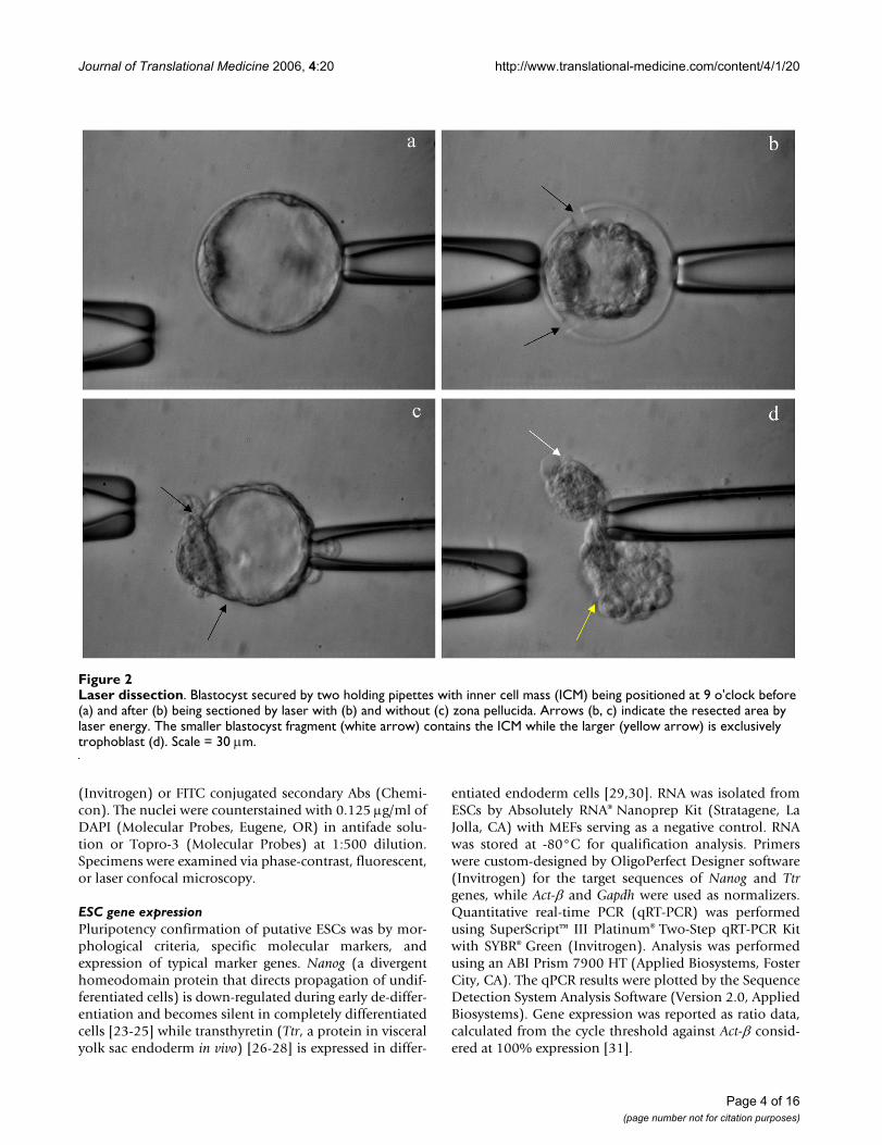
ICM isolationIntact blastocysts were plated onto feeder layers on postfertilization day 4–5. Zona-free blastocysts were obtainedfrom incubation of day 4 blastocysts for 4–5 min in 24 U/ml pronase (Sigma). These were washed x2 and culturedfor at least 1 h in medium with serum supplementation.To obtain ICMs, zona-free blastocysts were incubatedwith heat-inactivated rabbit anti-mouse serum (Sigma)diluted with DMEM (Invitrogen) in 1:25 for 15 min thenexposed to guinea pig complement (Cedarlane laborato-ries, Hornby, Ontario, Canada) at 1:10 dilution for 30min in 6% CO2 in air at 37°C (Fig. 1). The disrupted tro-phoblast was then removed by aspiration through a hand-pulled 50–60 μm (inner diameter) pipette [3]. Laser-assisted ICM isolation was performed as follows: Blasto-cysts were secured by two holding pipette with the ICMbeing positioned at 9 o'clock. Once adequate tension wasestablished, approximately 10 infrared laser pulses (300mW × 1 ms, ZILOS-tk™, Hamilton Thorne Research, Bev-erly, MA USA) were fired to split the blastocyst into twounequal portions – the smaller consisting of ICM, thelarger consisting exclusively of trophoblast (Fig. 2).
Feeder cell-free culture and ESC characterizationThe laser dissected ICM components were randomlyplated either on MEF feeder layer or directly on gelatin-coated dishes, and cultured in Ko-M with 15% of Ko-S(serum free) containing LIF, NEAA, β-ME, antibiotics, and
Page 2 of 16(page number not for citation purposes)

Journal of Translational Medicine 2006, 4:20 http://www.translational-medicine.com/content/4/1/20
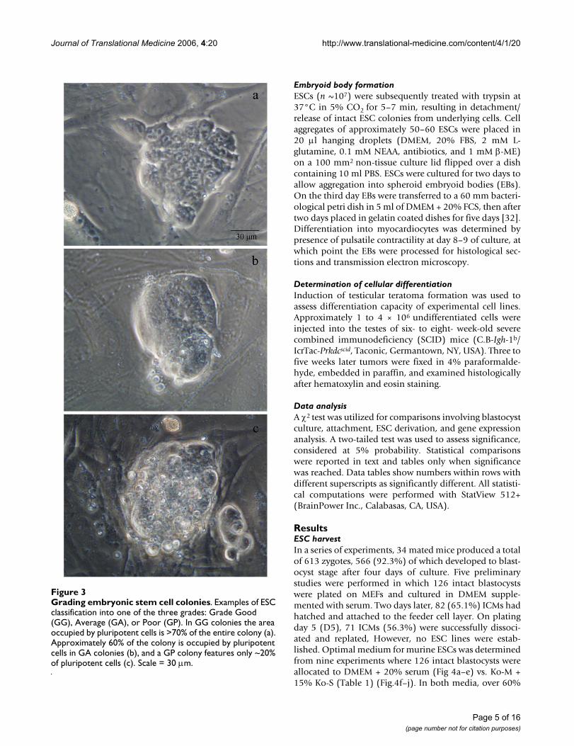
L-glutamine as described above. Intact blastocysts on MEFwere used as controls. ICM size was measured daily, ICMdissociation and ESC propagation was carried out asdescribed earlier. Pluripotency was evaluated according tocell size, nucleus/cytoplasm ratio, and nucleolar patterns.ESC colonies were graded according to number, density,and quality. Colony character was judged under aninverted microscope equipped with phase-contrast opticsand stratified as follows: good (GG), average (GA), orpoor (GP) according to colony composition of >70%, 40–70%, and <40% pluripotent cells, respectively (Fig. 3).Typically, pluripotent cells are large and have a highnucleus/cytoplasm ratio with one or more distinct nucle-oli. DAPI banding was performed on metaphase chromo-somes for ploidy assessment.
For this investigation, ESC pluripotency was determinedby two positive markers: alkaline phosphatase (AP) activ-
ity [8,14] and Oct-4 [15-18]. TROMA-1 monoclonal anti-body (Ab) directed against cytokeratin-like filamentspresent in trophectoderm and endodermal cells was usedas a negative marker [8,19-21]. All markers were tested oncontrol blastocysts. Specimens were fixed with 2% para-formaldehyde (Sigma) and permeabilized with 0.5% Tri-ton X-100 (Sigma). AP activity in fixed cells was detectedusing an azo-dye technique with a Texas-Red filter underfluorescence. The stain solution contained naphthol AS-MX phosphate (Sigma) and fast red TR salt (Sigma) [22].To detect expression of Oct-4 and TROMA-1, after incuba-tion at 20°C for 60 min in 0.1% bovine serum albumin(BSA, Sigma) and goat serum, ESCs were exposed to Oct-4 polyclonal Ab (Santa Cruz Biotechnology, Santa Cruz,CA) at 1:100 dilution and to monoclonal TROMA-1 Ab(Developmental Studies hybridoma Bank, Iowa City, IA)at 1:6 dilution. After rinsing unbound Abs with PBS/BSA,specimens were next incubated with Alexa Fluor® 488
ImmunosurgeryFigure 1Immunosurgery. Zona-free blastocyst (a) coated with rabbit anti-mouse antibodies. After addition of guinea pig comple-ment, the outcome of unselective toxicity can be seen at t = 5 min (b), 15 min (c) and 30 min (d). Scale = 30 μm.
Page 3 of 16(page number not for citation purposes)
Journal of Translational Medicine 2006, 4:20 http://www.translational-medicine.com/content/4/1/20
(Invitrogen) or FITC conjugated secondary Abs (Chemi-con). The nuclei were counterstained with 0.125 μg/ml ofDAPI (Molecular Probes, Eugene, OR) in antifade solu-tion or Topro-3 (Molecular Probes) at 1:500 dilution.Specimens were examined via phase-contrast, fluorescent,or laser confocal microscopy.
ESC gene expressionPluripotency confirmation of putative ESCs was by mor-phological criteria, specific molecular markers, andexpression of typical marker genes. Nanog (a divergenthomeodomain protein that directs propagation of undif-ferentiated cells) is down-regulated during early de-differ-entiation and becomes silent in completely differentiatedcells [23-25] while transthyretin (Ttr, a protein in visceralyolk sac endoderm in vivo) [26-28] is expressed in differ-
entiated endoderm cells [29,30]. RNA was isolated fromESCs by Absolutely RNA® Nanoprep Kit (Stratagene, LaJolla, CA) with MEFs serving as a negative control. RNAwas stored at -80°C for qualification analysis. Primerswere custom-designed by OligoPerfect Designer software(Invitrogen) for the target sequences of Nanog and Ttrgenes, while Act-β and Gapdh were used as normalizers.Quantitative real-time PCR (qRT-PCR) was performedusing SuperScript™ III Platinum® Two-Step qRT-PCR Kitwith SYBR® Green (Invitrogen). Analysis was performedusing an ABI Prism 7900 HT (Applied Biosystems, FosterCity, CA). The qPCR results were plotted by the SequenceDetection System Analysis Software (Version 2.0, AppliedBiosystems). Gene expression was reported as ratio data,calculated from the cycle threshold against Act-β consid-ered at 100% expression [31].
Laser dissectionFigure 2Laser dissection. Blastocyst secured by two holding pipettes with inner cell mass (ICM) being positioned at 9 o'clock before (a) and after (b) being sectioned by laser with (b) and without (c) zona pellucida. Arrows (b, c) indicate the resected area by laser energy. The smaller blastocyst fragment (white arrow) contains the ICM while the larger (yellow arrow) is exclusively trophoblast (d). Scale = 30 μm.
Page 4 of 16(page number not for citation purposes)
Journal of Translational Medicine 2006, 4:20 http://www.translational-medicine.com/content/4/1/20
Embryoid body formationESCs (n ~107) were subsequently treated with trypsin at37°C in 5% CO2 for 5–7 min, resulting in detachment/release of intact ESC colonies from underlying cells. Cellaggregates of approximately 50–60 ESCs were placed in20 μl hanging droplets (DMEM, 20% FBS, 2 mM L-glutamine, 0.1 mM NEAA, antibiotics, and 1 mM β-ME)on a 100 mm2 non-tissue culture lid flipped over a dishcontaining 10 ml PBS. ESCs were cultured for two days toallow aggregation into spheroid embryoid bodies (EBs).On the third day EBs were transferred to a 60 mm bacteri-ological petri dish in 5 ml of DMEM + 20% FCS, then aftertwo days placed in gelatin coated dishes for five days [32].Differentiation into myocardiocytes was determined bypresence of pulsatile contractility at day 8–9 of culture, atwhich point the EBs were processed for histological sec-tions and transmission electron microscopy.
Determination of cellular differentiationInduction of testicular teratoma formation was used toassess differentiation capacity of experimental cell lines.Approximately 1 to 4 × 10⁶ undifferentiated cells wereinjected into the testes of six- to eight- week-old severecombined immunodeficiency (SCID) mice (C.B-Igh-1b/IcrTac-Prkdcscid, Taconic, Germantown, NY, USA). Three tofive weeks later tumors were fixed in 4% paraformalde-hyde, embedded in paraffin, and examined histologicallyafter hematoxylin and eosin staining.
Data analysisA χ2 test was utilized for comparisons involving blastocystculture, attachment, ESC derivation, and gene expressionanalysis. A two-tailed test was used to assess significance,considered at 5% probability. Statistical comparisonswere reported in text and tables only when significancewas reached. Data tables show numbers within rows withdifferent superscripts as significantly different. All statisti-cal computations were performed with StatView 512+(BrainPower Inc., Calabasas, CA, USA).
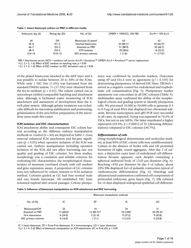
ResultsESC harvestIn a series of experiments, 34 mated mice produced a totalof 613 zygotes, 566 (92.3%) of which developed to blast-ocyst stage after four days of culture. Five preliminarystudies were performed in which 126 intact blastocystswere plated on MEFs and cultured in DMEM supple-mented with serum. Two days later, 82 (65.1%) ICMs hadhatched and attached to the feeder cell layer. On platingday 5 (D5), 71 ICMs (56.3%) were successfully dissoci-ated and replated, However, no ESC lines were estab-lished. Optimal medium for murine ESCs was determinedfrom nine experiments where 126 intact blastocysts wereallocated to DMEM + 20% serum (Fig 4a–e) vs. Ko-M +15% Ko-S (Table 1) (Fig.4f–j). In both media, over 60%
Grading embryonic stem cell coloniesFigure 3Grading embryonic stem cell colonies. Examples of ESC classification into one of the three grades: Grade Good (GG), Average (GA), or Poor (GP). In GG colonies the area occupied by pluripotent cells is >70% of the entire colony (a). Approximately 60% of the colony is occupied by pluripotent cells in GA colonies (b), and a GP colony features only ~20% of pluripotent cells (c). Scale = 30 μm.
Page 5 of 16(page number not for citation purposes)
Journal of Translational Medicine 2006, 4:20 http://www.translational-medicine.com/content/4/1/20
Page 6 of 16(page number not for citation purposes)
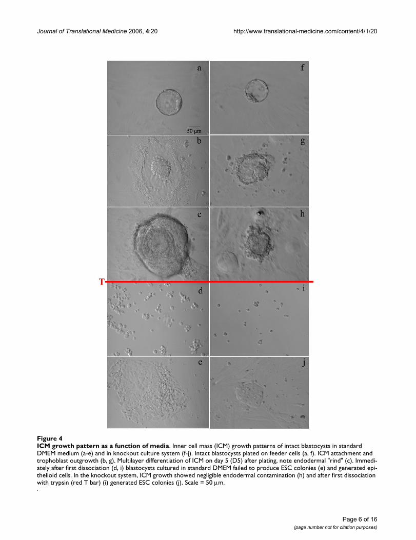
ICM growth pattern as a function of mediaFigure 4ICM growth pattern as a function of media. Inner cell mass (ICM) growth patterns of intact blastocysts in standard DMEM medium (a-e) and in knockout culture system (f-j). Intact blastocysts plated on feeder cells (a, f). ICM attachment and trophoblast outgrowth (b, g). Multilayer differentiation of ICM on day 5 (D5) after plating, note endodermal "rind" (c). Immedi-ately after first dissociation (d, i) blastocysts cultured in standard DMEM failed to produce ESC colonies (e) and generated epi-thelioid cells. In the knockout system, ICM growth showed negligible endodermal contamination (h) and after first dissociation with trypsin (red T bar) (i) generated ESC colonies (j). Scale = 50 μm.
Journal of Translational Medicine 2006, 4:20 http://www.translational-medicine.com/content/4/1/20
of the plated blastocysts attached to the MEF layer and itwas possible to isolate between 20 to 30% of the ICMs.While only 1 ESC line (1.6%) was harvested from thestandard DMEM system, 11 (17.5%) were obtained fromthe Ko-M medium (p < 0.01). The culture carried out inmicrodrops yielded comparable hatching and attachmentrates, although it facilitated easier monitoring of ICMattachment and assessment of development than the 4-well plate system. Although gelatin treatment was techni-cally difficult for microdrop stabilization and positioning,gelatinization of the area before preparation of the micro-drop areas made this easier.
ICM isolation and ESC characterizationICM attachment ability and consequent ESC colony har-vest according to the different embryo manipulationmethods vs. control (n = 84) are depicted in Table 2. Zonaremoval enhanced ICM attachment and became signifi-cant (p < 0.05) when operative isolation of the ICM wascarried out. Embryo manipulation including operativeisolation of the ICM did not affect harvesting rate norquality and grading of ESC colonies. For these studies,morphology was a consistent and reliable criterion forevaluating ESC characteristics, the morphological charac-teristics of stemness correlated closely with cell markersand gene expression assays. A proportion of GG colonieswere not influenced by culture systems or ICM isolationmethod. Colonies graded as GG had four normal maleand one female karyotype. All established ESC linesremained euploid after several passages. Colony pluripo-
tency was confirmed by molecular markers. Outcomesusing AP and Oct-4 were in agreement (χ2 = 0.105) fordetermining pluripotency of derived ESC lines. TROMA-1served as a negative control for endodermal and trophob-lastic cell contamination (Fig. 5). Pluripotency markerassessment was concordant in all ESC colonies (Table 3).Multimarker assay confirmed the ability of the morpho-logical criteria and grading system to identify pluripotentcells. We processed 10,000 to 30,000 cells to generate 0.3to 0.9 μg of total RNA that displayed two ribosomal sub-units. Reverse transcription and qRT-PCR were successfulin all cases. As expected, Nanog was expressed in 74.6% ofESCs, but not in any MEFs. The latter manifested a higherexpression (69.6%, p = 0.0001) of Ttr (denoting differen-tiation) compared to ESC colonies (40.7%).
Differentiation of cellsUsing morphological determinants and molecular mark-ers, at least 80% of ESC colony cells were undifferentiated.Culture in the absence of feeder cells and LIF permittedformation of tight, round aggregates. After day 2 of cul-ture, a distinctive outer layer of endodermal cell differen-tiation became apparent, each droplet containing aspherical embryoid body of ~320 μm diameter (Fig. 6).Reaching ~450 μm diameter by day 9 of culture, about20% EBs displayed foci of pulsatile contraction typical ofcardiomyocyte differentiation (Fig. 6). Histology andultrastructural examination confirmed cell components ofprimordial embryonic germ layers (Fig. 7). EBs culturedfor 16 days displayed widespread epithelial cell differenti-
Table 2: Influence of blastocyst manipulation on ICM attachment and ESC harvesting
Blastocyst manipulation methods
No. of (%) IC ZF IS LD
Blastocysts/ICM 25 14 24 21Attached on MEF 17 (68.0)a 12 (85.7)a,b 24 (100)b 20 (95.2)b
First dissociation 6 (24.0) 3 (21.4) 9 (37.5) 9 (42.8)ESC primary colonies 4 (16.0) 1 (7.1) 3 (12.5) 5 (23.8)
IC = Intact blastocyst, ZF = Zona-Free blastocyst, IS = Immunosurgery, LD = Laser dissectiona,bχ2, 2 × 4, 3 df, Effect of blastocyst manipulation on ICM attachment, (IC vs IS & LD), p < 0.05
Table 1: Intact blastocyst culture on MEF in different media
Embryonic day (d) Plating day (D) No. of (%) DMEM + 10%NCS, 10% FBS Ko-M + 15% Ko-S
d4 D0 Blastocysts (I) plated 63 63d5–6 D1–2 Hatched blastocysts 53 (84.1)a 42 (66.7)b
d6–7 D2–3 Attached on MEF 51 (80.9) 42 (66.7)d8–9 D4–5 ICM isolation 18 (28.6) 14 (22.2)
d14–16 D10–12 ESC primary colonies 1 (1.6)c 11 (17.5)d
FBS = fetal bovine serum, NCS = newborn calf serum, Ko-M = Knockout™ DMEM, Ko-S = Knockout™ serum replacementa,bχ2, 2 × 2, 1 df, Effect of ESC medium on hatching rate, p < 0.05c,dχ2, 2 × 2, 1 df, Effect of ESC medium on ESC derivation rate, p < 0.05
Page 7 of 16(page number not for citation purposes)

Journal of Translational Medicine 2006, 4:20 http://www.translational-medicine.com/content/4/1/20
ation as well as cells lining the lumen of cavity foundbetween the EBs. Both simple and stratified epithelial cellswere present. Testes injected with ESCs uniformly devel-oped tumors containing derivatives of all three embryonicgerm layers including mucus-producing columnar epithe-
lium, ciliated columnar epithelium, glandular epitheliumarranged in acini, endothelium lined vessels within intra-luminal neutrophils, striated muscle, bone, cartilage, neu-ral tissue (often forming rosettes), and keratinizingepithelium (Fig. 8).
Localization of pluripotency markersFigure 5Localization of pluripotency markers. Alkaline phosphatase (AP) activity (left), Oct-4 (center), and TROMA-1 (right) expression as measured in expanded and hatched blastocysts (top row, a-c), mouse embryonic fibroblasts (middle row, d-f) and embryonic stem cell colonies (bottom row, g-i) by immunofluorescent analysis. In left column, red signals indicate positive AP activity (a, g). In center, red signals identify positive Oct-4 expression (b, h) while the right column TROMA-1 expression is shown as green signals (c, hatched blastocyst). DAPI was used for nuclear DNA counterstaining as indicated by blue signals. Scale = 30 μm.
Page 8 of 16(page number not for citation purposes)

Journal of Translational Medicine 2006, 4:20 http://www.translational-medicine.com/content/4/1/20
Performance of cell- free culture systemIntact blastocysts (n = 131) were laser dissected and 65 ofthese were plated on MEF with the remainder (n = 66) ongelatin-coated dishes (Table 4). Laser dissection was suc-cessful in all cases. In the absence of MEF, attachment of
ICM occurred at a similar rate than control (81.8% vs.92.3%), and observed growth patterns were also compa-rable. A total of 6 (9.1%) ESC lines was established on gel-atin, while 15 (23.1%) on MEF. ESC harvest efficiency wassignificantly higher on feeder cells (p < 0.05). Cell lines
Coalescence and development of embryoid bodiesFigure 6Coalescence and development of embryoid bodies. Day 1: Aggregation of individual ESCs to form spherical bodies (EB = embryiod body). Day 3: EBs ranging from 100–250 μm mean diameter. Day 4: Growing EBs of ~350 μm. Day 9: An EB with a cystic structure. Day 17: EB with outgrowth and vesicular cavity.
Table 3: ESC marker expression in derived ESC lines
Stemness phenotype*
ESC line ID (Experiment# Blastocyst#) Blastocyst manipulation methods Initial grading AP activity‰ Oct-4™ TROMA-1∫
167 I Poor + + -1634 I Average + + -1635 I Good + + -176 ZF Poor + + -1713 IS Average + + -
* + and – indicate "positive" or "not detectable" activity or protein expression, respectively†AP activity is positive marker for pluripotent cells‡Oct-4, Octamer-binding transcriptional factor 4, is positive marker for pluripotent cells∫ TROMA-1, trophectodermal monoclonal antibody, which reacts to keratin filaments in trophectodermal and endodermal cells, is assessed as a negative marker for pluripotent cells
Page 9 of 16(page number not for citation purposes)

Journal of Translational Medicine 2006, 4:20 http://www.translational-medicine.com/content/4/1/20
obtained in feeder cell-free conditions maintainedpluripotency to the same degree as that observed in con-trols
DiscussionAs with any scientific investigation using animal models,it is envisioned that experience gained from murine ESCwork will supply answers to challenges still vexing thehuman ESC field. To be sure, the recent identification ofproteins and sialic acid residues on stem cell surfaces [33]resulting from MEF contamination has redoubled theneed to develop methods to culture ESCs without feedercells, and further research is needed to derive ESC lines inmore controlled systems [8,13,34]. In our investigationwe obtained ESC lines without feeder layers and sera,although the efficiency was lower than desired. These celllines demonstrated all ESC characteristics with respect tomorphology, marker expression, and differentiation capa-bility both in vivo and in vitro. These results therefore rep-
resent a start along the way to establish ESC lines incontrolled and xenogenic by-product free culture condi-tions.
The morphological grading method developed hereproved useful to characterize colonies as well as to moni-tor pluripotent status of the cells, and was helpful to iden-tify early differentiation. All cell lines obtainedexperimentally demonstrated typical ES morphology vialight microscopy, staining strongly for alkaline phos-phatase activity and expressing Oct-4 [13]. Molecularmarkers such as Oct-4 [15,16] and Nanog expression[23,24] are critical to confirm stemness because of theirrole in pluripotency regulation and cellular self-renewal.Assessment of trophoblastic markers such as TROMA-1[19,20] and Ttr [26,27] supported cytometric grading inour derived ESC lines. The development of embryoid bod-ies was a further indicator that harvested cells retained theability to develop into the three embryonic disc compo-
Ultrastructure of embryoid bodiesFigure 7Ultrastructure of embryoid bodies. Transmission electron microscopy of embryoid body showing three embryonic germ layers including keratin filaments (ectoderm), cardiomyocyte with contractile components (mesoderm), and microvilli (endo-derm).
Page 10 of 16(page number not for citation purposes)
Journal of Translational Medicine 2006, 4:20 http://www.translational-medicine.com/content/4/1/20
nents. Additionally, the observation that all establishedcell lines gave rise to teratomas in vivo served to confirmtheir pluripotency.
A recent study [35] described only a 2% rate of ESC deri-vation using C57BL/6 mice, a model with a similar geneticbackground compared to our B6D2-F1 (C57BL/6J × DBA/2J) strain. Results from the present study are in generalagreement with those of previous reports [36,37] whereDMEM + serum was utilized, although substantially lowerthan the 30% reported from the 129 strain [1]. The higherESC efficiency reported from the 129 strain may be due tothe fact that this is an inbred strain and is characterized bythe persistence of stem cells into adulthood [10], or, alter-natively, because of absence of a PGC survival factor[38,39]. In our experiments, ESC harvest was improved bysubstituting a combination of serum free Ko-M + Ko-S
media [40], the beneficial effect of which may be ascribedto the relatively low osmolarity and the absence of differ-entiating factors present in bovine sera [36,41-43].
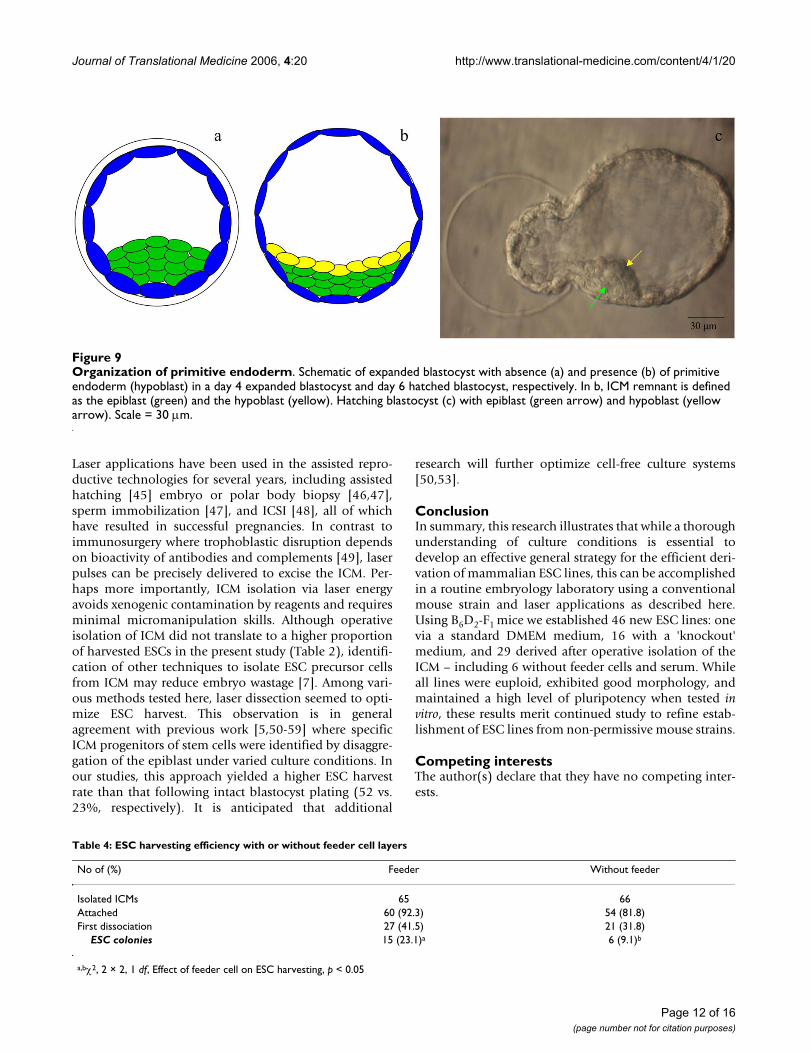
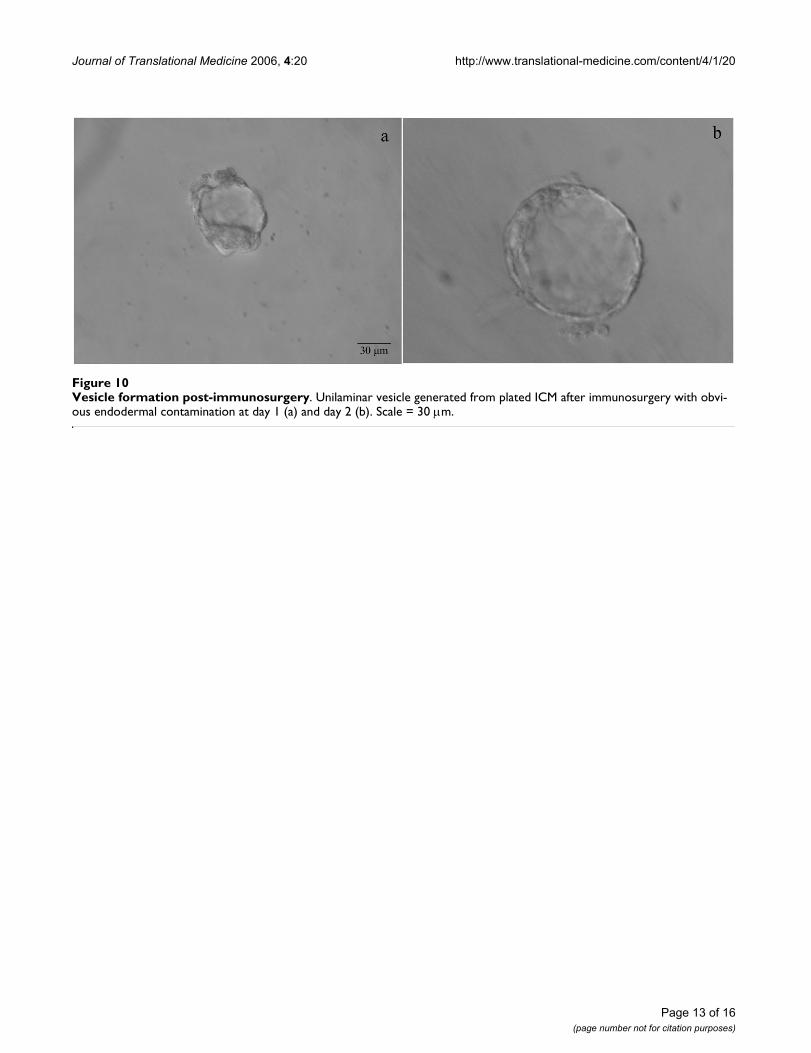
Reduced ESC harvesting following immunosurgery mightbe explained by endodermal contamination (Fig. 9) and/or persistence of trophoblastic cells as proven by the for-mation of unilaminar vesicles (Fig. 10). Most ICMs iso-lated by immunosurgery from day 5 blastocysts canregenerate an external layer of endodermal cells once cul-tured in vitro [44], offering further evidence that immuno-surgery is not ideal for isolation of uncontaminatedepiblasts. As cytotoxic antibodies destroy all blastocystcell types, a less than perfect timing of exposure to suchreagents might result in escape of some endodermal cells[44] allowing them to form the characteristic rind (Fig.11).
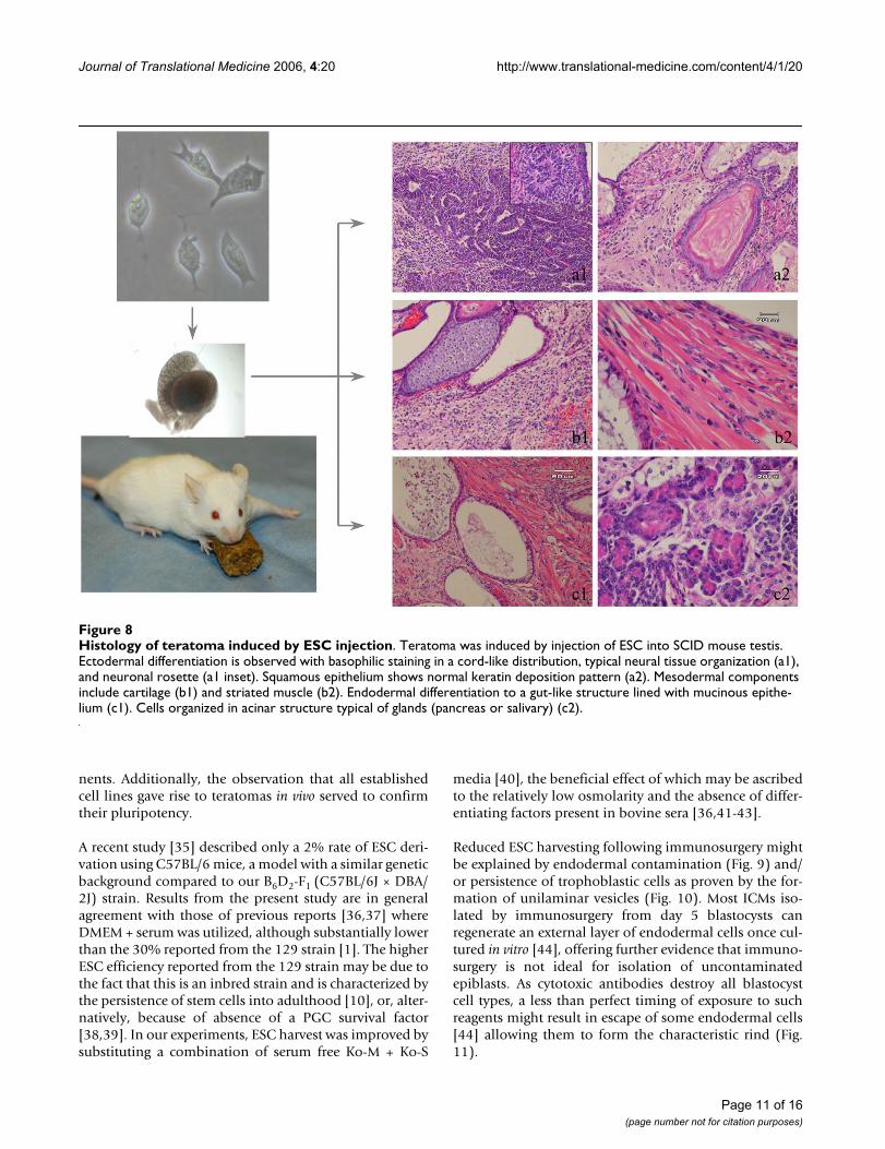
Histology of teratoma induced by ESC injectionFigure 8Histology of teratoma induced by ESC injection. Teratoma was induced by injection of ESC into SCID mouse testis. Ectodermal differentiation is observed with basophilic staining in a cord-like distribution, typical neural tissue organization (a1), and neuronal rosette (a1 inset). Squamous epithelium shows normal keratin deposition pattern (a2). Mesodermal components include cartilage (b1) and striated muscle (b2). Endodermal differentiation to a gut-like structure lined with mucinous epithe-lium (c1). Cells organized in acinar structure typical of glands (pancreas or salivary) (c2).
Page 11 of 16(page number not for citation purposes)
Journal of Translational Medicine 2006, 4:20 http://www.translational-medicine.com/content/4/1/20
Laser applications have been used in the assisted repro-ductive technologies for several years, including assistedhatching [45] embryo or polar body biopsy [46,47],sperm immobilization [47], and ICSI [48], all of whichhave resulted in successful pregnancies. In contrast toimmunosurgery where trophoblastic disruption dependson bioactivity of antibodies and complements [49], laserpulses can be precisely delivered to excise the ICM. Per-haps more importantly, ICM isolation via laser energyavoids xenogenic contamination by reagents and requiresminimal micromanipulation skills. Although operativeisolation of ICM did not translate to a higher proportionof harvested ESCs in the present study (Table 2), identifi-cation of other techniques to isolate ESC precursor cellsfrom ICM may reduce embryo wastage [7]. Among vari-ous methods tested here, laser dissection seemed to opti-mize ESC harvest. This observation is in generalagreement with previous work [5,50-59] where specificICM progenitors of stem cells were identified by disaggre-gation of the epiblast under varied culture conditions. Inour studies, this approach yielded a higher ESC harvestrate than that following intact blastocyst plating (52 vs.23%, respectively). It is anticipated that additional
research will further optimize cell-free culture systems[50,53].
ConclusionIn summary, this research illustrates that while a thoroughunderstanding of culture conditions is essential todevelop an effective general strategy for the efficient deri-vation of mammalian ESC lines, this can be accomplishedin a routine embryology laboratory using a conventionalmouse strain and laser applications as described here.Using B6D2-F1 mice we established 46 new ESC lines: onevia a standard DMEM medium, 16 with a 'knockout'medium, and 29 derived after operative isolation of theICM – including 6 without feeder cells and serum. Whileall lines were euploid, exhibited good morphology, andmaintained a high level of pluripotency when tested invitro, these results merit continued study to refine estab-lishment of ESC lines from non-permissive mouse strains.
Competing interestsThe author(s) declare that they have no competing inter-ests.
Organization of primitive endodermFigure 9Organization of primitive endoderm. Schematic of expanded blastocyst with absence (a) and presence (b) of primitive endoderm (hypoblast) in a day 4 expanded blastocyst and day 6 hatched blastocyst, respectively. In b, ICM remnant is defined as the epiblast (green) and the hypoblast (yellow). Hatching blastocyst (c) with epiblast (green arrow) and hypoblast (yellow arrow). Scale = 30 μm.
Table 4: ESC harvesting efficiency with or without feeder cell layers
No of (%) Feeder Without feeder
Isolated ICMs 65 66Attached 60 (92.3) 54 (81.8)First dissociation 27 (41.5) 21 (31.8)
ESC colonies 15 (23.1)a 6 (9.1)b
a,bχ2, 2 × 2, 1 df, Effect of feeder cell on ESC harvesting, p < 0.05
Page 12 of 16(page number not for citation purposes)
Journal of Translational Medicine 2006, 4:20 http://www.translational-medicine.com/content/4/1/20
Page 13 of 16(page number not for citation purposes)
Vesicle formation post-immunosurgeryFigure 10Vesicle formation post-immunosurgery. Unilaminar vesicle generated from plated ICM after immunosurgery with obvi-ous endodermal contamination at day 1 (a) and day 2 (b). Scale = 30 μm.

Journal of Translational Medicine 2006, 4:20 http://www.translational-medicine.com/content/4/1/20
Page 14 of 16(page number not for citation purposes)
Observed growth patterns according to ICM isolation techniqueFigure 11Observed growth patterns according to ICM isolation technique. Attachment of ICM and trophectodermal out-growth with large (b1, b2, after immunosurgery) or minimal (a1, a2, intact blastocyst) endodermal contamination. Such con-tamination is completely absent following laser dissection (c1, c2). First column shown at 200×, second column shown at 400×. Scale = 50 μm.
Journal of Translational Medicine 2006, 4:20 http://www.translational-medicine.com/content/4/1/20
Authors' contributionsNK coordinated all laboratory work on the project withassistance from QV, ESS, ZR, and GDP. GDP conceptual-ized the research, reviewed the data, and supervised man-uscript preparation.
AcknowledgementsWe are grateful to Drs. Willie Mark and Mohan Vemuri for their technical suggestions and Dr. Matthew S. Liao for his editorial comments. TROMA-1 antibody was provided by the Developmental Studies Hybridoma Bank under the auspices of the NICHD, and maintained at The University of Iowa, Department of Biological Sciences, Iowa City, Iowa USA.
References1. Evans MJ, Kaufman MH: Establishment in culture of pluripoten-
tial cells from mouse embryos. Nature 1981, 292:154-156.2. Robertson EJ: Teratocarcinomas and embryonic stem cells: a
practical approach. In Practical approach series IRL Press Oxford,UK; 1987.
3. Solter D, Knowles BB: Immunosurgery of mouse blastocyst.Proc Natl Acad Sci USA 1975, 72:5099-5102.
4. Martin GR: Isolation of a pluripotent cell line from earlymouse embryos cultured in medium conditioned by terato-carcinoma stem cells. Proc Natl Acad Sci USA 1981, 78:7634-7638.
5. Brook FA, Gardner RL: The origin and efficient derivation ofembryonic stem cells in the mouse. Proc Natl Acad Sci USA 1997,94:5709-5712.
6. Chung Y, Klimanskaya I, Becker S, Marh J, Lu SJ, Johnson J, Meisner L,Lanza R: Embryonic and extraembryonic stem cell linesderived from single mouse blastomeres. Nature 2006,439:216-219.
7. Sills ES, Takeuchi T, Tanaka N, Neri QV, Palermo GD: Identificationand isolation of embryonic stem cells in reproductive endo-crinology: theoretical protocols for conservation of humanembryos derived from in vitro fertilization. Theor Biol MedModel 2005, 2:25-32.
8. Pease S, Braghetta P, Gearing D, Grail D, Williams RL: Isolation ofembryonic stem (ES) cells in media supplemented withrecombinant leukemia inhibitory factor (LIF). Dev Biol 1990,141:44-52.
9. Nagy A, Rossant J, Nagy R, Abramow-Newerly W, Roder JC: Deri-vation of completely cell culture-derived mice from early-passage embryonic stem cells. Proc Natl Acad Sci USA 1993,90:8424-8428.
10. Stevens LC: A new inbred subline of mice (129-terSv) with ahigh incidence of spontaneous congenital testicular terato-mas. J Natl Cancer Inst 1973, 50:235-242.
11. Surani MA: Stem cells: how to make eggs and sperm. Nature2004, 427:106-107.
12. Smith AG, Heath JK, Donaldson DD, Wong GG, Moreau J, Stahl M,Rogers D: Inhibition of pluripotential embryonic stem cell dif-ferentiation by purified polypeptides. Nature 1988,336:688-690.
13. Nichols J, Evans EP, Smith AG: Establishment of germ-line-com-petent embryonic stem (ES) cells using differentiation inhib-iting activity. Development 1990, 110:1341-1348.
14. Johnson LV, Calarco PG, Siebert ML: Alkaline phosphatase activ-ity in the preimplantation mouse embryo. J Embryol Exp Mor-phol 1977, 40:83-89.
15. Okamoto K, Okazawa H, Okuda A, Sakai M, Muramatsu M, HamadaH: A novel octamer binding transcription factor is differen-tially expressed in mouse embryonic cells. Cell 1990,60:461-472.
16. Rosner MH, Vigano MA, Ozato K, Timmons PM, Poirier F, Rigby PW,Staudt LM: A POU-domain transcription factor in early stemcells and germ cells of the mammalian embryo. Nature 1990,345:686-692.
17. Scholer HR, Ruppert S, Suzuki N, Chowdhury K, Gruss P: New typeof POU domain in germ line-specific protein Oct-4. Nature1990, 344:435-439.
18. Palmieri SL, Peter W, Hess H, Scholer HR: Oct-4 transcription fac-tor is differentially expressed in the mouse embryo during
establishment of the first two extraembryonic cell lineagesinvolved in implantation. Dev Biol 1994, 166:259-267.
19. Brulet P, Babinet C, Kemler R, Jacob F: Monoclonal antibodiesagainst trophectoderm-specific markers during mouse blas-tocyst formation. Proc Natl Acad Sci USA 1980, 77:4113-4117.
20. Kemler R, Brulet P, Schnebelen MT, Gaillard J, Jacob F: Reactivity ofmonoclonal antibodies against intermediate filament pro-teins during embryonic development. J Embryol Exp Morphol1981, 64:45-60.
21. Doetschman TC, Eistetter H, Katz M, Schmidt W, Kemler R: The invitro development of blastocyst-derived embryonic stemcell lines: formation of visceral yolk sac, blood islands andmyocardium. J Embryol Exp Morphol 1985, 87:27-45.
22. Ziomek CA, Lepire ML, Torres I: A highly fluorescent simultane-ous azo dye technique for demonstration of nonspecific alka-line phosphatase activity. J Histochem Cytochem 1990,38:437-442.
23. Mitsui K, Tokuzawa Y, Itoh H, Segawa K, Murakami M, Takahashi K,Maruyama M, Maeda M, Yamanaka S: The homeoprotein Nanogis required for maintenance of pluripotency in mouse epi-blast and ES cells. Cell 2003, 113:631-642.
24. Chambers I, Colby D, Robertson M, Nichols J, Lee S, Tweedie S,Smith A: Functional expression cloning of Nanog, a pluripo-tency sustaining factor in embryonic stem cells. Cell 2003,113:643-655.
25. Hart AH, Hartley L, Ibrahim M, Robb L: Identification, cloning andexpression analysis of the pluripotency promoting Nanoggenes in mouse and human. Dev Dyn 2004, 230:187-198.
26. Soprano DR, Soprano KJ, Goodman DS: Retinol-binding proteinand transthyretin mRNA levels in visceral yolk sac and liverduring fetal development in the rat. Proc Natl Acad Sci USA 1986,83:7330-7334.
27. Makover A, Soprano DR, Wyatt ML, Goodman DS: An in situ-hybridization study of the localization of retinol-binding pro-tein and transthyretin messenger RNAs during fetal devel-opment in the rat. Differentiation 1989, 40:17-25.
28. Farrington SM, Belaoussoff M, Baron MH: Winged-helix, Hedge-hog and Bmp genes are differentially expressed in distinctcell layers of the murine yolk sac. Mech Dev 1997, 62:197-211.
29. Abe K, Niwa H, Iwase K, Takiguchi M, Mori M, Abe SI, Abe K, Yama-mura KI: Endoderm-specific gene expression in embryonicstem cells differentiated to embryoid bodies. Exp Cell Res1996, 229:27-34.
30. Kawase E, Yamazaki Y, Yagi T, Yanagimachi R, Pedersen RA: Mouseembryonic stem (ES) cell lines established from neuronalcell-derived cloned blastocysts. Genesis 2000, 28:156-163.
31. Takeuchi T, Neri QV, Katagiri Y, Rosenwaks Z, Palermo GD: Effectof treating induced mitochondrial damage on embryonicdevelopment and epigenesis. Biol Reprod 2005, 72:584-592.
32. Wobus AM, Wallukat G, Hescheler J: Pluripotent mouse embry-onic stem cells are able to differentiate into cardiomyocytesexpressing chronotropic responses to adrenergic and cholin-ergic agents and Ca2+ channel blockers. Differentiation 1991,48:173-182.
33. Martin MJ, Muotri A, Gage F, Varki A: Human embryonic stemcells express an immunogenic nonhuman sialic acid. Nat Med2005, 11:228-232.
34. Klimanskaya I, Chung Y, Meisner L, Johnson J, West MD, Lanza R:Human embryonic stem cells derived without feeder cells.Lancet 2005, 365:4997-5002.
35. Baharvand H, Matthaei KI: Culture condition difference forestablishment of new embryonic stem cell lines from theC57BL/6 and BALB/c mouse strains. In Vitro Cell Dev Biol Anim2004, 40:76-81.
36. Ledermann B, Burki K: Establishment of a germ-line competentC57BL/6 embryonic stem cell line. Exp Cell Res 1991,197:254-258.
37. Auerbach W, Dunmore JH, Fairchild-Huntress V, Fang Q, AuerbachAB, Huszar D, Joyner AL: Establishment and chimera analysis of129/SvEv- and C57BL/6-derived mouse embryonic stem celllines. Biotechniques 2000, 29:1024-1028. 1030, 1032
38. Matin A, Collin GB, Asada Y, Varnum D, Nadeau JH: Susceptibilityto testicular germ-cell tumours in a 129.MOLF-Chr 19 chro-mosome substitution strain. Nat Genet 1999, 23:237-240.
39. Donovan PJ, de Miguel MP: Turning germ cells into stem cells.Curr Opin Genet Dev 2003, 13:463-471.
Page 15 of 16(page number not for citation purposes)
http://www.ncbi.nlm.nih.gov/entrez/query.fcgi?cmd=Retrieve&db=PubMed&dopt=Abstract&list_uids=7242681
http://www.ncbi.nlm.nih.gov/entrez/query.fcgi?cmd=Retrieve&db=PubMed&dopt=Abstract&list_uids=7242681
http://www.ncbi.nlm.nih.gov/entrez/query.fcgi?cmd=Retrieve&db=PubMed&dopt=Abstract&list_uids=1108013
http://www.ncbi.nlm.nih.gov/entrez/query.fcgi?cmd=Retrieve&db=PubMed&dopt=Abstract&list_uids=6950406
http://www.ncbi.nlm.nih.gov/entrez/query.fcgi?cmd=Retrieve&db=PubMed&dopt=Abstract&list_uids=6950406
http://www.ncbi.nlm.nih.gov/entrez/query.fcgi?cmd=Retrieve&db=PubMed&dopt=Abstract&list_uids=6950406
http://www.ncbi.nlm.nih.gov/entrez/query.fcgi?cmd=Retrieve&db=PubMed&dopt=Abstract&list_uids=9159137
http://www.ncbi.nlm.nih.gov/entrez/query.fcgi?cmd=Retrieve&db=PubMed&dopt=Abstract&list_uids=9159137
http://www.ncbi.nlm.nih.gov/entrez/query.fcgi?cmd=Retrieve&db=PubMed&dopt=Abstract&list_uids=8378314
http://www.ncbi.nlm.nih.gov/entrez/query.fcgi?cmd=Retrieve&db=PubMed&dopt=Abstract&list_uids=8378314
http://www.ncbi.nlm.nih.gov/entrez/query.fcgi?cmd=Retrieve&db=PubMed&dopt=Abstract&list_uids=8378314
http://www.ncbi.nlm.nih.gov/entrez/query.fcgi?cmd=Retrieve&db=PubMed&dopt=Abstract&list_uids=4692863
http://www.ncbi.nlm.nih.gov/entrez/query.fcgi?cmd=Retrieve&db=PubMed&dopt=Abstract&list_uids=4692863
http://www.ncbi.nlm.nih.gov/entrez/query.fcgi?cmd=Retrieve&db=PubMed&dopt=Abstract&list_uids=4692863
http://www.ncbi.nlm.nih.gov/entrez/query.fcgi?cmd=Retrieve&db=PubMed&dopt=Abstract&list_uids=3143917
http://www.ncbi.nlm.nih.gov/entrez/query.fcgi?cmd=Retrieve&db=PubMed&dopt=Abstract&list_uids=3143917
http://www.ncbi.nlm.nih.gov/entrez/query.fcgi?cmd=Retrieve&db=PubMed&dopt=Abstract&list_uids=2129226
http://www.ncbi.nlm.nih.gov/entrez/query.fcgi?cmd=Retrieve&db=PubMed&dopt=Abstract&list_uids=2129226
http://www.ncbi.nlm.nih.gov/entrez/query.fcgi?cmd=Retrieve&db=PubMed&dopt=Abstract&list_uids=2129226
http://www.ncbi.nlm.nih.gov/entrez/query.fcgi?cmd=Retrieve&db=PubMed&dopt=Abstract&list_uids=1967980
http://www.ncbi.nlm.nih.gov/entrez/query.fcgi?cmd=Retrieve&db=PubMed&dopt=Abstract&list_uids=1967980
http://www.ncbi.nlm.nih.gov/entrez/query.fcgi?cmd=Retrieve&db=PubMed&dopt=Abstract&list_uids=1972777
http://www.ncbi.nlm.nih.gov/entrez/query.fcgi?cmd=Retrieve&db=PubMed&dopt=Abstract&list_uids=1972777
http://www.ncbi.nlm.nih.gov/entrez/query.fcgi?cmd=Retrieve&db=PubMed&dopt=Abstract&list_uids=1690859
http://www.ncbi.nlm.nih.gov/entrez/query.fcgi?cmd=Retrieve&db=PubMed&dopt=Abstract&list_uids=1690859
http://www.ncbi.nlm.nih.gov/entrez/query.fcgi?cmd=Retrieve&db=PubMed&dopt=Abstract&list_uids=7958450
http://www.ncbi.nlm.nih.gov/entrez/query.fcgi?cmd=Retrieve&db=PubMed&dopt=Abstract&list_uids=7958450
http://www.ncbi.nlm.nih.gov/entrez/query.fcgi?cmd=Retrieve&db=PubMed&dopt=Abstract&list_uids=7958450
http://www.ncbi.nlm.nih.gov/entrez/query.fcgi?cmd=Retrieve&db=PubMed&dopt=Abstract&list_uids=7958450
http://www.ncbi.nlm.nih.gov/entrez/query.fcgi?cmd=Retrieve&db=PubMed&dopt=Abstract&list_uids=6933460
http://www.ncbi.nlm.nih.gov/entrez/query.fcgi?cmd=Retrieve&db=PubMed&dopt=Abstract&list_uids=6933460
http://www.ncbi.nlm.nih.gov/entrez/query.fcgi?cmd=Retrieve&db=PubMed&dopt=Abstract&list_uids=6933460
http://www.ncbi.nlm.nih.gov/entrez/query.fcgi?cmd=Retrieve&db=PubMed&dopt=Abstract&list_uids=6171607
http://www.ncbi.nlm.nih.gov/entrez/query.fcgi?cmd=Retrieve&db=PubMed&dopt=Abstract&list_uids=6171607
http://www.ncbi.nlm.nih.gov/entrez/query.fcgi?cmd=Retrieve&db=PubMed&dopt=Abstract&list_uids=6171607
http://www.ncbi.nlm.nih.gov/entrez/query.fcgi?cmd=Retrieve&db=PubMed&dopt=Abstract&list_uids=3897439
http://www.ncbi.nlm.nih.gov/entrez/query.fcgi?cmd=Retrieve&db=PubMed&dopt=Abstract&list_uids=3897439
http://www.ncbi.nlm.nih.gov/entrez/query.fcgi?cmd=Retrieve&db=PubMed&dopt=Abstract&list_uids=3897439
http://www.ncbi.nlm.nih.gov/entrez/query.fcgi?cmd=Retrieve&db=PubMed&dopt=Abstract&list_uids=1689343
http://www.ncbi.nlm.nih.gov/entrez/query.fcgi?cmd=Retrieve&db=PubMed&dopt=Abstract&list_uids=1689343
http://www.ncbi.nlm.nih.gov/entrez/query.fcgi?cmd=Retrieve&db=PubMed&dopt=Abstract&list_uids=1689343
http://www.ncbi.nlm.nih.gov/entrez/query.fcgi?cmd=Retrieve&db=PubMed&dopt=Abstract&list_uids=3463972
http://www.ncbi.nlm.nih.gov/entrez/query.fcgi?cmd=Retrieve&db=PubMed&dopt=Abstract&list_uids=3463972
http://www.ncbi.nlm.nih.gov/entrez/query.fcgi?cmd=Retrieve&db=PubMed&dopt=Abstract&list_uids=3463972
http://www.ncbi.nlm.nih.gov/entrez/query.fcgi?cmd=Retrieve&db=PubMed&dopt=Abstract&list_uids=2744271
http://www.ncbi.nlm.nih.gov/entrez/query.fcgi?cmd=Retrieve&db=PubMed&dopt=Abstract&list_uids=2744271
http://www.ncbi.nlm.nih.gov/entrez/query.fcgi?cmd=Retrieve&db=PubMed&dopt=Abstract&list_uids=2744271
http://www.ncbi.nlm.nih.gov/entrez/query.fcgi?cmd=Retrieve&db=PubMed&dopt=Abstract&list_uids=9152011
http://www.ncbi.nlm.nih.gov/entrez/query.fcgi?cmd=Retrieve&db=PubMed&dopt=Abstract&list_uids=9152011
http://www.ncbi.nlm.nih.gov/entrez/query.fcgi?cmd=Retrieve&db=PubMed&dopt=Abstract&list_uids=9152011
http://www.ncbi.nlm.nih.gov/entrez/query.fcgi?cmd=Retrieve&db=PubMed&dopt=Abstract&list_uids=8940246
http://www.ncbi.nlm.nih.gov/entrez/query.fcgi?cmd=Retrieve&db=PubMed&dopt=Abstract&list_uids=8940246
http://www.ncbi.nlm.nih.gov/entrez/query.fcgi?cmd=Retrieve&db=PubMed&dopt=Abstract&list_uids=1725163
http://www.ncbi.nlm.nih.gov/entrez/query.fcgi?cmd=Retrieve&db=PubMed&dopt=Abstract&list_uids=1725163
http://www.ncbi.nlm.nih.gov/entrez/query.fcgi?cmd=Retrieve&db=PubMed&dopt=Abstract&list_uids=1725163
http://www.ncbi.nlm.nih.gov/entrez/query.fcgi?cmd=Retrieve&db=PubMed&dopt=Abstract&list_uids=1959560

Journal of Translational Medicine 2006, 4:20 http://www.translational-medicine.com/content/4/1/20
Publish with BioMed Central and every scientist can read your work free of charge
"BioMed Central will be the most significant development for disseminating the results of biomedical research in our lifetime."
Sir Paul Nurse, Cancer Research UK
Your research papers will be:
available free of charge to the entire biomedical community
peer reviewed and published immediately upon acceptance
cited in PubMed and archived on PubMed Central
yours — you keep the copyright
Submit your manuscript here:http://www.biomedcentral.com/info/publishing_adv.asp
BioMedcentral
40. Cheng J, Dutra A, Takesono A, Garrett-Beal L, Schwartzberg PL:Improved generation of C57BL/6J mouse embryonic stemcells in a defined serum-free media. Genesis 2004, 39:100-104.
41. Gossler A, Doetschman T, Korn R, Serfling E, Kemler R: Transgen-esis by means of blastocyst-derived embryonic stem celllines. Proc Natl Acad Sci USA 1986, 83:9065-9069.
42. Hooper M, Hardy K, Handyside A, Hunter S, Monk M: HPRT-defi-cient (Lesch-Nyhan) mouse embryos derived from germlinecolonization by cultured cells. Nature 1987, 326:292-295.
43. McMahon AP, Bradley A: The Wnt-1 (int-1) proto-oncogene isrequired for development of a large region of the mousebrain. Cell 1990, 62:1073-1085.
44. Gardner RL: Regeneration of endoderm from primitive ecto-derm in the mouse embryo: fact or artifact? J Embryol Exp Mor-phol 1985, 88:303-326.
45. Obruca A, Strohmer H, Sakkas D, Menezo Y, Kogosowski A, BarakY, Feichtinger W: Use of lasers in assisted fertilization andhatching. Hum Reprod 1994, 9:1723-1726.
46. Veiga A, Sandalinas M, Benkhalifa M, Boada M, Carrera M, Santalo J,Barri PN, Menezo Y: Laser blastocyst biopsy for preimplanta-tion diagnosis in the human. Zygote 1997, 5:351-354.
47. Montag M, Rink K, Delacretaz G, van der Ven H: Laser-inducedimmobilization and plasma membrane permeabilization inhuman spermatozoa. Hum Reprod 2000, 15:846-52.
48. Rienzi L, Greco E, Ubaldi F, Iacobelli M, Martinez F, Tesarik J: Laser-assisted intracytoplasmic sperm injection. Fertil Steril 2001,76:1045-1047.
49. Hipp J, Atala A: Tissue engineering, stem cells, cloning, andparthenogenesis: new paradigms for therapy. J Exp Clin AssistReprod 2004, 1:3.
50. Ulloa-Montoya F, Verfaillie CM, Hu WS: Culture systems forpluripotent stem cells. J Biosci Bioeng 2005, 100:12-27.
51. Wolf DP, Kuo H-C, Pau F, Lester L: Progress with nonhuman pri-mate embryonic stem cells. Biol Reprod 2004, 71:1766-1771.
52. Stojkovic M, Lako M, Stojkovic P, Stewart R, Przyborski S, ArmstrongL, Evans J, Herbert M, Hyslop L, Ahmad S, Murdoch A, Strachan T:Derivation of human embryonic stem cells from day-8 blast-ocysts recovered after three-step in vitro culture. Stem Cells2004, 22:790-797.
53. Draper JS, Moore HD, Ruban LN, Gokhale PJ, Andrews PW: Cultureand characterization of human embryonic stem cells. StemCells Dev 2004, 13:325-336.
54. Conley BJ, Young JC, Trounson AO, Mollard R: Derivation, propa-gation and differentiation of human embryonic stem cells.Int J Biochem Cell Biol 2004, 36:555-567.
55. McLaren A, Durcova-Hills G: Germ cells and pluripotent stemcells in the mouse. Reprod Fertil Dev 2001, 13:661-664.
56. De Vos A, Van Steirteghem A: Zona harvesting, zona drilling andassisted hatching: new achievements in assisted reproduc-tion. Cells Tissues Organs 2000, 166:220-227.
57. Labosky PA, Barlow DP, Hogan BL: Embryonic germ cell linesand their derivation from mouse primordial germ cells. CibaFound Symp 1994, 182:157-168.
58. First NL, Sims MM, Park SP, Kent-First MJ: Systems for productionof calves from cultured bovine embryonic cells. Reprod FertilDev 1994, 6:553-562.
59. Cohen J, Talansky B, Alikani M: Laboratory techniques for han-dling gametes and embryos. Br Med Bull 1990, 46:643-653.
Page 16 of 16(page number not for citation purposes)
http://www.ncbi.nlm.nih.gov/entrez/query.fcgi?cmd=Retrieve&db=PubMed&dopt=Abstract&list_uids=3024164
http://www.ncbi.nlm.nih.gov/entrez/query.fcgi?cmd=Retrieve&db=PubMed&dopt=Abstract&list_uids=3024164
http://www.ncbi.nlm.nih.gov/entrez/query.fcgi?cmd=Retrieve&db=PubMed&dopt=Abstract&list_uids=3024164
http://www.ncbi.nlm.nih.gov/entrez/query.fcgi?cmd=Retrieve&db=PubMed&dopt=Abstract&list_uids=3821905
http://www.ncbi.nlm.nih.gov/entrez/query.fcgi?cmd=Retrieve&db=PubMed&dopt=Abstract&list_uids=3821905
http://www.ncbi.nlm.nih.gov/entrez/query.fcgi?cmd=Retrieve&db=PubMed&dopt=Abstract&list_uids=3821905
http://www.ncbi.nlm.nih.gov/entrez/query.fcgi?cmd=Retrieve&db=PubMed&dopt=Abstract&list_uids=2205396
http://www.ncbi.nlm.nih.gov/entrez/query.fcgi?cmd=Retrieve&db=PubMed&dopt=Abstract&list_uids=2205396
http://www.ncbi.nlm.nih.gov/entrez/query.fcgi?cmd=Retrieve&db=PubMed&dopt=Abstract&list_uids=2205396
http://www.ncbi.nlm.nih.gov/entrez/query.fcgi?cmd=Retrieve&db=PubMed&dopt=Abstract&list_uids=4078535
http://www.ncbi.nlm.nih.gov/entrez/query.fcgi?cmd=Retrieve&db=PubMed&dopt=Abstract&list_uids=4078535
http://www.ncbi.nlm.nih.gov/entrez/query.fcgi?cmd=Retrieve&db=PubMed&dopt=Abstract&list_uids=7836525
http://www.ncbi.nlm.nih.gov/entrez/query.fcgi?cmd=Retrieve&db=PubMed&dopt=Abstract&list_uids=7836525
http://www.ncbi.nlm.nih.gov/entrez/query.fcgi?cmd=Retrieve&db=PubMed&dopt=Abstract&list_uids=9563682
http://www.ncbi.nlm.nih.gov/entrez/query.fcgi?cmd=Retrieve&db=PubMed&dopt=Abstract&list_uids=9563682
http://www.ncbi.nlm.nih.gov/entrez/query.fcgi?cmd=Retrieve&db=PubMed&dopt=Abstract&list_uids=7835148
http://www.ncbi.nlm.nih.gov/entrez/query.fcgi?cmd=Retrieve&db=PubMed&dopt=Abstract&list_uids=7835148
http://www.ncbi.nlm.nih.gov/entrez/query.fcgi?cmd=Retrieve&db=PubMed&dopt=Abstract&list_uids=7569033
http://www.ncbi.nlm.nih.gov/entrez/query.fcgi?cmd=Retrieve&db=PubMed&dopt=Abstract&list_uids=7569033
http://www.ncbi.nlm.nih.gov/entrez/query.fcgi?cmd=Retrieve&db=PubMed&dopt=Abstract&list_uids=2207598