Large T antigen coding sequences of two DNA tumor viruses, BK and SV40, and nonrandom chromosome...
7
ELSEVIER Large T Antigen Coding Sequences of Two DNA Tumor Viruses, BK and SV40, and Nonrandom Chromosome Changes in Two Glioblastoma Cell Lines M. Tognon, R. Casalone, E Martini, M. De Mattei, P. Granata, E. Minelli, C. Arcuri, P. Collini, and V. Bocchini ABSTRACT: T~he T antigen (TAg) coding sequences of two DNA tumor viruses, BKV and SV40, were detected by Polymerase Chain Reaction (PCR) amplification followed by Southern-blot hybridization in two human glioblastoma multiforme derived cell lines. RT-PCR analysis indicated that these two TAg coding sequences were expressed in both tumor cell lines carrying the viral early region DNAs. More- over, analytical polyacrylamide gel electrophoresis (PAGE) and DNA sequence analyses showed that the amplified PCR products are indistinguishable from the TAg coding sequences of BKV and SV40 wild- type strains. Cytogenetic study performed in the two cell lines showed unbalanced changes, mainly gains of chromgsomes 3p, 5, 6, 7, and 19 and losses of chromosomes 3, 3q, 16, 9p22->pter, 18, and 20. Excess of chromosomes 6 and 7 are common to the two cell lines. The putative role of the TAg of the two DNA tumor viruses in transformation and karyotype changes is discussed. INTRODUCTION Several investigations reported on the associations between human BK, JC, simian SV40 polyomaviruses, and different tumors [1]. IlK virus (BKV) is an ubiquitous infectious agent for humans and latent BKV was found in various human tissues and organs, such as kidneys, ton- sils, normal brain tissues, bone, and peripheral blood cells [1-3]. Moreover, BKV DNA sequences have been detected in many human tumols, such as osteosarcomas, Ewing's tumors, and brain tumors of different histiotypes, includ- ing glioblastoma [3, 4]. BKV exhibits its oncogenicity in vivo, when inoculated in rodents [5]. JC virus (JCV) is From the Institute of Histology and General Embryology (M. T., F. M., M. D. M), School of Medicine, and lnterdepartment Center for Biotechnology (M. T.), University of Ferrara, Ferrara; Institute of General Biology and Medical Genetics (R. C.), School of Medi- cine, University of Pavia, Pavia; Laboratory of Cytogenetics (P. G., E. M.), Circle's Hospital, Varese; Department of Clinical Medicine, Pathology and Pharmacology (C. A., V. B.), School of Medicine, University of Peragia, Perugia; and the Institute of Gynecology and Obstetrics 2 (P C.), School of Medicine, University of Rome "La Sapienza," Rome, Italy. Address reprint reque',~tsto: Dr. Virginia Bocchini, Department of Clinical Medicine, Pathology and Pharmacology, Section of General Pathology and Immunology, School of Medicine, Univer- sity of Perugia, Montel~ce Polyclinic, Via Brunamonti, 06100 Perugia. Italy. Received August 11, 1995; accepted February 9, 1996. Cancer GenetCytogenet 90:17-23 (1996) ©Elsevier ScienceInc., 199~ 655 Avenueof the Americas.New York,NY 10010 another ubiquitous human polyomavirus associated with a rare demyelinating disease, the progressive multifocal leukoencephalopathy (PML]. Although JCV is oncogenic in rodents and it is able to infect human oligodendrocytes, this virus was never detected in human tumors [1, 2]. SV40 polyomavirus is an infectious agent for monkeys, but is incapable of infecting humans naturally. However, SV40 has been detected in human tumors, mainly brain tumors [1]. Recently, by Polymerase Chain Reaction (PCR) analysis, SV40-1ike sequences have been detected in pleural me- sotheliomas, in childhood ependymomas and choroid plexus papillomas, as well as in other brain tumors [6-8]. SV40 is a DNA tumor virus that exerts its oncogenicity in vivo by inducing brain tumors in rodents [9]. BKV, JCV, and SV40 transformation and tumorigenicity are essen- tially dependent on the expression of the early region, coding for large T antigen (TAg). This TAg, a transforming nuclear phosphoprotein, is mutagenic and induces karyo- typic changes in human cells, concomitant with cell im- mortalization [10]. Although human glioblastomas and their derived cell lines exhibit clonal nonrandom chromo- somal abnormalities [11], little is known about the events that cause chromosomal instability and rearrangements. Cytogenetic studies, using banding techniques performed on polyomaviruses, transformed fibroblasts, or latently in- fected tumor cells, are very few. Interestingly, some au- thors indicated that these transformed cell lines exhibit characteristic chromosome rearrangements [12, 13]. Iden- 0165-4608/96/$15.00 PII S0165-4608(96)00067-2
-
Upload
independent -
Category
Documents
-
view
1 -
download
0
Transcript of Large T antigen coding sequences of two DNA tumor viruses, BK and SV40, and nonrandom chromosome...
ELSEVIER
Large T Antigen Coding Sequences of Two DNA Tumor Viruses, BK and SV40, and Nonrandom Chromosome Changes in Two Glioblastoma Cell Lines
M. Tognon, R. Casalone, E Martini, M. De Mattei, P. Granata, E. Minelli, C. Arcuri, P. Collini, and V. Bocchini
ABSTRACT: T~he T antigen (TAg) coding sequences of two DNA tumor viruses, BKV and SV40, were detected by Polymerase Chain Reaction (PCR) amplification followed by Southern-blot hybridization in two human glioblastoma multiforme derived cell lines. RT-PCR analysis indicated that these two TAg coding sequences were expressed in both tumor cell lines carrying the viral early region DNAs. More- over, analytical polyacrylamide gel electrophoresis (PAGE) and DNA sequence analyses showed that the amplified PCR products are indistinguishable from the TAg coding sequences of BKV and SV40 wild- type strains. Cytogenetic study performed in the two cell lines showed unbalanced changes, mainly gains of chromgsomes 3p, 5, 6, 7, and 19 and losses of chromosomes 3, 3q, 16, 9p22->pter, 18, and 20. Excess of chromosomes 6 and 7 are common to the two cell lines. The putative role of the TAg of the two DNA tumor viruses in transformation and karyotype changes is discussed.
INTRODUCTION
Several investigations reported on the associations between human BK, JC, simian SV40 polyomaviruses, and different tumors [1]. IlK virus (BKV) is an ubiquitous infectious agent for humans and latent BKV was found in various human tissues and organs, such as kidneys, ton- sils, normal brain tissues, bone, and peripheral blood cells [1-3]. Moreover, BKV DNA sequences have been detected in many human tumols, such as osteosarcomas, Ewing's tumors, and brain tumors of different histiotypes, includ- ing glioblastoma [3, 4]. BKV exhibits its oncogenicity in vivo, when inoculated in rodents [5]. JC virus (JCV) is
From the Institute of Histology and General Embryology (M. T., F. M., M. D. M), School of Medicine, and lnterdepartment Center for Biotechnology (M. T.), University of Ferrara, Ferrara; Institute of General Biology and Medical Genetics (R. C.), School of Medi- cine, University of Pavia, Pavia; Laboratory of Cytogenetics (P. G., E. M.), Circle's Hospital, Varese; Department of Clinical Medicine, Pathology and Pharmacology (C. A., V. B.), School of Medicine, University of Peragia, Perugia; and the Institute of Gynecology and Obstetrics 2 (P C.), School of Medicine, University of Rome "La Sapienza," Rome, Italy.
Address reprint reque',~ts to: Dr. Virginia Bocchini, Department of Clinical Medicine, Pathology and Pharmacology, Section of General Pathology and Immunology, School of Medicine, Univer- sity of Perugia, Montel~ce Polyclinic, Via Brunamonti, 06100 Perugia. Italy.
Received August 11, 1995; accepted February 9, 1996.
Cancer Genet Cytogenet 90:17-23 (1996) © Elsevier Science Inc., 199~ 655 Avenue of the Americas. New York, NY 10010
another ubiquitous human polyomavirus associated with a rare demyelinating disease, the progressive multifocal leukoencephalopathy (PML]. Although JCV is oncogenic in rodents and it is able to infect human oligodendrocytes, this virus was never detected in human tumors [1, 2]. SV40 polyomavirus is an infectious agent for monkeys, but is incapable of infecting humans naturally. However, SV40 has been detected in human tumors, mainly brain tumors [1].
Recently, by Polymerase Chain Reaction (PCR) analysis, SV40-1ike sequences have been detected in pleural me- sotheliomas, in childhood ependymomas and choroid plexus papillomas, as well as in other brain tumors [6-8]. SV40 is a DNA tumor virus that exerts its oncogenicity in vivo by inducing brain tumors in rodents [9]. BKV, JCV, and SV40 transformation and tumorigenicity are essen- tially dependent on the expression of the early region, coding for large T antigen (TAg). This TAg, a transforming nuclear phosphoprotein, is mutagenic and induces karyo- typic changes in human cells, concomitant with cell im- mortalization [10]. Although human glioblastomas and their derived cell lines exhibit clonal nonrandom chromo- somal abnormalities [11], little is known about the events that cause chromosomal instability and rearrangements. Cytogenetic studies, using banding techniques performed on polyomaviruses, transformed fibroblasts, or latently in- fected tumor cells, are very few. Interestingly, some au- thors indicated that these transformed cell lines exhibit characteristic chromosome rearrangements [12, 13]. Iden-
0165-4608/96/$15.00 PII S0165-4608(96)00067-2
18 M. Tognon et al.
tification of specific chromosome changes in glioblastoma cell lines expressing the TAg of these polyomaviruses may lead to an understanding of the mechanisms underlying the development of the malignancy. On these bases our ex- perimental design was to investigate the: (i) BKV, JCV, and SV40 TAg coding sequences and their expression in two human glioblastoma cell lines, GL15 and GL22, earlier es- tablished by us [14]; (ii) clonal karyotype changes com- mon to the GL15 and GL22 cell lines and the comparison of these with those changes induced in cultured normal cells by TAg transforming activities; and (iii) control of the phenotypic stability of both cell lines by studying, during serial in vitro passages, the expression of the most reliable cell marker of astrocyte lineage, the glial fibrillary acidic protein (GFAP) [15].
MATERIALS AND METHODS
Cell Culture For this study GL15 and GL22 glioblastoma cell lines were selected from 25 other glioma-derived cell lines estab- lished in our laboratory, because they consistently ex- pressed the astrocyte cell marker, GFAP protein, thus demonstrating unequivocally their astrocyte origin. They were established as previously described [14] from two different tumor biopsies diagnosed as glioblastoma multi- forme. Cell monolayers were grown in Dulbecco's modi- fied Eagle's medium (DMEM) with 10% fetal bovine serum (FBS), in 5% CO2 humidified atmosphere. For all investi- gations both GL15 and GL22 were used at the second pas- sage. For GFAP expression the two cell lines were also an- alyzed at the 60th passage.
DNA and RNA Isolation Cell monolayers were rinsed twice with saline and digested with sodium dodecylsulfide (SDS) and proteinase K as described before [4]. DNA samples were extracted with phenol-chloroform-isoamyl alcohol mixture (25:24:1) and dialyzed for 48 hours. Total RNA was extracted from cell lines by standard protocols [4].
Oligonucleatides, Amplification and Hybridization DNA extracted from cells was first assessed for PCR analy- sis by a control reaction designed to amplify [3-globin-gene sequences. Then, the samples were further investigated for the target DNA sequences. To confirm the reproducibility of PCR assays and to avoid possible contaminations, DNA from glioblastoma cells GL15 and GL22 under investiga- tion, as well as DNA of L697 (human neuroblastoma cell line) and GBM1 (human glioblastoma cell line), [5] used as controls, were extracted several times by different opera- tors and analyzed at least 5 times in two different laborato- ries with specific PCR facilities, at the Institute of Histol- ogy and General Embryology and at the Interdepartment Center for Biotechnology, University of Ferrara. Primers for [3-globin gene amplification were KM 38, 5'-TGGTCT- CCTTAAACCTGTCTTG-3' and PCO3 5'-ACACAACTGTG TTCACTAGC-3'. BKV, JCV and SV40 early regions (181 bp, 178 bp,and 172 bp, respectively) were amplified in the positive control experiments from recombinant plasmid
DNAs by using the following primers: 5'-TAGGTGCCAAC CTATGGAACAGA-3' and 5'GAAAGTCTTTAGGGTCTTCT ACC-3' [4, 7]. DNA from cell lines under investigation (0.5 txg) was PCR amplified with the same primers used in the control experiments in a total volume of 50 txl containing 10 mM Tris-HC1 pH 8.3, 50 mM KC1, 1.5 mM MgC12, 0.01% gelatin, 150 ~M of each dNTP, 50 nM of each primer. Taq-DNA polymerase (0.75 U) was added at 80°C, after DNA denaturation at 95°C for 5 rain (hot-start PCR). Cycling parameters were: 1 rain at 94°C, 30 seconds at 56°C, 16 seconds at 72°C for 35 cycles. PCR products were electrophoresed in a 2.5% agarose gel and transferred to a nylon membrane. DNA was cross-linked to filters by 2 rain of UV irradiation. Filter was hybridized with specific BKV, or JCV, or SV40 internal oligoprobe, which were 5'-AAT CTTCATCCCATTTTTCA-3' [4], or 5'-GTTGGGATCCTGTT TTCAT-3', or 5'-ATGTTGAGAGTCAGCAGTAGCC [7], re- spectively. The oligoprobes were previously end-labeled with 32p by polynucleotide kinase. The hybridization con- dition was slightly different for the oligoprobes: for BKV it was at 37°C in 5 × SSPE, 5 x Denhardt's solution, 100 Ixg/ ml salmon sperm DNA, and 0.5% SDS [4]. For SV40 and JCV it was 42°C in 6 × SSPE, 5 × Denhardt's solution, 100 ixg/ml salmon sperm DNA, and 0.5% SDS [7].
Analytical Gel Electrophoresis To ascertain the hybridization results, the PCR amplified products, which differ with each other by few base pairs (181 bp BKV and 172 SV40), were separated by electro- phoresis on 8% acrylamide-bisacrylamide (19:1) gel. The gel was run for 3 hours at 130 V. and stained with ethid- ium bromide or silver nitrate.
RT-PCR RNA (5 rag) was resuspended in 100 ml of a buffer con- taining 40 mM Tris-HC1 pH 7.5, 10 mM NaC1, and 6 mM MgC12. Contaminant DNA was removed by two repeated treatments with RNAse-free DNAse (50 U) (Boehringer) at 370C for 20 min. Then, the samples were phenol extracted and ethanol precipitated. Reverse transcription was car- ried out as indicated by the supplier (Invitrogen Corpora- tion) and then amplified by PCR. Southern-blot hybridiza- tions were performed as described for PCR-products.
Cloning, Calony Hybridization, and DNA Sequencing After two amplifications, 30 cycles each, the PCR products were purified by the Gene clean kit (Bio-101) and cloned into the plasmid TA (TA cloning kit, Invitrogen Corpora- tion). Then, to screen the recombinant clones, the bacteria were colony hybridized with the BKV or SV40 specifc oli- goprobes. Briefly, the colonies were transferred to the nylon filters (Amersham) and denatured in 0.5 N NaOH, 1.5 M NaC1, and neutralized in 1.5 M NaC1, 0.5 M Tris- HC1, 1 mM EDTA, pH 7.2. The filters were fixed by UV exposure and hybridized as above. The positive clones were DNA sequenced by the Sanger's technique with the universal primers [16].
Cytogenetics Cell culture techniques, cytogenetic analysis methods, and cytogenetic results were reported in detail in our previous
Large T Antigen Coding Sequences of Two DNA Tumor Viruses 19
paper [14]. In this study we try to identify the most fre- quently involved numerical clonal karyotype changes. We counted the number of copies for each chromosome or chromosome segment to estimate the average number of copies of each chromosome per cell line and chromo- somes or chromosome segments most frequently lost or present in excess by comparison with the average number (number of chromosome,; divided by the haploid number). We considered significant all the total or partial chromo- some gains or losses exceeding 10% of the average number of each chromosome. The results are reported in Table 1.
SDS-PAGE and Immunoblotting GL15 and GL22 cell cultures were washed with PBS and scraped off into a protein disruption buffer (2% SDS, 37.5 mM Tris-HC1 pH 6.8, 10% glycerol, and 5% ~-mercapto- ethanol). Cell proteins were solubilized and denatured by boiling for 3 rain. Protein content was determined by the Lowry's method [17]. Proteins were separated on a 10% SDS-PAGE by the Laemmli's method [18] and transferred to nitrocellulose filters according to Towbin's technique [19]. The filters were pretreated with 10% FBS in TBS for 20 rain and then incubaI:ed overnight at room temperature with rabbit anti-GFAP (Dakopatts) diluted 1:600 in TBS containing 10% FBS. After extensive washing in TBS, the filters were incubated !!or 2 hours at room temperature with the peroxidase-labeled sheep anti-rabbit immunoglob- ulins (Dakopatts) diluted 1:600 in TBS containing 10% FBS. The filters were washed for 1 hour with TBS and incubated for 30 rain at room temperature with 3 mg of 4-chloro-l-naphthol in 1_0 ml 0.05 Tris buffer, pH 7.6, with 0.03% H202. The reaction was stopped by rapidly rinsing the filter with Tris buffe:r.
RESULTS
DNA Sequences Specific for the BKV and SV40 Early-Region in Human Glioblastoma Cell Lines In a preliminary PCR assay three different DNA control samples containing BKV, SV40, and JCV were amplified. Then, to verify the specificity of each internal oligoprobe for the three polyomavirus sequences, the filter containing the three different PCR products was hybridized. The PCR products containing BKV, SV40, and JCV sequences were able to anneal only to the specific oligoprobe in the three different hybridization experiments (Fig. 1). In our study,
Table I Chromosomes; or chromosome segments gained or lost in GL15 and GL22 glioblastoma cell lines
Cells lines GL15 GL22
No of metaphases karyotyped 15 18
Mean No of chromosomes 77.13 61.28
Range 75-90 42-86
Gains 3p, 6, 7 5, 6, 7, 19
Losses 3, 3q, 9p22->pter lp33->pter, 16, 18, 2O 14q
B
A
C
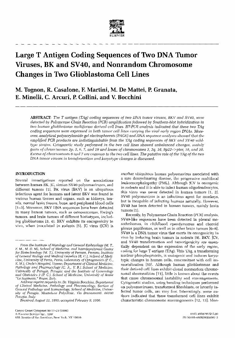
Figure 1 Hybridizations of the amplified samples with the oli- goprobes specific for the early region sequences of BKV (panel A), SV40 (panel B), and JCV (panel C). BKV, JCV, and SV40 are the amplified products used as positive controls, GL15 and GL22 are the amplified products under investigation from the two glioblas- toma cell lines, whereas R- is the PCR negative control, which contained the reagents without the DNA. The autoradiography for the GL15 and GL22 DNA samples indicated positive hybridiza- tion signals for BKV and SV40, whereas no signal was detected for JCV specific sequences.
two DNA samples from glioblastoma cell lines were ana- lyzed by PCR as described in Materials and Methods. By these technical conditions, DNA sequences homologous to BKV early region were detected in both samples (Fig. 1 A). Because previous papers reported SV40 and JCV sequences either in human brain tumors [1, 7, 8] or in human brain tissues [1, 2], we analyzed the two glioblastoma ceil DNA samples, previously investigated for the BKV-DNA sequences, for the presence of these two polyomavirus early-regions. As shown in Fig. 1 B both samples were positive for SV40 specific sequences, whereas they were negative for JCV (Fig. 1 C). Negative hybridization signals for the three polyomaviruses were obtained in control experiments with human neuroblastoma and glioblastoma cell lines,
20 M. Tognon et al.
I0(~1
181 - - - 1 7 2
1 2 3 4 5 1 2 3 4 5
i! i~ ~ ~i i i!"
A B
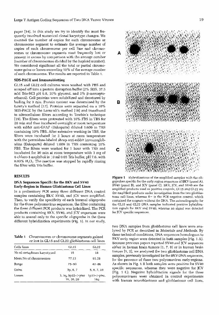
Figure 3 A. Hybridization of PCR amplified products from cDNAs to BKV specific: probe. Lane 1 represents the negative hybridization control from the RT-PCR reaction; lane 2 is the pos- itive hybridization signal of BKV; lane 3 is the negative hybridiza- tion of SV40: and lanes 4 and 5 are the positive hybridization signals in GL15 and GL22. B. Hybridization of PCR amplified products from cDNAs to SV40 specific probe. Lane i represents the negative hybridization control from the RT-PCR reaction; lane 2 is the negative hybridization of BKV; lane 3 is the positive hybridization signal of SV40; lanes 4 and 5 are the positive hybridization signals in GL15 and GL22. cDNAs were obtained by reverse transcription of RNAs from the two glioblastoma cell lines, GL15 and GL22.
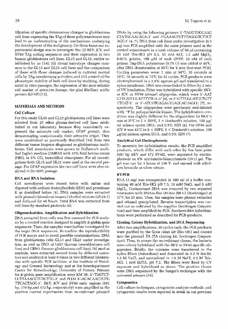
Figure 2 Composition from PAGE showing the separated PCR amplified products from GL15 and GL22. The bands correspond- ing to the 181 bp and 172 bp of BKV and SV40-early region are shown in lanes 2 and 3, respectively. In lanes 1 and 4 the MW are those of pBR 322-DNA cut with HAE III (marker VI from Boeh- ringer).
L697 and GBM1 (data not shown). The length specifici ty of the amplif ied bands were confirmed by analyt ical gel electrophoresis , where the PCR products were separated. Figure 2 shows the DNA bands of 181 bp and 172 bp, that correspond to the specific length products of BKV and SV40 sequences previously amplif ied by PCR [4, 7].
BKV and SV40 Early-Region Specific Expression in Human Glioblastoma Cell Lines Because DNA sequences of BKV and SV40-early region were detected in our samples we investigated whether these coding sequences were also expressed. For this purpose, RNAs from the two cell lines, GL15 and GL22, were ana- lyzed by RT-PCR followed by Southern-blot hybridiza- tions wi th specific internal oligoprobes. The results of this analysis indica ted that both BKV and SV40 early-region
were expressed in the two human gl ioblastoma cell l ines (Fig. 3).
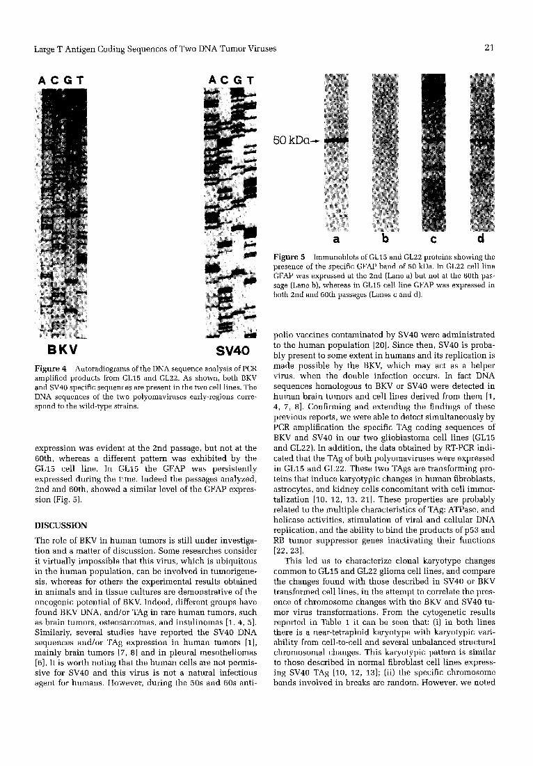
DNA Sequence Analysis In the previous sections we de termined by PCR amplifica- t ion fol lowed by Southern blot hybridizat ion, and by ana- lytical gel e lectrophoresis the presence of the BKV and SV40 early-region. Ultimately, to verify the DNA specific- ity of the two polyomaviruses , the amplif ied products were c loned and sequenced. As shown in Figure 4 the sequences correspond to BKV and SV40 wi ld- type strains.
Cytogenetic Analysis Balanced rearrangements were not observed. The most fre- quent structural abnormali t ies found were delet ions and unbalanced marker chromosomes. The break points were randomly distr ibuted. The chromosome bands involved are 3 cen, 9p22, 17p13 (GL15), and lp33 (GL22). Table 1 shows the total and part ial chromosome gains and losses exceeding 10%. In GL15 cell l ine the chromosome excesses involved chromosomes 6 and 7 and short arm of chromosome 3; chromosomes 3, 6, 16, 18, 20, 9 p 2 2 - ~ p t e r region, and the long arm of chromosome 3 were lost. In GL22 cell l ine chromosomes 5, 6, 7, and 19 resul ted in a gain, whereas the long arm of chromosome 14 and l p 3 3 - ~ pter region were lost. A clonal loss of both chromosomes 10 was previously descr ibed in GL22 cell l ine [14]. By paint ing fluorescence in situ hybr id iza t ion (FISH) analysis we had evidence of chromosome 10 material t ranslocated to different chromosomes (data not shown). Because it was not possible to exactly identify the t ranslocated chromo- some 10 regions, we did not take into account in the present s tudy chromosome 10 of the GL22 cell line.
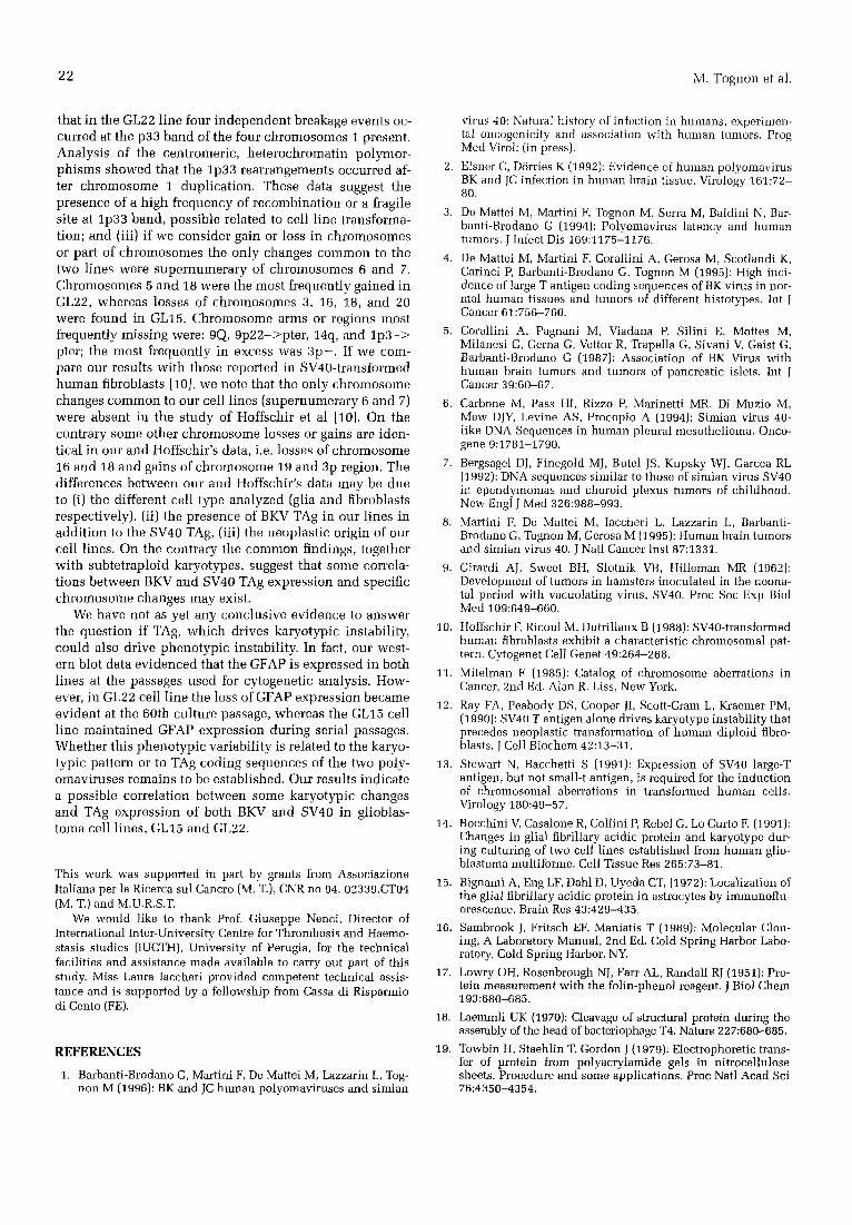
SDS-PAGE and Immunohlotting Our immunoblot t ing data showed a single band corre- sponding approximate ly to 50 kDa. In GL22 cell l ine GFAP
Large T Antigen Coding Sequences of Two DNA Tumor Viruses 21
A C G T A C G T
B K V S V 4 0 Figure 4 Autoradiograms of the DNA sequence analysis of PCR amplified products from GL15 and GL22. As shown, both BKV and SV40 specific sequences are present in the two cell lines. The DNA sequences of the two polyomaviruses early-regions corre- spond to the wild-type strains.
expression was evidem at the 2nd passage, but not at the 60th, whereas a diffeient pattern was exhibited by the GL15 cell line. In GL15 the GFAP was persistently expressed during the t!me. Indeed the passages analyzed, 2nd and 60th, showed a similar level of the GFAP expres- sion (Fig. 5).
DISCUSSION
The role of BKV in human tumors is still under investiga- tion and a matter of discussion. Some researches consider it virtually impossible that this virus, which is ubiquitous in the human population, can be involved in tumorigene- sis, whereas for others; the experimental results obtained in animals and in tissue cultures are demonstrative of the oncogenic potential of BKV. Indeed, different groups have found BKV DNA, and/or TAg in rare human tumors, such as brain tumors, osteosarcomas, and insulinomas [1, 4, 5]. Similarly, several studies have reported the SV40 DNA sequences and/or TAg expression in human tumors [1], mainly brain tumors [7, 8] and in pleural mesotheliomas [6]. It is worth noting that the human cells are not permis- sive for SV40 and this virus is not a natural infectious agent for humans. However, during the 50s and 60s anti-
50 kDa--~
a b c d Figure 5 Immunoblots of GL15 and GL22 proteins showing the presence of the specific GFAP band of 50 kDa. In GL22 cell line GFAP was expressed at the 2nd (Lane a) but not at the 60th pas- sage (Lane b), whereas in GL15 cell line GFAP was expressed in both 2nd and 60th passages (Lanes c and d).
polio vaccines contaminated by SV40 were administrated to the human population [20]. Since then, SV40 is proba- bly present to some extent in humans and its replication is made possible by the BKV, which may act as a helper virus, when the double infection occurs. In fact DNA sequences homologous to BKV or SV40 were detected in human brain tumors and cell lines derived from them [1, 4, 7, 8]. Confirming and extending the findings of these previous reports, we were able to detect simultaneously by PCR amplification the specific TAg coding sequences of BKV and SV40 in our two glioblastoma cell lines (GL15 and GL22). In addition, the data obtained by RT-PCR indi- cated that the TAg of both polyomaviruses were expressed in GL15 and GL22. These two TAgs are transforming pro- teins that induce karyotypic changes in human fibroblasts, astrocytes, and kidney cells concomitant with cell immor- talization [10, 12, 13, 21]. These properties are probably related to the multiple characteristics of TAg: ATPase, and helicase activities, stimulation of viral and cellular DNA replication, and the ability to bind the products of p53 and RB tumor suppressor genes inactivating their functions [22, 23].
This led us to characterize clonal karyotype changes common to GL15 and GL22 glioma cell lines, and compare the changes found with those described in SV40 or BKV transformed cell lines, in the attempt to correlate the pres- ence of chromosome changes with the BKV and SV40 tu- mor virus transformations. From the cytogenetic results reported in Table 1 it can be seen that: (i) in both lines there is a near-tetraploid karyotype with karyotypic vari- ability from cell-to-cell and several unbalanced structural chromosomal changes. This karyotypic pattern is similar to those described in normal fibroblast cell lines express- ing SV40 TAg [10, 12, 13]; (ii) the specific chromosome bands involved in breaks are random. However, we noted
22 M. Tognon et al.
that in the GL22 line four independent breakage events oc- curred at the p33 band of the four chromosomes 1 present. Analysis of the centromeric, heterochromatin polymor- phisms showed that the 1p33 rearrangements occurred af- ter chromosome 1 duplication. These data suggest the presence of a high frequency of recombination or a fragile site at lp33 band, possible related to cell l ine transforma- tion; and (iii) if we consider gain or loss in chromosomes or part of chromosomes the only changes common to the two lines were supernumerary of chromosomes 6 and 7. Chromosomes 5 and 18 were the most frequently gained in GL22, whereas losses of chromosomes 3, 16, 18, and 20 were found in GL15. Chromosome arms or regions most frequently missing were: 9Q, 9p22->pter, 14q, and l p 3 - > pter; the most frequently in excess was 3 p - . If we com- pare our results with those reported in SV40-transformed human fibroblasts [10], we note that the only chromosome changes common to our cell lines (supernumerary 6 and 7) were absent in the study of Hoffschir et al [10]. On the contrary some other chromosome losses or gains are iden- tical in our and Hoffschir's data, i.e. losses of chromosome 16 and 18 and gains of chromosome 19 and 3p region. The differences between our and Hoffschir's data may be due to (i) the different cell type analyzed (glia and fibroblasts respectively), (ii) the presence of BKV TAg in our lines in addit ion to the SV40 TAg, (iii) the neoplastic origin of our cell lines. On the contrary the common findings, together with subtetraploid karyotypes, suggest that some correla- tions between BKV and SV40 TAg expression and specific chromosome changes may exist.
We have not as yet any conclusive evidence to answer the question if TAg, which drives karyotypic instability, could also drive phenotypic instability. In fact, our west- ern blot data evidenced that the GFAP is expressed in both lines at the passages used for cytogenetic analysis. How- ever, in GL22 cell l ine the loss of GFAP expression became evident at the 60th culture passage, whereas the GL15 cell l ine mainta ined GFAP expression during serial passages. Whether this phenotypic variability is related to the karyo- typic pattern or to TAg coding sequences of the two poly- omaviruses remains to be established. Our results indicate a possible correlation between some karyotypic changes and TAg expression of both BKV and SV40 in glioblas- toma cell lines, GL15 and GL22.
This work was supported in part by grants from Associazione Italiana per la Ricerca sul Cancro (M. T.), CNR no 94. 02339.CT04 (M. T.) and M.U.R.S.T.
We would like to thank Prof. Giuseppe Nenci, Director of International Inter-University Centre for Thrombosis and Haemo- stasis studies (1UCTH), University of Perugia, for the technical facilities and assistance made available to carry out part of this study. Miss Laura Iaccheri provided competent technical assis- tance and is supported by a fellowship from Cassa di Risparmio di Cento (FE).
REFERENCES
1. Barbanti-Brodano G, Martini F, De Mattei M, Lazzarin L. Tog- non M (1996): BK and JC human polyomaviruses and simian
virus 40: Natural history of inlection in humans, experimen- tal oncogenicity and association with human tumors. Prog Med Viroh (in press).
2. Eisner C, D6rries K (1992): Evidence of human polyomavirus BK and JC infection in human brain tissue. Virology 161:72- 80.
3. De Mattei M, Martini F, Tognon M, Serra M, Baldini N, Bar- banti-Brodano G (1994): Polyomavirus latency and human tumors. J Infect Dis 169:1175-1176.
4. De Mattei M, Martini F, Corallini A, Gerosa M, Scotlandi K, Carinci P, Barbanti-Brodano G, Tognon M (19951: High inci- dence of large T antigen coding sequences of BK virus in nor- mal human tissues and tumors of different histotypes. Int J Cancer 61:756-760.
5. Corallini A, Pagnani M, Viadana P, Silini E, Mottes M, Milanesi G, Gerna G, Vettor R, Trapella G, Sivani V, Gaist G, Barbanti-Brodano G (1987): Association of BK Virus with human brain tumors and tumors of pancreatic islets. Int J Cancer 39:60-67.
6. Carbone M, Pass HI, Rizzo P, Marinetti MR, Di Muzio M, Mew DJY, Levine AS, Procopio A (1994): Simian virus 40- like DNA Sequences in human plenral mesothelioma, Onco- gene 9:1781-1790.
7. Bergsagel DJ, Finegold MJ, Butel JS, Kupsky WJ, Garcea RL (1992): DNA sequences similar to those of simian virus SV40 in ependymomas and choroid plexus tumors of childhood. New Engl ~ Med 326:968-993.
8. Martini F, De Mattei M, laccheri L, Lazzarin L, Barbanti- Brodano G, Tognon M, Gerosa M (1995): Human brain tumors and simian virus 40. J Nail Cancer lust 87:1331.
9. Girardi AJ, Sweet BH, Slotnik VB, Hilleman MR (1962): Development of tumors in hamsters inoculated in the neona- tal period with vacuolating virus, SV40. Proc Soc Exp Biol Med 109:649-660.
10. Hoffschir F, Ricoul M, Dutrillaux B (1988): SV40-transformed human fibroblasts exhibit a characteristic chromosomal pat- tern. Cytogenet Cell Genet 49:264-268.
11. Mitelman F (1985): Catalog of chromosome aberrations in Cancer, 2nd Ed. Alan R. Liss, New York.
12. Ray FA, Peabody DS, Cooper JI, Scott-Cram L, Kraemar PM, (1990): SV40 T antigen alone drives karyotype instability that precedes neoplastic transformation of human diploid fibre- blasts. J Cell Biochem 42:13-31.
13. Stewart N, Bacchetti S (1991): Expression of SV40 large-T antigen, but not small-t antigen, is required for the induction of chromosomal aberrations in transformed human cells. Virology 180:49-57.
14. Bocchini V, Casalone R, Cellini P, Rebel G, Lo Curto F. (1991): Changes in glial fibrillary acidic protein and karyotype dur- ing culturing of two cell lines established from human glio- blastoma multiforme. Cell Tissue Res 265:73-81.
15. Bignami A, Eng LF, Dahl D, Uyeda CT, (1972): Localization of the glial fibrillary acidic protein in astrocytes by immunoflu- orescence. Brain Res 43:429-435.
16. Sambrook J, Fritsch EF, Maniatis T (1989): Molecular Clon- ing, A Laboratory Manual, 2rid Ed, Cold Spring Harbor Labo- ratory, Cold Spring Harbor, NY.
17. Lowry OH, Rosenbrough NJ, Farr AL, Randall RJ (1951): Pro- tein measurement with the folin-phenol reagent. J Biol Chem 193:680-685.
18. Laemmli UK (1970): Cleavage of structural protein during the assembly of the head of bacteriophage T4. Nature 227:680-685.
19. Towbin H, Staehlin T, Gordon J (1979): Electrophoretic trans- fer of protein from polyacrylamide gels in nitrocellulose sheets. Procedure and some applications. Proc Natl Acad Sci 76:4350-4354.

Large T Antigen Coding Sequences of Two DNA Tumor Viruses 23
20. Shah K, Nathanson N (1976): Human exposure to SV40: Review and comment. Amer J Epidem 103:1-12.
21. Thei|e M, Grabowski G (1990): Mutagenic activity of BKV and JCV in human and other mammalian cells. Arch Virol 113:221-233.
22. Grossi MP, Caputo A, M,~neguzzi G, Corallini A, Carra L, Poro tolani M, Borgatti M, Milanesi G, Barbanti-Brodano G (1982):
Transformation of human embryonic fibroblasts by BK virus, BK virus DNA and a subgenomic BK virus DNA fragment. J Gen Virol 63:393-403.
23. Dyson N, Bernards R, Friend SH, Gooding LR, Hassel JA, Major EO, Pipas JM, Vandyke T, Harlow E (1990): Large T antigens of many polyomaviruses are able to form complexes with the retinoblastoma protein, J Virol 64:1353-1356.