Lacerda Bezerra, Iglesias de (v.r) (1).pdf - Gredos Principal
159
CARACTERIZACIÓN DE LAS FRACCIONES POLISACARÍDICA Y POLIFENÓLICA DE VINOS Y SU RELACIÓN CON PROPIEDADES ORGANOLÉPTICAS Y FARMACOLÓGICAS CARACTERIZAÇÃO DAS FRAÇÕES POLISACARÍDICAS E POLIFENÓLICAS DE VINHOS E SUA RELAÇÃO COM PROPRIEDADES ORGANOLÉPTICAS E FARMACOLÓGICAS CHARACTERIZATION OF THE POLYSACCHARIDE AND POLYPHENOLIC FRACTIONS OF WINES AND THEIR RELATIONSHIP WITH ORGANOLEPTIC AND PHARMACOLOGICAL PROPERTIES IGLESIAS DE LACERDA BEZERRA FACULTAD DE FARMACIA - DEPARTAMENTO DE QUÍMICA ANALÍTICA, NUTRICIÓN Y BROMATOLOGÍA - USAL DEPARTAMENTO DE BIOQUÍMICA E BIOLGOIA MOLECULAR - UFPR 2020
-
Upload
khangminh22 -
Category
Documents
-
view
0 -
download
0
Transcript of Lacerda Bezerra, Iglesias de (v.r) (1).pdf - Gredos Principal
CARACTERIZACIÓN DE LAS FRACCIONES POLISACARÍDICA Y
POLIFENÓLICA DE VINOS Y SU RELACIÓN CON PROPIEDADES
ORGANOLÉPTICAS Y FARMACOLÓGICAS
CARACTERIZAÇÃO DAS FRAÇÕES POLISACARÍDICAS E
POLIFENÓLICAS DE VINHOS E SUA RELAÇÃO COM
PROPRIEDADES ORGANOLÉPTICAS E FARMACOLÓGICAS
CHARACTERIZATION OF THE POLYSACCHARIDE AND
POLYPHENOLIC FRACTIONS OF WINES AND THEIR
RELATIONSHIP WITH ORGANOLEPTIC AND
PHARMACOLOGICAL PROPERTIES
IGLESIAS DE LACERDA BEZERRA
FACULTAD DE FARMACIA - DEPARTAMENTO DE QUÍMICA ANALÍTICA,
NUTRICIÓN Y BROMATOLOGÍA - USAL
DEPARTAMENTO DE BIOQUÍMICA E BIOLGOIA MOLECULAR - UFPR
2020

El Dr. Gilherme Lanzi Sassaki, Profesor de la Universidade Federal do Paraná, la Dra. Mª
Teresa Escribano Bailón, Profesora Titular del Área de Nutrición y Bromatología de la
Universidad de Salamanca, y el Dr. Ignacio García Estévez, de la Universidad de Salamanca,
directores del trabajo "Caracterización de las fracciones polisacarídica y polifenólica de
vinos y su relación con propiedades organolépticas y farmacológicas", realizado por Iglesias
de Lacerda Bezerra para optar al título de Doctor por la Universidad de Salamanca,
AUTORIZAN la presentación del mismo al considerar que se han alcanzado los objetivos
inicialmente previstos.
Salamanca, 10 de febrero de 2020
Producción científica:
Durante el desarrollo de esta Tesis Doctoral se ha generado la siguiente producción científica:
Stipp, M. C., Bezerra, I.D. L., Corso, C. R., Dos Reis Livero, F. A., Lomba, L. A., Caillot, A.
R. C., Zampronio, A. R., Queiroz-Telles, J. E., Klassen, G., Ramos, E. A. S., Sassaki, G. L.,
Acco, A. Necroptosis mediates the antineoplastic effects of the soluble fraction of
polysaccharide from red wine in Walker-256 tumor-bearing rats. Carbohydrate Polymers,
160, 123–133. (2017).
Bezerra, I.L.; Caillot, A. R. C.; Palhares, L. C. G. F.; Santana-Filho, A. P.; Chavante, S. F.
Guilherme Lanzi Sassaki, G. L. Structural characterization of polysaccharides from Cabernet
Franc, Cabernet Sauvignon and Sauvignon Blanc wines: Anti-inflammatory activity in LPS
stimulated RAW 264.7 cells. Carbohydrate Polymers, 186, 91-99. (2018).
Caillot, A. R. C.; Bezerra, I.L.; Palhares, L. C. G. F.; Santana-Filho, A. P.; Chavante, S.;
Sassaki, G. L. Structural characterization of blackberry wine polysaccharides and
immunomodulatory effects on LPS-activated RAW 264.7 macrophages. Food Chemistry,
257, 143-159. (2018).
Bezerra, I. L., Caillot, A. R. C., Oliveira, A. F., Santana-Filho, A. P. & Sassaki, G. L.
Cabernet Sauvignon wine polysaccharides attenuate sepsis inflammation and lethality in mice.
Carbohydrate Polymers, 210, 254-263. (2019).
Bezerra, I. L., Caillot, R. C., Menezes, L. R. A., Cavalcante, R. S., Sassaki, G. L..
Biodistribution and anti-inflammatory activity of red wine polysaccharides: a sweet
fluorescent trip in the mice body. Submitted to Nature. (2020).
Bezerra, I. L., Menezes, L. R. A., Sassaki, G. L., García-Estévez, I., Escribano-Bailón, M. T.
Influence of red wine soluble polysaccharides profile on the flavanol composition and
precipitation. Submitted to Food Chemistry. (2020).

2
AGRADECIMENTOS
Ao meu amado Deus por me guiar e me dar força para seguir. Ao Programa de Pós-
graduação em Biquímica-UFPR pela oportunidade de estudo e pela dedicação pelo
crescimento do curso. Ao meu querido orientador prof. Guilherme Lanzi Sassaki, que é minha
referência cientista, por toda sua genialidade e dedicação à ciência, serei grata por ter
acreditado em mim, me apoiado e aconselhado em todas minhas decisões acadêmicas, pela
paciência, um grande professor. A banca interna Prof. Thales Ricardo Cipriani e Prof.
Alexandra Acco pelas correções importantes do meu projeto e relatórios. A Rosane, Elis e
Flavia pelas análises de GC-MS e HPSEC-MALLS. Aos técnicos do Centro de Tecnologias
Avançadas em Fluorescência, pelas análises histológicas. A prof. Suely Ferreira Chavante,
Lais Palhares e Rômulo Cavalcante pela parceira sempre valiosa, tê-los como parceiros foi
muito especial. Ao prof. Edvaldo da Silva Trindade e seus alunos pelo auxilio, por serem
sempre tão gentis e prestativos. A prof. Yanna Dantas Rattmann, sempre tão doce e querida,
por me socorrer com seu conhecimento quando eu não sabia o que fazer nos experimentos de
sepse. Ao Arquimedes e Leociley pela colaboração com as análises de RMN e por todo apoio
dado durante a execução do meu trabalho. A Adriana e Aninha, pela ajuda com meus
experimentos, por todo apoio, pelos conselhos, amizade, carinho, paciência, risadas e
aventuras que marcaram meu doutorado. A todos os amigos do laboratório, pelo apoio, pela
amizade e convivência agradável, em especial ao grupo Sassaketes, pelos dias tão alegres e
pelo companheirismo. Ao grupo Carbocats pelo carinho, companheirismo, tantas risadas e
almoço as 11h, em especial a Giuliana, minha artista cientista, pela amizade, pelo carinho, por
toda ajuda durante meu sanduíche e pelas caronas na volta para casa. A Shayane e Philippe,
por serem tão prestativos e me socorrerem tantas vezes durante minha estadia fora do país. A
todos do grupo Química de Carboidratos, as melhores pessoas do mundo estão aqui. A todos
os professores do Departamento de Bioquímica e Biologia Molecular (UFPR), pelas
disciplinas ministradas, pela contribuição para a minha formação. A todos os pós-graduandos
e funcionários do Departamento de Bioquímica e Biologia Molecular, pela colaboração e
amizade. A CAPES e CNPq pelo auxílio financeiro e pela oportunidade de estudo fora do
país.
A minha mãezinha Socorro por ser meu apoio, minha base e minha fortaleza, por toda
a vida ter se dedicado a minha educação. Aos meus paidrinhos, Francisco e Francinaldo, e ao
meu paidrasto, José, pelo apoio, pelos conselhos e incentivo, por terem batalhado tanto pelo
meu sucesso. Aos meus irmãos, Sara e Lucas, pelo companheirismo, por dar leveza a minha
vida. A toda minha família pelo apoio e por me tratarem com tanto carinho e zelo.
3
Ao meu querido e amado marido Marcelo, meu companheiro, por ser tão incentivador,
por me ajudar sempre que preciso e cuidar de mim. Obrigada pelo amor, por acreditar que eu
sou capaz e me fazer acreditar. A toda Família Ramos, pelo carinho, apoio e amizade.
A minha querida orientadora prof. María Teresa Escribano Bailón, tão doce, a voz
mais acalmadora da Espanha. Obrigada por ter me aceitado em seu grupo, por ser tão paciente
e atenciosa comigo e não ter desistido da minha cotutela, por todo apoio e pelos
ensinamentos. Ao meu orientador prof. Ignacio Estévez Gárcia, excelentíssimo Nacho, por ser
tão prestativo, amável e acolhedor, por todo apoio, por ser a calmaria quando tudo parece ser
caos. Obrigada pela força, por ser a base do laboratório e pelas risadas que deixavam os dias
mais leves.
Ao programa em Enologia, Vicultura e Sustentabilidade pela oportunidade de estudo e
por terem aceitado minha cotutela. A María José e Joaquín, por serem sempre prestativos e
pelo carinho. Joaquin, obrigada por me salvar com vidrarias e me chamar sempre para o café
e dizer que vai embora antes de mim. A Montse, o olhar mais confortante, por ser tão
atenciosa, pelo companheirismo e carinho, por todo apoio, por me socorrer sempre, por torcer
por mim. A Rebeca, tão doce e prestativa, obrigada pelo apoio, por toda a ajuda nos
experimentos e pela atenção. A Cristina, a gênia dos antocianos, pelos ensinamentos, pela
paciência e carinho, minha grande admiração. A minha querida amiga, dona da alegria
contagiante, Bárbara, que tornou minha estadia mais feliz, por todo companheirismo e apoio
psicológico tão importante, pela ajuda com trâmites espanhóis tão difíceis, por estar sempre
ao meu lado. A Eva, por ser tão atenciosa, pela amizade e por me chamar para tomar café,
almoçar e tomar vermut. A Elvira, um poço de corações, pelo apoio, pela ajuda nos
experimentos, por ser tão atenciosa. A Alba, por todo apoio, por toda ajuda durante minha
estadia. A Si Yu pelo toque chinês, pela amizade, pelos dias divertidos que passei tentado dar
um abraço. A todos do grupo Polifenois pelo carinho, pela atenção e pelo apoio, fui muito
feliz nesse grupo maravilhoso, fui bem acolhida, todos foram importantes para o meu
crescimento acadêmico e pessoal. Meus queridos espanhóis! ¡Muchas gracias!
A todas as pessoas que me ajudaram de alguma forma na minha formação e na
realização desse trabalho, e que torceram pelo meu sucesso, muito obrigada!

4
Com amor:
A minha família,
Ao meu marido,
Dedico
5
RESUMEN
El vino es una de las bebidas más consumidas y económicamente más importantes,
con una facturación de aproximadamente € 30 mil millones por año en el mercado debido a
una producción mundial de 292 millones de hectolitros. Los enólogos buscan continuamente
la producción de vinos de alta calidad, por lo tanto, hay mucho interés en la investigación
dirigida a identificar los componentes del vino y comprender mejor su efecto durante la
vinificación, lo que puede resultar de ayuda para el avance del sector del vino. Los
polisacáridos y los compuestos fenólicos son compuestos altamente relacionados con la
calidad del vino, debido a su impacto no sólo en sus propiedades organolépticas (color, sabor,
astringencia) sino también en sus propiedades saludables. Por lo tanto, este trabajo tuvo como
objetivo identificar, caracterizar y cuantificar los polisacáridos y compuestos fenólicos
encontrados en los vinos y evaluar la interacción entre ellos. Además, pretende evaluar la
similitud entre los polisacáridos extraídos de vinos comerciales de diferentes añadas y
estudiar las propiedades antiinflamatorias de los polisacáridos in vitro en células RAW 264.7
estimuladas con LPS e in vivo por el modelo de septicemia inducida por cirugía de ligadura y
perforación del ciego en ratones, así como la biodistribución de estos polisacáridos durante el
proceso inflamatorio. Para este propósito, se eligieron vinos comerciales elaborados con uvas
Vitis vinífera de diferentes variedades. Los polisacáridos fueron extraídos, caracterizados y
cuantificados por GC-MS y RMN. Se identificó la presencia de manano, arabinogalactano
tipo II y ramannogalactonano tipo I y II. También se ha demostrado que los vinos producidos
a partir de la misma variedad de uva y de la misma bodega tienen patrones de polisacáridos
similares, aunque hayan sido producidos en años diferentes. Los compuestos fenólicos fueron
identificados y cuantificados por HPLC-DAD-MS. El análisis mostró una prevalencia del
contenido de procianidinas (entre 58 y 275 mg / L) frente al de las prodelfinidinas (entre 4 y 8
mg / L). El análisis de antocianinas mostró la mayoría de los derivados de monoglucósidos y
sus derivados acetilados. Los análisis estadísticos revelaron la existencia de relaciones entre la
estructura de los polisacáridos y su capacidad para precipitar procianidinas, lo que explicaría
la relación observada en los vinos entre la composición de polisacáridos y la concentración de
flavanoles. Las fracciones de polisacárido inhibieron la producción de citoquinas
inflamatorias (TNF-α e IL-1β) y mediador (NO) en células RAW 264.7. La administración
subcutánea y oral de polisacáridos redujo la tasa de mortalidad de los ratones. El tratamiento
redujo la migración de leucocitos, inhibió las citocinas proinflamatorias y aumentó la
producción de la citocina antiinflamatoria IL-10. También redujeron los niveles plasmáticos
de AST, ALT, bilirrubina, urea y creatinina, con la consiguiente protección contra el daño
6
tisular. Además, la biodistribución de polisacáridos por el cuerpo cuando se administra por vía
subcutánea parece ocurrir a través del sistema inmune. Los polisacáridos han demostrado un
potente efecto antiinflamatorio in vitro e in vivo, lo que puede indicar efectos beneficiosos del
consumo moderado de vino en la salud humana.
Palabras clave: vinos, polisacáridos, compuestos fenólicos, RMN, HPLC, inflamación.
7
RESUMO
O vinho é uma das bebidas mais consumidas e de maior importância econômica,
movimentando por ano aproximadamente 30 bilhões de euros no mercado decorrentes de uma
produção mundial de 292 milhões de hectolitros. Os produtores de vinho buscam
continuamente a produção de vinhos de alta qualidade; portanto, há muito interesse em
pesquisas destinadas a identificar os componentes do vinho e a entender melhor seu efeito
durante a vinificação, o que pode ser útil para o avanço do setor vitivinícola. Polissacarídeos e
compostos fenólicos são compostos altamente relacionados à qualidade do vinho, devido ao
seu impacto não apenas em suas propriedades organolépticas (cor, sabor, adstringência), mas
também em suas propriedades saudáveis. Portanto, este trabalho teve como objetivo
identificar, caracterizar e quantificar os polissacarídeos e compostos fenólicos encontrados
nos vinhos e avaliar a interação entre eles. Além disso, avaliar a semelhança entre os
polissacarídeos extraídos de vinhos comerciais de diferentes safras e estudar as propriedades
anti-inflamatórias dos polissacarídeos in vitro em células RAW 264.7 estimuladas com LPS e
in vivo pelo modelo de septicemia induzido pela ligação e perfuração do ceco em
camundongos, bem como a biodistribuição desses polissacarídeos durante o processo
inflamatório. Para isso, foram escolhidos vinhos comerciais feitos com uvas Vitis vinífera de
diferentes variedades. Os polissacarídeos foram extraídos, caracterizados e quantificados por
GC-MS e RMN. Foi identificada a presença de manana, arabinogalactana tipo II e
ramannogalactonana tipo I e II. Também foi demonstrado que os vinhos produzidos a partir
da mesma variedade de uva e da mesma vinícola têm padrões de polissacarídeos semelhantes,
embora tenham sido produzidos em anos diferentes. Os compostos fenólicos foram
identificados e quantificados por HPLC-DAD-MS. A análise mostrou uma prevalência de
conteúdo de procianidina (entre 58 e 275 mg/L) versus a de prodelfinidinas (entre 4 e 8 mg /
L). A análise de antocianina mostrou a maioria dos derivados monoglicósidos e seus
derivados acetilados. As análises estatísticas revelaram a existência de relações entre a
estrutura dos polissacarídeos e sua capacidade de precipitar procianidinas, o que explicaria a
relação observada nos vinhos entre a composição dos polissacarídeos e a concentração de
flavanóis. As frações de polissacarídeos inibiram a produção de citocinas inflamatórias (TNF-
α e IL-1β) e mediador (NO) em células RAW 264.7. A administração subcutânea e oral de
polissacarídeos reduziu a taxa de mortalidade dos camundongos. O tratamento reduziu a
migração de leucócitos, inibiu citocinas pró-inflamatórias e aumentou a produção da citocina
anti-inflamatória IL-10. Eles também reduziram os níveis plasmáticos de AST, ALT,
bilirrubina, uréia e creatinina, com a consequente proteção contra danos nos tecidos. Além

8
disso, a biodistribuição de polissacarídeos pelo organismo, quando administrada via
subcutânea, parece ocorrer através do sistema imunológico. Os polissacarídeos demonstraram
um potente efeito anti-inflamatório in vitro e in vivo, o que pode indicar efeitos benéficos do
consumo moderado de vinho na saúde humana.
Palavras-chave: vinhos, polissacarídeos, compostos fenólicos, NMR, HPLC,
inflamação.

9
ABSTRACT
Wine is one of the most consumed and economically important beverages, handling
approximately € 30 billion a year on the market resulting from a worldwide production of 292
million hectoliters. Wine producers continually seek to produce high quality wines; therefore,
there is much interest in research aimed at identifying the components of wine and better
understanding its effect during winemaking, which can be useful for the advancement of the
wine sector. Polysaccharides and phenolic compounds are compounds highly related to wine
quality, due to their impact not only on their organoleptic properties (color, flavor,
astringency), but also on their healthy properties. Therefore, this work aimed to identify,
characterize and quantify the polysaccharides and phenolic compounds found in wines and to
evaluate the interaction between them. In addition, to evaluate the similarity between the
polysaccharides extracted from commercial wines from different vintages and to study the
anti-inflammatory properties of the polysaccharides in vitro in RAW 264.7 cells stimulated
with LPS and in vivo by the septicemia model induced by the cecum binding and perforation
in mice, as well as the biodistribution of these polysaccharides during the inflammatory
process. For this, commercial wines made with Vitis vinífera grapes of different varieties were
chosen. Polysaccharides were extracted, characterized and quantified by GC-MS and NMR.
The presence of mannan, type II arabinogalactans and type I and II rhamannogalactonans
were identified. It has also been shown that wines produced from the same grape variety and
from the same winery have similar polysaccharide patterns, although they have been produced
in different years. The phenolic compounds were identified and quantified by HPLC-DAD-
MS. The analysis showed a prevalence of procyanidin content (between 58 and 275 mg/L)
versus that of prodelphinidin (between 4 and 8 mg/L). The anthocyanin analysis showed the
majority of monoglycoside derivatives and their acetylated derivatives. Statistical analyzes
revealed the existence of relationships between the structure of polysaccharides and their
ability to precipitate procyanidins, which would explain the relationship observed in wines
between the composition of polysaccharides and the concentration of flavanols. The
polysaccharide fractions inhibited the production of inflammatory cytokines (TNF-α and IL-
1β) and mediator (NO) in RAW 264.7 cells. Subcutaneous and oral administration of
polysaccharides reduced the mortality rate of mice. The treatment reduced leukocyte
migration, inhibited pro-inflammatory cytokines and increased production of the anti-
inflammatory cytokine IL-10. They also reduced plasma levels of AST, ALT, bilirubin, urea
and creatinine, with consequent protection against tissue damage. In addition, the
polysaccharide biodistribution by the body, when administered subcutaneously, seems to
10
occur through the immune system. Polysaccharides have demonstrated a potent anti-
inflammatory effect in vitro and in vivo, which may indicate beneficial effects of moderate
wine consumption on human health.
Keywords: wines, polysaccharides, phenolic compounds, NMR, HPLC, inflammation.
11
SUMÁRIO
1. INTRODUÇÃO ............................................................................................................... 17
2. OBJETIVOS .................................................................................................................... 20
2.1. OBJETIVO GERAL.......................................................................................................... 20
2.2. OBJETIVOS ESPECÍFICOS ............................................................................................ 20
3. REVISÃO BIBLIOGRÁFICA ....................................................................................... 21
3.1. UVA .................................................................................................................................. 21
3.1.1. Tipos de videira ................................................................................................... 22
3.2. VINHO .............................................................................................................................. 22
3.2.1. Processo de produção do vinho ........................................................................... 23
3.2.2. Benefícios do vinho ............................................................................................. 25
3.2.3. Carboidratos dos vinhos ...................................................................................... 26
3.2.3.1. Parede celular vegetal e polissacarídeos ............................................................. 26
3.2.3.1.1. Arabinogalactanas ............................................................................................... 28
3.2.3.1.2. Ramnogalacturonanas ......................................................................................... 28
3.2.3.2. Parede celular de leveduras e polissacarídeos ..................................................... 29
3.2.3.2.1. Mananas .............................................................................................................. 30
3.2.4. Polifenóis do vinho .............................................................................................. 30
3.2.4.1. Flavanóis ............................................................................................................. 31
3.2.4.2. Pigmentos antociânicos ....................................................................................... 32
3.2.4.3. Compostos fenólicos e índice de cor ................................................................... 32
3.3. PROCESSO INFLAMATÓRIO E POLISSACARÍDEOS ............................................... 33
3.3.1. Sepse .................................................................................................................... 34
REFERÊNCIAS BIBLIOGRÁFICAS ................................................................................. 37
12
CAPITULO I .......................................................................................................................... 47
2. Materials and methods.................................................................................................... 48
2.1. Materials ............................................................................................................................ 48
2.2. Polysaccharides extraction and purification ...................................................................... 49
2.3. Monosaccharide analysis ................................................................................................... 49
2.4. Methylation analysis.......................................................................................................... 50
2.5. Nuclear magnetic resonance (NMR) spectroscopy ........................................................... 50
2.6. Determination of homogeneity and molar mass................................................................ 51
2.7. Cell Culture ....................................................................................................................... 51
2.8. Analysis of cell viability.................................................................................................... 51
2.9. Measurement of cytokines production and NO production .............................................. 52
2.10. Statistical analysis ............................................................................................... 52
3. Results and Discussion .................................................................................................... 53
3.1. Isolation and structural analysis of the polysaccharides .................................................... 53
3.2. Biological experiments ...................................................................................................... 59
3.2.1. Analysis of cell viability ..................................................................................... 59
3.2.2. Anti-inflammatory activity .................................................................................. 59
4. Conclusions ...................................................................................................................... 63
Supporting Information ......................................................................................................... 63
References ................................................................................................................................ 66

13
CAPITULO II ......................................................................................................................... 74
1. Materials and methods.................................................................................................... 75
1.1. Materials ............................................................................................................................ 75
1.2. Polysaccharides extraction and purification ...................................................................... 76
1.3. Monosaccharide analysis ................................................................................................... 76
1.4. Structural identification by NMR ...................................................................................... 77
1.5. In vivo experiments ........................................................................................................... 77
1.6. Sepsis induction by cecal ligation and puncture (CLP) .................................................... 77
1.7. Hematological assays ........................................................................................................ 78
1.8. Biochemical assays............................................................................................................ 79
1.9. Histopathological analysis ................................................................................................. 79
1.10. Statistical analysis ............................................................................................... 79
2. Results and Discussion .................................................................................................... 80
2.1. Analysis of the polysaccharides of Cabernet Sauvignon wine .......................................... 80
2.2. Biological experiments ...................................................................................................... 83
3. Conclusions ...................................................................................................................... 93
Supporting Information ......................................................................................................... 93
References ................................................................................................................................ 95
14
CAPITULO III ..................................................................................................................... 103
2. Materials and methods.................................................................................................. 104
2.1. Materials .......................................................................................................................... 104
2.2. Polysaccharides extraction .............................................................................................. 104
2.3. Labeling of WCF with fluoresceinamine ........................................................................ 105
2.4. Structural characterization ............................................................................................... 105
2.5. Sepsis induction by cecal ligation and puncture (CLP) .................................................. 106
2.6. Hematological assays ...................................................................................................... 107
2.7. Biochemical assays.......................................................................................................... 107
2.8. Histopathological analysis ............................................................................................... 108
2.9. Cell culture ...................................................................................................................... 108
2.10. Analysis of cell viability ................................................................................... 108
2.11. Macrophage internalization of WCF-F ............................................................. 109
2.12. Imaging of Multi-wavelength fluorescence ...................................................... 109
2.13. Statistical analysis ............................................................................................. 109
3. Results ............................................................................................................................ 110
3.1. Structural characteristics of fluorescent labeled WCF-F ................................................ 110
3.2. Anti-inflammatory effect of wine polysaccharides ......................................................... 112
3.3. Analysis of cell viability.................................................................................................. 118
3.4. Macrophage internalization of fluorescent labeled WCF-F ............................................ 119
3.5. Imaging of Multi-wavelength fluorescence in vivo ......................................................... 120
4. Discussion ....................................................................................................................... 122
Supporting Information ....................................................................................................... 124
References .............................................................................................................................. 124
15
CAPÍTULO IV ...................................................................................................................... 129
1. Materials and methods.................................................................................................. 130
1.1. Materials .......................................................................................................................... 130
1.2. Polysaccharides extraction .............................................................................................. 130
1.3. Polysaccharide identification by NMR ........................................................................... 131
1.4. Determination of molecular weight distribution of polysaccharides .............................. 131
1.5. Analysis of phenolic compounds in wines ...................................................................... 132
1.6. Flavanols extraction from grape seeds ............................................................................ 132
1.7. Interaction assays ............................................................................................................ 132
1.8. Statistical analysis ........................................................................................................... 133
2. Results and Discussion .................................................................................................. 133
2.1. Analysis of the polysaccharide extracts from wines ....................................................... 133
2.2. Analysis of flavanols extract of wines ............................................................................ 136
2.3. Interaction experiments ................................................................................................... 138
3. Conclusions .................................................................................................................... 142
Supporting Information ....................................................................................................... 143
References.............................................................................................................................. 146
16
CAPITULO V ....................................................................................................................... 151
1. RESULTADOS COMPLEMENTARES .................................................................... 151
1.1. Análise dos pigmentos antocianos em vinhos ................................................................. 151
1.2. Medidas colorimétricas ................................................................................................... 152
Referências ............................................................................................................................ 153
CONCLUSÃO GERAL ....................................................................................................... 154
CONCLUSIONES ................................................................................................................ 156
17
1. INTRODUÇÃO
O vinho é produzido a partir de uvas das variedades (ou castas) da espécie Vitis
vinifera utilizada para produção de vinhos finos e da espécie Vitis labrusca utilizada para
produção de vinhos de mesa (comuns). A constituição química das uvas permite que elas
fermentem durante a vinificação sem que lhes sejam adicionados açúcares, ácidos, enzimas ou
outros nutrientes. Essa fermentação é feita por leveduras que consomem os açúcares presentes
nas uvas transformando-os em álcool (JOHNSON, 1989; (CRUZ, 2006). As principais
macromoléculas encontradas na bebida são polissacarídeos (até 15%) e polifenóis (até 35%)
(CARPITA et al., 1993).
Os polissacarídeos presentes nos vinhos possuem influência importante em várias
etapas do processo de vinificação, incluindo fermentação, filtração, estabilização e são
parcialmente responsáveis pelas propriedades organolépticas dos vinhos (GERBAUD et al.,
1997; MOINE-LEDOUX & DUBOURDIEU, 1999). No entanto, estudos já mostraram que
nem todos os polissacarídeos apresentam o mesmo comportamento em relação aos vinhos, e
sua influência no processamento e nas propriedades sensoriais depende da quantidade, classe
e características estruturais (RIOU et al, 2002; VIDAL et al., 2003; GUADALUPE &
AYESTARÁN, 2007).
Essas macromoléculas são provenientes de bagas da uva, de leveduras, de bactérias e
da contaminação por fungos. Do ponto de vista enológico e quantitativo, os polissacarídeos de
uvas e de leveduras são os mais importantes. A estrutura dos polissacarídeos e suas
concentrações dependem de muitos parâmetros, tais como cultivo, estágio de maturação,
técnicas de vinificação e os tratamentos para aumento da solubilização dos componentes
macromoleculares das paredes celulares de bagas de uva (DOCO et al., 1999; CABANIS &
CABANIS, 2000; AYESTARAN et al., 2004). Arabinose e polissacarídeos ricos em
galactose (AGs), como as arabinogalactanas-proteínas tipo II e arabinanas,
ramnogalacturonanas tipo I e tipo II, e homogalacturonanas são provenientes de bagas de uva,
enquanto as glucanas, mananas e manoproteínas são liberadas da levedura durante a
fermentação ou pela atividade enzimática da levedura por autólise durante o envelhecimento
em barris (PELLERIN et al., 1995; VIDAL et al., 2003; AYESTARÁN et al., 2004). Esses
açúcares parecem ser uma das moléculas mais interessantes em enologia devido aos seus
efeitos na qualidade final do vinho (FOURNAIRON et al., 2002; DOCO et al., 2003), no
entanto, informações detalhadas sobre composição e caracterização estrutural dos
18
polissacarídeos são relativamente escassas, o que torna importante o estudo desses
polissacarídeos encontrados nos vinhos como proposto nesse trabalho.
Os polifenóis presentes no vinho são provenientes das uvas, do metabolismo dos
microrganismos e também da madeira dos barris utilizados durante o amadurecimento
(JACKSON, 2008). Portanto, sua composição e concentração também dependem da variedade
de uva usada na vinificação, do procedimento usado para a vinificação e das reações químicas
que ocorrem durante o envelhecimento (RIBÉREAU-GAYON et al., 2006; RECAMALES et
al., 2006).
Os compostos fenólicos derivados de uvas, de grande importância para a enologia, são
encontrados principalmente em sementes e peles (ESCRIBANO-BAILÓN et al, 1992; 1995
HANLIN et al., 2011). Esses compostos estão envolvidos na estabilidade e nas propriedades
organolépticas, como cor e adstringência, ou seja, na qualidade do vinho (RIBÉREAU-
GAYON et al., 2006; QUIJADA-MORÍN et al., 2014). Nesse sentido, é amplamente
reconhecido que os vinhos tintos de alta qualidade têm um nível equilibrado de adstringência,
ou seja, possuem níveis equilibrados de compostos fenólicos e polissacarídeos (BATE-
SMITH, 1954; GAWEL et al., 2000; BROSSAUD et al., 2001; PREYS et al., 2006;
QUIJADA-MORÍN et al., 2014).
Estudos mostram que os polissacarídeos podem afetar as propriedades organolépticas
do vinho (OZAWA et al, 1987; QUIJADA-MORÍN et al., 2014; OSETE-ALCARAZ et al.,
2019). Polissacarídeos podem limitar a concentração de proantocianidinas disponíveis e
reduzir a adstringência do vinho (ESCOT et al., 2001; MATEUS et al., 2004; CARVALHO
et al., 2006). A falta de equilíbrio entre os compostos fenólicos e polissacarídeos e a
consequente alteração das propriedades organolépticas leva a uma diminuição na qualidade e
estabilidade do vinho (COOMBE, 1973; ORDUÑA, 2010;). Portanto, o estudo desses
compostos e a relação entre eles são extremamente importantes para a produção de um vinho
de qualidade e competitivo no mercado.
Além disso, há um grande interesse em conhecer os benefícios que o consumo
moderado de vinho pode trazer à saúde. Alguns autores descreveram importantes efeitos
imunomoduladores, antioxidantes, anti-septicêmicos, anti-inflamatórios, antineoplásicos e
gastroprotetores dos polissacarídeos extraídos de plantas, frutas e vinho (MUELLER &
ANDERER, 1990; MELLINGER et al., 2008; CIPRIANI et al., 2009; NASCIMENTO et al.,
2013; PARK et al., 2013; DARTORA et al., 2013; INNGJERDINGEN et al., 2014; STIPP et
al., 2016; CAILLOT et al., 2018). Alguns efeitos benéficos do vinho na saúde têm sido
atribuídos aos polifenóis presentes na pele das uvas (DIAZ-GEREVINI et al., 2016;
GOJKOVIC-BUKARICA et al., 2018). No entanto, existem poucos estudos sobre atividades
19
biológicas e modulação de mediadores inflamatórios por polissacarídeos de vinho o que torna
importante o estudo dos possíveis benefícios dos polissacarídeos encontrados nos vinhos.

20
2. OBJETIVOS
2.1. OBJETIVO GERAL
Extração, caracterização, quantificação dos polissacarídeos e polifenóis de
vinhos e avaliação da similaridade entre polissacarídeos extraídos de vinhos comerciais de
diferentes safras. Como também, investigar interação entre essas moléculas e os benefícios
dos polissacarídeos para a saúde, através da avaliação da propriedade anti-inflamatória em
ensaios in vitro e in vivo.
2.2. OBJETIVOS ESPECÍFICOS
Extrair, caracterizar e quantificar os polissacarídeos de vinhos.
Avaliar a similaridade entre polissacarídeos extraídos de vinhos
comerciais de diferentes safras.
Caracterizar e quantificar os compostos fenólicos extraídos de vinhos e
estabelecer uma possível relação com polissacarídeos.
Analisar o efeito anti-inflamatório in vitro dos polissacarídeos extraídos
dos vinhos utilizando células RAW 264.7 estimuladas com lipopolisacarídeos
(LPS).
Avaliar os efeitos anti-inflamatórios das frações polissacarídicas obtidas
dos vinhos, através do modelo de septicemia induzida pela cirurgia de ligadura e
perfuração do ceco (CLP) e verificar a taxa de sobrevida dos camundongos
tratados com os polissacarídeos.
21
3. REVISÃO BIBLIOGRÁFICA
3.1. UVA
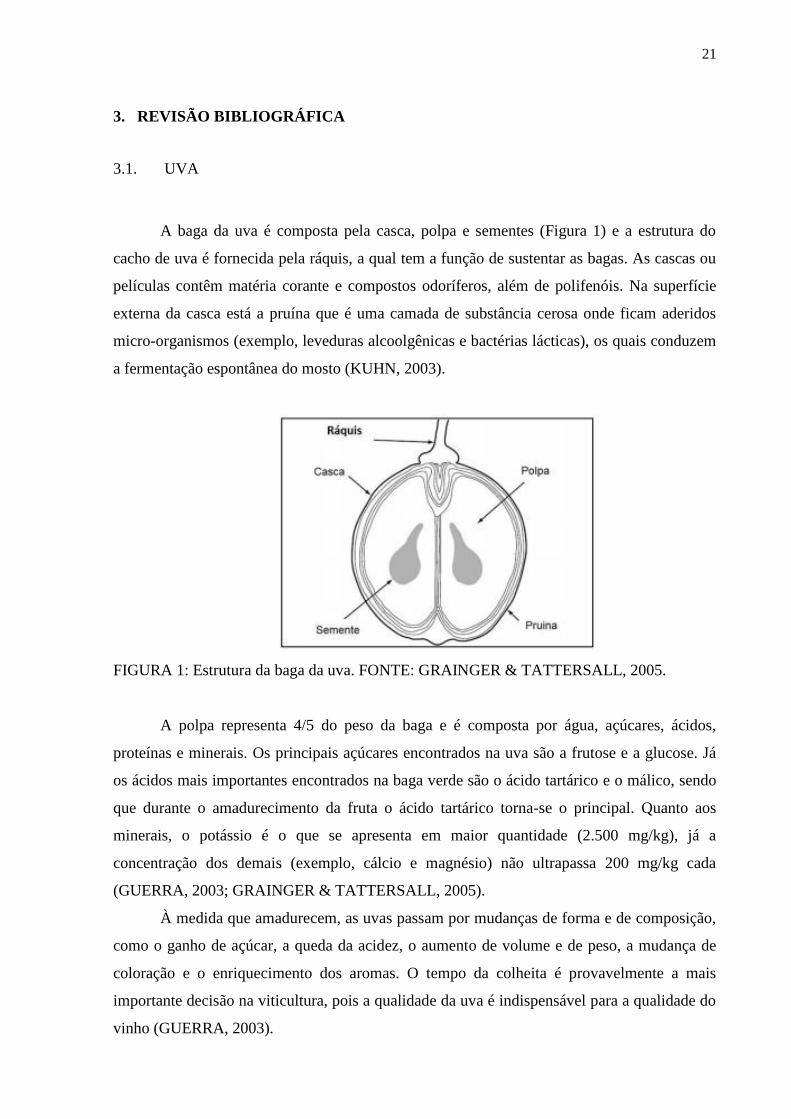
A baga da uva é composta pela casca, polpa e sementes (Figura 1) e a estrutura do
cacho de uva é fornecida pela ráquis, a qual tem a função de sustentar as bagas. As cascas ou
películas contêm matéria corante e compostos odoríferos, além de polifenóis. Na superfície
externa da casca está a pruína que é uma camada de substância cerosa onde ficam aderidos
micro-organismos (exemplo, leveduras alcoolgênicas e bactérias lácticas), os quais conduzem
a fermentação espontânea do mosto (KUHN, 2003).
FIGURA 1: Estrutura da baga da uva. FONTE: GRAINGER & TATTERSALL, 2005.
A polpa representa 4/5 do peso da baga e é composta por água, açúcares, ácidos,
proteínas e minerais. Os principais açúcares encontrados na uva são a frutose e a glucose. Já
os ácidos mais importantes encontrados na baga verde são o ácido tartárico e o málico, sendo
que durante o amadurecimento da fruta o ácido tartárico torna-se o principal. Quanto aos
minerais, o potássio é o que se apresenta em maior quantidade (2.500 mg/kg), já a
concentração dos demais (exemplo, cálcio e magnésio) não ultrapassa 200 mg/kg cada
(GUERRA, 2003; GRAINGER & TATTERSALL, 2005).
À medida que amadurecem, as uvas passam por mudanças de forma e de composição,
como o ganho de açúcar, a queda da acidez, o aumento de volume e de peso, a mudança de
coloração e o enriquecimento dos aromas. O tempo da colheita é provavelmente a mais
importante decisão na viticultura, pois a qualidade da uva é indispensável para a qualidade do
vinho (GUERRA, 2003).
22
3.1.1. Tipos de videira
Mundialmente existem milhares de variedades de uva. A maioria delas pertence à
espécie Vitis vinifera, originária do Cáucaso entre 7.000 e 5.000 a.C., de onde foi difundida
por toda a costa mediterrânea. Entre as variedades dessa espécie estão as conhecidas uvas
tintas Cabernet Sauvignon, Cabernet Franc, Merlot, Pinot Noir, Tempranillo, Syrah e Tannat
e, entre as uvas brancas, destacam-se a Chardonnay, Sauvignon Blanc, Prosecco e Riesling.
Algumas delas se difundiram pelo mundo em virtude da sua capacidade de adaptação e pelas
características dos vinhos que originam. Outras, de adaptação mais restrita, permaneceram em
suas regiões de origem, proporcionando aos seus habitantes a oportunidade de elaboração de
produtos típicos e exclusivos (GUERRA et al., 2009).
As uvas oriundas de videiras Vitis vinifera, cultivares europeus, são comumente
usadas na elaboração de vinho pelo mundo, principalmente na Europa. Nos Estados Unidos,
espécies como a Vitis labrusca, Vitis riparia, Vitis aestivalis, Vitis rupestris e Vitis
rotundifolia são as mais usadas para a vinificação (YANG, MARTINSON & LIU, 2009). As
frutas destas últimas espécies contêm menos açúcar que as oriundas de espécies Vitis vinifera.
Desta forma, seus vinhos também não apresentam uma alta qualidade (ESTREICHER, 2006).
3.2. VINHO
O vinho é uma das primeiras criações da humanidade e ocupou um lugar privilegiado
em inúmeras civilizações. Há cerca de 60 milhões de anos, surgiram as espécies Vitis vinifera
e as genericamente chamadas de americanas, como Vitis labrusca e Vitis riparia. Desde há
muito que esta bebida vem acompanhando ascensões e declínios de civilizações e impérios,
estando a sua produção fortemente interligada com a agricultura ao longo dos anos. O cultivo
de vinha é realizado entre os povos europeus desde os anos 12.000 a.C. no entanto na há
qualquer registro de produção de vinho que remonte a essas dadas. Pensa-se ter sido na Ásia
há 9000 anos que a utilização de uva fermentada terá tido o seu início e em 4000 a.C. a
produção de vinho terá chegado aos povos europeus (ROSADO, 2013; LAROUSSE DO
VINHO, 2007).
Com crescimento do comércio do vinho, a ciência passou a ocupar papel importante
na vinicultura, e desenvolveram-se programas de pesquisa sobre a vinha, a fermentação e o
envelhecimento em adega. Paralelamente, o consumo do vinho disseminou para o mundo

23
inteiro. Com isso, novos países produtores chegaram ao mercado, ocorrendo a ascensão de
países do Novo Mundo, que atualmente competem em qualidade e quantidade com os vinhos
europeus (LAROUSSE DO VINHO, 2007).
Dados mostram que o vinho é uma das bebidas mais consumidas atualmente e de
maior importância econômica. O nível de produção global em 2018 foi de 292 milhões de
hectolitros de vinho, sendo França, Itália e Espanha os três maiores produtores de vinho do
mundo, movimentando em torno de 30 bilhões de euros no mercado mundial (fig. 2) (dados
da Organização Internacional da Vinha e do Vinho - OIV). O Brasil vem se destacando nesse
mercado e, produziu 3,1 milhões de litros em 2018, assumindo a 15ª posição no ranking de
maiores produtores de vinho segundo a OIV.
FIGURA 2: Produção mundial de vinho em 2018. FONTE: 2019 Statistical Report on World
Vitiviniculture. International Organisation of Vine and Wine.
3.2.1. Processo de produção do vinho
Os vinhos tintos são produzidos a partir de uvas tintas, já os vinhos brancos, são
geralmente elaborados a partir de uvas brancas, mas também podem ser produzidos com uvas
rosadas e tintas. Para isso, é necessário separar imediatamente a fase líquida da sólida, que
contém os pigmentos responsáveis pela cor (CRUZ, 2006; HOFFMAN, 2009).
O processo de produção do vinho inicia-se com a colheita seguida da recepção das
uvas, esmagamento e desengace, fermentação, clarificação, filtração, envelhecimento,

24
engarrafamento e rotulagem, análises do vinho e resíduos (figura 3) (CRUZ, 2006;
HOFFMAN, 2009). Os vinhos apresentam diferenças ao nível das operações tecnológicas
dependendo da vinícola. No processo de fabricação do vinho, as etapas mais importantes
descritas detalhadamente são (GRAINGER, et. al., 2005):
Recepção e Descarga no Tegão: Na recepção da uva ocorre o primeiro controle
de qualidade para a produção de vinho. Este controle define a ordem de descarga e o tipo
de vinho que as uvas irão dar origem e baseia-se no grau alcoólico provável e peso das
uvas bem como no resultado da inspecção visual à presença de corpos estranhos e do
estado sanitário das uvas.
Desengace e Esmagamento: Após a descarga no tegão, as uvas sofrem um
processo de desengace para remoção da parte lenhosa do cacho e um processo de
esmagamento para quebrar a película da uva e promover o arejamento que facilita a
multiplicação de leveduras. Os vinhos são trasfegados para depósitos onde são mantidos a
temperaturas entre 14 e 16ºC. Pode ser adicionado SO2 para proteger de contaminações
microbianas e também enzimas de extracção aromática e ácido tartárico.
Fermentação alcoólica. A fermentação é um processo natural de transformação
dos açúcares das uvas em etanol. Este processo ocorre a menos de 18 ºC, permitindo
assim manter os aromas característicos das uvas, e dura 10 a 15 dias. Nesta fase podem
ser adicionadas leveduras, de acção conhecida.
Prensagem: Após o esmagamento, o sumo passa pela prensa pneumática para
separar a parte sólida da parte líquida.
Fermentação malolática: Nessa etapa o ácido málico é convertido em ácido
lático, menos ácido e menos agressivo, dando sabore mais agradável ao vinho.
Geralmente é feita apenas nos vinhos tintos.
Trasfega: Processo de transferência do vinho para um novo depósito. Após a
fermentação alcoólica, o vinho é transferido para um depósito de armazenamento e
decantação e mantido entre 15 e 17 ºC. Depois, é novamente trasfegado permitindo a
remoção das partículas que se encontram no fundo – borras.
Escolha e estudo do lote: Permite a uniformização das características dos
vinhos de acordo com as especificações. Nesta fase o vinho pode sofrer vários processos
de alteração do aroma como, por exemplo, a adição de madeiras, mistura de vinhos de
castas diferentes, etc.
Clarificação. Este processo tem como propósito a eliminação de impurezas em
suspensão por coagulação e formação de partículas mais densas que sedimentam.
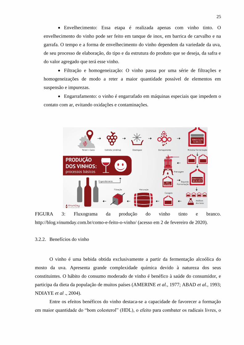
25
Envelhecimento: Essa etapa é realizada apenas com vinho tinto. O
envelhecimento do vinho pode ser feito em tanque de inox, em barrica de carvalho e na
garrafa. O tempo e a forma de envelhecimento do vinho dependem da variedade da uva,
de seu processo de elaboração, do tipo e da estrutura do produto que se deseja, da safra e
do valor agregado que terá esse vinho.
Filtração e homogeneização: O vinho passa por uma série de filtrações e
homogeneizações de modo a reter a maior quantidade possível de elementos em
suspensão e impurezas.
Engarrafamento: o vinho é engarrafado em máquinas especiais que impedem o
contato com ar, evitando oxidações e contaminações.
FIGURA 3: Fluxograma da produção do vinho tinto e branco.
http://blog.vinumday.com.br/como-e-feito-o-vinho/ (acesso em 2 de fevereiro de 2020).
3.2.2. Benefícios do vinho
O vinho é uma bebida obtida exclusivamente a partir da fermentação alcoólica do
mosto da uva. Apresenta grande complexidade química devido à natureza dos seus
constituintes. O hábito do consumo moderado de vinho é benéfico à saúde do consumidor, e
participa da dieta da população de muitos países (AMERINE et al., 1977; ABAD et al., 1993;
NDIAYE et al ., 2004).
Entre os efeitos benéficos do vinho destaca-se a capacidade de favorecer a formação
em maior quantidade do “bom colesterol” (HDL), o efeito para combater os radicais livres, o
26
efeito antimicrobiano, diurético, anti-inflamatório e antineoplásico (ABAD et al., 1993;
STIPP et al., 2017; BEZERRA et al., 2018).
Os taninos, flavonóis e estilbenos são compostos fenólicos presentes em maior
quantidade nos vinhos envelhecidos. Os taninos favorecem a digestão e os flavonóis têm sido
estudados devido ao seu poder antioxidante e efeito protetor em relação aos radicais livres. No
grupo dos estilbenos encontra-se o resveratrol, que é uma fitoalexina. O resveratrol tem sido
muito estudado devido ao provável efeito protetor contra doenças cardiovasculares. Na uva, o
resveratrol encontra-se na película, e o vinho tinto apresenta maior quantidade, devido ao
processo de elaboração. A concentração de resveratrol no vinho varia de 1,3 a 7,0 mg/L
(BOURZEIX et al., 1989; ADRIAN et al., 2000; JANNIN et al., 2001). Muitos efeitos
benéficos do vinho na saúde têm sido atribuídos ao resveratrol, um polifenol presente na pele
das uvas (DIAZ-GEREVINI et al., 2016; GOJKOVIC-BUKARICA et al., 2018). No entanto,
existem poucos estudos sobre atividades biológicas e modulação de mediadores inflamatórios
por outros componentes do vinho, como os polissacarídeos de vinho.
3.2.3. Carboidratos dos vinhos
Os carboidratos são um dos principais constituintes químicos da maioria dos tecidos e
células dos organismos (SIMÕES et al., 2003). Eles existem como monossacarídeos,
oligossacarídeos, polissacarídeos e seus derivados (AVIGAD & DEY, 1997). De acordo com
a sua função, os carboidratos podem ser classificados em dois grandes grupos: estruturais e de
reserva. Os carboidratos estruturais são responsáveis pela formação da parede celular e de
outras estruturas, sendo considerados os compostos orgânicos mais abundantes da Terra
(REID, 1997).
Os carboidratos presentes no vinho são derivados tanto das paredes das células de
micro-organismos quanto das uvas (DOCO et al., 1999; CABANIS & CABANIS, 2000b;
VIDAL et al., 2003; AYESTARAN et al., 2004).). Os originários das paredes celulares de
uva incluem as arabinogalactanas, ramnogalacturonanas e homogalacturonanas, enquanto que
aquelas originadas a partir de paredes das células de levedura são principalmente glucanas,
mananas e manoproteínas (WATERS et al., 1994; PELLERIN et al., 1995; MARTÍNEZ-
LAPUENTE et al., 2013).
3.2.3.1. Parede celular vegetal e polissacarídeos
27
A parede celular é dividida em: primária, não lignificada, com células jovens que
ainda mantém a capacidade de divisão e alongamento, apresentam uma parede celular muito
fina, com 0,1 a 1 µm de espessura; e secundária, lignificada, com células sem capacidade de
crescimento e divisão, que conferem rigidez aos tecidos (REID, 1997).
A parede celular dos vegetais é altamente organizada e constituída de polissacarídeos,
proteínas e substâncias aromáticas. Os polissacarídeos são os principais componentes da
parede celular e são divididos em pectinas, hemiceluloses e celulose, como pode ser
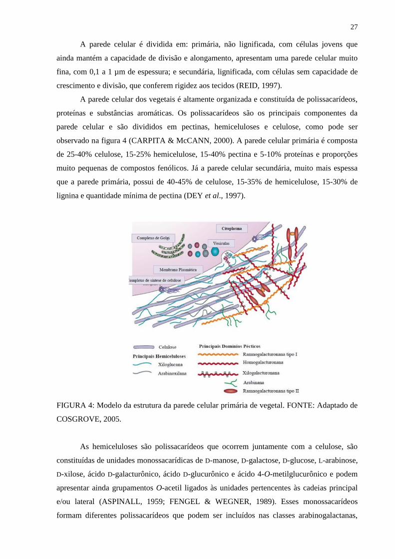
observado na figura 4 (CARPITA & McCANN, 2000). A parede celular primária é composta
de 25-40% celulose, 15-25% hemicelulose, 15-40% pectina e 5-10% proteínas e proporções
muito pequenas de compostos fenólicos. Já a parede celular secundária, muito mais espessa
que a parede primária, possui de 40-45% de celulose, 15-35% de hemicelulose, 15-30% de
lignina e quantidade mínima de pectina (DEY et al., 1997).
FIGURA 4: Modelo da estrutura da parede celular primária de vegetal. FONTE: Adaptado de
COSGROVE, 2005.
As hemiceluloses são polissacarídeos que ocorrem juntamente com a celulose, são
constituídas de unidades monossacarídicas de D-manose, D-galactose, D-glucose, L-arabinose,
D-xilose, ácido D-galacturônico, ácido D-glucurônico e ácido 4-O-metilglucurônico e podem
apresentar ainda grupamentos O-acetil ligados às unidades pertencentes às cadeias principal
e/ou lateral (ASPINALL, 1959; FENGEL & WEGNER, 1989). Esses monossacarídeos
formam diferentes polissacarídeos que podem ser incluídos nas classes arabinogalactanas,
28
glucuronoxilanas arabinoxilanas, xilanas, galactoglucomananas, galactomananas,
glucuronomananas, glucomananas, mananas e xiloglucanas (ASPINALL, 1980; BRETT &
WALDRON, 1990; PULS & SCHUSEIL, 1993; KACURÁCOVÁ et al., 2000). Alguns
estudos já relataram a presença de arabinogalactanas e mananas no vinho (PELLERIN et al.,
1995; MARTÍNEZ-LAPUENTE et al., 2013).
As pectinas são polissacarídeos ricos em ácido galacturônico, ramnose, arabinose e
galactose. O termo pectina refere-se a polissacarídeos complexos que contém unidades de
ácido α-D-galacturônico (GalA). Os principais representantes são: ramnogalacturonas do tipo
I (RG I) e ramnogalacturonanas do tipo II (RG II) e homogalacturonanas (ASPINALL, 1980;
BRETT & WALDRON, 1990; CARPITA & GIBEAUT, 1993). Dentre elas, as RG I e RG II
já foram relatadas em estudos feitos com o vinho (PELLERIN et al., 1995; MARTÍNEZ-
LAPUENTE et al., 2013).
3.2.3.1.1. Arabinogalactanas
As arabinogalactanas podem ser classificadas em dois grupos de acordo com as
ligações químicas da cadeia principal: arabinogalactanas do tipo I (AG I) e arabinogalactanas
do tipo II (AG II) (ASPINAL, 1973).
As arabinogalactanas do tipo I apresentam cadeia principal formada por (1→4)-β-D-
galactanas. São pouco ramificadas e, na maioria das vezes, apresentam unidades de arabinose
ligadas no O-3 das unidades de galactose da cadeia principal (CARPITA & GIBEAUT,
1993).
As arabinogalactanas do tipo II são altamente ramificadas, possuem cadeia principal
formada por (1→3) e/ou (1→6)-β-D-galactanas ligadas umas as outras por pontos de
ramificação em O-3 e O-6 e apresentam a maior parte das posições O-3 e O-6 restantes
ocupadas por unidades de arabinose (CARPITA & GIBEAUT, 1993). Embora apresentem
uma estrutura geral semelhante, a estrutura fina das arabinogalactanas do tipo II varia muito
entre as espécies (CARPITA & GIBEAUT, 1993; ALBERSHEIM et al., 1996).
3.2.3.1.2. Ramnogalacturonanas
29
As ramnogalacturonanas são polissacarídeos ricos em ácido galacturônico e ramnose
(REID, 1997). Esses polímeros podem ser classificados em dois grupos: ramnogalacturonana
do tipo I (RG I) e ramnogalacturonana do tipo II (RG II).
As ramnogalacturonanas do tipo I são heteropolímeros formados por cadeia principal
constituída de unidades alternadas de ácido α-D-galacturônico ligadas α-(1→4) e unidades de
α-L-ramnose ligadas α-(1→2). Esses polímeros podem ser estendidos por ácido
poligalacturônico em seus terminais e as unidades ramnosil podem interromper longos
períodos de ácido poligalacturônico (CARPITA & GIBEAUT, 1993).
A ramnogalacturonana do tipo II é o polissacarídeo péctico mais complexo, porém o
que apresenta a estrutura mais conservada. A característica mais marcante dessa molécula é
possuir açúcares raros. São polímeros altamente substituídos que apresentam cadeia principal
constituída por unidades de ácido α-D-galacturônico unidas por ligação do tipo (1→4)
(O´NEILL et al., 1990). Esses polissacarídeos apresentam cadeias laterais geralmente
substituídas em C-2 das unidades de α-D-GalpA e seus substituintes são formados por
oligossacarídeos contendo monossacarídeos raros como apiose, 2-O-metil-fucose, 2-O-metil-
xilose, ácido acérico, Kdo (ácido 2-ceto-3-deoxi-D-mano-octulosônico), Dha (ácido 3-deoxi-
D-lixo-2-heotulosárico) entre outros, formando estruturas altamente complexas (CARPITA &
GIBEAUT, 1993; REID, 1997; PÉREZ, MAZEAU & PENHOAT, 2000; PAULSEN &
BARSETT, 2005; ATMODJO et al., 2013).
3.2.3.2. Parede celular de leveduras e polissacarídeos
Leveduras são fungos que possuem a mesma estrutura subcelular de células animais e
vegetais (OSUMI, 1998). São micro-organismos unicelulares, com tamanho que varia de 5 a
10 microns. As espécies variam entre si segundo a morfologia ou forma, metabolismo com
relação a diferentes substratos, modo de reprodução e onde são encontrados. Enquanto
existem aproximadamente 50.000 espécies de fungos, existem apenas 60 gêneros diferentes
de leveduras, com aproximadamente 500 espécies diferentes (STONE & MILLS, 2006).
A parede celular da levedura é constituída por aproximadamente 40% de β-glucanas,
40% de α-mananas, 8% de proteínas, 7% de lipídios, 3% de substâncias inorgânicas e 2% de
hexosaminas e quitina (HOUGH, 1990). Os principais monossacarídeos presentes nos
polímeros são a glucose e a manose, seguidos pela galactose, xilose, N-acetil-D-glucosamina,
ácidos urônicos e outros componentes secundários. A composição química qualitativa da
parede celular é característica de cada espécie, podendo ser empregada como marcador
taxonômico (FARKAS, 1989).

30
A principal levedura responsável pela fermentação na produção do vinho é a
Saccharomyces cerevisiae, a qual possui em sua parede celular cerca de 10-20% de proteínas
e lipídios e 80-90% de carboidratos, dos quais 50% são glucanas e mananas (NORTHCOTE
& HORNE, 1952; BARTNICKI-GARCIA & LIPPMAN, 1982; VUKOVIC et al., 1994).
Segundo Vukovic (1994), a S. cerevisiae apresenta a parede celular em camadas, sendo as
cadeias de mananas externas e as glucanas, internas. Já foi demonstrada a presença de
mananas entre os polissacarídeos encontrados no vinho (MARTÍNEZ-LAPUENTE et al.,
2013).
3.2.3.2.1. Mananas
As mananas são um dos polissacarídeos de maior importância da parede celular da
levedura, responsáveis por eventos de reconhecimento célula-célula, além de limitar o acesso
de agentes líticos às porções mais internas da parede celular e à membrana celular (KLIS, et
al., 2002).
Há evidências de que as mananas juntamente com os peptídeos complexos constituem
a camada mais externa da parede celular das leveduras (BALLOU, 1970; CABIB, 1975).
Esses polissacarídeos são altamente ramificado, contendo uma cadeia principal constituída
por ligações α-(1→6) substituída nas posições O-2 por cadeias laterais de unidades de α-D-
manopiranose ou oligossacarídeos com unidades de manose (1→2) e (1→3) ligadas
(STEWART & BALLOU, 1968; BARRETO BERTGER & GORIN, 1983; HALÁSZ &
LÁSZTITI, 1991).
3.2.4. Polifenóis do vinho
Os compostos fenólicos constituem um grupo diversificado de metabólitos
secundários presentes nas uvas e no vinho. O conteúdo fenólico e a composição dos produtos
processados da uva (vinho) são grandemente influenciados pela prática tecnológica à qual as
uvas estão expostas. Os polifenóis são componentes intrínsecos das uvas e produtos
relacionados, particularmente o vinho (LINSKENS & JACKSON, 1988; SCALBERT, 1993).
Os compostos fenólicos desempenham um papel importante nas características sensoriais do
vinho, são responsáveis por algumas das propriedades organolépticas como: aroma, cor, sabor
e adstringência (LINSKENS & JACKSON, 1988; SCALBERT, 1993).
31
Existe uma grande diversidade química na composição fenólica das uvas e do vinho.
Isto se deve não apenas às diferentes variedades de uvas, como também ao fato de que esse
tipo de compostos existe em ambas as formas livre e conjugada, pois podem estar ligadas ao
ácido quínico ácido ou a uma ou mais moléculas de açúcar (glicose, galactose, sacarose e
manose) produzindo mono-, di-, tri- ou mesmo tetraglicósidos (CHEYNIER et al., 2010;
LINSKENS & JACKSON, 1988; SCALBERT, 1993).
Os componentes fenólicos não voláteis das uvas e do vinho compreendem diversas
classes: ácidos fenólicos, flavonóides, taninos, estilbenos, cumarinas, derivados de
fenilpropanol, lignanas e neolignanas (LINSKENS & JACKSON, 1988; SCALBERT, 1993)..
Os polifenóis econtrados no vinho são provenientes da uva que estão localizados
principalmente nas partes sólidas: pele, semente e tecido vascular. Os flavonóis e antocianinas
são encontrados na pele e sementes da uva, já as antocianinas são encontradas apenas na pele
da uva de tinta, sendo responsável pela cor vermelha característica dos vinhos tintos (KOSIR
& KIDRIC, 2002; FLAMINI, 2003).
Os flavonóides pertencem a uma classe química que exibe uma estrutura básica de 15
átomos de carbono compreendendo dois anéis aromáticos ligados através de uma cadeia de 3
carbonos (C6 - C3 - C6), que pode ou não fazer parte de um terceiro anel. Esse esqueleto de
carbono é responsável pela diversidade química dessa família de compostos. Os flavonóides
são geralmente agrupados em várias classes, que diferem principalmente no grau de oxidação
do anel pirano central, exceto no caso das chalconas. Eles compreendem diferentes tipos de
compostos tais como flavonas, flavonóis, flavanonas, flavononóis, flavanos, flavanóis,
antocianidinas e antocianinas, chalconas e di-hidrochalcones (BADERSCHNEIDER &
WINTERHALTER, 2001).
3.2.4.1. Flavanóis
Flavanóis são benzopiranos que possuem carbono saturado cadeia entre C2 e C3, uma
função hidroxila em C3 e nenhuma carbonila grupo em C4. Tanto o flavan-3-ols quanto o
flavan-3,4-dióis podem ser encontrado na natureza, sendo este último frequentemente
presente em madeira e casca de árvore, mas raramente encontrado em frutas. Flavan-3,4-dióis
também são freqüentemente chamados de leucoantocianidinas (GARRIDO & BORGES,
2013).
Os flavanoís mais abundantes na natureza são a catequina e seu enantiômero
epicatequina. Estes compostos estão presentes na casca e sementes de uvas, bem como no
vinho. Nos vinhos brancos produzidos em condições especiais, evitando contato prolongado
32
com a pele das uvas, a catequina foi considerada a mais abundante flavonóide, sendo
amplamente responsável pelo sabor característico do vinho (LUNTE, BLANKENSHIP, &
READ, 1988). Alguns derivados da catequina, nomeados galocatequina, epigalocatequina, e
epicatequina galato foram identificados em uvas e vinho (DECENDIT et al., 2002; MATTIVI
et al, 2009).
3.2.4.2. Pigmentos antociânicos
Nos últimos anos, várias famílias de derivados de antocianinas têm sido relatadas em
uvas e vinho. Pigmentos antociânicos são responsáveis pela cor das uvas e vinho, uma
característica determinada pela sua estrutura química, grau de hidroxilação, metilação e/ou
glicosilação (HE et al., 2010). Em uvas vermelhas e vinho, seis antocianidinas foram
identificadas: cianidina, peonidina, delfinidina, pelargonidina, petunidina e malvidina (HE et
al., 2010; KOPONEN et al, 2007). Este último é considerado o composto mais representativo
em uvas Vitis vinifera (CASTILLO-MUÑOZ et al., 2010). As antocianidinas são geralmente
encontradas na natureza nas formas glicosiladas: 3-monoglucósidos 3,5 e 3,7-diglucósidos
(KOPONEN et al., 2007). São encontradas apenas na pele da uva, a quantidade de
antocianinas e flavonóides extraídos após a vinificação dependem da duração do processo e
das condições em que ele ocorre, como a temperatura ou a extensão da ruptura das uvas
(ZOECKLEIN et al., 1995).
3.2.4.3. Compostos fenólicos e índice de cor
A coloração dos vinhos está estreitamente relacionada com a sua composição em
pigmentos fenólicos (MAZZA et al., 1999). Esses pigmentos apresentam uma variação de cor
de acordo com certos parâmetros físico-químicos do meio onde se encontram. Um dos
aspectos principais a se ter em conta é o pH do vinho, uma vez que, em meio ácido, as
antocianinas apresentam uma coloração vermelha que se dissipa à medida que sobe o pH,
apresentando uma cor azul-arroxeada quando o pH é superior a 4 e uma cor amarela quando o
pH é neutro ou básico (RIBÉREAU-GAYON, et al. 2006b). Algumas classes de pigmentos
antociânicos apresentam maior estabilidade com mudança de pH e com evelhecimento.
Polifenóis são compostos instáveis e suas reações começam assim que a uva é
esmagada ou prensada e continuam durante o processo de vinificação e envelhecimento,
resultando uma diversidade grande de novos produtos. Tais produtos apresentam propriedades

33
organolépticas específicas, frequentemente diferentes daquelas de seus precursores. A
composição fenólica nos vinhos depende da uva utilizada e das condições de vinificação que
influenciam na extração de vários compostos da uva e das subsequentes reações (CHEYNIER
et al. 2006). Durante o armazenamento, os pigmentos antociânicos podem provocar mudança
de coloração (CEJUDO-BASTANTE et al. 2011). O aumento da concentração dos compostos
fenólicos ao longo do tempo é atribuído às reações de oxidação e polimerização, onde
também ocorrer um aumento na absorbância de 420 nm, diretamente relacionada com o
escurecimento dos vinhos (BARÓN et al. 1997). Por tanto, os compostos fenólicos são
meléculas de grande importância para a enologia, sendo um dos principais responsáveis pelas
porpriedades organolépticas do vinho.
3.3. PROCESSO INFLAMATÓRIO E POLISSACARÍDEOS
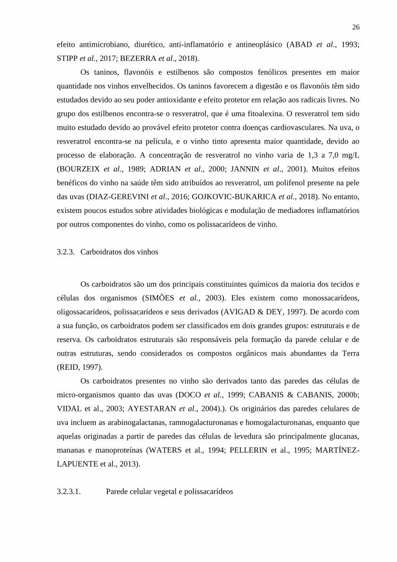
A reação da imunidade natural, ou inflamação, é uma resposta do organismo a
estímulos nocivos. Durante uma invasão por patógeno ou danos nos tecidos do hospedeiro, a
imunidade natural é a primeira linha de defesa do organismo (WANG et al., 2002). Essa ação
protetora é obtida principalmente pelo recrutamento de leucócitos, seguida por uma cascata de
reações bioquímicas que se propagam e iniciam a resposta inflamatória (NAKAMURA et al.,
1992; VARKI, 1997; SIMON et al., 1999; MULLER, 2003; YADAV et al., 2003). Essa
migração leucocitária ocorre do compartimento intravascular, principalmente de vênulas pós-
capilares, para o tecido onde ocorreu o estímulo nocivo, sendo denominado de
extravasamento ou diapedese. Esse mecanismo é constituído de várias etapas: marginação,
ligação inicial e rolamento dos leucócitos no endotélio inflamado, ativação dos leucócitos,
firme aderência à parede vascular, degradação da membrana basal e migração dos leucócitos
para o sítio de inflamação (Figura 4) (SEELY et al., 2003; PARISH, 2006; SHERWOOD et
al., 2004).
Os indutores inflamatórios no sítio específico levam à produção de citocinas pró-
inflamatórias, TNF (Fator de Necrose Tumoral) e IL-1 (Interleucina-1) pelas células locais
(BARRINGTON et al., 2001; NAKAMURA et al., 1992; SIEDLE et al., 2003). Essas
substâncias irão agir no endotélio vascular, causando alterações intracelulares que culminam
na expressão de moléculas de adesão e citocinas, seguida pela modificação do seu
citoesqueleto de retração. Isso garantirá a passagem dos leucócitos da circulação para o local
da lesão (SIMON et al., 1999; HOGG et al., 1995; LASKY, 1992; VARKY, 1997). A
aderência e o rolamento de leucócitos no endotélio ativado pelo estímulo inflamatório
envolvem selectinas de linfócitos, plaquetas e células endoteliais (L-, P-, e E-selectinas,
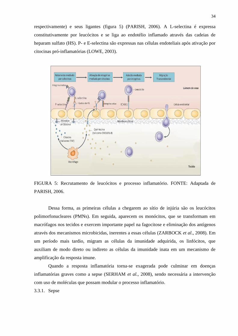
34
respectivamente) e seus ligantes (figura 5) (PARISH, 2006). A L-selectina é expressa
constitutivamente por leucócitos e se liga ao endotélio inflamado através das cadeias de
heparam sulfato (HS). P- e E-selectina são expressas nas células endoteliais após ativação por
citocinas pró-inflamatórias (LOWE, 2003).
FIGURA 5: Recrutamento de leucócitos e processo inflamatório. FONTE: Adaptada de
PARISH, 2006.
Dessa forma, as primeiras células a chegarem ao sítio de injúria são os leucócitos
polimorfonucleares (PMNs). Em seguida, aparecem os monócitos, que se transformam em
macrófagos nos tecidos e exercem importante papel na fagocitose e eliminação dos antígenos
através dos mecanismos microbicidas, inerentes a essas células (ZARBOCK et al., 2008). Em
um período mais tardio, migram as células da imunidade adquirida, os linfócitos, que
auxiliam de modo direto ou indireto as células da imunidade inata em um mecanismo de
amplificação da resposta imune.
Quando a resposta inflamatória torna-se exagerada pode culminar em doenças
inflamatórias graves como a sepse (SERHAM et al., 2008), sendo necessária a intervenção
com uso de moléculas que possam modular o processo inflamatório.
3.3.1. Sepse
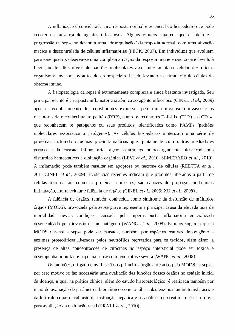
35
A inflamação é considerada uma resposta normal e essencial do hospedeiro que pode
ocorrer na presença de agentes infecciosos. Alguns estudos sugerem que o início e a
progressão da sepse se devem a uma “desregulação” da resposta normal, com uma ativação
maciça e descontrolada de células inflamatórias (PECK, 2007). Em indivíduos que evoluem
para esse quadro, observa-se uma completa ativação da resposta imune e isso ocorre devido à
liberação de altos níveis de padrões moleculares associados ao dano celular dos micro-
organismos invasores e/ou tecido do hospedeiro lesado levando a estimulação de células do
sistema imune.
A fisiopatologia da sepse é extremamente complexa e ainda bastante investigada. Seu
principal evento é a resposta inflamatória sistêmica ao agente infeccioso (CINEL et al., 2009)
após o reconhecimento dos constituintes expressos pelo micro-organismo invasor e os
receptores de reconhecimento padrão (RRP), como os receptores Toll-like (TLR) e o CD14,
que reconhecem os patógenos ou seus produtos, identificados como PAMPs (padrões
moleculares associados a patógenos). As células hospedeiras sintetizam uma série de
proteínas incluindo citocinas pró-inflamatórias que, juntamente com outros mediadores
gerados pela cascata inflamatória, agem contra os micro-organismos desencadeando
distúrbios hemostáticos e disfunção orgânica (LEVI et al., 2010; SEMERARO et al., 2010).
A inflamação pode também resultar em apoptose ou necrose de células (REETTA et al.,
2011;CINEL et al., 2009). Evidências recentes indicam que produtos liberados a partir de
células mortas, tais como as proteínas nucleares, são capazes de propagar ainda mais
inflamação, morte celular e falência de órgãos (CINEL et al., 2009; XU et al., 2009) .
A falência de órgãos, também conhecida como síndrome da disfunção de múltiplos
órgãos (MODS), provocada pela sepse grave representa a principal causa da elevada taxa de
mortalidade nessas condições, causada pela hiper-resposta inflamatória generalizada
desencadeada pela invasão de um patógeno (WANG et al., 2008). Estudos sugerem que a
MODS durante a sepse pode ser causada, também, por espécies reativas de oxigênio e
enzimas proteolíticas liberadas pelos neutrófilos recrutados para os tecidos, além disso, a
presença de altas concentrações de citocinas no espaço intersticial pode ser tóxica e
desempenha importante papel na sepse com leucocitose severa (WANG et al., 2008).
Os pulmões, o fígado e os rins são os primeiros órgãos afetados pela MODS na sepse,
por esse motivo se faz necessária uma avaliação das funções desses órgãos no estágio inicial
da doença, a qual na prática clínica, além do estudo histopatológico, é realizada também por
meio de avaliação de parâmetros bioquímico como análises das enzimas aminotransferases e
da bilirrubina para avaliação da disfunção hepática e as análises de creatinina sérica e ureia
para avaliação da disfunção renal (PRATT et al., 2010).
36
Diversos trabalhos foram realizados e as atividades biológicas mais comumente
atribuídas aos carboidratos de diversas fontes são: atividade anti-inflamatória, antitumoral,
antiviral e anticoagulante (BOHN & MILLER, 1995; CAPEK et al., 2003; RENARD et al.,
2005, CIPRIANI et al., 2006, 2009; SIMAS-TOSIN et al., 2012; DARTORA et al., 2013).
Polissacarídeos, como ramnogalactofuranana e arabinogalactana de várias fontes
apresentaram atividade anti-inflamatória e redução da letalidade de camundongos quando
submetidos à sepse polimicrobiana induzida por ligadura e punção cecal (CLP) (SCOPARO
et al., 2013). Esse modelo biológico mimetiza a sepse em humanos, causada por patógenos
derivados do trato intestinal e é considerada como uma simulação próxima da situação clínica
(RITTIRSCH, HUBER-LANG, FLIERL, & WARD, 2009).
37
REFERÊNCIAS BIBLIOGRÁFICAS
ABAD, F. B.; PLASENCIA, J. M. B. Consumo moderado de bebidas alcohólicas:
salud y civilización. Madrid: Inesiba, 135 p, 1993.
ADRIAN, M.; JEANDET, P.; BREUIL, A. C.; LEVITE, D.; DEBORD, S.; BESSIS,
R. Assay of resveratrol and derivative stilbenes in wines by direct injection High Performance
Liquid Chromatography. American Journal of Enology & Viticulture, Davis, v. 51, n. 1, p.
37-40, 2000.
ALBERSHEIM, P.; DARVILL, A. G.; OíNEILL, M. A.; SCHOLS, H. A.;
VORAGEN, A. G. J. An hypothesis: the same six polysaccharides are components of the
primary cell walls of all higher plants. Progress in biotechnology: Pectins and Pectinases.
Amsterdam: Elsevier, v. 14, p. 47-55, 1996.
AMERINE, M. A. Le vin dans l’alimentation humaine. Bulletin de l’O.I.V. Paris,
número especial, p. 137-151, 1977.
ASPINAL, G. O. In: Biogenesis of Plant Cell Wall Polysaccharides. New York:
Academic Press, p. 95-115,1973.
ASPINALL, G. O. Chemistry of cell wall polysaccharides. In: STUMPF, P. K.;
CONN, E. E. The Biochemistry of Plants. New York: Academic Press. v. 3, p. 473-500, 1980.
ASPINALL, G.O. Structural chemistry of the hemicelluloses. Advances in
Carbohydrate Chemistry. New York, p. 429-430, 1959.
ATMODJO, M. A.; HAO, Z.; MOHNEN, D. Evolving views of pectin biosynthesis.
Annual Review of Plant Biology, vol. 64, p. 747-749, 2013.
AVIGAD, G.; DEY, P. M. Carbohydrate metabolism: storage carbohydrates. In: DEY,
P. M.; HARBORNE, J. B. Plant Biochemistry. Bristol: Academic Press, p.143, 1997.
AYESTARAN, B.; GUADALUPE, Z.; LEON, D. Quantification of major grape
polysaccharides (Tremanillo v.) released by maceration enzymes during the fermentation
process. Analytica Chimica Acta, v. 513, p. 29-39, 2004.
BADERSCHNEIDER, B., & WINTERHALTER, P. Isolation and characterization of
novel benzoates, cinnamates, flavonoids, and lignans from Riesling wine and screening for
antioxidant activity. Journal of Agricultural and Food Chemistry, 49(6), 2788–2798. (2001).
BALLOU, C.E. A study of the immunochemistry of three yeast mannans. Journal of
Biological Chemistry, v.245, n.5, p.1197-1203, 1970.
Barón, R.; Mayén, M.; Mérida, J.; Medina, M. Changes in phenolic compounds and
browning during biological aging of SherryType Wine. J. Agric. Food Chem. 45: 1682- 1685.
(1997).
38
BARRETO-BERGTER, E.; GORIN, P. A. J.; TRAVASSOS, L. R. Cell constiuents of
the mycelia and conidia of A. fumigatus. Carbohydrates Letters, 95 (1): 205-218, 1981.
BARRINGTON, R.; ZHANG, M., FISCHER, M., CARROLL, M.C. The role of
complemente in inflammation and adaptative immunity. Immunolo Rev. 180, 5-15, 2001.
BARTNICKI-GARCIA, S; LIPPMAN, E. Fungal morphogenesis: Cell wall
construction in Muca rouxii. Scienc, 1659(1): 302-304, 1969.
BEZERRA, I.L.; CAILLOT, A. R. C.; PALHARES, L. C. G. F.; SANTANA-FILHO,
A. P.; CHAVANTE, S. F. GUILHERME LANZI SASSAKI, G. L. Structural characterization
of polysaccharides from Cabernet Franc, Cabernet Sauvignon and Sauvignon Blanc wines:
Anti-inflammatory activity in LPS stimulated RAW 264.7 cells. Carbohydrate Polymers, 186,
91-99. 2018.
BOHN, J. A.; Be MILLER, J. N. 1 →3 β-D-glucans as biological response modifiers:
a review of structure-functional activity relationships. Carbohydrate Polymers, v. 28, p. 3-14,
1995.
BOURZEIX, M. Raisin, vin, nutrition et santé. Les catéchines et proanthocyanidols du
raisin et du vin. Progrés Agricole et Viticole, Montpellier, v. 106, n. 22, p. 487-491, 1989.
BRETT, C.; WALDRON, K. Physiology and biochemistry of plant cell walls.
London: Unwin Hyman, 194p, 1990.
CAILLOT, A. R. C.; BEZERRA, I.L.; PALHARES, L. C. G. F.; SANTANA-FILHO,
A. P.; CHAVANTE, S.; SASSAKI, G. L. Structural characterization of blackberry wine
polysaccharides and immunomodulatory effects on LPS-activated RAW 264.7 macrophages.
Food Chemistry, 257, 143-159.,2018.
CABANIS, J. C.; CABANIS, M. T. Tablas de composición. Cap.8. In: FLANZY, C.
Enología: Fundamentos científicos y tecnológicos. Madrid: Ediciones A. Madrid Vicente,
Ediciones Mundi-Prensa. 1ª edición, 783p, 2000b.
CABIB, E. Molecular aspects of yeast morphogenesis. Annual Review of
Microbiology, 29: 191-214, 1975.
CAPEK, P.; HRIBALOV£, V.; SVANDOV£, E.; EBRINGEROV£, A.; SASINKOV£,
V., MASAROV£, J. Characterization of immunomodulatory polysaccharides from Salvia
officinalis L. International Journal of Biological Macromolecules, v. 33, p. 113-119, 2003.
CARPITA, N. C.; GIBEAUT, D. M. Structural models of primary cell walls in
flowering plants: consistency of molecular structure with the physical properties of the walls
during growth. Plant Journal, v. 3, p. 1-30, 1993.

39
CARPITA, N.; McCANN, M. The cell wall. In: BUCHANAN, B. B.; GRUISSEM,
W.; JONES, R. L. Biochemistry & Molecular Biology of Plants. USA: Courier. p. 52-
108,2000.
CEJUDO-BASTANTE, M.J.; HERMOSÍN-GUTIERREZ, I.; CASTRO-VAZQUEZ,
L.I.; PÉREZ-COELLO, M.S. Hyperoxygenation and bottle storage of Chardonnay white
wines: Effects on color-related phenolics, volatile composition, and sensory characteristics. J.
Agric. Food Chem. 59: 4171- 4182. (2011).
CHEYNIER, V.; DUEÑAS-PATON, M.; SALAS, E.; MAURY, C.; SOUQUET, J.
M.; SARNI-MANCHADO, P.; FULCRAND, H. Structure and properties of wine pigments
and tannins. Am. J. Enology Vitic., 57: 298-305. (2006).
CINEL I, OPAL SM. Molecular biology of inflammation and sepsis: a primer.Crit
Care Med;37:291–304. 2009.
CASTILLO-MUÑOZ, N., WINTERHALTER, P., WEBER, F., GÓMEZ, M. V.,
GÓMEZ-ALONSO, S., GARCÍA-ROMERO, E. Structure elucidation of peonidin 3,7-O-β-
diglucoside isolated from Garnacha Tintorera (Vitis vinifera L.) grapes. Journal of
Agricultural and Food Chemistry, 58(20), 11105–11111. (2010).
CHEYNIER, V., SCHNEIDER, R., SALMON, J. -M., & FULCRAND, H. Chemistry
of wine. Comprehensive Natural Products II, 3, 1119–1172. (2010).
CIPRIANI, T. R.; MELLINGER, C. G.; BERTOLINI, L. C.; BAGGIO, C. H.;
FREITAS, C. S.; MARQUES, M. C. A.; GORIN, P. A. J; SASSAKI, G. L.; IACOMINI, M.
Gastroprotective effect of a type I arabinogalactan from soybean meal, Food Chemistry.
V115. p. 687-690, 2009a.
CIPRIANI, T. R.; MELLINGER, C. G.; BERTOLINI, L. C.; BAGGIO, C. H.;
FREITAS, C. S.; MARQUES, M. C. A.; GORIN, P. A. J; SASSAKI, G. L.; IACOMINI, M.
Gastroprotective effect of a type I arabinogalactan from soybean meal, Food Chemistry.
V115. p. 687-690, 2009a.
CIPRIANI, T. R.; MELLINGER, C. G.; SOUZA, L. M., BAGGIO, C. H., FREITAS,
C. S. Polysaccharide from a tea (Infusion) of Maytenus ilicifolia leaves with anti-ulcer
protective effects. Journal of Natural Products, v. 69, p. 1018-1021, 2006.
CIPRIANI, T. R.; MELLINGER, C. G.; SOUZA, L. M.; BAGGIO, C. H., FREITAS,
C. S.; MARQUES, M. C. A.; GORIN, P. A. J.; SASSAKI, G. L. IACOMINI, M.
Polygalacturonic acid: Another anti-ulcer polysaccharide from the medicinal plant Maytenus
ilicifolia. Carbohydrate Polymers, v. 78, p.361-363, 2009b.
CRUZ, INÊZ. A Vitis vinifera e as demais Vitis. Saúde de Viver. p.63-95. 2006.

40
DARTORA, N.; SOUZA, L. M.; SIMONE M.M. PAIVA, S. M. M.; CAMILA T.
SCOPARO, C. T.; MARCELLO IACOMINI, M.; GORIN, P. A. J.; YANNA D.
RATTMANN, Y. D.; SASSAKI, G. L. Rhamnogalacturonan from Ilex paraguariensis: A
potential adjuvant in sepsis treatment. Carbohydrate Polymers,1776–1782, 2013.
DECENDIT, A., WAFFO-TEGUO, P., RICHARD, T., KRISA, S., VERCAUTEREN,
J., MONTI, J. -P., et al. Galloylated catechins and stilbene diglucosides in Vitis vinifera cell
suspensions cultures. Phytochemistry, 60(8), 795–798. (2002).
DEY, P.M.; BROWNLEADER, M.D.; HARBORNE, J.B. The plant, the cell and its
molecular components. In: DEY, P. M.; HARBORNE, J. B. Plant Biochemistry. Bristol:
Academic Press, p. 6-9. 1997.
DIAZ-GEREVINI, G. T., REPOSSI, G., DAIN, A., TARRES, M. C., DAS, U. N.,
EYNARD, A. R. Beneficial action of resveratrol: How and why?. Nutrition, 2, 174-178.
2016.
DOCO, T.; VUCHOT, P.; CHEYNIER, V.; MOUTOUNET, M. Structural
modification of arabinogalactan-proteins during aging of red wines on lees. American Journal
of Enology and Viticulture, 54, 150–157. 2003.
DOCO,T.; QUELLEC, N.; MOUTOUNET, M.; PELLERIN, P.; Polysaccharide
patterns during the aging of Carignan noir red wines. American Journal of nology and
Viticulture, v. 50, p. 28-32, 1999.
ESTREICHER, S. K., Ed. Wine: From Neolithic Times to 21st Century. Nova York:
Algora, 1 ed. 2006.
FARKAŠ, V. Polysaccharide Metabolism. In: ROSE, A.H.; HARRISON, J.S. The
yeasts: metabolism and physiology of yeasts, 2ed. London: Academic Press, Vol.3, cap. 9, p.
317-366, 1989.
FENGEL, D.; WEGNER, G. Wood: chemistry, ultrastructure, reactions. Berlin:
Gruyter, W. 613p, 1989.
FLAMINI, R. Mass spectrometry in grape and wine chemistry. Part I: Polyphenols.
Mass Spectrometry Reviews, 22(4), 218–250. (2003).
FOURNAIRON, C.; CAMARASA, C.; MOUTOUNET, M.: SALMON, J. M. New
trends on yeast autolysis and wine ageing on lees. Journal International de la Vigne et du Vin,
36, 49–69, 2002.
GARRIDO J., BORGES F. Wine and grape polyphenols. A chemical perspective.
Food Rearch International; 54: 1844-1858,2013.

41
GERBAUD, V.; GABAS, N., BLOUNIN, J.; PELLERIN, P.; MOUTOUNET, M.
Influence of wine polysaccharides and polyphenols on the crystallization of potassium
hydrogen tartrate. Journal International Science Vigne et du Vin, 31, 65–83. 1997.
GOJKOVIC-BUKARICA, L., MARKOVIC-LIPKOVSKI, J., HEINLE, H.,
CIROVIC, S., JOVANA RAJKOVIC, J., DJOKIC, V., ZIVANOVIC , V., ANA
BUKARICA, A., NOVAKOVIC, R. The red wine polyphenol resveratrol induced relaxation
of the isolated renalartery of diabetic rats: The role of potassium channels. Journal of
Functional Foods, 52, 266–275. 2018.
GRAINGER, K.; TATTERSALL, H., Eds. Wine Production: Vine to Bottle. Oxford:
Blackwell, p.127, 1 ed. 2005.
GUADALUPE, Z.; AYESTARAN, B. Polysaccharide profile and content during the
vinification and aging of Trempanillo red wines. Journal of Agricultural and Food Chemistry,
v. 55, p. 10720-10728, 2007.
GUERRA, C. C. Conhecendo o Essencial sobre Uvas e Vinhos. 2009. Disponível
em:<http://www.cnpuv.embrapa.br/publica/documentos/doc048.pdf>. Acesso em:
20/06/2018.
GUERRA, C. C. Maturação e Colheita. In: GUERRA, C. C. UVA para processamento
Pós-colheita. Brasília: Embrapa, 2003.
HALÁSZ, A., LÁSZTITY, R. Use of yeast biomass in food production. New York,
CRC press, 312p, 1991.
HE, F., MU, L., YAN, G. -L., LIANG, N. -N., PAN, Q. -H., WANG, J. Biosynthesis
of anthocyanins and their regulation in colored grapes. Molecules, 15(12), 9057–9091.
(2010).
HOFFMAN, ALEXANDRE. Sistema de produção de destilado de vinho. EMBRAPA.
2009.
HOGG, J.C.; DORESCHUK, CM. Leukocyte traffic in the lung. Annu. Rev. Physiol.
57, 97-114, 1995.
HOUGH, J. S. Biotecnologia de la cerveza y de la malta. Zaragoza, Acribia. 194.p,
1990.
INNGJERDINGEN, K. T., THÖLE, C., DIALLO, D., PAULSEN, B. S. HENSEL, A.
Inhibition of Helicobacter pylori adhesion to human gastric adenocarcinoma epithelial cells
by aqueous extracts and pectic polysaccharides from the roots of Cochlospermum tinctorium
A. Rich. And Vernonia kotschyana Sch. Bip. Ex Walp. Fitoterapia, 95, 127–132, 2014.
42
JANNIN, B.; DELMAS, D.; CHERKAOUI MALKI, M.; LATRUFFE, N. Étude du
mécanisme d’action du resvératrol, un polyphénol du vin, sur la prolifération cellulaire.
Bulletin de l’O.I.V., Paris, v. 74, n. 839-840, p. 54-60, 2001.
JOHNSON, H. Vintage: The Story of Wine .Simon & Schuster. p.11-16.1989.
KAKURÁKOVÁ, M.; CAPEK, P.; WELLNER, N.; EBRINGEROVÁ, A. FT-IR
study of plant cell model compounds: pectic polysaccharides and hemicelluloses.
Carbohydrate Polymers, v. 43, p. 195-203, 2000.
KOPONEN, J. M., HAPPONEN, A.M., MATTILA, P. H., & TÖRRÖNEN, A.R.
Contents of anthocyanins and ellagitannins in selected foods consumed in Finland. Journal of
Agricultural and Food Chemistry, 55(4), 1612–1619. (2007).
KOSIR, I. J., & KIDRIC, J. Use of modern nuclear magnetic resonance spectroscopy
in wine analysis: Determination of minor compounds. Analytica Chimica Acta, 458(1), 77–
84. (2002).
KLIS, F. M.; MOL, P.; HELLINGWERF, K.; BRUL, S. Dynamics of cell wall
structure in Saccharomyces cerevisiae. FEMS Microbiology Reviews, v. 26, p. 239-256,
2002.
LAROUSSE DO VINHO. Edição brasileira atualizada. 2ª edição, 2007.
LASKY, L.A. Selectins: interpreters of cell-specific information during inflammation.
Science. 258, 964-969, 1992.
LEVI M, SCHULTZ M, VAN DER POLL T. Disseminated intravascular coagulation
in infectious disease. Semin Thromb Hemost.;36:367–77. 2010.
LINSKENS, H. F., & JACKSON, J. F. Phenolic composition of natural wine types —
Wine analysis. Berlin: Springer-Verlag Press. (1988).
LOWE, J.B. Glycan-dependent leukocyte adhesion and recruitment in inflammation.
Curr. Opin. Cell Biol. 15: 531–538, 2003.
MARTÍNEZ-LAPUENTE, L.; GUADALUPE, Z.; AYESTARAN, B.; ORTEGA-
HERAS, M.; PEREZ-MAGARINO, S. Changes in polisaccharide composition during
sparkling wine marking and aging. Journal Agricultural and Food Chemistry, v. 61, p. 12362-
12373, 2013.
MATTIVI, F., GUZZON, R., VRHOVSEK, U., STEFANINI, M., & VELASCO, R.
Metabolite profiling of grape: Flavonols and anthocyanins. Journal of Agricultural and Food
Chemistry, 54(20), 7692–7702. (2006).
MAZZA, G., FUKUMOTO, L., DELAQUIS, P., GIRARD, B., & EWERT, B.
Anthocyanins, phenolics, and color of Cabernet Franc, Merlot, and Pinot Noir Wines from
British Columbia. Journal of Agricultural and Food Chemistry, 47(10), 4009–4017. (1999).
43
MELLINGER, C. G., CIPRIANI, T. R., NOLETO, G. R., CARBONERO, E. R.,
OLIVEIRA, M. B. M., GORIN, P., IACOMINI, M. Chemical and immunological
modifications of an arabinogalactan present in tea preparations of Phyllanthus niruri after
treatment with gastric fluid. International Journal of Biological Macromolecules, 43, 115–
120, 2008.
MUELLER, E. A., & ANDERER, F. A. Synergistic action of a plant
rhamnogalacturonan enhancing antitumor cytotoxicity of human natural killer and
lymphokine-activated killer cells: Chemical specificity of target cell recognition. Cancer
Research, 50(12), 3646–3651. 1990.
MULLER, W.A. Leukocyte-endothelial-cell interactions in leukocyte transmigration
and the inflammatory response. Trends Immunology. 24: 327-334, 2003.
NAKAMURA, H. Netrophil elastase in respiratory epithelial lining fluid of
individuals with cystic fibrosis induces interleukin-8 gene expression in a human bronchial
epithelial cell line. J. Clin Invest. 89, 1478-1484, 1992.
NDIAYE, M.; CHATAIGNEAU, M.; LOBYSHEVA, I.; CHATAIGNEAU, T.;
SCHINI-KERTH, V.B. Red wine polyphenol-induced, endothelium-dependent NO-mediated
relaxation is due to the redox-sensitive PI3-kinase/Akt-dependent phosphorylation of
endothelial NO-synthase in the isolated porcine coronary artery. FASEB J. 2005
Mar;19(3):455-7. Epub 2004.
NORTHCOTE, D. H.; HORNE, R. W. The Chemical composition and structure of the
yeast cell wall. Biochemical Journal 51: 232-236, 1952.
O´NEILL, M.; ALBERSHEIM, P.; DARVILL, A. The pectic polysaccharides os
primary cell walls. In: DEY, D. M. Methods in Plant Biochemistry. London: Academic Press,
p. 415-441, 1990.
OIV (Office International de la Vigne et du Vin. 2019 Statistical Report on World
Vitiviniculture. (2019)
OSUMI, M. The ultrastructure of yeast: Cell wall formation and structure. Micron,
v.29, n.2/3, p.207-233, 1998.
PARISH, C.R. The role of Heparam sulphate in inflammation. Nature Reviews
Immunology. 6: 633-643, 2006.
PARK, S. N., NOH, K. T., JEONG, Y.-I., JUNG, I. D., KANG, H. K., CHA, G. S.,
PARK, Y.-M. Rhamnogalacturonan II is a Toll-like receptor 4 agonist that inhibits tumor
growth by activating dendritic cell-mediated CD8+ T cells. Experimental & Molecular
Medicine, 45(2), e8, 2013.
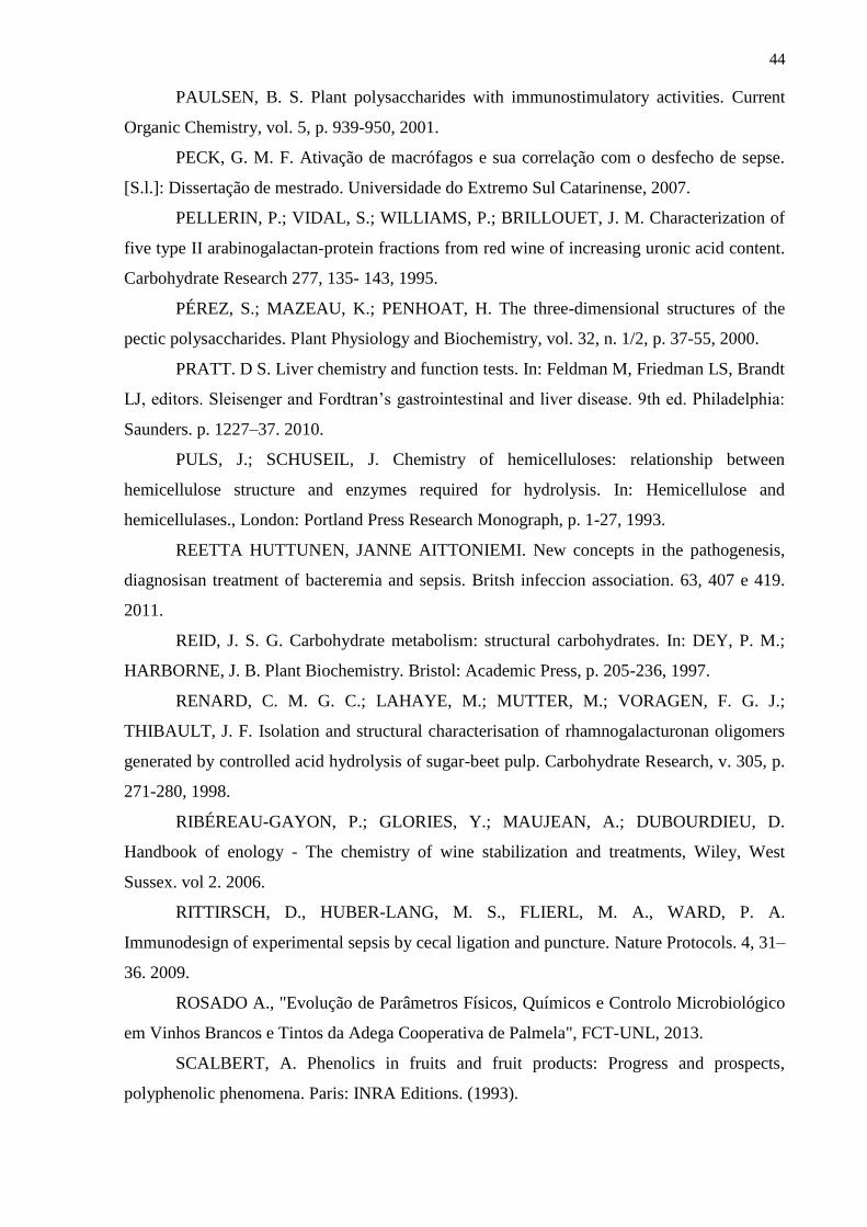
44
PAULSEN, B. S. Plant polysaccharides with immunostimulatory activities. Current
Organic Chemistry, vol. 5, p. 939-950, 2001.
PECK, G. M. F. Ativação de macrófagos e sua correlação com o desfecho de sepse.
[S.l.]: Dissertação de mestrado. Universidade do Extremo Sul Catarinense, 2007.
PELLERIN, P.; VIDAL, S.; WILLIAMS, P.; BRILLOUET, J. M. Characterization of
five type II arabinogalactan-protein fractions from red wine of increasing uronic acid content.
Carbohydrate Research 277, 135- 143, 1995.
PÉREZ, S.; MAZEAU, K.; PENHOAT, H. The three-dimensional structures of the
pectic polysaccharides. Plant Physiology and Biochemistry, vol. 32, n. 1/2, p. 37-55, 2000.
PRATT. D S. Liver chemistry and function tests. In: Feldman M, Friedman LS, Brandt
LJ, editors. Sleisenger and Fordtran’s gastrointestinal and liver disease. 9th ed. Philadelphia:
Saunders. p. 1227–37. 2010.
PULS, J.; SCHUSEIL, J. Chemistry of hemicelluloses: relationship between
hemicellulose structure and enzymes required for hydrolysis. In: Hemicellulose and
hemicellulases., London: Portland Press Research Monograph, p. 1-27, 1993.
REETTA HUTTUNEN, JANNE AITTONIEMI. New concepts in the pathogenesis,
diagnosisan treatment of bacteremia and sepsis. Britsh infeccion association. 63, 407 e 419.
2011.
REID, J. S. G. Carbohydrate metabolism: structural carbohydrates. In: DEY, P. M.;
HARBORNE, J. B. Plant Biochemistry. Bristol: Academic Press, p. 205-236, 1997.
RENARD, C. M. G. C.; LAHAYE, M.; MUTTER, M.; VORAGEN, F. G. J.;
THIBAULT, J. F. Isolation and structural characterisation of rhamnogalacturonan oligomers
generated by controlled acid hydrolysis of sugar-beet pulp. Carbohydrate Research, v. 305, p.
271-280, 1998.
RIBÉREAU-GAYON, P.; GLORIES, Y.; MAUJEAN, A.; DUBOURDIEU, D.
Handbook of enology - The chemistry of wine stabilization and treatments, Wiley, West
Sussex. vol 2. 2006.
RITTIRSCH, D., HUBER-LANG, M. S., FLIERL, M. A., WARD, P. A.
Immunodesign of experimental sepsis by cecal ligation and puncture. Nature Protocols. 4, 31–
36. 2009.
ROSADO A., "Evolução de Parâmetros Físicos, Químicos e Controlo Microbiológico
em Vinhos Brancos e Tintos da Adega Cooperativa de Palmela", FCT-UNL, 2013.
SCALBERT, A. Phenolics in fruits and fruit products: Progress and prospects,
polyphenolic phenomena. Paris: INRA Editions. (1993).
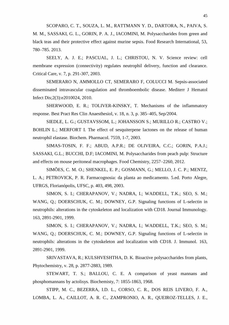
45
SCOPARO, C. T., SOUZA, L. M., RATTMANN Y. D., DARTORA, N., PAIVA, S.
M. M., SASSAKI, G. L., GORIN, P. A. J., IACOMINI, M. Polysaccharides from green and
black teas and their protective effect against murine sepsis. Food Research International, 53,
780–785. 2013.
SEELY, A. J. E.; PASCUAL, J. L.; CHRISTOU, N. V. Science review: cell
membrane expression (connectivity) regulates neutrophil delivery, function and clearance.
Critical Care, v. 7, p. 291-307, 2003.
SEMERARO N, AMMOLLO CT, SEMERARO F, COLUCCI M. Sepsis-associated
disseminated intravascular coagulation and thromboembolic disease. Mediterr J Hematol
Infect Dis;2(3):e2010024, 2010.
SHERWOOD, E. R.; TOLIVER-KINSKY, T. Mechanisms of the inflammatory
response. Best Pract Res Clin Anaesthesiol, v. 18, n. 3, p. 385–405, Sep/2004.
SIEDLE, L. G.; GUSTAVSSOM, L.; JOHANSSON S.; MURILLO R.; CASTRO V.;
BOHLIN L.; MERFORT I. The effect of sesquiterpene lactones on the release of human
neutrophil elastase. Biochem. Pharmacol. 7559, 1-7, 2003.
SIMAS-TOSIN, F. F.; ABUD, A.P.R.; DE OLIVEIRA, C.C.; GORIN, P.A.J.;
SASSAKI, G.L.; BUCCHI, D.F; IACOMINI, M. Polysaccharides from peach pulp: Structure
and effects on mouse peritoneal macrophages. Food Chemistry, 2257–2260, 2012.
SIMÕES, C. M. O.; SHENKEL, E. P.; GOSMANN, G.; MELLO, J. C. P.; MENTZ,
L. A.; PETROVICK, P. R. Farmacognosia: da planta ao medicamento. 5.ed. Porto Alegre,
UFRGS, Florianópolis, UFSC, p. 403, 498, 2003.
SIMON, S. I.; CHERAPANOV, V.; NADRA, I.; WADDELL, T.K.; SEO, S. M.;
WANG, Q.; DOERSCHUK, C. M.; DOWNEY, G.P. Signaling functions of L-selectin in
neutrophils: alterations in the cytoskeleton and localization with CD18. Journal Immunology.
163, 2891-2901, 1999.
SIMON, S. I.; CHERAPANOV, V.; NADRA, I.; WADDELL, T.K.; SEO, S. M.;
WANG, Q.; DOERSCHUK, C. M.; DOWNEY, G.P. Signaling functions of L-selectin in
neutrophils: alterations in the cytoskeleton and localization with CD18. J. Immunol. 163,
2891-2901, 1999.
SRIVASTAVA, R.; KULSHVESHTHA, D. K. Bioactive polysaccharides from plants,
Phytochemistry, v. 28, p. 2877-2883, 1989.
STEWART, T. S.; BALLOU, C. E. A comparison of yeast mannans and
phosphomannans by actolisys. Biochemisty, 7: 1855-1863, 1968.
STIPP, M. C., BEZERRA, I.D. L., CORSO, C. R., DOS REIS LIVERO, F. A.,
LOMBA, L. A., CAILLOT, A. R. C., ZAMPRONIO, A. R., QUEIROZ-TELLES, J. E.,

46
KLASSEN, G., RAMOS, E. A. S., SASSAKI, G. L., ACCO, A. Necroptosis mediates the
antineoplastic effects of the soluble fraction of polysaccharide from red wine in Walker-256
tumor-bearing rats. Carbohydrate Polymers, 160, 123–133. (2017).
STONE, C.W.; MILLS, D.V. Yeast products in the feed industry: a practical guide for
feed professionals. 2007. Disponível em:
<http://www.engormix.com/e_articles_view.asp?art=243&AREA=MAS>. Acesso em: 04
nov. 2017.
VARKI, A. Selectin ligands: will the real ones please stand up? J. Clin Invest., 100,
31-35. 1997.
VIDAL, S. et al. Structural characterization of the pectic polysaccharide
rhamnogalacturonan II: Evidence for the backbone location of the aceric acidcontaining
oligoglycosyl side chain. Carbohydrate Research, v. 326, p. 277-294, 2000.
VIDAL, S.; WILLIAMS, P.; DOCO, T.; MOUTOUNET, M.; PELLERIN, P. The
polysaccharides of red wine: total fractionation and characterization. Carbohydrate Polymers,
54 439–447, 2003.
VUKOVIC, R.; HUNDINA-DOMLADOVEC, M.; MRSA, V. Molecular orgazation
of the Saccharomyces cerevisiae cell wall. Prehrambeno-Tehnology Review, 32: (2-3) 99-
102. 1994.
WANG H, MA S. The cytokine storm and factors determining the sequence and
severity of organ dysfunction in multiple organ dysfunction syndrome. Am J Emerg Med;
26:711–5. 2008.
WANG, L.; BROWN, J.R.; VARKI, A.; ESKO J.D. Heparin’s anti-inflammatory
effects require glucosamine 6-O-sulfation and are mediated by blockade of L- and P-selectins.
J Clin Invest. 110: 127–136, 2002.
Xu J, Zhang X, Pelayo R, Monestier M, Ammollo CT, Semeraro F, et al. Extracellular
histones are major mediators of death in sepsis. Nat Med;15:1318–21. 2009.
YADAV, R.; LARBI, K.Y.; YOUNG, R.E. & NOURSHARGH, S. Migration of
leukocytes through the vessel wall and beyond. Thrombosis Haemostasis. 90: 598-606, 2003.
YAMADA, H. Pectic polysaccharides from Chinese herbs: structure and biological
activity. Carbohydrate Polymers, v. 25, p. 269-276, 1994.
YANG, J.; MARTINSON, T. E.; LIU, R. H. Phytochemical profiles and antioxidant
activities of wine grapes. Food Chemistry. v. 116, n.1, p. 332-339. 2009.
ZOECKLEIN, B., FUGELSANG, K. C., GUMP, B. H., NURY, F. S. Wine analysis
and production. New York: Chapman & Hall. (1995).
47
CAPITULO I
Structural Characterization of Polysaccharides from Cabernet Franc, Cabernet
Sauvignon and Sauvignon Blanc wines: anti-inflammatory activity in LPS stimulated
RAW 264.7 cells
Polysaccharides are present in wines and have important influence on several stages of
the winemaking process, including fermentation, filtration, stabilization and are partially
responsible for the organoleptic properties of wines (Gerbaud, Gabas, Blounin, Pellerin &
Moutounet, 1997; Moine-Ledoux & Dubourdieu, 1999; Vernhet, Pellerin, Belleville, Planque
& Moutounet, 1999). However, it has been shown that not all polysaccharides have the same
behavior regarding wines. Their influence on wine processing and sensory properties depend
on their quantity, class and structural features (Del Barrio-Galan, Pérez-Magarino, Ortega-
Heras, Williams & Doco, 2011; Guadalupe & Ayestaran, 2007; 2008; Riou, Vernhet, Doco &
Moutounet, 2002; Vidal et al., 2004).
The polysaccharides come from grape berries, yeast, bacterial and fungal grape
microbiome. From the enological and quantitative perspective, polysaccharides from grapes
and yeast are the most important. Their concentrations depend on many parameters, such as,
cultivation, stage of maturity, wine-making techniques and the treatments leading to increased
solubilization of the macromolecular components of the grape berry cell walls (Pellerin &
Cabanis, 1998). Arabinose and galactose rich polysaccharides (AGs), such as type II
arabinogalactan-proteins (AGPs) and arabinans, rhamnogalacturonans type I (RG-I) and type
II (RG-II), and homogalacturonans (HLs) come from grape berries, while glucans (GLs),
mannans and mannoproteins (MPs) are released by yeast either during fermentation or by
enzymatic activity during aging on yeast lees by autolysis (Ayestarán, Guadalupe, León,
2004; Belleville, Brillouet, Tarodo de la Fuente & Moutounet, 1991; Brillouet, Bosso &
Moutounet, 1990; Doco & Brillouet, 1993; Doco, Vuchot, Cheynier & Moutounet, 2003;
Pellerin, Vidal, Williams & Brillouet, 1995; Vidal, Williams, Doco, Moutounet & Pellerin,
2003).
Nowadays one of the main targets of the wine sector is to improve wine quality,
elaborating wines that satisfy consumer demand, and expand the offer of quality wines.
Cabernet Franc, Cabernet Sauvignon and Sauvignon Blanc wines, widely produced from
grapes of Vitis vinifera originating in France, are very popular and quite consumed
worldwide. Curiously, there is no information regarding their polysaccharides composition

48
and structural characterization. Despite this, polysaccharides appear to be the most interesting
molecules in enology due to their positive effects on the final quality of the wine (Doco et al.,
2003; Fournairon, Camarasa, Moutounet, & Salmon, 2002).
Besides concerns related to the wine quality, there is also great interest in knowing the
benefits that moderate consumption of wine can bring to health. So far, many other
polysaccharides presented reported biological activities, such as antiviral, antitumor,
immunostimulatory, anti-inflammatory, anticomplementary, anticoagulant, hypoglycemic,
and anti-ulcer (Capek et al., 2003; Cipriani et al., 2008, 2009; Nergard et al., 2005; Srivastava
& Kulshveshtha, 1989; Yamada, 1994). Nevertheless, there are very few reports regarding
biological activities and modulation of inflammatory mediators by polysaccharides from
wines.
The pathology of inflammation is a complicated process triggered by microbial pathogens,
such as: viruses, bacteria, prion, and fungi (Vitaliti et al., 2014). Macrophages account for the
first defense line of human body. LPS is usually employed as a model for inflammation due to
its ability to stimulate macrophages. Different inflammatory mediators are secreted by
macrophages induced by LPS: cytokines, such as tumor necrosis factor alpha (TNF-α) and
interleukin-1β (IL-1β), and inflammatory mediator nitric oxide (NO) (Agarwal, Piesco, Johns
& Riccelli, 1995; Lee et al., 2014). The LPS induced RAW 264.7 (mouse macrophages cell
lines) is commonly employed as the anti-inflammation model for anti-inflammation candidate
screening in vitro (Zhang et al., 2015).
Given the importance of polysaccharides in the wine making process and sensory
properties of the beverage, understanding their content, identification and quantification is
essential. Different analytical methodologies were developed to determine wine
polysaccharides in this study. This preliminary work was aimed at characterizing the spectra
of the purified polysaccharides. Such spectral identification of each polysaccharide family
(AGs, MPs, and RGs) in Cabernet Franc, Cabernet Sauvignon and Sauvignon Blanc wines
have not been reported previously as well as their in vitro involvement in the inflammatory
process.
2. Materials and methods
2.1. Materials
Cabernet Franc, Cabernet Sauvignon and Sauvignon Blanc wines used in this study
belonged to four vintages (2011, 2012, 2013 and 2014). All the wines were made from Vitis
49
vinifera var. and that have evolved during the ageing time. They were stored under cellar
conditions before the analyses were conducted. Oenological parameters such as pH or ethanol
content were similar across the studied wines, and they were not included in the statistical
data treatment.
2.2. Polysaccharides extraction and purification
The polysaccharides were precipitated by addition of cold EtOH (3 vol.), and
separated by centrifugation (8.000 rpm at 4 ◦C, 20 min). The sediment was dissolved in H2O,
dialyzed against water for 72 h to remove the remaining low-molecular weight compounds,
giving rise to a crude polysaccharide fraction: WCF (Cabernet Franc), WCS (Cabernet
Sauvignon) and WSB (Sauvignon Blanc). For the purification of the polysaccharides, it was
chosen the fraction with the highest yield. This fraction was frozen and then allowed to
unfreeze at room temperature (Gorin & Iacomini, 1984), resulting in soluble and insoluble
fractions which were separated by centrifugation as previously described. The insoluble
fraction was not analyzed in this study due to its lower yield and difficult solubilization. The
water-soluble fraction was treated with α-amylase and then with Fehling solution (Jones &
Stoodley, 1965). The soluble fraction (FCF) was isolated from the insoluble fraction (PCF) by
Cu2+ complex (Fehling solution) and centrifugation under the same conditions previously
described. The respective fractions were both neutralized with HOAc, dialyzed against water
and deionized with mixed ion exchange resins and then freeze dried.
2.3. Monosaccharide analysis
WCF, WCS, WSB, FCF and PCF fractions (2 mg) were hydrolyzed with 1 M TFA at
100 ◦C for 14 h, the solution was then evaporated, and the residue dissolved in water (2 mL).
The resulting monosaccharide mixture was examined by thin layer chromatography (TLC)
silica-gel 60 (Merck), with ethyl acetate:acetic acid:n-propanol:water (4:2:2:1, v/v), then
stained with orcinol-sulfuric acid (Sassaki, Souza, Cipriani, & Iacomini, 2008; Skipski, 1975).
The monosaccharides were then reduced with 2 mg NaBH4, yielding alditols, which were
acetylated in Ac2O-pyridine (1:1, v/v, 0.5 mL) at room temperature for 12 h (Wolfrom &
Thompson, 1963a; Wolfrom & Thompson, 1963b). The resulting alditol acetates were
extracted with CHCl3 and analyzed by gas chromatography–mass spectrometry (GC–MS –
Varian, Saturn 2000R, Ion-Trap detector), using a DB-225-MS column (30 m × 0.25 mm ×

50
0.25 µm), programmed from 50 to 220 ◦C at 40 ◦C/min, with he as carrier gas. Components
were identified by their typical retention times and electron ionization (EI 70 eV) spectra. The
uronic acids content of the fractions were determined using the colorimetric m-
hydroxybiphenyl method (Filisetti-Cozzi & Carpita, 1991).
Carboxy-reduction of FCF (10 mg) was carried out by the carbodiimide method
(Taylor & Conrad, 1972), using NaBH4 as the reducing agent, having its uronic acid carboxyl
groups reduced to primary alcohols and the results were given as mol % (Pettolino, Walsh,
Fincher & Bacic, 2013).
2.4. Methylation analysis
Per-O-methylation of each isolated polysaccharide (10 mg) was carried out using
NaOH-Me2SO-MeI as described by Ciucanu and Kerek (1984). The process, after isolation of
the products by neutralization (HOAc), dialysis and evaporation, was repeated, and the
methylation was complete. The per-O-methylated derivatives were hydrolyzed with 72% (v/v)
aq. H2SO4 (0.5 mL, v/v, 1 h, 0 ◦C), followed by dilution to 8% (v/v). The solution was kept at
100 ◦C for 17 h, then neutralized with BaCO3, filtered and evaporated to dryness (Saeman,
Moore, Mitchell, & Millet, 1954). The hydrolyzate was reduced with NaBD4 (sodium
borodeuteride) and then acetylated, giving rise to partially O-methylated alditol acetates and
analyzed by GC–MS, as the previously described, but with a final temperature of 215 ◦C. The
compounds were identified by their typical retention times and electron impact spectra
(Sassaki, Gorin, Souza, Czelusniak, & Iacomini, 2005).
2.5. Nuclear magnetic resonance (NMR) spectroscopy
1D and 2D NMR spectra were obtained with a 400 or 600 MHz Bruker spectrometer
using 5 mm direct or inverse probeheads (Avance III and HD, Bruker, Billerica,
Massachusetts, USA). 1D 1H and 13C-NMR at 600 MHz were collected after a 90° (p1) pulse
calibration at 10.75 µs. The 1H and 13C chemical shifts were determined after 1H assignments
trough 2D COSY and TOCSY analysis; bonding connections were performed by HMBC and
1H/13C chemical shift correlation mapping was finally determined by HSQCed (Heteronuclear
Single Quantum Coherence edited spectroscopy CH, CH3 positive phase CH2 negative phase)
performed at 30 ºC in deuterium oxide (D2O). 2D NMR experiments were recorded using
quadrature detection at indirect dimension and acquired using 24 scans per series of 1024x320
51
data points, with zero filling in F1 (4096) prior to Fourier transformation, using the Software
TOPSPIN version 3.2 pl6 (Bruker Biospin, Rheinstetten, Germany). The chemical shifts of
the polysaccharide were expressed in δ (ppm) relative to trimethylsilyl propionic acid
(TMSP). The ratios of the polysaccharides were calculated by integrating the area of specific
anomeric signals corresponding to each cross-peak assignment on the 2D-HSQCed spectra,
the volumes were normalized and calibrated to 1.00, using the most intense volume. The
structural model was designed using the software ChemSketch v. 12.0 (ACD/Labs, Toronto,
Ontario, Canada).
2.6. Determination of homogeneity and molar mass
The homogeneity and molar mass of the purified polysaccharide was determined by
high performance steric exclusion chromatography (HPSEC), using a refractive index (RI)
detector. Four columns were used in series, with exclusion sizes of 7 x 10⁶ Da (Ultrahydrogel
2000, Waters), 4 x 10⁵ Da (Ultrahydrogel 500, Waters), 8 x 10⁴ Da (Ultrahydrogel 250,
Waters) and 5 x 10³ Da (Ultrahydrogel 120, Waters). The eluent was 0.1 M NaNO3,
containing 0.5 g/L NaN3. The solutions were filtered through a membrane of 0.22 µm pore
size (Millipore) and loaded (100 µL, loop) at a concentration of 1 mg/mL. The molar mass of
the polymer was estimated using Astra software 4.70.
2.7. Cell Culture
RAW 264.7 macrophages were maintained at 37 ◦C in humidified atmosphere of 5%
CO2 in DMEM medium supplemented with 10% fetal bovine serum and
penicillin/streptomycin/normocin (50 U/mL, 50 g/mL and 100 g/mL, respectively; InvivoGen,
San Diego,CA, USA). Exponential phase cells were used throughout the experiments.
2.8. Analysis of cell viability
RAW 264.7 cells in exponential growth phase were seeded in 24-well plates at a
density of 4.8 x 10⁵ cells/well. The WCF, WCS, WSB, FCF and PCF were added at indicated
concentrations (0.1, 1, 10 and 100 µg/mL). The mitochondrial-dependent reduction of 3- (4,5-
dimethylthizaol-2yl)-2,5-diphenyl tetrazolium bromide (MTT) to formazan was used to
measure cell respiration as an indicator of cell viability (Mosmann, 1983). Briefly, after 24 h

52
incubation with or without polysaccharides, 350 μL/well of MTT solution (1 mg/mL) was
added and the cells were incubated for another 4 h at 37 ºC. After removing the supernatant,
500 µL of DMSO (dimethyl sulfoxide) was added to the cells to dissolve the formazan. The
absorbance of each group was measured by a microplate reader at wavelength of 570 nm. The
control group consisted of untreated cells was considered as 100% of viable cells. Results
were expressed as percentage of viable cells when compared with control group.
2.9. Measurement of cytokines production and NO production
The analysis was based on exposing cell line to fractions WCF, WCS, WSB, FCF and
PCF (0.1, 1, 10 and 100 µg/mL) in the presence of lipopolysaccharide (LPS) in the wells.
RAW 264.7 cells (4.8 x 10⁵ cells/well) were plated into 96-well plates, then were stimulated
with LPS (2 µg/mL) and after 1h, were treated with different concentrations of the mentioned
fractions. After 24 h, the culture media were collected. The amount of cytokines in the
supernatants for TNF-α and IL-1β, respectively, were determined by enzyme-linked
immunosorbent assay kits according to the manufacturer's instructions. Three replicates were
carried out for each different treatment. The absorbance of each group was measured by a
microplate reader at wavelength of 450 nm. To determine the total concentration of NO in the
culture media, Griess reagent was added to 40 mL of supernatant and the absorbance at 545
nm was evaluated with an ELISA kit (BD Biosciences, San Jose, CA, USA) following the
manufacture manual.
2.10. Statistical analysis
Data were expressed as means ± standard deviation (SD) of five or ten mice examined
in each group. Statistical error was determined by one-way ANOVA; the post hoc test was
Bonferroni’s. Calculations were performed with Graphpad Prism 5.0. p-values < 0.001 were
considered significant.
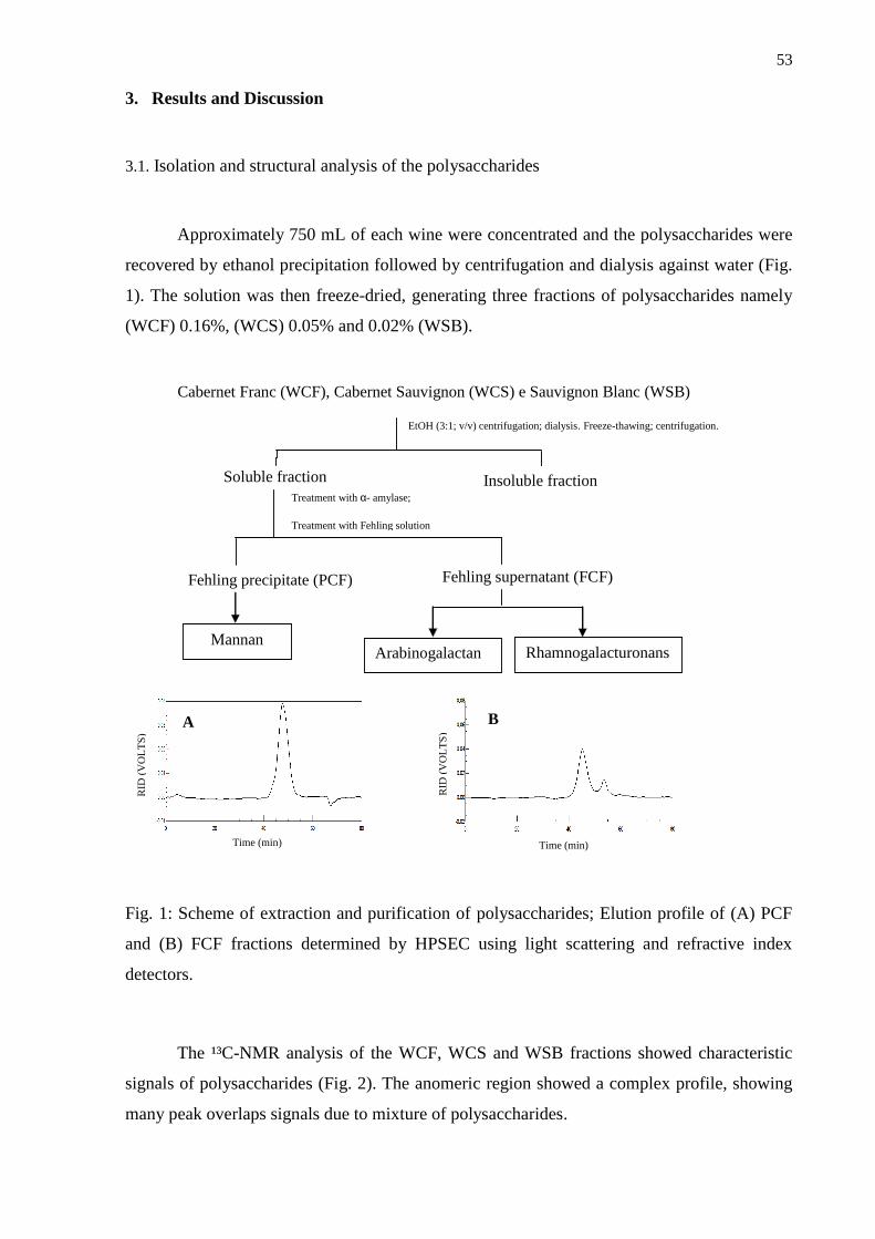
53
3. Results and Discussion
3.1. Isolation and structural analysis of the polysaccharides
Approximately 750 mL of each wine were concentrated and the polysaccharides were
recovered by ethanol precipitation followed by centrifugation and dialysis against water (Fig.
1). The solution was then freeze-dried, generating three fractions of polysaccharides namely
(WCF) 0.16%, (WCS) 0.05% and 0.02% (WSB).
EtOH (3:1; v/v) centrifugation; dialysis. Freeze-thawing; centrifugation.
Soluble fraction
Mannan
Insoluble fraction Treatment with α- amylase;
Treatment with Fehling solution
Fehling precipitate (PCF)
Fehling supernatant (FCF)
Rhamnogalacturonans Arabinogalactan
RID
(V
OL
TS
)
Time (min) Time (min)
RID
(V
OL
TS
)
A B
Cabernet Franc (WCF), Cabernet Sauvignon (WCS) e Sauvignon Blanc (WSB)
Fig. 1: Scheme of extraction and purification of polysaccharides; Elution profile of (A) PCF
and (B) FCF fractions determined by HPSEC using light scattering and refractive index
detectors.
The ¹³C-NMR analysis of the WCF, WCS and WSB fractions showed characteristic
signals of polysaccharides (Fig. 2). The anomeric region showed a complex profile, showing
many peak overlaps signals due to mixture of polysaccharides.

54
Since all the polysaccharide fractions showed the same monosaccharide composition,
but with different yields, the highest one was chosen for isolation of polysaccharides (WCF).
The fractionation and purification of polysaccharides were carried out by a freeze-thawing
procedure (Gorin & Iacomini, 1984), resulting in a cold water-soluble fraction. Therefore, this
fraction was submitted to complexation with copper, resulting in insoluble (PCF) and soluble
(FCF) fractions.
A
C
B
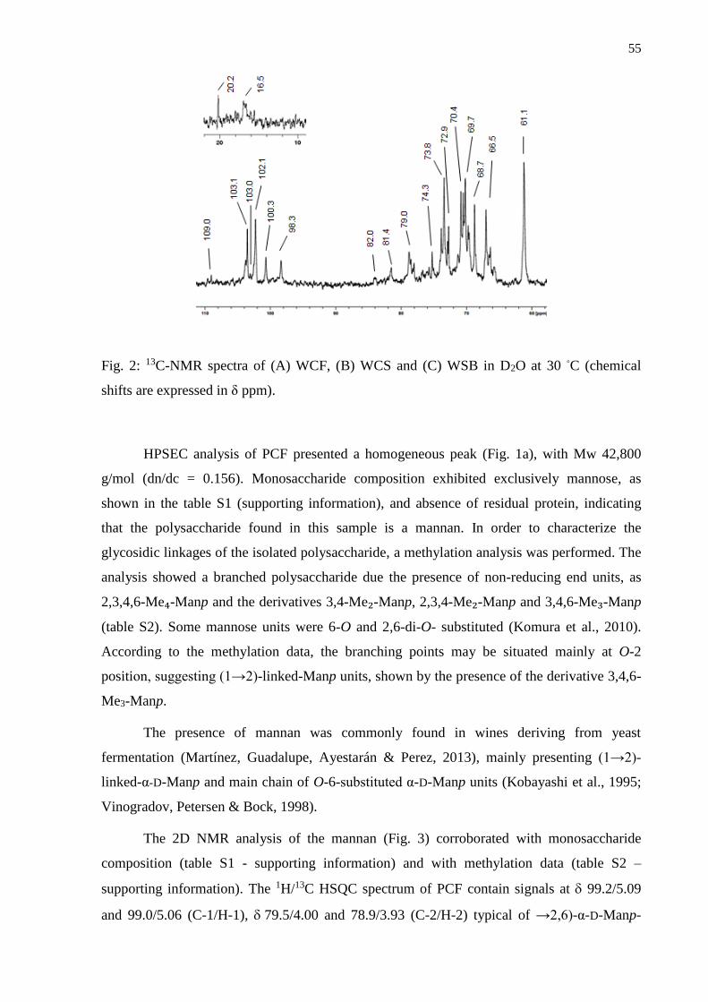
55
Fig. 2: 13C-NMR spectra of (A) WCF, (B) WCS and (C) WSB in D2O at 30 ◦C (chemical
shifts are expressed in δ ppm).
HPSEC analysis of PCF presented a homogeneous peak (Fig. 1a), with Mw 42,800
g/mol (dn/dc = 0.156). Monosaccharide composition exhibited exclusively mannose, as
shown in the table S1 (supporting information), and absence of residual protein, indicating
that the polysaccharide found in this sample is a mannan. In order to characterize the
glycosidic linkages of the isolated polysaccharide, a methylation analysis was performed. The
analysis showed a branched polysaccharide due the presence of non-reducing end units, as
2,3,4,6-Me₄-Manp and the derivatives 3,4-Me₂-Manp, 2,3,4-Me₂-Manp and 3,4,6-Me₃-Manp
(table S2). Some mannose units were 6-O and 2,6-di-O- substituted (Komura et al., 2010).
According to the methylation data, the branching points may be situated mainly at O-2
position, suggesting (1→2)-linked-Manp units, shown by the presence of the derivative 3,4,6-
Me3-Manp.
The presence of mannan was commonly found in wines deriving from yeast
fermentation (Martínez, Guadalupe, Ayestarán & Perez, 2013), mainly presenting (1→2)-
linked-α-D-Manp and main chain of O-6-substituted α-D-Manp units (Kobayashi et al., 1995;
Vinogradov, Petersen & Bock, 1998).
The 2D NMR analysis of the mannan (Fig. 3) corroborated with monosaccharide
composition (table S1 - supporting information) and with methylation data (table S2 –
supporting information). The 1H/13C HSQC spectrum of PCF contain signals at 99.2/5.09
and 99.0/5.06 (C-1/H-1), 79.5/4.00 and 78.9/3.93 (C-2/H-2) typical of →2,6)-α-D-Manp-
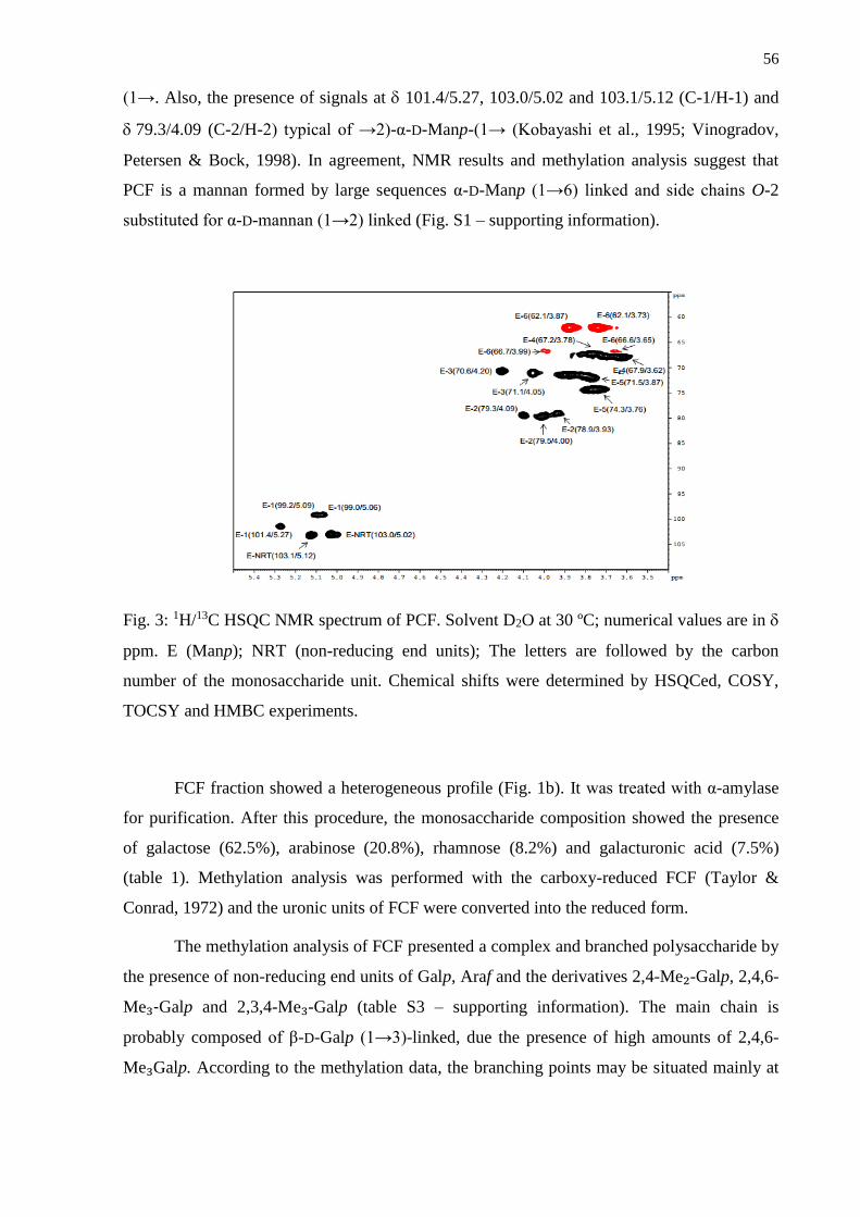
56
(1→. Also, the presence of signals at 101.4/5.27, 103.0/5.02 and 103.1/5.12 (C-1/H-1) and
79.3/4.09 (C-2/H-2) typical of →2)-α-D-Manp-(1→ (Kobayashi et al., 1995; Vinogradov,
Petersen & Bock, 1998). In agreement, NMR results and methylation analysis suggest that
PCF is a mannan formed by large sequences α-D-Manp (1→6) linked and side chains O-2
substituted for α-D-mannan (1→2) linked (Fig. S1 – supporting information).
Fig. 3: 1H/13C HSQC NMR spectrum of PCF. Solvent D2O at 30 ºC; numerical values are in
ppm. E (Manp); NRT (non-reducing end units); The letters are followed by the carbon
number of the monosaccharide unit. Chemical shifts were determined by HSQCed, COSY,
TOCSY and HMBC experiments.
FCF fraction showed a heterogeneous profile (Fig. 1b). It was treated with α-amylase
for purification. After this procedure, the monosaccharide composition showed the presence
of galactose (62.5%), arabinose (20.8%), rhamnose (8.2%) and galacturonic acid (7.5%)
(table 1). Methylation analysis was performed with the carboxy-reduced FCF (Taylor &
Conrad, 1972) and the uronic units of FCF were converted into the reduced form.
The methylation analysis of FCF presented a complex and branched polysaccharide by
the presence of non-reducing end units of Galp, Araf and the derivatives 2,4-Me₂-Galp, 2,4,6-
Me₃-Galp and 2,3,4-Me₃-Galp (table S3 – supporting information). The main chain is
probably composed of β-D-Galp (1→3)-linked, due the presence of high amounts of 2,4,6-
Me₃Galp. According to the methylation data, the branching points may be situated mainly at
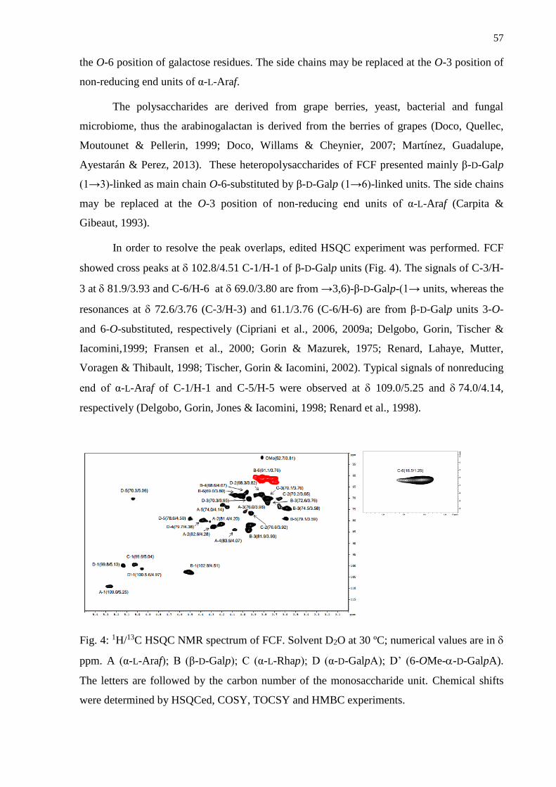
57
the O-6 position of galactose residues. The side chains may be replaced at the O-3 position of
non-reducing end units of α-L-Araf.
The polysaccharides are derived from grape berries, yeast, bacterial and fungal
microbiome, thus the arabinogalactan is derived from the berries of grapes (Doco, Quellec,
Moutounet & Pellerin, 1999; Doco, Willams & Cheynier, 2007; Martínez, Guadalupe,
Ayestarán & Perez, 2013). These heteropolysaccharides of FCF presented mainly β-D-Galp
(1→3)-linked as main chain O-6-substituted by β-D-Galp (1→6)-linked units. The side chains
may be replaced at the O-3 position of non-reducing end units of α-L-Araf (Carpita &
Gibeaut, 1993).
In order to resolve the peak overlaps, edited HSQC experiment was performed. FCF
showed cross peaks at 102.8/4.51 C-1/H-1 of β-D-Galp units (Fig. 4). The signals of C-3/H-
3 at 81.9/3.93 and C-6/H-6 at 69.0/3.80 are from →3,6)-β-D-Galp-(1→ units, whereas the
resonances at 72.6/3.76 (C-3/H-3) and 61.1/3.76 (C-6/H-6) are from β-D-Galp units 3-O-
and 6-O-substituted, respectively (Cipriani et al., 2006, 2009a; Delgobo, Gorin, Tischer &
Iacomini,1999; Fransen et al., 2000; Gorin & Mazurek, 1975; Renard, Lahaye, Mutter,
Voragen & Thibault, 1998; Tischer, Gorin & Iacomini, 2002). Typical signals of nonreducing
end of α-L-Araf of C-1/H-1 and C-5/H-5 were observed at 109.0/5.25 and 74.0/4.14,
respectively (Delgobo, Gorin, Jones & Iacomini, 1998; Renard et al., 1998).
Fig. 4: 1H/13C HSQC NMR spectrum of FCF. Solvent D2O at 30 ºC; numerical values are in
ppm. A (α-L-Araf); B (β-D-Galp); C (α-L-Rhap); D (α-D-GalpA); D’ (6-OMe--D-GalpA).
The letters are followed by the carbon number of the monosaccharide unit. Chemical shifts
were determined by HSQCed, COSY, TOCSY and HMBC experiments.
58
The arabinogalactans are classified according to their main chains. Therefore, the
NMR result is in agreement with methylation analysis (table S3 – supporting information) and
with monosaccharide composition shown in the table S1 (supporting information) that
indicated the presence of galactose and arabinose in this fraction, suggesting that the
polysaccharide FCF is a type II arabinogalactan, formed by a (1→3)-linked β-D-Galp main
chain, substituted at HO-6 by (1→6)-linked β-D-Galp side chains and substituted by
nonreducing end-units of arabinose 3-O-substituted α-L-Araf chains (Fig. S2).
Moreover, carboxy-reduced derivatives from FCF showed non-reducing end units of
Rhap and the presence of GalpA (1→4)-linkages, due the derivative 2,3,6-Me₃-Galp (table S3
– supporting information) (Sassaki, Gorin, Souza, Czelusniak & Iacomini, 2005). The
presence of 3,4-Me₂-Rhap, indicates O-2-substitution, which is often found in type I
rhamnogalacturonan. According to the methylation data, this polysaccharide is formed by a
chain of repeat units (1→4)-linked α-D-GalpA and (1→2)-linked α-L-Rhap (Fig. S3) (Carpita
& Gibeaut, 1993).
The presence of pectins was confirmed by detection of the sequence of (1 → 4)-linked
α- galacturonic acid residues, which gives a fingerprint cross peaks at 99.8/5.13 and
100.5/4.97 attributed to C-1/H-1(α-D-GalpA and 6-OMe--D-GalpA, respectively) and
79.7/4.38 (C-4/H-4), indicating this type of linkage (Fig. 4). The latter was confirmed by
complete signaling of GalpA units at 68.3/3.82 (C-2/H-2), 70.3/3.95 (C-3/H-3), 70.3/5.06
(C-5/H-5 substituted) and 78.6/4.50 (C-5/H-5). Also, the methyl esterification of galacturonic
acid is commonly found in type II ramnogalacturonans, being confirmed by the presence of a
typical CO2CH3 cross peak at 52.7/3.81 (Renard et al., 1998; Ovodova et al., 2009; Popov et
al., 2011). C-1/H-1 and C-6/H-6 of Rhap units (1→2)-linked were shown by resonances at
99.6/5.04 and 16.5/1.25, respectively (Renard et al., 1998). The combination of NMR data
and methylation analysis suggests that the FCF also presents a type I rhamnogalacturonan,
formed by large sequences of →4)-6-OMe--D-GalpA-(1→ units, interspersed with a few -
L-Rhap units, that was also confirmed by the presence of rhamnose and galacturonic acid in
the monosaccharide composition (table S1 – supporting information).
Further, the monosaccharide composition analysis of the FCF fraction by GC-MS
revealed the presence of units of 2-O-methyl-xylose and 2-O-methyl-fucose (table S1 –
supporting information), which are considered markers of type II rhamnogalacturonan. They
also suggest a type II rhamnogalacturonan, although these signals were not observed in NMR
spectrum, because of their small amounts in this polysaccharide, corresponding to less than
1% of the total. The 2-O-methyl substitution was characterized by electron ionization mass
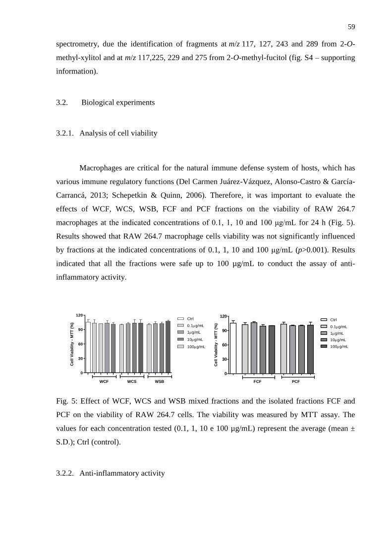
59
spectrometry, due the identification of fragments at m/z 117, 127, 243 and 289 from 2-O-
methyl-xylitol and at m/z 117,225, 229 and 275 from 2-O-methyl-fucitol (fig. S4 – supporting
information).
3.2. Biological experiments
3.2.1. Analysis of cell viability
Macrophages are critical for the natural immune defense system of hosts, which has
various immune regulatory functions (Del Carmen Juárez-Vázquez, Alonso-Castro & García-
Carrancá, 2013; Schepetkin & Quinn, 2006). Therefore, it was important to evaluate the
effects of WCF, WCS, WSB, FCF and PCF fractions on the viability of RAW 264.7
macrophages at the indicated concentrations of 0.1, 1, 10 and 100 μg/mL for 24 h (Fig. 5).
Results showed that RAW 264.7 macrophage cells viability was not significantly influenced
by fractions at the indicated concentrations of 0.1, 1, 10 and 100 μg/mL (p>0.001). Results
indicated that all the fractions were safe up to 100 µg/mL to conduct the assay of anti-
inflammatory activity.
0
30
60
90
120
10g/mL
Ctrl
0.1g/mL
WCF WCS WSB
1g/mL
100g/mL
Ce
ll V
iab
ilit
y -
MT
T (
%)
0
30
60
90
120
FCF PCF
Ctrl
0.1g/mL
1g/mL
10g/mL
100g/mL
Ce
ll V
iab
ilit
y -
MT
T (
%)
Fig. 5: Effect of WCF, WCS and WSB mixed fractions and the isolated fractions FCF and
PCF on the viability of RAW 264.7 cells. The viability was measured by MTT assay. The
values for each concentration tested (0.1, 1, 10 e 100 µg/mL) represent the average (mean ±
S.D.); Ctrl (control).
3.2.2. Anti-inflammatory activity
60
Inflammation is a host response to foreign pathogens or tissue injury to eliminate
harmful stimuli as well as to initiate the healing and repair process of the damaged tissue
(Mariathasan & Monack, 2007). LPS is a major constituent of the cell wall of gram-negative
bacteria, which can bind to the TLR 4 receptor of macrophages and induce inflammation
(Rossol et al., 2011). In response to LPS, macrophages synthesize and release inflammatory
mediators such as NO and produce pro-inflammatory cytokines, such as TNF-α and IL-1β, as
already mentioned (Pan, Lin-Shiau & Lin, 2000). To test the inhibitory effects of WCF,
WCS, WSB, FCF and PCF fractions on the production of the inflammatory cytokines (TNF-α
and IL-1β) and mediator (NO) from LPS-stimulated RAW 264.7 cells, RAW 264.7 cells were
treated with various concentrations of fractions (0.1, 1, 10 and 100 μg/mL) and then were
incubated with or without 2 µg/mL of LPS for another 24 h.
LPS is one of the leading activators of macrophages. The macrophages play important
roles in inflammation through the production of several pro-inflammatory molecules,
including NO. Production of excessive NO has been associated with a range of inflammatory
diseases, including arteriosclerosis, ischemic reperfusion, hypertension and septic shock
(Pacher, Beckman & Liaudet, 2007; Terao, 2009). Therefore, inhibition of NO production in
LPS stimulated RAW 264.7 cells is one of the possible ways to screen for anti-inflammatory
drugs or disease prevention thorough moderate wine intake. In this study WCF, WCS, WSB,
FCF and PCF fractions showed the inhibition of NO production in cells, indicating the anti-
inflammatory properties. As shown in figure 6 (K-O), while treatment both with the mixed
fractions and fractions with isolated polysaccharides induced a significant inhibitory effect
and it was observed that this effect was enhanced increasing the concentration of the fractions
with a maximal inhibitory effect at the dose of 100 μg/mL.
Pro-inflammatory mediator NO plays a key role in the pathogenesis of many
inflammatory diseases. The NO pathway is known to induce ROS production (Jung et al.,
2010; Liu, Cheng, Chen & Yang, 2005). ROS is critical for LPS-induced inflammation
through the activation of NF-κB related signaling (Asehnoune, Strassheim, Mitra, Kim &
Abraham, 2004). Activated NF-κB acts as a transcription factor, leading to increased
production of pro-inflammatory cytokines such as TNF-α and IL-1β (Janeway & Medzhitov,
2002). Thus, blocking the effects of pro-inflammatory mediators could be an effective
therapeutic strategy. In figure 6 (A-J) it is shown that all fractions tested significantly
decreased the TNF-α and IL-1β production at all doses, indicating that the polysaccharides
found in wine have anti-inflammatory properties. Thus, blocking the effects of pro-
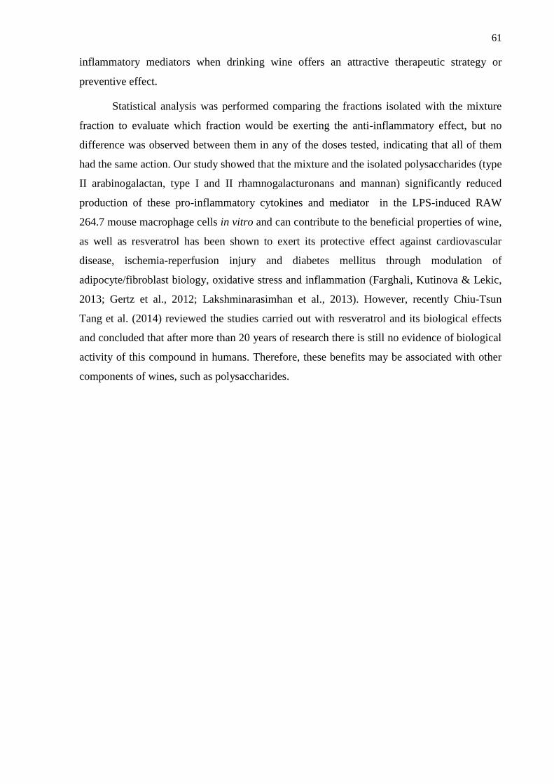
61
inflammatory mediators when drinking wine offers an attractive therapeutic strategy or
preventive effect.
Statistical analysis was performed comparing the fractions isolated with the mixture
fraction to evaluate which fraction would be exerting the anti-inflammatory effect, but no
difference was observed between them in any of the doses tested, indicating that all of them
had the same action. Our study showed that the mixture and the isolated polysaccharides (type
II arabinogalactan, type I and II rhamnogalacturonans and mannan) significantly reduced
production of these pro-inflammatory cytokines and mediator in the LPS-induced RAW
264.7 mouse macrophage cells in vitro and can contribute to the beneficial properties of wine,
as well as resveratrol has been shown to exert its protective effect against cardiovascular
disease, ischemia-reperfusion injury and diabetes mellitus through modulation of
adipocyte/fibroblast biology, oxidative stress and inflammation (Farghali, Kutinova & Lekic,
2013; Gertz et al., 2012; Lakshminarasimhan et al., 2013). However, recently Chiu-Tsun
Tang et al. (2014) reviewed the studies carried out with resveratrol and its biological effects
and concluded that after more than 20 years of research there is still no evidence of biological
activity of this compound in humans. Therefore, these benefits may be associated with other
components of wines, such as polysaccharides.
62
WCF
Ctrl Vehicle 0.1 1 10 1000
500
1000
1500
2000
µg/mL
***
LPS 2 µg/mL - + + + + +
TN
F-
(p
g/m
L)
WCF
Ctrl Vehicle 0.1 1 10 1000
50
100
150
µg/mL
LPS 2 µg/mL - + + + + +
***
+
++
+++
++
IL-1
(p
g/m
L)
WCF
Ctrl Vehicle 0.1 1 10 1000
5
10
15
20
***
+++
µg/mL
LPS 2 µg/mL - + + + + +
NO
(
M)
WCS
Ctrl Vehicle 0.1 1 10 1000
500
1000
1500
2000
***
µg/mL
LPS 2 µg/mL - + + + + +
+
TN
F-
(p
g/m
L)
WCS
Ctrl Vehicle 0,1 1 10 1000
50
100
150
µg/mL
LPS 2 µg/mL - + + + + +
***++
++
IL-1
(p
g/m
L)
+
WCS
Ctrl Vehicle 0,1 1 10 1000
5
10
15
20
µg/mL
- + + + + +LPS 2 µg/mL
***++
NO
(
M)
WSB
Ctrl Vehicle 0.1 1 10 1000
500
1000
1500
2000
***
µg/mL
LPS 2 µg/mL - + + + + +
+
TN
F-
(p
g/m
L)
WSB
Ctrl Vehicle 0,1 1 10 1000
50
100
150
LPS 2 µg/mL - + + + + +
***
IL-1
(p
g/m
L)
++
+
+++
+++
+
WSB
Ctrl Vehicle 0.1 1 10 1000
5
10
15
20
***
+++
µg/mL
LPS 2 µg/mL - + + + + +
NO
(
M)
PCF
Ctrl Vehicle 0.1 1 10 1000
500
1000
1500
2000
µg/mL
LPS 2 µg/mL - + + + + +
***
TN
F-
(p
g/m
L)
PCF
Ctrl Vehicle 0.1 1 10 1000
50
100
150
µg/mL
LPS 2 µg/mL - + + + + +
***
+++
++
IL-1
(p
g/m
L)
PCF
Ctrl Vehicle 0.1 1 10 1000
5
10
15
20
µg/mL
LPS 2 µg/mL - + + + + +
***
+++NO
(
M)
FCF
Ctrl Vehicle 0.1 1 10 1000
500
1000
1500
2000
µg/mL
LPS 2 µg/mL - + + + + +
+
***
TN
F-
(p
g/m
L)
FCF
Ctrl Vehicle 0.1 1 10 1000
50
100
150
µg/mL
LPS 2 µg/mL - + + + + +
***
+++
IL-1
(p
g/m
L)
FCF
Ctrl Vehicle 0.1 1 10 1000
5
10
15
20
***
+++
µg/mL
LPS 2 µg/mL - + + + + +
NO
(
M)
Fig. 6: Anti-inflammatory activities of WCF, WCS, WSB, PCF and FCF on RAW 264.7 cells. (A-
E) Expression levels of TNF-α. (F-J) Expression levels of IL-1β. (K-O) NO production levels. Ctrl
(control).
A
B
c
D
E
F
G
H
I
J
K
L
M
N
O
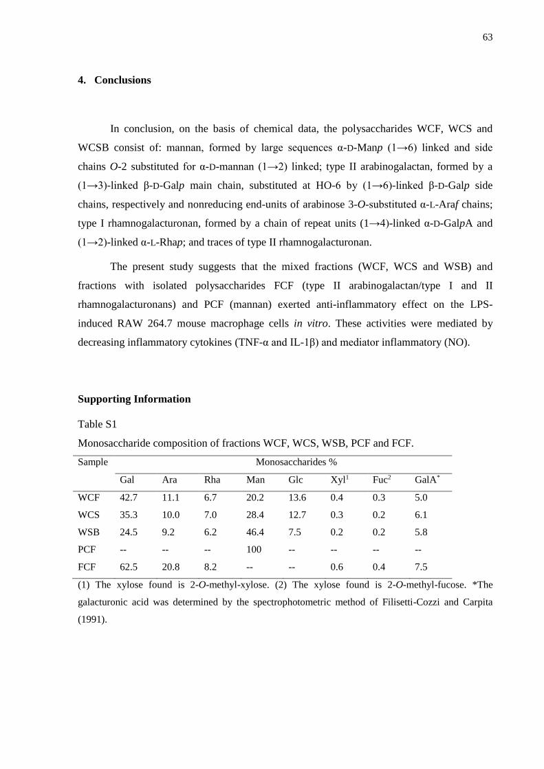
63
4. Conclusions
In conclusion, on the basis of chemical data, the polysaccharides WCF, WCS and
WCSB consist of: mannan, formed by large sequences α-D-Manp (1→6) linked and side
chains O-2 substituted for α-D-mannan (1→2) linked; type II arabinogalactan, formed by a
(1→3)-linked β-D-Galp main chain, substituted at HO-6 by (1→6)-linked β-D-Galp side
chains, respectively and nonreducing end-units of arabinose 3-O-substituted α-L-Araf chains;
type I rhamnogalacturonan, formed by a chain of repeat units (1→4)-linked α-D-GalpA and
(1→2)-linked α-L-Rhap; and traces of type II rhamnogalacturonan.
The present study suggests that the mixed fractions (WCF, WCS and WSB) and
fractions with isolated polysaccharides FCF (type II arabinogalactan/type I and II
rhamnogalacturonans) and PCF (mannan) exerted anti-inflammatory effect on the LPS-
induced RAW 264.7 mouse macrophage cells in vitro. These activities were mediated by
decreasing inflammatory cytokines (TNF-α and IL-1β) and mediator inflammatory (NO).
Supporting Information
Table S1
Monosaccharide composition of fractions WCF, WCS, WSB, PCF and FCF.
Sample Monosaccharides %
Gal Ara Rha Man Glc Xyl1 Fuc2 GalA*
WCF 42.7 11.1 6.7 20.2 13.6 0.4 0.3 5.0
WCS 35.3 10.0 7.0 28.4 12.7 0.3 0.2 6.1
WSB 24.5 9.2 6.2 46.4 7.5 0.2 0.2 5.8
PCF -- -- -- 100 -- -- -- --
FCF 62.5 20.8 8.2 -- -- 0.6 0.4 7.5
(1) The xylose found is 2-O-methyl-xylose. (2) The xylose found is 2-O-methyl-fucose. *The
galacturonic acid was determined by the spectrophotometric method of Filisetti-Cozzi and Carpita
(1991).
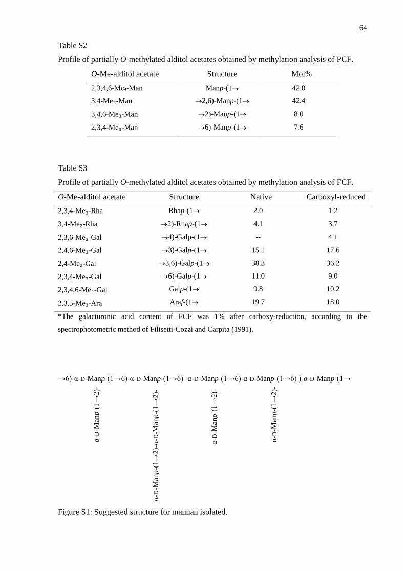
64
Table S2
Profile of partially O-methylated alditol acetates obtained by methylation analysis of PCF.
O-Me-alditol acetate Structure Mol%
2,3,4,6-Me₄-Man Manp-(1 42.0
3,4-Me₂-Man 2,6)-Manp-(1 42.4
3,4,6-Me₃-Man 2)-Manp-(1 8.0
2,3,4-Me₃-Man 6)-Manp-(1 7.6
Table S3
Profile of partially O-methylated alditol acetates obtained by methylation analysis of FCF.
O-Me-alditol acetate Structure Native Carboxyl-reduced
2,3,4-Me₃-Rha Rhap-(1 2.0 1.2
3,4-Me₂-Rha
2,3,6-Me₃-Gal
2)-Rhap-(1
4)-Galp-(1
4.1 3.7
4.1 --
2,4,6-Me₃-Gal
2,4-Me₂-Gal
2,3,4-Me₃-Gal
2,3,4,6-Me₄-Gal
2,3,5-Me₃-Ara
3)-Galp-(1
3,6)-Galp-(1
6)-Galp-(1
Galp-(1
Araf-(1
15.1 17.6
36.2
9.0
10.2
18.0
38.3
11.0
9.8
19.7
*The galacturonic acid content of FCF was 1% after carboxy-reduction, according to the
spectrophotometric method of Filisetti-Cozzi and Carpita (1991).
→6)-α-D-Manp-(1→6)-α-D-Manp-(1→6) -α-D-Manp-(1→6)-α-D-Manp-(1→6) )-α-D-Manp-(1→
Figure S1: Suggested structure for mannan isolated.
α-D
-Man
p-(
1→
2)-
α-D
-Man
p-(
1→
2)-
α-D
-Man
p-(
1→
2)-
α-D
-Man
p-(
1→
2)-
α-D
-Man
p-(
1→
2)-
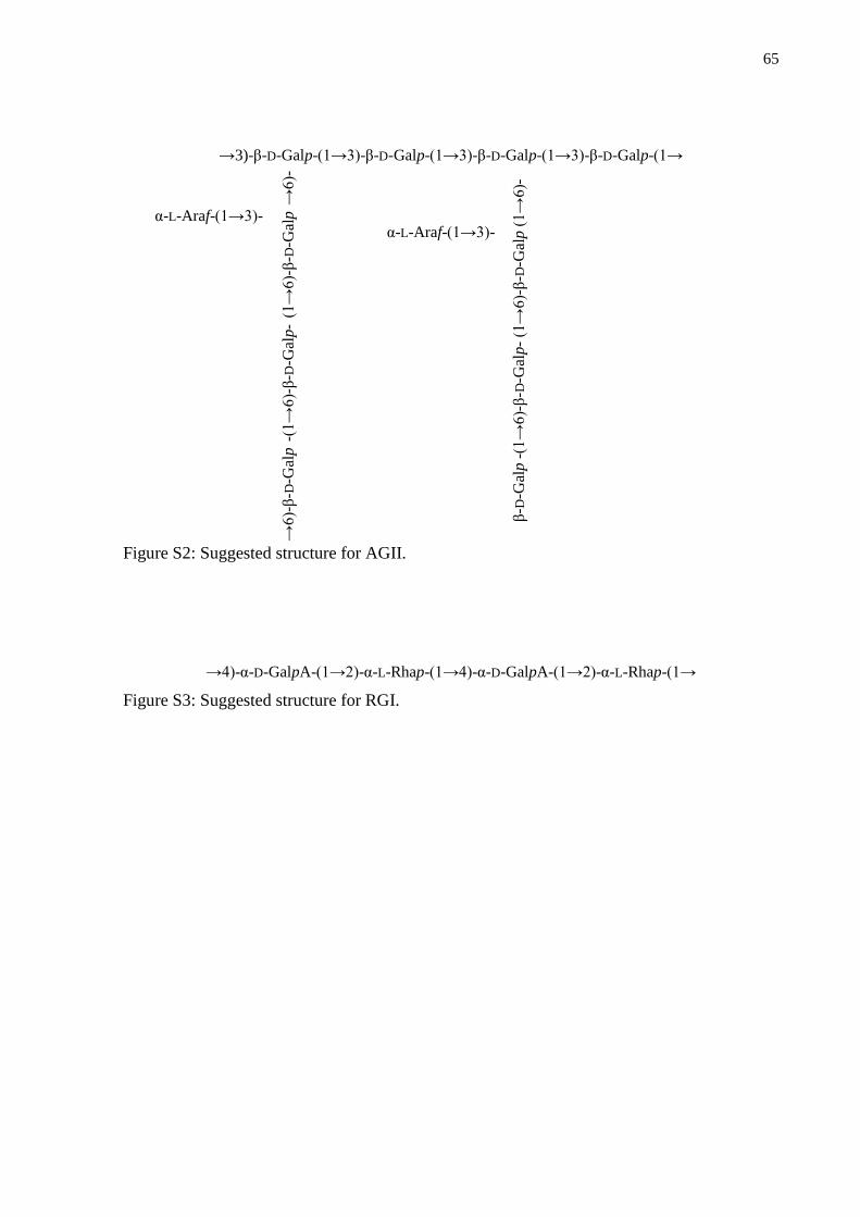
65
→3)-β-D-Galp-(1→3)-β-D-Galp-(1→3)-β-D-Galp-(1→3)-β-D-Galp-(1→
Figure S2: Suggested structure for AGII.
→4)-α-D-GalpA-(1→2)-α-L-Rhap-(1→4)-α-D-GalpA-(1→2)-α-L-Rhap-(1→
Figure S3: Suggested structure for RGI.
→6)-
β-D
-Gal
p -
(1→
6)-
β-D
-Gal
p-
(1→
6)-
β-D
-Gal
p →
6)-
(1→
6)-
β-D
-Gal
p -
(1→
6)-
β-D
-Gal
p-
(1→
6)-
β-D
-Gal
p (
1→
6)-
α-L-Araf-(1→3)- α-L-Araf-(1→3)-
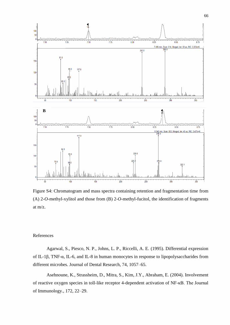
66
Figure S4: Chromatogram and mass spectra containing retention and fragmentation time from
(A) 2-O-methyl-xylitol and those from (B) 2-O-methyl-fucitol, the identification of fragments
at m/z.
References
Agarwal, S., Piesco, N. P., Johns, L. P., Riccelli, A. E. (1995). Differential expression
of IL-1β, TNF-α, IL-6, and IL-8 in human monocytes in response to lipopolysaccharides from
different microbes. Journal of Dental Research, 74, 1057–65.
Asehnoune, K., Strassheim, D., Mitra, S., Kim, J.Y., Abraham, E. (2004). Involvement
of reactive oxygen species in toll-like receptor 4-dependent activation of NF-κB. The Journal
of Immunology., 172, 22–29.
B
67
Ayestarán, B., Guadalupe, Z., León, D. (2004). Quantification of major grape
polysaccharides (Tempranillo v.) released by maceration enzymes during the fermentation
process. Analytica Chimica Acta, 513, 29-39.
Belleville, M. P., Brillouet, J. M., Tarodo de la Fuente, B., Moutounet, M. (1991).
Polysaccharide effects on cross-flow microfiltration of two red wines with a microporous
alumina membrane. Journal Food Science, 55, 1598-1602.
Capek, P., Hribalová, V., Svandová, E., Ebringerová, A., Sasinková, V., Masarová, J.
(2003). Characterization of immunomodulatory polysaccharides from Salvia officinalis.
International Journal Biology Macromolecules, 33, 113-119.
Carpita, N. C., Gibeaut, D. M. (1993). Structural models of primary cell walls in
flowering plants: consistency of molecular structure with the physical properties of the walls
during growth. Plant Journal, 3, 1-30.
Chiu-Tsun, T. P., Ng Y-F., Ho S., Gyda M., Chan S-W. (2014). Resveratrol and
cardiovascular health – promising therapeutic or hopeless illusion? Pharmacological
Research, 90, 88–115.
Cipriani, T.R., Mellinger, C.G., Souza, L.M., Baggio, C.H., Freitas, C.S., Marques,
M.C.A., Gorin, P.A.J., Sassaki, G.L., Iacomini, M., (2008). Acidic heteroxylans from
medicinal plants and their anti-ulcer activity. Carbohydrate Polymers, 74, 274-278.
Cipriani, T. R., Mellinger, C. G., Bertolini, L. C., Baggio, C. H., Freitas, C. S.,
Marques, M. C. A., Gorin, P. A. J., Sassaki, G. L., Iacomini, M. (2009a). Gastroprotective
effect of a type I arabinogalactan from soybean meal. Food Chemistry, 115, 687-690.
Ciucanu, I., Kerek, F., 1984. A simple and rapid method for the permethylation of
carbohydrates. Carbohydrate Research, 131, 209-217.
Brillouet, J. M., Bosso, C., Moutounet, M. (1990). Isolation, purification, and
characterization of an arabinogalactan from a red wine. American Journal of Enology and
Viticulture, 41, 29-36.
Del Barrio-Galan, R., Pérez-Magarino, S., Ortega-Heras, M., Williams, P., Doco, T.
(2011). Effect of aging on lees and of three different dry yeast derivative products on Verdejo
white wine composition and sensorial characteristics. Journal of Agricultural and Food
Chemistry, 59, 12433−12442.
Del Carmen Juárez-Vázquez, M., Alonso-Castro, A. J., García-Carrancá, A. (2013).
Kaempferitrin induces immunostimulatory effects in vitro. Journal of Ethnopharmacology,
148, 337-340.
68
Delgobo, C. L., Gorin, P. A. J., Jones, C., Iacomini, M. (1998). Gum
heteropolysaccharide and free reducing mono- and oligosaccharides of Anadenanthera
colubrina. Phytochemistry, 47, 1207-1214.
Delgobo, C. L., Gorin, P. A. J., Tischer, C. A., Iacomini, M. (1999). The free reducing
oligosaccharides of angico branco (Anadanthera colubrina) gum exudates: an aid for
structural assignments in the heteropolysaccharide. Carbohydrate Research, 320, 167-175.
Doco, T., Brillouet, J. M. (1993). Isolation and characterization of a
rhamnogalacturonan II from red wine. Carbohydrate Research, 243, 333-343.
Doco, T., Quellec, N., Moutounet, M., Pellerin, P. (1999). Polysaccharide patterns
during the aging of Carignan noir red wines. American Journal of Enology and Viticulture,
50, 28-32.
Doco, T., Vuchot, P., Cheynier, V., Moutounet, M. (2003). Structural modification of
arabinogalactan-proteins during aging of red wines on lees. American Journal of Enology and
Viticulture, 54, 150-157.
Farghali, H., Kutinova C. N., Lekic N. (2013). Resveratrol and related compounds as
antioxidants with an allosteric mechanism of action in epigenetic drug targets. Physiological
Research, 62, 1-13.
Filisetti-Cozzi, T. M. C. C., Carpita, N. C. (1991). Measurement of uronic acids
without interference from neutral sugars. Analytical Biochemistry, 197, 157-162.
Fournairon, C., Camarasa, C., Moutounet, M., & Salmon, J. M. (2002). New trends on
yeast autolysis and wine ageing on lees. Journal International de la Vigne et du Vin, 36, 49-
69.
Fransen, C. T. M., Haseley, S. R., Huisman, M. M. H., Schols, H. A., Voragen, A. G.
J., Kamerling, J. P., Vliegenthart, J. F. G. (2000). Studies on the structure of a lithium-treated
soybean pectin: characteristics of the fragments and determination of the carbohydrate
substituents of galacturonic acid. Carbohydrate Research, 328, 539-547.
Gerbaud, V., Gabas, N., Blounin, J., Pellerin, P., Moutounet, M. (1997). Influence of
wine polysaccharides and polyphenols on the crystallization of potassium hydrogen tartrate.
Journal International Science Vigne et du Vin, 31, 65−83.
Gertz M, Nguyen GT, Fischer F, Suenkel B, Schlicker C, Franzel B, Jana
Tomaschewski, J., Aladini, F., Becker, C., Wolters, D., Steegborn, C. (2012). A molecular
mechanism for direct sirtuin activation by resveratrol. PLoS ONE, 7(11): e49761.
69
Gorin, P. A. J., Mazurek, M. (1975). Further studies on the assignment of signals in
13C magnetic resonance spectra of aldoses and derived methyl glycosides. Canadian Journal
of Chemistry, 53, 1212-1222.
Gorin, P. A. J., Iacomini, M. (1984). Polysaccharides of the lichens Cetraria islandica
and Ramalina usnea. Carbohydrate Research, 142, 119-132.
Guadalupe, Z., Ayestarán, B. (2008). Effect of commercial mannoprotein addition on
polysaccharide, polyphenolic, and color composition in red wines. Journal of Agricultural and
Food Chemistry, 56, 9022−9029.
Janeway, C. A. Jr., Medzhitov, R. (2002). Innate immune recognition. Annual Review
of Immunology, 20, 197-216.
Jones, J. K. N., Stoodley, R. J. (1965). Fractionation using copper complexes. Methods
in Carbohydrate Chemistry, 5, 36–38.
Jung, C.H., Kim, J.H., Park, S.J., Kweon, D.H., Kim, S.H., Ko, S.G. (2010). Inhibitory
effect of Agrimonia pilosa Ledeb. on inflammation by suppression of iNOS and ROS
production. Immunological Investigations, 39, 159-70.
Kobayashi, H., Watanabe, M., Komido, M., Matsuda, K., Ikeda-Hasebe, K., Suzuki,
M., Shibata, N., Hisamichi, K., Suzuki, S. (1995). Assignment of 1H and 13C NMR chemical
shifts of a D-mannan composed of α-( 1→2) and α-(1→6) linkages obtained from Candida
kefyr IFO 0586 strain. Carbohydrate Research, 267, 299-306.
Komura, D. L., Ruthes, A. C., Carbonero, E. R., Alquini, G., Rosa, M. C. C., Sassaki,
G. L., Iacomini, M. (2010). The origin of mannans found in submerged culture of
basidiomycetes. Carbohydrate Polymers, 79, 1052–1056.
Lakshminarasimhan M., Curth U., Moniot S., Mosalaganti S., Raunser S., Steegborn
C. (2013). Molecular architecture of the human protein deacetylase Sirt1 and its regulation by
AROS and resveratrol. Bioscience Reports, 33, e00037.
Lee, K. P., Sudjarwo, G.W., Kim, J.S., Dirgantara, S., Maeng, W.J., Hong, H. (2014).
The anti-inflammatory effect of Indonesian Areca catechu leaf extract in vitro and in vivo.
Nutrition Research Practice. 8, 267–271.
Liu, M.Y., Cheng, Y.J., Chen, C.K., Yang. B.C. (2005) Coexposure of lead- and
lipopolysaccharide-induced liver injury in rats: involvement of nitric oxide-initiated oxidative
stress and TNF- α. Shock, 23, 360–64.
70
Mariathasan S., Monack, D. M. (2007). Inflamma some adaptors and sensors:
intracellular regulators of infection and inflammation. Nature Reviews Immunology, 7, 31–
40.
Martínez-Lapuente, L., Guadalupe, Z., Ayestaran, B., Ortega-Heras, M., Perez-
Magarino, S. (2013). Changes in polysaccharide composition during sparkling wine marking
and aging. Journal Agricultural and Food Chemistry, 61, 12362-12373.
Moine-Ledoux, V., Dubourdieu, D. (1999). An invertase fragment responsible for
improving the protein stability of dry white wines. Journal of the Science of Food and
Agriculture, 79, 537−543.
Mosmann, T. (1983). Rapid colorimetric assay for cellular growth and survival:
Application to proliferation and cytotoxicity assays. Journal of Immunological Methods, 65,
55-63.
Nergard, C.S., Diallo, D., Inngjerdingen, K., Michaelsen, T.E., Matsumoto, T.,
Kiyohara, H., Yamada, H., Paulsen, B.S., (2005). Medicinal use of Cochlospermum
tinctorium in Mali: anti-ulcer-, radical scavenging- and immunomodulating activities of
polymers in the aqueous extract of the roots. Journal of Ethnopharmacology, 96, 255–269.
Ovodova, R. G., Golovchenko, V. V., Popov, S. V., Popova, G. Y., Paderin, N. M.,
Shashkov, A. S., Ovodov, Y. S. (2009). Chemical composition and anti-inflammatory activity
of pectic polysaccharide isolated from celery stalks. Food Chemistry, 114, 610-615.
Pacher, P., Beckman, J.S., Liaudet, L. (2007). Nitric oxide and peroxynitrite in health
and disease. Physiological Review; 87, 315–424.
Pan, M. H., Lin-Shiau, S. Y., Lin, J. K. (2000). Comparative studies on the
suppression of nitric oxide synthase by curcumin and its hydrogenated metabolites through
down-regulation of I kappa B kinase and NF kappa B activation in macrophages. Biochemical
Pharmacology 60, 1665–1676.
Pellerin, P., Vidal, S., Williams, P., Brillouet, J. M. (1995). Characterization of five
type II arabinogalactan-protein complexes from red wine with increasing uronic acid content.
Carbohydrate Research, 190, 183-197.
Pellerin, P., Doco, T., Vidal, S., Williams, P., Brillouet, J. M., O’Neill, M. A. (1996).
Structural characterization of red wine rhamnogalacturonan II. Carbohydrate Research, 190,
183-197.
Pellerin, P., Cabanis, J-C. (1998). Les glucides. In nologie: fondements scientifiques et
technologiques. Lavoisier, Tech. & Doc, France: Paris, 40-92.

71
Pettolino, F.A., Walsh, C., Fincher, G. B., Bacic, A. (2012). Determining the
polysaccharide composition of plant cell walls, Nature Protocols, 1590-1607.
Popov, S. V., Ovodova, R. G., Golovchenko, V. V., Pupova, G. Y., Viatyasev, F. V.,
Shashkov, A. S., Ovodov, Y. S. (2011). Chemical composition and anti-inflammatory activity
of a pectic polysaccharide isolated from sweet pepper using a simulated gastric medium. Food
Chemistry, 124, 309-315.
Renard, C. M. G. C., Lahaye, M., Mutter, M., Voragen, F. G. J., Thibault, J. F. (1998)
Isolation and structural characterization of rhamnogalacturonan oligomers generated by
controlled acid hydrolysis of sugar-beet pulp. Carbohydrate Research, 305, 271-280.
Riou, V., Vernhet, A., Doco, T., Moutounet, M. (2002). Aggregation of grape seed
tannins in model wine: effect of wine polysaccharides. Food Hydrocolloids, 16, 17−23.
Rossol, M., Heine, H., Meusch, U., Quandt, D., Klein, C., Sweet, M.J., Hauschildt, S.
(2011). LPS-induced cytokine production in human monocytes and macrophages. Critical
Reviews in Immunology, 31, 379–446.
Ruthes, A. C., Komura, D. L., Carbonero, E. R., Sassaki, G. L., Gorin, P. A. J., &
Iacomini, M. (2010). Structural characterization of the uncommon polysaccharides obtained
from Peltigera canina photobiont Nostoc muscorum. Carbohydrate Polymers, 81, 29–34.
Saeman, J. F., Moore, W. E., Mitchell, R. L., Millet, M. A. (1954). Techniques for the
determination of pulp constituents by quantitative paper chromatography. Technical
Association of Pulp and Paper Industry, 37, 336-343.
Sassaki, G. L., Gorin, P. A. J., Souza, L. M., Czelusniak, P. A., Iacomini, M. (2005).
Rapid synthesis of partially O-methylated alditol acetate standards for GC-MS: some relative
activities of hydroxyl groups of methyl glycopyranosides on Purdie methylation.
Carbohydrate Research, 340, 731-739.
Sassaki, G. L., Souza, L. M., Cipriani, T. R., Iacomini, M. (2008a). TLC of
Carbohydrates in M. Waksmundzka-Hajnos, J. Sherma, T. Kowalska (Ed), Thin Layer
Chromatography in Phytochemistry. CRC Press: Boca Raton, USA, 255-276.
Schepetkin, I. A., & Quinn, M. T. (2006). Botanical polysaccharides: Macrophage
immunomodulation and therapeutic potential. International Immunopharmacology, 6(3), 317–
333.
Skipski, V. P. (1975). Thin layer chromatography of neutral glycolipids. Methods in
Enzymology, 35, 396–425.
Srivastava, R., Kulshveshtha, D.K. (1989). Bioactive polysaccharides from plants.
Phytochemistry 28, 2877–2883.
72
Taylor, R. L., Conrad, H. E. (1972). Stoichiometric depolymerization of polyuronides
and glycosaminoglycuronans to monosaccharides following reduction of their carbodiimide-
activated carboxyl groups. Biochemistry, 11, 1383-1388.
Terao J. (2009). Dietary flavonoids as antioxidants. Forum of Nutrition; 61, 87–94.
Tischer, C. A., Gorin, P. A. J., Iacomini, M. (2002). The free reducing
oligosaccharides of gum arabic: aids for structural assignments in the polysaccharide.
Carbohydrate Polymers, 47, 151-158.
Vernhet, A., Pellerin, P., Belleville, M. P., Planque, J., Moutounet, M. (1999). Relative
impact of major wine polysaccharides on the performances of an organic microfiltration
membrane. American Journal of Enology and Viticulture, 50, 51−56.
Vidal, S., Williams, P., Doco, T., Moutounet, M., Pellerin, P. (2003). The
polysaccharides of red wine: Total fractionation and characterization. Carbohydrate Polymers,
54, 439-447.
Vidal, S., Francis, L., Williams, P., Kwitkowski, M., Gawel, R., Cheynier, V., Waters,
E. (2004). The mouth-feel properties of polysaccharides and anthocyanins in a wine like
medium. Food Chemistry, 85, 519−525.
Vitaliti, G., Pavone, P., Mahmood, F., Nunnari, G., Falsaperla, R. (2014). Targeting
inflammation as a therapeutic strategy for drug-resistant epilepsies: an update of new
immune-modulating, Human Vaccines and Immunotherapeuthics. 10, 868–875.
Wolfrom, M. L., Thompson, A. (1963a). Acetylation. Methods in Carbohydrate
Chemistry, 2, 211–215.
Wolfrom, M. L., Thompson, A. (1963b). Reduction with sodium borohydride.
Methods in Carbohydrate Chemistry, 2, 65–67.
Yamada, H. (1994). Pectic polysaccharides from Chinese herbs: structural and
biological activity. Carbohydrate Polymers, 25, 269–276.
Zhang, X., Sun, J., Xin, W., Li, Y., Ni, L., Ma, X., Zhang, D., Zhang, D., Zhang, T.,
Du, G. (2015). Anti-inflammation effect of methyl salicylate 2-O-beta-D-lactoside on
adjuvant induced-arthritis rats and lipopolysaccharide (LPS)-treated murine macrophages
RAW264.7 cells, International Immunopharmacology, 25, 88–95.
Doco, T., Williams, P.; Cheynier, V. (2007). Effect of flash release and pectinolytic
enzyme treatments on wine polysaccharide composition. Journal Agricultural nd Food
Chemistry, 55 (16), 6643–6649.
73
Guadalupe, Z.; Ayestarán, B. (2007). Polysaccharide profile and content during the
vinification and aging of tempranillo red wines. Journal Agricultural nd Food Chemistry,55
(26), 10720–10728.
Vinogradov E,; Petersen B,; Bock K. (1998). Structural analysis of the intact
polysaccharide mannan from Saccharomyces cerevisiae yeast using 1H and 13C NMR
spectroscopy at 750 MHz. Carbohydrate Research, 307, 177-183
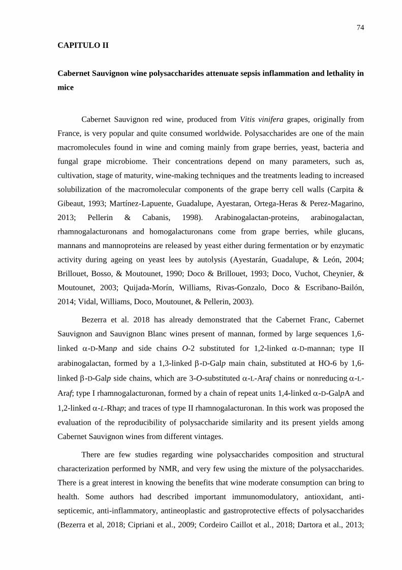
74
CAPITULO II
Cabernet Sauvignon wine polysaccharides attenuate sepsis inflammation and lethality in
mice
Cabernet Sauvignon red wine, produced from Vitis vinifera grapes, originally from
France, is very popular and quite consumed worldwide. Polysaccharides are one of the main
macromolecules found in wine and coming mainly from grape berries, yeast, bacteria and
fungal grape microbiome. Their concentrations depend on many parameters, such as,
cultivation, stage of maturity, wine-making techniques and the treatments leading to increased
solubilization of the macromolecular components of the grape berry cell walls (Carpita &
Gibeaut, 1993; Martínez-Lapuente, Guadalupe, Ayestaran, Ortega-Heras & Perez-Magarino,
2013; Pellerin & Cabanis, 1998). Arabinogalactan-proteins, arabinogalactan,
rhamnogalacturonans and homogalacturonans come from grape berries, while glucans,
mannans and mannoproteins are released by yeast either during fermentation or by enzymatic
activity during ageing on yeast lees by autolysis (Ayestarán, Guadalupe, & León, 2004;
Brillouet, Bosso, & Moutounet, 1990; Doco & Brillouet, 1993; Doco, Vuchot, Cheynier, &
Moutounet, 2003; Quijada-Morín, Williams, Rivas-Gonzalo, Doco & Escribano-Bailón,
2014; Vidal, Williams, Doco, Moutounet, & Pellerin, 2003).
Bezerra et al. 2018 has already demonstrated that the Cabernet Franc, Cabernet
Sauvignon and Sauvignon Blanc wines present of mannan, formed by large sequences 1,6-
linked -D-Manp and side chains O-2 substituted for 1,2-linked -D-mannan; type II
arabinogalactan, formed by a 1,3-linked -D-Galp main chain, substituted at HO-6 by 1,6-
linked -D-Galp side chains, which are 3-O-substituted -L-Araf chains or nonreducing-L-
Araf; type I rhamnogalacturonan, formed by a chain of repeat units 1,4-linked -D-GalpA and
1,2-linked -L-Rhap; and traces of type II rhamnogalacturonan. In this work was proposed the
evaluation of the reproducibility of polysaccharide similarity and its present yields among
Cabernet Sauvignon wines from different vintages.
There are few studies regarding wine polysaccharides composition and structural
characterization performed by NMR, and very few using the mixture of the polysaccharides.
There is a great interest in knowing the benefits that wine moderate consumption can bring to
health. Some authors had described important immunomodulatory, antioxidant, anti-
septicemic, anti-inflammatory, antineoplastic and gastroprotective effects of polysaccharides
(Bezerra et al, 2018; Cipriani et al., 2009; Cordeiro Caillot et al., 2018; Dartora et al., 2013;
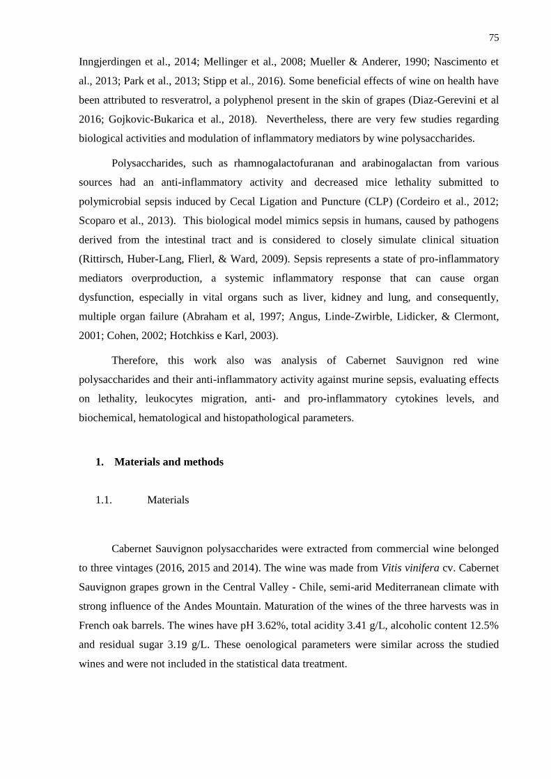
75
Inngjerdingen et al., 2014; Mellinger et al., 2008; Mueller & Anderer, 1990; Nascimento et
al., 2013; Park et al., 2013; Stipp et al., 2016). Some beneficial effects of wine on health have
been attributed to resveratrol, a polyphenol present in the skin of grapes (Diaz-Gerevini et al
2016; Gojkovic-Bukarica et al., 2018). Nevertheless, there are very few studies regarding
biological activities and modulation of inflammatory mediators by wine polysaccharides.
Polysaccharides, such as rhamnogalactofuranan and arabinogalactan from various
sources had an anti-inflammatory activity and decreased mice lethality submitted to
polymicrobial sepsis induced by Cecal Ligation and Puncture (CLP) (Cordeiro et al., 2012;
Scoparo et al., 2013). This biological model mimics sepsis in humans, caused by pathogens
derived from the intestinal tract and is considered to closely simulate clinical situation
(Rittirsch, Huber-Lang, Flierl, & Ward, 2009). Sepsis represents a state of pro-inflammatory
mediators overproduction, a systemic inflammatory response that can cause organ
dysfunction, especially in vital organs such as liver, kidney and lung, and consequently,
multiple organ failure (Abraham et al, 1997; Angus, Linde-Zwirble, Lidicker, & Clermont,
2001; Cohen, 2002; Hotchkiss e Karl, 2003).
Therefore, this work also was analysis of Cabernet Sauvignon red wine
polysaccharides and their anti-inflammatory activity against murine sepsis, evaluating effects
on lethality, leukocytes migration, anti- and pro-inflammatory cytokines levels, and
biochemical, hematological and histopathological parameters.
1. Materials and methods
1.1. Materials
Cabernet Sauvignon polysaccharides were extracted from commercial wine belonged
to three vintages (2016, 2015 and 2014). The wine was made from Vitis vinifera cv. Cabernet
Sauvignon grapes grown in the Central Valley - Chile, semi-arid Mediterranean climate with
strong influence of the Andes Mountain. Maturation of the wines of the three harvests was in
French oak barrels. The wines have pH 3.62%, total acidity 3.41 g/L, alcoholic content 12.5%
and residual sugar 3.19 g/L. These oenological parameters were similar across the studied
wines and were not included in the statistical data treatment.
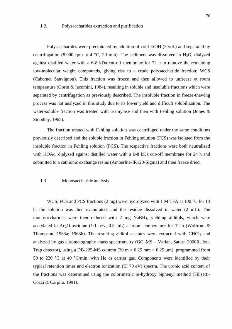
76
1.2. Polysaccharides extraction and purification
Polysaccharides were precipitated by addition of cold EtOH (3 vol.) and separated by
centrifugation (8.000 rpm at 4 °C, 20 min). The sediment was dissolved in H2O, dialyzed
against distilled water with a 6-8 kDa cut-off membrane for 72 h to remove the remaining
low-molecular weight compounds, giving rise to a crude polysaccharide fraction: WCS
(Cabernet Sauvignon). This fraction was frozen and then allowed to unfreeze at room
temperature (Gorin & Iacomini, 1984), resulting in soluble and insoluble fractions which were
separated by centrifugation as previously described. The insoluble fraction in freeze-thawing
process was not analyzed in this study due to its lower yield and difficult solubilization. The
water-soluble fraction was treated with α-amylase and then with Fehling solution (Jones &
Stoodley, 1965).
The fraction treated with Fehling solution was centrifuged under the same conditions
previously described and the soluble fraction in Fehling solution (FCS) was isolated from the
insoluble fraction in Fehling solution (PCS). The respective fractions were both neutralized
with HOAc, dialyzed against distilled water with a 6-8 kDa cut-off membrane for 24 h and
submitted to a cathionic exchange resins (Amberlite-IR120-Sigma) and then freeze dried.
1.3. Monosaccharide analysis
WCS, FCS and PCS fractions (2 mg) were hydrolyzed with 1 M TFA at 100 °C for 14
h, the solution was then evaporated, and the residue dissolved in water (2 mL). The
monosaccharides were then reduced with 2 mg NaBH4, yielding alditols, which were
acetylated in Ac2O-pyridine (1:1, v/v, 0.5 mL) at room temperature for 12 h (Wolfrom &
Thompson, 1963a; 1963b). The resulting alditol acetates were extracted with CHCl3 and
analyzed by gas chromatography–mass spectrometry (GC–MS – Varian, Saturn 2000R, Ion-
Trap detector), using a DB-225-MS column (30 m × 0.25 mm × 0.25 μm), programmed from
50 to 220 °C at 40 °C/min, with He as carrier gas. Components were identified by their
typical retention times and electron ionization (EI 70 eV) spectra. The uronic acid content of
the fractions was determined using the colorimetric m-hydroxy biphenyl method (Filisetti-
Cozzi & Carpita, 1991).
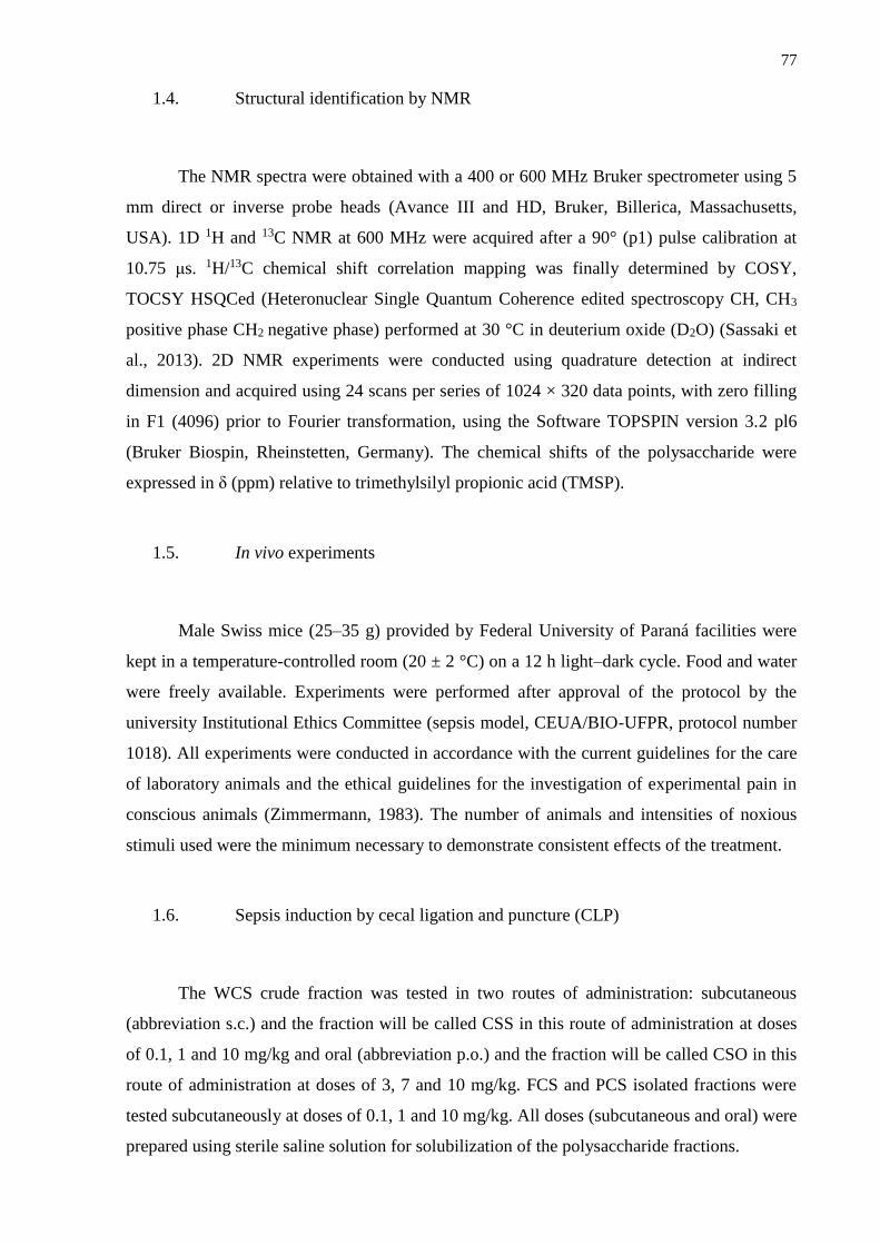
77
1.4. Structural identification by NMR
The NMR spectra were obtained with a 400 or 600 MHz Bruker spectrometer using 5
mm direct or inverse probe heads (Avance III and HD, Bruker, Billerica, Massachusetts,
USA). 1D 1H and 13C NMR at 600 MHz were acquired after a 90° (p1) pulse calibration at
10.75 μs. 1H/13C chemical shift correlation mapping was finally determined by COSY,
TOCSY HSQCed (Heteronuclear Single Quantum Coherence edited spectroscopy CH, CH3
positive phase CH2 negative phase) performed at 30 °C in deuterium oxide (D2O) (Sassaki et
al., 2013). 2D NMR experiments were conducted using quadrature detection at indirect
dimension and acquired using 24 scans per series of 1024 × 320 data points, with zero filling
in F1 (4096) prior to Fourier transformation, using the Software TOPSPIN version 3.2 pl6
(Bruker Biospin, Rheinstetten, Germany). The chemical shifts of the polysaccharide were
expressed in δ (ppm) relative to trimethylsilyl propionic acid (TMSP).
1.5. In vivo experiments
Male Swiss mice (25–35 g) provided by Federal University of Paraná facilities were
kept in a temperature-controlled room (20 ± 2 °C) on a 12 h light–dark cycle. Food and water
were freely available. Experiments were performed after approval of the protocol by the
university Institutional Ethics Committee (sepsis model, CEUA/BIO-UFPR, protocol number
1018). All experiments were conducted in accordance with the current guidelines for the care
of laboratory animals and the ethical guidelines for the investigation of experimental pain in
conscious animals (Zimmermann, 1983). The number of animals and intensities of noxious
stimuli used were the minimum necessary to demonstrate consistent effects of the treatment.
1.6. Sepsis induction by cecal ligation and puncture (CLP)
The WCS crude fraction was tested in two routes of administration: subcutaneous
(abbreviation s.c.) and the fraction will be called CSS in this route of administration at doses
of 0.1, 1 and 10 mg/kg and oral (abbreviation p.o.) and the fraction will be called CSO in this
route of administration at doses of 3, 7 and 10 mg/kg. FCS and PCS isolated fractions were
tested subcutaneously at doses of 0.1, 1 and 10 mg/kg. All doses (subcutaneous and oral) were
prepared using sterile saline solution for solubilization of the polysaccharide fractions.
78
Mice were randomly divided into groups with 10 animals each: sham-operation, CLP
plus saline (10 mL/kg s.c.), CLP plus CSS (0.1 mg/kg s.c.), CLP plus CSS (1 mg/kg s.c.),
CLP plus CSS (10 mg/kg s.c.), CLP plus FCS (0.1 mg/kg s.c.), CLP plus FCS (1 mg/kg s.c.),
CLP plus FCS (10 mg/kg s.c.), CLP plus PCS (0.1 mg/kg s.c.), CLP plus PCS (1 mg/kg s.c.),
CLP plus PCS (10 mg/kg s.c.), CLP plus CSO (3 mg/kg, p.o.), CLP plus CSO (7 mg/kg, p.o.),
CLP plus CSO (10 mg/kg, p.o.) and CLP plus dexamethasone - Dex (0.5 mg/kg s.c.). Saline
was used as vehicle for dissolving the polysaccharide and dexamethasone was commercially
purchased.
Ketamine (80 mg/kg) and xylazine (20 mg/kg) were injected intraperitoneally to
anesthetize the mice before surgical procedures. Polymicrobial sepsis was induced by CLP as
previously described (Rittirsch, Huber-Lang, Flierl, & Ward, 2009). A midline incision of
∼1.5 cm was carried out on the abdomen. The cecum was carefully exposed and 50% of the
distal moiety was ligated. The cecum was then punctured thrice with a sterile 16-gauge needle
and squeezed to extrude the fecal material from the wounds. The cecum was replaced and the
abdomen was stitched surgically. Sham-control animals were treated identically, but no cecal
ligation or puncture was carried out. Each mouse received a subcutaneous sterile saline
injection (1 mL) for fluid resuscitation after surgery. The mice were then kept on a heating
pad (35 °C) until they recovered from the anesthesia. Food and water, ad libitum, were
provided throughout the experiment. The survival rate was monitored for 7 days, every 12 h.
During this period, water (vehicle) and drugs were orally or subcutaneously administered
daily. Dexamethasone was commercially purchased and subcutaneously administered at doses
of 0.5 mg/kg (Lapa, Silva, Almeida Cabrini, & Santos, 2012; Longhi-Balbinot et al., 2012;
Silva et al., 2012).
In this experimental set, mice were orally or subcutaneously treated with vehicle (1 h
before surgery), sham-operation, CLP plus saline (10 mL/kg s.c.) and dexamethasone (0.5
mg/kg s.c.). The best dose of each fraction in the survival test was chosen for the anti-
inflammatory evaluation tests, as follows: CSS (1 mg/kg, s.c.), CSO (10 mg/kg, p.o.), FCS (1
mg/kg, s.c.) and PCS (1 mg/kg, s.c.). For these doses, after a post-operation time of 6 h, mice
were sacrificed. Liver, lung and kidney tissues were collected for histological analysis. Blood
was collected for hematological and biochemical parameters evaluation.
1.7. Hematological assays

79
Blood was collected from the abdominal vena cava. Total leukocytes and mononuclear
(MN) and polymorphonuclear (PMN) leukocytes were counted by an automated system,
according to the manufacturer’s instructions (Mindray BC-2800 vet, São Paulo, SP, Brazil).
1.8. Biochemical assays
The amount of TNF-α, IL-1β and IL-10 cytokines in the blood plasma were
determined by enzyme-linked immunosorbent assay kits according to the manufacturer's
instructions. Three replicates were carried out for each different treatment. The absorbance of
each group was measured by a microplate reader at the wavelength of 450 nm. To determine
the total concentration of nitric oxide (NO) in the blood plasma, Griess reagent was added to
40 mL of plasma and the absorbance at 545 nm was evaluated with an ELISA kit (BD
Biosciences, San Jose, CA, USA) following the manufacturer´s manual.
Bilirubin, creatinine (CREA) and urea, as well as alanine aminotransferase (ALT) and
aspartate aminotransferase (AST) levels and activities were analyzed from plasma by an
automated system, according to the manufacturer’s instructions (Mindray BS-200, São Paulo,
SP, Brazil).
1.9. Histopathological analysis
Lungs, liver and kidney tissue sections were fixed in ethanol at room temperature.
After fixation, samples were dehydrated in a graded series of increasing ethanol before
paraffin embedding. Thin sections (4 mm) from paraffin blocks were processed for histology,
and the tissue was stained with hematoxylin eosin. The slides were then analyzed by light
microscopy.
1.10. Statistical analysis
The results were expressed as mean ±SEM and were statistically analyzed by one-way
analysis of variance (ANOVA) and Tukey's test as a post hoc test (P < 0.001). GraphPad
Prism 5.0 (La Jolla, CA) program was used for both statistical analysis and figure design.
Histological analysis was performed by qualitative observations.
80
2. Results and Discussion
2.1. Analysis of the polysaccharides of Cabernet Sauvignon wine
Approximately 750 mL from commercial wine was concentrated and precipitated with
excess ethanol. The polysaccharides were recovered by ethanol precipitation and then
centrifugated and dialysed against water. The solution was then freeze-dried, generating the
WCS polysaccharide fraction with yield of 0.05% in the harvests of the three years evaluated
and the table 2 show total polysaccharide yield in mass to 750 mL of wine.
Previous studies carried out by our group on polysaccharide structure demonstrated
that fraction WCS from Cabernet Sauvignon is composed by a mixture of mannan, type II
arabinogalactan, type I and II rhamnogalacturonans (Bezerra et al., 2018).
1H NMR spectra analyses of the crude polysaccharide fractions from Cabernet
Sauvignon wine from 2014 (WCS-2014), 2015 (WCS-2105) and 2016 (WCS-2016) were
performed (Fig. 1). 1H-NMR data of WCS of the three vintages showed characteristic
polysaccharide NMR signals, showing a complex profile at the anomeric region (4.0~6.0
ppm). The 1H NMR signals shown in figure 1 the anomeric region and the binding region
were similar for all three fractions. This experiment demonstrated that the wines have similar
polysaccharide patterns even though they were produced in different years.
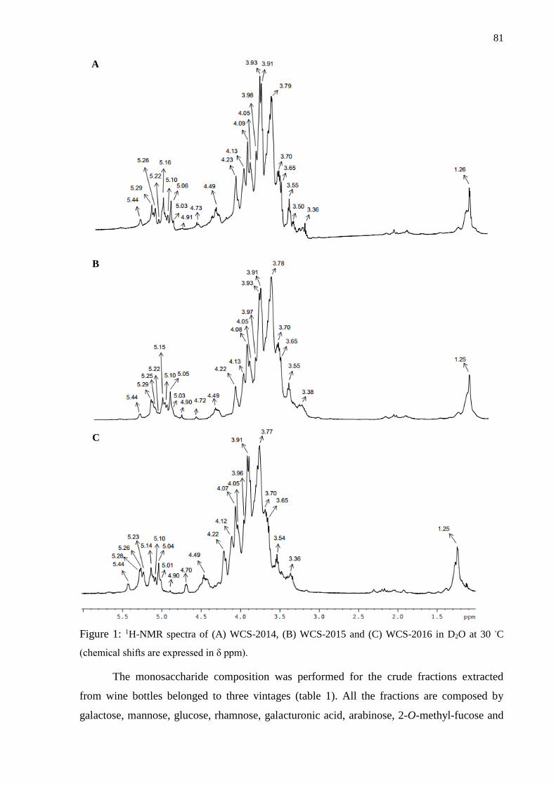
81
Figure 1: 1H-NMR spectra of (A) WCS-2014, (B) WCS-2015 and (C) WCS-2016 in D2O at 30 ◦C
(chemical shifts are expressed in δ ppm).
The monosaccharide composition was performed for the crude fractions extracted
from wine bottles belonged to three vintages (table 1). All the fractions are composed by
galactose, mannose, glucose, rhamnose, galacturonic acid, arabinose, 2-O-methyl-fucose and
B
C
A
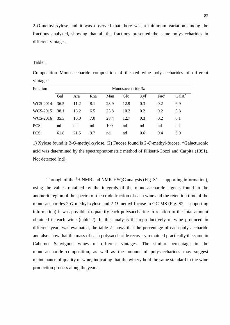
82
2-O-methyl-xylose and it was observed that there was a minimum variation among the
fractions analyzed, showing that all the fractions presented the same polysaccharides in
different vintages.
Table 1
Composition Monosaccharide composition of the red wine polysaccharides of different
vintages
Fraction Monosaccharide %
Gal Ara Rha Man Glc Xyl1 Fuc2 GalA*
WCS-2014 36.5 11.2 8.1 23.9 12.9 0.3 0.2 6,9
WCS-2015 38.1 13.2 6.5 25.8 10.2 0.2 0.2 5,8
WCS-2016 35.3 10.0 7.0 28.4 12.7 0.3 0.2 6.1
PCS nd nd nd 100 nd nd nd nd
FCS 61.8 21.5 9.7 nd nd 0.6 0.4 6.0
1) Xylose found is 2-O-methyl-xylose. (2) Fucose found is 2-O-methyl-fucose. *Galacturonic
acid was determined by the spectrophotometric method of Filisetti-Cozzi and Carpita (1991).
Not detected (nd).
Through of the 1H NMR and NMR-HSQC analysis (Fig. S1 – supporting information),
using the values obtained by the integrals of the monosaccharide signals found in the
anomeric region of the spectra of the crude fraction of each wine and the retention time of the
monosaccharides 2-O-methyl xylose and 2-O-methyl-fucose in GC-MS (Fig. S2 – supporting
information) it was possible to quantify each polysaccharide in relation to the total amount
obtained in each wine (table 2). In this analysis the reproductively of wine produced in
different years was evaluated, the table 2 shows that the percentage of each polysaccharide
and also show that the mass of each polysaccharide recovery remained practically the same in
Cabernet Sauvignon wines of different vintages. The similar percentage in the
monosaccharide composition, as well as the amount of polysaccharides may suggest
maintenance of quality of wine, indicating that the winery hold the same standard in the wine
production process along the years.
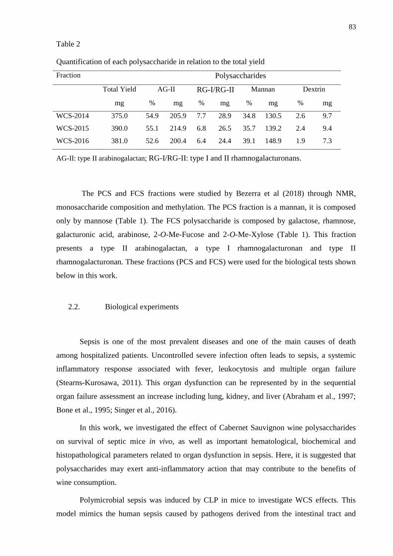
83
Table 2
Quantification of each polysaccharide in relation to the total yield
Fraction Polysaccharides
Total Yield AG-II RG-I/RG-II Mannan Dextrin
mg % mg % mg % mg % mg
WCS-2014 375.0 54.9 205.9 7.7 28.9 34.8 130.5 2.6 9.7
WCS-2015 390.0 55.1 214.9 6.8 26.5 35.7 139.2 2.4 9.4
WCS-2016 381.0 52.6 200.4 6.4 24.4 39.1 148.9 1.9 7.3
AG-II: type II arabinogalactan; RG-I/RG-II: type I and II rhamnogalacturonans.
The PCS and FCS fractions were studied by Bezerra et al (2018) through NMR,
monosaccharide composition and methylation. The PCS fraction is a mannan, it is composed
only by mannose (Table 1). The FCS polysaccharide is composed by galactose, rhamnose,
galacturonic acid, arabinose, 2-O-Me-Fucose and 2-O-Me-Xylose (Table 1). This fraction
presents a type II arabinogalactan, a type I rhamnogalacturonan and type II
rhamnogalacturonan. These fractions (PCS and FCS) were used for the biological tests shown
below in this work.
2.2. Biological experiments
Sepsis is one of the most prevalent diseases and one of the main causes of death
among hospitalized patients. Uncontrolled severe infection often leads to sepsis, a systemic
inflammatory response associated with fever, leukocytosis and multiple organ failure
(Stearns-Kurosawa, 2011). This organ dysfunction can be represented by in the sequential
organ failure assessment an increase including lung, kidney, and liver (Abraham et al., 1997;
Bone et al., 1995; Singer et al., 2016).
In this work, we investigated the effect of Cabernet Sauvignon wine polysaccharides
on survival of septic mice in vivo, as well as important hematological, biochemical and
histopathological parameters related to organ dysfunction in sepsis. Here, it is suggested that
polysaccharides may exert anti-inflammatory action that may contribute to the benefits of
wine consumption.
Polymicrobial sepsis was induced by CLP in mice to investigate WCS effects. This
model mimics the human sepsis caused by pathogens derived from the intestinal tract and
84
simulates a clinical situation (Otero-Anton et al., 2001). Many polysaccharides isolated from a
variety of sources have shown immune responses in vivo and in vitro. Polysaccharides such as
pectins and hemicelluloses had their anti-septicemic effect determined by being tested
subcutaneously (Lim et al., 2002; Ruthes, Rattmann, Carbonero, Gorin & Iacomini, 2012;
Tzianabos, Kasper, Cisneiros, Smith & Onderdonk, 1995) and orally (Dartora et al., 2013;
Scoparo et al., 2013).
In this work, we treated orally the animals with the Cabernet Sauvignon wine
polysaccharide mixture (CSO) to evaluate the protective effect of wine consumption and
subcutaneously with the polysaccharide mixture (CSS) and the isolated fractions (FCS and
PCS) to achieve very low doses and still be able to show the efficiency of the polysaccharides
as anti-inflammatory and protective in an aggressive model of sepsis by CLP.
Lethality was found to be markedly reduced in mice receiving CSO, CSS, FCS and
PCS treatments (Fig. 2). It was demonstrated that the CSO fraction prevents lethality caused
by polymicrobial sepsis in mice. Lethality was markedly delayed in mice treated orally with
CSO. The corresponding areas under the lethality curve increased to 9.120, 11.400 and
13.500 (arbitrary units) after 3, 7 and 10 mg/kg administration, respectively (Fig. 2A). The
overall survival in the CSO group was 30%, 50% and 70%, increasing with the dose. The 10
mg/kg CSO dose was established for the analysis of this fraction effects in CLP.
The CSS fraction prevents lethality, obtaining a survival rate of 40% (0.1 mg/kg), 80%
(1 mg/kg) and 78% (10 mg/kg) (Fig. 2B). Lethality was markedly delayed in subcutaneously
treated mice. The corresponding areas under the lethality curve were 10.680, 14.160 and
14.040 (arbitrary units) after administrating 0.1, 1 and 10 mg/kg, respectively. The 1 and 10
mg/kg doses presented similar effects and obtained better results when compared to
dexamethasone (0.5 mg/kg), which promoted a 50% survival rate. Therefore, the 1 mg/kg
CSS dose was established for the analysis of the CLP fraction effects.
Mice treated with FCS had a survival rate of 45% at the 0.1 mg/kg dose and 80% at 1
and 10 mg/kg doses (Fig. 2C). The 1 and 10 mg/kg doses presented similar effects. The
corresponding areas under the lethality curve were 12.300, 14.860 and 15.120 (0.1, 1, 10
mg/kg, respectively). Dexamethasone, used as control, had an area under curve of 12.000,
with an overall survival rate of 50% at the end of the observation period. As the doses of 1
and 10 mg/kg presented similar results, the 1 mg/kg FCS dose was established for the analysis
of the effects of this fraction in CLP.

85
Lethality was markedly delayed in mice subcutaneously treated with PCS and their
areas under the curve were 10.560, 13.020 and 12.900 after administrating 0.1, 1 and 10
mg/kg doses, respectively (Fig. 2D). At the end, the corresponding overall survival rates were
40%, 65% and 63%. The 1 and 10 mg/kg doses presented similar effects. Therefore, the 1
mg/kg PCS dose was established for the analysis of the effects of this fraction in CLP. In
summary, considering survival rates, the following doses were chosen for following analyses
in sepsis: 10 mg/kg for CSO and 1 mg/kg for CSS, FCS and PCS.
CSO - Oral
0 24 48 72 96 120 144 1680
25
50
75
100Sham
Vehicle
Dexamethasone
3 mg/kg
7 mg/kg
10 mg/kg
Hours post CLP
Pe
rce
nt
su
rviv
al
(%)
***
***
***
***
0 24 48 72 96 120 144 1680
25
50
75
100Sham
Vehicle
0.1 mg/kg
1 mg/kg
10 mg/kg
CSS - Subcutaneous
Dexamethasone
Hours post CLP
Pe
rce
nt
su
rviv
al
(%)
***
******
******
FCS
0 24 48 72 96 120 144 1680
20
40
60
80
100Sham
Vehicle
0.1 mg/kg
1 mg/kg
10 mg/kg
Dexamethasone
Hours post CLP
Pe
rce
nt
su
rviv
al
(%)
***
******
******
PCS
0 24 48 72 96 120 144 1680
20
40
60
80
100Sham
Vehicle
0.1 mg/kg
1 mg/kg
10 mg/kg
Dexamethasone
Hours post CLP
Pe
rce
nt
su
rviv
al
(%)
***
************
Fig. 2: Effects of CSO (A), CSS (B), FCS (C) and PCS (D) fractions on survival rate. Mice
(10 animals/group) were orally administered various doses of CSO (3, 7 or 10 mg/kg) or
subcutaneous administered various doses of CSS, FCS and PCS (0.1, 1, 10 mg/kg), vehicle
(saline s.c.) and dexamethasone (0.5 mg/kg s.c.). Values represent means ± SEM. ***p <
0.001 when compared with CLP plus vehicle group; ANOVA followed by the Tukey's test.
In sepsis model, the acute immune response is organized and executed predominantly
by innate immunity. Sepsis by CLP promotes migration of leukocytes through the
bloodstream to the site of infection, peritoneal cavity and affected organs such as lung, liver
and kidneys (Bone et al., 1992). Results showed that CSS, CSO, FCS, PCS and
dexamethasone were able to reduce by 60%, 59%, 59%, 51% and 55% respectively, white
blood cell (leukocyte count), when compared to the vehicle group (Table 3). Differential
leukocyte counts demonstrated a decrease in the mononuclear leukocyte (MN) subset and the
A B
C D
86
PMN subset in the bloodstream (Table 3). Mononuclear leukocyte subset reduction consisted
in 62%, 59%, 59%, 58% and 60% for CSS, CSO, FCS, PCS and dexamethasone, respectively.
For de PMN subset, these reductions were 51%, 53%, 50%, 46% and 47% (CSS, CSO, FCS
PCS, dexamethasone, respectively), when compared to the vehicle group. This exacerbated
inflammatory response was observed on the vehicle group, in which there was an increased
number of leukocytes, mononuclear leukocytes and PMN, whereas all fractions tested were
able to reduce these effects significantly, reducing the amplification of the inflammatory
response.
Neutrophils are the first cells to migrate to the site of infection, where they produce
both reactive oxygen and nitrogen species that act to kill the infectious agents. These
neutrophils and tissue resident cells also produce inflammatory cytokines, that can recruit and
activate other inflammatory cells that, in turn, amplify and extend the initial systemic
inflammatory response, as happened in the vehicle group. However, treatment with wine
polysaccharides seems to reduce this effect (Yamashiro et al., 2001).
Table 3
Hematological parameters evaluated during sepsis in mice
Treatment WBC (U/L) PMN (U/L) MN (U/L)
Sham 3316.7 ± 365.6 1450.0 ± 216.8 1850.0 ± 104.9
Vehicle 9317.9 ± 248.3*** 3850.0 ± 225.8*** 5283.3 ± 297.1***
CSS 3766.0 ± 266.5# 1900.0 ± 322.5 2016.7 ± 271.4
CSO 3850.0 ± 176.1# 1890.0 ± 206.6 2183.3 ± 183.3
FCS 3816.7 ± 343.0## 1916.7 ± 292.7 2186.7 ± 116.0
PCS 4600.0 ± 346.4 2133.3 ± 311.6 2200.0 ± 282.8
Dex 4216.7 ± 242.7 2050.0 ± 242.9 2116.7 ± 216.1
***p < 0.001 when vehicle was compared with all groups; # p < 0.05 and ## p < 0.01 when
compared with PCS; ANOVA followed by the Tukey's test. WBC:White blood cell count;
PMN: polymorphonuclear leukocytes; MN: mononuclear leukocytes; Dex: dexamethasone.
During sepsis, the immune response starts sensing of danger by pattern-recognition
receptors on the immune competent cells, such as macrophages. The sensed danger signals,
through specific signaling pathways, activate transcription factors and gene regulatory
systems, which up-regulate the expression of proinflammatory mediators (Akira & Takeda,
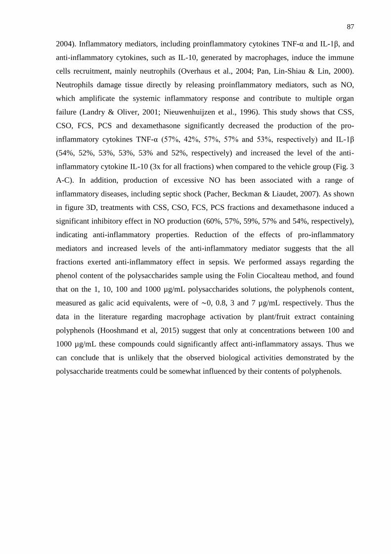
87
2004). Inflammatory mediators, including proinflammatory cytokines TNF-α and IL-1β, and
anti-inflammatory cytokines, such as IL-10, generated by macrophages, induce the immune
cells recruitment, mainly neutrophils (Overhaus et al., 2004; Pan, Lin-Shiau & Lin, 2000).
Neutrophils damage tissue directly by releasing proinflammatory mediators, such as NO,
which amplificate the systemic inflammatory response and contribute to multiple organ
failure (Landry & Oliver, 2001; Nieuwenhuijzen et al., 1996). This study shows that CSS,
CSO, FCS, PCS and dexamethasone significantly decreased the production of the pro-
inflammatory cytokines TNF-α (57%, 42%, 57%, 57% and 53%, respectively) and IL-1β
(54%, 52%, 53%, 53%, 53% and 52%, respectively) and increased the level of the anti-
inflammatory cytokine IL-10 (3x for all fractions) when compared to the vehicle group (Fig. 3
A-C). In addition, production of excessive NO has been associated with a range of
inflammatory diseases, including septic shock (Pacher, Beckman & Liaudet, 2007). As shown
in figure 3D, treatments with CSS, CSO, FCS, PCS fractions and dexamethasone induced a
significant inhibitory effect in NO production (60%, 57%, 59%, 57% and 54%, respectively),
indicating anti-inflammatory properties. Reduction of the effects of pro-inflammatory
mediators and increased levels of the anti-inflammatory mediator suggests that the all
fractions exerted anti-inflammatory effect in sepsis. We performed assays regarding the
phenol content of the polysaccharides sample using the Folin Ciocalteau method, and found
that on the 1, 10, 100 and 1000 µg/mL polysaccharides solutions, the polyphenols content,
measured as galic acid equivalents, were of ∼0, 0.8, 3 and 7 µg/mL respectively. Thus the
data in the literature regarding macrophage activation by plant/fruit extract containing
polyphenols (Hooshmand et al, 2015) suggest that only at concentrations between 100 and
1000 µg/mL these compounds could significantly affect anti-inflammatory assays. Thus we
can conclude that is unlikely that the observed biological activities demonstrated by the
polysaccharide treatments could be somewhat influenced by their contents of polyphenols.
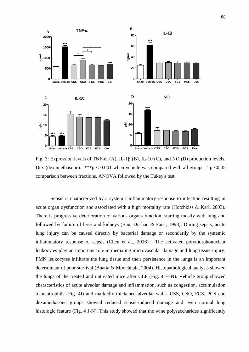
88
TNF-
Sham Vehicle CSS CSO FCS PCS Dex0
500
1000
1500
2000
***
+
+
+
pg
/mL
Sham Vehicle CSS CSO FCS PCS Dex0
20
40
60
80
***
IL-1
pg
/mL
IL-10
Sham Vehicle CSS CSO FCS PCS Dex0
5
10
15
20
*** ***
pg
/mL
NO
Sham Vehicle CSS CSO FCS PCS Dex0
5
10
15
20
***
M
Fig. 3: Expression levels of TNF-α. (A), IL-1β (B), IL-10 (C), and NO (D) production levels.
Dex (dexamethasone). ***p < 0.001 when vehicle was compared with all groups; + p <0,05
comparison between fractions. ANOVA followed by the Tukey's test.
Sepsis is characterized by a systemic inflammatory response to infection resulting in
acute organ dysfunction and associated with a high mortality rate (Hotchkiss & Karl, 2003).
There is progressive deterioration of various organs function, starting mostly with lung and
followed by failure of liver and kidneys (Bau, Durhan & Faist, 1998). During sepsis, acute
lung injury can be caused directly by bacterial damage or secondarily by the systemic
inflammatory response of sepsis (Chen et al., 2016). The activated polymorphonuclear
leukocytes play an important role in mediating microvascular damage and lung tissue injury.
PMN leukocytes infiltrate the lung tissue and their persistence in the lungs is an important
determinant of poor survival (Bhatia & Moochhala, 2004). Histopathological analysis showed
the lungs of the treated and untreated mice after CLP (Fig. 4 H-N). Vehicle group showed
characteristics of acute alveolar damage and inflammation, such as congestion, accumulation
of neutrophils (Fig. 4I) and markedly thickened alveolar walls. CSS, CSO, FCS, PCS and
dexamethasone groups showed reduced sepsis-induced damage and even normal lung
histologic feature (Fig. 4 J-N). This study showed that the wine polysaccharides significantly
A B
C D

89
reduced the PMN number in blood and this may have reduced leukocyte infiltration in tissues
and protected against lung injury.
Liver plays a major function in host defense in sepsis. Kupffer cells scavenge bacteria
as well as clear cytokines from the liver. Hepatocytes modify metabolic pathways, increase
gluconeogenesis, activate cellular repair and enzymes such as transaminases (AST and ALT)
and alter bilirubin levels (Dhainaut, Marin, Mignon & Vinsonneau, 2001). ALT and AST
transaminases are markers of hepatocyte injuries, so they were evaluated in this study. It was
observed an increase in plasma AST and ALT activity in the septic group (Table 4).
Treatment with CSS, CSO, FCS, PCS and dexamethasone showed a significant reduction of
ALT in the blood in 59%, 64%, 57%, 58% and 58%, respectively, compared to the vehicle
group (Table 4). Although ALT is an enzyme present in the hepatocyte cytoplasm, AST is
present in mitochondria and at the cytoplasm. Moreover, AST has activity in the liver, heart,
kidneys, pancreas, muscles and erythrocytes. Therefore, increased levels of plasma AST
indicate the occurrence of major damage to the hepatocyte mitochondria or significant
damage in a different organ. Thus, AST levels are higher than ALT in the vehicle group, but
were not affected in all fractions treated groups and dexamethasone, which were able to
maintain the low levels of the two enzymes. When compared to the vehicle group, it was
observed the reduction in AST levels by 65% (CSS), 69% (CSO), 64% (FCS), 63% (PCS)
and 63% (dexamethasone) (Table 4).
This study also evaluated liver function by measuring bilirubin levels. Bilirubin is
recognized as an indicator of liver dysfunction, so severe elevations can cause damage to
brain and erythrocytes (Brito, Silva & Brites, 2006; Fevery, 2008). Increased serum level of
bilirubin was observed in the septic group, which was in accordance with other results already
shown (Table 4). All treatments with fractions and dexamethasone showed a significant
reduction of bilirubin serum in relation to septic group, consisting in 67% (CSS), 61% (CSO),
61% (FCS), 58% (PCS) and 58% (dexamethasone) (Table 4). Histopathological analyses
demonstrated hepatic alterations in the vehicle group (Fig. 4B). Lesions were characterized by
a reduction in sinusoids within the lumen, indicating tumefaction, and capsular infiltration of
inflammatory cells. These lesions were found in septic group and could be prevented by
treatments with each of all fractions, showing that the wine polysaccharides were able to
preserve the liver, similar to dexamethasone (Fig. 4C-G) and sham group (Fig. 4A). These
results showed that the wine polysaccharides were effective in protecting the liver and
maintaining the organ functions during sepsis.

90
Table 4
Plasmatic parameters evaluate during sepsis in mice
Treatment AST
(U/L)
ALT
(U/L)
Bilirubin
(mg/dL)
Urea
(mg/dL)
Creatinine
(mg/dL)
Sham 138.4 ± 28.1 73.9 ± 5.8 1.0 ± 0.1 60.7 ± 9.4 0.4 ± 0.1
Vehicle 459.8 ± 25.5*** 248.7 ± 19.1*** 3.6 ± 0.4*** 209.1 ± 14.0*** 1.3 ± 0.1***
CSS 161.2 ± 14.5 101.8 ± 5.7 1.2 ± 0.3 86.2 ± 9.6 0.4 ± 0.1
CSO 140.5 ± 14.9 90.7 ± 6.9 1.4 ± 0.2 89.9 ± 6.8 0.5 ± 0.1
FCS 166.0 ± 21.5 106.9 ± 9.1 1.2 ± 0.1 97.5 ± 10.6 0.4 ± 0.1
PCS 169.5 ± 24.6 104.6 ± 14.5 1.5 ± 0.3 101.8 ± 5.4 0.5 ± 0.1
Dex 168.7 ± 10.9 105.0 ± 2.8 1.5 ± 0.2 99.9 ± 4.1 0.5 ± 0.1
***p < 0.001 when vehicle was compared with all groups; There was no statistical difference
between the treated groups. ANOVA followed by the Tukey's test. WBC:White blood cell
count; Dex: dexamethasone.
Sepsis is a systemic inflammatory response syndrome caused by bacteria, viruses or
fungi. It can cause organ damage far from the primary site of infection, leading to multiple
organ failure and even death. Acute kidney injury (AKI) is one of the complications of sepsis
that can cause increased mortality rate in patients (Rewa & Bagshaw, 2014; Yamada et al.,
2013). Kidney infiltration by neutrophils and subsequent release of free radicals is one major
mechanism leading to AKI (Huang et al., 2015). The ability of CSS, CSO, FCS and PCS to
have a protective effect on the kidneys was evaluated through the analysis of creatinine and
urea (Table 4), which are markers of renal function (Fig. 4 O-U and Fig. S3 - supporting
information). There was a significant increase in serum urea and creatinine levels in mice in
vehicle group, while all fractions showed a significant reduction of serum levels of these
markers (Table 4). The observed reductions were 59% (CSS), 57% (CSO), 53% (FCS), 51%
(PCS) and 52% (dexamethasone), in relation to the vehicle group for urea levels. For
creatinine level, the reductions were of 69% (CSS), 62% (CSO), 69% (FCS), 62% (PCS) and
62% (dexamethasone). Histological analysis of kidney reveled a renal tubular dilatation,
bleeding, leukocyte infiltrate and tubular epithelial cell vacuolar degeneration with presence
of necrosis, Bowman's capsule and proximal convoluted tubule retraction with degeneration
were observed in the vehicle group (Fig. 4P and Fig. S3B - supporting information). Pre-
treatment with each of all fractions effectively improved these kidney histopathological
changes in mice with sepsis, as well as dexamethasone (Fig. 4Q-U and Fig. S3 C-G -
91
supporting information) and sham group (Fig. 4O and Fig. S3A - supporting information).
Wine polysaccharides were able to preserve the kidneys during sepsis and seem to have
helped in maintaining kidney function.
Due to the results presented, we were able to show that pre-treatment with Cabernet
Sauvignon wine polysaccharides prevented lethality caused by polymicrobial sepsis in mice,
by both subcutaneous and oral administration. Polysaccharides demonstrated a potent anti-
inflammatory effect in the sepsis model, what may indicate beneficial effects of wine
moderate consumption for human health. The results were very satisfactory and encouraging,
in future work we aim to include polysaccharides from commercial suppliers as controls. The
anti-inflammatory effect of polysaccharides obtained from Caberrnet Sauvignon wine and
pure polysaccharide preparations (AGs, RGs and mannans) from commercial suppliers will be
compared in the biological experiments, we will evaluate if the same polysaccharide obtained
from different sources in which it presents small variations in its structure and different
branching points may influence the biological response studied.
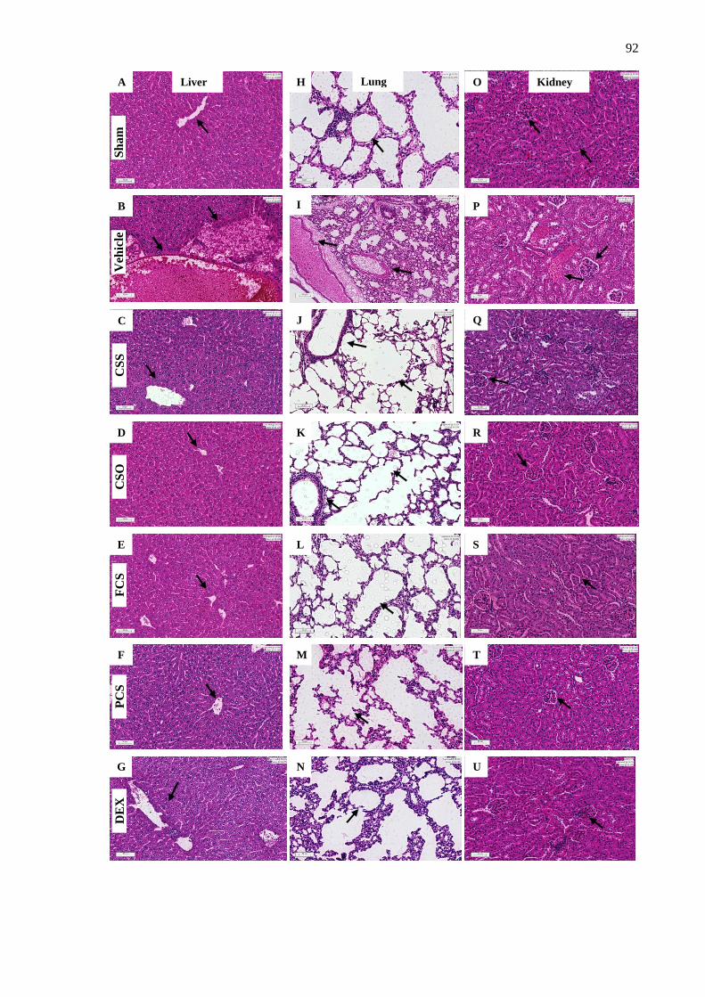
92
A
B
C
D
E
F
G
H
I
L
J
K
M
N
O
P
Q
R
R
S
R
T
R
U
R
Lung Liver Kidney
Sh
am
V
ehic
le
CS
S
CS
O
FC
S
PC
S
DE
X
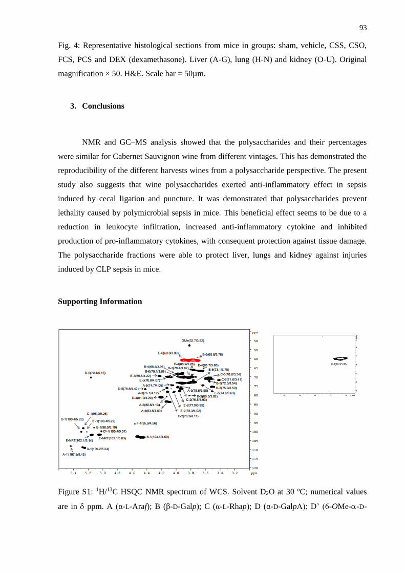
93
Fig. 4: Representative histological sections from mice in groups: sham, vehicle, CSS, CSO,
FCS, PCS and DEX (dexamethasone). Liver (A-G), lung (H-N) and kidney (O-U). Original
magnification × 50. H&E. Scale bar = 50µm.
3. Conclusions
NMR and GC–MS analysis showed that the polysaccharides and their percentages
were similar for Cabernet Sauvignon wine from different vintages. This has demonstrated the
reproducibility of the different harvests wines from a polysaccharide perspective. The present
study also suggests that wine polysaccharides exerted anti-inflammatory effect in sepsis
induced by cecal ligation and puncture. It was demonstrated that polysaccharides prevent
lethality caused by polymicrobial sepsis in mice. This beneficial effect seems to be due to a
reduction in leukocyte infiltration, increased anti-inflammatory cytokine and inhibited
production of pro-inflammatory cytokines, with consequent protection against tissue damage.
The polysaccharide fractions were able to protect liver, lungs and kidney against injuries
induced by CLP sepsis in mice.
Supporting Information
Figure S1: 1H/13C HSQC NMR spectrum of WCS. Solvent D2O at 30 ºC; numerical values
are in ppm. A (α-L-Araf); B (β-D-Galp); C (α-L-Rhap); D (α-D-GalpA); D’ (6-OMe--D-
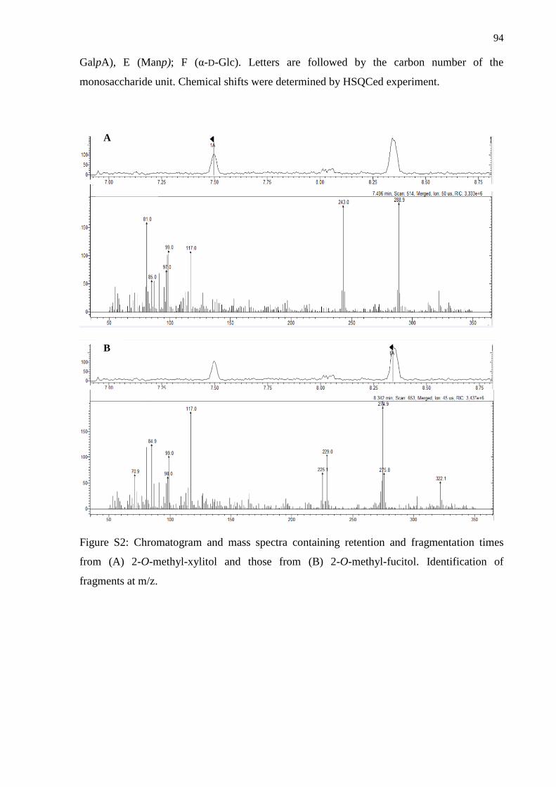
94
GalpA), E (Manp); F (α-D-Glc). Letters are followed by the carbon number of the
monosaccharide unit. Chemical shifts were determined by HSQCed experiment.
Figure S2: Chromatogram and mass spectra containing retention and fragmentation times
from (A) 2-O-methyl-xylitol and those from (B) 2-O-methyl-fucitol. Identification of
fragments at m/z.
B
A
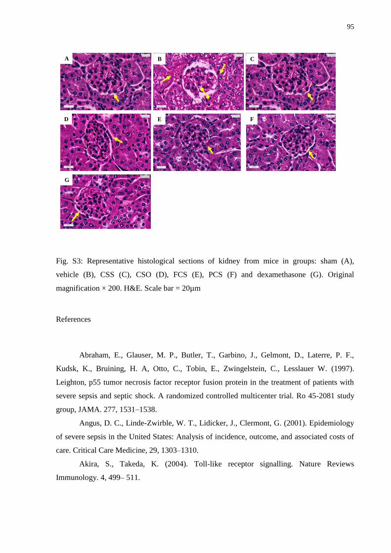
95
A B C
D E F
G
Fig. S3: Representative histological sections of kidney from mice in groups: sham (A),
vehicle (B), CSS (C), CSO (D), FCS (E), PCS (F) and dexamethasone (G). Original
magnification × 200. H&E. Scale bar = 20µm
References
Abraham, E., Glauser, M. P., Butler, T., Garbino, J., Gelmont, D., Laterre, P. F.,
Kudsk, K., Bruining, H. A, Otto, C., Tobin, E., Zwingelstein, C., Lesslauer W. (1997).
Leighton, p55 tumor necrosis factor receptor fusion protein in the treatment of patients with
severe sepsis and septic shock. A randomized controlled multicenter trial. Ro 45-2081 study
group, JAMA. 277, 1531–1538.
Angus, D. C., Linde-Zwirble, W. T., Lidicker, J., Clermont, G. (2001). Epidemiology
of severe sepsis in the United States: Analysis of incidence, outcome, and associated costs of
care. Critical Care Medicine, 29, 1303–1310.
Akira, S., Takeda, K. (2004). Toll-like receptor signalling. Nature Reviews
Immunology. 4, 499– 511.
96
Ayestarán, B., Guadalupe, Z., León, D. (2004). Quantification of major grape
polysaccharides (Tempranillo v.) released by maceration enzymes during the fermentation
process. Analytica Chimica Acta, 513, 29-39.
Bau, A. E., Durhan, R., Faist, E. (1998). Systemic inflammatory response syndrome
(SIRS), multiple organ dysfunction syndrome (MODS), multiple organ failure (MOF): Are
we winning the battle? Shock, 10, 79-89.
Bezerra, I.L.; Caillot, A. R. C.; Palhares, L. C. G. F.; Santana-Filho, A. P.; Chavante,
S. F. Guilherme Lanzi Sassaki, G. L. (2018). Structural characterization of polysaccharides
from Cabernet Franc, Cabernet Sauvignon and Sauvignon Blanc wines: Anti-inflammatory
activity in LPS stimulated RAW 264.7 cells. Carbohydrate Polymers, 186, 91-99.
Bhatia M, Moochhala S. (2004). Role of inflammatory mediators in the
pathophysiology of acute respiratory distress syndrome. The Journal of Pathology, 202(2),
145-56.
Bone, R. C., Balk, R. A, Cerra, F. B., Dellinger, R. P., Fein, A. M., Knaus, W. A.,
Schein, R. M., Sibbald, W. J. (1992). American College of Chest Physicians/Society of
Critical Care Medicine Consensus Conference: definitions for sepsis and organ failure and
guidelines for the use of innovative therapies in sepsis, Chest. 101, 1644-1655.
Bone, R. C., Balk, R. A., Fein, A. M., Perl, T. M., Wenzel, R. P. Reines, H. D.,
Quenzer, R.W., Iberti, T. J., Macintyre, N., Schein R. M. (1995). A second large controlled
clinical study of E5, a monoclonal antibody to endotoxin: Results of a prospective,
multicenter, randomized, controlled trial. The E5 Study Investigators. Critical Care Medicine,
23, 94–1006.
Brillouet, J. M., Bosso, C., Moutounet, M. (1990). Isolation, purification, and
characterization of an arabinogalactan from a red wine. American Journal of Enology and
Viticulture, 41, 29-36.
Brito, M. A., Silva, R. F., Brites, D. (2006). Bilirubin toxicity to human erythrocytes: a
review, Clinica Chimica Acta. 374, 46-56.
Carpita, N. C., Gibeaut, D. M. (1993). Structural models of primary cell walls in
flowering plants: consistency of molecular structure with the physical properties of the walls
during growth. Plant Journal, 3, 1-30.
Chen, J. K., Wang, W. C, Zang, L., Zhao, J., Li, W., Jiang, T. (2016). Repression of a
chromatin modifier aggravates lipopolysaccharide-induced acute lung injury in mouse.
Biochemical and Biophysical Research Communications. 471, 515-521.

97
Cipriani, T. R., Mellinger, C. G., Souza, L. M., Baggio, C. H., Freitas, C. S., Marques,
M. C. A., Gorin, P. A. J., Sassaki, G. L., Iacomini, M.. (2008). Acidic heteroxylans from
medicinal plants and their anti-ulcer activity. Carbohydrate Polymers, 74, 274-278.
Cipriani, T. R., Mellinger, C. G., Bertolini, L. C., Baggio, C. H., Freitas, C. S.,
Marques, M. C. A., Gorin, P. A. J., Sassaki, G. L., Iacomini, M. (2009). Gastroprotective
effect of a type I arabinogalactan from soybean meal. Food Chemistry, 115, 687-690.
Cohen, J. The immunopathogenesis of sepsis. Nature. (2002). 420(6917), 885-91.
Cordeiro, L. M., Beilke, F., Bettim, F. L., Reinhardt, V de, F., Rattmann, Y. D.,
Iacomini, M. (2012). (1->2) and (1->6)-Linked beta-D-galactofuranan of microalga Myrmecia
biatorellae, symbiotic partner of Lobaria linita. (2012). Carbohydrate Polymers. 90, 1779–
1785.
Cordeiro Caillot, A. R.; Bezerra, I. L.; Palhares, L. C. G. F.; Santana-Filho, A. P.;
Chavante, S.; Sassaki, G. L. (2018). Structural characterization of blackberry wine
polysaccharides and immunomodulatory effects on LPS-activated RAW 264.7 macrophages.
Food Chemistry, 257, 143-159.
Dartora, N.; Souza, L. M.; Paiva, S. M. M; Scoparo, C. T.; Iacomini, M.; Gorin Philip
A. J., Rattmann, Y. D., Sassaki, G. L., (2013). Rhamnogalacturonan from Ilex paraguariensis:
A potential adjuvant in sepsis treatment. Carbohydrate Polymers, 92, 1776-1782.
Delgobo, C. L., Gorin, P. A. J., Jones, C., & Iacomini, M. (1998). Gum
heteropolysaccharide and free reducing mono- and oligosaccharides of Anadenanthera
colubrina. Phytochemistry, 47, 1207–1214.
Delgobo, C. L., Gorin, P. A. J., Tischer, C. A., Iacomini, M. (1999). The free reducing
oligosaccharides of angico branco (Anadanthera colubrina) gum exudates: an aid for
structural assignments in the heteropolysaccharide. Carbohydrate Research, 320, 167-175.
Dhainaut, J. F., Marin, N., Mignon, A., Vinsonneau, C. (2001). Hepatic response to
sepsis: interaction between coagulation and inflammatory processes. Critical Care Medicine.
29, 42-47.
Diaz-Gerevini, G. T., Repossi, G., Dain, A., Tarres, M. C., Das, U. N., Eynard, A. R.
(2016). Beneficial action of resveratrol: How and why?. Nutrition, 2, 174-178.
Doco, T., Brillouet, J. M. (1993). Isolation and characterization of a
rhamnogalacturonan II from red wine. Carbohydrate Research, 243, 333-343.
Doco, T., Quellec, N., Moutounet, M., Pellerin, P. (1999). Polysaccharide patterns
during the aging of Carignan noir red wines. American Journal of Enology and Viticulture,
50, 28-32.
98
Doco, T., Vuchot, P., Cheynier, V., Moutounet, M. (2003). Structural modification of
arabinogalactan-proteins during aging of red wines on lees. American Journal of Enology and
Viticulture, 54, 150-157.
Doco, T., Williams, P., & Cheynier, V. (2007). Effect of flash release and pectinolytic
enzyme treatments on wine polysaccharide composition. Journal of Agricultural and Food
Chemistry, 55(16), 6643–6649.
Fevery, J. (2008). Bilirubin in clinical practice: a review. Liver International. 28, 592-
605.
Filisetti-Cozzi, T. M. C. C., Carpita, N. C. (1991). Measurement of uronic acids
without interference from neutral sugars. Analytical Biochemistry, 197, 157-162.
Fransen, C. T. M., Haseley, S. R., Huisman, M. M. H., Schols, H. A., Voragen, A. G.
J., Kamerling, J. P., Vliegenthart, J. F. G. (2000). Studies on the structure of a lithium-treated
soybean pectin: characteristics of the fragments and determination of the carbohydrate
substituents of galacturonic acid. Carbohydrate Research, 328, 539-547.
Gojkovic-Bukarica, L., Markovic-Lipkovski, J., Heinle, H., Cirovic, S., Jovana
Rajkovic, J., Djokic, V., Zivanovic , V., Ana Bukarica, A., Novakovic, R. (2018). The red
wine polyphenol resveratrol induced relaxation of the isolated renalartery of diabetic rats: The
role of potassium channels. Journal of Functional Foods, 52, 266–275.
Gorin, P. A. J., Mazurek, M. (1975). Further studies on the assignment of signals in
13C magnetic resonance spectra of aldoses and derived methyl glycosides. Canadian Journal
of Chemistry, 53, 1212-1222.
Gorin, P. A. J., Iacomini, M. (1984). Polysaccharides of the lichens Cetraria islandica
and Ramalina usnea. Carbohydrate Research, 142, 119-132.
Hooshmand, S., Kumar, A., Zhang, J. Y., Johnson, S. A., Chai, S. C., & Arjmandi, B.
H. (2015). Evidence for anti-inflammatory and antioxidative properties of dried plum
polyphenols in macrophage RAW 264.7 cells. Food Funct, 6(5), 1719-1725.
Hotchkiss R.S., Karl, I. E. (2003). The pathophysiology and treatment of sepsis. New
England Journal of Medicine, 348:138.
Huang, W., Liu, X., Liu, X. Y., Lu, Y., Li, Y., Zhang, Y. P., Kuang, Z. M., Cao, D.,
Chen, A.F., Yuan, H. (2015). Value of neutrophil counts in predicting surgery-related acute
kidney injury and the interaction of these counts with diabetes in chronic kidney disease
patients with hypertension. Medicine, 94, 1–7.
Inngjerdingen, K. T., Thöle, C., Diallo, D., Paulsen, B. S. Hensel, A. (2014).
Inhibition of Helicobacter pylori adhesion to human gastric adenocarcinoma epithelial cells
99
by aqueous extracts and pectic polysaccharides from the roots of Cochlospermum tinctorium
A. Rich. And Vernonia kotschyana Sch. Bip. Ex Walp. Fitoterapia, 95, 127–132.
Jones, J. K. N., Stoodley, R. J. (1965). Fractionation using copper complexes. Methods
in Carbohydrate Chemistry, 5, 36–38.
Kobayashi, H., Watanabe, M., Komido, M., Matsuda, K., Ikeda-Hasebe, K., Suzuki,
M., Shibata, N., Hisamichi, K., Suzuki, S. (1995). Assignment of 1H and 13C NMR chemical
shifts of a D-mannan composed of α-(1→2) and α-(1→6) linkages obtained from Candida
kefyr IFO 0586 strain. Carbohydrate Research, 267, 299-306.
Landry, D. W., Oliver, J. A.. (2001). The pathogenesis of vasodilatory shock. The
New England Journal of Medicine, 345, 588–595.
Lapa, F. R., Silva, M. D., Almeida Cabrini, D., & Santos, A. R. (2012). Anti-
inflammatory effects of purine nucleosides, adenosine and inosine, in a mouse model of
pleurisy: Evidence for the role of adenosine A (2) receptors. Purinergic Signal, 8, 693–704.
Lim, D. S., Bae, K.G., Jung, I. S., Kim, C.H, Yun, Y.S., Song, J.Y. (2002). Anti-
septicaemic effect of polysaccharide from Panax ginseng by macrophage activation, Journal
of Infection Prevention, 45, 32–38.
Longhi-Balbinot, D. T., Lanznaster, D., Baggio, C. H., Silva, M. D., Cabrera, C. H.,
Facundo, V. A., et al. (2012). Anti-inflammatory effect of triterpene 3β, 6β, 16β-
trihydroxylup-20(29)-ene obtained from Combretum leprosum Mart & Eich in mice. Journal
of Ethnopharmacology, 26, 59–64.
Martínez-Lapuente, L., Guadalupe, Z., Ayestaran, B., Ortega-Heras, M., Perez-
Magarino, S. (2013). Changes in polysaccharide composition during sparkling wine marking
and aging. Journal Agricultural and Food Chemistry, 61, 12362-12373.
Mellinger, C. G., Cipriani, T. R., Noleto, G. R., Carbonero, E. R., Oliveira, M. B. M.,
Gorin, P., Iacomini, M. (2008). Chemical and immunological modifications of an
arabinogalactan present in tea preparations of Phyllanthus niruri after treatment with gastric
fluid. International Journal of Biological Macromolecules, 43, 115–120.
Mueller, E. A., & Anderer, F. A. (1990). Synergistic action of a plant
rhamnogalacturonan enhancing antitumor cytotoxicity of human natural killer and
lymphokine-activated killer cells: Chemical specificity of target cell recognition. Cancer
Research, 50(12), 3646–3651.
Nascimento, A. M., De Souza, L. M., Baggio, C. H., Werner, M. F. D. P., Maria-
Ferreira, D., Da Silva, L. M., Cipriani, T. R. (2013). Gastroprotective effect and structure of a
rhamnogalacturonan from Acmella oleracea. Phytochemistry, 85, 137–142.
100
Nieuwenhuijzen, G. A., Deitch, E. A., Goris, R. J. (1996). Infection, the gut and the
development of the multiple organ dysfunction syndrome. European Journal of Surgery. 162,
259–273.
Otero-Anton, E., Gonzalez-Quintela, A., Lopez-Soto, A., Lopez-Bem, S., Llovo, J., &
Pérez, L. F. (2001). Cecal ligation and puncture as a model of sepsis in the rat: Influence of
the puncture size on mortality, bacteremia, endotoxemia and tumor necrosis factor alpha
levels. European Surgical Research, 33, 77–79.
Overhaus, M., Togel, S., Pezzone, M. A., Bauer, A.J., (2004). Mechanisms of
polymicrobial sepsis-induced ileus. American Journal of Physiology-Gastrointestinal and
Liver Physiology, 287, G685–694.
Ovodova, R. G., Golovchenko, V. V., Popov, S. V., Popova, G. Y., Paderin, N. M.,
Shashkov, A. S., Ovodov, Y. S. (2009). Chemical composition and anti-inflammatory activity
of pectic polysaccharide isolated from celery stalks. Food Chemistry, 114, 610-615.
Pacher, P., Beckman, J. S., Liaudet, L. (2007). Nitric oxide and peroxynitrite in health
and disease. Physiological Review; 87, 315–424.
Pan, M. H., Lin-Shiau, S. Y., Lin, J. K. (2000). Comparative studies on the
suppression of nitric oxide synthase by curcumin and its hydrogenated metabolites through
down-regulation of I kappa B kinase and NF kappa B activation in macrophages. Biochemical
Pharmacology 60, 1665–1676.
Park, S. N., Noh, K. T., Jeong, Y.-I., Jung, I. D., Kang, H. K., Cha, G. S., Park, Y.-M.
(2013). Rhamnogalacturonan II is a Toll-like receptor 4 agonist that inhibits tumor growth by
activating dendritic cell-mediated CD8+ T cells. Experimental & Molecular Medicine, 45(2),
e8.
Pellerin, P., Doco, T., Vidal, S., Williams, P., Brillouet, J. M., O’Neill, M. A. (1996).
Structural characterization of red wine rhamnogalacturonan II. Carbohydrate Research, 190,
183-197.
Pellerin, P., Cabanis, J-C. (1998). Les glucides. In nologie: fondements scientifiques et
technologiques. Lavoisier, Tech. & Doc, France: Paris, 40-92.
Popov, S. V., Ovodova, R. G., Golovchenko, V. V., Pupova, G. Y., Viatyasev, F. V.,
Shashkov, A. S., Ovodov, Y. S. (2011). Chemical composition and anti-inflammatory activity
of a pectic polysaccharide isolated from sweet pepper using a simulated gastric medium. Food
Chemistry, 124, 309-315.
Quijada-Morín, N., Williams, P., Rivas-Gonzalo, J. C., Doco, T., Escribano-Bailón.
M. T. (2014). Polyphenolic, polysaccharide and oligosaccharide composition of Tempranillo
red wines and their relationship with the perceived astringency. Food Chemistry, 154, 44–51.
101
Renard, C. M. G. C., Lahaye, M., Mutter, M., Voragen, F. G. J., Thibault, J. F. (1998)
Isolation and structural characterization of rhamnogalacturonan oligomers generated by
controlled acid hydrolysis of sugar-beet pulp. Carbohydrate Research, 305, 271-280.
Rewa, O., Bagshaw S. M. (2014). Acute kidney injury-epidemiology, outcomes and
economics, Nature Reviews Nephrology. 10, 193–207.
Rittirsch, D., Huber-Lang, M. S., Flierl, M. A., Ward, P. A., (2009). Immunodesign of
experimental sepsis by cecal ligation and puncture. Nature Protocols. 4, 31–36.
Ruthes, A. C., Komura, D. L., Carbonero, E. R., Sassaki, G. L., Gorin, P. A. J., &
Iacomini, M. (2010). Structural characterization of the uncommon polysaccharides obtained
from Peltigera canina photobiont Nostoc muscorum. Carbohydrate Polymers, 81, 29–34.
Ruthes, A. C., Rattmann, Y. D., Carbonero, E. R., Gorin, A. J., & Iacomini, M. (2012).
Structural characterization and protective effect against murine sepsis of fucogalactans from
Agaricus bisporus and Lactarius rufus. Carbohydrate Polymers, 87, 1620–1627.
Sassaki, G. L., Elli, S., Rudd, T. R., Macchi, E., Yates, E. A., Naggi, A. (2013).
Human (α-2,6) and avian (α-2,3) sialylated receptors of influenza A virus show distinct
conformations and dynamics in solution. Biochemistry, 52(41), 7217–7230.
Scoparo, C. T., Souza, L. M., Rattmann Y. D., Dartora, N., Paiva, S. M. M., Sassaki,
G. L., Gorin, P. A. J., Iacomini, M. (2013). Polysaccharides from green and black teas and
their protective effect against murine sepsis. Food Research International, 53, 780–785.
Silva, M. D., Guginski, G., Werner, M. F. P., Baggio, C. H., Marcon, R., & Santos, A.
R. (2012). (2011). Involvement of interleukin-10 in the anti-inflammatory effect of sanyinjiao
(SP6) acupuncture in a mouse model of peritonitis. Evidence-Based and Complementary and
Alternative Medicine, 217946.
Singer, M., Deutschman, C. S., Seymour, C. W., Shankar-Hari, M., Annane, D.,
Bauer, M., Bellomo, R., Bernard, G. R, Chiche, J. D., Coopersmith, C. M., Hotchkiss, R. S.,
Levy, M. M., Marshall, J. C., Martin, G. S., Opal, S. M., Rubenfeld, G. D., Poll, T., Vincent,
J. L, Angus, D. C. (2016). The sepsis definitions task force the third international consensus
definitions for sepsis and septic shock (Sepsis-3). JAMA 315, 801–810.
Stearns-Kurosawa, D. J., Osuchowski, M.F., Valentine, C., Kurosawa, S., Remick, D.
G. (2011). The pathogenesis of sepsis. Annual Review of Pathology. 6, 19e48.
Stipp, M. C., Bezerra, I. L., Corso, C. R., Livero, F. A. R, Lomba, L. A., Caillot, A. R.
C., Zampronio, A. R., Queiroz-Telles, J. E., Klassen, G., Ramos, E. A. S., Sassaki, G. L.,
Acco, A. (2016). Necroptosis mediates the antineoplastic effects of the soluble fraction of
polysaccharide from red wine in Walker-256 tumor-bearing rats. Carbohydrate Polymers,
160, 123–133.

102
Tischer, C. A., Gorin, P. A. J., Iacomini, M. (2002). The free reducing
oligosaccharides of gum arabic: aids for structural assignments in the polysaccharide.
Carbohydrate Polymers, 47, 151-158.
Tzianabos, A. O., Kasper, D. L., Cisneiros, R. L., Smith, R. S., Onderdonk, A. B.
(1995). Polysaccharide-mediated protection against abscess formation in experimental intra-
abdominal sepsis. Journal of Clinical Investigation. 96, 2727–2731.
Vidal, S., Williams, P., Doco, T., Moutounet, M., Pellerin, P. (2003). The
polysaccharides of red wine: Total fractionation and characterization. Carbohydrate Polymers,
54, 439-447.
Vinogradov E., Petersen B., Bock K. (1998). Structural analysis of the intact
polysaccharide mannan from Saccharomyces cerevisiae yeast using 1H and 13C NMR
spectroscopy at 750 MHz. Carbohydrate Research, 307, 177-183.
Wolfrom, M. L., Thompson, A. (1963a). Acetylation. Methods in Carbohydrate
Chemistry, 2, 211–215.
Wolfrom, M. L., Thompson, A. (1963b). Reduction with sodium borohydride.
Methods in Carbohydrate Chemistry, 2, 65–67.
Yamada, T., Ishikawa, S., Kataoka S., Uda, T., Iinuma, Y., Hattori, R., Yamada, T.,
Morikawa, M., Kaneuchi, M., Minakami, H. (2013). Coagulation fibrinolysis and laboratory
characteristics of pregnant women with severely depressed antithrombin activity.
Hypertension in Pregnancy 32, 235.
Yamashiro, S., Kamohara, H., Wang, J. M., Yang, D., Gong, W. H., Yoshimura, T.
(2001). Phenotypic and functional change of cytokine activated neutrophils: inflammatory
neutrophils are heterogeneous and enhance adaptive immune response. Journal of Leukocyte
Biology, 69, 698-704.
Zimmermann, M. (1983). Ethical guidelines for investigations of experimental pain in
conscious animals. Pain, 16, 109–110.

CAPITULO III
Biodistribution and anti-inflammatory activity of red wine polysaccharides: a sweet
fluorescent trip in the mice body

CAPITULO III
Biodistribution and anti-inflammatory activity of red wine polysaccharides: a sweet
fluorescent trip in the mice body

CAPITULO III
Biodistribution and anti-inflammatory activity of red wine polysaccharides: a sweet
fluorescent trip in the mice body
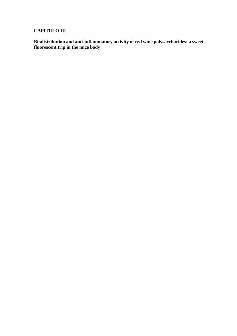
CAPITULO III
Biodistribution and anti-inflammatory activity of red wine polysaccharides: a sweet
fluorescent trip in the mice body

CAPITULO III
Biodistribution and anti-inflammatory activity of red wine polysaccharides: a sweet
fluorescent trip in the mice body

CAPITULO III
Biodistribution and anti-inflammatory activity of red wine polysaccharides: a sweet
fluorescent trip in the mice body

CAPITULO III
Biodistribution and anti-inflammatory activity of red wine polysaccharides: a sweet
fluorescent trip in the mice body

CAPITULO III
Biodistribution and anti-inflammatory activity of red wine polysaccharides: a sweet
fluorescent trip in the mice body

CAPITULO III
Biodistribution and anti-inflammatory activity of red wine polysaccharides: a sweet
fluorescent trip in the mice body

CAPITULO III
Biodistribution and anti-inflammatory activity of red wine polysaccharides: a sweet
fluorescent trip in the mice body

CAPITULO III
Biodistribution and anti-inflammatory activity of red wine polysaccharides: a sweet
fluorescent trip in the mice body

CAPITULO III
Biodistribution and anti-inflammatory activity of red wine polysaccharides: a sweet
fluorescent trip in the mice body

CAPITULO III
Biodistribution and anti-inflammatory activity of red wine polysaccharides: a sweet
fluorescent trip in the mice body

CAPITULO III
Biodistribution and anti-inflammatory activity of red wine polysaccharides: a sweet
fluorescent trip in the mice body

CAPITULO III
Biodistribution and anti-inflammatory activity of red wine polysaccharides: a sweet
fluorescent trip in the mice body

CAPITULO III
Biodistribution and anti-inflammatory activity of red wine polysaccharides: a sweet
fluorescent trip in the mice body

CAPITULO III
Biodistribution and anti-inflammatory activity of red wine polysaccharides: a sweet
fluorescent trip in the mice body

CAPITULO III
Biodistribution and anti-inflammatory activity of red wine polysaccharides: a sweet
fluorescent trip in the mice body

CAPITULO III
Biodistribution and anti-inflammatory activity of red wine polysaccharides: a sweet
fluorescent trip in the mice body

CAPITULO III
Biodistribution and anti-inflammatory activity of red wine polysaccharides: a sweet
fluorescent trip in the mice body

CAPITULO III
Biodistribution and anti-inflammatory activity of red wine polysaccharides: a sweet
fluorescent trip in the mice body

CAPITULO III
Biodistribution and anti-inflammatory activity of red wine polysaccharides: a sweet
fluorescent trip in the mice body

CAPITULO III
Biodistribution and anti-inflammatory activity of red wine polysaccharides: a sweet
fluorescent trip in the mice body

CAPITULO III
Biodistribution and anti-inflammatory activity of red wine polysaccharides: a sweet
fluorescent trip in the mice body

CAPITULO III
Biodistribution and anti-inflammatory activity of red wine polysaccharides: a sweet
fluorescent trip in the mice body

CAPITULO III
Biodistribution and anti-inflammatory activity of red wine polysaccharides: a sweet
fluorescent trip in the mice body

CAPÍTULO IV
Influence of red wine soluble polysaccharides profile on the flavanol composition
and precipitation

CAPÍTULO IV
Influence of red wine soluble polysaccharides profile on the flavanol composition
and precipitation

CAPÍTULO IV
Influence of red wine soluble polysaccharides profile on the flavanol composition
and precipitation

CAPÍTULO IV
Influence of red wine soluble polysaccharides profile on the flavanol composition
and precipitation

CAPÍTULO IV
Influence of red wine soluble polysaccharides profile on the flavanol composition
and precipitation

CAPÍTULO IV
Influence of red wine soluble polysaccharides profile on the flavanol composition
and precipitation

CAPÍTULO IV
Influence of red wine soluble polysaccharides profile on the flavanol composition
and precipitation

CAPÍTULO IV
Influence of red wine soluble polysaccharides profile on the flavanol composition
and precipitation

CAPÍTULO IV
Influence of red wine soluble polysaccharides profile on the flavanol composition
and precipitation

CAPÍTULO IV
Influence of red wine soluble polysaccharides profile on the flavanol composition
and precipitation

CAPÍTULO IV
Influence of red wine soluble polysaccharides profile on the flavanol composition
and precipitation

CAPÍTULO IV
Influence of red wine soluble polysaccharides profile on the flavanol composition
and precipitation

CAPÍTULO IV
Influence of red wine soluble polysaccharides profile on the flavanol composition
and precipitation

CAPÍTULO IV
Influence of red wine soluble polysaccharides profile on the flavanol composition
and precipitation

CAPÍTULO IV
Influence of red wine soluble polysaccharides profile on the flavanol composition
and precipitation

CAPÍTULO IV
Influence of red wine soluble polysaccharides profile on the flavanol composition
and precipitation

CAPÍTULO IV
Influence of red wine soluble polysaccharides profile on the flavanol composition
and precipitation

CAPÍTULO IV
Influence of red wine soluble polysaccharides profile on the flavanol composition
and precipitation

CAPÍTULO IV
Influence of red wine soluble polysaccharides profile on the flavanol composition
and precipitation

CAPÍTULO IV
Influence of red wine soluble polysaccharides profile on the flavanol composition
and precipitation

CAPÍTULO IV
Influence of red wine soluble polysaccharides profile on the flavanol composition
and precipitation

CAPÍTULO IV
Influence of red wine soluble polysaccharides profile on the flavanol composition
and precipitation
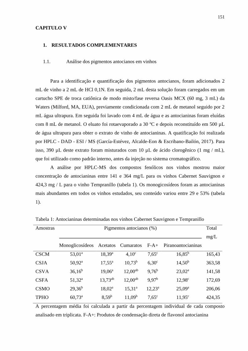
151
CAPITULO V
1. RESULTADOS COMPLEMENTARES
1.1. Análise dos pigmentos antocianos em vinhos
Para a identificação e quantificação dos pigmentos antocianos, foram adicionados 2
mL de vinho a 2 mL de HCl 0,1N. Em seguida, 2 mL desta solução foram carregados em um
cartucho SPE de troca catiônica de modo misto/fase reversa Oasis MCX (60 mg, 3 mL) da
Waters (Milford, MA, EUA), previamente condicionada com 2 mL de metanol seguido por 2
mL água ultrapura. Em seguida foi lavado com 4 mL de água e as antocianinas foram eluídas
com 8 mL de metanol. O eluato foi rotaevaporado a 30 ºC e depois reconstituído em 500 µL
de água ultrapura para obter o extrato de vinho de antocianinas. A quatificação foi realizada
por HPLC - DAD - ESI / MS (García-Estévez, Alcalde-Eon & Escribano-Bailón, 2017). Para
isso, 390 µL deste extrato foram misturados com 10 µL de ácido clorogênico (1 mg / mL),
que foi utilizado como padrão interno, antes da injeção no sistema cromatográfico.
A análise por HPLC-MS dos compostos fenólicos nos vinhos mostrou maior
concentração de antocianinas entre 141 e 364 mg/L para os vinhos Cabernet Sauvignon e
424,3 mg / L para o vinho Tempranillo (tabela 1). Os monogicosídeos foram as antocianinas
mais abundantes em todos os vinhos estudados, seu conteúdo variou entre 29 e 53% (tabela
1).
Tabela 1: Antocianinas determinadas nos vinhos Cabernet Sauvignon e Tempranillo
Amostras Pigmentos antocianos (%)
Total
mg/L
Monoglicosídeos Acetatos Cumaratos F-A+ Piranoantocianinas
CSCM 53,01a 18,39a 4,10c 7,65c 16,85b 165,43
CSJA 50,92a 17,55a 10,73b 6,30c 14,50b 363,58
CSVA 36,16b 19,06a 12,00ab 9,76b 23,02a 141,58
CSFA 51,32a 13,73ab 12,00ab 9,97b 12,98c 172,69
CSMO 29,36b 18,02a 15,31a 12,23a 25,09a 206,06
TPHO 60,73a 8,59b 11,09b 7,65c 11,95c 424,35
A percentagem média foi calculada a partir da percentagem individual de cada composto
analisado em triplicata. F-A+: Produtos de condensação direta de flavonol antocianina
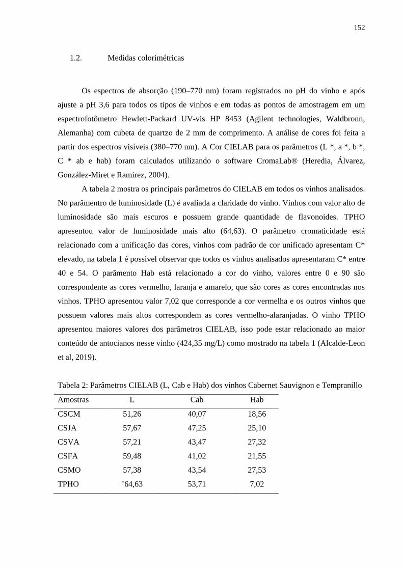
152
1.2. Medidas colorimétricas
Os espectros de absorção (190–770 nm) foram registrados no pH do vinho e após
ajuste a pH 3,6 para todos os tipos de vinhos e em todas as pontos de amostragem em um
espectrofotômetro Hewlett-Packard UV-vis HP 8453 (Agilent technologies, Waldbronn,
Alemanha) com cubeta de quartzo de 2 mm de comprimento. A análise de cores foi feita a
partir dos espectros visíveis (380–770 nm). A Cor CIELAB para os parâmetros (L *, a *, b *,
C * ab e hab) foram calculados utilizando o software CromaLab® (Heredia, Álvarez,
González-Miret e Ramirez, 2004).
A tabela 2 mostra os principais parâmetros do CIELAB em todos os vinhos analisados.
No parâmentro de luminosidade (L) é avaliada a claridade do vinho. Vinhos com valor alto de
luminosidade são mais escuros e possuem grande quantidade de flavonoides. TPHO
apresentou valor de luminosidade mais alto (64,63). O parâmetro cromaticidade está
relacionado com a unificação das cores, vinhos com padrão de cor unificado apresentam C*
elevado, na tabela 1 é possivel observar que todos os vinhos analisados apresentaram C* entre
40 e 54. O parâmento Hab está relacionado a cor do vinho, valores entre 0 e 90 são
correspondente as cores vermelho, laranja e amarelo, que são cores as cores encontradas nos
vinhos. TPHO apresentou valor 7,02 que corresponde a cor vermelha e os outros vinhos que
possuem valores mais altos correspondem as cores vermelho-alaranjadas. O vinho TPHO
apresentou maiores valores dos parâmetros CIELAB, isso pode estar relacionado ao maior
conteúdo de antocianos nesse vinho (424,35 mg/L) como mostrado na tabela 1 (Alcalde-Leon
et al, 2019).
Tabela 2: Parâmetros CIELAB (L, Cab e Hab) dos vinhos Cabernet Sauvignon e Tempranillo
Amostras L Cab Hab
CSCM 51,26 40,07 18,56
CSJA 57,67 47,25 25,10
CSVA 57,21 43,47 27,32
CSFA 59,48 41,02 21,55
CSMO 57,38 43,54 27,53
TPHO ¨64,63 53,71 7,02

153
Referências
Alcalde-Eon, C., Ferreras-Charro, R., Ferrer-Gallego, R., Rivero, R. J., Heredia,
Escribano-Bailón, M. T. Monitoring the effects and side-effects on wine colour and flavonoid
composition of the combined post-fermentative additions of seeds and mannoproteins. Food
Research International, 126. (2019)
García-Estévez, I., Alcalde-Eon, C., Escribano-Bailón, M. T. Flavanol Quantification
of Grapes via Multiple Reaction Monitoring Mass Spectrometry. Application to
Differentiation among Clones of Vitis vinifera L. cv. Rufete Grapes. Journal of Agricultural
and Food Chemistry, 65 (31), 6359-6368. (2017).
Heredia, F. J., Álvarez, C., González-Miret, M. L., & Ramirez, A. ChromaLab
Análisis de Color; Registro General de la Propiedad Intelectual SE-1052-04: Sevilla, Spain.
(2004).

154
CONCLUSÃO GERAL
Após a análise dos resultados obtidos nesta pesquisa, pode-se concluir que:
- Os polissacarídeos encontrados nos vinhos Cabernet Franc, Cabernet Sauvignon e
Sauvignon Blanc foram: arabinogalactana do tipo II, ramnogalaturonana do tipo I e II,
manana e dextrina. O polissacarídeo majoritário presente nos três vinhos é uma
arabinogalactana tipo II. Ela é formada por uma cadeia principal de unidades de β-D-Galp
(1→3)-ligadas, substituída em O-6 por cadeias laterais de β-D-Galp (1→6)-ligadas. As
cadeias laterais são substituídas em O-3 por terminais não redutores de α-L-Araf. A
ramnogalacturonana do tipo I encontrada é formada pela repetição da unidade dissacarídica
[→4)-α-D-GalpA-(1→2)-α-L-Rhap-(1→]. A manana presente nos vinhos estudados é um
polissacarídeo constituído por uma cadeia principal de unidades α-D-Manp (1→6) ligadas,
substituída em O-2 por cadeias laterais curtas de α-D-Manp (1→2) ligadas.
- O estudo realizado com vinhos Cabernet Sauvignon da mesma vinícola em diferentes safras
mostrou que a porcentagem de cada polissacarídeo e também o rendimento total de
polissacarídeos permaneceram praticamente inalterados nas diferentes safras. A porcentagem
semelhante na composição de monossacarídeos, bem como a quantidade de polissacarídeos,
pode sugerir manutenção da qualidade do vinho, indicando que a vinícola mantém o mesmo
padrão no processo de produção de vinho ao longo dos anos.
- O efeito anti-inflamatório das frações polissacarídicas extraídas de vinhos foi avaliada em
células RAW 264.7. As frações reduziram marcadamente a produção de óxido nítrico (NO) e
citocinas pró-inflamatórias (TNF-α e IL-1β) em células estimuladas com LPS. Além disso, as
análises realizadas in vivo em camundongos submetidos a CLP mostraram que a
administração oral e subcutânea dos polissacarídeos apresentaram um aumento na
sobrevivência dos camundongos com sepse, quando comparados com o controle. Levando
também a diminuição dos níveis de citocinas pró-inflamatórias, bem como os níveis séricos
de AST, ALT, bilirrubina, uréia e creatinina, com consequente proteção contra dano tissular.
Além disso, foi observada a migração dos polissacarídeos marcados nos tecidos e órgãos após
administração subcutânea em camundongos com sepse por CLP e a biodistribuição desse
polímero pelo organismo aparentemente acontece através do sistema imunológico. Os
polissacarídeos demonstraram potencial anti-inflamatório in vitro e in vivo, o que pode indicar
efeitos benéficos do consumo moderado de vinho para a saúde humana.
155
- As análises realizadas mostraram que os vinhos Cabernet Sauvignon e Tempranillo têm um
padrão semelhante em sua composição flavânica, embora haja diferenças marcantes entre as
porcentagens de cada família de flavanóis entre os diferentes vinhos. As análises estatísticas
indicaram que uma maior afinidade de certas famílias de polissacarídeos, como mananas,
manoprotreínas, xilanas e dextrinas, em relação aos flavanóis, causando maior precipitação
destes quando eles interagem. Isso pode explicar a relação inversa observada entre a
concentração desses polissacarídeos e a dos flavanóis nos vinhos estudados, o que indica que
uma quantidade maior de determinadas famílias de polissacarídeos em um vinho pode afetar o
conteúdo dos flavanóis, o que teria consequências importantes para propriedades
organolépticas do vinho. Além disso, a análise da composição pigmentos dos vinhos também
mostrou uma variabilidade importante entre os vinhos estudados, o que se traduz em
diferenças importantes nos parâmetros colorimétricos dos vinhos (principalmente clareza e
tom) e, portanto, na cor destes.

156
CONCLUSIONES
Después de analizar los resultados obtenidos en esta investigación, se puede concluir
que:
- Los polisacáridos encontrados em los vino Cabernet Fran, Cabernet Sauvignon y Sauvignon
Blanc fueron: arabinogalactanos de tipo II, ramnogalacturonanos de tipo I y II, manano y
dextrina. La familia de polisacáridos mayoritaria encontrada en los tres vinos fueron los
arabinogalactanos de tipo II, los cuales están formados por una cadena principal de unidades
de β-(1→3)-D-Galp que presenta ramificaciones en las posiciones O-6 formadas por cadenas
laterales de β-(1→6)-D-Galp. A su vez, en las cadenas laterales aparecen sustituciones en las
posiciones O-3 de monosacáridos no reductores de α-L-Araf. Los ramnogalacturonanos de
tipo I encontrados estaban formados por la repetición de la unidad disacarídica [→4)-α-D-
GalpA-(1→2)-α-L-Rhap-(1→]. Por último, los mananos presentes en los vinos estudiados son
polisacáridos constituídos por una cadena principal de unidades α-(1→6)- D-Manp que
presenta ramificaciones en las posiciones O-2 formadas por pequeñas cadenas laterales de α-
(1→2)-D-Manp.
- El estudio realizado con los vinos Cabernet Sauvignon elaborados por la misma bodega en
diferentes vendimias mostró que el porcentaje de cada polisacárido, así como el contenido
total de estos compuestos en el vino, no se ven modificados en las diferentes vendimias. La
estabilidad en la composición de polisacáridos (tanto en su contenido total como en su
composición porcentual), podría indicar una gran regularidad en la calidad del vino,
mostrando así que la bodega mantiene el mismo patrón en el proceso de producción de los
vinos a lo largo de los diferentes años.
- El efecto antinflamatorio de las fracciones de polisacáridos extraídas de los vinos se estudió
en células RAW 264.7. Los polisacáridos ensayados redujeron de forma importante la
producción de óxido nítrico (NO) y citocinas proinflamatorias (TNF-α e IL-1β) en las células
estimuladas con LPS. Además, los análisis realizados in vivo con ratones sometidos a CLP
mostraron que la administración oral y subcutánea de los polisacáridos obtenidos no solo
generaba un aumento en la supervivencia de los ratones con sepsis en comparación con la
población control, sino que también provocaba la disminución de los niveles de citocinas
proinflamatorias, así como los niveles séricos de AST, ALT, bilirrubina, urea y creatinina, lo
que puede asociarse a una protección contra el daño celular. Por otro parte, se pudo observar
157
una migración de los polisacáridos marcados en los tejidos y órganos después de la
administración subcutánea en los ratones con sepsis por CLP. Esta biodistribución de los
polisacáridos por los órganos ocurre, aparentemente, a través del sistema inmunológico. Los
polisacáridos demostraton, por tanto, un potencial anti-inflamatorio in vitro e in vivo, lo que
puede indicar un posible efecto beneficioso del consumo moderado de vino para la salud
humana.
- Los análisis realizados mostraton que los vinos de Cabernet Sauvignon y Tempranillo tienen
un patrón similar en su composición flavánica, aunque existen diferencias marcadas entre los
porcentajes de cada familia de flavanoles entre los diferentes vinos. Los análisis estadísticos
señalaron que una mayor afinidad de ciertas familias de polisacáridos, como mananos,
manoprotreínas, xilanos y dextrinas, hacia los flavanoles, provocando una mayor
precipitación de éstos cuando interaccionan. Esto puede explicar la relación inversa observada
entre la concentración de estos polisacáridos y la de flavanoles en los vinos estudiados, lo que
indica que una mayor cantidad de ciertas familias de polisacáridos en un vino puede afectar al
contenido de flavanoles, lo que tendría consecuencias importantes en las propiedades
organolépticas del vino. Además, el análisis de la composición de pigmentos de los vinos
mostró también una importante variabilidad entre los vinos estudiados, lo que se traduce en
importantes diferencias en los parámetros colorimétricos de los vinos (principalmente claridad
y tono) y, por tanto, en el color de éstos.