ISSN 1991-3036 International Journal of Sustainable Crop Production (IJSCP)...
20
International Journal of Sustainable Crop Production (IJSCP) ( Online Version) Int. J. Sust. Crp. Prodn. 1(1):August 2006 Volume Issue August 200 International Journal of Sustainable Crop Production is an journal published quarterly; August, November, February, and May in English. Original Research papers and scientific notes dealing with Agricultural Science (e.g., Agronomy, Plant Protection, Plant Breeding, Soil Science, Biotechnology, Environmental Science) are published in this Journal. Publisher : GWF Publication House Issuing Body : centre for innovation & development strategy (cids) E-mail : [email protected] URL : http://www.cidsbd.org/IJSCP.html/ IPT : 880-09666-910-396 Phone : 880-153-43-103-72 ISSN 1991-3036
Transcript of ISSN 1991-3036 International Journal of Sustainable Crop Production (IJSCP)...

International Journal of Sustainable Crop Production (IJSCP)( Online Version)
Int. J. Sust. Crp. Prodn. 1(1):August 2006
Volume 2 Issue 4 (FT) August 2007
International Journal of Sustainable Crop Production is an journal published quarterly;August, November, February, and May in English. Original Research papers andscientific notes dealing with Agricultural Science (e.g., Agronomy, Plant Protection,Plant Breeding, Soil Science, Biotechnology, Environmental Science) are published inthis Journal.
Publisher : GWF Publication House
Issuing Body : centre for innovation & development strategy (cids)
E-mail : [email protected] : http://www.cidsbd.org/IJSCP.html/IPT : 880-09666-910-396Phone : 880-153-43-103-72
ISSN 1991-3036

ISSN 1991-3036 (Online Version)
List of papers: V2 I4 September (Fast Track)-2007
1. KHALAFALLA, M. M., ABDELLATEF, E., MOHAMEED AHMED, M. M. AND OSMAN, M. G. 2007. MICROPROPAGATION OF CACTUS (OPUNTIA FICUS-INDICA) AS STRATEGIC TOOL TO COMBAT DESERTIFICATION IN ARID AND SEMI ARID REGIONS. INT. J. SUSTAIN. CROP PROD. 2(4):1-8.
2. ISLAM, M. N., HOSSAIN, M. A. AND AHSAN, M. N. 2007. DEVELOPMENT OF A PCR BASED PROTOCOL FOR WSSV SCREENING FOR MAJOR CRUSTACEANS INHABITING IN CULTURED SHRIMP FARM. INT. J. SUSTAIN. CROP PROD. 2(4): 9-17.

ISSN 1991-3036 (Online Version)
International Journal of Sustainable Crop Production (IJSCP)
Editorial Board
Editor in chief : Dr. Atrolitae Akandh
Executive Editor : Mr. Abrar Ahnaf Kabir
Technical Editor : MS. Sharmeen M. Khan
Editorial Board Members : Mr. Akandh S. Muhammed
: Mr. Saidur Rahman Khan
: MS. Jarin Subah Kabir
: Mr. Muhammed A. Zaman
: Mr. M. Mutaharul Kabir
: Mr. M. Sahinul Kabir

Int. J. Sustain. Crop Prod. 2(4) (September 2007) 1
MICROPROPAGATION OF CACTUS (Opuntia ficus-indica) AS STRATEGIC TOOL TO COMBAT DESERTIFICATION IN ARID AND SEMI ARID REGIONS
M. M. KHALAFALLA, E. ABDELLATEF, M. M. MOHAMEED AHMED AND M. G. OSMAN
Commission for Biotechnology and Genetic Engineering, National Centre for Research, P.O. Box 2404, Khartoum, Sudan,
Accepted for publication: September 05, 2007
ABSTRACT Khalafalla, M. M., Abdellatef, E., Mohameed Ahmed, M. M. and Osman, M. G. 2007. Micropropagation of Cactus (Opuntia ficus-indica) as Strategic Tool to Combat Desertification in Arid and Semi Arid Regions. Int. J. Sustain. Crop Prod. 2(4):1-8
With aim of large production of plant material, a protocol for micropropagation of Opuntia ficus-indica was developed at the laboratory of plant tissue culture, Commission for Biotechnology and Genetic Engineering, Khartoum, Sudan, during the period of August 2006 to July 2007. Young cladode explant containing one areole was cultured on Murashige and Skoog medium (MS) supplemented with benzyladenine (BA) and kinetin (Kin) alone or in combination with naphthalene acetic acid (NAA). The highest shoot multiplication (26.5±1.74 shoots per explant) was achieved in 90 days on MS supplemented with BA (5.0 mg/l). Excised shoot cuttings of 3.0 cm were placed on the MS basal medium hormone-free or supplemented with different levels (0.25-1.0 mg/l) of indole-3-acetic acid (IAA), indole-3-butyric acid (IBA) and NAA. Although, satisfactory rooting percentage (100%) was achieved on both MS free-hormone or supplemented with IAA at 0.5 mg/l but, the highest number (15.0 ± 1.1) of root per shoot was obtained on MS medium supplemented with 0.5 mg/l IAA. Plants were successfully established in soil and adapted to greenhouse conditions. The protocols developed in this work provide a basis to achieve massive propagation of O. ficus-indica by in vitro culture of areoles as strategic tool to combat desertification in arid and semi arid regions.
Key words: Micropropagation, cactus, desertification, acclimatization.
INTRODUCTION The Cactaceae family includes approximately 130 genera and 1500 species. Of these, the Opuntia and Nopalea genera are the most important due to their usefulness to man (Flores-Valdez and Osorio, 1996). Opuntia has a specialized photosynthetic mechanism known as Crassulacean Acid Metabolism (CAM), whereby these plants open their stomates and take up CO2 at night, when temperatures are lower and humidity higher than during the daytime. This invariably results in reduced water loss (Nobel, 1995) offers exceptional possibilities for large quantities of biomass in water-limited areas that are useful for livestock feed (Felker et al. 2006).
Within the genus Opuntia, Opuntia ficus-indica is the most agronomically important species for the production of edible fruits and cladodes, which can be used as a vegetable and valuable forage resource in arid and semiarid lands during periods of drought and shortage of herbaceous plants (Scheinvar, 1995; Le Houérou, 2000; Juárez and Passera, 2002).
In Sudan, the arid and semiarid regions occupy around two-thirds of the total area (El Gamri, 2004); there fore the search for appropriate plant species to grow in this area is a permanent concern of most people living in harsh environments. O. ficus-indica represents the best choice since it shows great adaptability to various soil conditions, prevent environmental destruction caused by erosion. Moreover, farmers in many arid areas of the world use opuntia extensively as emergency forage that is harvested from both wild and cultivated populations to prevent the disastrous consequences of frequent and severe droughts (Le Houérou, 1992; Mirza, 2000; Medeiros et al. 2006). Opuntia has become an important crop for exotic fruit, vegetable, and forage production in Mexico, USA, Chile, Argentina, Israel, Italy, and South Africa, where it has adapted to dry areas with droughty conditions, scarce rainfall and poor, erosion prone soils (Pimienta-Barrios and Muñoz-Urias, 1995; Flores-Valdez, 1994).
Generally CAM plants are slow growing plants that sometimes have limited reproductive capacities and often have very specific and limited conditions for seed germination, flowering and seed production. Although, conventional propagation has been attempted for Opuntia, genetic segregation and slow growth and development represent serious practical problems (Malda et al. 1999). Moreover, cacti seeds are frequently difficult to be obtained (Mauseth, 1977) and plantlets are reported to be susceptible to damping-off (Mauseth, 1979; Ault and Blackmon, 1987). Therefore in vitro propagation by tissue culture is a feasible alternative option for the rapid multiplication and maintenance of germplasm (Smith et al. 1991; Johnson and Emino, 1979).Tissue culture has already proved to be successful for several members of the Cactaceae family (Johnson and Emino, 1979; Mauseth, 1979; Escobar et al. 1986; Clayton et al. 1990; Hubstenberger et al. 1992). However, for O. ficus-indica micropropagation only the effect of BA was tested (Garcıa- Saucedo., et al. 2005).
The main goal of this study was to achieve massive propagation of O. ficus-indica by in vitro culture of areoles, and to acclimatize propagated material to field conditions. The specific goals were to a) elaborate successful protocols
Int. J. Sustain. Crop Prod. 2(4):1-8 (September 2007)
© 2006 Green World Foundation (GWF)

Int. J. Sustain. Crop Prod. 2(4) (September 2007) 2
for cladode sterilization; b) find the optimal growth regulator and its concentration able to induce shooting from areoles of sterilized cladodes; c) determine optimum auxin concentrations able to induce rooting.
MATERIALS AND METHODS Plant material Young plants of O. ficus-indica were obtained from the cactus plantations of the Horticultural Department, Federal Ministry of Agriculture, Khartoum, Sudan. Plants were kept under green house conditions and used as a source for explants throughout the experiment.
Surface sterilization and isolation of explants Young cactus cladodes were cut into pieces and surface sterilized by washing under running tap water and laundry bleach for 20 min. The pieces of the cladodes were sprayed with 70% ethanol, cleaned with a clean towel and transferred to a laminar flow. Under a laminar flow the cut pieces were immersed in 15% sodium hypochlorite for 20 min, and then rinsed in sterile distilled water for three times. Disinfested cladodes were cut to 1.0 cm2 pieces each containing one areole and transferred into Petri dishes as explant ready for inoculation.
Inoculation Explants were cultured in culture bottles containing MS (Murashige and Skoog, 1962) basal media supplemented with benzyladenine (BA) (0.5, 1.0, 1.5, 3.0 and 5.0 mg L-1) or Kinetin (Kin) (0.5, 1.0, 1.5, 3.0 and 5.0 mg L-1) alone or in combination with naphthaleneacetic acid (NAA) (0.5 mg L-1). All media were prepared by standard procedures and the culture bottles were transferred to growth chamber with a photoperiod of 16 h light and 8 h of dark at 25ºC ± 2 ºC and incubated for 3 months.
Rooting of in vitro induced shoots Shoots (3.0 cm) derived from shoot bunches were excised and rooted on medium consisting of MS basal medium supplemented with NAA, Indole Acetic Acid (IAA) or Indole Butric Acid (IBA) each at 0.25, 0.5 or 1.0 mg/l. All the media used in this study were supplemented with 3% (w/v) sucrose, solidified with 0.8% (w/v) agar and the pH was adjusted to 5.8± 0.1 before autoclaving at 121°C and 15 lb psi for 15 min.
Acclimatization In vitro rooted plants were removed from rooting medium and washed to remove adhering gel and transplanted to plastic pots containing autoclaved garden soil and sand at 3:1 ratio. Plants were kept under culture room conditions for 15 days then transferred to green house and placed under shade until growth was observed.
Statistical analysis Results on the number of buds and shoot per explants, rooting percentage and the number of roots developed per shoot in each treatment was observed at regular intervals. Data were collected from three independent experiments and subjected to analysis of variance. Means were compared with Duncan’s Multiple Range Test (Duncan, 1955) and presented as average ± standard error (SE).
RESULTS AND DISCUSSION The surface sterilization of cacti is extremely important because the thorns and hairs normally found in such plants host a large variety of microorganisms (Garcıa- Saucedo., et al. 2005) Here in this study, washing the cladodes of the O. ficus-indica under running tap water and laundry bleach followed by the standard method of ethanol/ sodium hypochlorite was insufficient for the in vitro establishment of this species. The surface sterilization was only efficient when the pieces of the cladodes after washing under running tap water and laundry bleach for 20 min were sprayed with 70% ethanol, cleaned with a clean towel and transferred to a laminar flow and immersed in 15% sodium hypochlorite for 20 min then rinsed in sterile distilled water for three times
The morphogenetic responses of cladodes explants (Figure 1A) to BA and Kin either alone or in combination with NAA are summarized in Table 1. Explants cultured on MS medium without growth regulator did not show any response. However on MS medium supplemented with growth regulators a 100% of all areole swell in their size and a varying degree of shoot bud differentiation was observed under a 16-h photoperiod after one months of culture. This is consistent with results of Juarezi and Passera, 2002, who found that 100% areole shooting, was obtained on solid medium containing BA, under a 16-h photoperiod on the 35th day of culture.
M. M. Khalafalla et al

Int. J. Sustain. Crop Prod. 2(4) (September 2007) 3
Table 1. Effects of benzyladenine (BA) and kinetin (Kin) alone or in combination with 1-naphtalenacetic acid (NAA) on different morphogenetic responses of Opuntia ficus-indica on MS medium after3 months of culture Growth regulator (mg/l) Number of shoot per explant
(Mean± SE) Number of bud per explant
(Mean± SE) Bud forming explant (%) NAA Kin BA
3.0 ± 0.26 cd 22.8 ± 2.4 j 100 0.0 0.0 0.5 4.5 ± 0.51 cd 31.8 ± 2.9 hi 100 0.0 0.0 1.0 5.3 ± 0.43 cd 53.0 ± 5.7 de 100 0.0 0.0 1.5 7.0 ± 0.53 cd 52.5 ± 8.6 de 100 0.0 0.0 3.0 26.5 ± 1.74 a 31.8 ± 2.0 hi 100 0.0 0.0 5.0 5.5 ± 0.44 cd 72.5 ± 6.6 c 100 0.0 0.5 0.0 7.3 ± 0.50 cd 49.8 ± 9.3 e 100 0.0 1.0 0.0 8.0 ± 0.53 c 40.5 ± 1.7 fg 100 0.0 1.5 0.0 3.5 ± 0.23 cd 33.5 ± 4.1 ghi 100 0.0 3.0 0.0 2.7 ± 0.34 d 30.0 ± 1.8 i 100 0.0 5.0 0.0 4.0 ± 0.37 cd 31.5 ± 3.9 hi 100 0.5 0.0 0.5 4.5 ± 0.03 cd 30.5 ± 2.7i 100 0.5 0.0 1.0
10.2 ± 0.72 bc 46.3 ± 3.4 ef 100 0.5 0.0 3.0 17.8 ± 2.07 b 58.7 ± 5.2 d 100 0.5 0.0 5.0 7.0 ± 0.70 cd 47.5 ± 10.9 ef 100 0.5 0.5 0.0 6.5 ± 0.78 cd 39.8 ± 5.8 fgh 100 0.5 1.0 0.0 3.8 ± 0.49 cd 93.3 ± 15.6 b 100 0.5 3.0 0.0 2.0 ± 0.26 d 104.7 ± 12.6 a 100 0.5 5.0 0.0
Means with same letter (s) in the same column are not significantly different at 5% using Duncan’s multiple range test
All the concentrations of BA and Kin alone or in combination with NAA facilitate bud and shoot differentiation, but after three months of culture BA alone being the most efficient in terms of number of shoot per explant. Shoots number increases with the increase of BA concentration. BA at the highest concentration (5.0 mg/l) gave the highest number of shoot per explant (26.5±1.74) (Figure 1B), thus being the most efficient growth regulator tested for the optimal multiplication of this plant material. Generally cytokinin is considered to be essential for the development of cactus axillary shoots (Mauseth, 1977). Mohamed-Yassen (1995) reported that, increasing the BA concentration to 8.8 µM resulted in significantly more shoots being produced. Moreover, the effectiveness of BA in shoot differentiation has been documented in number of cactus plants such as Opuntia amyclea (Escobar et al.1986).
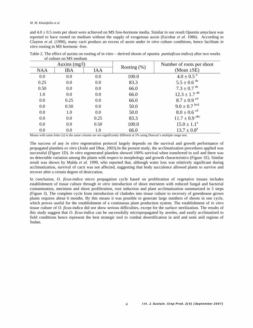
The synergistic influence of NAA with cytokinins (BA and Kin) did not improve the number of shoots per explant. However, Kin in combination with NAA markedly enhanced the number of buds per explant. Kin at 5.0 mg/l in combination with 0.5 mg/l NAA induced the maximum number of buds per explant (104.7 ± 12.6).However, the induced buds failed to elongate on the same medium resulting in rosettes of shoots compared to those obtained on media containing BA a lone (Figure 2). These findings are in agreement with those reported by Bustamante and Heras (1990) on Cacti (Pelecyphora aselliformis) and Nealolydia lophophoroides; Feng-Feng et al. (2000) on Aloe barbebsis and Mata-Rosas (2001) on Turbinicapus laui. They concluded that applying a combination of BA and NAA in different concentrations was a limiting factor for shoot formation. Moreover, Clayton et al., 1990 reported that low levels of auxin with cytokinin increased axillary bud production in some cactus species. Previous studies have pointed out that the optimal hormone combination may be unique for each cactus species (Johnson and Emino, 1977, 1979).
Elongated and well-developed (3.0 cm) shoots derived from cladodes explants (grown on MS medium supplemented with 5.0 mg/L BA) were excised from the shoot clumps and transferred to MS medium hormone-free or augmented with 0.25, 0.5 or 1.0 mg/l of either IAA, NAA or IBA for root initiation. Of the three auxins tested, IAA at 0.5 induced the maximum rooting percentage (100%) and the highest number (15.0 ± 1.1) of roots per rooted plantlet (Table 2) (Figure 1c) after two weeks of culture. The use of MS medium for rooting of in vitro induced shoots has already been reported for O. ficus indica (Garcıa- Saucedo et al. 2005). Here in this study 100% rooting percentage
Micropropagation of Cactus (Opuntia ficus-indica) as Strategic Tool to Combat Desertification in Arid and Semi Arid Regions
Int. J. Sustain. Crop Prod. 2(4) (September 2007) 4
and 4.0 ± 0.5 roots per shoot were achieved on MS free-hormone media. Similar to our result Opuntia amyclaea was reported to have rooted on medium without the supply of exogenous auxin (Escobar et al. 1986). According to Clayton et al. (1990), many cacti produce an excess of auxin under in vitro culture conditions, hence facilitate in vitro rooting in MS hormone -free.
Table 2. The effect of auxins on rooting of in vitro – derived shoots of opuntia puntiaficus-indica) after two weeks of culture on MS medium
Auxins (mg/l) NAA IBA IAA Rooting (%) Number of roots per shoot
(Mean ±SE) 0.0 0.0 0.0 100.0 4.0 ± 0.5 e
0.25 0.0 0.0 83.3 5.5 ± 0.6 de 0.50 0.0 0.0 66.0 7.3 ± 0.7 de 1.0 0.0 0.0 66.0 12.3 ± 1.7 ab 0.0 0.25 0.0 66.0 8.7 ± 0.9 cd 0.0 0.50 0.0 50.0 9.0 ± 0.7 bcd 0.0 1.0 0.0 50.0 8.0 ± 0.6 cd 0.0 0.0 0.25 83.3 11.7 ± 0.9 abc 0.0 0.0 0.50 100.0 15.0 ± 1.1a
0.0 0.0 1.0 66.0 13.7 ± 0.8a Means with same letter (s) in the same column are not significantly different at 5% using Duncan’s multiple range test.
The success of any in vitro regeneration protocol largely depends on the survival and growth performance of propagated plantlets ex vitro (Joshi and Dhar, 2003).In the present study, the acclimatization procedures applied was successful (Figure 1D). In vitro regenerated plantlets showed 100% survival when transferred to soil and there was no detectable variation among the plants with respect to morphology and growth characteristics (Figure 1E). Similar result was shown by Malda et al. 1999, who reported that, although water loss was relatively significant during acclimatization, survival of cacti was not affected, suggesting that body succulence allowed plants to survive and recover after a certain degree of desiccation.
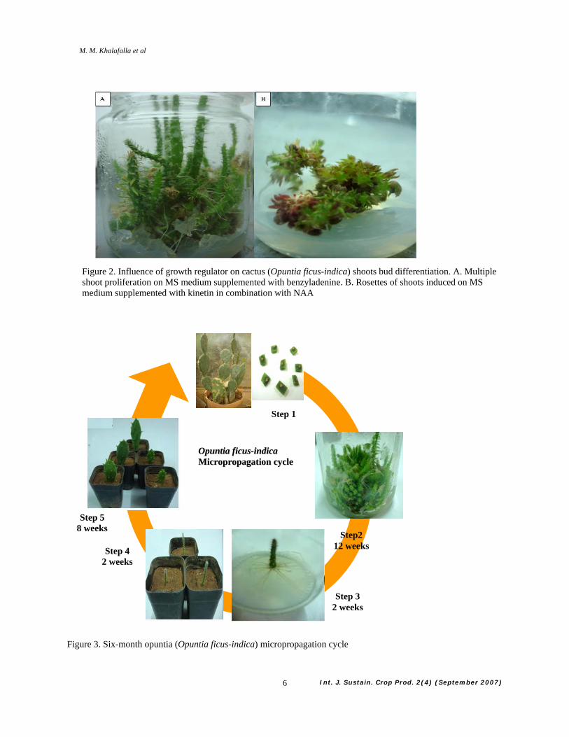
In conclusion, O. ficus-indica micro propagation cycle based on proliferation of vegetative tissues includes establishment of tissue culture through in vitro introduction of shoot meristem with reduced fungal and bacterial contamination, meristem and shoot proliferation, root induction and plant acclimatization summarized in 5 steps (Figure 3). The complete cycle from introduction of cladodes into tissue culture to recovery of greenhouse grown plants requires about 6 months. By this means it was possible to generate large numbers of shoots in one cycle, which proves useful for the establishment of a continuous plant production system. The establishment of in vitro tissue culture of O. ficus-indica did not show serious difficulties, except for the surface sterilization. The results of this study suggest that O. ficus-indica can be successfully micropropagated by areoles, and easily acclimatized to field conditions hence represent the best strategic tool to combat desertification in arid and semi arid regions of Sudan.
M. M. Khalafalla et al

Int. J. Sustain. Crop Prod. 2(4) (September 2007) 5
Figure 1. In vitro micropropagation system for cactus (Opuntia ficus-indica) (A-E). A. Cladode explants containing one areole. B, Multiple shoot formation on MS medium supplemented with BA (5.0 mg/l). C, Root induction on MS supplemented with IAA at 0.5 mg/l. D, Plant acclimatization in growth chamber. E, Recovered plant in greenhouse
BA
C D E
Micropropagation of Cactus (Opuntia ficus-indica) as Strategic Tool to Combat Desertification in Arid and Semi Arid Regions
Int. J. Sustain. Crop Prod. 2(4) (September 2007) 6
Figure 3. Six-month opuntia (Opuntia ficus-indica) micropropagation cycle
OOppuunnttiiaa ffiiccuuss--iinnddiiccaa MMiiccrroopprrooppaaggaattiioonn ccyyccllee
Step 4 2 weeks
Step 5 8 weeks
Step2 12 weeks
Step 3 2 weeks
Step 1
Figure 2. Influence of growth regulator on cactus (Opuntia ficus-indica) shoots bud differentiation. A. Multiple shoot proliferation on MS medium supplemented with benzyladenine. B. Rosettes of shoots induced on MS medium supplemented with kinetin in combination with NAA
M. M. Khalafalla et al

Int. J. Sustain. Crop Prod. 2(4) (September 2007) 7
Acknowledgements
This work was supported by Commission for Biotechnology and Genetic Engineering, (CBGE), National Centre for Research (NCR), Khartoum, Sudan.
REFERENCES
Ault, J.R., and Blackmon, W.J. 1987. In vitro propagation of Ferocactus acanthodes (Cactaceae). HortScience, 22 (1):126-127
Bustamante, M.A., and Heras, M.G. 1990. Tissue culture of Cacti species. XXIIIrd
International Horticultural Congress, Firenze (Italy) Aug-27-sept-1, 1990 contributed papers (oral) No 1344 page 163
Clayton, P.W., Hubstenberger, J.F., Phillips, G.C. and Butler-Nance, S. 1990. Micropropagation of members of the Cactaceae subtribe Cactinae. J. Am. Soc. Horticult. Sci, 115: 337–343
Duncan, D. B. 1955. Multiple range and multiple F test. Biometrics, 11: 1- 42
El gamri, T. 2004. Prospects and Constraints of Desert Agriculture: Lessons from West Omdurman. Environmental Monitoring and Assessment 99: 57–73
Escobar, H.A., Villalobos, V.M., and Villegas, A. 1986. Opuntia micropropagation by axillary proliferation. Plant Cell Tissue Org. Cult, 7: 269–277
Felker, P., Paterson, A., and Jenderek, M. M. 2006. Forage Potential of Opuntia Clone Maintained by the USDA, National Plant Germplasm System (NPGS) Collection. Crop Sci, 46:2161–2168
Feng-Feng., Li-HongBo., Lu-QingFang., Xie-JianYing., Fen, F., Li, H.B., Lu, Q.F., and Xie, J.Y. 2000. Tissue culture of Aloe spp. Journal of Southwest Agricultural University, 22(2): 157-159
Flores-Valdez, C. A. 1994. "Nopalitos" production, processing and marketing. In: Barbera et al., (ed.). "Agro-ecology cultivation and uses of cactus pear ".FAO International Technical Cooperation Network on Cactus Pear. pp. 92-99
Flores-Valdez, C. A., and Osorio G. A. 1996 Opuntia-based Ruminant Feeding Systems in Mexico. FAO International Technical Cooperation Network on Cactus Pear, found in http://www.fao.org/AG/againfo/resources/documents/frg/conf96htm/flores.htm)
Garcıa-Saucedo, Pedro A., Maribel, Valdez-Morales., Maria, Elena Valverde., Andre´s, Cruz-Hernandez., and Octavio, Paredes-Lopez. 2005. Regeneration of three Opuntia genotypes used as human food. Plant Cell, Tissue and Organ Culture, 80:215–219
.Hubstenberger. J.F., Clayton, P.W., and Phillips, G.C. 1992. Micropropagation of cacti (Cactaceae). In: Biotechnology in Agriculture and Forestry. High tech and micropropagation IV. Vol. 20. Ed: Bajaj, Y. P. S. Springer-Verlag, Berlin International Horticultural Congress (pp 49– 68)
Johnson, J.L., and Emino, E.R. 1977. Propagation of selected cactus species by tissue culture. Hort Science, 12(4): 404
Johnson, J.L., Emino, E.R. 1979. In vitro propagation of Mammillaria elongata. Hort Science, 14(5): 605-606.
Joshi, M., and Dhar, U. 2003. In vitro propagation of Saussurea obvallata (DC.) Edgew an endangered ethnoreligious medicinal herb of Himalaya. Plant Cell Rep, 21: 933-939
Juarezi, M.C., and Passera, C.B. 2002. In vitro propagation of Opuntia ellisiana Griff. and acclimatization to field conditions. Biocell, 26: 319–324
Le Houerou, H.N. 1992. The role of salt bushes (Atriplex spp.) in arid land rehabilitation in the Mediterranean Basin. A review Agroforestry System, 18: 107-148
Le Houerou, H.N. 2000. Utilization of fodder trees and shrubs in the arid and semiarid zones of west Asia and north Africa. Arid soil Res. and Rehabilitation, 14: 101-135
Micropropagation of Cactus (Opuntia ficus-indica) as Strategic Tool to Combat Desertification in Arid and Semi Arid Regions

Int. J. Sustain. Crop Prod. 2(4) (September 2007) 8
Malda, G., Suza´n, H., and Backhaus, R. 1999. In vitro culture as a potential method for the conservation of endangered plants possessing crassulacean acid metabolism. Sci.Hortic. Amsterdam, 81: 71–87
Mata-Rosas, M., Monroy-de-la-Rosa, M.A., Goldammer, K.M., Chavez-Avilla, V.M., and Monroy-de-la-Rosa, M.A. 2001. Micropropagation of Tubinicapus laui glass et Foster, an endemic and endangered species. In vitro Cellular and Developmental Biology Plant, 37(3): 400-404
Mauseth, J.D. 1977. Cactus tissue culture: a potencial method of propagation. Cact. Succ.J, (US) 49: 80–81
Mauseth, J.D. 1979. A new method for the propagation of cacti: sterile culture of axillary buds. Cact. Succ. J, (US) 51: 186–187
Medeiros, L. A., de Ribeiro, R. C. S, Gallo, L. A, A., Shideed, K., Awawedha Enio de Oliveira E. T, Dematte, M. E. S. 2006. In vitro propagation of Notocactus magnificus. Plant Cell, Tissue and Organ Culture, 84: 165–169
Mirza, S.N. 2000. Fodder shrubs and trees in Pakistan. In: Gintzburger G., M. Bounejmate and A. Nefzaoui (eds.). Fodder Shrub Development in Arid and Semi-arid Zones. Proceedings of the Workshop on Native and Exotic Fodder Shrubs in Arid and Semi-arid Zones, 27 October-2 November 1996, Hammamet, Tunisia. ICARDA, Aleppo (Syria). Vol. I: 153-177
Mohamed-Yassen, Y., Barringer, S.A., Splittstoesser, W.E., and Schnell, R. 1995. Rapid propagation of tuna (Opuntia ficusindica) and plant establishment in soil. Plant Cell Tiss. Org. Cult, 42: 117–119
Murashige, T., and Skoog, F. 1962. A revised medium for rapid growth and bioassays with tobacco tissue cultures. Physiol. Plant, 15: 473-497
Nobel, J.L. 1995. Changes in water potential and its component during shoot formation in cacti callus. Physiol. Plant, 45: 92-97
Pimienta-Barrios, E., and Munoz-Urias, A. 1995.Domestication of Opuntia and cultivated varieties.In Barbera, G., P. Inglese, and E. Pimienta-Barrios (eds.).Agro-ecology, cultivation and uses of cactus pear. FAO International Technical Cooperation Network on Cactus Pear. pp. 58-63
Scheinvar, L. 1995. Taxonomy of utilized Opuntias. In: Agroecology, cultivation and uses of cactus pear. Barbera, G., P. Inglese, and E. Pimienta-Barrios (eds). FAO Plant Production and protection paper 132. Rome, Italy 20-27
Smith, R.H., Burdick, J.P., Anthony, J., and Reilley, A.A. 1991. In vitro propagation of Coryphantha macromeris. Hort Science, 26(3): 315
M. M. Khalafalla et al

Int. J. Sustain. Crop Prod. 2(4) (September 2007)
9
DEVELOPMENT OF A PCR BASED PROTOCOL FOR WSSV SCREENING FOR MAJOR CRUSTACEANS INHABITING IN CULTURED SHRIMP FARM
M. N. ISLAM1, M. A. HOSSAIN2 AND M. N. AHSAN1
1Fisheries and Marine Resource Technology Discipline, Life Science School, Khulna University, Khulna 9208, Bangladesh, 2Assistant Director, Bangladesh Betar, Commercial Service, Shahabagh, Dhaka.
Accepted for publication: September 07, 2007 ABSTRACT
Islam, M. N., Hossain, M. A. and Ahsan, M. N. 2007. Development of a PCR Based Protocol for WSSV Screening for Major Crustaceans Inhabiting in Cultured Shrimp Farm. Int. J. Sustain. Crop Prod. 2(4): 9-17
The array of prevalence of white spot syndrome virus (WSSV) in cultured tiger shrimp, Bagda (Penaeus monodon) and non cultured Metapenaeus monoceros & crab, Scylla serrata from the coastal region of Bangladesh was determined by a two-step polymerase chain reactions (PCR)-based screening protocol using two set of oligonucleotide primers. The product of the first-step PCR of total genomic DNA isolated by a single-step DNA preparation without involving hazardous organic solvents from the muscle and hepatopancreas of sample from apparently diseased free pond and diseased pond served as the templates for the second-step PCR. The ability of identify a single piece of target DNA with high specificity, easiness, and cost-effectiveness of this method do not depend on using a patented WSSV detection kits as in elsewhere in the country. Up to date this is the first scientific report on applying PCR-based WSSV screening protocol in a diversity of species in Bangladesh. The ground-breaking works in Bangladesh and the results suggest that a PCR screening of WSSV infection for shrimp brood and larval stocks and other potential virus carriers could be an effective tool in battle against the fatal virus that has almost devastated the shrimp commerce of the country.
Key words: White spot, virus, DNA, primer
INTRODUCTION Outstanding knowledge on the wide spreading of WSSV (white spot syndrome virus) over a farm, broadly over an area are the initiative against this destructive virus that could restrict a huge shrimp production loss (Hossain, 2001) finally could retain the overall economic sustainability of a farm. A recent major outbreak of WSSV infection in China, Japan, Taiwan, Bangladesh, Thailand, India (Otta et al., 2003; Cen, 1998; Zhan and Wang, 1998; Pongmaneerat et al., 2001; ASCC, 1996), resulted in a high reduction in shrimp production, has raised major concerns in aquaculture around the world. WSSV is a member of a new virus family (Van et al., 2001) with high virulence (Yoganandhan et al., 2003) first discovered in Taiwan in 1992 (Chen 1995), its vituperative nature contains a wide spreading host range as crayfish , Penaeus monodon, Macrobrachium rosenbergii, Metapenaeus monoceros, crabs (eg., Scylla serrata), copepods, lobsters etc (Lo et al., 1996a; Supamattaya et al.,1998; Cai et al., 1995; Takahashi et al., 1994; Peng et al., 1998; Wongteerasupaya et al., 1995); multi transportation mode as both vertical (Lo et al. 1999) and horizontal (ASCC, 1996) transportation since it can transmit through brood to offspring, plankton to shrimp, food prepared from infected animals to cultured shrimp.
Due to the lack of methods to cure its infection; no alternative(s) if infect once, emphasize should be intense on its prevention by lay outing its presence and the level of infection (Hossain, 2001). WSSV takes some time to express itself but once after its expression, infected animal die within three to eight days resulting high mortality (Corbell et al., 2001). Expression of WSSV infection furnished with some characters as white to reddish to discoloration(Corbell et al., 2001; Chou et al., 1995) over the head and carapace, low appetite, gather near the embankment etc.; those are not distinct from some other viral infection as the infection of bacterial white spot syndrome (Wang et al., 2002). WSSV is a completely known virus with regard to its proteomic analysis (Canhua et al., 2002), genomic studies (Yang et al., 2001) the morphological characteristics ( Chou et al., 1995), the general biological properties (Wongteerasupaya et al., 1995; Lo et al., 1996b) and developed diagnosis method. Though it is a recognized virus in sense of its known host range, molecular knowledge and diagnosis method, its virulent nature and unexpected effect leads to early dead overshadow the mentioned success. Hence require a rapid and sensitive diagnosis protocol that will be able to screen the virus in its various hosts at every life stage. There are various diagnosis developed by Durand et al.,1996; Takahashi et al., 1994 ; Kim et al., 1998; et al., 1997; Corbell et al., 2001; Tapay et al., 1999 among them PCR is the most rapid, sensitive (Lightner and Redman, 1998; Lo et al., 1996c), specific (Tan et al., 2001 ) and reliable method (Tang and Lightner, 2000).
The present experiment was conducted using PCR protocol accompanished with a simple procedure consists of isolation of DNA, PCR preparation then subjected to polymerase chain reaction in a thermal cycler followed by 35 cycles. The competitor and the target DNA in a polymerase chain reaction are similar in size and share the same primer recognition sites, thus ensuring similar amplification efficiency for both (Gilliland et al., 1990). Thus PCR procedure can quantify very low amounts of nucleic acid, more accurately it can identify a single piece of target DNA if present in the sample.
© 2006 Green World Foundation (GWF)
Int. J. Sustain. Crop Prod. 2(4):9-17 (September 2007)

Int. J. Sustain. Crop Prod. 2(4) (September 2007)
10
The present study was therefore carried out to optimise the screening protocol in a wide host species spectrum (P. monodon, Metapenaeus monoceros and Scylla serrata) in Bangladesh context with regard to on-farm preparation of their tissues for rapid extraction of DNA, optimal thermal cycling condition and ingredient compositions for PCR. The simplicity and cost-effectiveness of this protocol rely on using a single-step DNA preparation without involving hazardous organic solvents as well as two pairs of oligonucleotide primers highly specific for WSSV gene. PCR protocol used in this experiment shows an admirable result as virus was detected both in nested and non-nested reaction and in all sample in a short period of time illustrating efficiency to investigate into a wide host range that could be pioneer for the farmer.
MATERIALS AND METHODS Sample Adult tiger shrimp, bagda (Penaeus monodon), Metapenaeus monoceros and Scylla serrata from diseased (animals containing external disease symptoms and being death for some reasons) pond and apparently disease free pond (having no external disease symptoms) were collected from Shamnagor under Shatkhira district, a remote area of coastal region of Bangladesh devoted to viral diseases endlessly. Moreover, animals of diseased pond investigated as infected and uninfected sample. About 100 mg each of muscle and hepatopancreas of selected animals was dissected out by sterilized, separate surgical blades to avoid cross contamination. The 18 dissected tissue samples were kept in 18 correspondingly labelled 1.5-ml tubes containing 500 µl of DNAzol, a guanidinium isothiocyanate (Cox, 1968) based buffer (Invitrogen, Life Technologies, USA) that enable a single step DNA extraction and transported in an ice bag to the laboratory for the extraction of total genomic DNA.
Extraction of DNA Collected tissue samples flooded in DNAzol were homogenised by separate sterilised tissue grinder and centrifuged at 13,000 rpm for 15 min with a table top centrifuge machine (Eppendorf; model: 5415D). Four hundred micro litres of supernatant was transferred to fresh and new 1.5-ml tubes containing equal volumes of cold 100% ethanol. The two solutions were mixed by inverting the capped tubes for a few times and kept for 10 min to allow the precipitation of DNA. The mixture was then subjected to centrifugation at 13,000 rpm for 30 min. The precipitated pellet of DNA was obtained by removing supernatant alcohol. The DNA pellet thus obtained was washed with 500µl of 70% ethanol, kept for five minutes and centrifuged at 13000 rpm for another five minutes. Finally, the pellets of DNA were obtained at the bottom of tube by removing supernatant alcohol and were allowed to evaporate any traces of alcohol at room temperature followed by dissolving in 100µl sterile distilled water.
Quantification and qualification of extracted DNA Absorbance at 260 nm using a spectrophotometer against distilled water blank was applied to determine the concentrations of DNA samples while their integrity was studied by agarose gel electrophoresis at 50 v for 40 min with a 0.5% agarose gel incorporating ethidium bromide at a concentration of 0.5 µg/ml in TAE buffer (40 mM Tris- Acetate & 1mM EDTA, pH 8.0) as described by Sambrook et al. (1989). DNA was visualized under UV illumination and the resulting banding pattern was captured by a digital camera. DNA size was determined by comparison with a DNA size marker (λ / Hind III • EcoR I double digest; Nippon Gene, Japan).
Design of primers Two pairs of oligonucleotide primers of 22 bp and 21 bp named as LoF1, LoR1 and LoF2, LoR2 (PROLIGO Primers & Probes, Australia) specific for WSSV DNA sequences were used. These primers correspond to a cloned, 1461-bp, SalI-digested WSSV genome fragment as described by Lo et al. (1996b). First pairs of primers, LoF1 and LoR1 designed to serve as outer primers targeted to amplify WSSV genomic DNA while the remaining primers serve as inner, nested primers that act on amplified DNA resulting from the first pair of primers. The nucleotide sequences of these primers are shown in Figure 1 with their estimated location and orientation with regard to a hypothetical WWSV genome.
PCR conditions Control reactions: In both PCR run, DNA extracted from Penaeus monodon reported to be WSSV positive by a commercial testing agent in Madras in India (personal comm.) used as positive control, whereas from a hypogenetically distant species walking catfish, Clarias batrachus were used as negative control template. Instead of any template DNA, reaction mixture with distil water were used as reagent control.
First step PCR: 20-µl reaction mixture, a combination of appropriately diluted approximately 40 ng sample tissue DNA as the templates with 20 pmol of LoF1 and LoR1 primers, 20 nmol of dNTP mixture, 2 µl of 10× AmpliTaq™ buffer and 0.5 unit of AmpliTaq™ DNA polymerase (Roche Molecular System, USA) in 0.2 ml thin-walled PCR tubes were applied for each sample for PCR assessment using an automated thermal cycler (Mastercycler Personal, Eppendorf, Hamburg, Germany). PCR reaction times were 3 min at 940 C for the first round followed by 35 cycles of denaturation for 30 sec at 94°C, 10 sec of annealing at 58°C, and 1 min of extension at 72°C. The last extension step at 72°C was extended for 5 min. Following completion of the thermal
M. N. Islam et al

Int. J. Sustain. Crop Prod. 2(4) (September 2007)
11
cycling, a sample of 10 µl from each amplified PCR products was analysed electrophoretically by running on a 1% agarose gel while preparing for the second round of amplification (nested PCR).
Second step PCR: In this case, instead of tissue DNA an aliquot of 2 µl from the first PCR product was used as the DNA template together with the nested primer pair, LoF2 and LoR2. The rest of the PCR condition was the same as described above.
PCR product analysis: Aliquots of the PCR products were analyzed by 1% agarose gel electrophoresis visualised under UV transillumination and the amplified product size was determined by comparison with a 1 kb plus DNA ladder (Invitrogen, Life Technologies, USA).
RESULTS AND DISCUSSION PCR based screening for the existence of WSSV in different crustaceans of shrimp farm starts with a vast spectrum intended to simple DNA extraction to DNA materials amplification. Total genomic DNA extraction accompanied with a single step DNA extraction reagent, DNAZol which talent to solubilize all cellular components yet allows selective precipitation of DNA in the presence of alcohol (Cox, 1968) also improves the release of DNA into solution. Resulted electrophoretic patterns of the extracted genomic DNA samples are shown in Figure 2 (A and B). As shown in the figure a DNA band at about 20 kbp position were produced by Penaeus monodon, Metapenaeus monoceros and Scylla serrata tissue samples with varying degree of intensity and smearing pattern. The smearing indicate the presence of numerous smaller size DNAs resulting from the mechanical shearing of the centrifugation process and pipetting involved. The smearing might contain cellular RNAs as well which, however, should not interfere with amplification of a target DNA virus.
The final yield of purified DNA ranged between 0.88 to 1.35 mg with the highest yield being from hepatopancreas of Penaeus monodon and the lowest from muscle tissues of Scylla serrata. The average A260/280 ratio of the extracted DNA materials was 1.68 ±0.22, which was quite enough for semi-preparative diagnostic PCR (Sambrook et al., 1989) because the PCR is so sensitive and requires little initial DNA templates thus very small–scale DNA isolation are suitable. Various protocols developed to extract DNA for the PCR detection of WSSV in animal sample by different workers (Lo et al.1996b; Nonaka et al., 1998) include use of proteinase K and organic solvents such as phenol, chloroform and iso-amyl alcohol to digests cellular protein require 1 to 8 hrs. (Hoelzel, 1998). The present method of extraction using DNAZOL provides an advanced isolation method as because it solubilizes all cellular components yet allows selective precipitation of DNA in the presence of alcohol as well as the main ingredient of DNAZol , guanidinium isothiocyanate is a strong denaturant of proteins capable of dissolving cytoplasmic and nuclear membranes. Thus combines both reliability and efficiency with simplicity of the isolation protocol as well as it is a fast method and permits isolation of genomic DNA from a large number of samples in less than an hour following tissue grinding. Although there are an option to remove the interfering RNA molecules by an additional step of RNAse treatment followed by washing with ethanol. DNA isolated by this method can thus be used for applications such as Southern analysis, dot blot hybridization and molecular cloning.
In two pairs of used oligonucleotide primers, the outer primer pair (LoF1 and LoR1) amplifies a 1,447 bp fragment from the WSSV genome of 3, 05,107 bp while the second pair of primers (LoF2 and LoR2) amplifies a 941 bp internal sequence to the first set of primers. The electrophoretic patterns of PCR products using the first primer set are shown in Figure 3 (A and B). A 1,447-bp DNA band indicative of the presence of WSSV was observed only in the diseased pond sample and positive control sample (Figure 3, lane 1 through 9 of ‘A’ and lane 10 through 12 of ‘B’) suggesting moderate to high WSSV infection while rest of the sample tested were either free from the virus or the infection level was very low. The look of band in the positive control sample consequent to 1,447-bp did not result from PCR artifacts as no such band could also be obtained from the sample containing unrelated, vertebrate DNA as the template or from the sample lacking the template DNA. Subsequent amplification ( nested PCR ) of the first round PCR product as template with the nested primer pair, LoF2 and LoR2 followed by agarose gel electrophoresis demonstrated the presence of WSSV in all the tested samples including the positive control one, as evidenced by an expected DNA fragment of 941-bp with varying degree of intensity (Figure 4). Visualization of a specific DNA fragment corresponding to the expected 941bp in samples including the positive control but not in the negative control and reagent control (no template DNA) confirmed the existence of WSSV in the samples tested.
In this experiment, specificity, ability to amplify a conserved sequence and high sensitivity of PCR protocol was exhibited by the following way: 1) since no product was generated when the same primer set was used to amplify negative control sample in both non-nested and nested PCR; 2) Amplification of disease free pond samples (Figure 4, lane 13, 14, 15, 16, 17, 18 and 19 of ‘B’) WSSV DNA was only observed in the nested PCR reaction step; 3) no amplified product in the control reaction containing water instead of any template DNA. Tan et al. (2001) describe that only a single step PCR is sufficient to amplify WSSV specific DNA from moribund shrimp or shrimp having clinical signs of disease. In the present experiment, contamination of disease
Development of a PCR Based Protocol for WSSV Screening for Major Crustaceans Inhabiting in Cultured Shrimp Farm

Int. J. Sustain. Crop Prod. 2(4) (September 2007)
12
free pond samples with WSSV was only confirmed by a two-step (nested PCR) reaction as because the WSSV infection in this stage falls into light or carrier stage characterized by a low viral load of a few hundred viral copies, detectable only by sensitive methods such as nested PCR (Tan et al., 2001). However, for healthy shrimp with no clinical syndrome a second round of PCR (nested PCR) is required to confirm the presence of virus as nested PCR has been observed to increase the sensitivity of detection of WSSV by 103 to 104 fold (Lo et al., 1996a).
An investigation only into the dead/ dying or healthy shrimp with obvious sign of infection is of no significance as because the viral infection in this stage can not be quarantine. In viewing this fact the present experiment was aimed to establish the protocol that could be routinely applied to detect virus in various crustaceans of shrimp farm in all life stage as brood to offspring. Hence we purposively preferred apparently healthy shrimp (Penaeus monodon), Metapenaeus monoceros and Scylla serrata with moribund shrimp (Penaeus monodon), Metapenaeus monoceros and Scylla serrata.
The shrimp production region of Bangladesh has been believed to be a harbor of virus that causes crop devastation in a consistent and sporadic manner. Sampling in the present experiment covered a range as Penaeus monodon, Metapenaeus monoceros and Scylla serrate with healthy and declining conditions suggesting a complexity of infection in the sampling site. The complexity of infection suggesting that every investigated animal’s type is highly vulnerable to WSSV which can be extended to other uninvestigated animals. The infection pattern as some in early or latent stage while rests are in highly infection stage, emphasis that the contamination time for two different ponds is not same and the route of contamination may be the brooders, larvae they used, food, water and plankton that served in a linked system.
To this date, the outbreaks nature and mortality pattern due to WSSV infection are of mysterious knowledge. Thus the health management attempt has become a chance and suggesting to invention of a revolutionary prevention and control methods to control the spread of the WSSV disease. Knowledge on the presence of virus in every possible invention route as post larvae, food, brood, supplied water, plankton as well as other inhabitant of shrimp farm will greatly help the farmer to understand the risk associated with stocking infected larvae and appropriate management measures for their culture. The PCR protocols described in this present communication for scrutinized judgment of viral presence should not dependent on the mortality rate and clinical signs but can be applied at any time will aid farm managers in decision making in culture management, perhaps allowing positive steps to be taken to reduce losses when WSSV strikes.
In Bangladesh, this PCR based WSSV screening protocol committed to be applied in a wide scale and the results suggest that a PCR screening of WSSV infection for shrimp brood and larval stocks and other potential WSSV carriers could be an effective tool in fight against the deadly virus that has almost devastated the shrimp commerce of the country.
Figure 1. The nucleotide sequences of two primer sets with their estimated location and orientation with regard to a hypothetical WSSV genome.
LoF1: 5΄-ACTACTAACTTCAGCCTATCTA-3΄LoR1: 5´-TAATGCGGGTGTAATGTTCTTA-3´LoF2: 5΄-GTAACTGCCCCTTCCATCTCC-3΄LoR2: 5´-TACGGCAGCTGCTGCACCTTG-3´
WSSV3,05,107 bp
941 bpLoF1 LoR1LoF2 LoR2
1447 bp
M. N. Islam et al

Int. J. Sustain. Crop Prod. 2(4) (September 2007)
13
Figure 2. UV visualization of extracted total genomic DNA following electrophoresis in a 0.5% agarose gel. Lanes 1, 2, 3, 4, 5, & 6 of ‘A’ and lanes 7, 8 & 9 of ‘A’ and 10,11 & 12 of ‘B’ corresponded with the diseased pond infected and uninfected animals, respectively, therefore, represent the total DNA isolated from muscle and hepatopancreas of Peneaus monodon, Metapenaeus monoceros and Scylla serrata individuals, respectively. Lanes13, 14, 15, 16, 17 and 18 of ‘B’ analogous with the diseased free pond representing the total DNA isolated from muscle and hepatopancreas of Peneaus monodon, Metapenaeus monoceros and Scylla serrata individuals, respectively. Lane ‘P’ corresponds to control DNA isolated from shrimp diagnosed to be WSSV infected by a commercial testing agent in Madras, India while lane ‘N’ corresponds to that isolated from a phylogenetically distant species, Clarias batrachus. Lane M, Hind III • EcoR I double digest of λ DNA as a size marker.
Development of a PCR Based Protocol for WSSV Screening for Major Crustaceans Inhabiting in Cultured Shrimp Farm

Int. J. Sustain. Crop Prod. 2(4) (September 2007)
14
Figure 3. UV visualization of the first step PCR product following electrophoresis in a 1% agarose gel. Lanes 1
through N (A through B) represent samples as cited in Figure 2 while lane ‘R’ correspond to PCR product containing H2O instead of template DNA. Lane M, DNA size marker
M. N. Islam et al

Int. J. Sustain. Crop Prod. 2(4) (September 2007)
15
Figure 4. UV visualization of the second step PCR product (nested PCR) following electrophoresis in a 1% agarose gel. Refer to Figure 3 for description of the samples in each lane.
REFERENCES
ASCC (Asian Shrimp Culture Council) 1996. SEMBV-An emerging viral treatment to cultured shrimp in Asia. In: Asian Shrimp News Collected Volume, 1989-95 (ed. By C.K. Lin & G.L. Nash, compilers), 170-177. The Asian Shrimp Culture Council, Bangkok, Thailand
Cai, S., Huang, J.,Wang, C., Song, X., Sun, X., Yu, J., Zhang, Y., and Yang, C., 1995. Epidemiological studies on the explosive epidermic disease of prawn in1993-1994. J. of Fish. in China. 19:112-117
Canhua, H., Xiaobo, Z., Qingsong, L., Xun, X., Zhihong, H., and Choy-L, H. 2002. Proteomic Analysis of Shrimp White Spot Syndrome Viral Proteins and Characterization of a Novel Envelope Protein VP466. Molec. and Cell. Prot. 1:223-231
Cen, F., 1998. The existing condition and development strategy of shrimp culture industry in China. In: Su, Y.Q., ed., The Health Culture of Shrimps. p. 32–38. China Ocean Press, Beijing, China
Chen, S.N., 1995. Current status of shrimp aquaculture in Taiwan. In Swimming through Troubled Water. Browdy, C.L. and Hopkins, J.S., ed., Proceedings of Special Session on Shrimp Farming Aquaculture 1995. p. 24–34. Baton Rouge, LA: World Aquaculture Society.
Development of a PCR Based Protocol for WSSV Screening for Major Crustaceans Inhabiting in Cultured Shrimp Farm

Int. J. Sustain. Crop Prod. 2(4) (September 2007)
16
Chou, H. Y., Huang, C. Y., Wang, C. H., Chiang, H. C., and Lo, C. F. 1995. Pathogenicity of a baculovirus infection causing white spot syndrome in cultured penaeid shrimp in Taiwan. Dis. Aquat. Org. 23:165 –173
Corbell, V., Zuprizal1, Shi1, Z., Huang, C., Sumartono, Arcier1, J. M., and Bonami, J. R. 2001. Experimental infection of European crustaceans with white spot syndrome virus (WSSV). J. Fish Dis. 24:377-382
Cox, R. A. 1968. Nucleic acids. In Grossman, L. and Moldave, K., ed., Methods in enzymology, Vol. 12B, p. 120. Academic Press, New York
Durand, S., Lightner, D.V., Nunan, L. M., Redman, R. M., Mari, J., and Bonami, J.R. 1996. Application of gene probes as a diagnostic tool for white spot baculovirus (WSBV) of penaeid shrimp. Dis. Aquat. Org. 27:59-66
Gilliland, G., Perrin, S., Blanchard, K., and Bunn, H.F. 1990. Analysis of cytokine mRNA and DNA: detection and quantitation by competitive polymerase chain reaction. Proceedings of the National Academy of Sciences, USA. 87:2725-2729
Hoelzel, A. R. 1998. Molecular genetic analysis of populations, a practical approach. 2nd edn., Oxford university press, New York, USA
Hossain, M. S. 2001. Pathogenicity and epidemiology of white spot syndrome virus (WSSV) affecting cultured Penaeid shrimp. A thesis for the degree of Doctor of Philosophy, submitted to the Bangolore University, India.
Kim, C.K., Kim, P.K, Sohn, S. G., Sim, D.S., Park, M.A., Heo, M.S., Lee, T. H., Lee, J.D., Jun, H.K., and Jang, K.L. 1998. Development of a polymerase chain reaction (PCR) procedure for the detection of baculovirus associated with white spot syndrome (WSBV) in penaeid shrimp. J. Fish Dis. 21:11-17.
Lightner, D.V. and Redman, R.M. 1998. Shrimp diseases and current diagnostic methods. Aquaculture1. 64:201-220
Lo, C.F., Hsu, H.C., Tsai, M.F., Ho, C.H., Peng, S.E., Kou, G.H., and Lightner, D.V. 1999. Specific genomic DNA fragment analysis of different geographical clinical samples of Shrimp white spot syndrome virus. Dis. Aquat. Org. 35:175– 185
Lo, C.F., Ho, C.H., Peng, S.E., Chen, C.H., Hsu, H.C., Chiu, Y.L., Chang, C.F., Liu, K.F., Su, M.S., Wang, C.H., and Kou, G.H. 1996a. White spot syndrome baculovirus (WSBV) detected in cultured and captured shrimp, crabs and other arthroponds. Dis. Aquat. Org. 27:215–225
Lo, C.F., Ho, C.H., Peng, S.E., Chen, C.H., Hsu, H.C., Chiu, Y.L., Chang, C.F., Liu, K.F., Su, M.S., Wang, C.H., and Kou, G.H. 1996b. White spot syndrome baculovirus (WSBV) detected in cultured and captured shrimps, crabs and other arthropods. Dis. Aquat. Org. 27:215–225
Lo, C.F., Leu, J.H., Ho, C.H., Chen, C.H., Peng, S.E., Chen, Y.T., Chou, C.M., Yeh, P.Y., Huang, C.J., Chou, H.Y., Wang, C.H., and Kou, G.H. 1996c. Detection of baculovirus associated with white spot syndrome (WSBV) in penaeid shrimps using polymerase chain reaction. Dis. Aquat. Org. 25:133-141
Nadala, E.C.B., Tapay, J. L. M., Cao, S., and Loh, P.C. 1997. Detection of yellow head virus and Chinese baculovirus in penaeid shrimp by the Western blot technique. J. Virol. Meth. 69:39-44
Nonaka, L., Venegas, C.A., Nishizwa, L., and Mouroga, K. 1998. A method for nucleic acid extraction from kuruma prawn for PCR detection of PRDV (Penaeid Rod-shaped DNA virus). Fish Pathol. 33:115-121
Otta, S.K., Karunasagar, I., Karunasagar, I. 2003. Detection of monodon baculovirus and whitespot syndrome virus in apparently healthy Penaeus monodon postlarvae from India by polymerase chain reaction. Aquaculture. 220: 59–67
Peng, S.E., Lo, C.F., Ho, C.H., Chang, C.F., and Kou, G.H. 1998. Detection of white spot baculovirus (WSBV) in giant freshwater prawn, Macrobrachium rosenbergii using polymerase chain reaction. Aquaculture. 164:253–262
Pongmaneerat, J., Kasornchandra, J., Boonyaratpalin, S., and Boonyaratpalin, M. 2001. Effect of dietary shrimp head meal contaminated with white spot syndrome virus (WSSV) on detection of WSSV in black tiger shrimp (Penaeus monodon Fabricius) Aqua. Res. 32(1):383-387
Sambrook, J., Fritsch, E. F., and Maniatis, T. 1989. Molecular Cloning: A Laboratory Manual, 2nd edn. Cold Spring Harbor, New Yourk: Cold Spring Harbor Laboratory
Supamattaya, K., Hoffmann, R.W., Boonyaratpalin, S., and Kanchanaphum, P. 1998. Experimental transmission of white spot syndrome virus (WSSV) from black tiger shrimp Penaeus monodon to the sand crab Portunus pelagicus, mud crab Scylla serrata and krill Acetes sp. Dis. Aquat. Org. 32:79–85
M. N. Islam et al

Int. J. Sustain. Crop Prod. 2(4) (September 2007)
17
Takahashi, Y., Itami, T., Kondo, M., Maeda, M., Fujii, R., Tomonaga, S., Supamattaya, K., and Boonyaratpalin, S. 1994. Electron microscopy evidence of bacilliform virus infection in Kuruma shrimp (Penaeus japonicus). Fish Pathol. 29:121-125
Tan, L. T., Soon, S., Lee, K. L., Shariff, M., Hassan, M. D., and Omar, A. R. 2001. Quantitative analysis of an experimental white spot syndrome virus (WSSV) infection in Penaeus monodon Fabricius using competitive polymerase chain reaction. J. Fish Dis. 24:315-323
Tang, K. F. J. and Lightner, D.V. 2000. Quantification of white spot syndrome virus DNA through a competitive polymerase chain reaction. Aquaculture. 189:11-21
Tapay, L. M., Cesar, E., , E. C. B. J., and Loh, P.C. 1999. A polymerase chain reaction (PCR) protocol for the detection of various geographical isolates of white spot virus (WSV). J. Virol. Meth. 82:39-43
Van, H. M. C. W., Witteveldt, J., Peters, S., Kloosterboer, N., Tarchini, R., Fiers, M., Sandbrink, H., Lankhorst, R. K., and Vlak, J.M. 2001. The white spot syndrome virus DNA genome sequence. Virology. 286:7–22
Wang, Y.G., Lee, K.L., Najiah, M., Shariff, M., and Hassan, M. D. 2002. A new bacterial white spot syndrome (BWSS) in cultured tiger shrimp Penaeus monodon and its comparison with white spot syndrome (WSS) caused by virus. Dis. Aquat. Org. 41 (1):9–18
Wongteerasupaya, C., Vickers, J. E., Sriurairatana, S., Nash, G. L., Akarajamorn, A., Boonsaeng, V., Panyim, S., Tassanakajon, A., Withyachumnarnkul, B., and Flegel, T. W. 1995. A non occluded, systemic baculovirus that occurs in cells of ectodermal and mesodermal origin and causes high mortality in the black tiger prawn Penaeus monodon. Dis. Aquat. Org. 21:69-77
Yang, F., He, J., Lin, X., Li, Q., Pan, D., Zhang, X., and Xu, X. 2001. Complete Genome Sequence of the Shrimp White Spot Bacilliform Virus. J. Virol. 75(23):11811-11820
Yoganandhan, K., Sathish1, S., Narayanan, R. B., and Hameed, A. S. S. 2003. A rapid non-enzymatic method of DNA extraction for PCR detection of white spot syndrome virus in shrimp. Aqua. Res. 34:1093-1097
Zhan, W. B. and Wang, Y. H., 1998. White spot syndrome virus infection of cultured shrimp in China. J. Aquat. Anim. Health. 10:405–410
Development of a PCR Based Protocol for WSSV Screening for Major Crustaceans Inhabiting in Cultured Shrimp Farm