Invited Speaker 1..1 - Bone Abstracts
113
June 2013 Volume 2 ISSN 2052-1219 (online) Online version available at www.bone-abstracts.org 22-25 June 2013, Rotterdam, The Netherlands Bone Abstracts published by bioscientifica 6th International Conference on Children's Bone Health
-
Upload
khangminh22 -
Category
Documents
-
view
3 -
download
0
Transcript of Invited Speaker 1..1 - Bone Abstracts

1470-3947(201104)26;1-Y
June 2013 Volume 2ISSN 2052-1219 (online)
Online version available at www.bone-abstracts.org
22-25 June 2013, Rotterdam, The Netherlands
Bone Abstracts
Society for Endocrinology BES 2012
19 –22 March 2012, Harrogate, UK
En
do
crine A
bstracts
Volum
e 31 March 2013
published by bioscientifi ca
6th International Conference on Children's Bone Health
BA_2_01_cover.indd 1BA_2_01_cover.indd 1 7/16/13 12:01:47 PM7/16/13 12:01:47 PM

Bone AbstractsVolume 2
June 2013
6th international Conference onChildren’s Bone health22–25 June 2013, Rotterdam, The Netherlands
Members
N Bishop Sheffield, UK
C B Langman Chicago, USA
C Netelenbos Amsterdam, Netherlands
ORGANISERS
Organising Committee
M L Bianchi (Milan, Italy) Chair
Programme Organising Committee
M L Bianchi (Milan, Italy) Chair
Members
Local Organising Committee
Co-chairs
Members
N Bishop Sheffield, UK
A Boot Groningen, Netherlands
H Jueppner Boston, USA
C B Langman Chicago, USA
M Leonard Philadelphia, USA
R Lorenc Warsaw, Poland
C Netelenbos Amsterdam, Netherlands
A Sawyer San Francisco, USA
E Schonau Cologne, Germany
S de Muinck Keizer-Schrama Rotterdam, Netherlands
C Netelenbos Amsterdam, Netherlands
A Boot Groningen, Netherlands
H van Leeuwen Rotterdam, Netherlands
A Uitterlinden Rotterdam, Netherlands
C Zillikens Rotterdam, Netherlands

SPONSORS AND OTHER SUPPORTERSICCBH are extremely grateful to the following organisations for their support
Platinum Sponsors:AlexionDanone Baby nutrition
Gold sponsor:Amgen
Bronze sponsor:Novotec Medical
Other Supporters:American Society for bone and Mineral research (ASBMR)European Calcified Tissue Society (ECTS)International one and mineral Society (IBMS)NovartisUCB
Endorsed byInternational Osteoporosis Foundation (IOF)
ICCBH 2013
Bone Abstracts (2013) Vol 2
Meeting organisationContact: Janet CromptonTel: +44 (0)1453 549929Fax: +44 (0)1453 549929E-mail: [email protected] site: www.iccbh.org

ICCBH 2013
Bone Abstracts (2013) Vol 2
CONTENTS
ICCBH 2013
INVITED SPEAKER ABSTRACTS
The fracturing child: epidemiology . . . . . . . . . . . . . . . . . . . . . . . . . . . . . . . . . . . . . . . . . . . . . IS1–IS2The fracturing child: biology . . . . . . . . . . . . . . . . . . . . . . . . . . . . . . . . . . . . . . . . . . . . . . . . IS3–IS4Rare diseases . . . . . . . . . . . . . . . . . . . . . . . . . . . . . . . . . . . . . . . . . . . . . . . . . . . . . . . . IS5–IS7The fracturing child: diagnostics . . . . . . . . . . . . . . . . . . . . . . . . . . . . . . . . . . . . . . . . . . . . . . IS8–IS9The fracturing child: therapeutics . . . . . . . . . . . . . . . . . . . . . . . . . . . . . . . . . . . . . . . . . . . . IS10–IS11Chronic diseases . . . . . . . . . . . . . . . . . . . . . . . . . . . . . . . . . . . . . . . . . . . . . . . . . . . . . IS12–IS14Paediatric cancer and bone: round table . . . . . . . . . . . . . . . . . . . . . . . . . . . . . . . . . . . . . . . . IS15–IS16Obesity as a bone disease: round table . . . . . . . . . . . . . . . . . . . . . . . . . . . . . . . . . . . . . . . . . IS17–IS18
ORAL COMMUNICATIONS
Epidemiology . . . . . . . . . . . . . . . . . . . . . . . . . . . . . . . . . . . . . . . . . . . . . . . . . . . . . . . . OC1–OC6Biology . . . . . . . . . . . . . . . . . . . . . . . . . . . . . . . . . . . . . . . . . . . . . . . . . . . . . . . . . . OC7–OC12Diagnostics . . . . . . . . . . . . . . . . . . . . . . . . . . . . . . . . . . . . . . . . . . . . . . . . . . . . . . . OC13–OC18Miscellaneous . . . . . . . . . . . . . . . . . . . . . . . . . . . . . . . . . . . . . . . . . . . . . . . . . . . . . . OC19–OC24Chronic diseases . . . . . . . . . . . . . . . . . . . . . . . . . . . . . . . . . . . . . . . . . . . . . . . . . . . . OC25–OC30
ORAL POSTERS . . . . . . . . . . . . . . . . . . . . . . . . . . . . . . . . . . . . . . . . . . . . . . . . . . . . . OP1–OP15
POSTER PRESENTATIONS . . . . . . . . . . . . . . . . . . . . . . . . . . . . . . . . . . . . . . . . . . . . . . . . P1–P201
LATE BREAKING ABSTRACTS . . . . . . . . . . . . . . . . . . . . . . . . . . . . . . . . . . . . . . . . . . . . . LB1–LB2
INDEX OF AUTHORS

ICCBH 2013
Invited Speaker Abstracts and
Biographical Notes
Bone Abstracts (2013) Vol 2

ICCBH 2013
The fracturing child: epidemiology
IS1
Bone mass and other determinants of fractures in children and adolescents
Emma Clark
Musculoskeletal Research Unit, School of Clinical Sciences, Avon Orthopaedic Centre, Southmead Hospital,
University of Bristol, Southmead Road, Westbury-on-Trym, Bristol, UK
There is evidence from case-control and prospective cohort studies that low bone volumetric density is a risk factor for
fractures in children and adolescents, and the size of effect is similar to that seen in postmenopausal women. Bone
density and size is important even in childhood fractures due to moderate or severe trauma. However, there are
determinants that may influence fracture risk through other pathways than bone fragility. These include gender,
ethnicity, obesity and physical activity. Understanding how all the determinants of fractures in childhood and
adolescence interact may allow us to intervene and reduce the burden of fractures in this age group.
DOI: 10.1530/boneabs.2.IS1
Biographical Details
Bone Abstracts (2013
I am a Consultant Senior Lecturer in Rheumatology at the University of Bristol, UK. My research
area is Musculoskeletal Epidemiology, with a particular interest in the role of bone mass in
determining fracture risk in children and adolescents. Other areas of interest are the epidemiology of
hypermobility and scoliosis in adolescents, and identification of osteoporotic vertebral fractures in
older adults. I am on the Editorial Boards of Therapeutic Advances in Chronic Diseases and Frontiers
in Medicine. I am a member of the NIHR Health Technology Assessment Programme, and convene
the British Society for Rheumatology Osteoporosis Special Interest Group. I have been a recipient of
the American Society for Bone and Mineral Research (ASBMR) President’s Book Award.
) Vol 2

ICCBH 2013
IS2
Epigenetic influences on childhood bone accrual
Kassim Javaid
Nuffield Department of Orthopaedics, Rheumatology and Musculoskeletal Sciences, University of Oxford, Oxford, UK
Fragility fractures including hip fracture are a significant global burden. There is a growing body of evidence that the
early environment influences an individual’s risk of fracture. Evidence from longitudinal studies have demonstrated the
relationship between measures of body size in early life with later bone mass and risk of fragility fracture. These
observations have been extended by parent/offspring cohorts with detailed examination of the maternal environment
and specific effects on foetal and neonatal bone size and post natal trajectories. The mechanism for persisting effects on
an individual’s bone phenotype are likely to involve epigenetic changes of key regulators of bone mass. Current work
has focused on CpG methylation of the vitamin D/RXR and eNOS pathways and offer potential insights as well as
surrogate outcomes and therapeutic targets for future studies.
Declaration of interest
K Javaid has an advisory role in Consilient.
Funding
NIHR systematic review of vitamin D treatment during pregnancy.
DOI: 10.1530/boneabs.2.IS2
Biographical Details
After completing medical training at Charing Cross and Westminster Medical School, I specialized in
adult rheumatology at the Wessex Deanery. During that time, I completed a PhD examining the
maternal determinants of intra-uterine bone growth as part of an ARC Clinical Fellowship at the
University of Southampton. In my last year of clinical training, I was fortunate to be awarded an ARC
travelling fellowship and worked with the OA group in UCSF to study the role of vitamin D and bone
in lower limb OA, a fantastic opportunity. Since my return to the UK, I have been appointed as Senior
Research Fellow in Metabolic Bone Disease/Honorary Consultant Rheumatologist at Oxford and
further extended my research into the role of vitamin D status in musculoskeletal disease, improving
outcomes after fragility fracture as well as continuing work looking into the bone phenotypes in
osteoarthritis. Balancing clinical and teaching, my direction of research is evermore linking the basic
science with the key clinical issues in OA and OP.
Bone Abstracts (2013) Vol 2

ICCBH 2013
The fracturing child: biology
IS3
Bone structure and fractures
Salman Kirmani
Medical Genetics and Pediatric Endocrinology, Mayo Clinic, Rochester, Minnesota, USA
The incidence of distal forearm fractures in children is increasing, and peaks during the adolescent growth spurt.
Advances in bone imaging have allowed us and other groups to obtain non-invasive ‘virtual bone biopsy’ data in
growing children using high resolution peripheral quantitative computed tomography (HRpQCT). We studied changes
in bone structure at the distal radius in individuals ranging from age 6 to till 21 years using HRpQCT. Transient
regional deficits in cortical strength due to increased cortical porosity were observed during the pubertal growth spurt,
mirroring the peak incidence of forearm fractures. In males during this period of rapid growth, we observed that a rise
in serum osteocalcin (OCN) was associated with increasing serum testosterone (T), which in turn correlated with
increasing periosteal circumference. This supports the evidence for a novel bone-testis axis, where OCN may further
stimulate testicular testosterone production, which, in turn, contributes to an increase in bone size. Serum sclerostin (S)
levels were found to be higher in boys compared to girls, and declined in both sexes after puberty. In both males and
females, serum sclerostin levels were inversely related to cortical porosity, suggesting that changes in sclerostin
production may play a role in defining cortical structure. We then went on to perform a fracture case-control study to
directly compare bone structure in children aged 8–14 years with and without a distal forearm fracture. We found that
in children with a distal forearm fracture due to mild trauma, there was cortical thinning and deficits in trabecular
microstructure not only at the distal radius, but also at the tibia. No such differences were found between cases and
controls in children who fractured due to moderate trauma, indicating that fractures caused by mild trauma are due to
underlying skeletal deficits. Our population-based cohort studies indicate that a distal forearm fracture in boys, but not
girls, is associated with an increased risk for fragility fractures as older adults. Further work needs to be done to clarify
these sex-differences, and to show if lifestyle and nutritional interventions will prevent these deficits in bone quality.
DOI: 10.1530/boneabs.2.IS3
Biographical Details
Bone Abstracts (201
Dr S Kirmani is an Assistant Professor in Medical Genetics and Pediatrics at Mayo Clinic, Rochester,
MN. He received his medical degree from Dow Medical College, University of Karachi, Pakistan. He
went to Mayo Clinic to pursue a residency in Pediatric & Adolescent Medicine, and stayed there for a
fellowship in Pediatric Endocrinology. He went on to pursue further training in Medical Genetics and
has been on staff at Mayo Clinic since 2009. His clinical and research interests include pubertal bone
mass accrual, hereditary metabolic bone disease and connective tissue dysplasias.
3) Vol 2

ICCBH 2013
IS4
The biology of bone revealed through bone biopsy
Katherine Wesseling-Perry
Division of Pediatric Nephrology, David Geffen School of Medicine at UCLA, 10833 Le Conte Boulevard,
Los Angeles, California 90095, USA
Children with long-standing chronic kidney disease (CKD) display clinical symptoms of bone disease, including boney
deformities and fractures, which contribute to long-standing disability. Bone biopsy is the only available method for
assessing all three recommended areas of bone histology (turnover, mineralization, and volume) and new techniques in
human bone tissue analysis have shed light on the progression of renal ROD throughout the course of CKD, including
its early stages, as well as on the alterations in cell biology that accompany ROD.
Recent studies have identified that bone expression of fibroblast growth factor 23 (FGF23), dentin matrix protein 1
(DMP1) and sclerostin (SOST) increase early in the course of CKD and are linked to abnormalities in bone turnover an
mineralization skeletal mineralization, thus defining osteocytes as endocrine cells which generate hormones that affect
bone healthy. In contrast to patients with normal kidney function, FGF23 processing and osteocyte biology appear to
change with a progressive decline in kidney function. Indeed, although circulating FGF23 undergoes cleavage in
patients with normal kidney function and in those with mild CKD, the majority of circulating FGF23 in dialysis patients
is in its full-length form. Changes in circulating mineral ion and hormone concentrations may play a significant role in
osteocytic protein expression as CKD advances. Current data suggest that increasing PTH levels suppress osteocytic
SOST expression and that circulating phosphorus and PTH both increase FGF23 concentrations. Vitamin D sterols, the
most common therapy for controlling secondary hyperparathyroidism and bone turnover, also alters osteocytic protein
expression. The effect of these changes on long-term outcomes, including on the systemic effects of altered mineral
metabolism in CKD (i.e. cardiac morbidity and mortality), remain to be determined.
DOI: 10.1530/boneabs.2.IS4
Biographical Details
Kate Wesseling-Perry, MD, is an Assistant Professor in Pediatric Nephrology at UCLA. Her research
is focused on understanding the regulation of skeletal mineralization in patients with all stages of
chronic kidney disease. Her research interest is identifying the abnormalities in bone that lead to the
early development of renal bone disease.
Bone Abstracts (2013) Vol 2

ICCBH 2013
Rare diseases
IS5
Acrodysostosis
Agnes Linglart
Paediatric Endocrine Service, Le Kremlin-Bicetre, Paris, France
Acrodysostosis refers to a group of rare chondrodysplasia that share severe brachydactyly, short stature and nasal
hypoplasia. Through a candidate gene approach or exome sequencing, heterozygous mutations in PRKAR1A or in
PDE4D, respectively, have been identified in patients with acrodysostosis. PRKAR1A encodes the regulatory subunit
of the protein kinase A (PKA), which allows, upon binding of cAMP, phosphorylation of target proteins by the catalytic
subunit of PKA. PDE4D is a cAMP-specific phosphodiesterase. Interestingly, patients with PRKAR1A mutations
present with resistance to hormones that signal through G-protein coupled receptors including PTH, TSH and
epinephrine resistance. PDE4D mutations have been identified in patients with acrodysostosis yet, in most patients, no
hormone resistance. In addition, impaired cognitive function is more prevalent in patients with acrodysostosis due to
PDE4D mutations. We propose that acrodysostosis results from the deficient action of PTHrp, hence PKA, because the
chondrodysplasia is highly reminiscent of bone features observed in patients with mutations in PTHLH, the gene
encoding PTHrp and PHP1A/pseudoPHP syndromes caused by inactivating loss of function mutation in Gsa, the asubunit of the G-protein necessary for the signaling of GPCRs. Our in vitro studies indicate that PRKAR1A mutants are
expressed when transfected in a cell model, and prevent the dissociation of the catalytic subunit of PKA. The impact of
the PDE4D mutations on the protein function remains unsolved. Further investigation of the growth pattern,
chondrodysplasia and hormone resistance in patients with acrodysostosis is required to decipher the roles of key
components of the cAMP pathway in endocrine diseases.
DOI: 10.1530/boneabs.2.IS5
Biographical Details
Bone Abstracts (201
Dr A Linglart is a Paediatric Endocrinologist working at the Hopital St Vincent de Paul in Parism,
France. She has a special interest in rare diseases.
3) Vol 2

ICCBH 2013
IS6
Gaucher disease
Maja Di Rocco
Unit of Rare Disease, Department of Pediatrics, Gaslini Institute, Largo Gaslini 3, 16147 Genoa, Italy
Gaucher disease (GD) is a lysosomal storage disorder due to deficiency of glucocerebrosidase, leading to
glucocerebroside storage mainly in macrophages, but also in other cells (lymphocytes, osteoblasts, and neurons).
Clinically important bone manifestations of GD include severe acute ‘bone crisis’ (acute avascular osteonecrosis),
medullary infarction, osteopenia or osteoporosis, osteolytic lesions, pathologic fractures, defective bone remodelling
(Erlenmeyer flask deformity) and growth failure in children. At diagnosis nearly 100% of patients exhibit symptomatic
or imaging evidence of at least one of these skeletal manifestations.
Decreased bone mineral density has generally been attributed to increased bone resorption, possibly due to
osteoclastogenesis mediated by T cell via TNF-a or by macrophages via other cytokines. However biomarkers of
osteoclast function are inconsistently increased in GD patients and no significant clinical response arises from
inhibition of bone resorption with biphosphonates. Recently osteoblast dysfunction mediated by accumulating
glycolipid through inhibition of protein kinase C has been demonstrated in GD. Nonetheless poor osteoclast-osteoblast
signalling from osteoclasts via reduced sphingosine 1 phosphate production may also play a causal role.
Avascular necrosis and medullary infarction are generally related to bone marrow infiltration by macrophages, causing
vascular occlusion, compression and increased intraosseous pressure. However the inflammatory mediators secreted by
macrophages may also play a causal role. The biomarkers PARC/CCL18 and chitotriosidase, which are directly related
to burden of storage in macrophages, are associated with prevalent osteonecrosis, and, in particular, with osteonecrosis
occurring despite treatment.
The golden standard of treatment in GD is enzyme replacement therapy with macrophage targeted recombinat
glucocerebrosidase. Enzyme replacement therapy reverses haematological and visceral complications, but bone
improvement occurs slowly and incompletely. In particular the achievement of age- and sex-adjusted normal bone
mineral density takes a longer period of time and require higher doses of ERT than other complications of GD. Other
GD therapies, like substrate inhibitors, which are not macrophage targeted seems to have better efficacy to bone
mineral density.
Declaration of interest
M Di Rocco has an advisory role in genzyme, Shire and has received honoraria from Genzyme, Shire, Actelion and
Biomarin.
DOI: 10.1530/boneabs.2.IS6
Biographical Details
Maja Di Rocco, MD, is Head of the Unit of Rare Diseases, Department of Pediatrics, at the IRCCS
Gaslini, Genoa, Italy, and a professor of metabolic diseases at the Postgraduate Schools of Pediatrics,
Medical Genetics, and Pediatric Neurology and Psychiatry at the University of Genoa. She graduated
in medicine and surgery from the University of Genoa in 1979, before completing a postgraduate
degree in paediatrics in 1983, and in paediatric neurology and psychiatry in 1987, at the same
institution. In 1986 she completed a fellowship in the Department of Neurology at Columbia
University, New York, NY, USA. Her research interests include the biochemical and molecular bases
of inborn errors of metabolism, the treatment of lysosomal diseases, and the molecular bases of genetic
diseases. She is a member of several national and international societies for Inborn Errors of Metabolism and Genetics
and published over 160 original articles on metabolic and genetic matter in peer-reviewed journals.
Bone Abstracts (2013) Vol 2

ICCBH 2013
IS7
Osteogenesis imperfecta
Gerard Pals
VU University Medical Center, Amsterdam, The Netherlands
Osteogenesis imperfecta (OI) is a genetic disorder, leading to fragility of the bones. The clinical variability is extreme,
ranging from relatively mild to perinatally lethal. Secondary features such as short stature, blue sclerae, dentinogenesis
imperfecta and hearing loss may also exist in affected individuals. OI is most often caused by mutations in the collagen
type I genes COL1A1 and COL1A2, that show a dominant mode of inheritance. The least severe OI cases are usually
caused by a reduction in the production of collagen type I protein, due to an effective null allele or deletion of one allele
of COL1A1, leading to haploinsufficiency. The more severe forms of OI are often caused by missense mutations in the
type I collagen genes, that affect the formation of the triple helix of the collagen type I protein. Incorporation of
aberrant collagen in the extracellular matrix leads to a dominant negative effect.
In recent years, several genes have been discovered that convey recessive forms of OI. Many of these genes encode
proteins, involved in processing, modification or transport of type I collagen.
In our laboratory, some 25% of the O1200 OI cases that were tested by DNA analysis were negative for mutations in
all known OI causing genes. So it is evident that several hitherto unknown genes are involved in OI. These will most
likely be identified by exome sequencing in the near future.
Severe OI is evident at birth, but milder cases may not be noticed immediately. Therefore, in young children, OI may
lead to suspicion of non-accidental injury, which may have devastating effects on families. Differential diagnosis,
based on radiological and molecular findings, is essential in cases of suspected child abuse, if suspicion is mainly based
on recurrent unexplained fractures.
The primary defect in most OI cases is the production of insufficient amounts of collagen type I, or deposition of
structurally abnormal collagen type I in the extracellular matrix. Collagen fibers in the correct orientation are essential
for proper bone mineralization. The therapy that is currently used is based on bisphosphonate inhibition of osteoclasts
to reduce bone turnover. This leads to reduction of bone fragility by increasing the bone mineral density, but the
resulting harder bone is still brittle. Bisphosphonate therapy does not target the primary defect of reduced, or
structurally abnormal collagen. Long term administering of these drugs may eventually have adverse effects, such as
complete loss of osteoclasts. Consequently, new therapeutic targets are needed for OI.
DOI: 10.1530/boneabs.2.IS7
Biographical Details
Bone Abstracts (201
G Pals is Director of the Centre for Connective Tissue Research at the VU University Medical Center
(VUMC) in Amsterdam, The Netherlands. Following completion of his MSc in Biochemistry in
Utrecht he completed his PhD training in Human Genetics in Amsterdam. In 1987 he held a position as
Visiting Scientist/Professor at Wayne State University in Detroit, USA, and then as Research Scientist
from 1989 to 1997 in the Department of Clinical Genetics at the VUMC. He directed the VUMC
Molecular Diagnostic Laboratory from 1997 to 2007 when he took up his current position. He is active
in many national and international societies, particularly those relating to Human Genetics.
3) Vol 2

ICCBH 2013
The fracturing child: diagnostics
IS8
Non-invasive assessment of bone structure and strength using QCT and MRI
Mary Leonard
Division of Nephrology, Children’s Hospital of Philadelphia, Philadelphia, Pennsylvania, USA
Skeletal development is characterized by sex-, race- and maturation-specific increases in bone strength. Studies using
conventional QCT in the spine and femur, and peripheral QCT (pQCT) in the extremities provided insight into
differences in compartment volumetric BMD (vBMD) and cortical dimensions but were limited by inadequate
resolution to assess microarchitecture. For example, pQCT studies demonstrated that cortical vBMD was greater in
females, while cortical section modulus was greater in males and these differences in structure were more pronounced
in later Tanner stages (Leonard JCEM 2010). In contrast, high resolution (HR-pQCT) scanners have a voxel size of
82 mm (conventional pQCT scanners have a voxel size of 400 mm and a slice thickness of 2.3 mm) and provide
estimates of trabecular microarchitecture and cortical porosity. HR-pQCT images can be used as input for micro-finite
element (mFE) analysis to estimate bone strength. These methods have been used to identify changes in cortical
porosity and the proportion of load borne by cortical bone during mid- to late puberty that mirror the timing and sex
differences in distal forearm fractures in epidemiologic studies (Kirmani JBMR 2009). Similarly, a study comparing the
tibia and radius suggested that more rapid modeling at the distal radial metaphysis results in a greater dissociation
between growth and mineral accrual than observed at the distal tibia with transitory low cortical thickness and vBMD
in boys but not in girls (Wang JBMR 2010). To our knowledge, no prospective studies have examined associations
between HR-pQCT results and subsequent fractures. However, Chevallley et al. reported that fractures in healthy
females during childhood were associated with lower trabecular thickness and mFE measures of bone stiffness and
failure load at age 20 years (JCEM 2012). Micro-MRI also has sufficient resolution for in vivo assessment of bone
microarchitecture; however, relatively long scan times (e.g. 20 min) and the lack of automated methods for the
quantitative analysis of microstructural parameters have limited use in children. Of note, MR spectroscopy measures of
marrow adipose tissue has been used in children and may provide an index of adipocyte vs osteoblast differentiation in
the mesenchymal stem cell pool.
DOI: 10.1530/boneabs.2.IS8
Biographical Details
Dr Mary Leonard, MD, MSCE is a Professor of Paediatrics and Epidemiology at the Perlman School
of Medicine at the University of Pennsylvania, and the Director of the Office of Clinical and
Translational Research at the Children’s Hospital of Philadelphia. Her multidisciplinary research
program is focused on the assessment of bone health in children, and the detrimental effects of
glucocorticoid therapy, chronic kidney disease, muscle deficits, vitamin D deficiency and
inflammation on bone development in chronic pediatric disease. Her research uses quantitative
computed tomography and novel micro-MRI imaging techniques. Her research program is supported
by multiple NIH investigator-initiated grants.
Bone Abstracts (2013) Vol 2

ICCBH 2013
IS9
DXA and vertebral fracture assessment
Judith Adams
Consultant Radiologist, Manchester Academic Health Science Centre, The Royal Infirmary, Oxford Road, Manchester,
M13 9WL, UK
Vertebral fractures (VF) in adults are the most common osteoporotic fracture, are powerful predictors of future fracture
risk (hip X2; spine X5) and their prevalence increases as bone mineral density (BMD) declines. The most common
imaging method for diagnosis is spinal radiography, but they can be identified fortuitously also on other imaging
techniques performed for various clinical indications.1 Midline reformations of multi-detector CT (MDCT) scans of
thorax and abdomen are particularly sensitive to identify VF.1,2 There is underreporting of VF in adults3,4 which
stimulated the Vertebral Fracture Initiative of the International Osteoporosis Foundation (http://www.iofbonehealth.
org/vertebral-fracture-teaching-program). For assessment of VF grading the semi-quantitative method (SQ) method is
most widely applied.5 Vertebral fracture assessment (VFA) from DXA images is being used increasingly in adults with
improvement in spatial resolution to 0.35 mm (6).
In children the epidemiological study of VF is much less extensive and relation to low BMD less clearly defined.7 VFmay
occur in children in relation to trauma8 and in various diseases and therapies which compromise bone strength. Spinal
radiographs are themost common imaging technique used to identifyVF in children and studies applying the SQmethod of
grading have indicated the prevalence is higher than previously perceived.9 This might in part be that clinicians have been
reluctant to perform spinal radiographs because of the high dose of ionising radiation involved (500–600 microSv for
lateral projection).10 DXA VFA has several advantages with the entire spine being depicted on a single image, the X-ray
beam being parallel to the vertebral endplates, so avoiding the biconcavity of endplates (‘bean can’ effect) caused by the
divergent X-ray beam in radiographs and most importantly a low radiation dose (3–10 microSv).10 Single (SE) and dual-
energy (DE) VFA images are obtained, but differently between scanner manufacturers; simultaneously in a single pass
with Lunar General Electric (Madison,MI, USA) and separately byHologic (Bedford,MA, USA). Former has advantages
in children with DE images obtained more rapidly. In adults DE images are superior to SE images to visualize the thoracic
vertebrae. An initial report in 2007 of DXA VFA in children was disappointing,11 but further improvements in image
quality give VFA the potential for routine application to identify VF in children.
References
1. Link TM et al. Eur Radiol 200 15 (8) 1521–1532.2. Williams AL et al. Eur J Radiol 2009 69 (1) 179–183.3. Gehlbach S et al. Osteoporos Int 2000 11 577–582.4. Delmas PD et al. J Bone Miner Res 2005 20 (4) 557–563.5. Genant HK et al. J Bone Miner Res 1993 8 (9) 1137–1148.6. Diacinti D et al. Calcif Tissue Int 2012 91 (5) 335–342.7. Rauch F et al. ISCD 2007 Pediatric Official Positions. J Clin Densitom 2008 11 (1) 22–28.8. Roche C & Carty H. Pediatr Radiol 2001 31 (10) 677–700.9. Halton et al. of Canadian STOPP Consortium. J Bone Miner Res 2009 24 (7) 1326–1334.
10. Damilakis J et al. Eur Radiol 2010 20 (11) 2707–2714.11. Mayranpaa MK et al. Bone 2007 41 (3) 353–359.
DOI: 10.1530/boneabs.2.IS9
Biographical Details
Bone Abstracts (201
J Adams is Consultant Radiologist, Manchester Royal Infirmary and Honorary Professor of Diagnostic Radiology, ImagingScience and Biomedical Engineering (ISBE) at the University of Manchester, UK. She is a musculo-skeletal radiologist with aparticular interest in metabolic bone disease (especially osteoporosis) and quantitative assessment of the skeleton. Herpublications include 155 scientific papers, 20 reviews and 23 chapters and she has collaborated in over £3M research grants. Prof.Adams has served as Dean (Vice President) of the Royal College Radiologists, Chairman of the Osteoporosis Group of theEuropean Society of Skeletal Radiology (ESSR) and of the National Osteoporosis Society (NOS) Bone Densitometry Forum.
3) Vol 2

ICCBH 2013
The fracturing child: therapeutics
IS10
Medical therapies: present and future
Craig Munns
The Children’s Hospital at Westmead, Sydney, Australia/School of Medicine, University of Sydney, Sydney, Australia
Bisphosphonates are the mainstay of medical therapy in the fracturing child with osteoporosis. The majority of the data
in children pertains to i.v. pamidronate use in children and adolescents with osteogenesis imperfecta (OI), where
pamidronate has been associated with improvements in bone mineral density, cortical thickness, vertebral shape, pain,
mobility and height.1 Side-effects of pamidronate including acute phase response to the initial dose and retardation of
bone healing have also become apparent. To date, there have been no reports of osteonecrosis of the jaw. The best
functional outcomes occur when bisphosphonates are given as part of a multidisciplinary approach to treatment.
More recently, bisphosphonates have been used to treat other primary and secondary osteoporotic disorders e.g.
immobility and glucocorticoid. Zoledronate is a third generation bisphosphonate with a potency 100–200 times that of
pamidronate. Even though both pamidronate and zoledronate have a similar mechanism of action, zoledronate has
potential advantages over pamidronate in the management of paediatric bone disorders due to its shorter infusion time
and longer duration of action. Zoledronate has been shown to be effective in the management of osteogenesis
imperfecta2 and secondary osteoporosis.3 The optimal regimen for intravenous bisphosphonate use in both the acute
and maintenance phase of treatment remains to be developed.
Oral bisphosphonates do not appear to be as beneficial as intravenous bisphosphonates in children. Although they result
in increased bone density, they do not improve bone pain or alter bone histomorphometry.4 Larger studies await
publication. Further the use of bisphosphonates in primary fracture prevention in children is yet to be investigated.
Biological agents hold promise for the future. Denosumab (RANKL inhibitor) use in children has been reported but it
would appear unlikely it will be used widely. Anti-sclerostin antibodies and Dickkopf-1 (DKK1), two Wnt pathway
inhibitors, however are potential treatments for primary and secondary osteoporosis with their potent effects on
periosteal bone formation.5
In summary, bisphosphonates have improved the life of children with significant bone fragility. Their use in primary
fracture prevention and the utility of new agents such as anti-sclerostin antibodies and DKK1 require further
investigation.
References
1. Rauch F & Glorieux FH. Lancet 2004 363 1377–1385.
2. Vuorimies I, Toiviainen-Salo S, Hero M, et al. Horm Res Paediatr 2011 75 (5) 346–353.
3. Simm PJ, Johannesen J, Briody J, et al. Bone 2011 49 939–943.
4. Rauch F, Munns CF, Land C, et al. J Bone Miner Res 2009 24 (7) 1282–1289.
5. Ke HZ, Richards WG, Li X & Ominsky MS. Endocr Rev 2012 33 (5) 747–783.
Declaration of interest
C Munns has an advisory role in Aventis, Sanofi.
DOI: 10.1530/boneabs.2.IS10
Biographical Details
Bone Abstracts (201
Associate Professor Munns is a Senior Staff Specialist in Bone and Mineral Medicine and Endocrinology at the Children’s
Hospital at Westmead and Conjoint Associate Professor in the Sydney Medical School at the University of Sydney, Australia.
Following the completion of his Paediatric and Endocrinology training at The Royal Children’s Hospital, Brisbane, Australia,
Associate Professor Munns was Clinical Associate in Genetic and Metabolic Bone Disorders at the Shriners Hospital for
Children, Montreal, Canada. He was awarded his PhD through the University of Queensland in 2004. Associate Professor Munns’
major clinical and research focus is the diagnosis and management of primary and secondary bone disorders in children.
3) Vol 2

ICCBH 2013
IS11
Other therapeutic options: nutrition, vitamin D, and physical activity
Catherine Gordon
Hasbro Children’s Hospital and Brown University, Providence, Rhode Island, USA
The childhood and adolescent years represent a critical period for bone acquisition. Extrinsic factors such as diet and
physical activity represent modifiable variables that may have a significant impact on a young adult’s peak bone mass.
This lecture will consider dietary supplementation with specific nutrients as a strategy to augment bone density during
the childhood and teenage years. An overview will be provided, as well as data reviewed from supplementation trials in
the pediatric age group. Calcium and vitamin D will be discussed as traditional approaches to increase bone mass, as
well as data from trials of vitamin K and magnesium, with discussion on these and other less common nutrients. Lastly,
physical activity will be discussed as part of the skeletal therapeutic armamentarium for children and adolescents.
Different types of activities will be reviewed (weight bearing vs not), as well as the use of vibrating plates for pediatric
chronic disease groups. These platforms represent a unique means by which high frequency, low magnitude mechanical
stimulation can be provided to change bone turnover to increase bone mass in children.
Declaration of interest
C Gordon has an advisory role in Pfizer, Johnson & Johnson.
DOI: 10.1530/boneabs.2.IS11
Biographical Details
Bone Abstracts (201
Catherine M Gordon, MD, MSc is a Professor of Pediatrics at the Alpert Medical School of Brown
University and is Director of the Division of Adolescent Medicine at Hasbro Children’s Hospital. She
is board-certified in adolescent medicine and pediatric endocrinology. She is on the Board of Directors
for the International Society for Clinical Densitometry, and directs the Student Research Program
co-sponsored by the American Pediatric Society and Society for Pediatric Research. Her clinical
interests include bone loss in pediatric chronic disease, pediatric densitometry, disorders of vitamin D
and calcium metabolism, and eating disorders. Her research focuses on the effect of malnutrition on
bone loss including the early osteoporosis seen in adolescents with anorexia nervosa, cystic fibrosis,
and inflammatory bowel disease.
3) Vol 2

ICCBH 2013
Chronic diseases
IS12
Bone mineral density and fractures in pediatric inflammatory bowel disease
Susanne Schmidt
Department of Pediatrics, The Sahlgrenska Academy at University of Gothenburg, Gothenburg, Sweden
The term ‘inflammatory bowel disease’ (IBD) describes a chronic and relapsing inflammation. Up to 25% of all
patients with IBD develop the disease during childhood and adolescence. IBD is considered one of the most common
chronic childhood diseases in the Western world. Besides epidemiologic data, a short overview about disease
presentation, diagnostic criteria and current treatment strategies will be given.
The etiology of IBD is still unknown but it is considered to be multifactorial. It has been hypothesized that a yet
unidentified trigger may, in a genetically susceptible individual with an altered intestinal microbial flora and in
association with particular environmental factors, activate an aberrant immune response, which results in a chronic
intestinal inflammation. Key players in the inflammatory process are cytokines such as tumor necrosis factor-a(TNF-a) and interleukin6 (IL6) that are released from the inflamed mucosa. Possible links between autoimmune
disease and bone metabolism will be highlighted.
The association between IBD and bone mineral density (BMD) was first described in the 1960s. Since the introduction
of new methods of BMD measurement in the early 1990s, several studies have concluded that low BMD is common in
both adults and children who suffer from IBD. However, only few studies have investigated the clinical relevance of
low BMD, the occurrence of fractures, in IBD patients. An overview over studies that are available to date will be
given. In addition, current guidelines and recommendations regarding evaluation, treatment and follow-up of low BMD
in IBD patients will be reviewed.
DOI: 10.1530/boneabs.2.IS12
Biographical Details
S Schmidt graduated with a medical degree from the University of Rostock (Germany) in 1996 and
received her pediatric training in Germany, Norway and Sweden. Already during this time she became
interested in pediatric inflammatory bowel disease and bone mineral density. After residency, she
worked as a pediatric gastroenterologist in the region of Gothenburg (Sweden) and intermittently also
at the tertiary centre of pediatric gastroenterology of Sahlgrenska University Hospital. At the Institute
of Clinical Science at Sahlgrenska Academy she conducted clinical studies as part of her PhD work,
and in 2010 she defended her PhD thesis with the title ‘Bone mineral density in pediatric inflammatory
bowel disease’. S moved very recently to New Jersey (USA), but remains involved in an ongoing
project in Gothenburg focusing on the development of bone mineral density in patients with inflammatory bowel
disease during the transition from adolescence into adulthood.
Bone Abstracts (2013) Vol 2

ICCBH 2013
IS13
Muscle–bone interaction in pediatric bone diseases
Frank Rauch
Shriners Hospital for Children and McGill University, Montreal, Quebec, Canada
Muscle size and function are closely correlated with skeletal development. Examining the relationship between muscle
and bone is thus of central interest in clinical bone research. Surprisingly, however, there is little information on how to
evaluate the functional muscle-bone relationship in clinical studies. Many past studies on muscle–bone interaction
seem to have analyzed muscle and bone measures that were convenient to collect but did not evaluate a specific model
of the muscle-bone relationship. Recently, Anliker et al. (Med Sci Sports Exerc 2011 43 2102–2109) have proposed a
an approach to examine the relationship between bone and muscle function that is based on the mechanostat model.
According to this model, bone strength adapts to the largest physiological forces to which it is exposed. The proposed
approach relates tibia characteristics, as assessed by peripheral quantitative computed tomography to results of muscle
performance tests on a force plate (‘mechanography’). Bone mineral content at the 14% site of the tibia (measured from
the distal articular surface) is used as a surrogate parameter of bone strength, as this is the cross-sectional location
where bone mineral content is at its minimum. As the largest physiological forces on bones result from eccentric
muscle contraction, the approach uses peak force during forefoot hopping as a measure of muscle function (‘functional
muscle-relationship’). In a study on 30 individuals with X-linked hypophosphatemic rickets (XLH), we found that
muscle force was significantly lower in XLH patients than in age- and sex-matched controls. The XLH cohort had
statistically significant higher bone mineral content, due to a larger bone cross-sectional area. Thus, patients with XLH
had increased bone mass and size at the distal tibia despite muscle function deficits. Viewed from the perspective of the
mechanostat model, these results suggest that the bones of individuals with XLH are more sensitive to mechanical
forces than those of healthy controls.
Funding
Research was funded by Novartis and Alexion.
DOI: 10.1530/boneabs.2.IS13
Biographical Details
Bone Abstracts (201
Frank Rauch trained as a pediatrician at the Children’s Hospital of Cologne University, Germany,
where he started working on pediatric bone disorders in Dr Schoenau’s laboratory. He then performed
a research fellowship on metabolic bone disorders at the Shriners Hospital for Children, Montreal,
Canada. Since 2001 he has been a clinician scientist at the Shriners Hospital and he is an Associate
Professor at the Department of Pediatrics of McGill University. Dr Rauch has published 140 peer-
reviewed publications and since 2009 has been the Editor of the Journal of Musculoskeletal and
Neuronal Interactions. His main research areas are muscle-bone interaction and heritable bone
disorders in children.
3) Vol 2

ICCBH 2013
IS14
Chronic diseases: type I diabetes
Susanne Bechtold
Division of Endocrinology and Diabetology, University Children’s Hospital, Munich, Germany
Numerous studies in adult patients with type 1 diabetes (T1D) described an association with reduced bone mineral
density, altered bone geometry and osteoporosis. Epidemiologic data on hip fractures demonstrate an increased risk in a
large adult population with T1D. Diabetes is therefore categorized as adversely affecting the skeleton.
In children and adolescence observations have been more controversial regarding bone mineral content, bone mineral
density and markers of bone turn-over. Several studies have documented a lower bone mineral density (BMD) or bone
mass, altered bone strength and postponed attainment of peak bone mass. However, other studies found normal levels
of bone mass and BMD. The methods used for measuring bone quality have varied making comparison of results from
individual studies difficult. The majority of studies are cross-sectional using dual-energy X-ray absorptiometry (DXA)
of the spine. The clinical impact of possibly lower bone mineralization in children with T1D has to be discussed since
fracture rate data are lacking. Analyzing bone histomorphometry and micro CT in young adults with T1D normal
results were seen. However, with diabetes associated complications lower bone mass was present.
Low rates of bone formation along with reduced trabecular bone structure and strength have been shown in rat and mice
models of T1D but it is unclear whether this could be transferred to humans. The mechanisms behind impaired bone
metabolism in T1D are not clear. Lack of insulin, IGF1 and further osteoanabolic factors (e.g. amylin), chronic
hyperglycemia, inflammation and increased concentrations of advanced glycation end products (AGE) as well as
diabetic complications like microangiopathy or neuropathy were reported. Further a smaller muscle mass and an
intrinsic bone disease were discussed.
The extent of diagnostic and therapeutic activities in patients with T1D in respect to generalized bone disease or
diabetic osteopenia should be based on individual conditions and risk profile. Encouraging patients to optimize
glycemic control in the long run, to follow a healthy life style and to increase muscle mass by emphasizing physical
activity may help to prevent the reported decrease of bone mass and elevated fracture risk later in the course of the
disease.
DOI: 10.1530/boneabs.2.IS14
Biographical Details
S Bechtold-Dalla Pozza is a Consultant Pediatric Endocrinologist working at the Department of
Endocrinology and Diabetology of the Dr von Haunersches Kinderspital, Ludwig-Maximilians
University, Munich, Germany. She completed her pediatric training at the department of Pediatrics at
the University Children’s Hospital, Munich, following a clinical and research fellowship at the same
institution. Her past and current research focuses on the influence of chronic diseases on growth and
bone strength and density. She performed a 10 year study on GH treatment in juvenile idiopathic
arthritis and the influence on bone and growth development. She received twice a grant for a one year
scholarship each for excellent researchers from the Ludwig-Maximilians University.
She published more than 80 research papers and is a reviewer for about 20 international journals.
Bone Abstracts (2013) Vol 2

ICCBH 2013
Paediatric cancer and bone: round table
IS15
Osteogenic complications during and after childhood cancer
Marry van den Heuvel-Eibrink
Associate Professor in Pediatric Oncology/Hematology, Erasmus Medical Center, Rotterdam, The Netherlands
Childhood cancer has become curable in the majority (O70%) of patients. This is mainly due to rising intensity of
treatment, including (combinations of) surgery, chemotherapy, radiotherapy and stem cell transplantation. In addition,
intensive international collaboration for rare subgroups, enhanced stratification for treatment regimens and optimised
supportive care has contributed to the improved survival of pediatric cancer that was accomplished over the last
decades.
Decreased bone mineral density and subsequent increased fracture risk is an important consequence of childhood
cancer treatment in which substantial dosages and long-term administration steroids is required. Not only the total
cumulative dosage (TCD), but also the type of steroids has been shown to be relevant. In addition, therapeutic agents,
used for solid tumors such as ifosfamide are detrimental for bone turnover, directly or due to renal toxicity (Renal
Fanconi). Recently, the use of the promising drug Imatinib, has shown to cause serious bone toxicity. Apart from
treatment, the diseases itself (bone marrow infiltration), lymphokine production, immobilisation and general illness are
obvious contributing factors for impaired bone mineral density. In addition, similar treatment can cause differenct bone
toxicity in different individuals, suggesting a role for genetic variation, which has been confirmed by identifying
several single nucleotide polymorphisms (SNPs) that predispose to a higher risk for osteopenia in childhood cancer.
Another important osteogenic complication of childhood cancer treatment is avascular bone necrosis or osteonecrosis
(ON), which is especially observed in children suffering from acute lymphoblastic leukemia (ALL). Risk factors for
developing this serious invalidating side effect are female gender and pubertal age. Also it has been show that treatment
factors are important, and especially the individual combined steroids and asparaginase pharmacokinetics, seems to be
the most important factor for the development of ON. In addition, also for ON, genetic variance is relevant. Avoidance
of ON in a teenager with disease as ALL is a challenge, as treatment adjustment carries the risk of impaired survival.
As survival rates improve, the absolute number of childhood cancer survivors increases substantially, and late sequelae
become increasingly important. Interestingly, avascular bone necrosis has been shown to be reversible in a substantial
number of children that have survived childhood ALL. Long term effect studies as based on national survivor cohorts
are currently being designed. Although it has been shown that most childhood cancer survivors reach normal peak bone
mass in young adolescency, whether the risk of osteoporosis is increased in survivors reaching menopause, to be
determined as the first cohorts of childhood cancer survivors now reach that era.
DOI: 10.1530/boneabs.2.IS15
Biographical Details
Bone Abstracts (201
M van den Heuvel-Eibrink is Associate Professor of Pediatric Oncology at the Erasmus MC/Sophia
Children’s Hospital, Rotterdam, The Netherlands. She began her medical career with an MD from the
University of Utrecht and following a number of years of clinical work in the pediatric oncology field
completed her PhD in Rotterdam in 2001. Since 2009 Dr M van den Heuvel-Eibrink has been Head of
the ‘late effects after childhood cancer treatment’ outpatient clinic. She is a member of the SIOP Renal
Tumour Study Group (RTSG) Steering Committee, and Chair of the EWOG-MDS Study Group
Protocol Committee of the DCOG. Her research is dedicated to translational research in pediatric
oncology/hematology and the genetic variation of toxicity and late effects of childhood cancer, with a
special interest in endocrine sequelae.
3) Vol 2

ICCBH 2013
IS16
Fractures among long-term survivors of childhood cancer
Carmen Wilson
Department of Epidemiology and Cancer Control, St Jude Children’s Research Hospital, Memphis, Tennessee, USA
Improvements in diagnosis, multi-modal therapy and supportive care over the past several decades have resulted in
substantial reductions in mortality rates for childhood cancer. Approximately 80% of children diagnosed with cancer
are now expected to survive for at least 5 years after initial diagnosis. Nevertheless, improvements in survival rates
have not come without cost, with survivors at risk of skeletal morbidities as a result of disturbances in normal bone
metabolism during childhood or adolescence. Deficits in bone mineral density (BMD) are among the most commonly
reported skeletal morbidity among long-term survivors of childhood cancer. Reductions in BMD may develop as a
consequence of the side effects of childhood cancer, such as nutritional deficiencies and reduced physical activity
levels, or as a result of the anti-cancer therapies, such as methotrexate and corticosteroids, received in childhood.
Among survivors, BMD deficits may also occur as a result of gonadal failure following exposure to pelvic radiation or
alkylating agents, or as a consequence of hypothalamic pituitary endocrinopathies following radiation to the CNS. In
the general population, reductions in BMD can significantly increase the risk of fracture, which may result in acute or
chronic pain, impaired mobility and physical function, as well as an increased risk of mortality. Failure to accrue
sufficient bone mass during childhood may place childhood cancer survivors at an increased risk for fracture and
skeletal morbidity later in life. This presentation will review the literature and findings from two large cohort studies,
the Childhood Cancer Survivor Study and the St Jude Lifetime Cohort Study, regarding the current state of knowledge
of fractures and associated risk factors among childhood cancer survivors. A brief discussion of the methodological
challenges encountered when conducting research among cancer survivors will also be included. This presentation will
conclude with a short discussion of several innovative approaches and interventions being explored in an effort to
ameliorate or reverse bone loss among individuals treated for childhood cancer.
DOI: 10.1530/boneabs.2.IS16
Biographical Details
Dr C Wilson received a PhD in Epidemiology from the University of New South Wales, Australia, in
2008 for research focusing on the late complications of anti-cancer therapies among individuals
diagnosed with childhood cancer. She then worked for a short time as study coordinator for the New
South Wales Childhood Cancer Survivor Study before coming to work in the Department of
Epidemiology and Cancer Control at St Jude Children’s Research Hospital, Memphis, Tennessee in
2010. Dr Wilson’s studies have focused on examining genetic and treatment-related factors which
modify the risk of long-term complications among cancer survivors. Dr C Wilson is the principal
investigator of several genome-wide association studies evaluating genetic variation associated with
cardiac dysfunction, obesity, bone health and neurosensory impairments in childhood cancer survivors.
Her work has been funded by the Rally Foundation.
Bone Abstracts (2013) Vol 2

ICCBH 2013
Obesity as a bone disease: round table
IS17
Bone as an endocrine organ
Paul Baldock
Head Bone and Metabolism Group, Osteoporosis and Bone Biology Division, Faculty of Medicine, Garvan Institute of
Medical Research, University of New South Wales, Sydney, New South Wales, Australia
Our understanding of skeletal biology has revealed bone as a tissue under complex regulatory control, with numerous
systems influencing bone development and remodeling. In contrast, the regulatory output from bone tissue is very
minimal. However, skeletal research is currently undergoing a period of marked expansion. One aspect in particular is
the relationship between bone and fat metabolism. In addition to well-defined responses to weight bearing, emerging
evidence indicates that bone and adipose tissue are co-regulated and interdependent. Signals from fat cells are known to
regulate bone mass, with prominent adipokines such as leptin and adiponectin. Interestingly, signals produced by bone
cells are now being identified that are capable of regulating fat cells, both directly and through central hypothalamic
signalling, thereby providing feedback from bone to the regulatory elements of energy homeostasis, within the
adipocyte and the brain.
Osteocalcin, a protein secreted by osteoblasts, has emerged as a bone-specific endocrine signal, capable of feedback
control of energy homeostasis. Osteocalcin null mice are obese, hyperglycaemic, glucose intolerant and
hypoinsulinaemic. Importantly, opposing changes were evident following treatment with exogenous Ocn. Increasing
Ocn levels reduced fat mass and improved insulin sensitivity in wild type mice. In subsequent studies investigating
energy and glucose homeostasis, osteocalcin has been demonstrated to beneficially regulate energy metabolism through
secretion of the undercarboxylated form of osteocalcin (ucOC), acting to increase the insulin sensitising adipokines
adiponectin, and directly increasing insulin production in the beta cell. Recently, our laboratory has produced data
suggesting a novel central loop for OC signalling, also capable of regulating adipose and glucose homeostasis. To date
human data are emerging with varied results, with several recent studies in children conflicting with this view.
In conclusion, the link between energy and bone homeostasis is far more complex than a response to weigh bearing,
with multiple axes of control, involving both central and direct signalling pathways. Feedback signals must exist for
this bone/fat cross talk to be controlled, and osteocalcin has emerged as a candidate for that role. Further research,
particularly in children, is required to define this novel axis.
DOI: 10.1530/boneabs.2.IS17
Biographical Details
Bone Abstracts (201
Paul Baldock is Senior Research Fellow and Group Leader of the Bone Regulation Group,
Neuroscience Research Program, Garvan Institute of Medical Research, Sydney, Australia. He
completed his PhD in Human Physiology at the University of Adelaide in 2001 and since then has gone
on to win several awards. His areas of interest are bone mass, neuropeptide Y, bone strength,
osteoporosis, leptin, and the hypothalamus.
3) Vol 2

ICCBH 2013
IS18
Obesity and skeletal health
Paul Dimitri
Sheffield Children’s NHS Trust, Western Bank, Sheffield, S10 2TH, UK
Child and adolescent obesity has reached epidemic proportions worldwide. The impact of excess fat on musculoskeletal
health is of significant concern. Abnormal mechanical loading of the lower limbs in obese children may lead to
anatomic alterations and an increased prevalence of slipped capital femoral epiphysis and tibia vara. Obese children are
also over-represented in fracture groups and excess fat may result in low bone mass relative to body size, although this
effect may be confined to adolescence. Paradoxically, obese adults have a higher bone mass and fracture less, although
this observation may be bone site-specific. The factors underpinning this paradox are poorly understood. Changes in
bone microarchitecture may differ in relation to the relative proportion of subcutaneous and visceral fat. Fat-induced
alterations in hormonal factors and cytokines during growth and pubertal development may play a pivotal role in
disturbing osteoblast and osteoclast function and thus bone accrual. These changes may be more prevalent in obese
children who have obesity-related metabolic risk factors. Reduced levels of physical activity and calcium intake during
childhood and adolescence may further exacerbate poor bone mass accrual in obesity.
Despite a considerable body of bone densitometry data there remains controversy about the affect of obesity in children
and adults. DXA is unable to capture changes in bone compartments and adjustments are required during DXA analysis
to account for body size in children. Longitudinal analysis using novel scanning techniques including high resolution
peripheral quantitative computed tomography (HRpQCT) and skeletal MRI may help to overcome the limitations in
using DXA and provide information about obesity-mediated alterations in skeletal microstructure, geometry and
strength during growth. Understanding the effects of fat mass on skeletal health during growth is vital in informing
future health strategies to optimise peak bone mass and prevent fracture across all ages.
DOI: 10.1530/boneabs.2.IS18
Biographical Details
Dr P Dimitri studied Medicine at the University of St Andrew’s in Scotland and the University of
Manchester where he received a medal in pathology and a distinction in Paediatrics. In 2010 he was
awarded a PhD in Medicine and the Michael Blacow Award from the Royal College of Paediatrics and
Child Health for his work on the relationship of fat and bone in children. P Dimitri currently works as a
Consultant in Paediatric Endocrinology at Sheffield Children’s Hospital. He was appointed as the
Director of Research and Innovation and the Deputy Director for the Medicines for Children Research
Network (East of England) in 2012. P Dimitri’s research interests include the effect of obesity on
skeletal growth and the development of novel imaging of bone in children and adults.
Bone Abstracts (2013) Vol 2

Other Biographies
Maria Luisa Bianchi
Bone Abstracts (201
Dr M L Bianchi has been engaged in clinical activity and scientific research on bone metabolic diseases since the early 1980 s. Dr M L Bianchi iscurrently working at the Istituto Auxologico Italiano IRCCS, a hospital and research institute in Milan, Italy. She has special interests inpathophysiology, problems of diagnosis and treatment of primary and secondary osteoporosis in children and adolescents; evaluation of adherenceto treatment and quality of life in osteoporosis. Dr M L Bianchi has been a member of the ICCBH International Scientific Committee since 1997 andis on the Board of Directors of the International Bone and Mineral Society and the European Calcified Tissue Society (ECTS) and the EditorialBoards of Bone and Calcified Tissue International. She is author or co-author of over 200 scientific articles and book chapters.
DOI: 10.1530/boneabs.2.BN1
Nick Bishop
The UK’s only Professor of Paediatric Bone Disease, N Bishop trained in Manchester (clinical), Cambridge (MRC and Wellcome Fellowships) andMontreal (visiting Professor at McGill). He was appointed to Chair in Sheffield in 1998. In 2002 Nick was appointed Head of the Academic Unit ofChild Health and in 2008 Director of the Children’s Clinical Research Facility. He is also Director for Undergraduate Medical Education at theSheffield Children’s NHS Foundation Trust. His clinical research group focuses on the treatment of childhood osteoporosis and his basic sciencegroup on pathophysiology of childhood bone diseases. Prof. N Bishop has been a member of the ICCBH International Scientific Committee since1997 and organised the 2002 and 2009 ICCBH meetings in Sheffield and Cambridge.DOI: 10.1530/boneabs.2.BN2
Annemieke Boot
AM Boot is Paediatric Endocrinologist at University Medical Center Groningen, Beatrix Children’s Hospital in Groningen, The Netherlands. Afterher study she worked as general medical doctor in Blantyre and Mangochi in Malawi from 1989 to 1992, Africa. Her training of paediatrics andpaediatric-endocrinology (Head Prof. Dr S L S Drop) was in Sophia Children’s Hospital, Erasmus MC in Rotterdam, The Netherlands. Her researchis focussed on bone mineral density and bone metabolism in children. She was awarded her PhD in 1997. She is a member of The European Societyof Paediatric Endocrinology (ESPE) and is active in the ESPE Bone and Growth Plate working group.DOI: 10.1530/boneabs.2.BN3
Rachel Gafni
R I Gafni received her BA from Barnard College and her MD from Temple University. She completed a pediatric residency at the Children’sHospital of Philadelphia followed by a pediatric endocrinology fellowship at the National Institutes of Health (NIH), serving as an officer in thePublic Health Service from 1996 to 2002. She subsequently served as an Assistant Professor at the University of Maryland. Dr R I Gafni returned toNIH in 2007 as a staff clinician in the National Institute of Dental and Craniofacial Research. She is also faculty in the NIH Pediatric EndocrinologyTraining Program. She is an investigator on several protocols studying and treating patients with endocrine disorders including hypoparathyroidism,McCune–Albright Syndrome, hypophosphatemic rickets, tumoral calcinosis, and other metabolic bone diseases.DOI: 10.1530/boneabs.2.BN4
Francis Glorieux
Dr F H Glorieux received his MD from the University of Louvain and his PhD from McGill University. It is there that he developed his interest inheritable pediatric bone diseases. His doctoral thesis focused on hypophosphatemic rickets and the demonstration that calcitriol and phosphateallowed for control of the bone disease, a regimen still used worldwide in such patients. Since 1992 he has documented the beneficial effects ofbisphosphonate in severe forms of osteogenesis imperfecta. Programs based on the Montreal protocols are now used all over the world. For 40 years,he has been the Head of the Genetics Unit at the Montreal Shriners Hospital for Children and a Professor at McGill University. He is the recipient ofnumerous Awards, and in 2004 was made an Officer of the Order of Canada, the country’s highest honor for lifetime achievement.DOI: 10.1530/boneabs.2.BN5
3) Vol 2

Wolfgang Hogler
Bone Abstracts (2013
WHogler is a Consultant Paediatric Endocrinologist, heading the Department of Endocrinology and Diabetes at Birmingham Children’s Hospital,Birmingham, UK. Dr W Hogler is also Honorary Senior Lecturer at the School of Medical and Dental Sciences at the University of Birmingham,UK and current Chair of the ESPE Working Group on Bone and Growth Plate.Dr W Hogler completed his paediatric training at the Department of Paediatrics at Innsbruck University Hospital in Innsbruck, Austria. Followinga clinical and research fellowship at the Institute of Endocrinology and Metabolism, Children’s Hospital at Westmead, in Sydney, Australia,he worked as an Associate Professor in Paediatrics at the Medical University in Innsbruck, Austria before moving to the United Kingdom.Dr W Hogler’s current research focuses on novel measures of mobility, bone strength and density and growth disorders. His group is currentlyinvestigating the role of whole body vibration on mobility and bone strength in children with osteogenesis imperfecta, as well as the complicationsof the rare growth disorder ALS deficiency on glucose metabolism and bone strength, including novel treatment options. His commitment topostgraduate education has led him to chair the endocrine branch of the IPOKRaTES Foundation. He organizes paediatric endocrine specialistseminars across the globe.
DOI: 10.1530/boneabs.2.BN6
Harald Juppner
Dr H Juppner is a Professor of Pediatrics at Harvard Medical School, Chief of Pediatric Nephrology at Massachusetts General Hospital for Children,and a senior member of the Endocrine Unit at Massachusetts General Hospital. His research focuses on the regulation of mineral ion homeostasis andbone metabolism, with a primary interest in the PTH/PTHrP receptor and understanding its role in bone, kidney and cartilage biology. He isfurthermore interested in parathyroid hormone (PTH) and fibroblast growth factor 23 (FGF23), has developed assays to measure these hormones,and helped assess their role in patients with phosphate-wasting disorders and chronic kidney disease. For more than a decade, molecular geneticstudies have been the main focus of his research. His laboratory identified the molecular defect of several inherited disorders, includingpseudohypoparathyroidism type Ib, Jansen metaphyseal chondrodysplasia, infantile cortical hyperostosis, and several hypophosphatemic disorders.To explore the molecular basis of these and other inherited human disorders, he has collaborated with a large numbers of investigators and clinicians.DOI: 10.1530/boneabs.2.BN7
Craig B Langman
C B Langman MD is the Isaac A Abt, MD Professor of Kidney Diseases and Tenured Professor of Pediatrics at the Feinberg School of Medicine,Northwestern University and Head, Kidney Diseases at the Ann and Robert H Lurie Children’s Hospital of Chicago. He has had a career-longinterest in the understanding of genetic or acquired rare and ultra-orphan diseases of kidney and bone, including nephrolithiasis. He participates increation of evidence-based medicine guidelines, and his research is funded through the National Institutes of Health in the areas of genetic stonedisease, chronic kidney disease, and cystinosis. Dr C B Langman served as the Chair of the ICCBH meeting held in Montreal CA in 2007.DOI: 10.1530/boneabs.2.BN8
Coen Netelenbos
J C Netelenbos is Emeritus Professor of Endocrinology in the Department of Internal Medicine at the VU University Medical Center (VUMC),Amsterdam, The Netherlands. He is author/co-author of more than 200 scientific publications and reviewer/member editorial advisory board ofseveral international journals and reviewer of national/international grant applications for MRC’s. Prof. J C Netelenbos holds several positions onnational and international societies, including President and Chair of the Scientific Committee of the Dutch Osteoporosis Foundation. He organisedthe First International Symposium on Children’s Bone Health in Maastricht, The Netherlands (4–7th May 1999), and has remained on theorganising committee since.DOI: 10.1530/boneabs.2.BN9
Farzana Perwad
Dr F Perwad is an Assistant Professor at the Department of Pediatrics, University of California San Francisco. Dr F Perwad’s research focuses on theregulation of vitamin D and phosphorus homeostasis in health and disease. Her research projects include investigating the pathophysiology ofX-linked hypophosphatemia in mouse models of the human disease, and to study the molecular mechanisms of action of fibroblast growth factor-23in the kidney.DOI: 10.1530/boneabs.2.BN10) Vol 2

ICCBH 2013
Oral Communications
Bone Abstracts (2013) Vol 2

ICCBH 2013
Epidemiology
OC1The Amalgamated Paediatric Bone Density Study (The ALPHABETStudy): the collation and generation of UK based reference data forpaediatric bone densitometryNicola Crabtree1, Mike Machin2, Natalie Bebbington1, Judith Adams2,Faisal Ahmed3, Paul Arundel4, Nicholas Bishop4, Mary Fewtrell5,Wolgang Hogler1, M Zulf Mughal6, Laura Rhodes7, Nicholas Shaw1 &Kate Ward8
1Birmingham Children’s Hospital, Birmingham, UK; 2Central ManchesterUniversity Hospital’s NHS Foundation Trust, Manchester, UK; 3RoyalHospital for Sick Children, Glasgow, UK; 4Sheffield Children’s Hospital,Sheffield, UK; 5Institute for Child Health, London, UK; 6Royal ManchesterChildren’s Hospital, Manchester, UK; 7University of Leeds, Leeds, UK;8MRC Human Nutrition Research Elsie Widdowson Laboratory,Cambridge, UK.
Understanding normal patterns of bone growth is important for optimising bonehealth in children and reducing osteoporotic fractures in later life. Recentlypublished guidelines for bone assessment in children state that to predict fracturesa technique should identify children at risk of clinically significant fractures andthat dual-energy absorptiometry (DXA) is the preferred method of assessment.Despite these guidelines there is still inconsistency and lack of consensusregarding the management of paediatric bone disease across centres, bothnationally and internationally. The major inconsistencies arise from lack of robustreference data and clarity regarding the diagnostic application of these data. Theaim of this project was to create a large robust but modifiable reference data setfor the assessment of bone status in children by collating all currently availableUK measurements of bone density in healthy children.Seven centres provided data on just over 3000 healthy children aged between 5and 20 years. DXA scans were acquired from either GE Lunar (DPX-L, Prodigyand iDXA) or Hologic Discovery. To account for the known differences betweenHologic and GE scanners and between different generations of machines in vivoand in vitro cross calibration was performed. To ensure consistency all scans wereanalysed using the latest version of software (Apex 4.0; Hologic, Encore 14.0GE). To test for significant historic changes in software versions and ensuresustainability of the reference data for future software iterations a subset of 100Hologic and 100 GE scans were randomly selected from the database.Analysis between software versions was stable for lumbar spine and hip scans.However, significant changes were recorded for the latest versions of whole bodyanalysis. Standardisation of the data was achieved using transformation equationsgenerated from the in-vivo cross calibration and published data (Shepherd 2011).Overall, 3030 whole body, 2823 lumbar spine and 1221 hip scans were included inthe dataset from which gender, age and size specific reference curves were generated.In summary, the newly generated curves provide robust reference data which enablessuccessful interpretation of paediatric DXA scans and permits clinicians to followinternationally agreed guidelines on the interpretation of paediatric DXA.Declaration of fundingThis work was funded by an Arthritis Research-UK Grant.K Ward is funded by Medical Research Council Grant Code U105960371.
DOI: 10.1530/boneabs.2.OC1
OC2Fracture patterns and bone mass in South African adolescent–motherpairs: the Birth to Twenty CohortKebashni Thandrayen*, Shane Norris, Lisa Micklesfield & John PettiforMRC/Wits Developmental Pathways for Health Research Unit, Departmentof Paediatrics, Faculty of Health Sciences, University of the Witwatersrand,Johannesburg, Gauteng, South Africa.*Winner of New Investigator Award
Differences in fracture rates and bone mass in families and individuals of differentethnic origins may be due to differing lifestyles and/or genetic backgrounds. Thisstudy aimed to assess the associations of bone mass and fracture prevalence inadolescents with maternal bone mass and fracture history, and sibling fracture history.Data from 1389 adolescent-biological mother pairs from the Birth to Twenty (Bt20)longitudinal study were obtained. Questionnaires were completed by adolescents onfractures until 17/18 years of age. Caregivers completed questionnaires on fracturesoccurring at any age in the adolescent’s sibling/s. Biological mothers completedquestionnaires on their own fractures prior to the age of 18 years. Anthropometric andbone mass data on adolescents-biological mother pairs was collected.White adolescents reported more than double the fracture prevalence of that of otherethnic groups (white (W): 42% vs black (B): 20% and mixed ancestry (MA): 20%;both P!0.001). White mothers reported a higher prevalence of fractures before the
Bone Abstracts (2013) Vol 2
age of 18 years than the other groups (W: 31% vs B: 6%; P!0.001 and MA: 16%;MAOB: P!0.01). An adolescent’s risk of fracture was higher if a sibling had ahistory of fracture (ORZ1.5; 95% CI 1.02–2.21;P%0.05), and if they were white andmale, and decreased with increasing maternal lumbar spine BMC (24% reduction infracture risk for every unit increase in maternal BMC Z-score). Adolescent height,maternal bone area (BA) and bone mineral content (BMC), and white ethnicity werepositive predictors for adolescents’ bone mass.In conclusion we had demonstrated firstly, a strong familial component in fracturepatterns among South African adolescents and their siblings, and secondly asignificant influence of maternal bone mass on their adolescents’ fracture rates;a novel finding across all ethnic groups.
DOI: 10.1530/boneabs.2.OC2
OC3Pediatric differences in bone mineral density according to ethnicbackground in children: The Generation R StudyCarolina Medina-Gomez1,2, Denise Heppe2,3, Albert Hofman2,3,Andre G Uitterlinden1,3, Vincent Jaddoe2,3 & Fernando Rivadeneira1,2
1Department of Internal Medicine, Erasmus MC, Rotterdam, The Nether-lands; 2The Generation R Study Group, Erasmus MC, Rotterdam, TheNetherlands; 3Department of Epidemiology, Erasmus MC, Rotterdam,The Netherlands.
AimDifferences in fracture risk between ethnic groups have been documented. Thebasis for these differences is yet incomplete and the age at what ethnic differencesappear is uncertain. Assessment of bone health in pediatric populations couldbring insights on factors compromising bone accrual. We describe heredifferences in total body bone mineral density (TB-BMD) in a unique setting ofchildren of the same age, measured with the same device (iDXA) different ethnicbackground and in a well-defined geographic region.MethodsThe Generation R study is a prospective multiethnic birth cohort in Rotterdam,The Netherlands including in this study 6134 children visiting the research centerat 6 years. Up to 45% of the children were of non-Dutch background andbelonging to 15 ethnic groups (Dutch Central Office of Statistics) and regroupedinto European, Asian and African descent. Differences in TB-BMD were assessedby multivariate regression with multiple comparisons of least-squares (LS) meansusing the Dutch/European population as reference, adjusting for age, gender,(followed by) fat mass, lean mass and height.ResultsTB-BMD was highest in groups of African descent and lower in groups of Asiandescent as compared with Europeans when adjusting for gender and age. Afteradjustment for body height and lean mass, BMD levels in Asians were equal toDutch and Europeans, while differences in children of African descents remainedsignificantly higher even after correction for diverse lifestyle variables.ConclusionEthnic differences in bone mass are already present in childhood. Lower BMD inAsian children (as compared to Dutch and Europeans) results from smallerskeletal frame size and adaptation to loading (i.e. lean mass); while the higherBMD in African children is independent of body size or loading. These findingsprovide further understanding into the differences in fracture risk observed at agiven BMD value across ethnicities.
DOI: 10.1530/boneabs.2.OC3
OC4Maternal antenatal 25(OH)-vitamin D status is associated withoffspring muscle strength at 4 years of ageRebecca Moon1,2, Avan A Sayer1, Georgia Ntani1, Justin Davies2,Sian Robinson1,3, Keith Godfrey1,3, Hazel Inskip1, Cyrus Cooper1 &Nicholas Harvey1
1MRC Lifecourse Epidemiology Unit, University of Southampton, South-ampton, UK; 2Paediatric Endocrinology, University Hospital SouthamptonNHS Foundation Trust, Southampton, UK; 3NIHR Southampton Biomedi-cal Research Centre, University of Southampton and University HospitalSouthampton NHS Foundation Trust, Southampton, UK.
ObjectivesSerum 25(OH)-vitamin D (25(OH)D) concentration is known to influence musclefunction in postnatal life. Maternal 25(OH)D status during pregnancy has been

ICCBH 2013
implicated in the fetal programming of bone and fat mass, but little is knownabout its role in determining offspring muscle development. We investigated theassociations between maternal serum 25(OH)D concentration in pregnancy andoffspring muscle mass and strength at 4 years.Materials and methodsA prospective mother–offspring birth cohort, the Southampton Women’s Survey(Southampton, UK), was studied. Maternal serum 25(OH)D status was measuredat 34 weeks gestation. At 4 years, offspring hand-grip strength (JamarDynamometer) and body composition by dual energy X-ray absorptiometry(Hologic Discovery) were assessed. Offspring physical activity (PA) was assessedover seven days using accelerometry (Cambridge Neurotechnology Actiheart).Results326 mother–offspring pairs were included. Maternal serum 25(OH)D concen-tration in late pregnancy was positively associated with offspring height-adjustedhand grip strength (bZ0.12 S.D./S.D., PZ0.02), which persisted after adjustmentfor a number of maternal confounding factors (including maternal height, pre-pregnancy BMI, gestational weight gain, walking speed in late pregnancy andsmoking status), duration of breastfeeding and child’s physical activity at 4 years(bZ0.12 S.D./S.D., PZ0.03). Maternal 25(OH)D was also positively associatedwith offspring percent lean mass (bZ0.11 S.D./S.D., PZ0.05), but not total leanmass (bZ0.02 S.D./S.D., PZ0.67). This however did not persist after adjustmentfor confounding factors (bZ0.07 S.D., PZ0.24).ConclusionsMaternal 25(OH)D status during pregnancy is positively associated with offspringgrip strength at four years. These results are consistent with a role for antenatal25(OH)-vitamin D exposure in offspring muscle development.
DOI: 10.1530/boneabs.2.OC4
OC5Novel musculoskeletal phenotypes during childhood for epidemiologicalstudiesRajbir N Batra1,*, Nicholas C Harvey2, Zoe A Cole2, Pat Taylor3,Cyrus C Cooper1,2, M Kassim Javaid1,2 & The Southampton Women’sSurvey Study Group2
1Oxford NIHR Musculoskeletal Biomedical Research Unit, NuffieldDepartment of Orthopaedics, Rheumatology and Musculoskeletal Sciences,University of Oxford, Oxford, UK; 2MRC Lifecourse Epidemiology Unit,University of Southampton, Southampton General Hospital, Southampton,UK; 3Department of Medical Physics and Bioengineering, SouthamptonUniversity Hospitals, National Health Service Trust, Southampton GeneralHospital, Southampton, UK.*Winner of New Investigator Award
ObjectivesBone, muscle and fat share common mesenchymal origins yet current methodsseparately examine lean, fat and/or bone content. We hypothesized specificmusculoskeletal phenotypes derived from relative contributions of each tissue.MethodsDesign: We obtained information from the 6-year follow-up of mother–offspringpairs within the Southampton Women’s Survey, a prospective population-basedcohort study of 12 583 initially non-pregnant women.Measurements: Fat, lean and bone mineral content of the offspring’s upper limbswere measured by DXA (Hologic Discovery, MA, USA). The following maternalcharacteristics were used: pre-pregnancy height and weight; early pregnancytriceps skinfold thickness; late pregnancy smoking status, strenuous activity andserum 25(OH)-vitamin D concentration (Diasorin, MN, USA).
Phenotypes
Proportionate
DisproportionateLean bone
(36%)
Fat bone (19%)
Small (28%)
Big (17%)
–
+
–
++
+
–
–
++
+
average
Upper limbFat
Upper limbLean
Upper limbBone Mineral
Content
– –
++
Figure 1 DXA-derived fat, lean and bone mineral contentcomposition for each of the four upper limb phenotypes. ‘C’ and‘K’ for each of upper limb fat, lean and bone mineral contentindicate comparison of each phenotype to the total sample.
Statistics: Two-stage variable clustering was used to identify statisticallyindependent phenotypes based on arm fat, lean and bone mineral content usinglog-likelihood and Bayesian Information Criterion. We tested predictors of theclusters adjusting for the child’s sex and age at time of DXA scan using logisticregression.Results1050 offspring (median age 6.8 years; 52% boys) had whole body DXA. Using theabove methods, four independent phenotypes were identified (Fig. 1): twoproportionate phenotypes – big (17%) and small (28%); and two disproportionate –fatty bone (19%) and lean bone (36%). Within proportionate offspring, bigphenotype had significantly higher maternal weight (PZ0.001), height (P!0.001)and smoking during pregnancy (PZ0.024) compared to small. Withindisproportionate offspring, increased maternal triceps skinfold (PZ0.001) andlower 25(OH)-vitamin D concentrations (PZ0.012) were independentdeterminants of fatty vs lean bone phenotypes.ConclusionWe have identified novel composition phenotypes that have distinct early lifepredictors. These phenotypes may offer insights into aetiopathogenesis of growthand inform future research into the impact of early environment on clinicaloutcomes in later life.
DOI: 10.1530/boneabs.2.OC5
OC6Role of bone-associated loci identified in GWAS meta-analyses in thecontext of longitudinal pediatric BMD in European AmericansBabette Zemel1,3, Heidi Kalkwarf2, Mingyao Li3, Sandra Deliard1,Celia Kim1, Liming Qu3, Rosetta Chiavacci1, Donna Paulhamus1,Joan Lappe4, Vicente Gilsanz5, Hakon Hakonarson1,3, Sharon Oberfield6,John Shepherd7, Ben Voight1,3, Andrea Kelly1,3 & Struan Grant1,3
1Children’s Hospital of Philadelphia, Philadelphia, Pennsylvania, USA;2Cincinnati Children’s Hospital Medical Center, Cincinnati, Ohio, USA;3University of Pennsylvania, Philadelphia, Pennsylvania, USA; 4CreightonUniversity, Omaha, Nebraska, USA; 5Children’s Hospital, Los Angeles,California, USA; 6Columbia University Medical Center, New York, NewYork, USA; 7University of California, San Francisco, California, USA.
ObjectiveWith recent genome wide association studies (GWAS), w70 loci have beenrobustly and reproducibly associated with adult bone density and/or osteoporosis.However, to date no systematic effort has investigated which of these loci operateearly in life. We investigated whether these single nucleotide polymorphisms(SNPs) are associated with childhood areal bone mineral density (aBMD). Inaddition we determined if any of the associations were age dependent.MethodsThe Bone Mineral Density in Childhood Study (BMDCS) was a multi-center,multi-ethnic longitudinal study that enrolled w2000 subjects, (ages 5–19). aBMDwas measured annually (up to 7 years) using Hologic DXA devices. Scans wereanalyzed centrally and adjusted for inter-site differences and longitudinal drift.Additional measures included growth, puberty stage, and dietary intake andphysical activity. Blood or saliva was collected at the final visit and genotyped onthe Illumina Human OmniExpress BeadChip, involving in excess of 700 000markers. Principal component analysis and self-report were used to restrict thecohort to subjects of European ancestry. Longitudinal mixed effects models wereused to test for associations between spine BMD Z-score and all SNPs previouslyidentified in adulthood, while accounting for sibships and multiple observationson the same subject. All models included adjustment for height and BMI Z-score,age, sex, puberty, calcium intake and physical activity. SNPs were included in themodel as an additive trait. A SNP!age interaction term was also includedto assess whether the associations changed relative to age. Significance wasdetermined at a nominal P value of 0.05.ResultsThe following GWAS-established adult bone density loci were associated withspine aBMD: LACTB2(C), GPATCH1(C), DHH(C), WLS(K), IDUA(C),LRP5(C), SPTBN1(C) and STARD3NL(C). In addition, significant SNP!ageinteractions were identified for: ARHGAP1, SP7, GPATCH1, JAG1, MEF2C,FOXL1, PKDCC, SMG6, C17orf53, C16orf38/CLCN7, CYLD, INSIG2, WNT16,CTNNB1, NTAN1, FAM9B, LRP4, SPTBN1, TNFRSF11B, FUBP3, KCNMA1,SOX9, KLHDC5/PTHLH and C6orf97/ESR1.ConclusionsThese findings suggest that multiple variants originally associated with adult bonedensity are in fact exerting their effects early on in life, and become increasinglyexpressed as children increase in age.Support: R01 HD058886 and U54 RR024134.
DOI: 10.1530/boneabs.2.OC6
Bone Abstracts (2013) Vol 2

ICCBH 2013
Biology
OC7The PPP6R3/LRP5 locus influences lean mass in children of differentethnic background and highlights pleiotropic effects and muscle–boneinteractionsCarolina Medina-Gomez1,2,*, Denise Heppe2,3, Karol Estrada1,3,Albert Hofman2,3, Yi-Hsiang Hsu4, David Karasik4, Vincent Jaddoe2,3,M Carola Zillikens1, Andre G Uitterlinden1,3 & Fernando Rivadeneira1,2
1Department of Internal Medicine, Erasmus MC, Rotterdam, The Nether-lands; 2The Generation R Study Group, Erasmus MC, Rotterdam, TheNetherlands; 3Department of Epidemiology, Erasmus MC, Rotterdam, TheNetherlands; 4Hebrew SeniorLife and Harvard Medical School, Boston,Massachusetts, USA.*Winner of New Investigator Award
AimLean and bone mass have considerably high phenotypic and genetic correlationswith a shared heritability estimate ranging between 30 and 40% in adults. Agenome-wide association study (GWAS) on total body lean mass and a bivariateGWAS on lean mass and BMD were ran in a cohort of children to identify geneswith pleiotropic effects on muscle mass and peak bone mass attainment.MethodsSubjects are part of the Generation R study, a prospective multiethnic birth cohortin Rotterdam, The Netherlands; we included 4096 children (mean ageZ6.2,S.D.Z0.50 years) with total body DXA measurements (GE-Lunar iDXA) andgenomewide genotyping (Illumina 660K). The univariate and bivariate GWASwere adjusted for age, sex, height, fat percent and 20 genomic principalcomponents using bivariate PLINK. A P!5!10K8 was considered genome-wide significant (GWS).ResultsGenomic inflation factors were close to unity indicating adequate correctionfor stratification. In the univariate analysis we identified a GWS association withlean mass (bZ0.13, PZ2.9!10K8) for a SNP mapping to 11q13.2, in thePPP6R3/LRP5 locus. The SNP explained 0.8% of the variation in lean mass andwas nominally significantly associated with BMD (bZ0.10, PZ7.6!10K5)explaining 0.4% of BMD variation. The association with lean mass was reducedafter additional correction for bone mineral content (bZ0.08, PZ0.001),explaining 0.2% of the phenotypic variance. In the bivariate GWAS this SNPwas also associated at GWS level (PZ4.8!10K8) showing positive correlationsof the bivariate trait with both lean mass (0.96) and BMD (0.68).ConclusionWhile LRP5 (an ubiquitously expressed gene member of the Wnt signalingpathway) is known to play a key role in bone mechanosensing with GWASshowing association with BMD and fracture in elderly adults; we showed thatgenetic variation in the PPP6R3/LRP5 locus exerts pleitropic effects on musclemass and peak bone mass acquisition of children. Given the high regional LD it isdifficult to establish from which gene the GWAS signal is arising. PPP6R3 is agene of unknown function ubiquitously expressed across tissues among others inbone and muscle. Replication in additional children cohorts is underway while theexact same SNP has been found associated at genome significant level in abivariate GWAS of bone strength and lean mass in two large consortia of adultindividuals. Such pleiotropic effects on muscle mass and BMD observed inchildren are still evident later in life.
DOI: 10.1530/boneabs.2.OC7
OC8Generation of the first mouse model of autosomal dominant type IIosteopetrosis harbouring the pG213R-clc7 mutationAndrea Del Fattore1,*, Amie Gray2, Shoji Ichikawa2, Kang Chu2, KhalidS Mohammad2, Marta Capannolo3, Mattia Capulli3, Maurizio Muraca1,Michael J Econs2, Anna Teti3 & Imranul Alam2
1Bambino Gesu Children’s Hospital, Rome, Italy; 2IUPUI, Indianapolis,Indiana, USA; 3University of L’Aquila, L’Aquila, Italy.*Winner of New Investigator Award
Autosomal dominant type II osteopetrosis (ADO2) is a rare osteoscleroticdisorder due to heterozygous missense mutations of CLC7 gene encoding the type7 chloride channel. Our two labs (L’Aquila and Indianapolis) independentlygenerated the first C57 black 6 (B6) mouse model of ADO2 by inserting thepG213R-clc7 mutation. We created pG213R-clc7 KI mice using a gene targetingapproach. Homozygous mice showed lack of tooth eruption and died within30 days of age with severe osteopetrosis and central nervous system degeneration.Compared to WT, heterozygous B6 ADO2 mice showed increase of whole bodyaBMD (4%, P!0.05) and much greater change at distal femur for BV/TV and
Bone Abstracts (2013) Vol 2
Trab.N (75 and 65%, P!0.01). Histomorphometric analysis revealed twofoldincrease of osteoclast number in the proximal tibia compared to WT mice. Bonemarrow monocytes from B6 ADO2 mice showed twofold increase of TRAcP-positive mononuclear cells and of osteoclast formation, and 80% reduction ofresorption pits, confirming cell autonomous impairment of bone resorption. Sincethe penetrance of the disorder in human is w66% and severity variesconsiderably, we cross-bred B6 ADO2 with mice of different genetic back-grounds (129, D2, Balb/c and CD1). Compared to WT, the whole body aBMDand BMC at 12 weeks of age were very high in ADO2 mice on 129 background (8and 12%, P!0.01). ADO2 mice on D2 background also had significantly higherwhole body aBMD (4%, P!0.02). The BV/TV was significantly higher at distalfemur in ADO2 mice on 129, D2 and Balb/c backgrounds. CTX:TRAcP ratio wassignificantly lower in all ADO2 backgrounds, except the D2. Our resultsdemonstrate that we have generated the first animal model of ADO2 that will helpus to study the penetrance and to test innovative therapies to treat this incurabledisease.
DOI: 10.1530/boneabs.2.OC8
OC9EPO signaling and hematopoietic expansion as causes of osteoporosis ina thalassemia mouse modelMaria Vogiatzi1, Zhiwei Yang1, Rea Oikonomidou1, Stefano Rivella1,Adele Boskey2 & F Paddy Ross2
1Weill Cornell Medical College, New York, New York 10021, USA;2Hospital for Special Surgery, New York, New York 10021, USA.
Thalassemia is a chronic anemia associate with high rates of osteoporosis.To determine how erythropoiesis leads to bone loss in thalassemia, we used theth3/C thalassemia mouse to study the role of erythropoietin (EPO) andhematopoietic progenitors (HP), since they are both increased in thalassemia.MethodsBone marrow mesenchymal stem cells (MSCs) cultures and MSC cocultures withvarious HP from wild type (wt) and th3/C mice were differentiated intoosteoblasts. Osteogenesis was determined by colony forming units for Osteoblasts(CFU-O) and qPCR for osteogenesis genes (Runx2, BSP, osterix, andosteocalcin).ResultsCompared to wt, bone formation was decreased in th3/C animals. MSC culturesfrom th3/C showed decreased osteogenesis, indicating that altered MSC fateleads to decreased osteogenesis in thalassemia. Treatment of MSC cultures withEPO antibody (EPO-Ab) or soluble EPO receptor (sEPOR) reversed thedecreased osteogenesis in th3/, demonstrating that EPO signaling guides MSCfate. Furthermore, increased EPO and EPOR expression and changes inJAK2/Stat signaling were seen in thalassemia MSC.We then isolated various HP and co-cultured them with MSC from wt animals.Lin-ScaC ckitC (LSK) precursors from th3/C mice led to decreasedosteogenesis, while mature erythroid cells (Ter119C) had no effect. Treatmentof LSK–MSC co-cultures with EPO-Ab or sEPOR normalized the suppressedosteogenesis in th3/C, indicating a role of EPO signaling in LSK–MSKinteractions. Consistently, EPOR and EPO expression in thalassemia LSK wasincreased.Bone morphogenic proteins (BMPs) potentiate both erythroid expansion andosteogenesis. BMP-2, -4,and -6 expression in LSK was decreased in th3/Ccompared to wt, and reversed with treatment with EPO-Ab or JAK2 inhibitor. InLSK–MSC cocultures, the effect of EPO-Ab on osteogenesis was lost withadditional treatment with noggin, a BMP inhibitor, suggesting that EPO affectsLSK–MSC interaction in thalassemia by regulating BMP expression in LSK.Overall, in th3/C thalassemia mice, we observed i) decreased osteogenesis as aresult of altered MSC differentiation. ii) EPO produced by thalassemic MSCdecreases osteogenesis via direct EPO signaling, supporting an extra-hemato-poietic action of EPO. iii) LSK but not mature erythroid cells interact with MSCsin th3/C and decrease osteogenesis iii). EPO guides MSC–LSK interactions byregulating BMP expression in hematopoietic progenitors.
DOI: 10.1530/boneabs.2.OC9
OC10Phenotypic dissection of bone mineral density facilitates theidentification of skeletal site specificity on the genetic regulation of boneJohn P Kemp1,2,*, Carolina Medina-Gomez3,4, Karol Estrada3,4, DeniseH M Heppe4,5, Carola M Zillikens3, Nicholas J Timpson1,2, Beate StPourcain1, Susan M Ring1,2, Albert Hofman4,5, Vincent W V Jaddoe4,5,

ICCBH 2013
George Davey Smith1,2, Andre G Uitterlinden3,5, Jonathan H Tobias6,Fernando Rivadeneira3,4 & David M Evans1,2
1MRC Centre for Causal Analyses in Translational Epidemiology,University of Bristol, Bristol, UK; 2School of Social and CommunityMedicine, University of Bristol, Bristol, UK; 3Department of InternalMedicine, Erasmus Medical Center, Rotterdam, The Netherlands;4Department of Epidemiology, Erasmus Medical Center, Rotterdam,The Netherlands; 5The Generation R Study Group, Erasmus Medical Center,Rotterdam, The Netherlands; 6School of Clinical Sciences, University ofBristol, Bristol, UK.*Winner of New Investigator Award
Heritability of bone mineral density (BMD) varies at skeletal sites, possiblyreflecting different relative contributions of environmental and genetic influences.To quantify shared genetic influences across different sites, we estimated thegenetic correlation of BMD at the upper limb (UL), lower limb (LL) and skull (S)obtained from whole body DXA scans, using bivariate genome-wide complextrait analysis (GCTA). The study (nZ9395) combined data from the AvonLongitudinal Study of Parents and their Children (nZ5299, mean ageZ9.9 years)and the Generation R study (nZ4096, mean ageZ6.2 years). GCTA estimatesindicated that LL- and UL-BMD shared a high proportion of common geneticarchitecture (rgZ0.78), compared to UL- and S-BMD (rgZ0.58) and LL andS-BMD (rgZ0.43). To explore the basis for these differences, genome-wideassociation analyses (GWAS) (with meta-analysis) were performed to identifygenetic signals associated with specific skeletal regions. A novel variant wasidentified within the RIN3 gene, independent of that previously reported inassociation with BMD, which was specifically associated with LL-BMD (P!5!10K8). Several genetic variants previously reported to be associated with BMDdiffered in their associations with BMD at different sub-regions. Specifically,effect sizes of variants which were independent, but proximal, revealedconsiderable degrees of site specificity at the WNT16 (7q31.31) and CENPW(6q22.32) loci. WNT16: rs13223036 showed stronger associations with S-BMD(bZ0.17, PZ1.5!10K28) and UL-BMD (bZ0.19, PZ1.3!10K34) comparedto LL-BMD (bZ0.02, PZ0.2); rs2908004 was more strongly associated withUL-BMD (bZ0.18, PZ1.4!10K32) compared to S-BMD (bZ0.09, PZ3.6!10K9) and LL-BMD (bZ0.10, PZ3!10K11). CENPW: rs2130604 wasassociated with S-BMD (bZ0.11, PZ3.3!10K11) more strongly than withUL-BMD (bZ0.04, PZ0.02) and LL-BMD (bZ0.02, PZ0.28). Our resultssuggest that BMD at different skeletal sites are to a certain extent under distinctgenetic influences. Allowing for these differences may help to uncover newgenetic influences on BMD, by providing greater power due to stronger sitespecific genetic effects.
DOI: 10.1530/boneabs.2.OC10
OC11Connectivity map-based discovery of novel compounds that induceosteoblast differentiationA M Brum1,*, J van de Peppel1, A van Kerkwijk2, M Janssen2,M Schreuders-Koedam1, T Strini1, M Eijken2, J P T M van Leeuwen1
& B C J van der Eerden1
1Internal Medicine, Erasmus MC, Rotterdam, The Netherlands; 2ArcariosBV, Rotterdam, The Netherlands.*Winner of New Investigator Award
Osteoporosis is a common skeletal disorder characterized by low bone massleading to increased bone fragility and fracture susceptibility. Little is currentlyknown about what specific factors stimulate osteoblast differentiation fromhuman mesenchymal stem cells (hMSCs). Therefore, the aim for this project is todetermine novel factors and mechanisms involved in human bone productionwhich can be targeted to treat osteoporosis, using gene expression profiling andbioinformatic analyses, including the connectivity map, as an in silico approach.Gene expression profiling was performed on hMSCs differentiated towardsosteoblasts using Illumina microarrays. Osteogenic hMSC differentiation wasassessed by analyses of alkaline phosphatase activity (ALP) and mineralizationby calcium assay and alizarin red staining. Gene expression was determined byqPCR. Immunofluorescent analysis was performed to examine changes in thecytoskeleton. Kegg analysis was performed to determine enriched pathways.The gene signature of osteogenic hMSCs (top significantly regulated genes 6 hafter induction by dexamethasone) was uploaded into connectivity map (www.broadinstitute.org/cmap/). This identified parbendazole as a compound with astatistically significant correlating gene signature to osteogenic hMSCs.Parbendazole stimulated osteogenic hMSC differentiation as indicated byincreased ALP and mineralization, which interestingly occurs independent ofthe presence of glucocorticoids. Moreover, strong upregulation of glucocorticoid
receptor target genes by glucocorticoids, is absent in parbendazole-treated cells.Parbendazole caused profound cell morphological and cytosketetal changesincluding strong inhibition of microtubules. Kegg analysis of the gene signatureindicated TGF-b signalling, mineral absorption, and MAPK signalling pathwayswere enriched.By combining genomic and bioinfomatic tools against the backdrop of highlycharacterized human osteogenic differentiating hMSCs we have identified a novelbone anabolic candidate that induces osteoblast differentiation independent ofglucocorticoid stimulation. In combination with the Kegg analysis we willidentify important cellular processes and signalling cascades that can bemanipulated to stimulate bone formation.Declaration of fundingThis work was supported by EC FP-7 program Interbone and is financiallysupported by Arcarios BV.
DOI: 10.1530/boneabs.2.OC11
OC12Improvement of collagen synthesis in fibroblasts of Brtl model forosteogenesis imperfecta following lentiviral-shRNA-mediateddown-expression of mutant Col1a1 alleleValerie Trichet1,2, Julie Rousseau1,2, Roberta Gioia3, Pierre Layrolle1,2,Dominique Heymann1,2, Antonio Rossi3, Joan Marini4 & Antonella Forlino3
1INSERM, UMR 957, Nantes, France; 2Universite de Nantes, Nantes,France; 3Section of Biochemistry, Department of Molecular Medicine,University of Pavia, Pavia, Italy; 4NIH, Section of Connective TissueDisorders, Bone and Extracellular Matrix Branch (BEMB), NICHD,Bethesda, Maryland, USA.
ObjectivesThe Brtl mouse, a unique model for the autosomal dominant forms ofosteogenesis imperfecta was used to prove the feasibility of a lentiviral-shRNA-based strategy to improve collagen quality by targeting the mutantCol1a1 allele at the point mutation responsible for the causative substitutionGly349Cys. The ability to specifically suppress the mutant allele should convertthe moderate Brtl outcome to the mild one caused by quantitative defect.MethodsA model of human embryonic kidney cell lines which express the firefly luciferasegene combined with either wild-type or mutant Brtl Col1a1 exon 23 sequencesenabled the identification of two siRNAs which were effective specifically againstmutant sequence and not active against the wild-type allele.. The correspondingshRNA subcloned in a lentiviral vector were evaluated ex vivo in Brtl fibroblastsat transcript and protein levels, respectively measured by allele specific qPCR andcollagen quantification after fluorescent labeling and SDS–PAGE in non reducingconditions.ResultsNo effect was detected on cell proliferation, but a preferential reduction of themutant allele transcript (up to 52%) associated to a decrease of the mutant protein(about 40%) was obtained. Interestingly a down regulation of Hsp47, a specificcollagen chaperone known to be up regulated in some osteogenesis imperfectacase, was also detected. Brtl fibroblasts stably expressing the shRNA of interestand down-regulating the mutant Col1a1 were seeded in alginate to measureengraftment in mice and collagen synthesis in vivo.ConclusionOur data support the use of cautiously designed, lentivirus delivered-shRNAs as astrategy to specifically suppress the mutant allele making appealing themodification of mesenchymal stem cells of osteogenesis imperfecta patients forautologous transplantation.
DOI: 10.1530/boneabs.2.OC12
Diagnostics
OC13Level of calcium intake modifies the correlation between parathyroidhormone and 25-hydroxyvitamin D: a proposal of adequate25-hydroxyvitamin D levels in childrenMarıa Loreto Reyes1, Marcela Molina1, Raul Escobar2,Marıa Isabel Hernandez4, Gabriel Cavada3 & Carlos Arturo Jr Camargo5
1Endocrine Unit, Department of Pediatrics, Faculty of Medicine, PontificiaUniversidad Catolica de Chile, Santiago, Chile; 2Neurorehabilitation andNeuromuscular Diseases (RE, MH) Units, Department of Pediatrics, Facultyof Medicine, Pontificia Universidad Catolica de Chile, Santiago, Chile;3Department of Public Health and Epidemiology, Universidad de los Andes,
Bone Abstracts (2013) Vol 2

ICCBH 2013
Santiago, Chile; 4Institute of Maternal and Child Research, University ofChile, Santiago, Chile; 5Department of Emergency Medicine, HarvardMedical School, Massachusetts General Hospital, Boston, Massachusetts,USA.
BackgroundThe ‘adequate’ level of 25-hydroxyvitamin D (25OHD) in children remainsunclear. For bone outcomes, parathyroid hormone (PTH) is an importantfunctional biomarker. Prior studies have shown too weak a correlation between25OHD and PTH levels to determine adequate 25OHD level.ObjectiveTo determine adequate 25OHD level(s) in children using a normal PTH value of65 pg/ml while adjusting for calcium intake.DesignWe performed a cross-sectional study in 403 children; 237 were healthy (age10.2G4.4 years; 51% male) and 166 were chronically ill (9.9G4.2 years; 53%male). Diseases in ill children were cancer, osteogenesis imperfecta, connectivetissue disorders, and motor disabilities.ResultsSerum 25OHD !20 ng/ml was present in 41% of healthy and 66% of ill children,while 25OHD !10 ng/ml was present in 3.0% of healthy and 11.4% of illchildren. PTH was negatively correlated with both 25OHD and caIcium intake(rZK0.11, PZ0.02 and rZK0.25, PZ0.01 respectively). The 25OHD–PTHcorrelation became stronger when adjusted for calcium intake: For caIcium!600 mg/day: rZK0.45 (PZ0.02), 600–1500 mg/day: rZK0.30 (P!0.001)and O1500 mg/day: rZK0.27 (P!0.001). Serum 25OHD levels that maintaina PTH concentration of !65 pg/ml were: 24 ng/ml at caIcium intake!600 mg/day, 19 ng/ml at 600–1500 mg/day, and 11 ng/ml at O1500 mg/day.
0
10
20
30
24
19
11
0 65 100 200 300
Parathyroid hormone (pg/ml)
Calcium intake (mg/d)
≤ 600600–1500
≥ 1500
25-h
ydro
xy v
itam
in D
(ng
/ml)
ConclusionsPTH can help define adequate 25OHD levels for bone outcomes when the25OHD–PTH correlation is adjusted for caIcium intake in children. Based onthe serum 25OHD level that maintains PTH !65 pg/ml, we propose adequate25OHD levels for three ranges of calcium intake. To improve generalizability ofthese findings, we encourage replication of our analysis in diverse populations.
DOI: 10.1530/boneabs.2.OC13
OC14What DXA measurement sites are best for bone health assessment inchildren? Effect of inter-machine differences on bone outcomes fromthe Bone Mineral Density in Childhood StudyBabette Zemel1,2, Heidi Kalkwarf3, Mary Leonard1,2, Vicente Gilsanz4,Joan Lappe5, Justine Shults1,2, John Shepherd7, Sharon Oberfield6 &Karen Winer8
1Children’s Hospital of Philadelphia, Philadelphia, Pennsylvania, USA;2University of Pennsylvania, Philadelphia, Pennsylvania, USA; 3CincinnatiChildren’s Hospital Medical Center, Cincinnati, Ohio, USA; 4Children’sHospital, Los Angeles, California, USA; 5Creighton University, Omaha,Nebraska, USA; 6Columbia University Medical Center, New York, NewYork, USA; 7University of California, San Francisco, California, USA;8National Institute of Child Health and Human Development, Bethesda,Maryland, USA.
Bone Abstracts (2013) Vol 2
ObjectiveTotal body less head (TBLH) and spine are the recommended DXA sites for bonehealth assessment in children and adolescents. However, inter-machinedifferences will affect use and interpretation of results in clinical care andresearch applications. We examined BMC and areal-BMD (aBMD) at 4 skeletalsites among healthy children to identify the magnitude of inter-machinedifferences in Z-scores.MethodsBMDCS evaluated healthy participants, ages 5–20 years, as previously described.All five centers used Hologic devices and scans were analyzed centrally. Datafrom the first study visit for subjects with complete data for the distal 1/3 radius,hip, spine and and TBLH, and covariates (demographic information, height andBMI Z-score, race, puberty stage, calcium intake and physical activity) wereincluded in the analysis. BMDCS Z-scores for BMC and areal-BMD werecalculated, and differences among study centers assessed by ANCOVA adjustingfor covariates. Logistic regression assessed the probability of having a BMC oraBMD Z-score %K1.5 among centers after adjusting for covariates.Results1889 subjects (48% females, 24% African American, 17% Hispanic, 47% non-Hispanic white, 12% other) were evaluated. There were significant differences inZ-scores adjusted for covariates among study centers for all measures except totalhip aBMD Z-score. Differences from the group mean in adjusted Z-scores werelowest for spine and hip measures (K0.14 to 0.14 SDS), and highest for TBLHaBMD (K0.19 to 0.53 SDS). After adjusting for covariates, the probability ofhaving a Z-score %K1.5 (expected probability .067) was not significantlydifferent between centers for most skeletal sites, except for TBLH aBMD(probabilities ranged from 0.02 (95% CI: 0.1–0.04) to 0.09 (95% CI: 0.07–0.15)).ConclusionsSpine aBMD and hip measurements showed relatively good agreement betweencenters. There was wide variation in DXA whole body scan results obtained onhealthy children measured on comparable Hologic DXA devices. This canpotentially result in misdiagnosis of children with low bone status in clinical careand research. The recommendation of whole body scans as an optimalmeasurement site in children should be reconsidered.Support: R01 HD058886, U54 RR024134, N01-HD-1-3331.
DOI: 10.1530/boneabs.2.OC14
OC15Bone health index: Swiss children have less in the bank than ageneration agoHans Henrik Thodberg1, David D Martin2, Jon Caflisch3 & Oskar Jenni31Visiana, Holte, Denmark; 2University of Tubingen, Tubingen, Germany;3University of Zurich, Zurich, Switzerland.
ObjectiveThe aim of this study is to compare the bone health index (BHI) for healthy Swisschildren born in 1955 with healthy Swiss children born a generation later.MethodBHI is derived from the cortical thickness in the three middle metacarpals. It isdetermined with the BoneXpert medical device, which automatically analyses astandard bone age hand radiograph. The measurement result is independent of thesharpness of the image. The image data are from two longitudinal studiesperformed on healthy children at Zurich University Hospital. The first studyrecruited 232 children who were followed longitudinally with annual hand X-raysuntil age 20 years. The second study includes 200 children which have aparticipant in the first study as one of their parents, and these children wereimaged biannually until adulthood.ResultsReference curves of BHI are presented. The shapes of the curves are similar in thetwo generations, but the level of BHI is different. To quantify the level we formthe average BHI over the bone age range 7–14 years for boys and 6–14 years forgirls. This BHI level is found to be 1.5% lower in the modern generation.ConclusionThe two generations exhibit the same adult height and the same tempo ofmaturation. In contrast, the BHI showed a secular trend of K1.5%. The stringentdesign of the study minimises other factors, which could explain the difference.Secular trends in bone health are difficult to study, because DEXA scanners werenot available a generation ago, and even if they were, it would be difficult toensure compatible calibration given the change of technology in these machines.In contrast, plain X-rays were a mature technology already 100 years ago, andlarge archives of hand X-rays have been collected for the purpose of bone agedetermination around the world and over the time. With the BHI method thesebecome a valuable resource for studies of secular trends, population differencesand relationship between children’s bone health and fracture incidence laterin life.

ICCBH 2013
Declaration of interestH H Thodberg is the owner of Visiana, which holds and markets the BoneXpertmedical device for automated determination of bone age. The other authors havenothing to disclose.
DOI: 10.1530/boneabs.2.OC15
OC16Longitudinal analysis of volumetric density, size and strength towardsthe end of skeletal maturation in Gambian males habituated to lowcalcium intakeSimon Schoenbuchner1,*, Ann Prentice1,2, Yankuba Sawo2,Mustapha Ceesay2, Michael Mendy2 & Kate Ward1
1MRC Human Nutrition Research, Cambridge, UK; 2MRC Keneba, WestKiang, Gambia.*Winner of New Investigator Award
To understand differences in bone health between and within populations, it iscrucial to characterise bone development during childhood and adolescence. Peakheight velocity at age 16 and young adult height at age 23.5 years were recentlyreported in Gambian males accustomed to low calcium intake1. Our study aims todescribe bone accrual after peak height velocity in the same population.We used peripheral quantitative computed tomography to measure the 66% radiusbiennially, at mean (S.D., n) ages 19.4 (0.9, 60), 21.7 (1.0, 59) and 23.6 (0.9, 51)years. Outcomes were cortical volumetric bone mineral density (vBMD), bonemineral content (BMC), total and cortical cross-sectional area (CSA), and stress-strain index (SSI). We used random intercept and random slope models to assessthe relationship between each outcome and changes in age, height and weight.Random intercept models assume that all individuals’ slopes share the samegradient, but allow their intercepts to vary. Random slope models allow forbetween-individual variation in gradients as well as in intercepts.Random slope models were a significantly better fit than random intercept modelsfor BMC, vBMD and total CSA against age (likelihood-ratio tests P!0.05),indicating individual differences in the rate at which these bone measureschanged. For all other variables, random intercept models were sufficient. Annualchange in SSI and cortical CSA was similar for all individuals. No bone measureshowed significant differences between individuals’ gradients when modelledagainst height or weight. Slope coefficients in all models were significant andpositive (P!5!10K5).Individual differences in the rate of age-related change in BMC, vBMD and totalCSA may reflect differences in deceleration of bone development in olderindividuals. Alternatively, differences in maturational stage may modify the effectof age on bone. Anthropometric measures are more directly associated with bonedevelopment than chronological age i.e. somatic and skeletal growth areco-ordinated but not determined by chronological age alone. This may reflectbiomechanical responses in the skeleton, but could equally be due to joint controlof growth and bone development by genetic, hormonal or nutritional factors. Theage of cessation of bone accrual remains to be determined, and the implicationsfor population bone health require further investigation.FundingUK Medical Research Council (Grant codes U105960371 and U123261351).Reference1. Prentice et al. AJCN 2012 96 1042–1050.
DOI: 10.1530/boneabs.2.OC16
OC17Determinants of bone mineral density in long-term adult survivors ofchildhood cancerB C Klap, M L te Winkel, M den Hoed, M van Waas, S J C M M Neggers,A M Boot, R Pieters, S M F Pluijm & M M van den Heuvel-EibrinkErasmus MC, Rotterdam, The Netherlands.
ObjectivesOsteopenia is a complication of childhood cancer treatment but it is unknown towhich extend this occurs in adult survivors, and which subgroups are at risk. Weexamined bone mineral density (BMD) and assed the relative importance ofpotential determinants of low BMD in very long-term adult survivors ofchildhood cancer.MethodsThe single-center cohort study included 410 adult CCSs (median age at diagnosis:6.6 years (range: 0–17 years) with a median follow-up time of 15.2 years (range5–39 years). Total body BMD (BMDTB) and BMD of the lumbar spine (BMDLS)
were measured with dual X-ray absorptiometry (DXA). In a subsample of 188survivors, a sequential DXA scan was performed (median time after first DXA:3.2 years).ResultsSurvivors had a lower BMDTB (median standardised difference score (SDS):K0.60; P!0.001) and BMDLS (SDS: K0.40; P!0.001) as compared to healthypeers. In the subsample, BMDTB and BMDLS significantly increased between thefirst and second DXA (mean SDS: BMDTBZ0.08; PZ0.003; BMDLS: 0.06;PZ0.03). We identified higher age (st. bZK0.17), having had a brain tumor(st. bZK0.20) or sarcoma (st. bZK0.13), cranial (st. bZK0.23) or totalbody irradiation (st. bZK0.16), use of glucocorticoids (GC) during treatment (st.bK0.22), and lower BMI at follow-up (st. bZ0.53) as important determinants oflow BMDTB. BMDLS was only associated with BMI (st. bZ0.22) and use ofcyclophosphamide (st. bZK0.18).ConclusionsLong-term childhood cancer survivors have a lower BMDTB and BMDLS ascompared to healthy peers, but improvement in BMD may still occur.Furthermore, we showed that patients who have had a brain tumor or sarcoma,those who were irradiated, who were treated with glucocorticoids and who havelow BMI, are at risk of osteopenia.
DOI: 10.1530/boneabs.2.OC17
OC18Trabecular bone score applied to normal children’s lumbar spineDXA scansJudith Adams1, Elizabeth Marjanovic2, Stephen Roberts3, Zulf Mughal4
& Kate Ward5
1Manchester Royal Infirmary and Manchester Academic Health ScienceCentre, Central Manchester University Hospitals NHS Foundation Trust,Manchester, UK; 2Arthritis Research UK Epidemiology Unit, Institute ofInflammation and Repair, Centre for Musculoskeletal Research, Universityof Manchester, Manchester, UK; 3Institute of Population Health, Centre forBiostatistics, University of Manchester, Manchester, UK; 4Paediatrics andManchester Academic Health Science Centre, Central ManchesterUniversity Hospitals NHS Foundation Trust, Manchester, UK; 5MRCHuman Nutrition Research, Elsie Widdowson Laboratory, Cambridge, UK.
Trabecular bone score (TBS) extracts a texture parameter from pixel grey-levelvariations in DXA lumbar spine images. The TBS is claimed to be a measure oftrabecular structure and was validated in an in-vitro study of vertebral bodies withmicro-CT1. TBS has shown the potential for fracture pre-diction in adults2.However, data are sparse regarding the reliability and usefulness of TBS3 and themethod has not previously been applied in children.MethodsThe Manchester Children’s DXA BMD reference database (Hologic QDRDiscovery) was published and is widely applied in UK4. The LS-DXA (nZ463;males; age mean (S.D.) range; nZ252, 15.0 (5.6), 5.2–25 years; females nZ211;12.6 (3.9), 5.4–20.7 years) have been retrospectively analyzed and TBS L1–L4extracted. The relationships between TBS and age, BMD (g/cm2), bone mineralapparent density (BMAD), height, weight and BMI were examined. Linearregression analysis explored predictors of TBS; model 1 included LSBMD, age,BMI, model 2 LSBMAD, age, BMI. Significant predictors are reported.ResultsThere was a significant correlation between TBS and age in females (rZ0.63,P!0.001) and males (rZ0.67, P!0.001), and between TBS and BMI (femalesrZ0.41, P!0.001; males rZ0.41, P!0.001). For both males and females meanTBS was lower in the younger (!13 years) relative to the older (O13 years) agegroups (P!0.001), but not significantly different between children in annuallyadjacent year groups. TBS was lower (P!0.001) in children with a lower(!20 kg/m2) vs a higher BMI (O20 kg/m2). Correlation between BMAD andTBS was rZ0.61, P!0.01 in females and rZ0.62, P!0.01 in males. Predictorsof TBS were: females: i) L1–L4 aBMD (P!0.001), and BMI (PZ0.002);ii) L1–L4 BMAD (P!0.001), age (P!0.001), BMI (PZ0.086); males: i) L1–L4BMD (P!0.001), BMI (P!0.001); ii) L1–L4 BMAD (P!0.001), age(P!0.001), BMI (PZ0.004).ConclusionsTBS is related to age, aBMD and BMAD in growing children. The dichotomy ofthe relationship with age and BMI may be due to technical limitations in themethod. Our findings require further investigation in this and other populations. Acritical evaluation of the tool and whether it improves our understanding ofchildren’s bone health is required.K Ward is funded by Medical Research Council Grant Code U10596037.Acknowledgement for access to specialist TBS analysis software: http://www.medimaps.fr/.
Bone Abstracts (2013) Vol 2

ICCBH 2013
References1. J Clin Densitom 2011 14 302–312.2. J Bone Miner Res 2011 26 2762–2769.3. Osteoporos Int 2012 23 1489–1501.4. Arch Dis Child 2007 92 53–59.
DOI: 10.1530/boneabs.2.OC18
Miscellaneous
OC19New therapeutic approach in OI VI: suppression of bone resorptionusing the RANKL antibody denosumabHeike Hoyer-Kuhn1,*, Oliver Semler1, Christian Netzer2, Jorg Dotsch1 &Eckhard Schonau1
1Department of Pediatric and Adolescent Medicine, University of Cologne,Cologne, Germany; 2Institute of Human Genetics, University of Cologne,Cologne, Germany.*Winner of New Investigator Award
BackgroundOsteogenesis imperfecta (OI) as a rare disease is characterized by reduced bonemass, increased fracture rate, bone deformities and skeletal pain.Currently patients are treated with i.v. bisphosphonates regardless of theunderlying mutation.Recently the gene causing OI type VI was described (SERPINF-1, alteredRANKL-pathway). This leads to a new understanding of the underlyingpathophysiology and offered a new therapeutic approach.OI type VI is not caused by a reduced synthesis of collagen in the osteoblasts butby an increased activity of osteoclasts leading to a higher proportion of boneresorption.ObjectiveSuppression of bone resorption in children with OI type VI treated with theRANKL antibody denosumab?MethodsThis case serie includes four patients (maleZ4, median age 10.0 years) withconfirmed mutation in SERPINF-1 and reduced response to bisphosphonates(measured by inadequate reductions of urinary deoxypyridinolin (DPD) level asparameter for osteoclastic suppression) who were treated with denosumab1 mg/kg body weight s.c. every 12 weeks. DPD levels were determined undertreatment as a marker of bone resorption. Bone mineral density (BMD (g/cm2))was determined after a completed 1 year course of treatment by DEXA of thelumbar spine.ResultsUrinary DPD/creatinin dropped to the normal range after each injection (Table 1).BMD of the lumbar spine increased during the first year of treatment more thanduring bisphosphonate treatment. Levels of serum calcium were stable under oralsubstitution (Table 1).
Table 1 Changes of calcium and DPD levels during the first treatmentcycle.
Calcium (mmol/l) (2.20–2.65) DPD/Crea (nM/mM)
Days afterdenosumabinjection Pat 1 Pat 2 Pat 3 Pat 4 Pat 1 Pat 2 Pat 3 Pat 4
Day 1 2.34 2.24 2.21 2.35 70.8 33.3 66.9 41.5Day 2 2.22 2.16 2.13 2.26 58.9 28.7 81.7 45.2Day 7 2.34 2.10 2.24 23.2 25.1 26.3 19.7Day 14 2.27 1.99 2.36 23.7 13.4 26.0 20.4Day 29 2.26 2.21 25.2 22.7 25.1 16.8
ConclusionsDenosumab was well tolerated and laboratory parameters and BMD measurementprovided evidence that this treatment reversibly reduced bone resorption in OI VI.A prospective trial to evaluate efficacy and safety is needed for further proof ofthe effect of denosumab in patients with OI caused by mutations inCOL1A1/COL1A2.
DOI: 10.1530/boneabs.2.OC19
Bone Abstracts (2013) Vol 2
OC20Exercise completed when young provides lifelong benefit to corticalbone structure and estimated strengthStuart Warden, Sara Mantila Roosa, Andrea Hurd & Robyn FuchsIndiana University, Indianapolis, Indiana, USA.
Objectives and methodsExercise induces greatest bone gains during growth, yet reduced bone strength isan age-related phenomenon. This raises the question of whether exercise-inducedbone changes when young persist into adulthood. Previous work has suggestedexercise-induced gains in bone mass are lost with aging. However, exerciseduring growth primarily influences bone structure rather than mass to increasestrength and mechanisms exist for the maintenance of exercise effects onstructure. The current studies used major/minor league baseball (MLB/MiLB)players to explore whether exercise-induced gains in humeral bone structure andstrength accrued when young persist lifelong. MLB/MiLB players are a uniquemodel as the unilateral upper extremity loading associated with throwing enablesthe contralateral side to serve as an internal control site and former MLB/MiLBplayers were consistently exposed to extreme loading reducing secular variationsin exercise levels between generations. Dominant-to-nondominant (D-to-ND)differences in humeral cross-sectional properties in MLB/MiLB players in thecurrent studies were normalized to matched controls to correct for side-to-sidedifferences due to elevated habitual loading associated with arm dominance.ResultsExercise when young induced significant skeletal benefits, with activeMLB/MiLB players having nearly double the estimated ability to resist torsion(polar moment of inertia, IP) in the humerus of their dominant arm. The corticalbone mass and area benefits of exercise observed in active MLB/MiLB playerswere lost in former MLB players following 40–49 years of detraining (ageZ68.4G4.9 years) as a result of elevated medullary expansion and endocorticaltrabecularization. However, 42% of the total bone area benefit persisted following50C years of detraining (ageZ81.4G6.4 years) and contributed to themaintenance of 24% of the benefit on IP. In MLB players who continued toexercise during aging, medullary expansion and endocortical trabecularizationwere reduced and there was maintenance of the cortical bone mass and areabenefits of exercise.ConclusionsThese cumulative data indicate: i) the extreme plasticity of the growing skeletonto exercise; ii) that exercise when young has lifelong benefits on cortical bone sizeand estimated strength, but not bone mass, and; iii) exercise continued duringaging maintains the bone mass benefits of exercise.
DOI: 10.1530/boneabs.2.OC20
OC21A randomized, double-blind, placebo-controlled trial of alendronatetreatment for fibrous dysplasia of boneAlison M Boyce1,2,*, Marilyn H Kelly3, Beth A Brillante3,Harvey Kushner4, Shlomo Wientroub5, Mara Riminucci6, Paolo Bianco6,Pamela G Robey3 & Michael T Collins3
1Bone Health Program, Division of Orthopaedics and Sports Medicine,Children’s National Medical Center, Washington, District of Columbia,USA; 2Division of Endocrinology and Diabetes, Children’s NationalMedical Center, Washington, District of Columbia, USA; 3Clinical SkeletalDiseases Branch, National Institute of Dental and Craniofacial Research,National Institutes of Health, Bethesda, Maryland, USA; 4BiomedicalComputer Research Institute Corp., Philadelphia, Pennsylvania, USA; 5TelAviv Sourasky Medical Center, Dana Children Hospital, Tel Aviv, Israel;6Department of Experimental Medicine and Pathology, La SapienzaUniversity, Rome, Italy.*Winner of New Investigator Award
Fibrous dysplasia (FD) is a benign skeletal disease caused by activating mutationsof Gsa. These mutations lead to formation of abnormal and mechanically unsoundbone and fibrotic tissue. Clinical sequelae include deformity, fracture, and pain.Studies in bisphosphonates have shown improvement in bone pain andinconsistent effects on FD mineralization; however interpretation has beenlimited by a lack of controlled trials.ObjectiveTo determine the efficacy of oral alendronate for treatment of FD.MethodsForty subjects with FD (15 children, median age 10, range 6–16, and 23 adults,median age 40, range 20–57) were enrolled in a randomized, double-blind

ICCBH 2013
placebo-controlled trial of alendronate (one subject in each group withdrew priorto treatment). The co-primary efficacy endpoints were the change from baseline inN-telopeptide and osteocalcin at 18 months. Secondary endpoints included effectson pain, and bone mineral density at FD and non-FD sites. Subjects received drugor placebo for 6 months, followed by 6 months off, 6 months on, and 6 months off.Dosing was stratified as follows: 40 mg/day for subjects O50 kg, 20 mg for30–50 kg, and 10 mg for 20–30 kg.ResultsCompared to placebo at baseline, at 18 months alendronate use was associatedwith a decrease in the resorption marker N-telopeptide (PZ0.001), with nosignificant effect on the formation marker osteocalcin (PZ0.7). Subjects in thealendronate group did not show significant changes in pain as compared toplacebo. At FD sites there was no significant change in bone mineral density ataffected femora or humeri. At non-FD sites there was a significant increase inbone mineral density (PZ0.003). Three subjects in the placebo and three in thealendronate group sustained fractures during the study period. One alendronate-treated subject with an undisclosed history of reflux developed an esophagealstricture.ConclusionsAlendronate at four times the osteoporosis dose decreased bone resorptionmarkers, but did not improve FD mineralization or bone pain. These data supportfindings from previous open-label studies which demonstrated a lack of effect ofbisphosphonates on FD mineralization, and suggest that oral bisphosphonatesmay be ineffective for FD-related bone pain.
DOI: 10.1530/boneabs.2.OC21
OC22Prolidase deficient mice are osteoporotic in early lifeSarah Foster1,*, Peter Grabowski3, Orla Gallagher1, Roberta Besio3,Antonio Rossi3, Nick Bishop1,2 & Antonella Forlino3
1Department of Human Metabolism, University of Sheffield, Sheffield, UK;2Sheffield Children’s Hospital, Sheffield, UK; 3Department of MolecularMedicine, University of Pavia, Pavia, Italy; 4Department of Oncology,University of Sheffield, Sheffield, UK.*Winner of New Investigator Award
BackgroundProline and hydroxyproline account for w25% of aminoacids in collagen.,Prolidase (peptidase D (EC 3.4.14.9)), cleaves iminodipeptides with a C-terminalproline or hydroxyproline, playing a major role in collagen catabolism. Mice withprolidase deficiency (PD) present with varied phenotypes including reduced sizecompared to wild-type littermates. We measured structural and mechanicalproperties of bones in PD mice.MethodsWhole femurs from 28-day-old male (nZ6) and female (nZ10) mice with PD(HOMO) and wild-type mice (WT: male, nZ6; female, nZ10) and male 2 monthold mice (HOMO: nZ6, WT: nZ6) were scanned on a Skyscan m-CT 1172 usingpublished protocols. Bone mineral density was calculated by linear extrapolationusing a 2 mm phantom rod pair containing 0.25 and 0.75 g/cm3 calciumhydroxyapatite. Data were analysed by two-way ANOVA with Tukey post-hocanalysis for genotype and gender effects. Mechanical properties of whole femurswere analysed using microindentation (Biodent).
1 month old miceHOMO WT
BV/TV Mean S.E.M. n Mean S.E.M. n
Female 5.95 0.71 9* 10.20 0.72 10Male 7.05 1.06 6 15.48 1.63 6
Tb.ThFemale 0.033 0.0006 9* 0.034 0.0007 10Male 0.033 0.0009 6 0.035 0.0004 6
Tb.NFemale 1.690 0.165 9* 2.109 0.212 10Male 2.149 0.315 6 4.433 0.435 6
Femur lengthFemale 10.324 0.0786 9* 10.985 0.1653 9*Male 9.960 0.4999 6 11.420 0.3435 6
Ct.BVFemale 0.359 0.004 10 0.388 0.015 10Male 0.110 0.007 6 0.121 0.004 6
2 monthFemur lengthMale 13.66067 0.255952 6 14.49217 0.141096 6
ResultsPD resulted in significant reductions in trabecular bone volume (P!0.0001),trabecular number (P!0.0001), trabecular thickness (PZ0.018), cortical bonevolume (PZ0.004) in 28-day-old mice only and femoral length at 28 days(PZ0.0005) and 2 months (PZ0.017). There was no effect of genotype onmechanical properties.ConclusionsPD in mice reduced trabecular bone volume, thickness and number as well ascortical bone volume and femoral length in comparison to wild-type mice at28 days. This indicates that delayed collagen recycling through PD affected bonestructure at early stages of development. The catch up seen by 2 monthspotentially reflects reduced need for rapid collagen turnover. There was no cleareffect of PD on bone material properties measured by microindentation. Theprolidase deficiency phenotype is essentially osteoporotic in early life.
DOI: 10.1530/boneabs.2.OC22
OC23Switching from alendronate to RANKL blockade alters bone propertiesafter 14 weeks of therapy in the oim/oim mouseJosephine Marino1, Nancy Pleshko2, Steve Doty1, Erin Carter1,Adele Boskey1 & Cathleen Raggio1
1Hospital For Special Surgery, New York, New York, USA; 2TempleUniversity, Philadelphia, Pennsylvania, USA.
ObjectiveThe purpose of this study was to determine whether osteogenesis imperfecta (OI)patients entering adulthood should continue with bisphosphonate therapy orwould benefit from switching to a RANKL blockade therapy. To address thisquestion, we used a mouse model of type III OI.MethodsAnimal studies were performed under IACUC approval. OI (oim/oim) and wild-type (WT) mice were treated from 2–26 weeks with i) saline; ii) alendronate(ALN; 0.21 mg/kg per dose weekly); or iii) ALN from 2–14 weeks followed by1.5 mg/kg per dose biweekly RANK-Fc from 14–26 weeks (ALNCRANK-Fc).There were 20 mice per group. Fracture number was calculated from high-resolution faxitrons obtained in the anterior–posterior and medial-lateral planes at14 weeks and sacrifice. Femurs (nZ4–10/group) were analyzed by micro-computed tomography (micro-CT) using a Scanco mCT 35 system (ScancoMedical, Basserdorf, Switzerland). A subset of the femora were embedded in polymethyl methacrylate and section at 2 mm for Fourier transform infrared imaging(FTIRI) analysis (nZ3–5/group). Nine images of cortical and trabecular bonewere taken from each femur using a Perkin Elmer Spotlight ImagingSpectrometer and ISys (Malevern Instruments) software. Results are statisticallysignificant P!0.05 by two-way ANOVA (SigmaStat).ResultsFractures: at 14 weeks, ALN-treated oim/oim mice had fewer fractures comparedto saline. At 26 weeks, both treatment groups had fewer fractures compared tosaline. There were no differences between treatment groups at either time-pointand no new fractures after 14 weeks in either group.Micro-CT: there were no differences in cortical bone in any parameters studied.Trabeculae number was increased in oim/oim for both treatment groups comparedto saline. ALNCRANK-Fc increased trabeculae number compared to ALN.Trabeculae spacing decreased and bone volume fraction increased in both treatedoim/oim groups compared to saline. Changes in the WT mirror oim/oim.FTIRI: oim/oim trabecular bone: ALN treatment increased mineral:matrix ratiocompared to saline and both treatment groups increased collagen maturitycompared to saline. oim/oim cortical bone mineral:matrix ratio was increased inALN compared to ALNCRANK-Fc treatment. WT cortical bone: ALNCRANK-Fc treatment resulted in more perfect crystals compared to saline.DiscussionBased on our current data, treatment with ALN or ALNCRANK-Fc comparablyreduces fracture number, increases bone quantity, and alters bone quality inoim/oim mice. Mechanical testing will be essential in determining the implicationof these changes on patient life.Declaration of interestA Boskey owns stock in Amgen. Amgen supplied the drugs for this study.
DOI: 10.1530/boneabs.2.OC23
Bone Abstracts (2013) Vol 2

ICCBH 2013
OC24Fibroblast Growth Factor 23 Plasma Levels Are Elevated with EarlyChronic Kidney Disease and Positively Associated with Steroid Based vsSteroid Free Immunosuppression in Pediatric Renal TransplantPatientsRachana Srivastava1,2,*, Poyyapakkam Srivaths1,2 & Eillen Brewer1,2
1Baylor College of Medicine, Houston, Texas, USA; 2Texas ChildrensHospital, Houston, Texas, USA.
*Winner of New Investigator Award
BackgroundFibroblast growth factor 23 (FGF23) is a circulating phosphaturic hormone thatalso suppresses renal 1a-hydroxylase activity. In adult patients (pts) plasmaFGF23 increases in early stages of chronic kidney disease (CKD), even beforeserum phosphorus changes. In a study of 984 adult renal transplant pts, higherFGF23 levels were independently associated with increased risk of all-causemortality and allograft loss during 39 months follow-up2. Few studies havefocused on FGF23 and CKD in immunosuppression in pediatric (ped) renaltransplant (Tx) pts.AimDetermine plasma FGF23 and evaluate potential associations with renal functionand mineral metabolism parameters in ped Tx pts.MethodsCross-sectional study of 59 ped Tx pts (21F; median age 15.2 years, range3.9–21.8 years; 25 Hispanic/17 White/16 Black/1 other) with CKD stages 1–3(meanGS.D. Schwartz eGFR 97G25 ml/min per 1.73 m2); median time since Tx1.2 years (range 0.16–11.1 years). Immunosuppression was 47 pts steroid free(SF) and 12 steroid based (SB). Serum Cr, Ca, P, iPTH, 1,25vitD; plasmaC-terminal FGF23 by ELISA; and urine Ca, P, and Cr were measured. 12/59 pts(209%) were on P supplements for hypophosphatemia for age. 13/59 pts (25%)were prescribed calciferol, and 4/59 pts (7%), calcitriol.Results17/59 pts (28.8%) had plasma FGF23 O140 RU/ml (normalG2 S.D.Z69G39 inhealthy controls in two ped studies). Plasma FGF23 levels were not associatedwith gender, age, ethnicity, time since Tx, tubular reabsorption P (TRP), or serumP, iPTH, or 1,25vitD. Elevation of plasma FGF23 was the only mineralmetabolism abnormality associated with lower eGFR to CKD stages 2–3 (Table).Plasma FGF23 was higher in pts receiving SB vs SF (206G207 vs 95G67 RU/ml, PZ0.004).
Table 1
eGFR
Schwartz
(ml/min per
1.73 m2)
Serum Ca
(mg/dl)
Serum P
(mg/dl) iPTH (pg/ml)
25vitD
(ng/ml)
1,25vitD
(pg/ml) TRP (%)
FGF23
(RU/ml)
eGFR R90;
nZ35
9.9 (9.6, 10.2) 4.1 (3.6, 4.6) 62 (35, 104) 19 (12, 23) 67 (55–87) 85 (81–89) 73 (49, 113)
eGFR 30–89;
nZ24
9.7 (9.4, 9.9) 4.4 (3.2, 4.7) 63 (39, 163) 22 (18, 27) 47 (39–60) 84 (80–88) 113* (93, 164)
1. Values as median (interquartile range); *PZ0.0003, CKD 1 vs CKD 2–3.
0
200
400
FG
F 2
3
FGF 23 and ped Renal Tx
Steroidfree Steroidbased
600
800
Control
800 µm400 µm
100
µm
100 µm
Nf1K/O Nf1K/O, β-cat K/O Nf1K/O; β-catStabilized
Nf1K/O+Ad-Dkk1
ConclusionsIn ped renal Tx pts with CKD stages 2–3 plasma FGF23 is the earliest mineralmetabolism marker of decreasing GFR. Elevated FGF23 is strongly associatedwith steroid-based transplant immunosuppression, suggesting an effect of steroidson FGF23 metabolism in ped renal Tx pts. Longitudinal study of this pediatricrenal Tx population will be needed to assess whether early elevation of plasmaFGF23 predicts increased risk for worsening graft function/loss.
DOI: 10.1530/boneabs.2.OC24
Bone Abstracts (2013) Vol 2
Chronic diseases
OC25Wnt/b-catenin: a candidate pathway for bone repair in neurofibroma-tosis type-1Saber Ghadakzadeh1,2,*, Saeid Amini Nik2, Gurpreet Baht2,Heather Whetstone2 & Benjamin Alman2,3
1Institute of Medical Science, University of Toronto, Toronto, Ontario,Canada; 2Hospital for Sick Children, Program for Developmental and StemCell Biology, Toronto, Ontario, Canada; 3Division of Orthopaedics,Department of Surgery, Toronto, Ontario, Canada.*Winner of New Investigator Award
ObjectivesSkeletal abnormalities occur in half of the individuals with neurofibromatosistype-1 (NF1), usually in the first 2 years of life; the most difficult to manage iscongenital pseudarthrosis of tibia (CPT, tibial non-union) which may lead to theamputation of the limb. Previous work in our lab identified that high levels ofb-catenin early in fracture repair, result in a phenotype similar to CPT. There isdata showing higher than normal b-catenin levels in CPT samples from NF1patients, therefore we hypothesized that Wnt/b-catenin pathway mediates theeffect of NF1 mutation on bone by altering the differentiation of mesenchymalstromal cells (MSCs) into osteoblasts.MethodsNf1fl/fl mice were crossed with conditional b-catenin over-expressing and knock-out models. Adenovirus containing Cre was injected at the site of tibial fracturesto both knock-out (k/o) Nf1 and modulate b-catenin levels. Fracture healing wasevaluated using radiological, histological and biomechanichal tests.ResultsNf1K/K tibial fractures were associated with higher cellular b-catenin levels earlyafter fracture and did not heal after 21 days, exhibiting a fibro-cartilaginousphenotype at the site of fracture. Nf1K/K; b-catenin k/o fractures showedrepair and union with significantly decreased fibro-cartilaginous tissue. Nf1K/K;b-catenin stabilized fractures did not heal and the amount of fibro-cartilaginoustissue increased significantly. Utilizing Dkk-1, as a specific Wnt/b-cateninsignaling inhibitor, at the site of Nf1K/K fractures resulted in markedly improvedbone formation and union of fractures.Consistent with our in-vivo mouse models, human NF1 CPT samples containedincreased b-catenin levels. Unlike wild-type bone marrow (BM) MSCs, culturedhuman NF1 and mouse Nf1K/K BMMSCs showed impaired osteoblasticdifferentiation which was either rescued or further inhibited by b-cateninsignaling down- or up-regulation, respectively.
ConclusionThese data indicate that the level of b-catenin needs to be precisely controlled fornormal osteoblastogenesis during fracture repair, and that in NF1 patients, thislevel is abnormally elevated, inhibiting osteoblastogenesis and bone formation.This study provides important insights into the pathways regulating impairedfracture healing in relation to NF1 patients and will assist the clinical managementof non-unions healing.
DOI: 10.1530/boneabs.2.OC25

Table 1 Percentage change in femoral neck a BMD from baseline to12-month follow-up for the treatment and control group (S.D.: P fromindependent samples t-test, *:P!0.05).
Percentage change
Treatment group Control group
Mean S.D. Mean S.D. P
All cases nZ61 (Treatment group) and (nZ61 Control group)Dominant femoral neck aBMD(g/cm2)
2.15 4.32 0.14 3.67 0.007*
Non-dominant femoral neck aBMD(g/cm2)
2.11 3.95 1.97 3.29 0.835
For those with serum 25(OH)Vit-D level !Z40 (nZ46 (Treatment group), nZ42Control group)Dominant femoral neck aBMD(g/cm2)
1.50 4.11 K0.17 3.37 0.039*
Non-dominant femoral neck aBMD(g/cm2)
1.40 3.36 2.26 3.39 0.238
For those with serum 25(OH)Vit-D level O 40 (nZ15 (Treatment group), nZ19Control group)Dominant femoral neck aBMD(g/cm2)
4.14 4.48 0.83 4.28 0.035*
Non-dominant femoral neck aBMD(g/cm2)
4.27 4.91 1.33 3.040 0.040*
ICCBH 2013
OC26Vertebral fractures in the 3-year period following steroid initiationamong children with chronic illnessesP M Miettunen1, M Taljaard2, N Alos3, S Atkinson4, D Cabral5, C Clarson6,R Couch7, E A Cummings8, J Feber2, R M Grant9, B Lentle5, M Matzinger2,H Nadel5, C Rodd10, N Shenouda2, R Stein6, D Stephure1, S Taback11,F Rauch10, K Siminoski7, L M Ward2 & the Canadian STOPP Consortium12
1University of Calgary, Calgary, Alberta, Canada; 2University of Ottawa,Ottawa, Ontario, Canada; 3Universite de Montreal, Montreal, Quebec,Canada; 4McMaster University, Hamilton, Ontario, Canada; 5University ofBritish Columbia, Vancouver, British Columbia, Canada; 6University ofWestern Ontario, London, Ontario, Canada; 7University of Alberta,Edmonton, Alberta, Canada; 8Dalhousie University, Halifax, Nova Scotia,Canada; 9University of Toronto, Ontario, Canada; 10McGill University,Montreal, Quebec, Canada; 11University of Manitoba, Winnipeg, Manitoba,Canada; 12Canadian Pediatric Bone Health Working Group, Ottawa,Ontario, Canada.
ObjectivesTo describe the incidence of vertebral fractures in steroid-treated children.MethodsFractures were assessed prospectively each year for 3 years according to theGenant semi-quantitative method. Proportions of children with incident fractureswere determined annually over the study period. To examine associations withbaseline clinical factors, the 3-year total number of incident fractures wasanalyzed using multivariable Poisson regression.Results404 children were enrolled at a median age of 6.2 years, range 1–17; 50% boys;188 (46%) had leukemia, 136 (34%) rheumatic conditions, and 80 (20%)nephrotic syndrome. The baseline study visit occurred at a median of 18 daysfollowing steroid initiation (inter-quartile range 11–24 days). Overall, theprevalence of vertebral fractures at baseline was 11% (95% CI 8–14), and 19% ofchildren (95% CI 15–24) had at least one incident fracture over the 3 years.Among those with incident fractures, 23/52 children (44%) had R1 moderate orsevere fracture; in addition, 53/130 incident fractures (41%) were moderate orsevere. Disease-specific results for baseline fracture prevalence and 3 yearincidence were as follows: Leukemia: 16% (95% CI 11–21) and 25% (95% CI19–33); rheumatic disorders: 7% (95% CI 4–12) and 14% (95% CI 9–22);nephrotic syndrome 8% (95% CI 4–16) and 11% (95% CI 4–25). The annualproportion of children with incident fractures peaked at 12 months and declinedthereafter (PZ0.04). In Poisson multivariable modeling assessing baselineclinical factors, the following were associated with higher fracture incidence:prevalent fractures (incidence rate ratio (RR) 6.3, 95% CI 3.2–12.4), femalegender (RR 1.8; 95% CI 1.0–3.3), pre-pubertal status (RR 2.1; 95% CI 0.8–5.4),and lower BMD Z-scores (RR 1.4; 95% CI 1.1–1.7).ConclusionsWithin 3 years of steroid initiation, 19% of children had incident vertebral fractures.Fracture incidence peaked at 12 months, and almost half of the incident fractures weremoderate or severe. Of the clinical factors measured at baseline, prevalent fractureswere most strongly associated with fracture incidence over the study period.Declaration of fundingFunded by CIHR FRN 64285.
DOI: 10.1530/boneabs.2.OC26
OC27Serum vitamin D level can affect the treatment outcome of whole-bodyvibration for low bone mass in girls with adolescent idiopathic scoliosisTsz Ping Lam1, Franco Tsz Fung Cheung1, Queenie Wah Yan Mak1,Fiona Wai Ping Yu1, Kwong Man Lee2, Bobby Kin Wah Ng1 &Jack Chun Yiu Cheng1
1Department of Orthopaedics and Traumatology; 2Lee Hysan ClinicalResearch Laboratory, The Chinese University of Hong Kong, Hong Kong,China.
ObjectivesAdolescent idiopathic scoliosis (AIS) was associated with low bone mass which,apart from being an important health issue that could persist into adulthood, wasalso a significant prognostic factor for curve progression in AIS. We haveperformed a randomized controlled trial on whole-body vibration (WBV) andreported its effect on increasing femoral neck areal bone mineral density (aBMD)mainly at the dominant leg. The objective of this study was to evaluate the role ofVit-D in moderating the anabolic bone effect of WBV.MethodsThis was a study nested within a randomized controlled trial, with enrolment of
122 AIS girls (15–25 years old) with BMD Z-scores !K1. They were randomlyallocated to the treatment or control group. The treatment group received WBVby standing on a low-magnitude high-frequency WBV platform 20 mins/day,5 days/week (acceleration 0.3 g, frequency 35 Hz). The control group receivedobservation alone. The study period was one year. aBMD at bilateral femoralnecks was measured with Dual-Energy X-ray Absorptiometry at baseline and at12-month. Serum 25(OH)Vit-D level by liquid chromatography–tandem massspectrometry was measured at 6-month within the treatment period.ResultsThe mean age was 17.8(S.D.Z1.5) years old and mean Cobb angle was29.4(S.D.Z8.8) degrees. Subgroup analysis for those with serum 25(OH)Vit-DO40 nmol/l revealed not only the positive effects of WBV were greater at bothsides, treatment effects were explicitly also noted at the non-dominantleg(Table 1). In addition, the positive correlation between serum 25(OH)Vit-Dand percentage increase in femoral neck aBMD that was not present in theControl group was explicitly detectable in the Treatment group at the non-dominant leg (PZ0.004).
ConclusionThe results strongly suggested the treatment effect of WBV could be enhancedthrough its synergistic factor interaction with Vit-D. The study carried significantclinical implication in that Vit-D insufficiency could affect negatively thetreatment outcome of WBV for low bone mass in girls with AIS.Funding source: General Research Fund, Research Grants Council of Hong Kong(project nos: 467808 and 468809).
DOI: 10.1530/boneabs.2.OC27
OC28Bone mineral density at diagnosis determines fracture rate in children-treated according to the DCOG-ALL9 protocolMariel Lizet te Winkel1,*, Rob Pieters1,2, Wim C J Hop3, Jan C Roos4,Inge M van der Sluis1, Jos P M Bokkerink2,5, Jan A Leeuw2,6, Marrie CA Bruin2,7, Wouter J W Kollen2,8, Anjo J P Veerman2,9,Hester A de Groot-Kruseman2 & Marry M van den Heuvel-Eibrink1,2
1Department of Pediatric Oncology/ Hematology, Erasmus MC-SophiaChildren’s Hospital, Rotterdam, The Netherlands; 2The Dutch ChildhoodOncology Group, The Hague, The Netherlands; 3Department of Biostatis-tics, Erasmus MC - University Medical Center, Rotterdam, The Nether-lands; 4Department of Nuclear Medicine, VU University Medical Center,Amsterdam, The Netherlands; 5Department of Pediatric Oncology,Hematology, University Medical Center St Radboud, Nijmegen, TheNetherlands; 6Department of Pediatric Oncology, Beatrix Children’sHospital, University of Groningen, The Netherlands; 7Department ofPediatric Oncology, University Medical Center Utrecht, The Netherlands;8Department of Pediatrics, Leiden University Medical Center, TheNetherlands; 9Department of Pediatric Oncology, Hematology, VUUniversity Medical Center, Amsterdam, The Netherlands.*Winner of New Investigator Award
Bone Abstracts (2013) Vol 2

Table 1 Final multivariate model of determinants of change in CortBMD-Z*.
Covariate b 95% CI P-value
Change in tibia length* K1.21 K2.06, K0.37 0.006Change in calcium* K0.78 K1.58, 0.01 0.053Change in calcium by change intibia length interaction
0.45 0.12, 0.79 0.009
Change in PTH* K0.26 K0.37, K0.14 !0.001Baseline 1,25(OH)2D* K0.07 K0.13, K0.0003 0.049
Also adjusted for age, study site, baseline CortBMD-Z, baseline calcium, and change in
renal function.
*Natural log transformed.
ICCBH 2013
ObjectivesTo elucidate the incidence and risk factors of skeletal toxicity in children withALL treated with the dexamethasone-based DCOG-ALL9 protocol.MethodsProspectively, the cumulative incidence of fractures was assessed in 672 patientsand compared between different subgroups using the log-rank test. Serialmeasurements of bone mineral density of the lumbar spine (BMDLS) wereperformed in 399 ALL patients using dual energy X-ray absorptiometry (DXA).We evaluated risk factors for a low BMD using multivariate regression.Osteoporosis was defined as having a BMDLS %K2 SDS combined with clinicalsignificant fractures.ResultsThe cumulative incidence of fractures at 3 years was 17.8%. At diagnosis, meanBMDLS of ALL patients was lower than that of healthy peers (meanBMDLSZK1.10 SDS, P!0.001), and this remained significant lower duringand after treatment (8 months: BMDLSZK1.10 SDS, P!0.001; 24 months:BMDLSZK1.27 SDS, P!0.001; 36 months: BMDLSZK0.95 SDS, P!0.001).Multivariate linear regression analysis showed that age at diagnosis, weight andB-cell-immunophenotype were associated with a lower BMDLS at diagnosis.After correction for weight, height, gender and immunophenotype, stratificationto the HR protocol arm and older age had a significant independent larger declineof BMDLS during treatment (HR group: bZK0.52, P!0.01 and age: bZK0.16,P!0.001) Cumulative incidences of fractures between ALL risk groups and agegroups were not significantly different. Patients who developed fractures had alower BMDLS during treatment than those without fractures. Treatment-relatedbone loss was similar in patients with and without fractures (respectively:DBMDLSZK0.36 SDS and DBMDLSZK0.12 SDS; interaction group time,PZ0.295). Twenty of the 399 patients (5%) met the criteria of osteoporosis.ConclusionThis large prospective study shows that a low absolute value of BMDLS both atdiagnosis and during treatment, rather than the treatment-related decline ofBMDLS, determines the markedly increased fracture risk of 17.8% in childrenwith ALL.
DOI: 10.1530/boneabs.2.OC28
OC29Mineral metabolism, cortical volumetric bone mineral density andfracture risk in childhood chronic kidney diseaseMichelle Denburg1,5, Anne Tsampalieros1,2, Ian de Boer3, Justine Shults1,5,Heidi Kalkwarf4, Babette Zemel1,5, Debbie Foerster1, David Stokes1 &Mary Leonard1,5
1The Children’s Hospital of Philadelphia, Pennsylvania, USA; 2Children’sHospital of Eastern Ontario, University of Ottawa, Ottawa, Ontario, Canada;3Kidney Research Institute, University of Washington, Seattle, Washington,USA; 4Cincinnati Children’s Hospital Medical Center, Cincinnati, Ohio,USA; 5Perelman School of Medicine, University of Pennsylvania,Philadelphia, Pennsylvania, USA.
Background and ObjectivesThe relations among cortical volumetric bone mineral density (CortBMD) andcomprehensive measures of mineral metabolism have not been addressed inchronic kidney disease (CKD). The aim was to identify determinants of CortBMDin childhood CKD. A secondary objective was to assess if CortBMD wasassociated with subsequent fracture.Design/participantsThis prospective cohort study in children, adolescents and young adults (ages5–21 years) with CKD stages 2-5D included 171 participants at enrollment and89 1 year later.OutcomesSerum measures included vitamin D (25(OH)D, 1,25(OH)2D, 24,25(OH)2D),vitamin D-binding protein (DBP), intact parathyroid hormone (iPTH), fibroblastgrowth factor 23 (FGF23), calcium, and phosphorus. Tibia pQCT measures ofCortBMD were expressed as sex-, race-, and age-specific Z-scores based on 675controls. Multivariable linear regression identified independent correlates ofCortBMD-Z and change in CortBMD-Z. Cox regression analysis, adjusted for sexand age, was used to determine if baseline CortBMD-Z was associated withfracture (hazard ratio for time to first fracture).ResultsLower calcium (bZ0.31/1 mg/dl, PZ0.01) and 25(OH)D (bZ0.18/10 ng/ml,PZ0.04) and higher PTH (bZK0.02/10%, PZ0.002) and 1.25(OH)2D(bZK0.07/10%, P!0.001) were independently associated with lower baselineCortBMD-Z. The correlations of total, free and bioavailable 25(OH)D withCortBMD did not differ. Higher baseline 1.25(OH)2D (P!0.05) and greaterincreases in PTH (P!0.001) were associated with greater declines in CortBMD-Z (Table). Greater increases in calcium concentrations were associated with
Bone Abstracts (2013) Vol 2
greater increases in CortBMD-Z in growing children (interaction PZ0.009). Thehazard ratio for fracture was 1.75 (95% CI: 1.15, 2.67; PZ0.009) per S.D. lowerbaseline CortBMD-Z.ConclusionsGreater PTH and 1.25(OH)2D and lower calcium concentrations wereindependently associated with baseline and progressive cortical deficits inchildhood CKD. Lower CortBMD-Z was associated with increased fracture risk.Declaration of interestI de Boer receives research funding from Abbott Laboratories. The remainingauthors have nothing to disclose.
DOI: 10.1530/boneabs.2.OC29
OC30Alteration of wnt/b-catenin signaling in HIV-infected youths: amechanism leading to impaired bone health?Stefano Mora1, Vania Giacomet2, Maria Puzzovio1, Katia Maruca1,Sara Stucchi2, Paola Erba2, Silvia Capelli1, Alessandra Vigano2 &Gian Vincenzo Zuccotti21San Raffaele Scientific Institute, Milano, Italy; 2L. Sacco Hospital,University of Milano, Milano, Italy.
ObjectivesImpairment of bone mass accrual and alterations of bone metabolism is a commonfinding in HIV-infected youths. In particular, previous studies demonstratedhigher bone formation and bone resorption rates in HIV-infected children andadolescents. Wnt ligands promote bone formation by stimulating osteoblastdifferentiation and their survival. Recent studies demonstrated that sclerostin(Scl) and dickkopf factor 1 (DKK-1), Wnt antagonists, are important negativeregulators of bone formation. The aim of the study was to measure serumconcentration of Scl and DKK-1 in young HIV-infected patients.MethodsWe studied 86 HIV-infected patients whose ages ranged from 5.7 to 27.9 years.Their mean CD4 number was 674 (S.D. 274) and their CD4% was 33.2 (9.5).Patients were all on highly active antiretroviral therapy (HAART), but seven whowere naıve to antiretroviral treatment. Scl and DKK-1 were measured in serum byenzyme immunoassay. Bone mass was measured by dual-energy X-rayabsorptiometry at the lumbar spine and in the whole skeleton. We also measuredScl and DKK-1 concentration in 105 healthy youths (age range 4.5–17.7 years).The values of young adult patients (O20 years) have been compared withpublished reference data.ResultsHIV-infected patients had lower than normal bone mineral density measurements(spine P!0.005, and whole skeleton P!0.03). Serum concentration of Scl ofHIV-infected children and adolescents were lower than in controls (25.0 (11.5) vs34.2 (16.3) pmol/l respectively; PZ0.003). Similarly, DKK-1 concentration waslower in these patients than in controls (19.2 (12.5) vs 22.5 (14.1) pmol/lrespectively; PZ0.05). HIV-infected young adults showed both Scl and DKK-1serum concentration (19.1 (12.1) and 15.6 (7.4) pmol/l respectively), lower thanreference (P!0.001). The serum concentration of both analytes of patients naıve toantiretroviral treatment was not different from that of HAART-treated patients. Nocorrelations were found between Scl, DKK-1 and bone mineral measurements.ConclusionOur data confirm the alteration of bone metabolism pathways in HIV-infectedindividuals, possibly leading to reduced bone mineral density. The increasedconcentration of Wnt antagonists is consistent with the increased bone formationmarkers observed in previous studies.
DOI: 10.1530/boneabs.2.OC30

ICCBH 2013
Oral Posters
Bone Abstracts (2013) Vol 2

ICCBH 2013
Oral Posters
OP1Skeletal effects of hypothyroidism are mediated by thyroid hormonereceptor aMoira Cheung1,*, Alan Boyde1,2, Holly Evans1,3, Duncan Bassett1 &Graham Williams11Imperial College, London, UK; 2Barts and The London School of Medicineand Dentistry, London, UK; 3University of Sheffield, Sheffield, UK.*Winner of New Investigator Award
Childhood hypothyroidism results in delayed skeletal maturation and impairedgrowth. Thyroid hormones act via temporo-spatially regulated thyroid hormonereceptors a (TRa) and (TRb).In the skeleton, TRa is the predominant receptor and we hypothesise that theskeletal effects of hypothyroidism are mediated by TRa.To investigate this we assessed the response of wild-type (WT), TRa knockout(TRa0/0) and TRb knockout (TRbK/K) mice to hypothyroidism. Adult mice fromeach genotype were rendered/remained hypothyroid (hypo) or euthyroid (eu) forsix weeks before skeletal phenotyping.WT hypo mice had increased bone mineral content (BMC) by 26 kV pointprojection digital X-ray microscopy analysis compared to eu controls (P!0.001,nZ6). Trabecular bone volume (BV/TV) by mCT (4.3 mm voxel) was elevated inWT hypo mice compared to controls (15.6G1.3 vs 10.9G1.1%, meanGS.E.M.,nZ5–6, P!0.05 t-test). Bone formation rate (BFR) in WT hypo mice wasreduced compared to controls (0.04G0.03 vs 0.45G0.04 mm3/mm2 per day,meanGS.E.M., P!0.001 Students t-test, nZ4). TRbK/K hypo mice also hadincreased BMC (P!0.001, nZ6) and BV/TV (13.4G0.8 vs 7.7G0.8%P!0.001, nZ5–6) and BFR was similarly reduced compared to controls(0.03G0.01 vs 0.33G0.06 mm3/mm2 per day, P!0.01, nZ4). In contrast to WTand TRbK/K mice, TRa0/0 hypo mice had similar BMC (PZNS, nZ6) andBV/TV (12.1G0.5 vs 13.1G0.4%, PZNS, nZ5–6) compared to controls. BFRwas reduced in TRa0/0 hypo mice compared to controls (0.18G0.07, 0.53G0.04 mm3/mm2 per day, P!0.001 nZ4). In summary, WT and TRbK/K mice hadsimilar responses to hypothyroidism resulting in increased BMC and BV/TV andreduced BFR. In contrast, TRa0/0 mice showed no change in BMC or BV/TV.BFR in TRa0/0 hypo mice, although reduced compared to controls, was fourfoldhigher than WT hypo or TRbK/K hypo mice.In conclusion, these studies indicate that TRa has a key role in the skeletalresponse to hypothyroidism.Declaration of fundingFunded by a MRC clinical research training grant.
DOI: 10.1530/boneabs.2.OP1
Table 1
Start End P
Calcitriol dose (ng/kg per 24 h) 22.0G12.0 12.5G8.9 !0.001K-Phos dose (mg/kg per 24 h) 40.4G24.4 23.3G20.7 !0.01PTH (pg/ml) 45.0G38.8 21.9G16.3 !0.05CaCC(mmol/l) 1.20G0.06 1.06G0.07 !0.005P (mg/dl) 3.01G0.31 3.10G0.41 NSAlK. Phos. (m/l) 242G160 196G120 0.06FGF23 (RU/ml) 272G71 197G82 !0.005TP/GFR (mg/dl) 2.24G0.33 2.49G0.41 !0.005Urine Ca (mg/kg per 24 h) 1.9G1.4 1.1G0.7 NS
OP2High FSH serum levels may support the altered bone remodeling inTurner syndrome patientsGiacomina Brunetti1, Anna Maria Ventura2, Laura Piacente2,Angela Oranger1, Maria Ciccarelli2, Giorgio Mori3, Silvia Colucci1,Luciano Cavallo2, Maria Grano1 & Maria Felicia Faienza21Section of Human Anatomy and Histology, Department of Basic MedicalSciences, Neuroscience and Sense Organs, University of Bari, Bari, Italy;2Department of Biomedical Sciences and Human Oncology, University ofBari, Bari, Italy; 3Department of Biomedical Science, University of Foggia,Foggia, Italy.
ObjectiveTurner syndrome (TS) is a chromosomal aberration characterized by total orpartial loss of one of the two X-chromosomes, and affects about 1 in every 2500girls. TS patients can develop the bone disease with decreased bone density andselective reduction in cortical bone thickness, which probably contributes to theincreased fracture risk. However, the mechanisms underlying the bone diseaseremain poorly understood. Thus, the aim of this study was to investigate theosteoclastogenic potential of unfractionated peripheral blood mononuclear cells(PBMCs) and T cell-depleted PBMC cultures from TS patients (mean age10.44G3.48) with high or normal FSH serum level and controls.MethodsPBMCs and T-cell depleted culture from patients and controls were cultured inpresence/absence of M-CSF and RANKL, or neutralizing antibodies anti-RANKL and anti-TNF-a. At the end of the culture period, mature multinucleatedOCs were identified as TRAPC cells. By real-time PCR we studied geneexpression in freshly isolated T cells and monocytes. ELISA were performed insera and media.
Bone Abstracts (2013) Vol 2
ResultsSpontaneous formation of active resorbing osteoclasts, without adding M-CSFand RANKL, occurred in PBMC cultures from TS patients with elevated FSHserum levels. Conversely, M-CSF and RANKL were essential to trigger andsustain osteoclastogenesis in PBMCs from controls as well as TS patients withnormal FSH serum levels. T-cell depleted PBMC cultures from TS patients withhigh FSH serum level showed only a partial reduction of spontaneous osteoclastformation, suggesting that both monocytes and T cells have an important rolesupporting the elevated osteoclastogenesis. In fact, in these patients, thepercentage of circulating osteoclast precursors (OCPs) is increased andmonocytes expressed elevated c-fms, IL-1, TNF-a and RANK levels, whereasT cells showed high RANKL amounts. Moreover, elevated amount of RANKLwere detected in PBMC culture media and sera from TS patients with high FSHcirculating levels by ELISA. Finally, functional anti-RANKL and anti-TNF-aantibodies significantly inhibited osteoclastogenesis.ConclusionWe showed for the first time the high osteoclastogenic potential of PBMCs fromyoung TS patients with high FSH circulating levels. This condition couldcontribute to the bone disease that become evident in their adult life.
DOI: 10.1530/boneabs.2.OP2
OP3Calcimimetics as adjunct therapy in XLH: 18 months experienceUri Alon & Connie HaneyBone and Mineral Disorders Clinic, Children’s Mercy Hospital, KansasCity, Missouri, USA.
ObjectiveBased on our preliminary findings, indicating a potential for Cinacalcet (due to itssuppression of PTH (CJASN 2008 3 658)), to allow the use of lower doses of oralphosphate (OP) and calcitriol in treating XLH, the objective of this study was toexamine its long-term effect as adjunct treatment in children with XLH.MethodsEight children (F5), ages 7.8–20.9 years (median 13.9), who were already treatedby OP and calcitriol were enrolled in the study. Cinacalcet was added at 30 mgonce daily to those weighing %30 kg and 60 mg to those O30 kg. Clinic visitswere done every 3 m; dose of medications adjusted to maintain serum P similar tobaseline, PTH within its normal range and CaCCO1.00 mmol/l. In case ofhypercalciuria (O4 mg/kg per 24 h) a thiazide/amiloride preparation was added.Blood was checked for creatinine, CaCC, P, Alk. Phosph., PTH and FGF23; andurine for TP/GFR and Ca excretion. Radiographs of lower joints were done every6 months and renal ultrasound annually.ResultsFollow-up lasted 21.2G5.3 m (median 18).
ConclusionsThe addition of the calcimimetic: i) resulted in significant decreases in PTH andFGF23 and increase in TP/GFR enabling significant decreases in calcitriol and OPdoses and ii) requires careful attention to serum and urine calcium. Further studiesare warranted exploring the beneficial effects of calcimimetics in the treatment ofchildren with XLH.Rickets stayed healed in all. Nephrocalcinosis grade one was present at baselineand did not change in one child, and in another developed after 2 years into thestudy. Serum creatinine was normal in all. At the end of the study, seven childrenwere receiving Cinacalcet 30 mg, and the oldest 60 mg. Three children had theirgrowth plates closed at start. Height SDS improved in two patients, wereunchanged in two and decreased in one. One child was on thiazide/amiloride atbaseline and in three it was added during the study. No side effects were observedand none of the patients discontinued the study.
DOI: 10.1530/boneabs.2.OP3

ICCBH 2013
OP4A new start-codon in IFITM5 causes osteogenesis imperfecta type VOliver Semler1, Heike Hoyer-Kuhn1, Lutz Garbes2, Christian Netzer2 &Eckhard Schoenau11Department of Pediatric and Adolescent Medicine, University HospitalCologne, Cologne, Germany; 2Institute of Human Genetics, University ofCologne, Cologne, Germany.
BackgroundOsteogenesis imperfecta (OI) is a rare disease characterized by increased fracturerate and bone deformities. Patients are classified by phenotype and most areaffected by mutations in COL1A1/2.Patients with OI type V present with specific clinical symptoms includinghyperplastic callus formation, only mildly decreased height, metaphyseal linesand a calcification of the membrane interossea of the forearm. The diseasecausing mutation for OI type V was unknown till now.ObjectiveIdentification of the mutation causing OI type V.Patients/methodsIn one patient (age 4.8 years) with clinical signs of OI type V we performed‘whole exome sequencing’ to identify the underlying mutation Additionally, thephenotypical healthy parents were analyzed for ‘de novo mutations’. A secondpatient with clinically proven OI type V (15.1 years of age) and his unaffectedparents were analyzed per Sanger sequencing.ResultsUsing whole exome sequencing we identified a ‘de novo mutation’ in the 5 0-UTRregion of IFITM5, a bone-specific gene. The mutation was 14 bp above theregular startcodon. Exactly the same mutation was found in our second patient.Therefore this mutation could be described as the causative mutation for OI typeV. This mutation leads to a new startcodon. Further functional analysis couldproof that this codon is predominantly used in vitro. The additional fiveaminoacids (M-A-L-E-P) will be added to the den N-terminus and change itsfunction as a transmembrane receptor interacting with the osteoblastogenesis.ConclusionsUsing whole exome sequencing and Sanger sequencing we were able to identifythe molecular reason for OI type V. The dominant mutation leads to the creationof a new startcodon, which prolong the resulting transmembrane receptor IFITM5by five aminoacids. By now for all clinically defined types of OI the molecularreasons are known.
DOI: 10.1530/boneabs.2.OP4
OP5
Abstract withdrawn.
DOI: 10.1530/boneabs.2.OP5
OP6Children with nephrotic syndrome have increased tibial bone area butsimilar volumetric bone mineral density to healthy controlsRebecca Moon1,2,*, Rodney Gilbert3, Anna Page1, Liam Murphy1,Pat Taylor4, Cyrus Cooper2, Elaine Dennison2 & Justin Davies11Paediatric Endocrinology, University Hospital SouthamptonNHS Foundation Trust, Southampton, UK; 2MRC Lifecourse EpidemiologyUnit, University of Southampton, Southampton, UK; 3PaediatricNephrology, University Hospital Southampton NHS Foundation Trust,Southampton, UK; 4Osteoporosis Centre, University Hospital SouthamptonNHS Foundation Trust, Southampton, UK.*Winner of New Investigator Award
ObjectivesAn increased fracture risk is reported in children requiring recurrent courses ofglucocorticoids. Reduced bonemineral density (BMD), particularly in the trabecularcompartment, has also been demonstrated in a number of childhood diseases treatedwith glucocorticoids.The differential contribution of glucocorticoids andunderlyinginflammatory disease to bone demineralisation is poorly understood. Childhoodnephrotic syndrome (NS) often follows a relapsing-remitting course, requiringmultiple steroid courses, but with low systemic inflammation during remission.
We therefore used peripheral quantitative computed tomography (pQCT) toinvestigate compartmental volumetric BMD and bone geometry in NS, andevaluated the influence of treatment factors on bone outcomes.MethodsChildren with NS (nZ29, 55% males, age 10.7G3.1 years) were compared tohealthy controls (nZ29, 55% males, age 11.0G3.0 years). Body composition wasassessed by whole body DXA. pQCT scans were obtained at metaphyseal (4%) anddiaphyseal (66%) sites of the tibia. Lifetime cumulative glucocorticoid exposurewas calculated frommedical records. Bone outcomes were adjusted for age, gender,height, ethnicity and pubertal status using linear regression.ResultsChildren with NS had similar height SDS to controls (PZ0.28), but were heavier(0.65G1.28 vs K0.04G0.89 SDS, PZ0.022) and had greater body fat SDS(0.31G1.01 vs K0.52G1.10, PZ0.008). Tibial trabecular and cortical vBMDwere similar between the two groups but bone cross-sectional area (CSA) wassignificantly greater in children with NS at both the metaphysis (954G234vs 817G197 mm2, PZ0.002) and diaphysis (511.0G1.4 vs 442.1G1.4 mm2,PZ0.006). Endosteal and periosteal circumferences were greater in children withNS than controls (both P!0.01), resulting in reduced cortical thickness (2.4G0.7vs 2.8G0.7 mm, PZ0.018), but similar cortical CSA (PZ0.22). The differences incortical geometry were not statistically significant when weight was included as aconfounding factor. There were no associations between cumulative steroidexposure, duration of NS or number of relapses and any bone parameter.ConclusionsTibial bone CSA is increased in children with NS. We speculate this is acompensatory response to increased body weight. Defects in trabecular BMD werenot identified in this cohort of children with NS.
DOI: 10.1530/boneabs.2.OP6
OP7Bone mineralization density distribution, bone formation rate and boneturnover markers in a cohort of children with CKD before and after GHtreatmentNadja Fratzl-Zelman1, Kamilla Nawrot-Wawrzyniak1, Barbara Misof1,Malgorzata Panczyk-Tomaszewska2, Helena Ziołkowska2, Paul Roschger1
& Klaus Klaushofer111st Medical Department, Ludwig Boltzmann Institute of Osteology atHanusch Hospital of WGKK and AUVA Trauma Centre Meidling, Vienna,Austria; 2Department of Pediatrics and Nephrology Medical University,Warsaw, Poland.
Background and methodsA specific skeletal complication in children with CKD is growth impairment. Theeffects of recombinant human growth hormone (rhGH) therapy on bone turnoverand bone material quality are not well understood. Bone matrix mineralizationdensity distribution (BMDD) as assessed by quantitative backscattered electronmicroscopy (qBEI) is an important determinant of bone material quality andreflects bone turnover (average tissue age) and mineralization kinetics of theindividual bone packets of the sample.We have recently evaluated the BMDD in cancellous and cortical bone in pairedtransiliac biopsies (before/after one year rhGH) in short children on dialysis(AJKD in press) and in the present study we extended the cohort to 20 pediatricpatients (CKD stage 4: nZ2; end-stage renal disease treated by dialysis: nZ18).Bone histomorphometry was performed and all biopsies were classified accordingto the new TMV system.ResultsPrior treatment, strong associations between the mean bone matrix mineralizationand bone turnover were found: bone matrix mineralization was higher whileBFR/BS was lower compared to reference data.After rhGH-treatment, the mean height acquisition was about 8.5 cm, serum ALPlevels and bone turnover indices were significantly increased compared tobaseline, and bone matrix mineralization in cancellous and cortical compartmentswas decreased towards normal range. A striking exception was the case of anadolescent with difficulties to adhere to therapy, who developed severehyperparathyroidism and osteitis fibrosa concomitantly with abnormally lowbone matrix mineralization.ConclusionOur data show that bone turnover rate is a strong predictor of the BMDD in youngpatients with CKD and growth deficiency. At baseline, our cohort had low boneturnover and, increased bone matrix mineralization, indicating further normalmineralization kinetics in these patients with CKD. The data suggest that rhGHtreatment in children with CKD does not only increase height but also boneturnover, which appears beneficial for bone matrix mineralization.
DOI: 10.1530/boneabs.2.OP7
Bone Abstracts (2013) Vol 2

ICCBH 2013
OP8Quantitative measurement of abnormal bone quality and strength withbone micro-architecture and rod-plate configuration in osteopenicadolescent idiopathic scoliosisWing Sze Yu*, Ka Yan Chan, Fiona Wai Ping Yu, Kwong Man Lee,Kin Wah Ng, Tsz Ping Lam, Ling Qin & Chun Yiu Jack ChengThe Chinese University of Hong Kong, Shatin, Hong Kong.*Winner of New Investigator Award
ObjectivesMultiple studies have documented the presence of systemic osteopenia inadolescent idiopathic scoliosis and in simulated animal models. Osteopenia wasalso found to be associated with severe curves and as one of the prognostic factorin predicting curve progression in AIS. The objective of this study was to comparethe bone quality and its association with osteopenia between AIS and normalcontrols.MethodsAIS (nZ234) and non-AIS girls (nZ211) between 11–13 years old wererecruited. aBMD of bilateral femoral necks was measured by DXA. Subjects wereclassified into the osteopenic (Z-score%K1) and non-osteopenic (Z-scoreOK1)subgroup. Trabecular Bone Micro-architecture and Structural Model Index (SMI),which measures the degree of rod/plate-like configuration of trabeculae, weremeasured using HR-pQCT.ResultsIn AIS, osteopenic group showed a lower trabecular thickness, greater trabecularseparation and SMI, reflecting more rod-like trabeculae when compared to thenormal BMDAIS subjects. In contrast, among the controls, only lower BV/TV andtrabecular thickness were found between the osteopenic and normal BMDsubgroups. Two-way ANOVA showed a significant interaction between AIS andosteopenia on Trabecular Thickness.ConclusionsWe revealed a unique alteration of trabecular bone micro-architecture inosteopenic AIS. Besides, there is an interaction between osteopenia and AIS ontrabecular thickness, which indicates an important role of trabecular bone micro-architecture in the etiopathogensis of AIS. The % difference in SMI betweenosteopenic and non-osteopenic AIS (15.6%) was highest among all the parameters.One of the clinical significance was the call for developing a composite prognosticfactor incorporating both BMD and SMI for more accurate prediction of curveoccurrence and progression in AIS in clinical practice.This study was supported by Research Grants Council of the Hong Kong S.A.R.,China (project nos: 468809 and 468411).
DOI: 10.1530/boneabs.2.OP8
Figure 1 Distribution of metacarpal dimensions in children with
OP9Anticalciuric effect of recombinant PTH in patients with activatingmutations of the calcium-sensing receptor causing autosomal dominanthypocalcaemia–hypercalciuriaAnya Rothenbuhler1, Jeremy Allgrove2, Regis Coutant3, Klaus Kapelari4,Lucie Bessenay5, Myriam Isnard5, Wolfgang Hogler6, Agnes Linglart1 &ESPE Working Group on Bone & Growth Plate71Paris 11 University and INSERM U986, Kremlin-Bicetre, France; 2RoyalLondon and Great Ormond Street Hospitals, London, UK; 3HopitalUniversitaire d’Angers, Angers, France; 4Innsbruck Hospital, Innsbruck,Austria; 5Hopital de Clermont-Ferand, Clermont-Ferand, France;6Birmingham Children’s Hospital, Birmingham, UK; 7European Society ofPediatric Endocrinology, ESPE, UK.
BackgroundMost patients with hypoparathyroidism are controlled under conventionaltreatment with calcium and vitamin D analogues. However, this treatment maybe difficult to manage, especially in patients with ADHH who have an increasedrisk of nephrocalcinosis and chronic renal insufficiency. ADHH is caused byactivating mutations in the calcium-sensing receptor (CaSR) resulting insuppressed PTH secretion and decreased calcium reabsorption within the thickascending limb of loop of Henle. The CaSR modulates urinary calciumreabsorption through a PTH-independent mechanism.AimEvaluate the efficacy of rPTH1–34 as an alternative to vitamin D analogue therapyfor ADHH patients, in particular regarding the prevention of hypercalciuria andnephrocalcinosis.PatientsFour patients, three toddlers (8, 18, and 30 months old; P1, P2, and P3) andone young adult (19 years old, P4) with ADHH and CaSR mutations, receivedrPTH1–34 by continuous subcutaneous infusion via an insulin pump. The observed
Bone Abstracts (2013) Vol 2
duration of therapy was 2 to 8 months (ongoing in all patients), with a mean dailydose of rPTH1–34 0.54, 0.57 and 0.37 mg/kg per day in the toddlers and 0.20 mg/kgper day in the adult patient. Three additional patients received rPTH1–34 (nZ1, 48months 0.75 mg/kg per day, twice daily injections) of rPTH1–84 for 6 and 19months, and (nZ2, 100 mg/day). Additional treatments received were adjustedcalcium supplements and cholecalciferol (none received vitamin D analoguesafter switching to rPTH).ResultsOn rPTH therapy mean serum calcium levels increased in P1, P3 and P4respectively from 1.4G0.15 to 2.1G0.25 mmol/l (P!0.05), 1.5G0.3 to 1.8G0.5 mmol/l (NS) and from 2.2G0.3 to 2.38G0.3 mmol/l (NS). P2 decreased hercalcemia from 2.13G1.8 to 1.95G0.1 mmol/l (P!0.05). All four patientsshowed a decrease in mean urinary calcium excretion respectively from1.8G0.9to 0.5G0.5 mM/mM (NS), 2.4G0.5to 1.1G0.4 mM/mM (P!0.05), 1.3 to 0.6G0.26 mM/mM (NS) and from 4.8G3.4 to 3G0.9 mmol/l (NS) in P1, P2, P3 andP4. When the three toddlers are analyzed as one group, the mean calcium levelincreased from 1.8G0.4 to 1.95G0.2 mmol/l (NS) and the mean urinary calciumexcretion decreased from 2.1G1 to 1G0.5 mM/mM (P!0.05).ConclusionOur data show that rPTH allows the maintenance of serum calcium at near-normallevels in ADHH and correction of the clinically severe manifestations ofhypocalcaemia. More importantly, even with near-normal blood calcium, rPTHhad a significant anticalciuric effect, i.e. decreased significantly the urinarycalcium excretion in ADHH patients, likely preventing or delaying renal damage.Treatment was safe and well tolerated.
DOI: 10.1530/boneabs.2.OP9
OP10Metacarpal width, length, medullary diameter and cortical thickness inhypophosphatemic ricketsSigne Sparre Beck-Nielsen1,2, Kim Brixen1,3, Jeppe Gram4 &Mette Ramsdal Poulsen1,51Institute of Clinical Research, University of Southern Denmark, Odense,Denmark; 2Department of Pediatrics, Hospital of Southwest Denmark,Esbjerg, Denmark; 3Department of Endocrinology, Odense UniversityHospital, Odense, Denmark; 4Department of Endocrinology, Hospital ofSouthwest Denmark, Esbjerg, Denmark; 5Department of ClinicalRadiology, Odense University Hospital, Odense, Denmark.
Hand X-rays from patients with hypophosphatemic rickets (HR) were assessed toevaluate if HR influences the dimensions of the metacarpal bones and thedistribution between cortical and cancellous bone. In addition, we aimed to testthe hypothesis that HR caused by mutation in DMP1 has a greater impact on bonedimensions than HR caused by mutations in PHEX.Hand X-rays from 17 children with HR were evaluated. Three children had HRcaused by aDMP1mutation, and the remaining 14 had a verified PHEXmutation.BoneXpertw (Visiana) was used to calculate the dimensions of the second, thirdand fourth metacarpal bones. HR patients were compared with age- and gendermatched healthy children from Switzerland. For the whole HR group, data arereported as mean Z-scores (95% CI) and p value, followed by the mean Z-score insubgroups of patients with a PHEX mutation followed by patients with a DMP1mutation.Overall, patients with HR had significantly broader metacarpal bones,C2.5 (95%CI 1.7–3.2) S.D., P!0.001, (2.0; 4.6) S.D., with a wider medullary diameter ofC2.4 (95% CI 1.8–2.1) S.D., P!0.001, (2.1; 4.2) S.D. and a reduced cortical
PHEX and DMP1 mutations respectively.

ICCBH 2013
thickness of -0.6 (95% CI –1.3 to –0.3) S.D., P!0.02, (K0.8; 0.3) SD. Themetacarpal length ofC0.7 (0.2–1.2) S.D., PZ0.3, (0.4; 1.9) S.D., was not differentcompared with controls (Fig. 1).Children with HR have significantly broader metacarpal bones, a wider medullarydiameter, and decreased cortical thickness. The metacarpal bones in children witha DMP1 mutation appear longer and broader with a wider medullary diametercompared to children with a PHEX mutation. Our data suggest that DMP1mutations affect bone size more severely than PHEX mutations.
DOI: 10.1530/boneabs.2.OP10
OP11Artistic gymnasts display micro-architectural advantages overtrampolining and tumbling gymnasts: a high resolution peripheralquantitative computed tomography studyLauren A Burt3,4,*, Scott P Stanger1,3, John D Schipilow1,2 &Steven K Boyd3,41Schulich School of Engineering, 2Roger Jackson Centre for Health andWellness Research, 3McCaig Institute for Bone and Joint Health,4Department of Radiology, Faculty of Medicine, University of Calgary,Calgary, Alberta, Canada.*Winner of New Investigator Award
Participation in high impact sports during growth increases bone quality.Gymnasts have previously displayed increased bone mass and strength at both theupper and lower limbs compared with controls. However, it is not yet understoodhow bone microarchitecture is affected by gymnastics participation, and if thisdiffers based on gymnastics discipline. Therefore, the objective of this study wasto investigate the influence of gymnastics discipline on bone microarchitecturein a youth cohort using high-resolution peripheral quantitative computedtomography (HR-pQCT).Seven artistic and 18 trampolining and tumbling (T&T) gymnasts were recruited.Gymnasts were male and female 16–24 years. HR-pQCT was used to determinebone microarchitecture, specifically total volumetric bone mineral density(Tt.BMD), cortical BMD (Ct.BMD), trabecular BMD (Tb.BMD), total area(Tt.Ar) and cortical thickness (Ct.Th) of the radius and tibia. Finite elementanalysis estimated apparent bone strength. Muscle strength was determined bygrip strength and Biodex dynamometers. The gymnastics-specific non-dominantlimb was used for all procedures. Independent sample t-tests and two-wayANOVAs compared group means.At the radius, artistic gymnasts had bigger bones (Tt.Ar C30%), greater bonedensity (Tt.BMD C18%; Tb.BMD C36%), higher Ct.Th (C12%) and superiorbone strength (C57%) than T&T gymnasts (P!0.05). While there was a trendfor artistic gymnasts to have enhanced microarchitectural parameters at the tibia,significant differences between groups did not emerge (PO0.05). Furthermore,gymnasts displayed enhanced bone quality compared to our normativepopulation. There were no significant muscle strength differences betweengymnastics groups, and no interaction effect for gymnastics discipline and sex(PO0.05).Compared with T&T gymnasts, artistic gymnasts had bigger, stronger and denserbones at the radius. Trends for advantageous microarchitectural parametersdisplayed in these gymnasts are likely the result of the increased mechanicalloading observed at the wrist. While both gymnastics disciplines are associatedwith high ground reaction forces on landings, the trampoline likely dissipates theground reaction forces observed at the wrist of T&T gymnasts. In addition, manyT&T skills do not involve wrist contact with the trampoline or sprung floor. Inconclusion, gymnastics discipline influences bone microarchitecture on a skeletalsite-specific basis at the radius, but not the tibia.
DOI: 10.1530/boneabs.2.OP11
OP12The bone/vessels interplay in teenagers with chronic kidney diseaseJustine Bacchetta1,*, Anke Doyon2, Sophie Vershelde3,Nicolas Vilayphiou4, Roland Chapurlat5 & Bruno Ranchin11Hospices Civils de Lyon and Universite de Lyon, France; 2University ofHeidelberg, Germany; 3CHU Saint-Etienne, France; 4Scanco Medical,Zurich, Switzerland; 5INSERM UMR 1033, Lyon, France.*Winner of New Investigator Award
BackgroundBone fragility and vascular calcifications are to the two main morbidities ofthe mineral and bone disorders associated with chronic kidney disease (CKD),
resulting from a combination of abnormalities such as impaired GH axis, vitaminD deficiency, hyperparathyroidism, increased FGF23 levels, hypogonadism,denutrition and drug toxicity.MethodsIn a single-centre ancillary study of the longitudinal prospective European 4Cstudy (Cardiovascular Comorbidities in Children with CKD), we evaluated boneand cardiovascular status in 22 teenagers with pre-dialysis CKD. We used thetridimensional non-invasive high resolution peripheral quantitative computedtomography (HR-pQCT, SCANCO), as well as pulse wave velocity (PWV),carotid intima-media thickness (IMT) and ambulatory blood pressure (BP)monitoring (ABPM). Statistical analyses were performed with non-parametrictests (SPSS).ResultsAt a mean age of 14.1G2.7 years (skeletal age 12.8G2.5 years, 16 boys), averageheight and BMI were 152G15 cm and 18.9G2.5 kg/m2 corresponding to SDS ofK0.8G1.2 and 0.0G1.0 respectively. Median serum phosphorus was 1.41 (0.90–2.58) mmol/l for an average GFR of 32G10 ml/min per 1.73 m2. PTH and 25OH-vitamin D levels were 107 (29–359) pg/ml and 66G18 nmol/l respectively.Bivariate analyses between bone and cardiovascular data showed strong positiveassociations between trabecular densities/thickness and BP (24-h and daytimeABPM results for systolic, diastolic and mean BP), the Spearman correlationcoefficient ranging from 0.46 to 0.65 (P!0.05). Trabecular thickness was alsostrongly associated with PWV (rZ0.598, PZ0.003). In contrast, IMT resultswere not associated with any bone parameter.ConclusionFor the first time, these results suggest a significant interplay between trabecularbone and BP in teenagers with pre-dialysis CKD: the greater the trabecularthickness, the greater the PWV and ABPM parameters, and thus the greater thearterial stiffness. These data seem conflicting with previously published studies inadults showing increased vascular calcifications with decreased bone densities asassessed by DXA. In pediatric CKD however, one may hypothesize that the use ofcalcium therapies may be efficient for improving bone quality while deleteriousfor vessels.
DOI: 10.1530/boneabs.2.OP12
OP13Long-term effects of bisphosphonate therapy in children withosteogenesis imperfectaAndrew Biggin1,2,*, Linda Zheng1, Julie Briody3, Mary McQuade1 &Craig Munns1,21Institute of Endocrinology & Diabetes, Sydney Children’s HospitalsNetwork - Westmead, NSW, Australia; 2Discipline of Paediatrics & ChildHealth, University of Sydney, NSW, Australia; 3Department of NuclearMedicine, Sydney Children’s Hospitals Network - Westmead, NSW,Australia.*Winner of New Investigator Award
ObjectivesTo evaluate the clinical outcomes of intravenous bisphosphonate treatment inchildren with mild-moderate osteogenesis imperfecta (OI) who had progressedfrom active bisphosphonate treatment to maintenance therapy for O2 years.MethodsA retrospective review was conducted on 17 patients with mild-moderate OI.Clinical data, fracture history, biochemistry, dual energy X-ray absorptiometry(DXA) parameters, vertebral measurements, bone age and metacarpal corticalthickness were collected at three time points: before treatment, following activetreatment with high dose bisphosphonates and after establishment on a low dosemaintenance treatment phase. Active treatment was defined as zoledronic acid0.05 mg/kg 6-monthly or pamidronate 6–9 mg/kg per year. Maintenancetreatment was defined as zoledronic acid 0.025 mg/kg 6-monthly or pamidronate!4 mg/kg per year.ResultsThe mean age at commencement of active treatment was 4.8G2.6 years andprogression to maintenance therapy was 10.0G2.6 years. Mean time onmaintenance therapy was 4.1G1.4 years. Height Z-scores did not changesignificantly over time but weight Z-scores during active and maintenancetreatment were higher than pre-treatment levels (see Table 1). There was asignificant reduction in fracture rate when active treatment was commenced. Thisimprovement was maintained during maintenance treatment. Biochemicalanalysis of bone homeostasis revealed a significant reduction in bone turnovermarkers between active and maintenance treatment.DXA showed a significant improvement in bone mineral density (BMD), bonemineral content (BMC) and BMC for lean-tissue mass Z-scores. Vertebral heightincreased in both normal lumbar vertebrae (L1–L4) and fractured thoracic andlumbar vertebrae from pre-treatment to active therapy and was maintained during
Bone Abstracts (2013) Vol 2

ICCBH 2013
maintenance treatment. Assessment of hand X-rays showed that 2nd metacarpalcortical thickness and relative cortical area increased over the treatment periods.
Table 1 Mineral homeostasis, DXA data and bone morphometry
Pre-treatment Active Maintenance
Height Z-score K1.4G1.6 K1.3G1.7 K1.7G2.4Weight Z-score K1.2G1.6 0.1G1.5* 0.0G1.6*Fracture rate (number/year)
1.5G1.1 0.7G0.7* 0.7G0.9*
Calcium (mmol/l) 2.43G0.10 2.35G0.07 2.30G0.07Alkaline phosphatase(U/l)
277G63 210G46* 164G74*
Phosphorus (mmol/l) 1.61G0.17 1.50G0.13* 1.40G0.15*Osteocalcin (nmol/l) 7.4G3.7 7.8G6.2 3.4G3.0*25-OH-Vitamin D (nmol/l) 75G32 69G22 62G17Urine Deoxypyridinoli-ne:Cr ratio (nM/mM)
115G75 117G89 57G52
Total BMD Z-score K0.9G1.2 K0.6G1.2* K0.8G1.8L1–L4 BMD Z-score K2.5G1.2 K0.5G1.3* K0.3G1.1*Total BMC Z-score K1.1G0.6 K0.7G1.5* K0.9G1.9*BMC for LTM Z-score K1.4G1.1 0.2G1.7* 0.5G1.5*Vertebral height (ante-rior:length ratio)
0.70G0.06 0.76G0.07* 0.76G0.09*
2nd Metacarpal relativecortical area
8.6G4.0 14.6G4.9* 21.8G6.5*
Values represent meanGS.D.,*Represents P!0.05 compared to pre-treatment values.
Table 1 Results at baseline and LTFU.
Baseline LTFU P value
Pediatric CD activity index,n (%)
! 0.001
No active disease (% 10) 1 (2) 41 (85)Mild (11–30) 14 (29) 7(15)Moderate to severe (O30)
33 (69) 0 (0)
Trabecular BMD-Z K1.47G1.19* K1.01G1.21* 0.0001Cortical BMD-Z K0.25G1.06 K1.06G1.06* !0.0001Cortical area-Z K0.83G1.18* K0.57G1.10* 0.03Periostealcircumference-Z
K0.22G0.92 K0.22G0.83 0.98
Endostealcircumference-Z
0.60G0.91* 0.38G0.92* 0.004
*P!0.05 compared to reference participants. Table limited to the 51 thatcompleted the LTFU visit. Z-scores presented as mean G S.D.
ConclusionMaintenance intravenous bisphosphonate therapy preserved the beneficial effectsof a high dose active treatment regimen. Further studies are required to determinethe optimal bisphosphonate treatment regimen in the management of childrenwith OI.
DOI: 10.1530/boneabs.2.OP13
OP14Inflammation and glucocorticoid therapy impair skeletal modelingduring growth following crohn disease diagnosisAnne Tsampalieros1, Justine Shults2, Babette Zemel2, Robert Baldassano2
& Mary Leonard21Children’s Hospital of Eastern Ontario, Ottawa, Ontario, Canada;2Children’s Hospital of Philadelphia, Philadelphia, Pennsylvania, USA.
ObjectivesExamine changes in volumetric BMD and cortical structure following CrohnDisease (CD) diagnosis, and identify associations with growth, glucocorticoidexposure and disease activity.MethodsProspective cohort study in 76 CD participants, ages 5–21 years. Tibia pQCTscans were obtained at diagnosis, 6, 12 and a median of 42 months later. Sex, raceand age-specific Z-scores were generated based on O650 controls. Corticaldimension Z-scores were adjusted for tibia length-for-age Z-score. Generalizedestimating equations were used to identify correlates of changes in Z-scores.ResultsDisease activity improved markedly over the study interval (Table 1). TrabecularBMD Z-scores increased significantly: improvements were greater over the first 6months and in younger children (PZ0.005), and were associated withimprovements in disease activity (P!0.001). Cortical BMD Z-scores decreasedsignificantly: greater increases in tibia length were associated with greaterincreases in cortical area Z-scores and declines (improvements) in endostealcircumference Z-scores (both P!0.001). Increases in cortical area Z-scores wereassociated with declines in cortical BMD Z-scores (P!0.001). Greaterglucocorticoid doses and disease activity were significantly associated withlesser gains in cortical area, and these associations were more pronounced withgreater linear growth (interaction P!0.05). Despite minimal disease activity atthe final visit, trabecular and cortical BMD and cortical area Z-scores weresignificantly reduced, compared with controls (all P!0.001).ConclusionsThese data suggest glucocorticoids and inflammatory disease activity indepen-dently impair cortical bone accrual relative to increases in tibia length, reflectingthe unique vulnerability of the growing skeleton. In contrast, the greater
Bone Abstracts (2013) Vol 2
improvements in trabecular BMD in younger participants, and the positiveassociation between growth and accrual of cortical dimensions suggest a windowof opportunity for recovery in the absence of significant glucocorticoid exposureand disease activity.
DOI: 10.1530/boneabs.2.OP14
OP15Dissecting the role of osteoblast derived nitric oxide in bone remodelingMonica Grover*, Sandesh Nagamani, Ayelet Erez & Brendan LeeBaylor College of Medicine, Houston, Texas, USA.
*Winner of New Investigator Award
BackgroundBone accrual during adolescence is the main contributor to peak bone mass. Boneremodeling is a balance between bone formation and resorption as directed byosteoblast and osteoclast activity, respectively. nitric oxide (NO) is a potentregulator of bone remodeling via mediating effects of cytokines, estrogen andmechanical strain. NO is synthesized from the conversion of L-arginine toL-citrulline by the enzyme nitric oxide synthase (NOS). Citrulline can bereconverted to arginine by the enzymes argininosuccinate synthase (ASS) andargininosuccinate lyase (ASL). ASL is the sole mammalian enzyme responsiblefor endogenous L-arginine production and routing exogenous arginine to NOS forNO synthesis. Deletion of ASL abolishes arginine dependent NO production inthe cell. Multiple studies have attempted to determine the role(s) of NO in boneremodeling. However, as of yet no study has been able to dissect its role in a cellspecific manner.HypothesisNO produced by osteoblasts stimulates osteoblast proliferation and increasesbone mass in homeostasis and under hormonal stress.Research design and methodsUsing Cre-Lox technology, we created an osteoblast specific knockout of Asl inthe mouse model. Bilateral ovariectomy was performed at 12 weeks of age toinduce estrogen deficiency. Osteoblast activity and bone remodeling wasevaluated at 12, 20 and 24 weeks using bone turnover markers, mCT andhistomorphometry. Asl cKO female mice were compared to their sham-operatedand Asl Flox/Flox littermates at each time point.Preliminary resultsAt 12 weeks of age, Asl cKO female mice have significantly lower trabecularbone density (BV/TV), trabecular number (Tb.N.) and higher trabecularseparation (Tb.Sp.) in lumbar spine when compared to AslFlox/Flox littermatefemale controls (P!0.05).DiscussionOur preliminary results show that Asl cKO female mice have lower bone mass inlumbar spine at baseline suggesting an inherent role of NO in normal bonemineralization. Evaluation of bone remodeling in the Asl cKO female mice underan estrogen deficient state is ongoing. The translational value of the Asl cKOmodel is potential development of pharmacologic and genetic manipulation ofASL as an effective regulator of NO metabolism, ultimately leading to improvedbone mineralization.
DOI: 10.1530/boneabs.2.OP15

ICCBH 2013
Poster Presentations
Bone Abstracts (2013) Vol 2

ICCBH 2013
Poster Presentations
P1
Abstract withdrawn.
DOI: 10.1530/boneabs.2.P1
P2
Abstract withdrawn.
DOI: 10.1530/boneabs.2.P2
P3
Abstract withdrawn.
DOI: 10.1530/boneabs.2.P3
P4
Abstract withdrawn.
DOI: 10.1530/boneabs.2.P4
P5Camptodactyly-arthropathy-coxa vara-pericarditis syndrome: clinicaland molecular genetic findingsSulaiman Al-Mayouf & Intisar AlbuhairanKing Faisal Specialist Hospital and Research Center, Riyadh, Saudi Arabia.
ObjectivesTo describe the clinical, laboratory, radiological and genetic findings ofcamptodactyly-arthropathycoxa vara-pericarditis (CACP) syndrome in Saudichildren.MethodsMedical records of all children with CACP syndrome seen between June 1990 andJune 2012 at KFSHRC-Riyadh were reviewed. The data included: gender, age atdisease onset, referral diagnosis, clinical and radiological features and moleculargenetic studies as well as functional status at the last follow-up visit.ResultsTwenty-two patients (15 boys) with mean age at diagnosis of 3.7 years wereidentified. Juvenile idiopathic arthritis (JIA) was the referral diagnosis in themajority. Camptodactyly and large joints arthropathy were present in all cases.Camptodactyly was observed in the neonatal period in all patients, while otherjoint involvement was observed through the 1st year of life. Two children hadevidence of pericarditis. All patients had normal inflammatory markers andnegative rheumatoid factor. Radiological findings included coxa vara with shortfemoral neck and flat, irregular femoral heads and intra-osseous cysts. Synovialbiopsy from three patients revealed proliferating synovial epithelium with
moderate fibro-collagenous densities and multinucleated giant cells. A locusBone Abstracts (2013) Vol 2
responsible for causing CACP syndrome has been reported previously in eightpatients of our cohort; it has been assigned to 1q25–q31. Furthermore; in sevennewly diagnosed patients from four unrelated families; five novel mutations werefound. No genotype/phenotype association was observed. All patients referred tous while they were on NSAIDs, ten patients used prednisone and methotrexate;and two patients treated with etanercept. Treatment in all patients was ineffectiveapart from mild pain relief.ConclusionCACP syndrome is not uncommon disorder in Saudi Arabia. Pericarditis is rarelyseen in our patients. Our data suggests that CACP syndrome may be easilyconfused with JIA, causing a delay in diagnosis and probably unnecessarytreatment with anti-rheumatic drugs including biologic agents.
DOI: 10.1530/boneabs.2.P5
P6The relationship between bone health and body composition profile inpatients with galactose metabolic disorders: implications for practiceArtemis Doulgeraki, Ioannis Monopolis, Domna Deligianni,Maria Kalogerakou & Kleopatra SchulpisInstitute of Child Health, Athens, Greece.
ObjectivesTo evaluate bone health and its possible correlations to body compositionparameters in young patients with galactose metabolic disorders, aiming tosuggest appropriate lifestyle interventions.MethodsWe studied 22 patients, aged 5–16 years with galactose metabolic disorders,detected by neonatal screening. Fourteen suffered from classic galactosemia andeight from other galactose metabolic disorders (i.e. epimerase or galactokinasedeficiency or homozygosity in Duarte type 2). All subjects had normal growth andwere under strict dietary control, using a non-soya product, which is a casein-based, lactose-free feed, enriched with vitamins and minerals. They werefollowed-up closely (every 6 months). Bone mineral density and bodycomposition were assessed with dual X-ray absorptiometry and the results werecompared to the reference population of the pediatric software of the apparatus.We evaluated areal bone mineral density of the lumbar spine and total body, leantissue mass, body fat percentage, fat mass index and bone strength (bone mineralcontent/lean tissue mass ratio).ResultsIn both groups, bone strength and median bone mineral density Z-scores werewithin normal limits at both sites. However, classic galactosemia patientsappeared sarcopenic (median lean tissue mass Z-score ZK1.93, P!0.05),whereas subjects with other enzymatic defects had low-normal muscle mass.Also, nearly half of all patients were either overweight or obese. In classicgalactosemia, spine and total body bone mineral density were strongly correlatedto muscle mass (rZ0.81 and rZ0.9 respectively, P!0.05). In patients with othergalactose metabolic disorders, bone mineral density of total body was positivelycorrelated to BMI (rZ0.78, P!0.05). Finally, an almost linear relationship wasfound between body fat percentage and fat mass index.ConclusionAdequate dietary control and close follow-up favour our patients’ bone health.However, it appears that there is an imbalance between muscle and fat mass,especially in patients with classic galactosemia. Given the observed strongcorrelation between bone and muscle mass, counseling on exercise is needed, inorder to prevent sarcopenia, combat fatness and preserve bone quality.
DOI: 10.1530/boneabs.2.P6
P7Osteometric parameters of mature rats mandible molars whenimplanted in the tibia biogenic hydroxyapatite, saturated with ironLuzin Vladislav, Morozov Vitaly & Morozova HelenLuhansk State Medical University, Luhansk, Ukraine.
ObjectivesThe aim of the study was to examine experimentally the possibility smoothing ofadverse effects of «fracture syndrome» in the parameters of the growth of themolar row of the mandible with implant in the proximal tibial shaft biogenichydroxyapatite, saturated with iron at concentrations of 0.05, 0.15 and 0.50%.MethodsFor the experiment were collected 168 white mature male rats were divided intofour groups: 1st group, animals that in the proximal tibial shaft was applied the

ICCBH 2013
defect, 2nd, 3rd, 4th groups, the animals that formed in the bone defect wasimplanted biogenic hydroxyapatite, saturated with iron at concentrations of 0.05,0.15 and 0.50% respectively. Observation periods were 7, 15, 30, 60, 90 and180 days. After the time of observation rats were euthanized under ether maskanesthesia, extract of mandibles and using calipers measure the length and heightof the molar row of the mandible. The results obtained were processed in theprogram «STATISTICA 5.5».ResultsThe height of the molar row in the group of animals with implanted biogenichydroxyapatite, saturated with iron in a concentration of 0.05% was moreparameters comparison group to 90 days at 5.03%, with 0.15% – length of themolar row increased from 30 to 90 per day for 4.85, 2.74 and 3.00%, and theheight to only 90 days 3.91%, with 0.50% – length of the molar row increased to60 days at 2.31%, and the height – decreased to 180 days at 7.88% (P!0.05).ConclusionThus, we can assume that the implantation of biogenic hydroxyapatite, saturatedwith iron in a concentration of 0.15% is preferable to smooth the negativemanifestations of the ‘fracture syndrome’ in relation to the growth parameters ofthe molar row of the mandible than 0.05 and 0.50%.
DOI: 10.1530/boneabs.2.P7
P8Compound heterozygosity of two functional null mutations in the ALPLgene associated with deleterious neurological outcome in an infant withhypophosphatasiaChristine Hofmann1, Johannes Liese1, Hermann Girschick2, Franz Jakob3 &Birgit Mentrup31Children’s Hospital, University of Wurzburg, Wurzburg, Germany;2Children’s Hospital, Vivantes Hospital im Friedrichshain, Berlin,Germany; 3Orthopedic Department, Orthopedic Center for MusculoskeletalResearch, University of Wurzburg, Wurzburg, Germany.
BackgroundHypophosphatasia (HPP) is a heterogeneous rare, inherited disorder of bone andmineral metabolism caused by different mutations in the ALPL gene encoding theisoenzyme, tissue-nonspecific alkaline phosphatase (TNAP). Prognosis is verypoor in severe perinatal forms with most patients dying from pulmonarycomplications of their skeletal disease. TNAP, a ubiquitous enzyme, is mostlyknown for its role in bone mineralization. TNAP deficiency, however, may alsoresult in neurological symptoms such as neonatal seizures. The exact biologicalrole of TNAP in the human brain is still not known and the pathophysiology ofneurological symptoms due to TNAP deficiency in HPP are not understood indetail.Presenting problem and clinical managementIn this report, we describe the clinical features and functional studies of a patientwith severe perinatal HPP which presented with rapidly progressive encephalo-pathy caused by new compound heterozygous mutations in the ALPL gene whichresult in a functional ALPL ‘knock out’, demonstrated in vitro. In contrast, anin vitro simulation of the genetic status of his currently asymptomatic parents whoare both heterozygous for one mutation, showed a residual in vitro AP activity ofabove 50%. Interestingly, in our patient, the fatal outcome was due to progressiveencephalopathy which was refractory to antiepileptic therapy includingpyridoxine, rather than hypomineralization and respiratory insufficiency oftenseen in HPP patients. The patient‘s cranial MRI showed progressive cysticdegradation of the white matter and nearly complete destruction of the cerebrum.To our knowledge, this is the first MRI-based report of a deleterious neurologicalclinical outcome due to a progressive encephalopathy in an infant harboring afunctional human ALPL ‘knock out’.ConclusionThis clinical course of disease suggests that TNAP is involved in developmentand may be responsible for multiple functions of the human brain. According toour data, a certain amount of residual TNAP activity might be mandatory fornormal CNS function in newborns and early childhood.FundingCH received a scholarship from the Interdisciplinary Centre for Clinical ResearchIZKF Wuerzburg, Germany, B Mentrup is supported by Bundesministerium furBildung und Forschung BMBF, Berlin, Germany. J Liese and C Hofmannreceived a study grant for a phase two study on Asfotase alpha treatment forsevere forms of HPP.
DOI: 10.1530/boneabs.2.P8
P9A 6-month intervention study with vibration therapy in severelydisabled children: effects on bone, biochemical markers and acceptanceDiana Swolin-Eide1, Gunnar Braathen2, Roger Emilsson2, Ulla Glansen2,Ann-Charlott Soderpalm3, Per Magnusson4, Bosse Zetterlund2,Barbro Westerberg2 & Sophie Kilebrant21Department of Paediatrics, Institute of Clinical Sciences, The Queen SilviaChildren’s Hospital, Gothenburg, Sweden; 2Habilitation and Health, VastraGotalandregionen, Child and Youth Habilitation, Gothenburg, Sweden;3Department of Orthopaedics, Institute of Clinical Sciences, SahlgrenskaAcademy at University of Gothenburg, Gothenburg, Sweden; 4Division ofClinical Chemistry, Linkoping University, Linkoping, Sweden.
ObjectivesTo study acceptance and the effects on bone during a 6-month whole bodyvibration (WBV) therapy in severely disabled children.MethodsNineteen patients, age 5–16 years, with severe motor disabilities completed the6-month WBV therapy standing on a self-controlled dynamic platform withvibration, jumps and rotation. The WBV was performed twice per week at 40–42 Hz. Bone mass and biochemical markers were measured at start and after 6 and12 months.ResultsWBV was perceived as positive by children and staff. Fractures were reportedduring the study and 58% of the children had sustained fractures earlier in life.Total body bone mineral density (BMD) (head excluded) and total body bonemineral content (BMC) (head excluded), increased over the study period(P!0.05). Total body BMD Z-scores were low in the group (range K4.4 toK0.8) at start and did not change. Their BMI, fat and lean mass were unchangedduring the WBV. Markers of bone and mineral metabolism did not changesignificantly, except for a small decrease in serum calcium. CTX, a marker ofbone resorption, was increased in 8 of 19 patients. Some patients normalized theirCTX levels after WBV, which could indicate a positive response to WBV.However, the actual CTX values did not change significantly.ConclusionWBV appears to be safe and well-tolerated. Total body BMD and BMC (headexcluded) increased; however, the Z-scores were low and unchanged over thestudy period. The low BMD Z-scores could be an effect of increased boneresorption and decreased bone formation. Larger study groups, and possiblylonger vibration treatment periods, are needed to elucidate the potential benefitsof WBV on bone mass in children with severe motor disabilities.
DOI: 10.1530/boneabs.2.P9
P10Osteoporosis in young patients with neurological impairmentsYasser Yaghi1,2, Fatiha E L Horr2, Youssef Mousa2, Kinda Yaghi2 &Zeinab Hneineh21Hammoud Hospital UMC, Saida, Lebanon; 2Lebanese Welfare Associationfor the Handicapped, Sarafand, Lebanon.
AimOsteoporosis and resulting spontaneous fractures in young patients withneurological impairments living outside institutions have not received muchattention. The aim of this study was to determine the degree of demineralizationin children and teens with such disabilities living in South Lebanon, an underprivileged region.Subjects and methodsWe reviewed 40 patients attending outpatient clinics in a referral rehabilitationcenter in South Lebanon. All patients were under 20 years of age and theirimpairment ranged from paraplegia to spastic palsy and polio. All patientsunderwent CUS measurements, serum vitamin D testing and calculation of dailydietary calcium intake.ResultsWe observed bone loss in almost all patients, Their mean serum vitamin D levelwas 19.32 ng/ml while their mean daily calcium intake was 429.25 mg/day.ConclusionYounger patients with neurological disabilities are super fast bone loserpopulation and this could lead to high fracture rate, their management shouldinclude supplementation with calcium and vitamin D and nutritional interventionto improve dietary calcium intake and malnutrition as well as intensiverehabilitation programs that include standing using various devices and walkingwith orthotics for weight bearing the skeleton.
DOI: 10.1530/boneabs.2.P10
Bone Abstracts (2013) Vol 2

ICCBH 2013
P11
Abstract withdrawn
DOI: 10.1530/boneabs.2.P11
P12DXL measurements in children 2–10 yearsAnn-Charlott Soderpalm1, Ragnar Kullenberg3, KerstinAlbertsson Wikland2 & Diana Swolin-Eide21Department of Orthopaedics, Clinical Sciences Sahlgrenska Academy,Gothenburg, Sweden; 2Department of Paediatrics, Clinical SciencesSahlgrenska Academy, Gothenburg, Sweden; 3Department of Radiology,Halmstad, Sweden.
ObjectivesTo generate pediatric reference values for calcaneal bone mineral density (BMD)in healthy 2–10 years old Swedish children.MethodsDual energy X-ray absorptiometry (DXA) in combination with a lasermeasurement of the heel thickness, DXL Calscan (Demetech AB), measuresbone mass in the calcaneus and an apparent density (BMAD) is calculated.Healthy, Swedish children were included. The left foot was scanned in 117,2-year-old; 110, 4-year-old; and 107, 7-year-old children using the DXLtechnique. Half of the children were boys. More than 35% of the children fromeach age group were followed for another 2 years. Height and weight weredetermined annually and questionnaires concerning general health werecompleted at every visit.ResultsThe mean BMD in the 2-year-old was 0.170G0.003 g/cm2, in the 4-year-old0.221G0.003 g/cm2 and in the 7-year-old 0.296G0.005 g/cm2. The 7-year-oldgirls had a significantly higher BMD than the boys (PZ0.026) but there were nosignificant gender differences in the calcaneal BMD in 2- and 4-year-old. BMDwas significantly correlated with age (P!0.001, rZ0.78). A weaker correlationwas found between BMAD and age (P!0.001, rZ0.23). Based on the data fromthe 2-year follow-up; a total of 645 (328 girls/317 boys) measurements, referencecurves (meanG2 S.D.) were produced for calcaneal BMD in girls and boys aged2–10 years according to age and height.ConclusionCalcaneus can be an alternative for measurements when other bone massmeasurement techniques are not possible to use. Gender differences are present inthe calcaneal BMD at an early age. This study presents gender specific calcanealBMD reference curves for children 2–10 years of age. These data will be valuablein future research and for evaluating the bone health in children with differentdisorders.
DOI: 10.1530/boneabs.2.P12
P13Bone turnover compensates for the delayed growth in small forgestational age neonatesRoxane Tenta1, Ifigeneia Bourgiezi1, Evangelos Aliferis1,Magda Papadopoulou2, Antonis Gounaris2 & Maria Skouroliakou11Department of Nutrition Science and Dietetics, Harokopio University,Athens, Greece; 2NICU, Neonatal Intensive Care Unit, General Hospital ofNikaia, Athens, Greece.
ObjectivesTo investigate the possible relationship between neonatal anthropometriccharacteristics and bone turnover and growth markers in a sample of neonatesand their mothers, taking into account the size for the gestational age.MethodsA sample which consisted of 20 small for the gestational age (SGA), appropriatefor the gestational age (AGA), and large for the gestational age (LGA) randomlyselected term neonates and their 20 mothers were analyzed twice, at birth and atexit. Elisa method was used to measure the osteoprotegerin (OPG), receptoractivator of nuclear factor-kB (RANK), RANK ligand (RANKL), IGF1, IGFBP3,and leptin levels in neonate and maternal serum. Data was analyzed using SPSSSoftware (16.0).
Bone Abstracts (2013) Vol 2
ResultsBirth weight and height were positively correlated with RANKL, IGF1, IGFBP3,and negatively with the ratio OPG:RANKL. The multi-adjusted analysis showedthat SGA neonates presented lower RANKL values (bZK0.520, PZ0.036) andhigher OPG:RANKL ratio (bZ0.498, PZ0.044) comparing to AGA neonates,while LGA neonates had higher RANK levels than AGA neonates (bZ0.593,PZ0.022). Moreover, a positive association was shown between neonatalIGFBP3 and maternal IGF1 values at birth (rZ0.466, PZ0.038) and at exit (rZ0.558, PZ0.011) as well as between neonatal and maternal RANK values at birth(rZ0.464, PZ0.039) and at exit (rZ0.732, P!0.001). Leptin levels did notshow any statistically significant results. The kind of birth, the number of birthand the maternal habits (smoking, alcohol and coffee consumption) did not alterthese correlations.ConclusionThese results reveal a remarkable up regulation of OPG:RANKL ratio in SGAneonates, pointing out the role of bone turnover in compensating for the delayedneonatal growth.
DOI: 10.1530/boneabs.2.P13
P14Bone status and body composition analysis in young patients withphenylketonuria and hyperphenylalaninemiaArtemis Doulgeraki1, Ioannis Monopolis1, Astrinia Skarpalezou1,Areti Theodosiadou2 & Kleopatra Schulpis11Institute of Child Health, Athens, Greece; 2Agia Sophia Children’sHospital, Athens, Greece.
ObjectivesTo evaluate bone status and body composition in patients with phenylketonuriaand hyperphenylalaninemia.MethodsEighty patients (48 with phenylketonuria and 32 with hyperphenylalaninemia),aged 5–18 years, early-diagnosed, underwent dual energy X-ray absorptiometry.Bone mineral density (lumbar spine and total body), bone strength (bone mineralcontent:lean tissue mass ratio), lean tissue mass, body fat percentage, and fat massindex were measured. The above parameters were compared to referencepopulation values.ResultsCompared to controls, all patients had bone strength and bone mineral densitywithin normal range at both sites (Z-score between K2 and 2), with low-normalZ-scores at the spine (between K1 and K2) in 16% of patients withphenylketonuria and 31% of hyperphenylalaninemics. Nearly 20% of patientsin both groups appeared sarcopenic (lean tissue mass Z-score!K2) and 33% hadlow-normal muscle mass. Regarding fatness, 33% of the subjects in both groupswere either overweight or obese. Phenylketonuric, female teenagers with poordietary compliance were most at risk of being fat. In the hyperphenylalaninemiagroup, no sex-related difference was detected; however, adolescents also tendedto be heavier. A positive correlation was found between bone mineral density andmuscle mass; also, between bone and fat mass. Moreover, Phenylalanine levelsand fat indices were positively correlated. Between the two groups, a higher bodyfat percentage was recorded in the hyperphenylalaninemic subjects. There was nodifference in muscle and fat mass.ConclusionDespite the muscle-fat imbalance in patients with phenylketonuria andhyperphenylalaninemia, bone status is relatively preserved. Adolescent patientswith poor dietary compliance or low-normal bone mineral density should betargeted towards participation in more active exercise programs and morefrequent follow-up.
DOI: 10.1530/boneabs.2.P14
P15Osteogenesis imperfecta-bone mass acquisition under bisphosphonatestreatment and additional gain in BMD in time of rhGH treatment forgrowth delayCorina Galesanu, Valentin Zaharia & Luminita ApostuUniversity of Medicine and Pharmacy ‘Gr.T.Popa’, Iasi, Romania.
ObjectivesPatients with osteogenesis imperfecta (OI) type IA have a mild phenotype withnormal or near-normal height. The addition of recombinant human GH (rhGH) toongoing treatment with bisphosphonates can increase measures of BMD and

ICCBH 2013
growth. We studied growth rate bone density and bone metabolism in two girlsaffected by OI type IA and growth delay.Materials and methodsEight children (six girls and two boys) with OI type IA were treated withRisedronate. Among them two girls with mean age 6.5 years old presented growthdelay. These girls were treated with rhGH at a dose 0.35 mg/kg per week for 4years or more. Auxologic data were measured every 3 months, bone age wasdetermined at the start and at every 6 months. At every 3 months IGFI,osteocalcin, alkalinphosphatase, calcium and phosphorus blood levels, andurinary hydroxyproline and calcium levels were determined. Bone mineraldensity (BMD) measurements were made at the start and repeated at 12, 24, 36,and 48 months at the lumbar spine and whole body by DXA.ResultsAfter 48 months, linear growth velocity increased from 4.5 to 8 cm/year in thefirst year, to 8.5 cm/year in the second, to 9.5 cm/year in the third year, and5 cm/year in the fourth. DXA–BMD at lumbar spine increased significantly from0.359 to 0.464 g/cm2 (C29%) in the first year to 0.505 g/cm2 (C9%) in thesecond and to 0.585 g/cm2 (C15%) in the third and 0.649 g/cm2 (C10%) in thefourth year. Whole body:BMD improvedC36% in the first year, 4.4% in thesecond year, 9.5% in the third, and 16% in the fourth year. Serum osteocalcinlevels increased significantly after rhGH treatment.ConclusionrhGH treatment addition at bisphosphonates in OI type IA increases significantlythe rate of linear growth velocity. The bone turnover increases, and BMD inlumbar spine and whole body increase also. The fractures did not increase. Morechildren shoud be treated with rhGH to see the long-term benefits of this treatmentin final adult height and quality of bone in OI.
DOI: 10.1530/boneabs.2.P15
Table 1
Groups 25(OH)D level
300 000C
50 000 IU
every
month
Group I
300 000C
100 000 IU
every 3
months
Group II
50 000 IU
every 3
months
Group III
100 000 IU
every 3
months
Group IV
Sever deficiency (25(OH)D !12) (%) 0 0 13.5 0
Deficiency (25(OH)DZ10–20) (%) 0 0 73 45
Adequate (25(OH)DZ20–30) (%) 47 57 10.8 35.5
Ideal (25(OH)DZ30–100) (%) 53 43 2.7 19.5
P16Bone size and bone mineral content in adolescents and young adultswith eating disordersSheila Shepherd1, Syed Faisal Ahmed3, Charlotte Oakley2,Michelle Thrower2 & Guftar Shaikh11RHSC, Yorkhill Hospitals, Glasgow, UK; 2Connect Eating Disorders,Greater Glasgow and Clyde NHS, Glasgow, UK; 3University of Glasgow,Glasgow, UK.
ObjectiveThe incidence of eating disorders has increased in 15–19 years old1. There isgrowing concern as to its impact on bone health during adolescence where peakbone mass acquisition is of paramount importance. This paper describes bone sizeand size adjusted bone mineral content in an adolescent/young adult eatingdisorder population.MethodsA total of 68 patients (63 F/5 M, 90% anorexia nervosa and 10% atypical eatingdisorder), median age 15.4 years (range 10.9–19.8) median BMI SDS K1.2(rangeK4.7 to 0.8) attended the bone densitometry service between January 2009and December 2012 for total body (TB) and lumbar spine (LS) DXA scans (LunarProdigy DXA scanner driven by Encore Paediatric Software Version 13.3). Bonesize is reported as percent predicted bone area for age (ppBA-for-age), and forsize adjustment, the extent of bone mineralisation within the bone is described aspercent predicted bone mineral content for bone area (ppBMC-for-BA).ResultsMedian ppBA-for-age was 89% (range 66–114) at TB site, with 59% of patientspresenting with ppBA-for-age%90. When size adjusted, median ppBMC-for-BAwas 99.5% (range 89–116), with only 3% being in the %90% category at TB. AtLS site, median ppBA-for-age was 95% (range 65–128), while median ppBMC-for-BA was 94% (range 73–131). TB ppBA-for-age correlated positively withBMI SDS (rZ0.417,PZ0.001), and negatively with age (rZK0.245, PZ0.021).Median DXA Software derived paediatric analysis centiles were 35th (range0–91st) for height for age (shorter bonesZless linear growth), 22nd (range 0–75th)for bone area for height (thinner bonesZless periosteal expansion), 27th (range0–95th) for lean mass for height (reduced muscle mass), and 52nd (range 1–99th)for BMC for lean mass centile (adequate BMC for reduced lean mass).
ConclusionIn the adolescent/young adult eating disorder population, bones are smallcompared to the normal healthy population, but not obviously pathological at thisstage. Muscle mass is low, but bone mineral content appears preserved at theexpense of a combination of periosteal expansion and linear growth. It is likelythat bone pathology in anorexia nervosa begins when restricted calorific intake isprolonged following closure of the endplates, where little scope remains for sizeadaptation. Long-term follow-up in these patients is required.Reference1. Epidemiology of eating disorders: incidence, prevalence and mortality rates.Curr Psychiatry Rep 2012 14 406–414.
DOI: 10.1530/boneabs.2.P16
P17The optimal dose of vitamin D in growing girls during academic years: arandomized trialSamane Tefagh, Mehrdad Shakiba & Zahra NafeiShahid Sadoughi University of Medical Sciences, Yazd, Iran.
ObjectivesVitamin D is an essential hormone for growth and development of bones inchildren. Prevalence of vitamin D deficiency is remarkable during childhood andadolescence throughout the world. The aim of this study was to specify theoptimal dose of vitamin D in growing girls during an academic year in a sunnycity.Materials and methodsThis randomized clinical trial study was carried out in Yazd in the Center of Iranin 2007; 120 junior high school girls (aged 12–15 years) were randomly dividedinto four groups. Sixty students in groups I and II were initially treated for vitaminD deficiency with 300 000 IU vitamin D3 and then randomly received50 000 U/monthly or 100 000 IU/3 months vitamin D3; 60 other students ingroups III and IV received 50 000 and 100 000/3 months from the beginning ofthe academic year. Medication continued for the entire academic year; 1 monthafter the last dose, serum 25(OH)D levels were measured.ResultsThe mean level of 25 (OH) D was 29.7G4.60 ng/ml in group I and 30G5.61 ng/ml in group II. Mean serum levels of 25(OH)D were 15.2G6 and 23G6.8 ng/ml for groups III and IV respectively (Table 1).ConclusionThis study shows that in the area with high prevalence of vitamin D deficiency(more than 50%), recommended doses of neither 400 nor 800 IU/day aresufficient to maintain optimal level in all. But after treatment of deficiency with300 000 IU of vitamin D, both doses of 1000 or 2000 IU/day would maintainserum 25(OH)D concentrationsR20 ng/ml in all of the samples during academicyears. Yazd is one the sunniest provinces in the country and the Middle East,therefore this study could reveal the minimum requirement of vitamin D in otherareas with less sunshine than our region.
DOI: 10.1530/boneabs.2.P17
Bone Abstracts (2013) Vol 2

ICCBH 2013
P18Influence of anthropometric parameters on assessment of paediatricbone mineral density and bone mineral contentThomas N Hangartner1, David F Short1, Vicente Gilsanz2, HeidiJ Kalkwarf3, Joan M Lappe4, Sharon Oberfield5, John A Shepherd6, BabetteS Zemel7 & Karen Winer81Wright State University, Dayton, Ohio, USA; 2Children’s Hospital, LosAngeles, California, USA; 3Cincinnati Children’s Hospital Medical Center,Cincinnati, Ohio, USA; 4Creighton University, Omaha, Nebraska, USA;5Columbia University, New York, New York, USA; 6University ofCalifornia, San Francisco, California, USA; 7Children’s Hospital, Phila-delphia, Pennsylvania, USA; 8Eunice Kennedy Shriver National Institute ofChild Health and Human Development, Bethesda, Maryland, USA.
ObjectivesCreation of reference curves for areal bone mineral density (aBMD) and bonemineral content (BMC) with consideration of relevant anthropometric variables.MethodsAnalysis of the dual-energy X-ray absorptiometry (DXA) data collected as part ofthe Bone Mineral Density in Childhood Study1, including 2012 boys and girls, 5–22 years old, with a total of 10 525 visits, resulting in aBMD and BMCobservations at the lumbar spine, hip (neck and total), forearm and whole body(total and sub-cranial). Multivariate statistics were used to rank order theinfluence of the independent variables age, gender, race (black/non-black), height,weight, percent body fat (%fat), and sexual maturity. Two different models werecreated for each aBMD and BMC parameter, the practical model containing age,gender, race, height, and weight as well as the full model adding %fat. Wecompared the number of cases that fell below 2 S.D.s in our models with thosebelow the same limit of the currently standard LMS model2, which is based onage, gender and race, and of the height adjusted Z-scores3.ResultsFor the six aBMD parameters, age, gender, weight and %fat were the mostinfluential predictors, whereas height, race and maturity added little improvementto the models. In contrast, for the six BMC parameters, age, weight and %fat werethe top predictors, but not gender. In comparing the overlap of subjects identifiedas below the normal limit ofK2 S.D.s, between 56 and 84% of subjects identifiedas below normal based on the LMS model are not classified as being belownormal in our practical model. Using the full model, the misclassificationincreases for all aBMD and BMC parameters, ranging from 61 to 92%. Theheight-adjusted Z-scores reduced the misclassifications to 41–59% in comparisonto the practical model and to 55–73% in comparison to the full model.ConclusionThe traditional comparison of paediatric BMD and BMC data against age-,gender-, and race-matched controls can be refined if anthropometric parametersare taken into account.
References1. Sponsored by the National Institute for Child Health and Human Development.2. Zemel BS et al. J Clin Endocrinol Metab 2011 96 3160–3169.3. Zemel BS et al. J Clin Endocrinol Metab 2011 95 1265–1273.
DOI: 10.1530/boneabs.2.P18
P19Positive correlation of serum vitamin D status with bone density andbone quality among adolescent girls in Hong KongTsz Fung Cheung1, Wing Sze Yu1, Tsz Ping Lam1, Ka Yan Chan1, FionaWai Ping Yu1, Bobby Kin Wah Ng1, Simon Kwong Man Lee2, Ling Qin1 &Jack Chun Yiu Cheng11Department of Orthopaedics and Traumatology, The Chinese University ofHong Kong, Hong Kong; 2Lee Hysan Clinical Research Laboratory, Facultyof Medicine, The Chinese University of Hong Kong, Hong Kong.
ObjectivesVitamin D is essential for bone modeling/remodeling but the relationship betweenvitamin D status, bone mineral density (BMD) and bone quality parametersremains controversial especially in adolescent population. The aims of this study
Bone Abstracts (2013) Vol 2
wase to evaluate the vitamin D status and its correlation with BMD and bonequality parameters among adolescent girls in Hong Kong where information islacking.Methods156 adolescent girls (11–15 years old) were recruited separately in both summerand winter. Serum 25(OH) VitD was measured by liquid chromatography–tandem mass-spectrometry. BMD of bilateral femoral necks and bone qualityparameters (bone morphometry, compartmental volumetric BMD (vBMD), andtrabecular micro-architecture) of the non-dominant distal radius were measuredby dual-energy X-ray absorptiometry and high-resolution peripheral quantitativecomputed tomography respectively. Multivariate linear regression was used todetect any correlation between bone parameters and serum 25(OH)VitD levelsand P!0.05 was considered statistically significant.ResultsPrevalence of significant vitamin D insufficiency (25%25(OH)VitD!50 nmol/l)and deficiency (25(OH)VitD !25 nmol/l) was 69.2 and 11.5% respectively. Themean serum 25(OH)VitD level in summer and winter were 43.9G12.0 and34.9G10.0 nmol/l respectively. After adjustment for age, BMI, Tanner staging,physical activity level, dietary calcium intake and season, the positive correlationsbetween 25(OH)VitD and femoral BMD was statistically significant. The positivecorrelations between 25(OH)VitD and femoral BMD, distal radius trabecularvBMD, meta-trabecular vBMD and bone volume-over-tissue volume remainedsignificant for those with 25(OH)VitD level O25 nmol/l in summer. In contrast,no significant correlation was detected with the winter data.ConclusionDespite being a subtropical city located at a latitude of 228N, vitamin Dinsufficiency with associated low femoral BMD and abnormal key bone qualityparameters was found to be highly prevalent among adolescent girls in HongKong. This could post an important bone health issue of significant public healthconcern. Further studies to investigate the association with lifestyle factors andrelated therapeutic measures targeted on vitamin D insufficiency are warranted.FundingThe project was funded by General Research Fund, University Grants Council ofHong Kong (project numbers: 468411, 468809).
DOI: 10.1530/boneabs.2.P19
P20Assessment of vitamin D nutrition status and dietary calcium intake inchildren 2–5 years of agePriyanka Kureel, Anju Seth, Ritu Singh, R K Marwah & Satvinder AnejaLady Hardinge Medical College, New Delhi, India.
ObjectivesTo assess the vitamin D nutrition status of healthy children in the age group 2–5years and to assess the daily dietary calcium intake of these children.MethodsA total of 100 healthy children (age group 2–5 years) were studied in this crosssectional study. Dietary calcium and energy intake was estimated using 24 hdietary recall method. Average sun exposure over last 3 days was assessed bycalculating u.v. score. Biochemical parameters assessed included serum calcium(total and ionic), inorganic phosphorous, alkaline phosphatase (ALP), para-thormone (PTH) and 25 hydroxy vitamin D (25OHD).ResultsDiet of 32% children provided met the ICMR recommendation of daily dietarycalcium intake R600 mg. 23% children had an adequate vitamin D nutritionstatus (serum 25OHD R20 ng/ml). Average sun exposure was 21.4G19.8min/day (range 5–120 min). Only 9% children had adequate calcium intake(R600 mg/day) as well as optimal vitamin D status. 54% children did not haveeither adequate calcium intake or optimal vitamin D status. Serum PTH waselevated in 5% and ALP in 10% cases. Serum ALP was significantly higher insubjects with 25OHD !10 ng/ml. No correlation was observed between serum25OHD and PTH levels.ConclusionBone health status of children 2–5 years of age is sub-optimal, with prevalence ofvitamin D deficiency in 77% and low dietary calcium intake in 68% subjects.
DOI: 10.1530/boneabs.2.P20

Table 1 Correlations of cortical and trabecular bone parameters with leptinand sOB-R in AIS and controls. This study was supported by Institut de
ICCBH 2013
P21Bone disease in children with geroderma osteodysplasticum: a 25-yearexperience from a single tertiary centreJ S Gopal-Kothandapani1, R Padidela1, J Clayton-Smith2, K E Chandler2,J E Adams3, A J Freemont4 & M Z Mughal11Royal Manchester Children’s Hospital, Manchester, UK; 2School ofBiomedicine, Genetic Medicine, Manchester Academic Health SciencesCentre (MAHSC), University of Manchester, Manchester, UK; 3Departmentof Radiology, Manchester Academic Health Science Centre (MAHSC), TheRoyal Infirmary and University of Manchester, Manchester, UK;4Regenerative Medicine, Developmental Biomedicine Research Group,School of Medicine, Stopford Building, University of Manchester,Manchester, UK.
Geroderma osteodysplasticum (GO) is a rare autosomal recessive connectivetissue disorder characterised by progeria like facies, wrinkled lax skin, jointhypermobility, congenital dislocation of hips and propensity to fragility fractures.In the past 25 years, five patients (three females and two males) diagnosed withGO were referred to our Paediatric metabolic bone service for assessment andmanagement of secondary bone problems. All five children were born toconsanguineous parents of Pakistani origin.Four out of five patients had significant bone problems at presentation ordeveloped them subsequently. Two patients had congenital dislocation of hipsand two had talipes equinovarus. Four patients with radiologically apparentosteopaenia developed vertebral wedge fractures. Three children suffered non-vertebral fractures, which included: the femur, the tibia, the clavicle, a metatarsalbone and a middle phalanx.
Age(years)
Lumbar spine bonemineral apparentdensity Z-score(g/ml3)
LumbarspinevolumetrictrabecularBMD Z-score(mg/ml3)
Distal radialtotal BMDZ-score(mg/ml3)
Distal radialtrabecularBMD Z-score(mg/ml3)
5 0.169 K1.62 1.3 0.2911 K3.736 K5.16 K0.03 K2.035.4 0.498 K1.98 K2.35 K1.6
France Fondation Yves Cotrel.
Leptin sOB-R
Controls AIS Controls AIS
Cortical vBMD 0.287† 0.362† K0.229* NSCortical area 0.294† 0.367† K0.248* NSCortical thickness 0.249* 0.385* K0.239* NSCortical bone perimeter 0.261* NS NS NSTrabecular vBMD NS 0.303† NS NSMeta trabecular vBMD NS 0.288† NS NSInner trabecular vBMD NS 0.296† NS NSTrabecular area NS K0.269† NS NSTrabecular BV/TV NS 0.303† NS NSTrabecular thickness NS 0.254* NS NS
*P!0.05 and †P!0.01. NS, not significant.
Trans-iliac bone biopsies in two childrens showed severe cortical and trabecularosteopaenia. Data on bone densitometry, which was performed in three childrens,is shown in the table below. Cyclical intravenous Pamidronate therapy resulted inreduced number of long-bone fractures and remodelling of vertebral fractures. Inone patient who had serial bone densitometry performed, intravenousPamidronate therapy resulted in an improvement in bone mineral density(BMD) values at axial and appedicular skeletal sites.In our experience, severity of bone disease in children with GO is variable. It ischaracterised by cortical and trabecular osteopenia and intravenousbisphosphonate therapy appears to improve bone mineral density and reducethe fracture risk.DOI: 10.1530/boneabs.2.P21
P22Association of volumetric bone mineral density, bone morphometry andtrabecular bone micro-architecture with leptin and soluble leptinreceptor in adolescent idiopathic scoliosisElisa M S Tam1,4, Fiona W P Yu1,4, Vivian W Y Hung1, Zhen Liu2,4, Tsz-Ping Lam1,4, King Lok Liu1,4, Bobby K W Ng1,4, Simon K M Lee3,4,Yong Qiu2,4 & Jack C Y Cheng1,4
1Department of Orthopaedics and Traumatology, The Chinese University ofHong Kong, Hong Kong; 2Spine Surgery, The Affiliated Drum TowerHospital of Nanjing University Medical School, Nanjing, China; 3LeeHysan Clinical Research Laboratories, The Chinese University of HongKong, Hong Kong; 4Joint Scoliosis Research Center, Chinese University ofHong Kong and Nanjing University, Hong Kong.
ObjectivesLow bone mass in adolescent idiopathic scoliosis (AIS) has been well reported,however the etiologies of the disease and this abnormal phenotype were stillunknown. Leptin have profound effects on bone metabolism and skeletal growth,and was speculated to play a role in the etiopathogenesis of AIS. The objective of
this study was to investigate the bone quality in AIS and its association with leptinand soluble leptin receptor (sOB-R).MethodsThis was a case–control study involving 94 newly radiologically diagnosed AISgirls (Cobb angle 12–488) aged 12–14 years old and 87 age and gender-matchednormal controls. Serum total leptin and sOB-R were assayed with ELISA. Non-dominant distal radius was scanned with high resolution peripheral quantitativecomputed tomography for assessing bone quality in terms of bone morphometry,volumetric bone mineral density (vBMD) and trabecular bone micro-architecture.ResultsAIS girls had higher sOB-R (PZ0.006), lower cortical vBMD (PZ0.023), highercortical bone perimeter (PZ0.024), and higher trabecular area (PZ0.026).Correlation analysis (Table 1) on leptin level indicated that while its correlationswith cortical bone parameters were present in both AIS and controls, thecorrelations with trabecular bone parameters were only present significantly inAIS. For sOB-R, significant correlations were detected with cortical boneparameters only in controls.ConclusionThis study showed that bone quality in AIS was deranged as compared withnormal controls. In addition, the difference in correlation pattern between leptin,sOB-R and bone parameters indicated possible abnormalities in bone metabolismand disturbance on leptin signaling. The implication and how this is linked to thegeneralized low bone mass and the etiopathogenesis of AIS warrant furtherstudies.
DOI: 10.1530/boneabs.2.P22
P23Drop in allograft function associates with drop in bone mineral densityZ-score in pediatric renal transplant recipientsElizabeth Anyaegbu, Jie Zheng, Stanley Hmiel & Vikas DharnidharkaWashington University, St Louis, Missouri, USA.
The high prevalence of renal osteodystrophy (ROD) in pediatric renal transplantrecipients has been linked to the duration on dialysis pre transplant, exposure tosteroids, nutritional deficiencies, limitations in physical activity and ethnicity.The incomplete resolution of ROD in transplant recipients has been found to beassociated persistent abnormalities in mineral metabolism associated withallograft dysfunction in the post transplant period.We hypothesized that the rate of change in bone mineral density is associated tothe change in allograft function after correcting for the change expected withincreasing age.We retrospectively reviewed the DEXA scan and iothalamate GFR (iGFR) resultsin 48 post pubertal pediatric renal transplant recipients who had at least one studydone between 2005 and 2012. These tests are standard of care in our program at1 year post-transplant in children above 12. The mean age was 16G2.3 (12–20)years and 60% (29/48) were male. The average age at transplant was 13G4.2 (2–19) years and 62.5% (30/48) were transplanted preemptively. The duration ofCKD prior to transplant was 904.6G829.6 (37–3856) days. 17/48 patients hadmore than one DEXA scan done. The mean Z-score for the spine, femur and hipwere K0.2G1.3, K0.5G1.1, and K0.4G1.0. Spine, femur and total hip BMD Z-scores were O2 S.D. below the mean in 6.25, 8.33, and 6.25% respectively.Change in allograft function correlated inversely with change in Z-score at the hip(r2ZK0.725; P valueZ0.027) but did not reach statistical significance for thespine (r2ZK0.625; PZ0.057).
Bone Abstracts (2013) Vol 2

ICCBH 2013
Although the DEXA scan has limitations in the detection of impaired bonemineralization in the pediatric population, it appears to maintain a strongcorrelation with allograft function in this limited cohort.
DOI: 10.1530/boneabs.2.P23
P24Effects of endurance training on somatic growth in a rat model ofchronic kidney disease related growth retardationDaniel Landau1, Maayan Guterman1, Ari Yahalom1, Ariel Troib1,Ralph Rabkin2 & Yael Segev1
1Ben Gurion University, Beer Sheva, Israel; 2Stanford University, Palo Alto,California, USA.
ObjectivesCKD in children is associated with suppressed body growth. Physical activity hasbeen previously shown to increase expression of IGF1 signaling in muscles of ratswith CKD, but the effects of this intervention on bone tissue have not beeninvestigated yet. The purpose of this study was to examine the effects of aerobicexercise on CKD related bone disease.MethodsTwenty-day old/50 g male rats underwent a two step subtotal nephrectomy (Nx)or sham surgeries. The animals were divided into two running groups: control(Cr) and CKD (CKDr) (treadmill running, 20 m/min, 0.5 h/day, 5 days/week) andtwo non running groups: control (C) and CKD. Food delivery was equal for allgroups. The intervention lasted 4 weeks. We monitored somatic growth levels,kidney function and proximal tibiae epiphyseal growth plate (EGP) histomor-phometry.ResultsGrowth retardation (both longitudinal and weight gain) was well noticed in CKDvs C. Exercise caused an increase in longitudinal growth and tibial length in Cr VsC rats. However, CKDr rats did not grow better than CKD. CKDr rats consumedless food than CKD but grew to the same extent, hinting for better food efficiency.The EGP hypertrophic zone was wider and vascularization at the primaryossification center was decreased in CKD vs C. EGP immunostainable IGF1 andVEGF as well as VEGF mRNA were reduced in CKD. However, total EGP width(both proliferative and hypertrophic zones) was increased in CKDr vs CKD.ConclusionsCKD induces growth retardation irrespective of food intake, associated withwidened EGP hypertrophic zone but less vacularization, hinting for a chondrocytematuration arrest. Endurance training did not rescue CKD somatic growthretardation, but longitudinal bone formation (as reflected by EGP histomorpho-metry) was better organized.
DOI: 10.1530/boneabs.2.P24
P25Maternal bone density and rickets in Nigerian childrenJennifer Hsu1, Philip Fischer1, John Pettifor2 & Thomas Thacher1
1Mayo Clinic, Rochester, Minnesota, USA; 2Third Department ofPaediatrics, University of the Witwatersrand and Chris Hani BaragwanathHospital, Johannesburg, South Africa.
ObjectivesWhile nutritional rickets is traditionally associated with vitamin D deficiency, anumber of other etiological factors have been proposed, including low calciumintake. Maternal nutrition can affect fetal and infant skeletal growth anddevelopment. Our aim was to determine the relationship between maternal bonedensity and rickets in Nigerian children.MethodsWe measured areal forearm bone mineral density (BMD) in 56 and 131 mothersof children with and without nutritional rickets, respectively. Active rickets wasconfirmed with radiographs of the wrists and knees. Pregnancy and lactationstatus were recorded, but bone density measurements were not performed in thefirst trimester of pregnancy. Using logistic regression, we assessed the associationof maternal forearm bone density, controlling for parity, pregnancy and lactationstatus, duration of most recent breastfeeding, age of menarche, and maternal agewith nutritional rickets.
Bone Abstracts (2013) Vol 2
ResultsThe median (range) age of mothers was 30 years (17–47 years), and parity was 4(1–12). A total of 36 (19%) were pregnant and 55 (29%) were currently breastfeeding. Mean (GS.D.) distal forearm BMDs were 0.321G0.057 and 0.314G0.052 g/cm2 in mothers of children with and without rickets respectively(PZ0.43). Proximal 1/3 forearm BMDs were 0.719G0.071 and 0.713G0.070 g/cm2 respectively (PZ0.58). Bone mineral content and bone area werenot significantly different between the two groups. Neither the distal nor proximal1/3 forearm maternal BMD was associated with nutritional rickets in multivariatelogistic regression (PZ0.42 and 0.84 respectively). In the adjusted analysis,rickets was associated with shorter duration of breast feeding (OR 0.90 for eachadditional month; PZ0.01) and use of lead-containing eye cosmetics by mothers(OR 4.78; PZ0.01). Maternal age, parity, age of menarche, and BMI were notassociated with having had a child with rickets in multivariate analysis.ConclusionThere was no relationship between maternal BMD and nutritional rickets inchildren. However, early discontinuation of breast feeding and use of lead-containing eye cosmetics may increase the risk of nutritional rickets in Nigerianchildren.
DOI: 10.1530/boneabs.2.P25
P26Improvement in genu valgus deformity in hypophosphatemic ricketsdue to primary de Toni-Debre-Fanconi syndrome treated withphosphate, calcitriol and alkali therapySasigarn Bowden, Hiren Patel, Allan Beebe & Kim McBrideNationwide Children’s Hospital, The Ohio State University, Columbus,Ohio, USA.
BackgroundPrimary de Toni-Debre-Fanconi syndrome is a metabolic disorder characterizedby hypophosphatemic rickets or osteomalacia, renal tubular acidosis, renalglycosuria, generalized aminoaciduria. It is a non-FGF23-mediated hypopho-sphatemic disorder, with primary defect in proximal tubular dysfunction. Theorthopaedic sequela of this rare disorder in the literature is scarce.Presenting problemWe present a clinical case of a 10-year-old female with primary de Toni-Debre-Fanconi syndrome resulting in hypophosphatemic rickets treated withphosphorus, calcitriol and sodium citrate with a satisfactory orthopaedic outcome.Clinical managementThe patient presented with genu varum with biochemical and radiographic ricketsat age 1 year. She was initially diagnosed with vitamin D deficiency rickets andtreated with ergocalciferol at various doses for 2 years with no improvement. Shehad complaints of polyuria, polydipsia, enuresis, and bone pain. Furtherinvestigations showed phosphaturia, glycosuria, proteinuria, and acidosis.Diagnosis of hypophosphatemic rickets due to primary de Toni-Debre-Fanconisyndrome was subsequently made, without evidence of cystinosis. Respiratorychain enzyme analysis identified a complex I mitochondrial deficiency asunderlying cause. She was then started on low dose phosphate therapy, andsodium citrate at age 3 years with some improvement in bone pain but continuedbone deformity. At her first evaluation at the Pediatric Metabolic Bone Clinic atage 5 years, she was noted to have swelling of her knees with genu valgusdeformities of 24 degrees. Her phosphate dose was increased to 70 mg/kg per dayand she was started on calcitriol 0.5 mg/day. She had significant improvement inher genu valgum with normal growth. By age 10 years, her genu valgusdeformities were 4 degrees with healing of rickets. She had no fractures. Her bonemineral density (BMD) at age 10 years showed normal lumbar BMD Z-score at 0,while total body BMD Z-score was low at K2.2.DiscussionThis patient had excellent improvement in bone deformity associated withhypophosphatemic rickets, despite late proper medical intervention. Non-FGF23-mediated hypophosphatemic rickets may have better response to medical therapyas compared to FGF23-mediated hypophosphatemic rickets in which bonedeformity continues to progress on medical therapy and surgical correction isoften required.
DOI: 10.1530/boneabs.2.P26

ICCBH 2013
P27Randomized-controlled study in kidney transplanted children: earlycorticoids withdrawal and effect on bone health recoveryMaria Loreto Reyes1, Veronica Mericq2, Paulina Salas3, Viola Pinto3,Francisco Cano4, Magdalena Gonzalez6, Keenan Brown5 &Angela Delucchi41Unit of Pediatric Endocrinology, Pontificia Universidad Catolica de Chile,Santiago, Chile; 2Institute of Maternal and Child Research, University ofChile, Santiago, Chile; 3Pediatric Nephrology Unit, Hospital ExcequielGonzalez Cortes, Santiago, Chile; 4Pediatric Nephrology Unit, HospitalLuis Calvo Mackenna, Santiago, Chile; 5Mindways Software, Austin,Texas, USA; 6Institute of Biomedical Sciences, University of Chile,Santiago, Chile.
BackgroundGlucocorticoid immune suppression in kidney transplanted children jeopardizesoptimal bone health recovery. So far, there are no studies that evaluate the effectof transplant and corticoid in bone parameters separately.
Figure 1 Percentage changes in pQCT variables (mean and S.E.M.). Rose bars, corticoidwithdrawal; blue bars, corticoid group. *P!0.05. In all sites, cortical cross sectional area(CSA) decreases and trabecular CSA increases, significantly more in the C group.
ObjectiveTo determine the effect of early corticosteroid withdrawal in bone parameters.MethodsRandomized, controlled study; two groups: corticosteroid withdrawal (at the6th day post-transplant, then tacrolimus/mycophenolate; CW) and corticosteroids(C). Evaluations: PTH, 25OH and 1,25 (OH)2 vitamin D, bone turnover markers,Klotho, FGF-23, IGF1, IGFBP3, pQCT (radius 4% and tibia y radio 65%) atbaseline and 12 months post-transplant.ResultsThirty patients: 14 CW and 16 SC, age: 7.8G4.3 years, 17 males. Significantly,there was a decrease in PTH, increase in IGF1; IGFBP3; 25OHD, bone specificalkaline phosphatase. Acid phosphatases increase only in C group (Fig. 1).Cortical bone mineral density (BMD) decreases in both groups significantly at the4% radius, and tends to increase at the 65% tibia and 65% radius. These increasescorrelate positively with IGF1 and lean mass. In both groups, 4% radius changes,in trabecular and cortical BMD and CSA correlate with changes in height-SD,bone turnover markers and PTH. In 65% radius and tibia, increase in trabecularCSA correlated positively with height–S.D., IGF1 and 25OHD. In both groups,FGF-23 and Klotho decrease. Klotho correlates negatively with trabecular CSA in4% radius and 25OHD.ConclusionsTransplant produces significant changes in pQCT and turnover markersindependently of corticoids. These changes are more favorable whencorticosteroids are removed earlier.Supported by Fondecyt 1080166. https://register.clinicaltrials.gov (IDr:NCT00707759).
DOI: 10.1530/boneabs.2.P27
P28Vitamin D insufficiency and its correlation with low bone mass inadolescent idiopathic scoliosisTsz Ping Lam1, Fish Wing Sze Yu1, Queenie Wah Yan Mak1, Franco TszFung Cheung1, Kwong Man Lee2, Bobby Kin Wah Ng1, Ling Qin1 & JackChun Yiu Cheng1
1Department of Orthopaedics and Traumatology, The Chinese Universityof Hong Kong, Hong Kong, China; 2Lee Hysan Clinical ResearchLaboratories, The Chinese University of Hong Kong, Hong Kong, China.
ObjectivesAIS is associated with both low bone mass and elevated serum bone alkalinephosphatase. The greater the latitude of the geographical region, the higher is theprevalence of AIS. These specific features were compatible with the presence ofeither Vit-D insufficiency or abnormal physiology with Vit-D. It is important toevaluate these potentially treatable conditions regarding their roles in theetiopathogenesis of AIS. The objectives of this case-control study were toevaluate Vit-D status and its correlation with areal bone mineral density (aBMD)in AIS subjects and normal controls.Methods215 AIS girls and 186 gender-matched non-AIS healthy controls (mean age12.9G0.6 and 12.9G0.5 yeas old respectively, PZ0.449) were recruited in thesummer and winter season. aBMD at bilateral femoral necks was measured withdual energy X-ray absorptiometry (DXA) and serum 25(OH)Vit-D was measuredwith liquid chromatography–tandem mass spectroscopy.ResultsThe mean aBMD at the right and left side for AIS subjects were 0.752G0.109 and0.745G0.107, and that for controls were 0.785G0.114 and 0.785G0.117 g/cm2
respectively. The mean 25(OH)Vit-D levels for AIS and controls were 41.6G14.4 and 39.5G11.5 nmol/l respectively(PZ0.103). With multivariate linearregression analysis using aBMD as the dependent variable and after adjustmentfor age, body weight, armspan, season, physical activity and dietary calciumintake levels, the P value of the regression coefficient for 25(OH)Vit-D level forthe right and left side for controls were 0.055 and 0.047, while that for AIS were0.804 and 0.466 respectively.ConclusionBoth the AIS and control group had mean 25(OH)Vit-D levels at the insufficiencyrange. The positive correlation between Vit-D level and aBMD that was seen innormal controls was not present in AIS subjects, thus spelling out the possibilityof certain degree of Vit-D resistance being present in AIS. Whether the lack ofcorrelation is responsible for low bone mass that characterizes AIS and how this isrelated to the etiopathogenesis of AIS warrant further studies.FundingResearch Grants Council of the Hong Kong S.A.R., China (project nos: 468809and 468411).
DOI: 10.1530/boneabs.2.P28
Bone Abstracts (2013) Vol 2

ICCBH 2013
P29Coping with osteogenesis imperfecta; what kind of difficulties arefamilies living with?Leyla Baysan Arabacı2, Sati Bozkurt1, Senay Vara3, Samim Ozen3,Damla Goksen3 & Sukran Darcan3
1Nursing Faculty, Ege University, Izmir, Turkey; 2Health Science Institute,Izmir Katip Celebi University, Izmir, Turkey; 3Department of Pediatrics,Medical Faculty, Ege University, Izmir, Turkey.
AimOsteogenesis imperfecta (OI) the most common genetic disorder of bone ischaracterized by frequent, unpredictable fractures of long bones with progressiveskeletal deformity. Although patients with OI are severely affected physically,this disorder may have a profound influence on patients and their families. Thisreport reviews the extent of this influence, which includes the emotional burdens,the social costs of immobilization and repeated hospitalization.MethodsA total of 46 OI family members were included in this cross sectional report. Theinterviews were based on a questionnaire including a total of 16 open ended and19 close ended questions. Descriptive statistical methods were used.ResultsThe mean age of the study group was 35.52G6.65 years and 93.5% of the weremothers. 37% of the families were consanguineous. 95.7% of the family membersdescribed OI as brittle bones but included emotional emphasis as ‘very bad, damn,something that needs sacrifice, hard, disappointing, a disease that composesperturbation and hopelessness’. 91.3% of them thought that their knowledgeabout OI and books in Turkish about OI were inadequate. When they were firstdiagnosed all of them felt anxiety, 95.7% sadness, 89.1% amazement, 87%uneasiness, 87% fear, 84.8% disappointment, and 60.9% depressive. They copedwith this situation by; trying to learn about the disease 67.4%, taking socialsupport from their relatives 65.2%, with the help of religion 54.3%, help of theirdoctor 45.7%, with the help of the health care providers 37% and by drifting apartfrom people 43.5%. 56.5% them had physical, 97.8% psychological, 97.8%social, and 76.1% had economical problems. 82.6% of the caregivers claimedinadequate information was given by the health care professionals.ConclusionAlthough medical treatment is of utmost importance it is necessary to regard otheraspects as psychological, social, and moral in patients and their families with OI.
DOI: 10.1530/boneabs.2.P29
P30The effect of psychoeducation in families with osteogenezis imperfectaSati Bozkurt1, Leyla B Aysan Arabaci2, Senay Vara3, Samim Ozen3,Sukran Darcan3 & Damla Goksen3
1Nursing Faculty, Ege University, Izmir, Turkey; 2Health Science Institute,Izmir Katip Celebi University, Izmir, Turkey; 3Department of Pediatrics,Medical Faculty, Ege University, Izmir, Turkey.
AimTo investigate the effect of psychoeducation program in families withosteogenesis imperfecta (OI).MethodsSixteen family members of OI patients were included in the program. Theresearch was designed as a semi structural, semi experimental pre and post test.The scales used were; Introductory Information Form, Burden Interview (BI),Coping Strategies Scale (CSS), Problem Solving Inventory (PSI), andPsychosocial Adjustment to Illness Scale (PAIS). A ten session psycho educationprogram was conducted between September and December 2012 with thecaregivers of OI patients. SPSS 16 package program was used for the statisticalanalyses.ResultsThe mean age of the caregivers’ was 35.25G6.79 years. In the pre educationperiod the participants described OI as; fragile bones (100%) and the emotionalimpression was described as ‘very bad, damn’. After psychoeducation thesechanged to ‘a decrease in collagen’ (68.8%) and a disease that effects a familylifelong without a definite cure. The psychological effects of the disease decreasedwith education. Anger from 43.8 to 6.3%, sadness from 93.8 to 6.3%, anxietyfrom 68.8 to 31.3%, stress from 81.3 to 6.3%, and intimidation from 50 to 0%.Similarly social isolation decreased from 50 to 18%, social scantiness from 87.5to 12.5%, protective behavior from 93.5 to 12.5%. Besides all of these theparticipants reported a decrease; in neglecting the healthy child, channeling theanger to the other child or to the spouse and feeling of anger. The ambiguity inthe progress of the disease decreased from 100 to 56.3%, lack of information from75 to 6.3%, and the cosmetic anxiety from 18.8 to 6.3%. After psychoeducation
Bone Abstracts (2013) Vol 2
75% of the participants stated positive modifications in their lives. In varianceanalyses the differences before and after psychoeducation was significant inburden interview scale; compliance subscale of coping strategies scale;orientation, professional surrounding, social surrounding and psychosocialadjustment subscales of PAIS-SR (P!0.05).ConclusionWith the psychoeducation as their knowledge increased about OI, the feeling ofanxiety have decreased. Positive modifications have been stated in social area andtheir family relations. With this education program the participants had the chanceof evaluating their lives and making changes in their psychosocial environment.
DOI: 10.1530/boneabs.2.P30
P31Craniofacial consequences of high dose zoledronic acid injections inonco-pediatric patientsFrederic Lezot1, Julie Chesneau1, Severine Battaglia1, Regis Brion1, Jean-Christophe Farges2, Dominique Heymann1 & Francoise Redini11INSERM UMR 957 – Universite de Nantes EA 3822, Nantes, France;2IGFL, CNRS UMR-5242, ENS de Lyon, Lyon, France.
BackgroundHigh doses of zoledronic acid (ZOL), one of the most potent inhibitors of boneresorption; is currently evaluated in a phase III clinical trial in Europe for thetreatment of malignant pediatric primary bone tumors. The impact of such anintensive treatment on the craniofacial skeleton is a critical question in the contextof patients with actively growing skeleton; in particular in the light of ourprevious studies evidencing that endochondral bone formation was transientlydisturbed by high doses of ZOL.MethodsTwo protocols adapted from pediatric treatments were developed for newbornmice (A total of five or ten injections of ZOL 50 mg/kg every 2 days). Their impacton skull bones and teeth growth was analyzed by microCT and histology up to 3months after the last injection.ResultsZOL administrations induced a transient delay of skull bone growth and anirreversible delay in incisor and first molar eruption and root elongation. Otherteeth eruption was affected, but most were erupted by 3 months. All molars roothistogenesis was severely impacted and massive odontogenic tumor-likestructures were observed in all mandibular incisors.ConclusionHigh doses of ZOL irreversibly disturbed teeth eruption and elongation, anddelayed skull bone formation. These preclinical observations are essential for thefollow-up of onco-pediatric patients treated with ZOL.
DOI: 10.1530/boneabs.2.P31
P32The effect of vitamin D supplementation on calcium excretion inthalassemiaSadana Balachandar, Maria Vogiatzi, Patricia Giardina, Sujit Sheth,Dorothy Kleinert & Rachel RandolphWeill Cornell Medical Center, New York Presbyterian Hospital, New York,USA.
ObjectiveTransfusion dependent thalassemia (TM) patients have routinely been placed onvitamin D supplementation due to their increased risk of osteoporosis, as well astheir high rates of vitamin D deficiency (serum 25 hydroxyvitamin D (25-OHD)!11 ng/ml) and insufficiency (25-OHD !30 ng/ml). Furthermore, recent studieshave linked 25-OHD levels to hypercalciuria and nephrolithiasis in TM. Theobjective of this study is to determine the effect of vitamin D supplementation onvitamin D stores and calcium excretion in TM patients.MethodsProspective, single-blind, placebo-controlled study of TM patients followed in thetransfusion clinic at Weill Cornell/New York Presbyterian Hospital. Patients with25-OHD concentrations between 15 and 29 ng/ml were eligible for this 3-monthstudy. Subjects were assigned in a block type of enrollment to the ‘high dose’(2,000 IU of vitamin D/day) group vs placebo group.ResultsTwenty subjects were evaluated, with 10 assigned to each group. The ‘high dose’group consisted of seven females/three males, aged 14.6–45.7 years. The placebogroup consisted of five females/five males, aged 15.2–45.5 years.

ICCBH 2013
After the 3 month study period, hypercalciuria developed more frequently inthose treated in the ‘high dose’ group. In the placebo group, the average 25-OHDlevel did not change significantly (baseline: 17.6 ng/ml, 3 month 17.0 ng/ml,PZ0.65). 2/10 (20.0%) and 0/5 (0%) developed hypercalciuria or had worseninghypercalciuria, based on spot urine calcium/creatinine and 24 h urine calciumexcretion, respectively. In the ‘high dose’ group, the average 25-OHD levelincreased from 20.1 to 31.9 ng/ml (P!0.01). 5/10 (50.0%) and 3/7 (42.8%)developed hypercalciuria or had worsening hypercalciuria, based on spot urinecalcium/creatinine and 24 h urine calcium excretion, respectively.ConclusionOur findings suggest that ‘high dose’ vitamin D supplementation results in highrates of hypercalciuria in TM patients. Further studies are necessary to determinethe optimal dose of vitamin D supplementation and ideal 25-OHD level tominimize the risk of osteoporosis while preventing nephrolithiasis in TM patients.
DOI: 10.1530/boneabs.2.P32
P33Patients with mutations in PHEX or FGF23 share FGF23 excess butpresent distinct bone and mineral metabolism featuresClaire Theret1, Laure Esterle2, Pierre-Francois Souchon3, Emma Allain-Launay4, Gwennaelle Roussey4, Georges Deschenes5,Catherine Chaussain6, Anya Rothenbuhler2, Dominique Prie7,Caroline Silve2, Peter Kamenicky2 & Agnes Linglart21Hopital de Lons Le Saunier, Saunier, France; 2INSERM and Paris 11University, le Kremlin Bicetre, Kremlin, France; 3CHU de Reims, Reims,France; 4CHU de Nantes, Nantes, France; 5Hopital Robert Debre, Paris,France; 6Paris-Descartes University, Paris, France; 7Hopital Necker, Paris,France.
Mutations in PHEX and specific missense mutations of FGF23 result in elevatedcirculating FGF23 and hypophosphatemic rickets, respectively X-linkedhypophosphatemic rickets (XLHR) and autosomal dominant HR (ADHR).FGF23, secreted by osteoblasts and osteocytes, regulates phosphate handling andvitamin D metabolism through its action on kidney. Extra renal effects of FGF23,including bone, have been very recently suspected mainly from overexpression orunderexpression of FGF23 in mouse models. PHEX is expressed by osteoblasts,osteocytes and odontoblasts; its precise function in controlling circulating FGF23level is still unclear.AimExamine the role of PHEX on bone and mineral metabolism by comparing thephenotype of patients with elevated FGF23 levels due to PHEX or FGF23mutations.PatientsSix patients with FGF23 mutations and ADHR (four children and two untreatedadults); 23 patients with PHEX mutations and XLHR (18 children and 5 untreatedadults); XLHR patients were matched to ADHR patients for age at start oftreatment.ResultsChildren with ADHR were diagnosed earlier (1.5G0.0 years) than children withXLHR (2.3G0.2 years, PZ0.03), with similar leg bowing (intercondylar distance7.9G2.3 and 5.0G0 .7, respectively). At diagnosis, they presented with bonedemineralization and fractures in one patient, whereas none of the 18 XLHRpatients had fractures nor bone demineralization. ADHR children hadsignificantly higher alkaline phosphatases than XLHR children (649G103 and2037G439, PZ0.01, respectively). Phosphate, PTH and calciuria were similar inboth groups. Follow-up revealed that, in opposition to XLHR, phosphatesupplements and vitamin D analogs easily restored serum phosphate levels inADHR. Final height of untreated ADHR adults appears higher than that ofuntreated XLHR.ConclusionDespite the limited number of patients, we pinpointed differences in thephenotypes of ADHR and XLHR. This suggests that the phenotype associatedwith PHEX deficiency does not uniquely result from FGF23 excess, yet advocatesa direct role of PHEX on bone mineralization and growth.
DOI: 10.1530/boneabs.2.P33
P34Reliability of pQCT scan protocol of second metatarsal for childrenwith juvenile idiopathic arthritisDavid Greene1, Elodie Chaplais1, Gordon Hendry2, Anita Hood3 &Dan Schiferl41Australian Catholic University, Strathfield, New South Wales, Australia;2University of Western Sydney, Campbelltown, New South Wales,Australia; 3Royal Prince Alfred Hospital, Camperdown, New South Wales,Australia; 4Bone Diagnostic, Inc., Fort Atkinson, Wisconsin, USA.
Juvenile idiopathic arthritis (JIA) is associated with low bone mass, poor bonestrength, and an increased fracture risk. Children with JIA enter adulthood withsuboptimal bone mass. In children with JIA, fracturing of the 2nd metatarsal iscommon due to poor bone strength. Currently no gold standard measure exists forbone quality in the foot. A reproducible protocol is required to assess key boneoutcomes at the 2nd metatarsal using pQCT.ObjectiveTo develop a pQCT scan protocol for the measurement of 2nd metatarsal boneoutcomes.MethodsA custom-made foot plate standardised the optimal scanning position. Elevenembalmed cadaveric lower leg specimens were scanned six times; three timeswith, and three times without repositioning. 66 scans were obtained at 15% (distalend) and 50% (mid shaft) of the 2nd metatarsal. Voxel size and scan speed werereduced to 0.40 mm and 25 mm/s, respectively. The reference line was positionedat the most distal portion of the 2nd metatarsal. To maximise trabecular boneanalysis at the 15% distal end, we selected a zero contour threshold, 650 mg/cm2
inner threshold, peel mode four, and 1% concentric peel. At 50% mid shaft, a600 mg/cm2 threshold and separation mode two were used.ResultsReliability of scans without repositioning: trabecular area (intraclass correlationcoefficient (ICC) 0.86, 95% CI 0.63–0.96), trabecular density (ICC 0.96, 95% CI0.90–0.99), strength strain index (SSI) (ICC 0.99, 95% CI 0.99–1.0), cortical area(ICC 0.99, 95% CI 0.98–1.0). Reliability for scans after repositioning: trabeculararea (ICC 0.96, 95% CI 0.90–0.98), trabecular density (ICC 0.98, 95% CI 0.95–0.99), SSI (ICC 0.99, 95% CI 0.98–1.0), cortical area (ICC 0.99, 95% CI 0.98–1.0).ConclusionThe scanning protocol generated excellent reliability for key bone outcomesmeasured at the distal and mid-shaft regions of the 2nd metatarsal. The pQCTprotocol will now be applied to children with JIA for identifying insufficiencyfractures.
DOI: 10.1530/boneabs.2.P34
P35Six-monthly i.v. zoledronic acid in childhood osteoporosisAndrew Biggin1,2, Hooi Leng Ooi1, Julie Briody3, Chris Cowell1,2 &Craig Munns1,2
1Institute of Endocrinology and Diabetes, Sydney Children’s HospitalsNetwork, Westmead, Sydney, New South Wales, Australia; 2Discipline ofPaediatrics and Child Health, University of Sydney, Sydney, New SouthWales, Australia; 3Department of Nuclear Medicine, Sydney Children’sHospitals Network, Westmead, Sydney, New South Wales, Australia.
ObjectivesChildhood osteoporosis can be treated with i.v. bisphosphonates in order toimprove bone mass and density. The aims of this study were to evaluate the safetyand efficacy of 6-monthly zoledronic acid (ZA) in children with osteoporosis.MethodsA retrospective cohort study of 27 patients (16 males and 11 females) were treatedwith 6-monthly ZA (0.05 mg/kg per dose) for a minimum of 1 year. Seventeenpatients were immobile, 4 had steroid-induced osteoporosis, 2 had osteogenesisimperfecta, and 4 had other diagnoses. 16/27 (59%) had long bone fractures and12/27 (44.4%) had vertebral wedging at baseline. Mineral homeostasis, bonemineral density by DXA and vertebral morphometry were evaluated at baselineand 1 year.ResultsThe median age at commencement of treatment was 12.3 years (range 8–15.8).Following the first infusion, 2/27 (7%) and 1/27 (4%) developed asymptomatichypocalcemia at 48 and 72 h respectively. A fever above 38 8C developed in14/27 (52%), generalised aches/pains in 13/27 (48%) and nausea in 6/27 (22%).At 1 year there was a significant reduction in bone turnover and improvement inbone mineral density (BMD) (Table 1). Patients with vertebral wedging at
Bone Abstracts (2013) Vol 2

ICCBH 2013
baseline showed significant improvement in anterior, middle and posteriorvertebral height ratios at 1 year. Only one patient fractured after starting ZA.There was normal growth.ConclusionSix-monthly ZA was associated with an acute phase reaction to the first dose andimprovement in BMD, reduction in bone turnover and improved vertebral shapeat 1 year.
Table 1 Mineral homeostasis and DXA data at baseline and 1 year.
Baseline 1 year
Calcium (mmol/l) 2.38 (2.35 to 2.44) 2.36 (2.28 to 2.42)Alkaline phosphatase (U/l) 188 (143 to 271)* 148.5 (127.25 to 205.5)*Osteocalcin (nmol/l) 7.9 (4.35 to 11.35)* 2.5 (1.1 to 3.95)*25-OH-VitD (nmol/l) 75 (67 to 94) 76 (57.5 to 86)Parathyroid hormone (pmol/l) 3.5 (2.3 to 4.1) 3.65 (2.93 to 5.4)Total body aerial BMD Z-score K0.56 (K1.7 to 0.35)* K0.03 (K1.13 to 0.86)*L2–L4 aerial BMD Z-score K1.73 (K2.43 to K0.96)* K0.37 (K1.44 to 0.09)*Bone mineral content for lean
tissue mass Z-scoreK1.68 (K2.51 to K0.60)* K0.10 (K0.9 to 1.35)*
Values represent median (interquartile range), *P!0.05 comparing baseline to 1 year.
DOI: 10.1530/boneabs.2.P35
Table 1 Mineral homeostasis.
Baseline Day 2 Day 3
No steroid Steroid No steroid Steroid No steroid Steroid
Ca (mmol/l) 2.35G0.19 2.37G0.13 2.18G0.18* 2.26G0.18* 2.17G0.18* 2.22G0.15*ALP (U/l) 211G77 232G139 224G73 231G131 206G78 214G128PO4
(mmol/l)1.51G0.22 1.52G0.26 1.18G0.25* 1.06G0.30* 1.00G0.24* 0.90G0.25*
Values represent meanGS.D. *P!0.05 compared to corresponding baseline value.
Table 2 Symptomatology.
No steroid (nZ108 (%)) Steroid (nZ58 (%)) Relative risk (95% CI)
Headache 30 (28) 5 (9) 0.3 (0.1–0.8)*Nausea 36 (33) 7 (12) 0.4 (0.2–0.8)*Malaise 71 (66) 23 (40) 0.6 (0.4–0.9)*Vomiting 14 (13) 5 (8) 0.7 (0.3–1.8)Fever 43 (40) 28 (48) 1.2 (0.9–1.7)
*P!0.05.
P36Vitamin D deficiency and its relationship to parathyroid hormone inmorbidly obese adolescents prior to bariatric surgeryMarisa Censani, Emily Stein, Elizabeth Shane, Sharon Oberfield,Donald McMahon, Shulamit Lerner & Ilene FennoyColumbia University Medical Center, New York, New York, USA.
ObjectivesObese adults commonly have vitamin D deficiency and secondary hyperpar-athyroidism prior to bariatric surgery. Whether similar metabolic abnormalitiesexist in morbidly obese adolescents is not known. This study investigated theprevalence of vitamin D deficiency and evaluated the relationship betweenvitamin D and parathyroid hormone (PTH) in morbidly obese adolescentsundergoing evaluation for bariatric surgery.MethodsA cross-sectional study of preoperative laboratory measures from 236 adolescentsevaluated for bariatric surgery between March 2006 and June 2011.ResultsOf 236 patients, 219 patients had 25-hydroxyvitamin D (25OHD) and PTH levels.Patients (76 boys and 143 girls; age, 15.9G1.2 years; 43% Caucasian; 35%Hispanic; and 15% African–American) had mean BMI of 47.6G8.1 kg/m2.Serum 25OHD levels (mean 20.7G9.8 ng/ml) were deficient (!20 ng/ml) in53%; 8% had severe deficiency (!10 ng/ml); only 18% of patients were replete(O30 ng/ml). Findings varied by race, with 82% of African-Americans, 59% ofHispanics, and 37% of Caucasians vitamin D deficient. Race was the strongestpredictor of 25OHD (P!0.0002). Greater BMI was associated with lower serum25OHD (rZK0.28, P!0.0001); this persisted after controlling for PTH (rZK0.22, P!0.002). PTH levels varied inversely with 25OHD (rZK0.24,PZ0.0003). Although only eight patients (3.7%) had frankly elevated PTH levels(O55 pg/ml), 37 patients (16.9%) had PTH levels in the upper third of normal (O40 pg/ml), 89% of whom had 25OHD levels !30 pg/ml. African American race,BMI, and PTH explained 21% of the variance in 25OHD (P!0.0001).ConclusionsThe vast majority of severely obese adolescent patients presenting for bariatricsurgery have sub-optimal vitamin D levels. Those at greatest risk for deficiencywere African-American and those with highest BMIs. Our results also suggestthat the current laboratory normal range for PTH may significantly overestimatenormal values for children and adolescents and underestimate the prevalence ofsecondary hyperparathyroidism with its subsequent negative effects on long-termbone health. This is particularly important prior to bariatric surgery, where weightloss itself and decreased calcium and vitamin D absorption in some proceduresmay place these patients at further risk.
DOI: 10.1530/boneabs.2.P36
Bone Abstracts (2013) Vol 2
P37Reduction in bisphosphonate side effect profile using short-term steroidcoverAndrew Biggin1,2, Tina McLean1, Mary McQuade1, Chris Cowell1,2 &Craig Munns1,2
1Institute of Endocrinology and Diabetes, Sydney Children’s HospitalsNetwork, Westmead, Sydney, New South Wales, Australia; 2Discipline ofPaediatrics and Child Health, University of Sydney, Sydney, New SouthWales, Australia.
ObjectivesBisphosphonate infusions are associated with numerous adverse effects includingacute systemic inflammatory reactions and electrolyte abnormalities. The aims ofthis study were to evaluate the safety and efficacy of a 3-day course of prednisoneon children receiving their first dose of pamidronate or zoledronic acid.MethodsA retrospective cohort of 166 patients (85 males) were commenced onpamidronate (16%) or zoledronic acid (84%) for treatment of osteoporosis. 58patients (35%) received a 3-day course of prednisone (1 mg/kg per day) startingon the day of bisphosphonate treatment. All patients received supplementationwith calcium (1 g twice-daily) and calcitriol (250 ng twice-daily) for 3 days.Mineral homeostasis was assessed on days 2 and 3. Symptomatology (includingfever, nausea, vomiting, headache, and malaise) was evaluated on day 3.ResultsThe mean age at commencement of treatment was 11G4 years for both groups(PO0.05). There was a significant decrease in serum calcium and phosphate onday 2 that persisted to day 3 post bisphosphonate infusion for both groups(Table 1). Alkaline phosphatase levels remained unchanged throughout. Therewere no differences in mineral homeostasis between those receiving prednisoneand those who did not. There were no new cases of significant hypocalcemia(!1.9 mmol/l) on day 3 that were not evident by day 2.
Patients receiving steroid cover had a significantly lower incidence of headache,nausea and malaise compared to those who did not (Table 2).
There were no differences in mineral homeostasis or symptomatology betweenthose receiving pamidronate and zoledronic acid.ConclusionA 3-day course of prednisone at the time of initiating intravenous bisphosphonatetherapy significantly reduced the incidence of headache, nausea and malaise.Steroid use did not effect bisphosphonate-induced hypocalcemia or hypopho-sphatemia. Biochemical monitoring for hypocalcemia was not necessarybeyond 48 h.
DOI: 10.1530/boneabs.2.P37
P38Abnormal bone quality in both cortical and trabecular compartments inadolescent idiopathic scoliosisWing Sze Yu, Ka Yan Chan, Wai Ping, Fiona Yu, Kwong Man Lee, KinWah Ng, Tsz Ping Lam, Ling Qin, Chun Yiu & Jack ChengThe Chinese University of Hong Kong, Shatin, Hong Kong.
IntroductionAdolescent idiopathic scoliosis (AIS) is associated with osteopenia, which wasfound to be a significant prognostic factor for curve progression. However,

ICCBH 2013
in-depth quantitative assessment of bone quality was previously hampered byinvasive nature of the investigations. The recently available high-resolutionpQCT (HR-pQCT) allows a three-dimensional assessment of the bone qualityin-vivo. This study aimed to evaluate and compare the bone quality in AIS girlsand controls.Methods234 untreated AIS and 211 non-AIS healthy girls between 11 and 13 years oldwere recruited. Bone quality, including bone morphometry, vBMD, andtrabecular bone micro-architecture were measured at the non-dominant distalradius using HR-pQCT. To consolidate our understanding on the trabecular bonemicro-architecture, Structural model index (SMI), which measures the degree ofrod/plate-like configuration of trabeculae, was further determined in 73 AIS and48 controls.ResultsBone quality is altered in both cortical bone and trabecular bone in AIS. Withmultivariate linear regression analysis, after adjusted for age, arm span and sexualmaturity, AIS was significantly associated with lower cortical area, corticalthickness, cortical bone vBMD, trabecular bone vBMD, BV/TV, trabecularnumber and greater trabecular separation, and SMI.ConclusionThe abnormal profile in bone quality suggested the presence of disturbedendocortical modeling, trabecular bone formation and bone mineralization andcould contribute to a better understanding of the role of abnormal bone quality inthe etiopathogenesis of AIS. Furthermore, cortical area, an important parameter inbone strength index associated with cross-sectional moment of inertial (CSMI),could be an important potential clinical parameter for further longitudinal studieson the prognostication of curve progression in AIS.This study was supported by Research Grants Council of the Hong Kong S.A.R.,China (project nos: 468809 and 468411).
DOI: 10.1530/boneabs.2.P38
P39Prevalence of vitamin D deficiency in adolescent Muslim girls attendinga school in the UK, which adheres to a conservative dress codeS Lukman1, R Syahanee2, J L Berry3 & M Z Mughal11Royal Manchester Children’s Hospital, Central Manchester UniversityHospitals Foundation NHS Trust, Manchester, UK; 2Alder Hey Children’sNHS Foundation Trust, Liverpool, UK; 3Vitamin D Research Group,Endocrine Sciences, University of Manchester, Manchester, UK.
AimTo determine the prevalence of vitamin D deficiency among adolescent Muslimgirls attending a school in the UK, which adheres to a conservative dress code.MethodsFifty-six (31%) out of 180 girls attending a Muslim High School for Girls (medianage 13.2years, (IQR 12.5–13.8 years)) took part in this cross-sectional study.Seventy-nine percent (nZ45) were of South Asian origin, 3.5% were BlackAfrican origin (nZ2), 1.8% was Middle Eastern origin (nZ1), and 15.8% wereother ethnic origin (nZ9). The participant’s serum concentration of calcium (Ca),phosphate (P), alkaline phosphatase (ALP), parathyroid hormone (PTH), andserum 25-hydroxyvitamin D (25OHD) were measured. Dietary intake of calciumand vitamin D were also estimated.ResultsFifty-one out of 56 (91%) (95% CI 86–95) girls were found to be vitamin Ddeficient (25OHD !25 nmol/l) and 4 out of 56 (7%) (95% CI 5–17) were vitaminD insufficient (25 nmol/l!25OHD !50 nmol/l). The median serum concen-tration of 25OHD was 12.8 nmol/l (IQR 10–17 nmol/l). Serum concentration ofCa, P, ALP, and PTH were all within the normal limits. The median vitamin Dintake was 69 IU/day (IQR 29–122 IU/day), which is 17% of the recommendeddaily intake (400 IU). The median dietary calcium intake was 662 mg (IQR 456–860 mg), which is four-fifth of the recommended daily intake (800 mg). Themedian serum 25OHD of subjects taken multivitamin (MV) supplements
(medianZ17.3 nmol/l (IQR 13–26.3)) was (P!0.01) higher than that of subjectsnot taking MV supplements (11.3 nmol/l (IQR 8.8–14)).ConclusionsAll subjects (98%) bar one had vitamin D deficiency (91%) and vitamin Dinsufficiency (7%). Those taking MV supplements had higher serum 25OHDconcentrations, although optimum serum concentrations of O50 nmol/l were notachieved. Further studies are needed to determine the dose of vitamin Dsupplementation needed to optimise the vitamin D status of these groups ofadolescent girls.
DOI: 10.1530/boneabs.2.P39
P40Altered bone metabolism in obese children: the pathogenic role ofadipocytokines and inflammationPawel Abramowicz*, Jerzy Konstantynowicz, Irena Werpachowska,Jacek Jamiolkowski, Maciej Kaczmarski & Janina Piotrowska-JastrzebskaMedical University of Bialystok, Bialystok, Poland.*Winner of New Investigator Award
ObjectiveAssociations between childhood obesity and disturbed bone metabolism havebeen extensively studied. Both RANK/RANKL/OPG and adipocytokines areinvolved in bone metabolism in obese individuals although published data remaininconsistent. Some reports show evidence of protective role of adiposity in themaintenance of skeletal mass, whereas others fail to support this evidence or evendemonstrate an increased fragility. The aim of the study was to assess bonemetabolism in obese children and to evaluate influence of adipocytokines on theirbone mass.MethodsIn 40 obese children aged 7.1–17.6 years and 30 non-obese controls, total andlumbar spine bone mineral content (BMC) and density (BMD) were determinedusing dual-energy X-ray absorptiometry (DXA). Serum lipids, total calcium(Catotal), 25-hydroxycholecalciferol (25OHD), parathormone (PTH), leptin,adiponectin, osteocalcin (OC), osteoprotegerin (OPG), RANK-ligand (RANKL),and high-sensitivity CRP (hsCRP) were measured.ResultsHigher levels of leptin (P!0.001), hsCRP (P!0.001), Catotal (PZ0.012), PTH(P!0.001) were found in obese children. Serum adiponectin and OC were lowerin obese individuals (PZ0.005 and PZ0.12 respectively). Total and spine BMDand BMC were higher in the obese than in controls. We found associationbetween adiposity and hsCRP which correlated with leptin (rZ0.55; P!0.001),RANKL (rZ0.25; PZ0.04), PTH (rZ0.36; PZ0.004), and total cholesterol andtriglycerides (rZ0.38, 0.39; both PZ0.001). Furthermore, PTH activitycorrelated positively with leptin and inversely with adiponectin levels. Therewas no difference in RANK/RANKL/OPG pathway activity between obese andcontrols, and no correlations were found between its activity and adipocytokineslevels either. No differences were found in concentration of 25OHD between thegroups.ConclusionsThese results support the role of inflammation in obesity. Chronic inflammationwith altered adipocytokine levels may be responsible for inadequate bonemetabolism in childhood obesity. Higher PTH and Catotal concentrations mayreflect parathyroid hyperactivity as a mechanism leading to bone resorption inobese individuals despite excessive BMD accrual. Coincidence of hypercalcemiaand decreased adiponectin seems an additional risk factor of atherosclerosisduring growth. More research is needed to understand the role of PTH in bone andfat relationships in children.The study was supported by the Human Capital Programme (8.2.1.) organized bythe European Social Fund (EU Structural Fund).
DOI: 10.1530/boneabs.2.P40
Bone Abstracts (2013) Vol 2

Totalcohort
Pvaluea
Rheumaticgroup
Oncologicalgroup P valueb
Bone volumeBV/TV (%)
21.0 (6.5) 0.02 21.4 (6.9) 20.3 (6.3) 0.71
Trabecularthickness (mm)
118.6 (55.2) 0.01 114.7 (33.2) 125.2 (83.9) 0.70
Trabecularseparation (mm)
470.6 (123.8) 0.49 469.5 (133.1) 469.7 (114.4) 0.99
Osteoidvolume (%)
1.9 (0.1) 0.02 1.8 (0.7) 2.0 (0.7) 0.75
Osteoidsurface (%)
19.5 (9.5) 0.02 18.5 (7.8) 21.5 (12.9) 0.55
Osteoblastsurface
4.6 (1.6) 0.01 4.6 (1.6) 4.6 (1.6) 0.99
Adiposevolume (%)
25.5 (7.0) NA 24.3 (8.0) 28.3 (3.5) 0.30
Data are mean (S.D.).aCompared to published controls (Glorieux et al. 2000).bBetween groups.
ICCBH 2013
P41Management of a new case of neonatal hypocalciuric hypercalcemiarelated to mutation of the calcium-sensing receptor gene with boneabnormalitiesThomas Edouard1,2, Celine Mouly1, Emmanuelle Mimoun1,Isabelle Gennero1, Corinne Magdelaine3 & Jean Pierre Salles1,2
1Endocrine and Bone Diseases Unit, Children Hospital, Toulouse, France;2INSERM UMR 1043; University of Toulouse, Toulouse, France; 3EA 6309Faculte de Medecine Universite de Limoges, Toulouse, France.
BackgroundA 5-month-old girl was referred to our unit after a systemic screening for hipdislocation by X-rays revealed bilateral femoral bowing. She was the first child ofhealthy non-consanguineous parents, and her family history was unremarkable.Her parents had a normal physical examination, and normal laboratory findings.At presentation, her height was 64.0 cm (Z-score: 0.0) with a regular heightvelocity. Weight was 7.4 kg (Z-score: 1.0). On physical examination, there wasbilateral bowing of the femurs. The remaining examination was unremarkable.Presenting problemLaboratory investigations revealed hypercalcemia (total calcium 3.20 mmol/l),phosphatemia at lower limit (1.6 mmol/l), normal alkaline phosphatase level(643 IU/l), inappropriate level of intact PTH (85 pg/ml), and urinary calcium:-creatinine ratio at upper limit (1.08 mmol/mmol). Serum 25-hydroxyvitamin D(25OH D) level was normal (23 ng/ml). A skeletal survey revealed bilateralfemoral bowing without other bone abnormalities. Calcium-sensing receptor(CaSR) gene analysis found combined heterozygote mutations with a missensemutation resulting in amino-acid N592S substitution in the extracellular domainand a R648X nonsense mutation.Clinical managementIntravenous disodium pamidronate (two infusions of 0.5 and 1.0 mg/kg at months6 and 7 respectively) was administrated to control the excess of bone resorptionand hypercalcemia. Tolerance was good. Calciuria decreased under treatment.Cholecalciferol was associated.DiscussionWe report here a new case of neonatal hypocalciuric hypercalcemia responsiblefor bone deformity with combined mutations of the CaSR gene. Inactivatingmutations of the CaSR gene usually cause familial hypocalciuric hypercalcemia,an autosomal dominant disorder characterized by hypercalcemia, inappropriatelyhigh PTH levels, and low urinary calcium excretion. Various managements havebeen proposed. Because of the bone presentation, instead of reduction of calciumintake and cholecalciferol administration, we indicated biphosphonate treatmentin order to reduce hypercalcemia and the consequences of chronic hyperpar-athyroidism. Calcemia remained controlled and calciuria was reduced, indicatingreduction of the bone turn-over. Like in other cases of hyperparathyroidism, theuse of biphosphonates seemed to us logical as transient treatment in cases ofCaSR mutations associated with bone lesions. Use of calcimimetics also shouldbe further considered when the mutations potentially affect CaSR binding.
DOI: 10.1530/boneabs.2.P41
P42The effect of glucocorticoids on bone indices in children with rheumaticand oncological conditionsJennifer Harrington1, Etienne Sochett1 & Marc Grynpas2
1Hospital for Sick Children, University of Toronto, Toronto, Ontario,Canada; 2Samuel Lunenfeld Research Institute, Toronto, Ontario, Canada.
Children with chronic medical conditions are at increased risk for bone fragilityfrom multiple mechanisms, related both to the underlying condition and itstreatment, in particular glucocorticoids. The differential effects of the underlyingmedical disease on bone micro-architecture have not been well elucidated.ObjectivesTo describe the bone micro-architectural characteristics in children withrheumatic and oncological disorders treated with glucocorticoids, and todetermine associations between micro-architectural findings with clinicalvariables.MethodsTrans-iliac biopsies were performed on 25 children (13.9G3.4 years, 10 males)on chronic glucocorticoids; 15 with either systemic lupus erythematosis oridiopathic juvenile arthritis, 10 with previous malignancy or transplant. Subjectspresented with at least one vertebral compression fracture. Static histomorpho-metry results were compared between groups and to published controls.ResultsMean duration of glucocorticoid exposure was 3.7 (0.4–14.4) years with acumulative prednisone dose of 0.27G0.26 mg/kg per day in the 12 months prior
Bone Abstracts (2013) Vol 2
to biopsy. Lumbar spine bone mineral density (BMD) was reduced (Z-scoreK3.3G1.4), with no difference between groups. On histomorphometry, therewere significantly lower structural and formation parameters compared tocontrols, but no difference between groups.Glucocorticoid dose correlated with bone volume (BV/TV rZK0.5, PZ0.01),osteoblast surface (rZK0.42, PZ0.02) and bone adipose volume (rZ0.47,PZ0.02), while steroid duration was associated with osteoid volume (rZK0.42,PZ0.03). Controlling for glucocorticoid dose, lower bone volume was associatedwith slower growth velocity (rZ0.51, PZ0.01). Lower bone formation(PZ0.01) and greater bone adipose tissue (PZ0.02) was seen in femalescompared to males. There were no significant associations between lumbar spineBMD and histomorphometry parameters.ConclusionChildren on chronic glucocorticoids have significant impairments in bonestructural and formation parameters, independent of the underlying condition.These deficits relate primarily to cumulative glucocorticoid dose.
DOI: 10.1530/boneabs.2.P42
P43Bone quality in young thalassaemic patientsAlberto Argentiero1, Nadia Agnello1, Cosimo Neglia1, Giovanna Chitano1,Alessandra Della Rosa1, Giovanni Quarta2, Antonella Quarta2,Prisco Piscitelli1 & Alessandro Distante1
1Euro Mediterranean Biomedical Scientific Institute, ISBEM, Brindisi,Puglia, Italy; 2Local Health Authority, ASL Brindisi, Brindisi, Puglia, Italy.
Osteoporosis is a leading cause of morbidity in patients affected byb-thalassaemia major (TM) and intermediate thalassaemia (TI). Appropriatesupportive care and identification of long-term sequels of therapy are important inthalassaemic patients. As low bone mineral quality (BMQ) in patients can beconsidered a marker of possible degeneration to osteopenia and osteoporosis inadulthood, we evaluated bone features in a young population followed at ‘A.Perrino’ Hospital in Brindisi. Fifty-five thalassaemic patients (29 males, 26females; aged 18–45 years) were analyzed during 2012 and compared vs. amatched control population (55 healthy adults: 24 males, 31 females; aged 18-46years). Seven patients were affected by TI while the rest was affected by TM.BMQ was assessed by quantitative ultrasound (QUS) technique at the phalanxlevel. The main values of phalangeal QUS are the amplitude-dependent speed ofsound (AD-SoS, m/s) and the bone transmission time (BTT, microsec). QUSvalues were significantly lower in cases than in controls (AD-SoS: 2119.4G53.9and BTT: 1.75G0.3 in controls; AD-SoS 2031G75.2 and BTT: 1.43G0.3 incases). AD-SoS was negatively associated with BMI (rZK0.36, PZ0.0067 incontrols; rZK0.37, PZ0.0054 in cases), while BTT was correlated with genderin both cases (P!0.01) and controls (P!0.0001), showing lower values infemales. Our results suggest that bone quality in thalassaemic young patients isinfluenced by many factors that were not present in control subjects, such as ironchelation therapy, delayed sexual maturation, growth hormone deficiency,parathyroid dysfunction, hypothyroidism and liver diseases.
DOI: 10.1530/boneabs.2.P43

ICCBH 2013
P44A homozygous mutation in the DNA binding domain of humanvitamin D receptor causes vitamin D resistant ricketsBram van der Eerden1, Josine van der Heyden2, Jan Piet Hamburger3,Marijke Schreuders-Koedam1, Patrick Asmawidjaja3, Sabine de MuinckKeizer-Schrama2, Erik Lubberts3, Johannes van Leeuwen1 &Stenvert Drop2
1Internal Medicine, Erasmus MC, Rotterdam, The Netherlands; 2PediatricEndocrinology, Erasmus MC, Rotterdam, The Netherlands; 3 Rheumatol-ogy, Erasmus MC, Rotterdam, The Netherlands.
In this case report, we present a brother and sister with hereditary vitamin Dresistant rickets (HVDRR). Both children presented at the age of 18 months withsevere rickets and elevated serum levels of 1,25-(OH)2D3. They differ from eachother in that the girl presented with hypophosphatemia instead of hypocalcemia.Besides, she developed alopecia earlier than the boy and needed more 1,25-(OH)2D3 supplementation. Interestingly, the boy does not require supple-mentation anymore since the age of 19.We performed DNA sequencing of both patients along with their three siblings andtheir parents, who are first cousins. Both patients carry a homozygous point mutation(A133G) in the vitamin D receptor (VDR) gene, leading to an amino acid change inthe DNA binding domain (K45E). Both parents were heterozygous for the samemutation. We collected skin fibroblasts from the boy. 1,25-(OH)2D3 was unable toinhibit their proliferation (in contrast to a healthy subject). Moreover, 1,25-(OH)2D3
failed to enhance 24-hydroxylase activity and VDR expression. Binding of 1,25-(OH)2D3 to the VDR, however, was not affected in the patient.Since 1,25-(OH)2D3 plays a role in the innate and adaptive immune system, wescreened the 1,25-(OH)2D3 response of peripheral blood mononuclear cells(PBMCs) from the boy, his parents and healthy controls. Both the T helper cell(Th) population-specific transcription of Tbet (Th1), Gata3 (Th2) and Roryt (Th17)as well as production of cytokines, such as interferon g (Th1) and interleukins IL4(Th2), IL10 (Th17) and IL22 (memory T cells) was altered in control PBMCs. Notranscriptional or translational changes were observed in the PBMCs from the boyfurther supporting the inability to respond to 1,25-(OH)2D3.Collectively, these data show that the A133G mutation in the VDR prevents nuclearbinding, which leads to unresponsive 1,25-(OH)2D3 downstream signalling.Although 1,25(OH)2D3 acts on the immune system and has been associated withautoimmune diseases such as diabetes and multiple sclerosis, the patient, who is 1,25-(OH)2D3 insensitive, has no immunopathies but long-term follow-up necessary tomonitor this. The treatment independence of the boy may be related to enhancedcalcium uptake capacity in the intestine between 15 and 25 years of age, an age rangehe currently is in. The intriguing differences in onset of alopecia between the boy andthe girl as well as their respective reduced serum calcium and phosphate at 18 monthsof age remain to be scrutinized.
DOI: 10.1530/boneabs.2.P44
P45Bone mineral density impairment in marfan syndrome: a hidden andneglected issueGiuliana Trifiro1, Susan Marelli2, Stefano Mora3 & Alessandro Pini21Pediatrics Department, AO Salvini Garbagnate, Rho (Milano), Italy;2Marfan Clinic-Cardiology Department, Sacco Hospital, Milano, Italy; 3SanRaffaele Scientific Institute, Milano, Italy.
ObjectivesMarfan syndrome (MFS) is a connective disorder caused by mutations in FBN1gene which encodes the extracellular matrix protein fibrillin 1. Pathogenesis relieson a dysregulation of activated TGF-b. Cardiovascular, ocular and skeletalsystems are involved with a variable expressivity. Findings evolve with age,making the diagnosis in children more difficult. Skeletal involvement includesdisproportionate long bone overgrowth, scoliosis, and chest deformity. Althoughseveral surveys investigated bone mineral density (BMD) in adult MFS patients,data on children are scarce. Aim of our study was to assess BMD in a large cohortof children with an ascertained diagnosis of MFS.MethodsSixty Caucasian patients with MFS diagnosis based on 2010 Ghent nosology (29females and 31 males, mean age 10 (3.7) years), were investigated. Neithercalcium or vitamin D supplementation, nor exercise restriction was performed.Height, as expected, was above mean values (1.5 (0.2) m, Z-score 1.9 (1.4)). Wemeasured BMD by DXA (Discovery W- Hologic) at the lumbar spine andproximal femur. PTH and 25(OH)D3 concentrations were measured in serum.ResultsBMD Z-score were lower than normal: K0.9 (1.1) at lumbar spine, K0.69 (1.3)total femur, K1.1 (1.4) at the femoral neck (FN). As DXA overestimates density
of large bones, an accurate estimate of bone mass requires a correction of thepotential confounding effects of body size. The adjustments for tall stature weremade using height-for-age Z-score (HAZ). Corrected data showed worse valuesthan those standardised only to age and gender: K1.8 (1.0) at lumbar spine, K1.5(1.3) at total femur and K0.97 (1.7) at FN. All these data were significantlydifferent from 0 (P!0.0001). Four fractures were reported (6.6%), above themean incidence per year from literature. PTH levels were within normal range(44.3 (25.8) pg/ml), while 25(OH)D3 levels were below international referencevalues (22.6 (7.8) ng/ml).ConclusionsMFS children have low BMD values and a high prevalence of fractures. Bonedensity should be considered among the skeletal features in MFS. Managementguidelines should take into account bone density impairment.
DOI: 10.1530/boneabs.2.P45
P46Interactions of adipokines and bone metabolism in patients with severejuvenile idiopathic arthritisKati Markula-Patjas1, Kaisa Ivaska2, Minna Pekkinen3, Sture Andersson4,Heli Viljakainen4 & Outi Makitie3,4
1Paediatric Research Centre, University of Tampere and TampereUniversity Hospital, Tampere, Finland; 2Department of Cell Biology andAnatomy, Institute of Biomedicine, University of Turku, Turku, Finland;3Folkhalsan Research Center, Helsinki, Finland; 4Children’s Hospital,Helsinki University Central Hospital and University of Helsinki, Helsinki,Finland.
ObjectivesThe skeleton and adipose tissue interact. Our aim was to evaluate the interactionsbetween adipokines and bone metabolism, and their association with disease-related factors in patients with severe juvenile idiopathic arthritis (JIA).MethodsThe study included 49 patients (median age 14.8 years, median disease duration10.2 years) with refractory polyarticular JIA and 89 sex- and age-matched healthycontrols. The subjects underwent clinical examination, body compositionmeasurement with DXA and analyses for leptin, adiponectin and bone turnovermarkers.ResultsPatients with JIA were shorter and more often overweight (PZ0.001) or obese(P!0.001) than controls. They had significantly higher leptin levels, even whenadjusted for fat mass (P!0.001), and similar adiponectin but slightly loweradiponectin-to-fat mass ratio (PZ0.073) than controls. Concentrations of boneformation markers were similar, but the level of carboxyterminal telopeptide oftype I collagen (ICTP) (PZ0.006) and the ratios of ICTP to osteocalcin oralkaline phosphatase were higher in the patients, indicating increased boneresorption. In controls, the inverse association between leptin and bone turnovermarkers was largely dependent of fat mass. Conversely, in patients the inverseassociation between leptin and bone formation markers was strengthened andbecame significant when adjusted for fat mass. Leptin, adiponectin or bonemarkers did not associate with disease duration or activity. Adiponectinassociated inversely with ICTP in the controls.ConclusionsWe observed high adiposity and thus increased risk for metabolic complicationsin a cohort of patients with severe JIA. This was accompanied with increasedbone resorption. Serum leptin was higher in patients, even when adjusted for fatmass. In patients leptin tended to associate inversely with bone formation markersbut it did not associate with disease activity.
DOI: 10.1530/boneabs.2.P46
P47Vitamin D deficiency and structural and functional state of bone tissuein schoolchildren of UkraineVladyslav Povoroznyuk1, O Tyazhka2, Nataliya Balatska1, T Budnik3,I Kubey4 & N Haliyash4
1D.F. Chebotarev Institute of Gerontology NAMS Ukraine, Kyiv, Ukraine;2Bogomolets National Medical University, Kyiv, Ukraine; 3Lugansk StateMedical University, Lugansk, Ukraine; 4I.Y. Horbachevsky Ternopil StateMedical University, Ternopil, Ukraine.
IntroductionVitamin D is an essential material in bone metabolism, and regulation of bodyminerals. Vitamin D deficiency has various causes, including limitations in
Bone Abstracts (2013) Vol 2

ICCBH 2013
sunlight exposure (type of clothing, sunscreen usage, indoor activity), seasonalgeographic latitude and altitude, atmospheric pollution, diet, and ageing.The aim of the work was to determine the frequency of vitamin D deficiencyamong Ukrainian schoolchildren and its influence on bone mineral density.MethodsThere were examined 304 children aged 10–18 years. The boys consisted 55.0%.The average age of boys was 12.9G0.2 and girls – 12.4G0.2-year-old. The studywas performed within 2 months – October and November 2011, to exclude theinfluence of seasonal factors on the level of 25(OH)D. Researches includedultrasound densitometry of calcaneus by SAHARA (Hologic), blood chemistry,25(OH)D and intact parathyroid hormone (iPTH) in plasma were determined byElecsys 2010. Also, it was evaluated the average content of calcium and vitaminD in the diet form the products consumption frequency questionnaire.ResultsVitamin D deficiency was founded in 92.2% of schoolchildren, and vitamin Dinsufficiency was diagnosed in 6.1% of cases. Secondary hyperparathyroidismwas verified in 0.9% of children. The average level of consumption of calciumand vitamin D in children was below recommended data, and consisted (Me 649(488.7; 691.86)) mg/day for calcium and (Me 68.69 (58.45; 117.3)) IU/day forvitamin D.Children with vitamin D insufficiency had significantly higher data of structuraland functional state of bone tissue in comparison with the data of pupils withsevere deficiency of vitamin D: stiffness index 105.03G6.12 vs 93.7G2.51%(P!0.02); BMD 0.574G0.024 vs 0.528G0.019 (P!0.02) and speed of sound1573.61G6.70 vs 1557.2G5.41 (P!0.01).ConclusionHigh level of vitamin D deficiency (92.2%), secondary hyperparathyroidism(0.9%), low data of ultrasound densitometry in severe vitamin D deficientchildren make doctors to research the effective methods of treatment andprophylactics of revealed disorders.
DOI: 10.1530/boneabs.2.P47
P48Pubertal induction with testosterone of a boy with bilateral anorchiaguided by the development of his monozygotic twin brotherEva Van Caenegem1, Sara Vandewalle1, Youri Taes1, Jean-Marc Kaufman1,Margarita Craen2 & Guy T’Sjoen1
1Department of Endocrinology, Ghent University Hospital, Ghent, Belgium;2Department of Pediatrics, Ghent University Hospital, Ghent, Belgium.
IntroductionWe describe a monozygotic twin pair, of which one boy was diagnosed withanorchia. Both were followed-up till age 17.Case reportAt birth, in one twin 46 XY boy (A), testes were not palpable while his brother (B)was unaffected. Stimulation with human chorionic gonadotrophin (hCG) andorchidopexia were unsuccessful at age 3. A second hCG-stimulation test wasperformed at age 8, where serum testosterone response failed to increase. Notesticular tissue was detected by abdominal laparoscopy. At the age of 10.5, whenthe bone age was 11.6 years both in A and B, low dose testosterone substitutiontherapy (25 mg/2 weeks) was started. Before puberty induction, A and B hadsimilar weight and height. During puberty, a slightly faster increase in weight(A/B 11–19%) and height (A/B 3–7%) was observed in A. A and B ended up witha similar and normal final height, weight, arm span and sitting height. Secondarysexual characteristics developed normally in both brothers. At the age of 17, bonemineral density, body composition (dual X-ray absorptiometry, DXA-scan),volumetric bone parameters at forearm and calf (peripheral quantitative CT-scan)were evaluated. We observed similar bone mineral density at the lumbar spine,total hip, distal radius and whole body (DXA, A and B!5%). Fat percentage was14% in A vs 11% in B. Trabecular (distal radius) and cortical volumetric boneparameters (mid and proximal tibia) were comparable (A and B !0.5%).However, at one cortical site (proximal radius), A had a smaller cortical bone sizewith a thicker cortex (all 10–20%).ConclusionLow dose testosterone substitution in bilateral anorchia, guided by the pubertalevolution in the healthy twin, led to a comparable pubertal development, finalheight and bone mineral density. Moreover, testosterone did not seem to benecessary for normal increase in length before puberty.
DOI: 10.1530/boneabs.2.P48
Bone Abstracts (2013) Vol 2
P49Some features of regulation of the bone tissue metabolisms at thenewborns who have transferred infectious influence during intrauterinedevelopmentGulsym Manasova1, Vladyslav Povoroznyuk2 & Alexander Zelinsky3
1City Maternity House no. 5, Odessa, Ukraine; 2Institute of Gerontology,Kyiv, Ukraine; 3National Medical University, Odessa, Ukraine.
Perinatal infections appreciably defines the level’s of the neonatal deseases anddeath rates. The systemic inflammatory response syndrome at the infections canpromote the softening of the bone’s tissue in pregnant women and their fetuses.ObjectivesDefinition of the connection of the level’s calcium-regulated hormones and bonetissue remodeling marker’s in blood at 32 (II) healthy and 48 (I) pregnant womenwith perinatal infections, also in the funic blood at their fetuses with developmentof bone-muscular system of children.MethodIn dynamics were defined the blood levels of 25 (OH) vitamin D, parathyroidhormone (PTG), osteocalcin (OC), b-CrossLaps by immunoassay methods. Atchildren’ birth anthropometrical data, development of the bone-muscular system,quality of neonatal period’s reflexes were estimated.ResultsAt the third newborns from I-st group clinical attributes of insufficientmineralization of bone system are revealed. At women I-st group concentrationof vitamin D (72.17G3.29 vs 84.22G5.08 mmol/l) was authentically smaller; inthe funic blood was above (85.39G6.72 vs 51.40G3.01 mmol/l, P!0.01).Significant distinction in PTG levels between groups at women (25.46G2.20 vs30.24G1.61 pg/ml) and in funic blood (6.12G0.14 vs 6.14G0.12 pg/ml,PO0.05) is not revealed. Concentration of OC (90.37G5.96 vs 53.00G6.09 ng/ml, P!0.01) and b-Cross-Laps in the umbilical cord (0.85G0.03 vs0.65G0.04 pg/ml, P!0.01) was above in II-nd group. At women with infectionbones synthesis and resorbtion’s speed were significant above (OK-24.46G1.79vs 18.48G1.17 ng/ml, b-CrossLaps – 0.80G0.07 vs 0.61G0.04 pg/ml).ConclusionAt women with perinatal infections and their newborns infringement of processesof regulation of the bone’s tissue metabolism and the mineralization is observed.
DOI: 10.1530/boneabs.2.P49
P50Bioelectrical impedance as a screening tool for low bone mass in Indianchildren and adolescentsVeena Ekbote1, Anuradha Khadilkar1, Shashi Chiplonkar1, M Zulf Mughal2
& Vaman Khadilkar1
1Hirabai Cowasji Jehangir Medical Research Institute, Jehangir Hospital,Pune, Maharashtra, India; 2Department of Paediatric Endocrinology, RoyalManchester Children’s Hospital, Manchester, UK.
Objectivesi) To assess bone (BMC) by BIA in apparently healthy Indian children. ii) Togenerate percentile curves for BIA measured BMC by age/gender. iii) Toinvestigate relationship between BMC measured by BIA and DXA in separatesubset.MethodsIn a multicentre study, 4154 children (2298 boys), 5–18 years underwent BMCassessment by BIA. BMC for age percentiles were computed using LMS method.In 41 children (not from the multicentre study) total body BMC was measured byDXA and BIA. Z-scores for BMC for age by DXA and BIA (derived from thepresent study) were computed. Pearson’s correlation and Bland-Altman limits ofagreement were derived between BMC by BIA and DXA. Receiver operatingcurve (ROC) analysis was performed for cut-off point for BIA Z-scorecorresponding to Z-score of K2 by DXA to discriminate between those with orwithout risk of low bone mass.ResultsMean age of children was 10.7G3.3; height, weight and BMI for age Z-scoreswere close to zero for all ages. Correlation coefficient between BMC by BIA andDXA was 0.83. Bland-Altman limits of agreement indicated that a BIA readingwould be K1.52 below or 1.3 above the DXA, thus ROC analysis was performed.Cut-off yielding the maximal sensitivity and specificity for predicting low bonemass by BIA was Z-score of K1.3 (10th percentile) (sensitivityZ94.1%,specificityZ40%, AUC 0.85) (95% CI: 0.694–0.941).

ICCBH 2013
ConclusionsA Z-score of K1.3 by BIA corresponds to K2 of DXA which may be a useful toolfor screening for the risk of low bone status. Further studies are required toconfirm our results.
4.0
3.5
3.0
2.5
2.0
1.5
1.0
0.5
0.0
3.0
2.5
2.0
1.5
1.0
0.5
0.05 6 7 8 9 10 11 12 13 14 15 16 17
Age (Years)
5 6 7 8 9 10 11 12 13 14 15 16 17
Age (Years)
Boys Girls
BM
C Z
-sco
re b
y B
LA
BM
C Z
-sco
re b
y B
LA
97th
90th
75th
50th
25th
10th
3rd
97th
90th
75th
50th
25th
10th
3rd
DOI: 10.1530/boneabs.2.P50
Average physical activity (minutes/week) inchildren with and without history of fracture
370 *
360
350
340
330
320Yes No
History of fracture
Phys
ical
act
ivity
(M
in/W
k)
*P<0.05
P51Mild visual impairment in a 13-year-old child with osteoporosis-pseudoglioma syndromeMoira Cheung, Caroline Brain & Jeremy AllgroveGreat Ormond Street Hospital, London, UK.
BackgroundOsteoporosis-pseudoglioma (OPPG) syndrome is an autosomal recessive disordercharacterised by severe juvenile osteoporosis and congenital or infancy-onsetvisual loss. OPPG is caused by loss of function mutations in LDL receptor-relatedprotein 5 (LRP5) gene. We present a 13-year-old child with a homozygousmutation in LRP5 and low bone mass but without visual loss.Presenting problem/clinical managementThis child presented with multiple low trauma fractures and baseline bonemineral density at the lumbar spine was 0.759 g/cm2 (age matched Z-score ofK2.0) prior to treatment with cyclical intravenous bisphosphonates. Ophthal-mology assessment detected retinal folds and myopia (0.56 on the LogMARscale). Functionally the patient was able to attend normal school, read and travelon public transport without support.The LRP5 gene was analysed and found to be homozygous for the sequencevariant c1517AOT predicted to result in a p.Asn506lle amino acid change. Bothparents were non affected heterozygous carriers.DiscussionMutations in the LRP5 gene are known to be associated with skeletal disorders:gain of function mutations lead to high bone mass; loss of function mutationsresults in OPPG and heterozygous mutations can result in primary osteoporosis.The homozygous mutation in this patient was in the highly conserved YWTDmotif and supported the diagnosis of OPPG. Most patients with OPPG are blindby 15 years of age; however, this patient’s visual impairment only caused mildfunctional compromise. Whilst further functional studies are needed, this unusualcase of mild visual impairment in a patient with OPPG is important in furtheringour understanding of genotype-phenotype correlations in this disease.
DOI: 10.1530/boneabs.2.P51
P52Incidence of fractures in 2–18 years old affluent Indian children: amulticentre studyVeena Ekbote1, Anuradha Khadilkar1, Deepa Pillay1, Shashi Chiplonkar1,M Zulf Mughal2 & Vaman Khadilkar1
1Hirabai Cowasji Jehangir Medical Research Institute, Jehangir Hospital,Pune, Maharashtra, India; 2Department of Paediatric Endocrinology, RoyalManchester Children’s Hospital, Manchester, UK.
ObjectiveFractures represent a common injury during childhood and adolescence.Knowledge of epidemiology of fractures is crucially important for implemen-tation of prevention strategies for target population. Our objective was to evaluate
incidence of fractures in Indian children and adolescents and to investigateassociation of fractures with physical activity.MethodsData on history of fracture, age, site of fracture and physical activity werecollected from 9496 (5230 boys) apparently healthy children and adolescentsfrom five major cities from the north, south, east and western regions of India(2011–2012). Height and weight were measured and BMI was computed; SDSwere calculated using Indian reference data (Indian Pediatr 46 477–489, 2009).ResultsMean age of the study children was 10.6G3.6 years. The mean height for age,weight for age and BMI for age Z-scores were K0.2G1.2, K0.1G1.4 and 0.0G1.5 respectively. Of the children studied, 9.4% (890 children) had suffered at leastone fracture (5.9% fractures were in boys). The incidence of fractures wassignificantly (P!0.05) greater in boys than in girls. Fractures had occurredbetween 2 and 5 years in 2% children, between 6 and 9 years in 26%, between 10and 14 years in 53% and between 15 and 18 in 18% children. Of the fractures,58% were in the upper limbs, 26% in the lower limbs and 16% were otherfractures. After adjusting for age, as minutes of physical activity (sports; Football,Volleyball, etc.) per week increased the odds of fracture increased by 1.1.ConclusionFractures were commoner in boys; in upper limbs; 10–14 years was commonestage. Strategies for fracture prevention are important, especially with increasedphysical activity.
DOI: 10.1530/boneabs.2.P52
P53The influence of anthropometry and body composition on children’sbone health the Childhood Health, Activity and Motor PerformanceSchool (The CHAMPS) study, DenmarkMalene Heidemann1, Rene Holst4, Anders Schou1, Heidi Klakk2,Steffen Husby1, Niels Wedderkopp2,3 & Christian Mølgaard1,5
1Hans Christian Andersen Children’s Hospital, Odense, Denmark; 2RICH,Centre of Research in Childhood Health, Odense, Denmark; 3Spine Centreof Southern Denmark, Middelfart, Denmark; 4Department of Biostatistics,Institute of Regional Health Research, Odense, Denmark; 5Department ofNutrition, Exercise and Sport, Faculty of Science, Copenhagen, Denmark.
Adiposity, physical inactivity and sedentary behavior have become an increasingproblem during the past decade and raise concerns about future health. Increasedsedentary behavior may change the body composition by increasing the fat mass(FM) relative to the lean mass (LM). These changes may influence bone health.This study aimed at evaluating the influence of BMI and body fat percent (BF%)and LM on children’s bone health represented by bone mineral content (BMC),bone area (BA) and bone mineral density (BMD) during a 2-year follow-upperiod.MethodsThe study is a part of The CHAMPS study- DK. In this longitudinal observationalcohort study children were DXA scanned at baseline and at a 2-year follow-up.Body composition (BC) represented by LM, FM and BMC, BMD, and BA weremeasured, using total body less head (TBLH). The relationship between bonetraits and anthropometry (height and BMI) and BC were analyzed by multipleregression analyses.
Bone Abstracts (2013) Vol 2

ICCBH 2013
ResultsOf the invited children, 742/800 (93%) accepted to participate in the DXA scans.Of these, 682/742 (92%) participated at follow-up. Mean (range) of age atbaseline were 9.5 years (7.7–12.1). Mean (range) BMI, BF% and LM were 16.7(12.3–28.5), 20 (6–43), 24.1 (16.3–40.1), respectively.ConclusionsBMI is an important predictor of bone traits obtained by DXA in boys and girls.LM is a better predictor of bone traits in boys and BF% is a better predictor ingirls. BMI do not detect gender differences in bone outcome as well asmeasurements of BC by DXA.
DOI: 10.1530/boneabs.2.P53
P54Association of calcium and dairy intake with growth in Indian childrenVeena Ekbote1, Anuradha Khadilkar1, Shashi Chiplonkar1, M Zulf Mughal2
& Vaman Khadilkar1
1Hirabai Cowasji Jehangir Medical Research Institute, Jehangir Hospital,Pune, Maharashtra, India; 2Department of Paediatric Endocrinology, RoyalManchester Children’s Hospital, Manchester, UK.
ObjectiveOptimal intakes of calcium and milk are necessary in children and adolescents tofacilitate not only mineralization but also growth in stature. Low intakes ofcalcium and also, of milk and milk products in Indian children have beenreported. Hence, the objective was to study Indian children’s growth with respectto their calcium and dairy intakes.MethodsWe studied 220 children (boys 104, age range 2–16 years). Data on their height,weight and dietary intakes (by 24 h dietary recall for 2 non-consecutive week daysand a Sunday) were collected. Height, weight and BMI for age Z-scores werecomputed using contemporary Indian normative standards (Indian Pediatr 46477–489, 2009).ResultsMean height for age (HAZ) and weight for age (WAZ) Z-scores were K0.8G1.2and K1.2G1.5 respectively. Eighteen percent children had their HAZ below K2and 27% had their WAZ below K2. Average daily energy intakes were 83% ofthe age and gender matched RDA. Mean % RDA intake of protein was 115% andof calcium was 55%. The median daily calcium intake was 367 (258–556) mg; ofthis, 30% was from dairy sources and 70% was from plant foods. A significant(P!0.05) positive correlation was found between the HAZ with the intake ofprotein (rZ0.27), calcium (rZ0.19), phosphorus (rZ0.23) after controlling forenergy intake. Further, a significant (P!0.05) positive correlation of HAZ wasfound with the consumption of dairy products (rZ0.14).ConclusionIndian children studied had low intakes of calcium which was chiefly derivedfrom non-dairy foods. Consumption of dairy foods is likely to be associated withbetter growth.
180
Mean dairy intake (g/day) in children withheight for age Z-score above and below –2
140
100
60
20
Mea
n da
iry
inta
ke (
g/da
y)
> –2
*
< –2
Height for age Z-score
* P < 0.05
DOI: 10.1530/boneabs.2.P54
Bone Abstracts (2013) Vol 2
P55Body composition, anthropometric parameters and bone densitometryin young Ukrainian maleV Luzin, L Stklyanina, A Turenkov, A Ignatyev & H NuzhnaLugansk State Medical University, Lugansk, Ukraine.
ObjectivesTo establish the correlations between the body composition, somatotypes andaverage bone mineral density (BMD) and bone mineral content (BMC) in young(17–18 y.o.) male living in Donbass region (Ukraine).Materials and methodsAnthropometric and skinfold measurements were carried out. Estimations of thecalcaneal BMD (g/cm2) and BMC, (r), estimated on ALOKA-5.0 DXA machineamong 156 male were done. Total body fat percentage was calculated by theMateigka (1921) equation, total body muscular mass by the Kuczmarski RJ andFlegal KM equation (2000). The correlation analysis was carried out between theanthropometric measurements and densitometry data.ResultsDensitometry reveals that both BMD and BMC were lower in D than in B persons(BMD 0.96G0.02 g/cm2 in D and 1.41G0.01 g/cm2 in B; BMC 67.67G2.53 r inD while 91.20G0.04 r – in B). BMD and BMC in D have no correlations with thelongitudinal parameters (height, limbs length), but show the negative correlationswith the transverse body parameters, such as the intercondylar distances of theelbow, knee and ankle (K0.41 to K0.50). In B the BMD was strongly inverselydependent on the pelvic length, the BMC – with the thigh length. D bear thehigher body mass and lean muscular mass (71.80G2.80 and 58.20G1.66 kg) thanthe B (63.44G0.98 and 52.09G0.41 kg), but the B have higher body fat content(up to the 3.00% than in D, P!0.05). BMC in D directly correlates (rx/y 0.65)with the body fat, and negatively – with the lean muscular mass (rx/y K0.42),when in B both the BMD and BMC were predicted (rx/y 0.27–0.51) by the leanmuscular mass.ConclusionBMC in young dolychomorphes directly correlates with the body fat. Inbrachymorphes the mineral density higher than in dolychomorphes and directlycorrelates with the muscular content of the body.
DOI: 10.1530/boneabs.2.P55
P56Effect of puberty on the muscle–bone relationships in Indian childrenand adolescentsAnuradha Khadilkar1, Neha Sanwalka1, M Zulf Mughal2,Shashi Chiplonkar1, Veena Ekbote1 & Vaman Khadilkar1
1Hirabai Cowasji Jehangir Medical Research Institute, Jehangir Hospital,Pune, Maharashtra, India; 2Department of Paediatric Endocrinology, RoyalManchester Children’s Hospital, Manchester, UK.
ObjectiveTo describe changes in the muscle–bone unit, assessed as the ratio of bonemineral content (BMC) to lean body mass (LBM) at skeletal sites during pubertyin Indian males and females, after adjusting for age and fat.MethodsData on arm, leg and total body (less head) BMC, LBM and fat mass (FM)assessed by DXA for 888 apparently healthy children and adolescents (426females), 5–17 years of age from a cross-sectional study used to generate bonemineral density reference data were used (Bone 2011 48 (4) 810–819). Tannerstaging (TS) was performed. Amount of BMC per unit LBM (arms, legs and totalbody) was computed. Linear regression was performed to examine change inmean BMC/LBM at various TS (adjustment: age, fat).ResultsMean total BMC/LBM increased significantly (7.7%) at successive TS from 1(4.76G0.04) to 4 and by 5% from stage 4 to 5 (adjusted for age) in females(P!0.001). In males, % increase in total BMC/LBM was small (1.8 to 4%) andinsignificant from TS 1 (4.89G0.05) to 4 (PO0.1) but increased significantly by7.4% from 4 to 5 (P!0.001). Similar results were seen for total BMC/LBMadjusted for fat. Significant increase of 6.6–14.7% in mean arm BMC/LBM (ageadjusted) was seen in females; in males increase (7.4%) was significant only fromstage 1 to 2 and 4 to 5 (4.9%) (P!0.001). BMC/LBM increased significantly by18.5% from stage 3 to 4 in males (P!0.001); increase in leg BMC/LBM infemales was small (PO0.1).
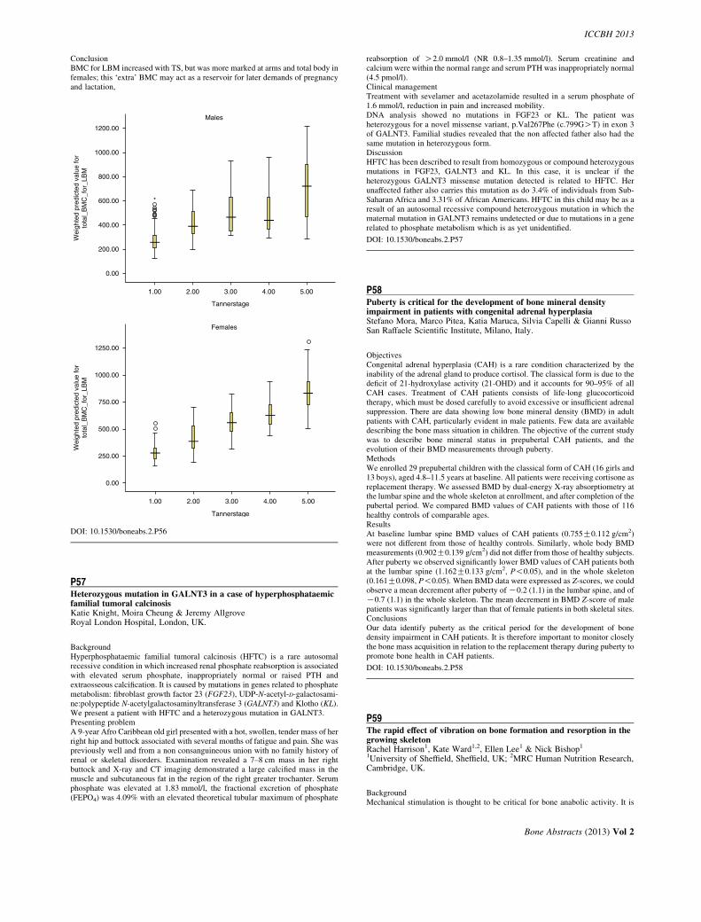
ICCBH 2013
ConclusionBMC for LBM increased with TS, but was more marked at arms and total body infemales; this ‘extra’ BMC may act as a reservoir for later demands of pregnancyand lactation,
1200.00
Males
1000.00
800.00
600.00 *
400.00
200.00
0.00
1250.00
1000.00
750.00
500.00
250.00
0.00
1.00 2.00 3.00 4.00 5.00
Tannerstage
Females
1.00 2.00 3.00 4.00 5.00
Tannerstage
Wei
ghte
d pr
edic
ted
valu
e fo
rto
tal_
BM
C_f
or_L
BM
Wei
ghte
d pr
edic
ted
valu
e fo
rto
tal_
BM
C_f
or_L
BM
DOI: 10.1530/boneabs.2.P56
P57Heterozygous mutation in GALNT3 in a case of hyperphosphataemicfamilial tumoral calcinosisKatie Knight, Moira Cheung & Jeremy AllgroveRoyal London Hospital, London, UK.
BackgroundHyperphosphataemic familial tumoral calcinosis (HFTC) is a rare autosomalrecessive condition in which increased renal phosphate reabsorption is associatedwith elevated serum phosphate, inappropriately normal or raised PTH andextraosseous calcification. It is caused by mutations in genes related to phosphatemetabolism: fibroblast growth factor 23 (FGF23), UDP-N-acetyl-D-galactosami-ne:polypeptide N-acetylgalactosaminyltransferase 3 (GALNT3) and Klotho (KL).We present a patient with HFTC and a heterozygous mutation in GALNT3.Presenting problemA 9-year Afro Caribbean old girl presented with a hot, swollen, tender mass of herright hip and buttock associated with several months of fatigue and pain. She waspreviously well and from a non consanguineous union with no family history ofrenal or skeletal disorders. Examination revealed a 7–8 cm mass in her rightbuttock and X-ray and CT imaging demonstrated a large calcified mass in themuscle and subcutaneous fat in the region of the right greater trochanter. Serumphosphate was elevated at 1.83 mmol/l, the fractional excretion of phosphate(FEPO4) was 4.09% with an elevated theoretical tubular maximum of phosphate
reabsorption of O2.0 mmol/l (NR 0.8–1.35 mmol/l). Serum creatinine andcalcium were within the normal range and serum PTH was inappropriately normal(4.5 pmol/l).Clinical managementTreatment with sevelamer and acetazolamide resulted in a serum phosphate of1.6 mmol/l, reduction in pain and increased mobility.DNA analysis showed no mutations in FGF23 or KL. The patient washeterozygous for a novel missense variant, p.Val267Phe (c.799GOT) in exon 3of GALNT3. Familial studies revealed that the non affected father also had thesame mutation in heterozygous form.DiscussionHFTC has been described to result from homozygous or compound heterozygousmutations in FGF23, GALNT3 and KL. In this case, it is unclear if theheterozygous GALNT3 missense mutation detected is related to HFTC. Herunaffected father also carries this mutation as do 3.4% of individuals from Sub-Saharan Africa and 3.31% of African Americans. HFTC in this child may be as aresult of an autosomal recessive compound heterozygous mutation in which thematernal mutation in GALNT3 remains undetected or due to mutations in a generelated to phosphate metabolism which is as yet unidentified.
DOI: 10.1530/boneabs.2.P57
P58Puberty is critical for the development of bone mineral densityimpairment in patients with congenital adrenal hyperplasiaStefano Mora, Marco Pitea, Katia Maruca, Silvia Capelli & Gianni RussoSan Raffaele Scientific Institute, Milano, Italy.
ObjectivesCongenital adrenal hyperplasia (CAH) is a rare condition characterized by theinability of the adrenal gland to produce cortisol. The classical form is due to thedeficit of 21-hydroxylase activity (21-OHD) and it accounts for 90–95% of allCAH cases. Treatment of CAH patients consists of life-long glucocorticoidtherapy, which must be dosed carefully to avoid excessive or insufficient adrenalsuppression. There are data showing low bone mineral density (BMD) in adultpatients with CAH, particularly evident in male patients. Few data are availabledescribing the bone mass situation in children. The objective of the current studywas to describe bone mineral status in prepubertal CAH patients, and theevolution of their BMD measurements through puberty.MethodsWe enrolled 29 prepubertal children with the classical form of CAH (16 girls and13 boys), aged 4.8–11.5 years at baseline. All patients were receiving cortisone asreplacement therapy. We assessed BMD by dual-energy X-ray absorptiometry atthe lumbar spine and the whole skeleton at enrollment, and after completion of thepubertal period. We compared BMD values of CAH patients with those of 116healthy controls of comparable ages.ResultsAt baseline lumbar spine BMD values of CAH patients (0.755G0.112 g/cm2)were not different from those of healthy controls. Similarly, whole body BMDmeasurements (0.902G0.139 g/cm2) did not differ from those of healthy subjects.After puberty we observed significantly lower BMD values of CAH patients bothat the lumbar spine (1.162G0.133 g/cm2, P!0.05), and in the whole skeleton(0.161G0.098, P!0.05). When BMD data were expressed as Z-scores, we couldobserve a mean decrement after puberty of K0.2 (1.1) in the lumbar spine, and ofK0.7 (1.1) in the whole skeleton. The mean decrement in BMD Z-score of malepatients was significantly larger than that of female patients in both skeletal sites.ConclusionsOur data identify puberty as the critical period for the development of bonedensity impairment in CAH patients. It is therefore important to monitor closelythe bone mass acquisition in relation to the replacement therapy during puberty topromote bone health in CAH patients.
DOI: 10.1530/boneabs.2.P58
P59The rapid effect of vibration on bone formation and resorption in thegrowing skeletonRachel Harrison1, Kate Ward1,2, Ellen Lee1 & Nick Bishop1
1University of Sheffield, Sheffield, UK; 2MRC Human Nutrition Research,Cambridge, UK.
BackgroundMechanical stimulation is thought to be critical for bone anabolic activity. It is
Bone Abstracts (2013) Vol 2

ICCBH 2013
unclear how quickly the growing skeleton responds to additional externally-applied mechanical stimuli. We wished to determine the acute effect of astandardised mechanical stimulus to the growing skeleton.ObjectivesTo determine the acute time course and magnitude of bone’s response to wholebody vibration (WBV) in pre-pubertal boys.MethodologyHealthy boys aged 9–12 years were recruited to stand on either the Juvent 1000(low magnitude, high frequency) or Galileo Advanced (high magnitude, variablefrequency) vibrating platforms for 1, 3 or 5 days for 10 min (nZ36). A controlgroup (nZ15) stood on a non-vibrating platform. Pre- and 10 min post vibrationblood samples were taken to measure changes in bone formation (P1NP,osteocalcin), bone resorption (CTx) and the bone derived factors osteoprotegerinand sclerostin. Samples were collected half hourly for 2 h in the control group.FindingsIn the immediate pre to post vibration period P1NP decreased by up to 7.9% andCTx by up to 8.8% in the Juvent and Control groups; no change was seen in theGalileo group. There was no difference in the response between groups across thepre- to post-vibration period. At 8 days, in boys exposed to 5 days of WBV oneither platform, there was an increase from baseline in P1NP of 25.1% (PZ0.005)and in CTx of 10.9% (PZ0.009). There was no intergroup difference. There wasno change in osteocalcin. Osteoprotegerin increased by 7.2% at day 8 (PZ0.08)possibly explaining the smaller increase in resorption. No change was seen insclerostin.ConclusionIn the growing skeleton a short period of mechanical stimulation resulted inincreased bone activity, with a greater response in the formation marker P1NPthan the resorption marker CTx after 5 days WBV. The lack of change inosteocalcin may reflect the ‘early’ nature of the response measured by P1NP in thebone formation process. Increased formation was not due to repression ofsclerostin, nor due solely to increased remodelling/resorption, suggesting thatother pathways mediate the acute anabolic response seen here to WBV.Declaration of fundingK Ward is funded by Medical Research Council Grant Code U105960371.
DOI: 10.1530/boneabs.2.P59
Table 1 Results of blood biochemistry and hormone concentrations acrosstime in preterm infants.
Weeks
Ionized
calcium
(mM)
Phos
(mM)
iFGF23
(pg/ml)
cFGF23
(RU/ml)
25(OH)
D3 (nM)
C3a epi-
mer (%) PTH (pM) 1,25(OH)2D (pM)
1 (nZ10) 1.39G0.05 2.26G0.5 68.2G59.9 839G494 54.3G21.0 32.4G5.7 6.0G5.0 213.0*
2 (nZ10) 1.42G0.05 2.23G0.26 71.7G31.6 737G355 57.1G25.7 43.4G6.7 6.0G3.2 134.7G40.5^
5 (nZ2) 1.41G0.02 2.17G0.13 95.5G3.5 888G80 53G3.9 49. 9G14.8 5.7G1.8 185.0G53.3^
Term
(nZ7)
1.40G0.03 2.35G0.20 75.8G22.7 1060G397 62.2G21.1 47.7G13.6 3.2G1.9 224.0*
*nZ1, nZ3.
P60Failure of free, public vitamin D supplementation program for Quebecinfants: temporal trends and significant predictorsMaude Millette1, Atul Sharma1, Hope Weiler1, Odile Sheehy2,Anick Berard2 & Celia Rodd1
1McGill University, Montreal, Quebec, Canada; 2Centre HospitalierUniversitaire Ste-Justine, Montreal, Quebec, Canada.
Over 80% of Quebec woman initiate breastfeeding, and rates of exclusivebreastfeeding at 6 months doubled from 2003 (9.7%) to 2009 (19.0%). To preventdeficiency, current recommendations for these infants include 400 IU/day ofvitamin D. For 20 years, Quebec has offered a program of free vitamin Dsupplements through its public medication insurance plan (RAMQ).ObjectiveProgram evaluation over the last decade.MethodsThis is a retrospective descriptive analysis of infants born between Jan 1998 andDec 2008. All healthy term infants covered by RAMQ drug plan were eligible;w30% of pregnant women and infants are covered. Data were extracted from theQuebec Pregnancy Registry linking three databases: RAMQ (diagnoses,medications, and SES), MedEcho (hospitalizations), and IQ20 (birthweight,SES). Multivariable logistic regression (GEE) examined predictors of partici-pation.ResultsA total of 123 018 infants were eligible. Mean annual prevalence of vitamin Dexposure was 17.9%G5.6. Median age at which the first bottle of supplementswas obtained was 36 days (rangeZ0–370). The majority (51.0%) obtained onlyone bottle of fifty doses (medianZ1, rangeZ1–20). Significant predictors(P!0.05) were prescription by pediatrician (odds ratio (OR)Z1.23, with 95%CIZ1.19–1.27); increasing maternal age (ORZ1.018/year, 95% CIZ1.015–1.021); non-synthesizing season for vitamin D (ORZ1.04, 95% CIZ1.01–1.07);women living alone (ORZ0.87, 95% CIZ0.83–0.91), less education (OR 0.88,
Bone Abstracts (2013) Vol 2
95% CIZ0.84–0.92) and rural residence (OR 0.94, 95% CIZ0.91–0.98). Therewas also a decline in program participation over time (OR 0.89/year, 95% CIZ0.88–0.90).ConclusionsProgram participation was low and decreased with time. Additional risks forrickets included long delay prior to filling the first prescription, failure to renew,younger mothers, mothers with less education, and care by a non-pediatrician.Overall, poor uptake suggests difficulty in administration of supplements, poorunderstanding of their potential benefits, or low acceptance of healthprofessionals’ advice. New preventive strategies need to be developed to increaselong-term vitamin D compliance.Declaration of fundingFunded by Montreal Children’s Hospital-Research Institute and Gray FamilyFund.
DOI: 10.1530/boneabs.2.P60
P61Elevated FGF23 levels in premature infants without excessivephosphaturiaTarah Fatani, Asma Binjab, Hope Weiler, Atul Sharma & Celia RoddMcGill University, Montreal, Quebec, Canada.
Preterm infants develop reduced bone mass and fragility fractures. Nevertheless,normal ranges in preterm infants are poorly defined for concentrations of vitaminD; its isomers (C3a-epimer of 25(OH)D3) and metabolites (24, 25(OH)2D3, 1,24(OH)2D); and other mineral-regulating hormones, including FGF23 in bothintact (iFRG23) and inactive (C- terminal) forms.ObjectiveTo clarify normal concentrations of minerals and hormones in healthy, preterminfants (AGA).MethodsWe conducted a longitudinal, pilot, cohort study on infants born at 28–32 weeksgestation, monitoring blood and urine at 1, 3, and 5 weeks and at term. Analysesincluded morning plasma and urine calcium, phosphorus, creatinine (BeckmanCoulter, USA), whole blood ionized calcium (Radiometer, Denmark), PTH,cFGF23 (C-terminal and intact; Immunotopics, USA), iFGF23 (Kainos, Japan), 1,25(OH)2D (IDS, UK) and 25(OH)D3 and its C3a-epimer and metabolites by LC–MS/MS (Warnex, Canada). Data are meanGS.D.ResultsEleven infants (mean GA 30.9G1.5 weeks, wt 1866G21.6 g, maleZ5) wererecruited. At weeks 1, 3, 5, and term, the % achieving 50 nM concentrations of25(OH)D3 were 40, 60, 100, and 67%, with good intakes of vitamin D. Levels ofiFGF23 were mildly elevated (double adult norms) with a marked increase incFGF23 (ten times adult norms). Nevertheless, tubular resorption of phosphorus(TRP) was normal (88G8%) for those with urine samples. Preterm infants hadsubstantial concentrations of C3a-epimer, much higher than in adults. Calcitriollevels were robust at all ages.
ConclusionsMany infants achieved acceptable 25(OH)D3 concentrations. Despite elevatedconcentrations of FGF23, there was not excessive phoshaturia.
DOI: 10.1530/boneabs.2.P61

LSBMAD Z score for disease duration
–4.6
–4.8
–5
–5.2
–5.4
–5.6
<1.5 years
<1.5 years>1.5 years
*
>1.5 years
*-P<0.05
Figure 1
ICCBH 2013
P62The effect of the ketogenic diet on the developing skeletonPeter Simm1,3, Jill Bicknell-Royle4, Judy Nation5, Kellie Draffin6,Karen Stewart7, Fergus Cameron1,3, Ingrid Scheffer8,9 & Mark Mackay1,21Department of Endocrinology and Diabetes, The Royal Children’s HospitalMelbourne, Parkville, Victoria, Australia; 2Department of Paediatrics,University of Melbourne, Parkville, Victoria, Australia; 3MurdochChildrens Research Institute, Parkville, Victoria, Australia; 4Department ofNeurology, The Royal Children’s Hospital Melbourne, Parkville, Victoria,Australia; 5Department of Nutrition and Food Services, The RoyalChildren’s Hospital Melbourne, Parkville, Victoria, Australia; 6Departmentof Nutrition and Dietetics, Austin Health, Heidelberg, Victoria, Australia;7Department of Paediatrics, Austin Health, Heidelberg, Victoria, Australia;8Department of Medicine, Epilepsy Research Centre, University ofMelbourne, Heidelberg, Victoria, Australia; 9Florey NeuroscienceInstitutes, Melbourne, Victoria, Australia.
ObjectivesThe ketogenic diet (KD) is a medically supervised, high fat, low carbohydrate andrestricted protein diet which has been used successfully in patients with refractoryepilepsy. Only one published report has explored its effect on the skeleton. Wepostulated that KD impairs bone mass accrual and examined skeletal healthparameters in this patient group.MethodsPatients commenced on the KD from 2002–2009 were enrolled in a prospective,longitudinal study; with monitoring of dual-energy X-ray absorptiometry (DXA)derived bone parameter including bone mineral content and density (BMD). ArealBMD was converted to bone mineral apparent density (BMAD) where possible.Biochemical parameters, including vitamin D, and bone turnover markers,including osteocalcin and urinary deoxypiridinolone, were assessed.Results29 patients were on the KD for a minimum of 6 months (range 0.5–6.5 years,mean 2.1 years). There was a mean reduction in lumbar spine (LS) BMD Z-scoreof 0.37 S.D. 20 patients (68%) had a lower BMD at the end of treatment. There wasno correlation between change in LS BMD and ambulatory status (RZ0.02).Height adjustment was possible for 13 patients, with a mean reduction in BMADZ-score of 0.19 S.D. ALP levels were in the normal range but osteocalcin showed amean 26.5 nmol/l, which was elevated. Only one patient sustained fractures(bilateral femoral fractures). Urinary calcium-creatinine ratios were elevated(mean 0.77) but only one patient developed renal calculi.ConclusionThe KD has a small but significant effect on the developing skeleton, independent ofheight and ambulatory status. Effects on bone turnover and calcium/creatinine ratiospoint to abnormal mineral metabolism. Clinicians should be aware of potentialskeletal side effects and monitor bone health during KD treatment. Longer termfollow up is still required to determine adult/peak bone mass and fracture risk.
DOI: 10.1530/boneabs.2.P62
P63Bone status of Indian children and adolescents with type 1 diabetesmellitusLavanya Parthasarathy1, Anuradha Khadilkar1, Veena Ekbote1,Shashi Chiplonkar1, Zulf Mughal2 & Vaman Khadilkar11Hirabai Cowasji Jehangir Medical Research Institute, Pune, India;2Department of Pediatric Endocrinology, Royal Manchester Children’sHospital, Manchester, UK.
ObjectiveType 1 diabetes mellitus (TIDM) has been shown to adversely affect bone healthin children. Hence, objective was to assess bone health status of children withT1DM, and to assess relationship between bone status and disease duration.MethodsBone mineral content for total body (less head) (TBBMC) and lumbar spine wasmeasured by DXA in 47 (25 boys) children with T1DM. Z-scores for TBBMC forbone area (TBBA), TBBA for height, lean body mass (LBM) for height, TBBMCfor LBM and lumbar spine bone mineral apparent density (LSBMAD) werecomputed reference data (Bone 2011 48 (4) 810–819).ResultsMean age was 10.9G2.5 years, height Z-scores wereK1.1G1.2 (32% belowK2S.D.); duration of diabetes was 2.7G3 years, glycosylated hemoglobin was 9.1G2.2%. Mean Z-scores for total body (TBBMC for TBBA, TBBA for height, LBMfor height, TBBMC for LBM) were within normal range for all; in contrast, meanLSBAMD Z-score was K5.2G0.8. Children were divided according to medianfor disease duration; children with diabetes forO1.5 years had significantly lower
LSBMAD scores (K5.0G0.7 vsK5.5G0.8, P!0.05, Fig. 1). 71% children withdiabetes forO1.5 years had LSBMAD Z-scores lower than the median values (Z-scoreZK5.1). Whereas 64% children who had diabetes for !1.5 years hadLSBMAD Z-scores aboveK5.1. Further, boys had significantly lower LSBMADZ-scores (K5.5G0.8 vsK4.9G0.6) whereas the LBM for height Z-scores in girlswere significantly lower than in boys (K0.2G0.8 vs K0.3G0.9, P!0.05).ConclusionChildren with T1DM with longer duration had lower bone mineral content at thelumbar spine possibly due to trabecular bone loss.
DOI: 10.1530/boneabs.2.P63
P64The level of vitamin D and calcium in urban pregnant women inRussian FederationOlga Ershova1,2, Valentina Dzhalatova1,2, Ksenia Belova1,2 &Ekaterina Svetalkina1,21Yaroslavl State Medical Academy, Yaroslavl, Russia; 2State ClinicalHospital for Emergency Medical Care n.a. N.V. Solovyev of YaroslavlRegion, Yaroslavl, Russia.
BackgroundOptimal intake of calcium and vitamin D of pregnant women is an importantcomponent for the normal course of pregnancy, but here are the contradictoryopinions about the case of shortage of the consumption, there is no uniformapproach to the appointment of them.AimTo assess the level of consumption of alimentary calcium, and characteristic ofthe status of vitamin D and calcium in blood serum of pregnant women.Materials and methodsWe examined 80 women at the age of 20–35 years (mean age of 27.12G4.36years) in the third trimester of pregnancy (gestation 31.71G3.37 weeks) residentsof Yaroslavl city, Russian Federation. The average consumption of calcium infoodstuff was estimated according to the tables with norms from Russianguidelines for osteoporosis, 2012. The analysis of calcium and vitamin D (25(OH)vitamin D) in blood samples was carried out during the whole year. Theconcentration of vitamin D in blood serum was determined by the electro-chemiluminescence immunoassay («ECLIA»), the concentration of calcium – bythe method of optical density on the unit COBAS 400 PLUS.ResultsThe average consumption of calcium in foodstuff was 1148.25G368.91 mg/day(from350 to 2064 mg). 29 (36.25%) ofwomen consumed!1000 mg,!1200 mg –46 (57.50%), more than 1500 mg – only 16 (20.00%) women. The total bloodcalcium was 2.14G0.28 mmol/l (from 1.43 to 2.48 mmol/l). Hypocalcaemia was in40 (50.00%) of the pregnant women. The average level of 25(OH) – vitamin D was26.61G13.69 ng/ml (from 4.10 to 58.16). The normal content ration (more than30 ng/ml) was detected in 29 (36.25%) of people, insufficiency (from 21 to29.9 ng/ml) – at 18 (22.50%), deficit (!20 ng/ml) – at 33 (41.25%) women. Therewas nodifference inmeanconcentrationofvitaminD inblood samples fromdifferentseasons (from Oct to Apr – 26.51G11.69 ng/ml, from May to Sep – 26.79G17.32 ng/ml, PO0.05). The clinical manifestations of the vitamin D deficiencyand/or hypocalcaemia were noted in 17 (21.25%) women.ConclusionsOur data demonstrate the wide prevalence of the deficiency of dietary calcium andlow level of vitamin D of the urban pregnant women in the third trimester ofpregnancy.
DOI: 10.1530/boneabs.2.P64
Bone Abstracts (2013) Vol 2

ICCBH 2013
P65Improvement in morphological properties of trabecular bones andlongitudinal growth in tibia for growing rats through an impactstimulationSinae Eom1, Chang-Yong Ko2, Ji Hyung Park1, Dong-Hyun Seo1,Young Jin Jung1 & Han Sung Kim1
1Department of Biomedical Engineering, Yonsei University, Wonju,Republic of Korea; 2Korea Orthopedics and Rehabilitation Research Center,Incheon, Republic of Korea.
Mechanical stress and strain generated by physical exercises or the other passivestimulations are well known to have a positive effect on the growingmusculoskeletal system. Especially, when the impact stimulation which evokeshigh magnitude of strain in a second is applied to bone, it improves bone qualities.Thus, to verify the effect of impact stimulation, we conducted longitudinal studyon morphological properties of the tibia in growing rats. Free falls fromdesignated heights were implemented to induce high impact stimulation. Six-week-old male Wistar rats were randomly allocated to one of three conditions:free fall from 20-cm-height (W20; nZ7), free fall from 40-cm-height (W40;nZ7), and control (WC; nZ7). The impact stimulations were administered to thefree fall groups, ten times/day, and 5 days/week for 8 weeks. The right tibia wasscanned by in-vivo micro-CT at 0, 4, and 8 weeks of experiment and structuralparameters and tibia‘s length were measured to evaluate the variation inmorphological characteristics and bone length with maturing. After 4 weeks ofthe experiment, relative values (value of 4 or 8 weeks/0 week, thus 1 at 0 week) ofBV/TV and Tb.N were significantly higher in W40 group than in WC and W20groups (all P!0.05), while relative values of Tb.Sp was significantly smaller(P!0.05). However, there were no difference in relative values in Tb.Th andbone length among the groups (all PO0.05). No significant difference in allstructural parameters between W20 and WC groups (PO0.05). After 8 weeks ofthe experiments, relative values in Tb.Th and tibial length were significantlyhigher in W40 group than in WC and W20 groups (P!0.05). Only relative valueof Tb.Sp in W20 group was significantly smaller than that in WC group(P!0.05). Overall, impact stimulation for 8 weeks generated by free falls at a40-cm-height affected not only trabecular bone homeostasis but also bone growth.Furthermore, since a significant bone growth was appeared after 8 weeks ofstimulation, but not after 4 weeks, continuous stimulation may be needed toimprove bone growth length in growing rats.
DOI: 10.1530/boneabs.2.P65
P66Effect of resistance training on peripheral bone mineral density andmuscle strength in adolescents with motor difficultiesBeth Hands, Fleur McIntyre, Francisco Bervenotti & Aris SiafarikasUniversity of Notre Dame Australia, Perth, Western Australia, Australia.
ObjectivesAdolescents with motor difficulties may have a higher fracture risk due to limitedparticipation in high impact physical activities that improve bone mineral density(BMD). Equipment constrained resistance training (RT) interventions may be aneffective way to improve both muscle strength and BMD in this population. Theaims of this study were to investigate the effect of a GYM-based RT interventionon peripheral BMD, and to determine the extent of the relationship between BMDand muscle strength, among adolescents with motor difficulties.MethodsParticipants were 21 adolescents (13 intervention and 8 control) with motordifficulties, with a mean age of 14 (1.54) years. The participants were recruitedfrom a larger research project (Adolescent Movement Program; AMPitup) and itswait list. The intervention was a 13-week aerobic and resistance exercise programthat participants attended for 90-min twice a week. The exercise programincluded five pre-set exercises targeting the forearm and lower leg (leg–press,push-ups, seated row, calf raises, and up-right rows) to be completed everysession. Measures taken pre and post intervention included peripheral BMD scans(tibia and radius; trabecular and cortical density) using peripheral quantitativecomputer tomography (pQCT), height, weight, upper (grip strength, chest pass)and lower (IRM leg press, distance and vertical jump) body muscle strength.General linear models, adjusting for physical maturity, and correlations were usedto analyse the data.ResultsImprovements in muscle strength, in particular for the upper body (right hand gripstrength PZ0.01; chest pass PZ0.01) were observed in the intervention groupbut not the control group. Changes in BMD measures from pre to post test in theintervention group were less conclusive due to the small sample size and shorttime frame, however positive trends were apparent. Muscle strength and BMD
Bone Abstracts (2013) Vol 2
was related as evidenced by moderate to strong correlations, particularly for thelower leg.ConclusionA targeted resistance training program may be effective in improving musclestrength and stimulating bone changes in adolescents with motor difficulties.Further research is needed to clarify the most effective exercises for site specificBMD improvements in this group.
DOI: 10.1530/boneabs.2.P66
P67Dual-energy X-ray absorptiometry in predicting clinical bone disease inadults with childhood onset end-stage renal diseaseMaike van Huis1, Judith Vogelzang1, Hanneke van der Lee2,Annemieke Boot3 & Jaap Groothoff11Department of Pediatric Nephrology, Academic Medical Center, EmmaChildren’s Hospital, Amsterdam, The Netherlands; 2Clinical Research Unit,Division Woman-Child, Academic Medical Center, Amsterdam, TheNetherlands; 3Department of Pediatric Endocrinology, Beatrix Children’sHospital, University Medical Center Groningen, Groningen, TheNetherlands.
ObjectivesMetabolic bone disease is a frequent complication of end-stage renal disease,characterised by a decreased bone mineral density, which can be measured withdual-energy X-ray absorptiometry. Its validity as a marker for clinical bonedisease and increased fracture risk has never been established in adults withpediatric onset of end-stage renal disease (1–3). Adult survivors of pediatric endstage renal disease have very low bone mineral density and small stature (4). Inthis cohort study, the association between low bone mineral density measuredwith dual-energy X-ray absorptiometry in adult survivors of pediatric end-stagerenal disease in 2000 and manifestations of clinical bone disease between 2000and 2010 was assessed.Methods113 from all 187 living patients of the so called LERIC-cohort (4) agreed toparticipate. Data regarding bone mineral density and clinical bone disease weregathered in 2000. Volumetric bone mineral density was calculated. Data onclinical outcome were gathered retrospectively by reviewing medical charts overthe period 2000–2010 and by questionnaires.ResultsMean volumetric bone mineral density Z-scores (95% CI) of the lumbar spinewas K1.0 (K1.2 to K0.7) and femoral neck was K3.0 (K3.5 to K2.5). 27% ofthe patients suffered from clinical bone disease. 16 patients (14%) had sufferedfrom a fracture in the last 10 years, of which four were pathological fractures. Theodds ratios (95% CI) of volumetric bone mineral density for clinical bone diseaseand fractures over 10 years were 1.033 (0.867 to 1.231) and 0.679 (0.437 to 1.055)respectively.ConclusionAdult survivors of pediatric end stage renal disease have low volumetric bonemineral density and the prevalence of clinical bone disease is high. However, wefound no significant association between bone mineral density and clinical bonedisease. Therefore, the usefulness of dual-energy X-ray absorptiometry inpredicting clinical bone disease in patients with pediatric end-stage renal diseaseis still uncertain.
DOI: 10.1530/boneabs.2.P67
P68Engineered tridimensional hydroxyapatite scaffold to support boneresorptionAlfredo Cappariello1, Eleonora Mirabile2, Maurizio Muraca1 & Anna Teti21Children Hospital Bambino Gesu, Rome, Italy; 2University of L’Aquila,L’Aquila, Italy.
ObjectiveIn many traumatic or pathological conditions, bone turnover is low and osteoclastactivity is reduced or abolished. We developed innovative hydroxyapatite (HA)scaffolds carrying RANKL expressing cells with the aim of supporting boneresorption when this is defective. Membrane-bound (m)RANKL is cleaved intosoluble (s)RANKL by MMP14. We hypothesized that the osteoclastogenicpotential of RANKL-producing cells could be improved if they were seeded onscaffolds engineered by immobilization of MMP14 to rise the shedding ofsRANKL.

ICCBH 2013
MethodsPrimary osteoblasts (OBs) and stromal bone marrow cells from 10 days old WTCD1 mice were used. MC3T3 cells were stably transfected with sRANKL-vector.MMP14 was immobilized on 3D-HA scaffolds using procedures based onprotein-to-substrate binding by glutaraldehyde (10% v/v). For in vivo studies, thescaffolds were entrapped in diffusion chambers (Millipore).ResultsWe identified in OBs the best spontaneous sRANKL source, then on these cellswe demonstrated a concentration-dependent RANKL shedding ability of thecatalytic domain of MMP14. Next, we quantified in about 50% the enzymaticefficiency of MMP-14 functionalized scaffolds vs soluble MMP14, and tested theefficacy of MMP14-engineered 3D-HA scaffolds on OBs noting increased releaseof sRANKL vs scaffolds not subjected to MMP14 immobilization. Intactscaffolds are also seeded with MC3T3 cells stably overexpressing sRANKL.Moreover, we assembled devices with scaffolds embedded in diffusion chambersand proved the safety of their implants in WT mice. Finally, we tested theefficiency of devices harboring either OBS or sRANKL-MC3T3 implanted inRANKL KO mice. In tibial sections we noted the appearance of TRAcP positivecells in both groups of implanted animals in contrast with sham KO mice, whichwere TRAcP-negative.ConclusionOur results demonstrated the feasibility of a strategy based on engineered bio-device for supporting sRANKL release from osteoblast mRANKL.
DOI: 10.1530/boneabs.2.P68
P69sRANKL/OPG in children with idiopathic hypercalciuriaMaria Pavlou1, Ekaterini Siomou1, Vasileios Cholevas2,Antigone Siamopoulou1 & Anna Challa21Division of Paediatric Nephrology, Health Department, Ioannina, Greece;2Pediatric’s Research Laboratory, Child Health Department, MedicalSchool, University of Ioannina, Ioannina, Greece.
ObjectivesTo determine any relationship of serum concentrations of osteoprotegerin (OPG),sRANKL and sRANKL:OPG ratio with idiopathic hypercalciuria (IH) inchildren, as there is some evidence of increased bone resorption in these patients.MethodsIn a prospective study, twenty four children of median age 6.5 years (range 2.3–16.4) with IH (five had urolithiasis and two nephrocalcinosis) were examined atthe time of diagnosis and after 3 months of salt free and adequate Ca diet.Clinically healthy children (nZ46) matched for age, sex and season were used ascontrols (median 8.0 years, range 1.8–16.3). sRANKL (total), OPG, 25(OH)D,and 1.25(OH)2D, PTH, Ca, Pi, osteocalcin (N-MID OC), ALP and CTX-Crosslaps were determined in serum and Ca, creatinine, oxalate and citrate inurine. Height was recorded and BMI Z-score was assessed.ResultsThe BMI Z-score was lower in patients than controls (K0.425G0.641 vsC0.106G1.0, PZ0.016), but height did not differ. Although urinary Ca excretion(24 hCa and UCa/UCr) decreased at 3 months (24 hUCa: 6.29G2.0 vs 5.19G2.3 mg/kg, PZ0.06; UCa/UCr: 0.30G0.19 vs 0.21G0.12 mg/mg, PZ0.014) onaverage it had not reached control values (2.14G1.08, P!0.0001; 0.10G0.06,PZ0.0003). Ucitrate/UCr, UCa/UCitrate and 24 h oxalate did not differ inpatients before and after diet or compared to controls. No significant differenceswere found for serum Ca, Pi, 25OHD, 1.25(OH)2D, PTH, osteocalcin, ALP, OPGand sRANKL or for the sRANKL:OPG ratio. Only serum concentrations of CTX-Crosslaps were significantly higher in both patient samples (1.63G0.67, P!0.02;1.56G0.45, P!0.05 than controls (1.24G0.56 pmol/l).ConclusionNo evident changes in the serum cytokines OPG and sRANKL were noted inchildren with IH. However, growth might have been affected by increased boneresorption, as indicated by the higher levels of serum CTX-Crosslaps andunaffected formation, since osteocalcin was not found different from controls.Hence, an autocrine role of the above cytokines cannot be excluded.
DOI: 10.1530/boneabs.2.P69
P70Isolated bilateral zeugo-autopodal segments agenesis of the lower limb:unusual malformation case reportAntoine Christiaens1,2, Pierre M L Deprez1, Antonella Mendola2,Pierre Bernard3, Yves Gillerot3, Philippe Clapuyt3, Benoıt G Lengele1,Miikka Vikkula2 & Catherine Nyssen-Behets11Universite Catholique de Louvain – Institut de Recherche Experimentale etClinique – Pole de Morphologie, Brussels, Belgium; 2Universite Catholiquede Louvain – de Duve Institute – Human Molecular Genetics, Brussels,Belgium; 3Universite Catholique de Louvain – Cliniques UniversitairesSaint-Luc, Brussels, Belgium.
Congenital limb abnormalities represent a prevalence of 0.79/1000 of live births inMassachusetts1. A better understanding of their physiopathology could improvethe management of the patients. We report on a 23 weeks female fetus affected byan isolated bilateral terminal transverse defect of the lower limbs with nubbins.Both familial history and chromosomal analyses were irrelevant. We performed adeep morphological examination of the fetus in comparison with an age-matchedcontrol fetus in order to precise the diagnosis. The estimated skeleton agecorresponded to the gestational age. The bones of both legs and feet werecompletely lacking, whereas no abnormality was observed in the axial and upperlimb skeleton as well as in the other systems. The distal femur epiphysis presentedan unusual shape and was composed of a cartilage bud including the proximal tibiaanlage. Targeted sequencing of exons involved in limbmorphogenesis was carriedout with skin tissue and showed no significant mutation. Such a rare malformationwas never reported in the medical literature. Considering the data available in theliterature, we can suggest the genetic cause is the most probable. Only few theoriesexist to explain the limb and joint development and malformation. This extremelyrare malformation comes up against some contradictory theories, like the ProgressZone model2 and the Early Specification model3. The minute description of thepresent case is a real chance to give cues to the correct understanding of limb andjoint formation, especially the chronological sequence of the different genes,molecules and targets involved in limb morphogenesis.
DOI: 10.1530/boneabs.2.P70
P71The microarchitecture of bone in osteochondromasHeleen Staal1, Bert van Rietbergen2 & Lodewijk van Rhijn11Department of Orthopaedic Surgery, Maastricht University MedicalCentre, Maastricht, The Netherlands; 2Department of OrthopaedicBiomechanics, Eindhoven University of Technology, Eindhoven,The Netherlands.
IntroductionHereditary multiple osteochondromas (HMO) is characterized by the outwardgrowth of cartilage-capped bone tumors. Osteochondromas contain a bonemarrow cavity continuous with the normal bone cavity. Because of their off-axisposition, osteochondromas are expected to carry less load than normal bonetissue. According to Wolff’s law, we therefore hypothesized that osteochon-dromas would have a less developed, osteoporotic-like microstructure. To testthis hypothesis, we measured the bone morphology of osteochondromas withmicroCT scanning and compared it to normal bone. To our knowledge this is thefirst study done with microCT and human osteochondromas.Materials and methodsMicro-CT scans were made of thirteen osteochondromas from human subjects toevaluate tissue mineralization and bone structural parameters. Values for normalbone were taken from the literature.ResultsLarge differences in tissue mineralization were found, with three specimens beinghardly mineralized and the others being less mineralized than normal bone. Theosteochondromas have less but thicker trabeculae than normal bone. The spacingbetween the trabeculae is increased. The structure lacks a clear orientation, beingmore isotropic than normal bone.DiscussionOsteochondromas resemble an osteoporotic structure in some aspects (increasedtrabecular spacing, decreased trabecular number) but the osteochondromas have ahigher trabecular thickness. This may be due to the influence of the cartilage capnearby. The osteochondromas also lack a clear trabecular orientation, probablydue to the absence of mechanical loading.ConclusionOsteochondromas have a less developed structure, as witnessed by the lowermineralization and the absence of a clear trabecular orientation, but is notcomparable to osteoporotic bone.
Bone Abstracts (2013) Vol 2

ICCBH 2013
SignificanceThis study is the first to show that the architecture of the osteochondromas isnot osteoporotic. It only resembles an osteoporotic structure in some aspects(increased trabecular spacing, decreased trabecular number) but that they have ahigher trabecular thickness.
DOI: 10.1530/boneabs.2.P71
P72
Abstract withdrawn.
DOI: 10.1530/boneabs.2.P72
25
20
Percentage change in bone parameters post supplementation
P73Histomorphometric parameters of the alveolar ridges of the mandiblein immature rats after thymectomyV Luzin, A Kochubey & V MorozovLugansk State Medical University, Lugansk, Ukraine.
AimStudy the features changes of histomorphometric parameters of alveolar ridges inimmature rats after thymectomy.MethodsThe study was conducted on 120 immature male rats were divided into twogroups: 1st group – sham-operated animals, 2nd group – rats with removed of thethymus under mask ether anesthesia (thymectomy). Periods of observation were7, 15, 30, 90 and 180 days. Rats were euthanized under mask ether anesthesia. Forhistological examination the mandible was cut at the level of the second molar,separated dentoalveolar segments were fixed in 10% neutral formalin, decalcifiedin 5% formic acid, dehydrated in increasing concentrations of alcohol, embeddedin paraffin and stained with hematoxylin–eosin. Morphometry program includedthe following parameters: the total width of the alveolar ridge at the level of 2ndmolar and the width of its layers: the outer cortical plate, inner cortical plate,osteon layer and diameters of osteons and their channels.ResultsIn animals of 2nd group the total width of the alveolar ridge and the width of theouter cortical plate decreased, compared with the same parameters in 1st group,from 30 to 180 day of observation respectively by 3.55, 7.13, 8.89 and 5.44, 8.71,9.41%, width of osteon layer – by the 90th and the 180th days by 8.83 and 9.28%and the width of the inner cortical plate – by the 180th day by 7.87%. Diameterswere lower than those of 1st group by the 90th and 180th days of observation by4.35 and 4.46%, and the diameter of osteons channels, in contrast, were more by8.46 and 15.88% respectively.ConclusionsThus, under conditions of thymectomy in immature rats there was a disturbance inthe structural organization of alveolar ridge of the mandible in the later period ofobservation (from30 to180 per day),whichmay indicate a generalizedperiodontitis.
DOI: 10.1530/boneabs.2.P73
Ff ff FF
15
10
5
0Percentage
change TBBMCPercentage
change TBBAPercentage
change TBBMDPercentage
change LBM
P74Assessment of bone mass and bone metabolism in children with chronicinflammatory bowel diseasesVesna Kusec & Irena Senecic-CalaClinical Hospital Centre, Zagreb, Croatia.
Skeletal integrity during childhoodmaybecompromisedbydiseases interferingwithbone metabolism. Chronic inflammatory bowel diseases (CIBD; Crohn’s disease,ulcerative colitis) in children is a recognized risk of osteoporosis in adulthood. Thisstudy was aimed at assessment of bone mass and bone metabolism in children withCIBD at diagnosis and after 1 year during therapy. Patient population comprised 64children (boys 23 and girls 42) and adolescents aged 14.6G2.7 years (7–20). DualX-ray densitometry (lumbar spine) and, measurement of 25-OHD and bonemarkers(osteocalcin, P1CP, CTX, crosslaps urine, osteoprotegerin) by standard methodswere performed. No difference between sexes was found. Densitometry Z-scores
Bone Abstracts (2013) Vol 2
(K0.67G1.28; rangeK3.95 to 2.47) indicating decreased bone mass (!K2) werefound in five patients. At time of diagnosis hypovitaminosis D (!50 nmol/l) wasfound in 69% of patients. Increased bone markers (as compared to adult referenceranges)were observed in 75% for osteocalcin, 95% for P1CP, 98% forCTXand45%for crosslaps urine. Osteoprotegerin was similar to normal adult values in 92% ofpatients. After 1 year follow-up statistically significant increase was found forosteocalcin (0.002) and P1CP (0.001), and decrease for crosslaps urine (0.0005) andosteoprotegerin (0.03). Hypovitaminosis D was still present in 41% of cases. Theseresults indicate thatCIBD inchildren and adolescents did not considerably impair theskeleton. Increased bone markers probably indicate increased bone turnovercharacteristic of growth and puberty. The observed changes of bone markers duringfollow-up probably reflects recovery and continuation of growth processes. Normalosteoprotegerin levels suggest that bone resorption at diagnosis and duringmonitoring was not predominant. Prevalence of hypovitaminosis D is a seriousproblem of this disorder, its treatment and prevention should be a measure forpreventing osteoporosis risk.
DOI: 10.1530/boneabs.2.P74
P75Vitamin D receptor gene Fok1 polymorphism and bone mass accrual inIndian girlsAnuradha Khadilkar1, Neha Sanwalka2, Shashi Chiplonkar1,Kavita Khatod3, Nikhil Phadke3, vaman Khadilkar1 & Veena Ekbote11Hirabai Cowasji Jehangir Medical Research Institute, Jehangir Hospital,Pune, Maharashtra, India; 2NutriCanvas, Mumbai, Maharashtra, India;3GenePath Diagnostics, Pune, Maharashtra, India.
ObjectiveTo study association of VDR gene Fok1 polymorphism locus on bone massaccrual in adolescent girls.MethodsAn intervention trial was carried out in 102 girls aged 8–16 years (Pune, India).All girls received 500 mg elemental calcium (daily) and 30 000 IU of vitamin D3quarterly for 1 year. Outcome variables were measured at baseline and end of theyear. Serum levels of ionised calcium (iCa), inorganic phosphorous, parathyroidhormone (PTH) and 25-hydroxy vitamin-D were measured (25(OH)D). Bonemineral content (BMC), bone area (BA), bone mineral density (BMD) and leanbody mass (LBM) were measured at total body by DXA. Percentage increase inBMC, BA and BMD was calculated. Polymorphisms (FF, Ff, ff) of VDR gene atFok1 locus were detected using SYBR Green quantitative PCR.ResultsOverall prevalence of Fok1 genotypes were 43.1 Ff, 9.8 ff and 47.1% FF. Therewere no significant differences in percentage change in serum parameters duringthe intervention period between groups (PO0.05). At baseline, FF genotype hadsignificantly lower BMD as compared to ff and Ff genotype (P!0.05) andsignificantly lower BA as compared to ff genotype (P!0.05). A significantincrease in BMC, BA, BMD and LBM was observed post supplementation in allthe three Fok1 polymorphism groups (P value!0.05). However, there were nosignificant differences in the percentage increase in BMC (Ff (17.9%); ff (18.1%);FF (17.4%)), BA (Ff (11.6%); ff (11.2%); FF (11.8%)), BMD (Ff (5.4%); ff(6.3%); FF (4.9%)) and LBM (Ff (8.4%); ff (9.2%); FF (10.7%)) (PO0.05) (Fig).
ConclusionGirls with FF genotype had significantly lower bone indices as compared to ffand Ff genotypes. VDR gene polymorphism as defined by Fok1 genotype had nopositive influence on bone mass accrual in study girls.
DOI: 10.1530/boneabs.2.P75

ICCBH 2013
P76Final height and bone health in young adults, transplanted in childhoodMaria Van Dyck, Jean Herman & Rita LombaertsPediatric Nephrology Department, University Hospitals, Leuven, Belgium.
This study analyses the long-term effect of rhGH on final height (FH) and bonehealth in renal transplanted patients.Twenty-one young adult patients, aged 17–26 years, were studied. Group Aconsisted of 15 patients (12 boys) who received rhGH during 3.0 years beforetransplantation. After transplantation three boys needed rhGH again for 3.5 years.In group B six patients (three boys) didn’t need rhGH before transplantation, threegirls received rhGH afterwards for 4 years. There is nog significant differencebetween the two groups for median age at transplantation, median follow-upperiod afterward and median GFR at final evaluation. At FH (defined as adultbone age or height velocity of !1 cm/year), clinical evaluation includedmeasurement of height and weight and GFR measurements by Cr EDTA. Lumbarspine and total body mineral content and density and body composition werestudied by DXA (Discovery A).FH was expressed as SDS (Cole 1995). We compared the lumbar spine density,expressed as standardized lumbar bone density L2–L4(sLBMD), with normativedata of Belgian healthy young adults. Osteopenia was defined as a sLBMDT-score of!1.0, osteoporosis as sLBMD T-score ofK2.5 SDS. Lean body massand fat mass were expressed as percentage of the total body weight.Median height SDS at transplantation was better in group A (S.D. K0.3) than ingroup B (S.D. K1.7, PZ0.03). Final height SDS was no longer different. For thetotal group FH SDS ranged from K2.3 to C1.0 SDS. BMI SDS was within thenormal range. Fat mass % was lower in group A (16.5) compared to group B(24.5) (PZ0.016).Lumbar density values were better at the moment of renal transplantation in groupA, however there was no difference at final height. In both groups one patient hasa osteoporotic sLBMD.In children with chronic renal failure rhGH treatment improves height and resultsin better bone health at the moment of transplantation. At final height patientswith rhGH before transplantation have significantly better lean body mass. Futurestudies in a larger cohort should confirm these data.
DOI: 10.1530/boneabs.2.P76
P77A novel mutation in CRTAP gene in a patient with severe ostegenesisimperfecta type VIIIlkka Vuorimies1,2, Minna Pekkinen1,2, Jutta Becker2, Helena Valta2,Christian Netzer3 & Outi Makitie1,21Folkhalsan Institute of Genetics, Helsinki, Finland; 2Children’s Hospital,Helsinki University Central Hospital and University of Helsinki, Helsinki,Finland; 3Institute of Human Genetics, University of Cologne, Cologne,Germany.
BackgroundOsteogenesis imperfecta (OI) is a genetic disorder with low bone mass and bonefragility. Type VII OI is one of the autosomal recessive subtypes and clinicallymoderate to lethal. It is caused by mutations in the cartilage associated protein(CRTAP) gene. Currently !20 mutations are known.Case descriptionAn 11-year-old Iraqi female was referred to our hospital after immigration toFinland. She had suffered numerous peripheral and spinal fractures. Atpresentation she had severe vertebral and limb deformities and could not stand,her height was 75 cm. The lumbar spine and whole body BMD Z-scores wereK5.6 and K3.0, respectively. The clinical and radiographic features wereconsistent with OI. Her parents and three siblings were healthy; one sibling haddied soon after birth due to similar bone disease. The parents were 1st cousins.All known OI genes were analysed with flanking markers. The genes flanked byhomozygous markers were chosen for direct sequencing. A homozygous mutation(c.141dupC) was identified in exon 1 of the CRTAP gene. The mutation leads to apremature stop codon. Both parents were heterozygous carriers of the mutation.The mutation has not previously been described.The patient has received intravenous pamidronate and zoledronic acid treatment for3years. Spinaldeformity required surgery.Bisphosphonate treatment improved subjectivewell-being and reduced bone pain and she is able to sit for much longer periods.DiscussionThis family illustrates the extreme severity of type VII OI. Bisphosphonate treatment wasof subjective benefit even though it was not started until age 11. The reported novelCRTAP mutation expands the genetic defects associated with OI type VII.
DOI: 10.1530/boneabs.2.P77
P78Variable relationship of fat mass and bone with pubertal staging inobese and normal weight Indian children and adolescentsAnuradha Khadilkar1, Veena Ekbote1, Deepa Pandit1, Shashi Chiplonkar1,M Zulf Mughal2 & Vaman Khadilkar11Hirabai Cowasji Jehangir Medical Research Institute, Jehangir Hospital,Pune, Mumbai, India; 2Department of Paediatric Endocrinology, RoyalManchester Children’s Hospital, Manchester, UK.
ObjectiveIn children, factors such as age and puberty are confounders when investigatingassociations of fat or lean with bone mineral content (BMC). Our aim was toassess influence of fat on BMC in overweight/obese (ow/ob) and normal weight(nw) children at different stages of puberty.MethodsCross-sectionally, 244 childrenCadolescents (6–16 years) (73nw, 177ow/ob)were assessed (tanner staging (TS), body composition (DXA) (total body BMC,lean mass (LBM) and fat (BF))).ResultsBMC was highly correlated with LBM (rZ0.68 in boys; rZ0.72 in girls,P!0.0001) more so than with BF (rZ0.56 in boys, rZ0.67 in girls, P!0.0001).After adjustments (age, LBM), ow/ob boys showed higher values of BMC thancontrols at TS1&2 (910G74 vs 1340G41 g; 1598G39 vs 1358G64 grespectively) (P!0.05); no difference was observed at TS3,4&5; adjustedBMC values in ow/ob girls were significantly higher than controls at all TS’s(P!0.05). Multiple linear regression (entire cohort) was performed at each TS inboth genders to assess simultaneous effect of lean, fat on BMC (age adjusted). Inboys, at all TS’s, lean was significant predictor of BMC (P!0.001); additionallyat TS’s 2, 3, 5 fat was significant predictor (r2Z0.71, P!0.01). In girls, at TS’s 1,2, 3 (r2Z0.77) and 5 (r2Z0.41) lean was significant predictor (P!0.05); at TS3fat was also predictor (r2Z0.77), at stage 4 (r2Z0.53) fat was the only predictor.ConclusionDifferential relationships between lean, fat and bone were observed in bothgenders at different TS’s; lean was the main predictor in all children (nw, ow/ob)at almost all TS’s. In boys, at earlier TS’s, bone mass was higher in ow/ob boys,but this effect of fat on bone was not maintained at later TS’s, suggesting thatintervention may be necessary at later TS’s to improve bone mass inoverweight/obese boys.
DOI: 10.1530/boneabs.2.P78
P79High sedentary lifestyle practices has adverse influence onmusculoskeletal health in adolescent boys and girls: findings from apopulation-based study in MalaysiaPey Sze Teo1, Nurul-Fadhilah Abdullah1, Chee Keong Chen2,Mohd Ezane Aziz2 & Leng Huat Foo11School of Health Sciences, Universiti Sains Malaysia, Kubang Kerian,Kelantan, Malaysia; 2School of Medical Sciences, Universiti SainsMalaysia, Kubang Kerian, Kelantan, Malaysia.
The combined effect of lifestyle physical activity (PA) and sedentary behavioralpractices on musculoskeletal health in growing populations is still poorlyunderstood.ObjectivesTo investigate the combined influence of mechanical loading of weight-bearingPA (WBPA) and screen-based sedentary practice (SSR) on musculoskeletalprofiles among 455 adolescents aged 12–19 years.MethodsValidated PA and SSR behaviour assessments were used to determine dailyphysical activities and sedentary practices, respectively. Bone mass of total body(TB), lumbar spine (LS) and proximal femur (PF) was assessed using dual energyX-ray absorptiometry, while muscular strength of the upper and lower extremitywere determined using handgrip and isokinetic dynamometers respectively.ResultsFour groups of combined influence of the WBPA and SSR were i) HighWBPA-
LowSSR, ii) HighWBPAHighSSR, iii) LowWBPALowSSR and iv) LowWBPAHighSSR.Multivariate analysis model showed that adolescents with high SSR and lowWBPA had significantly lower size-adjusted bone mineral content (BMC) of theLS and PF (all, P!0.01) and TB bone area (TBBA) (P!0.001), muscularstrength of the handgrip (P!0.001) and hamstring (P!0.01) than those at highWBPA and low SSR group, after adjusting for body size, pubertal growth status,socio-demographic and calcium intakes. Furthermore, participants with higherlevels of WBPA and SSR practices had significantly higher size-adjusted PFBMC
Bone Abstracts (2013) Vol 2

ICCBH 2013
(P!0.01) and TBBA (P!0.01) compared to those adolescents engaged only inhigh SSR levels.ConclusionThese findings suggest that adolescents with higher participation of daily WBPAand low SSR practices exert positive impact on bone mass and muscular strengthsthan those involved only in high sedentary practice. Effective strategies topromote active lifestyle practices should be emphasised in children andadolescents in order to maximise bone mass accretion and muscular strengthduring these growing years.Declaration of interestThis study was funded by University Science Malaysia Research University (RUgrant: 1001/PPSK/812015).
DOI: 10.1530/boneabs.2.P79
P80Osteogenesis imperfecta and short stature: effect of sclerostin antibodytreatment in oim/oim miceMickael Cardinal1, Catherine Nyssen-Behets1, Mike Ominsky3,Jean-Pierre Devogelaer2 & Daniel H Manicourt21Institut de Recherche Experimentale et Clinique, Pole de Morphologie,Universite Catholique de Louvain, Brussels, Belgium; 2Institut deRecherche Experimentale et Clinique, Pole de Rhumatologie, UniversiteCatholique de Louvain, Brussels, Belgium; 3Metabolic Disorders, Amgen,Inc., Thousand Oaks, California, USA.
IntroductionOsteogenesis imperfecta (OI) is characterized by low bone mass, skeletal fragilityand, frequently, short stature. We previously showed in oim/oim mice thatsclerostin inhibition increased bone mass, mineral content and strength. Here, wecompared the body length and the sizes of long bones, head and vertebraebetween oim/oim and wildtype mice and analyzed the effect of sclerostinantibody (Scl-Ab) on these parameters.Materials and methodsFive-week-old oim/oim and wild-type mice received s.c. injections of either Scl-Ab VI (25 mg/kg) or vehicle (PBS) twice a week for 10 weeks. Body length andbone growth were evaluated on DXA scans collected at 5 and 15 weeks of age bymeasuring the snout-sacrum length. After termination at 15 weeks, the length ofhumerus and tibia and the craniofacial dimensions of the head were measured onradiographs. The heights of vertebrae L5, L6 and sacrum were measured onsagittal pQCT slices.ResultsAll mice had a significant increase in body length during the experiment, but Scl-Ab-treated oim/oim mice remained significantly shorter than the vehicle-treatedanimals. Humerus and tibia length was similar in all the groups, without influenceof Scl-Ab-treatment. Similar results were obtained in the craniofacial dimensions.In the spine, the sacrum (S1–S3) was significantly shorter in vehicle-treatedoim/oim than in vehicle-treated controls (K20%). Individual vertebrae ofoim/oim mice showed a non-significant increase in their height with Scl-Abtreatment.ConclusionsIn oim/oim mice, short stature could be due to global vertebral shortening. Theabsence of an effect of sclerostin antibody on stature could be related to itsadministration after the rapid growth phase. Future studies will investigate earlierphases of growth.Declaration of interestM Ominsky is an employee of Amgen, Inc.
DOI: 10.1530/boneabs.2.P80
P81Reference point indentation testing detects age-related changes in tissuemechanical properties in miceMhairi Forbes1,2, Nick Bishop1,2 & Peter Grabowski31Department of Human Metabolism, University of Sheffield, Sheffield, UK;2Sheffield Children’s Hospital, Sheffield, UK; 3Department of Oncology,University of Sheffield, Sheffield, UK.
ObjectivesThe ability to discriminate bone fractures that result from non-accidental injuriesand those that result from underlying bone fragility is limited by the lack ofclinical instrumentation to directly measure bone mechanical properties. The
Bone Abstracts (2013) Vol 2
BioDent Hfc (Active Life Scientific) is an experimental device that has beenvalidated for measuring cortical bone fracture resistance in adults. We arecurrently developing protocols to test the feasibility of its use in infants andchildren. As part of our studies, we are investigating the practical limitations oftesting fracture resistance using animal models. In this study we assessed theability of the instrument to detect age-related differences in bone mechanicalproperties between young and old mice.MethodsFemora from C57BL/6 male mice aged 3, 12 and 24 months (nZ6 per group)were dissected free of muscles and tendons and stored at K80 8C wrapped ingauze wetted in Hanks Balanced Salt Solution. For measurements, bones werethawed at room temperature for 2 h and indented at five locations using areference force of 260 g and a test force of 2N over 10 cycles at 2 Hz. Indentationparameters were calculated using dedicated software from the manufacturer.ResultsBoth the total indentation distance (TID) and indentation distance increment (IDI)were significantly higher in the 24-month-old mice: (TID (mm): 3 mo, 30.7G2.8;12 mo, 38.3G8.9; 24 mo, 48.4G11.8; P!0.05, 3 vs 24 mo. IDI (mm): 3 mo,6.4G0.8; 12 mo, 9.4G2.8; 24 mo, 15.5G7.0; P!0.01, 3 mo vs 24 mo).DiscussionThe increase in both IDI and TID implies a reduction in fracture resistance withaging. With increasing age, the variation in both measurements increased,possibly indicating that genotype exerts less of an effect than environmentalfactors in the age-related deterioration of the skeleton. The data show that age-related changes in bone mechanical properties can be detected using the referencepoint indentation technique, and suggest that the instrument will be a useful toolin studying paediatric bone diseases.
DOI: 10.1530/boneabs.2.P81
P82The influence of sclerostin serum levels on bone mineral density andbody composition in patients with Rett syndrome and healthyadolescent girlsCarla Caffarelli1, Loredana Tanzilli1, Maria Dea Tomai Pitinca1,Joseph Hayek2, Valentina Francolini1, Beatrice Franci1, Ranuccio Nuti1
& Stefano Gonnelli11Department of Internal Medicine, Endocrine-Metabolic Science andBiochemistry, University of Siena, Siena, Italy; 2Paediatrics Neuropsy-chiatry Unit, Azienda Ospedaliera Universitaria Senese, Siena, Italy.
ObjectiveSclerostin, product of the SOST gene, is an important determinant of boneformation and resorption. Rett patients, frequently present marked decreases inbone mineral density (BMD) beyond that expected from disuse atrophy.However, sclerostin has not been yet examined in Rett subjects as a potentialmediator of impaired bone metabolism.MethodsThis study aimed to investigate whether there is any associations betweensclerostin levels, body composition and BMD in Rett patients and in healthycontrols. We studied 32 Rett girls (mean age 11.8G5.9 years) and 25 age-matched controls. Serum calcium, bone alkaline phosphatase, 25-hydroxyvitaminD and sclerostin were measured in both Rett patients and controls. In all subjectsbone mineral density at whole body (BMD–WB), BMC-WB and bodycomposition were measured by using a DXA machine (Hologic QDR 4500).QUS parameters were assessed at phalanxes by Bone Profiler-IGEA (amplitudedependent speed of sound: AD-SoS and bone transmission time: BTT).ResultsThe values of BMD-WB, AD-SoS and BTT were significantly lower in Rettsubjects than in controls; as expected Rett patients had lower weight, lean massand fat mass. The values of sclerostin were not different in Rett girl respect tohealthy controls. Sclerostin showed a significative correlations with BMD-WBthat remained significant after adjustment for age (rZ0.27; P!0.05). Sclerostinwas significally correlated with both lean mass (rZK0.41; P!0.05) and fat masspercentage (rZK0.42; P!0.05). In Rett girls sclerostin serum levels were notpredictors of bone status. Instead, we found that in the Rett subjects, weight andage were positively associated with BMD-WB (P!0.05).ConclusionOur preliminary study shown that sclerostin presents mild, but significantrelationship with both BMD-WB and body composition parameters. Furtherstudies are needed to elucidate the role of sclerostin on bone metabolism and bodycomposition in Rett subjects.
DOI: 10.1530/boneabs.2.P82

1940
1930
1920
1910
1900
AD
-SoS
(95
% C
I)
High (A1 e A2) Medium (B1 E B2) Low (C, D e E)
Socioeconomic status
Figure 1 Comparison of AD-SoS values according to thesocioeconomic status.
ICCBH 2013
P83Anti-RANKL nanobody ALX-0141 shows sustained biomarkerinhibition in a phase I study in healthy postmenopausal womenPieter Schoen, Sandy Jacobs, Katrien Verschueren, Ingrid Ottevaere,Sigrid Sobry & Josefin-Beate HolzAblynx nv, Zwijnaarde, Belgium.
ObjectivesThe interaction between RANK and its ligand RANKL is critical for theregulation of osteoclastogenesis and bone resorption. Inhibition of this interactionhelps restore the balance between bone resorption and bone formation. ALX-0141, a novel biological agent (Nanobody) that specifically targets RANKL, wasstudied in a phase I trial to assess the safety, tolerability, immunogenicity andPK after a single s.c. injection.MethodsForty-two healthy postmenopausal women (53–77 years, mean age 66 years)were included in this randomised, double-blind, placebo controlled study.Participants received a single s.c. injection of ALX-0141 (nZ31) at 6 dose levels,ranging from 0.003 to 1 mg/kg, or placebo (nZ11). PK, PD and safety parameterswere monitored for 3 months in the lowest dose level and for more than a year inthe higher dose levels.ResultsThe safety analysis indicated that ALX-0141 was well tolerated. No seriousadverse events related to ALX-0141 or dose-limiting toxicity occurred. Thefrequency of treatment emergent adverse events (TEAE) was similar in placebo-treated subjects (16 events in 7 subjects (64%)) and in subjects treated with ALX-0141 (93 events in 23 subjects (74%)). The most frequent TEAE weremusculoskeletal and connective tissue disorders (nZ27, reported by 14 subjects)and all TEAE were transient, of mild intensity, and did not result in any studywithdrawals. After s.c. injection, ALX-0141 showed a favourable PK profile,triggering a prolonged PD response. Serum levels of the lead biomarker for boneresorption, cross-linking telopeptide of type 1 collagen (CTX-1), decreasedrapidly in all ALX-0141 treated subjects and stayed suppressed (below 70% of thebaseline level) up to 390 days after a single administration of 1 mg/kg ALX-0141.ConclusionThe results from this phase I trial indicate that ALX-0141 is a potent RANKLinhibitor that is well tolerated over a wide range of doses. Collectively, this datasupports the further development of ALX-0141 in bone-resorptive diseases with areduced bone mineral density and increased fracture risk, such as in cancer-related bone diseases, osteoporosis and other disorders.Declaration of interestAll authors are employees of Ablynx nv.
DOI: 10.1530/boneabs.2.P83
1950
1940
1930
1920
AD
-SoS
(95
% C
I)
High (A1 e A2) Medium (B1 E B2) Low (C, D e E)
Socioeconomic status
Figure 1 Comparison of AD-SoS values according to the
P84Comparison of socioeconomic status on bone mass assessed byquantitative ultrasound of hand phalanges of boys from 7 to 15 years oldLuiz Carlos de Barros Ramalho, Juan Eduardo Samur-San Martin,Fabio Bertapelli, Ezequiel Moreira Goncalves, Vinicius Barbeta,Tathyane Krahenbuhl, Roberto Regis Ribeiro, Marcos Tadeu Nolasco daSilva & Gil Guerra-JuniorDepartment of Pediatrics, Faculty of Medical Sciences (FCM), Center forInvestigation in Pediatrics (CIPED), University of Campinas (UNICAMP),Campinas (SP), Brazil.
Several environmental and genetic factors are determinant, also including sex,hormonal, ethnics, nutritional and lifestyle, may interferer on bone mass ofchildren. However, the pediatric studies that approach the socioeconomic statusand the quantitative ultrasound parameters (QUS) are scarce. The aim of thisstudy was to compare the socioeconomic status on bone mass assessed by QUS inboys from 7 to 15 years old. In this cross-sectional study, were evaluated 520Brazilian boys (9.7G1.5) from different socioeconomic status. The measure-ments of QUS parameter, amplitude dependent of speed of sound (AD-SoS) wereobtained by using the 3th generation of DBM Sonic BP device (IGEA-Carpi,Italy). The socioeconomic status was classified according with the Criteria ofEconomic Classification Brazil of Brazilian Association of Research Company(ABEP-Brasil). Regarding to the socioeconomic status, most boys (48.8%) wereclassified in the lower class. The AD-SoS values were significantly higher(P!0.05) in the high class (1926.8G45.1), when compared to the lower class(1906.6G50.8), as shown at Fig. 1.
The results of this study shows that the socioeconomic factors may interferersignificantly on the bone mass of this sample.
DOI: 10.1530/boneabs.2.P84
P85Comparison of socioeconomic status on bone mass assessed byquantitative ultrasound of the phalanges in girls from 7 to 15 years oldJuan Eduardo Samur-San Martin, Luiz Carlos de Barros Ramalho,Ezequiel Moreira Goncalves, Fabio Bertapelli, Vinicius Barbeta,Tathyane Krahenbuhl, Roberto Regis Ribeiro, Roberto Teixeira Mendes& Gil Guerra-JuniorCenter for Investigation in Pediatrics, University of Campinas, Campinas,Sao Paulo, Brazil.
Several environmental and genetic factors may interfere on bone mass in children.However, the pediatric studies that approach the socioeconomic status andquantitative ultrasound (QUS) parameters are scarce. The aim of this study was tocompare the bone mass in girls from 7 to 15 years old of different socioeconomicstatus. The sample consisted of 860 Brazilian girls (8.95G1.32) of differentsocioeconomic status. The bone mass parameter, amplitude dependent speedsound (AD-SoS) in meters for seconds (m/s) was assessed for QUS of theproximal phalanges using DBM Sonic BP (IGEA, Carpi, Italy) device. Thesocioeconomic status was classified according to the Economic ClassificationCriteria Brazil of Brazilian Association of Research Company (ABEP-Brazil).Regarding to the socioeconomic status, most girls (43.6%) were classified in thelower class. The AD-SoS values were significantly higher (P!0.05) in the lower
socioeconomic status.
Bone Abstracts (2013) Vol 2

ICCBH 2013
class (1941.2G53.7) when compared to medium (1927.4G52.1) and high class(1928.1G54.0), as illustrated in Fig. 1.In conclusion, the results presented in this study demonstrates that socioeconomicfactors may interfere significantly on the bone mass of the population studied.
DOI: 10.1530/boneabs.2.P85
P86Assessing bone quality and fracture resistance in children usingmicroindentationLydia Forestier-Zhang1, Peter Grabowski3, Orla Gallagher1,Ameeta Patel1,2, Sanjeev Madan2, Paul Arundel1,2 & Nick Bishop1,21Department of Human Metabolism, University of Sheffield, Sheffield, UK;2Sheffield Children’s Hospital, Sheffield, UK; 3Department of Oncology,University of Sheffield, Sheffield, UK.
BackgroundAt present, clinical assessment of bone strength in children predominantly reliesbone mass measurement using absorptiometry (DXA) or QCT densitometricapproaches. However, bone strength is not only dependent on mass/density, butalso structural and material mechanical properties. Currently no techniquemeasures bone mechanical properties. Recently, a new micro-indentation device,the reference point indentation (RPI) instrument has been validated for themeasure of cortical bone fracture resistance in adult subjects.AimTo establish safe and effective protocols for using the RPI instrument in infantsand children.MethodologyWe planned to perform RPI testing on bone samples from 18 children aged 0–16undergoing orthopaedic operations where bone was routinely excised. The RPIparameters for total indentation distance (TID) and indentation distance increase(IDI) were measured at 3, 5 and 7N force. The RPI parameters were comparedwith participants’ vitamin D, PTH and calcium levels. Scanning electronmicroscopy (S.E.M.) of bone samples was carried out to quantify damage cause byRPI testing.Preliminary resultsOf the 18 samples obtained, only 5 had cortices O2 mm and were suitable fortesting. In these samples, S.E.M. analysis showed that micro-indentations using thethree forces (3, 5 and 7N) did not cause cracks greater than microcracks formedphysiologically in cortical bone; crack length was statistically greater for the7N force (194.9 mm 95% CI 136.0–253.9) compared with either 3N (124.7 mm,95% CI 101.0, 148.3) or 5N force (116.8 mm, 95% CI 97.1, 136.5); PZ0.001 andPZ0.005 respectively. There was no difference between 3N and 5N. There wasno clear correlation of RPI outputs and measures of vitamin D homeostasis withinthis limited group.ConclusionPreliminary results suggest RPI testing using 3N and 5N indentation forces is safeon children’s bone with a cortical bone thickness of over 2 mm. Further testingwill ensure the RPI instrument is safe to use on children of all ages and on childrenwith metabolic bone diseases such as osteogenesis imperfecta, who have fragilebones.
DOI: 10.1530/boneabs.2.P86
P87Serum homocysteine levels in children and adolescents with impairedbone healthStepan Kutilek1,2, Sylva Skalova2 & Petra Rehackova31Pardubice Hospital and Faculty of Health Studies, Pardubice University,Pardubice, Czech Republic; 2Department of Pediatrics, Faculty of Medicinein Hradec Kralove, Charles University in Prague, Prague, Czech Republic;3Center for Clinical and Basic Research (CCBR), Pardubice, CzechRepublic.
BackgroundAssociation between high serum homocysteine (S-Hcy) levels and low bonemineral density (BMD) and increased fracture risk in postmenopausal women hasbeen repeatedly documented. There are scarce data concerning S-Hcy and bonehealth in children and adolescents.Patients and methodsWe assessed S-Hcy levels in 37 children and adolescents (22 boys and 15 girls;mean age 13.9G3.5 years) with prevalent low-energy trauma fractures (mean
Bone Abstracts (2013) Vol 2
3.3G2.3 per patient) and/or low spinal L1–L4 BMD (below K2 S.D. Z-score;DXA Lunar GE). We also evaluated S-ALP, serum CrossLaps, osteocalcin (S-OC), body height, weight, and BMI. At the time of assessment, the children werenot taking any drugs known to influence bone metabolism. The age-dependentparameters (S-Hcy, BMD, S-ALP, S-OC, S-Crosslaps, height, weight and BMI)were expressed as Z-scoresGS.D.ResultsMean S-Hcy Z-score was significantly higher (1.3G1.5; P!0.0001) and meanL1–L4 BMD Z-score was significantly lower (K1.7G1.3; P!0.0001),respectively, in comparison with reference values. S-ALP did not differ fromreference values (PZ0.88), while S-CrossLaps and S-OC were higher (1.2G1.8and 0.4G0.5 respectively; PZ0.001). Mean body height, body weight, and BMIZ-score was K0.05G1.31, 0.18G1.62 and 0.48G1.88, respectively, whichdid not differ from reference values. The body height, weight and BMIZ-scores positively correlated with L1–L4 BMD Z-score (rZ0.44, 0.53 and0.50, respectively; PZ0.01). S-Hcy was inversely correlated to L1–L4 BMD(rZK0.33; PZ0.05) and S-ALP (rZK0.40; PZ0.02) and not related to numberof prevalent fractures (rZ0.01), S-osteocalcin (rZK0.22) or S-CrossLaps(rZK0.003). There were positive correlations between S-ALP and S-OC(rZ0.58; PZ0.01), S-ALP and S-Crosslaps (rZ0.39; PZ0.05), S-OC andS-Crosslaps (rZ0.36; PZ0.05), respectively. These results suggest increasedbone turnover and negative influence of elevated S-Hcy on bone formation andBMD in children and adolescents with recurrent fractures.ConclusionOur results suggest that elevated S-Hcy should be considered a risk factor ofimpaired bone health in children and adolescents.
DOI: 10.1530/boneabs.2.P87
P88Children and adolescents with cystic fibrosis have normal volumetricBMD and geometry at the radius, but low muscle area at the forearmOndrej Soucek, Jan Lebl, Veronika Skalicka, Dana Zemkova &Zdenek SumnikDepartment of Pediatrics, 2nd Faculty of Medicine, Charles University inPrague and University Hospital Motol, Prague, Czech Republic.
ObjectivesWhile studies in adults with cystic fibrosis (CF) showed increased fracture riskand decreased bone mineral density (BMD), the results of the pediatric studieshave been contradictory. Our aims were to assess volumetric BMD, bonegeometry and the muscle–bone relation at the forearm in children with CF usingperipheral quantitative CT (pQCT), and to correlate these bone parameters topulmonary function.MethodsFifty-three patients with CF (median age 12.9 years, range 6.7–18.8, 29 girls)were examined by pQCT at the non-dominant forearm. Results were expressed asZ-scores using published reference data. Median forced expiratory volume in onesecond (FEV1, % predicted) of the spirometry examinations performed during thelast year before densitometry was selected as a surrogate of pulmonary function.The differences from reference data were tested by one-sample T-test, Pearsoncorrelation coefficient was used to correlate pQCT-derived bone parameterswith FEV1.ResultsTrabecular BMD was normal (mean Z-scoreK0.2G1.3, NS) in children with CF.Total bone cross-sectional area, cortical bone area and cortical thickness were allnormal when adjusted for height (mean Z-scores 0.0G1.1, 0.0G1.0 and 0.0G0.8,respectively). Cortical BMD was increased (mean Z-score 1.0G0.9, P!0.001).As a consequence of decreased muscle area (MA, mean Z-score K1.5G1.5,P!0.001) the bone mineral content to MA ratio was increased (mean Z-score1.3G1.0, P!0.001). Whereas FEV1 was positively correlated to muscle area(R2Z0.20, P!0.002) and bone geometry (i.e. cortical thickness (R2Z0.24,P!0.001) and cortical bone area (R2Z0.20, P!0.002)), the correlation withBMD was weak (R2Z0.08, PZ0.035 and R2Z0.007, NS for trabecular andcortical BMD, respectively).ConclusionsChildren and adolescents with CF have normal volumetric bone density andgeometry at the radius but decreased muscle mass at the forearm. FEV1 seemsto be a good predictor of changes in muscle area and bone geometry in childrenwith CF.Grant supportSupported by the project (Ministry of Health, Czech Republic) for conceptualdevelopment of research organization 00064203 (University Hospital Motol,Prague, Czech Republic).
DOI: 10.1530/boneabs.2.P88

ICCBH 2013
P89Painful vertebral fractures during pregnancy: be aware of a potentiallyunderlying genetic causeM Carola Zillikens1, Natalia Campos-Obando1, Ling Oei1 &Marleen Simon21Department of Internal Medicine, Erasmus MC, Rotterdam,The Netherlands; 2Department of Clinical Genetics, Erasmus MC,Rotterdam, The Netherlands.
BackgroundThe baby growing in its mother’s womb needs calcium for skeletal development.Maternal osteoporosis has been attributed to pregnancy in some cases.Presenting problemA 27-year-old woman in the 7th month of her first pregnancy complained of mid-thoracic back pain after lifting a non-heavy object. The pain remained withdiffering intensity and was attributed to her pregnancy. After the delivery of ahealthy child, the back pain prevented her from lifting her baby. Physical therapyhad no effect. Her past medical history was uneventful without fractures, exceptfor severely reduced vision of her left eye since birth. She used three dairyproducts daily and no medication. Family history revealed that her maternalgrandmother had osteoporosis at old age. Physical examination: height 1.58 m,weight: 53 kg. No blue sclerae; amblyopic left eye. No hyperlaxity of skin orjoints. Laboratory examination including serum calcium, phosphate, alkalinephosphatase, creatinine, bCTX 25-hydroxyvitamin D and TSH was normal.Spinal X-ray showed end-plate compressions of thoracic vertebrae (Th7, 9, 10and 12). DXA-scanning showed severe osteoporosis (T-score L2-L4: K5.7 S.D.,femur neck:K3.9 S.D.). DNA analyses revealed two mutations in the LRP5 gene.Family screening with DXA and DNA analyses was performed. Her mother,recently postmenopausal, had osteoporosis and carried the LRP5 mutation. Heronly brother, treated with cabergoline for a microprolactinoma, had osteopenia onDXA and was an LRP5 mutation carrier.Clinical managementThe patient was diagnosed with osteoporosis pseudoglioma syndrome and treatedwith risedronate for 2.5 years. BMD and back pain improved. She stopped the useof bisphosponate 6 months before planning a second pregnancy.DiscussionPotentially underlying genetic causes like osteogenesis imperfecta or osteoporo-sis pseudoglioma syndrome should be excluded in pregnancy-associatedosteoporosis. A genetic diagnosis has implications for the patient and relatives.More studies regarding bisphosphonate treatment preceding conception aredesirable.
DOI: 10.1530/boneabs.2.P89
P90Intravenous pamidronate in the treatment of severe idiopathic infantilehypercalcemiaSylva Skalova1, Stepan Kutilek1,2, Lucie Cerna1, Milan Bayer1,Karl-Peter Schlingmann3 & Martin Konrad31Department of Pediatrics, Faculty of Medicine in Hradec Kralove, CharlesUniversity in Prague, Prague, Czech Republic; 2Department of Pediatrics,Pardubice Hospital and Faculty of Health Studies, University of Pardubice,Pardubice, Czech Republic; 3Department of Pediatric Nephrology,University Children’s Hospital, Muenster, Germany.
BackgroundIdiopathic infantile hypercalcemia (IIH) is a rare disorder caused by CYP24A1loss-of-function mutation, resulting in impaired degradation of 1,25-dihydrox-yvitamin D3. Typical signs include muscle hypotonia, dehydration, failure tothrive, psychomotor retardation, constipation, nephrocalcinosis. IIH should bedistinguished from other causes of hypercalcemia in infancy. Treatment includeslow calcium diet, glucocorticoids, furosemide, calcitonin. Pamidronate, anintravenous bisphosphonate, which is a potent inhibitor of bone resorption, hasbeen so far used only in infants with osteogenesis imperfecta, hypercalcemia dueto Williams–Beuren syndrome or neonatal severe primary hyperparathyroidism,and just once in IIH.Case reportWe present a case of previously healthy 5-month old boy with IIH, wherecalcemia peaked to 5 mmol/l. The boy suffered from poor apetite, dehydration,constipation, and failure to thrive. Treatment with methylprednisone andfurosemide had only minor effect, therefore two intravenous infusions ofpamidronate (0.6 mg/kg per dose) were necessary to correct the serum calciumlevel to 2.95 mmol/l. Furthermore, CYP24A1 homozygous mutation p.R396W(c.1186cOt) was identified in this patient, confirming the clinical diagnosis ofIIH. Currently, the boy is 5 years old, thriving with normal psychomotor status
and is being followed on an out-patient basis.ConclusionIIH has a favourable outcome once properly detected and appropriately treated.Pamidronate has a beneficial effect in those patients with IIH whereglucocorticoids and furosemide fail to meet the expectations.
DOI: 10.1530/boneabs.2.P90
P91Hypovitaminosis D in chronically ill children in Greece and itsmanagementDamiani Tsiamasfirou1, Charalambos Tsakiropoulos2,Alexandros Vassilakis2 & Ioanna Paspati21Department of PM and R, KAT Hospital, Athens, Greece; 2Department ofOrthopaedics, Pendeli Children’s Hospital, Athens, Greece.
ObjectivesStudy the prevalence of hypovitaminosis D in chronically ill children in Greece,as well the efficacy of a proposed replacement therapy.MethodsA total number of 213 children (121 males and 92 females), with mean age 10.1years (1–18), suffering various pathological conditions (cerebral palsy, Duchennemyopathy, epilepsy), were sampled to measure serum 25(OH)D and parametersof bone turnover. 101 children were given replacement therapy according tolevels of hypovitaminosis (15–20 ng/ml pos ergocalciferol and calcium(4000 IUC500 mg/day), !15 ng/ml intramuscular ergocalciferol (400.000 IU)once per month for 2 months combined with pos ergocalciferol and calcium(4000 IUC500 mg/day)) while 60 children were re-evaluated during the 4 monthsfollow-up.ResultsThe mean serum 25(OH)D was 14.20 ng/ml on total (15.5 ng/ml for patients!12years old and 12.6 ng/ml for patients O12 years old), while the highest rate wasmeasured at 44.53 ng/ml. Respectively median value for PTH was 48.87 pg/ml ontotal. 34.74% of patients had vitamin D deficiency (10–20 ng/ml) while overtvitamin D deficiency (!10 ng/ml) was measured in 21.59% of patients of whom21.73% had !4 ng/ml. Of the 22 children receiving antiepileptics, 86.34% hadvitamin D deficiency. 9% presented secondary hyperparathyroidism. Higherserum PTH levels were observed in the group of 25(OH)D !10 ng/ml for bothage groups, however the inverse relationship between serum 25(OH)D and PTHwas statistically nonsignificant at 25(OH)D levels O10 ng/ml. A positivecorrelation between 25(OH)D and serum phosphorus was also observed(PZ0.003) After replacement therapy, children displayed higher levels of25(OH)D on total (median 25(OH)DZ25.8 ng/ml, P!0.0005) and for both agegroups (Fig. 1).
DOI: 10.1530/boneabs.2.P91
P92Bone mineralization in children with chronic gastroduodenitisLarisa Scheplyagina1, Irina Kruglova1 & Mikhail Kostik21Moscow Scientific and Research Clinical Institute Named M.F.Vladimirskiy, Moscow, Russia; 2Saint-Petersburg State Pediatric MedicalUniversity, Saint-Petersburg, Russia.
Children with chronic gastroduodenitis (CGD) have decreased absorption ofnutrients, minerals particularly, which can influence on bone strength and lineargrowth.ObjectivesWe evaluated bone mineral density (BMD) in children with CGD and comparedwith healthy individuals.MethodsBMD was measured in 97 children aged 5–13 years by lumbar spine DXA (DPXMDC, Lunar) with pediatric reference database. BMD was evaluated accordingofficial positions of International Society of Clinical Densitometry (2007).Calcium supplementation was estimated by urine calcium\creatinine relation. Themain group consists of 42 CGD children, the control group – 55 healthy children.ResultsBMD and calcium supplementation was related to age, height and weight (rZ0.25; rZ0.5 rZ0.3 relatively, P!0.05). BMD, BMC, BA and calcium\creatininerelation in CGD children were lower (P!0.04) then in healthy controls.Decreased BMD had 36% CGD children and 26% healthy controls whenchildren’s BMD was compared with densitometer reference database. Afteradjustment of BMD with height the number of children with decreased BMD
Bone Abstracts (2013) Vol 2

ICCBH 2013
reduced more than three times in both groups, so low BMD for chronologic age(!K2 S.D.) in CGD children was detected in 12% compare to healthy controls –7%, P!0.05). False-positive decreased BMD was detected predominantly inchildren with growth delay (height was lower 10th percentile for chronologicalage). Applying ISCD recommendations allowed reduce more than three times theoverdiagnosis of decreased BMD which prevent them from inappropriatetreatment.ConclusionChildren with CGD had insufficient calcium supplementation. Near 10% of CGDchildren had growth delay. Clinically significant BMD declining was detectedmore often in CGD children.
DOI: 10.1530/boneabs.2.P92
P93Long term efficacy of low dose pamidronate treatment in osteogenesisimperfecta and its effects on growthDamla Goksen, Ozgur Ozdemir, Samim Ozen & Sukran DarcanDepartment of Pediatric Endocrinology, Ege University Faculty ofMedicine, Izmir, Turkey.
AimTo assess long term safety of low dose pamidronate therapy and the effects onpubertal growth spurt.MethodsA retrospective study was conducted in 36 girls and 21 boys whose mean age was4.1G3.6 years at baseline. Intravenous pamidronate 3–4 mg/kg per year oncedaily therapy with 2–4 cycles/year for 0.5–15 years) was given with physicaltherapy and orthopedic surgery as appropriate. Mobility score, height, pubertyand bone mineral density (BMD) was evaluated throught follow up (6.4G4.08years).ResultsPuberty started before study inclusion in two patients and during the study 26additional patients. Ten of these patients completed puberty during study. Overalllumbar spine BMD Z-score improved from K3.2G1.8 to K2.1G1.7 g/cm2
(P: 0.0). The overall mobility score improved from 2.7G1.6 to 3.36G1.1 (P: 0.0)(13 of the patients were in infancy so they were not included in the mobilizationscores). Mean height SDS was K2.12G2.6 at baseline and K2.0G2.3 attreatment completion (P: 0.7). Catch-up linear growth did not occur. Mean heightSDS was K2.9G3.4 at the start of puberty and K3.6G4.7 at completion ofpuberty (P: 0.0). Although these patients did not catch up linear growth duringpuberty, their lumbar BMD Z-score increased from K2.1G1.5 to K0.9G0.95during puberty (P: 0.3).ConclusionOur data suggests that cyclic low dose pamidronate therapy combined withphysiotherapy and orthopedic procedures may improve the clinical mani-festations as mobilization and bone mineral density. No significant catch upgrowth occurred during the study period and especially a decrease in heightoccurred during puberty. This decrease in linear growth can be related to thenature of the disease or to pamidronate therapy.
DOI: 10.1530/boneabs.2.P93
P94Bone anomalies in Noonan syndromeIoana Bodescu, Cristina Rusu, Jeanina Idriceanu, Ioana Vasiliu,Carmen Vulpoi & Georgiana ConstantinescuUniversity of Medicine and Pharmacy ‘Gr.T. Popa’, Iasi, Romania.
BackgroundNoonan syndrome (NS) is a genetic multisystem disorder characterized bydistinctive facial features, learning difficulties, short stature, and cardiac defects.Other important findings include skeletal anomalies, especially spinal and chestdeformities. Skeletal dysplasia was proven to be secondary to a disorder of RAS-mitogen activated protein kinase (MAPK) pathway which is essential forregulation of cell differentiation and growth including bone homeostasis. Wedescribe the pattern and severity of bone deformities and other related clinicalcharacteristics in five patients with Noonan syndrome.MethodsWe reviewed the cases of five children with NS (three boys and two girls, agedbetween 5 and 13 years), evaluated at the Endocrinology Department betweenJanuary 2010 and January 2013. Evaluation included clinical and hormonal dataas well as radiographic assessment of deformities.
Bone Abstracts (2013) Vol 2
ResultsAll patients had specific phenotype for Noonan syndrome. Three had also amolecular confirmation (the first with KRAS mutation, the second – PTPN11, andthe third – SOS1). All children had short stature, with a mean S.D. K3.27 inheights. Hand–wrist radiography showed a moderately delayed bone age in allpatients. None of the patients had congenital bone deformities, and the birthweight and length were within normal ranges for gestational age. The mostfrequent skeletal anomaly was chest deformity – four children had inferior pectusexcavatum and two of them associated superior pectus carinatum. One patient,with SOS1 mutation, had scoliosis with double major curve, developed at agetwo. Another patient also associated cubitus valgus and slightly shortness of thefingers and toes.DiscussionsAll five patients with Noonan syndrome had short stature with late bonedevelopment and skeletal deformities. Chest deformities are usually correlatedwith a PTPN11 positive genotype, confirmed in only one case of the four childrenwith this type of skeletal anomaly. Scoliosis and kyphosis occur in about 15% ofpatients, mainly associated with SOS1 mutations, as it was in our case with severedouble curved scoliosis. Since bone anomalies tend to develop early and some arerelatively severe, patients require clinical and, if necessary, radiographicassessment with careful follow-up for early detection and treatment.
DOI: 10.1530/boneabs.2.P94
P95
Abstract withdrawn.
DOI: 10.1530/boneabs.2.P95
P96Cross-sectional associations of sex steroids with bone maturation, bonemineral density and bone geometry in boysSara Vandewalle1,*, Youri Taes1, Tom Fiers1, Kaatje Toye1, Inge Roggen2,Jean-Marc Kaufman1 & Jean De Schepper1,21Ghent University Hospital, Ghent, Belgium; 2Brussels University Hospital,Bruxelles, Belgium.*winner of New Investigator Award
BackgroundAlthough both testosterone and estrogens are considered essential for normalbone growth, epiphyseal maturation and bone mass accrual during adolescence,only very few data concerning the changes of estrogens, bone maturation, bonemineral density (BMD) and bone geometry in healthy boys at different pubertalstages have been published.ObjectivesThis study aimed to analyze the relationship between sex steroids and moreespecially estrogens and skeletal maturation, BMD and bone geometry in healthyboys and adolescents.MethodsTwo hundred and five healthy boys (aged 6–18 years; median age 13 years) wereincluded in this cross-sectional study. Pubertal status of the subjects was assessedaccording to the method established by Tanner. Estradiol (E2) and testosterone(T) levels were determined by liquid chromatography tandem mass spectrometry.Whole body areal bone mineral density (aBMD) and area, lumbar aBMD and areawere determined by DXA. Trabecular (4%) and cortical (66%) volumetric BMDand bone geometry (trabecular area, cortical area, periosteal circumference,endosteal circumference and cortical thickness) were assessed at the non-dominant forearm using pQCT. Skeletal age was determined by an X-ray of theleft hand and wrist.ResultsAs expected increasing sex steroids (E2 and T) levels were found with advancingpubertal development (P!0.001). Lumbar and whole body aBMD and area,trabecular and cortical vBMD, trabecular and cortical area, periosteal andendosteal circumference and cortical thickness increased significantly throughoutpuberty (P!0.001). Regression models including age and BMI showed thatestradiol (E2) was positively associated with lumbar (E2: b: 0.35 P!0.001) andwhole body aBMD (E2: b: 0.20 P!0.001), trabecular vBMD (E2: b: 0.32P!0.01), and cortical thickness (E2: b: 0.27 P!0.01) at the radius. Moreover, E2

ICCBH 2013
was positively associated with the degree of bone age advancement (E2: b: 0.26P!0.01). No associations were found between E2 and cortical vBMD andendosteal circumference. Testosterone was positively associated with lumbar (T:b: 0.36 P!0.001) and whole body area (T: b: 0.23 P!0.001), trabecular (T:b:0.29 P!0.01) and cortical area (T: b:0.21 P!0.02), periosteal circumference(T: b: 0.28 P!0.001).ConclusionCirculating estradiol is strongly associated with bone maturation and areal andvolumetric bone mineral density in healthy boys, whereas testosterone determinesmainly bone area (lumbar spine area, whole body area, cortical area andtrabecular area, periosteal circumference). These cross-sectional findings needconfirmation by a longitudinal study.
DOI: 10.1530/boneabs.2.P96
P97Vitamin D status and bone health in survivors of childhoodlymphoblastic leukemiaMichael M Schundeln2, Pia K Hauffa2, Sara C Goretzki1, Harald Lahner3,Laura Marschke1, Angelika Eggert2, Berthold P Hauffa1 &Corinna Grasemann11Department of Pediatric Endocrinology, Kinderklinik, Universitatsklini-kum Essen, Essen, Germany; 2Department of Pediatric Oncology andHematology, Kinderklinik, Universitatsklinikum Essen, Essen, Germany;3Klinik fur Endokrinologie und Stoffwechselerkrankungen, Universitatsk-linikum Essen, Germany.
IntroductionLymphoblastic leukemia is the predominant form of childhood malignancies withsurvival rates ofO80%. Late effects of cancer and treatment can affect endocrinefunction and may account for acute and chronic impairment of bone health.Aim and designTo assess bone health in pediatric patients after therapy for lymphoblasticleukemia we initiated a clinical trial investigating clinical and biochemicalparameters of growth, puberty, bone turnover, and vitamin D metabolism, as wellas bone densitometry using DXA scans in a subgroup of patients. Additionally aquestionnaire was developed assessing life style parameters including calciumand vitamin D intake and levels of physical activity. Patients (nZ90) were treatedfor leukemia according to applicable ALL-BFM protocol (1994–2011) andrecruited at follow-up visits at the Children’s Hospital Essen.Results39 of the 90 patients were female. Mean (range) chronological age (CA) was11.47 (3.8–20.9) years, age at diagnosis was 6.9 (0.8–16.9) years, height SDSK0.04 (K3.69 to 2.72), BMI SDS 0.48 (K4.12 to 2.74), SDS for Tanner stage0.22 (K3.2 to 3.5). Mean serum 25-OH vitamin D levels were 17.34 (1–62.6)ng/ml, 1.25 (OH)2 vitamin D levels were 60.2 (13–158) ng/l, BAP was 119.9(18.3–283) U/l, PTH 46.7 (9.6–159.8) pg/ml, N-telopeptide in urine 908.8 (21–3000) mmol/mg creatinine and calcium:creatinine ratio in urine was 0.12 (0.01–0.4) mg/mg. Mean calcium intake/week was 5.64 g (1.2–10.5), vitamin D intake:15.4 mg (0.22–46.6). Average screentime was 1.9 (0–5) h/day, average dailyactivity: 5.12 h/day. Mean age corrected Z-score in DXA scans wasK0.55 (K3.3to 2.1, nZ30).11% of the patients experienced fractures after chemotherapy, 3 patients reportedfrequent fractures. Vitamin D deficiency (25 OH-vitamin D!20 ng/ml) wasobserved in 68% and secondary hyperparathyroidism in 17%. 15% reported bonepain after physical activity. Male and female patients did not differ significantlywith respect to biochemical or clinical findings. There was a trend to lower heightSDS in boys in this cohort. Accordingly mean bone age (BA) was delayed in boys(Delta BA/CA: K1.7 (K5.1 to 0.33) years. Osteopenia (Z-score !K2) wasdetected in 15% of the patients screened. No correlation between Z-scores andN-telopeptide levels in urine or total dosage of prednisone, dexamethasone ormethotrexate was observed.ConclusionBone health is impaired in some patients after treatment for childhood leukemia.Clinically this is reflected in bone pain and/or fractures. Prediction oridentification of children at risk is difficult and requires assessment of additionalclinical, biochemical and radiological measures. As previously reported, vitaminD and calcium deficiencies are frequent findings in this group and adequatemonitoring and supplementation is recommended.
DOI: 10.1530/boneabs.2.P97
P98Effects of denosumab on bone biochemistry and calcium metabolism ina girl with Juvenile Paget’s diseaseCorinna Grasemann1, Michael Schundeln2, Regina Wieland2,Christoph Bergmann3, Dagmar Wieczorek4, Bernhard Zabel5,Bernd Schweiger6 & Berthold P Hauffa11Department of Pediatric Endocrinology, Kinderklinik, Universitatsklini-kum Essen, Essen, Germany; 2Department of Pediatric Oncology,Kinderklinik, Universitatsklinikum Essen, Essen, Germany; 3Department ofOtorhinolaryngology, University Hospital of Essen, Essen, Germany;4Institute for Human Genetics, Universitatsklinikum Essen, Essen,Germany; 5Children’s Hospital, University of Freiburg, Freiburg, Germany;6Department of Radiology, Universitatklinikum Essen, Essen, Germany.
Juvenile Paget’s disease (JPD) is an extremely rare, yet painful and debilitatingbone disease with onset occurring during early childhood. JPD can be caused byloss of function of osteoprotegerin, resulting in subsequent osteoclast stimulationvia the activated receptor activator of nuclear factor-kappa B (RANK) pathway.Increased bone turnover and a lack of bone modelling lead to severe deformities,frequent fractures, short stature and loss of hearing.The treatment of JPD is challenging and has previously been based on eithercalcitonin or cyclic administration of bisphosphonates. However, with thedevelopment of denosumab, a RANK-ligand antibody, a treatment targetingpathophysiology in JPD may be available.We report clinical and biochemical effects of denosumab treatment on an 8-year-old girl with a severe form of JPD. The patient is the second child of healthy,consanguineous parents of Turkish descent. Genetic analysis revealed a novelhomozygous mutation in the osteoprotegerin gene TNFRSF11B:c. (2TOG);(2TOG), which results in a methionine to arginine exchange at position one ofexon 1, presumably resulting in the loss of the start codon and therefore asignificant loss of functional osteoprotegerin protein. Osteoprotegerin levels inserum were diminished in the patient (0.7 pmol/l, normal range 1.69–3.6 pmol/l)and within the lower normal range in mother and father.Before starting the denosumab treatment regimen, the patient had been treated for3.5 years with i.v. pamidronate. However, with an intensified pamidronatetreatment bone turnover markers stayed elevated and the girl suffered fromfrequent bone pain and pathological fractures.The administration of denosumab resulted in a prompt improvement of diseasecontrol. Bone pain ceased on the day of the injection and N-telopeptide anddesoxypyridinoline levels in urine decreased the same day. Alkaline phosphataselevels dropped within the normal range and remained at normal levels for5 months following the final dose of denosumab.However, concomitantly with the first injection, severe hypocalcemia developed,for which the patient was hospitalized and i.v. calcium substitution was requiredfor a total of 13 days. Calcium demand remained high (900 mg/day) for the6 weeks following the injection. A second dose of denosumab was well toleratedand markers of bone turnover stayed within the normal range. With ongoingcalcium supplementation a sudden but severe hypercalcemia developed 6 weeksafter the second dose of denosumab. At that time denosumab was discontinueddespite the clinical improvement and pamidronate treatment was commenced.In summary, denosumab appears to be significantly effective for osteoclastinhibition for the treatment of JPD. However, severe hypocalcemia and possiblyhypercalcemia later on are side effect for which close patient monitoring isrequired.
DOI: 10.1530/boneabs.2.P98
P99Potential association between vitamin D deficiency and hypertension inadolescents: a Pilot studyEdyta Babinska-Malec, Jerzy Konstantynowicz, Pawel Abramowicz,Irena Werpachowska, Malgorzata Bazyluk-Muszynska &Janina Piotrowska-JastrzebskaMedical University of Bialystok, Bialystok, Poland.
ObjectiveInsuficient vitamin D supply defined as serum hydroxyvitamin D (25OHD)!20 ng/ml is considered as one of possibile cardiovascular risk factors amongadults with hypertension. Furthermore, some data also suggest an independentassociation between vitamin D deficits and hypertension and obesity duringgrowth. The aim of the study was to assess vitamin D status in children andadolescents with hypertension.MethodsThe cross sectional study was performed on a group of 99 subjects (43 girls and56 boys) aged 8.4–17.6 years (mean: 12.1), stratified according to their values of
Bone Abstracts (2013) Vol 2

ICCBH 2013
blood pressure (BP). Blood samples were taken to determine 25OHD;anthropometric traits and BP measurements were carried out using standardmethods and equipment. The study was performed after winter time i.e. in March2012.ResultsOnly 8% of all studied children and adolescents demonstrated vitamin D levelabove 20 ng/ml, whereas predominantly severe deficits were found (92%).Significant correlations were observed between 25OHD and systolic BP(rZK0.37, PZ0.0003) and mean BP (rZK0.3, PZ0.002). Children andadolescents with normal systolic BP had better supply with vitamin D comparedwith age- and sex-matched hypertensive pairs i.e. subjects with criticallyincreased systolic BP (O90th percentile). The association (PZ0.03) wasindependent of age, BMI and anthropometric parameters.
24
22
20
18
16
14
12
10
6
4
2
060 70 80 90
Mean BP
100 110 120 130
8
Vit
D
Wykres rozrzutu Vit D wzgledem Mean BP
Mean BP:Vit D: y = 24.308 – 0.1342xr = –0.2973; P = 0.0052: r 2 = 0.0884
Figure 1 Correlation between mean blood pressure (Mean BP)and serum concentration of 25(OH)D in subjects aged 8–17years.
ConclusionsOur findings show associations between decreased levels of serum 25OHD andhypertension in adolescents suggesting potential role of vitamin D deficits in thedevelopment of this condition. However, these relationships and possible causallinks need to be investigated in further prospective studies including larger groupsof pediatric patients.
DOI: 10.1530/boneabs.2.P99
P100Preliminary evidence of reduced volumetric trabecular bone mineraldensity in children with idiopathic hypercalciuria: a peripheralquantitative computed tomography studyErato Atsali1, Konstantinos D Stathopoulos2, Ilias Bournazos2,Polyxeni Nikolaidou1, Panagiotis Papagelopoulos2, Aristides B Zoubos2
& Grigoris Skarantavos213rd Pediatric Clinic, University of Athens, ‘Attikon’ University Hospital,Athens, Greece; 2Bone Metabolic Unit, 1st Department of Orthopedics,University of Athens, ‘Attikon’ University Hospital, Athens, Greece.
ObjectiveIdiopathic hypercalciuria (IH) is defined as excessive 24 h urinary calciumexcretion (O4 mg/kg per 24 h), that persists after correction of dietaryimbalances in the absence of secondary causes. Recent studies with DXA inchildren with IH provide evidence of decreased areal BMD. We used peripheralquantitative computed tomography (pQCT) of the tibia, to test the hypothesis thatIH results in decreases of volumetric (mg/cm3) BMD of the trabecular and/orcortical compartment of bone.Patients and methodsWe studied 14 children (eight boys and six girls, aged 6–18 years) with newlydiscovered IH that were admitted to our clinic. Most of them presented with eitherhematuria or recurrent abdominal or lumbar pain. After establishment of thediagnosis, all children underwent DXA of the lumbar spine. We also performedpQCT of the tibia (Stratec XCT-2000 scanner), 4 slices were obtained at the 4, 14,38 and 66% of tibia length sites. For the 4% slice, we assessed variables of
Bone Abstracts (2013) Vol 2
trabecular bone and especially trabecular BMD (TRAB_DEN, mg/cm3). For 14%we assessed parameters of subcortical bone and for the 38 and 66% sitesparameters of cortical bone. pQCT data of the children with IH were compared tothose of healthy race-, age- and sex-matched children from the published pQCTdatabase of Moyer–Miller et al. (J Clin Densitom 2008) who used the same pQCTdevice, software, and site measurements that we did.Results7/14 children with IH (50%) were found to have Z-scores!K1 S.D. in the DXAmeasurements of the lumbar spine. For the pQCT measurements, we report hereonly the preliminary results of trabecular BMD (ongoing analysis). 8/14 childrenwith IH (57%) had reduced volumetric bone mineral density (TRAB_DEN!2S.D.) when compared with healthy children of the same age, race, sex and height ofthe Moyer-Miller study.ConclusionOur study provides preliminary evidence of reduced trabecular bone mineraldensity in children with IH as compared to healthy ones.
DOI: 10.1530/boneabs.2.P100
P101Rapid bone mass recovery after parathyroidectomy for primaryhyperparathyroidism in a 15-year-old boyCristina Tau1, Gisela Viterbo1, Victor Ayarzabal2, Laura Felipe3 &Alicia Belgorosky11Endocrinologıa Hospital de Pediatria Garrahan, Buenos Aires, Argentina;2Cirugıa Hospital de Pediatria Garrahan, Buenos Aires, Argentina;3Medicina Nuclear, Hospital de Pediatria Garrahan, Buenos Aires,Argentina.
Primary hyperparathyroidism is extremely rare in childhood and adolescence.Here we report a clinical case of a 15-year-3-month-old boy who began 2 yearsbefore with pain in his knees, genu varum, and fatigue. Physical examinationshowed severe genu valgum with an inter-malleolar distance of more than 30 cm.Biochemical tests showed hypercalcemia (12.2 mg/dl), hypophosphatemia(2.3 mg/ dl), hypercalciuria (6.4 mg/kg per day), high alkaline phosphatase(2812 IU/l), low 25-hydroxyvitamin D (6 ng/ml), and hyperparathyroidism (PTH:2653 pg/ml, normal value 12–72). Skull X-rays showed salt-and-pepperappearance, and loss of lamina dura in the mandible, X-rays of the handsshowed subperiosteal resorption and severe rickets, and X-rays of the kneesshowed rickets and osteomalacia. Kidney ultrasonography showed increasedechogenicity on both sides. Urine catecholamines were normal. Neckultrasonography and technesium-99 m Sestamibi revealed parathyroid adenomas.Spine, femora, and whole body bone densitometry disclosed markedly reducedbone mineral density: L2–L4: 0.87 g/cm2, Z-score:K2.3, right femur 0.67 g/cm2,left femur 0.76 g/cm2, and whole body 0.82 g/cm2, Z-score: K3.4. Twoparathyroid adenomas situated on the upper and lower part of the right lobe ofthe thyroid gland were successfully removed. Histopathology reportedparathyroid adenoma of chief cells. PTH fell to 38 pg/ml 30 min after theadenomas were removed. He developed severe hungry bone syndrome 96 h aftersurgery requiring up to 7.5 mg of calcitriol and 14.5 g of oral calcium/day and i.v.calcium for 19 days to normalize serum calcium and phosphate. Bonedensitometry, repeated after 4 months, showed a dramatic increase of bonemineral density with normalization in all sites: spine: L2–L4: 1.192 g/cm2,Z-score: C0.7; right femur: 1.097 g/cm2, left femur: 1.088 g/cm2, whole body:1.065 g/cm2, Z-score C0.4. In conclusion: Here, we report a case of primaryhyperparathyroidism due to parathyroid adenomas in an adolescent boy. Aftersurgery, the patient needed high doses of calcitriol and calcium to normalizecalcemia and phosphatemia. Follow-up and outcome were good and 4 monthsafter surgery he had completely recovered bone mass.
DOI: 10.1530/boneabs.2.P101
P102Influence of nutritional status in the bone mass assessed by quantitativeultrasonography of boysVinıcius Justino de Oliveira Barbeta, Fabio Bertapelli, Ezequiel MoreiraGoncalves, Tathyane Krahenbuhl, Luiz Carlos de Barros Ramalho, JuanEduardo Samur-San Martin, Roberto Regis Ribeiro & Gil Guerra-JuniorUniversity of Campinas, Campinas, Sao Paulo, Brazil.
The childhood is an important period for bone mass acquisition, and this tissuemay be influenced by the nutritional status. However, the relation between thenutritional status and the bone parameters of quantitative ultrasound (QUS)

Table 1 (Result expressed in mean G S.D.)
Whole group(n: 52)
%6 years old atdiagnosis (n: 16)
O 6 anos yearsold at diagnosis
(n: 36)
Age at diagnosis (year) 9.11 (G4.60) (1.6– 18)
3.44 (G1.28) 11.6 (G2.99)
Z-score height (S.D.) K0.82 (G1.46) K0.40(G0.79) K1.01 (G1.66)Z-score weight (DS) K0.73 (G1.36) K0.33 (G1.03) K0.90 (G1.47)BMI 18.11 (G4.14) 18.10 (G0.9) 17.30 (G3.74)BMD Lumbar Spine (S.D.) K1.1 (G1.5) K0.57(G1.3)* K1.33 (G1.15)*BMD Lumbar Spine(g/cm2)
0.642 (G0.18) 0.48 (G0.45) 0.687 (G0.035)
25 (OH) D (ng/ml) 25.58 (G13.48) 30.26 (G10.36)† 22.94 (G11.94)†
ICCBH 2013
remains unclear. The aim of this study was to verify the influence of nutritionalstatus on bone mass assessed by QUS in male children from 7 to 10 years old. Thesample consisted of 461 Brazilians pre-pubertal schoolchildren (8.30G1.13years). The nutritional status was classified according to extended InternationalBMI cut-offs (Cole & Lobstein 2012). For the bone mass measurements was usedthe QUS of proximal hand phalanges, using the DBM Sonic BP (IGEA, Carpi,Italy) device, which provides the parameter amplitude-dependent speed of sound(AD-SoS). Most subjects (64%) presented a normal nutritional status(15.8 kg/m2), 11.3% at thinness (13.7 kg/m2), and 14.8% at overweight(19 kg/m2) and 9.5% at obese (23 kg/m2). The obese subjects (1875.4G43.76 m/s) presented lower values (P!0.04) of AD-SoS when compared to theother groups (1902.2G49.2; 1896.3G48.47; 1900.7G49.5 m/s, thinness, normale overweight, respectively), as shown at Fig. 1.
†P!0.03. *P!0.02 LS: L2 – L4 Lumbar Spine1920
1910
1900
1890
1880
1870
1860
95%
CI A
D-S
oS (
m/s
)
Thinness Normal Overweight Obese
Figure 1 95% CI of AD-SoS values according to nutritional status.
The results presented in this study shows that the nutritional status may interferernegatively at the bone mass parameters, and may implicate the bone health inadulthood. Preventive actions must take place with children to minimize thenegative effects of obesity.
DOI: 10.1530/boneabs.2.P102
P103Celiac disease and bone disorders: diagnosis age influenceOscar Brunetto1, Ana Arias Cau1, Claudia Insua1 &Chriatian Boggio Marzet21Hospital Gral de Ninos Pedro de Elizalde, Buenos Aires, Argentina;2Hospital Gral de Agudos I Pirovano, Buenos Aires, Argentina.
Celiac disease (CD) is a frequent cause of malabsortion in childhood and affectscalcium and vitamin D absorption.AimAnalyze the vitamin D levels and bone mineral density (BMD) at diagnosis inpatients affected with CD, and the influence of the age of diagnosis in the clinicalpresentation.Patient52 patients (female: 26) were evaluated.ResultsThe percentage with vitamin D deficiency (!20 ng/ml) was 37, 7% (nZ20) atthe moment of diagnosis and 26.4% (n: 14) of the patients had BMD LS below 2S.D.(Table 1). In the whole group we found positive correlation between: BMD Z-score and vitamin D levels (P!0.01, r: 0.527) and BMD Z-score andchronological age at the moment of diagnosis (P!0.05 r: K0, 345). Levels of25(OH)D correlated with and BMD Z-score (P!0.005, r: 0.486) in the groupwith more than 6 years old at diagnosis.
ConclusionsWe found an important percentage of vitamin D deficiency and low bone mass atthe moment of diagnosis in the whole group. The age at the diagnosis influencedbone mineral density and biochemical manifestations. Our results suggest that theearly diagnosis and treatment is essential for the bone health of patients affectedwith CD.
DOI: 10.1530/boneabs.2.P103
P104Immunohistochemical localization of bone morphogenetic proteins andtheir receptors in human osteochondromasAraceli Cuellar1, Atsuyuki Inui1, Michelle James1,2 & A Hari Reddi11University of California, Davis, California, USA; 2Northern CaliforniaShriners Hospital for Children, Sacramento, California, USA.
ObjectivesTo define the role of bone morphogenetic proteins (BMP) in humanosteochondromas. The expression of bone morphogenetic proteins and theircorresponding receptors has not been clarified in osteochondromas. Wedetermined immunohistochemically the localization and distribution of bonemorphogenetic proteins 2, 4, 6 and 7, bone morphogenetic protein receptor types1A, 1B and 2 and the functional effectors phosphorylated Smad proteins 1, 5 and 8in the cartilaginous cap of human osteochondromas and compared with growthplate and articular cartilage of bovine knee joints.MethodsThe localization of antibodies against human BMP-2/-4, BMP-6, BMP-7, BMPreceptor type 1A, 1B and 2 and phospho-Smads 1/5/8 on sections of solitary(nZ6) and multiple hereditary (nZ6) osteochondromas was examined usingimmunohistochemical localization methods.ResultsThe distribution and localization of BMP-2/-4, BMP-7 and receptors BMPR-1A,BMPR-1B and BMPR-2 was observed uniformly in chondrocytes in osteochon-dromas and growth plate. A difference in the distribution of BMP-6 and phospho-Smads 1/5/8 was observed between osteochondroma and growth plate. BMP-6was localized throughout the cartilaginous cap of osteochondromas while BMP-6was mainly found in the hypertrophic zone of the growth plate. Inosteochondromas phospho-Smads 1/5/8 was observed in hypertrophic chon-drocytes undergoing calcification. In contrast, phospho-Smads 1/5/8 wasobserved throughout the growth plate with greater staining intensity in theresting zone. The expression of BMP-6, BMPR-1A, BMPR-1B and BMPR-2varied among the cartilaginous cap of solitary and multiple hereditaryosteochondromas, the difference is not significant (PO0.05).ConclusionThe localization of BMP 2, 4, 6 and 7, BMP receptor types 1A, 1B and 2 andphosphorylated Smad proteins 1, 5 and 8 was analysed in human osteochon-dromas immunohistologically. Distributions of BMP-6 and phosphorylated Smadproteins 1, 5 and 8 in human osteochondromas were different from normal growthplate. Since bone morphogenetic proteins can stimulate proliferation anddifferentiation of chondrocytes, these findings suggests that there may be adysregulation of chondrocyte proliferation and differentiation in the cartilaginouscap of human osteochondromas.
DOI: 10.1530/boneabs.2.P104
Bone Abstracts (2013) Vol 2

ICCBH 2013
P105Bisphosphonate treatment in non-ambulatory patients with spasticquadriplegic cerebral palsy and other neuromuscular disorders:effectiveness of pamidronate vs zoledronic acidSasigarn Bowden, Ashley Jessup, Wei Wang & John MahanNationwide Children’s Hospital, The Ohio State University, Columbus,Ohio, USA.
ObjectivesTo examine the bone mineral density (BMD) response to i.v. pamidronate (Group1) vs i.v. zoledronic acid (Group 2) in non-ambulatory children and young adultswith severe cerebral palsy or other neuromuscular disorders.MethodsA total of 50 non-ambulatory children and young adults, (mean age 11.3 years,range 2.1–32) with low BMD and/or history of fractures were retrospectivelystudied. Thirty-nine patients (30 spastic quadriplegic cereal palsy, 6 spinalmuscular atrophy, 3 myelomeningocele) were treated with IV pamidronate (from1999–2007). Eleven patients (seven spastic CP, two spinal muscular dystrophy,and two myelomeningocele) were treated with zoledronic acid (from 2006–2011).I.v. pamidronate, 1 mg/kg per day was administered three consecutive days,repeated at 3-month intervals for the first year. I.v. zoledronic acid, at0.025 mg/kg per dose, was given at 3-month interval for the first year. Frequencyof both treatments was decreased in subsequent years to every 4, 6 or 12 monthsdepending on BMD response. Lumbar BMD (Hologic DEXA), Z-score (adjustedfor weight and pubertal status) at baseline and 1, 2 and 3 years were analyzed.ResultsMean lumbar BMD Z-score increased significantly from baseline to 1, 2, and 3years in both groups (P! 0.01): from K3.89 at baseline to K2.68 at 1 year,K1.97 at 2 years, and K1.73 at 3 years in Group 1; from K3.89 at baseline toK2.43 at 1 year, K1.52 at 2 years, and K1.35 at 3 years in Group 2. There was nosignificant difference in BMD Z-score response between groups; treatment waswell tolerated with no serious adverse reactions.ConclusionBoth pamidronate and zoledronic acid treatment are effective in improving BMDand BMD Z-score in children and young adults with neuromuscular disorders.The BMD response was comparable between these two treatments. Zoledronicacid should be considered as a treatment of choice in this patient population, giventhe much shorter duration of infusion. Long-term follow-up study is needed toconfirm safety and efficacy in fracture reduction and prevention.
DOI: 10.1530/boneabs.2.P105
P106Acquired hypophosphatemic rickets in a 13-year-old boy presentingwith knee pain and valgus deformitySasigarn Bowden, Allan Beebe & Sally WildmanNationwide Children’s Hospital/The Ohio State University, Columbus,Ohio, USA.
BackgroundHypophosphatemic rickets commonly presents in early childhood as inheriteddisorders. Acquired hypophosphatemic rickets or osteomalacia is a rare conditionin children caused by paraneoplastic production of phosphaturic factor or FGF23and is called tumor-induced rickets or osteomalacia. Localization of tumor isimportant as hypophosphatemic rickets completely resolves after resection oftumor.Presenting problemWe present a challenging case of a 13-year-old male with acquired hypopho-sphatemic rickets, the etiology of which has not yet been identified.Clinical managementThe patient presented to orthopaedic clinic with bilateral knee pain and genuvalgum at age 10.5 years. Knee X-ray showed widened physes in distal femur andproximal tibia with mild metaphyseal fraying of distal femur, suggesting remoterickets. Additional X-ray revealed mild widening and irregularity of distal radiusand ulnar physes. Lab showed normal calcium, normal 25-OH vitamin D(28 ng/dl; normal 32–100), normal 1,25 dihydroxy vitamin D (45 pg/ml; normal15–90). He continued to have worsening knee pain with difficulty walking. Hewas referred to the Metabolic Bone Clinic at age 13 years, and was noted to havevalgus angulation of 24 degree with persistent mildly widened physes on his kneeX-ray. Biochemical studies showed normal calcium (9.1), low phosphorus(2.8 mg/dl; normal 3.3–5.4), elevated alkaline phosphatase (488 U/l; normal 100–400), normal 25-OH vitamin (28), normal 1,25 dihydroxy vitamin D (51 pg/ml),and normal PTH (51 pg/ml; normal 10–65). Hypophosphatemic rickets wassuspected as he also had low tubular resorption of phosphorus. He had normalknee X-ray at age 1 year with no signs of rickets. Serum FGF23 was 220 RU/ml
Bone Abstracts (2013) Vol 2
(normal ! or Z230; Mayo Laboratory). His bone mineral density (BMD)showed normal lumbar BMD Z-score at K0.3, while his total body BMD Z-scorewas low at K2.3. Patient was started on calcitriol and phosphate therapy. Amonth later, he had medial distal femur hemiepiphysiodesis performed to correcthis deformity.DiscussionHypophosphatemic rickets in the case is acquired as he was well until age 10years and had normal joint X-ray during early childhood. Lab investigation isongoing.
DOI: 10.1530/boneabs.2.P106
P107Bone cross-sectional geometry and volumetric density at the distalradius in female adolescents with anorexia nervosaInge Roggen, Jesse Vanbesien, Inge Gies, Ursula Van den Eede,Annik Lampo, Olivia Louis & Jean De SchepperUniversitair Ziekenhuis Brussel, Vrije Universiteit Brussel, Jette, Belgium.
IntroductionOsteopenia is a well-known complication of anorexia nervosa (AN) in olderadolescents and adults, especially in those with a long duration of the disease anda severe underweight.AimWe investigated whether young premenarchal girls with AN have similar riskfactors for a disturbed bone growth and mineralization.MethodsTwenty-four female premenarchal AN patients as well as 24 age and heightmatched female controls underwent a peripheral quantitative computedtomography (pQCT) scan at the distal radius of the non-dominant arm.ResultsThe age of the AN subjects ranged between 11.8 and 18.5 years. Mean (GS.D.)weight loss was 22.3G9.3%, which occurred during the preceding 7.8G3.9months. Mean weight Z-score (K2.19G1.40 vs 0.15G0.90; P!0.0001) as wellas mean BMI Z-score (K2.28G1.09 vs 0.07G0.83; P!0.0001) was significantlylower in AN patients, whereas height Z-score (K0.36G1.02 vs 0.23G1.02;PZ0.1) was comparable at the moment of examination. AN patients had asignificantly lower mean total bone cross-sectional area (CSA) (294.9G31.5 vs334.1G54.4 mm2; PZ0.004) and periosteal circumference (60.8G3.2 vs 64.6G5.0 mm; PZ0.003) in comparison with the control subjects. Trabecular bonemineral density (186.1G32.2 vs 203.5G30.9 mg/cm3; PZ0.06) and bonestrength index (30.1G9.0 vs 34.6G10.5 mg/mm4; PZ0.1) were also lower,but at borderline significance. Total bone CSA Z-score correlated with duration ofthe weight loss (rZ0.43; PZ0.03), but not with the age and BMI Z-score atdiagnosis and the severity of the weight loss was found. None of the other boneparameters correlated with any of the disease related factors.ConclusionIn female premenarchal AN patients the rapidity but not the severity of the weightloss before diagnosis affects the periosteal bone apposition, which is moredisturbed than the bone mineralization at the distal bone end of radius.
DOI: 10.1530/boneabs.2.P107
P108Antenatal glucocorticoid injections do not aggravate stress-inducedbone loss in young adult miceHolger Henneicke1,*, Sylvia J Gasparini1, Tara C Brennan-Speranza1,Hong Zhou1 & Markus J Seibel1,2
1ANZAC Research Institute Concord Hospital, The University of Sydney,Sydney, New South Wales, Australia; 2Department of Endocrinology andMetabolism, Concord Hospital, The University of Sydney, Sydney,New South Wales, Australia.*Winner of New Investigator Award
Antenatal glucocorticoid (GC) injections are not only used to enhance fetal lungmaturation in preterm children but also for the treatment of maternal conditionssuch as autoimmune diseases or infections. Animal models and clinical studiessuggest that the regulation of the hypothalamic–pituitary–adrenal axis is altered inthe offspring of GC-treated mothers with increased sensitivity to stress.ObjectiveThe aim of this study is i) to define the effects of chronic mild stress (CMS) on theskeleton of young adult mice and the molecular pathways involved; and ii) to

ICCBH 2013
determine the effect of antenatal exposure to exogenous GCs on CMS-inducedbone loss in young adult mice.MethodsWe used a transgenic (tg) mouse model in which the GC-inactivating enzyme,11b-hydroxysteroid-dehydrogenase type 2, is overexpressed in osteoblasts andosteocytes, resulting in disruption of pre-receptor GC-signaling in these cells.8-week old tg mice and their wild type (WT) littermates were exposed to CMS forthe duration of 4 weeks, followed by euthanasia and analysis of lumbar spinevertebrae by micro-CT. Stressors included: restraint stress, exposure to hot andcold, tilted cages and overnight illuminations. A subset of mice was exposed toelevated levels of GC at the end of the gestational period when their mothersreceived dexamethasone injections (0.2 mg/kg) on day 14.5 and 15.5 of pregnancy.ResultsExposure to CMS resulted in spinal bone loss in WT but not tg mice (BV/TV WT:K17%; PZ0.10; tg: K0.3%, PZ0.97) when compared to their respectivenon-stressed controls. This was mainly due to a decrease in trabecular number(WT:K17%; PZ0.08; tg: K1%; PZ0.84) and a corresponding increase intrabecular separation (WT:C14%;PZ0.04; tg:C2%;PZ0.84) in WT mice only.At 12 weeks of age the bone phenotype of mice exposed to antenataldexamethasone did not differ from non-injected, non-stressed mice. Also,antenatal GC exposure did not aggravate stress-induced bone loss in either WTor tg mice (BV/TV WT: K10%, tg: K2%).Conclusioni). CMS induces vertebral bone loss in mice via osteoblastic GC signaling.ii). Antenatal exposure to GC does not aggravate stress-induced bone loss inyoung adult mice.
DOI: 10.1530/boneabs.2.P108
P109Vertebral morphology assessment in children: can a modifiedalgorithm-based qualitative technique be used reliably?Ese Adiotomre1,2, Lucy Summers3, Penny Broadley1, Isla Lang1,Giles Morrison2 & Amaka Offiah1,3
1Sheffield Children’s NHS Foundation Trust, Sheffield, UK; 2SheffieldTeaching Hospitals NHS Foundation Trust UK, Sheffield, UK; 3AcademicUnit of Child Health, University of Sheffield, Sheffield, UK.
PurposeThere is no agreed standardised method for assessing vertebral morphology. Thealgorithm-based qualitative technique appears to be promising in adults to ruleout non-osteoporotic fractures and non-fracture deformities, however it has notbeen previously trialled in children. We aim to modify a technique for use withboth dual energy X-ray absorptiometry and radiographs in children, and toevaluate its reliability and reproducibility.Methods50 patients with a suspected reduction in bone density, having a lateral spineradiograph and lateral dual energy X-ray absorptiometry on the same day,between the ages of 5–15 years, were recruited prospectively.Three observers have independently assed all images (blinded to correspondingresults of radiographs and dual energy X-ray absorptiometry) using the modifiedalgorithm-based qualitative criteria. A consensus read was performed by theobservers simultaneously to act as a gold standard for sensitivity and specificityassessment. Inter-observer and modality agreement of fracture detection andcharacteristics have been assessed.Interim results using the modified algorithm-based qualitative criteria hadlimitations. It was useful for fracture detection, severity grading and shape analysis,but not for endplate or position assessment. A survey was sent to 14 British Paediatricand Adolescent Bone Group members to assess the clinical relevance of the criteria;13 responded. Following consultation simplifications were made.The images will be re-assed after an interval of 3 months using the new modifiedalgorithm-based qualitative criteria and the data analysed as before. The reliability ofthe two techniques will be compared.ResultsAnalysis with the original modified algorithm-based qualitative technique showed 26patients had one or more vertebral fractures. The inter-observer agreement for fracturedetection, severity grading, shape analysis, end plates affected and position analysis ondual energy X-ray absorptiometry ranged between 68 and 78% and on radiographsbetween 66 and 77%. Analysis using the second scoring system is in progress.ConclusionThe modified algorithm-based qualitative assessment had limitations. We havefurther modified the scoring system to allow standardisation of readers’ observationsand note only findings of clinical relevance. We are currently evaluating the newsystem and comparing its reliability to the previous technique.
DOI: 10.1530/boneabs.2.P109
P110Replacing conventional spine radiographs with dual energy X-rayabsorptiometry in children with suspected reduction in bone densityEse Adiotomre1,2, Lucy Summers3, Penny Broadley2, Isla Lang2,Giles Morrison1 & Amaka Offiah2,3
1Sheffield Teaching Hospitals NHS Foundation Trust UK, Sheffield, UK;2Sheffield Children’s NHS Foundation Trust, Sheffield, UK; 3AcademicUnit of Child Health, University of Sheffield UK, Sheffield, UK.
PurposeChildren have greater lifetime risks of radiation-induced complications comparedto adults. In children with osteogenesis imperfecta conventional radiographs areobtained to assess spine morphometry, while DXA assesses bone density. Inadults DXA is now used for both. We aim to establish whether iDXA can replacespine radiographs in the assessment of paediatric vertebral morphometry.MethodsAn 18-month prospective recruitment of 200 consented children with, and 50without suspected reduction in bone density, aged 5–15 years, will have lateralspine radiographs and lateral DXA scans on the same day. Three observers willindependently assess all images (blinded to corresponding results of radiographsand DXA) using a modified ABQ technique. 100 random images will beinterpreted twice. Diagnostic accuracy and inter and intra-observer reliability ofDXA will be compared to the gold standard of radiography.Patient/carer experience, radiation dose of DXA compared to radiographs andhealth economics will be assessed.ResultsInterim results of the first 50 recruited patients showed 26 had 1 or more vertebralfractures. The fracture detection sensitivity for DXA was 62.7% and specificity87.8%. The overall accuracy for vertebral fracture detection with DXA was83.5%. The overall agreement between radiographs and DXA was 68.7%.ConclusionUsing the ABQ technique in children has limitations and a more reliable scoringsystem is required. Compared to radiography DXA does not appear to be worsefor morphometry and given the reduced dose and comparable CEC qualities,DXA should be recommended for morphometry in children after more robustanalyses.
DOI: 10.1530/boneabs.2.P110
P111Vitamin D status and association to bone health in 781 healthy 8–11years old Danish school children: preliminary results from the Opusschool meal studyR A Petersen1, C T Damsgaard1, S Dalskov1, L B Sørensen1, R P Laursen1,M F Hjorth1, R Andersen2, I Tetens2, H Krarup3, A Astrup1,K F Michaelsen1 & C Mølgaard1
1Department of Nutrition, Exercise and Sports, Faculty of Science,University of Copenhagen, Frederiksberg C, Denmark; 2Division ofNutrition, The National Food Institute, Technical University of Denmark,Søborg, Denmark; 3Section of Molecular Diagnostics, Department ofClinical Biochemistry, Aalborg University Hospital, Aalborg, Denmark.
BackgroundLow vitamin D concentrations among children and adolescents at northernlatitudes are frequently observed. Also, inverse associations between 25-hydro-xyvitamin D (25(OH)D) and PTH concentrations have been found in children ofdifferent ages. More studies on the link between vitamin D status and childhoodbone health are needed.ObjectiveTo evaluate the status of serum 25(OH)D in autumn and the association between25(OH)D concentrations and bone health in 781 healthy 8–11 years old Danishchildren (558N).MethodsA cross-sectional analysis was performed using baseline data from the optimalwell-being, development and health for Danish children through a healthy NewNordic Diet (OPUS) School Meal Study, including 3rd and 4th graders from ninepublic schools. In autumn 2011, fasting blood samples were drawn and serum25(OH)D and intact PTH analysed. Background interviews were conducted andanthropometry, puberty stage, intake of dietary supplements and physical activitywas measured. Whole body DXA scans were performed and total body less head(TBLH) DXA values were used in data analyses.ResultsSerum 25(OH)D ranged from 15.2 to 132 nmol/l, with mean of 60.7G18.7 nmol/l. Twenty-six percent of the children had concentrations between25 and 50 nmol/l, while 2.4% had concentrations !25 nmol/l. Intake of dietary
Bone Abstracts (2013) Vol 2

ICCBH 2013
supplements R4 days/week (nZ305) was associated with higher serum 25(OH)D(P!0.001). Girls had significantly lower 25(OH)D (P!0.001), and significantlyhigher iPTH (PZ0.012) concentrations than boys. Serum 25(OH)D was inverselyassociated with iPTH without, and with, adjustment for age, gender, pubertalstage, month and ethnicity (P!0.001). No significant associations were foundbetween serum 25(OH)D and bone mineral content (BMC) without, nor with,adjustment for bone area (BA), age, height, weight, gender, pubertal stage,ethnicity and physical activity. Likewise, no associations were found betweenserum 25(OH)D and BA or BMD.ConclusionA substantial number of Danish children did not reach the recommended levelof 25(OH)D (O50 nmol/l) during autumn. Despite a significant association withiPTH, no overt association between serum 25(OH)D and bone health wasestablished.The OPUS project (optimal well-being, development and health for Danishchildren through a healthy New Nordic Diet) is supported by a grant from theNordea Foundation.
DOI: 10.1530/boneabs.2.P111
P112
Abstract withdrawn.
Table 1 Descriptive values and correlations in patients with CAH.
DOI: 10.1530/boneabs.2.P112Correlations
Mean S.D. BMD BTT AD-SoS HC
BMC (g) 1153.96 305.83 0.94* 0.78* 0.46† 0.13BMD (g/cm2) 0.858 0.094 – 0.65* 0.53* 0.07BTT (mseg) 0.97 0.17 – 0.33 0.32AD-SoS (m/s) 1897.12 84.97 – 0.13HC (mg/m2 perday)
13.42 2.61 –
*P!0.01 and †P!0.05.
P113Everyday chemicals modulate bone properties during developmentMaria Herlin1, Ewa Bjorkeson1, Wayne J Bowers2 & Helen Hakansson1
1Institute of Environmental Medicine, Karolinska Institutet, Stockholm,Sweden; 2Environmental Health Sciences and Research Bureau, HealthCanada, Ottawa, Ontario, Canada.
ObjectivesIn the modern society, all individuals are exposed throughout life to a variety ofchemicals, which may contribute significantly to the human disease burden. Mostof these chemicals are transferred to the fetus in-utero and to the infant viamother’s milk. Until recently, few studies have addressed effects of chemicals onthe development of mineralized tissues following exposure during these earlystages of life. In this experimental study, we analysed consequences of humanrelevant chemical exposure on developing bone.MethodsPregnant Sprague–Dawley rats were exposed to a mixture of polychlorinatedbiphenyls (PCBs), pesticides and mercury, or to the PCB- and mercurycomponents of the mixture alone, from gestational day 1 to prenatal day 23.Tibias from male and female offspring at postnatal day 21 were analysed usingperipheral quantitative computed tomography.ResultsExposure to the mixture resulted in bone with smaller mid-diaphysis cross-sectional area, but higher cortical thickness. Further, the trabecular bone area wassmaller, while the trabecular bone mineral density was higher. The increase intrabecular bone mineral density was larger in the female offspring, while theeffects on bone geometry were generally more pronounced in the males. Offspringexposed to PCBs alone showed the same trend of bone alterations as caused by themixture, although less pronounced and with no increase of cortical thickness. Incontrast, exposure to mercury alone resulted in increased cortical thickness, buthad no effect on trabecular bone mineral density.ConclusionHigher trabecular bone mineral density and increased cortical thickness were themost notable alterations of bone following early life exposure to a mixture ofchemicals mimicking the human exposure situation. The functional consequencesof these observations should be further investigated in order to identify preventivemeasures.
DOI: 10.1530/boneabs.2.P113
Bone Abstracts (2013) Vol 2
P114Association between parameters of bone mass measured by dual energyX-ray absorptiometry and quantitative ultrasound of proximalphalanges in children and adolescents with congenital adrenalhyperplasiaEzequiel M Goncalves1, Vinicius J O Barbeta1, Fabio Bertapelli1,Tathyane Krahenbuhl1, Luiz Carlos B Ramalho1, Juan Eduardo Samur-SanMartin1, Sofia H V Lemos-Marini1,2 & Gil Guerra-Junior1,2
1Growth and Body Composition Laboratory, Department of Pediatrics,Center for Investigation in Pediatrics (CIPED), University of Campinas(UNICAMP), Sao Paulo, Brazil; 2Pediatric Endocrinology Unit, Departmentof Pediatrics, Faculty of Medical Sciences (FCM), University of Campinas(UNICAMP), Sao Paulo, Brazil.
The chronic use of glucocorticoids in patients with congenital adrenal hyperplasia(CAH) may result in decreased bone mass. Therefore, using simple and accuratemethods for assessing bone status in these patients could facilitate the treatmentof disease. The purpose of this study was to verify the association betweenparameters of bone mass measured by dual energy X-ray absorptiometry (DXA)and quantitative ultrasound (QUS) of proximal phalanges in children adolescentswith congenital adrenal hyperplasia. We evaluated 26 patients (15 girls and 11boys) with CAH due 21-hydroxylase deficiency, aged 6–14 years. Bone mineralcontent (BMC) and bone mineral density (BMD) were assessed with a DXAwhole-body scan, model Discovery Wi (Hologic, Bedford, MA, USA). Theultrasound bone parameters, amplitude dependent speed sound (AD-SoS) andbone transmission time (BTT) were assessed for QUS of proximal phalangesusing DBM Sonic BP (IGEA, Carpi, Italy) device. No significantly associationswere observed between hydrocortisone dose (HC) and bone parameters assessedby DXA and QUS. BTT demonstrated higher correlations with BMC and BMDthan AD-SoS (Table 1).
In our sample with children and adolescents with CAH due 21-hydroxylasedeficiency the bone parameters assessed by DXA and QUS of proximal phalangesdemonstrated significantly association. More studies are needed to evaluate theusefulness of QUS method in these patients, given that the QUS is a radiation-freemethod can be advantageous in monitoring of children and adolescentswith CAH.
DOI: 10.1530/boneabs.2.P114
P115Bone mineral density in late adolescence of transsexuals treated withGnRH-analogues in their teensDaniel Klink1,2, Martine Caris1, Mick van Trotsenburg2,3 &Joost Rotteveel1,2
1Department of Pediatrics, VU Medical Center, Amsterdam, The Nether-lands; 2Center of Expertise on Gender Dysphoria, Amsterdam, TheNetherlands; 3Department of Gynaecology and Obstetrics, VU MedicalCenter, Amsterdam, The Netherlands.
Young transsexuals at a minimum age of 12 years are treated with GnRH-analogues (GnRHa) to suspend pubertal development until the addition ofhormones of the desired sex is started at a minimum age of 16 years. The effect ofthis treatment on adult bone mineral density (BMD) is still unknown. We aimed toassess BMD at the age of 22, as this is near its peak in healthy individuals.In this prospective observational study 19 female to male (FtM) and 16 male tofemale (MtF) transsexuals were included. In the MtFs mean lumbar and hip BMDwere 0.85 (0.66–1.1) and 0.89 (0.68–1.0) g/cm2, respectively at start GnRHa (age15.0 years (12.3–17.9)) and at start estrogen (after 16.5 months (7–45) GnRHatreatment) 0.82 (0.63–0.95) and 0.88 (0.78–0.99) g/cm2 respectively. At the age

ICCBH 2013
of 22, these were 0.93 (0.75–1.1) and 0.94 (0.74–1.1) g/cm2 respectively.In the FtMs mean lumbar and hip BMD were 0.95 (0.76–1.1) and 0.92 (0.77–1.1)g/cm2, respectively, at start GnRHa (age 15.2 years (11.6–18.6)) and at starttestosterone (after a GnRHa-treatment duration of 18 months (3–62)) 0.92 (0.76–1.1) and 0.92 (0.75–1.0) respectively. At the age of 22, mean lumbar and hipBMD was 1.0 (0.77–1.1) and 0.94 (0.71–1.1) g/cm2 respectively.
BMD development in adolescent transsexuals
0.70
0.75
Start GnRHa Addition hormones Age 22
0.80
0.85
0.90
0.95
1.00
1.05
1.10
BM
D (
g/cm
2 )
Lumbar spine MtFLumbar spine FtM
Hip MtFHip FtM
Figure 1
BMD remained stable during GnRHa-therapy, deterioration was not observed.Upon hormonal administration, BMD increased until the age of 22. These firstdata appear to support the safety of the treatment protocol. However, studypopulation was heterogeneous regarding pubertal stage and age at start oftreatment and duration of GnRHa-treatment. Further investigation is aimed atcontinued monitoring of BMD until the age of 30.
DOI: 10.1530/boneabs.2.P115
2100
2050
2000
95%
CI A
D-S
oS (
m/s
)
1950
1900
10 11 12 13 14 15 I ll lll IV V
2100
2050
2000
95%
CI A
D-S
oS (
m/s
)
1950
1900
Age (years) Pubertal development
Figure 1 AD-SOS values for boys (green) and girls (blue) by age(left panel) and pubertal stage (right panel).
P116Vitamin D dependent rickets type 1: 2 years after discontinuation oftreatmentVictoria Kougia, Stella Seitanidou, Asterios Kampouras, Filotas Talidis,Konstantinos Kollios & Emmanuel RoilidesThird Department of Pediatrics, Hippokration Hospital, Thessaloniki,Greece.
BackroundVitamin D dependent rickets type 1 (VDDR-1) is an autosomal recessive disordercaused by 1a hydroxylase enzyme deficiency, that leads to low or undetectablelevels of 1,25 dihydroxyvitamin D despite normal levels of 25-hydroxyvitamin Dlevels. Additional laboratory findings include hypocalcemia and increased levelsof parathyroid hormone. Clinically, it presents as early onset rickets and severehypotonia.Presenting problemA 6-year-old Caucasian girl was referred to our Clinic after a 6 months historyof hypotonia. Rickets was diagnosed at the age of 9 months, based on typicallaboratory and radiological findings. Severe hypophosphatemia and generalizedaminoaciduria were identified then. The levels of 25-hydroxyvitamin D werenormal. She was started on 1a hydroxyvitamin D with prompt clinical andlaboratory improvement. Under medication, the biochemical findings normalizedand the child remained asymptomatic. That misled the parents into discontinuingtreatment 3.5 years after diagnosis and for the next 2 years.Clinical managementPhysical examination on admission revealed genu valgum, short stature andgeneralized hypotonia. Laboratory workup revealed hypocalcemia, hypopho-sphatemia, marked elevation of alkaline phosphatase and parathormone, withnormal levels of 25-hydroxyvitamin D and low levels of 1,25 dihydroxyvitaminD. Based on these findings and the fact that she had previously responded well to1a hydroxyvitamin D, VDDR-1 was diagnosed. Awaiting genetic confirmationshe was restarted on 1a hydroxyvitamin D. Biochemical and radiographicimprovement was achieved within 3 and 6 months respectively.DiscussionClinical and radiological findings of VDDR-1 are indistinguishable from those ofother forms of rickets. Distinction between the two can be based on characteristicbiochemical findings. Normal levels of 25-hydroxyvitamin D exclude nutritionalrickets, while normal serum calcium combined with elevated PTH excludeshypophosphatemic rickets.
Patients with DDR-1 should receive life long replacement treatment with 1ahydroxyvitamin D or calcitriol.
DOI: 10.1530/boneabs.2.P116
P117Impact of age and pubertal development on bone mass assessed byquantitative ultrasound of the proximal phalanges in boys and girlsaged 10–14 yearsTathyane Krahenbuhl, Ezequiel Moreira Goncalves, Vinicius Barbeta,Luiz Carlos Ramalho, Juan Samur-San Martin, Fabio Bertapelli,Roberto Regis Ribeiro, Antonio Azevedo Barros-Filho & Gil Guerra-JuniorUniversity of Campinas (UNICAMP), Sao Paulo, Brazil.
The quantitative ultrasound (QUS) of the proximal phalanges has been used forthe indirect evaluation of bone status. Furthermore, the relative simplicity andnon-exposure to radiation, presents advantages for the use of QUS compared toother methods in children and adolescents. The aim of this study was to determinethe influence of age and pubertal development on Amplitude Dependent SpeedSound (AD-SoS) assessed by QUS of proximal phalanges in girls and boys aged10–15 years old. The QUS measurements were performed on 1892 Brazilianwhite students (1165 girls: weightZ43.4G10.8 kg and heightZ152.7G10.1 cm,and 727 boys: weightZ43.8G11.9 kg and heightZ152.0G11.8 cm) using theDBM Sonic BP device (IGEA, Carpi, Italy). The pubertal development wasevaluated by self-assessment according to breast stage (I–V) for girls and pubichair (I–V) for boys. Girls demonstrated significantly higher AD-SOS values(range 1808–2190 m/s) compared to boys (range 1976–2140 m/s), in all ages(Fig.1 left panel) and pubertal stages, as illustrated in Fig. 1 (right panel). Thisresult is repeated when the groups were compared in relation to maturationalstage, the same stage for girls presented higher values of Ad-SOS. The AD-SoSincreased significantly from 11 to 14 years old (P!0.01) for girls and for boys theincrease was significant from 13 to 14 years old (P!0.001). As for the pubertaldevelopment, a statistical difference were found between the stages II and IV forgirls (P!0.001) and between stages III and IV for boys (P!0.001).
This sample of Brazilian children and adolescents, the values of AD-SoS of girlssuffered greater influence of age and pubertal stage. It is suggested that morelongitudinal studies are needed to confirm these results.
DOI: 10.1530/boneabs.2.P117
P118Variation in response to vitamin D therapy in a series of consecutivechildren referred to a paediatric bone disease serviceFawaz Arshad1,2, Sally Hinton2, Nick Bishop1,2 & Paul Arundel21Department of Human Metabolism, University of Sheffield, Sheffield, UK;2Sheffield Children’s Hospital, Sheffield, UK.
ObjectivesGuidelines for treatment of vitamin D deficiency (VDD) vary. We aimed toreview the range of treatment regimens for VDD used locally and variation inresponses.MethodsWe conducted a retrospective review of the records of consecutive patientsreferred to a Children’s Bone Disease Service with a putative diagnosis of VDDover a 14-month period. Data collected includes vitamin D type used, dose and
Bone Abstracts (2013) Vol 2

ICCBH 2013
duration of treatment, and pre- and post serum 25-hydroxy vitamin D (25OHD).Cases were excluded if these data were missing. Weight was used to calculatetotal dose of vitamin D per kg administered. In the absence of a contemporaryweight, subjects were assumed to have mean weight for age. Presence of rickets,radiographic reports, history of fracture and adverse events were collected.ResultsFrom 66 consecutive subjects, 29 had pre- and post-treatment 25OHDconcentrations and a confirmed serum 25OHD !25 nmol/l. Demographics –18/29 male; 12/29 South Asian, 7/29 Black African, and 4/29 White British. 13/29had radiographs looking for signs of rickets. In 2/13 rachitic features wereidentified. In both cases repeat radiographs confirmed resolution with treatment.4/29 had ever suffered fractures. Cholecalciferol treatment was variably institutedby primary or secondary care physicians. Daily doses ranged 3000–10 000 U(median 6000); duration 4–12 weeks (median 6). 2/29 subjects received two dosesof 100 000 U each. Estimated total dose/kg ranged 3400–35 000 U/kg (mean13 500, S.D. 7400). The increase in serum 25OHD correlated with estimated totaldose/kg (R2Z29%, PZ0.003). 2/29 remained vitamin D insufficient (34 and45 nmol/l). 4/29 subjects had an increase in 25OHD O200 nmol/l. These were allunder 18 months of age and received estimated total doses 11 000–35 000 U/kgover 4–12 weeks. There were no recorded adverse effects.ConclusionWe found that commonly used regimens for treatment of VDD were effective inraising 25OHD to O50 nmol/l with no evidence of harm. This study is limited byits retrospective character, small size, incomplete data and potential biasregarding subject selection. However, there is variability in response to vitaminD treatment and we believe that clinicians should consider the weight-related totaldose, particularly in younger children.
DOI: 10.1530/boneabs.2.P118
P119Depressive symptoms and bone mineral density in a cohort ofportuguese adolescents: no associationTeresa Monjardino1,2, Sara Lourenco1,2, Raquel Lucas1,2,Elisabete Ramos1,2 & Henrique Barros1,2
1Department of Clinical Epidemiology, Predictive Medicine and PublicHealth, University of Porto Medical School, Porto, Portugal; 2Institute ofPublic Health of the University of Porto, Porto, Portugal.
ObjectiveSince depressive symptoms, which have been related to low bone quality inadulthood, may also be associated with suboptimal bone mineral accrual, we aimat quantify the association between depressive symptoms and bone mineraldensity (BMD) throughout adolescence.MethodsWe analysed prospective data from 969 adolescents (56.2% girls) from apopulation-based cohort of urban adolescents, born in 1990, evaluated during the2003/2004 and 2007/2008 school years in public and private schools of Porto(EPITeen). In both evaluations, at 13 and 17 years of age, the severity ofdepressive symptoms in the previous 2 weeks was evaluated by self-report usingthe Beck Depression Inventory-II (BDI-II). Bone mineral density (g/cm2) wasmeasured at the non-dominant forearm by dual-energy X-ray absorptiometry(DXA) using a Lunar Peripheral Instantaneous X-ray Imager (PIXI) device. Inorder to estimate the cross-sectional and prospective associations betweendepressive symptoms and BMD during adolescence, linear regression coefficients(b) and 95% CIs crude and adjusted for BMI, age at menarche, smoking, andsports practice were estimated.ResultsIn girls, mean (S.D.) forearm BMD was 0.360 (0.057) at 13 years old and changed0.024 mg/cm2 per year, while in boys was 0.346 g/cm2 (0.053) at 13 years of ageand changed 0.035 mg/cm2 per year. In girls, median (25th and 75th percentiles)BDI-II score remain the same at 13 and at 17 years old (6.0 (2.0; 10.0) and 6.0(3.0; 11.0) respectively), while in boys BDI-II scores decreased from 13 to 17years of age (3.0 (1.0; 6.0) and 2.0 (1.0; 6.0) respectively) and, comparing withgirls, were lower in both evaluations. In both sexes, cross-sectional associationsbetween depressive symptoms and BMD at 13 and 17 years old were not detectedeven after adjustment for major confounders. Similarly, there were no significantassociations between depressive symptoms during adolescence and the annualBMD change from 13 to 17 years old.ConclusionDepressive symptoms were not associated with BMD accrual throughoutadolescence. The mechanisms explaining the relationship between depressionand BMD are not largely known and explored, but they may not be present at thisperiod of life.
DOI: 10.1530/boneabs.2.P119
Bone Abstracts (2013) Vol 2
P120Osteoblasts communicate with their neighbouring cells via extracellularvesiclesJess Morhayim1, Jeroen Demmers2, Ton de Jong3, Eric Braakman4,Jeroen van de Peppel1, Jan Cornelissen4 & Hans van Leeuwen1
1Department of Internal Medicine, Erasmus MC, Rotterdam, The Nether-lands; 2Proteomics Centre, Erasmus MC, Rotterdam, The Netherlands;3Department of Pathology, Erasmus MC, Rotterdam, The Netherlands;4Department of Hematology, Erasmus MC, Rotterdam, The Netherlands.
ObjectivesStrong coordination between osteoblasts and bone marrow cells is fundamentalfor the regulation of healthy bone turnover and other (patho)physiologicalprocesses. Extracellular vesicles (EVs) mediate communication between cells viahorizontal transfer of proteins and nucleic acids. Osteoblasts secrete EVs in theform of matrix vesicles involved in bone mineralization, however, informationabout a role in intercellular communication is still lacking. In this study, we focuson the characterization of human osteoblast-secreted EVs and study their role asmediators of communication with neighbouring cells in their bone marrowmicroenvironment.MethodsWe used a human pre-osteoblast-based in vitro bone formation model to isolateEVs at various time-points during osteoblast differentiation and mineralization bya series of ultracentrifugation steps. We characterized the EVs by electron andatomic force microscopy and proteomics. Furthermore, we studied theirinteraction with neighbouring cells by fluorescent labelling and flow cytometricanalysis.ResultsMicroscopic analyses demonstrated that osteoblast EVs are very heterogenic insize and morphology. Mass spectrometry-based proteomic analyses identifiedknown matrix vesicle proteins (annexins, phosphatases, etc.) and an interestingrange of membrane and signalling proteins that may be linked to cellcommunication. Fluorescently labelled osteoblast EVs were internalized byneighbouring cells such as CD34C hematopoietic stem cells (HSCs) as well asbone-metastasizing prostate cancer cells in a dose-dependent manner. Cultures ofCD34C HSCs with osteoblast EVs led to a donor-dependent two to three-foldincreased proliferation of the CD34C expressing cells.ConclusionWe demonstrated that osteoblasts secrete EVs that are taken up by the cellsresiding in the surrounding microenvironment. Osteoblast EVs are specificallypackaged not only with common matrix vesicle proteins showing theirimportance in bone metabolism but also with proteins not-primarily linked tomineralization suggestive of a novel mechanism of intercellular communication.This study has been supported by Erasmus Stem Cell Institute.
DOI: 10.1530/boneabs.2.P120
P121Tibial metaphyseal shape varies between children according to historyof fracturePaul Arundel1,2, Thomas Hangartner3, David Short3, Ben Holden2 &Nick Bishop1,2
1Sheffield Children’s Hospital, Sheffield, UK; 2Department of HumanMetabolism, University of Sheffield, Sheffield, UK; 3Department ofBiomedical, Industrial and Human Factors Engineering, Wright StateUniversity, Dayton, Ohio, USA.
BackgroundMeasurement and interpretation of metaphyseal shape in children is difficult. Weaim to develop relevant assessment methods using commonly available spiralcomputed tomography (CT) scanning.MethodsWe analysed 12 pairs of age and pubertal stage matched subjects from a largergroup of children recruited into a non-interventional case–control study in whichall had suffered trauma; half had sustained a fracture. Each subject underwentanthropometry, pubertal stage self-assessment, LS DXA and spiral CT of thewhole of both tibiae. Total area and volumetric trabecular bone density (TBD)were extracted from each 1.25 mm CT slice between the growth plates. Eightnested cylinders were defined within the metaphyseal volume. Metaphyseal

ICCBH 2013
extent was defined as being between the growth plate and where TBD reaches 0 inthe central 1/8 area. Equal-length datasets were created to allow data comparison.We assessed metaphyseal extent, absolute metaphyseal area at 50% metaphyseallength (50% area) and change in area across the 40–60% metaphyseal length(40–60 Darea) in relation to tibial length, tibial width at narrowest point andaspect ratio (length:width).ResultsSubjects were aged 5–13 years. Girls: 4/12 controls and 3/12 fractures. Mean tibialength was 29.2G5.1 cm. There was no significant difference in height ortibial length between fractures and controls. Aspect ratio varied from 13 to 19(median 16).The proximal metaphysis varied from 11 to 25% tibial length (median 15%);distal metaphysis from 8 to 20% (median 13%). There were no significantdifferences between fracture and control groups for these parameters or for theratio of 50% area to shaft width. 40–60 Darea correlated with age, height, tibiallength and height Z-score. 40–60 Darea of the proximal metaphysis was greater incontrols compared to fractures (PZ0.02). This was not the case for the distalmetaphysis. There was no correlation of aspect ratio or metaphyseal data with LSDXA values.ConclusionWe have described a method for determining the size and variability of the shapeof the tibia and its metaphyses. The combination of observed differences suggestsless in-waisting of the proximal metaphysis in children with a history of fracture.
DOI: 10.1530/boneabs.2.P121
P122Fasting total ghrelin levels are increased in patients with adolescentidiopathic scoliosisIsabelle Gennero2,5, Francoise Conte-Auriol2,4, Marianne Mus2,4,Catherine Molinas-Cazals2,4, Franck Accadbled3, Maıthe Tauber1,2,Jerome Sales De Gauzy3 & Jean Pierre Salles1,2
1Endocrine and Bone Diseases Unit, Children Hospital, Toulouse UniversityHospital, Toulouse, France; 2INSERM UMR 1043, University of Toulouse,Toulouse, France; 3Orthopaedics Unit, Children Hospital, ToulouseUniversity Hospital, Toulouse, France; 4Clinical Investigation Unit,Children Hospital, Toulouse University Hospital, Toulouse, France;5Biochemistry, Institute of Biology, Toulouse University Hospital,Toulouse, France.
ObjectivesGhrelin is an orexigenic hormone produced by the stomach that reflects bodyweight changes and stimulates GH secretion. Recently, it has been shown to beassociated with bone metabolism and eating behaviour. The underlyingpathophysiology of adolescent idiopathic scoliosis (AIS) refers to possibleabnormal bone development. AIS patients also frequently present with low BMIlevel.Eating behavioural disorders, endocrine disorders, abnormal growth pattern andosteopenia have been well documented in AIS. However, the circulating levels ofghrelin have never been evaluated in patients with AIS.MethodsA study was designed to investigate circulating ghrelin levels in adolescent girlswith AIS and normal control subjects. Forty-nine AIS girls and 15 controls wereincluded. Anthropometric parameters and fasting circulating total ghrelin weremeasured. Curve severity was evaluated in AIS girls. The relationships betweenghrelin and age, body weight, height, BMI, BMI Z-score and correctedanthropometric parameters were analyzed in AIS girls and controls.ResultsA significant increase of circulating ghrelin was found in AIS girls compared withcontrols. Elevation of ghrelin levels remained significant when consideringcorrected BMI or corrected BMI Z-score. Unlike in controls, positive correlationswere found between ghrelin and age in AIS girls. Indeed, a gradual increase ofcirculating ghrelin was observed until 13.9 years of age, while remaining stablethereafter. There was no significant difference in body weight, height, BMI orBMI Z-score between AIS and controls.ConclusionWe observed significantly higher circulating ghrelin levels in AIS as comparedto controls with a positive correlation with age. These observations suggestthat ghrelin might play a role in the initiation or development of AIS andconsequences on bone status.
DOI: 10.1530/boneabs.2.P122
P123Inadequately treated hypophosphataemic rickets in 14-year-old boy: acase reportAthanasios Athanasakos, Vasiliki Bizimi, Eri Atsali, Gregory Skaradavos,Natali Sideri, Georgios Oikonomoulas, Efthymia Alexopoulou &Olympia PapakonstantinouUniversity General Hospital Attikon, Athens, Greece.
Inadequately treated hypophosphataemic rickets (HR) in 14-year-old boy: a casereportBackgroundHR is a rare cause of short stature associated with limb deformities.ObjectiveTo report the clinical, laboratory and radiologic features of HR in a 14-year-oldboy.Presenting problemWe present the case of the 14-year-old boy, with short stature and deformities ofextremities, who complained for limb pain and weakness. The boy was diagnosedwith vitamin D deficiency 4 years and was receiving vitamin D. He had a previoussurgery for bowleg deformity.Clinical managementThe clinical examination showed signs of rickets/osteomalacia and biochemicalevaluation revealed severe renal loss of phosphate with hypophosphatemia,normal calcium, normal PTH, moderately elevated serum alkaline phosphatase,increased GFG23 and normal 1,25 (OH)2D3. Bone densitometry, PET-CT andabdominal ultrasound were normal. The final diagnosis was hypophosphatemicrickets.Radiographic findings included cupping and fraying of metaphysis, poorlymineralized epiphyseal centers with delayed appearance, irregular widenedepiphyseal plates, periosteal reaction, Looser’s zones and bowing of long bones.The child received phosphorus along with vitamin D resulting in remission ofpain and weakness.DiscussionHR occurs rarely, and although it leads to physical development disturbance andto severe deformities of the extremities, it makes a significant medical problem asit may be misdiagnosed or delayed diagnosed. Phosphorus along with vitamin Dare administered whereas in case of severe leg deformities, orthopaedic surgicalintervention is, additionally, needed.
DOI: 10.1530/boneabs.2.P123
P124Adipokines and bone turnover throughout adolescence: an exploratoryapproach in a cohort of girlsTeresa Monjardino1,2, Elisabete Ramos1,2, Raquel Lucas1,2,Margarida Prata1,2, Milton Severo1,2, Ana Rodrigues3,4, Helena Canhao3,4,Joao Eurico Fonseca3,4 & Henrique Barros1,2
1Department of Clinical Epidemiology, Predictive Medicine and PublicHealth, University of Porto Medical School, Porto, Portugal; 2Institute ofPublic Health of the University of Porto, Porto, Portugal; 3RheumatologyResearch Unit, Lisbon School of Medicine, Instituto de MedicinaMolecular, University of Lisbon, Lisbon, Portugal; 4Rheumatology andBone Metabolic Diseases Department, Hospital de Santa Maria, Lisbon,Portugal.
ObjectivesBy prospectively evaluating a cohort of girls we aim to identify populationpatterns linking adipokines and bone turnover during early and late adolescenceand to assess the associations of those patterns with forearm bone mineral density(BMD).MethodsThe study was developed within a population-based cohort of urban adolescentsborn in 1990 and assembled in public and private schools of Porto, Portugal(EPITeen). We analysed prospective data from 300 girls evaluated at 13 and 17years of age. Anthropometric assessment included height, weight and body fatpercentage. BMD was measured at the distal forearm using dual-energy X-rayabsorptiometry. Pubertal development status was estimated through menarcheage. Serum concentrations of leptin, adiponectin, receptor activator of nuclear
Bone Abstracts (2013) Vol 2

ICCBH 2013
factor kB ligand (RANKL), osteoprotegerin (OPG), collagen type 1 cross-linkedC-telopeptide (CTX), and procollagen I N-terminal propeptide (PINP) weredetermined using commercially available enzyme-linked immunosorbent assays.Exploratory factor analysis was used to identify patterns of associations betweenserum parameters at each age and to assess their maintenance between 13 and 17years of age. Associations between factors at each age and BMD were estimatedusing linear regression coefficients (95% CIs), crude and adjusted for height,weight, and menarche age.ResultsWe found that the same two factors at 13 and 17 years of age, named ‘factor leptinCTX RANKL’ and ‘factor PINP OPG’, accounted for more than 40% of thevariability observed in the selected set of variables. ‘Leptin CTX RANKL’ factorswere positively associated with leptin and negatively with CTX and RANKL at13 and at 17 years of age. There were crude positive associations between thesefactors and BMD, (bZ25.1 (18.8, 31.4) at 13 and bZ9.6 (3.8, 15.3) at 17 years),that lost significance after adjustment. ‘Factor PINP OPG’ was directly correlatedwith PINP and inversely with OPG but not associated with BMD at 13 or 17 yearsof age.ConclusionBy using an exploratory approach to population data, we identified an importantpattern linking fat and bone in adolescent girls, involving systemic mediation byleptin and local mediation by RANKL, reflecting an effect on bone resorption.
DOI: 10.1530/boneabs.2.P124
P125Growth plate modifications in lysophosphatidic acid LPA1 receptor-invalidated miceIsabelle Gennero1,3, Sara Laurencin-Dalicieux2, Francoise Conte-Auriol2,Fabienne Briand-Mesange2, Jerold Chun4 & Jean-Pierre Salles1,2
1Endocrine and Bone Diseases Unit, Children Hospital, Toulouse UniversityHospital, Toulouse, France; 2INSERM UMR 1043, University of Toulouse,Toulouse, France; 3Biochemistry, Institute of Biology, Toulouse UniversityHospital, Toulouse, France; 4Department of Molecular Biology, The ScrippsResearch Institute, Dorris Neuroscience Center, La Jolla, California, USA.
ObjectivesLysophosphatidic acid (LPA) is a potent lipid growth factor which possess severalG protein-coupled receptors LPA1-6. We have recently demonstrated that LPA1receptor-invalidated mice display abnormal bone development and osteoporosis,suggesting abnormal endochondral ossification. We have here further studied thegrowth plates of LPA1 receptor-invalidated mice.MethodsWe performed a microscopic and immuno-histochemistry analysis of the femoraland tibia bones of 1–4 weeks old LPA1(K/K) mice raised on a C57BL/6background.ResultsMicroscopic analysis demonstrated marked narrowing of the growth plate of thelong bones. The proximal tibia was significantly affected with a 27.7% decrease.In the proximal growth plate of the femur, the reserve zone was relativelypreserved in LPA1(K/K) mice. In contrast, the columnar proliferative zone wasclearly decreased. The hypertrophic zone was not significantly affected.Quantification of the different zones showed a significant decrease in theproliferative zone of LPA1(K/K) mice, especially in the tibia growth plateswhich were diminished by 37.8%. Other abnormalities were also detectable in thegrowth plate of LPA1(K/K) mice. The columnar structures were less regular andless organized in comparison to WT. Application of Ki67 antibody to quantify themitotic index in the proliferative zone, cell proliferation appeared decreased by31% in the proliferative zone of LPA1(K/K) mice.ConclusionThese results suggest that LPA1 receptor is involved in the development ofgrowth plate by influencing the proliferating rate of chondrocytes. LPA and LPA1are new factors potentially involved in patho-physiological situations whereendochondral ossification is impaired.
DOI: 10.1530/boneabs.2.P125
Bone Abstracts (2013) Vol 2
P126The role of severity of GH deficiency on clinical and instrumentalfeatures and response to treatment in childrenAnzhalika Solntsava, Olga Zagrebaeva & Hanna MikhnoBelarusian State Medical University, Minsk, Belarus.
AimTo determine the response to treatment, clinical and instrumental features inchildren with partial GH deficiency (pGHD) and severe GHD (sGHD).MethodsWe examined retrospectively 30 children with isolated GHD (stage on Tanner 1)in the Endocrinological department of University hospital (Minsk) over 2004–2012 years. Group 1 (G1) – children with pGHD (nZ5) meanGS.D., age 6.3G1.4years; group 2 (G2) – sGHD (nZ25), 4.8G0.9 years (PZ0.4). Children in bothgroups were treated with GH more than 3 years. Stimulating GH levels, height onthe moment of diagnosis (H); bone X-ray with the calculation years of delayrelative to chronological age on diagnosis (D1), 1 year (D2), 2 years (D3), and 3years (D4) of treatment; magnetic resonance imaging (MRI) were analyzed.The results were processed using SPSS 17.ResultsThe maximum GH levels (IU/l) in clonidine sample G1 was 14.8G0.5, G2 6.2G0.6 (PZ0.6), insulin sample 5.7G1.5 and 5.6G0.4 (PZ0.8). G1 was treated withGH in the average dose 0.75G0.3 mg/day, G2 0.7G0.3 (PZ0.5). H G2 3rdpercentile was in 5 (100%) children; G1 !3 percentile was in 13(52%), 3rd9(36%), and 10th 3(12%) children.Six (24%) children in G2 had been confirmed pituitary pathology by MPI(two microadenoma and four pituitary hypoplasia), G1 microadenoma in onepatient.Maturation by X-ray was delayed in both groups, the dynamics G2: D1 3.1G1.7,D2 2.4G1.2, D3 2.1G0.4 (p(D1–D2)Z0.2), (p(D2–D3)Z0.3), and(p(D1–D3)Z0.4); G1: D1 2G1.4, D2 1.5G0.5, D3 1G0.5 (p(D1–D2)Z0.1),(p(D2–D3)Z0.01), and (p(D1–D3)Z0.01).ConclusionsWe didn’t notice the reliable differences of bone age retardation in children withsGHD at the age of diagnosis and all years of treatment. There were the reliablereducing of bone age retardation in group with pGHD between the beginning ofthe treatment, 2nd (PZ0.01) and 1st years (PZ0.01).
DOI: 10.1530/boneabs.2.P126
P127Mechanosensitive TRP channels are essential for Ca2C signaling inosteoclastogenesisYu-Mi Yang & Dong Min ShinYonsei University College of Dentistry, Seoul, Republic of Korea.
ObjectivesBone remodeling and maintenance require a fine balance between bone formationof osteoblasts and resorption of osteoclasts. Therefore, various skeletal disorderscause by imbalanced differentiation and activities of these cells. Receptoractivator of NF-kB ligand (RANKL) induces Ca2C oscillations and activatesnuclear factor of activated T cells 1 (NFATc1) during osteoclast differentiation.Although Ca2C oscillations play a key role for osteoclastogenesis, the molecularidentification of Ca2C influx via mechanosensitive calcium channels located onthe plasma membrane for the generation of Ca2C oscillation are not well known.MethodsWe investigated the expression and functional role of mechanosensitive transientreceptor potential (TRP) channels on Ca2C signaling during osteoclastogenesis inRAW264.7 and bone marrow macrophage (BMM) cells using RT-PCR, westernblot, Ca2C imaging, TRAP staining, and immunocytochemistry.ResultsCa2C oscillations and entry were changed by the over-expression of TRPC3 andTRPC6, and the agonist of TRPM7. Activation of these channels had effects onthe expression of NFATc1, PLCg1, and IP3Rs, and TRAPC cell formations.ConclusionThese results suggest that mechanosensitive TRP channels play a key role in theCa2C signaling of osteoclastogenesis. This research was supported by the BasicScience Research Program through the National Research Foundation of Korea(NRF) funded by the Ministry of Education, Science and Technology (2012-007673).
DOI: 10.1530/boneabs.2.P127

ICCBH 2013
P128Bone mineral density in survivors of childhood lymphomaEryk Latoch, Katarzyna Muszynska-Roslan, Marcin Jakub Kaminski,Anna Panasiuk, Jerzy Konstantynowicz & Maryna Krawczuk-RybakMedical University of Bialystok, Bialystok, Poland.
ObjectiveAssessment of bone mineral density in children completing therapy forlymphoma.MethodsThirty-five children treated for childhood Hodgkin and non-Hodgkin lymphomaat the Department of Pediatric Hematology and Oncology of Medical Universityof Bialystok in Poland were included. Patients were scanned with DXA at twotime points: 1st – up to 5 years and 2nd – above 5 years after the completion ofthe treatment. Bone mineral density (total (TBMD) and spine (BMDS)) weredetermined using dual-energy X-ray absorptiometry (DXA). Results werecompared to age- and sex-matched references ranges and expressed as aZ-score for bone mineral density according to both the local reference populationand manufacture’s database. Statistical analysis were performed using theWilcoxon matched-pairs ranks test. A P value !0.05 was considered significant.ResultsMedian time interval from the completion of the treatment to the beginning of thestudy was 3.4 years (1.6, 4.5) for 1st group and 7.1 (6.4, 8.9) for 2nd group. Wedid not find any significant difference between analyzed patients for TBMD andBMDS Z-scores (PZ0.702, PZ0.093 respectively). Low bone mass (defined as aZ-score %K2) was observed in two children in the 1st group (5.7%) and threechildren in the 2nd group (8.6%).ConclusionIn this study, time interval from the completion of the treatment did not influenceon bone mineral density. Overall, pediatric lymphoma survivors had negligiblebone mineral density deficits.
DOI: 10.1530/boneabs.2.P128
P129Radiographic evidence of rapid healing of vitamin D deficient ricketsafter 2 weeks of therapyKathryn Stephens & Sasigarn BowdenNationwide Children’s Hospital, The Ohio State University, Columbus,Ohio, USA.
BackgroundFollowing supplementation with adequate vitamin D and calcium, healing ofvitamin D deficient rickets has generally been demonstrated on radiographic films3–6 months following the initiation of therapy. However, we report a case thatdemonstrates radiographic evidence of rapid healing of vitamin D deficientrickets in only 2 weeks after starting therapy.Presenting problemAn 8-month-old African American male presented to the emergency room with adistal left femur fracture near the metaphysis on radiographic film following a fallfrom height of w2 feet. Orthopedics was consulted and placed a long leg cast.Owing to the location and suggested mechanism of his fracture, non-accidentaltrauma was suspected. A skeletal survey was obtained which revealed irregularcupping and fraying of the metaphysis of the long bones, suggestive of rickets.Vitamin D studies, serum calcium, phosphorus and alkaline phosphorous wereobtained which established the diagnosis of severe nutritional vitamin D deficientrickets. He had markedly low 25-OH vitamin D (2.8 ng/ml; normal 32–100), lowserum calcium (6.7 mg/dl; normal 8–10.5), low phosphorous (4.1 mg/dl; normal4.2–6.5), elevated alkaline phosphatase (753 U/l; normal 55–380), and markedlyelevated PTH (368 pg/ml; normal 10–65). Subsequently, following a thoroughinvestigation, non-accidental trauma as a cause for the injury was excluded.Clinical managementThis patient was started on calcium and ergocalciferol 2000 IU daily for treatmentof nutritional vitamin D deficient rickets. Owing to his femur fracture, a repeatradiographic film was obtained w2 weeks following his initial injury. Repeatradiographic film demonstrated remarkable resolution in cupping and fraying ofthe metaphysis. Repeat vitamin D studies 4 months after starting treatment withergocalciferol demonstrated adequate vitamin D stores with 25-OH vitamin Dlevel of 59 ng/ml and normalization of serum calcium, phosphorous and PTH.DiscussionThis case demonstrates radiographic evidence of rapid healing of vitamin Ddeficient rickets following initiation of treatment with calcium and ergocalciferolafter only 2 weeks of therapy. This is much more rapid than previously
demonstrated. Furthermore, this case also demonstrates that vitamin D deficiencycan predispose infants to fractures mimicking those caused by non-accidentaltrauma.
DOI: 10.1530/boneabs.2.P129
P130‘Bone in bone’ sign in juvenile osteoporosis in a 13-year-old girlMaria Sakalidou2, Eri Atsali1, Vasiliki Bizimi1, Gregory Skaradavos1,Alexia Balanika2, Athanasios Athanasakos1, Efthymia Alexopoulou1 &Olymbia Papakonstantinou11University Hospital of Athens ‘ATTIKON’, Athens, Greece; 2AsklepionGeneral Hospital, Athens, Greece.
BackgroundWe present an interesting case of a young child under bisphosphonates, for thetreatment of juvenile osteoporosis (IJO), that developed a ‘bone in bone sign’, inseveral vertebral bodies, evident both on radiographs (CRX) and on magneticresonance (MR) studies.Presenting problemA 13-year-old girl was admitted in our hospital complaining with thoracolumbarpain. The patient had been diagnosed with IJO 2 years ago because of.a history ofa low energy fracture of a thoracic vertebra and a low BMD, measured by DEXA(Z-score: K2.2). The girl was receiving bisphosponates for 1 year and 6 monthsbefore her current admission.She underwent a new BMD and radiographic study, with CRX and MRI of thethoracolumbar spine and the pelvis.Clinical managementBone, new, densitometry values, by DEXA, were normal (Z-score: K1.2).Radiographs of the thoracolumbar spine and pelvis revealed a ‘bone within abone’ appearance of the vertebrae and iliac bones. MR imaging of the thoracicand lumbar spine, showed a moderately low signal of the vertebral end plates,producing a ‘frame’ like picture, on T1 weighted images after fat-suppression andadministration of contrast agent.In a retrospective study of previous radiographs of the thoracic spine, there was areduction of the visual height of the vertebral bodies, and the uncalcified endplates were barely visible.DiscussionThe bone within bone’ radiographic pattern, has been frequently described in IJOpatients and it is either attributed to the disease it’s self or the treatment withbisphosphonates or even the termination of the use of such drugs.
DOI: 10.1530/boneabs.2.P130
P131Ghrelin differentiates human osteoblasts via GHS-R1a receptorIsabelle Gennero2,3, Ronan Barre2, Francoise Conte-Auriol2, Nicolas Beton2
& Jean Pierre Salles1,21Endocrine and Bone Diseases Unit, Children Hospital, Toulouse UniversityHospital, Toulouse, France; 2INSERM UMR 1043, University of Toulouse,Toulouse, France; 3Biochemistry, Institute of Biology, Toulouse UniversityHospital, Toulouse, France.
ObjectivesGhrelin is a peptide hormone secreted in the stomach, which stimulates GHrelease and food intake. It is also known to have an effect on bone metabolism.The ghrelin specific receptor, GHS-R1a, belongs to the G protein-coupledreceptors (GPCRs). Its downstream pathway in osteoblasts remains unclear. Weattempted to clarify the way by which ghrelin acts on osteoblast differentiation.MethodsWe studied two human osteosarcoma cell lines, MG63 and SaOs, previouslydescribed as preosteoblastic and osteoblastic cell lines, respectively, which weresubmitted to ghrelin during differentiation.ResultsGhrelin stimulated alkaline phosphatase activity, mineralization and expressionof RUNX2 in differentiated osteoblastic cells: MG63 previously cultured inosteogenic medium, and SaOs cells. Ghrelin decreased the cAMP content of thesedifferentiated cells. The inhibiting effect of ghrelin on osteoblastic cells cAMPlevel was reversed by GHRP6 (D-Lys), a GHS-R1a inhibitor. Conversely, inundifferentiated MG63 cells, ghrelin slightly increased proliferation. Lastly,ghrelin increased the expression of GHS-R1a. Overall, our data showed thatghrelin contributes to progression in the osteoblastic lineage of alreadydifferentiated osteoblasts only, and not of undifferentiated cells. It is therefore
Bone Abstracts (2013) Vol 2

ICCBH 2013
suggested that GHS-R1a along the differentiation of osteoblastic cells contributesto their optimal differentiation.ConclusionThese results emphasize the potential role of ghrelin and GHS-R1a in theregulation of bone formation and maturation of osteoblasts. They may haveconsequences in pathophysiological conditions like puberty or anorexia, bothknown to significantly modify ghrelin secretion.
DOI: 10.1530/boneabs.2.P131
Table 1
Prior imiglucerase
exposure Tanner stage Height (cm) Percentile Z-score
Base-
line
age
(years) Gender
Units/
kg per
2
weeks
Duration
(months)
Base-
line 2 years
Base-
line
2
years
Base-
line
2
years
Base-
line 2 years
9 Female 35 69 1 Not donea
139.9 153.0 78.2 78.5 0.78 0.79
10 Female 51 65 1 3 147.0 161.5 70.9 81.6 0.55 0.90
12 Female 34 92 3 4 145.0 151.0 13.3 9.2 K1.11 K1.33
13 Female 56 54 5 5 165.6 171.5 82.6 91.1 0.94 1.35
13 Male 44 49 2 4 160.0 175.4 54.8 73.6 0.12 0.63
14 Female 57 95 4 4 170.6 170.2 90.5 86.4 1.31 1.10
14 Female 60 108 4 5 164.0 165.0 65.9 63.3 0.41 0.34
14 Male 47 93 4 5 169.8 175.8 74.2 62.2 0.65 0.31
P132Studies on bone and osteoclasts in patients with Shwachman DiamondsyndromeMiep Helfrich1, David Mellis1, Fraser Coxon1, John Greenhorn1,Taco Kuijpers2 & Julie Crockett11Musculoskeletal Research Programme, University of Aberdeen, Aberdeen,UK; 2Academic Medical Centre, Emma Children’s Hospital, University ofAmsterdam, Amsterdam, The Netherlands.
Shwachman Diamond syndrome (SDS; MIM 260400) is a monogenic, autosomalrecessive, pancreatic condition often accompanied by low bone mass and fracture.In SDS, as in cystic fibrosis, a low bone mass may be secondary to poor nutritionor chronic low-grade infection, but it has also been suggested there may be aprimary bone phenotype. Paradoxically, recent studies in cell lines and in a mouseknockout for the SBDS gene, have suggested changes in important osteoclastgrowth factors and impairment of osteoclast formation and migration. Here weevaluated whether there were osteoclast abnormalities in bone biopsies of sevenpatients with SDS and assessed bone histology (but not histomorphometry) usingtrephine biopsies. We also studied whether previously reported abnormalities insignalling through the osteoclast receptor RANK (receptor activator of nuclearfactor kB) in the mouse SBDS knockout, was seen in osteoclasts carrying mutantSBDS generated in vitro from patient monocytes (in five patients). Bone histologywas variable: osteoclast function was evident in all patients and evidence of boneformation, but also osteoid accumulation was seen in some. Osteoclastultrastructure revealed no gross anatomical abnormalities. In vitro studies didnot reveal defects in osteoclast formation and RANK signalling, both in osteoclastprecursors and in mature osteoclasts, was not impaired. Our data suggest that inSBDS patients, osteoclast formation is normal and that osteoclast activity may beincreased, rather than reduced, in keeping with the increased fracture risk inpatients. Our findings suggest that the SBDS mouse knockout model does notreproduce the phenotype seen in patients harbouring mutated SBDS alleles.Further studies using histomorphometry with dynamic bone markers are indicatedto fully understand the basis of the increased fracture risk in these patients. Bonemass in SBDS patients should be monitored, including by non-invasiveprocedures.
DOI: 10.1530/boneabs.2.P132
16 Female 62 124 4 5 170.9 172.2 89.3 91.6 1.24 1.38
a1 at 1 year.
P133Linear growth over 2 years of velaglucerase alfa therapy in childrenwith type 1 Gaucher disease previously treated with imigluceraseAri Zimran1, Derralynn Hughes2, Deborah Elstein1, Laurie Smith3,Paul Harmatz4, William Rhead5, Pilar Giraldo6, Nancy Mendelsohn7, Chan-Hoo Park8, David Zahrieh9 & Eric Crombez91Gaucher Clinic, Hadassah Medical School, Shaare Zedek Medical Centerand Hebrew University, Jerusalem, Israel; 2University College London,London, UK; 3Children’s Mercy Hospital, Kansas City, Missouri, USA;4Children’s Hospital Oakland, California, USA; 5Children’s Hospital ofWisconsin, Milwaukee, Wisconsin, USA; 6Centro de InvestigacionBiomedica en Red de Enfermedades Raras and Hospital UniversitarioMiguel Servet, Zaragoza, Spain; 7Children’s Hospitals and Clinics ofMinnesota and University of Minnesota, Minneapolis, Minnesota, USA;8Gyeongsang National University Hospital, Jinju, Republic of Korea; 9ShireHuman Genetic Therapies, Inc., Lexington, Massachusetts, USA.
ObjectivesAs children with confirmed type 1 Gaucher disease (inherited metabolic disorder)may have linear growth retardation, we evaluated linear growth over 2 years inchildren enrolled in the interventional study TKT034, in which patients receivingimiglucerase enzyme replacement therapy were switched to velaglucerase alfa.MethodsTrial TKT034 enrolled patients who were R2 years of age with type 1 Gaucher
Bone Abstracts (2013) Vol 2
disease and stable clinical parameters on imiglucerase therapy. They wereallocated to velaglucerase alfa at the same dose as their previous imiglucerasedose. Eligible patients who had completed TKT034 (1 year) could enrol in anextension study (HGT-GCB-044). The height percentiles and height Z-scores ofchildren were determined by gender and age (months) using WHO 2007 growthreference data (exploratory analysis).ResultsNine children, who had previously received imiglucerase for R4 years, receivedvelaglucerase alfa in both TKT034 and HGT-GCB-044 for a total of 2 years. Onechild was at or below the 50th percentile at baseline of TKT034 (Table). Atbaseline and 2 years respectively, the median (range) Z-scores were 0.65 (K1.11,1.31) and 0.79 (K1.33, 1.38), corresponding to percentiles 74.2 and 78.5.Comparing Baseline with 2-year values, no child crossed a major percentile (3,15, 50, 85, or 97) downward; the Z-score change in each child was no OG0.51(half a S.D.); and the mean Z-score change was not significant (0.06 (95% CIK0.17, 0.30); PO0.05).ConclusionIn nine children with type 1 Gaucher disease switched from long-termimiglucerase therapy, height relative to age- and gender-matched referencepopulations was stable on average over 2 years of velaglucerase alfa therapy.Declaration of interestA Zimran: consulting fees (Protalix Biotherapeutics); options in ProtalixBiotherapeutics and Scientific Advisory Board membership; honoraria (ShireHGT, Actelion, Pfizer). A Zimran’s institution: support (Genzyme (ICGGGaucher Registry), Shire HGT (GOS), Medison). D Hughes: consulting fees,travel and research grants, and honoraria for speaking (Shire HGT, Genzyme,Protalix, Amicus). D Elstein: reimbursement for travel expenses to meetings(Shire HGT). L Smith: research grants (Genzyme, Shire HGT, BioMarin);consultant fees (Shire HGT, BioMarin). P Harmatz: consulting support (ShireHGT); speakers’ honorarium and travel support (Shire HGT); advisory boards(Shire (HOS)); consulting support (BioMarin); advisory boards, research support,and travel and speakers’ honoraria (BioMarin); speakers’ honorarium (Genzyme).W Rhead: clinical trial support (TKT, Genzyme, Shire, Hyperion); speaker fees(Ucyclyd). P Giraldo: consulting fees (Shire HGT, Genzyme, Actelion). NMendelsohn: travel funding (Genzyme, BioMarin, Shire); grant funding(Genzyme, BioMarin, Shire); seat on North American Advisory Board andpublications committee for Shire. C-H Park: research grants (Genzyme, ShireHGT). D Zahrieh and E Crombez: Shire HGT employees.
FundingTKT034 and HGT-GCB-044 were sponsored by Shire Human Genetic Therapies,Inc.
DOI: 10.1530/boneabs.2.P133
P134Cessation of ambulation results in a dramatic loss of trabecular bonedensity in boys with Duchenne muscular dystrophyNicola Crabtree1, Natalie Bebbington1, Helen Roper2, Heather McMurchie2
& Nicholas Shaw1
1Birmingham Children’s Hospital, Birmingham, UK; 2Birmingham Heart-lands Hospital, Birmingham, UK.
Steroids are currently used to improve muscle strength and prolong ambulation inboys with Duchenne muscular dystrophy (DMD) although the effect on bonehealth is still unclear. The aim of this study was to compare bone strength inhealthy children and boys with DMD and investigate the interaction betweendiminished muscle function, loss of ambulation and high dose oral steroids.Fifty children were studied, 14 healthy boys (HB), 13 boys with DMD whoremained ambulant (DMD-RA) and 23 boys with DMD who lost ambulation

ICCBH 2013
(DMD-LA). All boys with DMDwere taking oral steroids. Peripheral quantitativecomputed tomography was used to measure bone geometry, density, strength, andmuscle mass of the non-dominant tibia. Measurements were made at baseline, 12and 24 months at the distal metaphysis and mid diaphysis sites. Differencesbetween the three groups were evaluated using ANOVA and a repeated measuresmodel.There were no significant differences in age between the groups, mean age was9.4 (2.7), 8.7 (1.9), and 8.8 (1.8) years for HB, DMD-RA and DMD-LArespectively. There was no significant difference in steroid exposure betweenDMD groups. However, boys who lost ambulation had significantly lower musclefunction. Healthy boys had significantly greater trabecular bone density (26%)than boys with DMD (P!0.001). However, the rate of change of trabecular bonedensity was only significant for boys who lost ambulation. By 2 years non-ambulant boys had 51% less trabecular bone than their healthy age matched peers(see Fig. 1).
215
195
175
155
135
115
95
75
Baseline 1 Year 2 Years
4% T
rab
dens
ity (
g/cc
m)
(95%
CI)
Lost mobilityRemained mobileRemained control
Figure 1
Previous work has suggested that loss of ambulation can be predicted byassessment of muscle function. Since this study highlights the dramatic loss ofbone density with loss of ambulation, it is likely that functional assessment canhelp identify the point at which medical intervention to strengthen bones shouldbe considered.
DOI: 10.1530/boneabs.2.P134
P135Is vertebral fracture assessment by DXA more useful in a high fracturerisk paediatric population than in a low-risk screening population?Nicola Crabtree, Steve Chapman, Wolfgang Hogler & Nicholas ShawBirmingham Children’s Hospital, Birmingham, UK.
Vertebral fracture assessment (VFA) by DXA is an accepted tool in adults.However, its use in children has not been validated. The aim of this study was tovalidate VFA using iDXA against spinal radiographic assessment (RA) for theidentification of vertebral fractures in children.Spine radiographs and VFA (L5–T2) by GE-iDXAwere acquired on the same dayin 80 children. Forty children were considered high-risk for fracture as theirmetabolic bone specialist had initiated a referral for a radiographic spineassessment. The remaining 40 subjects consisted of children participating in aprospective fracture study and were considered low-risk for vertebral fracture.Agreement between RA and VFA was assessed by an expert paediatric radiologistand two metabolic bone specialists. Vertebrae were ranked as normal, mild,moderate or severe if they had !10, 11–25, 26–50, and O50% deformityrespectively. Levels of agreement were calculated using the kappa statistic andconsistency by the intra-class correlation coefficient (ICC).
Depending on rater, 92.8–94.8% of the vertebrae were analysable by RA. Incontrast, 98.5% were analysable by VFA. For the high-risk group, moderateagreement was noted between raters for RA (k 0.496–0.556), between RA andVFA (k 0.510–0.586) and between raters for VFA (k 0.0.630–0.687). In contrast,for the low-risk group, where only mild deformities were observed, poor-to-slightagreement was noted between raters for RA (k 0.075–0.2116) and slight-to-fairagreement was noted between RA and VFA (k 0.100–0.365) and between ratersfor VFA (k 0.308–0.343). In the high-risk group, agreement improved tosubstantial if the deformities were dichotomised as normal or mild vs moderate orsevere (k 0.810–879). Subsequently, diagnostic agreement was tested bycategorising each subject as having at least one severe or at least one moderate,mild only or no deformity. This approach resulted in consistent levels of moderateagreement between rater and technique (k 0.483–0.645).In conclusion, VFA is accurate and consistent when identifying moderate andsevere fractures in chronically sick children. However, since the diagnosticsensitivity of VFA appeared to be comparable to RA for both groups, VFA shouldprove to be a useful tool in the assessment of bone health in all children.
DOI: 10.1530/boneabs.2.P135
P136Time to low bone mass occurrence in children diagnosed with acutelymphoblastic leukemiaEryk Latoch, Katarzyna Muszynska-Roslan, Anna Panasiuk, MarcinJakub Kaminski, Jerzy Konstantynowicz & Maryna Krawczuk-RybakMedical University of Bialystok, Bialystok, Poland.
ObjectiveAssessment of low bone mass in children with acute lymphoblastic leukemia.MethodsA total of 141 patients (83 boys and 57 girls) treated for acute lymphoblasticleukemia at the Department of Pediatric Hematology and Oncology of MedicalUniversity of Bialystok in Poland were assessed for low bone mineral density.Depending on age of diagnosis three clinical groups were analyzed: i) up to 5years old, ii) from 6 to 12 years old, and iii) from 13 to 18 years old. Survival wasmeasured as the date of birth to the date of low bone mass at DXA scans or mostrecent follow-up. Statistical analysis was performed using Kaplan–Meier Method,Wilcoxon’s test and c2 test. A P value !0.05 was considered significant.ResultsMedian age at diagnosis was 5.9 years (3.8, 9.5). The longest observation periodwas 20.9, while medium time for a whole group was 17.97 years. There was nostatistical difference in time to low bone mass between males and females(PZ0.218). However, the number of observed events of low bone mass in boyswas lower than expected. Patients diagnosed between 6 and 12 years of agetended to have bone deficits earlier then patients diagnosed before 5 and after 13year of life (PZ0.068). Among all patients a low bone mass (defined as a Z-score%K2) was observed in a total of 17/83 boys and 20/57 girls (PZ0.054).ConclusionThis study suggests that there is no difference in time of occurrence of low bonemass between the analyzed groups. However 20.5% of boys and 35% of girls hada low bone mass during the study. It seems that children diagnosed with acutelymphoblastic leukemia should be screened for bone mass.
DOI: 10.1530/boneabs.2.P136
P137Influence of nutritional status in the bone mass assessed by phalangealquantitative ultrasound of girls aged 7–10 years oldFabio Bertapelli, Ezequiel M Goncalves, Vinicius J O Barbeta,Tathyane Krahenbuhl, Luis C B Ramalho, Juan E Samur-San Martin,Roberto R Ribeiro & Gil Guerra-JuniorUniversity Campinas, Campinas, Sao Paulo, Brazil.
The childhood obesity is a global epidemic. Some studies with pre-pubertalchildren reported a positive relationship between fat mass and bone. However,there are no available data on the influence of the nutritional status on parametersof quantitative ultrasound (QUS). The aim of this study was to evaluate theinfluence of nutritional status in bone mass assessed by QUS of proximalphalanges in girls. The bone mass parameter, amplitude dependent speed sound(AD-SoS) in meters for seconds (m/s) was assessed for QUS of the phalangesusing DBM Sonic BP (IGEA, Carpi, Italy) device, in 514 pre-pubertal Brazilianschool girls aged 7–10 years old. The nutritional status was classified according toextended International body mass index cut-offs (Cole & Lobstein 2012). Most
Bone Abstracts (2013) Vol 2

ICCBH 2013
girls (69.8%) presented values of BMI considered appropriate for age (BMI:15.8G1.1 m/kg2 and Z-score: K0.02G0.54), 14.6% were classified as thinness(BMI: 13.5G0.5 m/kg2 and Z-score: K1.47G0.40), 11.1% as overweight (BMI:19.3G1.0 m/kg2 and Z-score: 1.44G0.28) and 4.5% as obese (BMI: 22.9G1.8 m/kg2 and Z-score: 2.48G0.31). The AD-SoS values were significantly lower(FZ7.54, P!0.05) among overweight (1915.6G46.6 m/s) and obese girls(1896.0G45.6 m/s) in comparison with normal (1935.9G47.8 m/s) and thinnessgroup (1937.6G52.5 m/s) as shown in Fig. 1.
1960
1940
1920
1900
1880
1860
AD
-SoS
(95
% C
I)
Thinness Normal Overweight Obesity
Figure 1 Comparison of AD-SoS values according to thenutritional status.
Our preliminary results demonstrated that the group overweight and/or obese mayinfluences the bone mass parameter measured by QUS at phalanges in girls from 7to 10 years old. This highlights the need of preventive actions to prevent theexcess weight gain in children.
DOI: 10.1530/boneabs.2.P137
P138Rare mutations associated with osteoclast-poor osteopetrosis providemolecular insights into receptor activator of NFkb signallingJulie Crockett1, Subhajit Das1, Cahal Dignan1, David Mellis1,Angela Duthie1, Cristina Sobacchi2,3, Ansgar Schulz4 & Miep Helfrich11University of Aberdeen, Aberdeen, UK; 2Institute of Genetic andBiomedical Research (IRGB), Milan, Italy; 3Istituto Clinico HumanitasIRCCS, Rozzano, Italy; 4University Children’s Hospital, Ulm, Germany.
Twelve different mutations in TNFRSF11A (encoding the RANK receptor) havebeen associated with osteoclast-poor autosomal recessive osteopetrosis inpatients. Two truncated RANK proteins resulting from substitution mutations(W434X and G280X), identified in two infants, cause loss of the intracellularoligomerisation motif and in the case of the G280X mutation the TRAF6 bindingdomain. A third mutation was identified in a 10-year-old patient and is aframeshift mutation encoding a protein that is truncated within the extracellular,N-terminal domain (R110Pfs52X) raising the possibility that translation of theC-terminal region of the protein from alternative translation initiation sites mayoccur. We have been investigating the effects of these mutations on receptorprocessing and downstream activation of NFkb to inform on the molecular causefor this condition.W434X-RANK and G280X-RANK proteins were overexpressed in Hela cellsand immunoprecipitation revealed that, unlike wild-type-RANK, the W434X–RANK and G280X-RANK mutant proteins failed to oligomerise. In addition,whereas wild-type-RANK and W434X-RANK interact with TRAF6, G280X-RANK does not. p65 translocation experiments revealed that RANKL activatesNFkb in cells transfected with wild-type-RANK or W434X-RANK, but notG280X-RANK. These results strongly suggest that, whilst the TRAF6 domain iscritical for activation of the RANK signalling pathway, the oligomerisation motifis not essential.For the R110Pfs52X mutation, in vitro osteoclast cultures surprisinglydemonstrated some osteoclast formation in the absence of RANK ligand. Wegenerated seven myc-tagged expression constructs representing the N-terminaland potential C-terminal translation products. p65 translocation experiments
Bone Abstracts (2013) Vol 2
demonstrated that the R110Pfs52X product does not support ligand-dependent orligand-independent activation of NFkb, whereas the putative C-terminal productsof the alternative translation start sites induced only ligand-independentactivation of NFkb which may explain the ligand-independent osteoclastformation observed in osteoclast cultures.Taken together, these results provide molecular insights into the regulation ofRANK signalling underlying the patient phenotypes associated with eachmutation.
DOI: 10.1530/boneabs.2.P138
P139How to cope with a case of heterotopic ossificationsGrazia Morandi1, Evelina Maines1, Claudia Piona1, Orsiol Pepaj1,Elena Monti2 & Franco Antoniazzi11Department of Life and Reproduction Sciences and Pediatric Clinic,University of Verona, Verona, Italy; 2Complex Operative Unit of Pediatric,A.U.L.S.S. 21, Legnago, Verona, Italy.
IntroductionHeterotopic ossification (OH) is a rare condition characterized by the presence ofextra-skeletal ossification; in most cases OH is due to the inactivation of the geneof guanine nucleotide-binding protein alpha-stimulating activity polypeptide(GNAS). In some cases they remain confined to skin and subcutaneus tissues(osteoma cutis, Albright hereditary osteodystrophy (AHO), pseudohypoparathyr-oidism type 1a and c (PHP1a/c), and pseudopseudohypothyroidism (PPHP)), inother they grow also into deep organs (e.g. progressive osseous heteroplasia(POH)).Case reportWe report the case of a 9-year-old boy, who presented soon after birth severalcutaneus lesions, increased over time, above all on legs, which were found to be‘cutanues osteoma’ with a skin biopsy. He was K1/M S.D. for height and M/C1S.D for weight, he had a small development delay, but no brachydactyly, nor roundface and obesity. The genetic analysis showed GNAS mutation (IVS8C2, TOGC IVS8C26 STOP). Soon before the visit after an incident to his back, heunderwent a MRI, showing an aberrant right paravertebral ossification. It wastherefore fundamental to understand if the new finding was a sign of a progressivedisease, although usually extra-skeletal ossifications in this kind of disorders arenot associated with trauma. Physical examination and laboratory determinationspermitted to exclude both AHO and PTH resistance. The most specific way todistinguish POH from PHP1a is the study of the genomic imprinting: maternally-inherited GNAS mutations lead to PHP1a, whereas paternally-inherited ones arelinked to POH. In our case RNA analysis clarified that the mutation came from thefather, confirming the prior suspect of POH. Our child began periodical visits tocontrol the progression of the disease and started a therapy with bisphosphonates,obtaining up to now conflicting effects: we observed no progression of the deeplesions by imaging but our patient’s pain has not improved.ConclusionAs there is no specific genotype-phenotype correlations between the moreprogressive forms of HO and the non-progressive ones it is important to followthe patient with appropriate clinical, laboratory and genetic assessments. Besides,the debate on therapy options and results is still open.
DOI: 10.1530/boneabs.2.P139
P140Defects of SERPINF1 cause progressively deforming recessive osteo-genesis imperfecta with normal collagen IGiacomo Venturi1, Alberto Gandini1, Elena Monti2, Massimiliano Corradi1,Monica Vincenzi1, Claudia Piona1, Evelina Maines1, Grazia Morandi1,Orsiol Pepaj1 & Franco Antoniazzi11Department of Life and Reproduction Sciences and Pediatric Clinic,University of Verona, Verona, Italy; 2Complex of Operative Unit ofPediatric, A.U.L.S.S. 21 Legnago, Verona, Italy.
BackgroundOsteogenesis Imperfecta is commonly due to dominant mutations in type Icollagen genes, COL1A1 and COL1A2. Recessive forms, which are rarer, arecaused instead by mutations in various genes coding for proteins involved incollagen post-translational modifications, folding and secretion. A novel diseaselocus, SERPINF1, coding for pigment-epithelium-derived-factor (PEDF), a likelykey factor in bone deposition and remodelling, has been found recently.

ICCBH 2013
MethodsWe have investigated a group of patients with unresolved genetic diagnoses bymeans of genomic DNA sequencing and, when possible, by means of furtherhistomorphometric characterization.ResultsIn two of them we found homozygous mutations leading to either nonsense-mediated decay or truncated mutant mRNA. A third subject harbours twodifferent mutations: compound heterozygosity results in a diminished mutantmRNA expression. Another patient, finally, is homozygous for a missensemutation which causes an E/K substitution in a highly conserved domain at theC-term region of the protein.All patients exhibit a common peculiar phenotype characterized by a progressiveworsening of the clinical symptoms, greyish sclerae, normal dentition, severedeformity of the long bones without Rhizomelia.Synthesis and secretion of mutant PEDF are differently affected, accordingly tothe type of mutation, but in all cases type I collagen seems to be quantitatively andqualitatively normal.Clinical, radiographic, histological and histomorphometric findings in theseprobands are reminiscent of type VI OI, previously described by Glorieux et al.(2002).
DOI: 10.1530/boneabs.2.P140
P141Novel splicing mutation in FKBP10 gene in a patient withmoderate/severe form of osteogenesis imperfectaGiacomo Venturi1, Alberto Gandini1, Elena Monti2, Massimiliano Corradi1,Monica Vincenzi1, Claudia Piona1, Grazia Morandi1, Orsiol Pepaj1 &Franco Antoniazzi11Department of Life and Reproduction Sciences and Pediatric Clinic,University of Verona, Verona, Italy; 2Complex of Operative Unit ofPediatric, A.U.L.S.S. 21 Legnago, Verona, Italy.
BackgroundOsteogenesis imperfecta (OI) is a group of hereditary disorders characterized bybone fragility and osteopaenia, with a broad spectrum of clinical severity. Themajority of cases are dominantly inherited and due to mutations in type I collagengenes, whereas recessive forms are less frequent and attributable to mutations indifferent genes involved in collagen I post translational modifications and folding(prolyl-3-hydroxylase complex, SERPINH1, FKBP10).Case reportWe report the case of an Italian 14-year-old boy with an initially mild and thenincreasingly moderate–severe form of osteogenesis imperfecta. He revealed wild-type sequence in COL1A1, COL1A2, CRTAP, LEPRE1 and SERPINH1 genes,respectively. Biochemical analysis, moreover, of dermal fibroblasts type Icollagen and procollagen chains showed normal migration and amounts.Subsequent sequencing of FKBP10 gene revealed instead a novel homozygoussplicing mutation in intron 8 (c.1399C1GOA), which results in aberrant mRNAprocessing and consequent lack of FKBP65 chaperone.The proband’s clinical features seem milder than those described in otherFKBP10 cases, resembling OI type I at infancy, with slight joint laxity but noarthrogryposis. The histomorphometric analysis confirms an osteomalacic aspectmeanwhile a qualitative evaluation shows loss of the normal orientation of thelamellae, with mixed ‘fish-scale’ and ‘mesh like’ patterns.
DOI: 10.1530/boneabs.2.P141
P142A case of geleophysic dysplasiaClaudia Piona1, Grazia Morandi1, Evelina Maines1, Elena Monti2,Giulia Rodella1, Orsiol Pepaj1 & Franco Antoniazzi11Department of Life and Reproduction Sciences and Pediatric Clinic,University of Verona, Verona, Italy; 2Complex of Operative Unit ofPediatric, A.U.L.S.S. 21 Legnago, Legnago, Verona, Italy.
BackgroundGeleophysic dysplasia is a rare genetic bone disorder characterized by severeshort stature, short hands and feet, characteristic facial features, limited jointmobility, thick skin, progressive cardiac valvular disorders and sometimes upperrespiratory stenosis. Diagnosis of this disorder is based on clinical andradiographic criteria. Until now only 60 cases have been reported in the literature.
Case reportOne-month-old male baby was initially referred to our Pediatric Clinic because ofshort limbs with small hands and feet and a diagnosis of Achondroplasia.He was born at term to non-consanguineous unaffected parents. At birth hisweight and length were both under the 3rd percentile.Amniocentesis performed at week 16 of gestation revealed a normal malekaryotype (46, XY). Ultrasound scan at 28 weeks of pregnancy showed short longbones with a femur length (FL) below the 10th percentile, which worsened belowthe 3rd percentile in the consecutive cheks.During the first year of life both weight and length remained under the 3rdpercentile with a marked growth retardation. At clinical examination the patienthad a broad nasal bridge and joint mobility was generally restricted especially atthe elbows. The thorax was flared and abdomen was prominent without anyapparent hepatosplenomegaly.Skeletal X-ray examination showed cone shaped and enlarged epiphyses of longbones, hand and feet abnormalities with short tubular bones, widening of the 1stand 5th metacarpals and abnormal shape of the proximal and middle phalanges.The upper respiratory tract and echocardiography findings were normal.Thus, we diagnosed a case of an intermediate form of geleophysic dysplasiawithout cardiac and upper respiratory involvement.DiscussionAs a consequence of the clinical variability of this rare genetic bone disorder, abetter knowledge of its pathogenesis may probably help to find a therapeuticapproach that is actually still lacking; finally only a regular follow up and amultidisciplinary approach could help us to early detect life-threatening signs ofthe disease and to improve the patient’s quality of life.
DOI: 10.1530/boneabs.2.P142
P143Fractures in juvenile idiopathic arthritis children: role of diseaseactivity and genetic factorsMikhail Mikhail1,*, Grigoriy Demin2, Arseniy Smirnov3,Alexandra Klyushina4, Larisa Scheplyagina5 & Valentina Larionova61Saint-Petersburg State Pediatric Medical University, Saint-Petersburg,Russia; 2‘Gene’ Ltd., Saint-Petersburg, Russia; 3‘Genetic Systems’, Saint-Petersburg, Russia; 4Federal Center of Heart, Blood and Endocrinology,Saint-Petersburg, Russia; 5Moscow Scientific and Research ClinicalInstitute Named M.F., Vladimirskiy, Russia; 6The Turner Scientific andResearch Institute for Children’s Orthopedics, Saint-Petersburg, Russia.*winner of New Investigator Award
ObjectivesWe evaluated role of disease activity and genetic factors in fractures predisposingamong juvenile idiopathic arthritis (JIA) children.MethodsBone mineralization parameters were detected by dual-energy X-ray absorptio-metry of lumbar spine L1–L4 in 197 (81 boys and 116 girls) JIA children. Bonebiochemical markers included osteocalcin, C-terminal telopeptides, parathyroidhormone (PTH), Ca, CaCC, P, total alkaline phosphatase (TAP) activity. We havedetected ApaI-, TaqI-, BsmI-, Cdx2 restriction length polymorphism assay ofvitamin D (VDR) receptor gene, HindIII polymorphism of osteocalcin gene, TaqIand K1997 G/T polymorphisms of I type collagen Ia chain (ColIaI), BclIpolymorphism of glucocorticoid receptor gene (GCR). Disease activity wasmeasured by clinical and laboratorial parameters and special indexes.ResultsDifferently localized fractures were in 29/197 children (14.7%), 7/81 (8.6%) inboys and 22/116 (19.0%) in boys. In fracture group of JIA children we haverevealed higher physician (PZ0.004) and parental (PZ0.01) overall diseaseactivity by visual-analog scale (VAS), higher Steinbrocker functional class(PZ0.01), indexes JADS10 (PZ0.027), JADAS27, JADAS71 (PZ0.02), lowerBMD Z-score (PZ0.027). Girls with fractures had longer disease duration(PZ0.04), higher frequency of low BMD (!K2S.D, PZ0.01), lower BMD Z-score (PZ0.02). Boys with fractures had higher physician (PZ0.02) and parental(PZ0.02) overall disease activity. Also we have no differences in fracture ratedue to glucocorticoids administration in JIA children. Independent predictors ofosteoporotic fractures (BMD Z-score !K2.0 S.D) were: physical VAS (ORK4.52, PZ0.044), DAS28 O5.3 (ORZ4.99, PZ0.01), Steinbrocker OI (ORZ6.17, PZ0.01), Steinbrocker OII (ORZ18.9, P!0.00001), polyarthicular andsystemic course (reference-oligoarthicular course, ORZ1.68 and 6.27, respect-ively, PZ0.03), disease duration O5years (ORZ25.5, PZ0.007), parental VASO5 (ORZ29, PZ0.0015), number of active joints O10 (ORZ5.5, PZ0,007),morning stiffness O120 min (ORZ4.12, PZ0.03), ESR O20 mm/h (ORZ3.9,PZ0.037), CRP O1.2 mg/dl (ORZ5.9, PZ0.007). Boys with fractures hadsignificantly frequent H allele of HindIII osteocalcin gene polymorphism(PZ0.047) and TT genotype (14.3 vs 0%) of TaqI ColIaI (PZ0.004).
Bone Abstracts (2013) Vol 2

ICCBH 2013
Conclusionwe have revealed that fractures in JIA children associated with higher diseaseactivity rather glucocorticoids administration. In JIA boys fractures wereassociated with presence of H allele of HindIII osteocalcine gene and TTgenotype of TaqI ColIaI.
DOI: 10.1530/boneabs.2.P143
P144Long-term bone sequelae following severe meningococcal septicaemiaShaila Sukthankar, Musa Kaleem & Zulf MughalRoyal Manchester Children’s Hospital, Manchester, UK.
BackgroundMeningococcal septicaemia in childhood has a high mortality rate in the acutestage, often requiring intensive care support. Survivors are well known to havelong-term sequelae in the form of neuropathy, renal scarring, loss of limbs andnecrotic tissue damage. We describe here a case where a survivor of this diseasedeveloped growth plate arrest and consequent severe bowing of both tibias whichnow require surgical correction. Relevant literature is also reviewed.Presenting problemA 14-month-old boy with severe meningococcal septicaemia and wide spreadnecrotic skin lesions over buttocks and knees had required ventilation, inotropesupport, and renal replacement therapy in the acute stage; but recovered well in 3weeks with none of the well recognised complications. Three months later hedeveloped progressive bowing of both legs (more noticeable on walking) and awaddling gait but no other clinical or biochemical features of rickets. X-ray oflower limbs revealed abnormal metaphyseal changes at the distal femoral andproximal tibial ends with linear lucencies, sclerosis and irregular widened growthplates. MR scans confirmed growth plate abnormalities secondary to septicaemicillness.Clinical managementOver the next few months, his bowing became further pronounced. Though hewas initially managed conservatively, he will need corrective osteotomies on themedial side. In addition to optimising calcium and vitamin D intake as well asactive physiotherapy, he remains under close follow up for growth, limb lengthand deformity monitoring.DiscussionFew studies in the literature (Canavese 2010, Monsell 2011, Park 2011) havedescribed similar experiences with larger cohorts of children, and recognised needfor monitoring for late bone sequelae, as well as role for corrective osteotomies inthis group. Likely aetiologies include ischemia secondary to endotoxin inducedmicrovascular damage to the physes, peripheral growth plate injury due totethering under areas of skin necrosis, and high metabolic rate with differentialperfusion of different areas of the growth plate.ConclusionOrthopaedic abnormalities though not very common, can have significant impacton growth and development of children surviving severe meningococcal disease.Increased awareness and prompt recognition as well as early orthopaedicintervention is required to optimise long-term growth potential of the long bonesand restoration of limb length as well as mechanical axis in this group.
DOI: 10.1530/boneabs.2.P144
P145The recurrent IFITM5 c.K14COT transition which causesosteogenesis imperfecta type V occurs at a highly methylated CpGdinucleotide: a novel mutational hot-spot?Elena Monti2, Margherita Mottes1, Giacomo Venturi1,Massimiliano Corradi1, Alberto Gandini1, Evelina Maines1,Grazia Morandi1, Claudia Piona1 & Franco Antoniazzi11Department of Life and Reproduction Sciences and Pediatric Clinic,University of Verona, Verona, Italy; 2Complex of Operative Unit ofPediatric, A.U.L.S.S. 21 Legnago, Verona, Italy.
BackgroundOsteogenesis imperfecta (OI) is a heterogeneous group of disorders characterizedby bone fragility. The current classification comprises five forms (OI types I–V)with autosomal dominant inheritance and seven rarer forms (OI types VI–XII)with recessive inheritance. OI type V (MIM 610967) has distinguishing
Bone Abstracts (2013) Vol 2
radiological features, such as propensity to hyperplastic callus formation,calcification of the forearm interosseous membrane, radial-head dislocation,and a subphyseal metaphyseal radiodense line. The disease gene, coding forinterferon-induced-transmembrane-protein-5 (IFITM5) has been identified veryrecently. A c.K14COT recurrent mutation in its 5 0UTR region has been found in63 patients so far.Case reportThe 7-year-old male proband was diagnosed as affected by OI soon after birthbecause of a forearm fracture and classified as type V after age five because ofpathognomonic radiological signs. Heterozygosity of the proband for the c.K14COT transition was revealed by direct sequencing of amplified patient’s genomicDNA obtained from a peripheral blood sample.Methylation analysisTotal genomic DNA was extracted from cultured skin fibroblasts, leukocytes,sperm and bone marrow, from healthy male and female subjects. Sequenceanalysis of all samples revealed that c.K14C is 100% methylated in sperm andleukocytes while it appears R80% methylated in bone marrow and in fibroblasts.DiscussionUnlike all other OI types, characterized by high genetic heterogeneity, OI type Vappears consistently associated to a unique de novo COT transition within the5 0UTR of IFITM5 gene.Our data demonstrate that the recurrent de novo mutation causing type V OIinvolves deamination at a CpG dinucleotide which showed the lowestmethylation levels, in the fibroblast DNA, in agreement with the connectivetissues-specific expression of the gene, while is 100% methylated in male germcells. We suggest that this recurrent base substitution may well represent amutational hot spot in the human genome.
DOI: 10.1530/boneabs.2.P145
P146A case of familial cherubismEvelina Maines1, Grazia Morandi1, Claudia Piona1, Elena Monti2,Francesco Doro1, Rossella Gaudino1 & Franco Antoniazzi11Department of Life and Reproduction Sciences and Pediatric Clinic,University of Verona, Verona, Italy; 2Complex of Operative Unit ofPediatric, A.U.L.S.S. 21 Legnago, Verona, Italy.
BackgroundCherubism is a rare autosomal dominant bone disease characterized by bilateralpainless enlargement of the jaws, that typically first appear at the age of 2–7 years.In this condition the affected bone is replaced with fibrous tissue, giving thepatient a cherubic appearance.Until now only 300 cases have been reported in the literature.Case reportA caucasian 4-year-old male child came to our Pediatric Clinic complaining painand bilateral swelling of the face. His parents reported that the swelling startedgradually, increasing in size from the age of three years. His past medical historywas unremarkable.Intraoral examination showed bilateral expansion of the mandibular angleextending to the retromolar and molar regions. Panoramic radiography showedgeneralized multicystic bilateral lesions affecting mandible and maxilla and theabsence of several teeth buds. MRI images confirmed the panoramic radiographyfindings. Mineral metabolism markers are within normal range.His mother reported that during childhood she suffered from the same problem. At12 years old she underwent jaw surgery for aesthetic problems. Histopathologicalexamination of the biopsy specimens showed proliferating fibrous connectivetissue interspersed by multinucleated giant cells.Her bone lesions presented a pattern of enlargement until 20 years and thenregressed. At the moment of the evaluation of the child, the mother was 35 yearsold and her condition showed a complete regression.Thus, we diagnosed a case of familial cherubism. The child is now under clinicaland radiological observation. He will undergo biopsy, when he will need surgery.DiscussionUsually, the diagnosis of cherubism is based on a combination of patient age,skeletal distribution of lesions, radiographic findings and histopathologicalfeatures. A positive family history for cherubism could confirm the diagnosis, butbiopsy and gene testing are often recommended. It is important to improve ourcurrent knowledge of pathophysiology of this disorder to evaluate future medicaltreatment.
DOI: 10.1530/boneabs.2.P146

ICCBH 2013
P147A case of Gorham-Stout syndrome with chylothoraxClaudia Piona1, Grazia Morandi1, Evelina Maines1, Elena Monti2,Orsiol Pepaj1 & Franco Antoniazzi11Department of Life and Reproduction Sciences and Pediatric Clinic,University of Verona, Verona, Italy; 2Complex of Operative Unit ofPediatric, A.U.L.S.S. Legnago, Verona, Italy.
BackgroundGorham Stout syndrome, also called disseminated lymphangiomatosis, is a raredisease of unknown etiology and pathogenesis. This syndrome is characterized byan abnormal proliferation of thin walled capillaries and small lymphatic vesselsthat results in the massive osteolysis of adjacent bone. Surrounding soft tissuessuch as muscle, connective tissue, and viscera may also be affected. Chylothoraxoccurs secondary to direct involvement of the pleural cavity or the thoracic ductand this complication is almost always fatal.Until now only about 35 cases of Gorham’s syndrome complicated bychylothorax have been reported in the literature.Case reportA 11-month-old boy presented to our hospital with respiratory distress andtachypnea. His medical history was significant for a mild growth retard andfeeding difficulties with recurrent vomiting since the 6th month.A chest radiograph showed left pleural effusion. MRI of thorax and abdomendemonstrated multiple cystic lesions containing multiple septa; the major was inthe mediastinum and extended from the neck base to diaphgram involving theaorta. Other cystic lesions were described in the spleen, ribs, omerus and dorsaland lumbar vertebrae. Thoracentesis revealed chylothorax and chest tubes wereplaced bilaterally with continuous and large volume of chylous drainage.Infection, malignancy and immunodeficiency were ruled out. Several surgicalinterventions were performed during the following months: excision of the biggerlymphatic mass, ligation of multiple oozing sites, pleurodesis and finally thoracicduct ligation. At 17 months the patient started therapy with a-2b interferon. 10months later MRI showed no recurrence of pleural effusions, but a progressiveenlargement of skeletal lesions was described. Thus, after an informed consent,we decided to start therapy with bisphosphonates.Discussionwe described a case of Gorham syndrome, complicated by chylotohrax, thatpresented a good response to aggressive surgical management. Due to the rarityand varied presentations of Gorham’s disease, there is no standard treatment forthis disorder but new and focused therapeutic interventions are needed. Only aregular follow up could help us to known if bisphosphonates are helpful in thisdisorder.
DOI: 10.1530/boneabs.2.P147
P148Low bone mineral density in a group of girls with Rett syndromeSteven Bachrach, Heidi Kecskemethy, H Theodore Harcke &Carolyn Schanen1Nemours/AI du Pont Hospital for Children, Wilmington, Delaware, USA.
ObjectiveIn girls with Rett syndrome
Describe bone mineral density (BMD) and contributing factors in a cross-sectionof subjects.
Examine serial DXA measures.Examine effect of pamidronate on BMD over time.
MethodsWe reviewed the clinical course, medications, level of ambulation, 25-OH-Vit Dlevels, fracture history and DXA results in 13 girls with Rett syndrome. Eightsubjects had more than one DXA study, including two treated with Pamidronate.Body sites scanned included whole body when possible (WB), lumbar spine (LS),and lateral distal femur (LDF). Age and gender-matched Z-scores were calculatedusing manufacturer-provided normative values for LS and WB, and publishednorms for the LDF.Results13 girls with a mean age of 9.6 years (range 6.6–18.2) at the time of initial DXAwere included in this study. Eight were partially or fully ambulatory.We were able to obtain LDF and LS DXAs on all subjects. Mean (and range) LSBMD Z-scores were K1.9 (K0.1 to K4.4); for the LDF: R1: K4.5 (K1.9 toK8.5); R2: K3.4 (K1.3 to K7.8); and R3: K3.5 (0.1 to K11.2).
Only six WB scans could be acquired, with a mean Z-score of K2.6 (K1.3 toK4.4). The inability to lie still and positioning limitations precluded WB DXAmeasurement.25-OH-Vit D levels ranged from 6.0 to 50.8 ng/dl at the time of initial DXA, witha median level of 26.0.Serial DXA studiesLength of observation in the six untreated subjects ranged from 3.1 to 6.1 yearsafter the initial study, and 2.8 to 8.2 years in the two treated girls. The two girlstreated with Pamidronate, of whom one was ambulatory, had a history of multiplefractures prior to treatment, and the lowest baseline LS and LDF DXA Z-scores.As expected, their Z-scores increased dramatically with Pamidronate. In contrast,there was no clear pattern in BMD Z-scores in the six untreated girls; slightincreases and decreases were observed over time, seemingly unrelated toambulatory status and vitamin D levels.ConclusionsGirls with Rett syndrome have below normal BMD, particularly of the lowerextremities. The two who suffered multiple fractures and then receivedbisphosphonate, have had no fractures since treatment began. As demonstratedin other disabled populations, the LDF proved to be an easily-obtained, usefulalternative DXA site in Rett syndrome.
DOI: 10.1530/boneabs.2.P148
P149Bone mineral content in healthy danish children assessed by DXA-scanning and by computerised determination from hand radiographsAnders Schou1, Malene Heidemann1, Mette Ramsdal Poulsen1,2 &Christian Molgaard1,3
1Hans Christian Andersen Children’s Hospital, Odense, Denmark;2Department of Radiology, Odense University Hospital, Odense, Denmark;3Department of Nutrition, Exercise and Sports, University of Copenhagen,Copenhagen, Denmark.
BackgroundThe mineral content of the skeleton in children may be estimated by a number ofmethods including DXA-scanning, ultrasound, pQCT-scanning and from plainradiographs. Recently, a new method offering an estimating of the bone mineralcontent in children based on computerised assessment from hand radiographs hasbeen introduced. The new measure is named bone health index (BHI) andexpresses the mineral content in the metacarpal bones divided by the volume ofthe same bones. However, it is not clear to what degree BHI correlates to bonemineral content measured by DXA.AimThe aim of the present study was to compare bone mineral content in healthyDanish children measured with DXA and BHI.Methods85 healthy Danish children were included in the study. The children were all partof the CHAMPS-study-DK. As a part of CHAMPS-study-DK, the children wereDXA-scanned on a Lunar Prodigy (GE Medical Systems, Madison, WI, USA),equipped with ENCORE Software (version 12.3, Prodigy; Lunar Corp., Madison,WI, USA). The total body less head (TBLH) BMC, BMD and BA were measured.On the same day, radiographs of the participants’ left hand were taken. The BHIwas determined by the BoneXpert Software (Visiana, Holte, Denmark).BMC, BMD and BA in each child were correlated to BHI by simple linearregression analyses (SPSS, version 19).ResultsBHI correlated well to BMD with r2Z0.56 (P!0.001). BHI also correlatedhighly significantly (P!0.001) to BMC and BA, although the r2 were lower (0.46and 0.38 respectively).ConclusionBHI may offer a new method to estimate the mineral content of the bones inchildren.The present data will be supplied with additional data on the whole studypopulation, which are 700 children. Furthermore, the methods will be comparedby Bland–Altman plot, and the change in BHI over a 2-year period will becompared to the change in BMD and BMC during the same period. These datawill be available at the time of the conference.
DOI: 10.1530/boneabs.2.P149
Bone Abstracts (2013) Vol 2

ICCBH 2013
P150Markers of bone turnover in obese children: relationship to thenutritional status and oxidative stress levelPawel Matusik1, Magdalena Olszanecka-Glinianowicz2, Jerzy Chudek2 &Ewa Malecka-Tendera1
1Department of Pediatrics, Pediatric Endocrinology and Diabetes,Katowice, Poland; 2Department of Pathophysiology, Medical University ofSilesia, Katowice, Poland.
BackgroundRecent data showed that some bone related markers (osteocalcin, 25OHD3)correlate with BMI in the pediatric population. From the other side, obesity inchildhood can increase the risk of cardiovascular morbidity and mortality inadulthood. Increased oxidative stress can be one from the causative mechanismsinvolved in the pathophysiology of almost every complication in obesity. The aimof this study was to determine the relationship between bone turnover markers,nutritional status and oxidative stress markers in obese children comparing to thelean control group.Material and methodsBone turnover markers (osteocalcin (OC), N-terminal telopeptide of type Icollagen (NTx), sRANKL), oxidative stress markers (TAC – total antioxidativecapacity, glutathione peroxidase, oxy-LDL) and leptin were determined in 50obese children and 79 healthy controls. Nutritional status assessed by BMIcalculation and body composition parameters as: fat mass (FAT), fat-free mass(FMM), predicted muscle mass (PMM) and total body water (TBW) wereevaluated using bioelectrical impedance analyzer in all children.ResultsOC was significantly lower in obese children and correlated significantly(negatively P!0.01) with BMI in the lean group. There was also significantpositive correlation between OC and TAC in obese children. NTx correlatedsignificantly with oxy-LDL (positively) in either, obese and lean group (P!0.05and P!0.01 respectively). In the lean group only, there were significant relationsbetween NTx vs leptin and body composition parameters (rZ0.245 vs leptin, rZ0.245 vs FAT%, rZK0.252 vs PMM%, and rZK0.245 vs FFM% respectively).There was no significant correlation between RANKL and every other parameterassessed in both studied groups.Conclusionsi) Bone turnover seems to be disturbed in the obese children and pathophysio-logical factor with can be involved in that mechanism may be an increaseoxidative stress level. ii) Even in lean children nutritional status is inversely anddirectly related with osteocalcin and NTx respectively.
DOI: 10.1530/boneabs.2.P150
P151Severity of spine deformity in children and adolescents with idiopathicscoliosis is associated with nutritional status and body compositionEdyta Matusik1, Jacek Durmala1, Pawel Matusik2 & Karol Wadolowski11Department of Rehabilitation, Katowice, Poland; 2Department ofPediatrics, Pediatric Endocrinology and Diabetes, Medical University ofSilesia, Katowice, Poland.
BackgroundBody composition changes during the developmental period and differs inchildren with idiopathic scoliosis (IS). No large-scale study has been performed toreveal the link between scoliotic deformity and body composition assessed bybioimpedance method (BIA). The study objective was to correlate the extent ofscoliotic-curve severity with nutritional status of patients with IS based onstandard anthropometrical analysis and BIA.Material and methods279 patients (224 girls/55 boys) in mean age of 14.21G2.75 years, with IS werequalified into the study. Scoliotic curve was assessed by Cobb angle and anglevertebra rotation (AVR). Curve severity was categorized into a mild (10–198), amoderate (20–398) and a severe group (R408) based on Cobb’s angle. Height,weight, waist and hip circumferences were measured and BMI, BMI Z-score,waist:height ratio (WHtR) and waist:hip ratio (WHR) were calculated in theentire group. Body composition parameters as: fat mass (FAT), fat-free mass(FMM), predicted muscle mass (PMM) and total body water (TBW) wereevaluated using bioelectrical impedance analyzer.ResultsMean Cobb angle of the mild, moderate and severe groups were 13.69G2.958,26.74G5.678 and 52.36G12.438 respectively. Cobb’s angle and AVR werepositively correlated (P!0.01) with FAT% and BMI, but inversely (P!0.01)with FFM, TBW and PMM% in the study group. Subgroups analysis revealed thesame relationship only in the group of severe spinal deformity, also for BMI Z-
Bone Abstracts (2013) Vol 2
score and WHtR. Body fatness expressed as FAT% was significantly higher(P!0.05) in the severe vs mild group, but FFM, TBW and PMM% weresignificantly (P!0.05) lower.Conclusionsi) Body composition parameters assessed by BIA are associated with scolioticcurve severity, mainly in severe spinal deformity patients. ii) Fatness degree(FAT% and BMI Z-score) and fat tissue distribution (WHtR) seems to havesignificant relation with clinical grade of IS.
DOI: 10.1530/boneabs.2.P151
P152Zinc supplementation improves bone density in young adults withthalassemiaEllen B Fung1,3, Janet L Kwiatkowski2, James N Huang4,Ginny Gildengorin3, Janet C King3, Anne C Queisser3 & ElliottP Vichinsky1
1Children’s Hospital and Research Center, Oakland, California, USA;2Children’s Hospital of Philadelphia, Philadelphia, Pennsylvania, USA;3Children’s Hospital Oakland Research Institute, Oakland, California, USA;4University of California, San Francisco, California, USA.
Poor bone mineralization remains a major health problem in patients with Thaland has been linked to functional zinc deficiency despite adequate dietary intake.The global etiology of poor bone mineralization includes inadequate dietaryintake of calcium and vitamin D, endocrinopathies leading to disturbed calciumhomeostasis, dysregulation of the GH–IGF1 axis, and delayed puberty, allresulting in limited growth, decreased bone formation and increased boneresorption. These effects are further aggravated by chronic blood transfusions andchelation therapy, which are indispensable for survival. The aim of this study wasto ascertain whether zinc supplementation leads to improved bone mineralizationin adolescents and adults with Thal. 42 subjects (21 females, 10–30 years) withThal and low bone mass were randomly assigned to receive 25 g zinc/day orplacebo; 33 completed the study (16.9G5.1 years, meanGS.D.), 81% weretransfusion dependent. Bone mineral content (BMC) and bone mineral arealdensity (aBMD) were assessed by dual energy X-ray absorptiometry. Dietaryintake was assessed by food frequency questionnaire at 0, 12 and 18 months.Fasting blood was assessed for plasma zinc and copper at 0, 3, 6, 12 and 18months. Plasma zinc was depressed (%70 mg/dl) in 11 subjects (26%) at baselineand increased significantly with zinc supplementation (PZ0.014). Zinc intake atbaseline averaged 123G66% of US dietary recommendations. Using intention totreat analysis, and mixed effects linear modeling controlling for baseline, the zincgroup (nZ24) had significantly greater relative increases in whole body BMC(3.2%, PZ0.027) and whole body aBMD (2.3%, PZ0.046) compared to placeboafter 18 months. Zinc-supplemented adults (nZ15) had significantly greaterrelative increase in spine BMC (PZ0.03), spine aBMD (PZ0.039), whole bodyBMC (PZ0.046) and whole body aBMD (PZ0.019) compared to placebo. Nosignificant change was observed in any of the bone variables in the adolescentsubjects taking zinc, likely due to the increased variability and smaller samplesize. These results suggest that Thal patients suffer a functional zinc deficiencywhich limits bone mineralization. This nutritional supplement was well-toleratedand merits further investigation in larger trials across a broader range of age anddisease severity.
DOI: 10.1530/boneabs.2.P152
P153Low urinary citrate: a risk factor for fragility fractures in children andadolescentsJarzy Konstantynowicz, Tadeusz Porowski, Pawel Abramowicz,Irena Bialokoz-Kalinowska & Janina Piotrowska-Jastrzebska1Medical University of Bialystok, Bialystok, Poland.
ObjectivesIdiopathic hypercalciuria may infer not only an increased risk of nephrolithiasisbut may also be associated with reduced bone mineral density (BMD) in adults.However, little is known about relationships between hypercalciuria, oxaluria,urolithiasis, citraturia and fracture risk in children. The aim of this cross-sectionalstudy was to evaluate associations between hypercalciuria, urinary oxalate andcitrate, BMD and fractures in hypercalciuric children.MethodsMedical records were studied to evaluate history of fractures, which weredocumented by X-ray examination. Dual energy X-ray absorptiometry (DXA)

ICCBH 2013
was used to assess body composition, bone mineral content (BMC) and BMD intotal body and lumbar spine L1–L4 in 40 children and adolescents (26 boys and14 girls) aged 3.5–18 years (meanGS.D.: 14.9G3.3) with hypercalciuria and/orurinary calcium oxalate stones (87.5% of the sample) diagnosed using highresolution ultrasonography. Urinary calcium, phosphate, uric acid excretion,oxaluria, and citraturia were invetsigated in the 24-h urine collections.ResultsMean Z-score for spine BMD was K1.08G1.09. Decreased BMD (Z-score belowK2.0) was found in nine subjects (22.5% of the sample). Of all studied children,13 (32.5%; boys/girls: 10/3) sustained 20 low-energy fractures in the peripheralskeleton (forearm, wrist, tibia, and ankle). Subjects with fractures hadsignificantly lower citraturia rate (467G296 mg/g creatinine/24 h) comparedwith those fracture-free (484G266 mg/g creatinine/24 h) (PZ0.02). Urinarycitrate excretion was positively correlated with BMD (adjusted for age and leanmass) only in the fractured children (rZ0.76, PZ0.04), whereas no suchcorrelation was observed in children without fractures. No associations werefound between calciuria, oxaluria, uricosuria, phosphaturia and bone mass orfractures.ConclusionsDeficits in BMD among hypercalciuric children and adolescents are commonalthough not associated with calciuria or oxaluria. Small sample size in this study,however, limits inferences that could be drawn. Our findings suggest thatprolonged hypocitraturia may be, at least partly, an independent risk factor of bothreduced peak bone mass and an increased fragility during growth. Whether thelife-long risk of osteoporotic fractures attributable to low citrates will be sustainedin these patients is unclear.
DOI: 10.1530/boneabs.2.P153
P154Smad4 regulates growth plate chondrocyte proliferation, columnarorganization and proteoglycan synthesisAmanda Whitaker*, Ellora Berthet, Andrea Cantu, Diana Laird &Tamara AllistonUniversity of California San Francisco, San Francisco, California, USA.*winner of New Investigator Award
ObjectiveThe physis, or growth plate, is comprised of precisely organized chondrocytesthat confer longitudinal growth of the bone. Multiple signaling pathwayscooperate to regulate growth through their control of chondrocyte shape, polarity,proliferation, and differentiation.1,2 Disruption of these cellular events result inphyseal defects, skeletal deformities, and abnormal limb growth. Loss of functionmutations in Smad4, a common intracellular effector of all transforming growthfactor b (TGFb) family members, is responsible for human Myhre syndromecharacterized by short stature, brachydactyly, and joint stiffness.3 In the physis ofSmad4 deficient mice, the columnar organization is replaced with a disorganizedmass of misshapen chondrocytes.4 Although many studies affirm the key role forSmad4/TGFb family signaling in chondrocytes, the mechanisms by which Smad4maintains normal growth plate organization are not defined.MethodsTibias of Col2a1-Cre;Smad4fl/fl mice were isolated at e18.5 and frozen sectionswere used for immunofluorescence and immunohistochemistry. Primarychondrocytes were isolated from Smad4fl/fl mice, underwent adenoviral-creinfection and cultured in a pellet for 21 days.ResultsOur data indicate Smad4 is responsible for maintaining normal proliferation,columnar organization and proteoglycan synthesis in vivo and in vitro withconsistent results in the physis and primary murine chondrocyte pellet cultures.Using immunofluorescence, we determined several markers of cell polarity,including cell shape and direction of cell elongation, are aberrant in Smad4-deficient chondrocytes. The number of cells in a proliferative column and markersof proliferation are reduced. Proteoglycan content, through Safranin-O stainingand immunohistochemistry, is also reduced.ConclusionsSmad4 is involved in cytoskeleton reorganization and proteoglycan synthesis inthe physis and could alter the extracellular signaling of many critical pathways,including the planar cell polarity (PCP) pathway. This study enriches ourknowledge in the development and regulation of the growth plate, cartilagebiology, tissue engineering, joint formation, articular cartilage development, andosteoarthritis.
References1. Abad V et al. Endocrinology 1999 140 (2) 958–962.2. Sasaki A et al. J Biol Chem 2001 276 (21) 17871–17877.3. Caputo V et al. Am J Hum Genet 2012 90 (1) 161–169.4. Zhang J et al. Dev Biol 2005 284 (2) 311–322.
DOI: 10.1530/boneabs.2.P154
P155Assessment of bone density in MPS IV (Morquio disease)Heidi Kecskemethy1, H Theodore Harcke1,2, Kristen Ruhnke1 &Shunji Tomatsu1,3
1Nemours/A.I. du Pont Hospital for Children, Wilmington, Delaware, USA;2Thomas Jefferson University, Philadelphia, Pennsylvania, USA;3University of Delaware, Newark, Delaware, USA.
Objectivesi) Describe bone mineral density (BMD) of children with MPS IV (Morquiodisease), a rare genetic disorder which produces skeletal deformity, small statureand results in physical limitations such as the ability to walk.ii) Examine fracture history and factors affecting bone health in Morquio.iii) Describe technical issues encountered in assessing BMD by DXA in Morquio.MethodsIn this prospective cross-sectional study, BMD of the whole body (WB), lumbarspine (LS) and lateral distal femur (LDF) were acquired by DXA on a group ofchildren with MPS IV(A) or MPS IV(B). Functional abilities, medical history,Tanner score, and laboratory results were reviewed. Radiologic images of thelateral spine, including X-rays and IVA by DXA were used to aid in correct regionof interest placement on the LS DXA and in interpretation. Age and gender-matched norms were used to calculate Z-scores.ResultsTen children (eight females) with a mean age of 11.8 years (range 3.3–18 years)participated in the study. Both subtypes of Morquio (A and B) were represented.While every subject was weightbearing, half were full-time ambulators. Wholebody could be obtained on only four subjects due to respiratory compromisecaused by the position, presence of hardware or positioning difficulties. Mean WBZ-score was K2.2 (range K0.6 to K4). Mean LS BMD Z-score was K3.5 (rangeK0.8 to K6.9) with seven subjects exhibiting low BMD. Technical issuesencountered with this metabolic condition included kyphosis at the thoracolumbarjunction and wedge configuration of the vertebrae. Lateral views of the spine wereneeded for correct identification of vertebrae. LDF BMD Z-scores at all regionswere low, with mean Z-scores of K2.6, K2.5, and K2.8 at regions 1, 2, and 3respectively. Only one fracture was reported and was due to trauma of the hand.ConclusionChildren with Morquio exhibited low BMD at all sites measured. Despite the lowBMD and skeletal dysplasia, fractures were not reported. WB DXA was not welltolerated or feasible. Anatomical abnormalities of the spine and technicallimitations of DXA make assessment of the LS challenging.
DOI: 10.1530/boneabs.2.P155
P156Reflection analysis of infant scans results may improve infant DXA bonedensity and body composition result that contain motionJohn Shepherd1, Bo Fan1, Cassidy Powers1, Lynda Stranix-Chibanda2,Mary Fowler3, Linda DiMeglio4, Kathy George5 & George Siberry6
1University of California, San Francisco, California, USA; 2University ofZimbabwe, Harare, Zambia; 3Makerere University, Kampala, Uganda;4Indiana University, Indianapolis, Indiana, USA; 5Family HealthInternational, Durham, North Carolina, USA; 6National Institutes of ChildHealth and Human Development, NIH, Bethesda, Maryland, USA.
ObjectivesSpecial dual-energy X-ray absorptiometry (DXA) protocols permit quantificationof bone mineralization, fat mass, and fat distribution in infants. Our objective wasto evaluate the accuracy and precision of a multiscan acquisition protocoldesigned to allow for reflection and imputation analysis for regions withmovement.
Bone Abstracts (2013) Vol 2

ICCBH 2013
MethodsThe IMPAACT P1084s Study assesses bone and kidney safety of antiretroviralsused for PMTCT. Newborns received a spine and whole body DXA scan with upto three attempts to acquire a motion free scan. A novel six-region whole bodyanalysis was used to isolate movement artifacts. Precision was estimated as a rootmean square error (RMSE) or percent coefficient of variation (%CV) for scanswith multiple valid results. Results from whole body scans without movementwere compared to estimated whole body results from either single-scan reflectionof arms and legs, or from multiscan imputation using the Student’s t-test.ResultsOf 229 sequentially recruited infants (100 males), 41 and 132 infants had repeatscans for spine and whole body respectively. Spine precision for duplicate scanswas 5.0% (BMD) and 4.4% BMC (nZ38). Whole body bone precision was 9.9%for BMC and 6.7% for BMD (nZ3), and 0.9, 4.9, 4.0, and 1.1% for lean, fat, %fat, and total mass respectively. Omitting one of the four vertebrae did notsignificantly impact the total BMD no matter which vertebrae was left out. One-vertebra imputation resulted in less than a 1.5% difference in total BMD. Thestandard error of total body measures using reflected appendages was in all casesless than the precision for any single region, and smaller than for imputed valuesin most cases.ConclusionMotion artifacts can be effectively removed from infant DXA scans using eitheromission, reflection, or imputation techniques. The reflection method provides thebest agreement to whole body results without movement.
DOI: 10.1530/boneabs.2.P156
P157Growth and bone health after hematopoietic stem cell transplantationor tyrosine kinase inhibitors in children with chronic myeloid leukemia:a single institution experienceKimberley Dilley1,2, Larisa Broglie1, Sonali Chaudhury1,2 &Nobuko Hijiya1,2
1Ann and Robert H. Lurie Children’s Hospital of Chicago, Chicago, Illinois,USA; 2Northwestern University Feinberg School of Medicine, Chicago,Illinois, USA.
ObjectiveTo examine the impact on growth and bone mineral density (BMD) of tyrosinekinase inhibitors (TKI) vs hematopoeitic cell transplant (HCT) for treatment ofchronic myelogenous leukemia (CML) in patients !18 years of age.MethodsWe performed a retrospective review of children with CML in chronic phasetreated between 1992 and 2011 at a single institution and evaluated availablegrowth and BMD data.ResultsTwenty-five patients were included in the analysis. Ten patients were treated byHCT without TKI (group 1), nine with TKI followed by HSCT (group 2) and sixwith TKI alone (group 3). Overall survival at 3 years for HCTGTKI (groups 1and 2) was 68%. All six patients in group 3 remained in complete hematologicalremission on TKI for a median of 10.5 months (range 5–72 months) at time ofstudy but one has since gone on to HCT. Among four patients in group 1 withevaluable long-term growth data, only one patient was definitely prepubertal atdiagnosis (defined as age !11 years girls and !12 years boys). Group 2 had 4/6prepubertal and group 3 had 1/6. Mean change in height Z-score diagnosis to 1year was C0.09 for group 1 (nZ2), K0.4 for group 2 (nZ3), and K0.03 forgroup 3 (nZ5). Combining all treatment groups, change in height Z-score at 1year was 0.01 for pubertal vs K0.3 for prepubertal children. For subjectsevaluable at 3–5 years from diagnosis, height Z-scores tend to be most negativelyaffected in group 1, while group 3 shows consistently negative Z-scores over timebut only one data point each at 3 and 5 years. BMD Z-score by DEXA (using thelowest value between lumbar or whole body less head as available) was positivefor the single group 3 patient with a scan at 1 year, but trends for group 2 (nZ4)appeared more negative at 3–5 years than for group 1 (nZ2).ConclusionOur study looked at patients treated with CML at our institution in the last twodecades. In the short term (1 year), growth retardation seems to be most affectedby prepubertal status, but in the longer term both HCT and TKI are likely to affectgrowth. Limited data suggest the BMD appears to be negatively affected byTKIGHSCT. Long-term monitoring of growth and BMD in pediatric patientstreated with TKI is needed.
DOI: 10.1530/boneabs.2.P157
Bone Abstracts (2013) Vol 2
P158Exploring vertebral abnormalities in patients with thalassemia andsickle cell diseaseEllen Fung1,2, Katie Reget2, Aenor Sawyer3, Drucilla Haines1 &Ashutosh Lal11Children’s Hospital and Research Center Oakland, Oakland, California,USA; 2Children’s Hospital Oakland Research Institute, Oakland, California,USA; 3University of California, San Francisco, California, USA.
Low bone mass is common in thalassemia (Thal) and sickle cell disease (SCD).Bone pain is also reported, though its relationship to low bone mass has not beenexplored. The aims of this study were to determine the prevalence of vertebralheight abnormalities (VHA) and evaluate the relationship between VHA, lowbone mass and patient assessed pain in Thal and SCD. Data were collected fromthe Thalassemia Clinical Research Network Pain Survey study and CHRCOClinical Bone Density database. The Pain study used a validated pain survey tocollect data on patients from CHRCO at multiple time points. This was thenanalyzed with clinical bone mineral density (aBMD) scans if G6 months. Fulllateral spine scans conducted at the time of aBMD scan were re-analyzed by oneobserver using the Vertebral Fracture Analysis (VFA) Software (Hologicv12.6.1), and scored according to Genant for VHA (Grades 1 to 3). 232 VFAscans were re-analyzed from 91 patients with Thal (21.5G10.7 years, 55% F) and46 SCD (38.1G12.7 years, 61% F). Of the patients with Thal who had VFA scans,26.9% had at least one VHA, compared to 34.8% of those with SCD (PZNS). Asimilar percentage of Thal patients had substantial VHA deficits (Grade 2 or 3;15.1%) compared to SCD (13.3%). The average number of vertebrae withabnormalities within a patient was lower in Thal (1.6G0.2) compared to SCD(2.8G0.4; PZ0.003). Thal had wedge-type whereas SCD had biconcavity-typedeformed vertebrae. The presence of a VHA was related to age in Thal(PZ0.029) with a trend towards a relationship to low bone mass (PZ0.12). In thepatients for which both VFA scans and pain studies were performed (nZ32),bodily pain was not related to VHA. A high percentage of both SCD and Thalpatients had significant VHA. The etiology of the abnormal morphology isunclear but appears to be more developmental in SCD, but apparently aconsequence of inherent bone fragility in Thal. Further research should focus onthe clinical relevance of VHA and possible temporal relationship to bone pain inthese at risk patient populations.
DOI: 10.1530/boneabs.2.P158
P159Novel SLC34A3 mutation causing mild hypophosphataemia, hypercal-ciuria and nephrolithiasis but no clinical or radiological evidence ofricketsCaroline Steele, Mark Bradbury & M Zulf MughalRoyal Manchester Children’s Hospital, Manchester, UK.
BackgroundGenetic disorders of mineral metabolism causing nephrolithiasis and boneabnormalities are uncommon and have a varied clinical spectrum. Hypopho-sphataemic rickets with hypercalciuria (HHRH) is a rare autosomal-recessivecondition, typically presenting with severe rickets and hypophosphataemia.Milder forms can present with hypercalciuria and nephrolithiasis without bonedisease. The underlying pathophysiology is due to mutations in the SLC34A3gene, which encodes the sodium-phosphate transporter NaPi-IIc in the proximalrenal tubules.We describe HHRH in two siblings; the elder presented with hypercalciuria andnephrolithiasis, the younger subsequently diagnosed on biochemical screening;neither with clinical or radiological evidence of rickets or osteomalacia.Presenting problemA 13-year-old Caucasian girl from a non-consanguineous family presented withintermittent loin pain, macroscopic haematuria and passage of a kidney stone.Ultrasound showed a right-sided renal calculus and bilateral nephrocalcinosis.Recurrent episodes of calculi followed. Urine biochemistry demonstratedhypercalciuria (0.16 mmol/kg per day (normal !0.1)) and hyperphosphaturia(TmP/GFR 0.59 mmol/l (0.93–1.71)). Plasma analysis revealed hypophospha-taemia 0.78 mmol/l (0.95–1.5), suppressed serum parathyroid hormone (9 pg/ml(15–65)), elevated serum 1,25-dihydroxyvitamin D 109 pg/ml (20–50) andinappropriately low FGF23 30 RU/ml considering the hypophosphataemia.

ICCBH 2013
Clinical managementGenetic studies found a heterozygous missense mutation c.413COT (a rare butknown SNP) and a homozygous inframe deletion c.1576_1578delCTC. ParentalDNA analysis found her mother heterozygous for both the missense mutationc.413COT and the inframe deletion c.1576_1578delCTC, but no evidence of anymutation in her father. Screening of her 10-year-old brother revealedhypercalciuria (urine calcium:creatinine ratio 0.87 mol/mol), mild nephrocalci-nosis on ultrasound, normal serum phosphate (1.25 mmol/l (1.0–1.8)), raised1,25-dihydroxyvitamin D (252.0 pmol/l (48–120)) and FGF23 84 RU/ml. DNAanalysis is awaited. Maternal biochemistry is also awaited.Treatment of the index patient with oral phosphate supplements (introduced inincremental doses to prevent side-effects) has reduced urinary calcium excretionto 0.096 mmol/kg per day, with no further episodes of renal calculi. Her brotherwill also be treated with oral phosphate.DiscussionOnly a few sporadic cases of HHRH without rickets are described worldwide. It islikely to be underdiagnosed and requires treatment with oral phosphatesupplementation alone, without vitamin D supplementation, which will worsenthe condition. Treatment with oral phosphate supplements may help to preventrenal calculi and worsening of renal function.
DOI: 10.1530/boneabs.2.P159
P160A familial case of osteogenesis imperfecta: study of genotype–phenotypecorrelationEmanuela Ponti, Alessandra Mihalich, Francesca Broggi, Anna Maria DiBlasio & Maria Luisa BianchiIstituto Auxologico Italiano IRCCS, Milano, Italy.
Osteogenesis imperfecta is a clinically heterogeneous heritable connective tissuedisorder. Most OI cases are due to mutations in type I collagen genes, COL1A1and COL1A2 encoding the pro-alpha1(I) and pro-alpha2(I) chains respectively.However, genotype–phenotype correlation has not been completely elucidatedyet. In this study we evaluated a familial case including a mother and a daughter,classified as OI type I. The daughter had more severe clinical features comparedto the mother. Both were carrying a 4005C1GOT mutation in COL1A1 gene,which leads to loss of a splicing site with retention of intron 49 and insertion of astop codon in the mRNA. Accordingly, both patients had lower levels of COL1A1transcripts compared to control subjects. Owing to the retention of intron 49,mRNA derived from the mutated allele should be longer than that derived fromthe wild-type allele. Differential expression analysis of the two alleles wasperformed on mRNA derived from dermal fibroblasts using RT-PCR. A lowamount of transcripts derived from the mutated allele was present only in thedaughter. Semi-quantitative determination of allele expression was evaluated byreal-time PCR with primers and probe specific for the mutated and the wild-typemRNA. In the daughter, the levels of the mutated transcripts were 17 times higherthan in the mother. In contrast, wild-type mRNA levels were similar in the twopatients. Based on these results, it is tempting to speculate that the more severeclinical characteristics of the daughter might be due to the concomitant presenceof a quantitative and a qualitative defect. Furthermore, these findings highlight theimportance of a detailed molecular characterization of each genetic variant toexplain the different phenotypic consequences of the same mutation.
DOI: 10.1530/boneabs.2.P160
P161Autoimmune hepatitis and bone density in childrenSilvia Vai1, Gabriella Nebbia2 & Maria Luisa Bianchi11Istituto Auxologico Italiano IRCCS, Milano, Italy; 2Clinica Pediatrica,Universita di Milano, Milano, Italy.
Autoimmune hepatitis (AIH) is an immune-mediated chronic inflammatorydisease of the liver of unknown origin, that suddenly appears in previously
healthy, normally growing children. Standard therapy is long-termprednisone, aimed at avoiding progression to cirrhosis. Considering theinflammatory origin of the disease and the long-term steroid therapy,negative consequences for bone health can be expected, but no data on thiscomplication have been published until now.We measured lumbar spine bone mineral density in 17 children (8 F, 9 M)affected by AIH and treated with steroids. Two DXA scans were performed,at the time of diagnosis (liver biopsy) and after 12 months of steroid therapy.The bone mineral apparent density (BMAD) Z-score was K1.8G1.3(calculated with respect to a healthy age- and sex-matched Italianpopulation). In particular, 12 children had a lower-than-normal Z-score(K1.2 to K3.8), and the lower values were observed in pubertal (Z-scoreK1.9G1.7) rather than in pre-pubertal children (Z-score K1G1.2).After 1 year of steroids, even with adequate calcium intake and vitamin Dsupplementation, BMAD showed a further decrease (Z-score K2.5G1.4):13 children (76.4%) showed a mean Z-score reduction of K0.95G.5. Threechildren sustained a fragility fracture in the first months of steroid treatment(vertebral fractures in two cases).The BMAD Z-score values were correlated to age at onset of AIH(rZK0.31, P!0.02) and cumulative steroid dose (rZK0.30, P!0.02).In conclusion, these data call attention upon a problem that has beenneglected until now. In AIH, bone mineral density may be reduced at a veryearly time, even before starting steroid therapy, particularly in pubertalchildren. The risk of vertebral fractures strongly recommends an evaluationof bone density with DXA as soon as possible after a diagnosis of AIH. Therequired immunosuppressive therapy must be carefully and individuallytailored to minimize further negative effects on bone.
DOI: 10.1530/boneabs.2.P161
P162Low bone mass and fractures in young patients with chronic diseasesMaria Luisa Bianchi1, Silvia Vai1, Carla Colombo2, Fabrizia Corona2,Luciana Ghio2, Lucia Morandi3 & Gabriella Nebbia2
1Istituto Auxologico Italiano IRCCS, Milano, Italy; 2Clinica Pediatrica,Universita di Milano, Milano, Italy; 3Clinica Neurologica, Istituto C. BestaIRCCS, Milano, Italy.
We performed a prospective study on 440 young patients (aged 3–20 years),affected by various chronic diseases (cystic fibrosis; juvenile idiopathic arthritis;nephrotic syndrome; systemic lupus erythematosus; Duchenne musculardystrophy; autoimmune hepatitis; transplants; etc.), with periodical bone mineraldensity (BMD) evaluations with DXA, for 3–14 years (7.8G6.2).266 patients were on long-term treatment with glucocorticoids (GCs); amongthem, 140 stopped GC treatment and were followed for 3–5 years after GCwithdrawal. BMD was measured at lumbar spine (LS) and on total body (TB); LSBMAD (g/cm3) was calculated. Z-scores were calculated with respect to a sex-and age-matched healthy Italian population.Patients never treated with GCs had a higher BMD (TB C2.6%; LS C5.9%) thanthose (of same sex, age, pubertal status, and disease) who received GCs.GCs mainly affected trabecular bone, independently of age and disease. GCsinfluenced bone mass differently in relation to disease and age at onset. The BMDreduction was related to GC cumulative dose (!10 g prednisone equivalent,K23%; 10–30 g, K40%; and O30 g, K68%). There was a significant correlationbetween BMAD Z-score vs cumulative GCs dose (rZK0.84, P!0.001). LSbone loss was higher during the first year on GCs (K16%), and was different indifferent diseases.Regarding fragility fractures, 83 (31%) of the 266 patients who received GCssustained at least one fracture, and 33 more than one. In total, 161 fractures (39vertebral) were recorded. 21 (15%) of the 140 patients followed after GCwithdrawal sustained new fractures (all peripheral). Thiry-three (18.9%) of the174 patients never treated with GCs sustained fractures (all peripheral), only sixof them having more than one fracture (P!0.01).This prospective study demonstrates that GCs cause bone loss and fractures alsoin young patients, affecting normal bone accrual. The fracture rate (at any site)significantly increases during GCs treatment. Vertebral fractures were observedonly in patients receiving GC treatment.
DOI: 10.1530/boneabs.2.P162
Bone Abstracts (2013) Vol 2

ICCBH 2013
P163Phenotype–genotype correlation and role of ancillary investigations inatypical and rare forms of osteogenesis imperfectaMeena Balasubramanian1, Michael Parker1 & Nicholas J Bishop2
1Sheffield Clinical Genetics Service, Sheffield Children’s NHS FoundationTrust, Sheffield, UK; 2Academic Unit of Child Health, University ofSheffield, Sheffield, UK.
BackgroundOsteogenesis imperfecta (OI) is a heterogeneous group of inherited disorders ofbone formation, resulting in low bone mass and an increased propensity tofracture. It is a variable condition with a range of clinical severity. About 90% ofpatients with a clinical diagnosis of OI have a mutation in the COL1A1 orCOL1A2 genes, which shows an autosomal dominant pattern (AD) of inheritance.Other genes are associated with the autosomal recessive (AR) forms of OI.Mutations in IFITM5 have recently been described in type V OI. Other, rarephenotypes have also been described.For the purposes of this study, we considered patients with OI who hadphenotypic similarities to Russell-Silver syndrome (RSS), OI syndromes (notcommonly fitting the Sillence classification) such as Cole-Carpenter and Brucksyndrome and patients with a clinical diagnosis of type V OI. All these conditionswere collectively referred to as atypical OI.AimsTo identify and investigate individuals with atypical forms of OI, with a view toproposing sub-classifications and identify genotype–phenotype correlations.MethodsPatients who fulfilled the inclusion criteria were recruited from the SheffieldClinical Genetics and OI Services. One patient was excluded as the family did notconsent to participation. Detailed phenotyping, skin biopsy for histology,including electron microscopy (EM) and collagen species analysis (CSA), urineN-terminal telopeptide (Urine NTx), skeletal survey, and sequencing of OI genesand aCGH were performed.ResultsRecruited patients (nZ14) were phenotypically divided into three sub-groups:Group 1, predominant features of RSS (nZ5); Group 2, OI with additionalfeatures (nZ6); and Group 3, type V OI (nZ3). Common clinical featuresincluded poor growth, feeding difficulties, facial dysmorphismGfractures.Pathogenic single gene mutations were identified in seven patients (four inGroups 1 and 2; three in Group 3); chromosomal imbalances were identified inthree patients. Skeletal surveys and skin biopsies (histology and EM) haverevealed some common findings. CSA has shown good correlation withmolecular findings.ConclusionsThis study has enabled us to start to sub-classify patients with atypical OI. It hasalso established the need to consider aCGH, skin histology, EM and CSA,routinely in the investigation of such patients.ReferenceParker MJ, Balasubramanian M, et al. Type 1 collagenopathy presenting with aRussell-Silver phenotype. Am J Med Genet A 2011 155 (6) 1414–1418.
DOI: 10.1530/boneabs.2.P163
P164Vitamin D deficiency in Moscow children and adolescentsDmitry Shilin1, Tatyana Osipova2 & Lidia Kostina2
1Moscow State University of Medicine and Dentistry, Moscow, Russia;2Scientific Center EFIS, Moscow, Russia.
ObjectivesTo determine the prevalence and intensity of D-deficiency in children andadolescents in the metropolitan area with subtotal deficiency of ultraviolet B(558 N).
Bone Abstracts (2013) Vol 2
MethodsFrom May 2008 to May 2010 in a random sample of 163 Muscovites 0–18 yearsold (9.9G0.4; girls/boys, 81/82) serum 25-hydroxyvitamin D content wasdetermined by chemiluminescent analysis (DiaSorin, Inc., USA; nZ56 andRoche Diagnostics; nZ107). The results were evaluated according to the criteriaMcKenna & Freaney (1998).ResultsThe 2-year overall frequency of subnormal vitamin values (!40 ng/ml) was 77%.Mild decrease (20–!40) is set at 32% of young Muscovites, moderate (10–!20)– 30%, severe (!10) – 15%. A mild deficiency prevailed from May to August(43G7%, nZ54 vs 27G4%, nZ109 in the remaining months, RRZ1.6 with95% CI (1.03–2.5); P!0.04), and more severe (!20 ng/ml) – from September toApril (53G5%, nZ109 vs 28G6%, nZ54 for others, RRZ1.9 (1.2–3.1);PZ0.002). Gender differences were not found. Between chronological age andseparate degrees of D-deficiency was found the weak correlation (rZC0.23–0.32; PZ0.0008–0.004). The sole category of children with the best vitaminstatus were at the age of 0–3 years: they had normal levels three times morecommon (‘O40 ng/ml’: 57G11%, nZ23 vs 18G3%, nZ140 in patients 4–18years, RRZ3.2 (1.9–5.2), PZ0.0001) and three times more rare were cases withmoderate to severe D-deficiency (‘!20’ 17G8% vs 49G4%, RRZ0.35 (0.14–0.87), P!0.009).ConclusionIn Russian largest metropolis with geographical and social disadvantage (due tolow insolation and absence of mass prophylaxis) most children older than 3 yearsand adolescents have vitamin D insufficiency; this unfavorable feature revealedregardless of sex, often manifests with moderate to severe degree, for the mostpart of calendar year.
DOI: 10.1530/boneabs.2.P164
P165Morquio disease in two sisters: clinical caseLiliana Mejia de Beldjenna1,2 & Juan Javier Lamoglia3
1Clinica Valle delLili, Cali, Colombia; 2Fundacion Clinica Infantil ClubNoel, Universidad Libre, Cali, Colombia; 3Fundacion Santafe, Bogota,Colombia.
BackgroundMorquio disease was described by the Uruguayan pediatrician Luis Morquio. It’sa congenital disease caused by a deficiency of the N-acetilgalactosamine 6sulfatase (MPS IV A) or B galactosidase (MPS IV B) and his frequency is1/100 000 live births Accumulation of mucopolysacharides in tissues results inshort stature, skeletal anomalies (vertebral column deformities), loss of hearing,visual anomalies (corneal opacities), cardiac, hepatic and respiratory problemswith a life expectancy of 40 years.Presenting problemsTwo sister with 18 months and 4 years of age, consulting for short stature (O3 S.D.lower for age), bone deformities at 18 months, dolichocefhaly, serrated teeth,thoracic kyphosis, pectum carinatun, globular abdomen, joint thickening andgenu valgus and corneal opacities in one. With spinal cord compromise toraxicand atlanto occipital subluxacion with fixing atlanto occcipital. Leukocytesenzymatic activity of galactose 6 sulfate with decreased (Morquio type IV A).Clinical managementPatient with short stature and skeletal deformities showed be suspected diseaseMorquio type IV A in this same family, currently in multidisciplinary follow-up.Since other tissue involvement appear later on, a cardiology and ophthalmologyevaluation should be done including a bone marrow analysis.DiscussionTheres is promising results with new enzymatic replacement therapy andmanagement multidisciplinary.
DOI: 10.1530/boneabs.2.P165

ICCBH 2013
P166Severe metabolic bone disease of prematurity following continuousveno–veno haemofiltrationCharlotte Elder1,2, Paul Arundel1,2, Jeff Perring2 & Nick Bishop1,21Department of Human Metabolism, University of Sheffield, Sheffield, UK;2Sheffield Children’s Hospital, Sheffield, UK.
A first twin born at 28 weeks gestation weighing 630 g underwent an end-to-endanastamosis for colonic stricture on day 92 of life. He collapsed with severe
SerumPO4
SerumALP
SerumALP
CVVH
3
2.5
2
1.5
1.0
0.5
022/12/2011 11/01/2012 31/01/2012
Upper rangeLower rangeValue
Upper rangeLower rangeValue
Upper rangeLower rangeValue
Sample collected
21/12/20110
200
400
600
800
1000
1200
10/01/2012 30/01/2012
Sample collected
22/12/2011 31/03/2012 09/07/2012
Sample collected
mm
ol/l
U/L
0
200
400
600
800
1000
1200
U/L
Escherichia coli sepsis post-operatively and became anuric. Veno–venohaemofiltration (CVVH) was instituted as a life-preserving measure, continuingfor 3 days.On day 119, osteopaenia and rachitic changes were noted on a chest X-ray.Review of his prior biochemistry showed a precipitous fall in serum phosphate, aswell as alkaline phosphatase, followed by a rapid rise in serum alkalinephosphatase activity dating to the period of haemofiltration. The filtrate solutioncontained no phosphate; phosphate supplementation (1 mmol/kg per day) hadbeen discontinued prior to his end-to-end anastamosis and not recommenced.His skeletal survey showed multiple fractures including both femurs, both lowerlegs, the small bones of the feet, left distal ulna and left distal humerus andvertebral crush fractures. The fractures had not been suspected clinically. X-raysprior to his operation showed thin cortices but no rickets or fractures.Fractures have been previously reported in the context of metabolic bone diseaseof prematurity (preterm MBD) in association with prolonged intravenous feeding,physiotherapy, diuretic and steroid use and with conjugated hyperbilirubinaemia.The profound loss of phosphate during hemofiltration likely resulted in severebone disease with substantial increase in bone fragility. Loss of alkalinephosphatase activity may have allowed a transient increase in mineralisationinhibitors, contributing to the problem. To our knowledge vertebral crushfractures have not been reported previously in preterm MBD. Care should betaken to ensure adequate provision of mineral substrate in infants undergoingeven brief periods of CVVH.
DOI: 10.1530/boneabs.2.P166
P167Recurrent fractures and low bone mass in a patient with new mutationof LRP5 geneAgnieszka Rusinska, Maciej Borowiec, Wojciech Mlynarski,Karolina Antosik, Izabela Michalus, Joanna Golec & Danuta Chlebna-SokolMedical University, Lodz, Poland.
In recent years, the important role in bone remodelling Wnt/b-catenin pathway ishighlighted. Key receptor of this pathway is LDL receptor-related protein 5(LRP5). It was demonstrated in adults that polymorphism in LRP5 gene wasassociated with bone mineral density and fracture risk. So far no such studies wereconducted in children.The aim of the study was the analysis of LRP5 and COL1A1 genes in a patientwith recurrent fractures and low bone mass of unknown aetiology.Description of the patient and the methodA 14-year-old boy was admitted to the hospital because of recurrent low-traumafractures. The patient sustained five fractures: the first took place at the age of 4,the last at 14 years of age; it was fracture of forearm (three times), humerus, andmetatarsal bone. There was no fracture history in the family. The father of thepatient suffered from pain in the extremities and in the spine; he was diagnosedwith scoliosis. There were no abnormalities in the physical examination of thepatient with exception of a slight scoliosis. We diagnosed low bone mass by DXAmethod: Z-score index in the AP spine was K2.2. Hormonal disorders,malabsorption syndrome, chronic inflammation, kidney and liver diseases wereexcluded. An analysis of the LRP5 and COL1A1 gene by direct sequencing wasperformed. LRP5 analysis was also conducted in the parents and siblings of thepatient.ResultsWe identified a new, not yet described in the literature, R1146C heterozygousmutation (c.3436 CO T, CGCO TGC) in the LRP5 gene sequences. The samemutation was found in the patient’s father, but it was not present in the mother andsiblings. We did not identify COL1A1 gene mutation.Conclusions† LRP5 gene mutation may be the cause of recurrent fractures and low bone massin the examined patient.† It seems likely that the scoliosis, and bone pain in the patient’s father are alsorelated to LRP5 mutation.AcknowledgementsThe study was financed as a grant NN407 060 938.
DOI: 10.1530/boneabs.2.P167
Bone Abstracts (2013) Vol 2

ICCBH 2013
P168Osteogenesis imperfecta: diagnostic difficulties due to clinical symptomsdiversity: the results of own studiesAgnieszka Rusinska, Elzbieta Jakubowska-Pietkiewicz, Izabela Michalus,Olga Kurnatowska, Ewa Rychlowska, Joanna Golec & Danuta Chlebna-SokolDepartment of Paediatric Propedeutics and Metabolic Bone Diseases,Medical University, Lodz, Poland.
Osteogenesis imperfecta (OI) is a genetic bone dysplasia characterized byrecurrent fractures and reduced bone mineral density. The severity of itssymptoms varied from very mild to severe, which strongly affect the quality oflife and cause premature death.The aim of the study is to compare the clinical symptoms of different types ofosteogenesis imperfecta and to present diagnostic difficulties based on theanalysis of our patients.Patients and methodsThe study included 83 patients with a diagnosis of osteogenesis imperfecta (typeI, 34 children; type III, 30; and type IV, 19), at the age from 1 week to 18 years. Asurvey on the appearing ailments and the previously used therapy, paediatric andanthropometric examination were conducted. Bone densitometry using dualenergy X-ray absorptiometry was performed. In 20 patients COL1A1 gene bydirect sequencing was analyzed.ResultsWe revealed a statistically significantly lower bone mineral density in patientswith type III OI, the best bone mineral density was in patients with OI type I(P!0.05). The total number of fractures ranged from 0 to 40, but there was nosignificant difference in the average fracture number between different types ofOI. Skeletal deformities were present in 50/83 subjects and were the mostcommon in patients with type III (27/30). The blue sclerae was present in the68/83 subjects, mostly in types I and III. Dentinogenesis imperfecta wasdiagnosed only in 18/83 patients, with a similar frequency in all types of OI. Therewere no significant differences in the number of fractures and bone mineraldensity, or in other phenotypic manifestations of OI between patients with andwithout identified mutation in COL1A1.Conclusions† OI is a heterogeneous group of skeletal disorders associated with increasedfracture risk, characterized by different genetic background and variable clinicalcourse.† Symptoms subject to variable phenotypic expression in different patients withthe same type of OI, which often make it difficult to determine the correctdiagnosis and prognosis.AcknowledgementsThe study was financed as a grant NN407 060 938.
DOI: 10.1530/boneabs.2.P168
P169Milk, childhood and postmenopausal osteoporosisAntonio Bazarrra-FernandezA Coruna University Hospital Trust, Coruna , Spain.
ObjectiveTo determine if milk is always good for preventing osteoporosis in childrenanyway.Materials and methodsWorldwide bibliography review on the problem and our experience.ResultsStudies performed in children and adolescents relate to the subject of the long-termrelative effects on bone health of the protein content of the diet comparedwith that ofthe diet’s net load of acid in the body. Long-term acid loading in humans causes anincrease in renal acid excretion. People taking high doses of PPIs are more likely tobreak a hip. PPIs interfere with the continuous breakdown and rebuilding of bone.Consumption of animal protein, grain, and high amounts ofmilk increases the acidityof the body. Metabolic acidosis in humans results in a significant decrease in serumIGF1 concentration without a demonstrable effect on IGF binding protein 3, and isrelated to a resistance to the hepatocellular action of GH. Scant evidence supportsnutrition guidelines focused specifically on increasing milk or other dairy productintake for promoting child and adolescent bone mineralization. The catabolic effecton bone related to the magnitude of the diet’s net acid load can offset the anaboliceffect of higher dietary protein intakes. Sodium chloride, and elevates urinarycalcium excretion. Consumption of fruits and vegetables has been implicated inlowering net acid excretion. The association between fruit and vegetableconsumption and indexes of bone health was first identified within the olderpopulation by the alkalizing effect of fruit and vegetable consumption.
Bone Abstracts (2013) Vol 2
ConclusionsIncreased dietary intake of calcium during childhood, usually as calcium in milk, isassociated with increased bone mass in adulthood. Whether the increase is due tothe calcium content of milk, however, is not certain. Humans take milk asnourishment at any age but not the other animals.
DOI: 10.1530/boneabs.2.P169
P170Achondroplasia: medical and orthopedic management in a pediatricpatientLiliana Mejia de Beldjenna1,2 & Juan Javier Lamoglia31Fundacion Clinic Valle del Lili, CES, Cali, Colombia; 2Fundacion InfantilClub NoeL, Universidad Libre, Cali, Colombia; 3Fundacion Santafe,Bogota, Colombia.
BackgroundAchondroplasia is a genetic disorder wich affects bone growth leading to shortstature. It occurs in 1 of every 25 000 live births and it is characteristized by shortextremities, hyperlordosis, small hands and macrocephaly, with high foreheadand saddle nose. Neurologic complications are due to narrowing of the vertebralforamen. Is transmitted as autosomal dominant and it is due to mutations of theFGFR3 gene witch codifies the fibroblastic receptor of GH expressed inprovisional cartilage.Presenting problemFemale with 15 years old with achondroplasia, hypothyroidism, short stature,dolichocephaliy, dental anormalities, shortering of the inferior segment, bilateralgenu varus, forearm varus, and trident sign of both hands.Clinical managementPatient was treated with sodium levotiroxine and GH at 0.85 mg/kg per weekfrom age 4 until age fourteen. Heigth increased by 54.5 cm, from 83 to 137.5 cmwith treatement, an elongation of lower extremities with ILISAROF wasperformed and height increased by 21.5 cm in 2 years.DiscussionBone elongation combined with GH therapy produced better result in height thanGH alone.
DOI: 10.1530/boneabs.2.P170
P171Vitamin D levels and signs of metabolic bone disease in adolescents withidiopathic scoliosisAnnika Adodra, Andreas Kouklinos, Priscilla Julies, Mathew Shaw &Benjamin JacobsRoyal National Orthopaedic Hospital, London, UK.
ObjectiveBiochemical assessment of teenage patients with adolescent idiopathic scoliosis(AIS).MethodsBlood sampling from 120 patients with AIS aged 12–17 years. The patients allhad significant scoliosis and blood samples were taken a few days or weeks beforethey underwent scoliosis surgery. Serum 25-hydroxy-vitamin D (25-OHD),calcium, phosphate, alkaline phosphatase and parathyroid hormone (PTH) weremeasured. Patients with a 25-OHD level below 25 nmol/l were classified asdeficient in line with current UK guidance.ResultsThe 25-OHD levels ranged between 8 and 150 nmol/l. The median level was45 nmol/l. The mean level was 51 nmol/l (S.D.Z28). 21 patients (17%) werevitamin D deficient, 47 patients (39%) were vitamin D insufficient meaning that56% of the AIS patients had inadequate vitamin D levels.PTH was significantly higher for individuals with vitamin D below 50 nmol/lwhen compared with individuals with vitamin D above 50 nmol/l (one-wayANOVA P!0.001).ConclusionVitamin D deficiency is common among patients with AIS. Both vitamin Ddeficiency and insufficiency were associated with higher PTH levels in thispopulation indicating that metabolic bone disease may play a role in the aetiologyof this scoliosis. Patients should be assessed and treated for vitamin D Deficiencybefore scoliosis surgery.
DOI: 10.1530/boneabs.2.P171

Table 1
Maintenance Treatement
Years
1 mC
(units)
6 m–12 y
(units)
12–18 y
(units)
1 mC
(units)
6 m–12 y
(units)
12–18 y
(units)
Indications of
prevalence
2003
Medicines for
Children
190 400 400 3000 6000 10 000
BNFc 2005 400–600
(neonatal
200–400)
400–600 400–600 3000 6000 10 000 ‘Nutritional deficiency
uncommon’, ‘Cer-
tain ethnic groups’
BNFc 2006 As above
BNFc 2007 400–600
(neonatal
200–400)
400–600 400–600 3000 6000 10 000 As above
BNFc 2008 400–600
(neonatal
200–400)
400–600 400–600 3000 6000 10 000 As above
BNFc 2009 400–600
(neonatal
400)
400–600 400–600 3000 6000 10 000 ‘Symptomatic defici-
ency uncommon’,
‘Certain ethnic
groups’
BNFc 2010 As above
BNFc 2011 400–600
(neonatal
400)
400–600 400–600 3000 6000 10 000 ‘Asymptomatic defici-
ency common’,
‘Certain ethnic
groups’
BNFc 2012 As above
ICCBH 2013
P172Longitudinal assessment of spinal bone mineral density in children withneurofibromatosis type 1 using dual energy absorptiometry andquantitative computed tomographyJudith Eelloo1, Kate Ward2, Susan M Huson1, Judith E Adams3,Sarah Russell1,3, Naville Wright3, Gareth Evans1 & M Zulf Mughal1,41Complex NF1 service, St Mary’s Hospital, Manchester, UK; 2MRC,Human Nutrition Research, Cambridge, UK; 3Radiology, Central Manche-ster University Hospitals NHS Foundation Trust, University of Manchester,Manchester, UK; 4Department of Paediatric Endocrinology, RoyalManchester Children’s Hospital, Manchester, UK.
AimScoliosis is a common skeletal problem affecting 10–30% of patients withneurofibromatosis type 1 (NF1). NF1 patients have been shown to have reducedbone mineral density (BMD) which may play a role in the pathogenesis orprogression of scoliosis. Our centre is one of four international centres currentlyevaluating the efficacy of various spinal imaging techniques and BMD aspredictors for scoliosis in NF1. In our cohort we measured the lumbar spine (LS)BMD both by dual energy absorptiometry (DXA) and quantitative computedtomography (QCT).MethodsClinical examination, spinal x-ray and bone densitometry was undertaken in 22children with NF1 aged 6–9 years (12 females). This was repeated at year 4. BMDof L1–L4 was measured by DXA; data was expressed as bone mineral apparentdensity (BMAD; g/cm3) and values transformed to Z-scores using previouslypublished normative data (ADC 2007 92 (1) 53–59). Volumetric trabecular BMD(TBMD; mg/cm3) of L1–L3 was also measured using QCT; values transformed toZ-scores using the Mindways Software (Austin, TX, USA). The mean differencebetween years 1 and 4 was calculated using a paired T-test.ResultsYear 1 mean Z-score LSBMAD (K0.62G1.1; PZ0.01) and TBMD (K0.86G0.7; P!0.001) were !0. Initial data for year 4 mean Z-score LSBMAD(K0.75G1.3; PZ0.01) and TBMD (K1.07G0.94; P!0.001) were also !0.Mean difference in LSBMAD between years 1 and 4 is (K0.07G0.64; PZ0.58)and for TBMD (K0.21G0.5; PZ0.07).
3.0
*1
23
2.0
1.0
0.0
–1.0LSB
MA
D
–2.0
–3.0
1.00
0.00
–1.00
TB
MD
–2.00
–3.00
Baseline Yr 4 Baseline Yr 4
6
9
ConclusionsChildren with NF1 had reduced LS BMD which was more marked in thetrabecular compartment. Furthermore this persisted during the period of follow-up.K Ward is funded by Medical Research Council Grant Code U105960371.
DOI: 10.1530/boneabs.2.P172
P173Vitamin D prescription: a review of British National Formulary forchildren recommendations, and a proposalAndreas Kouklinos, Julian Lim & Benjamin JacobsRoyal National Orthopaedic Hospital, Stanmore, UK.
AimsTo review changes in the advice regarding vitamin D deficiency in the UK andcompare these with other national guidance.MethodsAll Vitamin D guidance in the Royal College of Paediatrics and Child Health2003 guide (Medicines for Children) and in the eight editions since the BritishNational Formulary for Children (BNFc) was first published in 2005 wasreviewed.ResultsDosage and indications of prevalence are shown in the Table 1.Doses are higher than recommended by the UK Chief Medical Officers in 2005
and 2012. Emphasis in the remarks about prevalence has shifted from ‘deficiencyis uncommon’ to ‘symptoms are uncommon’. Each of the eight editions has statedthat children on treatment doses should have a blood test weekly (or twiceweekly) to check calcium levels.
ConclusionsThe BNFc portrayal of D deficiency as an uncommon problem has graduallychanged thorough the editions so that since 2011 the high prevalence ofasymptomatic deficiency has been acknowledged. However the recommendationthat children should have regular blood tests, which is not based on evidence, andis against the consensus of national experts, has remained through the editions.We propose a new clearer national dosage guide in line with the recommendationsof the Chief Medical Officer and withdrawal of the recommendation to monitorserum calcium in well children with asymptomatic D deficiency.
DOI: 10.1530/boneabs.2.P173
P174Elite child athlete is our future: bone lumbar spine adaptation inEgyptian children monofin athletesMagdy AbouzeidFaculty of Sports Education, Alexandria University, Alexandria, Egypt.
ObjectivesOver the last several years, the Monofin has appeared with increasing regularity atswim practices throughout the world. Physical activity during childhood isadvocated as one strategy for enhancing peak bone mass as a means to reduceosteoporosis. Clinical studies have found that non-impact sport like swimming areassociated with normal to low bone densities. Little is known about the influenceof monofin swimming during childhood on lumbar spine mass. This is a noveldescriptive study examining bone mass indices in prepubertal monofin children,and to quantify structural bone lumbar spine adaptation of monofin – as a non-weight-bearing sport, and compare the results with non active healthy age-matched children, and the potential determine the effects of monofin training onbone health.MethodsMonofin children athletes (nZ14) who had been training for a minimum of 3years with a volume of 10/h, aged (12.7G2.6 years), were compare to age-matched control non-active healthy children (nZ14), aged (13.04 years). Allgroups underwent Dual-energy X-ray absorptiometry (DXA) to determine bonedensity (BMD (g/cm2)), bone mineral content (BMC (g)), and bone area (cm2) oflumbar spine (L1–L4) were assessed. Anthropometric parameter (weight–height,chest G – leg length), vertical jump was measured. Statically methods SPSS,meanGS.D., paired t-test were used.ResultsBMD, BMC, and area of lumbar spine of monofin children was significantlygreater than control (PZ0.01), BMD (1.07G0.08 vs 0.74G0.05 g/cm2), BMC
Bone Abstracts (2013) Vol 2

ICCBH 2013
(44.8G2.6 vs 26.9G2.7 (g)), area (67.7G2.03 vs 49.4G2.31 cm2). Anthro-pometrical and muscular power (vertical jump) of monofin was significantly(PZ0.01) greater than controls, chest girth (cm) (83.1G3.2 vs 59.7G3.6), leglength (cm) (78.2G3.7 vs 66.5G1.7), vertical jump (cm) (39.6G2.5 vs 23G1.6).ConclusionParticipation in monofin training may enhance BMD, BMC, and area of lumbarspine in prebubertal children, and demonstrate superior morphological shape andfitness, and improved body composition. These positive physiological,anthropometrical, and bone health effects suggest the brisk monofin trainingcan be considered as a useful activity for osteogenic stimulus among prepubertalchildren.
DOI: 10.1530/boneabs.2.P174
P175Correlation analysis of bone vibration frequency and its mass:volumeratioHajar Razaghi1, Reza Saatchi1, Amaka Offiah2,3, Nick Bishop2,3 & DerekPatrick Anthony Burke31Sheffield Hallam University, Sheffield, UK; 2University of Sheffield,Sheffield, UK; 3Sheffield Children’s Hospital NHS Trust, Sheffield, UK.
BackgroundVibration analysis is a well-established technique in industry to analyse materialsphysical properties. The application to bone’s physical properties is unclear. Thisstudy investigated the relationship between bone vibration frequency andmass:volume ratio (r).MethodsWe used eight turkey bones (tibio tursus). Following soft tissue removal, a 12 cmdiaphyseal section was isolated, marrow removed using a water jet and the bonesdried at 25 8C for 1 week. Bone volume was determined by water displacement,and mass by weighing.Bones were held in a vice at one end in a consistent manner and vibrated eitherusing a miniature vibration motor (continuous vibration approach) or a miniatureelectronic hammer (impulse vibration approach). Vibration signals were recordedusing CM-01B sensor. For the impulse approach, the highest peak in themagnitude frequency spectrum of the vibration signal (F) was used to determinethe bone vibration frequency. For the continuous vibration approach, thedifference (FD) between the motor vibration frequency and the bone vibrationfrequency (obtained from the highest peak in the magnitude frequency spectrum)was used.ResultsThe impulse approach correlated more strongly with r than did the continuousapproach (correlation of F with r 0.57 vs 0.38 respectively).Summary
285275265255245
1.55 1.65 1.75 1.85
Mass/volume (ρ) (g/cm3)
F (
Hz)
F = 62.6r + 152.5
Figure 1 Vibration frequency versus r (impulse vibrationapproach).
3129272523
1.55 1.65 1.75 1.85
Mass/volume (ρ) (g/cm3)
FD (
Hz)
FD = 8.65r + 11.31
Figure 2 Vibration frequency difference versus r (Continuousvibration approach).
This study suggests that vibration analysis may be a valuable technique inassessing bone mass/volume properties. This was a preliminary study and we arecurrently conducting a larger study to explore the findings further.
DOI: 10.1530/boneabs.2.P175
Bone Abstracts (2013) Vol 2
P176One case of pseudohypoparathyroidism, clinical characterisation,follow-up and treatmentJuan Javier Lamoglia1 & Liliana Mejia de Beldjenna2,31Fundation Santa fe, Bogota, Colombia; 2FundationClin ica Valle delLILI.CES, Cali, Colombia; 3Fundation Club Noel UNILIBRE, Cali,Colombia.
BackgroundPseudohypoparathyroidism is characterized by a resistance to parathormone, withvariable phenotypical and biochemical manifestations. With genetic disordercaused by heterozygous inactivating mutations in GNAS1, the gene encoding thealpha-chain of G(s), and is associated with short stature, obesity, brachydactyly,and sc ossifications. AHO patients with GNAS1 mutations on maternallyinherited alleles also manifest resistance to multiple hormones (e.g. PTH, TSH,LH, and FSH).Presenting problemThe first patient consult at 7 years old with ataxia with serious episodes ofspasticity and tapeto retinal degeneration, neuropediatric evaluation shown in tetsPTH of 500 pg/ml and low ionized calcium of 1.04 mmol/l and total calcium of7.5 mg/dl without metabolic acidosis and normal lactato piruvate test andphosphate levels, for this reason the concept was made to us. Moderate deficiencyof vitamin D was treated without response in low levels of calcium and whithypocalciuric persistence. Sestamibi test was normal also renal function. DexaOsteodensitometer shown low bone mineral density of L1–L4. Calcitriol wasbegun with initial phosphate of 6.8 mg/dl, PTh of 581.9 pg/ml and hypocalciuria.Clinical managementTwo months later with high calcium diet and calcium citrate 1500 mg BID,calcitriol 0.25 mg BID. PTH antibodies was negative. Baclofen pump wasinitiated to this patient and clinical espasticity decrease.DiscussionWe comment some points of this clinical association and our experience with thisdifficult case.
DOI: 10.1530/boneabs.2.P176
P177The bone mineral density of children with CFMarie Roddy, Basil Elnazir, Ciara McDonnell, Montaseur Nadeem &Peter GreallyNCH, Tallaght, Dublin, Ireland.
ObjectivesLow bone mineral density (BMD) is reported frequently in adult cystic fibrosis(CF) patients but the data is less consistent for children and adolescents. The aimof our study is to describe bone mineral density (BMD) in a group of children overa period of 10 years and to determine if BMD is related to vitamin D level,calcium intake, lung function, height and age.MethodsA retrospective review of 123 DXA scans conducted in 50 children with CF wasevaluated (Lunar, DPXL/ PED, WI, USA). To adjust for body size we calculatedapparent BMD (BMAD) of DXA reports using the Dutch reference database1 asrecommended by the International Society for Clinical Densitometry (ISCD)2.Subsequent DXA scans were repeated at 1, 2 or 3 years depending on severity.Nutritional status (BMI) pulmonary status (FEV1%), vitamin D, calcium intake(5 day food diary) and age were evaluated as potential correlates of BMAD.Minitab statistical package (version 14) was used to analyse the data.ResultsFifty children (24 girls) with mean age 12.8G2.1 (S.D.) years at time of first DXAscan were included. BMAD was normal (DXA Z-score of OK1) in 64% ofchildren while 30% had DXA Z-score between K1 and K2 and 6% had lowBMD with DXA Z-score of !K2. Follow-up DXA scans at 1, 2 and 3 yearsinterval revealed deterioration rates of 2, 43 and 75% respectively. The maindeterminant of BMAD was FEV1, BMI and height. Calcium intake or vitamin Dlevel did not influence BMAD.ConclusionLow BMAD was present in a significant proportion of children and adolescentswith CF independent of age or sex. BMAD depended on pulmonary function,height and BMI. This study highlights the importance of earlier and more regularmonitoring of BMD in children with CF to help prevent deterioration of theirbones.
DOI: 10.1530/boneabs.2.P177

ICCBH 2013
P178Hypomagnesaemia with hypercalciuria secondary to mutations in theClaudin gene: a single-centre experienceShaila Sukthankar, Mohan Shenoy & Zulf MughalRoyal Manchester Children’s Hospital, UK.
IntroductionFamilial hypomagnesaemia with hypercalciuria and nephrocalcinosis is a rareautosomal recessive renal tubular disease caused by mutations in Claudin 16 and19 genes, often complicated by progressive renal failure. We describe the clinicaland genetic features and management of this condition in three patients at ourcentre.Case 1A 3-year-old south Asian boy with consanguinity presented with hypocalcemicseizures. Investigations revealed hypomagnesaemia and hypercalciuria withextensive nephrocalcinosis. Genetic studies confirmed homozygous inactivatingmutation in the Claudin 16 gene. Over last 2.5 years he had stable renal functionand normal blood pressure, and he remained on oral citrate and magnesiumsupplements, and hydrochlorothiazide. He has now developed hyperpho-sphatemia with secondary hyperparathyroidism and chronic kidney diseasestage 2 (GFR 69, normal being O80), and needs 1a hydroxycholecalciferol withdietetic management for phosphate control.Case 2A 3-year-old white Caucasian girl with consanguinity presented with recurrenturinary tract infections, hematuria, and a large bladder stone requiring lithotripsy.Her investigations confirmed hypomagnesaemia with hypercalciuria andextensive nephrocalcinosis. Genetic studies revealed homozygous missensemutation in the Claudin 16 gene. Over last 4 years, she has remained well on oralmagnesium and citrate supplements, and hydrochlorothiazide with normal bloodpressure and renal function.Case 3A 9-month-old white Caucasian girl presented with two episodes of febrileconvulsions. Investigations revealed profound hypomagnesaemia with normalother biochemical parameters. Urine calcium and magnesium levels were initiallyimmeasurable. Once serum magnesium was normalised on oral magnesiumsupplements, she developed progressive hypercalciuria. Genetic studies show aheterozygous mutation in the Claudin 19 gene. Over last 9 months she hasremained well with normal growth and development, and no vision problems. Herrenal ultrasonography is currently normal; with normal blood pressure and renalfunction.ConclusionMutations in the Claudin genes are known to cause hypomagnesaemia,hypercalciuria and progressive nephrocalcinosis due to paracellular absorptiondefects in the renal tubules. Long-term prognosis is poor with eventual renalfailure requiring renal replacement therapy. Our experience suggests that childrenwith this condition can have widely variable clinical presentation and progress.While Claudin 19 mutation is also associated with profound mental retardationand visual problems, the milder course in Case number three may be due to aheterozygous mutation that influenced the severity of the disease.
DOI: 10.1530/boneabs.2.P178
P179The impact of selective serotonin reuptake inhibitors on bone mineraldensity in the pediatric and young adult populationAlexis Feuer & Maria VogiatziNew York Presbyterian Weill Cornell Medical Center, New York, NewYork 10021, USA.
ObjectiveSerotonin is a neurotransmitter with multiple functions in the gastrointestinal tractand CNS. Recent animal studies indicate that serotonin regulates bone mass andremodelling. In humans, a handful of studies have shown decreased bone mineraldensity (BMD) in adults treated with selective serotonin reuptake inhibitors(SSRI) for depressive symptoms. Although SSRI’s are prescribed in pediatricpractice, there are few studies examining the effect of SSRIs on bone mass inchildren and young adults. The objective of this study was to examine if SSRI’sare related to BMD in pediatric subjects.MethodsCross sectional epidemiologic study utilizing data obtained from 2005 to 2010National Health and Nutrition Examination Survey (NHANES). We comparedDEXA scan data from subjects taking a single SSRI aged 8–20 years. DEXAinformation is only available for subjects aged 8 years and older. We then usedmultivariate regression analysis adjusting for age, gender, height, weight andethnicity.
ResultsOut of 8.838 subjects, we found 92 subjects with valid DEXA data. The mean ageof the subjects in the study group was 15 years. SSRI use was revealed to be anegative independent factor on femur BMD (R2 Z0.49, correlation coefficientZK0.054, P%0.00) and whole spine BMD (R2Z0.64, correlation coefficientZK0.037, P%0.011).ConclusionOur findings suggest that SSRI use has a negative impact on BMD in the pediatricand young adult population. Further prospective studies are needed tocharacterize this effect.
DOI: 10.1530/boneabs.2.P179
P180Beyond brittle bones: a preliminary report from the osteogenesisimperfecta adult natural history initiativeLaura Tosi1, Fergus McKiernan2, Matthew Oetgen1, Melanie Rak3,Carole Tucker4, Kyle Mulroy5, Barbara Simmonds5, Angela Mancuso5,Ann Kennelly1, Lauren Greco1, Winslow Blankenship1, Marianne Floor1,Mary Beth Huber5 & Tracy Hart51Children’s National Medical Center, Washington, District of Columbia,USA; 2Marhfield Clinic, Eau Claire, Wisconsin, USA; 3RehabilitationInstitute of Chicago, Illinois, USA; 4Temple University, Philadelphia,Pennsylvania, USA; 5Osteogenesis Imperfecta Foundation, Washington,District of Columbia, USA.
BackgroundOsteogenesis imperfecta (OI) is a heterogeneous, rare disorder most commonlyaffecting type I collagen. The OI Adult Natural History Initiative (OI ANHI) wasestablished following a 2010 Ostegenesis Imperfecta Foundation (OIF) nationalmeeting at which patient participants noted that i) there is little information aboutthe natural history and progression of OI beyond childhood, and ii) most of thedata available are focused on musculoskeletal issues. Adults with OI face thepossibility of complications in nearly every organ system in the body, and yetthere is little information about the extent of these complications.Presenting problemAlthough the cardinal manifestation of OI is bone fragility, the clinical pictureincludes impaired dentition, joint laxity, hearing loss, cardiopulmonary problems,etc. While the literature offers a range of case studies, the low prevalence of OImeans that few papers offer a full picture of these complications. The OIF andmembers of the medical community, in collaboration with adults with OI, createdthe OI ANHI to address this gap.Clinical managementThe OI ANHI established a web-based portal within the existing OIF website tocreate a cost-effective outreach and data management clearinghouse to survey theOI community and capture a first-ever ‘snapshot’ of the health status, needs, andpriorities of adults with OI. The survey included questions on general healthconcerns and health behaviors plus a review of systems, with scale-based rankingsof respondents’ priorities, concerns, and impacts of common impairments andconditions within each medical system.ConclusionsMore than 950 individuals completed the ANHI survey. Over 93% of respondentscompleted the survey on the web. The OI ANHI survey identified numerousopportunities for future research. As our next step, we plan to pilot a system-specific health module to determine whether a patient reported outcomes tool canmeasure discernible differences in pulmonary function in individuals with OI. Themarked heterogeneity of OI also demands a clearer methodology for stratifyingpatients; therefore we plan to use survey measures to explore the development ofa new classification/stratification system for OI. It will be essential to recruit moremen and minorities in future surveys.
DOI: 10.1530/boneabs.2.P180
P181Long-term imiglucerase/alglucerase treatment in Latin Americanchildren with type 1 Gaucher disease: lessons from the InternationalCollaborative Gaucher Group (ICGG) Gaucher RegistryJose Simon Camelo Jr1, Juan Francisco Cabello2, Guillermo G Drelichman3,Marcelo M Kerstenetzky4, Isabel C Sarmiento5 & Adriana Linares61Hospital de Medicina De Riberao Preto Universidade de Sao Paulo, SaoPaulo, Brazil; 2Laboratorio de Genetica y Enfermedades Metabolicas,INTA, Universidad de Chile, Santiago, Chile; 3Hospital de Ninos RicardoGutierrez, Buenos Aires, Argentina; 4Instituto Materno Infantil Prof.
Bone Abstracts (2013) Vol 2

ICCBH 2013
Fernando Figueira (IMIP), Recife de Brazil, Brazil; 5Institucion FundacionHospital de la Misericordia, Bogota, Colombia; 6Universidad Nacional deColombia, Bogota, Colombia.
ObjectiveEvaluate the clinical characteristics of all Latin American pediatric patients withGaucher disease type 1 (GD1) enrolled in the ICGG Gaucher Registry at baselineand investigate long-term outcomes and clinical benefit of prolongedimiglucerase/alglucerase therapy in patients with manifestations of GD1 atbaseline.MethodsAll Latin American patients with GD1 in the ICGG Gaucher Registry(NCT00358943) who were !18 years of age at the start of therapy and receivedtreatment with imiglucerase (Cerezymew, Genzyme) or alglucerase (Ceredasew,Genzyme) were identified. Of the eligible patients, subsets were classified basedon clinical diagnosis of anemia, thrombocytopenia, hepatomegaly, splenomegaly,bone disease and/or growth retardation at baseline and who had one or morefollow-up assessments. Data were analyzed using nonlinear mixed models. An a-level of 0.05 designated statistical significance.ResultsAs of October 2011, 443 Latin American patients with GD1 met the inclusioncriteria. At baseline, 174 patients reported anemia (mean hemoglobin 9.2G1.29 g/dl); 184 reported thrombocytopenia (mean platelet counts 80.6G1.37!103/mm3); 19 reported hepatomegaly (mean 2.1G0.65 MN); and 33 reportedsplenomegaly (mean spleen volume 21.4G9.98 MN). Mean height Z-scores wereK1.3G2.93 (225 observations); 74 reported bone pain and 24 reported bonecrises. After 8 years of imiglucerase/alglucerase treatment, patients showedsignificant (P!0.05) improvements in mean hemoglobin levels, platelet count,liver and spleen volumes, bone pain, bone crises and growth.ConclusionAmong ICGG Gaucher Registry GD1 pediatric patients from Latin America,continuous and long-term imiglucerase therapy improves hematological, visceraland skeletal symptoms and reverses most of the clinical and biochemicalmanifestations of GD1.Declaration of interestJ S Camelo, Jr works full time at the University of Sao Paulo, participates in theICGG Gaucher Registry and receives fees from Genzyme for lectures andeducational activities; J F Cabello is a member of the ICGG Gaucher RegistryLatin American Board; G G Drelichman is a member of the ICGG GaucherRegistry Latin American Board and receives fees from Genzyme as a lecturer andfor educational activities; M M Kerstenetzky is a member of the ICGG GaucherRegistry Latin American Board and receives fees from Genzyme as the Registrycoordinator for Brazil, for lectures and educational activities; ICS is a member ofthe ICGG Gaucher Registry Latin American Board and received fees fromGenzyme as the Registry coordinator for Colombia.
DOI: 10.1530/boneabs.2.P181
P182Reference curves for bone health index for Han children from five largecities in China, and a comparison to Asian-American childrenHans Henrik Thodberg1 & Shao-Yan Zhang21Visiana, Holte, Denmark; 2Hebei Research Institute of Sports Sciences,Shijiazhuang, China.
ObjectiveThe bone health index (BHI) has previously been shown to be 1.5% lower inAsian children than in Caucasian children, both living in USA. The aim of thisstudy is to present reference curves for BHI of Chinese Han children and tocompare to Asian children living in USA.MethodBHI is derived from the cortical thickness in the three middle metacarpals. It isdetermined with the BoneXpert medical device, which automatically analyses astandard bone age hand radiograph. The measurement result is independent of thesharpness of the image. The data were from 6200 children from five major citiesin China recorded in 2005. For comparison we use the Asian-American referencecurves based on 280 children from Los Angeles recorded 1993–2006.ResultsThe Chinese reference curves cover the bone age range 3–17 years for boys and3–15 years for girls and show a steeper rise in puberty for boys than for girls aspreviously seen in other populations. An average BHI level is defined by theaverage over the bone age range 7–14 years for boys and 6–14 years for girls. ThisBHI level is found to be consistent with being the same in Chinese and Asian-American children.
Bone Abstracts (2013) Vol 2
ConclusionBHI was designed to exhibit the minimum variance for children of the same boneage and gender in a given population. This lead to a scaling of the cortical bonethickens with a certain power of the bone width and the bone length, and this canbe understood to ‘take out’ the ‘uninteresting’ dependence on the highly variablesize and proportion of the children. The Asian children in USA and Chinacompared in his study could have different adult heights, but were found to havethe same BHI. This suggests that BHI could be a good candidate for a universalmeasure of bone health and that the amount of cortical bone could have reached asimilar optimum in these two populations. The broad availability of bone agehand X-rays across the world, as well as over the long history of radiology, makesBHI a useful tool for auxological studies of children’s bone health.Declaration of interestH H Thodberg is the owner of Visiana, which holds and markets the BoneXpertmedical device for automated determination of bone age. The other authors havenothing to disclose.
DOI: 10.1530/boneabs.2.P182
P183Juvenile idiopathic osteoporosis: a case studyCiara McDonnell1, Catherine Gibbons1, Nuala Murphy1, Mark Kilbane2,Susan van der Kamp2 & Malachi McKenna21Children’s University Hospital, Dublin, Ireland; 2St Vincent’ Hospital,Dublin, Ireland.
BackgroundThis case illustrates the longitudinal improvements quantified by serial bonemineral density scans in an adolescent female with juvenile idiopathicosteoporosis treated with pamidronate.Presenting problemThe girl in question presented at 11 years and 10 months with a 5-month history ofincreasing back pain. AMRI scan prior to referral had indicated vertebral collapseand osteopenia. She had no medical, social or developmental history of note.There was no family history of bone problems. Her diet was varied with norestrictions. Her exercise had been restricted by the back pain. Her height was138 cm (!10th centile) for a predicted mid parental height of 160 cm (25thcentile) while her weight had increased recently and was 47.3 kg (75–90thcentile).Clinical managementOnce all secondary causes of osteoporosis were excluded and a clinical diagnosisof juvenile idiopathic osteoporosis was reached, then i.v. pamidronate treatmentwas commenced. She received i.v. pamidronate 1 mg/kg for three doses every 3months for 3 years then 6 monthly for a fourth year. After the first 6 months shebecame asymptomatic for bone pain and serial DXA scans showed improvementin lumbar spine bone mineral density from 0.381 g/cm2 at diagnosis (Z-scoreK4.3) to a peak of 0.884 g/cm2 at cessation of bisphosphonate therapy and thishas been maintained at 0.855 g/cm2 (Z-score K1.4, 3 years post bisphosphonatetherapy). DXA spinal images demonstrate thickening of superior and inferiorendplates, reminiscent of a Rugger–Jersey spine. Resolution of pain resulted in anearly return to exercise and normalisation of her weight to the 25th centile overthe subsequent 3 years.DiscussionJuvenile osteoporosis is a condition mostly affecting female adolescents that canresult in spontaneous resolution. It is a condition of exclusion. Treatment withbisphosphonates has been proposed but the effect can be difficult to quantify inview of the natural improvement of the condition. This case underlines the rapidresolution of symptoms and serial improvements of bone mineral density withpamidronate therapy, but with residual endplate thickening.
DOI: 10.1530/boneabs.2.P183
P184High FGF23 measurements in a child with vitamin D dependent ricketstype I: cause or consequence?Ciara McDonnell1, Bryony Treston1, Nuala Murphy1, Mark Kilbane2 &Malachi McKenna21Children’s University Hospital, Dublin, Ireland; 2St Vincent’s Hospital,Dublin, Ireland.
BackgroundDefects in 1-a-hydroxylase enzyme activity result in reduced activity of1,25(OH)2D causing vitamin D dependent rickets. Physiologically FGF23 levels

1.6
1.4
1.2
1.0
0.8
0.6
0.4
aBM
D L
1-L4
(g/
cm2 )
L1-L4 aBMD age-related curvefor young girls (2 to 16 years)
(a)
ICCBH 2013
are stimulated by a rise in 1,25(OH)2D which in turn suppresses 1-a-hydroxylaseexpression to complete the feedback loop.Presenting problemA 15-month-old Irish Caucasian girl was referred by her GP for failure to weightbear. She was born at term via elective section, and was bottle fed with no dietaryor absorption issues. She had prominent swelling of her wrists, femora and tibiaebut no genu valgum or varum, talipes or spinal anomalies. X-rays demonstratedmetaphyseal flaring of all long bones suggestive of rickets. Skeletal biochemistryconfirmed this with raised alkaline phosphatase, low-normal calcium and lowphosphorus. Serum 25OHD was raised while 1,25(OH)2D was inappropriatelylow. Subsequent genetic testing identified a heterozygous mutation in the 1-a-hydroxylase gene considered to be pathogenic because it creates a frameshiftmutation changing the amino acid sequence from position 387 with a prematuretruncation. The mutation is paternally inherited.Clinical managementThe girl was commenced on calcium supplementation with 1-a replacementresulting in improvements in serum PTH, alkaline phosphatase, calcium andphosphorus over subsequent months. FGF23 levels were measured at initialassessment and during recovery and are persistently raised (range 127–190 RU/ml, reference range !100) despite persistent low phosphorus levelsthought to be secondary to the raised PTH level. Clinically, her demeanour hasimproved from initial treatment and after 6 months she began weight bearingwithout difficulty and showing interest in walking.DiscussionThe phenotype of this girl reflects the diagnosis of a 1-a-hydroxylasemutation albeitwith the identificationofonlyone affectedallele.Blunted activityof 1,25(OH)2Dandhypophosphataemia should both result in the lowering of FGF23 levels yet counterintuitively the levels remain high. Is this a cause or consequence of therapy?
DOI: 10.1530/boneabs.2.P184
5 10 15
Age (Years)
0.6
0.5
0.4
0.3
0.2
0.1
5 10 15
vBM
D L
1-L4
(g/
cm3 )
Age (Years)
L1-L4 vBMD age-related curvefor young girls (2 to 16 years)
(b)
1.8
1.6
1.4
1.2
1.0
0.8
5 10 15
TB
S L
1-L4
Age (Years)
L1-L4 TBS age-related curvefor young girls (2 to 16 years)
(c)
Figure 1 Age-related curves for aBMD and TBS at spine L1-L4 (The black linerepresents the 50th centile. The dark gray represents the 25th to 75th centiles; Themedium gray area represents the 5th 95th centiles; The light gray area represents the
P185An unusual presentation of progressive osseous heteroplasia in a 7-year-old female childD E Schrander1, T J Welting1,2, J J P Schrander1, L W van Rhijn1,2,I Korver-Keularts1 & C T R M Schrander-Stumpel11Maastricht Universitair Medisch CentrumC, Maastricht, The Netherlands;2CAPHRI School for Public Health and Primary Care, Maastricht, TheNetherlands; 3GROW School for Oncology and Developmental Biology,Maastricht, The Netherlands.
BackgroundProgressive osseous heteroplasia (POH) (OMIM 166350) is a rare autosomaldominant condition, characterized by heterotopic ossification of the skin,subcutaneous fat and deep connective tissue. This condition is distinct fromAlbright’s hereditary osteodystrophy or Mccune–Albright syndrome (AHO)(OMIM 103580) and fibrodysplasia ossificans progressiva (FOP) (OMIM 135100).Presenting problemWe present an unusual presentation of POH in a 7-year-old female child. Theclinical features included a painful swelling on the left heel, with mechanicalcomplaints. There was no congenital hallux valgus. Family anamnesis waspositive in the father. There were subcutaneous ossifications of his left upper arm,right-sided thorax and lateral side of the right ankle. The father did not allow anyradiographs or further examinations. Radiographic examination of the patientrevealed ossified subcutaneous plaques on the left heel, thoracic spine and bothscapulae. Additional blood samples were analyzed, revealing no pseudohypopar-athyroidism. Sequence analysis of the gene associated with POH, the GNAS1gene, revealed the heterozygote mutation c.565_588del, previously found inAHO. Histopathological examination of the subcutaneous ossification showedpresence of chondrocyte clusters, a feature usually found in FOP.DiscussionThe combination of the clinical features, the absence of pseudohypoparathyroidism,histology revealing chondrocyte clusters and the specific GNAS mutation in thispatient,makes this a truly unusual presentation of POH.The findings in the describedcasemight denote subdivisions of POH.The condition is associatedwith progressivesuperficial to deep ossification, progressive restriction of range of motion andrecurrence if excised. We hope to inform pediatricians and orthopedic surgeons inorder to createmore awareness of this disorder so that unnecessary treatments can beavoided, and proper counseling offered.
DOI: 10.1530/boneabs.2.P185
P186Bone mass, bone microarchitecture and anthropometric measurementsduring childhood growth in Spanish girlsLuis Del Rio1, Renaud Winzenrieth2, Catherine Cormier3 &Silvana DiGregorio11Cetir Grup Medic, Barcelona, Spain; 2R&D Department, Med-Imaps,Bordeaux, France; 3Service de Rheumatology A, Hospital Cochin, APHP,Paris, France.
3th to 97th centiles)
Bone Abstracts (2013) Vol 2

ICCBH 2013
The aim of the present study was to evaluate bone mass and bonemicroarchitectural texture as assessed by trabecular bone score (TBS)modification at spine during childhood growth in girls.The study group was composed of 415 healthy girls aged between 2 and 17 yearsold. Height, weight and BMI Z-scores were evaluated and compared to The WHOChild Growth Standards. Pubertal stage was evaluated using Tanner score. Theareal BMD (aBMD) was assessed at spine L1–L4 using a prodigy densitometer(GE-LUNAR, USA). Pseudo 3D BMD (vBMD) was calculated based oncylindrical model proposed by Kroeger et al. (Bone Mineral 1992). TBS wasevaluated using TBS iNsight v2.0 (Medimaps, France). The LMS statisticalmethod proposed by Cole & Green (Stat Med 1992) was used to construct aBMD,vBMD and TBS age-related curves using R software (v2.15.3).Mean age, weight, height and BMI Z-scores were respectively 10.9G4.4 years,K0.22G1.3, K0.44G1.3 and 0.02G1.2 S.D. respectively. Positive significantcorrelations (P!0.05) exists between TBS and age, BMI, aBMD and vBMD (rZ0.39, 0.27, 0.47 and 0.43 respectively). Height, weight and BMI followed normalpattern with age (data not show).aBMD increases with the growth with anacceleration at the puberty (as presented Fig. 1a). This finding is consistent withthe data of Kalkwarf et al. (JCEM 2007). When normalized by the 3D volume,effect of puberty on vBMD is more visible (see Fig. 1b). Before the puberty,vBMD trend seems to be flat. Concerning TBS, we observed first a decreasingphase until the puberty follow by an increasing phase until 17 (Fig. 1c).DXA can be used to assess trabecular bone microarchitectural texture, as assessedby TBS, in children with a high degree of reliability. Age-related TBS curve canbe useful, in complement to the BMD curve, to help clinician to identify childrenwith bone microarchitectural modifications induced by chronic diseases or drugtherapies.Declaration of interestR Winzenrieth is a senior scientist at Med-Imaps.
DOI: 10.1530/boneabs.2.P186
P187Juvenile idiopathic osteoporosis responsive to intravenous alendronateMiguel Angel Guagnelli, Diego Yeste, Marıa Clemente, Marta Garrido &Antonio CarrascosaHospital Universitario Vall d’Hebron, Barcelona, Spain.
BackgroundOsteoporosis in otherwise healthy children demands thorough study as it may bethe first manifestation of an occult illness. When other diseases can be ruled out,juvenile idiopathic osteoporosis (JIO) is the purported diagnosis.PresentationWe report the case of an 8-year -old boy with no personal or family history ofchronic disease who presented with sudden ankle pain unrelated to trauma and notresponsive to rest and ibuprofen treatment. Physical examination revealed noabnormalities other than ankle tenderness. X-ray showed a distal tibial fractureafter which DEXA was performed and a lumbar bone mineral density (BMD) of0.541 g/cm2 (Z-score K1.7) was found. Basic haematological, biochemical andendocrine determinations were within normal ranges except for vitamin D. Thepatient was discharged with supplementation and a 6-month follow-up wasscheduled, at the time of which he was unable to walk or stand because of legpain. He was admitted and new tests performed included renal function,rheumatological and bone turnover markers, with no significant alterations.X-rays found an asymptomatic fracture of the wrist, platyspondilia of lumbarvertebrae and a ‘salt-and-pepper’ image of the skull. Lumbar MRI confirmedlumbar deformities but showed no signs of radicular lesion. New DEXA showed aBMD of 0.448 g/cm2 (Z-score K2.8, K17.8% from previous). Bone SPECTfound no new bone lesions suggesting tumoral activity. Bone marrow aspiraterevealed normal cellularity, bone biopsy showed diminished number andthickness of bone trabeculae. Eventually the diagnosis of JIO was establishedand he received a 3-day treatment with i.v. pamidronate prior to discharge. 4 and 8months later, BMD Z-scores were K2.4 and K1.5 with no further treatmentrequired. The patient regained full mobility as pain subsided and he had amoderate linear catch-up growth during the following year.ConclusionsJIO is a rare disease of unknown cause presenting in prepubertal children, oftenself-limited and with spontaneous resolution, treatment is focused mainly onpreventing bone deformation, particularly scoliosis. Biphosphonate treatmentmay be attempted, as in this case, to hasten recovery.
DOI: 10.1530/boneabs.2.P187
Bone Abstracts (2013) Vol 2
P188
Abstract withdrawn.
DOI: 10.1530/boneabs.2.P188
P189Clinical features of temporary brittle bone diseaseColin R PatersonUniversity of Dundee (formerly), Dundee, Tayside, UK.
Temporary brittle bone disease has been a controversial explanation of multipleunexplained fractures in early childhood. Evidence for its existence is growing.We report the clinical and laboratory features of 104 patients investigatedpersonally. These patients had in aggregate 984 fractures or fracture-like lesions.Our patients included disproportionate numbers of infants born preterm or as aresult of multiple pregnancy. The fractures were mainly identified in the first 6months of life and entirely within the first year of life. Most fractures wereasymptomatic, particularly the many rib fractures and metaphyseal lesions. 140fractures were diaphyseal including 81 transverse fractures. Few patients hadevidence of bruising at presentation; none had clinical evidence of inflicted injurycommensurate with the fractures found.In several cases fractures took place while the children were in hospital.Unexplained bruising and sub-conjunctival haemorrhages also occurred inhospital, suggesting a collagen defect. Hernias were recorded; in most theseresolved spontaneously, again suggesting a transient collagen defect. Among theunexplained symptoms of the patients was a history of vomiting, often projectilevomiting. Some patients had unusually blue or grey sclerae for the child’s age.Many patients had abnormally large anterior fontanelles. Laboratory findingsincluded anaemia, neutropenia and an exceptionally high serum alkalinephosphatase.Our findings reinforce the view that children with temporary brittle bone diseasehave a distinctive and identifiable disorder and that they are not the victims ofnon-accidental injury. The potential causes of this disorder including rickets,scurvy and copper deficiency will be discussed. While the causes remain unclear,its characteristic features should be recognised.
DOI: 10.1530/boneabs.2.P189
P190Vitamin D deficiency rickets in neonatesColin R Paterson1 & David Ayoub21University of Dundee (formerly), Dundee, Tayside, UK; 2ClinicalRadiologists SC, Springfield, Illinois, USA.
This paper reviews clinical reports of vitamin D deficiency rickets in neonatesfrom 1930 onwards. In 24 reports there was good evidence of maternal deficiency.In some the diagnosis of the rickets led to the identification of symptomaticosteomalacia in the mothers; several had been severe and longstanding. Of the 15mothers who had assays for serum 25-hydroxyvitamin D (25OHD) 13 had values!25 nmol/l (10 ng/ml) and 10 had values !12.5 nmol/l (5 ng/ml).Presentations in the infants included craniotabes, wide skull sutures, rachiticrosaries, enlargement of the wrists and ankles, tetany and hypocalcemicconvulsions. In three cases rickets had been suspected from antenatal X-rays.In four cases fractures were found at the time of initial presentation. Of the 19infants with serum calcium assays 12 had values lower than 2.0 mmol/l(8.0 mg/dl). A variety of methods had been used for serum alkaline phosphataseassays but of 18 infants at least 15 had abnormally high levels for a child. Of the11 infants in whom serum 25OHD was measured before treatment, all had values!25 nmol/l (10 ng/ml). Eight had values !12.5 nmol/l (5 ng/ml). Seven infantshad assays for serum parathyroid hormone; all had substantially raised values.These reports provide strong support for the view that maternal deficiency leads toovert bone disease from before birth. Larger surveys have shown that maternaldeficiency leads to long-term impairment of bone quality in postnatal life. Theimportance of ensuring adequate vitamin D nutrition in pregnancy is emphasised.
DOI: 10.1530/boneabs.2.P190

ICCBH 2013
P191Long-term follow-up in Stuve–Wiedemann syndrome: a case reportBuonuomo Paola Sabrina1, Marina Macchiaiolo1, Paola Cambiaso2,Rossella Capolino1, Maria Cristina Digilio1 & Bartuli Andrea1
1Rare Diseases and Medical Genetic Unit, Department of Pediatrics,Bambino Gesu Children’s Hospital, Rome, Italy; 2DPUO EndocrinologicUnit, Bambino Gesu Children’s Hospital, Rome, Italy.
BackgroundStuve–Wiedemann syndrome (SWS, OMIM 601559) is a severe autosomalrecessive condition characterized by bowing of the lower limbs with corticalthickening, wide metaphyses, abnormal trabecular pattern and camptodactyly.Additional features include dysautonomia symptoms with temperature instability,respiratory distress and sucking/swallowing difficulties in the first months of life.Most SWS cases do not survive beyond the first year of age, unexplainedhyperthermia and respiratory distress being a frequent cause of death. The numberof survivors is small, and it is not known at present what proportion of infants withSWS may survive.Therefore, the clinical phenotype with age has not yet been clearly characterized.Presenting problemHere we report on the natural history of a girl with SWS, born from twoconsanguineous Romanian parents. She presented osteodysplastic signs andautonomic nervous system symptoms: lack of corneal reflex and neuropathickeratitis, absence of fungiform papillae, paradoxical sweating at low temperature,patellar hyporeflexia, and progressive scoliosis.The diagnosis was molecularly confirmed in Paris by the detection of ahomozygous mutation in exon 7 of LIFR gene: c.756_757insT.At the age of 9 years old she developed significant joint swelling of both knee withsubsequent inability to walk. Doppler-US detected synovial hypertrophy andsignificant effusion.Clinical managementCoagulation, metabolic, endocrine, and immunological studies were all normalwith the exception of elevated C-reactive protein (three, normal value !0.5 mg/dl) and serum alkaline phosphatase levels (1.439 U/l; normal values 145–420 U/l).Radiographically, our patients have a pattern of bowing, metaphyseal under-tubulation, rarefaction, and striation that has been reported before in long-termsurvivors.In order to avoid the progressive decrease in knee mobility described in survivalpatients that lead to became wheelchair dependent, ultrasonography was used forguidance of synovial aspiration and steroid injection. Synovial fluid cell countshowed a white cell count of about 1000 with prevalence of monocyte cells. Theincreased synovial fluid appears to originate from dilated capillaries adjacent toaltered joint structures, suggesting a primary role for mechanical factors.DiscussionFollow-up beyond adulthood will permit to better define the range of the clinicalmanifestations, and the natural history in adulthood.References1. Stuve A & Wiedemann HR. Congenital bowing of the long bones in two sisters.Lancet 1971 2 495.2. Gaspar IM, Saldanha T, Cabral P, Manuel Vilhena M, Tuna M, Costa C,Dagoneau N, Daire VC & Hennekam RCM. Long-term follow-up in Stuve–Wiedemann syndrome. Am J Med Genet Part A 2008 146A 1748–1753.3. Stuve-Wiedemann syndrome: long-term follow-up and genetic heterogeneity.Clin Genet 77 266–272.
DOI: 10.1530/boneabs.2.P191
P192The assessement of the vitamin D supply in the population of polishchildren at the age of 9–12 years: multicentre research: preliminaryreportDanuta Chlebna-Sokol1, Joanna Golec1, Jolanta Karalus1, Zenon Halaba2,Elzbieta Karczmarewicz3, Jerzy Konstantynowicz4, Beata Kulik-Rechber-ger5, Marek Niedziela6 & Anna Dobrzanska7
1Department of Propedeutics of Paediatrics and Bone Metabolic Diseases,Medical University of Lodz, Lodz, Poland; 2Department of Ginecology andNeonatology, Medical University of Wroclaw, Wroclaw, Poland; 3Depart-ment of Biochemistry and Experimental Medicine, The Children’sMemorial Hospital, Warsow, Poland; 4Department of Paediatrics andDevelopmental Disturbances in Children, Medical University of Bialystok,Bialystok, Poland; 5Department of Propedeutics of Paediatrics, MedicalUniversity of Lublin, Lublin, Poland; 6Department of Endocrinology andReumatology of Children, Medical University of Poznan, Poznan, Poland;7Department of Neonatology Pathology and Intensive Care for Newborns,Medical University of Warsow, Warsow, Poland.
IntroductionThe importance of vitamin D in metabolism, bone growth and functioning ofmany organs and systems (the plejotropic effect) has been broadly discussed inthe literature recently. The systemic deficiency of vitamin D connected with thelower sunlight exposure and the decreased diet supply favours to bone mineraldensity lowering and the bone structure disorganization. More and more often thecommon vitamin deficiency in children and adolescents has been observed. It isuniversally known that even temporary shortage of vitamin D during the rapidgrowth may hamper the correct peak bone mass acquistion.The aim of the study was to determine the vitamin D supply in schoolchildrenfrom the different regions of Poland.Patients and methodsThe study comprised research centers from the six cities in Poland: Łodz(coordinator), Białystok, Katowice, Szczecin, Lublin, Poznan. The healthyschoolchildren at the age of 9–11, 99 were examined. In every child the livermetabolite of vitamin D was detected twice (first after the winter season, secondlyafter the summer). The serum was analysed in the laboratory with theinternational certificate in the Department of Biochemistry and ExperimentalMedicine in The Children’s Memorial Health Institute in Warsow. The serumconcentration of vitamin D (25OHD) was detected with the immunochemilumi-nescence method with the DEQAS international control system. The sufficient 25OHD serum concentration was recognized at range of 20–100 ng/ml. Theconcentration at the range of 10–20 ng/ml was recognized as vitamin Dinsufficiency and below 10 ng/ml as vitamin D deficiency.The resultsThe 715 of children were examined. The greatest vitamin D shortages wereobserved in Szczecin and Białystok – in 95 and in 90% of children (insufficiencyin 64 and 67% and deficiency in 31 and 23%). In Katowice and Lublin the lowervitamin D concentration was detected in 89 and 88% of children (insufficiency in73 and 68% and deficiency in 16 and 20%). The lower shortages were revealed inchildren from Łodz and Poznan- in 77 and 74% (insufficiency in 57 and 59% anddeficiency in 20 and 15%). The results of the vitamin D in the same childrenproved considerable improvement after the summer. The greatest shortages wereobtained in Poznan and Szczecin – in 52.9 and 42.1% (insufficiency in 52.2 and42.1% and deficiency in 0.7 and 0%). On the other hand in Łodz the decreased25OHD concentration was observed in 41.5% of children and it was theinsufficiency only. The lowest shortages were revealed in Lublin, Białystok andKatowice- in 28, 26.3 and 26.3% of children and again it was the insufficiencyonly.Conclusions1. The lower concentration of liver metabolite of vitamin D in as many ofexamined children indicates on adverse diet and climatic conditions which mayinfluence on bone mineralization in children.2. The results of this study confirm the neccesity of the prophylaxis of vitamin Ddeficiency in schoolchildren in Poland.3. The considerable improvement of the 25OHD serum concentration in themajority of children after the summer may provide favourable influence of the sunexposure.
DOI: 10.1530/boneabs.2.P192
P193The effect of levothyroxine therapy on vitamin D and bone mineraldensityAtilla Cayir1, Mehmet Ibrahim Turan2 & Behzat Ozkan3,4
1Division of Pediatric Endocrinology, Ataturk University, Erzurum, Turkey;2Department of Pediatrics, Ataturk University, Erzurum, Turkey; 3Depart-ment of Pediatric Endocrinology, Medeniyet University, Istanbul, Turkey;4Division of Pediatric Endocrinology, Dr Behcet Uz Children’s Hospital,Izmir, Turkey.
IntroductionTreatment in thyroid function disorders, which lead to clinical hypothyroidism, isreplacement with levothyroxine. We aimed to investigate the effect of long-termlevothyroxine therapy on vitamin D metabolism and bone mineral density inchildren.Materials and methodsTwenty-seven children with hypothyroidism receiving levothyroxine therapy(M/F: 13/14, mean age, 12.1G0.7 years) and 21 healthy controls (M/F: 13/8,mean age, 11.8G0.5) were enrolled. Calcium, phosphorus, alkaline phosphatase,parathormone and 25-hydroxy vitamin D levels were measured from serumspecimens collected from the study and control groups. DEXA scans wereperformed and Z-scores determined.
Bone Abstracts (2013) Vol 2

ICCBH 2013
ResultsMean duration of levothyroxine treatment was 7.7G0.5 months. Serum vitamin Dlevels were significantly lower in the study group (17.1G7.6 ng/ml) compared tothe control group (23.6G7.8 ng/ml) (PZ0.006), but short-term treatment did notappear to have an effect on BMD Z-scores (PO0.05).ConclusionLong-term levothyroxine therapy can affect vitamin D levels and impair bonehealth. Accordingly, bone mineral density analysis would seem to be useful inlong-term.
DOI: 10.1530/boneabs.2.P193
P194Celiac disease underlying rickets in an adolescentKorcan Demir1,2, Coskun Celtik3,4 & Behzat Ozkan1,5
1Division of Pediatric Endocrinology, Dr Behcet Uz Children’s Hospital,Izmir, Turkey; 2Division of Pediatric Endocrinology, Gaziantep Children’sHospital, Gaziantep, Turkey; 3Division of Pediatric Gastroenterology, SifaUniversity, Izmir, Turkey; 4Division of Pediatric Gastroenterology,Gaziantep Children’s Hospital, Gaziantep, Turkey; 5Department of PediatricEndocrinology, Medeniyet University, Istanbul, Turkey.
BackgroundBone health is negatively affected in children with celiac disease. Alterations incalcium and vitamin D metabolism are frequently encountered in children withceliac disease but rickets is rarely a presenting complaint.Presenting problemThe patient was first admitted at the age of 13 due to waddling gait and weight lossfor 3 years.Clinical managementDetailed history revealed that loss of appetite and intermittent diarrhea wereapparent since 3–4 years of age. His past medical history was otherwiseunremarkable. Physical examination revealed a listless child with a height of130.7 cm (SDS, K3.6), weight 16.5 kg (SDS, K5.6), BMI 9.7 (SDS, K11.6),genu valgum, rachitic rosary, and pubertal stage Tanner I. Laboratory studiesshowed iron deficiency anemia, mildly elevated AST and ALT, hypocalcemia,hypophosphatemia, low vitamin D, and elevated levels of alkaline phosphataseand parathormone. Left hand X-ray revealed metaphyseal widening and fraying.Rickets due to malabsorption was considered. Positive celiac antibodies resultedin endoscopic and pathological evaluation, which confirmed the diagnosis.Gluten-free diet was introduced and intramuscular vitamin D (a single dose of600 000 IU) along with oral calcium (75 mg/kg per day for 1 week) was given.Evaluation after 3 months revealed a contented and alert child with a weight gainof 13 kg (29.5 kg, SDS K3.0; BMI 17.1, SDS K0.78) and normal levels ofcalcium, phosphorus, alkaline phosphatase, 25-OH vitamin D, and parathormone.DiscussionUndiagnosed celiac disease may result not only in vitamin D and calciumdeficiency but also in rickets. Intramuscular administration of high dose ofvitamin D is effective along with gluten-free diet and oral calcium.
DOI: 10.1530/boneabs.2.P194
P195A case of progressive generalised osteolysis: a fibrous dysplasia orsomething else?Veselin Boyadzhiev & Diqn HandjievMedical University, Varna, Bulgaria.
BackgroundIn pathological conditions the osteolysis is defined as a process of dissolution ordegeneration of bone tissue due to abnormal bone resorbtion. It is rare inchildhood and the diagnosis and the management is always a challenge.Presenting problemWe present a 10-year-old boy with multiple osteolytic lesions discovered initiallywhen he was 2 years of age because of leg length discrepancy and waddling gait.The lesions are located predominantly in the diaphyseal regions of the long bones,the pelvis and the basis of the skull. They have well-defined borders, with noperiosteal reaction and show slow, but progressive evolution. The childexperienced two pathological fractures in the affected zone of his distal rightfemur. A moderate congenital deformity of the head comprised of a hypoplastic
Bone Abstracts (2013) Vol 2
mandibulae, small mouth and crowded teeth is also present and multipleorthodontic interventions have been done till now. The patient suffers fromfrequent ENT infections, difficult breathing and recurrent nose bleeding withunclear etiology. Bone histology shows fibrous changes, but on bone scintigraphyscans there are no pathological accumulations. Interestinly, whole-bodydensitometric measurements are in the referent limits. There are no physicalsigns of any skin, hematologic, endocrine or other systemic involvement, too. In2013, because of an increasing neck pain and weakness in the left limbs, severalcrushed cervical vertebrae (C3–C6) with narrowing of the spinal canal werefound.Clinical managementTwo neurosurgical interventions were performed including vertebroplasty andoccipito-spinodesis. A bisphosphonate therapy has been commenced, too.DiscussionThe patient presents with multiple polyosteotic osteolytic lesions with aggressiveevolution. In the differential diagnosis except Fibrous dyplasia (McCune–Albright syndrome) as a leading working diagnosis, neurofibromatosis type 1(von Rekclinghausen’s disease), Gorham–Stout syndrome (generalized lym-phangiomatosis) and many others were discussed. For most of them, the geneticbackground and the underlying molecular pathoethiologic mechanisms arealready well-known but treatment options still remain quite limited.
DOI: 10.1530/boneabs.2.P195
P196Bigger but not stronger? GH treatment in Turner syndrome may conferno benefit to HR-pQCT determined bone micro-architectureMunier Nour1,2, Steven K Boyd2, Rebecca J Perry1,2, David K Stephure1,2 &David A Hanley3
1Department of Pediatric Endocrinology, Alberta Children’s Hospital,Calgary, Alberta, Canada; 2University of Calgary, Calgary, Alberta,Canada; 3Division of Endocrinology and Metabolism, University ofCalgary, Calgary, Alberta, Canada.
Turner syndrome (TS) is known to be associated with increased risk ofosteoporosis and fracture. Childhood treatment with GH has been consideredstandard of care for treatment of growth failure in TS, while the influence of GHon bone health has been poorly understood. The purpose of this study is to assessthe influence of GH on bone microarchitecture on a cohort of TS subjects.TS subjects aged 16–45 were included. Bone mineral density (BMD) wasassessed at the lumbar spine, hip and radius using dual-energy X-rayabsorptiometry (DXA). High resolution peripheral quantitative computedtomography (HR-pQCT) scans of the radius and tibia were completed. Bonemicroarchitecture analysis included total volumetric BMD (Tt.BMD), corticalBMD (Ct.BMD), trabecular BMD (Tb.BMD), total area (Tt.Ar) and corticalthickness (Ct.Th). Simulated bone strength was determined using finite element(FE) analysis. Group means were compared using independent t-tests and two-way ANOVA.Sixteen TS subjects were recruited, six GH-treated and ten non-GH-treated. Bothgroups were similar in regards to age, estrogen exposure and bone health relatedlifestyle parameters. GH-treated subjects were significantly taller than theuntreated group (152.0G 3.3 vs 143.9G 6.5 cm respectively, PZ0.005). DXABMD of hip, spine and radius was similar between treatment groups. At theradius, Tt.Ar was greater among GH-treated subjects (C17.3%, P!0.03), whileTt.BMD, Ct.BMD, Tb.BMD, and Ct.Th were similar in both groups. Similarly, atthe tibia, Tt.Ar was greater among the GH-treated subjects (C21.8%, P!0.005)while the remaining parameters were not significantly different. FE determinedbone strength trended higher in the GH-treated group (radius C2.5%, PZ0.9;tibia C6.3%, PZ0.5), but results were not statistically different.Despite GH treatment in TS resulting in increased height and larger bones, nosignificant difference in DXA derived BMD, HR-pQCT micro-architecturalparameters, or FE simulated bone strength were detected. While these earlyfindings may be due to insufficient statistical power, the significant difference infinal height and bone area suggest a substantial GH effect on other aspects of bonegrowth. Further study seeks to understand the effects of GH on bone health in thisunique patient population.
DOI: 10.1530/boneabs.2.P196

ICCBH 2013
P197Parathyroid hormone administered by continuous s.c. infusion is moreeffective than when given by intermittent injectionMoira Cheung1,3, Jackie Buck2, Caroline Brain3 & Jeremy Allgrove3
1Royal London Hospital, London, UK; 2Ipswich Hospital, Ipswich, UK;3Great Ormond Street Hospital, London, UK.
BackgroundActivating mutations in the calcium sensing receptor can result in severehypoparathyroidism with symptomatic hypocalcaemia. Complications of treat-ment with calcitriol or alfacacidol include hypercalciuria, nephrocalcinosis andrenal failure. The use of synthetic parathyroid hormone (PTH 1–34, teriparatide)provides a more physiological treatment option and reduces the risk ofhypercalciuria.We report our experience with such a patient who had increasing requirements ofPTH on multiple daily injections over 5 years. The large PTH requirementssignificantly reduced when multiple s.c. injections were changed to a continuoussubcutaneous infusion of PTH (CSIP).Presenting problemA 1-year-old girl presented with hypocalcaemic convulsions and was found tohave hypoparathyroidism due to an activating mutation of the calcium sensingreceptor (CaSR) (c.2528COA p.A843E) which prevents secretion of PTH underany circumstances. She was initially treated with alfacalcidol but continued tohave episodes of symptomatic hypocalcaemia, particularly associated withintercurrent infections. After lengthy informed discussion with the parents, shewas treated with daily injections of teriparatide. Normocalcaemia was initiallymaintained with twice daily injections but, after 3 years of treatment, her doseincreased to 75 mg daily in three divided injections, almost twice therecommended adult dose. In order to try to reduce the total dose of PTH andthe need for multiple daily injections, she was changed to CSIP using an insulininfusion pump to deliver the PTH at a constant rate throughout the day and night.Clinical managementPlasma calcium was 2.2 mmol/l prior to starting treatment. PTH 1–34 was infusedinitially at a rate of 72 mg/day (equivalent to the dose being received by injection)but she rapidly became hypercalcaemic. 72 h after starting the infusion, her totaldose had been reduced by 50% and her calcium stabilised.DiscussionShortly after starting treatment with a continuous infusion of PTH, the total dosewas able to be reduced significantly indicating that the PTH was much moreeffective when given in his manner. She no longer required multiple injectionsand only needed to have her giving set changed every 3 days. We recommend thatthis is the treatment of choice in such circumstances.
DOI: 10.1530/boneabs.2.P197
P198Severe hypercalcemia in an infant with idiopathic infantile hypercal-cemia caused by mutation in CYP24A1 geneFrancesca Olivieri1, Claudia Piona1, Milena Brugnara1, Grazia Morandi1,Evelina Maines1 & Martin Konrad2
1Department of Life and Reproduction Sciences and Pediatric Clinic,University of Verona, Verona, Italy; 2University Children’s Hospital,Muenster, Germany.
BackgroundIdiopathic infantile hypercalcemia (IIH) is a rare cause of infantile hypercalcemiacharacterized by failure to thrive, vomiting, dehydration, and nephrocalcinosis.This condition has recently been associated with mutations in the CYP24A1 gene,which encodes 25-hydroxyvitamin D3 24-hydroxylase, the key enzyme of 1,25-dihydroxyvitamin D3 degradation. Until now, only 13 cases genetically tested forIIH have been reported in the literature.Case reportWe reported a case of a 10-month-old male infant, who was referred to ourPediatric Clinic because of failure to thrive. His weight was 6560 g (between K3S.D and K2 S.D below the mean) and length was 69 cm (between K1 S.D and themean). Laboratory studies revealed severe hypercalcemia (4.58 mmol/l),hypercalciuria (3.05 u ca/u cr), suppressed parathyroid hormone (0.106 pmol/l)and elevated plasma levels of 25-hydroxyvitamin D3 (169.48 nmol/l). His renalultrasonography scan revealed marked medullary nephrocalcinosis.
We started a conservative therapy with i.v. rehydration, diuretics and i.v.neridronate (1 mg/kg), that normalized plasma calcium levels (2.7 mmol/l).Karyotype excluded Wiliams syndrome. Additional tests excluded malignancy,chronic renal disease, hypoadrenocorticism, granulomatous disorders andosteolysis.Molecular testing of the CYP24A1 gene revealed a homozygous deletion(E143del). Both parents were heterozygous for this mutation.At subsequent checks, we observed an improvement of his growth velocity and agood weight gain.In spite of bisphosphonate therapy, the patient maintained elevated plasma levelsof osteocalcin (10.4 nmol/l), bone alkaline phosphatase (92 mg/l) and theC-terminal telopeptide of type I collagen (1210 ng/l).DiscussionWe report the case of an infant affected by IHH, caused by a homozygousCYP24A1 gene mutation, who normalized plasma calcium levels and growthvelocity with conservative therapy.The prognosis of these patients is not still clearly known, but, considered thenephrocalcinosis and the high bone turnover markers, we suggest that a closelynephrologic and endocrinological follow-up is strictly necessary.
DOI: 10.1530/boneabs.2.P198
P199Levels of 25(OH)vitamin D in children and adolescents with type 1diabetes mellitus and in healthy controls in Bulgarian populationOlga Slavcheva1, Maia Konstantinova1, Adelina Tsekova2, Radka Savova1
& Margarita Arshinkova1
1University Pediatric Hospital, Sofia, Bulgaria; 2Alexandrovska UniversityHospital, Sofia, Bulgaria.
ObjectivesThe aim is to examine the serum levels of 25(OH)vitamin D in children andadolescents with type 1 diabetes mellitus and in healthy controls and to determinewhether patients with diabetes have higher prevalence of vitamin Ddeficiency/insufficiency and whether it is correlated to its metabolic control.MethodsA cross-sectional study of 73 patients (35 males) aged 11.84G4.44 years and 27healthy controls (15 males), aged 7.36G4.71 years. The participants are dividedin two subgroups according to the month the sample was taken – May–Septemberand October–April. Patients are divided in two subgroups according to theirmetabolic control: good (HbA1c%7.5%) and poor (HbA1cO7.5%). Levels of25(OH)vitamin D were determined by electrochemiluminescence detectiontechnology. The statistical methods used are Mann–Whitney and Kruskal–Wallistests.ResultsThere is no statistically significant difference between 25(OH)vitamin D levels indiabetic patients and in controls (PZ0.783). The mean level of 25(OH)vitamin Din patients is slightly higher – 25.39G8.14 ng/ml than in controls – 24.44G11.88 ng/ml. The mean levels of 25(OH)vitamin D are higher in the period withmore outdoor sunlight. In May-September period – 29.01G9.2 ng/ml in patients(nZ28) and 29.85G8.42 ng/ml (nZ9) in controls, for October-April period25(OH)vitamin D levels are 23.14G6.55 vs 21.74G12.63 ng/ml for patients(nZ45) and controls (nZ18) respectively. We use US Endocrine Societyguideline to define vitamin D deficiency and insufficiency as 25(OH)vitamin Dlevel %20 and 21–29 ng/ml respectively. 36% of all patients (nZ26) and 33% ofcontrols (nZ9) have vitamin D deficiency. Vitamin D insufficiency is observed in37% of patients (nZ27) and 37% of controls (nZ10).Mean level of HbA1c in good control group (nZ16) is 7.05G0.33%, in poorcontrol group(nZ39) – 9.18G1.36%. No correlation between metabolic controland vitamin D levels is found. Level of 25(OH)vitamin D are 25.86G7.29 and25.56G8.23 ng/ml respectively (PZ0.476).ConclusionsPresence of diabetes mellitus type 1 does not influence vitamin D metabolism.Our results show no significant differences between 25(OH)vitamin D levels indiabetic patients and in healthy controls. Vitamin D deficiency is slightly but notsignificantly prevalent in diabetic patients and is almost equal to that observed inhealthy Bulgarian adults.Supported by Grant from Medical University Sofia, Bulgaria.
DOI: 10.1530/boneabs.2.P199
Bone Abstracts (2013) Vol 2

ICCBH 2013
P200
Abstract withdrawn.
DOI: 10.1530/boneabs.2.P200
P201Guided growth with hinge plates for lack of extension and fixed flexionof the kneeMiguel Galban, Roceli Villanueva & Annie CarpioClinica Leopoldo Aguerrevere, Caracas, Venezuela.
Lack of extension of the knee and fixed flexion of the knee may occur in patientswith arthrogryposis, rheumatoid arthritis, achondroplasia, osteogenesis imper-fecta, cerebral palsy and other conditions. They develop a crouch gait and this is
Bone Abstracts (2013) Vol 2
an energy non-efficient condition that causes a compensatory flexion deformity ofthe hip and lumbar lordosis. Recommended treatments have included bracing,physical therapy, posterior release, distal femoral osteotomy or progressivedistraction with external fixation; but these treatments often fails. We studied 11patients (12 knees) with: arthrogryposis (7), achondroplasia (1), osteogenesisimperfecta (1), postaxial hypoplasia and short femur (1), rheumatoid arthritis (2)and 1 knee with cerebral palsy and hemiplegic pattern. The average age was 6years (3–13) and the average lack of extension was 408 (208–608). Clinicalassessment included measurement of knee range motion, gait evaluation, andscreening for concomitant deformities. We found three patterns: Type 1: fixedflexion with limitation of flexion and extension; Type 2: lack of extension butnormal flexion; and Type 3, pro-curvatum deformity of the distal femur with lackof extension. One patient had previous treatment with staples that failed becauseof forward migration and five patients had been treated previously with customplates (without hinges), in these cases we observed that the correction stops whenthe screws impinge the plate and lock. These six patients where treated changingthe staple or the plate to a Hinge Plate. 82% of the knees had a full correction in 14months, the rest are progressing. We do not find loosening of the screws ormigration. We observed in some cases that after the screws impinged the plate thehinge started to move. We observed that guided growth is effective with the HingePlates.Declaration of interestM Galban is unpaid consultant for Pega Medical, Inc.
DOI: 10.1530/boneabs.2.P201

ICCBH 2013
Late Breaking Abstracts
Bone Abstracts (2013) Vol 2

ICCBH 2013
Late Breaking Abstracts
LB1Influence of age and gender on spine bone density and TBSmicroarchitectural texture parameters in infantsRenaud Winzenrieth1, Catherine Cormier2, Silvana DiGregorio3 & LuisDel Rio31R&D Department, Med-Imaps, Bordeaux, France; 2Service de Rheuma-tology A, Hospital Cochin, APHP, Paris, France; 3Cetir Grup Medic,Barcelona, Spain.
Children bone knowledge is relatively sparse. This is especially true for infant andfor bone microarchitecture data. We have investigated, in this study, the age-related modifications of spine microarchitectural texture, as assessed by TBS, onmale and female infants during their two first years of life.The study group was composed of 143 and 109 healthy female and male infantsaged between 0 and 2 years. Height and weight Z-scores were significantly lowerthan zero (K0.61 and K0.65 for female; K0.62 and K0.77 for male), showingthat infants were shorter and underweight relative to The WHO Child GrowthStandards. The areal BMD (aBMD) was assessed at spine L1–L4 using a prodigydensitometer (GE-LUNAR, USA). TBS was evaluated using TBS iNsight v2.0(Medimaps, France). The LMS statistical method proposed by Cole & Green (StatMed 1992) was used to construct aBMD and TBS age-related curves for eachgender using R software (v2.15.3).Female and male infant shave the same mean age, height and weight Z-score andTBS (PO0.3) whereas female infants have higher aBMD than male (PZ0.01).Before and after 12 months of age, TBS and aBMD correlations in both femaleand male infants were low (r2!0.20).We have observed a first TBS decrease
0.6
0.5
0.4
0.3
0.2
0.1
1.8
1.6
1.4
1.2
1.0
0.8
0 5 10 15 20
Age (month)
TB
SL1
-L4
1.8
957550255
957550255
95755025
5
957550255
1.6
1.4
1.2
1.0
0.8
0 5 10 15 20
Age (month)
TB
SL1
-L4
0 5 10 15 20Age (month)
Spine aBMD age-related curve for Baby girls(1 to 24 month old)
Spine aBMD age-related curve for Baby boys(1 to 24 month old)
Spine TBS age-related curve for Baby girls(1 to 24 month old)
Spine TBS age-related curve for Baby boys(1 to 24 month old)
aBM
D L
1-L4
(g/
cm2 )
0.6
0.5
0.4
0.3
0.2
0.1
0 5 10 15 20Age (month)
aBM
D L
1-L4
(g/
cm2 )
Figure 1 Age-related curves for aBMD and TBS at spine L1-L4 (The black linerepresents the 50th centile. The dark gray area represents the 25th to 75th centiles; Themedium gray area represents the 5th to 95th centiles; The light gray area represents the3th to 97th centiles)
from birth to 12–15 months (for female and male infants respectively) followedby a TBS increasing without reaching the mean birth TBS value. In parallel, theaBMD always increases (see Fig. 1).To date, our study is the first which has evaluated the modification of spinemicroarchitectural texture occurring in infants. Results obtained showed similaraBMD and TBS evolution shapes for both male and female infants. Surprisingly,TBS evolution exhibits a decreasing/increasing pattern. We can hypothesis thatthis pattern correspond to the infants bed-rest phase (no weight loads on thespine/trabecular structure adaptation using minimum energy adaptation rule/TBS decreasing) and the stand/walking phase (weight loads on spine/mechanical stress increasing/trabecular structure positive adaptation/TBSincreasing). Further studies have to be conduct to confirm these first findings.
Bone Abstracts (2013) Vol 2
Declaration of interestR Winzenrieth is a senior scientist at Med-Imaps.
DOI: 10.1530/boneabs.2.LB1
LB2WNT1 mutations in early-onset osteoporosis and osteogenesis imper-fecta identify a key WNT ligand regulating bone massChristine Laine1,5, Kyu Sang Joeng6, Philippe Campeau6, Riku Kiviranta3,Kati Tarkkonen3, Monica Grover6, James Lu7, Minna Pekkinen1,Maija Wessman1, Terhi Heino4, Vappu Nieminen-Pihala3, Tero Laine5,Heikki Kroger8, William Cole9, Anna-Elina Lehesjoki1, Deborah Krakow10,Cynthia Curry11, Daniel Cohn10, Richard Gibbs6, Brendan Lee6 &Outi Makitie1,21Folkhalsan Institute of Genetics, Helsinki, Finland; 2Children’s Hospital,University of Helsinki, Helsinki, Finland; 3Department of MedicalBiochemistry and Genetics and Department of Medicine, University ofTurku, Turku, Finland; 4Department of Cell Biology and Anatomy,University of Turku, Turku, Finland; 5Sahlgrenska University Hospital,Gothenburg, Sweden; 6Department of Molecular and Human Genetics,Baylor College of Medicine, Houston, Texas, USA; 7Department ofStructural and Computational Biology and Molecular Biophysics, BaylorCollege of Medicine, Houston, Texas, USA; 8Bone and Cartilage ResearchUnit, University of Eastern Finland, Kuopio, Finland; 9Division of PediatricSurgery, University of Alberta, Alberta, Edmonton, Canada; 10University ofCalifornia–Los Angeles, Los Angeles, California, USA; 11University ofCalifornia–San Francisco, San Francisco, California, USA.
The role of the WNT pathway in skeletal maintenance has been extensivelystudied since the identification of mutations in key signaling WNT mediators(LRP5 and sclerostin) in high and low bone mass phenotypes. However, theidentity of the key WNT ligand that signals via LRP5/6 has remained unknown.We aimed to identify genes with a major effect on the skeleton by studyingindividuals and families with early-onset osteoporosis or osteogenesis imperfecta(OI).We recruited a Finnish family with severe early-onset and dominantly inheritedosteoporosis, characterized by low BMD and vertebral fractures, in tenindividuals. Histomorphometry showed severe low-turnover osteoporosis withlow bone formation in two adults and reduced bone remodeling in a 14-year-oldboy. A genome-wide microsatellite scan, fine-mapping and targeted next-generation sequencing of the linkage region identified a single novel variant inWNT1 (p.C218G) segregating with the phenotype. We also ascertained a LaotianHmong family with two severely affected daughters suffering from recessive OI.After exclusion of mutations in the known OI genes we performed whole-exomesequencing and identified a homozygous nonsense mutation inWNT1 (p.Ser295*)in both affected children.The mutant WNT1C218G and WNT1S295* proteins were stable and exhibitedsimilar cellular distribution to the wild type (wt) WNT1. In contrast withwt-WNT1, WNT1C218G and WNT1S295* did not induce significant accumulationof active b-catenin in the nucleus. Accordingly, WNT1C218G and WNT1S295*
showed significantly reduced capacity to induce canonical WNT signaling in aTOPFLASH reporter assay. They also had impaired capacity to induce the WNTtarget gene expression and osteoblast differentiation in vitro. In expressionprofiling by RT-PCR we detected Wnt1 expression in mouse brain, femur andspleen but not in calvarial osteoblasts, osteoclasts or human mesenchymal stromalcells.Wnt1was clearly expressed in bone marrow, especially in B cell lineage andhematopoietic progenitors. Using a Wnt1-Cre crossed with a reporter mice Wnt1expression was also detected in a subset of osteocytes.Our results suggest altered cross-talk of WNT signaling between hematopoieticand osteoblast lineage cells as the pathogenetic mechanism. These findingsindicate that loss-of-function heterozygous or bi-allelic mutations inWNT1 resultin early-onset osteoporosis or OI and identify WNT1 as a key WNT ligand in theregulation of bone mass.
DOI: 10.1530/boneabs.2.LB2

ICCBH 2013
Author Index
Abdullah, N P79Abramowicz, P P40 & P99Adams, J IS9, OC1 & OC18Adams, JE P21Agnello, N P43Ahmed, F OC1Ahmed, SF P16Al-Mayouf, S P5Alam, I OC8Albuhairan, I P5Aliferis, E P13Allain-Launay, E P33Allgrove, J OP9, P51 &
P57Alman, B OC25Alon, U OP3Alos, N OC26Andersson, S P46Aneja, S P20Anyaegbu, E P23Apostu, L P15Arabacı, LB P29Arabaci, LBA P30Argentiero, A P43Arundel, P OC1 & P86Asmawidjaja, P P44Atkinson, S OC26Atsali, E P100Ayarzabal, V P101Aziz, ME P79
Bokkerink, JPM OC28Babinska-Malec, E P99Bacchetta, J OP12Baht, G OC25Balachandar, S P32Balatska, N P47Baldassano, R OP14Baldock, P IS17Barbeta, V P84 & P85Bassett, D OP1Batra, RN OC5Battaglia, S P31Bayer, M P90Bazyluk-Muszynska, M
P99Bebbington, N OC1Bechtold, S IS14Beck-Nielsen, SS OP10Becker, J P77Beebe, A P26Belgorosky, A P101Belova, K P64Berard, A P60Bergmann, C P98
Bernard, P P70Berry, JL P39Bertapelli, F P102, P84 &
P85Bervenotti, F P66Besio, R OC22Bessenay, L OP9Bianco, P OC21Bicknell-Royle, J P62Biggin, A OP13, P35 &
P37Binjab, A P61Bishop, N OC1, OC22,
P59, P81 & P86Bodescu, I P94Boggio Marzet, C P103Boot, A P67Boot, AM OC17Boskey, A OC23 & OC9Bourgiezi, I P13Bournazos, I P100Bowden, S P26Boyce, AM OC21Boyd, SK OP11Boyde, A OP1Bozkurt, S P29 & P30Braathen, G P9Brain, C P51Brewer, E OC24Brillante, BA OC21Briody, J OP13 & P35Brion, R P31Brixen, K OP10Brown, K P27Bruin, MCA OC28Brum, AM OC11Brunetti, G OP2Brunetto, O P103Budnik, T P47Burt, LA OP11
Cabral, D OC26Caffarelli, C P82Caflisch, J OC15Camargo, CAJ OC13Cameron, F P62Campos-Obando, N P89Cano, F P27Capannolo, M OC8Capelli, S OC30 & P58Cappariello, A P68Capulli, M OC8Cardinal, M P80Carter, E OC23Cau, AA P103
Cavada, G OC13Cavallo, L OP2Ceesay, M OC16Censani, M P36Cerna, L P90Challa, A P69Chan, KY OP8, P19 & P38Chandler, KE P21Chaplais, E P34Chapurlat, R OP12Chaussain, C P33Chen, CP P79Cheng, CYJ OP8Cheng, J P38Cheng, JCY OC27, P19,
P22 & P28Chesneau, J P31Cheung, FTF OC27 & P28Cheung, M OP1, P51 & P57Cheung, TF P19Chiavacci, R OC6Chiplonkar, S P50, P52,
P54, P56, P63, P75 &P78
Chitano, G P43Cholevas, V P69Christiaens, A P70Chu, K OC8Ciccarelli, M OP2Clapuyt, P P70Clark, E IS1Clarson, C OC26Clayton-Smith, J P21Cole, ZA OC5Collins, MT OC21Colucci, S OP2Constantinescu, G P94Cooper, C OC4 & OP6Cooper, CC OC5Couch, R OC26Coutant, R OP9Cowell, C P35 & P37Crabtree, N OC1Craen, M P48Cuellar, A P104Cummings, EA OC26
Dotsch, J OC19Darcan, S P29, P30 & P93Davies, J OC4 & OP6de Barros Ramalho, LC
P102, P84 & P85de Boer, I OC29de Groot-Kruseman, HA
OC28
de Muinck Keizer-Schrama, S P44
de Oliveira Barbeta, VJP102
De Schepper, J P96Del Fattore, A OC8Deliard, S OC6Deligianni, D P6Delucchi, A P27den Hoed, M OC17Denburg, M OC29Dennison, E OP6Deprez, PML P70Deschenes, G P33Devogelaer, J P80Dharnidharka, V P23Di Rocco, M IS6Dimitri, P IS18Distante, A P43Doty, S OC23Doulgeraki, A P14 & P6Doyon, A OP12Draffin, K P62Drop, S P44Dzhalatova, V P64
Econs, MJ OC8Edouard, T P41Eggert, A P97Eijken, M OC11Ekbote, V P50, P52, P54,
P56, P63, P75 & P78Emilsson, R P9Eom, S P65Erba, P OC30Erez, A OP15Ershova, O P64Escobar, R OC13ESPE Working Group on
Bone & Growth Plate,OP9
Esterle, L P33Estrada, K OC10 & OC7Evans, DM OC10Evans, H OP1
Faienza, MF OP2Farges, J P31Fatani, T P61Feber, J OC26Felipe, L P101Fennoy, I P36Fewtrell, M OC1Fiers, T P96Fischer, P P25

ICCBH 2013
Foerster, D OC29Foo, LH P79Forbes, M P81Forestier-Zhang, L P86Forlino, A OC12 & OC22Foster, S OC22Franci, B P82Francolini, V P82Fratzl-Zelman, N OP7Freemont, AJ P21Fuchs, R OC20
Galesanu, C P15Gallagher, O OC22 & P86Garbes, L OP4Gennero, I P41Ghadakzadeh, S OC25Giacomet, V OC30Giardina, P P32Gilbert, R OP6Gillerot, Y P70Gilsanz, V OC14, OC6 &
P18Gioia, R OC12Girschick, H P8Glansen, U P9Godfrey, K OC4Goksen, D P29, P30 & P93Goncalves, EM P102, P84
& P85Gonnelli, S P82Gonzalez, M P27Gopal-Kothandapani, JS
P21Gordon, C IS11Goretzki, SC P97Gounaris, A P13Grabowski, P OC22, P81 &
P86Gram, J OP10Grano, M OP2Grant, RM OC26Grant, S OC6Grasemann, C P97 & P98Gray, A OC8Greene, D P34Groothoff, J P67Grover, M OP15Grynpas, M P42Guerra-Junior, G P102 &
P84Guerra-Junior, G P85Guterman, M P24
Hakonarson, H OC6Haliyash, N P47Hamburger, JP P44Hands, B P66
Haney, C OP3Hangartner, TN P18Harrington, J P42Harrison, R P59Harvey, N OC4Harvey, NC OC5Hauffa, BP P97 & P98Hauffa, PK P97Hayek, J P82Heidemann, M P53Helen, M P7Hendry, G P34Heppe, D OC3 & OC7Heppe, DHM OC10Herman, J P76Hernandez, MI OC13Heymann, D OC12 & P31Hmiel, S P23Hneineh, Z P10Hofman, A OC10, OC3 &
OC7Hofmann, C P8Hogler, W OC1 & OP9Holst, R P53Holz, J P83Hood, A P34Hop, WCJ OC28Horr, FEL P10Hoyer-Kuhn, H OC19 &
OP4Hsu, J P25Hsu, Y OC7Hung, VWY P22Hurd, A OC20Husby, S P53
Ichikawa, S OC8Idriceanu, J P94Ignatyev, A P55Inskip, H OC4Insua, C P103Inui, A P104Isnard, M OP9Ivaska, K P46
Jacobs, S P83Jaddoe, V OC3 & OC7Jaddoe, VWV OC10Jakob, F P8James, M P104Jamiolkowski, J P40Janssen, M OC11Javaid, K IS2Javaid, MK OC5Jenni, O OC15Jung, YJ P65
Kaczmarski, M P40
Kalkwarf, H OC14, OC29 &OC6
Kalkwarf, HJ P18Kalogerakou, M P6Kamenicky, P P33Kapelari, K OP9Karasik, D OC7Kaufman, J P48 & P96Kelly, A OC6Kelly, MH OC21Kemp, JP OC10Khadilkar, A P50, P52,
P54, P56, P63, P75 &P78
Khadilkar, V P50, P52,P54, P56, P63, P75 &P78
Khatod, K P75Kilebrant, S P9Kim, C OC6Kim, HS P65Kirmani, S IS3Klakk, H P53Klap, BC OC17Klaushofer, K OP7Kleinert, D P32Knight, K P57Ko, C P65Kochubey, A P73Kollen, WJW OC28Konrad, M P90Konstantynowicz, J P40 &
P99Kostik, M P92Krahenbuhl, T P102, P84
& P85Kruglova, I P92Kubey, I P47Kullenberg, R P12Kureel, P P20Kusec, V P74Kushner, H OC21Kutilek, S P87 & P90
Lahner, H P97Lam, T P22Lam, TP OC27, OP8, P19,
P28 & P38Landau, D P24Lappe, J OC14 & OC6Lappe, JM P18Layrolle, P OC12Lebl, J P88Lee, B OP15Lee, E P59Lee, KM OC27, OP8, P28 &
P38Lee, SKM P19 & P22
Leeuw, JA OC28Lengele, BG P70Lentle, B OC26Leonard, M IS8, OC14,
OC29 & OP14Lerner, S P36Lezot, F P31Li, M OC6Liese, J P8Linglart, A IS5, OP9 & P33Liu, KL P22Liu, Z P22Lombaerts, R P76Lubberts, E P44Lukman, S P39Luzin, V P55 & P73
Makitie, O P46 & P77Mølgaard, C P53Machin, M OC1Mackay, M P62Madan, S P86Magdelaine, C P41Magnusson, P P9Mak, QWY OC27 & P28Manasova, G P49Manicourt, DH P80Marelli, S P45Marini, J OC12Marino, J OC23Marjanovic, E OC18Markula-Patjas, K P46Marschke, L P97Martin, DD OC15Martin, JES P102Maruca, K OC30 & P58Marwah, RK P20Matzinger, M OC26McBride, K P26McIntyre, F P66McLean, T P37McMahon, D P36McQuade, M OP13 & P37Medina-Gomez, C OC10,
OC3 & OC7Mendes, RT P85Mendola, A P70Mendy, M OC16Mentrup, B P8Mericq, V P27Micklesfield, L OC2Miettunen, PM OC26Millette, M P60Mimoun, E P41Mirabile, E P68Misof, B OP7Mohammad, KS OC8Molina, M OC13

ICCBH 2013
Monopolis, I P14 & P6Moon, R OC4 & OP6Mora, S OC30, P45 & P58Mori, G OP2Morozov, V P73Mouly, C P41Mousa, Y P10Mughal, MZ OC1, P21,
P39, P50, P52, P54 &P56
Mughal, Z OC18 & P63Munns, C IS10, OP13, P35
& P37Muraca, M OC8 & P68Murphy, L OP6
Nadel, H OC26Nafei, Z P17Nagamani, S OP15Nation, J P62Nawrot-Wawrzyniak, K
OP7Neggers, SJCMM OC17Neglia, C P43Netzer, C OC19, OP4 &
P77Ng, BKW OC27, P19, P22
& P28Ng, KW OP8 & P38Nik, SA OC25Nikolaidou, P P100Nolasco da Silva, MT P84Norris, S OC2Ntani, G OC4Nuti, R P82Nuzhna, H P55Nyssen-Behets, C P70 &
P80
Oakley, C P16Oberfield, S OC14, OC6,
P18 & P36Oei, L P89Oikonomidou, R OC9Ominsky, M P80Ooi, HL P35Oranger, A OP2Ottevaere, I P83Ozdemir, O P93Ozen, S P29, P30 & P93
Panczyk-Tomaszewska, MOP7
Padidela, R P21Page, A OP6Pals, G IS7Pandit, D P78Papadopoulou, M P13
Papagelopoulos, P P100Park, JH P65Parthasarathy, L P63Paspati, I P91Patel, A P86Patel, H P26Paulhamus, D OC6Pavlou, M P69Pekkinen, M P46 & P77Pettifor, J OC2 & P25Phadke, N P75Piacente, L OP2Pieters, R OC17 & OC28Pillay, D P52Ping, W P38Pini, A P45Pinto, V P27Piotrowska-Jastrzebska, J
P40 & P99Piscitelli, P P43Pitea, M P58Pitinca, MDT P82Pleshko, N OC23Pluijm, SMF OC17Poulsen, MR OP10Povoroznyuk, V P47 & P49Prentice, A OC16Prie, D P33Puzzovio, M OC30
Qin, L OP8, P19, P28 &P38
Qiu, Y P22Qu, L OC6Quarta, A P43Quarta, G P43
Rabkin, R P24Raggio, C OC23Ranchin, B OP12Randolph, R P32Rauch, F IS13 & OC26Reddi, AH P104Redini, F P31Rehackova, P P87Reyes, ML OC13 & P27Rhodes, L OC1Ribeiro, RR P102, P84 &
P85Riminucci, M OC21Ring, SM OC10Rivadeneira, F OC10, OC3
& OC7Rivella, S OC9Roberts, S OC18Robey, PG OC21Robinson, S OC4Rodd, C OC26, P60 & P61
Roggen, I P96Roos, JC OC28Roosa, SM OC20Rosa, AD P43Roschger, P OP7Ross, FP OC9Rossi, A OC12 & OC22Rothenbuhler, A OP9 &
P33Rousseau, J OC12Roussey, G P33Russo, G P58Rusu, C P94
Soderpalm, A P12 & P9Salas, P P27Salles, JP P41Samur-San Martin, JE P84
& P85Sanwalka, N P56 & P75Sawo, Y OC16Sayer, AA OC4Schonau, E OC19Schundeln, M P98Schundeln, MM P97Scheffer, I P62Scheplyagina, L P92Schiferl, D P34Schipilow, JD OP11Schlingmann, K P90Schmidt, S IS12Schoen, P P83Schoenau, E OP4Schoenbuchner, S OC16Schou, A P53Schreuders-Koedam, M
OC11 & P44Schulpis, K P14 & P6Schweiger, B P98Segev, Y P24Semler, O OC19 & OP4Senecic-Cala, I P74Seo, D P65Seth, A P20Shaikh, G P16Shakiba, M P17Shane, E P36Sharma, A P60 & P61Shaw, N OC1Sheehy, O P60Shenouda, N OC26Shepherd, J OC14 & OC6Shepherd, JA P18Shepherd, S P16Sheth, S P32Short, DF P18Shults, J OC14, OC29 &
OP14
Siafarikas, A P66Siamopoulou, A P69Silve, C P33Siminoski, K OC26Simm, P P62Simon, M P89Singh, R P20Siomou, E P69Skalicka, V P88Skalova, S P87 & P90Skarantavos, G P100Skarpalezou, A P14Skouroliakou, M P13Smith, GD OC10Sobry, S P83Sochett, E P42Soucek, O P88Souchon, P P33Srivastava, R OC24Srivaths, P OC24St Pourcain, B OC10Staal, H P71Stanger, SP OP11Stathopoulos, KD P100Stein, E P36Stein, R OC26Stephure, D OC26Stewart, K P62Stklyanina, L P55Stokes, D OC29Strini, T OC11Stucchi, S OC30Sumnik, Z P88Svetalkina, E P64Swolin-Eide, D P12 & P9Syahanee, R P39Sze Teo, P P79
T’Sjoen, G P48Taback, S OC26Taes, Y P48 & P96Taljaard, M OC26Tam, EMS P22Tanzilli, L P82Tau, C P101Taylor, P OC5 & OP6te Winkel, ML OC17 & OC28Tefagh, S P17Tenta, R P13Teti, A OC8 & P68Theret, C P33Thacher, T P25Thandrayen, K OC2The Canadian STOPP
Consortium, OC26The Southampton
Women’s Survey StudyGroup, MK OC5

ICCBH 2013
Theodosiadou, A P14Thodberg, HH OC15Thrower, M P16Timpson, NJ OC10Tobias, JH OC10Toye, K P96Trichet, V OC12Trifiro, G P45Troib, A P24Tsakiropoulos, C P91Tsampalieros, A OC29 &
OP14Tsiamasfirou, D P91Turenkov, A P55Tyazhka, O P47
Uitterlinden, AG OC10,OC3 & OC7
Valta, H P77Van Caenegem, E P48van de Peppel, J OC11van den Heuvel-Eibrink, M
IS15van den Heuvel-Eibrink,
MM OC17 & OC28van der Eerden, B P44van der Eerden, BCJ OC11
van der Heyden, J P44van der Lee, H P67van der Sluis, IM OC28Van Dyck, M P76van Huis, M P67van Kerkwijk, A OC11van Leeuwen, J P44van Leeuwen, JPTM OC11van Rhijn, L P71van Rietbergen, B P71van Waas, M OC17Vandewalle, S P48 & P96Vara, S P29 & P30Vasiliu, I P94Vassilakis, A P91Veerman, AJP OC28Ventura, AM OP2Verschueren, K P83Vershelde, S OP12Vigano, A OC30Vikkula, M P70Vilayphiou, N OP12Viljakainen, H P46Vitaly, M P7Viterbo, G P101Vladislav, L P7Vogelzang, J P67
Vogiatzi, M OC9 & P32Voight, B OC6Vulpoi, C P94Vuorimies, I P77
Ward, K OC1, OC16,OC18 & P59
Ward, LM OC26Warden, S OC20Wedderkopp, N P53Weiler, H P60 & P61Werpachowska, I P40 &
P99Wesseling-Perry, K IS4Westerberg, B P9Whetstone, H OC25Wieczorek, D P98Wieland, R P98Wientroub, S OC21Wikland, KA P12Williams, G OP1Wilson, C IS16Winer, K OC14 & P18
Yaghi, K P10Yaghi, Y P10Yahalom, A P24
Yang, Z OC9Yiu, C P38Yu, F P38Yu, FWP OC27, OP8, P19
& P22Yu, FWS P28Yu, WS OP8, P19 & P38
Zabel, B P98Zaharia, V P15Zelinsky, A P49Zemel, B OC14, OC29, OC6
& OP14Zemel, BS P18Zemkova, D P88Zetterlund, B P9Zheng, J P23Zheng, L OP13Ziołkowska, H OP7Zillikens, CM OC10Zillikens, MC OC7 & P89Zoubos, AB P100Zuccotti, GV OC30Zulf Mughal, M P78