Docosahexaenoic Acid (DHA): An Ancient Nutrient for the Modern Human Brain
Upload
independentCategory
view
5download
0
Hadi Al-Hasani and Mark LevineChristopher P. Corpe, Peter Eck, Jin Wang, Sugar Transporters, GLUT2 and GLUT8Transport Mediated by the Facilitative Intestinal Dehydroascorbic Acid (DHA)Membrane Biology:
doi: 10.1074/jbc.M112.436790 originally published online February 8, 20132013, 288:9092-9101.J. Biol. Chem.
10.1074/jbc.M112.436790Access the most updated version of this article at doi:
.JBC Affinity SitesFind articles, minireviews, Reflections and Classics on similar topics on the
Alerts:
When a correction for this article is posted•
When this article is cited•
to choose from all of JBC's e-mail alertsClick here
http://www.jbc.org/content/288/13/9092.full.html#ref-list-1
This article cites 33 references, 12 of which can be accessed free at
at University of Manitoba Libraries on October 3, 2013http://www.jbc.org/Downloaded from at University of Manitoba Libraries on October 3, 2013http://www.jbc.org/Downloaded from at University of Manitoba Libraries on October 3, 2013http://www.jbc.org/Downloaded from at University of Manitoba Libraries on October 3, 2013http://www.jbc.org/Downloaded from at University of Manitoba Libraries on October 3, 2013http://www.jbc.org/Downloaded from at University of Manitoba Libraries on October 3, 2013http://www.jbc.org/Downloaded from at University of Manitoba Libraries on October 3, 2013http://www.jbc.org/Downloaded from at University of Manitoba Libraries on October 3, 2013http://www.jbc.org/Downloaded from at University of Manitoba Libraries on October 3, 2013http://www.jbc.org/Downloaded from at University of Manitoba Libraries on October 3, 2013http://www.jbc.org/Downloaded from at University of Manitoba Libraries on October 3, 2013http://www.jbc.org/Downloaded from
Intestinal Dehydroascorbic Acid (DHA) Transport Mediatedby the Facilitative Sugar Transporters, GLUT2 and GLUT8*
Received for publication, November 19, 2012, and in revised form, February 5, 2013 Published, JBC Papers in Press, February 8, 2013, DOI 10.1074/jbc.M112.436790
Christopher P. Corpe1, Peter Eck2, Jin Wang3, Hadi Al-Hasani4, and Mark LevineFrom the Molecular and Clinical Nutrition Section, Digestive Diseases Branch, Intramural Research Program, NIDDK, NationalInstitutes of Health, Bethesda, Maryland 20892-1372
Background: The molecular identity of the intestinal vitamin C transporters is incomplete.Results: Facilitative sugar transporters, GLUT2 and GLUT8, transport dehydroascorbic acid, the oxidized form of vitamin C.Conclusion: Intestinal vitamin C absorption can occur via facilitative sugar transporters.Significance: Vitamin C bioavailability may be inhibited by dietary factors, such as glucose and phytochemicals.
Intestinal vitaminC (Asc) absorptionwas believed to bemedi-ated by the Na�-dependent ascorbic acid transporter SVCT1.However, Asc transport across the intestines of SVCT1 knock-out mice is normal indicating that alternative ascorbic acidtransport mechanisms exist. To investigate these mechanisms,rodents were gavaged with Asc or its oxidized form dehy-droascorbic acid (DHA), and plasma Asc concentrations weremeasured. Asc concentrations doubled following DHA but notAsc gavage. We hypothesized that the transporters responsiblewere facilitated glucose transporters (GLUTs). Using Xenopusoocyte expression, we investigated whether facilitative glucosetransporters GLUT2 and GLUT5–12 transported DHA. OnlyGLUT2 andGLUT8, known to be expressed in intestines, trans-ported DHA with apparent transport affinities (Km) of 2.33 and3.23 mM and maximal transport rates (Vmax) of 25.9 and 10.1pmol/min/oocyte, respectively. Maximal rates for DHA trans-port mediated by GLUT2 and GLUT8 in oocytes were lowerthan maximal rates for 2-deoxy-D-glucose (Vmax of 224 and 32pmol/min/oocyte for GLUT2 and GLUT8, respectively) andfructose (Vmax of 406 and 116 pmol/min/oocyte for GLUT2 andGLUT8, respectively). These findings may be explained by dif-ferences in the exofacial binding of substrates, as shown by inhi-bition studies with ethylidine glucose. DHA transport activity inGLUT2- and GLUT8-expressing oocytes was inhibited by glu-cose, fructose, and by the flavonoids phloretin and quercetin.These studies indicate intestinal DHA transport may be medi-ated by the facilitative sugar transporters GLUT2 and GLUT8.Furthermore, dietary sugars and flavonoids in fruits and vegeta-bles may modulate Asc bioavailability via inhibition of smallintestinal GLUT2 and GLUT8.
Ascorbic acid (vitamin C, Asc)5 is essential for survival ofhumans, non-human primates, guinea pigs, and some labora-tory rodents. Asc oxidizes to DHA, which is either reducedenzymatically or chemically back to Asc, or irreversibly hydro-lyzed to 2,3-diketogulonic acid, with loss of vitamin C activity(1). The putative importance of Asc and DHA as physiologicsubstrates has been a subject of nutrition research for morethan 60 years. Identification of Asc-specific and DHA-specifictransporters has been vital to address the issue. Asc transport assuch is now known to be mediated by at least two sodium-de-pendent Asc transporters, SVCT1, which is expressed in theintestine, liver, and kidney, and SVCT2, which is expressed inall other tissues (2). Whether Asc alone or DHA was the sub-strate for tissue accumulation was addressed first by creatingknock-out mice lacking the generally distributed tissue trans-porter SVCT2. If Asc was the necessary substrate, mice engi-neered to lack SVCT2 would be expected to have severe defi-ciency and/or not survive. If DHA played a role in Ascphysiology, then knock-out SVCT2 mice should continue tohave Asc accumulated in tissues. SVCT2 knock-out mice wereobserved to have severe generalized Asc deficiency and die atbirth, indicating that Asc transport as such is necessary for tis-sue accumulation, at least in all tissues measured (3). A corol-lary conclusion is that DHA transport is not a salvage pathwayfor generalized tissue accumulation. This conclusion is consis-tent with plasma measurements of Asc and DHA. Asc is thedominant species measured in plasma, whereas DHA plasmaconcentrations cannot be distinguished from zero (4).Despite SVCT2 knock-out mouse data and plasmameasure-
ments, it remained possible that DHA had a tissue-specificphysiologic role relevant to humans. To further explore thispossibility, SVCT1 knock-out mice were created more recently(5). One potential tissue-specific role for DHA transport is inthe intestine. Although an intestinal epithelial transporter forAsc transport and absorption was identified as SVCT1 (2), sup-porting functional evidence was lacking. In fact, when Asc wasadministered orally by gavage to SVCT1 knock-outmice, blood
* This work was supported, in whole or in part, by National Institutes of HealthIntramural Research Program Grant DK054506-15 from NIDDK.
1 To whom correspondence should be addressed: Diabetes and NutritionalSciences Division, School of Medicine, King’s College London, Room 3.114,Franklin-Wilkins Bldg., 150 Stamford St., London, SE1 9NH, UK. Tel.: 44-20-7848-4269; Fax: 44-20-7848-4171.
2 Present address: Human Nutritional Sciences, Richardson Centre for Func-tional Foods and Nutraceuticals, University of Manitoba, Winnipeg, Mani-toba R3T 6C5, Canada.
3 Present address: Dept. of Translational Molecular Pathology, University ofTexas MD Anderson Cancer Center, Houston, TX 77054.
4 Present address: Institute for Clinical Biochemistry and Pathobiochemistry,German Diabetes-Center at Düsseldorf, 40225 Düsseldorf, Germany.
5 The abbreviations used are: Asc, ascorbic acid, DHA, dehydroascorbic acid,GLUT, facilitative glucose transporter, m, mouse; r, rat; WT, wild type; GLUT(di-leucine motif); m, mutant GLUT (di-alanine motif); HA, human influenzahemagglutinin; 2-DG, 2-deoxy-D-glucose; DcytB, duodenal cytochromeb561.
THE JOURNAL OF BIOLOGICAL CHEMISTRY VOL. 288, NO. 13, pp. 9092–9101, March 29, 2013Published in the U.S.A.
9092 JOURNAL OF BIOLOGICAL CHEMISTRY VOLUME 288 • NUMBER 13 • MARCH 29, 2013
Asc concentrations still increased (5). These data were consis-tent with the existence of an alternate pathway(s) for Asc intes-tinal absorption. One pathway could involve another unidenti-fied sodium-dependent Asc transporter(s). Another pathwaycould involve intraluminal intestinal oxidation to DHA, DHAtransport into enterocytes, and then internal reduction to Asc.Asc administered orally cures scurvy in animals and humans (6,7). DHA given enterally in about 10-fold higher amounts com-pared with Asc reverses scurvy in guinea pigs and the ODS rat,neither of which synthesize Asc (8, 9). These findings have twoexplanations, either DHA is reduced to Asc enterally in theseanimal models or DHA itself is transported.Until now, there has not been a clear molecular candidate to
mediate DHA intestinal absorption. DHA is transported byfacilitated GLUT transporters GLUT1 and GLUT3 (10) and toa lesser extent by GLUT4 (11). Of note, GLUT1, GLUT3, andGLUT4 are not expressed on either the apical membrane or thebasolateral membrane of the enterocyte in the gut. The majorintestinal glucose-facilitated glucose transporters are GLUT2and GLUT5, but these transporters do not appear to transportDHAwhen compared with GLUT1 andGLUT3 (10). However,because GLUT1 and GLUT3 are high capacity DHA transport-ers, lower DHA transport activity by GLUT2 andGLUT5 couldbe obscured in comparison, as was the case for GLUT4 (11).Alternatively, another facilitated glucose transporter could beresponsible for DHA intestinal transport. For example, GLUT8has recently been reported to be expressed in the intestine (12).Data showing that DHA is transported by intestinal GLUTtransporters is potentiallymeaningful, because it suggestsDHAtransport across the intestine could be blocked by dietary sug-ars. For these reasons, we compared DHA with Asc intestinalabsorption in vivousing the samedose of each, and based on thefindings, we investigated whether DHA was transported byfacilitated intestinal sugar transporters GLUT2, -5, -7, and -8,and as well as GLUT6, and -9–12 (13).
EXPERIMENTAL PROCEDURES
Measurement of Ascorbic Acid Concentrations in Rat PlasmaSamples—180–250 g of adult male Sprague-Dawley rats withcarotid artery catheters were purchased from Charles RiverLaboratories (Wilmington, MA). All animal experiments wereconducted according to protocols approved by theAnimalCareand Use Committee of NIDDK, National Institutes of Health.After 1 week of acclimatization, rodents were fasted overnightwith full access to water and gavaged with 12 mg of DHA, Asc,or water vehicle, and post-gavage blood samples were collectedat 0, 30, 60, 120, and 180min. Bloodwas centrifuged in heparin-treated plasma collector tubes (BD Biosciences) for 10 min at1,000 � g at 4 °C. Plasma was then diluted at 1:10 in 90%meth-anol plus 1 mM EDTA and centrifuged at 25,000 � g for 15 minat 4 °C. Plasma Asc levels were analyzed by HPLC with coulo-metric electrochemical detection as described previously (14).Treatments with alternative solutions were performed in eachrat within a 2-week time span. At each time point, at least 10animals were treated. Statistical significance between eachtreatment at individual time pointswas calculated by two-tailedpaired t test.
Plasmids and Inserts—Rat GLUT1was obtained as a plasmidconstructs from G. I. Bell and C. F. Burant (University of Chi-cago). HA-tagged wild type humanGLUT6 andmouse GLUT8(GenBankTM accession numbers Y17802 and Y17803) contain-ing the N-terminal di-leucine internalization motif, and HA-taggedmutant humanGLUT6 andmutantmouseGLUT8 con-taining the di-leucine to di-alanine substitution (LL-AA) wereobtained from H. Al-Hasani (University of Cologne). The HAtag was removed from GLUT6 and GLUT8 constructs usingNcoI. Mutant rat GLUT8 containing the di-leucine to di-ala-nine substitution was obtained from B. Thorens (University ofLausanne). Plasmid constructs were described previously (10,15–17).Subcloning Human GLUT7, -9, -10, -11, and -12 and Sub-
stituting Wild Type Di-leucine Motifs with Mutant Di-ala-nine Motifs—Human GLUT7, GLUT9, GLUT10, GLUT11,and GLUT12 (GenBankTM accession numbers AY571960,NM020041, BC113423, AJ271290, and BC070149, respec-tively) were amplified by PCR using human kidney, brain,and adrenal cDNA libraries (Clontech) and the followingprimers: 5�-GLUT7 forward primer 5�-ATGGAGACAAAG-AGGCGG-3� and 3�-GLUT7 reverse primer 5�-CTAAAAG-GAAGTTTCCTTG-3�; 5�-GLUT9 forward primer 5�-GGT-CACTGAGACCCATGGCAAG-3� and 3�-GLUT9 reverseprimer 5�-GAGGAGGAAACTTGTTAAGGCCT-3�; 5�-GLUT10 forward primer 5�-CCGAGTCCCGCTCGCCAT-GGGCCACTCCCC-3� and 3�-GLUT10 reverse primer 5�-TCCAGGCAGACGGATTCCTCAGGAGGCCGC-3�; 5�-GLUT11 forward primer 5�-AGTGCTGCGGCAGAGGCGGA-TGGAGGATGA-3� and 3�-GLUT11 primer reverse 5�-TCTGGCCACCCCTTTGGGACTAGAGTTCTG-3�; and5�-GLUT12 forward primer 5�-AACTTCTACGTGACCATGG-TACCTGTTGAA-3� and 3�-GLUT12 reverse 5�-TGTTGAGG-CCATTAGGTCTCTGGAGAAAGC-3�. Cloned Pfu DNApolymerase (Stratagene) was used for 24–32 amplification cycles,followed by a 20-min incubationwithTaqpolymerase. PCRprod-uctswere visualized on 1% agarose gel and subcloned into pGEM-Teasy (Promega). Substituting human GLUT9, -10, -11, and -12N-terminal di-leucine motifs with di-alanine was undertakenusing site-directed mutagenesis (Promega) and the followingprimers (mismatches underlined): human GLUT9 5�-GGC-CAGGGAGGGCAGCCGCCGAGTGTGACCACCT-3�; humanGLUT10 5�-TGTGTGCCTCTGTGTCTGCCGCCGGTGGC-CTGAC-3�; human GLUT11 5�-CAGGGCAGGATCGCCGC-CCTGACCATCTGCGCTG-3�; and human GLUT12 5�-ACCGAGGGCCCCAGTGCCGCCAACCAGAAGGGGA-3�. AllcDNA sequences were verified by automated DNA sequencing.Oocyte Isolation and Injection—Oocytes were isolated from
Xenopus laevis and injected with mRNA using establishedmethods (18). Briefly, mature adult female frogs were anesthe-tized with 3-aminobenzoic acid ethyl ester (2 g/750 ml) in icewater. Frog ovaries were resected, and their ovarian lobes wereopened and incubated in OR-2 without calcium (5 mMHEPES,82.5 mM NaCl, 2.5 mM KCl, 1 mM MgCl2, 1 mM Na2HPO4, 100�g/ml gentamicin, pH 7.8) with collagenase type IV (2 mg/ml)for 30 min at 23 °C. Individual oocytes were isolated and trans-ferred to OR-2 containing 1 mM CaCl2 and maintained at18–20 °C until injection with mRNA. GLUT mRNA was pre-
Intestinal Dehydroascorbic Acid Transport
MARCH 29, 2013 • VOLUME 288 • NUMBER 13 JOURNAL OF BIOLOGICAL CHEMISTRY 9093

pared by cutting plasmid vectors with appropriate restrictionenzymes followed by in vitro transcription utilizing SP6, T7, orT3mMessage,mMachine (Ambion). DHA transport in oocytesinjected to express GLUT2 is controversial with some authorsdetecting transport above sham (19), although others have not(10). To provide more consistent transport data, especially fortransporters with low transport activity, we therefore routinelypoly(A)-tailed GLUT cRNA prior to oocyte injection (20).mRNA was loaded into capillary pipettes generated using amicropipette puller (P-77, Sutter, Novato, CA), and oocyteswere injected using a pressure controlled injected (Eppendorftransjector model 5246, Eppendorf, Hamburg, Germany).Injection volumes were between 30 and 50 nl, and mRNA con-centration 0.5–1 mg/ml. Post-injected oocytes were incubatedat 20 °C in OR-2 containing 1 mM pyruvate with daily mediachanges. Experiments were performed on 3–5 days afteroocytes were injected with mRNA.Preparation of [14C]Dehydroascorbic Acid—[14C]DHA was
prepared from crystalline [14C]Asc (6.6 mCi/mmol, Perkin-Elmer Life Sciences) as described previously (10). Briefly, 5�l ofbromine solution (Fluka, Ronkonkoma, NY) was added to[14C]Asc solubilized in 600 �l of ultrapure water at a concen-tration of 20 mM. The solution was briefly vortexed and imme-diately purged with nitrogen on ice in the dark for 10 min.HPLC with electro-coulometric detection confirmed 100%conversion of [14C]Asc to DHA (14).Oocyte Transport Protocol—Transport of [14C]DHA and
2-[1,2-3H]deoxy-D-glucose (26.2 mCi/mmol) was examinedusing groups of 10–20 oocytes injected to express GLUTs orwater-injected shamoocytes at 23 °C inOR-2 containing differ-ent concentrations of freshly prepared [14C]DHA (0.6–5.5�Ci/ml) or sugar (0.5–1 �Ci/ml labeled sugar with added non-labeled sugar) for 10 min. After incubation in uptake medium,oocytes were immediately washed four times with ice-cold PBSsolution. Inhibitors were added to the incubation medium asdescribed in the text. Individual oocytes were dissolved in 500�l of 10% SDS, and internalized radioactivity was measuredusing scintillation spectrometry.Statistics and Kinetics Analyses—Data are expressed as the
arithmetic mean � S.D. of 10–20 oocytes at each data point,unless otherwise indicated. Statistical differences were deter-mined using Student’s t test. Transport kinetics were analyzedby best fit analysis of the data points in SigmaPlot using thenonlinear regression analysis equationY�Vmax�X/Kt�X. IC50values for inhibition of DHA transport were determined in Sig-maPlot using the nonlinear regression analysis equation, y �min � (max � min)/1 � (x/IC50) � Hill slope four-parameterlogistic curve.
RESULTS
To compare Asc andDHA intestinal transport capacities, weexamined their transport across rodent small intestine in vivo.Rats were gavaged with 12mg of Asc, DHA, or PBS vehicle, andthe appearance of Asc in peripheral blood was determined byHPLC. DHA gavage resulted in a rapid increase in plasma Ascconcentrations, rising from a base line of 45 to 120 �M within1 h of the DHA gavage (Fig. 1). In contrast, Asc gavage resultedin a modest increase in plasma Asc levels, from base line rising
to �70 �M within 1–2 h of the gavage. Vehicle gavage had noeffect on plasma Asc levels, which remained at base line 0–2 hafter the PBS gavage (Fig. 1).To identify candidate intestinal DHA transporters, we next
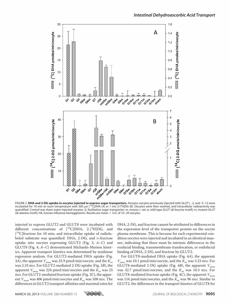
investigated the DHA transport properties of GLUT1, -2, and-5–12. Previous studies (16) had shown that mutating theN-terminal di-leucine motif in rat GLUT8 to a di-alanine motifwas necessary for plasmamembrane expression and functionalcharacterization in oocytes; we therefore studied the transportproperties of selected humanwild type (di-leucine) andmutant(di-alanine) GLUTs in injected oocytes. mRNA encoding theindividual isoforms were injected into oocytes, and 3–5 dayspost-injection, oocytes expressing putative GLUTs were incu-bated with 300 �M [14C]DHA for 10 min, and radiolabeleduptake was assessed. GLUT1 mRNA was injected into oocytesand served as a positive control for GLUT-mediated DHAtransport. 1 mM 2-[3H]DG uptake was also studied and servedas a test for the expression of functional sugar transporters.DHA uptake into oocytes expressing human GLUT2, ratGLUT8 (LL-AA), and wild type and mutant mouse GLUT8(LL-AA) was 5–10-fold greater than the sham-injected oocytes(Fig. 2A). Consistent with previous reports (10), DHA uptakeinto oocytes expressing GLUT1 was 100-fold greater than thesham control (Fig. 2A). DHA transport into GLUT5, -6, -7, -9,-10, -11, and -12 oocytes did not differ from the sham control.Radiolabeled 2-DG uptake into oocytes expressing GLUT1 and-2, rat GLUT8 (LL-AA), and wild type and mutant mouseGLUT8 was significantly greater than sham-injected oocytes(Fig. 2B). In contrast, radiolabeled 2-DG uptake in oocytesinjected with mouse GLUT8 HA and wild type and mutantGLUT6, -7, -9, -10, -11, and -12 mRNA was not significantlydifferent than the shamcontrol.On amole formole basis, 2-DGand DHA uptake was similar in GLUT1-expressing oocytes,whereas for GLUT2-expressing oocytes 2-DGuptakewas�30-fold greater than DHA, and for GLUT8 expressing oocytes2-DG uptake was �5-fold greater than DHA.We next studied the kinetics of DHA, 2-DG, and D-fructose
transport in GLUT2- and GLUT8-expressing oocytes. Oocytes
FIGURE 1. Ascorbic acid and dehydroascorbic acid transport across ratintestine. Rats were gavaged with 12 mg of dehydroascorbic acid (Œ), ascor-bic acid (�), or PBS vehicle (f). Post-gavage plasma ascorbic acid levels weredetermined by HPLC, as described under “Experimental Procedures.” Datarepresent the mean � S.D. of at least 10 rats per gavage treatment. Whencompared with vehicle and ascorbic acid gavage, time-diet interactionsreached significance at 60 min post-DHA gavage (*, p � 0.01 two-tailedpaired t test).
Intestinal Dehydroascorbic Acid Transport
9094 JOURNAL OF BIOLOGICAL CHEMISTRY VOLUME 288 • NUMBER 13 • MARCH 29, 2013
injected to express GLUT2 and GLUT8 were incubated withdifferent concentrations of [14C]DHA, 2-[3H]DG, and[14C]fructose for 10 min, and intracellular uptake of radiola-beled substrate was quantified. DHA, 2-DG, and D-fructoseuptake into oocytes expressing GLUT2 (Fig. 3, A–C) andGLUT8 (Fig. 4, A–C) demonstrated Michaelis-Menten kinet-ics. Apparent transport kinetics was determined by nonlinearregression analysis. For GLUT2-mediated DHA uptake (Fig.3A), the apparent Vmax was 25.9 pmol/min/oocyte, and the Kmwas 2.33 mM. For GLUT2-mediated 2-DG uptake (Fig. 3B), theapparent Vmax was 224 pmol/min/oocytes and the Km was 25mM. ForGLUT2-mediated fructose uptake (Fig. 3C), the appar-ent Vmax was 406 pmol/min/oocytes and Km was 108 mM. Thedifferences inGLUT2 transport affinities andmaximal rates for
DHA, 2-DG, and fructose cannot be attributed to differences inthe expression level of the transporter protein on the oocyteplasma membrane. This is because for each experimental con-dition oocytes were injected and incubated in an identicalman-ner, indicating that there must be intrinsic differences in theexofacial binding, transmembrane translocation, or endofacialbinding of DHA, 2-DG, and fructose by GLUT2.For GLUT8-mediated DHA uptake (Fig. 4A), the apparent
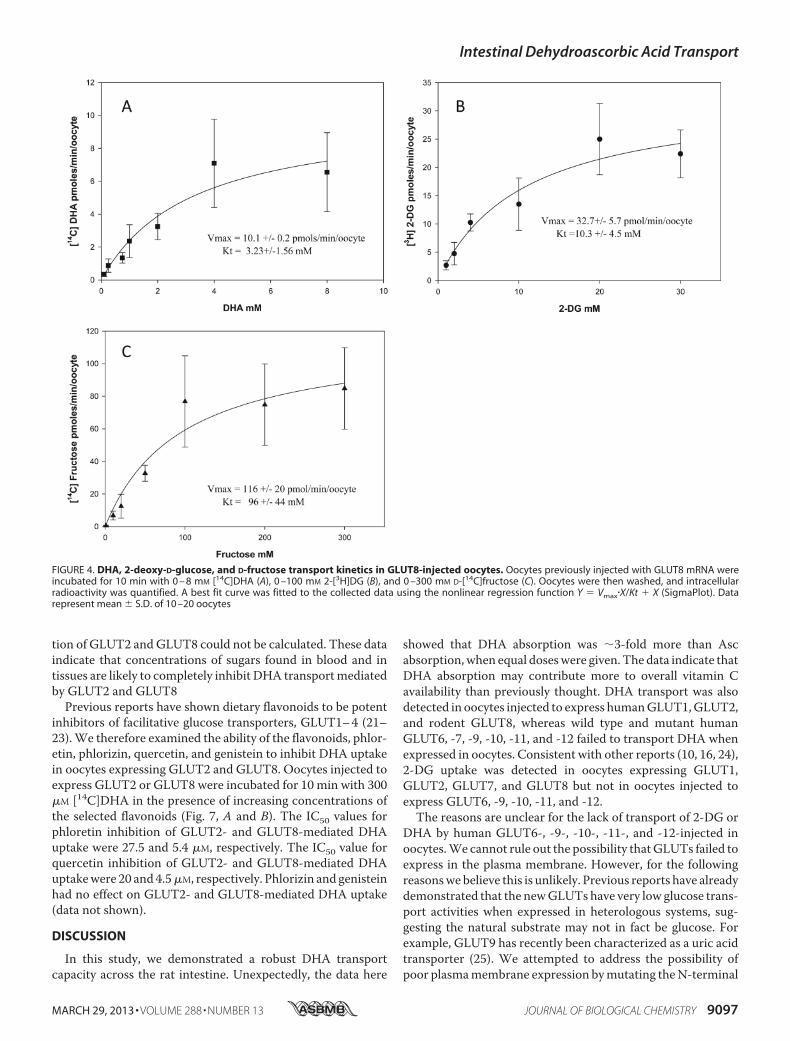
Vmax was 10.1 pmol/min/oocyte, and the Km was 3.23 mM. ForGLUT8-mediated 2-DG uptake (Fig. 4B), the apparent Vmaxwas 32.7 pmol/min/oocytes, and the Km was 10.3 mM. ForGLUT8-mediated fructose uptake (Fig. 4C), the apparent Vmaxwas 116 pmol/min/oocytes, and the Km was 96 mM. Similar toGLUT2, the differences in the transport kinetics of GLUT8 for
FIGURE 2. DHA and 2-DG uptake in oocytes injected to express sugar transporters. Xenopus oocytes previously injected with GLUT1, -2, and -5–12 wereincubated for 10 min at room temperature with 300 �M [14C]DHA (A) or 1 mM 2-[3H]DG (B). Oocytes were then washed, and intracellular radioactivity wasquantified. Control was sham water-injected oocytes. G, facilitative sugar transporter; m, mouse; r, rat; w, wild type GLUT (di-leucine motif); m, mutant GLUT(di-alanine motif); HA, human influenza hemagglutinin. Results are mean � S.D. of 10 –20 oocytes.
Intestinal Dehydroascorbic Acid Transport
MARCH 29, 2013 • VOLUME 288 • NUMBER 13 JOURNAL OF BIOLOGICAL CHEMISTRY 9095

its substrates are most likely to be due to differences in sub-strate binding or translocation. Transport affinities (Km) forDHA were similar in GLUT2- (Fig. 3A) and GLUT8 (Fig. 4A)-expressing oocytes; however, themaximal transport rate (Vmax)for DHA uptake mediated by GLUT2 was 2-fold greater thanthat ofGLUT8. This is unlikely to be due to differences betweenthe protein expression levels of GLUT2 and GLUT8 in oocytes,because the maximal transport rates for 2-DG and fructose byGLUT2 were not 2-fold greater than GLUT8-mediated 2-DGand fructose uptake (2-DG uptake by GLUT2was 7-fold higherthan GLUT8, and D-fructose uptake by GLUT2 was 4-foldhigher than GLUT8). It also unlikely to be due to intracellularreduction of DHA to Asc becoming rate-limiting for DHAtransport, because previous studies have shown that intracellu-lar accumulation of up to 1250 pmol of DHA/min/oocyte israpidly (�10 min) and completely reduced to Asc (10).Differences in the exofacial and endofacial binding affinities
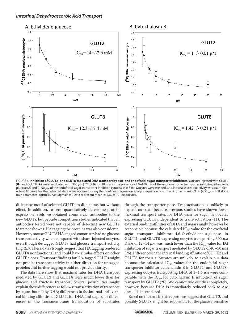
of GLUT2 and GLUT8 for DHA were investigated. Oocytesexpressing GLUT2 or GLUT8 were incubated with 300 �M
[14C]DHA in the presence of increasing concentrations of theexofacial glucose transporter inhibitor 4,6-O-ethylidene glu-cose (Fig. 5A) or increasing concentrations of the endofacialglucose transporter inhibitor cytochalasin B (Fig. 5B). Intracel-
lular uptake of radiolabeled DHA in the presence of inhibitorswas quantified, and best fit curves were fitted using nonlinearregression analysis. The IC50 values for 4,6-O-ethylidene glu-cose inhibition of GLUT2- and GLUT8-mediated DHA uptakewere 14 and 12 mM, respectively. The IC50 values for cytocha-lasin B inhibition of GLUT2- and GLUT8-mediated DHAuptake were 1 and 1.42 �M, respectively. These results suggestDHA binding to the external and internal face of GLUT2 andGLUT8 is comparable. Furthermore, these data suggest thatthe difference in the apparent maximal transport rates ofGLUT2 and GLUT8 for DHA is due to differences in the trans-membrane translocation of the substrate.The in vivo relevance of GLUT2- and GLUT8-mediated
DHA transport is dependent on oxidation of Asc to DHAand on the presence or absence of other substrates that couldinhibit transport. To test whether DHA uptake by GLUT2 andGLUT8 could occur under physiologically relevant concentra-tions of glucose and fructose, oocytes expressing GLUT2 orGLUT8 were incubated for 10 min with 300 �M [14C]DHA inthe presence of increasing concentrations of D-glucose andD-fructose (Fig. 6, A and B). The IC50 values for glucose inhibi-tion of GLUT2- andGLUT8-mediated DHAuptake were 0.366and 0.122 mM, respectively. The IC50 value for fructose inhibi-
FIGURE 3. DHA, 2-deoxy-D-glucose, and D-fructose transport kinetics in GLUT2-injected oocytes. Oocytes previously injected with GLUT2 mRNA wereincubated for 10 min with 0 – 8 mM [14C]DHA (A), 0 –100 mM 2-[3H]DG (B), and 0 –300 mM D-[14C]fructose (C). Oocytes were then washed and intracellularradioactivity quantified. A best fit curve was fitted to the collected data using the nonlinear regression function Y � Vmax�X/Kt � X (SigmaPlot). Data representmean � S.D. of 10 –20 oocytes.
Intestinal Dehydroascorbic Acid Transport
9096 JOURNAL OF BIOLOGICAL CHEMISTRY VOLUME 288 • NUMBER 13 • MARCH 29, 2013
tion of GLUT2 andGLUT8 could not be calculated. These dataindicate that concentrations of sugars found in blood and intissues are likely to completely inhibit DHA transportmediatedby GLUT2 and GLUT8Previous reports have shown dietary flavonoids to be potent
inhibitors of facilitative glucose transporters, GLUT1–4 (21–23).We therefore examined the ability of the flavonoids, phlor-etin, phlorizin, quercetin, and genistein to inhibit DHA uptakein oocytes expressing GLUT2 and GLUT8. Oocytes injected toexpress GLUT2 or GLUT8 were incubated for 10 min with 300�M [14C]DHA in the presence of increasing concentrations ofthe selected flavonoids (Fig. 7, A and B). The IC50 values forphloretin inhibition of GLUT2- and GLUT8-mediated DHAuptake were 27.5 and 5.4 �M, respectively. The IC50 value forquercetin inhibition of GLUT2- and GLUT8-mediated DHAuptakewere 20 and 4.5�M, respectively. Phlorizin and genisteinhad no effect on GLUT2- and GLUT8-mediated DHA uptake(data not shown).
DISCUSSION
In this study, we demonstrated a robust DHA transportcapacity across the rat intestine. Unexpectedly, the data here
showed that DHA absorption was �3-fold more than Ascabsorption,when equal doseswere given. The data indicate thatDHA absorption may contribute more to overall vitamin Cavailability than previously thought. DHA transport was alsodetected in oocytes injected to express humanGLUT1,GLUT2,and rodent GLUT8, whereas wild type and mutant humanGLUT6, -7, -9, -10, -11, and -12 failed to transport DHA whenexpressed in oocytes. Consistent with other reports (10, 16, 24),2-DG uptake was detected in oocytes expressing GLUT1,GLUT2, GLUT7, and GLUT8 but not in oocytes injected toexpress GLUT6, -9, -10, -11, and -12.The reasons are unclear for the lack of transport of 2-DG or
DHA by human GLUT6-, -9-, -10-, -11-, and -12-injected inoocytes.We cannot rule out the possibility thatGLUTs failed toexpress in the plasma membrane. However, for the followingreasonswebelieve this is unlikely. Previous reports have alreadydemonstrated that the newGLUTs have very low glucose trans-port activities when expressed in heterologous systems, sug-gesting the natural substrate may not in fact be glucose. Forexample, GLUT9 has recently been characterized as a uric acidtransporter (25). We attempted to address the possibility ofpoor plasmamembrane expression bymutating theN-terminal
FIGURE 4. DHA, 2-deoxy-D-glucose, and D-fructose transport kinetics in GLUT8-injected oocytes. Oocytes previously injected with GLUT8 mRNA wereincubated for 10 min with 0 – 8 mM [14C]DHA (A), 0 –100 mM 2-[3H]DG (B), and 0 –300 mM D-[14C]fructose (C). Oocytes were then washed, and intracellularradioactivity was quantified. A best fit curve was fitted to the collected data using the nonlinear regression function Y � Vmax�X/Kt � X (SigmaPlot). Datarepresent mean � S.D. of 10 –20 oocytes
Intestinal Dehydroascorbic Acid Transport
MARCH 29, 2013 • VOLUME 288 • NUMBER 13 JOURNAL OF BIOLOGICAL CHEMISTRY 9097
di-leucine motif of selected GLUTs to di-alanine, but withouteffect. In addition, to semi-quantitatively determine proteinexpression levels we obtained commercial antibodies to thenew GLUTs, but peptide competition studies indicated that allantibodies tested were not capable of detecting new GLUTs(data not shown). HA tagging the proteins was also considered.However, mouseGLUT8HA-tagged constructs had no glucosetransport activity when compared with sham-injected oocytes,even though de-tagged GLUT8 had glucose transport activity(Fig. 2B). These data strongly suggest that HA tagging renderedGLUT8 nonfunctional and could have similar effects on otherGLUT clones. Transport findings for HA-tagged GLUTsmightnot predict transport activity in either direction for untaggedproteins and further tagging would not provide clarity.The data here show that maximal rates for DHA transport
mediated by GLUT2 and GLUT8 were much lower than forglucose and fructose transport. Several possibilities mightexplain these differences as follows: transactivation of transportby sugars but not byDHA; differences in the internal and exter-nal binding affinities of GLUTs for DHA and sugars; or differ-ences in the transmembrane translocation of substrates
through the transporter pore. Transactivation is unlikely toexplain our data because previous studies have shown lowermaximal transport rates for DHA than for sugar in oocytesexpressing GLUTs independent to trans-activation (11). Theexternal binding affinities ofDHAand sugarsmight however beresponsible because the calculated IC50 value for the exofacialsugar transport inhibitor 4,6-O-ethyldiene-D-glucose inGLUT2- and GLUT8-expressing oocytes transporting 300 �M
DHA of 12–14 �M was much lower than the IC50 value for EGinhibition of sugar transportmediated byGLUT2 of 40–50mM
(26).Differences in the internal binding affinities ofGLUT2 andGLUT8 for their substrates are unlikely to explain our databecause the calculated IC50 values for the endofacial sugartransporter inhibitor cytochalasin B in GLUT2- and GLUT8-expressing oocytes transporting DHA of 1–1.4 �M were com-parable with the IC50 for cytochalasin B inhibition of sugartransport by GLUT2 (26). We cannot rule out this completely,however, because DHA is immediately reduced back to Asconce it is internalized.Based on the data in this report, we suggest that GLUT2, and
possibly GLUT8,might be responsible for the glucose-sensitive
FIGURE 5. Inhibition of GLUT2- and GLUT8-mediated DHA transport by exo- and endofacial sugar transporter inhibitors. Oocytes injected with GLUT2(●) and GLUT8 (Œ) were incubated with 300 �M [14C]DHA for 10 min in the presence of 0 –100 mM of the exofacial sugar transporter inhibitor, ethyldieneglucose (A) and 0 –50 �M of the endofacial sugar transporter inhibitor, cytochalasin B (B). Oocytes were washed, and internalized radioactivity was quantified.A best fit curve for the collected data were obtained using the nonlinear regression analysis equation, y � min � (max � min)/1 � (x/IC50) � Hill slopefour-parameter logistic curve (SigmaPlot). Data represent mean � S.D. of 10 –20 oocytes.
Intestinal Dehydroascorbic Acid Transport
9098 JOURNAL OF BIOLOGICAL CHEMISTRY VOLUME 288 • NUMBER 13 • MARCH 29, 2013

transport of luminal DHA across the intestine. GLUT2 is afacilitative glucose/fructose transporter expressed in the gut,liver, pancreatic beta cell, and kidney. In the classic model ofintestinal sugar transport, GLUT2 is expressed on the basolat-eral membrane and is responsible for the transport of glucose/fructose from the enterocyte into the portal circulation. Morerecent studies however have shown that luminal glucose acti-vates the sweet taste receptor, T1R2/3, resulting in GLUT2being expressed in the apical membrane (27). In this report,GLUT2-expressing oocytes transported DHA; and rat intes-tine, a tissue that is known to express GLUT2, also transportedDHA, as shown by a robust appearance of Asc in the circula-tion. Because the apical membrane glucose/galactose trans-porter, SGLT1, and the fructose transporter, GLUT5, do nottransport DHA (10), luminal uptake of DHA into the entero-cyte could be mediated by apical GLUT2. If correct, this sug-gests luminal DHA, which is a structural analog of glucose,regulates apical GLUT2 expression levels via T1r2/3 activation.The regulation of gastrointestinal function by DHA activationof the sweet taste receptor warrants further investigation.GLUT8 may also contribute toward intestinal DHA trans-
port. GLUT8 is expressed in the small intestine (12), and in our
report rodent GLUT8 transported DHA when expressed inoocytes. In previous reports a di-leucine motif in rat GLUT8had been identified that was responsible for preventing theexpression of the transporter in the plasmamembrane (16, 17).In this study, the di-leucinemotif did not prevent the functionalexpression of mouse GLUT8. We cannot explain why theremay be a difference in the motifs responsible for regulating theplasmamembrane expression of rat andmouseGLUT8, but thedata suggest other motifs may be important.The extent towhichAsc is oxidized in the intestinal lumen to
DHA is unknown.However, the robust increase in systemicAsclevels that occurred following DHA gavage when comparedwithAsc gavage suggests thatAsc oxidation and uptake ofDHAby apical GLUT2 (and possibly GLUT8) might substantiallycontribute to enteral absorption of vitamin C. DHA enteringthe enterocyte would be expected to be rapidly reduced back toAsc by NADPH-dependent thioredoxin reductase or glutathi-one-dependent DHA reductase (28). Asc would then be trans-ported across the basolateral membrane into the portal circu-lation by a mechanism that has yet to be identified. Analternative possibility is that intracellular DHA is not reducedback to Asc, and instead it effluxes the enterocyte via basolat-
FIGURE 6. Inhibition of GLUT2- and GLUT8-mediated DHA uptake by sugars. Oocytes injected with GLUT2 (●) and GLUT8 (Œ) were incubated with 300 �M
[14C]DHA for 10 min in the presence of 0 –100 mM D-glucose (A) and 0 –100 mM D-fructose (B). Oocytes were washed, and internalized radioactivity wasquantified. A best fit curve for the collected data was obtained using nonlinear regression analysis. Data represent mean � S.D. of 10 –20 oocytes.
Intestinal Dehydroascorbic Acid Transport
MARCH 29, 2013 • VOLUME 288 • NUMBER 13 JOURNAL OF BIOLOGICAL CHEMISTRY 9099

eral GLUT2; it could then be reduced back to ascorbic acid viaa basolateralmembrane-localized oxidoreductase protein, suchas hephaestin, which is involved in the conversion of Fe2� toFe3� in portal blood (29).Previous studies have shown that flavonoids present in vita-
min C-rich foods (fruits and vegetables) can inhibit sodium-de-pendent Asc transporter-mediated Asc transport (30) andGLUT-mediated DHA transport (21). Physiologic levels of glu-cose can also inhibit GLUT-mediated DHA transport (10). Inthis study, the IC50 value for D-glucose inhibition ofDHA trans-port by GLUT2 and GLUT8 in oocytes is 0.366 and 0.122 mM,respectively. Quercetin and phloretin were also potent inhibi-tors of DHA transport in oocytes expressing GLUT2 andGLUT8. Consumption of sugar-sweetened beverages or fruitsjuices containing free sugars and flavonoids might thereforeresult in diminished intestinal absorption of DHA, particularlyin the proximal regions of the small intestine, and result in alower predicted bioavailability for vitamin C. For meals con-taining complex carbohydrates, digestion and liberation of freesugars occur principally in the jejunum. We would thereforeexpect DHA uptake to be largely unimpeded in the duodenumand possibly the ileum.
In the intestine, glucose-insensitive DHA transport mightoccur if a fraction of luminal DHA is reduced back to Asc,which is then transported across the apical membrane by theglucose-insensitive SVCT1. For this to occur, a reducingagent must be present at the luminal surface. Duodenal cyto-chrome b561 (DcytB) is an apical membrane-expressed pro-tein that catalyzes the reduction of Fe3� to Fe2� and facili-tates iron absorption (31). Interestingly, iron absorption isnormal in DcytB knock-out mice (32), suggesting the proteinhas alternative functions in vivo. Erythrocyte Cyt b561 isbelieved to be responsible for extracellular recycling of Ascvia reduction of mono-DHA/DHA (33). We therefore spec-ulate that DcytB acts as an apical membrane electron donorreducing DHA to Asc.In summary, in this report we have shown the intestine can
absorb both Asc and DHA. Intestinal uptake of Asc is mediatedby the Na�-dependent transporter SVCT1. Kinetics studies inoocytes suggest GLUT2 andGLUT8 are low affinity/low capac-ity DHA transporters responsible for intestinal uptake of DHA.Furthermore, vitamin C bioavailability may be reduced byluminal dietary factors inhibiting DHA transport mediated byintestinal GLUT2 and GLUT8.
FIGURE 7. Inhibition of GLUT2- and GLUT8-mediated DHA uptake by flavonoids. Oocytes injected with GLUT2 (●) and GLUT8 (Œ) were incubated with 300�M [14C]DHA for 10 min in the presence of 0 –100 �M phloretin (A) and 0 –100 �M quercetin (B). Oocytes were washed, and internalized radioactivity wasquantified. A best fit curve for the collected data was obtained using nonlinear regression analysis. Data represent mean � S.D. of 10 –20 oocytes.
Intestinal Dehydroascorbic Acid Transport
9100 JOURNAL OF BIOLOGICAL CHEMISTRY VOLUME 288 • NUMBER 13 • MARCH 29, 2013

REFERENCES1. Koshiishi, I., Mamura, Y., Liu, J., and Imanari, T. (1998) Degradation of
dehydroascorbate to 2,3-diketogulonate in blood circulation. Biochim.Biophys. Acta 1425, 209–214
2. Tsukaguchi,H., Tokui, T.,Mackenzie, B., Berger, U.V., Chen,X. Z.,Wang,Y., Brubaker, R. F., and Hediger, M. A. (1999) A family of mammalianNa�-dependent L-ascorbic acid transporters. Nature 399, 70–75
3. Sotiriou, S., Gispert, S., Cheng, J.,Wang, Y., Chen, A., Hoogstraten-Miller,S., Miller, G. F., Kwon, O., Levine, M., Guttentag, S. H., and Nussbaum,R. L. (2002) Ascorbic acid transporter Slc23a1 is essential for vitamin Ctransport into the brain and for perinatal survival. Nat. Med. 8, 514–517
4. Li, H., Tu, H., Wang, Y., and Levine, M. (2012) Vitamin C in mouse andhuman red blood cells: an HPLC assay. Anal. Biochem. 426, 109–117
5. Corpe, C. P., Tu, H., Eck, P., Wang, J., Faulhaber-Walter, R., Schnermann,J., Margolis, S., Padayatty, S., Sun, H., Wang, Y., Nussbaum, R. L., Espey,M. G., and Levine, M. (2010) Vitamin C transporter Slc23a1 links renalreabsorption, vitamin C tissue accumulation, and perinatal survival inmice. J. Clin. Invest. 120, 1069–1083
6. Oliveira, J. E., Pearson, W. N., and Darby, W. J. (1959) Clinical usefulnessof the oral ascorbic acid tolerance test in scurvy. Am. J. Clin. Nutr. 7,630–633
7. Goldman, H. M., Gould, B. S., andMunro, H. N. (1981) The antiscorbuticaction of L-ascorbic acid and D-isoascorbic acid (erythorbic acid) in theguinea pig. Am. J. Clin. Nutr. 34, 24–33
8. Ogiri, Y., Sun, F., Hayami, S., Fujimura, A., Yamamoto, K., Yaita, M., andKojo, S. (2002) Very low vitamin C activity of orally administered L-dehy-droascorbic acid. J. Agric. Food Chem. 50, 227–229
9. Otsuka, M., Kurata, T., and Arakawa, N. (1986) Antiscorbutic effect ofdehydro-L-ascorbic acid in vitamin C-deficient guinea pigs. J. Nutr. Sci.Vitaminol. 32, 183–190
10. Rumsey, S. C., Kwon, O., Xu, G.W., Burant, C. F., Simpson, I., and Levine,M. (1997) Glucose transporter isoforms GLUT1 and GLUT3 transportdehydroascorbic acid. J. Biol. Chem. 272, 18982–18989
11. Rumsey, S. C., Daruwala, R., Al-Hasani, H., Zarnowski, M. J., Simpson,I. A., and Levine, M. (2000) Dehydroascorbic acid transport by GLUT4 inXenopus oocytes and isolated rat adipocytes. J. Biol. Chem. 275,28246–28253
12. Romero, A., Gomez, O., Terrado, J., andMesonero, J. E. (2009) ExpressionofGLUT8 inmouse intestine: identification of alternative spliced variants.J. Cell. Biochem. 106, 1068–1078
13. Manolescu, A. R., Witkowska, K., Kinnaird, A., Cessford, T., and Cheese-man, C. (2007) Facilitated hexose transporters: new perspectives on formand function. Physiology 22, 234–240
14. Washko, P. W., Hartzell, W. O., and Levine, M. (1989) Ascorbic acidanalysis using high-performance liquid chromatography with coulomet-ric electrochemical detection. Anal. Biochem. 181, 276–282
15. Burant, C. F., and Bell, G. I. (1992) Mammalian facilitative glucose trans-porters: evidence for similar substrate recognition sites in functionallymonomeric proteins. Biochemistry 31, 10414–10420
16. Ibberson, M., Uldry, M., and Thorens, B. (2000) GLUTX1, a novel mam-malian glucose transporter expressed in the central nervous system andinsulin-sensitive tissues. J. Biol. Chem. 275, 4607–4612
17. Lisinski, I., Schürmann, A., Joost, H. G., Cushman, S. W., and Al-Hasani,H. (2001) Targeting of GLUT6 (formerly GLUT9) and GLUT8 in rat ad-ipose cells. Biochem. J. 358, 517–522
18. Soreq, H., and Seidman, S. (1992) Xenopus oocyte microinjection: fromgene to protein.Methods Enzymol. 207, 225–265
19. Vera, J. C., Rivas, C. I., Fischbarg, J., and Golde, D. W. (1993) Mammalianfacilitative hexose transporters mediate the transport of dehydroascorbicacid. Nature 364, 79–82
20. Corpe, C. P., Lee, J. H., Kwon, O., Eck, P., Narayanan, J., Kirk, K. L., andLevine, M. (2005) 6-Bromo-6-deoxy-L-ascorbic acid: an ascorbate analogspecific for Na�-dependent vitamin C transporter but not glucose trans-porter pathways. J. Biol. Chem. 280, 5211–5220
21. Kwon, O., Eck, P., Chen, S., Corpe, C. P., Lee, J. H., Kruhlak, M., andLevine, M. (2007) Inhibition of the intestinal glucose transporter GLUT2by flavonoids. FASEB J. 21, 366–377
22. Smith, R. M., Tiesinga, J. J., Shah, N., Smith, J. A., and Jarett, L. (1993)Genistein inhibits insulin-stimulated glucose transport and decreases im-munocytochemical labeling of GLUT4 carboxyl terminus without affect-ing translocation of GLUT4 in isolated rat adipocytes: additional evidenceof GLUT4 activation by insulin. Arch. Biochem. Biophys. 300, 238–246
23. Park, J. B. (1999) Flavonoids are potential inhibitors of glucose uptake inU937 cells. Biochem. Biophys. Res. Commun. 260, 568–574
24. Li, Q., Manolescu, A., Ritzel, M., Yao, S., Slugoski, M., Young, J. D., Chen,X. Z., and Cheeseman, C. I. (2004) Cloning and functional characteriza-tion of the human GLUT7 isoform SLC2A7 from the small intestine.Am. J. Physiol. Gastrointest. Liver Physiol. 287, G236–G242
25. Bibert, S., Hess, S. K., Firsov, D., Thorens, B., Geering, K., Horisberger,J. D., andBonny,O. (2009)MouseGLUT9: evidences for a urate uniporter.Am. J. Physiol. Renal Physiol. 297, F612–F619
26. Colville, C. A., Seatter, M. J., Jess, T. J., Gould, G. W., and Thomas, H. M.(1993) Kinetic analysis of the liver-type (GLUT2) and brain-type (GLUT3)glucose transporters inXenopus oocytes: substrate specificities and effectsof transport inhibitors. Biochem. J. 290, 701–706
27. Mace, O. J., Affleck, J., Patel, N., and Kellett, G. L. (2007) Sweet tastereceptors in rat small intestine stimulate glucose absorption through api-cal GLUT2. J. Physiol. 582, 379–392
28. May, J. M. (2002) Recycling of vitamin C by mammalian thioredoxin re-ductase.Methods Enzymol. 347, 327–332
29. Anderson, G. J., and Frazer, D. M. (2005) Recent advances in intestinaliron transport. Curr. Gastroenterol. Rep. 7, 365–372
30. Song, J., Kwon, O., Chen, S., Daruwala, R., Eck, P., Park, J. B., and Levine,M. (2002) Flavonoid inhibition of sodium-dependent vitamin C trans-porter 1 (SVCT1) and glucose transporter isoform 2 (GLUT2), intestinaltransporters for vitamin C and glucose. J. Biol. Chem. 277, 15252–15260
31. Latunde-Dada, G. O., Van der Westhuizen, J., Vulpe, C. D., Anderson,G. J., Simpson, R. J., and McKie, A. T. (2002) Molecular and functionalroles of Dcytb in iron metabolism. Blood Cells Mol. Dis. 29, 356–360
32. McKie, A. T. (2008) The role of Dcytb in iron metabolism: an update.Biochem. Soc. Trans. 36, 1239–1241
33. Su, D., May, J. M., Koury, M. J., and Asard, H. (2006) Human erythrocytemembranes contain a cytochrome b561 that may be involved in extracel-lular ascorbate recycling. J. Biol. Chem. 281, 39852–39859
Intestinal Dehydroascorbic Acid Transport
MARCH 29, 2013 • VOLUME 288 • NUMBER 13 JOURNAL OF BIOLOGICAL CHEMISTRY 9101
Copyright © 2022 FDOKUMEN