International Society of Human and Animal Mycology (ISHAM)-ITS reference DNA barcoding database-the...
25
Medical Mycology, 2015, 00, 1–25 doi: 10.1093/mmy/myv008 Advance Access Publication Date: 0 2015 Review Article Review Article International Society of Human and Animal Mycology (ISHAM)-ITS reference DNA barcoding database—the quality controlled standard tool for routine identification of human and animal pathogenic fungi Laszlo Irinyi 1 , Carolina Serena 1, 22 , Dea Garcia-Hermoso 2 , Michael Arabatzis 3 , Marie Desnos-Ollivier 2 , Duong Vu 4 , Gianluigi Cardinali 5 , Ian Arthur 6 , Anne-C ´ ecile Normand 7 , Alejandra Giraldo 8 , Keith Cassia da Cunha 8 , Marcelo Sandoval-Denis 8 , Marijke Hendrickx 9 , Angela Satie Nishikaku 10 , Analy Salles de Azevedo Melo 10 , Karina Bellinghausen Merseguel 10 , Aziza Khan 1 , Juliana Alves Parente Rocha 11 , Paula Sampaio 12 , Marcelo Ribeiro da Silva Briones 13 , Renata Carmona e Ferreira 13 , Mauro de Medeiros Muniz 14 , Laura Rosio Casta ˜ n´ on-Olivares 15 , Daniel Estrada-Barcenas 15 , Carole Cassagne 7 , Charles Mary 7 , Shu Yao Duan 1 , Fanrong Kong 16 , Annie Ying Sun 17 , Xianyu Zeng 16 , Zuotao Zhao 16 , Nausicaa Gantois 18 , Franc ¸ oise Botterel 19 , Barbara Robbertse 20 , Conrad Schoch 20 , Walter Gams 4 , David Ellis 21 , Catriona Halliday 16 , Sharon Chen 1, 16 , Tania C. Sorrell 1 , Renaud Piarroux 7 , Arnaldo L. Colombo 10 ,C´ elia Pais 12 , Sybren de Hoog 4 , Rosely Maria Zancop ´ e-Oliveira 14 , Maria Lucia Taylor 15 , Conchita Toriello 15 ,C´ elia Maria de Almeida Soares 11 , Laurence Delhaes 18 , Dirk Stubbe 9 , Franc ¸ oise Dromer 2 , St ´ ephane Ranque 7 , Josep Guarro 8 , Jose F. Cano-Lira 8 , Vincent Robert 4 , Aristea Velegraki 3 and Wieland Meyer 1, ∗ 1 Molecular Mycology Research Laboratory, Centre for Infectious Diseases and Microbiology, Sydney Medical School-Westmead Hospital, Marie Bashir Institute for Infectious Diseases and Bioscurity, Uni- versity of Sydney, Westmead Millennium Institute, Sydney, Australia, 2 Institut Pasteur, National Reference Center for Invasive Mycoses and Antifungals, Molecular Mycology Unit; CNRS URA3012, Paris, France, 3 Mycology Research Laboratory, Department of Microbiology, Medical School, the University of Athens Hellenic Collection of Pathogenic Fungi (UOA/HCPF), National and Kapodistrian University of Athens, Athens, Greece, 4 CBS-KNAW, Fungal Biodiversity Centre, Utrecht, The Netherlands, 5 Department of Pharmaceutical Sciences-Universit ` a degli Studi di Perugia, Perugia, Italy, 6 Mycology Laboratory, De- partment of Microbiology and Infectious Diseases, PathWest Laboratory Medicine WA, QEII Medical C The Author 2015. Published by Oxford University Press on behalf of The International Society for Human and Animal Mycology. All rights reserved. For permissions, please e-mail: [email protected] 1 Medical Mycology Advance Access published March 22, 2015 at National Institutes of Health Library on March 29, 2015 http://mmy.oxfordjournals.org/ Downloaded from
-
Upload
independent -
Category
Documents
-
view
0 -
download
0
Transcript of International Society of Human and Animal Mycology (ISHAM)-ITS reference DNA barcoding database-the...

Medical Mycology, 2015, 00, 1–25doi: 10.1093/mmy/myv008
Advance Access Publication Date: 0 2015Review Article
Review Article
International Society of Human and Animal
Mycology (ISHAM)-ITS reference DNA barcoding
database—the quality controlled standard tool
for routine identification of human and animal
pathogenic fungi
Laszlo Irinyi1, Carolina Serena1,22, Dea Garcia-Hermoso2,
Michael Arabatzis3, Marie Desnos-Ollivier2, Duong Vu4,
Gianluigi Cardinali5, Ian Arthur6, Anne-Cecile Normand7,
Alejandra Giraldo8, Keith Cassia da Cunha8, Marcelo Sandoval-Denis8,
Marijke Hendrickx9, Angela Satie Nishikaku10,
Analy Salles de Azevedo Melo10, Karina Bellinghausen Merseguel10,
Aziza Khan1, Juliana Alves Parente Rocha11, Paula Sampaio12,
Marcelo Ribeiro da Silva Briones13, Renata Carmona e Ferreira13,
Mauro de Medeiros Muniz14, Laura Rosio Castanon-Olivares15,
Daniel Estrada-Barcenas15, Carole Cassagne7, Charles Mary7,
Shu Yao Duan1, Fanrong Kong16, Annie Ying Sun17, Xianyu Zeng16,
Zuotao Zhao16, Nausicaa Gantois18, Francoise Botterel19,
Barbara Robbertse20, Conrad Schoch20, Walter Gams4, David Ellis21,
Catriona Halliday16, Sharon Chen1,16, Tania C. Sorrell1, Renaud Piarroux7,
Arnaldo L. Colombo10, Celia Pais12, Sybren de Hoog4,
Rosely Maria Zancope-Oliveira14, Maria Lucia Taylor15,
Conchita Toriello15, Celia Maria de Almeida Soares11, Laurence Delhaes18,
Dirk Stubbe9, Francoise Dromer2, Stephane Ranque7, Josep Guarro8,
Jose F. Cano-Lira8, Vincent Robert4, Aristea Velegraki3 and
Wieland Meyer1,∗
1Molecular Mycology Research Laboratory, Centre for Infectious Diseases and Microbiology, SydneyMedical School-Westmead Hospital, Marie Bashir Institute for Infectious Diseases and Bioscurity, Uni-versity of Sydney, Westmead Millennium Institute, Sydney, Australia, 2Institut Pasteur, National ReferenceCenter for Invasive Mycoses and Antifungals, Molecular Mycology Unit; CNRS URA3012, Paris, France,3Mycology Research Laboratory, Department of Microbiology, Medical School, the University of AthensHellenic Collection of Pathogenic Fungi (UOA/HCPF), National and Kapodistrian University of Athens,Athens, Greece, 4CBS-KNAW, Fungal Biodiversity Centre, Utrecht, The Netherlands, 5Department ofPharmaceutical Sciences-Universita degli Studi di Perugia, Perugia, Italy, 6Mycology Laboratory, De-partment of Microbiology and Infectious Diseases, PathWest Laboratory Medicine WA, QEII Medical
C© The Author 2015. Published by Oxford University Press on behalf of The International Society for Human and Animal Mycology.All rights reserved. For permissions, please e-mail: [email protected]
1
Medical Mycology Advance Access published March 22, 2015 at N
ational Institutes of Health L
ibrary on March 29, 2015
http://mm
y.oxfordjournals.org/D
ownloaded from
2 Medical Mycology, 2015, Vol. 00, No. 00
Centre, Nedlands, Western Australia, Australia, 7Parasitology - Mycology, APHM, CHU Timone-Adultes,Marseille, France; Aix-Marseille University, UMR MD3 IP-TPT, Marseille, France, 8Unitat de Micro-biologia, Facultat de Medicina i Ciencies de la Salut, IISPV, Universitat Rovira i Virgili, Reus, Spain,9BCCM/IHEM, Biomedical fungi and yeasts collection, Scientific Institute of Public Health, Brussels,Belgium, 10Laboratorio Especial de Micologia, Escola Paulista de Medicina, Universidade Federal deSao Paulo, Sao Paulo, Brazil, 11Universidade Federal de Goias, Instituto de Ciencias Biologicas, Labo-ratorio de Biologia Molecular, Goiania, Goias, Brazil, 12Centre of Molecular and Environmental Biology(CBMA), Biology Department, School of Sciences, University of Minho, Braga, Portugal, 13Laboratoriode Genomica e Biocomplexidade Evolutiva, Escola Paulista de Medicina, Universidade Federal de SaoPaulo, Sao Paulo, Brazil, 14Instituto de Pesquisa Clınica Evandro Chagas (IPEC), Fundacao Oswaldo Cruz(Fiocruz), Rio de Janeiro, Brazil, 15Facultad de Medicina, Departamento de Microbiologıa y Parasitologıa(Unidad de Micologıa), Universidad Nacional Autonoma de Mexico, Ciudad de Mexico, Mexico, 16Centrefor Infectious Diseases and Microbiology, Westmead Hospital, Westmead, NSW, Australia, 17School ofPaediatrics and Reproductive Health, University of Adelaide, Adelaide, SA, Australia; Robinson Institute,University of Adelaide, Adelaide, SA, Australia, 18BDEEP-EA4547, CIIL, Institut Pasteur de Lille, CHU deLille, Universite de Lille2, Lille, France, 19Unite de Parasitologie – Mycologie, Dynamyc Team, CHU HenriMondor, AP-HP, Creteil, France, 20National Center for Biotechnology Information, National Library ofMedicine, National Institutes of Health, Bethesda, Maryland, USA, 21Mycology and Infectious Diseases,SA Pathology, University of Adelaide, Adelaide, SA, Australia and 22Unitat de Recerca, Hospital JoanXXIII, Institut de Investigacio Sanitaria Rovira I Virgili (IISPV), Universitat Rovira i Virgili, Tarragona,Spain
*To whom correspondence should be addressed. Prof. Wieland Meyer, Molecular Mycology Research Laboratory, MBI,CIDM, Level 4, Room O.4.04, 176 Hawkesbury Road, Westmead, SW 2145, Australia, Tel: +61-2-86273430;Fax +61-2-98915317; E-mail: [email protected]
Received 6 October 2014; Revised 22 December 2014; Accepted 19 January 2015
Abstract
Human and animal fungal pathogens are a growing threat worldwide leading to emerg-ing infections and creating new risks for established ones. There is a growing needfor a rapid and accurate identification of pathogens to enable early diagnosis and tar-geted antifungal therapy. Morphological and biochemical identification methods aretime-consuming and require trained experts. Alternatively, molecular methods, suchas DNA barcoding, a powerful and easy tool for rapid monophasic identification, of-fer a practical approach for species identification and less demanding in terms oftaxonomical expertise. However, its wide-spread use is still limited by a lack of quality-controlled reference databases and the evolving recognition and definition of new fungalspecies/complexes. An international consortium of medical mycology laboratories wasformed aiming to establish a quality controlled ITS database under the umbrella of theISHAM working group on “DNA barcoding of human and animal pathogenic fungi.” Anew database, containing 2800 ITS sequences representing 421 fungal species, provid-ing the medical community with a freely accessible tool at http://www.isham.org/ andhttp://its.mycologylab.org/ to rapidly and reliably identify most agents of mycoses, wasestablished. The generated sequences included in the new database were used to eval-uate the variation and overall utility of the ITS region for the identification of pathogenicfungi at intra-and interspecies level. The average intraspecies variation ranged from 0 to
at National Institutes of H
ealth Library on M
arch 29, 2015http://m
my.oxfordjournals.org/
Dow
nloaded from

Irinyi et al. 3
2.25%. This highlighted selected pathogenic fungal species, such as the dermatophytesand emerging yeast, for which additional molecular methods/genetic markers are re-quired for their reliable identification from clinical and veterinary specimens.
Key words: fungal identification, DNA barcoding, ITS region, reference ITS database, intraspecies/interspeciesgenetic diversity.
Introduction
The number of human and animal fungal infections, rang-ing from superficial infections of the nails and skin, throughmucocutaneous candidiasis to invasive fungal infections,have significantly increased over the last three decades,causing serious public health burdens and increased risk ofbiodiversity loss among animal species [1,2]. In humans, su-perficial infections affect an estimated 25% ( = 1.7 billion)individuals world-wide. Oropharyngeal or genital mucosalinfections are also common and can be disabling. For exam-ple, an estimated 75% of women of childbearing age suffer-ing from vulvovaginitis, mainly caused by Candida species[3], which are the third most common opportunistic fungaldisease agents after Aspergillus spp. worldwide [1]. Inva-sive fungal diseases are of great concern, due to their highmortality that can exceed 50%. More than 90% of fungal-related deaths are caused by four fungal genera: Aspergillus,Candida, Cryptococcus, and Pneumocystis [1,4,5]. Delaysin diagnosis are not only associated with high mortalitybut also severe organ dysfunction, for example, respiratoryfailure (endemic fungal infections and chronic pulmonaryaspergillosis), neurologic deficits (endemic fungal infectionsand cryptococcosis) [6], blindness and visual impairment(fungal keratitis) [7]. To better understand, control, andtreat these diseases, more rapid and accurate identificationof the causal agents is essential.
DNA barcoding, first proposed by Hebert et al. [8], uti-lizes DNA sequences to standardize the identification oforganisms from all kingdoms to the species level by com-parison to a reference collection of well-identified species.The principle behind barcoding is that species identifica-tion must be accurate, fast, cost-effective, culture indepen-dent, universally accessible, and feasible for nonexperts [9].As a consequence, its popularity as a species identificationtool has drastically increased. Barcodes are short diversegenetic sequences (500–800 bp) that are flanked by con-served regions allowing for the design of universal primers.From a pragmatic perspective, a universal sequence suit-able for all kingdoms would be ideal, but the identificationof a universal genetic region for a wide range of taxa re-mains elusive. The key concepts underlying barcoding arethat the interspecies distances should exceed intraspeciesdistances, creating a barcoding gap [10], and that identifi-
cation is straightforward when a sequence is unique to asingle species and constant within each species [8,11,12].The most important question in barcoding is: Howaccurate and reliable are the delineation and identificationof a species using a single gene?
The correct identification of fungi is essential for manybiological purposes, such as the assessment of biodiversity,taxonomy and species conservation [9,13]. It is manda-tory for clinical diagnosis and early initiation of appropri-ate antifungal therapy. Traditional identification based onmorphology and biochemistry of pathogenic fungi is time-consuming and requires a certain level of morphologicaland taxonomical expertise. To overcome these limitations,DNA barcoding was evaluated in fungi, targeting numer-ous genetic loci, including COX1 [14], protein-coding geneslike RNA polymerase I and II [15–19], partial translationelongation factor 1-α [20–22], β-tubulin [23], and the in-ternal transcribed spacer (ITS) regions [24,25]. The proteincoding genes have proven to be a powerful tool for speciesdelimitation, providing a high level of phylogenetic reso-lution and information [21,26,27]. However, the primersused to amplify these regions are usually restricted to spe-cific taxa and amplification can often be problematic [16].In contrast, the ITS regions are easily amplified with univer-sal primers that are compatible among most fungal species.It has shown sufficient genetic variability for identificationat interspecies level, and has been adopted as the officialstandard barcoding region for fungi [28]. However, use ofthe ITS region as a barcode has been criticized by Kiss [29]because of its inability to distinguish many closely relatedfungal species. In addition, for some fungi, the ITS regionsalone do not provide accurate identification to species level[30]. In some groups of fungi (Aspergillus, Colletotrichum)the interspecies variation is insignificant [31,32] and inother groups (Glomeromycota, Chytridiomycota) the di-versity within species is too high [33,34] Fungal genomesmay contain more than 200 copies of the ribosomal re-gion [35,36] dispersed over one or more chromosomal lo-cations [37]. This results in polymorphism within a genomeof one individual [38,39]. Intragenomic diversity is mainlyexplained by concerted evolutionary processes, for exam-ple, unequal crossing over between repeat units, gene con-version or gene amplification [39,40].
at National Institutes of H
ealth Library on M
arch 29, 2015http://m
my.oxfordjournals.org/
Dow
nloaded from
4 Medical Mycology, 2015, Vol. 00, No. 00
Despite these limitations the ITS region has been used inmolecular identification and phylogenetic studies of humanpathogenic fungi [41–48] long before its selection as theofficial fungal DNA barcode. The ITS sequences in pub-licly accessible databases are used routinely by the med-ical community to identify fungi at the species level onthe basis of matching sequences. However, its widespreadapplication has been compromised by the deposition of in-correctly identified or incomplete sequences in the com-monly used public databases of the International Nu-cleotide Sequence Database Collaboration (INSDC) [49].This includes GenBank [50], at the National Center forBiotechnology Information (NCBI), which is the major nu-cleotide sequence depository and is widely utilised by clin-ical microbiologists and the scientific community [51,52].Because GenBank acts primarily as an archive, many se-quences submitted have been annotated with incorrect orpoorly defined species names. It has also been shown thatmore than 10% of the publicly available fungal ITS se-quences were annotated incorrectly at species level [53]. Asa consequence, a number of curated ITS databases havebeen created to ensure the correct identification of fungalspecies, for example, within the Barcode of Life Data System(BOLD) [54] and UNITE [55]. Partially in response to re-quests to allow third party annotation of GenBank recordsNCBI has also initiated a curated database RefSeq TargetedLoci (RTL) [56] that will provide a limited set of curated se-quences obtained from type and verified material [57]. In asecond, broader approach NCBI is currently annotating thetype material associated with taxonomic names. This willallow type related searches to be conducted across multi-ple sequence markers or whole genomes [58]. Other refer-ence databases are available for specific taxonomic groups,for example, Fusarium [59] and Aspergillus [60]. The defi-ciency of these reference databases with respect to humanpathogenic fungi is the limited number of medically impor-tant fungal species contained within them. The demand forcurated, reliable reference databases has increased signifi-cantly due to diminishing expertise in fungal morphologyand its increasing replacement by the use of sequencing infungal diagnostic laboratories.
To address these issues, a working group of the Interna-tional Society for Human and Animal Mycology (ISHAM)on “Barcoding of Medical Fungi” was established in 2011[61]. The working group identified the need to: (a) gener-ate a medical barcode database by incorporating existingfungal group-specific databases; (b) extend the number ofquality-controlled ITS sequences to cover all medically im-portant fungal species; (c) evaluate the value of ITS as abarcode at intra-and interspecies level; (d) eventually incor-porate these sequences into the BOLD database; (e) UNITE;
and (f) achieve a species status as “quality controlled refer-ence sequences” for those sequences within RTL at NCBI.
The main objective of this study was to generate a pub-licly available, quality-controlled, ITS reference databasefor human and animal pathogenic fungal species and toevaluate the applicability of ITS sequences (the official bar-code for fungi) as a genetic marker for species identifica-tion. The secondary aim was to highlight fungal taxa whereadditional genetic sequence information is recommendedbeyond the ITS for a more accurate identification.
Materials and methods
Generating the database
The ISHAM-ITS reference database is a result of an inter-national collaboration between 14 medical mycology lab-oratories representing three continents (Table 1). The con-tributors provided a total of 2945 ITS sequences. Specieswere identified based on polyphasic identification includingmorphology, biochemical and physiological tests when ap-propriate and sequencing. After collecting all the data, theoverall identity of sequences obtained from more than twostrains per species was determined, including available typestrains. In the case of species with less than two strains,trace files were checked for the quality and integrity of se-quences. A total of 145 sequences that did not meet theinclusion criteria were discarded, as well as sequences thatwere misidentified or not identified to species level. Eachtaxon was provided with the taxonomic name, taking intoaccount the “One Name = One Fungus” concept of theInternational Code of Nomenclature for algae, fungi andplants (ICN) [62]. The current taxonomical names wereprovided by using online nomenclature data resources suchas MycoBank [63,64], Index Fungorum [65], the latest edi-tion of The Yeasts [66], as well as the latest publicationsand consulting taxonomical experts of specific taxa. Wherepossible, former anamorph or teleomorph names and themost-used synonyms were also listed to facilitate readingfor clinicians.
DNA isolation, amplification and sequencing
DNA was isolated and purified from cultures using themethods routinely used in the contributing laboratories. Anumber of fungal-specific universal primers (Table 2) wereused to amplify the ITS region, polymerase chain reaction(PCR), and sequencing protocols varied from laboratoryto laboratory according to the primers, chemical reagents,and thermocyclers used. Primers used differed dependingon the fungal species investigated or starting material used.
at National Institutes of H
ealth Library on M
arch 29, 2015http://m
my.oxfordjournals.org/
Dow
nloaded from

Irinyi et al. 5
Table 1. Institutions, number of quality controlled ITS sequences, and represented number of species contributed to the
ISHAM-ITS reference database.
Number of Number ofInstitutions strains species
Molecular Mycology Research Laboratory, CIDM, Sydney Medical School-Westmead Hospital,The University of Sydney, WMI, Australia
663 173
Mycology Research Laboratory, Department of Microbiology, Medical School, the University ofAthens Hellenic Collection of Pathogenic Fungi (UOA/HCPF), National and KapodistrianUniversity of Athens, Athens, Greece
417 117
Unitat de Microbiologia, Facultat de Medicina i Ciencies de la Salut, IISPV, Universitat Rovira iVirgili, Reus, Spain
360 52
CBS-KNAW, Fungal Biodiversity Centre, Utrecht, The Netherlands 352 33BCCM/IHEM, Biomedical fungi and yeasts collection, Scientific Institute of Public Health,Brussels, Belgium
289 92
Institut Pasteur, National Reference Center of Invasive Mycosis and Antifungals, MolecularMycology Unit, CNRS URA 3012, Paris, France
223 106
Parasitology - Mycology, APHM, CHU Timone-Adultes, Marseille, France; Aix-MarseilleUniversity, UMR MD3 IP-TPT, Marseille, France
146 55
Mycology Laboratory, Department of Microbiology and Infectious Diseases, PathWestLaboratory Medicine WA, QEII Medical Centre, Nedlands, Western Australia, Australia
99 31
BDEEP-EA4547, CIIL, Institut Pasteur de Lille, CHU de Lille, Universite de Lille2, Lille, France 73 18Laboratorio Especial de Micologia, Escola Paulista de Medicina, Universidade Federal de SaoPaulo, Sao Paulo, Brazil
58 18
Instituto de Pesquisa Clınica Evandro Chagas (IPEC) - Fundacao Oswaldo Cruz (Fiocruz), Rio deJaneiro, Brazil
50 1
Facultad de Medicina, Departamento de Microbiologıa y Parasitologıa (Unidad de Micologıa),Universidad Nacional Autonoma de Mexico, Ciudad de Mexico, Mexico
39 3
Centre of Molecular and Environmental Biology (CBMA), Biology Department, School ofSciences, University of Minho, Braga, Portugal
22 10
Universidade Federal de Goias, Instituto de Ciencias Biologicas, Laboratorio de BiologiaMolecular, Goiania, Goias, Brazil
9 2
In general ITS1, ITS3, ITS4 and ITS5 [67] are universal ri-bosomal primers, which are recommended being used if theamplification is based on pure fungal cultures. The primersSR6R and LR1 [68], V9D, V9G and LS266 [69] and ITS1F[70] have subsequently been designed to be fungal specific,they can be used for amplification based on pure culture aswell as directly form clinical specimens, as they will avoidco-amplification of human DNA. The general PCR ampli-fication conditions are given for each of the primer pairs inTable 2 [67–72]. All PCR products were sequenced in boththe forward and reverse directions. Bidirectional sequenceswere assembled and edited using Sequencher R© [73]. Tracefiles were manually checked and ambiguous bases were cor-rected based on the forward and reverse sequences takinginto account the PHRED scores received with the sequencetrace files.
Data analysis
The length, continuity and annotation of the ITS sequenceswere checked using ITSx 1.0.7. [74] and membership in
one species was verified by centrality analysis [75] using thesoftware BioloMICS ver. 7.5.44 [76]. Briefly, sequences ofeach species were aligned to find the “central sequence”,which is the one having the highest average similarity toother members of the group. Questionable sequences thatwere very divergent from their central sequence, thereforedoubtful as clear members of a species, were removed fromfurther analyses. The sequences for each taxon were alignedusing the program CLUSTALW [77] that is part of the soft-ware MEGA ver. 5.2.2 [78]. Resulting multiple alignmentswere then checked visually and edited when needed. Forfurther analyses, the sequences were truncated at conservedsites to obtain equal 3′- and 5′-endings.
The intraspecies diversity was estimated by calculatingthe average nucleotide diversity (π), which gives the propor-tion of nucleotide differences in all haplotypes in the stud-ied sample, the number of segregating polymorphic sites(S), and the proportion of polymorphic sites on base pairbasis in a sample (Theta, �) of each species with sequencesfrom more than two strains, using the software DnaSP ver.5.10.01 [79].
at National Institutes of H
ealth Library on M
arch 29, 2015http://m
my.oxfordjournals.org/
Dow
nloaded from

6 Medical Mycology, 2015, Vol. 00, No. 00
Table 2. Primers and amplification conditions used to amplify ITS sequences maintained in the ISHAM-ITS reference database.
Primers Amplification conditions
SR6R (5′ AAGTATAAGTCGTAACAAGG 3′) andLR1 (5′ GGTTGGTTTCTTTTCCT 3′)(68)
97oC for 3 min; 30 cycles of denaturation (94oC for 35 s), annealing (50oCfor 45 s), and extension (72oC for 45 s); and a final extension step at 72oC for7 min
ITS1 (5′ TCCGTAGGTGAACCTGCGG 3′) andITS4 (5′ TCCTCCGCTTATTGATATGC 3′)(67)
94oC for 3 min; 35 cycles of denaturation (94oC for 60 s), annealing (56oCfor 60 s), and extension (72oC for 2 min); and a final extension step at 72oCfor 7 min
ITS5 (5′ GGAAGTAAAAGTCGTAACAAGG 3′) andITS4 (5′ TCCTCCGCTTATTGATATGC 3′)(67)
94oC for 5 min.; 35 cycles of denaturation (94oC for 30 s), annealing (55oCfor 1 min), and extension (72oC 1 min and 20 s); and a final extension step at72oC for 7 min
ITS5 (5′ GGAAGTAAAAGTCGTAACAAGG 3′) andNL4b (5′ GGATTCTCACCCTCTATGAC 3′)(67,71)
94oC for 5 min.; 35 cycles of denaturation (94oC for 30 s), annealing (53oCfor 1 min), and extension (72oC 1 min and 30 s); and a final extension step at72oC for 7 min
V9D (5′ TTAAGTCCCTGCCCTTTGTA 3′) andLS266 (5′ GCATTCCCAAACAACTCGACTC 3′)(69)
95oC for 10 min; 30 cycles of denaturation (94oC for 30 s), annealing (58oCfor 30 s), and extension (72oC for 30 s); and a final extension step at 72oC for10 min
V9G (5′ TTACGTCCCTGCCCTTTGTA 3′) andLS266 (5′ GCATTCCCAAACAACTCGACTC 3′)(69)
94oC for 5 min; 35 cycles of denaturation (94oC for 60 min), annealing (56oCfor 30 s), and extension (72oC for 2 min); and a final extension step at 72oCfor 10 min
ITS1F (5′ CTTGGTCATTTAGAGGAAGTAA 3′) andITS4 (5′ TCCTCCGCTTATTGATATGC 3′)(67,70)
95oC for 5 min; 30 cycles of denaturation (95oC for 30 s), annealing (58oC for30 s), and extension (72oC for 1 min); and a final extension (72oC for 10 min).
ITS1 (5′ TCCGTAGGTGAACCTGCGG 3′) andIT2 (5′ CCTCCGCTTATTGATATGCTTAGG 3′)(67,72)
94oC for 3 min; 35 cycles of denaturation (94oC for 45 s), annealing (52oC for45 s), and extension (72oC for 60 s); and a final extension at 72oC for 7 min
ITS3 (5′ GCATCGATGAAGAACGCAGC 3′) andLS266 (5′ GCATTCCCAAACAACTCGACTC 3′)(67,69)
95oC for 10 min; 30 cycles of denaturation (94oC for 30 s), annealing (58oCfor 30 s), and extension (72oC for 30 s); and a final extension step at 72oC for10 min
For interspecies analyses, all taxa were subjected to pair-wise sequence divergence calculations using the Kimura2-parameter distance model (K2P) [80] using MEGA ver.5.2.2. [78]. This model provides the best metric when ge-netic distances are low [81].
Barcoding gaps were evaluated by comparing the dis-tribution of interspecies to intraspecies divergence withintaxa sharing the same phylogenetic lineage [10]. In total,17 barcoding gap analyses (of genera and phylogeneticclades), including two variants of the analysis for Cryp-tococcus neoformans/Cryptococcus gattii and Arthroder-mataceae/Trichophyton, were performed (Table 3).
Sequence data were stored in BioloMICS ver. 7.5.44 [76]and statistical analyses were carried out in the statisticalenvironment R [82].
Definitions
Species = a well-defined organism with a proven clinicalrelevance. Species complex = are organisms which form acryptic species for which currently no proven evidence ofindividual medical relevance is known [83].
Results
Establishment of the quality controlled ISHAM-ITSreference database
A quality-controlled ITS reference database for human andanimal pathogenic fungi was established as the result ofthe collaboration between 14 mycology laboratories fromthree continents. Altogether, the participating laborato-ries generated complete ITS (ITS1-5.8S-ITS2) sequencesrepresenting most of the pathogenic fungi. The numberof ITS sequences and species contributed are shown inTable 1. According to the most recent taxonomic nomen-clature, many species with different synonyms proved tobe identical. Each sequence was associated with the currenttaxonomic species name, as well as with the most com-monly used scientific names, used in a clinical setting. Thesequences are freely accessible at http://www.isham.org/,directly from http://its.mycologylab.org/ or as specificallylabelled ISHAM-ITS sequences in GenBank and UNITE.Of the 421 fungal species contained in the ISHAM-ITS se-quences 71 representing the type culture of the species havealso been submitted RTL at NCBI, following the principleslaid out in Schoch et al. [57].
at National Institutes of H
ealth Library on M
arch 29, 2015http://m
my.oxfordjournals.org/
Dow
nloaded from
Irinyi et al. 7
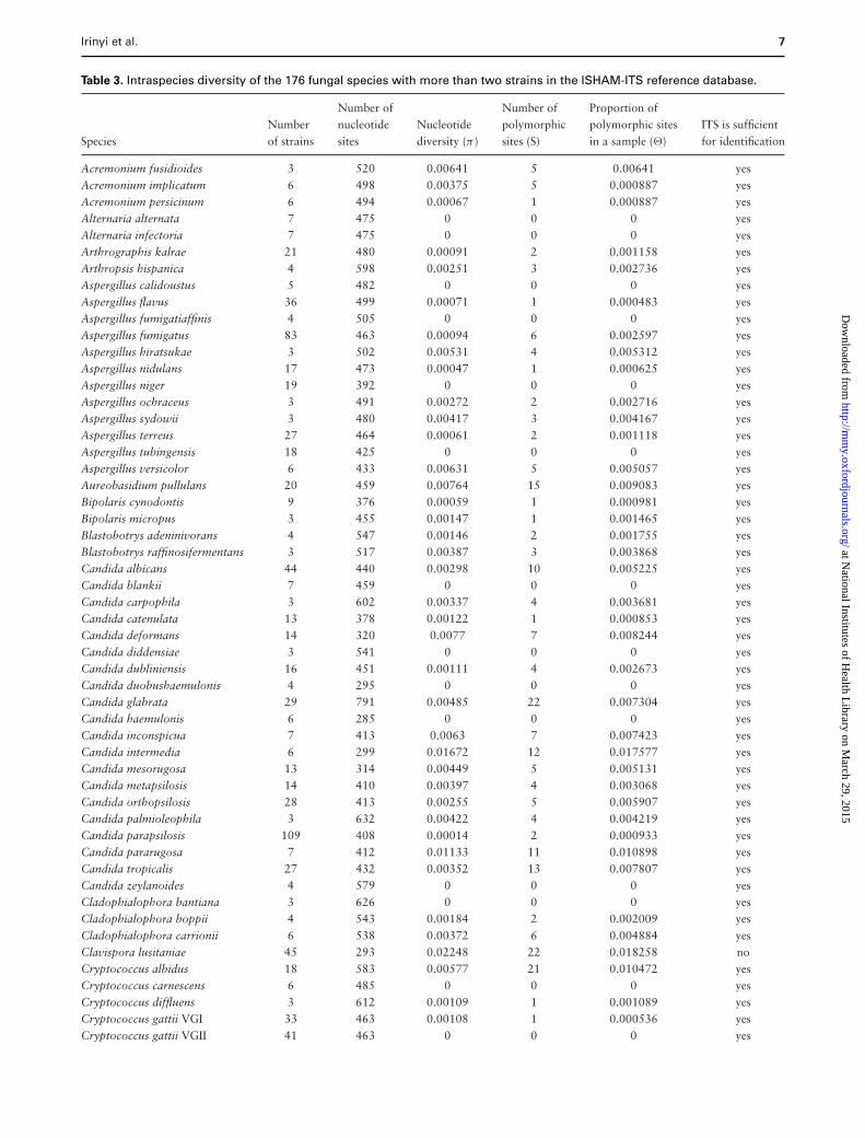
Table 3. Intraspecies diversity of the 176 fungal species with more than two strains in the ISHAM-ITS reference database.
Number of Number of Proportion ofNumber nucleotide Nucleotide polymorphic polymorphic sites ITS is sufficient
Species of strains sites diversity (π ) sites (S) in a sample (�) for identification
Acremonium fusidioides 3 520 0.00641 5 0.00641 yesAcremonium implicatum 6 498 0.00375 5 0.000887 yesAcremonium persicinum 6 494 0.00067 1 0.000887 yesAlternaria alternata 7 475 0 0 0 yesAlternaria infectoria 7 475 0 0 0 yesArthrographis kalrae 21 480 0.00091 2 0.001158 yesArthropsis hispanica 4 598 0.00251 3 0.002736 yesAspergillus calidoustus 5 482 0 0 0 yesAspergillus flavus 36 499 0.00071 1 0.000483 yesAspergillus fumigatiaffinis 4 505 0 0 0 yesAspergillus fumigatus 83 463 0.00094 6 0.002597 yesAspergillus hiratsukae 3 502 0.00531 4 0.005312 yesAspergillus nidulans 17 473 0.00047 1 0.000625 yesAspergillus niger 19 392 0 0 0 yesAspergillus ochraceus 3 491 0.00272 2 0.002716 yesAspergillus sydowii 3 480 0.00417 3 0.004167 yesAspergillus terreus 27 464 0.00061 2 0.001118 yesAspergillus tubingensis 18 425 0 0 0 yesAspergillus versicolor 6 433 0.00631 5 0.005057 yesAureobasidium pullulans 20 459 0.00764 15 0.009083 yesBipolaris cynodontis 9 376 0.00059 1 0.000981 yesBipolaris micropus 3 455 0.00147 1 0.001465 yesBlastobotrys adeninivorans 4 547 0.00146 2 0.001755 yesBlastobotrys raffinosifermentans 3 517 0.00387 3 0.003868 yesCandida albicans 44 440 0.00298 10 0.005225 yesCandida blankii 7 459 0 0 0 yesCandida carpophila 3 602 0.00337 4 0.003681 yesCandida catenulata 13 378 0.00122 1 0.000853 yesCandida deformans 14 320 0.0077 7 0.008244 yesCandida diddensiae 3 541 0 0 0 yesCandida dubliniensis 16 451 0.00111 4 0.002673 yesCandida duobushaemulonis 4 295 0 0 0 yesCandida glabrata 29 791 0.00485 22 0.007304 yesCandida haemulonis 6 285 0 0 0 yesCandida inconspicua 7 413 0.0063 7 0.007423 yesCandida intermedia 6 299 0.01672 12 0.017577 yesCandida mesorugosa 13 314 0.00449 5 0.005131 yesCandida metapsilosis 14 410 0.00397 4 0.003068 yesCandida orthopsilosis 28 413 0.00255 5 0.005907 yesCandida palmioleophila 3 632 0.00422 4 0.004219 yesCandida parapsilosis 109 408 0.00014 2 0.000933 yesCandida pararugosa 7 412 0.01133 11 0.010898 yesCandida tropicalis 27 432 0.00352 13 0.007807 yesCandida zeylanoides 4 579 0 0 0 yesCladophialophora bantiana 3 626 0 0 0 yesCladophialophora boppii 4 543 0.00184 2 0.002009 yesCladophialophora carrionii 6 538 0.00372 6 0.004884 yesClavispora lusitaniae 45 293 0.02248 22 0.018258 noCryptococcus albidus 18 583 0.00577 21 0.010472 yesCryptococcus carnescens 6 485 0 0 0 yesCryptococcus diffluens 3 612 0.00109 1 0.001089 yesCryptococcus gattii VGI 33 463 0.00108 1 0.000536 yesCryptococcus gattii VGII 41 463 0 0 0 yes
at National Institutes of H
ealth Library on M
arch 29, 2015http://m
my.oxfordjournals.org/
Dow
nloaded from
8 Medical Mycology, 2015, Vol. 00, No. 00
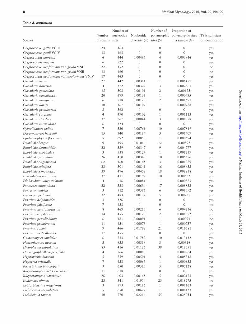
Table 3. continued
Number of Number of Proportion ofNumber nucleotide Nucleotide polymorphic polymorphic sites ITS is sufficient
Species of strains sites diversity (π ) sites (S) in a sample (�) for identification
Cryptococcus gattii VGIII 24 463 0 0 0 yesCryptococcus gattii VGIV 13 463 0 0 0 yesCryptococcus laurentii 6 444 0.00495 4 0.003946 yesCryptococcus magnus 6 522 0 0 0 yesCryptococcus neoformans var. grubii VNI 22 452 0 0 0 noCryptococcus neoformans var. grubii VNII 13 460 0 0 0 noCryptococcus neoformans var. neoformans VNIV 17 463 0 0 0 yesCurvularia aeria 27 442 0.00311 11 0.006457 yesCurvularia borreriae 4 572 0.00322 3 0.002861 yesCurvularia geniculata 15 503 0.00101 2 0.00125 yesCurvularia hawaiiensis 20 379 0.00136 1 0.000755 yesCurvularia inaequalis 6 518 0.00129 2 0.001691 yesCurvularia lunata 10 467 0.00107 1 0.000788 yesCurvularia protuberata 3 562 0 0 0 yesCurvularia sorghina 4 490 0.00102 1 0.001113 yesCurvularia spicifera 37 367 0.00044 3 0.001958 yesCurvularia verruculosa 6 524 0 0 0 yesCyberlindnera jadinii 7 520 0.00769 10 0.007849 yesDebaryomyces hansenii 15 540 0.00187 3 0.001709 yesEpidermophyton floccosum 5 692 0.00058 1 0.000694 yesExophiala bergeri 9 495 0.01016 12 0.00892 yesExophiala dermatitidis 22 539 0.00347 9 0.004777 yesExophiala exophialae 3 538 0.00124 1 0.001239 yesExophiala jeanselmei 26 470 0.00349 10 0.005576 yesExophiala oligosperma 62 460 0.00165 3 0.001389 yesExophiala spinifera 23 501 0.00841 16 0.008653 yesExophiala xenobiotica 39 476 0.00458 18 0.008838 yesExserohilum rostratum 37 411 0.00197 10 0.00532 yesFilobasidium uniguttulatum 4 616 0.00081 1 0.000885 yesFonsecaea monophora 22 528 0.00634 17 0.008832 yesFonsecaea nubica 3 512 0.00586 6 0.006392 yesFonsecaea pedrosoi 32 483 0.00132 5 0.00257 yesFusarium delphinoides 3 526 0 0 0 yesFusarium falciforme 7 458 0 0 0 noFusarium keratoplasticum 8 469 0.00213 6 0.004236 noFusarium oxysporum 14 455 0.00128 2 0.001382 yesFusarium petroliphilum 6 481 0.00091 1 0.00071 noFusarium proliferatum 11 451 0.00073 1 0.000757 yesFusarium solani 9 466 0.01788 21 0.016581 noFusarium verticillioides 17 455 0 0 0 yesGalactomyces candidus 6 333 0.01782 10 0.013152 yesHanseniaspora uvarum 3 633 0.00316 3 0.00316 yesHistoplasma capsulatum 83 416 0.01126 38 0.018351 yesHormographiella aspergillata 4 566 0.00088 1 0.000964 yesHyphopichia burtonii 5 359 0.00501 4 0.005348 yesHypocrea orientalis 7 438 0.00065 1 0.000932 yesKazachstania pintolopesii 3 650 0.00513 5 0.005128 yesKluyveromyces lactis var. lactis 11 618 0 0 0 yesKluyveromyces marxianus 26 603 0.00165 5 0.002173 yesKodamaea ohmeri 23 341 0.01954 23 0.018275 noLeptosphaeria senegalensis 3 573 0.00116 1 0.001163 yesLichtheimia corymbifera 5 650 0.00677 11 0.008123 yesLichtheimia ramosa 10 770 0.02214 55 0.025054 yes
at National Institutes of H
ealth Library on M
arch 29, 2015http://m
my.oxfordjournals.org/
Dow
nloaded from
Irinyi et al. 9
Table 3. continued
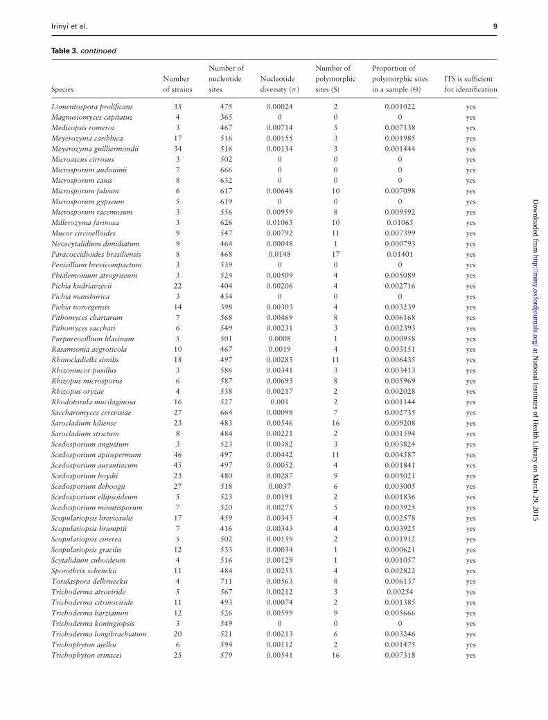
Number of Number of Proportion ofNumber nucleotide Nucleotide polymorphic polymorphic sites ITS is sufficient
Species of strains sites diversity (π ) sites (S) in a sample (�) for identification
Lomentospora prolificans 35 475 0.00024 2 0.001022 yesMagnusiomyces capitatus 4 365 0 0 0 yesMedicopsis romeroi 3 467 0.00714 5 0.007138 yesMeyerozyma caribbica 17 516 0.00155 3 0.001985 yesMeyerozyma guilliermondii 34 516 0.00134 3 0.001444 yesMicroascus cirrosus 3 502 0 0 0 yesMicrosporum audouinii 7 666 0 0 0 yesMicrosporum canis 8 632 0 0 0 yesMicrosporum fulvum 6 617 0.00648 10 0.007098 yesMicrosporum gypseum 5 619 0 0 0 yesMicrosporum racemosum 3 556 0.00959 8 0.009592 yesMillerozyma farinosa 3 626 0.01065 10 0.01065 yesMucor circinelloides 9 547 0.00792 11 0.007399 yesNeoscytalidium dimidiatum 9 464 0.00048 1 0.000793 yesParacoccidioides brasiliensis 8 468 0.0148 17 0.01401 yesPenicillium brevicompactum 3 539 0 0 0 yesPhialemonium atrogriseum 3 524 0.00509 4 0.005089 yesPichia kudriavzevii 22 404 0.00206 4 0.002716 yesPichia manshurica 3 434 0 0 0 yesPichia norvegensis 14 398 0.00303 4 0.003239 yesPithomyces chartarum 7 568 0.00469 8 0.006168 yesPithomyces sacchari 6 549 0.00231 3 0.002393 yesPurpureocillium lilacinum 5 501 0.0008 1 0.000958 yesRasamsonia aegroticola 10 467 0.0019 4 0.003151 yesRhinocladiella similis 18 497 0.00285 11 0.006435 yesRhizomucor pusillus 3 586 0.00341 3 0.003413 yesRhizopus microsporus 6 587 0.00693 8 0.005969 yesRhizopus oryzae 4 538 0.00217 2 0.002028 yesRhodotorula mucilaginosa 16 527 0.001 2 0.001144 yesSaccharomyces cerevisiae 27 664 0.00098 7 0.002735 yesSarocladium kiliense 23 483 0.00546 16 0.009208 yesSarocladium strictum 8 484 0.00221 2 0.001594 yesScedosporium angustum 3 523 0.00382 3 0.003824 yesScedosporium apiospermum 46 497 0.00442 11 0.004587 yesScedosporium aurantiacum 45 497 0.00052 4 0.001841 yesScedosporium boydii 23 480 0.00287 9 0.005021 yesScedosporium dehoogii 27 518 0.0037 6 0.003005 yesScedosporium ellipsoideum 5 523 0.00191 2 0.001836 yesScedosporium minutisporum 7 520 0.00275 5 0.003925 yesScopulariopsis brevicaulis 17 459 0.00343 4 0.002578 yesScopulariopsis brumptii 7 416 0.00343 4 0.003925 yesScopulariopsis cinerea 5 502 0.00159 2 0.001912 yesScopulariopsis gracilis 12 533 0.00034 1 0.000621 yesScytalidium cuboideum 4 516 0.00129 1 0.001057 yesSporothrix schenckii 11 484 0.00255 4 0.002822 yesTorulaspora delbrueckii 4 711 0.00563 8 0.006137 yesTrichoderma atroviride 5 567 0.00212 3 0.00254 yesTrichoderma citrinoviride 11 493 0.00074 2 0.001385 yesTrichoderma harzianum 12 526 0.00599 9 0.005666 yesTrichoderma koningiopsis 3 549 0 0 0 yesTrichoderma longibrachiatum 20 521 0.00213 6 0.003246 yesTrichophyton ajelloi 6 594 0.00112 2 0.001475 yesTrichophyton erinacei 25 579 0.00541 16 0.007318 yes
at National Institutes of H
ealth Library on M
arch 29, 2015http://m
my.oxfordjournals.org/
Dow
nloaded from

10 Medical Mycology, 2015, Vol. 00, No. 00
Table 3. continued
Number of Number of Proportion ofNumber nucleotide Nucleotide polymorphic polymorphic sites ITS is sufficient
Species of strains sites diversity (π ) sites (S) in a sample (�) for identification
Trichophyton interdigitale 68 525 0.00189 4 0.001591 yesTrichophyton mentagrophytes = T. quinckeanum 5 603 0 0 0 yesTrichophyton persicolor 3 601 0.00111 1 0.001109 yesTrichophyton rubrum 30 540 0.00228 4 0.00187 yesTrichophyton schoenleinii 4 623 0 0 0 yesTrichophyton simii 7 608 0.00157 2 0.001343 yesTrichophyton terrestre 4 615 0 0 0 yesTrichophyton tonsurans 6 597 0.00112 2 0.001467 yesTrichophyton verrucosum 4 534 0 0 0 yesTrichosporon asahii 7 447 0.00107 1 0.000913 yesTrichosporon dermatis 4 440 0 0 0 yesTrichosporon inkin 4 539 0.00371 4 0.004048 yesTrichosporon montevideense 4 528 0 0 0 yesWickerhamomyces anomalus 37 522 0.00131 7 0.003212 yesYamadazyma mexicana 3 561 0.00119 1 0.001188 yesYamadazyma scolyti 3 622 0.00536 5 0.005359 yesYarrowia lipolytica 24 347 0.0062 15 0.011576 yes
Figure 1. Distribution of the number of strains per species in the ISHAM-ITS reference database.
Number of sequencesAt present, the quality-controlled ISHAM-ITS referencedatabase contains 2800 complete ITS sequences represent-ing 421 human/animal pathogenic fungal species. It con-tains 176 species represented by one strain, 69 species bytwo strains, and 176 species by a minimum of three to amaximum of 109 sequences. The distribution of strains perspecies was hyperbolic, meaning that the species with fewstrains were more frequent than those with many (Fig. 1).
Lengths of the ITSThe lengths of complete ITS sequences in the ISHAM-ITS reference database varied between 285 and 791 bp.The distribution of the number of nucleotides per se-
Figure 2. Length distribution of ITS sequences in the ISHAM-ITS refer-ence database.
quence is given in Figure 2. The shortest complete ITS se-quences were assigned to Candida haemulonis (285 bp),Clavispora lusitaniae (293 bp), and the longest ones toCandida glabrata (791 bp) and Lichtheimia ramosa (770bp). The mean nucleotide length of ITS sequences in thedatabase was 503 bp, while the median was 500 bp,indicating that the distribution of the sequence lengthswas almost normal, with 0.08 skewness and 0.71 kur-tosis (Fig. 2). These two metrics indicate that the pop-ulation of sequences is centered around the average
at National Institutes of H
ealth Library on M
arch 29, 2015http://m
my.oxfordjournals.org/
Dow
nloaded from
Irinyi et al. 11
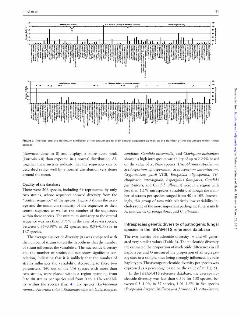
Figure 3. Average and the minimum similarity of the sequences to their central sequence as well as the number of the sequences within thesespecies.
(skewness close to 0) and displays a more acute peak(kurtosis >0) than expected in a normal distribution. Al-together these metrics indicate that the sequences can bedescribed rather well by a normal distribution very densearound the mean.
Quality of the databaseThere were 206 species, including 69 represented by onlytwo strains, whose sequences showed diversity from the“central sequence” of the species. Figure 3 shows the aver-age and the minimum similarity of the sequences to theircentral sequence as well as the number of the sequenceswithin these species. The minimum similarity to the centralsequence was less than 0.95% in the case of seven species,between 0.95–0.98% in 32 species and 0.98–0.998% in167 species.
The average nucleotide diversity (π) was compared withthe number of strains to test the hypothesis that the numberof strain influences the variability. The nucleotide diversityand the number of strains did not show significant cor-relation, indicating that it is unlikely that the number ofstrains influences the variability. According to these twoparameters, 160 out of the 176 species with more thantwo strains, were placed within a region spanning from0 to 40 strains per species and from 0 to 1.1% variabil-ity within the species (Fig. 4). Six species (Lichtheimiaramosa, Fusarium solani, Kodamaea ohmeri, Galactomyces
candidus, Candida intermedia, and Clavispora lusitaniae)showed a high intraspecies variability of up to 2.25% basedon the value of π . Nine species (Histoplasma capsulatum,Scedosporium apiospermum, Scedosporium aurantiacum,Cryptococcus gattii VGII, Exophiala oligosperma, Tri-chophyton interdigitale, Aspergillus fumigatus, Candidaparapsilosis, and Candida albicans) were in a region withless than 1.1% intraspecies variability, although the num-ber of strains per species ranged from 40 to 109. Interest-ingly, this group of taxa with relatively low variability in-cludes some of the more important pathogenic fungi namelyA. fumigatus, C. parapsilosis, and C. albicans.
Intraspecies genetic diversity of pathogenic fungalspecies in the ISHAM-ITS reference database
The two metrics of nucleotide diversity (π and �) gener-ated very similar values (Table 3). The nucleotide diversity(π) estimated the proportion of nucleotide differences in allhaplotypes and � measured the proportion of all segregat-ing sites in a sample, thus being strongly influenced by rarehaplotypes. The average nucleotide diversity per species wasexpressed as a percentage based on the value of π (Fig. 5).
In the ISHAM-ITS reference database, the average nu-cleotide diversity was less than 0.5% for 138 species, be-tween 0.5–1.0% in 27 species, 1.01–1.5% in five species(Exophiala bergeri, Millerozyma farinosa, H. capsulatum,
at National Institutes of H
ealth Library on M
arch 29, 2015http://m
my.oxfordjournals.org/
Dow
nloaded from

12 Medical Mycology, 2015, Vol. 00, No. 00
Figure 4. Nucleotide diversity (π) compared to the number of sequences by species in the ISHAM-ITS reference database.
Candida pararugosa, and Paracoccidioides brasiliensis),1.5–2.0% in four species (C. intermedia, G. candidus,F. solani, and K. ohmeri), and more than 2% in two species(Lichtheimia ramosa and C. lusitaniae) (Table 3, Fig. 5).
The distribution of the distances from the “central se-quence” of a species was hyperbolic, with the most fre-quent class, containing 63 species, representing more thanone third of the species with more than two strains in thedatabase, showing intraspecies variability ranging from 0to 0.1%. More than half of the species with more thantwo strains in the database (97 species) were represented byspecies with less than 0.4% distance (Fig. 6).
The polymorphic site distribution showed a similar re-sult. In 117 species, the number of polymorphic sites wasless than five, in 35 species it was between five and ten,in 11 species between 11 and 15, in six species between16 and 20 and finally more than 20 in seven species.The species with the highest number of segregating siteswere Cryptococcus albidus (21 sites), the complex of F.solani (21 sites), C. lusitaniae (22 sites), C. glabrata (22sites), K. ohmeri (23 sites), H. capsulatum (38 sites), andL. ramosa (55 sites) (Table 3). The value of � showeda strong correlation with the average nucleotide diversity
and the number of segregating sites. The proportion of rarehaplotypes in a given sample was the highest in F. solani,C. lusitaniae, K. ohmeri, H. capsulatum, and L. ramosa(Table 3).
The intraspecies genetic analyses showed that the major-ity of medically important species had a low variability inITS regions. Thus ITS sequencing can be used for the iden-tification of most medical relevant fungal species (Table 3).The species with high intraspecies diversity within the ITSregion require analysis of additional molecular markers tobe reliably identified (see Table 4).
Barcoding gap analysis of the species representedin the ISHAM-ITS reference database
For the estimation of the barcoding gap the distributionof the Kimura 2-parameter (K2P) genetic distances withinspecies and between species was calculated. In the ISHAM-ITS reference database, 17 taxonomical groups with morethan two species sharing the same phylogenetic clade wereidentified based on previous data in MycoBank [63,64],Index Fungorum [65], and The Yeasts [66] (Table 5).The barcoding gap analysis was performed in all 17 taxa,
at National Institutes of H
ealth Library on M
arch 29, 2015http://m
my.oxfordjournals.org/
Dow
nloaded from

Irinyi et al. 13
Figure 5. Average nucleotide diversity per species expressed as a percentage based on the value of π of the 176 fungal species with more than threestrains in the ISHAM-ITS reference database. The error bars indicate the standard deviation of nucleotide differences.
Figure 6. Distribution of average distance of s within species comparedto the number of species in the ISHAM-ITS reference database.
including two versions of analysis for C. neoformans/C.gattii and Arthrodermataceae/Trichophyton (see Table 5).The distribution of genetic distances (intra- and inter-species) in each taxon is shown in Figures 7–10 and Supple-mentary Figures S1–S13. In 13 taxa (phylogenetic clades),a clear barcoding gap (K2P distance) was found (Table 5).
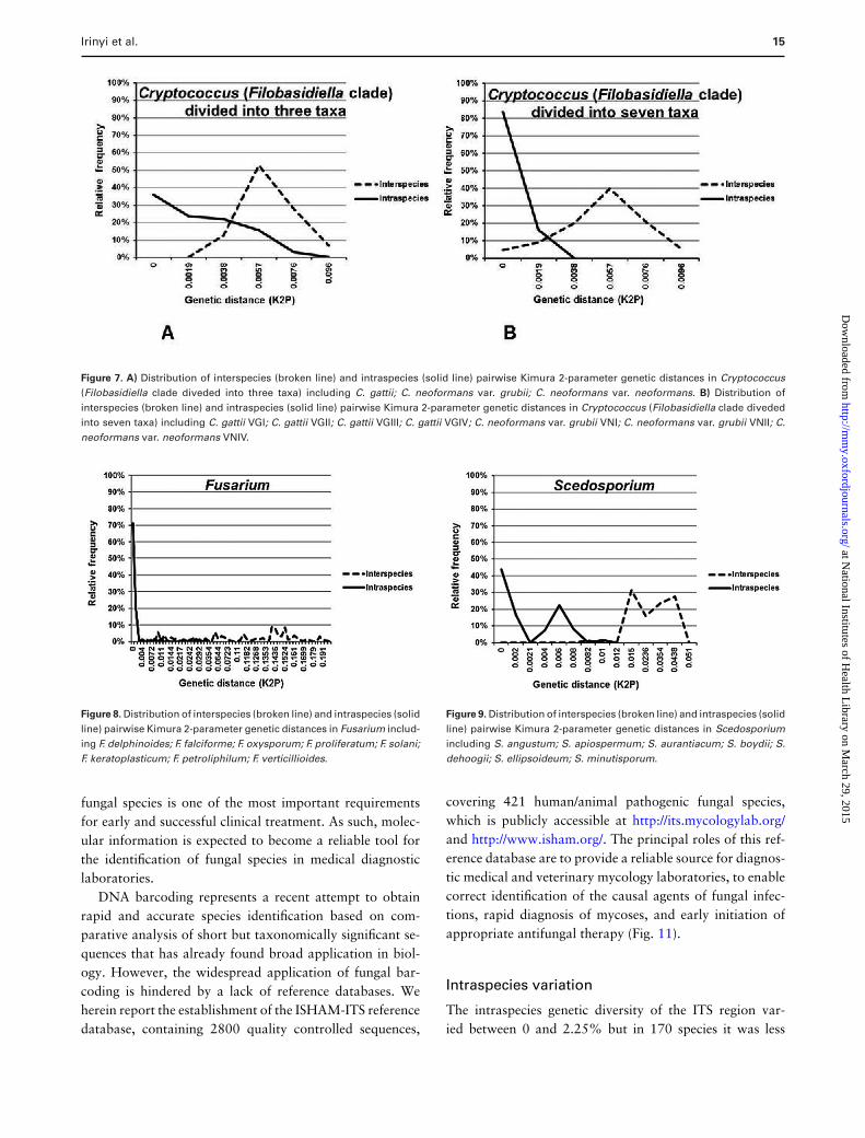
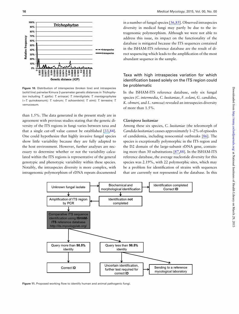
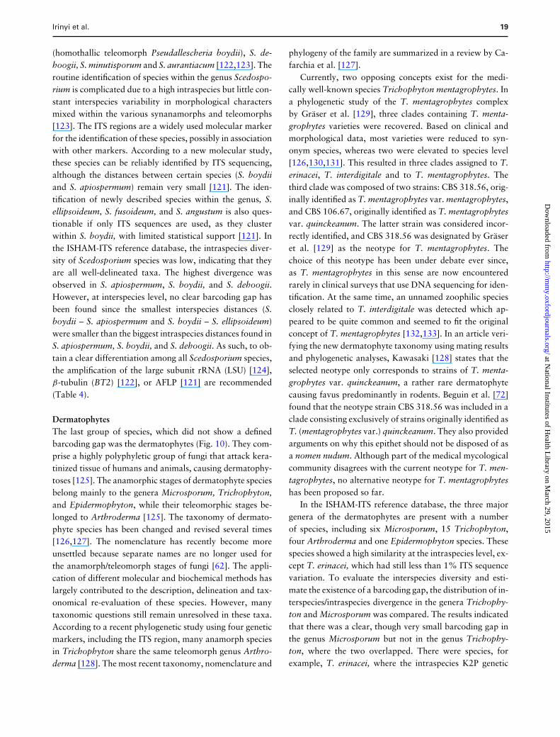
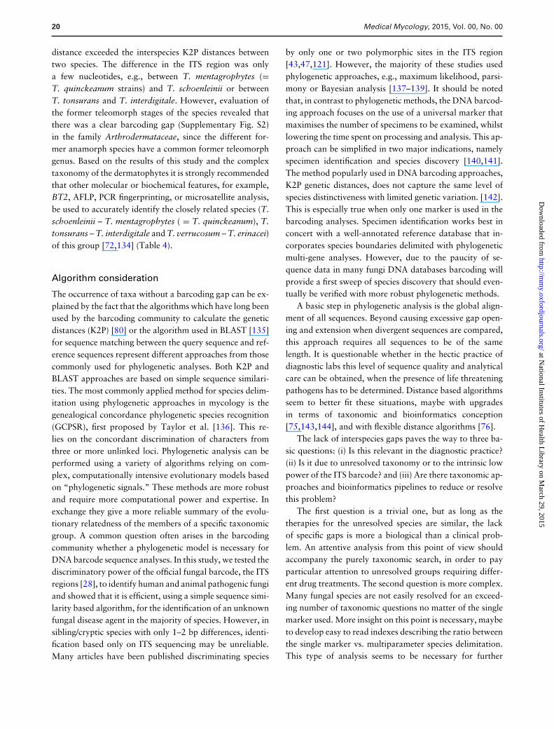
The smallest barcoding gap (0.0002) was found in the Mi-crosporum spp., while the largest one was found in theCladophialophora spp. (0.09). In these cases, the highestintraspecies distances were smaller than the lowest geneticdistances between species, creating a barcoding gap. Forthe remaining four taxa Cryptococcus (Fig. 7), Fusarium(Fig. 8), Scedosporium (Fig. 9), and Trichophyton (Fig. 10),it was not possible to define a clear barcoding gap, mean-ing that the distributions of genetic distances within andbetween species overlapped.
Most of the studied taxa could be identified with theITS barcode, although in some cases a clear discrimina-tion could not be observed. There are two possible reasonsfor this: either the taxa is insufficiently studied or the ITSregion is simply an inappropriate marker for discrimina-tion between biologically consistent groups. Alternative lociand/or molecular methods are required for correct identifi-cation of these species (Table 5).
Discussion
ISHAM-ITS reference database
With a significant rise in the diversity of etiologicalagents of fungal infections in human and animal popula-tions [1,2], rapid and accurate identification of pathogenic
at National Institutes of H
ealth Library on M
arch 29, 2015http://m
my.oxfordjournals.org/
Dow
nloaded from

14 Medical Mycology, 2015, Vol. 00, No. 00
Table 4. Taxa with high ITS diversity and alternative methods to be used for their reliable identification.
Taxa Proposed alternatives
Clavispora lusitaniae Morphological identification by mating unknowns with a strain of known mating type(89,90)
Fusarium solani speciescomplex (FSSC)
MLST(116); translation elongation factor 1-α (TEF-1α), RNA polymerase II gene (RPB2), secondarymetabolite profiles(94)
Kodamaea ohmeri Further taxonomic studies neededLichtheimia spp. D1/D2 region, translation elongation factor 1-α (TEF-1α)(102); MALDI-TOF(105)
Cryptococcus AFLP(110); PCR fingerprinting, RFLP of orotidine monophosphate pyrophosphorylase gene(URA5)(111); MLST(114)
Scedosporium β -tubulin (BT2)(122), AFLP(121); LSU(124)
Arthrodermataceae RAPD, PCR fingerprinting, AFLP, microsatellite markers(102)
Table 5. Barcoding gap based on Kimura 2-parameter genetic distances in 17 studied phylogenetic clades represented by
more than two species, with two variants of analysis for Cryptococcus neoformans/Cryptococcus gattii, and Arthrodermat-
aceae/Trichophyton in the ISHAM-ITS reference database.
TaxaBarcodinggap Species included in the analyses represented with more than two strains by species
Acremonium 0.055 Acremonium fusidioides; A. implicatum; A. persicinum; Phialemoniumatrogriseum; Sarocladium kiliense; S. strictum;
Arthrodermataceae 0.002 Arthroderma benhamiae; A. fulvum; A. gypseum; A. insingulare; A. otae; A.persicolor; A. simii; A. uncinatum; A. vanbreuseghemii
Aspergillus 0.002 Aspergillus calidoustus; A. flavus; A. fumigatiaffinis; A. fumigatus; A. hiratsukae;A. nidulans; A. niger; A. ochraceus; A. sydowii; A. terreus; A. tubingensis
Cladophialophora 0.09 Cladophialophora bantiana; C. boppii; C. carrioniiCryptococcus(Filobasidiella cladedivided into three taxa)
– Cryptococcus gattii; C. neoformans var. grubii; C. neoformans var. neoformans
Cryptococcus(Filobasidiella cladedivided into seven taxa)
– Cryptococcus gattii VGI; C. gattii VGII; C. gattii VGIII; C. gattii VGIV; C.neoformans var. grubii VNI; C. neoformans var. grubii VNII; C. neoformans var.neoformans VNIV
Curvularia 0.001 Curvularia aeria; C. borreriae; C. inaequalis; C. geniculata; C. hawaiiensis; C.inaequalis; C. lunata; C. protuberata; C. spicifera; C. sorghina; C. verruculosa
Debaryomycetaceae(Lodderomyces clade)
0.001 Candida albicans; C. dubliniensis; C. metapsilosis; C. orthopsilosis; C. parapsilosis;C. tropicalis; Debaryomyces hansenii
Exophiala 0.015 Exophiala bergeri; E. dermatitidis; E. exophialae; E. jeanselmei; E. oligosperma; E.spinifera; E. xenobiotica
Fusarium – Fusarium delphinoides; F. falciforme; F. oxysporum; F. proliferatum; F. solani; F.keratoplasticum; F. petroliphilum; F. verticillioides
Metschnikowiaceae 0.0603 Candida duobushaemulonis; C. haemulonis; C. intermedia; C. lusitaniae;Kodamaea ohmeri
Microsporum 0.0002 Microsporum audouinii; M. canis; M. fulvum; M. gypseumPichiaceae 0.005 Pichia kudriavzevii; P. norvegensis; P. manshuricaSaccharomycetaceae 0.009 Kluyveromyces marxianus; K. lactis var. lactis; Saccharomyces cerevisiae;
Torulaspora delbrueckiiScedosporium – Scedosporium angustum; S. apiospermum; S. aurantiacum; S. boydii; S. dehoogii; S.
ellipsoideum; S. minutisporumScopulariopsis 0.0034 Scopulariopsis brevicaulis; S. brumptii; S. cinerea; S. gracilisTrichophyton – Trichophyton ajelloi; T. erinacei; T. interdigitale; T. mentagrophytes ( = T.
quinckeanum); T. rubrum; T. schoenleinii; T. simii; T. terrestre; T. verrucosumTrichosporon 0.004 Trichosporon asahii; T. dermatis; T. inkin; T. montevideense
at National Institutes of H
ealth Library on M
arch 29, 2015http://m
my.oxfordjournals.org/
Dow
nloaded from
Irinyi et al. 15
Figure 7. A) Distribution of interspecies (broken line) and intraspecies (solid line) pairwise Kimura 2-parameter genetic distances in Cryptococcus(Filobasidiella clade diveded into three taxa) including C. gattii; C. neoformans var. grubii; C. neoformans var. neoformans. B) Distribution ofinterspecies (broken line) and intraspecies (solid line) pairwise Kimura 2-parameter genetic distances in Cryptococcus (Filobasidiella clade divededinto seven taxa) including C. gattii VGI; C. gattii VGII; C. gattii VGIII; C. gattii VGIV; C. neoformans var. grubii VNI; C. neoformans var. grubii VNII; C.neoformans var. neoformans VNIV.
Figure 8. Distribution of interspecies (broken line) and intraspecies (solidline) pairwise Kimura 2-parameter genetic distances in Fusarium includ-ing F. delphinoides; F. falciforme; F. oxysporum; F. proliferatum; F. solani;F. keratoplasticum; F. petroliphilum; F. verticillioides.
fungal species is one of the most important requirementsfor early and successful clinical treatment. As such, molec-ular information is expected to become a reliable tool forthe identification of fungal species in medical diagnosticlaboratories.
DNA barcoding represents a recent attempt to obtainrapid and accurate species identification based on com-parative analysis of short but taxonomically significant se-quences that has already found broad application in biol-ogy. However, the widespread application of fungal bar-coding is hindered by a lack of reference databases. Weherein report the establishment of the ISHAM-ITS referencedatabase, containing 2800 quality controlled sequences,
Figure 9. Distribution of interspecies (broken line) and intraspecies (solidline) pairwise Kimura 2-parameter genetic distances in Scedosporiumincluding S. angustum; S. apiospermum; S. aurantiacum; S. boydii; S.dehoogii; S. ellipsoideum; S. minutisporum.
covering 421 human/animal pathogenic fungal species,which is publicly accessible at http://its.mycologylab.org/and http://www.isham.org/. The principal roles of this ref-erence database are to provide a reliable source for diagnos-tic medical and veterinary mycology laboratories, to enablecorrect identification of the causal agents of fungal infec-tions, rapid diagnosis of mycoses, and early initiation ofappropriate antifungal therapy (Fig. 11).
Intraspecies variation
The intraspecies genetic diversity of the ITS region var-ied between 0 and 2.25% but in 170 species it was less
at National Institutes of H
ealth Library on M
arch 29, 2015http://m
my.oxfordjournals.org/
Dow
nloaded from
16 Medical Mycology, 2015, Vol. 00, No. 00
Figure 10. Distribution of interspecies (broken line) and intraspecies(solid line) pairwise Kimura 2-parameter genetic distances in Trichophy-ton including T. ajelloi; T. erinacei; T. interdigitale; T. mentagrophytes(=T. quinckeanum); T. rubrum; T. schoenleinii; T. simii; T. terrestre; T.verrucosum.
than 1.5%. The data generated in the present study are inagreement with previous studies stating that the genetic di-versity of the ITS regions in fungi varies between taxa andthat a single cut-off value cannot be established [33,84].One could hypothesize that highly invasive fungal speciesshow little variability because they are fully adapted tothe host environment. However, further analyses are nec-essary to determine whether or not the variability calcu-lated within the ITS regions is representative of the generalgenotypic and phenotypic variability within these species.Notably, the intraspecies diversity is more complex, withintragenomic polymorphism of rDNA repeats documented
in a number of fungal species [36,85]. Observed intraspeciesdiversity in medical fungi may partly be due to the in-tragenomic polymorphism. Although we were not able toaddress this issue, its impact on the functionality of thedatabase is mitigated because the ITS sequences containedin the ISHAM-ITS reference database are the result of di-rect sequencing which leads to the amplification of the mostabundant sequence in the sample.
Taxa with high intraspecies variation for whichidentification based solely on the ITS region couldbe problematic
In the ISHAM-ITS reference database, only six fungalspecies (C. intermedia, C. lusitaniae, F. solani, G. candidus,K. ohmeri, and L. ramosa) revealed an intraspecies diversityof more than 1.5%.
Clavispora lusitaniaeAmong these six species, C. lusitaniae (the teleomorph ofCandida lusitaniae) causes approximately 1–2% of episodesof candidemia, including nosocomial outbreaks [86]. Thespecies is exceptionally polymorphic in the ITS region andthe D2 domain of the large-subunit rDNA gene, contain-ing more than 30 substitutions [87,88]. In the ISHAM-ITSreference database, the average nucleotide diversity for thisspecies was 2.19%, with 22 polymorphic sites, which maybe a problem for identification of strains with sequencesthat are currently not represented in the database. In this
Figure 11. Proposed working flow to identify human and animal pathogenic fungi.
at National Institutes of H
ealth Library on M
arch 29, 2015http://m
my.oxfordjournals.org/
Dow
nloaded from

Irinyi et al. 17
case, correct species identification, may be determined bymating type for sexual reproduction [89,90] (see Table 4).The polyphyletic nature of the genus Clavispora was re-cently confirmed by multigene sequence analysis [91]. Fur-ther taxonomic studies are required for a better delimitationof this species.
Fusarium solani species complex (FSSC)The second highest intraspecies variation was foundamongst Fusarium species, which are primarily saprobes,plant pathogens, often linked with pathological infections,mainly keratitis, in both humans and animals. The F. solanispecies complex is the most common group of fusariaresponsible for human infections, primarily in immuno-compromised individuals [92,93]. Before taxonomicalreanalysis the ISHAM-ITS database contained ten differentFusarium species including the highly polyphyletic FSSC.Seven of these species showed below 0.5% intraspeciesvariability suggesting a good taxonomic delimitation whichcan in turn allow easy identification with the ITS. How-ever, within the FSSC the average nucleotide diversity was3.76%, indicating that this complex has remained unre-solved and contains multiple other cryptic species. Accord-ing to the latest taxonomic studies [94], F. keratoplasticum,F. petroliphilum, and F. falciforme have been separatedfrom the FSSC as new taxa, reducing the average nucleotidediversity to 1.65% in the ISHAM-ITS reference database.This variation still represents a significantly high degree ofsequence diversity, making it necessary to employ differ-ent markers for correct identification at the species level(Table 4 and see below).
Galactomyces candidusG. candidus (anamorph Geotrichum candidum) is a ubiq-uitous and dimorphic yeast, which occurs commonly onmoist substrates rich in nutrients. Occasionally it is foundas an opportunistic pathogen in the human respiratory andgastro-intestinal tracts [92,95]. The taxonomic classifica-tion of the species was revised in 2004 by de Hoog andSmith [96]. A standardized protocol was proposed for theidentification of G. candidus at species and strain level in2006 [97]. According to a recent study [38], the ITS re-gion, especially the ITS1 region of G. candidus, provedto be highly polymorphic at intraspecies and intragenomiclevels. In the ISHAM-ITS database, the species was repre-sented by five strains with 1.78% genetic diversity, mainlyin the ITS1 region. Although the 18S-ITS1-5.8S-ITS2-26Sas a whole provides an improved phylogenetic resolutionfor the different phylotypes, use of the ITS region alone isnot suitable for rapid identification of the species [38].
Kodamaea ohmeriUsing the ISHAM-ITS reference database, K. ohmeri (syn.:Pichia ohmeri, the teleomorph of Candida guilliermondiivar. membranifaciens) has been found to contain high in-traspecies diversity. This is an ascosporogenic yeast, mainlyused in the food industry for fermentation, but has recentlyemerged as a fungal pathogen, particularly in immuno-compromised patients [98,99]. However, few studies onthis species have been done. Recently a number of specieshave been found with characteristics similar to those ofK. ohmeri raising the possibility of cryptic species and thepotential misidentification of previously described isolates[100]. Phylogenetic analyses of the ITS sequences containedin the ISHAM-ITS reference database supported two cladesnot previously identified. Further studies are needed to tax-onomically resolve possible cryptic species.
Lichtheimia sppThe next group of fungi with marked ITS intraspeciesvariation was Lichtheimia species, which causes life-threating rhinocerebral and bronchorespiratory mucormy-coses [101]. Multigene sequence analysis (ITS, 28S, EF-1α)of 38 isolates identified morphologically as L. corymbiferarevealed a new species, named L. ramosa, which differed inmorphology and nucleotide sequences from L. corymbifera[102]. To date, from the five recognized species of the genusLichtheimia, only three L. corymbifera, L. ornata, and L.ramosa are of clinical relevance [103]. L. ramosa provedto be more polymorphic than L. corymbifera, with morethan 2% diversity in the ITS sequences. Similar values forthe ITS region of L. ramosa have been reported by Waltheret al. in 2013 [104], suggesting that different groups amongL. ramosa should be considered as a separate species. If so,the ITS region would be an appropriate marker for identi-fication of these species. In view of the high diversity ob-served among ITS sequences within Lichtheimia, currentlyit is recommended to use either a multiple gene approach[102] or MALDI-TOF [105] for a reliable identification (seeTable 4).
Barcoding gap analysis
At interspecies level, clear barcoding gaps, ranging from0.0002 to 0.09, were found in 13 of 17 taxonomi-cal clades, containing at least three species with morethan two strains. These included the taxa Acremo-nium, Arthrodermataceae, Aspergillus, Cladophialophora,Curvularia, Debaryomycetaceae (Lodderomyces clade),Exophiala, Metschnikowiaceae, Microsporum, Pichiaceae,Saccharomycetaceae, Scopulariopsis, and Trichosporon.Thus, the identification of these species based on ITSsequences is reliable, the taxonomy of the groups is well
at National Institutes of H
ealth Library on M
arch 29, 2015http://m
my.oxfordjournals.org/
Dow
nloaded from

18 Medical Mycology, 2015, Vol. 00, No. 00
defined and all the species in the current dataset are welldelimited. However, four taxa showed no clear barcod-ing gap: Cryptococcus, Fusarium, Scedosporium, and Tri-chophyton. The species of these four clades require moreinsight to fully understand if and why the ITS barcodingfails to dissect this specific group or if these species are notyet well isolated from a taxonomic point of view. Addi-tional molecular methods or genetic markers are requiredto accurately identify the species in this group (Table 4).The barcoding gap analyses presented herein are based onthe current dataset in the ISHAM-ITS database, which maynot reflect all known cryptic species of all studied taxa,for example, it is well known that A. fumigatus is speciescomplex, and ITS will only enable an identification to thespecies complex, with additional sequencing of either β-tubulin [106] and calmodulin [107] being needed to identifythe actual species.
Overall, ITS barcoding can be used as a screening systemto evaluate and indicate to specialists which species requiremore attention at the taxonomic level.
Cryptococcus neoformans/C. gattii species complexThe C. neoformans/C. gattii species complex is a good ex-ample of how the delimitation of a species can be improvedby molecular characterization. Cryptococcosis is a life-threatening systemic mycosis in a broad range of animalsand humans. Most cases are due two species belonging tothe family Tremellaceae. The causal agent of cryptococcosiswas originally considered as one species until four serotypeswere identified based on antigenic properties of the polysac-charide capsule [108]. Currently, the etiologic agents ofcryptococcosis are divided into two species, C. neoformans(serotypes A, D, and AD) and C. gattii (serotypes B and C)[109]. Molecular genotyping methods have more recentlyrevealed seven major haplotypes among the two species[110–113]. These include three lineages in C. neoformans(VNI/AFLP1, VNII/AFLP1A/1B, and VNIV/AFLP3) andfour in C. gattii (VGI/AFLP4, VGII/AFLP6, VGIII/AFLP5and VGIV/AFLP7) [114]. As with other species complexes,the C. neoformans/C. gattii species complex is a controver-sial topic and there is no agreement amongst taxonomistsregarding the delimitation of the species. This is likely due tothe absence of a consensus species definition for fungi. It hasbeen suggested that every molecular type should be consid-ered as a different variety or even as separate species [113].The ISHAM-ITS reference database contains a large set ofITS sequences representing all seven major haploid molecu-lar types of the C. neoformans/C. gattii species complex. Inorder to determine the effect of accurate taxonomic recog-nition, the genetic diversity within and between species wascalculated in two different ways: (a) considering only C.
neoformans and C. gattii as species and (b) considering theseven major haplotypes as “species.” In the first case, theaverage intraspecies diversity was 0.35% for C. gattii and0.19% for C. neoformans. These values are consistent withgenetic diversity within species. However, in the barcodinggap analyses the K2P genetic distances overlapped signif-icantly (Table 5, Fig. 7A). In the second analysis basedon the seven species assumption, the average genetic di-versity among molecular types was 0–0.1%, which wassignificantly less variation than in the analysis based onthe two-species assumption (Table 5, Fig. 7B). However, aclear barcoding gap was still absent, but the overlap wasconsiderably less than in the first set. The only reason forthe absence of a barcoding gap was that the VNI and VNIImolecular types of C. neoformans could not be separated byITS sequencing, which confirmed previous findings (43). Al-ternative methods are therefore needed to fully resolve thisspecies complex. Currently AFLP analysis [110], URA5-RFLP analysis [111], MLMT/SCAR analysis [115] andMLST analysis using the ISHAM consensus MLST schemefor the C. neoformans/C. gattii species complex, which in-cludes the following genetic loci: CAP59, GPD1, LAC1,PLB1, SOD1, URA5, and IGS1 [114] are recommended toseparate all major molecular types/potential species in thisspecies complex.
Fusarium solani species complex (FSSC)The second group of fungi lacking a clear barcoding gapcomprised the FSSC. No clear barcoding gap was identi-fied amongst Fusarium species in the ISHAM-ITS database(Fig. 8). The overlap of the K2P genetic distance withinand between species was undeniably due to the poorly re-solved F. solani species complex. For correct species iden-tification, the following additional genetic loci are recom-mended: translation elongation factor 1-α (TEF-1α) and theRNA polymerase II gene (RPB2) [94]. An MLST method,including eight protein-coding genes was also developed toidentify species in FSSC [116] (Table 4).
ScedosporiumThe third group that lacked a barcoding gap was theascomycetous fungal species of the genus Scedosporium(Microascaceae) (Fig. 9). They are well known emergingpathogens, which are associated with important humandiseases [117–119] and animal infections [120]. In thisgroup, important taxonomic changes have been made inrecent years using different molecular methodologies [121].Based on several genetic markers including the ITS region,S. apiospermum and S. boydii have been re-evaluated, re-sulting in the definition of S. apiospermum (heterothal-lic teleomorph Pseudallescheria apiosperma), S. boydii
at National Institutes of H
ealth Library on M
arch 29, 2015http://m
my.oxfordjournals.org/
Dow
nloaded from
Irinyi et al. 19
(homothallic teleomorph Pseudallescheria boydii), S. de-hoogii, S. minutisporum and S. aurantiacum [122,123]. Theroutine identification of species within the genus Scedospo-rium is complicated due to a high intraspecies but little con-stant interspecies variability in morphological charactersmixed within the various synanamorphs and teleomorphs[123]. The ITS regions are a widely used molecular markerfor the identification of these species, possibly in associationwith other markers. According to a new molecular study,these species can be reliably identified by ITS sequencing,although the distances between certain species (S. boydiiand S. apiospermum) remain very small [121]. The iden-tification of newly described species within the genus, S.ellipsoideum, S. fusoideum, and S. angustum is also ques-tionable if only ITS sequences are used, as they clusterwithin S. boydii, with limited statistical support [121]. Inthe ISHAM-ITS reference database, the intraspecies diver-sity of Scedosporium species was low, indicating that theyare all well-delineated taxa. The highest divergence wasobserved in S. apiospermum, S. boydii, and S. dehoogii.However, at interspecies level, no clear barcoding gap hasbeen found since the smallest interspecies distances (S.boydii – S. apiospermum and S. boydii – S. ellipsoideum)were smaller than the biggest intraspecies distances found inS. apiospermum, S. boydii, and S. dehoogii. As such, to ob-tain a clear differentiation among all Scedosporium species,the amplification of the large subunit rRNA (LSU) [124],β-tubulin (BT2) [122], or AFLP [121] are recommended(Table 4).
DermatophytesThe last group of species, which did not show a definedbarcoding gap was the dermatophytes (Fig. 10). They com-prise a highly polyphyletic group of fungi that attack kera-tinized tissue of humans and animals, causing dermatophy-toses [125]. The anamorphic stages of dermatophyte speciesbelong mainly to the genera Microsporum, Trichophyton,and Epidermophyton, while their teleomorphic stages be-longed to Arthroderma [125]. The taxonomy of dermato-phyte species has been changed and revised several times[126,127]. The nomenclature has recently become moreunsettled because separate names are no longer used forthe anamorph/teleomorph stages of fungi [62]. The appli-cation of different molecular and biochemical methods haslargely contributed to the description, delineation and tax-onomical re-evaluation of these species. However, manytaxonomic questions still remain unresolved in these taxa.According to a recent phylogenetic study using four geneticmarkers, including the ITS region, many anamorph speciesin Trichophyton share the same teleomorph genus Arthro-derma [128]. The most recent taxonomy, nomenclature and
phylogeny of the family are summarized in a review by Ca-farchia et al. [127].
Currently, two opposing concepts exist for the medi-cally well-known species Trichophyton mentagrophytes. Ina phylogenetic study of the T. mentagrophytes complexby Graser et al. [129], three clades containing T. menta-grophytes varieties were recovered. Based on clinical andmorphological data, most varieties were reduced to syn-onym species, whereas two were elevated to species level[126,130,131]. This resulted in three clades assigned to T.erinacei, T. interdigitale and to T. mentagrophytes. Thethird clade was composed of two strains: CBS 318.56, orig-inally identified as T. mentagrophytes var. mentagrophytes,and CBS 106.67, originally identified as T. mentagrophytesvar. quinckeanum. The latter strain was considered incor-rectly identified, and CBS 318.56 was designated by Graseret al. [129] as the neotype for T. mentagrophytes. Thechoice of this neotype has been under debate ever since,as T. mentagrophytes in this sense are now encounteredrarely in clinical surveys that use DNA sequencing for iden-tification. At the same time, an unnamed zoophilic speciesclosely related to T. interdigitale was detected which ap-peared to be quite common and seemed to fit the originalconcept of T. mentagrophytes [132,133]. In an article veri-fying the new dermatophyte taxonomy using mating resultsand phylogenetic analyses, Kawasaki [128] states that theselected neotype only corresponds to strains of T. menta-grophytes var. quinckeanum, a rather rare dermatophytecausing favus predominantly in rodents. Beguin et al. [72]found that the neotype strain CBS 318.56 was included in aclade consisting exclusively of strains originally identified asT. (mentagrophytes var.) quinckeanum. They also providedarguments on why this epithet should not be disposed of asa nomen nudum. Although part of the medical mycologicalcommunity disagrees with the current neotype for T. men-tagrophytes, no alternative neotype for T. mentagrophyteshas been proposed so far.
In the ISHAM-ITS reference database, the three majorgenera of the dermatophytes are present with a numberof species, including six Microsporum, 15 Trichophyton,four Arthroderma and one Epidermophyton species. Thesespecies showed a high similarity at the intraspecies level, ex-cept T. erinacei, which had still less than 1% ITS sequencevariation. To evaluate the interspecies diversity and esti-mate the existence of a barcoding gap, the distribution of in-terspecies/intraspecies divergence in the genera Trichophy-ton and Microsporum was compared. The results indicatedthat there was a clear, though very small barcoding gap inthe genus Microsporum but not in the genus Trichophy-ton, where the two overlapped. There were species, forexample, T. erinacei, where the intraspecies K2P genetic
at National Institutes of H
ealth Library on M
arch 29, 2015http://m
my.oxfordjournals.org/
Dow
nloaded from
20 Medical Mycology, 2015, Vol. 00, No. 00
distance exceeded the interspecies K2P distances betweentwo species. The difference in the ITS region was onlya few nucleotides, e.g., between T. mentagrophytes (=T. quinckeanum strains) and T. schoenleinii or betweenT. tonsurans and T. interdigitale. However, evaluation ofthe former teleomorph stages of the species revealed thatthere was a clear barcoding gap (Supplementary Fig. S2)in the family Arthrodermataceae, since the different for-mer anamorph species have a common former teleomorphgenus. Based on the results of this study and the complextaxonomy of the dermatophytes it is strongly recommendedthat other molecular or biochemical features, for example,BT2, AFLP, PCR fingerprinting, or microsatellite analysis,be used to accurately identify the closely related species (T.schoenleinii – T. mentagrophytes ( = T. quinckeanum), T.tonsurans – T. interdigitale and T. verrucosum – T. erinacei)of this group [72,134] (Table 4).
Algorithm consideration
The occurrence of taxa without a barcoding gap can be ex-plained by the fact that the algorithms which have long beenused by the barcoding community to calculate the geneticdistances (K2P) [80] or the algorithm used in BLAST [135]for sequence matching between the query sequence and ref-erence sequences represent different approaches from thosecommonly used for phylogenetic analyses. Both K2P andBLAST approaches are based on simple sequence similari-ties. The most commonly applied method for species delim-itation using phylogenetic approaches in mycology is thegenealogical concordance phylogenetic species recognition(GCPSR), first proposed by Taylor et al. [136]. This re-lies on the concordant discrimination of characters fromthree or more unlinked loci. Phylogenetic analysis can beperformed using a variety of algorithms relying on com-plex, computationally intensive evolutionary models basedon “phylogenetic signals.” These methods are more robustand require more computational power and expertise. Inexchange they give a more reliable summary of the evolu-tionary relatedness of the members of a specific taxonomicgroup. A common question often arises in the barcodingcommunity whether a phylogenetic model is necessary forDNA barcode sequence analyses. In this study, we tested thediscriminatory power of the official fungal barcode, the ITSregions [28], to identify human and animal pathogenic fungiand showed that it is efficient, using a simple sequence simi-larity based algorithm, for the identification of an unknownfungal disease agent in the majority of species. However, insibling/cryptic species with only 1–2 bp differences, identi-fication based only on ITS sequencing may be unreliable.Many articles have been published discriminating species
by only one or two polymorphic sites in the ITS region[43,47,121]. However, the majority of these studies usedphylogenetic approaches, e.g., maximum likelihood, parsi-mony or Bayesian analysis [137–139]. It should be notedthat, in contrast to phylogenetic methods, the DNA barcod-ing approach focuses on the use of a universal marker thatmaximises the number of specimens to be examined, whilstlowering the time spent on processing and analysis. This ap-proach can be simplified in two major indications, namelyspecimen identification and species discovery [140,141].The method popularly used in DNA barcoding approaches,K2P genetic distances, does not capture the same level ofspecies distinctiveness with limited genetic variation. [142].This is especially true when only one marker is used in thebarcoding analyses. Specimen identification works best inconcert with a well-annotated reference database that in-corporates species boundaries delimited with phylogeneticmulti-gene analyses. However, due to the paucity of se-quence data in many fungi DNA databases barcoding willprovide a first sweep of species discovery that should even-tually be verified with more robust phylogenetic methods.
A basic step in phylogenetic analysis is the global align-ment of all sequences. Beyond causing excessive gap open-ing and extension when divergent sequences are compared,this approach requires all sequences to be of the samelength. It is questionable whether in the hectic practice ofdiagnostic labs this level of sequence quality and analyticalcare can be obtained, when the presence of life threateningpathogens has to be determined. Distance based algorithmsseem to better fit these situations, maybe with upgradesin terms of taxonomic and bioinformatics conception[75,143,144], and with flexible distance algorithms [76].
The lack of interspecies gaps paves the way to three ba-sic questions: (i) Is this relevant in the diagnostic practice?(ii) Is it due to unresolved taxonomy or to the intrinsic lowpower of the ITS barcode? and (iii) Are there taxonomic ap-proaches and bioinformatics pipelines to reduce or resolvethis problem?
The first question is a trivial one, but as long as thetherapies for the unresolved species are similar, the lackof specific gaps is more a biological than a clinical prob-lem. An attentive analysis from this point of view shouldaccompany the purely taxonomic search, in order to payparticular attention to unresolved groups requiring differ-ent drug treatments. The second question is more complex.Many fungal species are not easily resolved for an exceed-ing number of taxonomic questions no matter of the singlemarker used. More insight on this point is necessary, maybeto develop easy to read indexes describing the ratio betweenthe single marker vs. multiparameter species delimitation.This type of analysis seems to be necessary for further
at National Institutes of H
ealth Library on M
arch 29, 2015http://m
my.oxfordjournals.org/
Dow
nloaded from
Irinyi et al. 21
development of molecular markers in order to define theireffective “taxonomic resolution power”. The evidence thatmany species presented a large variability does not impairthe validity of ITS as a barcoding gene but suggest that par-ticular attention must be paid in delimiting large species atthe taxonomic level. Finally, the third question calls for amore attentive analysis of the species structure and of thealgorithms necessary to discriminate them in fungi.
As a result of this study a quality–controlled referenceITS database, containing 2800 strains covering 421species has been established and is publically accessibleat http://its.mycologylab.org/ and http://www.isham.org/.The sequences selected in this study expand the numberof medical species represented in the RTL ITS referencedatabase at NCBI. There are several sequences with typeinformation shared between the ISHAM-ITS databaseand RTL. Curators at NCBI will continuously verifyadditional single ITS accessions representing specieswhere type information is currently unavailable. Aftera series of verifications these will serve as “verified”reference sequences [57] until a sequence obtainedfrom type material is available. ISHAM-ITS databaserecords are linked with their appropriate records atNCBI, similarly to the existing link between GenBankrecords and the UNITE and BOLD databases using Link-out (http://www.ncbi.nlm.nih.gov/projects/linkout/) anddb_xref (http://www.ncbi.nlm.nih.gov/genbank/collab/db_xref) links. The results of the analysis of the sequencesmaintained in the database showed that ITS works wellas a barcode for the majority of species. However, it haslimitations in resolving species within species complexesand in sibling species delineation, where the difference ofonly one or a few nucleotide positions exist at the ITSlocus. This study does not intend to challenge the currenttaxonomy of any fungal taxon. The goal was to highlightthose taxa for the scientific community where additionalgenetic markers or molecular algorithms should be usedfor the reliable species identification.
Call for participation
The database is intended to cover all clinically rele-vant fungal species. It is open for further sequence sub-mission to cover all medially relevant species with asufficient number of strains, either via direct submis-sion through the database (http://its.mycologylab.org/) orcontacting the curators of the database (Prof. WielandMeyer, [email protected] or Laszlo Irinyi, [email protected]).
Acknowledgments
This study was supported by an National Health and Medical Re-search Council of Australia (NH&MRC) grant [#APP1031952] to
W Meyer, S Chen, V Robert, and D Ellis; CNPq [350338/2000-0]and FAPERJ [E-26/103.157/2011] grants to RM Zancope-Oliveira;CNPq [308011/2010-4] and FAPESP [2007/08575-1] Fundacao deAmparo a Pesquisa do Estado de Sao Paulo (FAPESP) grants to ALColombo; PEst-OE/BIA/UI4050/2014 from Fundacao para a Cienciae Tecnologia (FCT) to C Pais; the Belgian Science Policy Office(Belspo) to BCCM/IHEM; the MEXBOL program of CONACyT-Mexico, [ref. number: 122896] to ML Taylor and [122481] to CToriello; the Institut Pasteur and Institut de Veille Sanitaire to FDromer and D Garcia-Hermoso; and the grants from the ConselhoNacional de Desenvolvimento Cientıfico e Tecnologico (CNPq) andthe Fundacao de Amparo a Pesquisa do Estado de Goias (FAPEG)to CM de Almeida Soares and JA Parente Rocha. I Arthur wouldlike to thank G Cherian, A Higgins and the staff of the Molecu-lar Diagnostics Laboratory, Division of Microbiology and InfectiousDiseases, PathWest, QEII Medical Centre. F Dromer would like tothank for the technical help of the sequencing facility and specif-ically that of L Diancourt, A-S Delannoy-Vieillard, J-M Thiberge(Genotyping of Pathogens and Public Health, Institut Pasteur). RMZancope-Oliveira would like to thank the Genomic/DNA SequencingPlatform at Fundacao Oswaldo Cruz—PDTIS/FIOCRUZ [RPT01A],Brazil for the sequencing. B Robbertse and CL Schoch acknowledgesupport from the Intramural Research Program of the NIH, NationalLibrary of Medicine. T Sorrell’s work is funded by the NH&MRCof Australia; she is a Sydney Medical School Foundation Fellow.
Declaration of interest
The authors report no conflicts of interest. The authors alone areresponsible for the content and the writing of the paper.
Supplementary material
Supplementary material is available at Medical Mycology online(http://www.mmy.oxfordjournals.org/).
References
1. Brown GD, Denning DW, Gow NA et al. Hidden killers: humanfungal infections. Sci Transl Med 2012; 4(165): 165rv113.
2. Fisher MC, Henk DA, Briggs CJ et al. Emerging fungalthreats to animal, plant and ecosystem health. Nature 2012;484(7393): 186–194.
3. Sobel JD. Vulvovaginal candidosis. Lancet 2007; 369(9577):1961–1971.
4. Bitar D, Lortholary O, Le Strat Y et al. Population-based anal-ysis of invasive fungal infections, France, 2001–2010. EmergInfect Dis 2014; 20(7): 1149–1155.
5. Nucci M, Queiroz-Telles F, Alvarado-Matute T et al. Epidemi-ology of candidemia in Latin America: a laboratory-based sur-vey. PLoS One 2013; 8(3): e59373.
6. Chen SC, Slavin MA, Heath CH et al. Clinical manifestationsof Cryptococcus gattii infection: determinants of neurologicalsequelae and death. Clin Infect Dis 2012; 55(6): 789–798.
7. Keay LJ, Gower EW, Iovieno A et al. Clinical and microbio-logical characteristics of fungal keratitis in the United States,2001-2007: a multicenter study. Ophthalmology 2011; 118(5):920–926.
8. Hebert PD, Cywinska A, Ball SL et al. Biological identifica-tions through DNA barcodes. Proc Biol Sci 2003; 270 (1512):313–321.
at National Institutes of H
ealth Library on M
arch 29, 2015http://m
my.oxfordjournals.org/
Dow
nloaded from

22 Medical Mycology, 2015, Vol. 00, No. 00
9. Frezal L, Leblois R. Four years of DNA barcoding: cur-rent advances and prospects. Infect Genet Evol 2008; 8(5):727–736.
10. Meyer CP, Paulay G. DNA barcoding: error rates based oncomprehensive sampling. PLoS Biol 2005; 3(12): e422.
11. Hebert PD, Ratnasingham S, deWaard JR. Barcoding animallife: cytochrome c oxidase subunit 1 divergences among closelyrelated species. Proc Biol Sci 2003; 270(Suppl 1): S96–99.
12. Letourneau A, Seena S, Marvanova L et al. Potential use of bar-coding to identify aquatic hyphomycetes. Fungal Divers 2010;40(1): 51–64.
13. Dayrat B. Towards integrative taxonomy. Biol J Linn Soc 2005;85(3): 407–415.
14. Seifert KA, Samson RA, Dewaard JR et al. Prospects for fungusidentification using CO1 DNA barcodes, with Penicillium as atest case. Proc Natl Acad Sci U S A 2007; 104(10): 3901–3906.
15. Tanabe Y, Watanabe MM, Sugiyama J. Evolutionary relation-ships among basal fungi (Chytridiomycota and Zygomycota):Insights from molecular phylogenetics. J Gen Appl Microbiol2005; 51(5): 267–276.
16. Hofstetter V, Miadlikowska J, Kauff F et al. Phylogenetic com-parison of protein-coding versus ribosomal RNA-coding se-quence data: a case study of the Lecanoromycetes (Ascomy-cota). Mol Phylogenet Evol 2007; 44(1): 412–426.
17. Crespo A, Lumbsch HT, Mattsson JE et al. Testingmorphology-based hypotheses of phylogenetic relationships inParmeliaceae (Ascomycota) using three ribosomal markers andthe nuclear RPB1 gene. Mol Phylogenet Evol 2007; 44(2):812–824.
18. McLaughlin DJ, Hibbett DS, Lutzoni F et al. The search for thefungal tree of life. Trends Microbiol 2009; 17(11): 488–497.
19. O’Donnell K, Rooney AP, Proctor RH et al. Phylogenetic anal-yses of RPB1 and RPB2 support a middle Cretaceous origin fora clade comprising all agriculturally and medically importantfusaria. Fungal Genet Biol 2013; 52: 20–31.
20. James TY, Kauff F, Schoch CL et al. Reconstructing the earlyevolution of Fungi using a six-gene phylogeny. Nature 2006;443(7113): 818–822.
21. Schoch CL, Sung GH, Lopez-Giraldez F et al. The Ascomycotatree of life: a phylum-wide phylogeny clarifies the origin andevolution of fundamental reproductive and ecological traits.Syst Biol 2009; 58(2): 224–239.
22. O’Donnell K, Sutton DA, Rinaldi MG et al. Internet-accessibleDNA sequence database for identifying fusaria from hu-man and animal infections. J Clin Microbiol 2010; 48(10):3708–3718.
23. Frisvad JC, Samson RA. Polyphasic taxonomy of Penicilliumsubgenus Penicillium—a guide to identification of food andair-borne terverticillate penicillia and their mycotoxins. StudMycol 2004; 49: 1–174.
24. Hibbett DS, Binder M, Bischoff JF et al. A higher-level phy-logenetic classification of the Fungi. Mycol Res 2007; 111(5):509–547.
25. Hibbett DS, Ohman A, Glotzer D et al. Progress in molecularand morphological taxon discovery in Fungi and options forformal classification of environmental sequences. Fungal BiolRev 2011; 25(1): 38–47.
26. Geiser DM, Gueidan C, Miadlikowska J et al.Eurotiomycetes: Eurotiomycetidae and Chaetothyriomyceti-dae. Mycologia 2006; 98(6): 1053–1064.
27. Spatafora JW, Sung GH, Johnson D et al. A five-gene phylogenyof Pezizomycotina. Mycologia 2006; 98(6): 1018–1028.
28. Schoch CL, Seifert KA, Huhndorf S et al. Nuclear ribosomalinternal transcribed spacer (ITS) region as a universal DNAbarcode marker for Fungi. Proc Natl Acad Sci U S A 2012;109(16): 6241–6246.
29. Kiss L. Limits of nuclear ribosomal DNA internal transcribedspacer (ITS) sequences as species barcodes for Fungi. Proc NatlAcad Sci U S A 2012; 109(27): E1811.
30. Rakeman JL, Bui U, Lafe K et al. Multilocus DNA sequencecomparisons rapidly identify pathogenic molds. J Clin Micro-biol 2005; 43(7): 3324–3333.
31. Balajee SA, Houbraken J, Verweij PE et al. Aspergillus speciesidentification in the clinical setting. Stud Mycol 2007; 59:39—46.
32. Rojas EI, Rehner SA, Samuels GJ et al. Colletotrichumgloeosporioides s.l. associated with Theobroma cacao andother plants in Panama: multilocus phylogenies distinguishhost-associated pathogens from asymptomatic endophytes.Mycologia 2010; 102(6): 1318–1338.
33. Nilsson RH, Kristiansson E, Ryberg M et al. Intraspecific ITSvariability in the kingdom Fungi as expressed in the interna-tional sequence databases and its implications for molecularspecies identification. Evol Bioinform Online 2008; 4: 193–201.
34. Blaalid R, Kumar S, Nilsson RH et al. ITS1 versus ITS2 asDNA metabarcodes for fungi. Mol Ecol Resour 2013; 13(2):218–224.
35. Maleszka R, Clark-Walker GD. Magnification of the rDNAcluster in Kluyveromyces lactis. Mol Gen Genet 1990; 223(2):342–344.
36. Ganley AR, Kobayashi T. Highly efficient concerted evolutionin the ribosomal DNA repeats: total rDNA repeat variationrevealed by whole-genome shotgun sequence data. GenomeRes 2007; 17(2): 184–191.
37. Pasero P, Marilley M. Size variation of rDNA clusters inthe yeasts Saccharomyces cerevisiae and Schizosaccharomycespombe. Mol Gen Genet 1993; 236(2–3): 448–452.
38. Alper I, Frenette M, Labrie S. Ribosomal DNA polymorphismsin the yeast Geotrichum candidum. Fungal Biol 2011; 115(12):1259–1269.
39. Lindner DL, Carlsen T, Henrik Nilsson R et al. Employing 454amplicon pyrosequencing to reveal intragenomic divergence inthe internal transcribed spacer rDNA region in Fungi. Ecologyand Evolution 2013; 3(6): 1751–1764.
40. Hughes KW, Petersen RH, Lodge DJ et al. Evolutionary con-sequences of putative intra-and interspecific hybridization inagaric fungi. Mycologia 2013; 105(6): 1577–1594.
41. Graser Y, Kuijpers AF, Presber W et al. Molecular taxonomyof the Trichophyton rubrum complex. J Clin Microbiol 2000;38(9): 3329–3336.
42. Meyer W, Gams W. Delimitation of Umbelopsis (Mucorales,Umbelopsidaceae fam. nov.) based on ITS sequence and RFLPdata. Mycol Res 2003; 107(Pt 3): 339–350.
at National Institutes of H
ealth Library on M
arch 29, 2015http://m
my.oxfordjournals.org/
Dow
nloaded from

Irinyi et al. 23
43. Katsu M, Kidd S, Ando A et al. The internal transcribed spacersand 5.8S rRNA gene show extensive diversity among isolatesof the Cryptococcus neoformans species complex. FEMS YeastRes 2004; 4(4–5): 377–388.
44. Leaw SN, Chang HC, Sun HF et al. Identification of medicallyimportant yeast species by sequence analysis of the internaltranscribed spacer regions. J Clin Microbiol 2006; 44(3): 693–699.
45. Begerow D, Nilsson H, Unterseher M et al. Current stateand perspectives of fungal DNA barcoding and rapid identi-fication procedures. Appl Microbiol Biotechnol 2010; 87(1):99–108.
46. Romanelli AM, Sutton DA, Thompson EH et al. Sequence-based identification of filamentous basidiomycetous fungi fromclinical specimens: a cautionary note. J Clin Microbiol 2010;48(3): 741–752.
47. Symoens F, Jousson O, Planard C et al. Molecular analysis andmating behaviour of the Trichophyton mentagrophytes speciescomplex. Int J Med Microbiol 2011; 301(3): 260–266.
48. Estrada-Barcenas DA, Vite-Garın T, Navarro-Barranco H et al.Genetic diversity of Histoplasma and Sporothrix complexesbased on sequences of their ITS1-5.8S-ITS2 regions from theBOLD System. Rev Iberoam Micol 2014; 31(1): 90–94.
49. Nakamura Y, Cochrane G, Karsch-Mizrachi I. The inter-national nucleotide sequence database collaboration. NucleicAcids Res 2013; 41(Database issue): D21–24.
50. Benson DA, Clark K, Karsch-Mizrachi I et al. GenBank. Nu-cleic Acids Res 2014; 42(D1): D32–D37.
51. Bridge PD, Roberts PJ, Spooner BM et al. On the unreliabilityof published DNA sequences. New Phytologist 2003; 160(1):43–48.
52. Bidartondo MI. Preserving accuracy in GenBank. Science 2008;319(5870): 1616.
53. Nilsson RH, Ryberg M, Kristiansson E et al. Taxonomic relia-bility of DNA sequences in public sequence databases: a fungalperspective. PLoS ONE 2006; 1(1): e59.
54. Ratnasingham S, Hebert PD. BOLD: The Barcode of Life DataSystem (http://www.barcodinglife.org). Mol Ecol Notes 2007;7(3): 355–364.
55. Koljalg U, Nilsson RH, Abarenkov K et al. Towards a unifiedparadigm for sequence-based identification of Fungi. Mol Ecol2013; 22(21): 5271–5277.
56. RefSeq Targeted Loci Project. http://www.ncbi.nlm.nih.gov/genomes/static/refseqtarget.html.
57. Schoch CL, Robbertse B, Robert V et al. Finding needles inhaystacks: linking scientific names, reference specimens andmolecular data for Fungi. Database (Oxford) 2014; 2014.
58. Federhen S. Type material in the NCBI Taxonomy Database.Nucleic Acids Research 2014.
59. Park B, Park J, Cheong KC et al. Cyber infrastructure for Fusar-ium: three integrated platforms supporting strain identification,phylogenetics, comparative genomics and knowledge sharing.Nucleic Acids Res 2011; 39(Database issue): D640–646.
60. Cerqueira GC, Arnaud MB, Inglis DO et al. The Aspergillusgenome database: multispecies curation and incorporation ofRNA-Seq data to improve structural gene annotations. NucleicAcids Res 2014; 42(Database issue): D705–710.
61. Barcoding of Medical Fungi. http://www.isham.org/WorkingGroups/barcoding/index.html.
62. McNeill J, Barrie FR, Buck WR et al. International Code ofNomenclature for Algae, Fungi, and Plants (Melbourne code).Konigstein: Koeltz Scientific Books, 2012.
63. Robert V, Stegehuis G, Stalpers D. The MycoBank engine andrelated databases. 2005; http://www.mycobank.org/.
64. Crous PW, Gams W, Stalpers D et al. MycoBank: an onlineinitiative to launch mycology into the 21st century. Stud Mycol2004; 50: 19–22.
65. Index Fungorum Partnership. 2014; http://www.indexfungorum.org/.
66. Kurtzman CP, Fell JW, Boekhout T. The Yeasts: a TaxonomicStudy. 5th ed. Amsterdam, The Netherlands: Elsevier; 2011.
67. White TJ, Bruns T, Lee S Amplification and direct sequenc-ing of fungal ribosomal RNA genes for phylogenetics. In: InnisMA, Gelfandm DH, Sninsky JJ et al. et al., ( eds.). PCR Proto-cols: a Guide to Methods and Applications. 1st ed. New York:Academic Press; 1990: 315–322.
68. Vilgalys R, Hester M. Rapid genetic identification and map-ping of enzymatically amplified ribosomal DNA from sev-eral Cryptococcus species. J Bacteriol 1990; 172(8): 4238–4246.
69. Gerrits van den Ende AHG, de Hoog GS. Variability and molec-ular diagnostics of the neurotropic species Cladophialophorabantiana. Stud Mycol 1999; 43: 151–162.
70. Gardes M, Bruns TD. ITS primers with enhanced specificityfor Basidiomycetes - application to the identification of mycor-rhizae and rusts. Mol Ecol 1993; 2(2): 113–118.
71. O’Donnell K. Fusarium and its near relatives. In: Reynolds DR,Taylor JW (eds.). The Fungal Holomorph: Mitotic, Meioticand Pleomorphic Speciation in Fungal Systematics. Walling-ford, United Kingdom: CAB International; 1993: 225–233.
72. Beguin H, Pyck N, Hendrickx M et al. The taxonomic statusof Trichophyton quinckeanum and T. interdigitale revisited:a multigene phylogenetic approach. Med Mycol 2012; 50(8):871–882.
73. Sequencher R© version 4.9 sequence analysis software, GeneCodes Corporation, Ann Arbor, MI USA. http://www.genecodes.com/.
74. Bengtsson-Palme J, Ryberg M, Hartmann M et al. Improvedsoftware detection and extraction of ITS1 and ITS2 from ribo-somal ITS sequences of fungi and other eukaryotes for analysisof environmental sequencing data. Methods Ecol Evol 2013; 4(10): 914–919.
75. Antonielli L, Robert V, Corte L et al. Centrality of objectsin a multidimensional space and its effects on distance-basedbiological classifications. Open Appl Inform J 2011; 5(Suppl1-M3): 11–19.
76. Robert V, Szoke S, Jabas B et al. BioloMICS Software: Bi-ological Data Management, Identification, Classification andStatistics. Open Appl Inform J 2011; 5(Suppl 1-M10): 87–98.
77. Thompson JD, Higgins DG, Gibson TJ. CLUSTAL W: improv-ing the sensitivity of progressive multiple sequence alignmentthrough sequence weighting, position-specific gap penalties andweight matrix choice. Nucleic Acids Res 1994; 22(22): 4673–4680.
at National Institutes of H
ealth Library on M
arch 29, 2015http://m
my.oxfordjournals.org/
Dow
nloaded from

24 Medical Mycology, 2015, Vol. 00, No. 00
78. Tamura K, Peterson D, Peterson N et al. MEGA5: molecu-lar evolutionary genetics analysis using maximum likelihood,evolutionary distance, and maximum parsimony methods. MolBiol Evol 2011; 28(10): 2731–2739.
79. Librado P, Rozas J. DnaSP v5: a software for comprehensiveanalysis of DNA polymorphism data. Bioinformatics 2009;25(11): 1451–1452.
80. Kimura M. A simple method for estimating evolutionary ratesof base substitutions through comparative studies of nucleotidesequences. J Mol Evol 1980; 16(2): 111–120.
81. Nei M, Kumar S. Molecular Evolution and Phylogenetics. Ox-ford: Oxford University Press; 2000.
82. R Core Team. R: A language and environment for statisticalcomputing. R Foundation for Statistical Computing, Vienna,Austria. 2013; http://www.R-project.org/.
83. de Hoog GS, Haase G, Chaturvedi V et al. Taxonomy of med-ically important fungi in the molecular era. Lancet Infect Dis2013; 13(5): 385–386.
84. Smith ME, Douhan GW, Rizzo DM. Intra-specific and intra-sporocarp ITS variation of ectomycorrhizal fungi as assessed byrDNA sequencing of sporocarps and pooled ectomycorrhizalroots from a Quercus woodland. Mycorrhiza 2007; 18(1): 15–22.
85. Simon UK, Weiß M. Intragenomic variation of fungal riboso-mal genes is higher than previously thought. Mol Biol Evol2008; 25(11): 2251–2254.
86. Atkinson BJ, Lewis RE, Kontoyiannis DP. Candida lusitaniaefungemia in cancer patients: risk factors for amphotericin Bfailure and outcome. Med Mycol 2008; 46(6): 541–546.
87. Lachance MA, Daniel HM, Meyer W et al. The D1/D2 domainof the large-subunit rDNA of the yeast species Clavispora lusi-taniae is unusually polymorphic. FEMS Yeast Res 2003; 4(3):253–258.
88. Taverna CG, Bosco-Borgeat ME, Murisengo OA et al. Com-parative analyses of classical phenotypic method and ribosomalRNA gene sequencing for identification of medically relevantCandida species. Mem Inst Oswaldo Cruz 2013; 108(2): 178–185.
89. Francois F, Noel T, Pepin R et al. Alternative identification testrelying upon sexual reproductive abilities of Candida lusitaniaestrains isolated from hospitalized patients. J Clin Microbiol2001; 39(11): 3906–3914.
90. Noel T, Favel A, Michel-Nguyen A et al. Differentiation be-tween atypical isolates of Candida lusitaniae and Candida pul-cherrima by determination of mating type. J Clin Microbiol2005; 43(3): 1430–1432.
91. Guzman B, Lachance MA, Herrera CM. Phylogenetic analy-sis of the angiosperm-floricolous insect-yeast association: haveyeast and angiosperm lineages co-diversified? Mol PhylogenetEvol 2013; 68(2): 161–175.
92. de Hoog GS, Guarro J, Gene J et al. Atlas of Clinical Fungi. 2nded. The Netherlands: Centraalbureau voor Schimmelcultures,Utrecht, The Netherlands and Facultat de Medicina, UniversitatRovira i Virgili, Reus, Spain; 2000.
93. Dignani MC, Anaissie E. Human fusariosis. Clin MicrobiolInfect 2004; 10(Suppl 1): 67–75.
94. Short DP, O’Donnell K, Thrane U et al. Phylogenetic relation-ships among members of the Fusarium solani species complex
in human infections and the descriptions of F. keratoplasticumsp. nov. and F. petroliphilum stat. nov. Fungal Genet Biol 2013;53: 59–70.
95. Carmichael JW. Geotrichum candidum. Mycologia 1957; 49:820–830.
96. de Hoog GS, Smith MT. Ribosomal gene phylogeny and speciesdelimitation in Geotrichum and its teleomorphs. Stud Mycol2004; 50: 489–516.
97. Gente S, Sohier D, Coton E et al. Identification of Geotrichumcandidum at the species and strain level: proposal for a stan-dardized protocol. J Ind Microbiol Biotechnol 2006; 33(12):1019–1031.
98. Han XY, Tarrand JJ, Escudero E. Infections by the yeast Ko-damaea (Pichia) ohmeri: two cases and literature review. Eur JClin Microbiol Infect Dis 2004; 23(2): 127–130.
99. Otag F, Kuyucu N, Erturan Z et al. An outbreak of Pichiaohmeri infection in the paediatric intensive care unit: case re-ports and review of the literature. Mycoses 2005; 48(4): 265–269.
100. Lachance MA, Kurtzman CP. Kodamaea Y. Yamada, T.Suzuki, Matsuda & Mikata emend. Rosa, Lachance, Starmer,Barker, Bowles & Schlag-Edler (1999). In: Kurtzman CP,Fell JW, Boekhout T, (eds.). The Yeasts: a taxonomic study.Vol 2. Fifth ed. Amsterdam, The Netherlands: Elsevier, 2011:483–490.
101. Roden MM, Zaoutis TE, Buchanan WL et al. Epidemiologyand outcome of zygomycosis: a review of 929 reported cases.Clin Infect Dis 2005; 41(5): 634–653.
102. Garcia-Hermoso D, Hoinard D, Gantier JC et al. Molec-ular and phenotypic evaluation of Lichtheimia corymbifera(formerly Absidia corymbifera) complex isolates associatedwith human mucormycosis: rehabilitation of L. ramosa.J Clin Microbiol 2009; 47(12): 3862–3870.
103. Alastruey-Izquierdo A, Hoffmann K, de Hoog GS et al.Species recognition and clinical relevance of the zygomyce-tous genus Lichtheimia (syn. Absidia pro parte, Mycocladus).J Clin Microbiol 2010; 48(6): 2154–2170.
104. Walther G, Pawlowska J, Alastruey-Izquierdo A et al. DNAbarcoding in Mucorales: an inventory of biodiversity. Persoonia2013; 30: 11–47.
105. Schrodl W, Heydel T, Schwartze VU et al. Direct analysis andidentification of pathogenic Lichtheimia species by matrix-sssisted laser desorption ionization–time of flight analyzer-mediated mass spectrometry. J Clin Microbiol 2012; 50(2):419–427.
106. Balajee SA, Borman AM, Brandt ME et al. Sequence-basedidentification of Aspergillus, Fusarium, and Mucorales speciesin the clinical mycology laboratory: where are we and whereshould we go from here? J Clin Microbiol 2009; 47(4): 877–884.
107. Samson RA, Visagie CM, Houbraken J et al. Phylogeny, iden-tification and nomenclature of the genus Aspergillus. Studies inMycology 2014; 78: 141–173.
108. Wilson DE, Bennett JE, Bailey JW. Serologic grouping of Cryp-tococcus neoformans. Proc Soc Exp Biol Med 1968; 127(3):820–823.
109. Kwon-Chung KJ, Boekhout T, Fell JW et al. Proposal to con-serve the name Cryptococcus gattii against C. hondurianus
at National Institutes of H
ealth Library on M
arch 29, 2015http://m
my.oxfordjournals.org/
Dow
nloaded from

Irinyi et al. 25
and C. bacillisporus (Basidiomycota, Hymenomycetes, Tremel-lomycetidae). Taxon 2002; 51: 804–806.
110. Boekhout T, Theelen B, Diaz M et al. Hybrid genotypes inthe pathogenic yeast Cryptococcus neoformans. Microbiology2001; 147(4): 891–907.
111. Meyer W, Castaneda A, Jackson S et al. Molecular typingof IberoAmerican Cryptococcus neoformans isolates. EmergInfect Dis 2003; 9(2): 189–195.
112. Bovers M, Hagen F, Kuramae EE et al. Six monophyletic lin-eages identified within Cryptococcus neoformans and Crypto-coccus gattii by multi-locus sequence typing. Fungal Genet Biol2008; 45(4): 400–421.
113. Ngamskulrungroj P, Gilgado F, Faganello J et al. Genetic diver-sity of the Cryptococcus species complex suggests that Crypto-coccus gattii deserves to have varieties. PLoS ONE 2009; 4(6):e5862.
114. Meyer W, Aanensen DM, Boekhout T et al. Consensus multi-locus sequence typing scheme for Cryptococcus neoformansand Cryptococcus gattii. Med Mycol 2009; 47(6): 561–570.
115. Hagen F, Illnait-Zaragozi MT, Meis JF et al. Extensive geneticdiversity within the Dutch clinical Cryptococcus neoformanspopulation. J Clin Microbiol 2012; 50(6): 1918–1926.
116. Debourgogne A, Gueidan C, Hennequin C et al. Developmentof a new MLST scheme for differentiation of Fusarium solaniSpecies Complex (FSSC) isolates. J Microbiol Methods 2010;82(3): 319–323.
117. Defontaine A, Zouhair R, Cimon B et al. Genotyping study ofScedosporium apiospermum isolates from patients with cysticfibrosis. J Clin Microbiol 2002; 40(6): 2108–2114.
118. Horre R, Marklein G, Siekmeier R et al. Selective isolationof Pseudallescheria and Scedosporium species from respiratorytract specimens of cystic fibrosis patients. Respiration 2009;77(3): 320–324.
119. Matsumoto Y, Oh IT, Nagai A et al. Case of cutaneous Sce-dosporium apiospermum infection successfully treated withvoriconazole. J Dermatol 2009; 36(2): 98–102.
120. Elad D. Infections caused by fungi of the Scedospo-rium/Pseudallescheria complex in veterinary species. Vet J2011; 187(1): 33–41.
121. Lackner M, Klaassen CH, Meis JF et al. Molecular identi-fication tools for sibling species of Scedosporium and Pseu-dallescheria. Med Mycol 2012; 50(5): 497–508.
122. Gilgado F, Cano J, Gene J et al. Molecular phylogeny of thePseudallescheria boydii species complex: proposal of two newspecies. J Clin Microbiol 2005; 43 (10): 4930–4942.
123. Gilgado F, Gene J, Cano J et al. Heterothallism in Scedospo-rium apiospermum and description of its teleomorph Pseu-dallescheria apiosperma sp. nov. Med Mycol 2010; 48(1):122–128.
124. Rainer J, de Hoog GS. Molecular taxonomy and ecologyof Pseudallescheria, Petriella and Scedosporium prolificans(Microascaceae) containing opportunistic agents on humans.Mycol Res 2006; 110(Pt 2): 151–160.
125. Weitzman I, Summerbell RC. The dermatophytes. ClinMicrobiol Rev 1995; 8(2): 240–259.
126. Graser Y, El Fari M, Vilgalys R et al. Phylogeny and taxon-omy of the family Arthrodermataceae (dermatophytes) using
sequence analysis of the ribosomal ITS region. Med Mycol1999; 37(2): 105–114.
127. Cafarchia C, Iatta R, Latrofa MS et al. Molecular epidemiol-ogy, phylogeny and evolution of dermatophytes. Infect GenetEvol 2013; 20: 336–351.
128. Kawasaki M. Verification of a taxonomy of dermatophytesbased on mating results and phylogenetic analyses. Med Mycol2011; 52(4): 291–295.
129. Graser Y, Kuijpers AFA, Presber W et al. Molecular taxonomyof Trichophyton mentagrophytes and T. tonsurans. Med Mycol1999; 37(5): 315–330.
130. Kaszubiak A, Klein S, de Hoog GS et al. Population struc-ture and evolutionary origins of Microsporum canis, M. fer-rugineum and M. audouinii. Infect Genet Evol 2004; 4(3):179–186.
131. Summerbell RC, Haugland RA, Li A et al. rRNA gene inter-nal transcribed spacer 1 and 2 sequences of asexual, anthro-pophilic dermatophytes related to Trichophyton rubrum. J ClinMicrobiol 1999; 37(12): 4005–4011.
132. Ninet B, Jan I, Bontems O et al. Identification of dermatophytespecies by 28S ribosomal DNA sequencing with a commercialkit. J Clin Microbiol 2003; 41(2): 826–830.
133. Sun PL, Hsieh HM, Ju YM et al. Molecular characterizationof dermatophytes of the Trichophyton mentagrophytes com-plex found in Taiwan with emphasis on their correlation withclinical observations. Br J Dermatol 2010; 163(6): 1312–1318.
134. Cafarchia C, Otranto D, Weigl S et al. Molecular characteriza-tion of selected dermatophytes and their identification by elec-trophoretic mutation scanning. Electrophoresis 2009; 30(20):3555–3564.
135. Altschul SF, Gish W, Miller W et al. Basic local alignmentsearch tool. J Mol Biol 1990; 215(3): 403–410.
136. Taylor JW, Jacobson DJ, Kroken S et al. Phylogenetic speciesrecognition and species concepts in Fungi. Fungal Genet Biol2000; 31(1): 21–32.
137. Fitch WM. Toward defining the course of evolution: minimumchange for a specific tree topology. Syst Zool 1971; 20(4): 406–416.
138. Farris JS. Estimating phylogenetic trees from distance matrices.Am Nat 1972; 106(951): 645–668.
139. Yang Z, Rannala B. Bayesian phylogenetic inference usingDNA sequences: a Markov Chain Monte Carlo Method. MolBiol Evol 1997; 14(7): 717–724.
140. Schindel DE, Miller SE. DNA barcoding a useful tool for tax-onomists. Nature 2005; 435(7038): 17.
141. Collins RA, Cruickshank RH. The seven deadly sins of DNAbarcoding. Mol Ecol Resour 2013; 13(6): 969–975.
142. Boykin LM, Armstrong KF, Kubatko L et al. Species delimi-tation and global biosecurity. Evol Bioinform Online 2012; 8:1–37.
143. Antonielli L, Corte L, Roscini L et al. A multidisciplinary ap-proach to the microbial species concept: the role of bioinfor-matics in the search of detectable discontinuities. Open ApplInform J 2011; 5(Suppl 1-M2): 3–10.
144. Robert V, Szoke S, Eberhardt U et al. The quest for a generaland reliable fungal DNA barcode. Open Appl Inform J 2011;5(Suppl 1-M6): 45–61.
at National Institutes of H
ealth Library on M
arch 29, 2015http://m
my.oxfordjournals.org/
Dow
nloaded from