International Journal of TROPICAL DISEASE and HEALTH
12
_____________________________________________________________________________________________________ *Corresponding author: Email: [email protected]; International Journal of TROPICAL DISEASE & Health X(X): XX-XX, 20YY, Article no.IJTDH.20YY.0XX ISSN: 2278–1005 SCIENCEDOMAIN international www.sciencedomain.org Active Case Detection and Prevalence of Urinary Schistosomiasis and Malaria in Pupils of Kotto Barombi, Southwest Cameroon using the CyScope® Fluorescence Microscope Helen Kuokuo Kimbi 1* , Godlove B. Wepnje 1 , Judith Anchang-Kimbi 1 , Calvin Tonga 2 , Bate Ayukenchengamba 1 , Conica Njabi 1 , Larissa Kouodjip Nono 3 , Hervé Nyabeyeu Nyabeyeu 2 and Leopold Gustave Lehman 2 1 Department of Zoology and Animal Physiology, Faculty of Science, University of Buea, P.O. Box 63 Buea, SWR, Cameroon. 2 Department of Animal Biology, Faculty of Science, University of Douala, P.O. Box 24157, Douala, Cameroon. 3 Department of Animal Biology, Faculty of Science, University of Yaoundé I, P.O. Box 812, Yaoundé, Cameroon. Authors’ contributions This work was carried out in collaboration between all authors. Author POI did the study design and wrote the protocol. Authors JJ and IDE did the statistical analysis and literature searches while analyses of study was by author ZO. All authors read and approved the final manuscript. Article Information DOI: 10.9734/IJTDH/2015/17109 Editor(s): (1). Reviewers: (1). (2). Complete Peer review History: Received 26 th February 2015 Accepted ……………… 2014 Published ……………. 2014 ABSTRACT Aim: This study was aimed at assessing the use of the CyScope ® fluorescence microscope to determine the prevalence of urinary schistosomiasis (US) and malaria in Kotto Barombi. Experimental Design: The study was a cross-sectional survey. Place and Duration of Study: The study was carried out in Kotto Barombi, Cameroon from April to Original Research Article
-
Upload
independent -
Category
Documents
-
view
0 -
download
0
Transcript of International Journal of TROPICAL DISEASE and HEALTH
_____________________________________________________________________________________________________ *Corresponding author: Email: [email protected];
International Journal of TROPICAL DISEASE & Health
X(X): XX-XX, 20YY, Article no.IJTDH.20YY.0XX ISSN: 2278–1005
SCIENCEDOMAIN international
www.sciencedomain.org
Active Case Detection and Prevalence of Urinary Schistosomiasis and Malaria in Pupils of Kotto
Barombi, Southwest Cameroon using the CyScope® Fluorescence Microscope
Helen Kuokuo Kimbi1*, Godlove B. Wepnje1, Judith Anchang-Kimbi1,
Calvin Tonga2, Bate Ayukenchengamba1, Conica Njabi1, Larissa Kouodjip Nono3, Hervé Nyabeyeu Nyabeyeu2
and Leopold Gustave Lehman2
1Department of Zoology and Animal Physiology, Faculty of Science, University of Buea, P.O. Box 63
Buea, SWR, Cameroon. 2Department of Animal Biology, Faculty of Science, University of Douala, P.O. Box 24157, Douala,
Cameroon. 3Department of Animal Biology, Faculty of Science, University of Yaoundé I, P.O. Box 812, Yaoundé,
Cameroon.
Authors’ contributions
This work was carried out in collaboration between all authors. Author POI did the study design and wrote the protocol. Authors JJ and IDE did the statistical analysis and literature searches while
analyses of study was by author ZO. All authors read and approved the final manuscript.
Article Information
DOI: 10.9734/IJTDH/2015/17109 Editor(s):
(1). Reviewers:
(1). (2).
Complete Peer review History:
Received 26th
February 2015 Accepted ……………… 2014 Published ……………. 2014
ABSTRACT Aim: This study was aimed at assessing the use of the CyScope
® fluorescence microscope to
determine the prevalence of urinary schistosomiasis (US) and malaria in Kotto Barombi. Experimental Design: The study was a cross-sectional survey. Place and Duration of Study: The study was carried out in Kotto Barombi, Cameroon from April to
Original Research Article

Kimbi et al.; IJTDH, X(X): xxx-xxx, 20YY; Article no.IJTDH.20YY.0XX
May, 2013. Methodology: Urine and blood samples were collected from 216 pupils. US eggs were detected in urine by centrifugation and CyScope
® methods for schistosome eggs. Malaria parasites were
detected using Giemsa-stained blood films and CyScope® methods. The performance
characteristics of the CyScope® for both infections were determined using light microscopy as gold
standard. Results: Overall prevalence of US was 43.4% and 48.5% by light microscopy and CyScope
®
respectively. Prevalence of US was significantly higher (P<0.01) in the Kotto Barombi Island (78.3%) than Mainland (33.8%). US prevalence was not affected by age, sex and socio-economic class (SEC). Mean intensity of US was 8.1 (Confidence interval, CI = 4.3 – 11.9). It was significantly higher (P =.01) in pupils from Island (36.5 eggs/10ml, CI: 17.7 – 55.3) than Mainland (8.8 eggs/10ml; 7.1 – 10.5), males (19.2 eggs/10ml urine; CI: 9.2 – 29.2) than females (17.8 eggs/10ml urine; CI: 13.1 – 22.5) and highest (P = .046) in the ≤6 years age group (36.9 eggs/10ml; CI: 20.4 – 53.4) when compared with pupils in other age groups. Sensitivity and specificity of CyScope
® for
US were 90.6% and 83.8% respectively. Overall prevalence of malaria was 19.0% and 41.2% by light microscopy and CyScope
® respectively and the difference was significant (P = 0.01). Malaria
prevalence and density were not influenced by age; sex and SEC. Sensitivity and specificity of CyScope
® for malaria were 68.3% and 64.9% respectively.
Conclusion: The CyScope® could be a useful tool for active case detection of both diseases
especially in areas that lack electricity.
Keywords: Malaria; Urinary schistosomiasis; diagnosis, pupils; Partec CyScope®; Kotto Barombi.
1. INTRODUCTION Malaria and schistosomiasis are diseases of poverty [1] and their distribution overlaps geographically in tropical areas, particularly in sub-Saharan Africa including Cameroon. Sub-Saharan Africa currently harbours more than 85% of the estimated global burden of the two diseases. In malaria endemic areas, co-infection with Schistosoma species such as S. haematobium (causing urinary schistosomiasis, US), S. mansoni and S. intercalatum (causing intestinal schistosomiasis) is common [2]. Malaria is caused by parasites of the genus Plasmodium and in 2010 it was responsible for 660.000 deaths out of 219 million cases recorded worldwide [3]. In terms of impact, schistosomiasis is second only to malaria as the most devastating parasitic disease. More than 200 million people are infected worldwide with schistosomiasis, of which more than 200,000 deaths are recorded annually in sub-Saharan Africa [4]. Malaria and schistosomiasis especially US share common risk factors and distributions and may therefore interact in the perpetuation of human disease. Community therapy programs usually focus on a single parasitic disease at a time. Since several diseases may co-exist in the same individual and affect the severity of each other, it could be worthwhile to use the same tool to diagnose the infections and thus, implement
integrated control programs that deliver multiple treatments against several parasitic infections simultaneously. The key to the proper management of both US and malaria is prompt and accurate diagnosis followed by appropriate treatment. The diagnosis of US is traditionally achieved through the use of urine filtration followed by observation of the filtrate under the light microscope, but light-intensity infections are often missed [5]. In the case of malaria, observation of stained blood films under the light microscope remains the corner stone for its diagnosis, but could be unreliable and wasteful when poorly executed. These techniques have inherent limitations which include the time taken to train personnel, prepare and apply reagents to ensure quality results as well as requiring electricity to complete diagnosis. Sometimes there could also be misdiagnosis due to low intensities of the infections. All these contribute to a delay in the treatment of the right infections, thus increasing drug (especially antimalarial) pressure and consequently resistance. This could render affordable drugs useless. Novel tools for rapid and mass diagnosis of malaria and other parasitic infections have recently been developed as alternatives to light microscopy [6]. Among the rapid diagnostic tests (RDTs), the Partec CyScope
® (fluorescent microscope
hereafter referred to as the CyScope®) has been

Kimbi et al.; IJTDH, X(X): xxx-xxx, 20YY; Article no.IJTDH.20YY.0XX
designed to counteract the demerits of light microscopy because it does not need a complete dark room to function, uses a rechargeable battery and can therefore be used in rural areas where there is no electricity. Lehman et al. [7] recorded a 100% sensitivity and specificity for the CyScope
® in the diagnosis of S. mansoni
among Cameroonians living in Djombe. However, the performance characteristics of the CyScope
® in the diagnosis of US have not been
evaluated in Cameroon and therefore needs to be done especially in an endemic area. S. haematobium is endemic among the people living near the Crater Lake of Kotto Barombi (which provides a good breeding ground for the snail intermediate hosts of US), Southwest Cameroon [8,9]. Similarly, malaria is also endemic in Kotto Barombi as a result of the tropical climatic conditions which favour the growth of the Anopheles vector and thus transmission. Previous studies in Sudan [10] and Ghana [11] showed that the CyScope
® is more reliable in the
diagnosis of malaria than light microscopy, making it a good alternative to the Giemsa thick smear technique. Considering the prevalence of US and malaria in Kotto Barombi, we carried out this study to assess the use of the CyScope
® in
determining the prevalence of both infections in primary school children in the area. The results of this study will be valuable to the Ministry of Public Health in planning and managing both infections in Cameroon.
2. MATERIALS AND METHODS 2.1 Study Site This study was carried out in school children of the Crater Lake area of Kotto Barombi located at Latitude 4
o28’4’’ N, Longitude 9
o15’2’’ E and
400m above sea level in Meme Division, Southwest Cameroon which is the main transmission site for urinary schistosomiasis. It harbours Bulinus truncatus and B. camerunensis as the main intermediate hosts of S. haematobium [12] and also provides breeding sites for mosquitoes for the transmission of malaria. The lake has an inlet stream and an outlet stream that joins other water bodies which flow across other villages. The village has an equatorial climate, with a long rainy season that starts from mid-March to November and a dry season that spans for the rest of the year. The total annual rainfall varies between 2000 – 10000
mm, humidity is about 80% and temperatures range from 18
°C to 32
°C. Kotto Barombi is made
up of two quarters: an Island situated in the middle of the lake which is inhabited and the Mainland (around the lake) the where majority of the population lives.
2.2 Study Population and Design The study was a cross-sectional survey conducted from April to May, 2013. Participants in the study were school children of both sexes attending Presbyterian School (P.S.), and Government School (G.S.) Kotto Barombi. After obtaining ethical and administrative clearances, contact visits were made to the schools to explain the procedures and benefits of the study (free diagnosis and treatment of US and malaria) to the head-teachers, teachers and pupils of the schools. Informed consent forms were sent to parents/legal guardians through the children stating the purpose of the study as well as the benefits and the amount of blood that had to be collected from each child. Only children who brought back signed informed consent forms and succumbed to the sample collection procedure were included in the study. A total of 216 pupils were sampled. Investigative methods included a questionnaire approach, clinical and parasitological analyses.
2.3 Administration of Questionnaire A simple structured questionnaire was administered in English (and exceptionally in Pidgin English) to pupils to obtain data on each child’s name and sex. Their ages were verified in the class registers with the teachers. The socio-economic status of each child was classified as poor (those living in plank houses, had no television/radio, no car, used firewood kitchen and pit toilets), middle class (those living in block houses, had flush toilets, television/radio and gas kitchen) and rich (those having all what the middle class children had and a car) as stated by Kimbi et al. [13].
2.4 Ethical and Administrative Approval Ethical and administrative clearances were obtained from the Regional Delegation of Public Health for the South West Region, Cameroon and the Divisional Delegation of Basic Education in Kumba, Meme Division respectively. Authorizations were also obtained from the head-teachers of the primary schools concerned in Kotto Barombi. Consent forms were distributed to
Kimbi et al.; IJTDH, X(X): xxx-xxx, 20YY; Article no.IJTDH.20YY.0XX
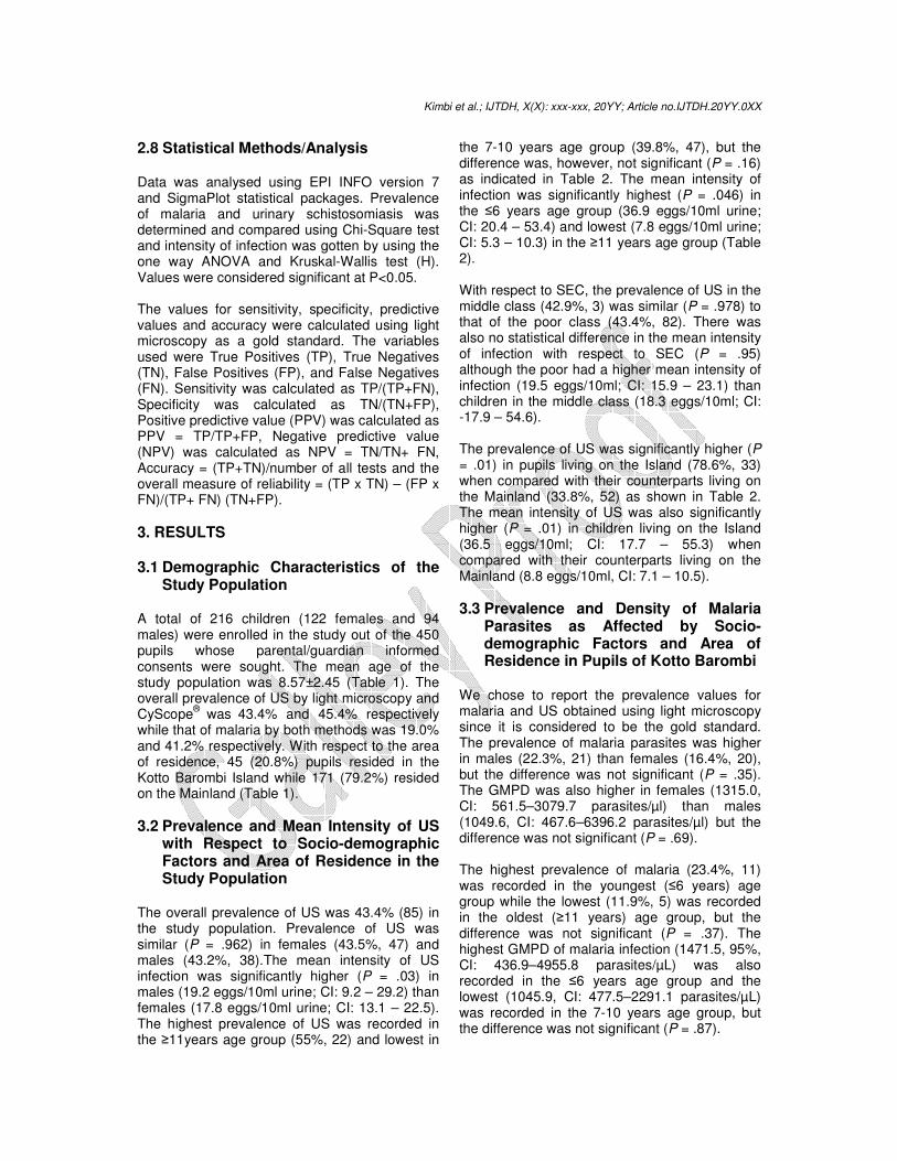
the children’s parents/legal guardians through the children to ask for the children’s participation in the study. Parents/legal guardians were allowed to ask questions and were also given room to refuse their children’s participation in the study. Urine and blood samples were collected only from children who returned signed consent forms from parents/legal guardians. Refusal of a child to participate in the study was fully respected.
2.5 Sample Collection Samples collected included urine and blood. Clean plastic containers were labeled (with pupil’s identification number and date of collection) and distributed to the children for urine collection. Clear instructions were given for proper urine collection. All necessary measures were taken to avoid contamination of the urine samples by continuously monitoring the collection process. Collection was done between 10 a.m. and 2 p.m. to coincide with the peak excretion of schistosome eggs. The urine samples were transported on ice blocks to the Malaria Research Laboratory at the University of Buea for analysis. Blood samples were collected by pricking the finger. Thick and thin blood films were prepared by the method described by Cheesbrough [14] for the determination of malaria parasite density and speciation respectively. The slides were labeled, air-dried and transported in slide boxes to the Malaria Research Laboratory of the University of Buea for staining and microscopy. Five (5) µL of blood was collected using a micropipette for the preparation of slides to be observed under the CyScope
®.
2.6 Rapid Detection of Malaria Parasites and Schistosome Eggs using the CyScope® Fluorescence Microscope
The test was performed according to the manufacturer’s (PARTEC GmbH, Münster, Germany) instructions. Briefly, the slide used to observe malaria parasites under the CyScope
® is
supplied with the dried reagent (4’-6 Di Amidino -2-Phenyl Indole, DAPI) which is a DNA-staining fluorochrome. The 5µL of capillary blood pipetted from the finger prick was placed on the dye-labeled portion of the slide. The blood was allowed to spread over the reagent, cover-slipped and incubated in a wet chamber at room temperature for I minute. The slide was then observed at x100 objective under UV light of the
CyScope® for malaria parasites. The presence of
distinct bright, shiny, tiny dots observed under the UV light indicated the presence of intracellular malaria parasites besides nuclei of white blood cells [15]. Positive and negative controls from known samples were observed with each batch of ready-to-use slides. Urine samples were analysed using the centrifugation method as described by Okanla [16]. Briefly, each bottle of urine was shaken and 10 ml of urine were drawn and transferred into a 20 ml centrifuge tube. The tubes were centrifuged at 1000 rpm for 5 minutes. The supernatant was discarded and the residue was put on a clean glass slide, a drop of Lugol’s iodine was added, cover-slipped and examined under the ×10 objective lens of a UNICO
® light
microscope. US status (infected or not infected) was determined by the detection of characteristic S. haematobium eggs in urine (presence of a terminal spine). Intensity of infection was estimated according to the number of eggs per 10ml of urine. The urine sample residues were also observed under the CyScope
®. Already prepared slides
(DAPI) impregnated with DNA dye were used and each urine sample was placed on a slide, cover-slipped and observed at ×40 objective and UV light at 365nm of the CyScope
®.
Characteristic morphology of the schistosome eggs was observed.
2.7 Staining of Blood Films and Microscopy
Blood films were stained with 10% Giemsa for 20 minutes. Malaria parasites (trophozoite, schizonts and gametocytes) were observed using the oil immersion (×100) objective of the light microscope. Slides were declared negative after observing at least 100 high power fields without detecting any parasites. Malaria parasites were counted in the thick films against 200 leukocytes, and counts were expressed as the number of parasites per µL of blood, assuming an average leukocyte count of 8000cells/µL blood [14]. Thin blood films were used for speciation using the identification tables [14]. All films were examined by two independent parasitologists who were blinded to the results of the CyScope
® and in the case of any
discrepancy, the film was re-read by a third parasitologist.
Kimbi et al.; IJTDH, X(X): xxx-xxx, 20YY; Article no.IJTDH.20YY.0XX
2.8 Statistical Methods/Analysis Data was analysed using EPI INFO version 7 and SigmaPlot statistical packages. Prevalence of malaria and urinary schistosomiasis was determined and compared using Chi-Square test and intensity of infection was gotten by using the one way ANOVA and Kruskal-Wallis test (H). Values were considered significant at P<0.05. The values for sensitivity, specificity, predictive values and accuracy were calculated using light microscopy as a gold standard. The variables used were True Positives (TP), True Negatives (TN), False Positives (FP), and False Negatives (FN). Sensitivity was calculated as TP/(TP+FN), Specificity was calculated as TN/(TN+FP), Positive predictive value (PPV) was calculated as PPV = TP/TP+FP, Negative predictive value (NPV) was calculated as NPV = TN/TN+ FN, Accuracy = (TP+TN)/number of all tests and the overall measure of reliability = (TP x TN) – (FP x FN)/(TP+ FN) (TN+FP).
3. RESULTS 3.1 Demographic Characteristics of the
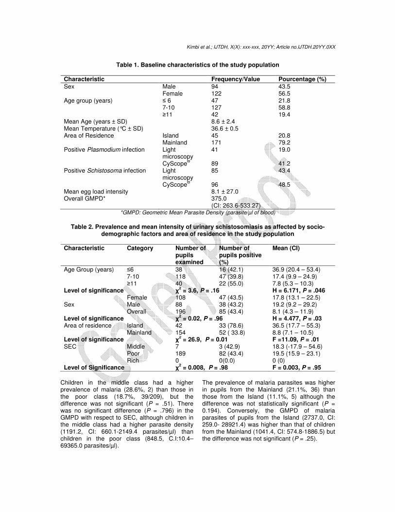
Study Population A total of 216 children (122 females and 94 males) were enrolled in the study out of the 450 pupils whose parental/guardian informed consents were sought. The mean age of the study population was 8.57±2.45 (Table 1). The overall prevalence of US by light microscopy and CyScope
® was
43.4% and 45.4% respectively
while that of malaria by both methods was 19.0% and 41.2% respectively. With respect to the area of residence, 45 (20.8%) pupils resided in the Kotto Barombi Island while 171 (79.2%) resided on the Mainland (Table 1).
3.2 Prevalence and Mean Intensity of US with Respect to Socio-demographic Factors and Area of Residence in the Study Population
The overall prevalence of US was 43.4% (85) in the study population. Prevalence of US was similar (P = .962) in females (43.5%, 47) and males (43.2%, 38).The mean intensity of US infection was significantly higher (P = .03) in males (19.2 eggs/10ml urine; CI: 9.2 – 29.2) than females (17.8 eggs/10ml urine; CI: 13.1 – 22.5). The highest prevalence of US was recorded in the ≥11years age group (55%, 22) and lowest in
the 7-10 years age group (39.8%, 47), but the difference was, however, not significant (P = .16) as indicated in Table 2. The mean intensity of infection was significantly highest (P = .046) in the ≤6 years age group (36.9 eggs/10ml urine; CI: 20.4 – 53.4) and lowest (7.8 eggs/10ml urine; CI: 5.3 – 10.3) in the ≥11 years age group (Table 2). With respect to SEC, the prevalence of US in the middle class (42.9%, 3) was similar (P = .978) to that of the poor class (43.4%, 82). There was also no statistical difference in the mean intensity of infection with respect to SEC (P = .95) although the poor had a higher mean intensity of infection (19.5 eggs/10ml; CI: 15.9 – 23.1) than children in the middle class (18.3 eggs/10ml; CI: -17.9 – 54.6). The prevalence of US was significantly higher (P = .01) in pupils living on the Island (78.6%, 33) when compared with their counterparts living on the Mainland (33.8%, 52) as shown in Table 2. The mean intensity of US was also significantly higher (P = .01) in children living on the Island (36.5 eggs/10ml; CI: 17.7 – 55.3) when compared with their counterparts living on the Mainland (8.8 eggs/10ml, CI: 7.1 – 10.5).
3.3 Prevalence and Density of Malaria Parasites as Affected by Socio-demographic Factors and Area of Residence in Pupils of Kotto Barombi
We chose to report the prevalence values for malaria and US obtained using light microscopy since it is considered to be the gold standard. The prevalence of malaria parasites was higher in males (22.3%, 21) than females (16.4%, 20), but the difference was not significant (P = .35). The GMPD was also higher in females (1315.0, CI: 561.5–3079.7 parasites/µl) than males (1049.6, CI: 467.6–6396.2 parasites/µl) but the difference was not significant (P = .69). The highest prevalence of malaria (23.4%, 11) was recorded in the youngest (≤6 years) age group while the lowest (11.9%, 5) was recorded in the oldest (≥11 years) age group, but the difference was not significant (P = .37). The highest GMPD of malaria infection (1471.5, 95%, CI: 436.9–4955.8 parasites/µL) was also recorded in the ≤6 years age group and the lowest (1045.9, CI: 477.5–2291.1 parasites/µL) was recorded in the 7-10 years age group, but the difference was not significant (P = .87).
Kimbi et al.; IJTDH, X(X): xxx-xxx, 20YY; Article no.IJTDH.20YY.0XX
Table 1. Baseline characteristics of the study population
Characteristic Frequency/Value Pourcentage (%) Sex Male 94 43.5
Female 122 56.5 Age group (years) ≤ 6 47 21.8
7-10 127 58.8 ≥11 42 19.4
Mean Age (years ± SD) 8.6 ± 2.4 Mean Temperature (°C ± SD) 36.6 ± 0.5 Area of Residence Island 45 20.8
Mainland 171 79.2 Positive Plasmodium infection Light
microscopy 41 19.0
CyScope® 89 41.2
Positive Schistosoma infection Light microscopy
85 43.4
CyScope® 96 48.5
Mean egg load intensity 8.1 ± 27.0 Overall GMPD* 375.0
(CI: 263.6-533.27)
*GMPD: Geometric Mean Parasite Density (parasite/µl of blood)
Table 2. Prevalence and mean intensity of urinary schistosomiasis as affected by socio-
demographic factors and area of residence in the study population
Characteristic Category
Number of pupils examined
Number of pupils positive (%)
Mean (CI)
Age Group (years) ≤6 38 16 (42.1) 36.9 (20.4 – 53.4) 7-10 118 47 (39.8) 17.4 (9.9 – 24.9) ≥11 40 22 (55.0) 7.8 (5.3 – 10.3)
Level of significance χ2 = 3.6, P = .16 H = 6.171, P = .046
Female 108 47 (43.5) 17.8 (13.1 – 22.5) Sex Male 88 38 (43.2) 19.2 (9.2 – 29.2) Overall 196 85 (43.4) 8.1 (4.3 – 11.9) Level of significance χ
2 = 0.02, P = .96 H = 4.477, P = .03
Area of residence Island 42 33 (78.6) 36.5 (17.7 – 55.3) Mainland 154 52 ( 33.8) 8.8 (7.1 – 10.5)
Level of significance χ2 = 26.9, P = 0.01 F =11.09, P = .01
SEC Middle 7 3 (42.9) 18.3 (-17.9 – 54.6) Poor 189 82 (43.4) 19.5 (15.9 – 23.1) Rich 0 0(0.0) 0 (0)
Level of Significance χ2 = 0.008, P = .98 F = 0.003, P = .95
Children in the middle class had a higher prevalence of malaria (28.6%, 2) than those in the poor class (18.7%, 39/209), but the difference was not significant (P = .51). There was no significant difference (P = .796) in the GMPD with respect to SEC, although children in the middle class had a higher parasite density (1191.2, CI: 660.1-2149.4 parasites/µl) than children in the poor class (848.5, C.I:10.4–69365.0 parasites/µl).
The prevalence of malaria parasites was higher in pupils from the Mainland (21.1%, 36) than those from the Island (11.1%, 5) although the difference was not statistically significant (P = 0.194). Conversely, the GMPD of malaria parasites of pupils from the Island (2737.0, CI: 259.0- 28921.4) was higher than that of children from the Mainland (1041.4, CI: 574.8-1886.5) but the difference was not significant (P = .25).

Kimbi et al.; IJTDH, X(X): xxx-xxx, 20YY; Article no.IJTDH.20YY.0XX
3.4 Comparing the Results of the CyScope® using Light Microscopy as a Gold Standard for US and Malaria
From a total of 216 pupils, 215 pupils were examined for both methods. With respect to US a total of 196 pupils, were examined by both methods - 77 pupils were positive both for light microscopy and the CyScope
® (TP), 18 pupils were positive for
CyScope® but negative for light microscopy (FP),
8 pupils were negative for CyScope® but positive
for light microscopy (FN) and 93 were negative for both methods (TN). Overall, 85 pupils were positive for US and 111 were negative for US using both diagnostic techniques. All with positive test (TP + FP) were 95 while all with negative test (FN+ TN) were = 101. Everyone = TP + FP + FN + TN =196. With respect to malaria, a total of 28 pupils were positive for both light microscopy and the CyScope
® (TP), 61 pupils were positive for
CyScope® but negative for light microscopy (FP),
13 pupils were negative for CyScope® but
positive for light microscopy (FN) and 113 were negative for both methods (TN). Overall, 41 pupils were positive for malaria and 113 were negative for malaria using both diagnostic techniques. All with positive test (TP + FP) were 89; while all with negative test (FN+ TN) were 126. Everyone (TP + FP + FN + TN) = 215.
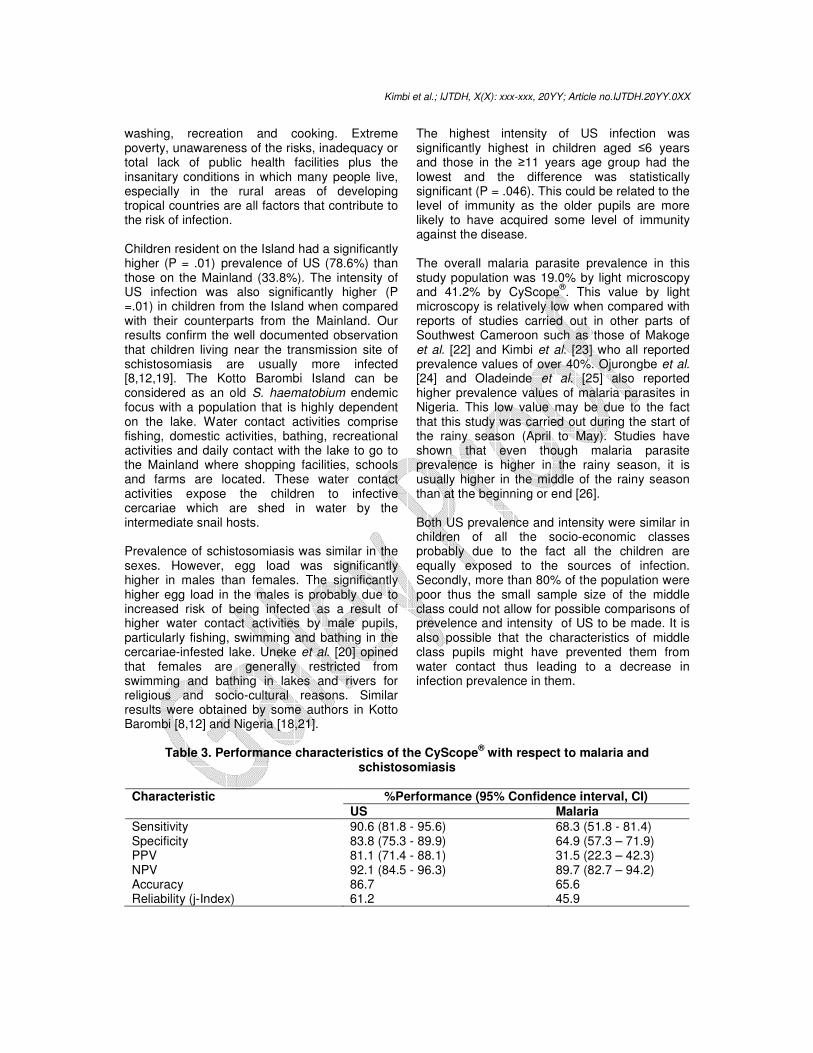
3.5 Performance Characteristics of the Partec CyScope® for US and Malaria
The general performance characteristics of the CyScope
® were evaluated for the diagnosis of
US and sensitivity was 90.6% (CI = 81.8 - 95.6%) while the specificity was 83.8% (CI = 75.3 - 89.9%) as shown on Table 3. Positive Predictive Value (PPV) was 81.1% (CI = 71.4 -88.1) and Negative Predictive Value (NPV) was 92.1% (CI = 84.5 - 96.3). Accuracy and Reliability were 86.7% and 61.2% respectively (Table 3). The general performance characteristics of the CyScope
® for the diagnosis of malaria were
evaluated and the sensitivity of the test was 68.3% (CI = 51.8 - 81.4%). Specificity was 64.9% (CI = 57.3 - 71.9%). PPV was 31.5% (CI = 22.3 - 42.3) and NPV was 89.7% (CI = 82.7 - 94.2). Accuracy and Reliability were 65.6% and 45.9% respectively (Table 3).
4. DISCUSSION Malaria and neglected tropical diseases such as schistosomiasis usually lead to an enormous burden on public health and affect mainly impoverished rural communities. One of the best control strategies for these diseases is early and accurate diagnosis followed by appropriate treatment. The prevalence of urinary schistosomiasis by light microscopy was 43.4%. This is slightly lower than the previous prevalence values reported by various authors [8,9,12] in the same area who all reported values above 50%. Generally, the recent momentum in the control of NTDs has generated a renewed interest in the control of schistosomiasis, resulting in the increase of large scale mass drug administration of praziquantel in several countries including Cameroon [17]. With the renewed momentum for NTDs, the national programme for the control of schistosomiasis and STHs in Cameroon was created in March 2003 with commitments from the Ministries of Public Health and Basic Education in Cameroon to eliminate the infection as a major public health problem in all regions of the country. In 2007, Cameroon launched a nationwide deworming campaign, following the WHO recommendations that lay emphasis on regular treatments of high risk groups such as school children and appropriate health education, as the most appropriate means of reducing morbidity due to the infection. WHO [4] reported that significant progress has been made in the control of schistosomiasis during the past ten years. However, this still falls short of the target set by a World Health Assembly Resolution adopted in 2001, which was aimed at reaching at least 75% of all school-age children at risk of morbidity by 2010 [4]. Consequently, there is a need to develop more sensitive diagnostic tools, in order to help target control or develop better screening strategies for the elimination of the disease. The high infection rate and intensity of infection of US in this study probably reflects the high level of exposure and dependence of the inhabitants on the infected lake since the area has no pipe-borne water or stream and so all the inhabitants of the village rely on the lake to fetch water for drinking, laundry and domestic use. Ekpo et al. [18] reported a similar situation in a village near Abeokuta in Nigeria where lack of pipe-borne water in the village led to a high US prevalence because all the villagers depend on the lone river as their source of water for bathing, drinking,
Kimbi et al.; IJTDH, X(X): xxx-xxx, 20YY; Article no.IJTDH.20YY.0XX
washing, recreation and cooking. Extreme poverty, unawareness of the risks, inadequacy or total lack of public health facilities plus the insanitary conditions in which many people live, especially in the rural areas of developing tropical countries are all factors that contribute to the risk of infection. Children resident on the Island had a significantly higher (P = .01) prevalence of US (78.6%) than those on the Mainland (33.8%). The intensity of US infection was also significantly higher (P =.01) in children from the Island when compared with their counterparts from the Mainland. Our results confirm the well documented observation that children living near the transmission site of schistosomiasis are usually more infected [8,12,19]. The Kotto Barombi Island can be considered as an old S. haematobium endemic focus with a population that is highly dependent on the lake. Water contact activities comprise fishing, domestic activities, bathing, recreational activities and daily contact with the lake to go to the Mainland where shopping facilities, schools and farms are located. These water contact activities expose the children to infective cercariae which are shed in water by the intermediate snail hosts. Prevalence of schistosomiasis was similar in the sexes. However, egg load was significantly higher in males than females. The significantly higher egg load in the males is probably due to increased risk of being infected as a result of higher water contact activities by male pupils, particularly fishing, swimming and bathing in the cercariae-infested lake. Uneke et al. [20] opined that females are generally restricted from swimming and bathing in lakes and rivers for religious and socio-cultural reasons. Similar results were obtained by some authors in Kotto Barombi [8,12] and Nigeria [18,21].
The highest intensity of US infection was significantly highest in children aged ≤6 years and those in the ≥11 years age group had the lowest and the difference was statistically significant (P = .046). This could be related to the level of immunity as the older pupils are more likely to have acquired some level of immunity against the disease. The overall malaria parasite prevalence in this study population was 19.0% by light microscopy and 41.2% by CyScope
®. This value by light
microscopy is relatively low when compared with reports of studies carried out in other parts of Southwest Cameroon such as those of Makoge et al. [22] and Kimbi et al. [23] who all reported prevalence values of over 40%. Ojurongbe et al. [24] and Oladeinde et al. [25] also reported higher prevalence values of malaria parasites in Nigeria. This low value may be due to the fact that this study was carried out during the start of the rainy season (April to May). Studies have shown that even though malaria parasite prevalence is higher in the rainy season, it is usually higher in the middle of the rainy season than at the beginning or end [26]. Both US prevalence and intensity were similar in children of all the socio-economic classes probably due to the fact all the children are equally exposed to the sources of infection. Secondly, more than 80% of the population were poor thus the small sample size of the middle class could not allow for possible comparisons of prevelence and intensity of US to be made. It is also possible that the characteristics of middle class pupils might have prevented them from water contact thus leading to a decrease in infection prevalence in them.
Table 3. Performance characteristics of the CyScope® with respect to malaria and
schistosomiasis
Characteristic %Performance (95% Confidence interval, CI) US Malaria
Sensitivity 90.6 (81.8 - 95.6) 68.3 (51.8 - 81.4) Specificity 83.8 (75.3 - 89.9) 64.9 (57.3 – 71.9) PPV 81.1 (71.4 - 88.1) 31.5 (22.3 – 42.3) NPV 92.1 (84.5 - 96.3) 89.7 (82.7 – 94.2) Accuracy 86.7 65.6 Reliability (j-Index) 61.2 45.9
Kimbi et al.; IJTDH, X(X): xxx-xxx, 20YY; Article no.IJTDH.20YY.0XX
Malaria prevalence was higher in males than females although the difference was not statistically significant. This observation may be attributed to the fact that males generally expose their bodies more than females especially when the weather is hot and thus are more likely to be bitten by infected Anopheles mosquitoes. Studies have also shown that females have a better immunity to parasitic diseases and this has been attributed to genetic and hormonal factors [27]. The highest malaria parasite prevalence and density were recorded in the youngest age group and the values decreased with age. Immunity to malaria is known to be acquired after repeated exposures to infection and so the observed high values in the youngest age group may be due to their relatively less developed immune systems [25]. The SEC did not significantly affect either malaria prevalence or density. We determined for the first time the performance characteristics of the CyScope
® fluorescence
microscope in the diagnosis of urinary schistosomiasis among school-age children in this locality who are known to have both low and high intensities of schistosome infection. Out of the 196 children whose urine samples were collected, 85 (43.4%) were diagnosed by light microscopy and 95 (48.47%) by the CyScope
®
methods. The slightly low prevalence using the light microscopy could be due to the fact that some infection intensities were very light and therefore could not be detected. The diagnosis of US is traditionally achieved through the use of parasitological (urine filtration) methods. This is the most direct and specific method of detecting active infections, but it often misses low intensity infections [5]. Furthermore, it is thought that with preventive chemotherapy-based helminthiases (including schistosomiasis) control programmes due to be scaled up [4], helminth egg output may likely decline further and the problem of insensitive egg counting methods may be exacerbated [28]. The higher prevalence of US recorded by the CyScope
® method when compared with the light
microscopy is probably due to the fact that low intensities of infection could be detected by the DAPI slide with DNA-binding schistosome eggs. Similar results using the CyScope
® fluorescence
were obtained in Douala and Njombe in Cameroon with the CyScope
® registering a
higher prevalence than the light microscopy [7]. The sensitivity, specificity, PPV and NPV of the CyScope
® were 90.6%, 83.8%, 81.1% and
92.1% respectively. This shows that the CyScope
® method is more sensitive than light
microscopy and could be used for rapid and mass diagnosis of US since it is able to detect both low and high intensities of US. So far malaria control relies mostly on prompt diagnosis and treatment. Consequently, poor diagnostic methods can under-estimate disease burden. Out of the 216 pupils sampled, 41 (19.0%) were positive for malaria by light microscopy while 89 (41.2%) were positive by CyScope
® fluorescence microscopy. Nkrumah et
al. [29] obtained similar results when comparing two rapid diagnostic tests (Partec Rapid Malaria Test (R) and Binax Now (R) Malaria Rapid Diagnostic Test) for malaria diagnosis in Ghana. The results obtained showed 61 false positive and 13 false negative. The high false positive results recorded ties with reports from Uganda by Sousa-Figueiredo et al. [30] who suggested that false positive results by the CyScope
® method
could be linked to the presence of schistosomes (in the blood stream) which usually regurgitate their blood meals sloughing their intestinal cells that could likely contain broken down nuclei (that can fluoresce and be detected by CyScope
®) into
the host blood stream. This study was carried out in an endemic area of US in which such possibilities could be highly observed. The performance characteristics of the CyScope
® were evaluated and revealed as
sensitivity, specificity, PPV and NPV of 68.3%, 64.9%, 31.5% and 89.7% respectively. Sensitivity level was much lower than the previously reported values for this method [10,15] (albeit under very different survey settings and study population). It was below the 95% threshold required in a malaria rapid diagnostic test [31]. Light microscopy with Giemsa-stained thick and thin blood films has been shown to be a less sensitive test, but it remains a gold standard because it can be used for both qualitative and quantitative purposes [10,11]. However, it cannot be used in rural areas where there is no electricity although malaria is more endemic in such areas. Apart from early detection and treatment of US and malaria, one of the ways to control both infections could be through school-based health education interventions. Such an approach could engage school children to reach community

Kimbi et al.; IJTDH, X(X): xxx-xxx, 20YY; Article no.IJTDH.20YY.0XX
adults with health messages and hygienic practices through action-oriented and participatory learning action [26]. Thus, the children could be playing the role of health change agents in the community. This will likely have a substantial impact not only on school children, but also on community adults in improving knowledge on cause and prevention of both diseases.
5. CONCLUSION This present study suggests that the CyScope
®
has a better diagnostic ability than light microscopy in the diagnosis of both US and malaria. If rapid diagnosis of the diseases is done using this tool and followed by mass treatment of all infected cases in the village this will probably lead to control of the diseases over time. The mothers or guardians and caregivers also need to be enlightened about the avoidance of the lakes, heating of water before using it to bathe and not taking the children along with them to the water bodies. For any control measure to succeed, it must involve the community, such as developing participatory health education programmes with community members to effect behavioural change especially by mothers and care givers so as to understand and avoid infection.
INFORMED CONSENT All pupils were issued consent forms to seek for their parents’ approval. Pupils were accepted for screening when they brought back signed informed consent forms following the approval of their parents/guardians.
ETHICAL CONSIDERATION An ethical clearance was obtained from the South West Regional Delegation of Public Health. An administrative clearance was obtained from the Divisional Delegation of Basic Education, Kumba, Meme Division.
COMPETING INTERESTS Authors have declared that no competing interests exist.
REFERENCES 1. WHO. Prevention and control of
schistosomiasis and soil-transmitted
helminthiasis. Report of WHO Expert Committee. WHO, Geneva. 2002. 1-24.
2. Snow RW, Guerra CA, Noor AM, Myint HY and Hay SI. The global distribution of clinical episodes of Plasmodium falciparum malaria. Nature 2005. 434: 214-217.
3. WHO. World Malaria Report 2012. World Health Organization, 2012. Geneva, 20 Avenue Appia, 1211 Geneva 27, Switzerland. 230-231.
4. World Health Organization. Schistosomiasis: number of people treated, 2009. Weekly Epidemiological Record. 2011, 9(86):73–80.
5. Utzinger J, N’Goran EK, Caffrey CR, Keiser J. From innovation to application: social-ecological context, diagnostics, drugs and integrated control of schistosomiasis. Acta Tropica. 2011, 120(Suppl 1):S121–S137.
6. Murray CK, Bennet JW. Rapid diagnosis of malaria. Interdiscipli Persp Infect Dis. 2009: 1-7.
7. Lehman LG, Nono LK, Bilong Bilong CF. Diagnostic des parasitoses intestinales à l’aide de la microscopie a fluorescence. Medécine d’Afrique Noire. 2012 59(7): 378-385.
8. Ndamukong KJN, Ayuk MA, Dinga JS, Nkuo-Akenji TN, Ndiforchu VA, Titanji VPK. Prevalence and intensity of urinary schistosomiasis in primary school children of the Kotto Barombi Health Area, Cameroon. E Afr Med J. 2001. 78: 287-289.
9. Nkengazong L. Njiokou F, Tenkeng F, Enyong P, Wanji S. Reassessment of endemicity level of urinary schistosomiasis in the Kotto Barombi focus (South West Cameroon) and impact of mass drug administration (MDA) on the parasitic indices. J Cell Ani Biol. 2009. 3(9): 159-164.
10. Hassan SE-DH, Okoued SI, Mudathir MA, Malik EM. Testing the sensitivity and specificity of the fluorescence microscope (CyScope
®) for malaria diagnosis. Malar J
2010, 9(88): 1-4. doi:10.1186/1475-2875-9-88
11. Nkrumah B, Agyekum A, Acquah S, May J, Tannick E, Brattig N et al. Comparison of the Novel Partec Rapid Malaria Test to the conventional Giemsa stain and the gold standard real-time PCR. J Clin Microbiol. 2010, 48(8): 2925-2928.
12. Moyou SR, Tagni, ZD, Kouamouo J, Enyong P, Ripert C. Epidemiologic and

Kimbi et al.; IJTDH, X(X): xxx-xxx, 20YY; Article no.IJTDH.20YY.0XX
radiologic study of urinary bilharziasis in the focus of Barombi Lake (Meme Department), Cameroon. Bulletin de la Société de Pathologie Exotique. 1987, 80(5): 813-825.
13. Kimbi HK, Sumbele IUN, Nweboh MN, Anchang-Kimbi JK, Lum E, Nana Y et al. Malaria and haematologic parameters of pupils at different altitudes along the slope of Mount Cameroon: a cross-sectional study. Malar J. 2013, 12:193.
14. Cheesbrough M. District laboratory practice in tropical countries part 1 and 2.Cambridge low price editions, 2nd ed. Cambridge University Press. 2010
15. Kimbi HK, Ajeagah HA, Keka FC, Lum E, Nyabeyeu HN, Tonga C et al. Asymptomatic Malaria in School Children and Evaluation of the Performance Characteristics of the Partec CyScope
® in
the Mount Cameroon Region. J Bacteriol Parasitol 2012a, 3:154.
16. Okanla EO. Schistosomiasis: influence of parental occupation and rural or urban dwelling on prevalence. Nig J Pure Appl Sci. 1991, 6: 154-159.
17. Tchuem Tchuenté L-A, Ngassam RIK, Sumo L, Ngassam P, Noumedem CD, Nzu DDL et al. Mapping of Schistosomiasis and Soil-Transmitted Helminthiasis in the Regions of Centre, East and West Cameroon. PLoS Negl Trop Dis. 2012, 6(3): e1553. doi:10.1371/journal.pntd.0001553.
18. Ekpo UF, Laja-Deile A, Oluwole AS, Sam-Wobo SO, Mafiana CF. Urinary schistosomiasis among preschool children in a rural community near Abeokuta, Nigeria. Parasites & Vectors. 2010, 3: 58.
19. Kabatereine NB, Standley CJ, Sousa-Figueiredo JC, Fleming FM, Stothard JR, Talisuna A et al. Integrated prevalence mapping of schistosomiasis, soil-transmitted helminthiasis and malaria in lakeside and island communities in Lake Victoria, Uganda. Parasites and Vectors. 2011, 4: 232.
20. Uneke CJ, Oyibo PG, Ugwuoru CDC, Nwanokwai AP, Iloegunam RO. Urinary Schistosomiasis Among School Age Children In Ebonyi State, Nigeria: Internet J Lab Med. 2007, 2(1): 15 – 19.
21. Tohon ZB, Mainassara HB, Garba A, Mahamane AE, Bosqué-Olia E, Ibrahim M, et al. Controlling Schistosomiasis: Significant decrease of anaemia prevalence one year after a single dose of
praziquantel in Nigerian school children. Plos Neglected Trop Dis. 2008, 2(5): e241. doi: 10.1371/journal.pntd.0000241.
22. Makoge VD, Mbah GA, Nkengazong L, Sahfe NE, Moyou RS. Falciparum malaria, helminth infection, and anaemia in asymptomatic pupils in four villages in Cameroon. European J Zoological Res. 2012, 1 (2): 54-59.
23. Kimbi HK, Keka FC, Nyabeyeu, HN, Ajeagah HU, Tonga C, Lum E et al. An update of asymptomatic Falciparum malaria in school children in Muea, Southwest Cameroon. J Bacteriol Parasitol. 2012b, 3: 153.
24. Ojurongbe O, Adegbayi AM, Bolaji OS, Akindele AA, Adefioye AO, Adeyeba OA. Asymptomatic falciparum malaria and intestinal helminths co-infection among school children in Osogbo, Nigeria. J Res Med Sci; 2011, 16(5): 680–686.
25. Oladeinde BH, Omoregie R, Olley M, Anunibe JA, Onifade AA, Oladeinde OB. Malaria and anemia among children in a low resource setting in Nigeria. Iranian J Parasitol, 2012; 7(3): 31-37.
26. Ayi I, Nonaka D, Adjovu JK, Hanafusa S, Jimba M, Bosompem KM, et al. Research School-based participatory health education for malaria control in Ghana: engaging children as health messengers. Malar J. 2010, 9: 98-110.
27. Zuk M and McKean KA. Sex differences in parasite infections: patterns and processes. Internat J Parasitol. 1996, 26: 1009–23.
28. Knopp S, Stothard JR, Rollinson D, Mohammed KA, Khamis IS, Marti H et al. From morbidity control to transmission control: time to change tactics against helminths on Unguja island, Zanzibar. Acta Tropica 2013: 128(2):412-22. doi: 10.1016/j.actatropica.2011.04.010.
29. Nkrumah B, Acquah SE, Ibrahim L, May J, Bratigg N, Tannich E et al. Comparative evaluation of two rapid field tests for malaria diagnosis: Partec Rapid Malaria Test® and Binax Now® Malaria Rapid Diagnostic Test BMC Infect Dis 2011; 11:143.
30. Sousa-Figueiredo JC, Oguttu D, Adriko M, Besigye F, Nankasi A, Arinaitwe M, et al. Investigating portable fluorescent microscopy (CyScope) as an alternative rapid diagnostic test for malaria in children and women of child-bearing age. Malar J. 2010, 9: 245.

Kimbi et al.; IJTDH, X(X): xxx-xxx, 20YY; Article no.IJTDH.20YY.0XX
31. WHO. Towards Quality Testing of Malaria Rapid Diagnostic Tests: Evidence and
Methods World Health Organisation, Philippines 2006.
_________________________________________________________________________________ © 2015 Kimbi et al.; This is an Open Access article distributed under the terms of the Creative Commons Attribution License (http://creativecommons.org/licenses/by/4.0), which permits unrestricted use, distribution, and reproduction in any medium, provided the original work is properly cited.