Impairment of syntax and lexical semantics in a patient with bilateral paramedian thalamic...
9
Brain and Language 96 (2006) 69–77 www.elsevier.com/locate/b&l 0093-934X/$ - see front matter 2005 Elsevier Inc. All rights reserved. doi:10.1016/j.bandl.2005.08.011 Impairment of syntax and lexical semantics in a patient with bilateral paramedian thalamic infarction Lieve De Witte a , Ineke Wilssens b , Sebastiaan Engelborghs c,d , Peter P. De Deyn c,d , Peter Mariën a,c,d,¤ a Department of Linguistics, Free University of Brussels, Belgium b Department of Speech Pathology, Middelheim General Hospital, Antwerp, Belgium c Department of Neurology, Middelheim General Hospital, Antwerp, Belgium d Laboratory of Neurochemistry and Behavior, Institute of Born-Bunge, University of Antwerp, Belgium Accepted 30 August 2005 Available online 13 October 2005 Abstract Bilateral vascular thalamic lesions are rare. Although a variety of neurobehavioral manifestations have been described, the literature is less documented with regard to accompanying linguistic disturbances. This article presents an in-depth neurolinguistic analysis of the lan- guage symptoms of a patient who incurred bilateral paramedian ischemic damage of the thalamus. In the post-acute phase of the stroke, a unique combination of transcortical sensory aphasia with syntactic impairment was found. Because of this atypical semiological associ- ation, additional analyses of spontaneous speech were performed. In spite of the typological aYnity with the grammatic characteristic of marked simpliWcation of syntax observed in Broca’s aphasia, only a wordclass speciWc, lexical-semantic deWcit for verbs was objectiWed. The hypothesis that lexical-semantic disturbances in our patient might result from a functional deaVerentiation of both thalami with the frontal lobe is supported by: (1) associated neuropsychological deWcits of frontal origin and (2) frontal-like behavioral disturbances. 2005 Elsevier Inc. All rights reserved. Keywords: Bithalamic stroke; SimpliWcation of syntax; Transcortical sensory aphasia 1. Introduction A variety of neurobehavioral symptoms have been reported in association with bilateral lesions of the thala- mus: (1) transient vigilance disorders, (2) hypersomnia, (3) vertical gaze palsy, (4) persistent anterograde amnesia, (5) speech and language disturbances, and (6) apathy (Bewermeyer, Dreesbach, Rackl, Neveling, & Heiss, 1985; Bogousslavsky, Regli, Delaloye-bischof, Assal, & Uske, 1991; Dehaene, 1982; van Domburg, ten Donkelaar, & Notermans, 1996; Fukatsu, Fujii, Yamadori, Nagasawa, & Sakurai, 1997; Guberman & Stuss, 1983; Krolak-Salmon, Croisile, Houzard, Setiey, & Girard-Madoux, 2000; Reilly, Connolly, Stack, Martin, & Hutchinson, 1992). Although speech and language functions have not been systematically investigated, dysarthric and aphasic disturbances have been reported after bithalamic lesions (Lhermitte, 1984). On the articulatory level, the deviant speech characteristics have been denoted as hypokinetic dysarthria (Ackermann, Ziegler, & Petersen, 1993; Benabdeljlil, El alaoui faris, Kissani, Aidi, & Laaouina, 2001; Bewermeyer et al., 1985; Dehaene, 1982; Gentilini, De Renzi, & Crisi, 1987; Gerber & Gudesblatt, 1986; Lazzarino & Nicolai, 1988; Malamut, GraV-Radford, Chawluk, Grossman, & Gur, 1992; Reilly et al., 1992). Reduced spontaneous speech and diminished verbal Xuency have been described at the linguistic level (Bogousslavsky et al., 1991; van Domburg et al., 1996; Engelborghs, Marien, Pickut, Verstraeten, & De Deyn, 2000; Guberman & Stuss, 1983; Krolak-salmon et al., 2000; Krolak-Salmon, Montavont, Hermier, Milliery, & Vighetto, 2002; Malamut et al., 1992; Rousseaux et al., 1986; Schott, Maugiere, Laurent, Serclerat, & Fischer, 1980). In addition, some authors have reported naming * Corresponding author. E-mail address: [email protected] (P. Mariën).
Transcript of Impairment of syntax and lexical semantics in a patient with bilateral paramedian thalamic...
Brain and Language 96 (2006) 69–77
www.elsevier.com/locate/b&l
Impairment of syntax and lexical semantics in a patient with bilateral paramedian thalamic infarction
Lieve De Witte a, Ineke Wilssens b, Sebastiaan Engelborghs c,d, Peter P. De Deyn c,d, Peter Mariën a,c,d,¤
a Department of Linguistics, Free University of Brussels, Belgiumb Department of Speech Pathology, Middelheim General Hospital, Antwerp, Belgium
c Department of Neurology, Middelheim General Hospital, Antwerp, Belgiumd Laboratory of Neurochemistry and Behavior, Institute of Born-Bunge, University of Antwerp, Belgium
Accepted 30 August 2005Available online 13 October 2005
Abstract
Bilateral vascular thalamic lesions are rare. Although a variety of neurobehavioral manifestations have been described, the literature isless documented with regard to accompanying linguistic disturbances. This article presents an in-depth neurolinguistic analysis of the lan-guage symptoms of a patient who incurred bilateral paramedian ischemic damage of the thalamus. In the post-acute phase of the stroke,a unique combination of transcortical sensory aphasia with syntactic impairment was found. Because of this atypical semiological associ-ation, additional analyses of spontaneous speech were performed. In spite of the typological aYnity with the grammatic characteristic ofmarked simpliWcation of syntax observed in Broca’s aphasia, only a wordclass speciWc, lexical-semantic deWcit for verbs was objectiWed.The hypothesis that lexical-semantic disturbances in our patient might result from a functional deaVerentiation of both thalami with thefrontal lobe is supported by: (1) associated neuropsychological deWcits of frontal origin and (2) frontal-like behavioral disturbances. 2005 Elsevier Inc. All rights reserved.
Keywords: Bithalamic stroke; SimpliWcation of syntax; Transcortical sensory aphasia
1. Introduction
A variety of neurobehavioral symptoms have beenreported in association with bilateral lesions of the thala-mus: (1) transient vigilance disorders, (2) hypersomnia, (3)vertical gaze palsy, (4) persistent anterograde amnesia, (5)speech and language disturbances, and (6) apathy(Bewermeyer, Dreesbach, Rackl, Neveling, & Heiss, 1985;Bogousslavsky, Regli, Delaloye-bischof, Assal, & Uske,1991; Dehaene, 1982; van Domburg, ten Donkelaar, &Notermans, 1996; Fukatsu, Fujii, Yamadori, Nagasawa, &Sakurai, 1997; Guberman & Stuss, 1983; Krolak-Salmon,Croisile, Houzard, Setiey, & Girard-Madoux, 2000; Reilly,Connolly, Stack, Martin, & Hutchinson, 1992). Althoughspeech and language functions have not been systematically
* Corresponding author.E-mail address: [email protected] (P. Mariën).
0093-934X/$ - see front matter 2005 Elsevier Inc. All rights reserved.doi:10.1016/j.bandl.2005.08.011
investigated, dysarthric and aphasic disturbances have beenreported after bithalamic lesions (Lhermitte, 1984). On thearticulatory level, the deviant speech characteristics havebeen denoted as hypokinetic dysarthria (Ackermann,Ziegler, & Petersen, 1993; Benabdeljlil, El alaoui faris,Kissani, Aidi, & Laaouina, 2001; Bewermeyer et al., 1985;Dehaene, 1982; Gentilini, De Renzi, & Crisi, 1987; Gerber& Gudesblatt, 1986; Lazzarino & Nicolai, 1988; Malamut,GraV-Radford, Chawluk, Grossman, & Gur, 1992; Reillyet al., 1992). Reduced spontaneous speech and diminishedverbal Xuency have been described at the linguistic level(Bogousslavsky et al., 1991; van Domburg et al., 1996;Engelborghs, Marien, Pickut, Verstraeten, & De Deyn,2000; Guberman & Stuss, 1983; Krolak-salmon et al., 2000;Krolak-Salmon, Montavont, Hermier, Milliery, &Vighetto, 2002; Malamut et al., 1992; Rousseaux et al.,1986; Schott, Maugiere, Laurent, Serclerat, & Fischer,1980). In addition, some authors have reported naming

70 L. De Witte et al. / Brain and Language 96 (2006) 69–77
diYculties and mild auditory-verbal comprehension distur-bances (Bogousslavsky et al., 1991; van Domburg et al.,1996; Engelborghs et al., 2000; Guberman & Stuss, 1983).
We report the Wndings of in-depth neurolinguistic inves-tigations in a patient who incurred transcortical sensoryaphasia (TSA) associated with marked simpliWcation ofsyntax due to a bilateral paramedian thalamic infarction. Thecombination of Xuent aphasia with syntax simpliWcation israther unexpected since this grammatical characteristic clas-sically relates to non-Xuent aphasia types. To the best of ourknowledge simpliWcation of syntax, characterized in ourpatient by simple sentences and sentence fragments with acomplete absence of embedded clauses, has never beenreported in association with transcortical sensory aphasia.
2. Case report
A 78-year-old right-handed woman was admitted to ourhospital after acute loss of strength in the right arm, diplo-pia, and speech and language disturbances. Medical ante-cedents consisted of arterial hypertension andhypercholesterolemia. In addition to Xuent aphasia, theclinical neurological examination on admission revealedvertical gaze palsy and restricted adduction of the left eyeresulting in divergent strabism due to left oculomotor nerveparesis. Except for a mild paresis of the right arm, musclestrength and tone were completely normal. The Wnger-to-nose test showed dysmetria of the right arm, the knee-to-heel test was normal. Tendon reXexes at the right arm andleg were increased. Plantar responses were indiVerent. Sen-sory and general physical examination revealed no abnor-malities. Computerized tomography (CT) scan of the braindisclosed a bilateral paramedian thalamic infarction. Brainmagnetic resonance imaging (MRI) demonstrated bilateralinvolvement of the medial thalamic nucleus (Engelborghs,Marien, Martin, & De Deyn, 1998; Kretschmann & Wein-rich, 1992) as well as a small hyperintense area in the leftinternal capsula. Extension of the left thalamic lesion to theleft red nucleus was shown as well (Fig. 1). A diagnosis ofacute bithalamic stroke with extension to the mesencepha-lon was made. Vertebral artery dissection and signiWcantstenoses were excluded by magnetic resonance angiography(MRA). Trans-oesophagal echocardiography was normal.Antiaggregant secondary preventive treatment as well asintensive physical and speech therapy were started.
After seven weeks the right hemiparesis had recoveredcompletely. The vertical gaze palsy and oculomotor nerveparesis had only partially improved. The aphasia did notresolve.
2.1. Neuropsychological investigations
2.1.1. MethodsLanguage was assessed according to a time-frame model
for the study of aphasia (Alexander, 1989; Mazzochi &Vignolo, 1979). In the post-acute phase of the stroke (lesionphase), extensive formal language investigations were per-
formed by means of standardized language tests, amongwhich: the Aachen Aphasia Test (AAT) (Graetz, De Bleser,& Willmes, 1990), the Token Test (De Renzi & Vignolo,1962), the Boston Naming Test (BNT) (Kaplan, Goodglass,& Weintraub, 1983; Marien, Mampaey, Vervaet, Saerens, &De Deyn, 1998), and a semantic verbal Xuency task (unpub-lished norms). Four weeks after the stroke (lesion phase)analysis of spontaneous speech and syntactic competencewas performed by means of a method developed byVermeulen, Bastiaanse, and Van wageningen (1989) andthe Werkwoorden- en Zinnen Test (WEZT; Bastiaanse,Maas, & Rispens, 2000).
Cognitive functions were examined during the lesionphase one and a half month after stroke by means of theMini Mental State Examination (MMSE) (Folstein, Fol-stein, & Mc Hugh, 1975), parts of the Alzheimer’s DiseaseAssessment Scale (ADAS) (Rosen, Mohs, & Davis, 1984),the Coloured Progressive Matrices (Court and Raven,1983), the Hierarchic Dementia Scale (HDS) (Cole &Dastoor, 1987), the Rey-Osterrieth Figure (Osterrieth,1944), and the Judgment of Line Orientation test (JLO)(Benton, Hamsher, Varney, & Spreen, 1983).
2.1.2. Neurolinguistic resultsAs shown in Table 1, the AAT results for language com-
prehension were within the low normal range at both theauditory and written comprehension level. However, asshown by the subtest results, auditory sentence comprehen-sion and written word comprehension were defective,whereas auditory word comprehension and written sen-tence comprehension were normal. Oral language wasXuent and characterized by word-Wnding diYculties andsemantic paraphasias. The spontaneous speech, character-ized by simple sentences, sentence fragments with a com-plete absence of embedded, subordinate clause indicated amarked simpliWcation of syntax (Fig. 2).
On the AAT confrontation naming tests a defectiveglobal score of 91/120 was found. The patient obtained anormal result for simple nouns. Naming of colors and com-pound nouns resulted in a deWcient score. Most errors wereof the semantic type: semantic paraphasias (e.g.,screwdriver D hammer) and semantic neologisms (e.g.,cleaner D suckmachine). Although the BNT-score waswithin the normal range, errors were of aphasic origin: per-severations, circumlocutions (e.g., camel D a seaƒ with twohunches), semantic paraphasias (e.g., octopus D seal;beaver D rat), and semantic neologisms (e.g., protractor Delectric measure organ; hammock D lie folder). In a seman-tic word Xuency task the patient produced a normal num-ber of items. On the AAT sentence construction subtest, inwhich pictures have to be described depicting complex sce-narios, she obtained a defective score. Repetition wasnormal.
Written language, as shown by the AAT results, was normal.Summing-up, the aphasia proWle of the patient corre-
sponds to a taxonomic diagnosis of TSA: language compre-hension was disturbed, speech was Xuent and contained a

L. De Witte et al. / Brain and Language 96 (2006) 69–77 71
Fig. 1. Brain MRI axial FLAIR slices (A, B, C—from rostral to caudal) performed one month after stroke disclosing bilateral involvement of the medialthalamic nucleus, a small hyperintense area in the left internal capsula and extension of the left thalamic lesion to the left red nucleus.

72 L. De Witte et al. / Brain and Language 96 (2006) 69–77
high incidence of semantic errors but repetition was intact.In addition, spontaneous speech was characterized bymarked simpliWcation of syntax.
Since the association of TSA with simpliWcation of syn-tax represents an atypical Wnding, grammatical competencewas further investigated.
Table 1AAT, BNT and word Xuency test results
Score Max Mean SD
Akense Aphasia Test (AAT)Language comprehension total 96 120 108.5 10.24
Auditory word comprehension 27 30 26.49 3.30Auditory sentence comprehension 20 30 26.79 3.41Total auditory comprehension 47 60 53.28 6.08Reading word comprehension 21 30 28.30 2.29Reading sentence comprehension 28 30 26.91 3.39Total reading comprehension 49 60 55.21 4.90
Token Test 13 errors 0
Spontaneous SpeechCommunicative behaviour 4 5 4.63 0.54Articulation and Prosody 5 5 4.63 0.67Automatisms 4 5 4.59 0.65Semantic structure 4 5 4.59 0.53Phonematic structure 5 5 4.54 0.56Syntactic structure 2 5 4.41 0.55
Imposed SpeechRepetition total 147 150 144.1 8.07
Phonemes 30 30 28.91 2.09One-syllable words 30 30 29.22 1.32Words of foreign origin 29 30 28.94 2.31Compound words 29 30 28.45 2.22Sentences 29 30 28.55 1.90
Naming total 91 120 109.3 8.42Simple nouns 28 30 27.92 2.90Colors 23 30 27.69 1.99Compound nouns 23 30 28.04 2.61Sentence construction 17 30 25.69 3.72
Written language total 80 90 85.52 7.63Reading aloud 28 30 28.95 1.93Composing on dictation 27 30 28.57 2.75Writing on dictation 25 30 28 3.67
Boston Naming Test 44 60 49.1 6.76
Verbal Fluency-Semantic 35 43.4 11.76Animals 14 1 minVegetables 8 1 minClothing 9 1 minMeans of transport 4 1 min
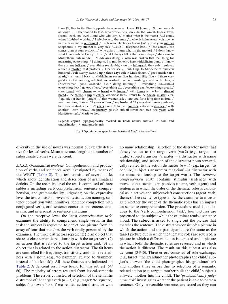
2.1.3. Additional language analyses2.1.3.1. Spontaneous language analysis. A sample size of318 words (Fig. 3) was randomly selected from an extensivecorpus of recorded spontaneous speech and compared withthe normative data obtained in healthy language users(Vermeulen & Bastiaanse, 1984). Four weeks after thestroke, the corpus was analyzed on both the morphosyntac-tic and lexical-semantic level.
On the morphosyntactic level, mean utterance length(MLU), number of modal verbs (may, must, can, will, ƒ)and copulae (be, become, seem, stay, ƒ), number of subor-dinate clauses, and the proportion of conjugated verbs werecalculated. The number of utterances with conjugated verbswas divided by the number of expressions with one or moreverbs. In the lexical-semantic analysis, the number of nounsand lexical verbs as well as the diversity of nouns and lexi-cal verbs represent important variables. To assess lexicaldiversity, the type token ratio for nouns and lexical verbswas computed. In the type token ratio, which is always cal-culated on words belonging to the same speciWc word class( D homogeneous words), the total amount of wordsbelonging to a speciWc word class (noun, lexical verb) iscounted Wrst (token). Then the diVerent, diverse wordsbelonging to that speciWc word class are counted ( D type).Each word is deWned as ‘diVerent,’ ‘diverse’ when it occursfor the Wrst time. Morphological forms (e.g., ‘eating’ or‘dogs’) are not counted as diverse words when already usedin another morphological form (‘eat’ or ‘dog,’ respectively).The number of types is then divided by the number oftokens to obtain the type token ratio. Semantic as well asphonological paraphasias were counted as well.
At the morfosyntactic level, analysis of the spontaneousspeech sample revealed a mean utterance length (MLU) of4.53 (245/54) words (normal mean: 8.22; range 5.7–10.7).Thirteen copulae and modal verbs were used (normalrange: 4–18). Only one subordinate clause was produced(normal range: 3–15). The ratio of conjugated verbs was0.63 (23/41) (normal range 0.83–1). At the lexical-semanticlevel the corpus only contained 34 nouns. The type tokenratio for nouns was 0.65 (22/34) (normal mean: 0.75; range0.54–0.88). Thirty-Wve lexical verbs were produced. Thetype token ratio for lexical verbs was 0.48 (17/34) (normalmean 0.74; range 0.63–0.86).
Quantitative analysis of a randomly selected spontane-ous speech sample thus showed no marked diVerencesbetween the number of nouns and the number of verbs. The
Fig. 2. Structured conversation (literal English translation).
L. De Witte et al. / Brain and Language 96 (2006) 69–77 73
diversity in the use of nouns was normal but clearly defec-tive for lexical verbs. Mean utterance length and number ofsubordinate clauses were deWcient.
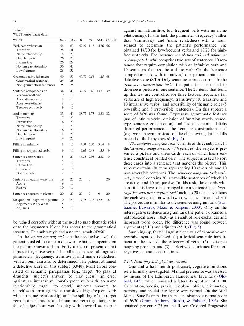
2.1.3.2. Grammatical analysis. Comprehension and produc-tion of verbs and sentences were investigated by means ofthe WEZT (Table 2). This test consists of several taskswhich allow identiWcation and description of grammaticaldeWcits. On the receptive level the test is composed of threesubtests including verb comprehension, sentence compre-hension, and grammaticality judgment; on the expressivelevel the test consists of seven subtests: action naming, sen-tence completion with inWnitives, sentence completion withconjugated verbs, oral sentence construction, sentence ana-grams, and interrogative sentence anagrams.
On the receptive level the ‘verb comprehension task’examines the ability to comprehend single verbs. In thistask the subject is requested to depict one picture from anarray of four that matches the verb orally presented by theexaminer. The three distractors represent: (1) an object thatshares a close semantic relationship with the target verb, (2)an action that is related to the target action and, (3) anobject that is related to the action distractor. The 60 itemsare controlled for frequency, transitivity, and name related-ness with a noun (e.g., ‘to hammer,’ related to ‘hammer’instead of ‘to knock’). All these features are indicated onTable 2. A deWcient result was found for this subtest (54/60). The majority of errors resulted from lexical-semanticproblems. The errors consisted of selection of the semanticdistractor of the target verb (n D 3) (e.g., target: ‘to squeeze,’subject’s answer: ‘to sift’ D a related action distractor with
no name relationship), selection of the distractor noun thatclosely relates to the target verb (n D 2) (e.g., target: ‘tograte,’ subject’s answer: ‘a grater’ D a distractor with namerelationship), and selection of the distractor noun semanti-cally related to the action distractor (n D 1) (e.g., target: ‘toconjure,’ subject’s answer: ‘a magician’ D a distractor withno name relationship to the target word). The ‘sentencecomprehension task’ contains stimulus sentences withmoved constituents as in passives (theme, verb, agent) andsentences in which the order of the thematic roles is canoni-cal as in actives and subject-cleft constructions (agent, verb,theme). These sentence types allow the examiner to investi-gate whether the order of the thematic roles has an impacton sentence comprehension. The procedure used is analo-gous to the ‘verb comprehension task’: four pictures arepresented to the subject while the examiner reads a sentencealoud. The subject is asked to single out the picture thatmatches the sentence. The distractors consist of: a picture inwhich the action and the participants are the same as thetarget picture but in which the thematic roles are reversed, apicture in which a diVerent action is depicted and a picturein which both the thematic roles are reversed and in whichthe action is diVerent. The result on this subtest was alsodeWcient (34/40). Three errors consisted of role exchanges(e.g., target: ‘the grandmother photographes the child,’ sub-ject’s answer: ‘the child photographes his grandmother’)and another three errors due to selection of a semanticrelated action (e.g., target: ‘mother pulls the child,’ subject’sanswer: ‘mother hits the child). The ‘grammaticality judg-ment task’ investigates whether the patient is able to parse asentence. Only irreversible sentences are tested as they can
Fig. 3. Spontaneous speech sample (literal English translation).
74 L. De Witte et al. / Brain and Language 96 (2006) 69–77
be judged correctly without the need to map thematic rolesonto the arguments if one has access to the grammaticalstructure. This subtest yielded a normal result (49/50).
In the ‘action naming task’ on the productive level, thepatient is asked to name in one word what is happening onthe picture shown to him. Forty items are presented thatrepresent agentive verbs. The inXuence of several linguisticparameters (frequency, transitivity, and name relatednesswith a noun) can also be determined. The patient obtaineda defective score on this subtest (33/40). Most errors con-sisted of semantic paraphasias (e.g., target: ‘to play atdraughts,’ subject’s answer: ‘to play chess’ D an erroragainst an intransitive, low-frequent verb with no namerelationship; target: ‘to crawl,’ subject’s answer: ‘tosearch’ D an error against a transitive, high-frequent verbwith no name relationship) and the splitting of the targetverb in a semantic related noun and verb (e.g., target: ‘tofence,’ subject’s answer: ‘to play with a sword’ D an error
Table 2WEZT lesion phase data
WEZT Score Max M SD ASD Cut-oV
Verb comprehension 54 60 59.27 1.13 4.66 56Transitive 28 31Name relationship 18 20High frequent 26 28Intransitive 26 29No name relationship 36 40Low frequent 28 32
Grammaticality judgment 49 50 49.70 0.56 1.25 48Grammatical sentences 24 25Non-grammatical sentences 25 25
Sentence comprehension 34 40 39.77 0.42 13.7 39Verb-agent-theme 9 10Agent-theme-verb 8 10Agent-verb-theme 8 10Theme-agent-verb 9 10
Action naming 33 40 38.77 1.73 3.33 32Transitive 17 20Intransitive 16 20Name relationship 17 20No name relationship 16 20High frequent 18 20Low frequent 14 20
Filling in inWnitive 8 10 9.57 0.50 3.14 9
Filling in conjugated verbs 9 10 9.65 0.48 1.35 9
Sentence construction 8 20 16.35 2.95 2.83 9Transitive 4 10Intransitive 4 10Reversible 2 5Not reversible 2 5
Sentence anagrams ¡ picture 19 20 20 0 20Active 9 10Passive 10 10
Sentence anagrams + picture 20 20 20 0 0 20
wh-question anagrams + picture 10 20 19.75 0.78 12.5 18Arguments Who/What 5 10Adjuncts 5 10
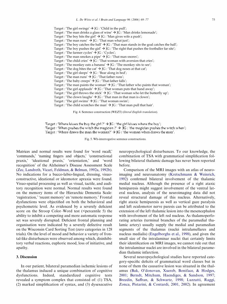
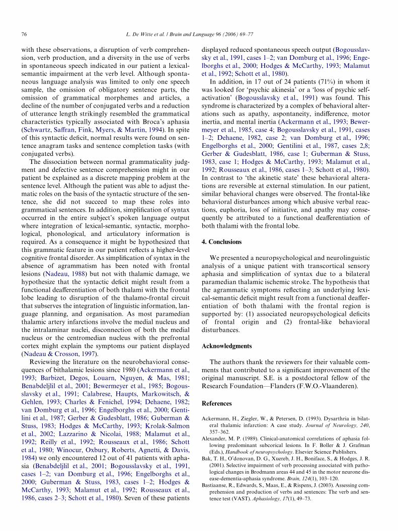
against an intransitive, low-frequent verb with no namerelationship). In this task the parameter ‘frequency’ ratherthan ‘transitivity’ and ‘name relatedness with a noun’seemed to determine the patient’s performance. Sheobtained 14/20 for low-frequent verbs and 18/20 for high-frequent verbs. The ‘sentence completion task with inWnitivesor conjugated verbs’ comprises two sets of sentences: 10 sen-tences that require completion with an inWnitive verb and10 sentences that require a Wnite verb. On the ‘sentencecompletion task with inWnitives,’ our patient obtained adefective score (8/10). Only semantic errors occurred. In the‘sentence construction task,’ the patient is instructed todescribe a picture in one sentence. The 20 items that buildup this test are controlled for three factors: frequency (allverbs are of high frequency), transitivity (10 transitive and10 intransitive verbs), and reversibility of thematic roles (5reversible and 5 irreversible sentences). On this subtest ascore of 8/20 was found. Expressive agrammatic features(use of inWnite verbs, omission of function words, stereo-type sentence construction) and lexical-semantic deWcitsdisrupted performance at the ‘sentence construction task’(e.g., woman swim instead of the child swims, father fallsinstead of the baby crawls) (Fig. 4).
‘The sentence anagram task’ consists of three subparts. Inthe ‘sentence anagram task with pictures’ the subject is pre-sented a picture and three cards, each of which has a sen-tence constituent printed on it. The subject is asked to sortthese cards into a sentence that matches the picture. Thissubtest contains 20 items representing 10 reversible and 10non-reversible sentences. The ‘sentence anagram task with-out pictures’ contains 20 irreversible sentences of which 10are active and 10 are passive. In this task, three cards withconstituents have to be arranged into a sentence. The ‘inter-rogative sentence anagram task’ includes 20 items: Wve itemsfor each wh-question word (who, what, where and when).The procedure is similar to the sentence anagram task (Bas-tiaanse, Edwards, Maas, & Rispens, 2003). Only on theinterrogative sentence anagram task the patient obtained apathological score (10/20) as a result of role exchanges andincorrect word order. No diVerence was found betweenarguments (5/10) and adjuncts (5/10) (Fig. 5).
Summing-up, formal linguistic analysis of expressive andreceptive syntax disclosed: (1) a lexical-semantic impair-ment at the level of the category of verbs, (2) a discretemapping problem, and (3) a selective disturbance for inter-rogative sentence constructions.
2.1.4. Neuropsychological test resultsOne and a half month post-onset, cognitive functions
were formally investigated. Manual preference was assessedby means of the Edinburgh Handedness Inventory (Old-Weld, 1971) which revealed a laterality quotient of +100.Orientation, gnosis, praxis, problem solving, arithmetics,memory, and spatial intelligence were normal. On the MiniMental State Examination the patient obtained a normal scoreof 26/30 (Crum, Anthony, Bassett, & Folstein, 1993). Sheobtained percentile 75 on the Raven Coloured Progressive
L. De Witte et al. / Brain and Language 96 (2006) 69–77 75
Matrices and normal results were found for ‘word recall,’‘commands,’ ‘naming Wngers and objects,’ ‘constructionalpraxis,’ ‘ideational praxis,’ ‘orientation,’ and ‘wordrecognition’ of the Alzheimer’s Disease Assessment Scale(Zec, Landreth, Vicari, Feldman, & Belman, 1992a, 1992b).No indications for a bucco-labio-lingual, dressing, visuo-constructive, ideational or ideomotor apraxia were found.Visuo-spatial processing as well as visual, tactile, and audi-tory recognition were normal. Normal results were foundon the memory items of the Hierarchic Dementia Scale:‘registration,’ ‘recent memory’ en ‘remote memory.’ Frontaldysfunctions were objectiWed on both the behavioral andpsychometric level. As evidenced by a severely deWcientscore on the Stroop Color–Word test (<percentile 5) theability to inhibit a competing and more automatic responseset was severely disrupted. DeWcient frontal planning andorganisation were indicated by a severely defective resulton the Wisconsin Card Sorting Test (zero categories in 128trials). On the level of mood and behavior a variety of fron-tal-like disturbances were observed among which, disinhibi-tory verbal reactions, euphoric mood, loss of initiative, andapathy.
3. Discussion
In our patient, bilateral paramedian ischemic lesions ofthe thalamus induced a unique combination of cognitivedysfunctions. Indeed, standardized cognitive testsrevealed a symptom complex that consisted of: (1) TSA,(2) marked simpliWcation of syntax, and (3) dysexecutive
neuropsychological disturbances. To our knowledge, thecombination of TSA with grammatical simpliWcation fol-lowing bilateral thalamic damage has never been reportedbefore.
Comparison of the MRI images with an atlas of neuro-imaging and neuroanatomy (Kretschmann & Weinrich,1992) conWrmed bilateral involvement of the thalamicmedial nucleus. Although the presence of a right ataxichemiparesis might suggest involvement of the ventral lat-eral nucleus, analysis of the neuroimaging data did notreveal structural damage of this nucleus. Alternatively,right ataxic hemiparesis as well as vertical gaze paralysisand left oculomotor nerve paresis can be attributed to theextension of the left thalamic lesion into the mesencephalonwith involvement of the left red nucleus. As thalamoperfo-rating arteries (terminal branches of the paramedial tha-lamic artery) usually supply the medial and paramediansegments of the thalamus (nuclei intralamellares andnucleus medialis) (Engelborghs et al., 1998), and given thesmall size of the intralaminar nuclei that certainly limitstheir identiWcation on MRI images, we cannot rule out thatthe intralaminar nuclei are involved in the bilateral parame-dian thalamic infarction.
Several neuropsychological studies have reported cate-gory-speciWc deWcits of grammatical word classes but innone of them the causative lesion was situated in the thal-amus (Bak, O’donovan, Xuereb, Boniface, & Hodges,2001; Berndt, Mitchum, Haendiges, & Sandson, 1997;Breedin, SaVran, & Schwartz, 1998; Luzzatti, Raggi,Zonca, Pistarini, & Contardi, 2001, 2002). In agreement
Fig. 4. Sentence construction (WEZT) (literal English translation).
Fig. 5. Wh-interrogative sentence constructions.
76 L. De Witte et al. / Brain and Language 96 (2006) 69–77
with these observations, a disruption of verb comprehen-sion, verb production, and a diversity in the use of verbsin spontaneous speech indicated in our patient a lexical-semantic impairment at the verb level. Although sponta-neous language analysis was limited to only one speechsample, the omission of obligatory sentence parts, theomission of grammatical morphemes and articles, adecline of the number of conjugated verbs and a reductionof utterance length strikingly resembled the grammaticalcharacteristics typically associated with Broca’s aphasia(Schwartz, SaVran, Fink, Myers, & Martin, 1994). In spiteof this syntactic deWcit, normal results were found on sen-tence anagram tasks and sentence completion tasks (withconjugated verbs).
The dissociation between normal grammaticality judg-ment and defective sentence comprehension might in ourpatient be explained as a discrete mapping problem at thesentence level. Although the patient was able to adjust the-matic roles on the basis of the syntactic structure of the sen-tence, she did not succeed to map these roles intogrammatical sentences. In addition, simpliWcation of syntaxoccurred in the entire subject’s spoken language outputwhere integration of lexical-semantic, syntactic, morpho-logical, phonological, and articulatory information isrequired. As a consequence it might be hypothesized thatthis grammatic feature in our patient reXects a higher-levelcognitive frontal disorder. As simpliWcation of syntax in theabsence of agrammatism has been noted with frontallesions (Nadeau, 1988) but not with thalamic damage, wehypothesize that the syntactic deWcit might result from afunctional deaVerentiation of both thalami with the frontallobe leading to disruption of the thalamo-frontal circuitthat subserves the integration of linguistic information, lan-guage planning, and organisation. As most paramedianthalamic artery infarctions involve the medial nucleus andthe intralaminar nuclei, disconnection of both the medialnucleus or the centromedian nucleus with the prefrontalcortex might explain the symptoms our patient displayed(Nadeau & Crosson, 1997).
Reviewing the literature on the neurobehavioral conse-quences of bithalamic lesions since 1980 (Ackermann et al.,1993; Barbizet, Degos, Louarn, Nguyen, & Mas, 1981;Benabdeljlil et al., 2001; Bewermeyer et al., 1985; Bogous-slavsky et al., 1991; Calabrese, Haupts, Markowitsch, &Gehlen, 1993; Charles & Fenichel, 1994; Dehaene, 1982;van Domburg et al., 1996; Engelborghs et al., 2000; Genti-lini et al., 1987; Gerber & Gudesblatt, 1986; Guberman &Stuss, 1983; Hodges & McCarthy, 1993; Krolak-Salmonet al., 2002; Lazzarino & Nicolai, 1988; Malamut et al.,1992; Reilly et al., 1992; Rousseaux et al., 1986; Schottet al., 1980; Winocur, Oxbury, Roberts, Agnetti, & Davis,1984) we only encountered 12 out of 41 patients with apha-sia (Benabdeljlil et al., 2001; Bogousslavsky et al., 1991,cases 1–2; van Domburg et al., 1996; Engelborghs et al.,2000; Guberman & Stuss, 1983, cases 1–2; Hodges &McCarthy, 1993; Malamut et al., 1992; Rousseaux et al.,1986, cases 2–3; Schott et al., 1980). Seven of these patients
displayed reduced spontaneous speech output (Bogousslav-sky et al., 1991, cases 1–2; van Domburg et al., 1996; Enge-lborghs et al., 2000; Hodges & McCarthy, 1993; Malamutet al., 1992; Schott et al., 1980).
In addition, in 17 out of 24 patients (71%) in whom itwas looked for ‘psychic akinesia’ or a ‘loss of psychic self-activation’ (Bogousslavsky et al., 1991) was found. Thissyndrome is characterized by a complex of behavioral alter-ations such as apathy, aspontaneity, indiVerence, motorinertia, and mental inertia (Ackermann et al., 1993; Bewer-meyer et al., 1985, case 4; Bogousslavsky et al., 1991, cases1–2; Dehaene, 1982, case 2; van Domburg et al., 1996;Engelborghs et al., 2000; Gentilini et al., 1987, cases 2,8;Gerber & Gudesblatt, 1986, case 1; Guberman & Stuss,1983, case 1; Hodges & McCarthy, 1993; Malamut et al.,1992; Rousseaux et al., 1986, cases 1–3; Schott et al., 1980).In contrast to ‘the akinetic state’ these behavioral altera-tions are reversible at external stimulation. In our patient,similar behavioral changes were observed. The frontal-likebehavioral disturbances among which abusive verbal reac-tions, euphoria, loss of initiative, and apathy may conse-quently be attributed to a functional deaVerentiation ofboth thalami with the frontal lobe.
4. Conclusions
We presented a neuropsychological and neurolinguisticanalysis of a unique patient with transcortical sensoryaphasia and simpliWcation of syntax due to a bilateralparamedian thalamic ischemic stroke. The hypothesis thatthe agrammatic symptoms reXecting an underlying lexi-cal-semantic deWcit might result from a functional deaVer-entiation of both thalami with the frontal region issupported by: (1) associated neuropsychological deWcitsof frontal origin and (2) frontal-like behavioraldisturbances.
Acknowledgments
The authors thank the reviewers for their valuable com-ments that contributed to a signiWcant improvement of theoriginal manuscript. S.E. is a postdoctoral fellow of theResearch Foundation—Flanders (F.W.O.-Vlaanderen).
References
Ackermann, H., Ziegler, W., & Petersen, D. (1993). Dysarthria in bilat-eral thalamic infarction: A case study. Journal of Neurology, 240,357–362.
Alexander, M. P. (1989). Clinical-anatomical correlations of aphasia fol-lowing predominant subcorical lesions. In F. Boller & J. Grafman(Eds.), Handbook of neuropsychology. Elsevier Science Publishers.
Bak, T. H., O’donovan, D. G., Xuereb, J. H., Boniface, S., & Hodges, J. R.(2001). Selective impairment of verb processing associated with patho-logical changes in Brodmann areas 44 and 45 in the motor neurone dis-ease-dementia-aphasia syndrome. Brain, 124(1), 103–120.
Bastiaanse, R., Edwards, S., Maas, E., & Rispens, J. (2003). Assessing com-prehension and production of verbs and sentences: The verb and sen-tence test (VAST). Aphasiology, 17(1), 49–73.

L. De Witte et al. / Brain and Language 96 (2006) 69–77 77
Bastiaanse, R., Maas, E., & Rispens, J. (2000). Werkwoorden-en zinnentest.Lisse: Swets and Zeitlinger.
Barbizet, J., Degos, J. D., Louarn, F., Nguyen, J. P., & Mas, J. L. (1981).Amnésie par lésion ischémique bi-thalamique. Revista de Neurologia,137, 415–424.
Benabdeljlil, M., El alaoui faris, M., Kissani, N., Aidi, S., Laaouina, Z., etal. (2001). Troubles neuro-psychologiques après infarctus bi-thalami-que par thrombose veineuse profonde. Revue Neurologique, 157,62–67.
Benton, A. L., Hamsher, K. deS., Varney, N. R., & Spreen, O. (1983). Con-tributions to neuropsychological assessment. A clinical manual. NewYork: Oxford University Press.
Berndt, R. S., Mitchum, C. C., Haendiges, A. N., & Sandson, J. (1997). Verbretrieval in aphasia. Characterizing single word impairments. Brain andLanguage, 56, 68–106.
Bewermeyer, H., Dreesbach, H. A., Rackl, A., Neveling, M., & Heiss, W. D.(1985). Presentation of bilateral thalamic infarction on CT, MRI andPET. Neuroradiology, 27, 414–419.
Bogousslavsky, J., Regli, F., Delaloye-bischof, A., Assal, G., & Uske, A.(1991). Loss of psychic self-activation with bithalamic infarction: Neu-robehavioral, CT, MRI and SPECT correlates. Acta Neurologica Scan-dinavica, 83, 309–316.
Breedin, S., SaVran, E., & Schwartz, M. (1998). Semantic factors in verbretrieval: An eVect of complexity. Brain and language, 63, 1–31.
Calabrese, P., Haupts, M., Markowitsch, H. J., & Gehlen, W. (1993). Thecognitive-mnestic performance proWle of a patient with bilateral asym-metrical thalamic infarction. The International Journal of Neuroscience,71, 101–106.
Charles, P. D., & Fenichel, G. M. (1994). Sneddon and antiphospholipidantibody syndromes causing bilateral thalamic infarction. PediatricNeurology, 10, 262–263.
Cole, M. G., & Dastoor, D. (1987). A new hierarchic approach to the mea-surement of dementia. Psychosomatics, 28, 298–305.
Crum, R. M., Anthony, I. C., Bassett, S. S., & Folstein, M. F. (1993). Popu-lation-based norms for the mini-mental state examination by age andeducational level. The Journal of the American Medical Association, 18,2386–2391.
Dehaene, I. (1982). Bilateral thalamo-subthalamic infarction. Acta Neuro-logica Belgica, 82, 253–261.
De Renzi, E., & Vignolo, L. A. (1962). The token test: A sensitive test todetect receptive disturbances in aphasics. Brain, 85, 665–678.
van Domburg, P. H. M. F., ten Donkelaar, H. J., & Notermans, S. L. H.(1996). Akinetic mutism with bithalamic infarction. Neurophysiologi-cal correlates. Journal of Neurological Sciences, 38, 58–65.
Engelborghs, S., Marien, P., Martin, J. J., & De Deyn, P. P. (1998). Func-tional anatomy, vascularisation and pathology of the human thalamus.Acta Neurologica Belgica, 98, 252–265.
Engelborghs, S., Marien, P., Pickut, B. A., Verstraeten, S., & De Deyn, P. P.(2000). Loss of psychic self-activation after paramedian bithalamicinfarction. Stroke, 31, 1762–1766.
Folstein, M. S., Folstein, S. E., & Mc Hugh, P. R. (1975). Mini-mental state:A practical method for grading the cognitive status of patients for theclinician. Journal of Psychiatric Research, 12, 189–198.
Fukatsu, R., Fujii, T., Yamadori, A., Nagasawa, H., & Sakurai, Y. (1997).Persisting childish behavior after bilateral thalamic infarcts. EuropeanNeurology, 37, 230–235.
Gentilini, M., De Renzi, E., & Crisi, G. (1987). Bilateral paramedian tha-lamic artery infarcts: Report of eight cases. Journal of Neurology, Neu-rosurgery, and Psychiatry, 50, 900–909.
Gerber, O., & Gudesblatt, M. (1986). Bilateral paramedian thalamicinfarctions: A CT study. Neuroradiology, 28, 128–131.
Graetz, P., De Bleser, R., & Willmes, K. (1990). Akense Afasietest. Handle-iding. Lisse: Swets & Zeitlinger.
Guberman, A., & Stuss, D. (1983). The syndrome of bilateral paramedianthalamic infarction. Neurology, 33, 540–546.
Hodges, J. R., & McCarthy, R. A. (1993). Autobiographical amnesiaresulting from bilateral paramedian thalamic infarction. Brain, 116,921–940.
Kaplan, E., Goodglass, H., & Weintraub, S. (1983). Boston naming test.USA: Lea & Febiger.
Kretschmann, H.-J., & Weinrich, W. (1992). Cranial neuroimaging and clin-ical neuroanatomy. New York: Thieme Medical Publishers.
Krolak-Salmon, P., Croisile, B., Houzard, C., Setiey, A., Girard-Madoux,P., et al. (2000). Total recovery after bilateral paramedian thalamicinfarct. European Neurology, 44, 216–218.
Krolak-Salmon, P., Montavont, A., Hermier, M., Milliery, M., & Vighetto,A. (2002). Thalamic venous infarction as a cause of subacute dementia.Neurology, 58, 1689–1691.
Lazzarino, L. G., & Nicolai, A. (1988). Aphonia as the only speech distur-bance from bilateral paramedian thalamic infarction. Clinical Neurol-ogy and Neurosurgery, 90, 265–267.
Lhermitte, F. (1984). Language disorders and their relationship to tha-lamic lesions. Advances in Neurology, 42, 99–113.
Luzzatti, C., Raggi, R., Zonca, G., Pistarini, C., Contardi, A., et al. (2001).On the nature of the selective impairment of verb and noun retrieval.Cortex, 37(5), 724–726.
Luzzatti, C., Raggi, R., Zonca, G., Pistarini, C., Contardi, A., et al. (2002).Verb–noun double dissociation in aphasic lexical impairments: The roleof word frequency and imageability. Brain and language, 81, 432–444.
Malamut, B. L., GraV-Radford, N., Chawluk, J., Grossman, R. I., & Gur,R. C. (1992). Memory in a case of bilateral thalamic infarction. Neurol-ogy, 42, 163–169.
Marien, P., Mampaey, E., Vervaet, A., Saerens, J., & De Deyn, P. P. (1998).Normative data for the Boston naming test in native Dutch-speakingBelgian elderly. Brain and Language, 65, 447–467.
Mazzochi, F., & Vignolo, L. A. (1979). Localisation of lesions in aphasia:Clinical CT-scan correlations in stroke patients. Cortex, 15, 627–654.
Nadeau, S. E. (1988). Impaired grammar with normal Xuency and phonol-ogy. Implications for Broca’s aphasia. Brain, 111, 1111–1137.
Nadeau, S. E., & Crosson, B. (1997). Subcortical aphasia. Brain and Lan-guage, 58, 355–402.
OldWeld, R. C. (1971). The assessment and analysis of handedness: TheEdinburgh inventory. Neuropsychologia, 9, 97–113.
Osterrieth, P. A. (1944). Le test de copie d’une Wgure complexe: Contribu-tion à l’étude de l’aperception en de la mémoire. Archives of Neurology,30, 205–353.
Raven, J. C., Court, J. H., & Raven, J., Manual for Raven’s progressivematrices and vocabulary scales. 1983, London.
Reilly, M., Connolly, S., Stack, J., Martin, E. A., & Hutchinson, M. (1992).Bilateral paramedian thalamic infarction: A distinct but poorly recog-nized stroke syndrome. Quarterly Journal of Medicine, 82, 63–70.
Rosen, W. G., Mohs, R. C., & Davis, K. L. (1984). A new rating scale forAlzheimer’s disease. American Journal of Psychiatry, 141, 1356–1364.
Rousseaux, M., Cabaret, M., Lesoin, F., Devos, P., Dubois, F., & Petit, H.(1986). Bilan de l’amnésie des infarctus thalamiques restreints–6 cas.Cortex, 22, 213–228.
Schott, B., Maugiere, F., Laurent, B., Serclerat, O., & Fischer, C. (1980).L’amnésie thalamique. Revue Neurologique, 136, 117–130.
Schwartz, M. F., SaVran, E. M., Fink, R. B., Myers, J. L., & Martin, N.(1994). Mapping therapy: A treatment program for agrammatism.Aphasiology, 8, 19–54.
Vermeulen, J., & Bastiaanse, R., 1984. Stoornissen in de spontane taal bijafasiepatiënten: een factoranalytisch onderzoek. Rapport voor StichtingAfasie Nederland.
Vermeulen, J., Bastiaanse, R., & Van wageningen, B. (1989). Spontaneousspeech in aphasia: A correlational study. Brain and Language, 36, 252–274.
Winocur, G., Oxbury, S., Roberts, R., Agnetti, V., & Davis, C. (1984).Amnesia in a patient with bilateral lesions to the thalamus. Neuro-psychologia, 2, 123–143.
Zec, R., Landreth, E., Vicari, S., Feldman, E., Belman, J., et al. (1992a). Alz-heimer disease assessment scale: Useful for both early detection andstaging of dementia of the Alzheimer type. Alzheimer Disease andRelated Disorders an International Journal, 6(2), 89–102.
Zec, R., Landreth, E., Vicari, S., Feldman, E., Belman, J., et al. (1992b). Alz-heimer disease assessment scale: A subtest analysis. Alzheimer Diseaseand Related Disorders, an International Journal, 6(3), 164–181.