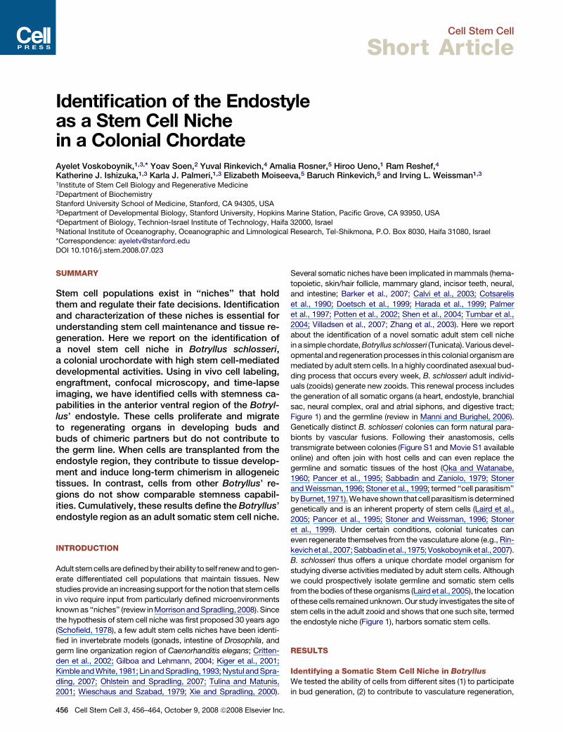
Identification of the Endostyle as a Stem Cell Niche in a Colonial Chordate
9
Cell Stem Cell Short Article Identification of the Endostyle as a Stem Cell Niche in a Colonial Chordate Ayelet Voskoboynik, 1,3, * Yoav Soen, 2 Yuval Rinkevich, 4 Amalia Rosner, 5 Hiroo Ueno, 1 Ram Reshef, 4 Katherine J. Ishizuka, 1,3 Karla J. Palmeri, 1,3 Elizabeth Moiseeva, 5 Baruch Rinkevich, 5 and Irving L. Weissman 1,3 1 Institute of Stem Cell Biology and Regenerative Medicine 2 Department of Biochemistry Stanford University School of Medicine, Stanford, CA 94305, USA 3 Department of Developmental Biology, Stanford University, Hopkins Marine Station, Pacific Grove, CA 93950, USA 4 Department of Biology, Technion-Israel Institute of Technology, Haifa 32000, Israel 5 National Institute of Oceanography, Oceanographic and Limnological Research, Tel-Shikmona, P.O. Box 8030, Haifa 31080, Israel *Correspondence: [email protected] DOI 10.1016/j.stem.2008.07.023 SUMMARY Stem cell populations exist in ‘‘niches’’ that hold them and regulate their fate decisions. Identification and characterization of these niches is essential for understanding stem cell maintenance and tissue re- generation. Here we report on the identification of a novel stem cell niche in Botryllus schlosseri, a colonial urochordate with high stem cell-mediated developmental activities. Using in vivo cell labeling, engraftment, confocal microscopy, and time-lapse imaging, we have identified cells with stemness ca- pabilities in the anterior ventral region of the Botryl- lus’ endostyle. These cells proliferate and migrate to regenerating organs in developing buds and buds of chimeric partners but do not contribute to the germ line. When cells are transplanted from the endostyle region, they contribute to tissue develop- ment and induce long-term chimerism in allogeneic tissues. In contrast, cells from other Botryllus’ re- gions do not show comparable stemness capabil- ities. Cumulatively, these results define the Botryllus’ endostyle region as an adult somatic stem cell niche. INTRODUCTION Adult stem cells are defined by their ability to self renew and to gen- erate differentiated cell populations that maintain tissues. New studies provide an increasing support for the notion that stem cells in vivo require input from particularly defined microenvironments known as ‘‘niches’’ (review in Morrison and Spradling, 2008). Since the hypothesis of stem cell niche was first proposed 30 years ago (Schofield, 1978), a few adult stem cells niches have been identi- fied in invertebrate models (gonads, intestine of Drosophila, and germ line organization region of Caenorhanditis elegans; Critten- den et al., 2002; Gilboa and Lehmann, 2004; Kiger et al., 2001; Kimble and White, 1981; Lin and Spradling, 1993; Nystul and Spra- dling, 2007; Ohlstein and Spradling, 2007; Tulina and Matunis, 2001; Wieschaus and Szabad, 1979; Xie and Spradling, 2000). Several somatic niches have been implicated in mammals (hema- topoietic, skin/hair follicle, mammary gland, incisor teeth, neural, and intestine; Barker et al., 2007; Calvi et al., 2003; Cotsarelis et al., 1990; Doetsch et al., 1999; Harada et al., 1999; Palmer et al., 1997; Potten et al., 2002; Shen et al., 2004; Tumbar et al., 2004; Villadsen et al., 2007; Zhang et al., 2003). Here we report about the identification of a novel somatic adult stem cell niche in a simple chordate, Botryllus schlosseri (Tunicata). Various devel- opmental and regeneration processes in this colonial organism are mediated by adult stem cells. In a highly coordinated asexual bud- ding process that occurs every week, B. schlosseri adult individ- uals (zooids) generate new zooids. This renewal process includes the generation of all somatic organs (a heart, endostyle, branchial sac, neural complex, oral and atrial siphons, and digestive tract; Figure 1) and the germline (review in Manni and Burighel, 2006). Genetically distinct B. schlosseri colonies can form natural para- bionts by vascular fusions. Following their anastomosis, cells transmigrate between colonies (Figure S1 and Movie S1 available online) and often join with host cells and can even replace the germline and somatic tissues of the host (Oka and Watanabe, 1960; Pancer et al., 1995; Sabbadin and Zaniolo, 1979; Stoner and Weissman, 1996; Stoner et al., 1999; termed ‘‘cell parasitism’’ by Burnet, 1971). We have shown that cell parasitism is determined genetically and is an inherent property of stem cells (Laird et al., 2005; Pancer et al., 1995; Stoner and Weissman, 1996; Stoner et al., 1999). Under certain conditions, colonial tunicates can even regenerate themselves from the vasculature alone (e.g., Rin- kevich et al., 2007; Sabbadin et al., 1975; Voskoboynik et al., 2007). B. schlosseri thus offers a unique chordate model organism for studying diverse activities mediated by adult stem cells. Although we could prospectively isolate germline and somatic stem cells from the bodies of these organisms (Laird et al., 2005), the location of these cells remained unknown. Our study investigates the site of stem cells in the adult zooid and shows that one such site, termed the endostyle niche (Figure 1), harbors somatic stem cells. RESULTS Identifying a Somatic Stem Cell Niche in Botryllus We tested the ability of cells from different sites (1) to participate in bud generation, (2) to contribute to vasculature regeneration, 456 Cell Stem Cell 3, 456–464, October 9, 2008 ª2008 Elsevier Inc.
Transcript of Identification of the Endostyle as a Stem Cell Niche in a Colonial Chordate
Cell Stem Cell
Short Article
Identification of the Endostyleas a Stem Cell Nichein a Colonial ChordateAyelet Voskoboynik,1,3,* Yoav Soen,2 Yuval Rinkevich,4 Amalia Rosner,5 Hiroo Ueno,1 Ram Reshef,4
Katherine J. Ishizuka,1,3 Karla J. Palmeri,1,3 Elizabeth Moiseeva,5 Baruch Rinkevich,5 and Irving L. Weissman1,3
1Institute of Stem Cell Biology and Regenerative Medicine2Department of BiochemistryStanford University School of Medicine, Stanford, CA 94305, USA3Department of Developmental Biology, Stanford University, Hopkins Marine Station, Pacific Grove, CA 93950, USA4Department of Biology, Technion-Israel Institute of Technology, Haifa 32000, Israel5National Institute of Oceanography, Oceanographic and Limnological Research, Tel-Shikmona, P.O. Box 8030, Haifa 31080, Israel*Correspondence: [email protected]
DOI 10.1016/j.stem.2008.07.023
SUMMARY
Stem cell populations exist in ‘‘niches’’ that holdthem and regulate their fate decisions. Identificationand characterization of these niches is essential forunderstanding stem cell maintenance and tissue re-generation. Here we report on the identification ofa novel stem cell niche in Botryllus schlosseri,a colonial urochordate with high stem cell-mediateddevelopmental activities. Using in vivo cell labeling,engraftment, confocal microscopy, and time-lapseimaging, we have identified cells with stemness ca-pabilities in the anterior ventral region of the Botryl-lus’ endostyle. These cells proliferate and migrateto regenerating organs in developing buds andbuds of chimeric partners but do not contribute tothe germ line. When cells are transplanted from theendostyle region, they contribute to tissue develop-ment and induce long-term chimerism in allogeneictissues. In contrast, cells from other Botryllus’ re-gions do not show comparable stemness capabil-ities. Cumulatively, these results define the Botryllus’endostyle region as an adult somatic stem cell niche.
INTRODUCTION
Adult stem cells are defined by their ability toself renew and togen-
erate differentiated cell populations that maintain tissues. New
studies provide an increasing support for the notion that stem cells
in vivo require input from particularly defined microenvironments
known as ‘‘niches’’ (review in Morrison and Spradling, 2008). Since
the hypothesis of stem cell niche was first proposed 30 years ago
(Schofield, 1978), a few adult stem cells niches have been identi-
fied in invertebrate models (gonads, intestine of Drosophila, and
germ line organization region of Caenorhanditis elegans; Critten-
den et al., 2002; Gilboa and Lehmann, 2004; Kiger et al., 2001;
Kimble and White, 1981; Lin and Spradling, 1993; Nystul and Spra-
dling, 2007; Ohlstein and Spradling, 2007; Tulina and Matunis,
2001; Wieschaus and Szabad, 1979; Xie and Spradling, 2000).
456 Cell Stem Cell 3, 456–464, October 9, 2008 ª2008 Elsevier Inc.
Several somatic niches have been implicated in mammals (hema-
topoietic, skin/hair follicle, mammary gland, incisor teeth, neural,
and intestine; Barker et al., 2007; Calvi et al., 2003; Cotsarelis
et al., 1990; Doetsch et al., 1999; Harada et al., 1999; Palmer
et al., 1997; Potten et al., 2002; Shen et al., 2004; Tumbar et al.,
2004; Villadsen et al., 2007; Zhang et al., 2003). Here we report
about the identification of a novel somatic adult stem cell niche
in a simple chordate, Botryllus schlosseri (Tunicata). Various devel-
opmental and regeneration processes in this colonial organism are
mediated by adult stem cells. In a highly coordinated asexual bud-
ding process that occurs every week, B. schlosseri adult individ-
uals (zooids) generate new zooids. This renewal process includes
the generation of all somatic organs (a heart, endostyle, branchial
sac, neural complex, oral and atrial siphons, and digestive tract;
Figure 1) and the germline (review in Manni and Burighel, 2006).
Genetically distinct B. schlosseri colonies can form natural para-
bionts by vascular fusions. Following their anastomosis, cells
transmigrate between colonies (Figure S1 and Movie S1 available
online) and often join with host cells and can even replace the
germline and somatic tissues of the host (Oka and Watanabe,
1960; Pancer et al., 1995; Sabbadin and Zaniolo, 1979; Stoner
and Weissman, 1996; Stoner et al., 1999; termed ‘‘cell parasitism’’
byBurnet, 1971).Wehaveshown thatcell parasitism isdetermined
genetically and is an inherent property of stem cells (Laird et al.,
2005; Pancer et al., 1995; Stoner and Weissman, 1996; Stoner
et al., 1999). Under certain conditions, colonial tunicates can
even regenerate themselves from the vasculature alone (e.g., Rin-
kevichetal., 2007;Sabbadin etal., 1975; Voskoboynik etal., 2007).
B. schlosseri thus offers a unique chordate model organism for
studying diverse activities mediated by adult stem cells. Although
we could prospectively isolate germline and somatic stem cells
from the bodies of these organisms (Laird et al., 2005), the location
of these cells remained unknown. Our study investigates the site of
stem cells in the adult zooid and shows that one such site, termed
the endostyle niche (Figure 1), harbors somatic stem cells.
RESULTS
Identifying a Somatic Stem Cell Niche in Botryllus
We tested the ability of cells from different sites (1) to participate
in bud generation, (2) to contribute to vasculature regeneration,
Cell Stem Cell
A Novel Stem Cell Niche in a Urochordate
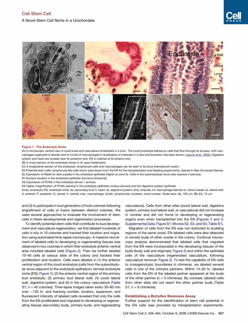
Figure 1. The Endostyle Niche
(A) A microscopic ventral view of zooid buds and vasculature embedded in a tunic. The zooid endostyle bathes by cells that flow through its sinuses, with mac-
rophages organized in islands next to it (role of macrophages in reutilization of materials in a new bud formation has been shown; Lauzon et al., 2002). Digestive
system and heart are located near its posterior end. EN is outlined at its anterior end.
(B) A cross-section of the endostyle zones (1–8; azan heidenhain).
(C) A longitudinal section of the endostyle, lymphocyte cells and macrophages can be seen in its sinus (hematoxylin eosin).
(D) Potential stem cells, lymphocyte like cells which were drawn from the EN for the transplantation and labeling experiments, stained in May Grunwald Giemsa.
(E) Expression of Raldh (in dark purple) in the endostyle epithelial (higher at zone 6). Cells in the subendostylar sinus also express it (arrows).
(F) Nucleus location in the endostyle epithelial and sinus (Hoechst).
(G) Expression of PCNA in the endostyle (brown / arrows).
(H) Higher magnification of PCNA staining in the endostyle epithelial nucleus (arrows) and the digestive system epithelial.
Endo, endostyle; EN, endostyle niche; sb, secondary bud; h, heart; ds, digestive system; amp, ampulla; mi, macrophage islands; bv, blood vessel; lw, lateral wall;
A, anterior; P, posterior; D, dorsal; V, ventral; mac, macrophage; lymph, lymphocyte; numbers, zone number. Scale bars: (A), 100 mm; (B)–(H), 10 mm.
and (3) to participate in bud generation of host colonies following
engraftment of cells or fusion between distinct colonies. We
used several approaches to evaluate the involvement of stem
cells in these developmental and regeneration processes.
To identify potential stem cells that contribute to bud develop-
ment and vasculature regeneration, we first labeled hundreds of
cells in situ in 10 colonies and tracked their location and migra-
tion using automated time-lapse microscopy. A massive recruit-
ment of labeled cells to developing or regenerating tissues was
observed in two colonies in which their endostyle anterior ventral
area included labeled cells (Figure 2). Then we further labeled
10–40 cells at various sites of the colony and tracked their
proliferation and location. Cells were labeled in (1) the anterior
ventral region of the zooid endostyle (cells from the subendosty-
lar sinus adjacent to the endostyle epithelium; termed endostyle
niche [EN]; Figure 1); (2) the anterior ventral region of the primary
bud endostyle; (3) primary bud lateral wall; (4) zooid lateral
wall, digestive system; and (5) in the colony vasculature (Table
S1; n = 40 colonies). Time-lapse images taken every 30–60 min
over �120 hr, and tracking number, location, expansion, and
fluorescent intensity of labeled cells revealed that only the cells
from the EN proliferated and migrated to developing or regener-
ating tissues (secondary buds, primary buds, and regenerating
vasculature). Cells from other sites (zooid lateral wall, digestive
system, primary bud lateral wall, or vasculature) did not increase
in number and did not home to developing or regenerating
organs even when transplanted into the EN (Figures 2 and 3;
Supplemental Data; Figure S1; Movies S2, S3, and S4; Table S1).
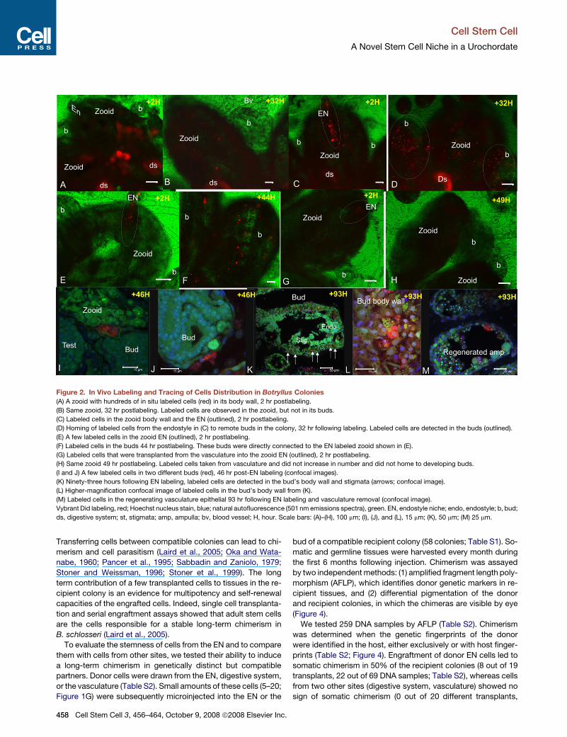
Migration of cells from the EN was not restricted to budding
regions of the same zooid. EN-labeled cells were also detected
in remote buds of other zooids in the colony. Confocal micros-
copy analysis demonstrated that labeled cells that migrated
from the EN were incorporated in the developing tissues of the
buds (body wall and stigmata; Figure 2) and within the epithelial
cells of the vasculature (regenerated vasculature, following
vasculature removal; Figure 2). To test the capability of EN cells
to crossgenotypic boundaries in chimeras, we labeled several
cells in one of the chimera partners. Within 14–20 hr, labeled
cells from the EN of the labeled partner appeared at the buds
of the other partner (n = 3 chimeras). By contrast, labeled cells
from other sites did not reach the other partner buds (Table
S1; n = 8 chimeras).
Establishing a Botryllus Stemness AssayFurther support for the identification of stem cell potential in
the EN cells was provided by transplantation experiments.
Cell Stem Cell 3, 456–464, October 9, 2008 ª2008 Elsevier Inc. 457
Cell Stem Cell
A Novel Stem Cell Niche in a Urochordate
Figure 2. In Vivo Labeling and Tracing of Cells Distribution in Botryllus Colonies
(A) A zooid with hundreds of in situ labeled cells (red) in its body wall, 2 hr postlabeling.
(B) Same zooid, 32 hr postlabeling. Labeled cells are observed in the zooid, but not in its buds.
(C) Labeled cells in the zooid body wall and the EN (outlined), 2 hr postlabeling.
(D) Homing of labeled cells from the endostyle in (C) to remote buds in the colony, 32 hr following labeling. Labeled cells are detected in the buds (outlined).
(E) A few labeled cells in the zooid EN (outlined), 2 hr postlabeling.
(F) Labeled cells in the buds 44 hr postlabeling. These buds were directly connected to the EN labeled zooid shown in (E).
(G) Labeled cells that were transplanted from the vasculature into the zooid EN (outlined), 2 hr postlabeling.
(H) Same zooid 49 hr postlabeling. Labeled cells taken from vasculature and did not increase in number and did not home to developing buds.
(I and J) A few labeled cells in two different buds (red), 46 hr post-EN labeling (confocal images).
(K) Ninety-three hours following EN labeling, labeled cells are detected in the bud’s body wall and stigmata (arrows; confocal image).
(L) Higher-magnification confocal image of labeled cells in the bud’s body wall from (K).
(M) Labeled cells in the regenerating vasculature epithelial 93 hr following EN labeling and vasculature removal (confocal image).
Vybrant Did labeling, red; Hoechst nucleus stain, blue; natural autofluorescence (501 nm emissions spectra), green. EN, endostyle niche; endo, endostyle; b, bud;
ds, digestive system; st, stigmata; amp, ampulla; bv, blood vessel; H, hour. Scale bars: (A)–(H), 100 mm; (I), (J), and (L), 15 mm; (K), 50 mm; (M) 25 mm.
Transferring cells between compatible colonies can lead to chi-
merism and cell parasitism (Laird et al., 2005; Oka and Wata-
nabe, 1960; Pancer et al., 1995; Sabbadin and Zaniolo, 1979;
Stoner and Weissman, 1996; Stoner et al., 1999). The long
term contribution of a few transplanted cells to tissues in the re-
cipient colony is an evidence for multipotency and self-renewal
capacities of the engrafted cells. Indeed, single cell transplanta-
tion and serial engraftment assays showed that adult stem cells
are the cells responsible for a stable long-term chimerism in
B. schlosseri (Laird et al., 2005).
To evaluate the stemness of cells from the EN and to compare
them with cells from other sites, we tested their ability to induce
a long-term chimerism in genetically distinct but compatible
partners. Donor cells were drawn from the EN, digestive system,
or the vasculature (Table S2). Small amounts of these cells (5–20;
Figure 1G) were subsequently microinjected into the EN or the
458 Cell Stem Cell 3, 456–464, October 9, 2008 ª2008 Elsevier Inc.
bud of a compatible recipient colony (58 colonies; Table S1). So-
matic and germline tissues were harvested every month during
the first 6 months following injection. Chimerism was assayed
by two independent methods: (1) amplified fragment length poly-
morphism (AFLP), which identifies donor genetic markers in re-
cipient tissues, and (2) differential pigmentation of the donor
and recipient colonies, in which the chimeras are visible by eye
(Figure 4).
We tested 259 DNA samples by AFLP (Table S2). Chimerism
was determined when the genetic fingerprints of the donor
were identified in the host, either exclusively or with host finger-
prints (Table S2; Figure 4). Engraftment of donor EN cells led to
somatic chimerism in 50% of the recipient colonies (8 out of 19
transplants, 22 out of 69 DNA samples; Table S2), whereas cells
from two other sites (digestive system, vasculature) showed no
sign of somatic chimerism (0 out of 20 different transplants,

Cell Stem Cell
A Novel Stem Cell Niche in a Urochordate
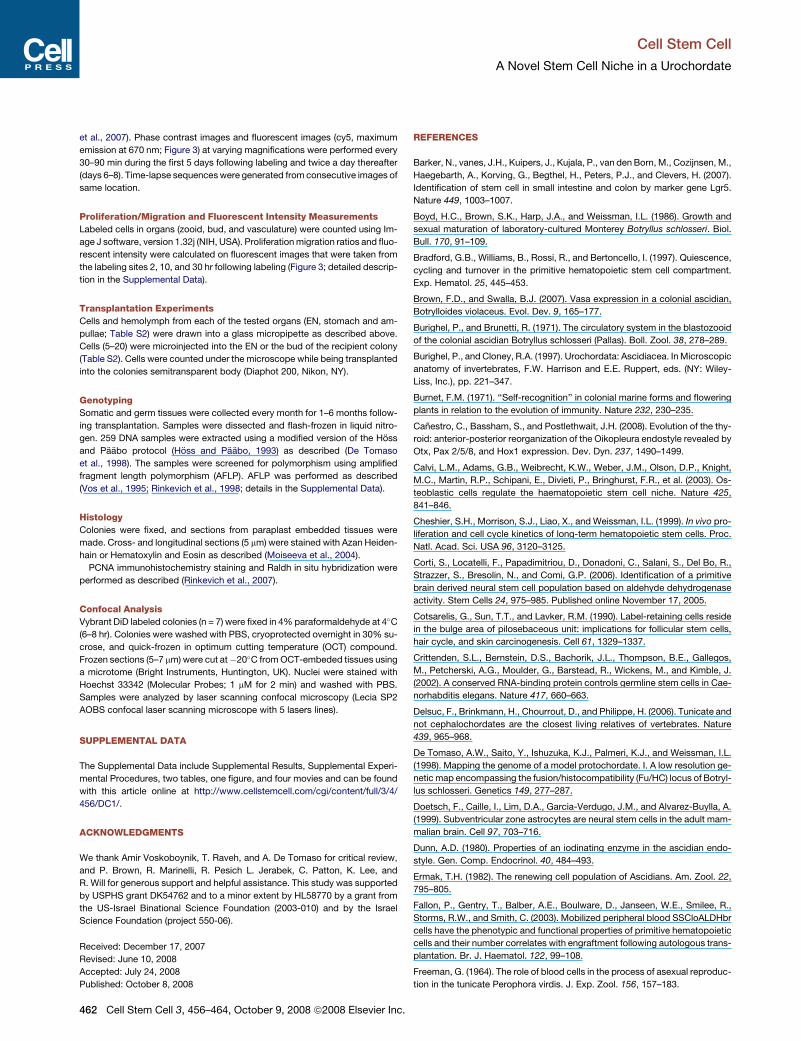
Figure 3. Fluorescent Intensity of Labeled Cells
(A) EN (outlined) in a zooid (phase contrast image) and �14 labeled cells within ([B], fluorescent image), 2 hr postlabeling. (C) Same EN 10 hr postlabeling. Fluo-
rescent intensity of the cells from the EN decreased and expansion of labeled cells in the zooid (�20 labeled cells) is observed. (D) Thirty hours postlabeling,�14
labeled cells detected in the bud that was directly connected to the EN of the labeled zooid (A). At that time, �40 labeled cells were tracked in other buds of the
colony. (E) Injection of labeled cells into an ampulla by a glass needle. (F) Two hours postlabeling,�50 labeled cells detected in ampullae near the injected site. (G)
Ten hours postlabeling, �17 labeled cells remained at this site, and their fluorescence intensity did not decrease. (H) Thirty hours following labeling, �12 cells
were detected in the labeling site. Their fluorescence intensity remained unchanged. (I) The average fluorescence intensity in labeled cells from ampullae versus
cells from the EN as a function of time.
EN, endostyle niche; H, hour; amp, ampulla; bv, blood vessel. Scale bar, 50 mm.
75 somatic DNA samples; Table S2). Germline chimerism was
not observed, irrespective of donor cell origin (0 out of 17 trans-
plants, 31 DNA samples; Table S2).
Cells from the donor EN survived, proliferated, migrated, and
integrated in the somatic organs of recipient colonies. Chime-
rism was detected 1 to 2 months following transplantation and
lasted up to 6 months after transplantation (last sampling date;
up to �25 generations of new buds; Table S2). The capacity of
cells from the EN to differentiate into multiple lineages in the
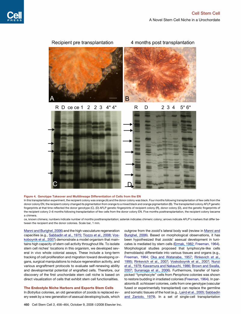
recipient colony was confirmed by a pigmentation-based assay,
where 5–20 cells from the EN of a black-pigmented donor colony
were injected into a compatible, orange-pigmented recipient
colony. Within 4 months from injection, recipient colonies
changed pigmentation from orange to a black and orange mix.
This shift was also reflected by the AFLP markers (Table S2,
n = 4; Figure 4). Pigmentation chimeras were not observed
when the donor cells were drawn from ampullae or the stomach
(Table S2, n = 4).
DISCUSSION
Stem cells are rare cells, uniquely capable of both reproducing
themselves (self-renewing) and differentiating into a diverse
range of specialized cell types. The rarity of stem cells and the
lack of specific markers to definitive identification of stem cells
in vivo make it difficult to confidently isolate stem cell niches
(Morrison and Spradling, 2008). In Botryllus colonies, the unique
weekly budding process of somatic self-renewal (review in
Cell Stem Cell 3, 456–464, October 9, 2008 ª2008 Elsevier Inc. 459
Cell Stem Cell
A Novel Stem Cell Niche in a Urochordate
Figure 4. Genotype Takeover and Multilineage Differentiation of Cells from the EN
In this transplantation experiment, the recipient colony was orange (A) and the donor colony was black. Four months following transplantation of few cells from the
donor colony EN, the recipient colony changed its pigmentation from orange to a mixed black and orange pigmentation (B). The transplanted colony AFLP genetic
fingerprints at that time reflected the donor genotype (C). (D) AFLP genetic fingerprints of recipient colony (R), donor colony (D), and the genetic fingerprints of
the recipient colony 2–6 months following transplantation of few cells from the donor colony EN. Five months posttransplantation, the recipient colony became
a chimera.
ce, known chimera; numbers indicate number of months posttransplantation; asterisk indicates chimeric colony; arrows indicate AFLP’s markers that differ be-
tween the recipient and the donor colonies. Scale bar, 1 mm.
Manni and Burighel, 2006) and the high vasculature regeneration
capacities (e.g., Sabbadin et al., 1975; Tiozzo et al., 2008; Vos-
koboynik et al., 2007) demonstrate a model organism that main-
tains high capacity of stem cell activity throughout life. To isolate
stem cell niches’ locations in this organism, we developed sev-
eral in vivo whole colonial assays. These include a long-term
tracking of cell proliferation and migration toward developing or-
gans, surgical manipulations to induce regeneration activity, and
various engraftment protocols to evaluate self-renewing ability
and developmental potential of engrafted cells. Therefore, our
discovery of the first urochordate stem cell niche is based on
direct visualization of cells that exhibit stem cell functionalities.
The Endostyle Niche Harbors and Exports Stem CellsIn Botryllus colonies, an old generation of zooids is replaced ev-
ery week by a new generation of asexual developing buds, which
460 Cell Stem Cell 3, 456–464, October 9, 2008 ª2008 Elsevier Inc.
outgrow from the zooid’s lateral body wall (review in Manni and
Burighel, 2006). Based on morphological observations, it has
been hypothesized that zooids’ asexual development in tuni-
cates is mediated by stem cells (Ermak, 1982; Freeman, 1964).
Morphological studies proposed that lymphocyte-like cells
(hemoblasts) differentiate into various tissues and organs (e.g.,
Freeman, 1964; Oka and Watanabe, 1957; Rinkevich et al.,
1995; Rinkevich et al., 2007; Voskoboynik et al., 2007; Nunzi
et al., 1979; Kawamura and Nakauchi, 1986; Brown and Swalla,
2007; Sunanaga et al., 2006). Furthermore, transfer of hand-
picked ‘‘lymphocyte’’ cells from Perophora colonies was shown
to restore budding in irradiated colonies (Freeman, 1964). In par-
abionts B. schlosseri colonies, cells from one genotype (vascular
fused or experimentally transplanted) can replace the germline
and somatic tissues of the host (e.g., Laird et al., 2005; Sabbadin
and Zaniolo, 1979). In a set of single-cell transplantation

Cell Stem Cell
A Novel Stem Cell Niche in a Urochordate
experiments and serial engraftments, Laird et al. (2005) have fur-
ther demonstrated precursor-progeny relationships and proved
that the cells responsible for stable long-term chimerism or par-
asitism in B. schlosseri are indeed adult stem cells, which as
a population include both the somatic and germline lineages.
The location of these cells, however, remained unknown. Here,
three different experimental approaches independently revealed
that cells with the properties of somatic stem cells reside in the
anterior ventral region of the endostyle (EN). As few as 5– 20
engrafted cells drawn from a donor EN sufficed to generate a
somatic chimerism and contributed to asexual development of
zooids for �25 asexual generations, demonstrating long-term
self-renewal potential. However, these cells do not contribute
to the germline.
The Endostyle: A Niche that Governs High-DemandStem Cell ActivityUpon demand, adult stem cells are divided throughout life to pro-
duce new progeny that undergo programs of differentiation and
maturation required to replace old or damaged tissue cells
(Weissman, 2000). In solitary organisms like humans and mice,
the demand usually occurs at low levels and occasionally at
high levels; tissue stem cells (for example, blood-forming) are
mainly quiescent in cell cycle G0 stage and enter the cell cycle
when tissue turnover demands new input (Bradford et al.,
1997; Cheshier et al., 1999). In contrast, Botryllus does not main-
tain its genome in long-lived individuals but in colonies where in-
dividuals have a lifespan of �21 days (14 days as developing
buds and 7 days as zooids), while the entire colony has a long life-
span. To support a complete turnover of the individuals in the
colony every week, the Botryllus stem cell niche must maintain
high levels of stem cell proliferation and outward migration. In-
deed, calculation of proliferation ratios and decline of fluorescent
intensity in labeled cells with time revealed higher levels of prolif-
eration in cells from the EN (versus other sites). Staining for pro-
liferating cell nuclear antigen (PCNA) revealed that the endostyle
is a highly proliferating organ and that many of its epithelial cells
are constantly proliferating throughout the colony life cycle as
well as circulating cells in its subendostylar sinus (Figure 1). Like-
wise, the intense migration of cells out of the EN (compared to
other sites) may reflect the export of precursors to developing tis-
sues. BrdU (5-bromo-20-deoxyuridine) labeling experiments in
Botryllus primigenus also revealed high proliferation activity in
the zooids endostyle and sinuses (Kawamura et al., 2008).
A stem cell niche is an interactive structural unit organized to
facilitate cell-fate decisions in a proper spatiotemporal manner
(Morrison and Spradling, 2008; Scadden, 2006). Indeed, the en-
dostyle of tunicates exhibits a special anatomic structure; it is
a long glandular groove extending medially at the ventral face
of the zooid branchial sac along its anterior posterior axis (Buri-
ghel and Cloney, 1997). It consists of eight distinct anatomical
zones and is immersed by blood flow through the large suben-
dostylar sinus and other sinuses (Burighel and Brunetti, 1971).
The endostyle is considered as the invertebrate chordate homo-
log of the vertebrate thyroid gland. It exhibits unique spatial and
temporal features with site-specific factors that were previously
implicated in developmental regulation and stem cell mainte-
nance in other systems. These include thyroid hormones, sero-
tonin, Otx, Hox1, Pax 2/5/8, PL10, and Cadherin (Canestro
et al., 2008; Dunn, 1980; Hiruta et al., 2005; Nilsson et al.,
1988; Pennati et al., 2001; Rosner et al., 2006; Rosner et al.,
2007). The canonical Wnt pathway was shown to be involved
in stem cell regulation and proliferation in various stem cells
niches (reviewed in Morrison and Spradling, 2008; Scadden,
2006). Two Wnt genes are expressed in the Amphioxus (Cepha-
lochordata) endostyle during embryogenesis and in larvae
(Schubert et al., 2000). Previous enrichment of stem cells in
B. schlosseri was based on the high enzymatic activity of alde-
hyde dehydrogenase (Laird et al., 2005) that delineates stem
and progenitor cell in multiple lineages (Corti et al., 2006; Fallon
et al., 2003; Storms et al., 1999). As might be expected from
Botryllus stem cells, we found that the cells of the EN (epithelial
and blood cells) also exhibit high levels of aldehyde dehydroge-
nase activity as indicated by the high expression of retinalde-
hyde dehydrogenase (Raldh).
Thus, the architecture and molecular features of the endostyle
are also consistent with its role as a stem cell niche, but a direct
role for these factors has not yet been shown in the generation
and maintenance of the closely associated somatic stem cells.
Our study establishes the anterior ventral region of the endo-
style, which includes adhering blood cells from the subendosty-
lar sinus, as a special stem cell niche. The EN-harbored stem
cells participate in organogenesis by migrating out of the niche
and homing to developing and regenerating sites. Botryllus
belong to a taxonomic group that is considered the closest living
invertebrate relative of the vertebrates (Delsuc et al., 2006).
Throughout life, this model organism exhibits a natural high de-
mand and high activity of adult stem cells, activity that can be vi-
sualized, measured, and manipulated in vivo. Characterization of
the various endostyle subsets and the types and stages of stem
cells with which they interact will contribute to our understanding
of how stem cell niches support extensive demand and utiliza-
tion of stem cells.
EXPERIMENTAL PROCEDURES
Animals
Colonies of Botryllus schlosseri were maintained as described (Boyd et al.,
1986). Colonies were chosen for the experiments as described in the Supple-
mental Data.
In Vivo Fluorescent Cell Labeling Assay
Cell drawing, labeling, and transplanting were performed under a microscope
(Diaphot 200, Nikon, NY). We used an air compressed microinjector (PLI-188,
Nikon, NY) and a glass needle micropipette (50–60 mm diameter sharp tip) to
draw cells from one of the tested sites. Cells were drawn into a glass micropi-
pette that contained 1 ml Vybrant DiD dye solution (emission 665 nm; Molecular
Probes, Eugene, OR) diluted in a tunicate saline buffer (TS; Negm et al., 1991;
details in the Supplemental Data). The cells in the micropipette were
counted under a microscope, incubated for 5 min (room temperature), and
then 10–40 cells (�0.5 ml), were injected back into the relevant tested sites
(Table S1). As a control, 0.5 ml of the dilution solution (TS) was injected. In ad-
dition, we performed heterotypic transplantation experiments where�40 cells
taken from the vasculature or zooid lateral wall were labeled and injected into
the EN (Table S1). In several colonies, including the control groups, the vascu-
lature (marginal vessel and ampullae) was dissected away from the colonies
following cell labeling (n = 13, Table S1).
Imaging
Time-lapse imaging was performed by an automated microscopy (ImageX-
press, Molecular Devices Corp., Palo Alto, CA) as described (Voskoboynik
Cell Stem Cell 3, 456–464, October 9, 2008 ª2008 Elsevier Inc. 461
Cell Stem Cell
A Novel Stem Cell Niche in a Urochordate
et al., 2007). Phase contrast images and fluorescent images (cy5, maximum
emission at 670 nm; Figure 3) at varying magnifications were performed every
30–90 min during the first 5 days following labeling and twice a day thereafter
(days 6–8). Time-lapse sequences were generated from consecutive images of
same location.
Proliferation/Migration and Fluorescent Intensity Measurements
Labeled cells in organs (zooid, bud, and vasculature) were counted using Im-
age J software, version 1.32j (NIH, USA). Proliferation migration ratios and fluo-
rescent intensity were calculated on fluorescent images that were taken from
the labeling sites 2, 10, and 30 hr following labeling (Figure 3; detailed descrip-
tion in the Supplemental Data).
Transplantation Experiments
Cells and hemolymph from each of the tested organs (EN, stomach and am-
pullae; Table S2) were drawn into a glass micropipette as described above.
Cells (5–20) were microinjected into the EN or the bud of the recipient colony
(Table S2). Cells were counted under the microscope while being transplanted
into the colonies semitransparent body (Diaphot 200, Nikon, NY).
Genotyping
Somatic and germ tissues were collected every month for 1–6 months follow-
ing transplantation. Samples were dissected and flash-frozen in liquid nitro-
gen. 259 DNA samples were extracted using a modified version of the Hoss
and Paabo protocol (Hoss and Paabo, 1993) as described (De Tomaso
et al., 1998). The samples were screened for polymorphism using amplified
fragment length polymorphism (AFLP). AFLP was performed as described
(Vos et al., 1995; Rinkevich et al., 1998; details in the Supplemental Data).
Histology
Colonies were fixed, and sections from paraplast embedded tissues were
made. Cross- and longitudinal sections (5 mm) were stained with Azan Heiden-
hain or Hematoxylin and Eosin as described (Moiseeva et al., 2004).
PCNA immunohistochemistry staining and Raldh in situ hybridization were
performed as described (Rinkevich et al., 2007).
Confocal Analysis
Vybrant DiD labeled colonies (n = 7) were fixed in 4% paraformaldehyde at 4�C
(6–8 hr). Colonies were washed with PBS, cryoprotected overnight in 30% su-
crose, and quick-frozen in optimum cutting temperature (OCT) compound.
Frozen sections (5–7 mm) were cut at�20�C from OCT-embeded tissues using
a microtome (Bright Instruments, Huntington, UK). Nuclei were stained with
Hoechst 33342 (Molecular Probes; 1 mM for 2 min) and washed with PBS.
Samples were analyzed by laser scanning confocal microscopy (Lecia SP2
AOBS confocal laser scanning microscope with 5 lasers lines).
SUPPLEMENTAL DATA
The Supplemental Data include Supplemental Results, Supplemental Experi-
mental Procedures, two tables, one figure, and four movies and can be found
with this article online at http://www.cellstemcell.com/cgi/content/full/3/4/
456/DC1/.
ACKNOWLEDGMENTS
We thank Amir Voskoboynik, T. Raveh, and A. De Tomaso for critical review,
and P. Brown, R. Marinelli, R. Pesich L. Jerabek, C. Patton, K. Lee, and
R. Will for generous support and helpful assistance. This study was supported
by USPHS grant DK54762 and to a minor extent by HL58770 by a grant from
the US-Israel Binational Science Foundation (2003-010) and by the Israel
Science Foundation (project 550-06).
Received: December 17, 2007
Revised: June 10, 2008
Accepted: July 24, 2008
Published: October 8, 2008
462 Cell Stem Cell 3, 456–464, October 9, 2008 ª2008 Elsevier Inc.
REFERENCES
Barker, N., vanes, J.H., Kuipers, J., Kujala, P., van den Born, M., Cozijnsen, M.,
Haegebarth, A., Korving, G., Begthel, H., Peters, P.J., and Clevers, H. (2007).
Identification of stem cell in small intestine and colon by marker gene Lgr5.
Nature 449, 1003–1007.
Boyd, H.C., Brown, S.K., Harp, J.A., and Weissman, I.L. (1986). Growth and
sexual maturation of laboratory-cultured Monterey Botryllus schlosseri. Biol.
Bull. 170, 91–109.
Bradford, G.B., Williams, B., Rossi, R., and Bertoncello, I. (1997). Quiescence,
cycling and turnover in the primitive hematopoietic stem cell compartment.
Exp. Hematol. 25, 445–453.
Brown, F.D., and Swalla, B.J. (2007). Vasa expression in a colonial ascidian,
Botrylloides violaceus. Evol. Dev. 9, 165–177.
Burighel, P., and Brunetti, R. (1971). The circulatory system in the blastozooid
of the colonial ascidian Botryllus schlosseri (Pallas). Boll. Zool. 38, 278–289.
Burighel, P., and Cloney, R.A. (1997). Urochordata: Ascidiacea. In Microscopic
anatomy of invertebrates, F.W. Harrison and E.E. Ruppert, eds. (NY: Wiley-
Liss, Inc.), pp. 221–347.
Burnet, F.M. (1971). ‘‘Self-recognition’’ in colonial marine forms and flowering
plants in relation to the evolution of immunity. Nature 232, 230–235.
Canestro, C., Bassham, S., and Postlethwait, J.H. (2008). Evolution of the thy-
roid: anterior-posterior reorganization of the Oikopleura endostyle revealed by
Otx, Pax 2/5/8, and Hox1 expression. Dev. Dyn. 237, 1490–1499.
Calvi, L.M., Adams, G.B., Weibrecht, K.W., Weber, J.M., Olson, D.P., Knight,
M.C., Martin, R.P., Schipani, E., Divieti, P., Bringhurst, F.R., et al. (2003). Os-
teoblastic cells regulate the haematopoietic stem cell niche. Nature 425,
841–846.
Cheshier, S.H., Morrison, S.J., Liao, X., and Weissman, I.L. (1999). In vivo pro-
liferation and cell cycle kinetics of long-term hematopoietic stem cells. Proc.
Natl. Acad. Sci. USA 96, 3120–3125.
Corti, S., Locatelli, F., Papadimitriou, D., Donadoni, C., Salani, S., Del Bo, R.,
Strazzer, S., Bresolin, N., and Comi, G.P. (2006). Identification of a primitive
brain derived neural stem cell population based on aldehyde dehydrogenase
activity. Stem Cells 24, 975–985. Published online November 17, 2005.
Cotsarelis, G., Sun, T.T., and Lavker, R.M. (1990). Label-retaining cells reside
in the bulge area of pilosebaceous unit: implications for follicular stem cells,
hair cycle, and skin carcinogenesis. Cell 61, 1329–1337.
Crittenden, S.L., Bernstein, D.S., Bachorik, J.L., Thompson, B.E., Gallegos,
M., Petcherski, A.G., Moulder, G., Barstead, R., Wickens, M., and Kimble, J.
(2002). A conserved RNA-binding protein controls germline stem cells in Cae-
norhabditis elegans. Nature 417, 660–663.
Delsuc, F., Brinkmann, H., Chourrout, D., and Philippe, H. (2006). Tunicate and
not cephalochordates are the closest living relatives of vertebrates. Nature
439, 965–968.
De Tomaso, A.W., Saito, Y., Ishuzuka, K.J., Palmeri, K.J., and Weissman, I.L.
(1998). Mapping the genome of a model protochordate. I. A low resolution ge-
netic map encompassing the fusion/histocompatibility (Fu/HC) locus of Botryl-
lus schlosseri. Genetics 149, 277–287.
Doetsch, F., Caille, I., Lim, D.A., Garcia-Verdugo, J.M., and Alvarez-Buylla, A.
(1999). Subventricular zone astrocytes are neural stem cells in the adult mam-
malian brain. Cell 97, 703–716.
Dunn, A.D. (1980). Properties of an iodinating enzyme in the ascidian endo-
style. Gen. Comp. Endocrinol. 40, 484–493.
Ermak, T.H. (1982). The renewing cell population of Ascidians. Am. Zool. 22,
795–805.
Fallon, P., Gentry, T., Balber, A.E., Boulware, D., Janseen, W.E., Smilee, R.,
Storms, R.W., and Smith, C. (2003). Mobilized peripheral blood SSCloALDHbr
cells have the phenotypic and functional properties of primitive hematopoietic
cells and their number correlates with engraftment following autologous trans-
plantation. Br. J. Haematol. 122, 99–108.
Freeman, G. (1964). The role of blood cells in the process of asexual reproduc-
tion in the tunicate Perophora virdis. J. Exp. Zool. 156, 157–183.
Cell Stem Cell
A Novel Stem Cell Niche in a Urochordate
Gilboa, L., and Lehmann, R. (2004). Soma-germline interactions coordinate
homeostasis and growth in the Drosophila gonad. Nature 443, 97–100.
Harada, H., Kettunen, P., Jung, H.S., Mustonen, T., Wang, Y.A., and Thesleff, I.
(1999). Localization of putative stem cells in dental epithelium and their asso-
ciation with notch and FGF signaling. J. Cell Biol. 147, 105–120.
Hiruta, J., Mazet, F., Yasui, K., Zhang, P., and Ogasawara, M. (2005). Compar-
ative expression analysis of transcription factor genes in the endostyle of inver-
tebrate chordates. Dev. Dyn. 233, 1031–1037.
Hoss, M., and Paabo, S. (1993). DNA extraction from Pleistocene bones by
a silica-based purification method. Nucleic Acids Res. 21, 3913–3914.
Kawamura, K., and Nakauchi, M. (1986). Development of spatial organization
in palleal buds of the compound ascidian, Symplegma reptans. Biol. Bull. 171,
580–597.
Kawamura, K., Tachibana, M., and Sunanaga, T. (2008). Cell proliferation dy-
namics of somatic and germline tissues during zooidal life span in the colonial
tunicates Botryllus primigenus. Dev. Dyn. 233, 1812–1825.
Kiger, A.A., Jones, D.L., Schulz, C., Rogers, M.B., and Fuller, M.T. (2001). Stem
cell self-renewal specified by JAK-STAT activation in response to a support
cell cue. Science 294, 2542–2545.
Kimble, J.E., and White, J.G. (1981). On the control of germ cell development in
Caenorhabditis elegans. Dev. Biol. 81, 208–219.
Laird, D.J., De Tomaso, A.W., and Weissman, I.L. (2005). Stem cells are units
of natural selection in a colonial ascidian. Cell 123, 1351–1360.
Lauzon, R.J., Ishizuka, K.J., and Weissman, I.L. (2002). Cyclical generation
and degeneration of organs in a colonial urochordate involves crosstalk be-
tween old and new. A model for development and regeneration. Dev. Biol.
249, 333–348.
Lin, H., and Spradling, A.C. (1993). Germline stem cell division and egg
chamber development in transplanted Drosophila germaria. Dev. Biol. 159,
140–152.
Manni, L., and Burighel, P. (2006). Common and divergent pathways in alter-
native developmental processes of ascidians. Bioessays 28, 902–912.
Moiseeva, E., Rabinowitz, C., Yankelevich, I., and Rinkevich, B. (2004). ‘‘Cup
cell disease’’ in the colonial tunicate Botryllus schlosseri. Dis. Aquat. Organ.
60, 77–84.
Morrison, S.J., and Spradling, A.C. (2008). Stem cells and niches: Mechanisms
that promote stem cell maintenance throughout life. Cell 132, 598–611.
Negm, H.I., Mansour, M.H., and Cooper, E.L. (1991). Identification and struc-
tural characterization of Lyt-1 glycoproteins from tunicate hemocytes and
mouse thymocytes. Comp. Biochem. Physiol. B 99, 741–749.
Nilsson, O., Fredriksson, G., Ofverholm, T., and Ericson, L.E. (1988). Electron-
microscopic immunocytochemistry of 5-hydroxytryptamine in the ascidian en-
dostyle. Cell Tissue Res. 253, 137–143.
Nunzi, M.G., Burighel, P., and Schiaffino, S. (1979). Muscle cell differentiation
in the ascidian heart. Dev. Biol. 68, 371–380.
Nystul, T., and Spradling, A. (2007). An epithelial niche in the Drosophila
Ovary undergoes long-range stem cell replacement. Cell Stem Cell 1, 277–
285.
Ohlstein, B., and Spradling, A. (2007). Multipotent Drosophila intestinal stem
cells specify daughter cell fates by differential Notch signaling. Science 315,
988–992.
Oka, H., and Watanabe, H. (1957). Vascular budding, a new type of budding in
Botryllus. Biol. Bull. 112, 225–240.
Oka, H., and Watanabe, H. (1960). Problems of colony specificity in compound
ascidians. Bull. Mar. Biol. Stat. Asamushi. Tohoku Univ. 10, 153–155.
Palmer, T.D., Takahashi, J., and Gage, F.H. (1997). The adult rat hippocampus
contains primordial neural stem cells. Mol. Cell. Neurosci. 8, 389–404.
Pancer, Z., Gershon, H., and Rinkevich, B. (1995). Coexistence and possible
parasitism of somatic and germ cell lines in chimeras of the colonial urochor-
date Botryllus schlosseri. Biol. Bull. 189, 106–112.
Pennati, R., Groppelli, S., Sotgia, C., Candiani, S., Pestarino, M., and De Ber-
nardi, F. (2001). Serotonin localization in Phallusia mammillata larvae and
C
effects of 5-HT antagonists during larval development. Dev. Growth Differ.
43, 647–656.
Potten, C.S., Owen, G., and Booth, D. (2002). Intestinal stem cells protect their
genome by selective segregation of template DNA strands. J. Cell Sci. 115,
2381–2388.
Rinkevich, B., Shlemberg, Z., and Fishelson, L. (1995). Whole-body protochor-
date regeneration from totipotent blood cells. Proc. Natl. Acad. Sci. USA 92,
7695–7699.
Rinkevich, B., Weissman, I.L., and De Tomaso, A.W. (1998). Transplantation of
Fu/HC-incompatible zooids in Botryllus schlosseri results in chimerism. Biol.
Bull. 195, 98–106.
Rinkevich, Y., Paz, G., Rinkevich, B., and Reshef, R. (2007). Systemic Bud In-
duction and Retinoic Acid Signaling Underlie Whole Body Regeneration in the
Urochordate Botrylloides leachi. PLoS Biol. 5, e71. 10.1371/journal.pbio.
0050071.
Rosner, A., Paz, G., and Rinkevich, B. (2006). Divergent roles of the DEAD-box
protein BS-PL10, the urochordate homologue of human DDX3 and DDX3Y
proteins, in colony astogeny and ontogeny. Dev. Dyn. 235, 1508–1521.
Rosner, A., Rabinowitz, C., Moiseeva, E., Voskoboynik, A., and Rinkevich, B.
(2007). BS-Cadherin in the colonial urochordate Botryllus schlosseri: One pro-
tein, many functions. Dev. Biol. 304, 687–700.
Sabbadin, A., Zaniolo, G., and Majone, F. (1975). Determination of polarity and
bilateral asymmetry in palleal and vascular buds of the ascidian Botryllus
schlosseri. Dev. Biol. 46, 79–87.
Sabbadin, A., and Zaniolo, G. (1979). Sexual differentiation and germ cell
transfer in the colonial ascidian Botryllus schlosseri. J. Exp. Zool. 207, 289–
304.
Schofield, R. (1978). The relationship between the spleen colony forming cell
and the haemopoietic stem cell. Blood Cells 4, 7–25.
Scadden, D.T. (2006). The stem-cell niche as an entity of action. Nature 441,
1075–1079.
Schubert, M., Holland, L.Z., and Holland, N.D. (2000). Characterization of two
amphioxus Wnt genes (AmphiWnt4 and AmphiWnt7b) with early expression in
the developing central nervous system. Dev. Dyn. 217, 205–215.
Shen, Q., Goderie, S.K., Jin, L., Karanth, N., Sun, Y., Abramova, N., Vincent, P.,
Pumiglia, K., and Temple, S. (2004). Endothelial cells stimulate self-renewal
and expand neurogenesis of neural stem cells. Science 304, 1338–1340.
Stoner, D.S., and Weissman, I.L. (1996). Somatic and germ cell parasitism in
a colonial ascidian: possible role for a highly polymorphic allorecognition sys-
tem. Proc. Natl. Acad. Sci. USA 93, 15254–15259.
Stoner, D.S., Rinkevich, B., and Weissman, I.L. (1999). Heritable germ and so-
matic cell lineage competitions in chimeric colonial protochordates. Proc. Natl.
Acad. Sci. USA 96, 9148–9153.
Storms, R.W., Trujillo, A.P., Springer, J.B., Shah, L., Colvin, O.M., Ludeman,
S.M., and Smith, C. (1999). Isolation of primitive human hematopoietic progen-
itors on the basis of aldehyde dehydrogenase activity. Proc. Natl. Acad. Sci.
USA 96, 9118–9123.
Sunanaga, T., Saito, Y., and Kawamura, K. (2006). Postembryonic epigenesis
of Vasa-positive germ cells from aggregated hemoblasts in the colonial ascid-
ian, Botryllus primigenus. Dev. Growth Differ. 48, 87–100.
Tiozzo, S., Voskoboynik, A., Brown, F.D., and De Tomaso, A.W. (2008). A con-
served role of the VEGF pathway in angiogenesis of an ectodermally-derived
vasculature. Dev. Biol. 315, 243–255.
Tulina, N., and Matunis, E. (2001). Control of stem cell self-renewal in Drosoph-
ila spermatogenesis by JAK-STAT signaling. Science 294, 2546–2549.
Tumbar, T., Guasch, G., Greco, V., Blanpain, C., Lowry, W.E., Rendl, M., and
Fuchs, E. (2004). Defining the epithelial stem cell niche in skin. Science 303,
359–363.
Villadsen, R., Fridriksdottir, A.J., Ronnov-Jessen, L., Gudjonsson, T., Rank, F.,
LaBarge, M.A., Bissell, M.J., and Peterson, O.W. (2007). Evidence for a stem
cell hierarchy in the adult human breast. J. Cell Biol. 177, 87–101.
ell Stem Cell 3, 456–464, October 9, 2008 ª2008 Elsevier Inc. 463
Cell Stem Cell
A Novel Stem Cell Niche in a Urochordate
Vos, P., Hogers, R., Bleeker, M., Reijans, M., van de Lee, T., Hornes, M.,
Frijters, A., Pot, J., Peleman, J., Kuiper, M., et al. (1995). AFLP: a new technique
for DNA fingerprinting. Nucleic Acids Res. 23, 4407–4414.
Voskoboynik, A., Simon-Blecher, N., Soen, Y., Rinkevich, B., De Tomaso, A.W.,
Ishizuka, K.J., and Weissman, I.L. (2007). Striving for normality: whole body re-
generation through a series of abnormal generations. FASEB J. 21, 1335–1344.
Weissman, I.L. (2000). Stem cells: units of development, units of regeneration,
and units in evolution. Cell 100, 157–168.
464 Cell Stem Cell 3, 456–464, October 9, 2008 ª2008 Elsevier Inc.
Wieschaus, E., and Szabad, J. (1979). The development and function of the fe-
male germ line in Drosophila melanogaster: a cell lineage study. Dev. Biol. 68,
29–46.
Xie, T., and Spradling, A.C. (2000). A niche maintaining germ line stem cells in
the Drosophila ovary. Science 290, 328–330.
Zhang, J., Niu, C., Ye, L., Huang, H., He, X., Tong, W.G., Ross, J., Haug, J.,
Johnson, T., Feng, J.Q., et al. (2003). Identification of the haematopoietic
stem cell niche and control of the niche size. Nature 425, 836–841.