Identification of an aspartylglucosaminidase-like protein in the venom of the parasitic wasp Asobara...
8
Insect Biochemistry and Molecular Biology 34 (2004) 485–492 www.elsevier.com/locate/ibmb Identification of an aspartylglucosaminidase-like protein in the venom of the parasitic wasp Asobara tabida (Hymenoptera: Braconidae) S.J.M. Moreau a,1 , A. Cherqui a, , G. Doury a , F. Dubois a , Y. Fourdrain a , L. Sabatier b , P. Bulet b , J. Saarela c , G. Pre ´vost a , P. Giordanengo a a Laboratoire de Biologie des Entomophages, Universite ´ de Picardie Jules Verne, 33, rue Saint Leu, 80039 Amiens cedex, France b UPR 9022, Institut de Biologie Mole ´culaire et Cellulaire, 15, rue Rene ´ Descartes, 67084 Strasbourg cedex, France c Department of Medical Genetics, University of Helsinki and Department of Molecular Medicine, National Public Health Institute, Haartmaninkatu 8, P.O. Box 104, FIN-00251 Helsinki, Finland Received 6 January 2004; received in revised form 4 March 2004; accepted 9 March 2004 Abstract This study was designed to identify one of the main components of venomous secretions of the endoparasitic wasp Asobara tabida. By using electrophoretic methods, partial amino acid sequencing and immunostaining, we demonstrated the presence of an aspartylglucosaminidase (AGA)-like protein in the venom of this insect. The enzyme had a polymeric conformation and was formed of 30 and 18 kDa subunits. The relative positions of several amino acids involved in substrate binding and catalytic activity of known AGA-proteins, which are usually lysosomal enzymes, were conserved in the NH 2 -terminal ends of these sub- units. Antibodies raised against human AGA recognized the two subunits of the protein and a 44 kDa protein, suggesting the presence of a precursor molecule of the enzyme in the venom. However, no reliable measurement of the AGA activity could be performed on the venom extracts, which could be explained by the fact the enzyme would be stored in the reservoir of the venom apparatus under an inactive form. These results constitute the first description of an AGA-like protein in an insect venom and are discussed with respect to the knowledge acquired on lysosomal and venom enzymes. # 2004 Elsevier Ltd. All rights reserved. Keywords: Parasitoid; Venom; Aspartylglucosaminidase; Insects; Enzymes; Asobara tabida; Drosophila melanogaster 1. Introduction During the deposit of their eggs, hymenopteran parasitoid females inject in the host insect factors of virulence contributing to the survival of their offspring. Among these factors, venom produced by acid glands associated to the females’ genital tractus can fulfill sev- eral functions according to the species: facilitation of the action of other parasitic factors (e.g. endosymbiotic viruses), induction of paralysis, depression of the host immune system and developmental arrest (Beard, 1978; Strand et al., 1994; Webb and Luckhart, 1994; Soller and Lanzrein, 1996). Few studies have identified the main components of parasitoid venoms and little is known on their modes of action and molecular targets. Six toxins paralytic to lepidopteran hosts were isolated from the venom of the ectoparasitoid Bracon hebetor (Quistad et al., 1994). The toxins block host neuromuscular transmission by inhibiting the exocytosis of presynaptic vesicles. Two proteins of 33 and 52 kDa were isolated and sequenced from the venom of the endoparasitoid Chelonus sp. near curvimaculatus. The primary structure of the 33 kDa protein was not closely related to any known sequence (Jones et al., 1992), but the 52 kDa protein shared high degree of sequence identity with an insect chitinase (Krishnan et al., 1994). Despite these findings, Corresponding author. Tel./fax: +33-322-827-547. E-mail address: [email protected] (A. Cherqui). 1 Present address: CNRS UPR 2511, Institut de Biologie de Lille, 1, rue du professeur Calmette, 59021 Lille, France. 0965-1748/$ - see front matter # 2004 Elsevier Ltd. All rights reserved. doi:10.1016/j.ibmb.2004.03.001
-
Upload
independent -
Category
Documents
-
view
4 -
download
0
Transcript of Identification of an aspartylglucosaminidase-like protein in the venom of the parasitic wasp Asobara...

� Corresponding author. Tel./fax: +
E-mail address: anas.cherqui@u-p1 Present address: CNRS UPR 25
1, rue du professeur Calmette, 59021
0965-1748/$ - see front matter # 200
doi:10.1016/j.ibmb.2004.03.001
33-322-827-547.
icardie.fr (A. Cherqui).
11, Institut de Biologie de Lille,
Lille, France.
4 Elsevier Ltd. All rights reserved.
Insect Biochemistry and Molecular Biology 34 (2004) 485–492
www.elsevier.com/locate/ibmbIdentification of an aspartylglucosaminidase-like proteinin the venom of the parasitic wasp Asobara tabida
(Hymenoptera: Braconidae)
S.J.M. Moreau a,1, A. Cherqui a,�, G. Doury a, F. Dubois a, Y. Fourdrain a, L. Sabatier b,P. Bulet b, J. Saarela c, G. Prevost a, P. Giordanengo a
a Laboratoire de Biologie des Entomophages, Universite de Picardie Jules Verne, 33, rue Saint Leu, 80039 Amiens cedex, Franceb UPR 9022, Institut de Biologie Moleculaire et Cellulaire, 15, rue Rene Descartes, 67084 Strasbourg cedex, France
c Department of Medical Genetics, University of Helsinki and Department of Molecular Medicine, National Public Health Institute, Haartmaninkatu
8, P.O. Box 104, FIN-00251 Helsinki, Finland
Received 6 January 2004; received in revised form 4 March 2004; accepted 9 March 2004
Abstract
This study was designed to identify one of the main components of venomous secretions of the endoparasitic wasp Asobaratabida. By using electrophoretic methods, partial amino acid sequencing and immunostaining, we demonstrated the presence ofan aspartylglucosaminidase (AGA)-like protein in the venom of this insect. The enzyme had a polymeric conformation and wasformed of 30 and 18 kDa subunits. The relative positions of several amino acids involved in substrate binding and catalyticactivity of known AGA-proteins, which are usually lysosomal enzymes, were conserved in the NH2-terminal ends of these sub-units. Antibodies raised against human AGA recognized the two subunits of the protein and a 44 kDa protein, suggesting thepresence of a precursor molecule of the enzyme in the venom. However, no reliable measurement of the AGA activity could beperformed on the venom extracts, which could be explained by the fact the enzyme would be stored in the reservoir of the venomapparatus under an inactive form. These results constitute the first description of an AGA-like protein in an insect venom and arediscussed with respect to the knowledge acquired on lysosomal and venom enzymes.# 2004 Elsevier Ltd. All rights reserved.
Keywords: Parasitoid; Venom; Aspartylglucosaminidase; Insects; Enzymes; Asobara tabida; Drosophila melanogaster
1. Introduction
During the deposit of their eggs, hymenopteran
parasitoid females inject in the host insect factors of
virulence contributing to the survival of their offspring.
Among these factors, venom produced by acid glands
associated to the females’ genital tractus can fulfill sev-
eral functions according to the species: facilitation of
the action of other parasitic factors (e.g. endosymbiotic
viruses), induction of paralysis, depression of the host
immune system and developmental arrest (Beard, 1978;
Strand et al., 1994; Webb and Luckhart, 1994; Sollerand Lanzrein, 1996).
Few studies have identified the main components ofparasitoid venoms and little is known on their modesof action and molecular targets. Six toxins paralytic tolepidopteran hosts were isolated from the venom of theectoparasitoid Bracon hebetor (Quistad et al., 1994).The toxins block host neuromuscular transmission byinhibiting the exocytosis of presynaptic vesicles. Twoproteins of 33 and 52 kDa were isolated and sequencedfrom the venom of the endoparasitoid Chelonus sp.near curvimaculatus. The primary structure of the 33kDa protein was not closely related to any knownsequence (Jones et al., 1992), but the 52 kDa proteinshared high degree of sequence identity with an insectchitinase (Krishnan et al., 1994). Despite these findings,

486 S.J.M. Moreau et al. / Insect Biochemistry and Molecular Biology 34 (2004) 485–492
no clear biological function could be assigned to thesemolecules. Hyaluronidase and phospholipase activitieswere detected in the venom of the ectoparasitoid Eupel-mus orientalis (Doury et al., 1997). These enzymes, wellknown in social Hymenoptera, could dissolve cellmembranes and extracellular matrix components of thehost, thus facilitating spreading of other venom com-ponents. More recently, phenoloxidases, a laccase, aserine protease and a reprolysin-like polypeptide havebeen isolated from the venom of the ectoparasitoidPimpla hypochondriaca (Parkinson and Weaver, 1999;Parkinson et al., 2001, 2002a, b, 2003). The authorssuggested that these enzymes could contribute to thesuppression of the immune system of the host. Such animmune suppressive role has also been proposed fortwo proteins (Vn50 and Vn4.6) reported from thevenom of the endoparasitoid Cotesia rubecula (Asgariet al., 2003a, b). Finally, two subunits of a large venomprotein from the endoparasitoid Aphidius ervi (Haliday)were found to induce the castration of the homopteranhost, Acyrthosiphon pisum (Harris) (Digilio et al., 2000)but so far, their sequence and identity remainunknown.
This diversity in both the possible biological rolesand the content of the venoms of parasitoids reflectstheir highly specialized adaptation under strong selec-tion pressures. Indeed, most of the parasitoid speciesare adapted to a restricted range of hosts and haveevolved specific strategies of virulence in which venomstake place with variable importance.
Asobara tabida (Hymenoptera: Braconidae) is a soli-tary larval endoparasitoid of several Drosophila species.The mechanism by which A. tabida’s eggs can survivethe immune reaction of D. melanogaster has been par-tially investigated. The fibrous exochorion of the para-sitoid egg attaches to the host internal tissues withinthe first hours after parasitization. Once embedded inthese tissues, parasitic eggs avoid the host immunereaction (Monconduit and Prevost, 1994; Kraaijeveldand van Alphen, 1994; Eslin et al., 1996). An interest-ing feature of A. tabida consists of the absence, in thisspecies, of endosymbiotic viruses (Eslin et al., 1996)and teratocytes (personal observations). Described inmost of the studied endoparasitoid braconids, thesefactors often contribute to the development of theparasitoid eggs by affecting the host physiology andimmune system (Beckage et al., 1990; Dahlman, 1990).It appears that the virulence of A. tabida is mainlybased on the properties of its eggs, a rare case amongparasitic wasps. However, the precise role of A. tabi-da’s venom towards the parasitic eggs and host larvaeis still obscure. Only its temporarily paralyzingproperty has been recently described (Moreau et al.,2002) and no biochemical data were available on itscomposition.
In this study, we give evidence for the presence of anaspartylglucosaminidase (AGA)-like enzyme in A. tabi-da’s venom by using electrophoretic methods, partialamino acid sequencing and immunostaining. In venom-ous secretions, this is the first description of a proteinrelated to this class of hydrolases usually located in thelysosomes. Its hypothetical structure and mode of acti-vation, and the biological significance of its presence ina venom are discussed with respect to the knowledgeacquired on AGAs.
2. Materials and methods
2.1. Rearing of insects
The D. melanogaster and A. tabida strains used inthis study originate from Lyon, France. Hosts werereared on axenic diet (David, 1959) at 20� 1
vC and
13 h light:11 h dark under the laboratory conditionspreviously described (Eslin et al., 1996). The para-sitoids were reared on D. melanogaster larvae under thesame conditions of light and temperature, and newlyemerging wasps were isolated from flies and providedwith honey and water until their experimental use.
2.2. Collection of venom extracts
Three- to five-day old A. tabida females were anes-thesized with ethyl acetate. Venom apparatuses (i.e.producing glands and reservoir) were dissected in 0.15 Mphosphate buffered saline (PBS, bioMerieux) pH 7.2,transfered and stored on ice in PBS. The venomextracts were obtained as described (Moreau et al.,2002) by manually squeezing freshly dissected venomapparatuses. Pure venom was prepared by carefullycutting the reservoirs of venom apparatuses and lettingtheir content to diffuse into PBS. For enzymatic assays,the venom extracts were collected as described above in67 mM potassium phosphate buffer pH 6. All sampleswere centrifugated at 10 000� g for 5 min at 4
vC
to discard cellular residues from supernatant. Thesupernatant protein concentration was determinedspectrophotometrically using Bio-Rad dye reagent formicro-assay. The standard curve was based on bovineserum albumin (Sigma). The extracts were kept at
�20vC until electrophoretic analysis or immediately
used for the enzymatic assays.
2.3. One-dimensional electrophoresis
Two different sets of electrophoresis were performedfor all the experiments involved in the current study.The proteins of venom extracts were submitted to anon-denaturing Pore limit poly-acrylamide gel electro-phoresis (PAGE) for 20 h using 5–22% acrylamidegradient gels as described by Blanche et al. (1981). The
S.J.M. Moreau et al. / Insect Biochemistry and Molecular Biology 34 (2004) 485–492 487
native molecular mass was determined using the fol-lowing proteins as standards: thyroglobulin (669 kDa),ferritin (440 kDa), catalase (232 kDa) and bovineserum albumin (67 kDa). Venom extracts and purevenom were heated for 5 min at 95
vC in Laemmli
(1970) buffer and analyzed by sodium dodecyl sulfate-PAGE (SDS-PAGE) on a 12% (w/v) acrylamide/bisa-crylamide gel. The following SDS-PAGE standard pro-teins were purchased from Bio-Rad: myosin (200kDa),b-galactosidase (116.25 kDa), phosphorylase b (97.4kDa), bovine serum albumin (66.2 kDa), ovalbumin(45 kDa), carbonic anhydrase (31 kDa), tryspin inhibi-tor (21.5 kDa), lysozyme (14.4 kDa) and aprotinin(6.5 kDa). After the run, the proteins were either silverstained according to Morrissey (1981) or stained withCoomassie Blue R-250.
2.4. Analysis of protein subunits
The analysis of the subunits of a particular venomprotein has been carried out by electrophoresis undernon-denaturing conditions followed by SDS-PAGE.Briefly, proteins of venom extracts were separated by10% PAGE as mentioned above and stained with Coo-massie Blue G-250 (Matsui et al., 1999). A band of gelcontaining the protein of interest was excised, rinsed inwater to eliminate the stain and incubated in Laemmlibuffer for 10 min at 55
vC. Both the piece of gel and
Laemmli buffer were then applied to a 15% SDS-PAGE. After the run, the gel was silver stained orincubated in transfer buffer (10 mM CAPS pH 11/10%(v/v) methanol) for equilibration for 15 min and blot-ted overnight at 90 V constant against an activatedpolyvinyl difluoride membrane (PVDF, Bio-Rad). Themembrane was stained with Ponceau red (Sigma), driedand stored at �20
vC until partial sequencing. The first
20 N-terminal amino acids of each subunit of the ana-lyzed protein were sequenced with a 492 cLC proteinsequencer (Perkin–Elmer Applied Biosystems).
2.5. Immunodetection
Among AGAs, human AGA is well characterizedand several antibodies are available against diverseforms or subunits of the protein. Two polyclonal anti-sera, M3 and 24, were kindly provided by the Depart-ment of Medical Genetics, National Public HealthInsitute, (Helsinki, Finland). The M3 antiserum wasobtained from guinea pigs immunized against thenative conformation of the human AGA (hetero-tetramer formed by the non-covalent association oftwo a- and two b-subunits). The 24 antiserum wasobtained from rabbits and was raised against the a-subunit of the human AGA.
Proteins of venom extracts were separated by 10%PAGE or 12% SDS-PAGE as described before, then
blotted onto Hybond nitrocellulose membranes (Amer-sham), and incubated with the M3 or 24 antibodies.Immunostained bands were detected by enhancedchemiluminescence (ECL), using appropriate secondaryantibodies coupled with horseradish peroxidase.
2.6. Assay for AGA activity
The AGA activity assay protocol is adapted fromPeltola et al. (1996) and is based on colorimetricmeasurement of released N-acetylglucosamine from thesynthetic substrate 2-acetamido-1-N-(b-l-aspartyl)-2-deoxy-b-d-glucopyranosylamine (AADG, Sigma). Ofthe proteins from venom extracts, 5–30 lg were incu-bated with 100 nmol of AADG in 67 mM potassiumphosphate buffer pH 6 in a final volume of 50 ll at37
vC for 18 h. The reaction was stopped with 107 ll
of 0.8 M disodium tetraborate buffer pH 8.8 followedby heating at 95
vC for 5 min. After cooling on ice and
addition of 1 ml of a diluted solution of Ehrlichreagent (67 mM p-dimethylaminobenzaldhehyde/99.5%(v/v) acetic acid/0.5% (v/v) HCl), the samples wereincubated for 30 min at 37
vC. Their absorbance was
then read at 585 nm and the corresponding quantity ofproduct liberated from the substrate was determined bycomparison with a reference curve of commercial N-acetylglucosamine (Sigma). The quantity of endogen-ous N-acetylglucosamine in the venom extracts wasassessed by measuring the absorbance of samples con-taining venom proteins incubated without substrate.
3. Results
3.1. Electrophoretic profiles of pure venom and venomextracts
The comparison between SDS-PAGE profiles ofpure venom and venom extracts showed that all majorvenom proteins (18–200 kDa) were found in equal pro-portions in both types of samples (Fig. 1). In our con-ditions, the method used to prepare proteins fromvenom extracts enabled us to recover the main venomproteins. For practical reasons, this method was usedfor all the experiments. The most abundant venom pro-teins had approximate molecular masses of 200, 130,110, 76, 69, 44, 30 and 18 kDa, and were designed asP200–P18. Two additional major proteins of 10 and 8.5kDa were also detected in venom extracts (Fig. 2B,lane 1) and in pure venom (not shown).
3.2. Isolation and N-terminal sequence analysis of theN3 protein’s subunits
After electrophoretic separation of venom extractsunder non-denaturing conditions (10% PAGE), several

488 S.J.M. Moreau et al. / Insect Biochemistry and Molecular Biology 34 (2004) 485–492
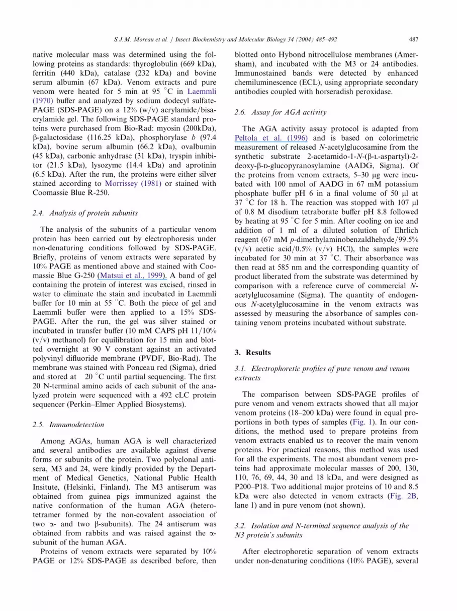
major venom protein complexes of various molecularmasses were revealed and designed by N1–N11(Fig. 2A).The treatment under SDS-PAGE conditions of the
native N3 venom protein led to its dissociation intotwo non-identical subunits of 30 and 18 kDa, respect-ively (Fig. 2B, lane 2), corresponding to the P30 andP18 major proteins of the venom (Fig. 2B, lane 1). Themolecular mass of N3 was determined using Pore limit-ing native PAGE to be 94 kDa (data not shown), sug-gesting that the AGA of A. tabida would be under aheterotetrameric form.The first 20 N-terminal amino acids were sequenced
for each of these two subunits of the N3 protein com-plex. The comparison of the N-terminal sequence of the18 kDa protein with protein sequences from the NCBIdatabases showed a high level of similarity (40–70% ofidentical amino acids) to the N-terminal regions ofAGA b-subunits from several species (Fig. 3). The foursequences exhibiting the highest levels of similarity tothe 18 kDa protein corresponded to b-subunits ofAGAs characterized in bacteria (Synechocystis sp.),insect cells (Spodoptera frugiperda) and mammals(Homo sapiens).Whether sequenced from the proteins or deduced
from the cDNAs, all the b-subunits share the consen-sus sequence Thr-Ile-Gly. In the case of the humanAGA, Wang and Guo (2003) showed that an auto-
extracts. Proteins of venom extracts (7 lg) were separated on 10% PAG
Fig. 2. (A) PAGE profile of venom E. Several protein complexes wereidentified (N1–N11) and the bands were cut and submitted to SDS-PAGE. (B) The N3 polymeric complex was analysed under SDS-PAGE con-
ditions. Samples eluted from a piece of native gel corresponding to the N3 protein complex (lane 2) were separated on 15% SDS-PAGE and com-
pared with proteins of venom extracts (lane 1, 7 lg). P30, P18, P10 and P8.5 indicate four venom proteins of 30, 18, 10 and 8.5 kDa, respectively.
Gels were silver stained according to Morrissey (1981).
DS-PAGE profiles of proteins from venom a
Fig. 1. S nd venomextracts. Venom (lane 1, 10 lg of proteins) and venom extracts pro-
teins (lane 2, 10 lg of proteins) were separated on 12% SDS-PAGE
and stained with Coomassie Blue R-250 for comparison.

S.J.M. Moreau et al. / Insect Biochemistry and Molecular Biology 34 (2004) 485–492 489
proteolysis of the precursor molecules enables gener-
ation of b-subunits with a new N-terminal end present-ing the three amino acids Thr-Ile-Gly, also found in
the N-terminal sequence of the 18 kDa protein. A thre-onine residue (Thr19) necessary to the catalytic activity
of the human enzyme (Tikkanen et al., 1996) was alsoconserved in the 18 kDa protein.
A weaker similarity (20% of identical amino acids)
was observed between the 20 N-terminal amino acidsof the 30 kDa venom protein and the a-subunits of
AGAs from five species: Chryseobacterium meningo-
septicum, H. sapiens, Mus musculus, Rattus norvegicusand Caenorhabditis elegans (Fig. 4). In particular, three
amino acids conserved in all the AGAs a-subunitsfrom bacteria, mammals and nematode were present in
the venom protein of 30 kDa (corresponding to Pro10,
Thr15 and Trp16 of the 30 kDa venom proteinsequence, respectively).
3.3. Immunostaining of venom proteins by antibodiesraised against the human AGA
After separation by 12% SDS-PAGE, several pro-teins in the venom of A. tabida were detected by theanti-human AGA antibodies. The polyclonal 24 anti-bodies raised against the a-subunit of the human AGAstrongly reacted with the P44 and P30 major venomproteins and more weakly with two minor bands of 26and 24 kDa (Fig. 5A, lane 2). The polyclonal M3 anti-bodies directed against the native conformation of thehuman enzyme also stained the P44 protein, inaddition to P18 (Fig. 5A, lane 3). Two minor bands of21 and 23 kDa, as well as the P30 protein were faintlyimmunostained by these antibodies. After electro-phoretic separation of venom extracts under non-dena-turing conditions (10% PAGE), the M3 and 24 antiseraboth reacted with the N3 major protein (Fig. 5B, lanes2 and 3). Another native protein complex of highermolecular mass and corresponding to N5 was alsoslightly immunostained by the 24 antibodies (Fig. 5B,lane 2).
3.4. AGA activity
Using the standard protocol previously and success-fully used for the detection of AGA activities in mam-mal cells (Peltola et al., 1996), we could not measureactivity in a reliable manner neither in A. tabida’svenom extracts nor in pure venom. Indeed indepen-dently from the experimental conditions (amount ofvenom proteins incubated with the synthetic substrate,pH, temperature, duration of incubation), the measuresrecorded were totally random: some assays led to therecord of some activity whereas others resulted in atotal lack of activity (data not shown).
4. Discussion
The venom extracts of A. tabida contained 10 majorproteins of which some were associated under a poly-meric conformation. In particular, the native N3 pro-tein was composed of two distinct subunits of 30 and18 kDa. These two proteins exhibited some similarityin N-terminal amino acid sequences and molecularmasses with the a- and b-subunits of several AGAs (e.g.28 and 17 kDa for the AGA subunits of S. frugiperda(Liu et al., 1996), 27 and 17 kDa for the subunits ofthe human AGA proenzyme (Ikonen et al., 1993)). Inaddition, the two major venom proteins P30 and P18that had the same molecular weights as the N3 pro-tein’s subunits were immunostained by antibodiesraised against the human AGA. These results stronglysupport the presence of a protein structurally related tothe AGA enzymes in the venom of the parasitoid wasp
Fig. 3. Alignment of the P18 protein partial N-terminal amino acids
sequence with sequences of b-subunits of AGA from different species.
The first 20 N-terminal amino acids of the P18 protein were
sequenced and compared with sequences of b-subunits from the fol-
lowing species: Synechocystis sp. (SWISS-PROT accession number
P74383), Chryseobacterium meningosepticum (Q47898), Mus musculus
(Q64191), Caenorhabditis elegans (Q21697), Rattus norvegicus (from
Liu et al., 1996), Bos taurus (from Liu et al., 1996), Homo sapiens
(P20933) and Spodoptera frugiperda (O02467). Boxed letters, identical
amino acids; bold letters, conserved residues. Numbers of first and
last amino acids are indicated on each side of the sequences. Percen-
tages of identical amino acids are on the right.
Fig. 4. Alignment of the P30 protein partial N-terminal amino acid
sequence with sequences of a-subunits of AGA from different species.
The first 20 N-terminal amino acids of the P30 protein were
sequenced and compared with sequences of a-subunits from the fol-
lowing species: Chryseobacterium meningosepticum, Mus musculus,
Caenorhabditis elegans, Rattus norvegicus, Bos taurus, Homo sapiens
and Spodoptera frugiperda. Boxed letters are identical amino acids.
Numbers of first and last amino acids are indicated on each side of
sequences. Percentages of identical amino acids are on the right.

490 S.J.M. Moreau et al. / Insect Biochemistry and Molecular Biology 34 (2004) 485–492
A. tabida. This constitutes the first report of the exist-ence of an AGA-like protein in an insect venom.
Also named glycosylasparaginases (N4-(b-N-acetyl-d-glucosaminyl)-l-asparaginases, EC 3.5.1.26), the AGAswere initially described in lysosomes of eukaryote cells(Makino et al., 1968). AGAs are involved in thehydrolysis of Asn-linked glycoproteins into mono-saccharides and amino acids by cleaving the b-amideon the asparagine moiety of their N-glycosidic linkage(Makino et al., 1968). AGAs have since been character-ized in several mammal tissues, insects cells andprokaryotes and share the same heterodimeric or het-erotetrameric conformation resulting from a complexintracellular processing. For example, the human AGAis synthesized in the endoplasmic reticulum as an enzy-matically inactive precursor (Ikonen et al., 1991) fromwhich a signal peptide is cleaved. Two precursor mole-cules dimerize after initial folding (Riikonen et al.,1996) prior to an auto-proteolytic cleavage, which pro-duces an active AGA molecule, consisting of 24 kDaa-subunits and 17 kDa b-subunits. The active moleculeis a heterotetramer composed of two proa- and twoprob-subunits in which the a- and b-subunits are joinedtogether by strong non-covalent forces (Oinonen et al.,1995; Saarela et al., 1998). Subsequently, proteolytic C-terminal trimming of the subunits occurs in the lyso-somes (for review see Saarela et al., 2001).
The present study raises several interesting questionsconcerning the biochemical features (amino acidssequence, polymeric framework, mode of activation)
that the A. tabida’s protein could share with the knownAGAs and its possible biological role.Our results show that the N3 venom protein shares
several intrinsic properties with some of the previouslydescribed AGAs. The Trp34 residue of the a-subunit ofthe human enzyme, involved in the substrate binding(Riikonen et al., 1995), was conserved in the partialsequence of the 30 kDa subunit (Trp15) of theN3 venom protein. On the other hand two residuesessential to the catalytic activity of the human AGA(Riikonen et al., 1995; Kaartinen et al., 1991), theThr206 and Thr224 residues of the enzyme b-subunit,were also conserved in the 18 kDa subunit of N3 (Thr1
and Thr19) in A. tabida. In addition, the polymericform of the N3 protein was consistent with the finalconformation of the AGA enzyme from most otherspecies and could correspond to a heterotetramericassociation of the 30 and 18 kDa proteins. Severalminor bands from 21 to 26 kDa were immunostainedby antibodies raised against the human AGA. We sug-gest that these proteins could represent different glyco-sylation forms of the subunits of the N3 protein, byanalogy to the heterogeneity of the oligosaccharidicchains observed in the subunits of the human AGA(Saarela et al., 1998). Finally, it has to be noted that anabundant 44 kDa protein (P44) was detected either inpure venom (data not shown) or venom extracts by thetwo antisera used in this study. On the basis of its anti-genic features similar to the human AGA and its mol-ecular weight close to that of the precursor molecule of
proteins with anti-human AGA antibodies. (A) Proteins of venom extract
Fig. 5. Immunostaining of venom s (15 lg of protein) were separatedby 12% SDS-PAGE and stained with Coomassie Blue R-250 (lane 1) or blotted on Hybond nitrocellulose membranes and incubated with the 24
(lane 2) or M3 (lane 3) polyclonal antibodies directed against the human AGA a-subunit or the heterodimeric native conformation of the enzyme,
respectively. For details see Materials and methods. P44, P30 and P18 correspond to venom proteins of 44, 30 and 18 kDa, respectively. (B) Ten
microgram of venom proteins were separated on 10% PAGE and silver stained according to Morrissey (1981) (lane 1) or blotted on Hybond
nitrocellulose membranes and incubated with the 24 (lane 2) or M3 (lane 3) polyclonal antibodies, respectively. N3 and N5 indicate two venom
proteins with polymeric conformations. Immunostained bands were detected by ECL, using appropriate secondary antibodies coupled with horse-
radish peroxidase.

S.J.M. Moreau et al. / Insect Biochemistry and Molecular Biology 34 (2004) 485–492 491
this enzyme (42 kDa) (Ikonen et al., 1993), we hypo-thesize that P44 may constitute a monomeric precursorof the N3 protein. The extracellular localization of anAGA enzyme and its precursor has already beenobserved in vitro in the case of the Ser72Pro mutanthuman enzyme overexpressed by COS-1 cells (Peltolaet al., 1996). The mutant enzymes are secreted in theextracellular medium as inactive precursors from whicha fraction can perform an auto-catalytic cleavage lead-ing to active polymers composed of proa- and b-sub-units. The complete sequencing of the 44, 30 and 18kDa proteins would bring essential information on thestructural similarities between the putative AGA fromA. tabida and the other AGAs.
The apparent variation of AGA activity in thevenom is worth underlining. In most cases and inde-pendently from the experimental conditions, venomextracts or pure venom did not exhibit a consistentAGA activity despite the presence of the N3 protein inthe samples. These observations suggest that the AGA-like enzyme of A. tabida could be stored as an inactiveform in the reservoir of the venom apparatus. Interest-ingly, Parkinson et al. (2002a, b) reported the presenceof two proteins structurally related to proteases in thevenom of the endoparasitoid P. hypochondriaca, butalso failed to characterize their corresponding enzy-matic activities with classical detection protocols. Somevenom enzymes of parasitoids could require additionalsteps for their activation or may have a selectiveactivity towards a specific substrate. Alternatively, ithas recently been shown that human AGA activity canbe inhibited by several compounds including competi-tive, non-competitive and irreversible inhibitors (Risleyet al., 2001). Work is currently in progress to test if theunreliable activity we observed could be due to thepresence of such inhibitors in the venom of A. tabida.
As the AGA-like enzyme represents one of the mainvenom proteins, it appears unlikely that such an abun-dant protein could not become functional under someinstances and would not fulfill a specific biological role.A. tabida females inject their venom during the depo-sition of their eggs in Drosophila host larvae. It is notexcluded that interactions with host’s molecules and/orwith non-venomous compounds of maternal origin(e.g. parasitoid egg, ovarian fluids) could be requiredfor the AGA-like protein to become functional. Inter-estingly, the hydrolytic action of AGAs towards theirglycosylasparagine substrate leads to the production ofaspartate which is a known excitatory neurotransmitterof the Drosophila nervous system (Besson et al., 2000).Thus, when injected within the venom in Drosophilalarvae during the parasitization process, an activatedform of an AGA-like enzyme could have potentialimplications in the temporary paralysis of the host thatoccurs at the early stage of the parasitization (Moreauet al., 2002).
The data gathered in the present work thus open sev-eral areas of investigation for a better knowledge of thephysiological relationships between A. tabida and itsDrosophila hosts and a full understanding of the func-tional diversity of hymenopteran venoms.
Acknowledgements
We thank Prof. F. Pennacchio for his contributionto the initial steps of this work, Prof. J. Hoffmann andL. Peltonen in whose laboratories sequencing, immuno-blotting and a part of the enzymatic assays were per-formed, and Mr. A. Roots for his critical reading ofthe manuscript. This work was partially supported byGDR-CNRS 2153.
References
Asgari, S., Zhang, G., Zareie, R., Schmidt, O., 2003a. A serine pro-
teinase homolog venom protein from an endoparasitoid wasp
inhibits melanization of the host hemolymph. Insect Biochem.
Mol. Biol. 33, 1017–1024.
Asgari, S., Zareie, R., Zhang, G., Schmidt, O., 2003b. Isolation and
characterization of a novel venom protein from an endopar-
asitoid, Cotesia rubecula (Hym.: Braconidae). Arch. Insect Bio-
chem. Physiol. 53, 92–100.
Beard, R.L., 1978. Arthropods venoms. In: Bettini, S. (Ed.), Hand-
buch der Experimentellen Pharmakologie, vol. 48. Springer Ver-
lag, Berlin, pp. 773–780.
Beckage, N.E., Metcalf, J.S., Nesbit, D.J., Schleifer, K.W., Zeltan,
S.R., de Buron, I., 1990. Host hemolymph monophenoloxidase
activity in parasitized Manduca sexta larvae and evidence for inhi-
bition by wasp polydnavirus. Insect Biochem. Mol. Biol. 20,
285–294.
Besson, M.T., Soustelle, L., Birman, S., 2000. Selective high-affinity
transport of aspartate by a Drosophila homologue of the excit-
atory amino acid transporters. Curr. Biol. 10, 207–210.
Blanche, P.J., Gong, E.L., Forte, T.M., Nichols, A.V., 1981. Char-
acterization of human high-density lipoproteins by gradient gel
electrophoresis. Biochim. Biophys. Acta. 665, 408–419.
Dahlman, D.L., 1990. Evaluation of teratocyte functions: an over-
view. Arch. Insect Biochem. Physiol. 13, 159–166.
David, J.R., 1959. Etude quantitative du developpement de la Droso-
phile elevee en milieu axenique. Bull. Biol. Fr. Belg. 93, 472–505.
Digilio, M.C., Isidoro, N., Tremblay, E., Pennacchio, F., 2000. Host
castration by Aphidius ervi venom proteins. J. Insect Physiol. 46,
1041–1050.
Doury, G., Bigot, Y., Periquet, G., 1997. Physiological and biochemi-
cal analysis of factors in the female venom gland and larval sali-
vary secretions of the ectoparasitoid wasp Eupelmus orientalis.
J. Insect Physiol. 43, 69–81.
Eslin, P., Giordanengo, P., Fourdrain, Y., Prevost, G., 1996. Avoid-
ance of encapsulation in the absence of VLP by a braconid para-
sitoid of Drosophila larvae: an ultrastructural study. Can. J. Zool.
74, 2193–2198.
Ikonen, E., Baumann, M., Gron, K., Syvanen, A.-C., Enomaa, N.,
Halila, R., Aula, P., Peltonen, L., 1991. Aspartylglucosaminuria:
cDNA encoding human aspartylglucosaminidase and the missense
mutation causing the disease. EMBO J. 10, 51–58.
Ikonen, E., Julkunen, I., Tollersrud, O.-K., Kalkkinen, N., Peltonen,
L., 1993. Lysosomal aspartylglucosaminidase is processed to the

492 S.J.M. Moreau et al. / Insect Biochemistry and Molecular Biology 34 (2004) 485–492
active subunit complex in the endoplasmic reticulum. EMBO J.
12, 295–302.
Jones, D., Sawicki, G., Wozniak, M., 1992. Sequence, structure and
expression of a wasp venom protein with a negatively charged sig-
nal peptide and a novel repeating internal structure. J. Biol.
Chem. 267, 14871–14878.
Kaartinen, V., Williams, J.C., Tomich, J., Yates, J.R., Hood, L.E.,
Mononen, I., 1991. Glycoasparaginase from human leukocytes.
Inactivation and covalent modification with diazo-oxonorvaline.
J. Biol. Chem. 266, 5860–5869.
Kraaijeveld, A.R., van Alphen, J.M., 1994. Geographical variation in
resistance of the parasitoid Asobara tabida against encapsulation
by Drosophila melanogaster larvae: the mechanism explored. Phys.
Entomol. 19, 9–14.
Krishnan, A., Nair, P.N., Jones, D.J., 1994. Isolation, cloning and
characterization of new chitinase stored in active form in chitin-
lined venom reservoir. J. Biol. Chem. 269, 20971–20976.
Laemmli, U.K., 1970. Cleavage of structural proteins during the
assembly of the head of bacteriophage T4. Nature 227, 680–685.
Liu, Y., Dunn, G.S., Aronson Jr., ., N.N., 1996. Purification, bio-
chemistry and molecular cloning of an insect glycosylasparaginase
from Spodoptera frugiperda. Glycobiology 6, 527–536.
Makino, M., Kojima, T., Ohgushi, T., Yamashina, I., 1968.
Studies on enzymes acting on glycopeptides. J. Biochem. 63, 186–
192.
Matsui, N.M., Smith-Beckerman, D.M., Epstein, L.B., 1999. Staining
of preparative 2-D gels. Coomassie blue and imidazole-zinc nega-
tive staining. Methods Mol. Biol. 112, 307–311.
Monconduit, H., Prevost, G., 1994. Avoidance of encapsulation by
Asobara tabida, a larval parasitoid of Drosophila species. Norw. J.
Agr. Sci. 16, 301–309.
Moreau, S.J.M., Dingremont, A., Doury, G., Giordanengo, P., 2002.
Effects of parasitism by Asobara tabida (Hymenoptera: Braconi-
dae) on the development, survival and activity of Drosophila mela-
nogaster larvae. J. Insect Physiol. 48, 337–347.
Morrissey, J.H., 1981. Silver stain for proteins in polyacrylamide gels:
a modified procedure with enhanced uniform sensitivity. Anal.
Biochem. 117, 307–310.
Oinonen, C., Tikkanen, R., Rouvinen, J., Peltonen, L., 1995. Three-
dimensional structure of human lysosomal aspartylglucosamini-
dase. Nat. Struct. Biol. 2, 1102–1108.
Parkinson, N.M., Weaver, R.J., 1999. Noxious components of venom
from the pupa-specific parasitoid Pimpla hypochondriaca. J. Inver-
tebr. Pathol. 73, 74–83.
Parkinson, N.M., Smith, I., Weaver, R.J., Edwards, J.P., 2001. A
new form of arthropod phenoloxidase is abundant in venom of
the parasitoid wasp Pimpla hypochondriaca. Insect Biochem. Mol.
Biol. 31, 57–63.
Parkinson, N.M., Conyers, C., Smith, I., 2002a. A venom protein
from the endoparasitoid wasp Pimpla hypocondriaca is similar to
snake venom reprolysin-type metalloproteases. J. Invertebr.
Pathol. 79, 129–131.
Parkinson, N.M., Richards, E.H., Conyers, C., Smith, I., Edwards,
J.P., 2002b. Analysis of venom constituents from the parasitoid
wasp Pimpla hypocondriaca and cloning of cDNA encoding a
venom protein. Insect Biochem. Mol. Biol. 32, 729–735.
Parkinson, N.M., Conyers, C., Keen, J.N., MacNicoll, A.D., Smith,
I., Weaver, R.J., 2003. cDNAs encoding large venom proteins
from the parasitoid wasp Pimpla hypocondriaca identified by ran-
dom sequence analysis. Comp. Biochem. Physiol. Part C 134,
513–520.
Peltola, M., Tikkanen, R., Peltonen, L., Jalanko, A., 1996. Ser72Pro
active-site disease mutation in human lysosomal aspartylglucosa-
minidase: abnormal intracellular processing and evidence for
extracellular activation. Hum. Mol. Genet. 6, 737–743.
Quistad, G.B., Nguyen, Q., Bernasconi, P., Leisy, D., 1994. Purifi-
cation and characterization of insecticidal toxins from venom
glands of the parasitic wasp, Bracon hebetor. Insect Biochem.
Mol. Biol. 24, 955–961.
Riikonen, A., Tikkanen, R., Jalanko, A., Peltonen, L., 1995. Immedi-
ate interaction between the nascent subunits and two conserved
amino acids Trp34 and Thr206 are needed for the catalytic
activity of aspartylglucosaminidase. J. Biol. Chem. 270, 4903–
4907.
Riikonen, A., Rouvinen, J., Tikkanen, R., Julkunen, I., Peltonen, L.,
Jalanko, A., 1996. Primary folding of aspartylglucosaminidase.
Significance of disulfide bridges and evidence of early multi-
merization. J. Biol. Chem. 271, 21340–21344.
Risley, J.M., Huang, D.H., Kaylor, J.J., Malik, J.J., Xia, Y.-Q.,
2001. Glycosylasparaginase activity requires the a-carboxyl group,but not the a-amino group, on N4-(2-acetamido-2-deoxy-b-d-glu-copyranosyl)-l-asparagine. J. Enzyme Inhib. 16, 269–274.
Saarela, J., Laine, M., Tikkanen, R., Oinonen, C., Jalanko, A.,
Rouvinen, J., Peltonen, L., 1998. Activation and oligomerization
of aspartylglucosaminidase. J. Biol. Chem. 273, 25320–25328.
Saarela, J., Laine, M., Oinonen, C., von Schantz, C., Jalanko, A.,
Rouvinen, J., Peltonen, L., 2001. Molecular pathogenesis of a dis-
ease: structural consequences of aspartylglucosaminuria muta-
tions. Hum. Mol. Genet. 10, 983–995.
Soller, M., Lanzrein, B., 1996. Polydnavirus and venom of the
egg-larval parasitoid Chelonus inanitus (Braconidae) induce devel-
opmental arrest in the prepupa of its host Spodoptera littoralis
(Noctuidae). J. Insect Physiol. 42, 471–481.
Strand, M.R., Johnson, J.A., Noda, T., Dover, B.A., 1994. Develop-
ment and partial characterization of monoclonal antibodies to
venom of the parasitoid Microplitis demolitor. Arch. Insect Bio-
chem. Physiol. 26, 123–136.
Tikkanen, R., Riikonen, A., Oinonen, C., Rouvinen, J., Peltonen, L.,
1996. Functional analyses of active site residues of human lysoso-
mal aspartylglucosaminidase: implications for catalytic mech-
anism and autocatalytic activation. EMBO J. 15, 2954–2960.
Wang, Y., Guo, H.C., 2003. Two-step dimerization for autoproteo-
lysis to activate glycosylasparaginase. J. Biol. Chem. 278, 3210–
3219.
Webb, B.A., Luckhart, S., 1994. Evidence for an early immunosup-
pressive role for related Campoletis sonorensis venom and ovarian
proteins in Heliothis virescens. Arch. Insect Biochem. Physiol. 26,
147–163.