Hypotensive and Diuretic Effects of Camboginol and ...
175
Hypotensive and Diuretic Effects of Camboginol and Morelloflavone from Garcinia dulcis in 2-Kidneys-1-Clip Renovascular Hypertensive Rat Nattaya Thongsepee A Thesis Submitted in Partial Fulfillment of the Requirements for the Degree of Doctor of Philosophy in Physiology Prince of Songkla University 2017 Copyright of Prince of Songkla University
-
Upload
khangminh22 -
Category
Documents
-
view
2 -
download
0
Transcript of Hypotensive and Diuretic Effects of Camboginol and ...

Hypotensive and Diuretic Effects of Camboginol and Morelloflavone
from Garcinia dulcis in 2-Kidneys-1-Clip Renovascular
Hypertensive Rat
Nattaya Thongsepee
A Thesis Submitted in Partial Fulfillment of the Requirements for the
Degree of Doctor of Philosophy in Physiology
Prince of Songkla University
2017
Copyright of Prince of Songkla University

i
Hypotensive and Diuretic Effects of Camboginol and Morelloflavone
from Garcinia dulcis in 2-Kidneys-1-Clip Renovascular
Hypertensive Rat
Nattaya Thongsepee
A Thesis Submitted in Partial Fulfillment of the Requirements for the
Degree of Doctor of Philosophy in Physiology
Prince of Songkla University
2017
Copyright of Prince of Songkla University

ii
Thesis Title Hypotensive and Diuretic Effects of Camboginol and
Morelloflavone from Garcinia dulcis in 2-Kidneys-1-Clip
Renovascular Hypertensive Rat
Author Ms. Nattaya Thongsepee
Major Program Physiology
________________________________________________________________________________
Major advisor Examining Committee:
...................................................................... ................................................... Chairperson
(Assoc. Prof. Dr. Siriphun Hiranyachattada) (Assoc. Prof. Dr. Kesorn Suwanprasert)
.................................................... Committee
Co-advisor (Assoc. Prof. Dr. Siriphun Hiranyachattada)
...................................................................... .................................................... Committee
(Assoc. Prof. Dr. Ekkasit Kumarnsit) (Assoc. Prof. Dr. Ekkasit Kumarnsit)
………….................................... Committee
(Assoc. Prof. Dr. Wilawan Mahabusarakam)
The Graduate School, Prince of Songkla University, has approved this thesis
as partial fulfillment of the requirements for the Doctor of Philosophy Degree in
Physiology
…………………………….……………
(Assoc. Prof. Dr. Teerapol Srichana)
Dean of Graduate School

iii
This is to certify that the work here submitted is the result of the candidate’s own
investigations. Due acknowledgement has been made of any assistance received.
……...…………..……………… Signature
(Assoc. Prof. Dr. Siriphun Hiranyachattada)
Major Advosor
…………………..……………… Signature
(Assoc. Prof. Dr. Ekkasit Kumarnsit)
Co-advisor
…………………..……………… Signature
(Ms. Nattaya Thongsepee)
Candidate

iv
I hereby certify that this work has not been accepted in substance for any degree, and
is not being currently submitted in candidature for any degree.
……………………………… Signature
(Ms. Nattaya Thongsepee)
Candidate

v
ชอวทยานพนธ ผลลดความดนเลอดและขบปสสาวะของ camboginol และ morelloflavone จาก Garcinia dulcis ในหน model 2-kidneys-1-
clip renovascular hypertension ก ผเขยน นางสาวนาตยา ทองเสภ สาขาวชา สรรวทยา ปการศกษา 2559
บทคดยอ
การศกษานมวตถประสงคเพอทดสอบฤทธลดความดนเลอดแดงและขบปสสาวะของ camboginol (isoprenylated bezophenone) และ morelloflavone (biflavonoid) ซงเปนสารตานอนมลอสระทสกดจากมะพด (Garcinia dulcis) ในหนความดนเลอดแดงสง 2-kidneys-1-
clip (2K1C) รวมทงทดสอบกลไกการคลายหลอดเลอด โดยการน าหน Wistar rat เพศผ (น าหนกตว 150-200 g, 6 ตว/กลม) มาผาตดหนบหลอด renal artery ดานซายเพอชกน าใหเกดภาวะความดนเลอดแดงสงเปนเวลา 4 สปดาห หนกลมควบคมจะไดรบการผาตดเชนเดยวกนแตไมมการหนบหลอดเลอด เมอครบระยะเวลาจะน าหนทงสองกลมมาศกษาแบบ in vivo โดยใหสารละลาย para-aminohippuric acid 0.5% และ inulin 1% ทาง jugular vein แกหนทสลบลก เพอใชในการประเมนคา effective renal plasma flow (ERPF) และ glomerular filtration
rate พรอมทงให camboginol และ morelloflavone (ขนาด 0.1 mg/kg + 5 µg/min/kg BW) ควบคกน ท าการบนทกคาความดนเลอดแดงและอตราการเตนของหวใจตลอดการทดลองและเกบตวอยางปสสาวะและเลอดเพอใชในการวเคราะหคา osmolar และ free water clearance
นอกจากนยงประเมนคา baroreflex sensitivity (BRS), ระดบของ malondialdehyde (MDA) ในเลอดและระดบของ endothelium nitric oxide synthase (eNOS) ในหลอดเลอดอกดวย การศกษาแบบ in vitro จะทดสอบฤทธและกลไกการออกฤทธคลายตวของสารทงสองทขนาดความเขมขน 10
-13-10
-5 M ในหลอดเลอด thoracic artery โดยผลการทดลองพบวา สารทงสอง
สามารถลดความดนเลอดแดง, เพมปรมาณ ERPF, เพมปรมาณปสสาวะ, เพมคา BRS , ลดระดบ MDA ในเลอด และเพมการแสดงออกของ eNOS ในหลอดเลอดของหน 2K1C สารทงสองสามารถออกฤทธคลายหลอดเลอดแดงของหนกลมควบคมไดดกวาหนกลม 2K1C โดยกลไกคลายตวผานทาง endothelium dependent vasorelaxtion pathway ทงนฤทธในการขบปสสาวะและลดความดนเลอดแดงของ camboginol และ morelloflavone นาจะสมพนธกบคณสมบตในการตอตานอนมลอสระของสารทงสอง

vi
Thesis Title Hypotensive and Diuretic Effects of Camboginol and
Morelloflavone from Garcinia dulcis in 2-Kidneys-1-Clip
Renovascular Hypertensive Rat
Author Ms. Nattaya Thongsepee
Major Program Physiology
Academic Year 2016
Abstract
This study aimed to evaluate hypotensive and diuretic effects of camboginol
(isoprenylated benzophenone) and morelloflavone (biflavonoid), a robust antioxidant
which isolated from Garcinia dulcis, in 2-kidneys-1-clip (2K1C) renovascular
hypertensive rats together with their vasorelaxant mechanisms. Male Wistar rats
(150-200 g) were undergone either 2K1C or sham operation (SO) (n=6, each). Four
weeks later, an in vivo study was performed by which clearance markers (0.5% para-
aminohippuric acid and 1% inulin) were given via a jugular vein to estimate effective
renal plasma flow (ERPF) and glomerular filtration rate in anesthetized rats.
Camboginol or morelloflavone was given at the dose of 0.1 mg/kg + 5 g/min/kg
body weight, while arterial blood pressure (ABP) and heart rate were monitored via a
carotid artery. Plasma and urine samples were collected for determination of osmolar
and free water clearances. Baroreflex sensitivity (BRS), plasma malondialdehyde
(MDA) level and an expression of endothelial nitric oxide synthase (eNOS) were also
investigated. For an in vitro study, the relaxation of isolated aortic ring which
phenylephrine-preconstriction was experimented using an organ bath technique by
cumulative additions of either camboginol or morelloflavaone (10-13
-10-5
M) in the
presence or absence of specific vasorelaxant inhibitors. It was found that camboginol
and morelloflavone significantly decreased ABP, increased ERPF and increased urine
flow rate in anesthetized 2K1C rats. Both also improved an impaired BRS, decreased
plasma MDA levels and enhanced eNOS expression in vascular endothelium. The
mechanisms of their vasorelaxant action mainly involved the different extent of
endothelial dependent vasorelaxation pathway. The responsive mechanisms of
diuretic and hypotensive effects of camboginol and morelloflavone are likely to
associate with their scavenging activity.

vii
Acknowledgements
I sincerely appreciate my major advisor Assoc. Prof. Dr. Siriphun
Hiranyachattada for her guidance, suggestions and all supports. This thesis would not
be completed and my Ph.D. course would not be achieved without her kindness.
I appreciate my co-advisor Assoc. Prof. Dr. Ekkasit Kumarnsit for his
suggestions. I also appreciate Assoc. Prof. Dr. Wilawan Mahabusarakam for her
provided substances, camboginol and morelloflavone, and for her suggestions in a
part of isolation of the plant compound. I appreciate Dr. Wachiryah Thong-asa for
her collaboration in a part of immunohistochemistry. In addition, I am grateful to
Assoc. Prof. Dr. Kesorn Suwanprasert and all examining committees for their
comments and suggestions.
I appreciate Prof. Dr. Andrew Allen for his guidance and suggestions
when I deeply conducted a research at the Cardiovascular Regulation Laboratory,
Department of Physiology, University of Melbourne, Australia.
I would like to thank Prince of Songkla University for the PSU-Ph.D
scholarships and the Graduate School Research Support Funding for Thesis, Faculty
of Science, Prince of Songkla University for the Overseas Research Scholarship, the
Office of the Higher Education Commission, Thailand for financial support belong to
the Higher Education Research Promotion and National Research University project
of Thailand.
I would like to extent my thanks to all scientists and officers of
Department of Physiology, Faculty of Science, Prince of Songkla University for their
kindness and all supports along my Ph.D. courses.
Finally, I would like to express my sincere gratitude to my family and my
friends for their encouragement and mental support.
Nattaya Thongsepee

viii
TABLE OF CONTENTS
CONTENTS Page
ABSTRACT IN THAI ................................................................................... v
ABSTRACT IN ENGLISH ........................................................................... vi
ACKNOWLEDGEMENTS .......................................................................... vii
TABLE OF CONTENTS .............................................................................. viii
LIST OF TABLES ......................................................................................... xii
LIST OF FIGURES ....................................................................................... xiii
LIST OF ABBREVIATIONS AND SYMBOLS ......................................... xix
CHAPTER 1 INTRODUCTION
1.1. Background and rationale ........................................................................ 1
1.2. Literature reviews .................................................................................... 6
1.2.1. Hypertension................................................................................ 6
1.2.2. Renovascular hypertension .......................................................... 17
1.2.3. Structure and function of kidney ................................................. 22
1.2.4. Structure and function of vascular smooth muscle ..................... 32
1.2.5. Arterial baroreceptor reflex ......................................................... 43
1.2.6. Treatment of hypertension ........................................................... 50
1.2.6. Camboginol and morelloflavone ................................................. 54
1.3. Objectives ................................................................................................ 61
1.4. Hypotheses ............................................................................................... 61

ix
TABLE OF CONTENTS (Continued)
CONTENTS Page
CHAPTER 2 MATERIALS AND METHODS
2.1. Extraction of camboginol and morelloflavone ........................................ 62
2.2. Experimental animals
2.2.1. Animals........................................................................................ 62
2.2.2. Establishment of 2K1C hypertensive rat ..................................... 63
2.3. Chemical reagents .................................................................................... 64
2.4. Equipment ................................................................................................ 65
2.5. The in vivo study of renal clearance and BRS ......................................... 66
2.6. Determination of plasma malondialdehyde (MDA) ................................. 67
2.7. The in vitro study of thoracic aortic ring relaxation
2.7.1. Preparation of isolated thoracic aortic ring ................................. 68
2.7.2. Concentration-response curve of camboginol and
morelloflavone ........................................................................................ 68
2.7.3. Effect of specific inhibitors on vasorelaxation response of
camboginol and morelloflavone ............................................................. 68
2.8. Immunohistochemistry ............................................................................. 69
2.9. Calculations
2.9.1. Mean arterial blood pressure(MABP) ......................................... 70
2.9.2. Baroreflex sensitivity (BRS) ....................................................... 70
2.9.3. Clearance of PAH (CPAH) ............................................................ 70
2.9.4. Renal vascular resistance (RVR) ................................................. 71
2.9.5. Clearance of inulin (Cin) .............................................................. 72
2.9.6. Urine flow rate ( ...................................................................... 72
2.9.7. Filtration fraction (FF) ................................................................. 72
2.9.8. Osmolar clearance (OOsm) ............................................................ 73
2.9.9. Free water clearance (CH2O) ......................................................... 74
2.9.10. Contraction responses of thoracic aorta .................................... 74
2.9.11. Relaxation responses of thoracic aorta ...................................... 74

x
TABLE OF CONTENTS (Continued)
CONTENTS Page
2.10. Statistical analyses .................................................................................. 75
CHAPTER 3 RESULTS
3.1. Effects of camboginol
3.1.1. Effects of camboginol on ABP and HR ...................................... 76
3.1.2. Effects of camboginol on renal clearance ................................... 79
3.1.3. Effects of camboginol on BRS .................................................... 81
3.1.4. Effect of camboginol on plasma MDA levels ............................. 83
3.1.5. Body weight, kidney weight and cardiac mass ........................... 84
3.1.6. Effects of camboginol on isolated aortic relaxation .................... 85
3.1.7. Effect of camboginol on eNOS expression ................................. 92
3.2. Effects of morelloflavone
3.2.1. Effect of morelloflavone on ABP and HR ................................... 93
3.2.2. Effects of morelloflavone on renal clearance .............................. 94
3.2.3. Effects of morelloflavone on BRS .............................................. 97
3.2.4. Effect of morelloflavone on plasma MDA levels ........................ 99
3.2.5. Body weight, kidney weight and cardiac mass ........................... 100
3.2.6. Effects of morelloflavone on isolated aortic relaxation .............. 101
3.2.7. Effect of morelloflavone on eNOS expression ............................ 108
CHAPTER 4 DISCUSSION
4.1. Development of 2K1C hypertension
4.1.1. Cardiovascular function changes in 2K1C .................................. 109
4.1.2. Renal function changes in 2K1C ................................................. 110
4.2. Diuretic and hypotensive effect of camboginol
4.2.1. Diuretic effect of camboginol ...................................................... 111
4.2.2. Hypotensive effect of camboginol ............................................... 111

xi
TABLE OF CONTENTS (Continued)
CONTENTS Page
4.3. Diuretic and hypotensive effect of morelloflavone
4.3.1. Diuretic effect of morelloflavone ................................................ 113
4.3.2. Hypotensive effect of morelloflavone ......................................... 114
CHAPTER 5 CONCLUSIONS .................................................................... 115
REFERENCES ............................................................................................... 116
APPENDICES ................................................................................................ 136
VITAE ............................................................................................................. 150

xii
LIST OF TABLES
Table Page
1.1 Definition and classification of office blood pressure levels (mm
Hg).
6
1.2 Estimates of age-standardized prevalence (%) of raise blood
pressurea in adults aged 25
+ years in country of the SEA Region,
2008.
9
1.3 Functions of the kidney. 26
3.1 Comparison of body weight at the beginning of experiment (Pre-
BW), 4 weeks after induction of hypertension by experimental
renal stenosis (Post-BW) and body weight changes (BW).
Left and right kidney weight (KW), cardiac mass and hematocrit
of 2-kidneys-1-clip (2K1C) and sham operation (SO) rats were
determined and compared after either camboginol (C) (0.1 mg/kg
+ 5 µg/min/kg BW) or vehicle (V or DMSO) treatment.
84
3.2 Comparison of body weight at the beginning of experiment (Pre-
BW), 4 weeks after induction of hypertension by experimental
renal stenosis (Post-BW) and body weight changes (BW).
Left and right kidney weight (KW), cardiac mass and hematocrit
of 2-kidneys-1-clip (2K1C) and sham operation (SO) rats were
determined and compared after either morelloflavone (M) (0.1
mg/kg + 5 µg/min/kg BW) or vehicle (V or DMSO) treatment.
100
Suppl. 1 Effect of camboginol on renal function in sham operative (SO)
and 2-kidneys-1-clip (2K1C) rat which treated with either
camboginol (C) or vehicle (V).
137
Suppl. 2 Effect of moreloflavone on renal function in sham operative (SO)
and 2-kidneys-1-clip (2K1C) rat which treated with either
camboginol (C) or vehicle (V).
138

xiii
LIST OF FIGURES
Figure Page
1.1 Conceptual framework of the study 5
1.2 Age-standardized prevalence of raised blood pressure in adult
aged 25+ years by WHO Region, 2008.
8
1.3 The heart, arteries and arteriole in regulation of blood pressure. 10
1.4 The autonomic nervous system and its control of blood pressure. 11
1.5 Renin angiotensin system and effected on blood pressure and
aldosterone release.
12
1.6 The control of peripheral arteriolar resistance. 14
1.7 Genetic factor contributes hypertension. 15
1.8 Pressure-volume curves demonstrating diastolic/systolic
dysfunction.
16
1.9 Three types of renovascular hypertensive (RVH) model. 18
1.10 Three theoretical temporal phases of 2K1C model. 18
1.11 The role of renin angiotensin system in 2K1C RVH. 19
1.12 Anatomy of (a) kidney and (b) renal tubules. 24
1.13 Structure of vessel wall. 32
1.14 Mechanism of smooth muscle contraction. 36
1.15 Mechanism of smooth muscle relaxation. 38
1.16 Schematic summarizing the release of relaxing factors from
endothelial cells and their effect on vascular smooth muscle cell.
41
1.17 The location of the aortic and carotid baroreceptors. 44
1.18 The arterial baroreceptor reflex arc. 48
1.19 Baroreceptor reflex sensitivity. 49
1.20 Acute and chronic resetting of the baroreceptor reflex. 50
1.21 Tree, flower and fruit of Garcinia dulcis. 55
1.22 Chemical structure of camboginol or garcinol. 56
1.23 Chemical structure of morelloflavone. 59

xiv
LIST OF FIGURES (Continued)
Figure Page
3.1 Tracing of recorded arterial blood pressure (ABP, red) and heart
rate (HR, blue) of 2-kidneys-1-clip (2K1C; upper panel) and
sham operative (SO; lower panel) rat.
77
3.2 Acute hypotensive effects of camboginol (C) (0.1 mg/kg BW) on
systolic blood pressure (SBP; a), diastolic blood pressure (DBP;
b), pulse pressure (PP; c) and mean arterial blood pressure
(MABP; d) in the 2-kidneys-1-clip (2K1C) and sham operation
(SO) groups.
78
3.3 Effects of camboginol (C; 0.1mg/kg BW + 5 g/min/kg BW) on
mean arterial blood pressure (MABP; a), effective renal plasma
flow (ERPF; b), renal vascular resistance (RVR; c), glomerular
filtration rate (GFR; d), urine flow rate ( e), urine osmolality
(UOsm; f), osmolar clearance (COsm; g) and negative free water
clearance (TCH2O; h), in 2-kidneys-1-clip (2K1C) and sham
operative (SO) group during clearance study.
80
3.4 Baroreflex sensitivity (BRS) in response to either phenylephrine
(PE; a) or sodium nitroprusside (SNP; b) in 2-kidneys-1-clip
(2K1C) and sham operation (SO) group during treatment with
camboginol (C; 0.1 mg/kg BW + 5 g/min/kg BW).
82
3.5 Effect of camboginol (0.1 mg/kg BW + 5 g/min/kg BW) on
plasma malondialdehyde (MDA) levels in 2-kidneys-1-clip
(2K1C) hypertensive and sham operative (SO) normotensive
rats.
83
3.6 Effects of camboginol or vehicle (DMSO) on vasorelaxation of
endothelium-intact (left panel; a, c and e) or -denuded (right
panel; b, d and f) aortic rings from 2-kidneys-1-clip (2K1C) or
sham operative (SO) group.
87

xv
LIST OF FIGURES (Continued)
Figure Page
3.7 Effects of camboginol on vasorelaxation of endothelium-intact
(left panel; a, c and e) or -denuded (right panel; b, d and f) aortic
rings from 2-kidneys-1-clip (2K1C) or sham operative (SO)
group in the presence of 10-4
M L-NAME.
88
3.8 Effects of camboginol on vasorelaxation of endothelium-intact
(left panel; a, c and e) or -denuded (right panel; b, d and f) aortic
rings from 2-kidneys-1-clip (2K1C) or sham operative (SO)
group in the presence of 10-6
M indomethacin (IDM).
89
3.9 Effects of camboginol on vasorelaxation of endothelium-intact
(left panel; a, c and e) or denuded (right panel; b, d and f) aortic
rings from 2-kidneys-1-clip (2K1C) or sham operative (SO)
group in the presence of 10-6
M glibenclamide (G).
90
3.10 Effects of camboginol or vehicle (DMSO) on vasorelaxation of
endothelium-intact (left panel; a, c and e) or -denuded (right
panel; b, d and f) aortic rings from 2-kidneys-1-clip (2K1C) or
sham operative (SO) group in the presence of 10-3
M
tetraethylammonium (TEA).
91
3.11 Endothelial nitric oxide synthase (eNOS) expression in aortic
endothelium of control and 2-kidneys-1-clip (2K1C) rat; control
(a), negative control (b), 2K1C (c) and after camboginol (0.1
mg/kg + 5µg/min/kg BW) treatment in 2K1C.
92
3.12 Effects of morelloflavone (M; 0.1 mg/kg BW + 5 μg/min/kg
BW) on systolic blood pressure (SBP; a), diastolic blood
pressure (DBP; b), pulse pressure (PP; c) and mean arterial
pressure (MABP; d) in 2-kidneys-1-clip (2K1C) and sham
operative (SO) group during clearance study.
94

xvi
LIST OF FIGURES (Continued)
Figure Page
3.13 Effects of morelloflavone (M; 0.1mg/kg BW bolus + 5
g/min/kg BW) on mean arterial blood pressure (MABP; a),
effective renal plasma flow (ERPF; b), renal vascular resistance
(RVR; c), glomerular filtration rate (GFR; d), urine flow rate
( e), urine osmolality (UOsm; f), osmolar clearance (COsm; g)
and negative free water clearance (TCH2O; h), in 2-kidneys-1-clip
(2K1C) and sham operative (SO) group during clearance study.
96
3.14 Baroreflex sensitivity (BRS) in response to either phenylephrine
(PE; left panel) or sodium nitroprusside (SNP; right panel) in 2-
kidneys-1-clip (2K1C) and sham operation (SO) group during
treatment with morelloflavone (M; 0.1 mg/kg BW + 5 g/min/kg
BW).
98
3.15 Effect of camboginol (0.1 mg/kg BW + 5 g/min/kg BW) on
plasma malondialdehyde (MDA) levels in 2-kidneys-1-clip
(2K1C) hypertensive and sham operative (SO) normotensive
rats.
99
3.16 Effects of morelloflavone or vehicle (DMSO) on vasorelaxation
of endothelium-intact (left panel; a, c and e) or -denuded (right
panel; b, d and f) aorta rings from 2-kidneys-1-clip (2K1C) or
sham operative (SO) group.
103
3.17 Effects of morelloflavone on vasorelaxation of endothelium-
intact (left panel; a, c and e) and -denuded (right panel; b, d and
f) aorta rings from 2-kidneys-1-clip (2K1C) and sham operative
(SO) group in the presence of 10-4
M L-NAME.
104

xvii
LIST OF FIGURES (Continued)
Figure Page
3.18 Effects of morelloflavone on vasorelaxation of endothelium-
intact (left panel; a, c and e) and -denuded (right panel; b, d and
f) aorta rings from 2-kidneys-1-clip (2K1C) and sham operative
(SO) group in the presence of 10-6
M indomethacin (IDM).
105
3.19 Effects of morelloflavone on vasorelaxation of endothelium-
intact (left panel; a, c and e) and -denuded (right panel; b, d and
f) aorta ring from 2-kidneys-1-clip (2K1C) and sham operative
(SO) group in the presence of 10-6
M glibenclamide (G).
106
3.20 Effects of morelloflavone on vasorelaxation of endothelium-
intact (left panel; a, c and e) and -denuded (right panel; b, d and
f) aorta ring from 2-kidneys-1-clip (2K1C) and sham operative
(SO) group in the presence of 10-3
M tetraethyl ammonium
(TEA).
107
3.21 Endothelial nitric oxide synthase (eNOS) expression in aortic
endothelial of control and 2-kidneys-1-clip (2K1C) rat; control
(a), negative control (b), 2K1C (c) and after morelloflavone (0.1
mg/kg BW + 5 g/min/kg BW) treatment in 2K1C.
108
Suppl. 1 Effects of phenylephrine (PE; 10-10
-10-4
M)) on vasoconstriction
of endothelium-intact vs. -denuded aortic ring from sham
operative (SO) group (a), endothelium-intact vs. -denuded aortic
ring from 2-kidneys-1-clip (2K1C) group (b), endothelium-intact
aortic rings between 2K1C and SO group (c) and endothelium-
denuded aortic rings between 2K1C and SO group (d).
140

xviii
LIST OF FIGURES (Continued)
Figure Page
Suppl. 2 Effects of acetylcholine (ACh) or vehicle on vasorelaxation of
endothelium-intact (left panel; a, c, e) or -denuded (right panel,
b, d, f) aorta rings from 2-kidneys-1-clip (2K1C) or sham
operative (SO) groups.
142
Suppl. 3 Effects of sodium nitroprusside (SNP) or vehicle on
vasorelaxation of endothelium-intact (left panel; a, c, e) or -
denuded (right panel, b, d, f) aorta rings from 2-kidneys-1-clip
(2K1C) or sham operative (SO) groups.
144

xix
LIST OF ABBREVIATIONS AND SYMBOLS
Delta/Difference
Micro
Urine flow rate
1K1C One-kidney-one-clip
2K1C Two-kidneys-one-clip
2K2C two-kidneys-two-clips
ABP Arterial blood pressure
ACE angiotensin-converting enzyme
ACh Acetylcholine
ADH Anti-diuretic hormone
AIDS Acquired immune deficiency syndrome
ANOVA Analysis of varience
ATP Adenosine triphosphate
BKCa Large conductance potassium channel
BP Blood pressure
BRS Baroreflex sensitivity
BW Body weight
C Camboginol
CaM Calmodulin
cAMP Cyclic adenosine monophosphate
cGMP Cyclic guanosine monophosphate
Cin Clearance of inulin
cNOS Constitutive nitric oxide synthase
CO Cardiac output
COsm Clearance of osmolarity
COX Cyclooxygenase
CPAH Clearance of para-aninohippurate
CVLM Caudal ventrolateral medulla
DAG Diacylglycerol
DBP Diastolic blood pressure

xx
LIST OF ABBREVIATIONS AND SYMBOLS (Continued)
DMSO Dimethyl sulfoxide
DPPH 1, 1-diphenyl-2-picrylhydrazyl
EC50 Half maximal effective concentration
EDHF Endothelial-derived hyperpolarizing factor
ERBF Effective renal blood flow
ERK extracellular signal regulated kinase
ERPF Effective renal plasma flow
ESC The European Society of Cardiology
ESH The European Society of Hypertension
ET-1 Endothelin-1
ETB Endothelin-B receptor
FAD Flavin adenine dinucleotide
FMN Flavin mononucleotide
g Gram
GFR Glomerular filtration rate
HAT Histone acetyltransferase
Hct Hematocrit
HMG-CoA 3-hydroxy-3-methylglutaryl-coenzyme A
HR Heart rate
IB Immunobuffer
iNOS Inducible nitric oxide synthase
IP3 Inositol 1, 4, 5 triphosphate
JAK Janus kinase
STAT Signal transducers and activators of transcription
KCa2+ channel Calcium-activated potassium channel
KATP channel Adenosine triphosphate-activated potassium channel
Kf Glomerular ultrafiltration coefficient
Kg Kilogram
KW Kidney weight
L Liter

xxi
LIST OF ABBREVIATIONS AND SYMBOLS (Continued)
LDL Low density lipoprotein
L-NAME Nω-Nitro-L-arginine methyl ester hydrochloride
LOX Lipoxygenase
LPS Lipopolysaccharide
M Morrelloflavone
M Molar
MABP Mean arterial blood pressure
mL Milliliter
MLCK Myocin light chain kinase
MW Molecular weight
NA Nucleus ambiguous
NADH Nicotinamide adenine dinucleotide + Hydrogen
NADPH Nicotinamide adenine dinucleotide phosphate + Hydrogen
NFB Nuclear factor kappa B
NGS Normal goat serun
nNOS Neuronal nitric oxide synthase
NO Nitric oxide
NOS Nitric oxide synthase
NTS Nucleus of the solitary tract
O2- Superoxide anion
PAH Para-aminohippuric acid
pD2 Negative logarithm of effective concentration
PE Phenylephrine
PGH2 Prostaglandin H2
PGI2 Prostacyclin
PI Phosphatidyl inositol
Pin Arterial plasma concentration of inulin
PIP2 Phosphatidylinositol 4, 5 bisphosphate
PKC Phosphokinase C
PLA2 Phosphilipase A2

xxii
LIST OF ABBREVIATIONS AND SYMBOLS (Continued)
POsm Plasma osmolality
PPAH Arterial plasma concentration of para-aminohippuric acid
PRA Plasma renin activity
R Resistance
RAS Renin angiotensin system
RBF Renal blood flow
RhoGEF Guanine nucleotide exchange Rho kinase
ROCC Receptor-operated Ca2+
channel
ROS Reactive oxygen species
RPF Renal Plasma flow
RU Resistance unit
RVH Renovascular hypertension
RVLM Rostral ventrolateral medulla
RVR renal vascular resistance
S.E.M. Standard error of mean
IDM Indomethacin
SBP Systolic blood pressure
sGC Soluble guanylate cyclase
SNP Sodium nitroprusside
SO Sham operation
SR Sarcoplasmic reticulum
T Tension
TBH4 (6R) -5, 6, 7, 8-tetrahydrobiopterin
TCH2O Nagative free water clearance
TEA Tetraethyl ammonium
TPBS Tris phosphate buffer solution
TPVR Total peripheral resistance
TRPF Total renal plasma flow
TXA2 Thromboxane A2
Uin Urine concentration of inulin

xxiii
LIST OF ABBREVIATIONS AND SYMBOLS (Continued)
UOsm Urine osmolality
UPAH Urine concentration of para-aminohippuric acid
V1 Vasopressin receptor type 1
V2 Vasopressin receptor type 2
VEGF Vascular endothelial growth factor
VOCC Voltage-operated Ca2+
channel
VR Venous return
VSMC Vascular smooth muscle cell
WHO World health organization

CHAPTER 1
INTRODUCTION
1.1. Background and rationale
It is well established that hypertension is a major cause of morbidity and
mortality since it is associated with coronary heart disease, cerebrovascular disease,
peripheral artery disease, renal disease and heart failure. Genetic- and non-genetic
animal models of hypertension have been used to study of pathophysiological changes
and treatments. As the kidney is vital cardiovascular homeostasis, renal damage is a
relatively minor cause of hypertension. Animal model of renovascular hypertension
(RVH) involved restricting blood flow by clip on the renal artery caused hypertension
in response to renal ischemia. The two-kidneys-one-clip (2K1C) model is most
relevant characteristics to human RVH, which involves unilateral stenosis of the renal
artery leading to a permanent reduction in renal perfusion pressure (RPP) and renal
blood flow (RBF) in one kidney (Goldblatt et al. 1934). The induced hypertension is
dependent upon activation of the renin angiotensin system (RAS), which plays an
important role in the control of cardiovascular homeostasis affecting both arterial
blood pressure (ABP) and fluid volume. Basically, the reduced RPP stimulates an
increase in renin synthesis and release from the clipped kidney. Then renin cleaves
angiotensin I from angiotensinogen. After that angiotensin-converting enzyme (ACE)
acts on angiotensin I to produce angiotensin II (AII). Finally, direct vascular effects
of circulating AII acutely increases total peripheral resistance (TPR) and raises ABP
(Navar et al. 1998).
Renal function is altered in 2K1C model, stenosis of one renal artery
results in an immediate fall in RBF and glomerular filtration rate (GFR) to that
kidney. Three weeks after renal stenosis, the stenotic kidney shows reduced RBF and
GFR, whereas the contralateral kidney has a tendency toward reduced GFR in spite of
unchanged RBF (Oliveira-Sales et al. 2016). Four weeks after clipping, RBF and
GFR of the non-clipped kidney are similar to that found in normotensive controls
despite the higher ABP, renal vascular resistance (RVR) and AII level (Anderson et

2
al. 1985; Martinez-Maldonado 1991). The balance between AII and endothelial nitric
oxide (NO), an effective endothelium-derived relaxing factor, is important in renal
hemodynamics and in development of hypertension. The NO synthesis in the non-
clipped increases within 4 weeks presumably related to an increase in vascular shear
stress which counteracts the constrictor influence of elevated circulating AII (Sigmon
& Beieirwaltes 1993). The natriuretic and diuretic effect of NO has been
demonstrated in an in vivo experiment which may be due to the decreased sodium
chloride and fluid reabsorption by the renal tubules (Ortiz & Garvin 2002).
Baroreflex modulation or baroreflex sensitivity (BRS) of heart rate (HR)
is impaired in animals (Jones & Floras 1980) and patients (Gao et al. 2002) with
RVH. The baroreflex is one of the body‟s homeostatic mechanisms to modulate
ABP. Activation of arterial baroreceptors by a rise in systemic ABP leads to an
increase in the discharge of vagal cardioinhibitory neurons and a decrease in the
discharge of sympathetic neurons both to the heart and peripheral blood vessels
resulting in bradycardia, decreased cardiac contractility and decreased TPR and
venous return (VR) (Kirchheim 1976). It is reported that nicotinamide adenine
dinucleotide phosphate (NADPH) oxidase seems to play an important role in the
blunted BRS in the 2K1C model, through increased reactive oxygen species (ROS),
mainly superoxide anion (O2-), production (Botelho-Ono et al. 2011).
Furthermore, impaired endothelium-dependent vasodilatation was
observed in a number of experimental models of hypertension, including 2K1C
hypertension (Lüscher et al. 1987; Dohi et al. 1996; Callera et al. 2000). The
responsible mechanism is related to an increase in production of O2- by
NADH/NADPH oxidase (Choi et al. 2014). The biological activity of NO is primary
associated with endothelial NO synthase (eNOS) activity or it interaction with O2-,
which is produces in the vascular wall by free radical-generating enzyme (Cai &
Hariison 2000). The NO may be scavenged by O2-, causing reduced bioavailability of
NO and diminishing vasorelaxation (Griendling et al. 1994). Therefore, it is well
accepted that endothelial dysfunction in 2K1C hypertension is partially linked to the
exaggerated production of O2- and that oxidative stress is responsible for impaired
endothelial modulation (Toba et al. 2012; Arnalich-Montiel et al. 2014; Lerman et al.
2001; Oliveira-Sales et al. 2009).

3
A lot of intensive efforts have been conducted to research the local
medical plants with hypotensive and/or antihypertensive potentials since the
conventional antihypertensive drugs are usually associated with many side effects.
Garcinia dulcis Kurz, a plant that belongs to the Guttiferae family, is widely
distributed in Thailand and other Southeast Asian countries. G. dulcis is known as
“maphuut” in Thailand and has been used in folk medicine. Its leaves and seeds are
known to treat lymphatitis, parotitis, and struma (Kasahara & Henmi 1986) while the
stem bark is used as antiseptic. The fruit juice possesses the properties as an anti-
scurvy and expectorant for the relief of cough and sore throat. In addition, its root
extract is also used as an antipyretic and antitoxin agent (Wuttidhammavej 1997).
It has been reported that G. dulcis contains at least four groups of
chemical compounds including flavoniods (e.g. epicatechin, dulcisflavone,
morelloflavone), benzophenone (e.g. garcinol and camboginol), xanthones (e.g.
dulcisxanthone and garciniaxanthone) (Deachathai et al. 2005; Deachathai et al. 2006;
Deachathai et al. 2008; Mahabusarakam et al. 2016) and benzophenone-xanthone
dimer (e.g. garciduols A-C) (Iinuma et al. 1996). The amount of chemical
constituents extracted from G. dulcis depended on the part of the plant specimens and
the purified procedure. Some of isolated phenolic compounds from G. dulcis possess
various biological activities. As previously reported, dulcisxanthone C-F and
dulcinone from the flowers of G. dulcis showed a radical scavenging and antibacterial
activity (Deachathai et al. 2006), dulcisxanthone G from the seed of G. dulcis showed
an antibacterial and anti-oxidative activity (Deachathai et al. 2008). Moreover,
cambogin, camboginol, dulcisflavone, epicatechin and morelloflavone from the fruits
of G. dulcis demonstrated the radical trapping and antibacterial activity (Deachathai et
al. 2005).
Camboginol (also called garcinol) is a plant benzophenone found in most
Garcinia species. The chemical structure was firstly elucidated in 1980 by Rao et al.
Camboginol exerts a wide range of physiological activities. The results from our
laboratory indicated that camboginol could induce the dilatation of the isolated aorta
rings from normotensive rats and its mechanism of action involved an endothelial-
dependent NO signaling pathway (Lamai et al. 2013). The free radical, O2-
scavenging activity of camboginol was shown to be as potent as that of gallic acid and

4
stronger than that of cathechin (Yamaguchi et al. 2000a). Camboginol also scavenges
the free radical DPPH (1, 1-diphenyl-2-picrylhydrazyl) with three times greater
potency than DL-α-tocopherol, a lipid-soluble natural anti-oxidant (Yamaguchi et al.
2000b). Camboginol exhibits an anti-inflammatory effect by interfering with various
inflammatory cascades. First, camboginol suppressed inducible NO synthase (iNOS)
synthesis by inhibiting nuclear factor kappa B (NFB) activation leading to reduce
NO generation (Liao et al. 2004). Second, camboginol inhibited the production of
cyclooxygenase-2 (COX-2) and prostaglandins H2 (PGH2) in lipopolysaccharided
(LPS)-activated macrophages (Hong et al. 2006). Third, camboginol inhibited the
activation of 5-lipoxygenase (5-LOX) which is responsible for producing
inflammatory molecules, leukotrienes (Kim et al. 2008). The other biological actions
of camboginol such as anti-cancer (Tanaka et al. 2000; Yoshida et al. 2005), anti-HIV
(Balasubramanyam et al. 2004; Mantelingu et al. 2007) and anti- ulcer (Das et al.
1997; Vaananen et al. 1991).
Morelloflavone, a biflavonoid comprising two covalently linked flavones,
apigenin and luteolin, also found in most Garcinia species (Ansari et al. 1976;
Verbeek et al. 2004). Several biological actions of morelloflavone have been
reported. Recently, it is found that in normotensive rats, morelloflavone could induce
the dilatation of the isolated aorta rings via an endothelial-dependent NO signaling
pathway (Lamai et al. 2013). Morelloflavone inhibits secretory phospholipase A2
from human synovial, bee and snake venom (Gil et al. 1997; Pereañez et al. 2014). In
animal models it has anti-inflammatory effects, with a potent inhibition of 12-O-
tetradecanoylphorbol 13-acetate-induced ear inflammation in mice after topical
administration (Gil et al. 1997). Morelloflavone inhibits vascular smooth muscle cell
migration, invasion and lamellipodium formation in culture through activation of
multiple migration-related kinases, including focal adhesion kinase (FAK), Src,
extracellular signal-regulated kinase (ERK) and RhoA (Pinkaew et al. 2009). The
inhibition of RhoA and ERK pathways is also observed in vascular endothelial growth
factor (VEGF)-stimulated human umbilical cord endothelial cells (Pang et al. 2009).
Furthermore, morelloflavone inhibits neointimal proliferation in a mouse model of
postangioplasty restenosis (Pinkaew et al. 2009). Morelloflavone inhibits 3-hydroxy-
3-methylglutarlycoenzyme A (HMG-CoA) reductase leading to a decrease in de novo

5
cholesterol synthesis (Tuansulong et al. 2011). Oral morelloflavone therapy for 8
months significantly reduced the atherosclerotic areas of the mouse aortae (a 26%
reduction), without changing plasma lipid profiles in Ldlr−/ −
Apobec1−/−
mice
(Pinkaew et al. 2012).
Against the background of increased ROS signaling and impaired NO
function in 2K1C rat and the antioxidant and anti-inflammatory effects of camboginol
and morelloflavone on vascular functions. The present study aimed to investigate the
acute effects of camboginol and morelloflavone from Garcinia dulcis on ABP and
HR, renal clearance, BRS, plasma malondialdehyde (MDA) and expression of aortic
endothelial nitric oxide synthase (eNOS expression) in anesthetized 2K1C
hypertensive and sham operative (SO) normotensive rats. The endothelium-
dependent vasorelaxant signaling mechanisms of camboginol and morelloflavone
action, including NO signaling pathway, prostacyclin signaling pathway, opening of
ATP-activated potassium (KATP) channel and Ca2+
-activated potassium (KCa) channel,
were also investigated in isolated thoracic aorta of both 2K1C and SO rats using
specific inhibitors including Nω-Nitro-L-arginine methyl ester (L-NAME),
indomethacin, glibenclamide and tetraethylammonium (TEA), respectively .
Figure 1.1. Conceptual framework of the study.

6
1.2. Literature review
1.2.1. Hypertension
1.2.1.1. Definition and classification of hypertension
Hypertension is a condition in which the blood vessels have persistently
raised pressure. It is defined as values ≥140 mm Hg systolic blood pressure (SBP)
and/or ≥90 mm Hg diastolic blood pressure (DBP). The recommended classification
guideline from the European Society of Hypertension and the European Society of
Cardiology (ESH/ESC) was shown in Table 1.1 (WHO 2013).
Table 1.1. Definition and classification of office blood pressure levels (mm Hg)
(WHO 2013).
Category Systolic Diastolic
Optimal <120 and <80
Normal 120-129 and/or 80-84
High normal 130-139 and/or 85-89
Grade 1 Hypertension 140-159 and/or 90-99
Grade 2 Hypertension 160-179 and/or 100-109
Grade 3 Hypertension 180 and/or 110
Isolated systolic hypertension 140 and <90
*The blood pressure (BP) category is defined by the highest level of BP, whether
systolic or diastolic. Isolated systolic hypertension should be graded 1, 2, or 3
according to systolic BP values in the ranges indicated.
1.2.1.2. Causes of hypertension
Essential hypertension
Essential hypertension or primary hypertension is the most prevalent type
of hypertension, affecting 90-95% of hypertension patients (Carretero & Oparil 2000).
However, no direct cause has identified itself, there are many factors such as
sedentary lifestyle, stress, visceral obesity, hypokalemia (Kytou et al. 2006), obesity
(Wofford & Hall 2004 ), salt sensitivity (Lackland & Egan 2007), alcohol intake
(Djousse & Mukamal 2009), vitamin D deficiency (Lee at al. 2008), aging (Touhimaa

7
2009), some inherited genetic mutations (Dickson & Sigmund 2006) and having a
family history of hypertension (Luma & Spiotta 2006), elevation of renin, (Segura &
Ruilope 2007), raised sympathetic nerve activity (Sorof & Daniels 2002), insulin
resistance (Hwang et al. 1987) are thought to contribute to hypertension.
Secondary hypertension
Secondary hypertension is caused from an identifiable factor. This type is
important to recognize because it has been treated differently from essential
hypertension by which treating the underlying causes of the elevated ABP. Some are
common and well-recognized causes such as Cushing‟s syndrome, which a condition
where the adrenal grand overproduces the hormone cortisol. Other causes of this
hypertension such as hyperthyroidism, hypothyroidism, adrenal gland cancer, kidney
disease and metabolic syndrome (Dodt et al. 2009).
1.2.1.3. Prevalence of hypertension
Global burden
Hypertension is one of the most important causes of premature death
worldwide killing near million people every year globally, and the problem is
growing. Over 1 billion people are living with hypertension. As shown in Figure 1.2,
the overall prevalence of hypertension globally in adult aged 25 and above was
around 40% in 2008. The prevalence was highest in African Region (46.7%) while
the lowest prevalence was found in the Americas (31.5%). In the Southeast Asia
region, 36% of the adults have hypertension. Males had a slightly higher prevalence
of hypertension than females. The prevalence of hypertension in low, lower-middle
and upper-middle income countries is higher (40%) than in high-income countries
(35%). In high-income countries, strong public health policies, effective preventive
action and widely available diagnosis and treatment have led to a reduction in the
prevalence of hypertension. In contrast, in many developing countries the disease
burden caused by the hypertension had increased over the past decade.

8
Figure 1.2. Age-standardized prevalence of raised blood pressure in adult aged 25+
years by WHO Region, 2008 (WHO 2013).
Burden in the Southeast Asia region
Hypertension is the leading risk factor for death claiming 1.5 million lives
each year in this region. One in three adults has hypertension. Males have a slightly
higher prevalence of hypertension than females. In the 10 countries from which data
were available, the prevalence of hypertension range from 19% in Democratic
People‟s Republic of Korea to 42% in Myanmar. Prevalence of hypertension is
increasing in many countries. In India, raised BP increased from 5% in the 1960s to
nearly 12% in 1990s, to more than 30% in 2008. In Indonesia, the percentage of
adults with raised BP increased from 8% in 1995 to 32% in 2008. In Myanmar, the
increased in hypertension prevalence from 18% to 31% in males and from 16% to
29% in females during 2004-2009 had been reported (WHO 2013). In Thailand, 37%
of men and 31.6% of women age 25 years old and above were attached by
hypertension as shown in Table 1.2 (Krishnan et al. 2013).

9
Table 1.2. Estimates of age-standardized prevalence (%) of raised blood pressure a in
adults aged 25+ years in country of the SEA Region, 2008.
Country Men Women Both
Bangladesh 39
(28.1-49.8)b
38.1
(26.6-49.7)
38.6
(30.8-46.5
Bhutan 40.4
(31.1-49.3)
37.4
(28.7-46.7)
39.1
(32.7-45.5)
Democratic People‟s
Republic of Korea
38.5
(27.0-49.8)
34.3
(22.3-46.2)
36.5
(27.9-44.8)
India 36
(29.7-41.8)
34.2
(28.6-39.9)
35.2
(30.9-35.2)
Indonesia 42.7
(35.3-49.9)
39.2
(32.5-46.0)
41.0
(35.9-45.8)
Maldives 41.5
(30.3-52.7)
35.1
(23.0-47.1)
38.4
(30.1-46.6)
Myanmar 44.3
(37.7-50.5)
39.8
(33.1-46.5)
42.0
(37.2-46.8)
Nepal 38.4
(27.0-49.2)
38.7
(26.9-50.4)
38.6
(30.2-46.7)
Sri Lanka 41.9
(34.0-38.2)
37.0
(29.4-44.6)
39.4
(33.8-44.6)
Thailand 37.0
(31.3-42.5)
31.6
(26.0-37.1)
34.2
(30.0-38.1)
Timor-Leste 39.7
(28.9-50.0)
35.2
(23.8-46.9)
37.5
(29.5-45.4)
SEA Region 37.6
(32.6-42.4)
35.4
(30.9-39.8)
36.6
(33.1-39.8)
Global 40.8
(37.7-43.7)
36.0
(33.3-38.6)
38.4
(36.3-40.5)
a Raised blood pressure defined as SBP ≥140 mm Hg or DBP ≥90 mm Hg or on
medication.
b Figures in parentheses are 95% confidence intervals of the estimates.

10
1.2.1.4. Pathophysiology of hypertension
Cardiac output (CO) and peripheral vascular resistance (PVR)
Maintenance of a normal BP is dependent on the balance between two
mainly factors; CO and PVR. It has been suggested that increased CO resulting from
sympathetic dysfunction is the trigger for the development of hypertension.
Moreover, increase in PVR is essentially the physiological response to accommodate
change in pressure and maintain homeostasis. At the beginning of hypertension, PVR
is not raised but the elevated BP is caused by an increased CO which is related to
sympathetic over-activity. Therefore, the subsequent rise in PVR might develop in a
compensatory manner to prevent the raised pressure being transmitted to the capillary
bed where it would substantially affect cell homeostasis. In long-term hypertension,
most patients with essential hypertension have a normal CO but a raised PVR. PVR
is determined by small arterioles which the walls contain smooth muscle cells.
Contraction of smooth muscle cells is through to be related to a rise in intracellular
calcium concentration. Prolonged smooth constriction is through to induce structural
changes with thickening of the arteriolar vessels walls possibly mediated by AII,
leading to an irreversible rise in PVR (Beevers et al. 2001).
Figure 1.3. The heart, arteries and arteriole in regulation of blood pressure (Beevers
et al. 2001).

11
Autonomic nervous system (ANS)
ANS has an important role in maintaining a normal ABP. Stimulation of
ANS can cause both arteriolar constriction and dilatation. It is also important in the
mediation of short term changes in ABP in response to stress and physical exercise.
Over the last decade the role of sympathetic nervous system in the development and
maintenance of ABP has been studied exhaustively. It has been identified that
sympathetic stimulation of the heart, peripheral vasculature and kidneys resulting in
increased CO and PVR (Beevers et al. 2001).
Figure 1.4. The autonomic nervous system and its control of blood pressure (Beevers
et al. 2001).

12
Renin angiotensin system (RAS)
RAS may be the most important of the endocrine system that affect the
control of ABP. Renin is secreted from the juxtaglomerular apparatus of the kidney
in response to glomerular under perfusion or a reduced salt intake. It is also released
in response to stimulation from the sympathetic nerve system. Renin is responsible
for converting angiotensinogen to angiotensin I, a physiologically inactive substance
which is rapidly converted to AII in the lungs by ACE. AII is a potent vasoconstrictor
and thus causes a rise in ABP. AII also stimulates the release of aldosterone from the
zona glomerulosa of the adrenal gland, which results in further rise in ABP related to
sodium and water retention. The circulating RAS is not thought to be directly
responsible for the rise in ABP in essential hypertension. In particular, many
hypertensive patients have low levels of renin and AII especially elderly and black
people.
Figure 1.5. Renin angiotensin system and effected on blood pressure and aldosterone
release (Beevers et al. 2001).

13
Endothelial dysfunction
Vascular endothelial cells play a key role in cardiovascular regulation by
producing a number of potent local vasoactive agents, including the vasodilator and
vasoconstrictor substances. Dysfunction of the endothelium has been implicated in
human essential hypertension. Modulation of endothelial function is an attractive
therapeutic option in attempting to minimize some of the important complications of
hypertension. Clinically effective antihypertensive therapy appears to restore
impaired production of NO, but does not seem to restore the impaired endothelium
dependent vascular relaxation or vascular response to endothelial agonists. This
indicates that such endothelial dysfunction is primary and becomes irreversible once
the hypertensive process has become established (Beevers et al. 2001).
Vasoactive substances
Endothelin-1 (ET-1), a potent vasoconstrictor, is one of the major
substances involved in maintaining vascular tone. It is secreted by endothelial cells
and exerts its affects in paracrine and autocrine manner on vascular smooth muscle
and counteracts the relaxing activity of NO (Hickey et al. 1985; Wagner et al. 1992).
Both in animals and human infusion of ET-1 resulting in increased ABP (Vierhapper
et al. 1990) and blocking the system using antagonists could restore the phenomenon
(Krum et al. 1998). However, plasma levels of ET-1 are normal in patients with
essential hypertension suggesting that activity of this system might not play a role in
all types of hypertension but rather in specific disease states such as salt-sensitive
hypertension and renal hypertension (Levin 1995).
Bradykinin is a vasodilatory peptide with autocrine and paracrine function
has long had an indirect association with hypertension since apart from its direct
vasodilatory effects. It stimulates release of other vasoactive substances like
prostaglandins. This peptide from the kinin-kallikrein system is shown to reduce
ABP by vasodilation as well as enhanced natriuresis and diuresis both achieved via
increased RBF mediated by NO and prostaglandin release (Mattson & Cowley 1993;
McGiff et al. 1975; Pasquié et al. 1999).
Atrial natriuretic peptide (ANP) belongs to a family of structurally and
functionally related peptide hormones with cardio-renal functions. ANP mediates its

14
function via membrane-bound guanylatecyclase linked receptor, which further
activates intracellular cGMP mediated processes. ANP releases from the atria in
response to atrial distention stemming from hemodynamic overload. ANP causes
natriuresis and diuresis resulting in modest reductions in ABP with concomitant
decreases in plasma renin and aldosterone. Thus, the natriuretic peptide system by
decreasing TPR balances the activity of the sympathetic nervous system and the RAS
system in maintaining ABP (Brenner et al. 1990; Garcia et al. 1985).
Figure 1.6. The control of peripheral arteriolar resistance (Beevers et al. 2001).

15
Genetic factors
Hypertension is about twice as common in subjects who have one or two
hypertensive parents. Many epidemiological studies suggest that genetic factors
account for approximately 30% of the variation in ABP in various populations. Some
specific genetic mutations can rarely cause hypertension. Experimental models of
genetic hypertension have shown that the inherited tendency to hypertension resides
primarily in the kidney. For instance, animal and human studies show that a
transplanted kidney from hypertensive donor raises the ABP and increases the
requirement to use antihypertensive drugs in recipients coming from normotensive
families. Conversely, a kidney from a normotensive donor does not raise the ABP in
the recipient. Hypertension is rarely found in urban areas of Africa but it is very
common in African cities and in black populations in Britain and the United State.
Whereas the urban differences in Africa are clearly due to lifestyle and dietary factors.
Hypertension is commoner in black people compared with white people may also
have some genetic basis. There is some evidence from salt loading studies in medical
students that black Americans are more susceptible to a given salt load than white
Americans and may be more sensitive to the beneficial effects of salt restriction
(Beevers et al. 2001).
Figure 1.7. Genetic factor contributes hypertension. Plasma renin in black and white
hypertensive patients (left). Renin and electrolytes in black and white people (right).
(Beevers et al. 2001).

16
Diastolic dysfunction
In hypertension left ventricular hypertrophied, the ventricle cannot relax
normally in diastole. Thus, to produce the necessary increase in ventricular input,
especially during exercise, there is an increase in left arterial pressure rather than the
normal reduction in ventricular pressure, which produces a suction effect. This can
lead to an increase in pulmonary capillary pressure that is sufficient to induce
pulmonary congestion. The raise in arterial pressure can also lead to atrial fibrillation,
and in hypertrophied ventricles dependent on atrial systole the loss of atrial transport
can result in a significant reduction in stroke volume and pulmonary edema (Beevers
et al. 2001).
Figure 1.8. Pressure-volume curves demonstrating diastolic/systolic dysfunction
Beevers et al. 2001).

17
1.2.2. Renovascular hypertension (RVH)
1.2.2.1. Epidemiology of RVH
RVH is a progressive condition characterized by the narrowing of one or
both renal arteries, mainly due to atherosclerosis, and then cause to rise in ABP.
Atherosclerotic disease is the predominant lesion detected in patients over 50 years of
age. Population-based studies indicated that vascular stenosis (> 60% lumen
occlusion base on Doppler flow) was found 6.8% of individual over 65 years of age
and more common in men (9.1%) greater than in women (5.5%) (Hansen et al. 2002).
Interestingly, renal artery stenosis resulting from atherosclerotic disease is common in
individuals undergoing coronary angiography (18% to 20%) (Rihal et al. 2002) and in
those undergoing peripheral vascular angiography for occlusive disease of the aorta
and legs (35% to 50%) (Olin et al. 1990). Moreover, screening angiography in
potential kidney donors indicates that renal arterial lesions can be asymptomatic and
may be detected in up to 3% to 6% of normotensive individuals (Cragg et al. 1989;
Neymark et al. 2000).
1.2.2.2 Establishment of RVH model
In 1934, Goldblatt and colleagues introduced the first animal model of
hypertension in dogs evoked by unilateral constriction of the renal artery (Goldblatt et
al. 1934). Later, partial occlusion of the renal artery resulted in hypertension was
established in rats (Wilson et al. 1939), rabbits (Pickering 1945), dogs (Romero et al.
1981), monkeys (Panek et al. 1991), mice (Weisel et al. 1997) and pigs (Lerman et al.
1999), respectively.
1.2.2.3. Type of RVH model
The RVH can be classified into three type base on surgical procedure as
shown in Figure 1.9.
1) One-kidney-one-clip (1K1C) model
In the 1K1C model, a clip is placed on the renal artery of the remaining
kidney while the contralateral kidney is removed. There is an increase in ABP within
a few hours after surgery resulting from RAS. Since there is a lacking of a functional
kidney, a compensatory increasing in sodium and water excretion cannot occur so the

18
fluid volume is retained. Therefore, this model is particularly useful for studying the
role of volume expansion in the development of hypertension (Gavras et al. 1975).
2) Two-kidneys-two-clips (2K2C) model
In the 2K2C model, both renal arteries are constricted and both kidneys
are remained. It differs relatively little from the 1K1C model in hemodynamic and
neurohumoral characteristics and parallels bilateral renal artery stenosis in humans.
3) Two-kidneys-one-clip (2K1C) model
In the 2K1C model, a constricting clip is placed on one side of renal artery
while both kidneys are left intact. This model seems to be a counterpart for RVH in
humans. Pathophysiology of hypertension in 2K1C RVH can be separated into 3
phases (Martinez-Maldonado 1991) as shown in Figure 1.10.
Figure 1.9. Three types of renovascular hypertension (RVH) model
Figure 1.10. Three theoretical temporal phases of 2K1C model (applied from
Martinez-Maldonado 1991).

19
Phase I: Renin-angiotensin-dependent phase
Phase I occurs within 4 weeks after unilateral stenosis of the renal artery.
Development of hypertension in this phase is mediated by the RAS (Laragh et al.
1975; Swales et al. 1971; Brown et al. 1976). As shown in Figure 1.11, the reduced
RPP stimulates an increase in renin synthesis and release from the clipped kidney.
The renin cleaves angiotensin I from angiotensinogen, and then ACE acts on
angiotensin I to produce AII. Direct vascular effects of circulating AII acutely
increase TPR and raises ABP (Navar et al. 1998). Three weeks after renal stenosis,
the stenotic kidney shows reduced RBF and GFR, whereas the contralateral kidney
has a tendency toward reduced GFR in spite of unchanged RBF (Oliveira-Sales et al.
2016). However, RBF and GFR of the non-clipped kidney are similar to that found in
normotensive controls by four weeks despite the higher ABP, RVR and AII level
(Anderson et al. 1985; Martinez-Maldonado 1994).
Figure 1.11. The role of renin angiotensin system in 2K1C RVH (applied from
https://en.wikipedia.org/wiki/Renin_angiotensin_system).

20
Phase II: Salt retention phase
Phase II occurs 4-9 weeks after clipping the renal artery. The changes in
this phase are explained as following;
Whole kidney effect of renal artery clipping
Whole kidney function between clipped and non-clipped kidney are
obviously different. In conscious dogs and anesthetized rats, there is an immediate
fall in RPF and GFR in the clipped kidney (Smith & Somova 1976). The unclipped
kidney also undergoes changes in an attempt to protect its microenvironment. AII has
a major vasoconstrictive action in the efferent arteriole of the glomerulus (Edwards
1983). Renal vascular resistance (RVR) rises as a result of an increase in both
afferent and efferent arteriolar vasoconstriction. The ratio of afferent to efferent
resistance falls in the clipped kidney but the ratio remains constant in unclipped
kidney (Gdthberg et al. 1983). It is clear that variation in renal hemodynamic
response in either kidney of 2K1C animals is dependent on the conditions under
which the animals are studied, their species, the severity and the duration of
hypertension.
Salt and water retention
Salt and water retention by the clipped kidney is initially balanced by the
pressure diuresis induced by the elevated systemic ABP. The increase in non-clipped
kidney RPF and GFR plays an important role in the attempt to maintain sodium and
volume balance. As clipped kidney GFR falls to or below normal, circulating and
local hormones influence salt and water reabsorption and lead to serious systemic
hemodynamic and volume changes. The systemic and local effects may be triggered
by the overproduction of AII (Hall 1986).
Role of AII and catecholamines
The systemic and local effects were triggered by the overproduction of
AII (Hall et al. 1986). AII leads to renal sodium and water retention in a variety of
ways because it is a major stimulus for the production of aldosterone by the zona
glomerulosa of the adrenal glands, it may lead to salt retention through the effects of
the mineralocorticoid (Hall et al. 1979). AII also is capable of central stimulation of

21
thirst and the secretion of anti-diuretic hormone (ADH), which will enhance water
ingestion, retention and volume expansion (Schrier & Berl 1975).
AII can trigger the release of norepinephrine from the adrenal medulla and
terminal nerve endings. Adrenergic receptor stimulation cause increased sodium
reabsorption in the renal proximal tubule (Cogan 1986), a site that is also a target for
the sodium-retentive effects of AII (Schuster 1986). Moreover, AII increases loop of
Henle sodium reabsorption, perhaps through its capacity to reduce medullary blood
flow (Faubert et al. 1987; Chou et al. 1886). Renal nerve stimulation is also known to
enhance sodium reabsorption and it is possible that AII may also exert a local effect
by afferent neural stimulation (DiBona 1978).
A vicious circle of further elevations of BP and structure damage of the
kidneys and other organs will characterize the untreated second and third phases of
2K1C hypertension. Counterbalancing forces such as putative polar medullary lipids,
ANF and prostaglandins are not sufficiently protective. The elevation of ANF does
not bring plasma sodium levels back to normal, while prostaglandins are diminished
or normal when they should be vigorously produced (Katayama et al. 1989).
Phase III: Systemic renin angiotensin independent phase
Phase III usually observed 9 weeks or more after clipping the renal artery.
In dog, chronic 2K1C hypertension is eventually accompanied by a fall in PRA
(Watkins et al. 1976). By contrast, PRA usually rises in the chronic model in the rat
(more than 12 weeks) (Morton & Wallace 1983). Although PRA may be normal in
humans with long-standing, predominantly unilateral RVH, its level is never low nor
does it closely correlate with the degree of blood pressure elevation (Brown et al.
1965). Yet hypertension will respond to converting enzyme inhibition in this stage
(Miller et al. 1972). The possibility must be considered that local renin-angiotensin
action, rather than the circulating concentrations of these substances, is what
determines the level of blood pressure and organ damage.
There is evidence that local vascular AII production may have an impact
in the control of sympathetic neurotransmission and in smooth muscle hyperplasia
(Naftilan et al. 1989). The presence of renin mRNA in the arterial wall, liver, adrenal
gland, heart and brain were reported (Dzau et al. 1986; Lynch et al. 1986). This

22
extrarenal gene expression system does not appear to be under control of feedback
regulation by either salt balance or the circulating renin-angiotensin levels (Swales &
Samani 1989). The 2K1C hypertension of 4 week‟s duration in rat resulted in a 6-fold
rise in renin mRNA levels in the clipped as compared with the control kidney. This
was accompanied by an 8-fold reduction in the unclipped kidney. By the end of 20
weeks, the right kidney renin mRNA had fallen 16-fold as compared with age-
matched, sham operated control rats, while the clipped kidney was only 4-fold higher
than the control (Samani et al. 1989). In summary, the compartmentalized RAS may
be responsible for persistent renal and perhaps peripheral vasoconstriction, increases
sympathetic tone and perhaps alterations in cardiac muscle leading to hypertrophy
(Pfeffer JM et al. 1982).
1.2.3. Structure and function of kidney
1.2.3.1. Structure of the kidney
According to Vander et al. (1991), the kidneys are paired organs that lie
outside the peritoneal cavity in the posterior abdominal wall, one on each side of the
vertebral column. The medial border of the kidney is indented by a deep fissure
called the helium through which pass the renal vessels and nerves and in which lies
the renal pelvis, the funnel-shaped continuation of the upper end of the ureter. The
outer convex border of the renal pelvis is divided into major calyxes, each of which
subdivides into several minor calyxes. Each of the latter is cupped around the
projecting apex of a cone-shaped mass of tissue, a renal pyramid.
Longitudinal section of the kidney shows two major regions; an inner
renal medulla and an outer renal cortex. The medulla is made up of a number of renal
pyramids, the apexes calyxes. Each apical tip is called a papilla. Each pyramid of the
medulla, topped by a region of renal cortex, forms a single lobe. Upon closer gross
examination, additional features can be discerned: 1) the cortex has a highly granular
appearance, missing from the medulla; 2) each medulla pyramid is divisible into an
outer zone and an inner zone, including the papilla. All these distinctions reflect the
arrangement of the various components of the microscopic subunits of the kidneys.
In human, each kidney is made up of approximately 1 million tiny unit
terms nephrons. Each nephron consists of a filtering component, called the renal

23
corpuscle, and a tubule extending out from the renal corpuscle. The renal corpuscle
consists of a compact tuft of interconnected capillary loops, the glomerulus and
Bowman‟s capsule, into which the glomerulus protrudes.
The segment of the tubule that drains Bowman‟s capsule is the proximal
tubule, which initially forms several coils follow by a straight segment which
descends toward the medulla. The next segment, into which the proximal straight
tubule drains, is the descending thin limb of Henle‟s loop. The descending thin limb
ends at a hairpin loop, and the tubule then begins to ascending parallel to the
descending limb. In long loops, the epithelium of the first portion of this ascending
limb remains thin, although different from that of the descending limb, and this
segment is called the ascending thin limb of Henle‟s loop. Beyond this segment, in
this long loop, the epithelium thickens and this next segment is termed the thick
ascending limb of Henle‟s loop. In short loops, there is no thin ascending limb and
the thick ascending limb begins right at the hairpin loop.
Near the end of every thick ascending limb, the tubule passes between the
arterioles supplying its renal corpuscle of origin. This very short segment is known as
the macula densa. A little beyond the macula densa, the thick ascending limb ends
and the distal convulated tubule begins. This is followed by the connecting tubule,
which leads to a cortical collecting duct. All cortical collecting ducts run downward
to enter the medulla and become outer medullary collecting ducts, and then inner
medullary collecting ducts. The latter then merge to form several hundred large
ducts, the last portions of which are called papillary collecting ducts, each of which
empties into a calyx of renal pelvis. The pelvis is continuous with the ureter, which
empties into the urinary bladder where urine is temporarily stored and from which it is
intermittently eliminated.

24
Figure 1.12. Anatomy of (a) kidney and (b) renal tubules (applied from
http://www.newhealthadvisor.com/kidney-structure-and-function.html and
http://tsbiomed.blogspot.com/2012/12/renal-physiology-fluid-and-electrolyte.html).
1.2.3.2. Renal functions
1) Regulation of water and electrolyte balance
The primary functions of the kidneys is to balance the body‟s waste and
inorganic ions to maintain stable concentrations of these substances in the
extracellular fluid, that is, the internal environment. Theoretically, a substance can
appear in the body either as a result of ingestion or as a product of metabolism. If the
quantity of any substance in the body is to be maintained at a constant level over a
period of time, the total amounts ingested and produced must equal the total amounts
excreted and consumed. This is a general statement of the balance concept. For
water and hydrogen ion, balance is simpler for the mineral electrolytes. Since they
are neither synthesized nor consumed by cells, their total-body balance reflects only
ingestion versus excretion. Reflexes that alter urinary excretion constitute the major
mechanisms that regulate the body balances of water and many of the inorganic ions
determining the properties of the extracellular fluids. To appreciate the importance of
these kidney regulations one need only make a partial list of the more important
simple inorganic substances in the internal environment that are regulated, in large
part, by the kidneys: water, sodium, potassium, chloride, calcium, magnesium, sulfate,
phosphate and hydrogen ions.

25
2) Excretion of metabolic waste products and foreign chemicals
The regulatory role just described is obviously quite different from the
popular conception of the kidneys as glorified garbage disposal units that rid the body
of assorted wastes and poisons. It is true that some of the chemical reactions that
occur within cells results ultimately in the end products that must be eliminated.
These end products are called wasted products because they serve no known
biological function in humans. For example, the catabolism of protein produces
approximately 30 g of urea per day. Other end products produced in relatively large
quantities are uric acid (from nucleic acids), creatinine (from muscle creatine),
bilirulin and other end products of hemoglobin breakdown, and the metabolites of
various hormones. Most of these substances are eliminated from the body as rapidly
as they are produced, primarily by way of the kidneys. Moreover, the kidneys have
another general excretory function, the elimination from the body of many foreign
chemicals such as drugs, pesticides, food additives and so on.
3) Regulation of arterial blood pressure (ABP)
The kidneys are intimately involved in the regulation of ABP through two
mechanisms. First, sodium balance is a critical determinant of cardiac output and,
possibly, arteriolar resistance over any long time period, and the kidneys, as stated,
regulate this balance. Second, the kidneys function as endocrine glands in the RAS, a
hormonal complex of enzymes, protein and peptides that are important in the
regulation of arterial pressure.
Renin is a proteolytic enzyme secreted into the blood by the kidneys,
specifically by the granular cells of the juxtaglomerular apparatuses. Once in the
blood stream, renin catalyzes the splitting of decapeptide, angiotensin I, from a
plasma protein known as angiotensinogen, which is secreted by the liver and is always
present in the plasma in high concentration. Under the influence of angiotensin
converting enzyme, the terminal two amino acids are then split from the relatively
inactive angiotensin I to yield the highly active octapeptide AII. Some converting
enzyme is present in plasma, but most is on the endothelial surface of blood vessels,
particularly the pulmonary capillaries. Accordingly, the conversion of angiotensin I
to AII occurs mainly as blood flows through the lungs.

26
Thus, AII is a hormone in that it reaches its target organs, including the
kidneys, via the arterial blood. However, because the kidneys produce renin and
because renal tissue also contains both angiotensinogen and converting enzyme, it is
likely that the reactions generating angiotensin I and, in turn, AII occur to some extent
within the kidneys. Accordingly, the kidneys can probably be influenced not only by
arterial AII but also by AII produced intrarenally. AII exerts a large number of effects
on diverse tissues, but the end results of most of them are to increase ABP.
3) Secretion of erythropoietin and 1, 25-Dihydroxyvitamin D3
The kidneys secrete another hormone, erythropoietin, which is involved in
the control of erythrocyte production by the bone marrow. Erythropoietin stimulates
the bone marrow to increase its production of erythrocytes. The renal disease may
result in diminished erythropoietin secretion and the ensuring decrease in bone
marrow activity is one important causal factor in the anemia of chronic renal disease.
The kidneys also produce 1, 25 dihydroxyvitamin D3, active form of vitamin D, which
play a role in calcium metabolism.
4) Gluconeogenesis
During prolong fasting, the kidneys synthesize glucose from amino acids
and other precursors and release it into the blood. Thus, like the liver, the kidneys are
gluconeogenesis organs.
Table 1.3. Functions of the kidney (Vander et al. 1991).
1. Regulation of water and electrolyte balance
2. Excretion of metabolic waste products from the blood and their excretion in urine
3. Excretion of foreign chemicals from blood and their excretion in urine
4. Regulation of arterial blood pressure
5. Secretion of erythropoietin
6. Secretion of 1, 25-Dihydroxyvitamin D3
7. Gluconeogenesis

27
1.2.3.3. Principle of renal clearance
The clearance of a substance is the volume of plasma from which that
substance is completely cleared by the kidneys per unit time. Every substance in the
blood has its own distinct clearance value and the units are always in volume of
plasma per time. It should be evident that the basic clearance formula for any
substance X is
C mass of excrete/time
P
C V
P
When CX = clearance of substance X
UX = urine concentration of X
V = urine volume per time
PX = arterial plasma concentration of X
Clearance of inulin (Cin) is a measure of GFR simply because the volume
of plasma completely cleared of inulin. Inulin offers an excellent example. Since all
excreted inulin must come from plasma, one can see that a certain volume of plasma
loses its inulin while flowing through the kidney. This volume is obvious equal to the
GFR since none of the inulin contained in the glomerular returns to the blood and
since none of the plasma that escapes filtration loses any of its inulin. Therefore, a
volume of plasma equal to the GFR has been completely cleared from inulin.
Cin UinV
Pin GFR
When Cin = clearance of inulin
Uin = urine concentration of inulin
V = urine volume per time
Pin = arterial plasma concentration of inulin

28
Another substance secreted by the proximal tubules is the organic anion
para-aminohippuric acid (PAH). PAH is also filtered at the glomerulus, and, when its
plasma concentration is fairly low, virtually all the PAH that escapes filtration is
secreted. Since PAH is not reabsorbed, the net effect is that all the plasma supplying
the nephrons is completely cleared of PAH. If PAH were completely clear from all
the plasma flowing through the entire kidney, its clearance would measure the total
renal plasma flow (TRPF). However, about 10 to 15 percent of the total renal plasma
flow supplies nonsecreting portions of the kidneys, such as peripelvic fat and the
medulla, and this plasma cannot, therefore, lose its PAH by secretion. Accordingly,
the PAH clearance actually measures the so-called effective renal plasma flow
(ERPF) and is approximately 85 to 90 percent of the TRPF. The clearance formula
for PAH is, of course.
C V
PP
When CPAH = clearance of PAH
UPAH = urine concentration of PAH
V = urine volume per time
PPAH = arterial plasma concentration of PAH
Once we have measured the ERPF, we can calculate easily the effective
renal blood flow (ERBF):
ERBF ERPF
1-Hct
When Hct = hematocrit
The renal blood flow (RBF) in a typical adult is approximately 1.1 L/min.
Thus, the kidneys receive 20 to 25 percent of the total cardiac output (5 L/min) even
though their combined weight is less than 1 percent of total body weight. The basic
equation for blood flow through any organ is
Organ blood flow = P/R

29
When P = mean arterial pressure minus venous pressure for that organ
R = resistance to flow through that organ
From this equation, we can calculate the RVR:
RVR = MABP/ERPF
When RVR = renal vascular resistance
MABP = mean arterial blood pressure
ERPF = effective renal plasma flow
1.2.3.4. Alterations of renal function in 2K1C
1) Whole kidney function in 2K1C
It is clear that urine flow, sodium excretion, potassium excretion and
functional sodium and potassium excretion of the non-clipped kidney increased to
level greater than those observed for normal animals. Quantitatively, values for the
same indexes of renal function for the clipped kidney are less than those for the non-
clipped kidney or for kidneys of control rats (Huang et al. 1982; Ploth et al. 1944).
The non-clipped kidney has a higher osmolar excretion rate and lower urine
osmolality than the clipped kidney and it also absorbs larger quantities of free water
(Kramer & Ochwadt 1972; Kramer & Ochwadt 1974). The non-clipped kidney
excretes urine of lower osmolality than control kidneys and exhibits an impaired
ability to elaborate a maximally concentrated urine that is not related to quantitative
deficit of ADH, although it may be related to the lower medullary and papillary
osmolalities observed in those tissue (Kramer & Ochwadt 1974). The non-clipped
kidney also exhibits exaggerated increase of osmolar clearance and free water
reabsorption in response to increase of ABP (Stumpe et al. 1969; Strumpe et al.
1970). The increased excretory function for the non-clipped kidney relative to the
clipped side persists throughout the course of hypertension, although total excretory
function for both kidneys may not be different from that for both kidneys of control
animal (Kramer & Ochwadt 1972; Kramer & Ochwadt 1974).

30
2) Renal hemodynamic alterations in 2K1C
Stenosis of one renal artery results in a decrease of RBF to clipped kidney
(Huang et al. 1982; Kramer & Ochwadt 1972) while RBF to the non-clipped kidney
increases immediately in rat (Kramer & Ochwadt 1972). The RBF may remain
greater than control (Huang et al. 1982; Ploth et al. 1981) or decrease slightly toward
levels observed for control animals in the next few days (Kramer & Ochwadt 1972).
Three weeks later, the clipped kidney shows reduced RBF and GFR, whereas the non-
clipped kidney has a tendency toward reduced GFR in spite of unchanged RBF
(Oliveira-Sale et al. 2016). However, the RBF for either the clipped or non-clipped
kidney may not be different from values in control animal when factored for
differences in kidneys weight (Folkow et al. 1977; Huang et al. 1982).
As ABP increases, the non-clipped kidney exhibits progressive increases
of RVR as reflected by the maintenance of modestly increased levels of RBF at
greatly elevated systemic BP. The increased RVR has been observed in in vivo
(Huang et al. 1982; Ploth & Nava 1979) and in vitro preparation (Rostand et al. 1982),
although RVR may not be disproportionately increased for the greater renal size
(Folkow et al. 1977) or as compared with other vascular beds. Since the application
of antagonists of the RAS reduced RVR in non-clipped kidney, it is accepted that this
increased resistance is mediated by AII (Huang et al. 1982; Ploth et al. 1977).
3) Autoregulation of renal function in 2K1C
The normal kidney exhibits intrinsic mechanisms that precisely control
RVR so that RBF and GFR are regulated with near constancy in spite of physiologic
alterations of perfusion pressure. Many evidences indicated that altered
autoregulatory function may be found in the non-clipped kidney of 2K1C
hypertension (Ploth et al. 1981; Ploth et al. 1977; Rostand 1982). Previous studies
extended to examine the efficiency of auroregulation of RBF in non-clipped kidneys
(Ploth et al. 1982; Ploth et al. 1979). These observations indicated that baseline RVR
in the non-clipped kidney is greater than that in control animals at similar perfusion
pressures (Ploth et al. 1982). Further, compared with the reductions of RVR that are
typically observed in normal kidneys when they are subjected to acute reductions of
perfusion pressure, the reduction of RVR in the non-clipped hypertensive kidneys

31
during the same maneuver are greatly attenuated (Ploth et al. 1982). These data
indicated that the capability of theses kidneys to autoregulate kidney RBF is also
impaired (Ploth et al. 1982).
4) Characteristics of glomerular hemodynamics in 2K1C
Schwietzer and Gertz investigated the glomerular hemodynamic behavior
for each kidney of 2K1C rats for 3-4 weeks after clipping when SBP was 180 mmHg.
They found that during condition of systemic hypertension perfusion pressure to the
clipped kidney distal to the stenotic clip was normal (124 mmHg). It must be
presumed that cumulative resistance of the vasculature distal to the clip but proximal
to the myoepithelial juxtaglomerular cells of the afferent arteriole results in
exaggerated dissipation of the normal pressure distal to the clip, which results in
stimulation of baroreceptor or other elements of the juxtaglomerular apparatus
(Schwietzer & Gertz 1979). This reduced perfusion pressure at the afferent arteriole
resulting in ongoing stimulation or activation of the RAS, which contributes to the
development and the early maintenance phase of the elevated SBP (Ledingham 1975;
Swales 1979).
5) Tubule absorptive behavior in non-clipped kidneys of 2K1C
Several micropuncture technique studies indicated that absorptive
function of the single nephron is altered in non-clipped kidneys of 2K1C
hypertension. Stumpe et al. (1969 & 1970) reported that absorption of sodium and
water from proximal tubules of non-clipped kidneys was not altered in the early
maintenance phase of RVH. However, they observed that decreased fractions of the
filtered loads of sodium and water were absorbed in the loop of Henle and that the
reduced fractional absorption was quantitatively grater in animals with higher BP
(Stumpe et al. 1969; Stumpe et al. 1970). These data indicated that increases of
medullary interstitial pressure following increases of systemic BP may reduce
absorptive function in loop of Henle (Stumpe et al. 1970). Ploth et al. suggested that
greater fractional water absorption in segments upstream from early distal tubule
micropuncture sites in non-clipped kidneys at hypertensive BP (180-200 mm Hg) than
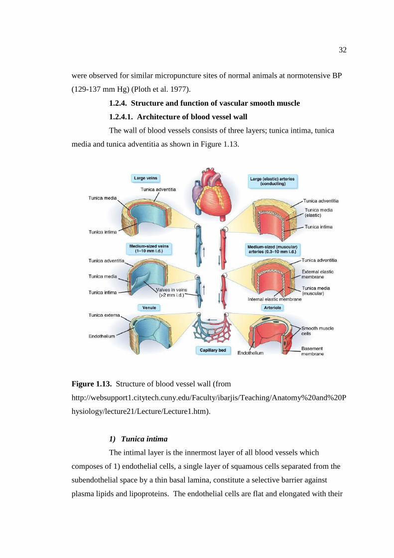
32
were observed for similar micropuncture sites of normal animals at normotensive BP
(129-137 mm Hg) (Ploth et al. 1977).
1.2.4. Structure and function of vascular smooth muscle
1.2.4.1. Architecture of blood vessel wall
The wall of blood vessels consists of three layers; tunica intima, tunica
media and tunica adventitia as shown in Figure 1.13.
Figure 1.13. Structure of blood vessel wall (from
http://websupport1.citytech.cuny.edu/Faculty/ibarjis/Teaching/Anatomy%20and%20P
hysiology/lecture21/Lecture/Lecture1.htm).
1) Tunica intima
The intimal layer is the innermost layer of all blood vessels which
composes of 1) endothelial cells, a single layer of squamous cells separated from the
subendothelial space by a thin basal lamina, constitute a selective barrier against
plasma lipids and lipoproteins. The endothelial cells are flat and elongated with their

33
long axis parallel to that of the blood vessels. They have a thickness of about 0.2-0.5
m, except in the area of nucleus, which makes the endothelial cell protrude slightly,
2) a thin basal lamina with about 80 nm thick and 3) subendothelial layer composes of
collagenous bundles, elastic fibrils, smooth muscle cells and perhaps some fibroblasts.
Such layer is usually present only in the large elastic arteries such as the human aorta,
whereas in the majority of other blood vessels the intima consists of only endothelial
cells and basal lamina.
2) Tunica media
The tunica media is the middle layer of the vascular wall. It is made up of
VSMCs, a varied number of elastic sheets, bundles of collagenous fibrils and a
network of elastic fibrils. This layer is thicker in arteries than in veins; smooth
muscle cells may even be absent in large veins such as the vena cava. In the aorta,
they may reach a thickness of 500 m, whereas it is about 20-50 m in medium-sized
veins. In arterioles, it averages 2-3 layers of smooth muscle cells with an often
incomplete elastic lamina. In venule, it consists of only 1-2 layers of smooth muscle
cells.
3) Tunica adventitia
The tunica adventitia is the outermost layer of the vascular wall. Its
thickness varies considerably depending on the type and location of the blood vessel.
For instance, cerebral blood vessels almost entirely lack an adventitia. In all arteries
and most veins, the adventitia consists of dense fibroelastic tissue without smooth
muscle cells, except in large veins such as the vena cava where there occur bundles of
longitudinally arranged smooth muscle cells. It also harbors the nutrient vessels of
the vascular wall: arterioles, venules, capillaries and lymphatic vessels, collectively
referred to as vasa vasorum. Nerves are also present in this layer. Many of these
nerves are part of the peripheral nerve trunks for sensory organs, muscles and visceral
organs, but some are vasomotor and sensory nerves for the vascular wall itself.

34
1.2.4.2. Vascular smooth muscle constraction
Contraction mechanism
In the intact body, the process of smooth muscle cell contraction is
regulated principally by receptor and stretch activation of the contractile protein
myosin and actin. A change in membrane potential, brought on by the firing of action
potentials or by activation of stretch-dependent ion channels in the cell membrane,
can also trigger contraction. For contraction to occur, myosin light chain kinase
(MLCK) must phosphorylate the 20-kDa light chain of myosin, enabling the
molecular interaction of myosin with actin. Energy released from ATP by myosin
ATPase activity results in the cycling of the myosin cross-bridges with actin for
contraction. Thus contractile activity in smooth muscle is determined primarily by
the phosphorylation state of the light chain of myosin.
Ca2+
-dependent contraction of vascular smooth muscle
Contractile of smooth muscle is initiated by a Ca2+
-mediated change in the
thick filaments, whereas in striated muscle Ca2+
mediates contraction by changes in
the thin filaments. In response to specific stimuli in smooth muscle, the [Ca2+
]i
increases and this activator Ca2+
combines with the acidic protein calmodulin (CaM).
Ca2+
-CaM complex activates MLC kinase to phosphorylate the light chain of myocin.
Cytosolic Ca2+
is increased through Ca2+
release from sarcoplasmic reticulum (SR) as
well as entry from the extracellular space through receptor-operated Ca2+
channels.
Agonists (norepinephrine, AII, ET-1, etc.) binding to serpentine receptors, coupled to
a heterotrimeric G protein, stimulates phospholipase C activity. This enzyme is
specific for the membrane lipid phosphatidylinositol 4, 5-bisphophate to catalyze the
formation of two potent second messengers; inositol triphosphate (IP3) and
diacylglycerol (DAG). The binding of IP3 to receptors on the SR results in the
release of Ca2+
into the cytosol. DAG, along with Ca2+
, activates protein kinase C
(PKC), which phosphorylates specific target proteins. PKC has contraction-promoting
effects such as phosphorylation of L-type Ca2+
channels or other proteins that regulate
cross-bridge cycling. Finally, L-type Ca2+
channel (voltage-operated Ca2+
channels)
in the membrane also open in response to membrane depolarization brought on by
stretch of smooth muscle cell.

35
Ca2+
sensitization mechanism and contraction of vascular smooth
muscle
In addition to the Ca2+
-dependent activation of MLC kinase, the state of
myosin light chain phosphorylation is further regulated by MLC phosphatase which
removed the high-energy phosphate from the light chain of myosin to promote smooth
muscle relaxation. There are three subunits of MLC phosphatase; a 37-kDa catalytic
subunit, a 20-kDa variable subunit and a 110- to 130-kDa myosin-binding subunit.
The myosin-binding subunit, when phosphorylated, inhibits the enzymatic activity of
MLC phosphatase, allowing the light chain of myosin to remain phosphorylated,
thereby promoting contraction. The small G protein RhoA and its downstream target
Rho kinase play an important role in the regulation of MLC phosphatase activity. Rho
kinase, a serine/threonine kinase, phosphorylates the myosin-binding subunit of MLC
phosphatase, inhibiting its activity and thus promoting the phosphorylated state of the
myosin light chain.
Moreover, it is known that receptors activate a heterotrimeric G protein
that is coupled to RhoA/Rho kinase signaling via guanine nucleotide exchange factors
(RhoGEFs). Because RhoGEFs facilitate activation of RhoA, they regulate the
duration and intensity of signaling via heterotrimeric G protein receptor coupling.
There are about 70 RhoGEFs in the human genome and three RhoGEFs have been
identified in smooth muscle; PDZ-RhoGEF, LARG (leukemia-associate RhoGEF)
and p115-RhoGEF. Several recent studies suggest a role for additional regulations of
MLC kinase and MLC phosphatase. Calmodulin-dependent protein kinase II
promotes smooth muscle relaxation by decreasing the sensitivity of MLC kinase for
Ca2+
.

36
Figure 1.14. Mechanism of vascular smooth muscle contraction. Various agonists
bind to specific receptors, subsequently, the response of the cell is to increase PLC
activity via coupling through a G protein. The PLC produces two potent secondary
messengers from the membrane lipid PIP2; DAG and IP3. The IP3 binds to specific
receptors on the SR, ryanodine receptor (RyR), cause a releasing of activator Ca2+
.
The DAG along with Ca2+
activated PKC, which phosphorylates specific target
proteins. The PKC has contractile-promoting effects such as phosphorylation of Ca2+
channels or other proteins that regulate cross-bridge cycling. Ca2+
binds to CaM
leading to activation of MLCK. This kinase phosphorylates the light chain of myocin
in connection with actin and cross-bridge cycling occurs. However, the elevation in
Ca2+
concentration within the cell is transient, and the contractile response is
maintained by a Ca2+
-sensitizing mechanism brought about by the initiation of
myosin phosphatase activity by Rho kinase. This Ca2+
-sensitizing mechanism is
initiated at the same time that PLC is activates and it involves the activation of the
RhoA-GTP. The activation of RhoA by the G protein-couple receptor is not clear but
involves a guanine nucleotide exchange Rho kinase (RhoGEF) and migration of
RhoA to the plasma membrane. Upon activation, RhoA increases Rho kinase activity
leading to inhibition of myosin phosphatase. This promotes the contractile state, since
the light chain of myocin cannot be dephosphorylated (Webb 2003).

37
1.2.4.3. Vascular smooth muscle relaxation
Smooth muscle relaxation occurs either as a result of removal of the
contractile stimulus or by the direct action of a substance that stimulates inhibitions of
the contractile mechanism. The process of relaxation requires a decreased
intracellular Ca2+
concentration and increased MLC phosphatase activity (Morgan
1990; Somlyo et al. 1999). The mechanisms that remove intracellular Ca2+
and/or
increase MLC phosphatase activity may become altered, contributing to abnormal
smooth muscle responsiveness.
A decrease in the intracellular concentration of activator Ca2+
elicits
smooth muscle cell relaxation. Several mechanisms are implicated in the removal of
cytosolic Ca2+
and involve the SR and the cell membrane. Ca2+
uptake into the SR is
dependent on ATP hydrolysis. The SR Ca-Mg-ATPase, when phosphorylated, binds
to Ca2+
ions, which are then translocated to the luminal side of the SR and released.
Mg2+
is necessary for the activity of the enzyme, it binds to the catalytic site of the
ATPase to mediate the reaction.
The cell membrane also contains Ca-Mg-ATPase, providing an additional
mechanism for reducing the concentration of activator Ca2+
in the cell. This enzyme
differs from the SR protein in that it has an autoinhibitory domain that can be bound
by CaM, causing stimulation of the cell membrane Ca2+
pump. Na+/Ca
2+ exchangers
are also located on the cell membrane and aid in decreasing intracellular Ca2+
. ROCC
and VOCC located in the cell membrane are important in Ca2+
influx and smooth
muscle contraction (Webb 2003).

38
Figure 1.15. Mechanism of vascular smooth muscle relaxation. Smooth muscle
relaxation occurs either as a result of the contractile stimuli or by direct action of
substance that stimulates the contractile mechanism. The relaxation requires a
decreased intracellular Ca2+
concentration and increased MLC phosphatase activity.
The SR and cell membrane contain Ca, Mg-ATPases that remove Ca2+
from the
cytosol. Na+/Ca
2+ exchangers are also located on the cell membrane and aid in
decreasing intracellular Ca2+
. During relaxation, ROCC and VOCC in the cell
membrane close resulting in a reduced Ca2+
entry into the cell (Webb 2003).

39
1.2.4.4. Role of endothelium on vascular tone
Endothelial cell is a thin layer of the cell line covers the inner surface of
the blood vessels of the entire circulatory system (about 5,000 m2), from the heart to
the smallest capillary. It comprises about 2% of the mass of the human body
(Eisenberg 1991). The endothelial cells secrete vasodilator mediators in response to
substances released from autonomic and sensory nerves (e.g. ACh, NE, ATP,
substance P), circulating hormones (e.g. catecholamine, vasopressin, AII, insulin),
coagulation derivatives and platelet products (serotonin, ADP, thrombin) or autacoids
produced by endothelial and vascular smooth muscle cells themselves (bradykinin,
ADP/ATP/UTP, AII, endothelin) (Furchgott & Zawadzki 1980; Furchgott 1983;
Vanhoutte 1988; Bassenge & Heuch 1990; Lucher & Vanhoutte 1990). Changes in
shear stress elicit endothelium- and flow-derived vasodilatation. In healthy arteries,
the endothelium presents a non-adhesive anti-thrombotic surface that is critical to
homeostasis (Luscher & Vanhoutte 1990; Vane 1994). Endothelium-derived
vasodilator substances also exert a profound regulatory influence on the thrombolytic
and anti-proliferative functions of the endothelium.
Endothelium-derived vasodilators
Nitric oxide
The endothelium releases a factor that relaxes the underlying VSMC and
this was later shown to be NO (Furchgott & Zawadzki 1980). Endothelial cells
constitutively express a NO synthase (eNOS) that generates NO using L-arginine as a
substrate. NO synthesis is enhanced after Ca2+
-CaM binding. eNOS is an NADPH-
dependent oxygenase that requires tetrahydrobiopterin, FAD and FMN as co-factors
(Moncada et al. 1991). In endothelial cells, the enzyme is localized preferentially in
caveolae following post-translational acylation. eNOS is negatively regulated by
caveolin. Stimulation of endothelial cells by agonists such as bradykinin dissociates
the caveolin/NOS complex and allows binding and activation of NO systhesis by
Ca2+
-CaM (Michel et al. 1997; Feron et al. 1998). Shear stress-induced activation of
eNOS involves a Ca2+
-independent, protein tyrosine kinase-dependent mechanism
(Ayajiki et al. 1996).

40
The relaxation of vascular smooth muscle by NO involves the stimulation
of soluble guanylate cyclase and consequently the increased formation of cyclic GMP
(Rapoport & Murad 1983). The latter activates cyclic GMP-dependent protein kinase
which leads to an increased extrusion of Ca2+
from the cytosol in vascular smooth
muscle and to the inhibition of contractile machinery. Cyclic GMP-dependent protein
kinase phosphorylates K+ channels to induce hyperpolarization and thereby inhibits
vasoconstriction (Lincoln et al. 1994). In arteries, NO activates K+ channels
independently of cyclic GMP (Bolotina et al. 1985). Inhibition of eNOS using
hydrolysis-resistant competitive analogs of L-arginine drastically reduces
endothelium-dependent relaxation (Moncada et al. 1991). NO contributes to the
balance between vasodilator and vasoconstrictor influences that determine vascular
tone.
Prostacyclin (PGI2)
PGI2 is produced by endothelial cells in the vascular wall and acts as
vasodilator (Moncada & Vane 1979). Its vasodilator activity is determined by the
expression of specific receptors in the VSMC (Coleman et al. 1994). Its receptors are
coupled to adenylate cyclase to elevate cAMP levels in VSMC (Kukovetz et al.
1979). Then the cAMP stimulates KATP channels to cause hyperpolarization of the
cell membrane and inhibit the development of contraction (Parkington et al. 1995).
The cAMP also increases the extrusion of Ca2+
from the cytosol in VSMC and
inhibits the contractile machinery (Bukoski et al. 1989; Abe & Karaki 1992). In
addition, PGI2 facilitates the release of NO by endothelial cells (Shimokawa et al.
1988) and the action of PGI2 in VSMC is potentiated by NO (Delpy et al. 1996).
Indeed, increase in cGMP in target cells inhibits a phosphodiesterase that breaks
down cAMP (Weigert et al. 1994; Delpy et al. 1996). Therefore, NO indirectly
prolongs the half-life of the second messenger of PGI2.
Endothelial-derived hyperpolarizing factors (EDHF)
In most medium- to resistance-sized arteries, electrophysiological studies
have established that endothelium-dependent hyperpolarization of vascular smooth
muscle is resistant to the combined inhibition of both NOS and cyclooxygenases. A
component of the endothelium-dependent relaxation in these arteries is mediated by a

41
substance different from NO and prostacyclin. This component of endothelium-
dependent vasodilatation has been ascribed to a diffusible endothelium-derived
hyperpolarizing factor (EDHF), the identity of which is still controversial (Feletou &
Vanhoutte 1996; Mombouli & Vanhoutte 1997). Epoxyeicosatrienoic acids, a
metabolite of arachidonic acid, have been shown to act on large conductance BKCa
channel (Gebremedhin et al. 1992; Hu & Kim 1993; Campbell et al. 1996). EDHF
plays a significant role in the regulation of peripheral vascular resistance and local
hemodynamics by activation of various K+ channels in VSMC (Feletou & Vanhuotte
1988). A combination of charybdotoxin and apamin, a blocker of KCa2+ channels, is
necessary to inhibit the responses attributed to EDHF in rat hepatic and mesenteric
arteries (Corriu et al. 1996) and in guinea-pig carotid arteries (Zygmunt & Hogestatt
1996) whereas apamin alone can inhibit EDHF-induced hyperpolarization in rabbit
mesenteric arteries (Murphy & Brayden 1995). In addition, the synthetic KATP
channel openers also inhibits by closing VOCC (Nelson et al. 1990), impairing the
receptor-dependent activation of PLC and the subsequent release of Ca2+
from
intracellular stores by reducing the Ca2+
sensitivity of the contractile proteins (Itoh et
al. 1992; Itoh et al. 1994). These data suggested that KCa2+ and KATP channels are
possibly involved in activation pathway of the EDHF.
Figure 1.16. Schematic summarizing the release of relaxing factors from endothelial
cells and their effect on vascular smooth muscle cell. The broken line indicates the
action of an inhibitor or antagonist (Mombouli & Vanhoutte 1999).

42
Endothelium-derived constriction factors
Vasoconstrictor prostaglandins
The metabolism of arachidonic acid by COX in endothelial cells may lead
to the secretion of prostaglandin H2 (PGH2), the precursor of all prostanoids including
thromboxane A2 (TXA2) (Moncada & Vane 1979). PGH2 and TXA2 act on
endoperoxide/thromboxane receptors on VSMC to induce vasoconstriction (Halushka
et al. 1989; Coleman et al. 1994). These substances attenuate endothelium-dependent
relaxation induced by ACh in isolated human renal arteries (Luscher et al. 1987a &
b). However, prostacyclin is the major endothelial metabolite of arachidonic acids
that is generated through the COX pathway (Moncada & Vane 1979; Luscher &
Banhoutte 1990). Thus, under normal circumstances, the influence of the small
amounts of TXA2 released by endothelial cells is masked by the production of
prostacyclin, NO and EDHF.
Reactive oxygen species (ROS)
Endothelial cells secrete ROS and hydrogen peroxide (H2O2) in response to
shear stress and endothelial agonists, such as bradykinin (Shimizu et al. 1994). O2-
inactivates NO resulting in vasoconstriction in arteries (Rubanyi & Vanhoutte 1986).
For instance, O2- mediates stretch- or aginist- induced endothelium-dependent
contractions in canine cerebral arteries (Katusic & Vanhoutte 1989). ROS also
facilitate the mobilization of cytosolic Ca2+
in VSMC (Suzuki & Ford 1992) or
promote Ca2+
sensitization of the contractile element (Jin et al. 1991). Since
endothelium-derived O2- is primarily a chemical antagonist of NO, the scavenger of
the free-radical O2- dismutase potentiates endothelium-dependent vasodilatation
(Rubanyi & Vanhoutte 1986).
Endothelin
Endothelial cells synthesize the prohormone big endothelin and express
endothelin-converting enzymes to generate endothelin-1 (ET-1), which is the most
potent vasoconstrictor (Yanagisawa et al. 1988; Masaki. 1995). The secretion of ET-
1 can be induced by thrombin, interleukin-1, transforming growth factor-1, platelet
products and neurohormones such as vasopressin and cathecholamines (Luscher et al.

43
1996). However, the synthesis of ET-1 by endothelial cells is initiated by basal and
stimulated production of NO (Masaki. 1995). The VSMCs express ETB endothelin
receptors type B (ETB) (Masaki 1995). Thus, the direct vasoconstrictors action of ET-
1 is modulated by the stimulation of NO and EDHF release at low concentrations of
the peptide (DeNucci et al. 1988; Nakashima & Vanhoutte. 1993; Masaki 1995).
Endothelial Dysfunction in 2K1C hypertension
Impaired endothelium-dependent vasodilatation was observed in a number
of experimental models of hypertension, including 2K1C hypertension (Lüscher et al.
1987; Dohi et al. 1996; Callera et al. 2000). The biological activity of nitric oxide
(NO), an effective endothelium-derived relaxing factor, is primary associated with
endothelial NO synthase (eNOS) activity or it interaction with superoxide anion,
which is produces in the vascular wall by free radical-generating enzyme (Cai &
Hariison 2000). Endothelium-derived NO may be scavenged by superoxide anion,
causing reduced bioavailability of NO and diminishing vasorelaxation (Griendling et
al. 1994). It is now well established that endothelial dysfunction in hypertension is
partially linked to the exaggerated production of superoxide anion and that oxidative
stress is responsible for impaired endothelial modulation in 2K1C hypertension (Toba
et al. 2012; Arnalich-Montiel et al. 2014; Lerman et al. 2001; Oliveira-Sales et al.
2009).
1.2.5. Arterial baroreceptor reflex
1.2.5.1. Principle of the arterial baroreceptor reflex
As first described by Corneille Heymans, who won the Nobel Prize for
Physiology in 1938, the sensors in the arterial baroreceptor reflex are the
mechanosensitive afferent nerve endings called baroreceptors. These receptors are
located in the carotic artery sinus and aortic arch where they provide instantaneous
feedback on arterial stretch proportional to changes in pressure in arterial lumen.
When the artery is distended due to elevated pressure, the embedded
barosensitive afferent nerve endings are activated. The baroreceptor sensors a stretch
of the vessel wall and changes into electrical signal in the afferent nerves that is
relayed to an integrating center in the brain through baroreceptor afferents from the

44
carotid sinus nerve and aortic depressor nerve synapse in the nucleus of the solitary
tract (NTS). The parasympathetic (vagus nerve) baroreflex is processed through the
nucleus ambiguous (NA) en route to the sinoatrial node of the heart. The sympathetic
baroreceptor reflex pathway continues from the NTS through the caudal ventrolateral
medulla (CLVM) and then to the rostral ventrolateral medulla (RVLM). The
sympathetic outflow then goes to the interomediolateral cell column in the spinal cord
and onto its effector organs (heart and blood vessels) (Wehrwein & Joyner 2013)
(Wehrwein & Joyner 2013).
1.2.5.2. The arterial baroreceptor reflex arc
The baroreceptor reflex arc composes of following components;
1) Baroreceptors and sensory transduction
Baroreceptors are mechanosensitive afferent nerve endings that are
interspersed in the arterial elastic layers. Baroreceptors detect mechanical
deformation of vascular wall stretch due to changes in intraluminal pressure. There
are two primary sites that contain clusters of arterial baroreceptors: the internal carotid
artery at the carotid sinus and the aortic arch as shown in Figure 1.17. A unique
feature of these baroreceptive regions is that the arterial wall has limited collagen and
is highly compliant to allow distention of the wall each phase (Wehrwein & Joyner
2013).
Figure 1.17. The location of the aortic and carotid baroreceptors (Kirkman &
Sawdon 2010).

45
2) Afferent pathways
The electrical response from the baroreceptor is carried centrally to the
medulla via afferent fiber. Information from the carotid baroreceptors towards via
afferent fibers in the carotid sinus nerve (a branch of cranial nerve IX;
glossopharyngeal) and the associated sensory cell bodies exist in the petrosal ganglia.
Information from the aortic arch baroreceptors travels in the aortic depressor nerve (a
branch of cranial nerve X; vagus nerve) with sensory cell bodies located primary in
the nodose ganglia (Wehrwein & Joyner 2013).
3) Central integration
The medulla oblongata is the primary site of cardiovascular and baroreflex
integration. In this region, the NTS serves as the primary site for the first synapse of
the baroreceptor afferents and is the key integrating site for all baroreceptor. The
NTS is a bilateral structure that receives monosynaptic inputs from afferents using
glutamate as the primary neurotransmitter. The NTS integrates and relays
baroreceptor afferent information via a polysynaptic pathway to other important
medullary centers to control parasympathetic and sympathetic pathways to the heart
and blood vessels (Wehrwein & Joyner 2013).
The baroreflex mediated parasympathetic pathways to the heart are
regulated by a simple autonomic reflex arc. Excitatory pathways from the NTS
project onto the cardioinhibitory area containing the NA and dorsal motor nucleus of
the vagus. The cardiac component of the baroreflex is largely mediated in these
regions where activation of vagal preganglionic efferent fibers results in vagal
stimulation to the heart and associated bradycardia (Wehrwein & Joyner 2013).
The sympathetic pathways are more complex, with important control
points in the NTS, CVLM and RVLM. The NTS sends projections to inhibitory
interneurons in the CVLM and also directly to the RVLM. Activation of GABAergic
fibers in the CVLM results in inhibition of sympathetiic excitatory neurons in the
RVLM resulting in a reduction in MABP and sympathetic tone. The RVLM contains
presympathetic neurons that project to the thoracic spinal cord and control
sympathetic preganglionic fibers. Neurons in the rostral RVLM display spontaneous

46
firing with each cardiac cycle and exhibit a variable firing frequency with each pulse
of arterial pressure (Wehrwein & Joyner 2013).
4) Efferent pathway
In the parasympathetic pathway, information from the NA and dorsal
motor nucleus of the vagus control preganglionic fibers that exit the spinal column
and synapse in the parasympathetic ganglia near the heart to activate postganglionic
fibers. The ganglionic synapse and the cardiac postganglionic neuroeffector junction
both rely on ACh as the neurotransmitter acting on 4, 2 nicotinic cholinergic
receptors. Of particular importance relating to the baroreflex is the parasympathetic
vagus nerve which has strong influence on the heart (Wehrwein & Joyner 2013).
In the sympathetic partway, information from the RVLM is passed to the
interomediolateral cell column, sympathetic preganglionic fibers and then into
sympathetic ganglia. ACh acting on 7 nicotinic cholinergic receptors is the
neurotransmitter in sympathetic ganglionic synapses. Sympathetic postganglionic
fibers exit the ganglia and travel to the key baroreflex-associated sites of the heart and
blood vessels. Norepinephrine is the key neurotransmitter released that acts on both
and subtypes of adrenergic receptors on these target organs (Wehrwein & Joyner
2013).
5) Peripheral actions of effector mechanisms
Angiotensin II
In the short-term, the main cardiovascular effects of angiotensin II are the
mediated by the activation of AT1 receptors in the membranes of VSMC. Such
activation elevates levels of intracellular calcium ions and leads to vasoconstriction.
Angiotensin II also increases renal retention of sodium ions and water by acting on
receptors in the proximal and late distal convoluted tubule of the nephron. Moreover,
angiotensin II promotes the synthesis and release of the salt-retaining hormone
aldosterone from adrenal cortical zona glomerulosa cells (Wehrwein & Joyner 2013)
(Wehrwein & Joyner 2013).

47
Activity of parasympathetic nerves
Efferent fibres of the vagus nerve innervate the cardiac pacemaker cell
and the atrial myocytes and conductive tissue of the cardiac ventricles. Action
potentials result in the release of ACh from the terminal butons and subsequent
activation of M2 muscarinic receptors, which leads mainly to a decrease in HR
(Wehrwein & Joyner 2013).
Activity of sympathetic nerves
Action potential is translated into release of adrenaline from chromaffin
cells in the adrenal medulla, renin release from renal juxtaglomerular cells and
noradrenaline release at peripheral synapses, which are found in cardiac myocytes and
the vascular smooth muscle that surrounds blood vessels. Subsequent cardiovascular
effects of adrenaline and noradrenaline are initiates when those catecholamines
activate and adrenergic receptors that are found in cell membranes of the target
tissues. The final effects of increased sympathetic nervous activity are increased heart
rate (1 activation), increased cardiac contractility (1 activation), and increased
vascular resistance (1 activation) (Wehrwein & Joyner 2013) (Wehrwein & Joyner
2013).
Vasopressin
The cardiovascular effects of vasopressin result from the activation of V1
receptors in VSMC and such activation causes vasoconstriction. Vasopressin also
acts on V2 receptors in the collecting duct of the nephron. Such actions results mostly
in the insertion of aquaporins into the luminal membrane and leads to increase
reabsorption of water from the nephrons (Wehrwein & Joyner 2013).

48
Figure 1.18. The arterial baroreceptor reflex arc (Wehrwein & Joyner 2013).
1.2.5.3. Baroreceptor reflex sensitivity (BRS)
BRS, also known as baroreflex gain, is used as a measurement of the
autonomic effector response to a given change in ABP. As an example, when ABP is
changed pharmacologically using the vasoconstrictor phenylephrine (PE) and
vasodilator sodium nitroprusside (SNP), a large range of pressure can be observed.
The changes in pressure are associated with the baroreceptor reflex counterregulation
in HR.
As shown in Figure 1.19, BRS is determined from the slope of the
relationship between MABP (systolic and diastolic arterial pressures or carotid sinus
pressures can also be used) and HR (or sympathetic nerve activity). The HR over a
range of MABP is plotted and analyzed by linear regression. The slope of this
regression line obtained from the linear part of the curve is the BRS. The BRS is high
when the slope is steep (solid line) such that there is a large heart rate response to a
given change in MABP. The BRS is low when the slope is flat (dashed line) such that
there is a minimal HR response to BP change (Wehrwein & Joyner 2013).

49
Figure 1.19. Baroreceptor reflex sensitivity (Wehrwein & Joyner 2013).
1.2.5.4. Baroreceptor reflex resetting in hypertension
An Increase in MABP results in activation of baroreceptors as vessel wall
stretch increases, however, baroreceptor activation declines rapidly within seconds of
an increase in pressure resulting in less afferent activity to the same pressure. When
MABP is acutely or chronically elevated for more that several seconds, there is
peripheral baroreceptor „resetting‟. Resetting refers to a shift in the threshold for
baroreceptor activation leading to a decline in afferent activation observed to a given
elevation in pressure.
As shown in Figure 1.20, the pressure increases from point 1 to point 2
and there is an initial rise in afferent activity. This is followed by a reduction in the
afferent traffic (point 3) that is generated in response to the same elevated pressure.
This blunting of afferent responses is the consequence of resetting. Notably, this new
level of afferent activity after resetting is still higher that at baseline, suggesting that
there is incomplete resetting during this acute rise in pressure. At point 2 on the
graph, the afferent response is nearing the flatter portion of the sigmiodal curve and
this would limit further afferent activation to increasing pressure. The resetting
response allows the baroreceptor reflex to continue to function over a dynamic range
on the linear portion of the curve rather than to have the prevailing pressure shifted to
the flat part of the sigmoidal relationship (Wehrwein & Joyner 2013).

50
Figure 1.20. Acute and chronic resetting of the baroreceptor reflex. Solid line (left
curve): normal response; dashed-dotted line (middle curve): acute resetting; dashed
line (right curve): chronic resetting (Chapleau et al. 1989).
1.2.6. Treatment of hypertension
1.2.6.1. Non-pharmacologic treatment
Life style modification is the first step in the treatment of hypertension; it
includes weight reduction, dietary sodium restriction, regular aerobic exercise,
smoking cessation and reduction in alcohol consumption. In pre-hypertensive
patients with SBP between 120-139 mm Hg and DBP between 80-89 mm Hg simply
making lifestyle changes would delay and possible halt progression to hypertension.
In stage I hypertensive patient, life style changes for 6-12 months might prevent the
necessity for drug therapies and should be encouraged in the absence of
cardiovascular and renal risk factors (Foëx 2004).
1.2.6.2. Pharmacologic treatment
It is difficult to control the ABP at the optimal level in hypertensive
patients through lifestyle changes alone. So treatment with anti-hypertensive drugs
becomes necessary. The classes of anti-hypertensive drugs are discussed following;

51
ACE inhibitors
ACE inhibitors are increasingly being used as first line therapy. They
have a dual mode of action; 1) prevent the production of AII and 2) decrease the
metabolism of vasodilator bradykinin (Delacroix et al. 2014). They have relatively
few side effects and contraindications except bilateral renal artery stenoses. Although
ACE inhibitors are effective in unilateral renovascular hypertension, there is risk of
ischaemic atrophy. Therefore, angioplasty or surgical renal artery reconstruction are
preferable to long-term purely medical therapy. ACE inhibitors are first choice agents
in diabetic and heart failure hypertensive patients. The HOPE trial has shown that
ramipril reduced the risk of cardiovascular events even in the absence of hypertension
suggesting that this ACE inhibitor may exert a protective effect by mechanisms other
than the reduction in blood pressure (Foëx 2004).
AII receptors blockers (ARBs)
ARBs possess a hypotensive effect by specifically binding to AII type 1
receptors and inhibiting the vasoconstriction, fluid retention and sympathomimetic
action mediated by AII. The decrease in ABP induced by ARBs is correlated to an
extent with the renin activity in individual patients. ARBs are used alone or in
combination with diuretics and Ca2+
channel blockers for the treatment of grade I-III
hypertension. ARBs exhibit the cardioprotective effects by inhibiting cardiac
hypertrophy and improving the outcome of heart failure. In kidneys, ARBs dilate
efferent arterioles, reduce the intraglomerular pressure, improve proteinuria and
prevent exacerbation of the renal function in the long term. Moreover, ARBs also
improve the regulation of cerebral blood flow, prevent atherosclerosis, inhibit
occurrence of atrial fibrillation, improve insulin sensitivity and prevent the new
occurrence of diabetes mellitus. Therefore, ARBs are used as the first choice for
patients with complication of the heart, kidney or brain and those with diabetes
mellitus. However, administration of ARBs to pregnant or breast-feeding women or
to patients with severe liver damage is contraindicated, and measures such as reducing
the dose are necessary if the creatinine level is 2 mg per 100 ml. ARBs must not be
used in patients with bilateral renal artery stenosis or patients with one kidney and
unilateral renal artery stenosis, in principle, because of the risk of a rapid decrease in

52
renal function. A decrease in body fluid volume and Na+ deficiency are also quasi-
contraindications. Attention to hyperkalemia is necessary while using ARBs with a
potassium-sparing diuretic (JSH 2009).
β-blockers
High sympathetic tone, angina, and previous myocardial infarction are
good reasons for using β-blockers. However, β-blockers therapy is associated with
symptoms of depression, fatigue, and sexual dysfunction. The β-blockers also exert
adverse effects on glucose and lipid metabolism when used alone or in combination
with diuretics. Therefore, they are not the first choice of treatment in elderly patients
or when hypertension is complicated by other diseases such as diabetes mellitus and
abnormal glucose tolerance. These side-effects have to be taken into consideration in
the evaluation of the benefits of treatment (Foëx 2004).
Calcium channel blockers
Calcium channel blockers produce hypotensive effects by inhibiting the
L-type Ca2+
channel involved in the influx of extracellular Ca2+
ions, thus relaxing the
vascular smooth muscle and reducing TPR. Calcium channel blockers can be divided
into dihydropyridines (DHPs; nifedipine, nimodipine,amlodipine) and non-DHP
(verapamil, diltiazem). DHPs rapidly and potently reduce ABP and show little
cardioinhibitory effect while non-DHP calcium channel blockers have slower and
milder hypotensive effects accompanied by a cardioinhibitory effect. Calcium
channel blockers are effective in the elderly and may be selected as monotherapy for
patients with Raynaud‟s phenomenon, peripheral vascular disease, or asthma, as such
patients do not tolerate β-blockers. Diltiazem and verapamil are contraindicated in
heart failure. Nifedipine is effective in severe hypertension and can be used
sublingually; there is need for caution because of the risk of excessive hypotension.
Calcium channel blockers are often associated with b-blockers, diuretics and/or ACE
inhibitors (Foëx 2004, JSH 2009).

53
Diuretics
Thiazide diuretics are primarily used as antihypertensive drugs. They
produce hypotensive effects by decreasing TPR in the long term, while they reduce
circulating blood volume in the short term by inhibiting Na+ reabsorption by the distal
convoluted tubules. Diuretics are expected to be effective in patients with increased
salt sensitivity, low renin hypertension, kidney disorders, diabetes mellitus and insulin
resistance. While diuretic affect metabolism and may cause hypokalemia, impaired
glucose tolerance and hyperuricemia.
Loop diuretics cause diuresis by inhibiting NaCl reabsorption in the
ascending limbs of the loop of Henle. They have stronger diuretic effects but weaker
hypotensive effects with a shorter duration than thiazide diuretics. As they are also
effective in patients with a reduced renal function, they are used for the treatment of
patients with hypertension and congestive heart failure (JSH 2009).
α-adrenergic blockers
α-blocker selectively block α1-receptors on the smooth muscle side of the
sympathetic nerve terminal. They do not inhibit suppressive α2-receptors on the
sympathetic nerve terminal side and rarely cause tachycardia, especially when they
are the long-acting type. Prazosin is shorter acting than doxazosin, indoramin and
terazosin. They have favorable effects on lipid metabolism, such as decrease in the
total cholesterol and triglyceride levels and increase in the high-density lipoprotein-
cholesterol level. They may cause dizziness, palpitation and syncope due to
orthostatic hypotension (Foëx 2004, JSH 2009).
Classic vasodilators
Classic vasodilators dilate blood vessels by acting directly on the vascular
smooth muscle. Hydralazine and minoxidil are directly acting vasodilators. The
usage of these drugs has declined because of the potential for serious side-effects such
as lupus syndrome with hydralazine and hirsutism with minoxidil. Other adverse
effects are headache, palpitation, tachycardia, angina pectoris and edema. Moreover,
fulminant hepatitis has been reported and hence liver disorder is a contraindication
(Foëx 2004, JSH 2009).

54
Central adrenergic inhibitors
Methyldopa is both a false neurotransmitter and α2-adrenoceptor agonist.
Clonidine and dexmedetomidine, agonists at centrally located α2-adrenoceptors, make
the circulation more stable, reduce the release of catecholamines in response to stress
and cause sedation such that dexmedetomidine is now use for sedation in intensive
care units. Moxonidine is representative of a new class of antihypertensive agents
acting on immidazoline1 receptors (I1). Moxonidine reduces sympathetic activity by
acting on centers in RVLM, thereby reducing TPR (Foëx 2004).
Aldosterone antagonists and potassium-sparing diuretics
These drugs promote Na+ excretion without the loss of K
+ by acting on
the distal convoluted tubules and common collecting ducts. Triamterene produces a
similar effect independently of aldosterone by suppressing the amiloride-sensitive
epithelial Na channel. Aldosterone antagonists are used to treat the low renin
hypertension and to protect the organ damage due to toxic effect of aldosterone.
Clinical studies have shown that the outcome of heart failure or myocardial infarction
is improved by aldosterone antagonists. Aldosterone antagonists may cause
hyperkalemia when used with an RAS inhibitor or in patient with kidney dysfunction.
Spironolactone has adverse effects such as erectile dysfunction, gynecomastia and
menorrhalgia (JSH 2009).
1.2.7. Camboginol and morelloflavone
1.2.7.1. Garcinia dulcis
Garcinia dulcis Kurz, a folk medical plant that belongs to the Guttiferae
family, is widely distributed Southeast Asian countries. It is known as “Maphuut”
(Thailand). It has a medium-size, evergreen, much branched perennial tree, 10-13 m
high, with an upright growth habit. Bark is dark brown and rough and the branches
are green, sparsely hairy angled and orthotropic or horizontal and have a white latex.
The leaves are opposite, simple, large, ovate, oval to oblong up to 10-30 cm long by
3-15 cm wide with acute apex and truncate base, entire margin, glossy green above
and dull pale green below and often sparsely hairy, but reddishpink when young.
Petioles are 1-2 cm long. Flowers are in small densely packed, axillary fascicle of 6-

55
12. Flowers are pentamerous with 5 sepals and 5 yellowish-green or yellowish-white
petals. Male flowers are smaller, 6 mm across with 5 adelphous stamens, yellow
lobed central disk and a rudimentary pistil. Female flower has thick disk lobes, 5-
lobed stigma, sessile and larger than 12 mm across. The fruits are globose to oblate,
5-8 cm in diameter, smooth, green when immature tuning yellow or deep yellow
when ripe. The fruit is thin-skinned with a yellow, fibrous but soft pulp and 1-5 brown
seeds, each 2.5 cm long (Lim 2012).
Figure 1.21. Tree, flower and fruit of Garcinia dulcis.
1.2.6.2. Camboginol
1.2.6.2.1. Chemical structure of camboginol
Camboginol (C38H50O16; molecular weight 602), also known as garcinol,
is a plant benzophenone with a yellow crystalline compound. Rao and colleagues
extracted camboginol from latex of Garcinia cambogia and elucidated its chemical
structure in 1980. Camboginal was also extracted from the rind of the fruit of
Garcinia indica (Yamaguchi et al. 2000 a&b) and the fruits of Garcinia dulcis
(Deachathai et al. 2005). The C-3 ketonic group and the phenolic ring bearing
hydroxyl group (-OH) are the principle oxidation sites of camboginol generating it
oxidized products during metabolic transformations some of which are also
biologically active (Sang et al. 2001; Sang et al. 2002). The 1, 2 carbon-carbon
double bond of , -unsaturated ketone is an important for apoptosis-inducing

56
activity and cytotoxicity of camboginol (Pan et al. 2001). The isoprenyl chain of
camboginol consists of hydrophobic faces which are important for its binging to
biological targets (Roux et al. 2000).
Figure 1.22. Chemical structure of camboginol or garcinol (Rao et al. 1980).
1.2.6.2.2 Biological activities of camboginol
1) Antioxidant activity
Yamaguchi et al. (2000a) reported that camboginol had a property to
retard O2- anion to nearly the same amount as DL-alpha-tocopherol, an established
anti-oxidant using a hypoxanthine/xanthine oxidase system, while its ability to quell
hydroxyl radicals in the Fenton reaction system was even better than that of alpha-
tocopherol. Sang et al. (2001 & 2002) proposed the antioxidant mechanism of
camboginol according to which the compound reacts with peroxyl radicals by a single
electron transfer followed by deprotonation of the hydroxyl group from the enolized
1, 3-diketone to form a resonance pair. Depending on the position of hydroxyl group
(C-3 or C-5) which initiates the reaction, different compounds are formed.

57
2) Anti-inflammatory activity
Liao et al. (2004) reported that camboginol inhibits the expression of
iNOS and COX-2 in lipopolysaccharide (LPS)-activated macrophages. Camboginol
also lowers the LPS-induced increase of intracellular ROS which contribute to the
activation of NF-κB. Hong et al. (2006) found that camboginol modulated
arachidonic acid metabolism by retarding the phosphorylation of cytosolic PLA2
through the inhibition of extracellular ERK1/2 kinase activation and suppression of
iNOS expression through modulation of the JAK/STAT-1 signaling pathway. When
added prior to LPS, cambiginol suppressed NF-κB activation and CO -2 expression
through the interruption of LPS binding to toll-like receptors. Koeberle et al. (2009)
reported that camboginol selectively suppressed PGE2 synthesis and 5-LOX product
formations provides molecular basis for its anti-inflammatory and anti-carcinogenic
effects.
3) Anti-cancer activity
Tanaka et al. (2000) reported that 0.01% and 0.05% dietary camboginol
significantly reduced the formation of azoxymethane-induced colonic aberrant crypt
foci, precursors for colon cancer, in male F344 rats in a dose-dependent manner. In
addition, camboginol also increased liver glutathione S-transferase and quinone
reductase levels, both detoxifying enzymes associated with cancer suppression.
Yoshida et al. (2005) found that camboginol decreased COX-2 expression in tongue
lesions induced with 4-nitroquinoline 1-oxide (4-NQO) in male F344 rats. Hong et al.
(2006) showed that camboginol exerted a much high potency of inhibition against the
colon cancer which was accompanied by increase in Caspase 3 activity, indicative of
an apoptotic pathway. Camboginol also inhibited breast, prostate and pancreatic
cancer cell growth by induction of apoptosis which was mediated by caspase-3
followed by down regulation of the NF-B pathway (Ahmad et al. 2010, Ahmad et al.
2011).

58
4) Anti-HIV activity
Balasubramanyam et al. (2004) reported that camboginol is a potent non-
specific inhibitor of histone acetyltransferases (HAT) p300 which strongly inhibited
HAT activity-dependent chromatin transcription. Since, dysfunction of HAT is
known to cause several diseases including cancer, neurodegenaration, asthma,
diabetes, AIDS and cardiac hypertrophy and p300 protein plays a critical role in cell
growth, differentiation, and death. Mantelingu et al. (2007) shown that camboginol
inhibited histone acetylation of HIV infected cells and consequently inhibited the
multiplication of HIV.
5) Anti-ulcer activity
Yamaguchi et al. (2000a) reported that camboginol has potent free radical
scavenging activity against hydroxyl radical which stronger than that of α-
Tocopherol. Since hydroxyl radical is regarded as the most damaging ROS,
camboginol is expected to be useful for preventing diseases caused by the hydroxyl
radical damages such as stress-induced gastric ulcer (Das et al. 1997; Das et al. 1998)
and NSAID drug-induced gastric ulcers (Vaananen et al. 1991; Yoshikawa et al.
1993).
1.2.6.3. Morelloflavone
1.2.6.3.1. Chemical structure of morelloflavone
Morelloflavone, a plant biflavonoid comprising two covalently linked
flavones; apigenin and luteolin, occurs in most Garcinia species (Verbeek et al 2004).
Its chemical structure was firstly elucidated in 1976 by Ansari et al. from the leaves
of Garcinia dulcis (Roxb.) Kurz. The configuration, substitution and total number of
hydroxyl groups substantially influence several mechanisms of antioxidant activity
such as radical scavenging and metal ion chelation ability. The hydroxyl configuration
is the most significant determinant of scavenging of ROS because it donates hydrogen
and an electron to hydroxyl, peroxyl and peroxynitrite radicals, stabilizing them and
giving rise to a relatively stable flavonoids radical.

59
O
O
O
OH
OH
OH
OH
OH
HO
O HO
Figure 1.23. Chemical structure of morelloflavone (C30H20O11, MW 556), a
biflavonoid comprising two colavently linked flavones; apigenin and luteolin (Ansari
et al. 1976).
1.2.6.3.2. Biological activity of morelloflavone
1) Anti-atherosclerosis activity
Pinkaew et al. (2009) reported that morelloflavone inhibited VSMC
migration, invasion and lamellipodium formation in a cell culture system through
activation of multiple migration-related kinases, including FAK, c-Src, ERK and
RhoA in VSMCs. The inhibition of RhoA and ERK pathways is also observed in
VEGF-stimulated human umbilical cord endothelial cells (Pang et al. 2009). Oral
morelloflavone therapy for 8 months significantly reduced the atherosclerotic areas of
the mouse aortas (a 26% reduction), without changing plasma lipid profiles or weights
in Ldlr-/-
Apobec1-/-
mice (Pinkaew et al. 2012). The immunohistochemistry study
showed that morelloflavone reduced the number of VSMC in the atherosclerotic
lesion while it did not change the density of macrophages in the lesion or the
percentages of proliferating and apoptotic cells (Pinkaew et al. 2012).
2) Anti-cancer activity
Pang et al. (2009) shown that morelloflavone inhibited VEGF-induced
cell proliferation, migration, invasion and capillary-like tube formation of primary
cultured human umbilical vascular endothelial cells in a dose-dependent fashion.
Morelloflavone also inhibited tumour growth and tumour angiogenesis of prostate
cancer cells in xenograft mouse tumor model, suggesting that morelloflavone

60
inhibited tumorigenesis by targeting angiogenesis. The antiangiogenic action of
morelloflavone was established by activation of Rho-GTPases and ERK signalling
pathways.
3) Anti-HIV activity
Lin et al. (1997) found that morelloflavone demonstrated significant
antiviral activity by inhibiting both HIV-1 reverse transcriptase (HIV-1 RT) and HIV-
1 (strain LAV-1) in phytohemagglutinin-stimulated primary human peripheral blood
mononuclear cells.
4) Antioxidant and anti-inflammatory activities
Gil et al. (1997) reported that morelloflavone inhibited secretory
phosphalipase A2 (PLA2) with a high potency on the human recombinant synovial and
bee venom enzymes and its anti-inflammatory activity apparently not related to the
synthesis of eicosanoids but likely dependent the scavenging of ROS. Hutadilok-
Towatana et al. (2007) also found that morelloflavone exhibited strong antioxidation
effects in both Fe2+
-mediated and non-metal induced human low-density lipoprotein
(LDL) oxidations.
5) Hypocholesterolemic activity
Tuansulong et al. (2011) reported that morelloflavone show to have
hypocholesterolemic activity by which affected the HMG-CoA reductase, the rate-
limiting enzyme of the cholesterol biosynthetic pathway. Morelloflavone inhibited
the enzyme activity by competing with the HMG-CoA whereas it was non-
competitive towards NADPH.
6) Vasorelaxant activities
Lamai et al. (2013) reported that morelloflavone induce the dilatation of
thoracic vessels from normotensive rat and the machanisms of action is endothelium-
dependent which involved nitric oxide signaling pathway and partially KATP and KCa2+
channels.

61
1.3. Objectives
1.3.1. To investigate effect of camboginol and morelloflavone on ABP,
HR, renal clearance, BRS, plasma malondialdehyde (MDA) level and endothelium
eNOS expression in anesthetized 2K1C hypertensive and sham operative (SO)
normotensive rats.
1.3.2. To study vasorelaxant effect of camboginol and morelloflavone on
isolated thoracic aortic ring from 2K1C and SO rats. The vasorelaxant mechanisms of
camboginol including 1) NO generation, 2) PGI2 synthesis, 3) activation of KATP
channel and 4) activation of KCa channel were also elucidated using specific
vasorelaxant inhibitors; Nω-Nitro-L-arginine methyl ester (L-NAME), indomethacin,
glibenclamide and tetraethylammonium, respectively.
1.4. Hypotheses
1.4.1. Camboginol and/or morelloflavone treatment exert hypotensive
and/or diuretic effect in anesthetized SO normotensive and/or 2K1C hypertensive rats.
They may improve the BRS, decrease plasma MDA levels and increase eNOS
expression on vascular endothelium in both or some group of rats.
1.4.2. Camboginol and/or morelloflavone treatment express vasodilatation
effect in isolated aortic ring from SO and/or 2K1C rats and mechanism of their action
may associated endothelial dependent vasorelaxant pathways.

CHAPTER 2
MATERIALS AND METHODS
2.1. Extraction of camboginol and morelloflavone
The extraction of camboginol and morelloflavone was performed as
previously described (Deachathai et al. 2005). Briefly, the fresh ripe fruits of G.
dulcis were collected from Songkhla province, Thailand. The voucher specimen has
been deposited at the Herbarium of Prince of Songkla University, Thailand. The
fruits were washed and chopped in to small pieces and then immersed in acetone
(Me2CO) for 5 days at room temperature. Acetone was removed by evaporation to
give a liquid extract that was further partitioned with hexane to yield a solid extract.
To purify camboginol, the solid extract was subjected to column
chromatography (CC) and eluted with a gradient of dichloromethane (CH2Cl2) and
methanol (MeOH). The collected fractions were combined according to their thin
layer chromatography (TLC) characteristics and fractionated by CC using CH2Cl2 and
CH2Cl2-Me2CO as eluents to yield camboginol (MW 602).
In case of morelloflavone, the solid was further fractionated by dissolving
in CH2Cl2 to give CH2Cl2 soluble fraction. This fraction was fractionated by quick
CC and was subjected to CC using CH2Cl2- MeOH to yield morelloflavone (MW
552).
2.2. Animals and experimental design
2.2.1. Animals
Male Wistar rats (body weight 150-200 g, n=96) were obtained from the
Southern Laboratory Animal Facility (Prince of Songkla University, Thailand). Rats
were housed under control conditions (temperature 23-24C; humidity 50-55%;
lighting 0600-1800 h), fed a laboratory diet containing 34.2 mmol sodium chloride/kg
dry weight food (S.W.T., Thailand) and were allow free access to reverse osmosis

63
water. All experiments were approved by the Prince of Songkla University Animal
Ethics Committee (Reference No. 31/2014).
There are two part of experiment in this study, the in vivo (n = 36) and in
vitro (n =60). Each part consist of six groups of rats (n = 6, each), namely SO +
Vehicle (SO), SO + camboginol (SO+C), SO + morelloflavone (SO+M), 2K1C +
Vehicle (2K1C), 2K1C + camboginol (2K1C+C) and 2K1C + morelloflavone
(2K1C+M).
In a part of immunohistochemistry, the animals (n=10) were purchased
from the National Laboratory Animal Center (Mahidol University, Thailand). The
animals were divided into 4 groups; control (n=2), 2K1C (n=2), 2K1C+C (n=3) and
2K1C+M (n=3). Rats were induced hypertension and housed under control
conditions as mentioned above at the Animal Facility of Department of Zoology,
Kasetsart University. Collection of thoracic aorta was performed four weeks after the
induction of hypertension.
2.2.2. Establishment of 2K1C hypertensive rat
Rats were anaesthetized with a single dose of pentobarbital sodium (50
mg/kg BW i.p.). Only the deep sedated animals were gone through the following
surgical protocol; the left kidney was exposed through a 1 cm retroperitoneal incision
the left renal artery was then exposed and cleared from surrounding connective
tissues, and a U-shaped silver clip with a 0.20 mm gap was placed around it close to
the junction with the abdominal aorta. The free edges of the clip were compressed
firmly to prevent dislodging. At the end of surgery, the muscle and skin layer were
sutured separately with catgut and silk No. 4/0, respectively. The SO included the
entire surgery with the exception of renal artery clipping. At the end of the surgery,
all animals received a single dose of ampicillin (50 mg/kg BW, i.m.) injection and
were allowed to recover in separate cages for 2-3 h under an angle poise lamp and
remained untouched for 4 weeks afterward in order to develop hypertension.

64
2.3. Chemical reagents
- ABC peroxidase staining kit, Thermo Scientific, Rockford, IL, USA
- Acetic acid (CH3COOH), Merck, Darmstadt, Germany
- Acetylcholine (ACh) chloride, Sigma Chemical Co., St. Louis, MO, USA
- Ammonium sulfate (H6N2O3S), Fluka, Buchs, Switzerland
- Anthrone (C14H10O), Fluka, Buchs, Switzerland
- Calcium chloride dehydrate (CaCl22H2O), Merck, Darmstadt, Germany
- D(+)-Glucose anhydrous (C6H12O6), Fluka, Buchs, Switzerland
- Dimethylsulfoxide (DMSO), Sigma Chemical Co., St. Louis, MO, USA
- Di-sodium hydrogen phosphate anhydrous (Na2HPO4), Catlo Erba Reagenti,
Milan Italy
- eNOS antibody, Thermo Fisher, Rockford, IL, USA
- Glibenclamide, Sigma Chemical Co., St. Louis, MO, USA
- Goat-anti-Rabbit IgG (H+L) Secondary Antibody, Biotin, Thermo Fisher,
Rockford, IL, USA
- Heparin, Leo pharmaceutical, Ballerup, Denmark
- Hydrochloric acid (HCl), Merck, Darmstadt, Germany
- Indomethacin, Sigma Chemical Co., St. Louis, MO, USA
- Inulin, Sigma Chemical Co., St. Louis, MO, USA
- Magnesium sulfate heptahydrate (MgSO47H2O), Merck, Darmstadt, Germany
- Metal Enhanced DAB Substrate Kit, Thermo Scientific, Rockford, IL, USA
- n-Butanol (CH3(CH2)3OH), Lab-Scan Analytical Science, Bangkok, Thailand
- N-(1-naphthyl)-ethylenediamine dihydrochloride (C12H16Cl2N2), Merck,
Darmstadt, Germany
- Normal goat serum (NGS), Novex® by Life Technologies Corporation,
Frederick, MD, USA
- Nω-Nitro-L-arginine methyl ester (L-NAME) , Sigma Chemical Co., St.
Louis, MO, USA
- Para-aminohippuric acid (PAH), Sigma Chemical Co., St. Louis, MO, USA
- Pentobarbital sodium, CEVA Santé Animal, Brussels, Belgium
- Phenylephrine (PE), Sigma Chemical Co., St. Louis, MO, USA
- Potassium chloride (KCl), Merck, Darmstadt, Germany

65
- Sodium chloride (NaCl), Ajax Finechem, Seven Hill, New South Wales,
Australia
- Sodium dodecyl sulfate (SDS, C12H25NaO4S), Fluka, Buchs, Switzerland
- Sodium dihydrogen phosphate dehydrate (NaH2PO42H2O), Merck,
Darmstadt, Germany
- Sodium hydrogen carbonate (NaHCO3), Riedel-de, Seelze, Denmark
- Sodium hydroxide (NaOH), Carlo Erba Reagenti, Milan, Italy
- Sodium nitrite (NaNO2), Carlo Erba Reagenti, Milan, Italy
- Sodium nitroprusside (SNP), Sigma Chemical Co., St. Louis, MO, USA
- Sulfuric acid (H2SO4), Merck, Darmstadt, Germany
- Tetraethyl ammonium (TEA), Sigma Chemical Co., St. Louis, MO, USA
- Thiobarbituric acid (TBA, C4H4N2O2S), Sigma, Deisenhofen, Germany
- Trichloroacetic acid (CCl3COOH), Carlo Erba Reagenti, Milan, Italy
2.4. Equipment
- Automatic pipettes, Eppendorf, Geritebau, Hamburg, Germany
- Balance, Model CC023D10ADBAAA, Avery Barkel, United Kingdom
- Centrifuge, Model 4232, A.L.C., Italy
- Digital infusion syringe pump, Model SP101i, World Precision Instruments,
Sarasota, Florida, USA
- Force transducer, Model FT03, Grass, USA
- Homeothermic blanket control unit, Harvard, Southnatick, Massachusetts,
USA
- IEC Micro-Hematocrit centrifuge, Model MB, Needham Heights,
Massachusetts, USA
- Microscope, Olympus DP 73, Shinjuku, Tokyo, Japan
- Osmometer, Fiske Model 210, USA
- pH meter, Model UB-5, Denver Instruments, Denver, USA
- Polyethylene tube, Clay Adams, Parsippany, New Jersey, USA
- PowerLab, ADInstruments, USA
- Spectrophotometer, Model Spectro SC, Labomed, Culver City, California,
USA

66
2.5. The in vivo study of renal clearance and BRS
Four weeks after the operation, renal clearance and BRS were studied in
four group of animals (n=6, ea). Rats were anesthetized with pentobarbital sodium
(60 mg/kg BW i.p.; an additional dose was given when necessary) and placed on a
thermostatically-controlled heated table to maintain body temperature at 37ºC. A
tracheotomy was performed and the left carotid artery was cannulated, using
polyethelene tube-50 (PE-50) filled with heparinized 0.9% NaCl, and connected to a
pressure transducer, coupled to a PowerLab system, to measure ABP and HR, and for
blood sampling. The right jugular vein was cannulated, using PE-50, and infused
with clearance markers containing 1% inulin and 0.5% PAH dissolved in 0.9% NaCl
at a rate of 1.6 mL/h/100g BW. Via a suprapubic midline incision, the left and right
ureters were cannulated, using PE-10 connected to PE-50 for urine sample collection.
Camboginol or morelloflavone dissolved in DMSO was given via the jugular vein as
a bolus injection (0.1 mg/kg BW; volume of injection was 0.05 mL/100g BW) and
then followed by continuous infusion at the rate of 5 g/min/kg BW along with the
clearance markers. This experimental dose of camboginol and morelloflavone was
chosen base on the minimal effective dose of a specific AII receptor antagonist,
candesartan (Hiranyachattada et al. 2005). The same amount of DMSO was given as
vehicle in both SO and 2K1C groups.
After 45 min infusion, the renal clearances of particular marker and
osmolar clearance were determined. Three consecutive 20 min urine collections were
made during the 60 min of experimental period. An arterial blood sample (400 µL)
was taken at the midpoint of the first and third urine collecting period. A small
amount of the blood sample was used to determine the hematocrit by the
microcapillary method, and the remainder was centrifuged and the plasma was
collected for determination of the concentrations of inulin and PAH. Blood cells were
resuspended in 200 µL 0.9% NaCl and returned to the animal via the right jugular
vein. Urine flow rate ( was determined gravimetrically by collecting urine into pre-
weighed tubes and assuming a density of 1 g/mL. The PAH and inulin
concentrations, either in plasma or urine samples, were determined by a
spectrophotometric method according to Smith et al. (1954) and Davidson and
Sackner (1963), respectively. Plasma and urine osmolality (POsm and UOsm) were

67
measured by using Micro-Osmometer, and then osmolar and negative free water
clearance (COsm and TCH2O) were calculated.
The BRS determination was performed after the 60 min of renal clearance
study by measuring HR and ABP before and after acute injection of the two
vasoactive drugs, PE (a specific 1 receptor agonist) and SNP (a NO donor) at the
same doses of 1, 2, 4, 8, 16 and 32 μg/kg BW. The maximal response in MABP and
HR were then determined.
At the end of experiment, a large blood sample was collected via carotid
artery catheter in heparinized tube and was centrifuged at 4000 rpm for 10 min then
plasma was stored at -20C until analysis for plasma MDA level. Then, rats were
killed by intravenous injection of saturated MgSO4. Both kidneys were removed,
decapsulated, dried on blotting paper and weighed. The heart was also removed and
weighed after carefully clearing away blood vessels, fat and connective tissues. The
organ weight to total body weight ratio was calculated.
2.6. Determination of plasma malondialdehyde (MDA)
The MDA content was assayed in the form of TBA reacting substances
(modified from Ohkawa et al. 1979. Briefly, 0.8 mL of plasma sample, distilled
water, and MDA standard was added into tubes followed by 0.2 mL of 8.1% SDS and
1.5 mL of 20% acetic acid. The mixture was mixed well and was adjusted the pH to
3.5 with 1.5 N NaOH, then 1.5 mL of 0.8% TBA was added, made up to 4.0 mL with
distilled water and mixed well. The mixture was heated in 95C water bath for 60 min
and was cooled down in ice-bath. Distilled water (1.0 mL) and n-butanol (5.0 mL)
were added into the mixture. After shake vigorously, the mixture was centrifuged at
4,000 rpm for 10 min. The absorbance of the upper organic layer was measured at
532 nm using spectrophotometer. The amount of TBA reactive substances was
determined from standard curve generation by MDA from acid hydrolysis of 1, 1, 3,
3-tetramethoxypropane. The values of plasma MDA were expressed as µmol/L.

68
2.7. The in vitro study of thoracic aortic ring relaxation
2.7.1. Preparation of isolated thoracic aortic ring
Separate groups of 2K1C and SO rats (n=6, ea) were anesthetized with
pentobarbital sodium (60 mg/kg, i.p.) and sacrificed by decapitation. The thoracic
aorta was dissected and cut into four ring segments approximately 5 mm in length
each (two intact- and two denuded-endothelial rings). The denuded rings were
performed by mechanical removal of endothelium. The rings were mounted in 20 mL
organ baths containing 37C Krebs Henseleit solution which was composed of (mM)
118.41 NaCl, 4.6 KCl, 1.12 MgSO4.7H2O, 1.18 KH2PO4, 1.9 CaCl2, 25.0 NaHCO3
and 11.66 D-glucose. The pH of solution was maintained at 7.4 by continuous
aeration with 95% O2 and 5% CO2. The resting tension of 1 g was set and the tension
changes during the courses of experiment were recorded using force displacement
transducer connected to PowerLab system. Endothelial function of aortic rings was
tested by the addition of 10-5
M ACh into the 10-7
M PE precontracted rings. The
80% relaxation was accepted and considered as an intact endothelium and the
disappearance of relaxation was considered denuded endothelium (Molina et al.
1992).
2.7.2. Concentration-response curve of camboginol and
morelloflavone
After 45 min equilibration, aortic rings (both intact and denuded
endothelium) from 2K1C and SO rats were precontracted by addition of 10-7
M PE.
When the maximal contraction response developed, the tension was recorded.
Camboginol, morelloflavone or vehicle (0.1% DMSO) was added cumulatively,
allowing the final concentration to be 10-13
-10-5
M. Subsequent concentrations were
added after the maximal response by the previous concentration developed and
recorded.
2.7.3. Effect of specific inhibitors on vasorelaxation response of
camboginol or morelloflavone
After 15 min equilibration, the intact-endothelial aortic rings from 2K1C
and SO rats were incubated with each specific inhibitor for 30 min before

69
precontraction with 10-7
M PE. The doses of each inhibitor were 10-4
M L-NAME,
10-6
M indomethacin, 10-5
M glibenclamide and 10-3
M TEA. Then, camboginol (10-
13-10
-5 M), morelloflavone (10
-13-10
-5 M) or vehicle (0.1% DMSO) was added
cumulatively after the maximal contraction was developed and 5-7 min sustained.
Subsequent concentrations were added after the maximal response by the previous
concentration developed and recorded.
2.8. Immunohistochemistry
One hour after camboginol or morelloflavone (0.1 mg/kg + 5 µg/min/kg)
was given via jugular vein of anesthetized 2K1C rat, it was then terminated by
overdose injection of pentobarbital sodium and thoracic aorta was removed, dissected
and cut into segment (5 mm each). The segment was fixed in 4% buffered
formaldehyde overnight and then was transferred to store in cryoprotectant solution at
-20C until analysis. The segment was embedded in Tissue Freezing Medium® and
the sections of 10 m were cut on a cryostat. The eNOS expression in aortic sections
was evaluated using floating-technique immunohistochemistry. The sections were
washed by allowing the section to swirl in the immunobuffer (IB; 0.1 M Tris
phosphate buffer solution (TPBS) + 0.3% Triton-X) for 10 min (3 times). These
sections were transferred to incubate in 3% peroxidase blocking solution for 15
minutes with gentle agitation and were washed with IB for 10 min (3 times). The
sections were blocked with 10% NGS for 30 minutes and then were incubated with
rabbit anti-eNOS antibody (dilute 1:500, Lot # QJ214409) at 4C for 24 h on the
shaker. The sections were washed with TPBS for 10 min (3 times). Next, sections
were incubated with biotinylated goat anti-rabbit IgG (dilute 1:2000, Lot #
QE215187) for 1 h at room temperature and were protected from light. The sections
were washed with TPBS for 10 min (3 times), and thereafter the sections were stained
using ABC peroxidase staining kit and metal enhanced DAB substrate kit,
respectively, and ensured these sections were covered from light. Finally, the
sections were mounted onto slide, allowed to dry, protected from light, and covered
slip section with Per Mount. Sections were digitized using an Olympus DP 73
microscope.

70
2.9. Calculations
2.9.1. Mean arterial blood pressure (MABP)
MABP is calculated by the following equation.
B 1
3( B B ) B
1
3
When MABP = Mean arterial blood pressure (mm Hg)
DBP = Diastolic blood pressure (mm Hg)
SBP = Systolic blood pressure (mm Hg)
PP = Pulse pressure (mm Hg)
2.9.2. Baroreflex sensitivity (BRS)
BRS is calculated by the following equation.
B
When HR = Changed heart rate (bpm)
MABP = Changed mean arterial blood pressure (mm Hg)
2.9.3. Clearance of PAH (CPAH)
CPAH was used to estimate renal plasma flow (RPF). Theoretically, if the
substance X is completely cleared from the plasma the amount of substance X
delivered to kidneys in blood (RPF x PX) were equaled to the amount extracted in
urine (UX x ) and therefore the clearance rate of that substance is equal to the RPF
(Guyton and Hall, 2006). However, no one known substance completely cleared from
plasma. PAH is about 90% cleared from the plasma via either glomerular filtration of
tubular secretion. Therefore, the clearance of PAH can be used as an approximation
of RPF. The reasons that PAH is not completely cleared from the plasma may result
from the fact that some blood flows through areas of the kidney that do not remove

71
PAH (e.g. renal capsule, renal pelvis, and perirenal fat), the circulation in some areas
such as the outer medulla does not come into intact with the tubular epithelium and
some tubular epithelium does not remove PAH completely. Therefore, the CPAH is
measure from the flow in areas that effective removes PAH from plasma, thus
determined is called the effective renal plasma flow (ERPF) (Valtin and Schafer,
1995). PAH clearance is calculated according to the following equation.
ERPF was estimated by CPAH and is not exact RPF. However, RPF can be
calculated from the knowledge that only about 90% of PAH is cleared from the
plasma. It is refer that ERPF is lower than RPF by 10%, and therefore RPF can
calculated by the following equation.
RPF = ERPF + 0.1ERPF
When RPF = Renal plasma flow (mL/min)
ERPF = Effective renal plasma flow (mL/min)
2.9.4. Ranal vascular resistance (RVR)
RVR is calculated by following equation.
When RVR = Renal vascular resistance (mm Hg/mL/min)
MABP = Mean arterial blood pressure (mm Hg)
EPPF = Effective renal plasma flow (mL/min)

72
2.9.5. Clearance of inulin (Cin)
Cin is used to estimate the GFR. Inulin is a substance that freely filtrated
via glomerulus and is not reabsorbed or secreted by renal tubules. Therefore, the rate
of inulin excreted in urine (Uin x ) is equal to the filtration rate of inulin by kidney
(GFR x Pin) and thus, GFR is equal to Cin (Guyton and Hall, 2006) as shown in below
equation.
in in
in
When Cin = clearance of inulin (mL/min)
GFR = Glomerular filtration rate (mL/min)
Uin = concentration of inulin in urine (mg%)
Pin = concentration of inulin in plasma (mg%)
= urine flow rate (mL/min)
2.9.6. Urine flow rate ( )
was determined gravimetrically by collecting urine into pre-weighed
tube and assuming a density of 1 g/mL. The is calculated by the following
equation.
Weigh of ine in min e
min e
When = Urine flow rate (mL/min)
2.9.7. Filtration fraction (FF)
FF is the ratio of the GRF to the RPF. The FF represents the proportion of
the fluid reaching the kidneys which passes into the renal tubules. The FF is
calculated by the following equation.

73
When FF = Filtration fraction
GFR = Glomerular filtration rate (mL/min)
RPF = Renal plasma flow (mL/min)
2.9.8. Osmolar clearance (COsm)
COsm is the theoretical volume if plasma per unit time that the kidney has
to filter in order to extract all the solutes out of that volume of plasma. The COsm is
calculated by following equation.
sm sm
sm
When COsm = Osmolar clearance (mL/min)
Uin = urine osmolarity (mOsm)
Pin = Plasma osmolarity (mOsm)
= urine flow rate (mL/min)
Moreover, fractional of salt excretion is also calculated by the
following equation.
a iona of sa e e ion sm
When FOsm = Fractional of osmolality
COsm = Osmolar clearance (mL/min)
GFR = Glomerular filtration rate (mL/min)

74
2.9.9. Free water clearance (CH2O)
CH2O is the theoretical volume of plasma that the kidney has to filter in a
certain amount of time in order to extract all the free water from that volume of
plasma. The CH2O is calculated by following equation.
2 sm
When COsm = Osmolar clearance (mL/min)
CH2O = Free water clearance (mL/min)
= urine flow rate (mL/min)
2.9.10. Contraction responses of thoracic aorta
Contraction response of thoracic aortic ring to PE is calculated by the
following equation.
on a ion o (
) 100
When TPE = Tension in response to PE (g)
Tr = resting tension (g)
2.9.11. Relaxation responses of thoracic aorta
Relaxation response of PE-preconstricted thoracic aortic ring to either
camboginol (C), morelloflavone (M), vehicle (DMSO), acethylcholine (ACh), or
sodium nitro prusside (SNP) is calculated by the following equation.
e a a ion o ( )
100
When TPE = Tension in response to PE (g)
Tr = resting tension (g)
Tx = Tension in response to X (g)

75
2.10. Statistical analyses
For in vivo study, all data were expressed as the meanS.E.M.
Comparison between the mean values were performed with one-way analysis of
variance (ANOVA) (GraphPad Prism 5, San Diego, CA, USA) followed by Student-
Newman Keuls post hoc test or Student t-test. A p < 0.05 was considered to be
statistically significant difference.
For in vitro study, the degree of vasorelaxation in each experiment were
expressed as a %relaxation from PE (10-7
M) precontraction tension. All values were
expressed as mean±S.E.M. Significant difference between the group means was
determined using ANOVA followed by Student-Newman Keuls post hoc test or
Student t-test. The negative logarithm (pD2) and half maximal effective concentration
(EC50) values were calculated using GraphPad Prism 5. Statistical significance of the
mean differences were accepted when p < 0.05.

CHAPTER 3
RESULTS
3.1. Effects of camboginol
3.1.1. Effects of camboginol on ABP and HR
As shown in Figure 3.1 and Figure 3.2, four weeks after experimental
renal stenosis, the basal SBP, DBP, PP and MABP in 2K1C rats were significantly
higher than in those of SO rats (SBP; 208±8 vs. 155±4, DBP; 143±4 vs. 123±4, PP;
64±7 vs. 32±3 and MABP; 165±5 vs. 134±4 mm Hg, respectively, p < 0.05). The HR
of 2K1C rats was not different from SO rats (212±5 vs. 210±5 bpm). The data
suggested the successful development of 2K1C hypertensive model.
When 0.1 mg/kg BW camboginol was injected in 2K1C rats, the SBP,
DBP, MABP and HR decreased significantly when compared to vehicle injection
(SBP; 118±9 vs. 208±9, DBP; 62±5 vs. 144±5, MABP; 81±4 vs. 165±5 mm Hg and
HR; 178± 3 vs. 212 ± 5 bpm, respectively, p < 0.05, Figure 3.2). These data
suggested that camboginol possess an acute bradycardic and hypotensive effect in
2K1C rats.
The bolus injection of 0.1 mg/kg BW camboginol in SO rats also showed
a significantly decrease in SBP, DBP, MABP and HR in comparison to the vehicle
injection (SBP; 104±3 vs. 153±4, DBP; 51±4 vs. 120±3, MABP; 69±3 vs.131±3 mm
Hg and HR 154± 9 vs. 210 ± 5 bpm, respectively, p < 0.05, Figure 3.2). These data
suggested that camboginol also possess an acute bradycardic and hypotensive effect
in SO rats.
However, the MABP between camboginol- (0.1 mg/kg BW + 5 g/min/kg
BW) and vehicle-treated 2K1C rats during the renal clearance study did not different
(152±4 vs. 152±2 mm Hg, respectively; Figure 3.3a).

77
Figure 3.1 Tracing of recorded arterial blood pressure (ABP, red) and heart rate (HR,
blue) of 2-kidneys-1-clip (2K1C; upper panel) and sham operative (SO; lower panel)
rat.

78
Figure 3.2. Effects of camboginol (C; 0.1 mg/kg BW) on systolic blood pressure
(SBP; a), diastolic blood pressure (DBP; b), pulse pressure (PP; c) and mean arterial
blood pressure (MABP; d) in the 2-kidneys-1-clip (2K1C) and sham operation (SO)
groups.
Data are mean ± S.E.M. *, #
p < 0.05 compared with SO and 2K1C group, respectively
(one-way ANOVA with Newman-Keuls post hoc test).
79
3.1.2. Effects of camboginol on renal clearance
As shown in Figure 3.3 and Table suppl. 1, 2K1C rats had significantly
higher RVR, FF, , COsm, COsm/GFR and TCH2O when compared to SO rats (RVR;
70±6 vs. 39±4 RU, FF; 0.63±0.10 vs. 0.36±0.02, ; 11.5±0.7 vs. 8.8±1.0 μL/min/g
KW, COsm; 75.6±6.2 vs. 51.1±4.7 μL/min/g KW, COsm/GFR; 6.25±0.93 vs.
3.79±0.48% and TCH2O; 56.2±3.5 vs. 41.0±4.1 μL/min/g KW, respectively, p < 0.05)
but had a significantly lower ERPF (2.29±0.25 vs. 3.63±0.36 mL/min/g KW, p <
0.05). However, there was no significant difference of GFR, UOsm and POsm between
2K1C and SO rats (GFR; 1.52±0.20 vs. 1.43±0.12 mL/min/g KW, UOsm; 1240±49 vs.
1425±46 mOsm/kg H2O and POsm; 330±4 vs. 326±3 mOsm/kg H2O, respectively).
When camboginol (0.1mg/kg + 5 g/min/kg BW) was given in 2K1C rats,
the RVR, UOsm and POsm significantly decreased in comparison to vehicle-treated
2K1C (RVR; 41±3 vs. 70±6 RU, UOsm; 558±30 vs. 1240±49 mOsm/kg H2O and POsm;
310±2 vs. 330±4 mOsm/kg H2O, respectively, p < 0.05). However, the ERPF, GFR,
, COsm, COsm/GFR and TCH2O were found to be significantly higher (ERPF;
3.88±0.37 vs. 2.29±0.25 mL/min/g KW, GFR; 2.03±0.05 vs. 1.52±0.20 mL/min/g
KW; ; 68.1±5.1 vs. 11.5±0.7 μL/min/g KW, COsm; 154.0±6.3 vs. 75.6±6.2 μL/min/g
KW, COsm/GFR: 9.28±0.48 vs. 6.25±0.93% and TCH2O; 65.9±2.2 vs. 56.2±3.5
μL/min/g KW, respectively, p < 0.05).
The administration of camboginol (0.1mg/kg + 5 g/min/kg BW) in SO
rats resulting in a significantlt increased in GFR, FF, and COsm (GFR; 2.14±0.07 vs.
1.43±0.12 mL/min/g KW, FF; 0.55±0.05 vs. 0.36±0.02, ; 42.5±5.4 vs. 8.8±1.0
μL/min/g KW and COsm; 78.3±7.5 vs. 51.1±4.7 μL/min/g KW, respectively, p < 0.05)
but a significantly decreased in UOsm and POsm (UOsm; 593±29 vs. 1425±46 mOsm/kg
H2O and POsm; 311±3 vs. 326±3 mOsm/kg H2O, p < 0.05) when compared to vehicle-
treated SO rats.
The data suggested that the decreased RBF of 2K1C rats was due to the
renal vasoconstriction. Renal tubular water and electrolytes excretion of 2K1C rats
were also impaired. Camboginol treatment can corrected this impairment of both
RBF and renal electrolytes excretion in 2K1C rats. The diuretic effect of camboginol
was also observed in SO rats.

80
Figure 3.3. Effects of camboginol (C; 0.1mg/kg + 5 g/min/kg BW) on mean arterial
blood pressure (MABP; a), effective renal plasma flow (ERPF; b), renal vascular
resistance (RVR; c), glomerular filtration rate (GFR; d), urine flow rate ( e), urine
osmolality (UOsm; f), osmolar clearance (COsm; g) and negative free water clearance
(TCH2O; h), in 2-kidneys-1-clip (2K1C) and sham operative (SO) group during
clearance study.
Values are mean ± S.E.M. *, #
p < 0.05 compared with SO and 2K1C group,
respectively (one-way ANOVA with Newman-Keuls post hoc test).

81
3.1.3. Effects of camboginol on BRS
As shown in Figure 3.4a, c and e, the BRS responses to the six doses of
PE (1, 2, 4, 8, 16 and 32 g/kg BW) intravenous injection in 2K1C were significantly
lowered when compared to those of SO rats (-0.11±0.04 vs. -0.61±0.13, -0.16±0.04
vs. -0.55±0.10, -0.22±0.05 vs. -0.60±0.08, 0.21±0.03 vs. -0.63±0.05, -0.20±0.03 vs.
0.78±0.08 and -0.23±0.04 vs. -1.03±0.09 bpm/mm Hg, respectively, p < 0.05). There
was no significantly difference in respective BRS to PE between camboginol- and
vehicle-treated SO rats. Camboginol treatment resulting in a significantly higher BRS
in 2K1C compared with vehicle-treated rats at the respective doses of PE injection (-
0.40±0.04 vs. -0.11±0.04, -0.41±0.07 vs. -0.16±0.04, -0.49±0.04 vs. -0.22±0.05, -
0.71±0.17 vs. 0.21±0.03, -1.26±0.14 vs. -0.20±0.03 and -1.29±0.21 vs. -0.23±0.04
bpm/mm Hg, respectively, p < 0.05). The data suggested that the BRS were blunted
in 2K1C rats and camboginol treatment can restore this BRS impairment.
As shown in Figure 3.4b, d and f, the BRS responses to the six doses of
SNP (1, 2, 4, 8, 16 and 32 g/kg BW) were significantly lowered when compared to
those of SO rats (-0.04±0.02 vs. -0.32±0.05, -0.10±0.04 vs. -0.34±0.08, -0.09±0.04 vs.
-0.28±0.04, -0.11±0.03 vs. -0.28±0.03, -0.10±0.02 vs. -0.29±0.04 and -0.12±0.02 vs. -
0.34±0.06 bpm/mm Hg, respectively, p < 0.05). There was no significantly difference
in respective BRS to SNP between camboginol- and vehicle-treated SO rats.
Camboginol treatment resulting in a higher significantly BRS in 2K1C compared with
vehicle-treated rats at respective doses of SNP (-0.22±0.03 vs. -0.04±0.02, -0.36±0.04
vs. -0.10±0.04, -0.36±0.03 vs. -0.09±0.04, -0.39±0.03 vs. -0.11±0.03, -0.42±0.03 vs. -
0.10±0.02 and -0.43±0.02 vs. -0.12±0.02 bpm/mm Hg, respectively, p < 0.05). The
data suggested that camboginol treatment had no effect on BRS in normotensive rat.

82
Figure 3.4. Baroreflex sensitivity (BRS) in response to either phenylephrine (PE; left
panel) or sodium nitroprusside (SNP; right panel) in 2-kidneys-1-clip (2K1C) and
sham operation (SO) group during treatment with camboginol (C; 0.1 mg/kg + 5
g/min/kg BW). BRS = HR/MABP.
Data are mean ± S.E.M. *, # p < 0.05 compared with the SO and 2K1C group at the
respective concentrations of PE or SNP, respectively (one-way ANOVA with
Newman-Keuls post hoc test).

83
3.1.4. Effect of camboginol on plasma MDA levels
As shown in Figure 3.5, levels of plasma MDA in 2K1C rats were
significantly higher than SO rats (64.3±8.9 vs. 30.0±2.5 µmol/L, p < 0.05,
respectively). Camboginol treatment significantly lower the plasma MDA level in
2K1C rats compared with vehicle-treated 2K1C rats (31.3±11.4 vs. 64.3±8.9 µmol/L,
p < 0.05, respectively). The data suggested that oxidative stress status was observed
in 2K1C rats and camboginol exert in vivo free radical scavenging activity.
Figure 3.5. Effect of camboginol (0.1 mg/kg + 5 g/min/kg BW) on plasma
malondialdehyde (MDA) levels in 2-kidneys-1-clip (2K1C) hypertensive and sham
operative (SO) normotensive rats.
Data are mean ± S.E.M. *, #
p < 0.05 compared with SO and 2K1C group, respectively
(one-way ANOVA with Newman-Keuls post hoc test).

84
3.1.5. Body weight, kidney weight and cardiac mass
As shown in Table 3.1, the body weight changed between SO and 2K1C
rats after four week of experimental renal stenosis were not significantly different.
The left clipped and the right non-clipped kidney weight of 2K1C rat decreased by
73% and increased by 24% when compared to the respective ipsilateral kidneys of SO
rat. The left atrophied kidney was due to the reduction in renal blood flow while the
right hypertrophied kidney was due to functional compensation. The cardiac mass of
2K1C groups significantly increased in comparison to those SO groups. This finding
suggested the increased afterload in this rat model. There was no significantly
different in hematocrit between these rats.
Table 3.1. Comparison of body weight at the beginning of experiment (Pre-BW), 4
weeks after induction of hypertension by experimental renal stenosis (Post-BW) and
body weight changes (BW). Left and right kidney weight (KW), cardiac mass and
hematocrit of 2-kidneys-1-clip (2K1C) and sham operation (SO) rats were determined
and compared after either camboginol (C) (0.1 mg/kg + 5 µg/min/kg BW) or vehicle
(V or DMSO) treatment.
Parameters SO+V SO+C 2K1C+V 2K1C+C
Number of rats 6 6 6 6
Pre-BW (g) 206 4 186 14 194 9 183 14
Post-BW (g) 388 3 350 17 379 20 349 10
BW (g) 182 6 160 21 185 19 161 22
Left KW (g) 1.05 0.02 0.96 0.03 0.28 0.07* 0.24 0.03
*
Left KW/BW (%) 0.27 0.01 0.28 0.01 0.07 0.01* 0.08 0.01
*
Right KW (g) 1.18 0.02 0.98 0.03 1.46 0.07* 1.30 0.10
*
Right KW/BW (%) 0.30 0.01 0.28 0.01 0.39 0.02* 0.36 0.02
*
Cardiac mass (g) 1.06 0.02 1.01 0.01 1.23 0.12* 1.17 0.03
*
Cardiac mass/BW (%) 0.27 0.01 0.29 0.01 0.32 0.02* 0.34 0.01
*
Hematocrit (%) 49.2 0.9 51.7 0.6 51.6 0.8 49.8 0.8
Data are mean S.E.M.
*p value < 0.05 compared with respective SO groups (Student t-test).

85
3.1.6. Effect of camboginol on isolated aortic relaxation
3.1.6.1. Vasorelaxant effect of camboginol
As shown in Figure 3.6a-f, the addition of the cumulative doses of
camboginol (10-13
-10-5
M) significantly relaxed the PE-preconstracted endothelium-
intact aortic rings in a concentration-dependent manner with the pD2 = 8.01 ± 0.66
(EC50 = 9.77 nM) of 2K1C and the pD2 = 9.67 ± 0.19 (EC50 = 0.21 nM) of SO rats (p
< 0.05). Denudation of the functional endothelium completely abolished camboginol-
induced vasorelaxation in both 2K1C and SO rats. These data indicated the
vasorelaxation effect of camboginol involved in endothelium-dependent mechanism.
3.1.6.2. Effect of L-NAME on camboginol-induced vasorelaxation
As shown in Figure 3.7a-f, pretreatment of the intact endothelium aortic
rings with 10-4
M L-NAME significantly and completely abolished camboginol-
induced vasorelaxation in both SO and 2K1C rats. Pretreatment of L-NAME had no
effect on the endothelium-denuded aortic rings from SO and 2K1C rats. The data
suggested that vasorelaxation effect of camboginol might involve in endothelial NO
signaling pathway.
3.1.6.3. Effect of indomethacin on camboginol-induced
vasorelaxation
As shown in Figure 3.8a-f, the %relaxation of intact endothelium aortic
rings from SO rats in response to 10-11
-10-5
M camboginol in the presence of 10-6
M
indomethacin were significantly decreased in comparison to in the absence of
inhibitor but this inhibition effect did not observe in endothelium-denuded rings.
Pretreatment of indomethacin in endothelium-intact and -denuded aortic rings from
2K1C rats did not change the %relaxation response to camboginol. The data
suggested that the vasorelaxation effect of camboginol might involve PGI2 signaling
pathway and this mechanism might be impaired in 2K1C rats.

86
3.1.6.4. Effect of glibenclamide on camboginol-induced
vasorelaxation
As shown in Figure 3.9a-f, the %relaxation of endothelium-intact aortic
rings from 2K1C and SO rats in response to 10-11
-10-5
M camboginol in the presence
of 10-5
M glibencamide were significantly decreased in comparison to in the absence
of inhibitor. Pretreatment of glibendlamide in endothelium-denuded aortic rings from
2K1C and SO rats did not change the %relaxation response to camboginol. The data
suggested that the vasorelaxation effect of camboginol in 2K1C and SO rats might
involve EDHF-activated KATP channel signaling pathway.
3.1.6.5. Effect of tetraethyl ammoniun on camboginol-induced
vasorelaxation
As shown in Figure 3.10a-f, pretreatment of the endothelium-intact and -
denuded aortic rings from 2K1C and SO rats with 10-3
M tetraethyl ammonium did
not affect the %relaxation response to camboginol. The data suggested that the
vasorelaxation effect of camboginol did not involve KCa channel signaling pathway.

87
Figure 3.6. Effects of camboginol or vehicle (DMSO) on vasorelaxation of
endothelium-intact (left panel; a, c and e) or -denuded (right panel; b, d and f) aortic
rings from 2-kidneys-1-clip (2K1C) or sham operative (SO) group.
Values are mean ± S.E.M of the %relaxation from 10-7
M phenylephrine (PE)
precontraction. *
p < 0.05 compared with respective control group (one-way ANOVA
with Newman-Keuls post hoc test).

88
Figure 3.7. Effects of camboginol on vasorelaxation of endothelium-intact (left
panel; a, c and e) or -denuded (right panel; b, d and f) aortic rings from 2-kidneys-1-
clip (2K1C) or sham operative (SO) group in the presence of 10-4
M L-NAME.
Values are mean ± S.E.M of the %relaxation from 10-7
M phenylephrine (PE)
precontraction. *
p < 0.05 compared with respective control group (one-way ANOVA
with Newman-Keuls post hoc test).

89
Figure 3.8. Effects of camboginol on vasorelaxation of endothelium-intact (left
panel; a, c and e) or -denuded (right panel; b, d and f) aortic rings from 2-kidneys-1-
clip (2K1C) or sham operative (SO) group in the presence of 10-6
M indomethacin
(IDM).
Values are mean ± S.E.M of the %relaxation from 10-7
M phenylephrine (PE)
precontraction. * p < 0.05 compared with respective control group (one-way ANOVA
with Newman-Keuls post hoc test).

90
Figure 3.9. Effects of camboginol on vasorelaxation of endothelium- intact (left
panel; a, c and e) or -denuded (right panel; b, d and f) aortic rings from 2-kidneys-1-
clip (2K1C) or sham operative (SO) group in the presence of 10-6
M glibenclamide
(G).
Values are mean ± S.E.M of the %relaxation from 10-7
M phenylephrine (PE)
precontraction. * p < 0.05 compared with respective control group (one-way ANOVA
with Newman-Keuls post hoc test).

91
Figure 3.10. Effects of camboginol or vehicle (DMSO) on vasorelaxation of
endothelium-intact (left panel; a, c and e) or -denuded (right panel; b, d and f) aortic
rings from 2-kidneys-1-clip (2K1C) or sham operative (SO) group in the presence of
10-3
M tetraethylammonium (TEA).
Values are mean ± S.E.M of the %relaxation from 10-7
M phenylephrine (PE)
precontraction. * p < 0.05 compared with respective control group (one-way ANOVA
with Newman-Keuls post hoc test).

92
3.1.7. Effect of camboginol on eNOS expression
As shown in Figure 3.11, 2K1C rats exhibited a significant reduction of
eNOS expression in aortic endothelium when compared with SO rats. Treatment with
camboginol was able to enhance protein expression of aortic eNOS in 2K1C rat. This
finding further supported the mechanism of vasorelaxation by camboginol which
involves the endothelial NO signaling pathway.
Figure 3.11. Endothelial nitric oxide synthase (eNOS) expression in aortic
endothelium of control and 2-kidneys-1-clip (2K1C) rat; control (a), negative control
(b), 2K1C (c) and after camboginol (0.1 mg/kg + 5µg/min/kg BW) treatment in
2K1C. Brown staining in aortic endothelium represents eNOS in the tunica intima
layer of aortic wall. Black staining represents nucleus of vascular smooth muscle
cells. Arrow indicates the eNOS location. Magnification 400X and scale bare = 20
µm.

93
3.2. Effects of morelloflavone
3.2.1 Effects of morelloflavone on ABP and HR
As shown in Figure 3.12, four weeks after experimental renal stenosis, the
SBP, DBP, PP and MABP in 2K1C rats were significantly higher than in those of SO
rats (SBP; 185±4 vs. 155±2, DBP; 136±1 vs. 125±4, PP; 49±4 vs. 30±2 and MABP;
152±1 vs. 135±3 mm Hg, respectively, p < 0.05). The resting HR of 2K1C group was
not significantly different from SO (208±6 vs. 213±3 bpm). The data suggested that
induction of 2K1C hypertensive rat was successful.
When morelloflavone (0.1 mg/kg + 5 μg/min/kg BW) was given 2K1C
rats, the SBP, DBP and MABP decreased significantly when compared to vehicle-
treated 2K1C rats (SBP; 165±4 vs. 185±4, DBP; 126±5 vs. 136±1 and MABP; 138±6
vs. 152±1 mm Hg, respectively, p < 0.05) whereas the PP and HR did not alter
statistically (PP; 41±4 vs. 49±4 mm Hg and HR; 207± 5 vs. 208 ± 6 bpm,
respectively). The data suggested that morelloflavone exerted a hypotensive potential
in 2K1C rat.
However, the treatment of either morelloflavone or its vehicle in SO rats
showed no significant alterations in SBP, DBP, PP, MABP and HR (SBP; 155±2 vs.
155±2, DBP; 121±2 vs. 125±4, PP; 35±2 vs. 30±2, MABP; 132±2 vs. 135±3 mm Hg
and HR; 203±5 vs. 213±3 bpm, respectively). This finding suggested that
morelloflavone might has no effect on ABP of normotensive rat.

94
Figure 3.12. Effects of morelloflavone (M; 0.1 mg/kg BW + 5 μg/min/kg BW) on
systolic blood pressure (SBP; a), diastolic blood pressure (DBP; b), pulse pressure
(PP; c) and mean arterial pressure (MABP; d) in 2-kidneys-1-clip (2K1C) and sham
operative (SO) group during clearance study.
Values are mean ± S.E.M. *, #
p < 0.05 compared with SO and 2K1C group,
respectively (one-way ANOVA with Newman-Keuls post hoc test).
3.2.2. Effects of morelloflavone on renal clearance
As shown in Figure 3.13 and Table Suppl. 2, 2K1C rats had significantly
higher RVR, FF, , COsm, COsm/GFR and TCH2O when compared to SO rats (RVR;
70±6 vs. 39±4 RU, FF; 0.63±0.10 vs. 0.36±0.02, ; 18.2±3.9 vs. 9.6±0.9 L/min/g
KW, COsm; 75.6±5.8 vs. 52.1±3.8 L/min/g KW, COsm/GFR; 6.25±0.93 vs.
3.79±0.48% and TCH2O; 56.2±5.4 vs. 41.0±3.6 L/min/g KW, respectively, p < 0.05)
but had a significantly lower ERPF (2.29±0.25 vs.3.63±0.36 mL/min/g KW, p <

95
0.05). However, there was no significant difference of GFR, UOsm and POsm between
2K1C and SO rats (GFR; 1.52±0.20 vs. 1.43±0.12 mL/min/g KW, UOsm; 1240±49 vs.
1425±46 mOsm/kg H2O and POsm; 330±4 vs. 326±3 mOsm/kg H2O, respectively).
When morelloflavone (0.1 mg/kg + 5 μg/min/kg BW) was given in 2K1C
rats, a significant decrease in RVR, FF, UOsm and TCH2O were observed when
compared to vehicle-treated 2K1C rats (RVR; 45±6 vs. 70±6 RU, FF: 0.42±0.06 vs.
0.63±0.10, UOsm 670±84 vs. 1240±49 mOsm/kg H2O and TCH2O; 40.4±3.8 vs.
56.2±5.4 L/min/g KW, respectively, p < 0.05). However, the ERPF, and COsm
were found to be significantly higher (ERPF; 3.44±0.49 vs. 2.29±0.25 mL/min/g KW,
; 42.0±9.4 vs. 11.5±0.7 L/min/g KW and COsm; 106.3±11.4 vs. 75.6±6.2 L/min/g
KW, respectively, p < 0.05). The GFR and POsm were not significantly different
between the two groups (GFR; 1.44±0.08 vs. 1.52±0.20 mL/min/g KW and POsm;
317±4 vs. 330±4 mOsm/kg H2O, respectively).
The administration of morelloflavone (0.1 mg/kg + 5 μg/min/kg BW) in
SO rats resulted in a significant increase in GFR, FF, and COsm but a decrease in
UOsm when compared to vehicle-treated SO rats (GFR; 1.72±0.14 vs. 1.43±0.12
mL/min/g KW, FF; 0.45±0.05 vs. 0.36±0.02, ; 44.7±4.3 vs. 8.79±0.97 L/min/g
KW, COsm; 87.5±7.2 vs. 51.1±4.7 L/min/g KW and UOsm; 613±41 vs. 1426±46
mOsm/kg H2O, respectively, p < 0.05) while the ERPF, RVR, POsm and TCH2O
remained unaltered (ERPF; 3.96±0.28 vs. 3.63±0.36 mL/min/g KW, RVR; 35±2 vs.
39±4 RU, POsm; 321±2 vs. 326±3 mOsm/kg H2O and TCH2O; 31.7±3.8 vs. 41.0±4.1
L/min/g KW, respectively).
The data suggested that the impairment of both RBF and renal electrolytes
excretion were observed in 2K1C rats. Morelloflavone treatment can correct the
impairment of both RBF and renal electrolytes excretion in 2K1C rats. The diuretic
effect of morelloflavone was also observed in SO rats.

96
Figure 3.13. Effects of morelloflavone (M; 0.1mg/kg + 5 g/min/kg BW) on mean
arterial blood pressure (MABP; a), effective renal plasma flow (ERPF; b), renal
vascular resistance (RVR; c), glomerular filtration rate (GFR; d), urine flow rate
( e), urine osmolality (UOsm; f), osmolar clearance (COsm; g) and negative free water
clearance (TCH2O; h), in 2-kidneys-1-clip (2K1C) and sham operative (SO) group
during clearance study.
Values are mean ± S.E.M. *, #
p < 0.05 compared with SO and 2K1C group,
respectively (one-way ANOVA with Newman-Keuls post hoc test).

97
3.2.3. Effects of morelloflavone on BRS
As shown in Figure 3.14a, c and e, the BRS responses to the six doses
of PE (1, 2, 4, 8, 16 and 32 g/kg BW) intravenous injection in 2K1C were
significantly lowered when compared to those of SO rats (-0.11±0.05 vs. -0.73±0.22, -
0.13±0.04 vs -0.55±0.10, -0.22±0.05 vs. -0.55±0.10, -0.21±0.03 vs. -0.63±0.05, -
0.20±0.03 vs. -0.78±0.08 and -0.23±0.04 vs. -1.03±0.08 bpm/mm Hg, respectively, p
< 0.05). There was no significant difference in respective BRS to PE between SO
with morelloflavone-treated and vehicle-treated rats. Morelloflavone treatment
significantly resulted in a higher BRS in 2K1C in comparison to vehicle-treated rats at
the respective doses of PE injection (-0.43±0.06 vs. -0.11±0.05, -0.42±0.09 vs. -
0.13±0.04, -0.43±0.11 vs. -0.22±0.05, -0.37±0.04 vs. -0.21±0.03, -0.53±0.14 vs. -
0.20±0.03 and -0.77±0.15 vs. -0.23±0.04 bpm/mm Hg, respectively, p < 0.05).
As shown in Figure 3.14b, d and f, The BRS to the six doses of SNP (1, 2, 4,
8, 16 and 32 g/kg BW) intravenous injection in 2K1C rats were significantly
lowered when compared to those of SO rats (-0.04±0.02 vs. -0.32±0.05, -0.10±0.04
vs. -0.34±0.08, -0.09±0.04 vs. -0.28±0.04, -0.11±0.03 vs. -0.28±0.03, -0.10±0.02 vs. -
0.29±0.04 and -0.12±0.02 vs. -0.34±0.06 bpm/mm Hg, respectively, p < 0.05). There
was no significant difference in BRS between SO with merelloflavone-treated and
vehicle-treated rats at the respective doses of SNP. Morelloflavone treatment
significantly induced a higher BRS in 2K1C in comparison to vehicle-treated rats at
the respective doses of SNP injection (-0.44±0.07 vs. -0.04±0.02, -0.31±0.06 vs. -
0.10±0.04, -0.21±0.03 vs. -0.09±0.04, -0.20±0.04 vs. -0.11±0.03, -0.21±0.03 vs. -
0.10±0.02 and -0.23±0.03 vs. -0.12±0.02 bpm/mm Hg, respectively, p < 0.05.
The data suggested that the BRS were blunted in 2K1C rats and
morelloflavone treatment can restore this BRS impairment.

98
Figure 3.14. Baroreflex sensitivity (BRS) in response to either phenylephrine (PE;
left panel) or sodium nitroprusside (SNP; right panel) in 2-kidneys-1-clip (2K1C) and
sham operation (SO) group during treatment with morelloflavone (M; 0.1 mg/kg + 5
g/min/kg BW). BRS = HR/MABP.
Data are mean ± S.E.M. *, # p < 0.05 compared with the SO and 2K1C group at the
respective concentrations of PE or SNP, respectively (one-way ANOVA with
Newman-Keuls post hoc test).

99
3.2.4. Effect of morelloflavone on plasma MDA levels
As shown in Figure 3.15, the level of plasma MDA in 2K1C rats were
significantly higher than SO rats (64.3±8.9 vs. 30.0±2.5 µmol/L, p < 0.05,
respectively). Morelloflavone treatment significantly lower the plasma MDA level in
2K1C rats compared with vehicle-treated 2K1C rats (36.1±9.7 vs. 64.3±8.9 µmol/L, p
< 0.05, respectively). These results demonstrated that an increase in plasma MDA
content confirmed that oxidative damage has been induced in 2K1C rat and
morelloflavone exerted an antioxidative activity.
Figure 3.15. Effect of camboginol (0.1 mg/kg + 5 g/min/kg BW) on plasma
malondialdehyde (MDA) levels in 2-kidneys-1-clip (2K1C) hypertensive and sham
operative (SO) normotensive rats.
Data are mean ± S.E.M. *, #
p < 0.05 compared with SO and 2K1C group,
respectively (one-way ANOVA with Newman-Keuls post hoc test).

100
3.2.5. Body weight, kidney weight and cardiac mass
As shown in Table 3.2, the body weight changed between SO and 2K1C
rats after four week of experimental renal stenosis were not significantly different.
The left clipped and the right non-clipped kidney weight of 2K1C rat decreased by
73% and increased by 24% when compared to the respective ipsilateral kidneys of SO
rat. The left atrophied kidney was due to the reduction in renal blood flow while the
right hypertrophied kidney was due to functional compensation. The cardiac mass of
2K1C groups significantly increased in comparison to those SO groups. This finding
suggested the increased afterload in this rat model. There was no significantly
different in hematocrit between these rats.
Table 3.2. Comparison of body weight at the beginning of experiment (Pre-BW), 4
weeks after induction of hypertension by experimental renal stenosis (Post-BW) and
body weight changes (BW). Left and right kidney weight (KW), cardiac mass and
hematocrit of 2-kidneys-1-clip (2K1C) and sham operation (SO) rats were determined
and compared after either morelloflavone (M) (0.1 mg/kg + 5 µg/min/kg BW) or
vehicle (V or DMSO) treatment.
Parameters SO+V SO+M 2K1C+V 2K1C+M
Number of rats 6 6 6 6
Pre-BW (g) 206 4 203 3 194 9 186 9
Post-BW (g) 388 3 383 10 379 20 358 13
BW (g) 182 6 181 10 185 19 172 19
Left KW (g) 1.05 0.02 0.99 0.02 0.28 0.07* 0.23 0.02
*
Left KW/BW (%) 0.27 0.01 0.26 0.01 0.07 0.01* 0.06 0.01
*
Right KW (g) 1.18 0.02 1.05 0.03 1.46 0.07* 1.25 0.03
*
Right KW/BW (%) 0.30 0.01 0.27 0.01 0.39 0.02* 0.35 0.01
*
Cardiac mass (g) 1.06 0.02 1.00 0.03 1.23 0.12* 1.22 0.02
*
Cardiac mass/BW (%) 0.27 0.01 0.26 0.01 0.32 0.02* 0.34 0.01
*
Hematocrit (%) 49.2 0.9 52.0 0.4 51.6 1.3 51.6 0.8
Data are mean S.E.M.
* p < 0.05 compared with respective SO groups (Student t-test).

101
3.2.6. Effect of morelloflavone on isolated aortic relaxation
3.2.6.1. Vasorelaxant effect of morelloflavone
As shown in Figure 3.16a-f, the addition of the cumulative doses of
morelloflavone (10-13
-10-5
M) significantly relaxed the PE-preconstracted intact
endothelium aortic rings in a concentration-dependent manner with the pD2 = 6.99 ±
0.91 (EC50 = 102.8 nM) of 2K1C and the pD2 = 9.92 ±0.86 (EC50 = 0.52 nM) of SO
rats (p < 0.05). Denudation of the functional endothelium completely abolished
morelloflavone-induced vasorelaxation in both 2K1C and SO rats. The data indicated
the vasorelaxation effect of morelloflavone which might be endothelium dependent.
3.2.6.2. Effect of L-NAME on morelloflavone-induced vasorelaxation
As shown in Figure 3.17a-f, pretreatment of the intact endothelium aortic
rings with 10-4
M L-NAME significantly and completely abolished morelloflavone -
induced vasorelaxation in both SO and 2K1C rats. Pretreatment of L-NAME had no
effect on the endothelium-denuded aortic rings from SO and 2K1C rats. The data
suggested that vasorelaxation effect of morelloflavone might involve in endothelial
NO signaling pathway.
3.2.6.3. Effect of indomethacin on morelloflavone-induced
vasorelaxation
As shown in Figure 3.18a-f, pretreatment of the intact endothelium aortic
rings from SO rats with 10-6
M indomethacin showed biphasic effect; the %relaxation
of these rings in response to 10-12
-10-11
M morelloflavone in the presence of inhibitor
were significantly decreased in comparison to in the absence of inhibitor, in the
contrast, the %relaxation of these rings in response to 10-8
-10-5
M morelloflavone in
the presence of inhibitor were significantly increased in comparison to in the absence
of inhibitor. Pretreatment of indomethacin had no effect on the denuded endothelium
aortic rings from SO and both intact and denuded endothelium aortic rings from
2K1C rats. The data suggested that the vasorelaxation effect of morelloflavone might
involve PGI2 pathway and this mechanism might be impaired in 2K1C rats.

102
3.2.6.4. Effect of glibenclamide on morelloflavone-induced
vasorelaxation
As shown in Figure 3.19a-f, the %relaxation of intact endothelium aortic
rings from SO rats in response to 10-13
-10-12
M morelloflavone in the presence of 10-5
M glibenclamide were significantly decreased in comparison to in the absence of
inhibitor, however these partially inhibition effect of this inhibitor was not observed
in the endothelium-denuded rings. Pretreatment of glibenclamide in intact and
denuded endothelium aortic rings from 2K1C rats did not change the %relaxation in
responses to morelloflavone. The data suggested that the vasorelaxation effect of
morelloflavone in SO rats might involve EDHF-activated KATP channel signaling
pathway and this mechanism might be impaired in 2K1C rats.
3.2.6.5. Effect of tetraethylammonium on morelloflavone-induced
vasorelaxation
As shown in Figure 3.20a-f, pretreatment of the intact endothelium aortic
rings from SO rats with 10-3
M tetraethylammonium showed biphasic effect; the
%relaxation of these rings in response to 10-13
-10-11
M morelloflavone in the presence
of inhibitor were significantly decreased in comparison to in the absence of inhibitor,
in the contrast, the %relaxation of these rings in response to 10-5
M morelloflavone in
the presence of inhibitor were significantly increased in comparison to in the absence
of inhibitor. Pretreatment of tetraethylammonium had no effect on the denuded
endothelium aortic rings from SO and both intact and denuded endothelium aortic
rings from 2K1C rats. The data suggested that the vasorelaxation effect of
morelloflavone might involve KCa channel signaling pathway and this mechanism
might be impaired in 2K1C rats.
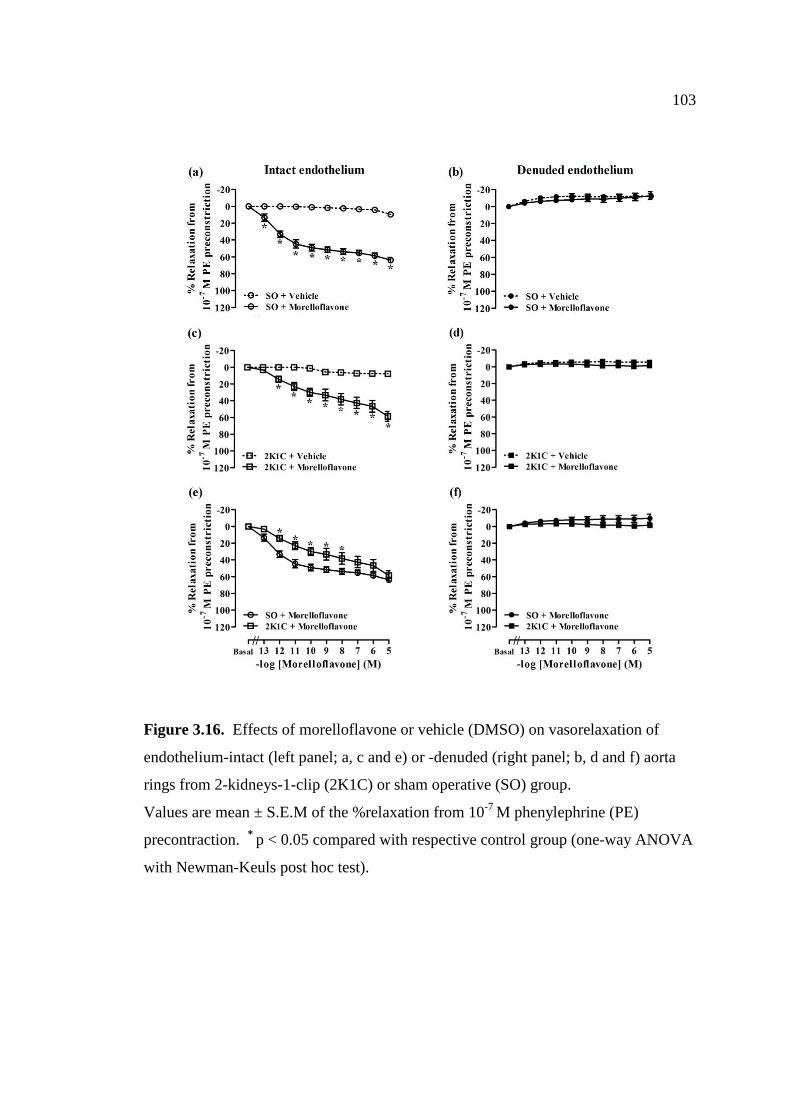
103
Figure 3.16. Effects of morelloflavone or vehicle (DMSO) on vasorelaxation of
endothelium-intact (left panel; a, c and e) or -denuded (right panel; b, d and f) aorta
rings from 2-kidneys-1-clip (2K1C) or sham operative (SO) group.
Values are mean ± S.E.M of the %relaxation from 10-7
M phenylephrine (PE)
precontraction. *
p < 0.05 compared with respective control group (one-way ANOVA
with Newman-Keuls post hoc test).

104
Figure 3.17. Effects of morelloflavone on vasorelaxation of endothelium-intact (left
panel; a, c and e) and -denuded (right panel; b, d and f) aorta rings from 2-kidneys-1-
clip (2K1C) and sham operative (SO) group in the presence of 10-4
M L-NAME.
Values are mean ± S.E.M of the %relaxation from 10-7
M phenylephrine (PE)
precontraction. * p < 0.05 compared with respective control group (one-way ANOVA
with Newman-Keuls post hoc test).

105
Figure 3.18. Effects of morelloflavone on vasorelaxation of endothelium-intact (left
panel; a, c and e) and -denuded (right panel; b, d and f) aorta rings from 2-kidneys-1-
clip (2K1C) and sham operative (SO) group in the presence of 10-6
M indomethacin
(IDM).
Values are mean ± S.E.M of the %relaxation from 10-7
M phenylephrine (PE)
precontraction. *
p < 0.05 compared with respective control group (one-way ANOVA
with Newman-Keuls post hoc test).

106
Figure 3.19. Effects of morelloflavone on vasorelaxation of endothelium-intact (left
panel; a, c and e) and -denuded (right panel; b, d and f) aorta ring from 2-kidneys-1-
clip (2K1C) and sham operative (SO) group in the presence of 10-6
M glibenclamide
(G).
Values are mean ± S.E.M of the %relaxation from 10-7
M phenylephrine (PE)
precontraction. * p < 0.05 compared with respective control group (one-way ANOVA
with Newman-Keuls post hoc test).

107
Figure 3.20. Effects of morelloflavone on vasorelaxation of endothelium- intact (left
panel; a, c and e) and -denuded (right panel; b, d and f) aorta ring from 2-kidneys-1-
clip (2K1C) and sham operative (SO) group in the presence of 10-3
M tetraethyl
ammonium (TEA).
Values are mean ± S.E.M of the %relaxation from 10-7
M phenylephrine (PE)
precontraction. * p < 0.05 compared with respective control group (one-way ANOVA
with Newman-Keuls post hoc test).

108
3.2.7. Effect of morelloflavone on eNOS expression
As shown in Figure 3.21, 2K1C rats exhibited a significant reduction of
eNOS expression in aortic endothelium when compared with SO rats. Treatment with
morelloflavone was able to enhance protein expression of aortic eNOS. This finding
further supported the mechanism of casorelaxation by camboginol which involves the
endothelial NO signaling pathway.
Figure 3.21. Endothelial nitric oxide synthase (eNOS) expression in aortic
endothelial of control and 2-kidneys-1-clip (2K1C) rat; control (a), negative control
(b), 2K1C (c) and after morelloflavone (0.1 mg/kg + 5 g/min/kg BW) treatment in
2K1C. Brown staining in aortic endothelium represents eNOS in the in the tunica
intima layer of aortic wall. Black staining represents nucleus of vascular smooth
muscle cells. Arrow indicates the eNOS location. Magnification 400X and scale bar
= 20 µm.

CHAPTER 4
DISCUSSION
4.1. Development of 2K1C hypertension
4.1.1. Cardiovascular function changes in 2K1C
2K1C is most relevant characteristic model to human RVH which
involves unilateral stenosis of the renal artery leading to a permanent reduction in
renal perfusion pressure in one kidney resulting in increased AII production
(Goldblatt et al. 1934; Navar et al. 1998). In this study, 4 weeks after experimental
renal stenosis, SBP, DBP, PP and MABP of 2K1C rat measuring under
pentobarbitone sodium anesthetization were significantly higher than those of SO rat.
It is also found that the RVR of 2K1C was significantly higher than that of SO rat
suggesting an increase peripheral resistance in the kidney. These data suggested that
the development of hypertension was successful. These results were also supported
by the significant increase in the cardiac mass and atrophy of clipped left kidney with
hypertrophy of non-clipped right kidney of 2K1C rat when compared to SO rat.
It is found that level of plasma MDA in 2K1C were significantly higher
than on SO rat which refer the oxidative stress status in this hypertensive rat. It is
accepted that an increase in circulating AII can activate the vascular smooth muscle
NADPH oxidase which is an important cellular source of ROS, especially O2-
(Griendling et al. 1994). The role of O2- is related to endothelial dysfunction by
which NO can be scavenged by O2- to form peroxynitrite (ONOO
-) resulting in
reduced NO bioavailability and diminishing vasorelaxation (Griendling et al. 1994;
Cai & Harrison et al. 2000; Oliveira-Sale et al. 2008). In this study, the
vasorelaxation in response to ACh in endothelium-intact aortic rings from 2K1C was
significantly lower than those from SO rat (Figure Suppl. 2) while vasoconstriction
response to PE in these rings was significantly higher (Figure Suppl. 1). The
immunohistological study represented the obviously less eNOS expression in 2K1C
when compared to control. This finding is similar to the previous reports by Sánchez

110
et al. (2006) and Ulker et al. (2003) that showed the eNOS expression significantly
decreases in the dysfunction arteries from hypertensive rat. An overproduction of O2-
reduced bioactive NO by promoting NO inactivation and by promoting eNOS
uncoupling (Förstermann, 2010). Regarding baroreceptor reflex, 2K1C rats also
exhibited the impairment of BRS which may due to the inability to increase HR in
response to a decrease ABP and vice versa. It has been proved that an impairment of
BRS in RVH was related to an overproduction of O2- by NADPH oxidase enzyme
(Botelho-Ono et al., 2011; Queiroz et al., 2012). Acute administration of vitamin C or
apocynin, a NADPH oxidase inhibitor prominently improved the blunt BRS in 2K1C
rat (Botelho-One et al., 2011). These data suggested that cardiovascular dysfunction
was observed in this animal model.
4.1.2. Renal function changes in 2K1C
It is found that 2K1C rats has significantly higher RVR, FF, , COsm,
COsm/GFR and TCH2O but has a significantly lower ERPF when compared to SO rats.
However, there was no significant different of GFR, UOsm and POsm between 2K1C
and SO rats. The pioneering study reported that RBF and GFR were not different in
the non-clipped kidneys of the hypertensive rats compared to normotensive rats (Ploth
et al. 1981). However, the non-clipped kidneys of 2K1C rats exhibited a significantly
higher RVR, , absolute and fractional sodium excretion and absolute and fractional
potassium excretion than in kidneys of normal rats (Ploth et al. 1981; Anderson et al.
1985; Martinez-Maldonado1991).
The higher in 2K1C may be due to an abnormal of tubular electrolyte
and water reabsorption in response to high circulating level of AII or NO similar to
experimented in normal control. In normotensive rats, AII has been shown to exert
biphasic effect in which low dose (10-12
-10-9
M) when applied to either luminal or
peritubular side stimulates renal tubular fluid absorption while the higher dose (10-7
-
10-6
M) causes inhibition (Harris and Young. 1997). Moreover, addition of SNP, a
NO donor, showed an inhibition of renal proximal fluid reabsorption (Eitle et al.
1998). Thus, the inhibition of tubular electrolytes and water reabsorption due to
higher levels of AII and perhaps NO may contribute to the high urine flow and
osmolar clearance of 2K1C.

111
4.2. Diuretic and hypotensive effect of camboginol
4.2.1. Diuretic effect of camboginol
The diuretic effect of camboginol was not only observed in normotensive
SO but also in hypertensive 2K1C with the similar degree, however, the mechanisms
of diuretic action might be different in some extent. In camboginol-treated 2K1C rats,
the RVR, UOsm and POsm were decreased significantly while ERPF, GFR, , COsm,
COsm/GFR and TCH2O were increased significantly when compared with the vehicle-
treated 2K1C rats. After camboginol administration in SO rats, the GFR, FF, and
COsm were increased significantly while UOsm and POsm decreased significantly in
comparison to the vehicle-treated SO rats. Since camboginol relaxes the vascular
smooth muscle of the thoracic aorta isolated from hypertensive and normotensive rats
and the mechanism of vasorelaxation may be involve the endothelial NO signaling
pathway, so we can assume that camboginol also relaxes the renal afferent arterioles
resulting in an increased GFR. The diuretic effect of camboginol in normotension and
hypertension may exert via both glomerular and tubular function.
4.2.2 Hypotensive effect of camboginol
It is found that an acute intravenous administration of camboginol showed
a significant transient reduction in SBP, DBP, MABP and HR in 2K1C and SO rats
suggesting the hypotensive potential of this plant compound. This hypotensive effect
did not sustain during the infusion of our experimental dose (0.1 mg/kg + 5 µg/min/kg
BW) in both 2K1C and SO. One of the explanations of this may be due to the well
regulation of baroreflex in SO and the improved BRS in 2K1C caused by camboginol.
The selected dose of camboginol in this in vivo study may not be high enough to
maintain its hypotensive action comparable to candesartan, a specific AII receptor
antagonist previously reported (Hiranyachattada et al., 2005).
However, the acute bradycardic effect of camboginol may involve NO
action on sinoatrial node firing rate since NO has been shown to exert biphasic effect
depending on its concentration. NO could increase HR by activating
hyperpolarization-activated pacemaker current (If) at low concentration but it could
potentially decrease HR by inhibition of L-type calcium current at high concentration

112
(Mani et al., 2002). Moreover, it is likely that camboginol may act directly on the
NTS, the cardiovascular regulating center in medulla oblongata, to cause bradycardia
since the overexpression of eNOS was reported in the NTS which consequent in the
reduction in sympathetic nerve activity, HR and ABP in conscious rats (Sakai et al.,
2000).
Camboginol treatment suppressed an elevated plasma MDA in 2K1C
supporting the robust antioxidant property of camboginol which previously reported
by Yamaguchi et al. (2000a & 2000b) and Hutadilok-Towatana et al. (2007). In
addition, camboginol treatment could improve an impaired BRS in 2K1C rat.
Likewise, it is possible that the antioxidant property of camboginol may improve the
impairment of 2K1C autonomic function.
The % relaxation of thoracic aorta from 2K1C by cumulative addition of
camboginol showed the higher pD2 and EC50 when compared to SO rat suggesting the
higher concentration of camboginol was required to induce the vasorelaxation to the
similar degree as in SO. The mechanisms of camboginol action in SO aorta were
found to be endothelial dependent involving mainly NO generation and partially both
PGI2 production and the operation of vascular KATP channel. However in 2K1C, the
camboginol actions occurred mainly through NO generation and partially KATP
channel with the lesser extent than in SO. This may be due to an impairment of
endothelial function in 2K1C rat.
The immunohistochemical study also found that camboginol treatment
was shown to augment eNOS expression in the vascular endothelium of 2K1C. This
finding further supported the mechanism of camboginol action in 2K1C that might
occur via eNOS expression in the vascular endothelium.

113
4.3. Diuretic and hypotensive effect of morelloflavone
4.3.1. Diuretic effect of morelloflavone
The diuretic effect of morelloflavone was observed both in SO
normotensive and 2K1C hypertensive with the similar degree, however, the
mechanisms of diuretic action might be different in some extent. After
morelloflavone administration in SO rats, GFR, FF, and COsm increased
significantly when compared to the vehicle-treated SO rats while other renal function
parameters remained unchanged. These finding suggested the diuretic effect of
morelloflavone in normotension may exert via both glomerular and tubular function.
Morelloflavone relaxes vascular smooth muscle of the thoracic aorta isolated from
normotensive rat (Lamai et al. 2013), thus, it is likely that it may relax the renal
afferent arterioles resulting in an increased GFR. It is also feasible that
morelloflavone might exert its diuretic effect by inhibit epithelial electrolyte transport
in either proximal or distal part of the nephron. Other plant flavonoids that elicited
renal vasodilation and increased urine flow and urinary sodium and water excretion
including the flavoniods extracted from Clerodendron trichotomum and Spergularia
purpurea (Lu et al. 1994; Jouad et al. 2001).
When morelloflavone was given in 2K1C rats, a significant decrease in
RVR, FF, UOsm and TCH2O were observed when compared to vehicle-treated 2K1C
rats. However, the ERPF, and COsm were found to be significantly higher. These
indicated the inhibition of renal tubular electrolyte reabsorption by morelloflavone
rather than its increased filtered load action like those in SO rats. In addition, in SO
rats, morelloflavone did not alter tubular free water reabsorption which contradicts to
in 2K1C rats suggesting another different pathway of its diuretic action in
hypertensive model. Distal tubular water reabsorption occurs mainly in response to
ADH action, so it is possible that morelloflavone can improve water reabsorption by
increase and expression of renal tubular water channel, aquaporin in this model.

114
4.3.2. Hypotensive effect of morelloflavone
Morelloflavone administration in 2K1C rats demonstrated a significant
reduction in SBP, DBP and MABP which was not found in SO. These results
suggested the pronounced hypotensive effect of morelloflavone in hypertension rather
than in normotension. The marked hypotensive effect of morelloflavone seen in
2K1C rat may be due to its both antioxidant and NO-dependent vasorelaxant property.
The reason that this hypotensive effect was not obviously seen in SO rats may be the
well regulation of baroreceptor reflex in response to the chosen dose of
morelloflavone (0.1 mg/kg + 5 g/min/kg BW). This dose was the minimal
hypotensive dose comparable to candesartan, an AII receptor blocker previously
reported (Hiranyachattada et al. 2005). At this minimal hypotensive effective dose of
morelloflavone selected in this study, taken together with the more impaired BRS of
2K1C than SO, the pronounced reduction in arterial blood pressure was then
observed.
Morelloflavone treatment suppressed an elevated plasma MDA in 2K1C
supporting the robust scavenging property of morelloflavone. Additionally, the BRS
morelloflavone could restore an impaired BRS in 2K1C rats. These data suggested
that the antioxidant property of morelloflavone may improve the impairment of
autonomic function in 2K1C hypertension.
The % relaxation of thoracic aorta from 2K1C by cumulative addition of
morelloflavone showed the higher pD2 and EC50 when compared to SO rat suggesting
the higher concentration of morelloflavone was required to induce the vasorelaxation
to the similar degree as in SO. The vasorelaxant mechanism of of morelloflavone in
2K1C is likely to be endothelium-dependent and involve NO signaling pathway
similar to those of SO rats even though the more endothelium damage occurred in
2K1C than SO. The immunohistochemical study also found that morelloflavone
treatment was shown to augment eNOS expression in the vascular endothelium of
2K1C rats. This finding further supported the mechanism of morelloflavone action in
2K1C that might occur via eNOS expression in the vascular endothelium. The other
signaling pathways which may contribute to the vasorelaxant mechanism of
morelloflavone in normotension included the operation of KCa, KATP channels or
EDHF pathway which was unlikely to occur in 2K1C hypertension.

CHAPTER 5
CONCLUSIONS
It is concluded that camboginol (an isoprenylated benzophenone) and
morelloflavone (a biflavonoids) from Garcinia dulcis exerted various effects in 2K1C
hypertensive rat including;
1. Camboginol and morelloflavone treatment increased RBF and
increased urine flow in both anesthetized 2K1C hypertensive and SO normotensive
rats suggesting theirs diuretic effect.
2. Camboginol treatment decreased ABP in both anesthetized 2K1C
hypertensive and SO normotensive rats. This effect of morelloflavone also observed
in 2K1C hypertension but not SO normotension. These data suggested theirs acute
hypotensive effect.
3. Camboginol and morelloflavone treatment decreased plasma MDA
levels in 2K1C hypertensive rats suggesting theirs scavenging activity.
4. Camboginol and morelloflavone treatment improved an impaired BRS
in anesthetized 2K1C hypertensive rats.
5. Camboginol and morelloflavone treatment enhanced eNOS expression
on vascular endothelium of 2K1C hypertensive rats.
6. Camboginol and morelloflavone treatment exerted an endothelial
dependent vasorelaxant action in both 2K1C hypertensive and SO normotensive rats.
7. The mechanism of camboginol and morelloflavone actions is an
endothelium dependent mechanism, mainly an endothelium NO signaling pathway.
These findings suggested that camboginol and morelloflavone exerted
hypotensive and diuretic effects in 2K1C hypertensive rats. These effects associated
with their antioxidant property. Camboginol and morelloflavone may have a potential
as anti-hypertensive drug. However, an application of these plant bioactive
compounds as an alternative treatment of vascular hypertension requires further
pharmacological studies regarding its effects in resistant vessels and intentsive
cellular mechanisms of its action.

REFERENCES
Abe A, Karaki H. Mechanisms underlying the inhibitory effect of dibutyryl cyclic
AMP in vascular smooth muscle. Eur J Pharmacol 1992; 211: 305-11.
Adams DJ, Rusko J, Van Slooten G. Calcium signaling in vascular endothelial cell,
Ca2+
entry and release. In: Ion flux in pulmonary vascular control. Weir
EK (eds.). New York: Plenum Press; 1993: 259-75.
Ahmad A, Wang Z, Ali R, Maitah MY, Kong D, Baberjee S, Padhye S, Sarkar FH.
Apoptosis-inducing effect of garcinol is mediated by NF-kappa B
signaling in breast cancer cells. J Cell Biochem 2010; 109: 1134-41.
Ahmad A, Wang Z, Wojewoda C, Ali R, Kong D, Maitah MY, Banerjee S, Bao B,
Padhye S, Sarkar FH. Garcinol-induced apoptosis in prostate and
pancreatic cancer cells is mediated by NF- kappaB signaling. Front Biosci
(Elite Ed) 2011; 3: 1483-92.
Alderton WK, Cooper CE, Knowles RG. Nitric oxide synthases: structure, function,
and inhibition. Biochem J 2001; 357: 593-615.
Anderson WP, Kline RL, Woods RL. Systemic and renal hemodynamic changes
during unilateral renal arterial stenosis. Am J Physiol 1985; 249: H956-7.
Ansari WH, Rahman W, Barraclough D, Maynard R, Scheinmann F. Biflavanoids and
a flavanone chromone from the leaves of Garcinia dulcis (Roxb.) Kurz. J
Chem Soc, Perlin Trans 1 1976; 13: 1458-63.
Arnalich-Montiel A, Gonz_alez MC, Delgado-Baeza E, Delgado-Martos MJ,
Condezo-Hoyos L, Martos-Rodríguez A, Rodríguez-Rodríguez P,
Quintana-Villamandos B: Short-term esmolol improves coronary artery
remodeling in spontaneously hypertensive rats through increased nitric
oxide bioavailability and superoxide dismutase activity. Biomed Res Int
2014; 2014: 531087.
Arthur J Vander. Renal Physiology. 4th
ed. Singapore: McGraw-Hill International
Edition; 1991.
Balasubramanyam K, Altaf M, Varier RA, Swaminathan V, Ravindran A, Sadhale
PP, Kundu TK. Polyisoprenylated benzophenone, garcinol, a natural

117
histone acetyltransferase inhibitor, represses chromatin transcription and
alters global gene expression. J Biol Chem 2004; 279: 33716-26.
Bolotina BM, Najibi S, Palacino JJ, Pagano PG, Cohen RA. Nitric oxide derectly
activated calcium-dependent potassium channels in vascular smooth
muscle. Nature 1995; 368: 850-3.
Botelho-Ono MS, Pina HV, Sousa KHF, Nunes FC, Medeiros IA, Braga VA. Acute
superoxide scavenging restores depressed baroreflex sensitivity in
renovascular hypertensive rats. Auton Neurosci: basic & clinical 2011;
159: 38-44.
Bredt DS, Hwang PM, Snyder SHc. Localization of nitric oxide synthase indicating a
neural role for nitric oxide. Nature 1990; 347: 768-70.
Brenner BM, Ballermann BJ, Gunning ME, Zeidel ML. Diverse biological actions of
atrial natriuretic peptide. Physiol Rev 1990; 70: 665-99.
Brice JM, Russell GI, Bing RF, Swales JD, Thurston H. Surgical reversal of
renovascular hypertension in rats: Changes in blood pressure, plasma and
aortic renin. Clin Sci 1983; 65:33-6.
Brown JJ, Davies DL, Lever AF, Robertson JS. Plasma renin concentration in human
hypertension: II. Renin in relation to etiology. Br Med J 1965; 2: 144-58.
Brown JJ, Davies DL, Morton JJ, Robertson JI, Cuesta V, Lever AF, Padfield PL,
Trust P. Mechanism of renal hypertension. Lancet 1976; 1:1219-21.
Bukoski RD, Bergmann C, Gairard A, Stoclet JC. Intracellular Ca2+
and force
determined simultaneously in isolated resistance arteries. Am J Physiol
1989; 257: H1728-35.
Busse R, Fichtner H, Luckhoff A, Kohlhardt M. Hyperpolarization and increased free
calcium in acetylcholine-stimulated endothelial cells. Am J Physiol 1988;
255: H965-9.
Cai H, Harrison DG. Endothelial dysfunction in cardiovascular diseases: The role of
oxidant stress. Circ Res 2000; 87: 840-4.
Callera GE, Varanda WA, Bendhack LM. Impaired relaxation to acetylcholine in
2K1C hypertensive rat aortas involves an abnormal contribution of
endothelial factors. Gen Pharm 2000; 34: 379-89.

118
Carretero OA, Oparil S. Essential hypertension.Part I: Definition and
etiology. Circulation 2000; 101: 329-35.
Chapleau MW, Hajduczok G, Abboud FM. Peripheral and central mechanisms of
baroreflex resetting. Clin Exp Pharmacol Physiol Suppl 1989; 15: 31-43.
Choi S, Ryu KH, Park SH, Jun JY, Shin BC, Chung JH, Yeum CH. Direct vascular
actions of quercetin in aorta from renal hypertensive rats. Kidney Res Clin
Pract 2016; 35: 15-21.
Choi S, Shin HR, Kim SH, Lee MJ, Jun JY, Kim HL, Chung JH, Yeum CH. Effects
of oxidative stress on endothelial modulation of contractions in aorta from
renal hypertensive rats. Kidney Res Clin Pract 2014; 33: 19-25.
Chou SY, Faubert PF, Porush JG. Contribution of angiotensin to the control of
medullary hemodynamics. Fed Proc 1986; 45: 1438-43.
Cogan MG. Neurogenic regulation of proximal bicarbonate and chloride reabsorption.
Am J Physiol 1986; 250: F22-6.
Coleman RA, Smith WL, Narumiya S. International union of pharmacology
classification of prostanoid receptors: properties, distribution, and
structure of the receptors and their subtypes. Pharmacol rev 1994; 46:
205-29.
Cornwell TL, Soff GA, Tranor AE, Lincoln TM. Regulation of the expression of
cyclic GMP-dependent protein kinase by cell density
in vascular smooth muscle cells. J Vasc Res1994; 31:330-7.
Corriu C, Feletou M, Canet E, Vanhoutte PM. Endothelium-derived factors and
hyperpolarization of the carotid artery of the guinea-pig. Br J Pharmacol
1996; 119: 959-64.
Cragg AH, Smith TP, Thompson BH, Maroney TP, Stanson AW, Shaw GT, Hunter
DW, Cochran ST. Incidental fibromuscular dysplasia in potential renal
donors: long-term clinical follow-up. Radiology 1989; 172:145-7.
Das D, Bandyopadhyay D, Banerjee RK. Oxidative inactivation of gastric peroxidase
by site-specific generation of hydroxyl radical and its role in stress-
induced gastric ulceration. Free Radic Biol Med 1998; 24:460-9.

119
Das D, Bandyopadhyay D, Bhattacharjee M, Banerjee RK. Hydroxyl radical is the
major causative factor in stress-induced gastric ulceration. Free Radic Biol
Med 1997; 23:8-18.
Davidson WD, Sackner MA. Simplification of anthrone method of the determination
of inulin in clearance studies. J Lab Clin Med 1963; 62: 351-6.
De Mey JG, Vanhoutte PM. Anoxia and endothelium-dependent reactivity of the
canine femoral artery. J Physiol 1983; 335:65-74.
Deachathai S, Mahabusarakam W, Phongpaichit S, Taylor WC, Zhang YJ, Yang CR.
Phenolic compounds from the flowers of Garcinia dulcis. Phytochemistry
2006; 67: 464-9.
Deachathai S, Mahabusarakam W, Phongpaichit S, Taylor WC. Phenolic compounds
from the fruit of Garcinia dulcis. Phytochemistry 2005; 66: 2368-75.
Deachathai S, Phongpaichit S, Mahabusarakam W. Phenolic compounds from the
seeds of Garcinia dulcis. Nat Prod Res 2008; 22: 1327-32.
Delpy E, Coste H, Gouville AC. Effects of cyclic GMP elevation on isoprenaline-
induced increase in cyclic AMP and relaxation in rat aortic smooth
muscle: role of phosphodiesterase 3. Br J Pharmacol 1996; 119: 471-8.
DeNucci G, Thomas R, D’Orleans-Juste P, Antunes E, Walder C, Warner TD, Vane
JR. Pressor effects of circulating endothelin are limited by its removal in
the pulmonary circulation and by the release of prostacyclin and
endothelium-derived relaxaing factor. Proc Natl Acad Sci USA 1988; 85:
9797-800.
DiBona GF. Neural control of renal tubular sodium reabsorption in the dog. Fed Proc
1978. 37: 1214-17.
Dickson ME, Sigmund CD. Genetic basis of hypertension: Revisiting
angiotensinogen. Hypertension 2006; 48: 14-20.
Diederich D, Yang Z, Bühler FR, Lüscher TF. 1990. Impaired endothelium-dependent
relaxation in hypertensive resistance arteries involves-cyclooxygenase
pathway. Am J Physiol 1990; 258: 445-51.
Djoussé L, Mukamal KJ. Alcohol consumption and risk of hypertension: Does the
type of beverage or drinking pattern matter? Rev Esp Cardiol 2009;
62:603-5.

120
Dodt C, Wellhöner JP, Schütt M, Sayk F. Glucocorticoids and hypertension (in
German) Der Internist 2009; 50:36-1
Dohi Y, Kojima M, Sato K. Endothelial modulation of contractile responses in
arteries from hypertensive rats. Hypertension 1996; 28: 732-7.
Dzau VJ, Ingelfinger JR, Pratt RE. Regulation of tissue renin and angiotensin gene
expressions. Cardiovasc Pharmacol 1986; 8L: 511-6.
Edwards RM. Segmental effects of norepinephrine and angiotensin II on isolated
renal microvessels. Am J Physiol 1983. 244: F526-34.
Eisenberg PR. Endothelial cell mediators of thrombosis and fibrinolysis: review in
depth. Coronary Artery Dis 1991; 2: 129-66.
Eitle E, Hiranyachattada S,Wang H, Harris PJ. Inhibition of proximal tubular fluid
absorption by nitric oxide and atrial natriuretic peptide in rat kidney. Am
J Physiol (Cell Physio) 1998; 274: C1075-80.
Elliot SJ, Meszaros GJ, Schilling M. Effect of oxidant stress on calcium signaling in
vascular endothelial cells. Free Rad Biol Med 1992; 13: 635-50.
Faubert PF, Chou SY, Porush JG. Regulation of papillary plasma flow by angiotensin
II. Kidney Int 1987;. 32: 472-8.
Feletou M, Vanhuotte PM. Endothelium-dependent hyperpolarization of canine
coronary smooth muscle. Br J Pharmacol 1988; 93: 515-24.
Feron O, Saldana F, Michel JB, Michel T. The endothelial nitric-oxide synthase-
caveolin regulatory cycle. J Biol Chem 1998; 273: 3125-28.
Fӧrstermann U. Nitric oxide and oxidative stress in vascular disease. Pflügers Archiv
2010; 459: 923-39.
Folkow Göthberg G, Lundin S, Ricksten SE. Structural renal vascular changes in
renal hypertensive rats (RHR). Acta Physiol Scand 1977; 101: 254-6.
Fuji J, Kurihara H, Yamaguchi H, Terasawa F, Murata K, Matsushita S, et al. A
persistent hypertension due to unilateral renalartery constriction in the
rabbit. Jpn Circ J 1967; 31:1197-200.
Furchgott RF, Zawadzki JV. The obligatory role of endothelial cells in the relaxation
of arterial smooth muscle by acetylcholine. Nature 1980; 288: 373-6.

121
Gao SA, Johansson M, Rundqvist B, Lambert G, Jensen G, Friberg P. Reduced
spontaneous baroreceptor sensitivity in patients with renovascular
hypertension. J Hypertens 2002; 20: 111-6.
Garcia R, Thibault G, Gutkowska J, Cantin M, Genest J. Changes of regional blood
flow induced by atrial natriuretic factor (ANF) in conscious rats. Life Sci
1985; 36: 1687-92.
Gavras H, Brunner HR, Thurston H, Laragh JH. Reciprocation of renin dependency
with sodium volume dependency in renal hypertension. Science 1975;188:
1316-17.
Gdthberg G, Hallback-Nordlander M, Karlstrom G, Ricksten SE, Folkow B.
Structurally based changes of renal vascular reactivity in spontaneously
hypertensive and two-kidney, one-clip renal hypertensive rats, as
compared with kidneys from uninephrectomized and intact normotensive
rats. Ada Physiol Scand 1983; 118: 61-7.
Gil B, Sanz MJ, Terencio MC, Gunaseqaran R, Payá M, Alcaraz MJ. Morelloflavone,
a novel biflavonoid inhibitor of human secretory phospholipase A2 with
anti-inflammatory activity. Biochem Pharmacol 1997; 53: 733-40.
Goldblatt H, Lynch J, Hanzal RF, Summerville WW. Studies on experimental
hypertension. I. The production of persistent elevation of systolic blood
pressure by means of renal ischemia. J Exp Med 1934; 59: 347-80.
Gratton JP, Fontana J, O’Connor DS, Garcia-Cardena G, McCabe TJ, Sessa WC.
Reconstitution of an endothelial nitric-oxide synthase (eNOS), hsp90 and
caveolin-1 complex in vitro. Evidence that hsp90 facilitates calmodulin
stimulated displacement of eNOS from caveolin-1. J Biol Chem 2000;
275: 22268-72.
Griendling KK, Minieri CA, Ollerenshaw JD, Alexander RW. Angiotensin II
stimulates NADH and NADPH oxidase activity in cultured vascular
smooth muscle cells. Cir Res 1994; 74: 1141-8.
Guyton AC, Hall JE. Textbook of Medical Physiology. 11th
edition. Philadelphia :
Elsevier Saunders: 2006
Hall JE, Guyton AC, Smith MJ Jr, Coleman TG. Chronic blockade of angiotensin II
formation during sodium deprivation. Am J Physiol 1979; 237: F424-32.

122
Hall JE, Mizelle HL, Woods LL. The renin-angiotensin system and long-term
regulation of arterial pressure. J Hypertens 1986; 4: 387-97.
Hall JE: Control of sodium excretion by angiotensin II: Intrarenal mechanisms and
blood pressure regulation. Am J Physiol 1986; 250: R960-72.
Halushka PV, Mais DE, Mayeux PR, Morinelli TA. Thromboxan, prostaglandin and
leukotriene receptors. Annu Rev Pharmacol Toxicol 1989; 29: 213-39.
Hansen KJ, Edwards MS, Craven TE, Cherr GS, Jackson SA, Appel RG, Burke GL,
Dean RH. Prevalence of renovascular disease in elderly: a population-
based study. J Vasc Surg 2002; 36:443-51.
Harris PL, Young JA. Dose-dependent stimulation and inhibition of proximal tubular
sodium reabsorption by angiotensin II in the rat kidney. Pflügers Archiv
1977; 367: 295-7.
Haslam DW, James WP. Obesity. Lancet 2005; 366: 1197-209.
Hickey KA, Rubanyi G, Paul RJ, Highsmith RF. Characterization of a coronary
vasoconstrictor produced by cultured endothelial cells. Am J Physiol
1985; 248: C550-6.
Hiranyachattada S, Saetew S, Harris PJ. Acute effects of candesartan on rat renal
haemodynamics and proximal tubular reabsorption. Clin Exp Pharmacol
Physiol 2005; 32: 714-20.
Hong J, Sang S, Park HJ, Kwon SJ, Suh N, Huang MT, Ho CT, Yang CS. Modulation
of arachidonic acid metabolism and nitric oxide synthesis by garcinol and
its derivatives. Carcinogenesis 2006; 27:278-86.
Huang, WC, Ploth, DW, Navar, LG. Angiotensin-mediated alterations in nephron
function in Goldblatt hypertensive rats. Am J Physiol 1982; 243:F553-60.
Hutadilok-Towatana N, Kongkachuay S, Mahabusarakam W. Inhibition of human
lipoprotein oxidation by morelloflavone and camboginol from Garcinia
dulcis. Nat Prod Res 2007; 21: 655-62.
Hwang IS, Ho H, Hoffman BB, Reaven GM. Fructose-induced insulin resistance and
hypertension in rats. Hypertension 1987; 10: 512-6.
Ignarro LJ. Biosynthesis and metabolism of endothelium-derived nitric oxide. Annu
rev Pharmacol Toxicol 1990; 30: 535-60.

123
Iinuma M, Tisa H, Ito T, Tanaka T, Riswan S. Three new benzophenone-xanthone
dimmers from the root of Garcinia dulcis. Chem Pharm Bull 1996; 44:
1744-7.
Itoh T, Ito S, Shafiq J, Suzuki H. Effects of newly systhesized K+ channel opener, Y-
26763, on noradrenaline-induced Ca2+
mobilization in smooth muscle of
rabbit mesenteric artery. Br J Pharmacol 1994; 104: 227-33.
Itoh T, Seki N, Suzuki H, Ito S, Kajikuri J, Kuriyama H. Membrane hyperpolarization
inhibits aginist-induced systhesis of inositol-1, 4, 5-triphosphate in
smooth muscle of the rabbit mesenteric artery. J Physiol 1992; 451: 307-
28.
Jin N, Packer CS, Rhoades RA. Reactive oxygen mediated contraction in pulmonary
arterial smooth muscle: cellular mechanisms. Can J Physiol pharmacol
1991; 69: 383-8.
Johns A, Lategan TW, Lodge NJ, Ryan US, van Breemen C, Adams DJ. Calcium
entry through receptor operated channels in bovine pulmonary artery
endothelial cells. Tissue Cell 1987; 19: 733-45.
Johns RA, Linden JM, Peach NJ. Endothelium-dependent relaxation and cyclic GMP
accumulation in rabbit pulmonary artery are selectively inpaired by
moderate hypoxia. Circ Res 1989; 65: 1508-15.
Jones JV, Floras JS. Baroreflex sensitivity changes during the development of
Goldblatt two-kidney one-clip hypertension in rats. Clin Sci 1980; 59:
347-52.
Kasahara S, Henmi S. Medical herb index in Indonesia. Jakata; Eisai Indonesia: 1986,
92.
Katayama S, Inaba M, Maruno Y, Omoto A, Itabashi A, Kawazu S, Ishii J. Effect of
captopril or enalapril on renal prostaglandin E2. Prostaglandins 1989;
38:401-11.
Kato T, Iwama Y, Okumura K, Hashimoto H, Ito T, Satake T. Prostaglandin H2 may
be the endothelium-derived contracting factor by acetylcholine
stimulation in the rat aorta. Hypertension 1990; 15: 475-81.
Katusic ZS, Vanhoutte PM. Superoxide anion is an endothelium-derived contracting
factor. Am J Physiol 1989; 257: H33-7.

124
Kim SY, Kim TB, Moon KA, Kim TJ, Shin D, Cho YS, Moon HB, Lee KY.
Regulation of pro-inflammatory responses by lipoxygenases via
intracellular reactive oxygen species in vitro and in vivo. Exp Mol Med
2008; 40: 461-76.
Kirchheim HR. Systemic arterial baroreceptor reflexs. Physiol Rev 1976; 56: 100-77.
Kirkman E, Sawdon Marisa. Neurological and humoral control of blood pressure.
Anesthesia and Intensive Care Medicine 2010; 11: 159-64.
Koeberle A, Northoff H, Werz O. Identification of 5-lipoxygenase and microsomal
prostaglandin E2 synthase-1 as functional targets of the anti-inflammatory
and anti-carcinogenic garcinol. Biochem Pharmacol 2009; 77: 1513-21.
Kramer P, Ochwadt B. Sodium excretion in goldblatt hypertension. Pflügers Archiv
1972; 332: 332-45.
Krum H, Viskoper RJ, Lacourciere Y, Budde M, Charlon V. The effect of an
endothelin-receptor antagonist, bosentan, on blood pressure in patients
with essential hypertension. Bosentan Hypertension Investigators. N Engl
J Med 1998; 338: 784-90.
Kukovetz WR, Holtzmann S, Wurm A, Poch G. Prostacyclin increase cAMP in
porcine coronary. J Cyclic Nucl Res 1979; 5: 469-76.
Kyrou I, Chrousos GP, Tsigos C. Stress, visceral obesity, and metabolic
complications. Ann N Y Acad Sci 2006; 1083:77-110.
Lackland DT, Egan BM. Dietary salt restriction and blood pressure in clinical
trials. Curr Hypertens Rep 2007; 9: 314-9.
Lamai J, Mahabusarakam W, Ratithammatorn T, and Hiranyachattada S. Effects of
morelloflavone from Garcinia dulcis on vasorelaxation of isolated rat
thoracic aorta. J Physiol Biomed Sci 2013; 26: 13-7.
Lambert TL, Kent RS, Whorton AR. Bradykinin stimulation of inositol
polyphosphate production in porcine aortic endothelial cells. J Biol Chem
1986; 261: 15288-93.
Laragh JH, Sealey JE, Buhler FR, Vaughan ED, Brunner HR, Gavras H, Baer L. The
renin axis and vasoconstriction volume analysis for understanding and
treating renovascular and renal hypertension. Am J Med 1975; 8: 4-13.
Ledingham. Experimental renal hypertension. Clin Nephrol 1975; 4:127-37.

125
Lee JH, O′Keefe JH, Bell D, Hensrud DD, Holick MF. Vitamin D deficiency an
important, common, and easily treatable cardiovascular risk factor. J Am
Coll Cardiol 2008; 52: 1949-56.
Leenen FHH, de Jong W. A solid silver clip for induction of predictable levels of
renal hypertension in the rat. J Appl Physiol 1971; 31:142- 4.
Lerman LO, Nath KA, Rodriguez-Porcel M, Krier JD, Schwartz RS, Napoli C,
Romero JC. Increased oxidative stress in experimental renovascular
hypertension. Hypertension 2001; 37: 541-6.
Lerman LO, Schwartz RS, Grande JP, Sheedy PF, Romero JC. Noninvasive
evaluation of a novel swine model of renal artery stenosis. J Am Soc
Nephrol 1999; 10:1455- 65.
Leuris M, Hendersen AH. A phorbolester inhibits the release of endothelium-derived
relaxing factor. Eur J Pharmacol 1987; 137: 167-71.
Levin ER. Endothelins. N Engl J Med 1995; 333: 356-63.
Liao CH, Sang S, Liang YC, Ho CT, Lin JK. Suppression of inducible nitric oxide
synthase and cyclooxygenase-2 in downregulating nuclear factor-kappa B
pathway by Garcinol. Mol Carcinog 2004; 413: 140-9.
Lim TK. Edible Medicinal And Non-Medicinal Plants: Volume 2, Fruits: Springer
Science+Business Media; 2012, p 35-40.
Lin YM, Anderson H, Flavin MT, Pai YH, Mata-Greenwood E, Pengsuparp T,
Pezzuto JM, Schinazi RF, Hughes SH, Chen FC. In vitro anti-HIV activity
of biflavonoids isolated from Rhus succedanea and Garcinia multiflora. J
Nat Prod 1997; 60: 884-8.
Luma GB, Spiotta RT. Hypertension in children and adolescents. Am Fam
Physician 2006; 73: 1558-68.
Lüscher TF, Raij L, Vanhoutte PM. Endothelium-dependent responses in
normotensive and hypertensive Dahl rats. Hypertension 1987; 9: 157-63.
Luscher TF, Vanhoutte PM. The endothelium: Modulator of Cardiovascular function.
Boca Raton, FL: CRC Press; 1990.
Lynch KR, Simnad VI, Ben-Ari ET, Garrison JC. Localization of pre-angiotensinogen
messenger RNA sequences in the rat brain. Hypertension 1986; 8: 540-3.

126
Mahabusarakam W, Mecawun P, Phongpaichit S. Xanthones from the green branch of
Garcinia dulcis. Nat Prod Res 2016; 30: 2323-8.
Mani AR, Nahavandi A, Moosavi M, Safarinejad R, Dehpour AR. Dual nitric oxide
mechanisms of cholestatis-induced bradycardia in the rat. Clin Exp
Pharmacol Physiol 2002; 29: 905-8.
Mantelingu K, Reddy BA, Swaminathan V, Kishore AH, Siddappa NB, Kumar GV,
Nagashankar G, Natesh N, Roy S, Sadhale PP, Ranga U, Narayana C,
Kundu TK. Specific inhibition of p300-HAT alters global gene expression
and represses HIV replication. Chem Biol 2007; 14: 645-57.
Martinez-Maldonado M. Pathophysiology of renovascular hypertension. Hypertension
1991; 17: 707-19.
Masaki T. Possible role of endothelin in endothelial regulation of vascular tone. Annu
Rev Pharmacol Toxicol 1995; 35: 235-55.
Masaki Z, Ferrario CM, Bumpus FM, Bravo EL, Khosia MC. The course of arterial
pressure and the effect of Sarxd1-Thr8- angiotensin II in a new model of
two-kidney hypertension in conscious dogs. Clin Sci Mol Med 1977; 52:
163-70.
Mattson DL, Cowley AW Jr. Kinin actions on renal papillary blood flow and sodium
excretion. Hypertension 1993. 21: 961-5.
McGiff JC, Itskovitz HD, Terragno NA. The actions of bradykinin and eledoisin in
the canine isolated kidney: relationships to prostaglandins. Clin Sci Mol
Med 1975; 49: 125-31.
Miller ED Jr, Samuels AI, Haber E, Barger AC. Inhibition of.angiotensin conversion
in experimental renovascular hypertension. Science 1972; 177: 1108-9.
Molina R, Hidalgo A, García de Boto MJ. Influence of mechanical endothelium
removal techniques and conservation conditions on rat aorta responses.
Methods Find Exp Clin Pharmacol 1992; 14: 91-6.
Moncada S, Palmer RMJ, Higgs EA. Biosynthesis of nitric oxide from L-argenine: a
pathway for the rogation of cell function and communication. Biochem
Pharmacol 1989; 38: 1709-15.

127
Moncada V, Vane JR. Pharmacology and endogenous roles of prostaglandin
endoperoxides, thromboxane A2, and prostacyclin. Pharmacol Rev 1979;
30: 293-331.
Morgan K. The role of calcium in the control of vascular tone as assessed by the Ca2+
indicator Aequorin. Cardiovasc Drugs Ther 1990; 4: 1355-62.
Morton JJ, Wallace ECH. The importance of the renin angiotensin system in the
development and maintenance of hypertension in the two-kidney one-clip
hypertensive rat. Clin Sci 1983; 6: 359-70.
Murphy ME, Brayden JE. Apamin-sensitive K+ channels mediate an endothelium-
dependent hyperpolarization in rabbit mesenteric arteries. J Physiol 1995;
489: 723-34.
Naftilan AJ, Pratt RE, Eldridge CS, Lin HL, Dzau VJ. Angiotensin II induces c-fos
expression in smooth muscle via transcriptional control. Hypertension
1989;13 (Part 2): 706-11.
Nakashima M, Vanhoutte PM. Age-dependent decrease in endothelium-dependent
hyperpolarizations to endothelin-3 in the rat mesenteric artery. J
Cardiovasc Pharmacol 1993; 22: S352-4.
Navar LG, Zou L, Thun AV, Wang CT, Imig JD, Mitchell KD. Unraveling the
mystery of Goldblatt hypertension. News Physiol Sci 1998; 13: 170-6.
Nelson MT, Patlak JB, Worley JF, Standen NB. Calcium channels, potassium
channels and voltage dependence of arterial smooth muscle tone. Am J
Physiol 1990; 259: C3-18.
Neymark E, LaBerge JM, Hirose R, Melzer JS, Kerlan RK, Wilson MW, Gordon RL.
Arteriographip detection of renovascular disease in potential renal donors:
Incidenceand effect on donor surgery. Radiology 2000; 214: 755-66.
Nishi EE, Oliveira-Sales EB, Bergamaschi CT, Oliveira TG, Boim MA, Campos RR.
2010. Chronic antioxidant treatment improves arterial renovascular
hypertension and oxidative stress markers in the kidney in Wistar rats. Am
J Hypertens 2010; 23: 473-80.
Ohkawa H, Ohishi N, Yagi K. Assay for lipid peroxidases in animal tissues by
thiobarbituric acid reaction. Anal Biochem 1979; 95: 351-8.

128
Olin JW, Melia M, Young JR, Graor RA, Risius B. Prevalence of atherosclerotic renal
artery stenosis in patients with atherosclerosis elsewhere. Am J Med 1990;
88: 46N-51N.
Oliveira-Sales EB, Nishi EE, Carillo BA, Boim MA, Dolnikoff MS, Bergamaschi CT,
Campos RR. Oxidative stress in the sympathetic premotor neurons
contributes to sympathetic activation in renovascular hypertension. Am J
Hypertens 2009; 22: 484-92.
Oliveira-Sales EB, Varela VA, Maquiqussa E, Borges FT, Shimoura CG, Gomes G,
Campos RR, Boim MA. Renovascular hypertension: Effect of
mesenchymal stem cells in the contralateral hypertensive kidney in rats.
Clin Exper Hypertens 2016; 38: 586-93.
Ortiz PA, Garvin JL. Role of nitric oxide in the regulation of nephron transport. Am J
Physiol Renal Physiol 2002; 282; F777-84.
Palmer RMJ, Ashton DS, Moncada S. Vascular endothelial cells synthetise nitric
oxide from L-argenine. Nature 1988; 333: 664-6.
Palmer RMJ, Ferrige AG, Moncada S. Nitric oxide release accounts for the biological
activity of endothelium-derived relaxing factor. Nature 1987; 327: 524-6.
Pan MH, Chang WL, Lin-Shiau SY, Ho CT, Lin JK. Induction of apoptosis by
garcinol and curcumin through cytochrome c release and activation of
caspases in human leukemia HL-60 cells. J. Agric. Food Chem 2001; 49:
1464-74.
Panek RL, Ryan MJ, Weishaar RE, Taylor DG Jr. Development of a high renin model
of hypertension in the cynomolgus monkey. Clin Exp Hypertens A 1991;
13:1395-414.
Pang X, Yi T, Yi Z, Cho SG, Qu W, Pinkaew D, Fujise K, Liu M. Morelloflavone, a
biflavonoid, inhibits tumor angiogenesis by targeting rho GTPases and
extracellular signal-regulated kinase signaling pathways. Cancer research
2009; 69: 518-25.
Parkington HC, Tonta MA, Coleman HA, Tare M. Role of membrane potential in
endothelium-dependent relaxation of guinea-pig coronary arterial smooth
muscle. J Physiol 1995; 484: 469-80.

129
Pasquié JL, Herizi A, Jover B, Mimran A. Chronic bradykinin infusion and receptor
blockade in angiotensin II hypertension in rats. Hypertension 1999; 33:
830-4.
Pereañez JA, Patiño AC, Núñez V, Osorio E. The biflavonoid morelloflavone inhibits
the enzymatic and biological activities of a snake venom phospholipase
A2. Chem Biol Interac 2014; 220: 94-101.
Pfeffer JM, Pfeffer MA, Mirsky I, Braunwald E. Regression of left ventricular
hypertrophy and prevention of left ventricular dysfunction by captopril in
the spontaneously hypertensive rat. Proc NatlAcad Sd USA 1982; 79:
3310-4.
Pickering. The role of the kidney in acute and chronic hypertension following renal
artery constriction in the rabbit. Clin Sci 1937; 5:229-47.
Pinkaew D, Cho SG, Hui DY, Wiktorowicz JE, Hutadilok-Towatana N,
Mahabusarakam W, Tonganunt M, Stafford LJ, Phongdara A, Liu M,
Fujise K. Morelloflavone blocks injury-induced neointimal formation by
inhibiting vascular smooth muscle cell migration. Biochim Biophys Acta
2009; 1790: 31-9.
Pinkaew D, Hutadilok-Towatana N, Teng BB, Mahabusarakam W and Fujise K.
Morelloflavone, a biflavonoid inhibitor of migration-related kinases,
ameliorates atherosclerosis in mice. Am J Physiol Heart Circ Physiol
2012; 302: H451-58.
Ploth DW, Fitzgibbon W. Pathophysiology of Altered Renal Function in Renal
Vascular Hypertension. Am J Kidney Dis 1994; 24:652-9.
Ploth DW, Nava LG. Intrarenal effects of the renin-angiotensin system. Fed Proc
1979; 38: 2280-5.
Ploth DW, Roy RN, Huang WC, Navar LG. Impaired renal blood flow and cortical
pressure autoregulation in contralateral kidneys of Goldblatt hypertensive
rats. Hypertension 1981; 3: 67-74.
Ploth DW, Schnermann J, Dahlheim H, Hermle M, Schmidmeier E. Autoregulation
and tubuloglomerular feedback in normotensive and hypertensive rats.
Kidney Int 1977; 12: 253-67.

130
Queiroz T, Guimarães D, Mendes-Jonior L, Braga V. α-Lipoic acid reduces
hypertension and increases baroreflex sensitivity in renovascular
hypertensive rat. Molecules 2012; 17: 13357-67.
Rao Rama AV, Venkatswamy G, Pendse D. Camboginol and Cambogin. Tetrahedron
Letters 1980; 21: 1975-8.
Rapoport RM, Murad F. Aginist-induced endothelium-dependent relaxation in rat
aorta may be mediated through cyclic GMP. Circ Res 1983; 52: 352-7.
Rihal CS, Textor SC, Breen JF, McKisick MA, Grill DE, Hallett JW, Holmes DR Jr.
Incidental renal artery stenosis among a prospective cohort of
hypertensive patients undergoing coronary angiography. Mayo Clin Proc
2002; 77: 309-16.
Romero JC, Fiksen-Olsen MJ, Schryver S. Pathophysiology of hypertension: the use
of experimental models to understand the clinical features of the
hypertensive disease. In: Spittel JA Jr, ed. Clin Med 1981; 7: 1-51.
Rostand SG, Lewis D, Watkins JB, Huang WC, Navar LG. Attenuated pressure
natriuresis in hypertensive rats. Kidney Int 1982; 21: 331-8.
Roux D, Hadi HA, Thoret S, Guenard D, Thoison O, Pais M, Sevenet T. Structure-
activity relationship of polyisoprenyl benzophenones from Garcinia
pyrifera on the tubulin/microtubule system. J Nat Prod 2000; 63:1070-6.
Rubanyi G, Vanhoutte PM. Superoxide anions and hyperoxia inactivated
endothelium-derived relaxing factor(s). Am J Physiol 1986; 250: H822-7.
Ryan US. Endothelium as transducing surface. J Mol Cell Cardiol 1989; 21 (suppl. 1):
85-90.
Sakai K, Hirooha Y, Matsuo I, Eshima K, Shigematsu H, Shimokawa H, Takeshita A.
Overexpression of eNOS in NTS causes hypotension and bradycardia in
vivo. Hypertension 2000; 36: 1023-8.
Samani NJ, Godfrey NP, Major JS, Brammar WJ, Swales JD. Kidney renin mRNA
levels in the early and chronic phases of two-kidney, one clip
hypertension in the rat. Hypertension 1989; 7: 105-12.
Sánchez M, Galisteo M, Vera R, Villar IC, Zarzuelo A, Tarmargo J, et al. Quercetin
downregulates NADPH oxidase, increases eNOS activity and prevents

131
endothelial dysfunction in spontaneously hypertensive rats. J Hypertens
2006; 24: 75-84.
Sander DB, Kelly T, Larson D. The role of nitric oxide synthase/nitric oxide in
vascular smooth muscle control. Perfusion 2000; 15: 97-104.
Sang S, Liao CH, Pan MH, Rosen RT, Lin-Shiau SY, Lin JK, et al., Chemical studies
on antioxidant mechanism of garcinol: analysis of radical reaction
products of garcinol with peroxyl radicals and their antitumor activities,
Tetrahedron 2002; 58: 10095-102.
Sang S, Pan MH, Cheng X, Bai N, Stark RE, Rosen RT, Lin-Shiau SY, Lin JK, Ho
CT. Chemical studies on antioxidant mechanisms of garcinol: analysis of
radical reaction products of garcinol and their antitumor activities.
Tetrahedron 2001; 57: 9931-8.
Schrier RW, Berl T. Nonosmolar factors affecting renal water secretion. N Engl J
Med 1975; 272: 81-8.
Schuster VL. Effects of angiotensin on proximal tubular reabsorption. Fed Proc 1986;
45: 1444-7.
Schwietzer G, Gertz KH. Changes of hemodynamics and glomerular ultrafiltration in
renal hypertension of rats. Kidney Int 1979; 15: 134-43.
Schwietzer G, Gertz KH. Changes of hemodynamics and glomerular ultrafiltration in
renal hypertension of rats. Kidney Int 1979; 15:134-43
Segura J, Ruilope LM. Obesity, essential hypertension and renin-angiotensin
system. Pub Hlth Nutr 2007; 10: 1151-5.
Shimizu S, Ishii M, Yamamoto T, Kawanishi T, Momose K, Kuroiwa Y. Bradykinin
induces generation of reactive oxygen species in bovine aortic endothelial
cells. Res Commun Chem Pathol Pharmacol 1994; 84: 301-14.
Shimokawa H, Flavahan NA, Lorenz RR, Vanhoutte PM. Prostacyclin releases
endothelium-derived relaxing factor and potentiates its action in coronary
arteries of the pig. Br J Pharmacol 1988; 95:1197-203.
Sigmon DH, Beieirwaltes WH. Influence of nitric oxide in the chronic phase of two
kidney, one clip renovascular hypertension. Hypertension 1993; 31: 649-
56.

132
Smith GW, Somova LI. Renal function and renal venous prostaglandin concentration
during different stages of experimental renal hypertension in the rat. Br J
Pharmacol 1976; 58: 253-9.
Smith HW, Finkelstein N, Aliminoso L, Crawford B, Graber M. The renal clearances
of substituted hippuric and derivatives and other aromatic acids in dogs
and man. J Clin Invest 1945; 24: 388-404.
Somlyo AP, Wu X, Lalker LA, Somlyo AV. Pharmacomechanical coupling: the role
of calcium, G-proteins, kinases and phosphatases. Rev Physiol Biochem
Pharmacol 1999; 134: 201-34.
Sorof J, Daniels S. Obesity hypertension in children: A problem of epidemic
proportions. Hypertension 2002; 40: 441-7.
Sotomayor MA, Perez-Guerrero C, Herrera MD, Marhuenda E. Effects of chronic
treatment with simvastatin on endothelial dysfunction in spontaneously
hypertensive rats. J Hypertens 1999; 17: 769-79.
Stanfield CM, Schilling WP, Possami LD, Kunze DL. Bradykinin-induced potassium
current in culture bovine aortic endothelial cells. J Neur Biol 1990; 116:
227-38.
Stumpe KO, Lowitz HD, Ochwadt B. Fluid reabsorption in Henle's loop and urinary
excretion of sodium and water in normal rats and rats with chronic
hypertension. J Clim Invest 1979; 49:1200-12.
Stumpe KO, Lowitz HD, Ochwadt B. Function of juxtamedullary nephrons in
normotensive and chronically hypertensive rats. Pflügers Archiv 1969;
313: 43-52.
Sunano S, Watanabe H, Tanaka S, Sekiguchi F, Shimamura K. Endothelium-derived
relaxing, contracting and hyperpolarizing factors of mesenteric arteries of
hypertensive and normotensive rats. Br J Pharmacol 1999; 126: 709-16.
Suzuki YJ, Ford GD. Superoxide stimulates IP3-induced Ca2+
release from vascular
smooth muscle sarcoplasmic reticulum. Am J Physiol 1992: 262: H114-6.
Swales JD, Samani NJ. Localisation and physiological effects of tissue renin-
angiotensin systems. Hum Hypertens 1989; 3: 71-7.
Swales JD, Thurston H, Queiroz FP, Medina A, Holland J. Dual mechanism for
experimental hypertension. Lancet 1971; 2:1181-3.

133
Tabassum N, Ahmad F. Role of natural herbs in the treatment of
hypertension. Pharmaco Rev 2011; 5: 30-40.
Tanaka T, Kohno H, Shimada R, Kagami S, Yamaguchi F, Kataoka S, et al.
Prevention of colonic aberrant crypt foci by dietary feeding of garcinol in
male F344 rats. Carcinogenesis 2000; 21:1183–9.
Toba H, Kojima Y, Wang J, Noda K, Tian W, Kobara M, Nakata T. Erythropoietin
attenuated vascular dysfunction and inflammation by inhibiting NADPH
oxidase-derived superoxide production in nitric oxide synthase-inhibited
hypertensive rat aorta. Eur J Pharmacol 2012; 691:190-7.
Tuansulong KA, Hutadilok-Towatana N, Mahabusarakam W, Pinkaew D, Fujise K.
Morelloflavone from Garcinia dulcis as a novel biflavonoid inhibitor of
HMG-CoA reductase. Phytother Res 2011; 25: 424-8.
Tuohimaa P. Vitamin D and aging. J Steroid Biochem Mole Biol 2009; 114: 78-84.
Ulker S, McMaster D, McKeown PP, Bayraktutan U. Impaired activities of
antioxidant enzymes elicit endothelial dysfunction in spontaneous
hypertensive rats despite enhanced vascular nitric oxide generation.
Cardiovas Res 2003; 59: 488-500.
Vaananen PM, Meddings JB, Wallace JL. Role of oxygen-derived free radicals in
indomethacin-induced gastric injury. Am J Physiol 1991; 261: G470-5.
Vanhoutte PM, Eber B. Endothelium-derived relaxing and contracting factors. Wein
Klin Wochenschr 1991; 103: 405-11.
Verbeek R, Plomp AC, van Tol EA, van Noort JM. The flavones luteolin and
apigenin inhibit in vitro antigen-specific proliferation and interferon-
gamma production by murine and human autoimmune T cells. Biochem
Pharmacol 2004; 68: 621-9.
Vierhapper H, Wagner O, Nowotny P, Waldhäusl W. Effect of endothelin-1 in man.
Circulation 1990; 81: 1415-8.
Wagner OF, Christ G, Wojta J, Vierhapper H, Parzer S, Nowotnt PJ, Schneider B,
Waldhäusl W, Binder BR. Polar secretion of endothelin-1 by cultured
endothelial cells. J BiolChem 1992; 267: 16066-8.

134
Watkins BE, Davis JO, Hansen RC, Lohmeier TE, Freeman RH. Incidence and
pathophysiological changes in chronic two-kidney hypertension in the
dog. Am J Physiol 1976; 231: 954-60.
Webb RC. Smooth muscle contraction and relaxation. Adv Physiol Educ 2003; 27:
201-6.
Wehrwein EA, Joyner MJ. Regulation of blood pressure by the arterial baroreflex and
autonomic nervous system. Handb Clin Neurol 2013; 117:89-102.
Weigert AL, Schini VB, Vabhoutte PM. Endothelium dependent potentiation of the
relaxation of the rat aorta to isoproterenol. Role of inhibition of cyclic
AMP phosphodiesterase by cyclic GMP. Life Sci Adv 1994; 13: 45-52.
Wiesel P, Mazzolai L, Nussberger J, Pedrazzini T. Two-kidney, one clip and one-
kidney, one clip hypertension in mice. Hypertension 1997; 29:1025-30.
Wilson C, Byron FB. Renal changes in malignant hypertension. The Lancet 1939;
233: e1.
Wofford MR, Hall JE. Pathophysiology and treatment of obesity hypertension. Curr
Pharma Design 2004; 10: 3621-37.
World Health Organization. A global brief on hypertension. Geneva, World Health
Organization; 2013.
World Health Organization. Causes of Death. Geneva, World Health Organization;
2008.
World Health Organization. Global Health Observatory Data Repository. Geneva,
World Health Organization, 2008.
Wuttidhammavej W. Thai Traditional Medicine. 2nd
edition. Bangkok: Odean Store;
1997.
Yamaguchi F, Ariga T, Yoshimura Y, Nakazawa H. Anioxidantive and anti-glycation
activity of garcinol from Garcinia indica fruit rind. J Agric Food Chem
2000a; 48: 180-5.
Yamaguchi F, Saito M, Ariga T, Yoshimura Y, Nakazawa H. Free radical scavenging
activity and antiulcer activity of garcinol from Garcinia indica fruit rind. J
Agric Food Chem 2000b; 48: 2320-5.

135
Yanagisawa M, Kurihara H, Kimura S, Mitsui Y, Kobayashi M, Watanabe TX,
Masaki T. A noval potent vasoconstrictor peptide produced by vascular
endothelial cells. Nature 1988; 322: 411-8.
Yoshida K, Tanaka T, Hirose Y, Yamaguchi F, Kohno H, Toida M, et al. Dietary
garcinol inhibits 4-nitroquinoline 1-oxide-induced tongue carcinogenesis
in rats. Cancer Lett 2005; 221: 29-39.
Yoshikawa T, Naito Y, Kishi A, Tomii T, Kaneko T, Iinuma S, et al. Role of active
oxygen, lipid peroxidation and antioxidants in the pathogenesis of gastric
mucosal injury induced by indomethacin in rats. Gut 1993; 34:732-7.
Zygmunt PM, Högestätt ED. Role of potassium channels in endothelium-dependent
relaxation resistant to nitroarginine in the rat hepatic artery. Br J
Pharmacol 1996; 117: 1600-6.

APPENDICES

137
APPENDIX A Supplementary data
Supplementary 1. Effect of camboginol and morelloflavone on renal function.
Table Suppl. 1. Effect of camboginol on renal function in sham operative (SO) and
2-kidneys-1-clip (2K1C) rat which treated with either camboginol (C) or vehicle (V).
Paremeters SO+V SO+C 2K1C+V 2K1C+C
MABP (mm Hg) 135±3 135±6 152±2* 152±4
ERPF (mL/min/g KW) 3.63±0.36 3.69±0.35 2.29±0.25* 3.88±0.37#
RPF (mL/min/g KW) 3.99±0.39 4.06±0.39 2.52±0.28* 4.26±0.4#
RVR (RU) 39±4 35±2 70±6* 41±3#
GFR (mL/min/g KW) 1.43±0.12 2.14±0.07* 1.52±0.20 2.03±0.05#
FF (GFR/RPF) 0.36±0.02 0.55±0.05* 0.63±0.10* 0.50±0.04
V (µL/min/g KW) 8.79±0.97 42.52±5.41* 11.49±0.65* 68.08±5.10#
UOsm (mOsm/kg H2O) 1425±46 593±29* 1240±49 558±30#
POsm (mOsm/kg H2O) 326±3 311±3* 330±4 310±2#
COsm (µL/min/g KW) 51.1±4.7 78.3±7.5* 75.6±6.2* 154.0±6.3#
COsm/GFR (%) 3.79±0.48 3.65±0.32 6.25±0.93* 9.28±0.48#
TCH2O (µL/min/g KW) 41.0±4.1 36.6±4.1 56.2±3.5* 65.9±2.2#
Data are mean±S.E.M. *, #p<0.05 compared with SO+V and 2K1C+V group, respectively.

138
Table Suppl. 2. Effect of moreloflavone on renal function in sham operative (SO)
and 2-kidneys-1-clip (2K1C) rat which treated with either camboginol (C) or vehicle
(V).
Paremeters SO+V SO+M 2K1C+V 2K1C+M
MABP (mm Hg) 135±3 132±2 152±2* 138±6#
ERPF (mL/min/g KW) 3.63±0.36 3.96±0.28 2.29±0.25* 3.44±0.49#
RPF (mL/min/g KW) 3.99±0.39 4.36±0.31 2.52±0.28* 3.78±0.54#
RVR (RU) 39±4 35±2 70±6 45±6#
GFR (mL/min/g KW) 1.43±0.12 1.72±0.14* 1.52±0.20 1.44±0.08
FF (GFR/RPF) 0.36±0.02 0.45±0.05* 0.63±0.10* 0.42±0.06#
V (µL/min/g KW) 8.79±0.97 44.65±4.34* 11.49±0.65* 42.00±9.36#
UOsm (mOsm/kg H2O) 1426±46 613±41* 1240±49 670±84#
POsm (mOsm/kg H2O) 326±3 321±2 330±4 317±4
COsm (µL/min/g KW) 51.1±4.7 87.5±7.2* 75.6±6.2* 106.3±11.4#
COsm/GFR (%) 3.79±0.48 4.97±1.06 6.25±0.93* 7.34±0.61
TCH2O (µL/min/g KW) 41.0±4.1 31.7±3.8 56.2±3.5* 40.36±3.76#
Data are mean±S.E.M. *, #p<0.05 compared with SO+V and 2K1C+V group, respectively.

139
Supplementary 2. Contraction responses to phenylephrine (PE)
The contractile responses to cumulative dose of PE (10-8-10-4 M) was
significantly augmented in endothelium-denuded aortic rings (94.2±9.7-152.6±6.8%;
pD2, 8.61±0.15) compared with the endothelium-intact rings from SO rats (98.2±11.1-
121.93±13.5%; pD2, 8.38±0.17; p<0.05, Figure Suppl. 1a). In 2K1C, the contractile
responses to PE at dose of 10-8 M was significantly augmented in endothelium-
denuded aortic rings (87.4±10.4 %; pD2, 8.60±0.13) when compared with the
endothelium-intact from 2K1C rats (57.1±11.6%; pD2, 8.19±0.13; p<0.05) (Figure
Suppl. 1b). The contractile responses to PE (10-7-10-5 M) was significantly
augmented in the intact-endothelium aortic rings from 2K1C rats (134.8±8.2 -
165.2±9.1%; pD2, 8.19±0.13) in comparison to the rings from 2K1C rats (98.2±11.1-
126.1±11.4%; pD2, 8.38±0.17; p<0.05, Figure Suppl. 1c). However, the contractile
response to PE between the endothelium-denuded ring from 2K1C and SO rats were
not different (Figure Suppl. 1d).

140
Figure Suppl. 1. Effects of phenylephrine (PE; 10-10-10-4 M)) on vasoconstriction of
endothelium-intact vs. -denuded aortic ring from sham operative (SO) group (a),
endothelium-intact vs. -denuded aortic ring from 2-kidneys-1-clip (2K1C) group (b),
endothelium-intact aortic rings between 2K1C and SO group (c) and endothelium-
denuded aortic rings between 2K1C and SO group (d).
Data are mean ± S.E.M.
*p<0.05 compared with respective control group.

141
Supplementary 3. Vasorelaxation responses to acetylcholine (ACh)
The relaxation responses to cumulative dose of ACh (10-10-10-4 M) were
significantly higher in endothelium-intact aortic rings from SO group than those in
response to vehicle (10.2±3.1-77.2±4.5 vs. 1.0±0.2-10.7±0.4%, respectively, p<0.05,
Figure Suppl. 2a). There was not found the difference between the responses to ACh
and vehicle in endothelium-denuded aortic rings from SO group (Figure Suppl. 2b).
The relaxation responses to cumulative dose of ACh (10-6-10-4 M) were
significantly higher in endothelium-intact aortic rings from 2K1C group than those in
response to vehicle (55.9±6.5-66.4±8.1 vs. 12.3±4.0-13.8±4.7%, respectively, p<0.05,
Figure Suppl. 2c). There was not found the difference between the responses to ACh
and vehicle in endothelium-denuded aortic rings from 2K1C group (Figure Suppl.
2b).
The relaxation responses to cumulative dose of ACh (10-8-10-7 M) in
endothelium-intact rings from 2K1C group were significantly less than those of SO
group (7.6±3.5-23.2±8.7%; pD2, 5.71±0.47 vs. 14.5±4.1-53.3±1.3%; pD2, 6.98±0.08,
respectively, p<0.05, Figure Suppl. 2e). In contrast, the vasoconstriction responses to
cumulative dose of ACh (10-11-10-4 M) in endothelium-denuded aortic ring from SO
group were significantly higher than those from 2K1C group (7.5±1.7-6.4±0.6 vs.
1.5±0.8-19.4±2.7%, respectively, p<0.05, Figure Suppl. 2f).

142
Figure Suppl. 2. Effects of acetylcholine (ACh) or vehicle on vasorelaxation of
endothelium-intact (left panel; a, c, e) or -denuded (right panel, b, d, f) aorta rings
from 2-kidneys-1-clip (2K1C) or sham operative (SO) groups.
Data are mean ± S.E.M.
*p<0.05 compared with respective control group.

143
Supplementary 4. Vasorelaxation responses to sodium nitroprusside (SNP)
The relaxation responses to cumulative dose of SNP (10-13-10-4 M) were
significantly higher in endothelium-intact aortic rings from SO group than those in
response to vehicle (31.6±5.9-98.0±0.8 vs. 1.0±0.2-10.7±0.4%, respectively, p<0.05,
Figure Suppl. 3a). The SNP (10-12-10-4 M) also induced vasorelaxation in
endothelium-denuded aortic rings from SO groups (1.04±1.03-102.9±3.0%; Figure
Suppl. 3b).
The relaxation responses to cumulative dose of SNP (10-13-10-4 M) were
significantly higher in endothelium-intact aortic rings from SO group than those in
response to vehicle (29.9±6.3-99.01±3.0 vs. 3.9±2.8-13.8±4.7%, respectively, p<0.05,
Figure Suppl. 3c). The SNP (10-9-10-4 M) also induced vasorelaxation in
endothelium-denuded aortic rings from SO groups (7.8±2.2-105.4±3.1%; Figure
Suppl. 3d).
The relaxation responses to cumulative dose of SNP (10-13-10-4 M)
between endothelium-intact aortic rings from 2K1C and SO group were no different
(Figure Suppl. 3e). In endothelium-denuded aortic rings, the relaxation responses to
SNP (10-9-10-8 M) in 2K1C were significantly less than those in SO group (7.8±2.2-
70.9±7.1%; pD2, 8.27±0.12 vs. 30.0±8.8-87.3±1.2%; pD2, 8.74±0.14, respectively,
p<0.05, Figure Suppl. 3f).

144
Figure Suppl. 3. Effects of sodium nitroprusside (SNP) or vehicle on vasorelaxation
of endothelium-intact (left panel; a, c, e) or -denuded (right panel, b, d, f) aorta rings
from 2-kidneys-1-clip (2K1C) or sham operative (SO) groups.
Data are mean ± S.E.M.
*p<0.05 compared with respective control group.

145
APPENDIX B Determination of inulin concentration
(Davidson et al., 1963)
Preparation of anthrone reagent
1. Slowly added 100 mL of H2SO4 to 26 mL of iced-cooled DW in a flask
immersed in an ice-bath. The mixture was allowed to cool.
2. Added 147 mg of anthrone.
3. Stir until the mixture completely dissolved.
Preparation of plasma protein
1. Mixed 30 mL of plasma with 1.5 mL of trichloroacrtic acid (CCl3COOH).
2. Mixed well and stand for 10 minutes, shaking again at least once.
3. Centrifuge at 4,000 rpm for 20 minutes to precipitated protein.
Procedure
1. Mixed 500 µL of distilled water (use as Blank), inulin standard solution,
plasma filtrate and diluted urine in to cold test tubes, which the bottom of
tubes are completed immersed in ice-bath. All determinations were done in
duplicated.
2. Added 300 µL of anthrone reagent, mix well. Cool in ice-bath before its place
in water bath.
3. Placed in water bath at 38 ºC for 50 minutes.
4. The concentration of inulin is determined directly from standard curve for
range of 1 to 6.8 mg% of fructose.

146
APPENDIX C Determination of para-aminohippuric (PAH) acid concentration
(Smith et al., 1945)
Reagents
1. 0.1% sodium nitrate (NaNO2)
Added 0.1 g of NaNO2 into DW and diluted to make 100 mL solution.
2. O.5% ammonium sulfamate (H6N2O3S)
Added 0.5 g of H6N2O3S into DW and diluted to make 100 mL solution.
3. 0.1% N-(1-naphthyl)-ethylenediamine dihydrochloride (C12H16Cl2N2)
Added 0.1 g of C12H16Cl2N2 into DW and diluted to make 100 mL solution.
4. 3.2% trichoroacetic acis (CCl3COOH)
Added 3.2 g of CCl3COOH into DW and diluted to make 100 mL solution.
5. 0.2N hydrochloric acid (HCl)
Diluted 1.67 mL of HCl with DW, made up to 100 mL solution.
Procedure
1. Added 50 µL of PAH standard solution, plasma and diluted urine into 1,100
µL of 3.2% trichloroacetic acid. Mixed well and after about 10 minutes,
centrifuged plasma for 4 minutes at 4,000 rpm to precipitate protein. All
determinations were done in duplicate.
2. Pipetted 1,000 µL of 3.2% trichoroacetic acid (blank) and solution from (1)
into test tubes.
3. Added 200 µL of 0.2 N HCl and 0.1 mL of 0.1% NaNO2. Mix thoroughly, let
stand not less than 3 min or more than 5 minutes after NaNO2.
4. Added 100 µL of 0.5% H6N2O3S. Mixed and let stand not less than 3 min or
more than 5 minutes later.
5. Added 100 µL of 0.1% C12H16Cl2N and Mixed well.
6. Let stand at room temperature for 15-60 min.
7. Measured the developed color by the spectrophotometer at 540 nm.
8. The concentration of PAH is determined directly from standard curve for the
range of 1-10 mg% of PAH.

147
APPENDIX D Method for the determination of malondialdehyde content plasma
(Modified from Ohkawa et al. 1979)
Solutions
1. 8.1% sodium dodecyl sulfate (C12H25NaO4S)
Added 8.1 g of C12H25NaO4S into distilled water and made up to 100 mL.
2. 20% acetic acid
Diluted 20 mL acetic acid with distilled water and made up to 100 mL.
3. 0.8% thiobarbituric acid (TBA)
Added 0.8 g of TBA into distilled water, heated, stir and made up to 100 mL.
4. 1.5 N sodium hydroxide (NaOH)
Added 6.122 g of NaOH into distilled water and made up to 100 mL.
5. N-nutanol
Procedure
1. Added 0.8 mL of distilled water, malonaldehyde bis-dimethyl acetal (MDA)
solution and plasma sample into test tubes for blank, standard and sample,
respectively. All determinations were performed in triplicate.
2. Added 0.2 mL of 8.1% C12H25NaO4S and 1.5 mL of 20% acetic acid, mixed
well.
3. Adjusted the pH of mixture to 3.5 with NaOH.
4. Added 1.5 mL of 0.8% TBA and made up to 4.0 mL with distilled water,
mixed well.
5. Heated in 95°C water bath for 60 min.
6. Cooled down in ice bath.
7. Added 1.0 mL of distilled water and 5.0 mL of n-butanol and mixed.
8. Centrifuged at 4,000 rpm for 10 min.
9. Read absorbance of upper organic layer at 532 nm.
10. The concentrations of MDA in plasma are determined from standard curve of
MDA for 0.25, 0.50, 0.75, 1.00 and 1.25 x 10⁻⁵ M, respectively.

148
APPENDIX E Immunohistochemistry of eNOS expression in rat aorta
(Floating technique)
Reagents
1. 4% buffered parafomaldehyde
2. Cryoprotectant buffer
3. Immunobuffer
4. 10% Normal goat serum (NGS)
5. Tris phosphate buffered solution (TPBS)
6. Rabbit anti-eNOS primary antibody
7. Biotinylated goat anti-rabbit IgG secondary antibody
8. Standard ABC peroxidase staining kits (ABC kit)
9. Metal enhanced DAB substrate kits (DAB kit)
Procedure
1. Fixed the thoracic aortic segments (5 mm) in 4% buffered parafomaldehyde
overnight at room temperature.
2. Cut the sections of 10 µm on a cryostat and stored the segtions in
cryoprotectant buffer at -20°C until analysis.
3. Washed the sections by allowing the section to swirl in the immunobuffer (IB)
for 10 min x 3 times.
4. Incubated the sections with 3% peroxidase block for 15 minutes with gentle
shaking and washed the sections with IB for 10 min x 3 times.
5. Blocked the section with 10% NGS in IB for 30 min with gentle shaking.
6. Incubated the sections with rabbit anti-eNOS primary antibody (1:500) for 24
h at 4°C with gentle shaking.
7. Washed the sections with TPBS for 10 min x 3 times and incubated the
sections with biotinylated goat anti-rabbit IgG secondary antibody (1:500) for
1 h at room temperature with gentle shaking. Covered the section from light.

149
8. Washed the sections with TPBS for 10 min x 3 times and stained the section
with ABC kit for 30 min at room temperature with gentle shaking. Covered
the section from light.
9. Washed the sections with TPBS for 10 min x 3 times and counter stained the
section with DAB kit for 10 min at room temperature with gentle shaking.
Covered the section from light.
10. Washed the sections with TPBS for 10 min x 3 times and mounted the sections
onto slide. Allowed the section dry and covered from light. Covered slip
section with Per Mount.

150
VITAE
Name Ms. Nattaya Thongsepee
Student ID 5410230030
Academic Background
Degree Name of Institution Year of Graduation
Bachelor of Nursing Mahidol University 2004
Master degree of Sciences Mahidol University 2010
(Physiology)
Doctor of Philosophy Prince of Songkla University 2016
(Physiology)
Scholarship Awards During Enrolment
- PSU- Ph.D. Scholarships, Prince of Songkla University.
- PSU Graduated Scholarships for Research, Prince of Songkla University.
- The Scholarships for Oversea Reseaech, Faculty of Sciences, Prince of Songkla
University.
- The Scholarships of Higher Education Research Promotion and National
Research University Project of Thailand, Office of the Higher Education
Commission.
List of Publications
Nattaya Thongsepee, Wilawan Mahabusarakam, Siriphun Hirunyachattada. Diuretic
and hypotensive effect of morelloflavone from Garcinia dulcis in two-
kidneys-one-clip hypertensive rat. Journal Sains Malaysiana 2017
(accepted).
Nattaya Thongsepee, Wilawan Mahabusarakam, Wachiryah Thong-asa, Siriphun
Hirunyachattada. Vasorelaxant mechanism of camboginol from Garcinia
dulcis in normotensive and 2-kidneys-1-clip hypertensive rat.
Songklanakarin Journal of Science and Technology (submitted).
Nattaya Thongsepee, Wilawan Mahabusarakam, Siriphun Hirunyachattada. Diuretic
effect of camboginol from Garcinia dulcis in 2-kidneys-1-clip

151
hypertensive rat. Proceeding in Burapha University International
Conference 2017. 3-4 August 2017, Pattaya, Thailand (submitted).
Y-T. Choong, D.A. Carter, A.A. Connelly, J.K. Bassi, N.O. Hunter, N. Thongsepee,
I.J. Llewellyn-Smith, A.Y. Fong, S.J. McDougall, A.M. Allen.
Neurochemical characterization of angiotensin type 1A receptor-
expressing neurons in the nucleus of the solitary tract. Journal of
Neurochemistry (submitted).
List of Presentations
Abstract & Oral Presentation
Nattaya Thongsepee, Wilawan Mahabusarakam, Siriphun Hiranyachattada. Diuretic
and hypotensive effect of morelloflavone from Garcinia dulcis in 2K1C
renovascular hypertensive rat. 113th
TRF Seminar Series in Basic Research:
From Basic Research to Application. 25 September 2015. Faculty of Science,
Prince of Songkla University, Thailand.
Abstract & Poster Presentation
Thongsepee N and Jansakul C. Effects of chronic administration of virgin coconut oil
on the cardiovascular profile in middle aged male rats. 37th
IUPS World
Congress, p602P July 21-26, 2013, Birmingham, UK.
Nattaya Thongsepee, Wilawan Mahabusarakam, Siriphun Hirunyachattada. Diuretic
effect of camboginol from Garcinia dulcis in 2-kidneys-1-clip
hypertensive rat. Burapha University International Conference 2017. 3-4
August 2017, Pattaya, Thailand (submitted).
Wilawan Mahabusarakam, Ilham Abdullah, Nattaya Thongsepee, Siriphun
Hiranyachatada. Biflavonoids from the green branches of Garcinia dulcis
and hypotensive effect of morelloflavone. 38th
IUPS World Congress:
Rhythms of Life (IUPS 2017), 1-5 August 2017, Rio De Janeiro, Brazil