Host Cell Factors in HIV Replication: Meta-Analysis of Genome-Wide Studies
12
Review Host Cell Factors in HIV Replication: Meta-Analysis of Genome-Wide Studies Frederic D. Bushman 1 *, Nirav Malani 1 , Jason Fernandes 2,3 , Iva ´ n D’Orso 2,3 , Gerard Cagney 3,4,5 , Tracy L. Diamond 6 , Honglin Zhou 6 , Daria J. Hazuda 6 , Amy S. Espeseth 6 , Renate Ko ¨ nig 7 , Sourav Bandyopadhyay 8 , Trey Ideker 8 , Stephen P. Goff 9 , Nevan J. Krogan 3,4 , Alan D. Frankel 2,3 , John A. T. Young 10 , Sumit K. Chanda 7 * 1 Department of Microbiology, University of Pennsylvania School of Medicine, Philadelphia, Pennsylvania, United States of America, 2 Department of Biochemistry and Biophysics, University of California, San Francisco, California, United States of America, 3 HARC Center, San Francisco, California, United States of America, 4 Department of Cellular and Molecular Pharmacology, University of California, San Francisco, California, United States of America, 5 Conway Institute, University College Dublin, Ireland, 6 Department of Antiviral Research, Merck Research Laboratories, West Point, Pennsylvania, United States of America, 7 Infectious & Inflammatory Disease Center, Burnham Institute for Medical Research, La Jolla, California, United States of America, 8 Department of Bioengineering, University of California, San Diego, La Jolla, California, United States of America, 9 Howard Hughes Medical Institute, Department of Biochemistry and Molecular Biophysics, College of Physicians and Surgeons, Columbia University, New York, New York, United States of America, 10 Infectious Disease Laboratory, The Salk Institute for Biological Studies, La Jolla, California, United States of America Abstract: We have analyzed host cell genes linked to HIV replication that were identified in nine genome-wide studies, including three independent siRNA screens. Overlaps among the siRNA screens were very modest (,7% for any pairwise combination), and similarly, only modest overlaps were seen in pairwise comparisons with other types of genome-wide studies. Combining all genes from the genome-wide studies together with genes reported in the literature to affect HIV yields 2,410 protein-coding genes, or fully 9.5% of all human genes (though of course some of these are false positive calls). Here we report an ‘‘encyclopedia’’ of all overlaps between studies (available at http://www.hostpathogen.org), which yielded a more extensively corroborated set of host factors assisting HIV replication. We used these genes to calculate refined networks that specify cellular subsystems recruited by HIV to assist in replication, and present additional analysis specifying host cell genes that are attractive as potential therapeutic targets. Introduction Genome-wide screening technologies offer unprecedented opportunities for discovery [1], but each method is imperfect, so that correct calls will be mixed with false positives, and authentic functions will be missed at some frequency, yielding false negatives. For example, three small interfering RNA (siRNA) screens have been reported that interrogated most of the human genes for effects on HIV infection [2–5], but though these screens identified many cellular factors previously implicated in HIV replication, the overlap between any pair of screens was ,7%. The siRNA method has many limitations [6,7]. For a gene to be detected as important during HIV infection, it must meet the following criteria: 1) It must be possible to achieve biologically meaningful reduction in mRNA levels with the siRNA(s) used. 2) The protein must be sufficiently unstable to allow functionally significant reduction over the time course tested. 3) The knockdown must not be toxic. 4) The function targeted cannot be provided by multiple redundant factors. In addition, genes may be called mistakenly due to experimental errors during high throughput analysis or off-target activities of the siRNAs used. Furthermore, siRNAs that do pass all of the above hurdles and affect viral infection may target factors that act only indirectly. Other screening technologies are also fraught with experimental limitations. However, genes identified independently in multiple studies should have a greater chance of being correctly called. Here, we report a meta-analysis of nine genome-wide screens for cellular factors associated with HIV replication. Genome-Wide Surveys Used in the Meta-Analysis We analyzed human gene products identified as important for HIV infection in the nine screens presented in Table 1. Three screens (lists 1–3) used transfection of siRNAs to knock down .20,000 human genes, then assessed the efficiency of HIV infection. The Ko ¨nig et al. study [3] (list 1) used 293T cells as targets and only examined the steps of uncoating through viral gene expression. This study used a relatively large number of siRNAs per gene and included extensive mapping of the effects of knockdown to steps in the HIV replication cycle. The Brass et al. [2] (list 2) and Zhou et al. [4] (list 3) studies used HeLa cells as targets, and had the advantage of examining all the steps of HIV replication, though with less redundant siRNA coverage. List 4 contains genes near human polymorphisms identified by Fellay et al. as associated with different HIV viral loads in patients [8]. List 5 is composed of genes encoding proteins found in HIV particles Citation: Bushman FD, Malani N, Fernandes J, D’Orso I, Cagney G, et al. (2009) Host Cell Factors in HIV Replication: Meta-Analysis of Genome-Wide Studies. PLoS Pathog 5(5): e1000437. doi:10.1371/journal.ppat.1000437 Editor: Glenn F. Rall, The Fox Chase Cancer Center, United States of America Published May 29, 2009 Copyright: ß 2009 Bushman et al. This is an open-access article distributed under the terms of the Creative Commons Attribution License, which permits unrestricted use, distribution, and reproduction in any medium, provided the original author and source are credited. Funding: This work was supported by NIH (http://www.nih.gov) grant AI52845 to FDB and the University of Pennsylvania Center for AIDS Research, by NIH grant AI72645 to JATY and SKC, and by NIH HARC Center grant P50 GM082250. ID is an amfAR (http://www.amfar.org) Mathilde Krim Fellow in Basic Biomedical Research (106988-43-RFNT). The funders had no role in study design, data collection and analysis, decision to publish, or preparation of the manuscript. Competing Interests: The authors have declared that no competing interests exist. * E-mail: [email protected] (FDB); [email protected] (SKC) PLoS Pathogens | www.plospathogens.org 1 May 2009 | Volume 5 | Issue 5 | e1000437
-
Upload
independent -
Category
Documents
-
view
3 -
download
0
Transcript of Host Cell Factors in HIV Replication: Meta-Analysis of Genome-Wide Studies
Review
Host Cell Factors in HIV Replication: Meta-Analysis ofGenome-Wide StudiesFrederic D. Bushman1*, Nirav Malani1, Jason Fernandes2,3, Ivan D’Orso2,3, Gerard Cagney3,4,5, Tracy L.
Diamond6, Honglin Zhou6, Daria J. Hazuda6, Amy S. Espeseth6, Renate Konig7, Sourav Bandyopadhyay8,
Trey Ideker8, Stephen P. Goff9, Nevan J. Krogan3,4, Alan D. Frankel2,3, John A. T. Young10, Sumit K.
Chanda7*
1 Department of Microbiology, University of Pennsylvania School of Medicine, Philadelphia, Pennsylvania, United States of America, 2 Department of Biochemistry and
Biophysics, University of California, San Francisco, California, United States of America, 3 HARC Center, San Francisco, California, United States of America, 4 Department of
Cellular and Molecular Pharmacology, University of California, San Francisco, California, United States of America, 5 Conway Institute, University College Dublin, Ireland,
6 Department of Antiviral Research, Merck Research Laboratories, West Point, Pennsylvania, United States of America, 7 Infectious & Inflammatory Disease Center,
Burnham Institute for Medical Research, La Jolla, California, United States of America, 8 Department of Bioengineering, University of California, San Diego, La Jolla,
California, United States of America, 9 Howard Hughes Medical Institute, Department of Biochemistry and Molecular Biophysics, College of Physicians and Surgeons,
Columbia University, New York, New York, United States of America, 10 Infectious Disease Laboratory, The Salk Institute for Biological Studies, La Jolla, California, United
States of America
Abstract: We have analyzed host cell genes linked to HIVreplication that were identified in nine genome-widestudies, including three independent siRNA screens.Overlaps among the siRNA screens were very modest(,7% for any pairwise combination), and similarly, onlymodest overlaps were seen in pairwise comparisons withother types of genome-wide studies. Combining all genesfrom the genome-wide studies together with genesreported in the literature to affect HIV yields 2,410protein-coding genes, or fully 9.5% of all human genes(though of course some of these are false positive calls).Here we report an ‘‘encyclopedia’’ of all overlaps betweenstudies (available at http://www.hostpathogen.org), whichyielded a more extensively corroborated set of hostfactors assisting HIV replication. We used these genes tocalculate refined networks that specify cellular subsystemsrecruited by HIV to assist in replication, and presentadditional analysis specifying host cell genes that areattractive as potential therapeutic targets.
Introduction
Genome-wide screening technologies offer unprecedented
opportunities for discovery [1], but each method is imperfect, so
that correct calls will be mixed with false positives, and authentic
functions will be missed at some frequency, yielding false
negatives. For example, three small interfering RNA (siRNA)
screens have been reported that interrogated most of the human
genes for effects on HIV infection [2–5], but though these screens
identified many cellular factors previously implicated in HIV
replication, the overlap between any pair of screens was ,7%.
The siRNA method has many limitations [6,7]. For a gene to be
detected as important during HIV infection, it must meet the
following criteria: 1) It must be possible to achieve biologically
meaningful reduction in mRNA levels with the siRNA(s) used. 2)
The protein must be sufficiently unstable to allow functionally
significant reduction over the time course tested. 3) The
knockdown must not be toxic. 4) The function targeted cannot
be provided by multiple redundant factors. In addition, genes may
be called mistakenly due to experimental errors during high
throughput analysis or off-target activities of the siRNAs used.
Furthermore, siRNAs that do pass all of the above hurdles and
affect viral infection may target factors that act only indirectly.
Other screening technologies are also fraught with experimental
limitations. However, genes identified independently in multiple
studies should have a greater chance of being correctly called.
Here, we report a meta-analysis of nine genome-wide screens for
cellular factors associated with HIV replication.
Genome-Wide Surveys Used in the Meta-Analysis
We analyzed human gene products identified as important for
HIV infection in the nine screens presented in Table 1. Three
screens (lists 1–3) used transfection of siRNAs to knock down
.20,000 human genes, then assessed the efficiency of HIV
infection. The Konig et al. study [3] (list 1) used 293T cells as
targets and only examined the steps of uncoating through viral
gene expression. This study used a relatively large number of
siRNAs per gene and included extensive mapping of the effects of
knockdown to steps in the HIV replication cycle. The Brass et al.
[2] (list 2) and Zhou et al. [4] (list 3) studies used HeLa cells as
targets, and had the advantage of examining all the steps of HIV
replication, though with less redundant siRNA coverage. List 4
contains genes near human polymorphisms identified by Fellay et
al. as associated with different HIV viral loads in patients [8]. List
5 is composed of genes encoding proteins found in HIV particles
Citation: Bushman FD, Malani N, Fernandes J, D’Orso I, Cagney G, etal. (2009) Host Cell Factors in HIV Replication: Meta-Analysis of Genome-WideStudies. PLoS Pathog 5(5): e1000437. doi:10.1371/journal.ppat.1000437
Editor: Glenn F. Rall, The Fox Chase Cancer Center, United States of America
Published May 29, 2009
Copyright: � 2009 Bushman et al. This is an open-access article distributedunder the terms of the Creative Commons Attribution License, which permitsunrestricted use, distribution, and reproduction in any medium, provided theoriginal author and source are credited.
Funding: This work was supported by NIH (http://www.nih.gov) grant AI52845to FDB and the University of Pennsylvania Center for AIDS Research, by NIH grantAI72645 to JATY and SKC, and by NIH HARC Center grant P50 GM082250. ID is anamfAR (http://www.amfar.org) Mathilde Krim Fellow in Basic Biomedical Research(106988-43-RFNT). The funders had no role in study design, data collection andanalysis, decision to publish, or preparation of the manuscript.
Competing Interests: The authors have declared that no competing interestsexist.
* E-mail: [email protected] (FDB); [email protected] (SKC)
PLoS Pathogens | www.plospathogens.org 1 May 2009 | Volume 5 | Issue 5 | e1000437
that budded out of monocyte-derived macrophages [9]. Lists 6–9
contain raw screening data on binding interactions between HIV
proteins and cellular proteins identified using a pull-down mass
spectrometry approach (lists 6–8, targeting Nef, Tat, and Rev) or
yeast two-hybrid analysis (list 9, targeting IN) [10]. List 10
summarizes interactions between HIV and cellular proteins from
the published literature [11]—the depth and quality of the
literature is quite variable among these proposed cellular factors,
but for this analysis all calls were treated equally. Lists 11 and 12
contain siRNA screen data for two additional viruses (influenza
virus studied in fly cells [12], and West Nile Virus studied in
human cells [13]), allowing comparison with HIV. Report S1
presents more detailed descriptions of the 12 data sets along with
extensive overlap analysis (also available at http://www.
hostpathogen.org).
Overview of Genes Proposed to Be Associatedwith HIV Infection
A total of 1,254 genes were called as important during HIV
infection in at least one genome-wide survey (lists 1–9 above),
representing about 5% of all human protein-coding genes (using
the RefSeq total number of 25,157). One measure of the accuracy
of the genome-wide methods is assessing the overlap with genes
previously identified in published peer-reviewed studies of HIV
(list 10). Comparing the genes called in the HIV interaction
database (list 10, 1,434 genes) to those identified in the nine
genome-wide surveys (lists 1–9) yielded an overlap of only 257
genes. The union of all genes called in the genome-wide studies
and the National Center for Biotechnology Information (NCBI)
interaction database (lists 1–10) contains a remarkable 2,393
human protein-coding genes associated with HIV infection, or
9.5% of all human genes.
Were the genes identified in the genome-wide screens (lists 1–9)
even enriched at all for previously identified HIV interacting genes
(list 10)? The significance of this overlap was assessed by
comparison to a random distribution. For the NCBI list of HIV-
interacting factors (list 10), 1,434 randomly selected genes were
drawn 1,000 times with replacement from the background of all
human genes, simulating the NCBI list, and 1,254 random genes
were drawn 1,000 times from all human genes as well, simulating
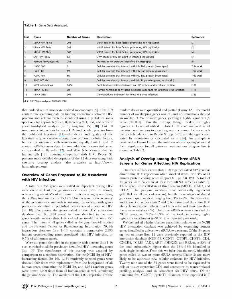
the genome-wide list. The overlaps of the 1,000 repetitions of the
random draws were quantified and plotted (Figure 1A). The modal
number of overlapping genes was 71, and no simulations showed
an overlap of 257 or more genes, yielding a highly significant p-
value (,0.001). Thus the overlap, though modest, is highly
significant. Genes identified in lists 1–10 were analyzed in all
pairwise combinations to identify genes in common between each
pair (detailed data are in Report S1, pp. 5–70) and the significance
tested by simulation or calculated as in [14]. An example is
presented in Figure 1B, and the numbers of overlapping genes and
their significance for all pairwise combinations of gene lists is
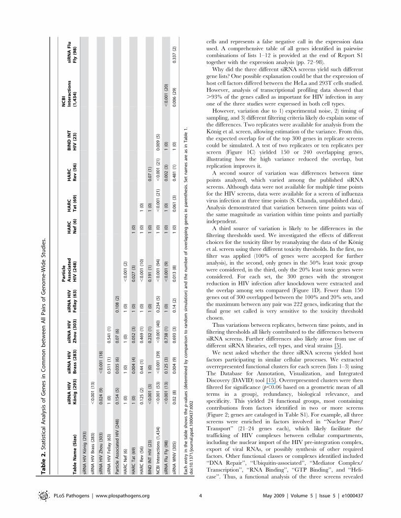
shown in Table 2.
Analysis of Overlap among the Three siRNAScreens for Genes Affecting HIV Replication
The three siRNA screens (lists 1–3) together called 842 genes as
diminishing HIV replication when knocked down, or 3.3% of all
human protein-coding genes (Report S1, pp. 98–120). A total of
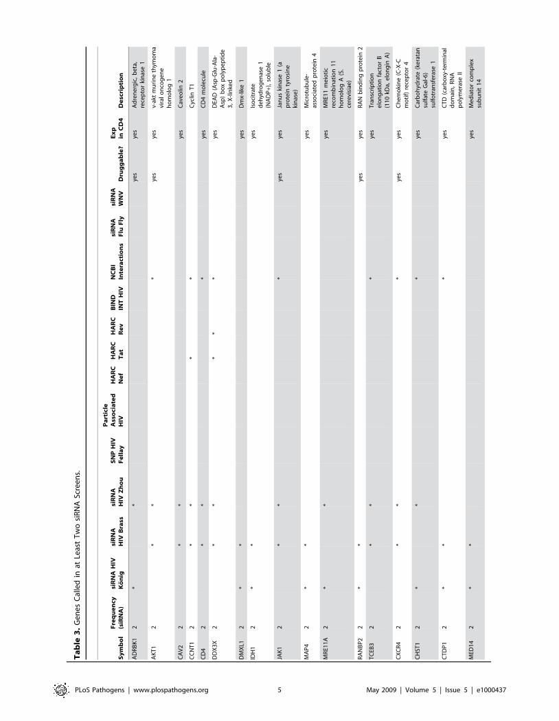
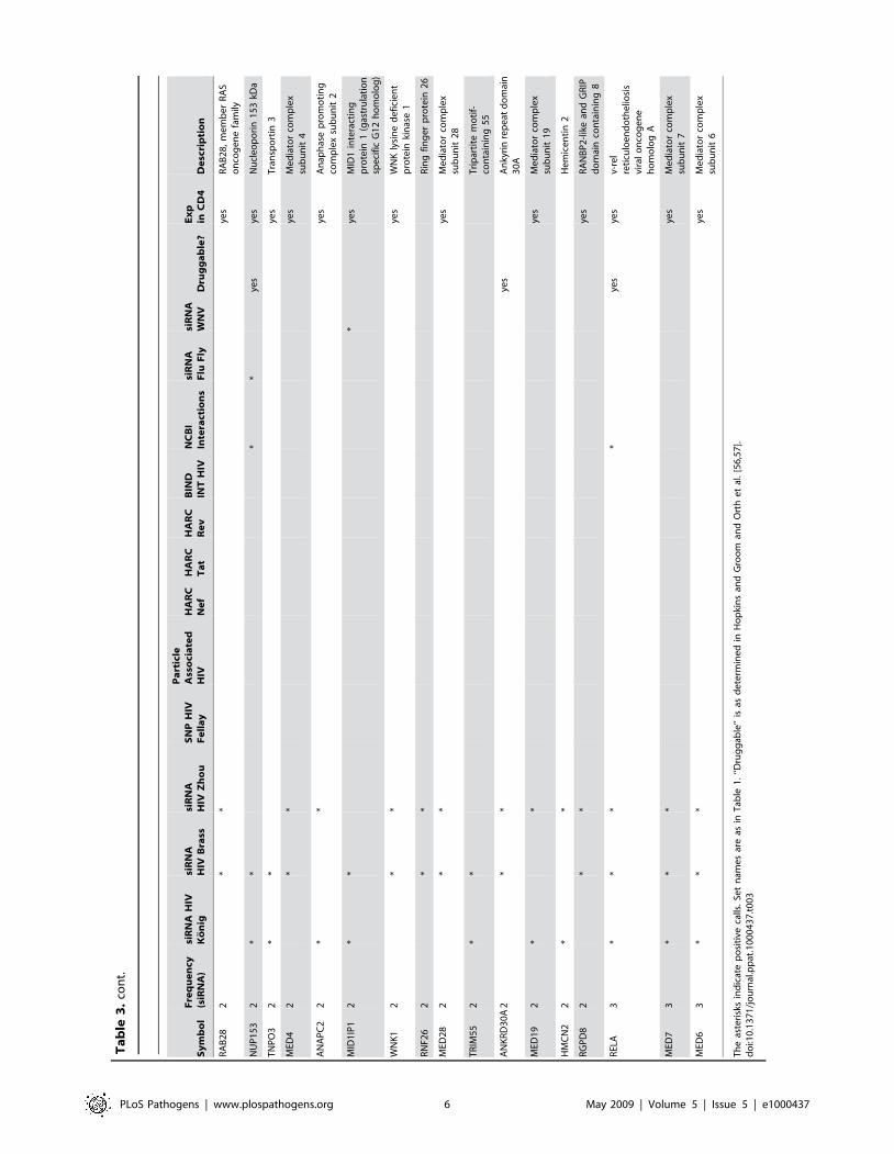
34 genes were called in at least two siRNA screens (Table 3).
Three genes were called in all three screens (MED6, MED7, and
RELA). The pairwise overlaps were statistically significant
(p,0.024 for all pairs of screens), but the percentages of shared
genes were quite modest, ranging from 3% to 6%. The Brass et al.
and Zhou et al. screens (lists 2 and 3) both surveyed the entire HIV
life cycle and studied infection in HeLa cells, and these two share
the greatest overlap (6%). The three siRNA screens identified the
NCBI genes as 13.3%–18.3% of the total, indicating highly
significant enrichment (p,0.001), as reported previously.
We then asked whether further enrichment relative to the NCBI
HIV interaction database was achieved by examining human
genes identified in at least two siRNA two screens. Of the 34 genes
on two or more lists, 11 were previously reported in the HIV
interaction database (NUP153, CCNT1, CTDP1, CHST1, CD4,
CXCR4, TCEB3, JAK1, AKT1, DDX3X, and RELA), or 30% of
the total, substantially higher than the 13%–18% identified in
each single list alone. From this we infer that the newly identified
genes called in two or more siRNA screens (Table 3) are more
likely to be authentic new cellular cofactors for HIV infection.
Twenty-nine out of the 34 genes were found to be expressed in
cells or tissues expressing CD4 and coreceptor by transcriptional
profiling analysis, and so competent for HIV entry. Of the
remaining five, CCNT1 (cyclinT1) is known to be expressed in T
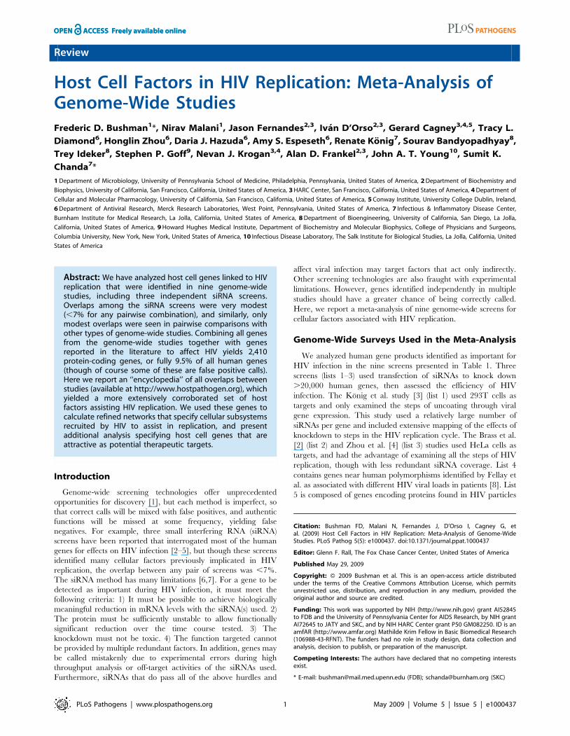
Table 1. Gene Sets Analyzed.
List Name Number of Genes Description Reference
1 siRNA HIV Konig 293 siRNA screen for host factors promoting HIV replication [3]
2 siRNA HIV Brass 283 siRNA screen for host factors promoting HIV replication [2]
3 siRNA HIV Zhou 303 siRNA screen for host factors promoting HIV replication [4]
4 SNP HIV Fellay 63 GWA study of HIV set point in infected individuals [7]
5 Particle Associated HIV 248 Proteins in HIV particles identified by mass spec [8]
6 HARC Nef 6 Cellular proteins that interact with HIV Nef protein (mass spec) This work
7 HARC Tat 69 Cellular proteins that interact with HIV Tat protein (mass spec) This work
8 HARC Rev 56 Cellular proteins that interact with HIV Rev protein (mass spec) This work
9 BIND INT HIV 23 Cellular proteins that interact with HIV IN protein (yeast two hybrid) [9]
10 NCBI Interactions 1434 Published interactions between an HIV protein and a cellular protien [10]
11 siRNA Flu Fly 98 Human homologs of fly gene products important for influenza virus infection [11]
12 siRNA WNV 305 Gene products important for West Nile virus infection [12]
doi:10.1371/journal.ppat.1000437.t001
PLoS Pathogens | www.plospathogens.org 2 May 2009 | Volume 5 | Issue 5 | e1000437
Figure 1. Overlap Analysis to Generate an Empirical p-Value. In each panel the downward arrow indicates the number of genes overlappingbetween the experimental data sets, and the bars show the frequencies of overlaps in comparisons to random distributions. (A) Simulation of overlapbetween all of the genes called in genome-wide screens (lists 1–9) and the NCBI database of factors reported to be involved in HIV replication (list 10).One thousand pairs of gene sets were drawn randomly from the set of all human genes, 1,254 to simulate the set of all genes from genome-sidescreens and 1,434 to simulate NCBI genes, and the overlap in each pair plotted. The experimental overlap was 257 genes. (B) Simulation ofoverlapping genes between the Konig et al. and Zhou et al. siRNA screens. The experimental overlap was nine genes. The p-value calculated usingthe hypergeometric distribution was slightly lower (p = 0.014). (C) Simulation of expected overlap between screens given the measured errorbetween replicates. The standard deviation of infectivity measurements were calculated from the Konig et al. siRNA screens, and then simulateddatasets were generated containing the measured error. For simulations, either two replicates (pink) or ten replicates (yellow) were generated andthe overlap quantified. The y-axis: number of top-scoring genes considered in overlap analysis; x-axis: actual number of overlapping genes seencomparing simulated data sets. (D) Choices for toxicity threshold strongly influence the recovery of genes affecting HIV infection. The genes tested inthe Konig et al. siRNA screen were ranked according to toxicity of knockdown, then sets containing 100% of genes, the least toxic 50%, or the leasttoxic 20% were extracted (top). From each of these, the 300 genes that when knocked down showed the strongest reduction in HIV infection werethen selected, and the overlap between gene sets calculated (bottom).doi:10.1371/journal.ppat.1000437.g001
PLoS Pathogens | www.plospathogens.org 3 May 2009 | Volume 5 | Issue 5 | e1000437
cells and represents a false negative call in the expression data
used. A comprehensive table of all genes identified in pairwise
combinations of lists 1–12 is provided at the end of Report S1
together with the expression analysis (pp. 72–98).
Why did the three different siRNA screens yield such different
gene lists? One possible explanation could be that the expression of
host cell factors differed between the HeLa and 293T cells studied.
However, analysis of transcriptional profiling data showed that
.93% of the genes called as important for HIV infection in any
one of the three studies were expressed in both cell types.
However, variation due to 1) experimental noise, 2) timing of
sampling, and 3) different filtering criteria likely do explain some of
the differences. Two replicates were available for analysis from the
Konig et al. screen, allowing estimation of the variance. From this,
the expected overlap for of the top 300 genes in replicate screens
could be simulated. A test of two replicates or ten replicates per
screen (Figure 1C) yielded 150 or 240 overlapping genes,
illustrating how the high variance reduced the overlap, but
replication improves it.
A second source of variation was differences between time
points analyzed, which varied among the published siRNA
screens. Although data were not available for multiple time points
for the HIV screens, data were available for a screen of influenza
virus infection at three time points (S. Chanda, unpublished data).
Analysis demonstrated that variation between time points was of
the same magnitude as variation within time points and partially
independent.
A third source of variation is likely to be differences in the
filtering thresholds used. We investigated the effects of different
choices for the toxicity filter by reanalyzing the data of the Konig
et al. screen using three different toxicity thresholds. In the first, no
filter was applied (100% of genes were accepted for further
analysis), in the second, only genes in the 50% least toxic group
were considered, in the third, only the 20% least toxic genes were
considered. For each set, the 300 genes with the strongest
reduction in HIV infection after knockdown were extracted and
the overlap among sets compared (Figure 1D). Fewer than 150
genes out of 300 overlapped between the 100% and 20% sets, and
the maximum between any pair was 222 genes, indicating that the
final gene set called is very sensitive to the toxicity threshold
chosen.
Thus variations between replicates, between time points, and in
filtering thresholds all likely contributed to the differences between
siRNA screens. Further differences also likely arose from use of
different siRNA libraries, cell types, and viral strains [5].
We next asked whether the three siRNA screens yielded host
factors participating in similar cellular processes. We extracted
overrepresented functional clusters for each screen (lists 1–3) using
The Database for Annotation, Visualization, and Integrated
Discovery (DAVID) tool [15]. Overrepresented clusters were then
filtered for significance (p,0.06 based on a geometric mean of all
terms in a group), redundancy, biological relevance, and
specificity. This yielded 24 functional groups, most containing
contributions from factors identified in two or more screens
(Figure 2; genes are cataloged in Table S1). For example, all three
screens were enriched in factors involved in ‘‘Nuclear Pore/
Transport’’ (21–24 genes each), which likely facilitate the
trafficking of HIV complexes between cellular compartments,
including the nuclear import of the HIV pre-integration complex,
export of viral RNAs, or possibly synthesis of other required
factors. Other functional classes or complexes identified included
‘‘DNA Repair’’, ‘‘Ubiquitin-associated’’, ‘‘Mediator Complex/
Transcription’’, ‘‘RNA Binding’’, ‘‘GTP Binding’’, and ‘‘Heli-
case’’. Thus, a functional analysis of the three screens revealed
Ta
ble
2.
Stat
isti
cal
An
alys
iso
fG
en
es
inC
om
mo
nb
etw
ee
nA
llP
airs
of
Ge
no
me
-Wid
eSt
ud
ies.
Ta
ble
Na
me
(Siz
e)
siR
NA
HIV
Ko
nig
(29
3)
siR
NA
HIV
Bra
ss(2
83
)si
RN
AH
IVZ
ho
u(3
03
)si
RN
AH
IVF
ell
ay
(63
)
Pa
rtic
leA
sso
cia
ted
HIV
(24
8)
HA
RC
Ne
f(6
)H
AR
CT
at
(69
)H
AR
CR
ev
(56
)B
IND
INT
HIV
(23
)
NC
BI
Inte
ract
ion
s(1
,43
4)
siR
NA
Flu
Fly
(98
)
siR
NA
HIV
Ko
nig
(29
3)
siR
NA
HIV
Bra
ss(2
83
),
0.0
01
(13
)
siR
NA
HIV
Zh
ou
(30
3)
0.0
24
(9)
,0
.00
1(1
8)
siR
NA
HIV
Fella
y(6
3)
1(0
)0
.51
1(1
)0
.54
1(1
)
Par
ticl
eA
sso
ciat
ed
HIV
(24
8)
0.1
54
(5)
0.0
35
(6)
0.0
7(6
)0
.10
8(2
)
HA
RC
Ne
f(6
)1
(0)
1(0
)1
(0)
1(0
),
0.0
01
(2)
HA
RC
Tat
(69
)1
(0)
0.0
04
(4)
0.0
52
(3)
1(0
)0
.02
7(3
)1
(0)
HA
RC
Re
v(5
6)
0.1
25
(2)
0.4
4(1
)0
.46
9(1
)1
(0)
,0
.00
1(1
0)
1(0
)1
(0)
BIN
DIN
TH
IV(2
3)
,0
.00
1(3
)1
(0)
0.2
32
(1)
1(0
)0
.19
1(1
)1
(0)
1(0
)0
.07
(1)
NC
BI
Inte
ract
ion
s(1
,43
4)
,0
.00
1(5
3)
,0
.00
1(3
9)
,0
.00
1(4
0)
0.2
34
(5)
,0
.00
1(9
4)
1(0
),
0.0
01
(21
),
0.0
01
(21
)0
.00
9(5
)
siR
NA
Flu
Fly
(98
),
0.0
01
(13
)0
.12
5(3
)0
.73
8(1
)1
(0)
,0
.00
1(9
)1
(0)
1(0
)0
.00
2(3
)1
(0)
,0
.00
1(2
0)
siR
NA
WN
V(3
05
)0
.02
(8)
0.0
04
(9)
0.6
93
(3)
0.1
4(2
)0
.01
3(8
)1
(0)
0.0
61
(3)
0.4
81
(1)
1(0
)0
.00
6(2
9)
0.3
37
(2)
Each
en
try
inth
eta
ble
sho
ws
the
p-v
alu
es
(de
term
ine
db
yco
mp
aris
on
tora
nd
om
sim
ula
tio
n)
and
the
nu
mb
er
of
ove
rlap
pin
gg
en
es
inp
are
nth
esi
s.Se
tn
ame
sar
eas
inT
able
1.
do
i:10
.13
71
/jo
urn
al.p
pat
.10
00
43
7.t
00
2
PLoS Pathogens | www.plospathogens.org 4 May 2009 | Volume 5 | Issue 5 | e1000437
Ta
ble
3.
Ge
ne
sC
alle
din
atLe
ast
Tw
osi
RN
ASc
ree
ns.
Sy
mb
ol
Fre
qu
en
cy(s
iRN
A)
siR
NA
HIV
Ko
nig
siR
NA
HIV
Bra
sssi
RN
AH
IVZ
ho
uS
NP
HIV
Fe
lla
y
Pa
rtic
leA
sso
cia
ted
HIV
HA
RC
Ne
fH
AR
CT
at
HA
RC
Re
vB
IND
INT
HIV
NC
BI
Inte
ract
ion
ssi
RN
AF
luF
lysi
RN
AW
NV
Dru
gg
ab
le?
Ex
pin
CD
4D
esc
rip
tio
n
AD
RB
K1
2*
*ye
sye
sA
dre
ne
rgic
,b
eta
,re
cep
tor
kin
ase
1
AK
T1
2*
**
yes
yes
v-ak
tm
uri
ne
thym
om
avi
ral
on
cog
en
eh
om
olo
g1
CA
V2
2*
*ye
sC
ave
olin
2
CC
NT
12
**
**
Cyc
linT
1
CD
42
**
*ye
sC
D4
mo
lecu
le
DD
X3
X2
**
**
*ye
sD
EAD
(Asp
-Glu
-Ala
-A
sp)
bo
xp
oly
pe
pti
de
3,
X-l
inke
d
DM
XL1
2*
*ye
sD
mx-
like
1
IDH
12
**
yes
Iso
citr
ate
de
hyd
rog
en
ase
1(N
AD
P+)
,so
lub
le
JAK
12
**
*ye
sye
sJa
nu
ski
nas
e1
(ap
rote
inty
rosi
ne
kin
ase
)
MA
P4
2*
*ye
sM
icro
tub
ule
-as
soci
ate
dp
rote
in4
MR
E11
A2
**
yes
MR
E11
me
ioti
cre
com
bin
atio
n1
1h
om
olo
gA
(S.
cere
visi
ae)
RA
NB
P2
2*
*ye
sye
sR
AN
bin
din
gp
rote
in2
TC
EB3
2*
**
yes
Tra
nsc
rip
tio
ne
lon
gat
ion
fact
or
B(1
10
kDa,
elo
ng
inA
)
CX
CR
42
**
*ye
sye
sC
he
mo
kin
e(C
-X-C
mo
tif)
rece
pto
r4
CH
ST1
2*
**
yes
Car
bo
hyd
rate
(ke
rata
nsu
lfat
eG
al-6
)su
lfo
tran
sfe
rase
1
CT
DP
12
**
*ye
sC
TD
(car
bo
xy-t
erm
inal
do
mai
n,
RN
Ap
oly
me
rase
II
MED
14
2*
*ye
sM
ed
iato
rco
mp
lex
sub
un
it1
4
PLoS Pathogens | www.plospathogens.org 5 May 2009 | Volume 5 | Issue 5 | e1000437
Sy
mb
ol
Fre
qu
en
cy(s
iRN
A)
siR
NA
HIV
Ko
nig
siR
NA
HIV
Bra
sssi
RN
AH
IVZ
ho
uS
NP
HIV
Fe
lla
y
Pa
rtic
leA
sso
cia
ted
HIV
HA
RC
Ne
fH
AR
CT
at
HA
RC
Re
vB
IND
INT
HIV
NC
BI
Inte
ract
ion
ssi
RN
AF
luF
lysi
RN
AW
NV
Dru
gg
ab
le?
Ex
pin
CD
4D
esc
rip
tio
n
RA
B2
82
**
yes
RA
B2
8,
me
mb
er
RA
So
nco
ge
ne
fam
ily
NU
P1
53
2*
**
*ye
sye
sN
ucl
eo
po
rin
15
3kD
a
TN
PO
32
**
yes
Tra
nsp
ort
in3
MED
42
**
yes
Me
dia
tor
com
ple
xsu
bu
nit
4
AN
AP
C2
2*
*ye
sA
nap
has
ep
rom
oti
ng
com
ple
xsu
bu
nit
2
MID
1IP
12
**
*ye
sM
ID1
inte
ract
ing
pro
tein
1(g
astr
ula
tio
nsp
eci
fic
G1
2h
om
olo
g)
WN
K1
2*
*ye
sW
NK
lysi
ne
de
fici
en
tp
rote
inki
nas
e1
RN
F26
2*
*R
ing
fin
ge
rp
rote
in2
6
MED
28
2*
*ye
sM
ed
iato
rco
mp
lex
sub
un
it2
8
TR
IM5
52
**
Tri
par
tite
mo
tif-
con
tain
ing
55
AN
KR
D3
0A
2*
*ye
sA
nky
rin
rep
eat
do
mai
n3
0A
MED
19
2*
*ye
sM
ed
iato
rco
mp
lex
sub
un
it1
9
HM
CN
22
**
He
mic
en
tin
2
RG
PD
82
**
yes
RA
NB
P2
-lik
ean
dG
RIP
do
mai
nco
nta
inin
g8
REL
A3
**
**
yes
yes
v-re
lre
ticu
loe
nd
oth
elio
sis
vira
lo
nco
ge
ne
ho
mo
log
A
MED
73
**
*ye
sM
ed
iato
rco
mp
lex
sub
un
it7
MED
63
**
*ye
sM
ed
iato
rco
mp
lex
sub
un
it6
Th
eas
teri
sks
ind
icat
ep
osi
tive
calls
.Se
tn
ame
sar
eas
inT
able
1.
‘‘Dru
gg
able
’’is
asd
ete
rmin
ed
inH
op
kin
san
dG
roo
man
dO
rth
et
al.
[56
,57
].d
oi:1
0.1
37
1/j
ou
rnal
.pp
at.1
00
04
37
.t0
03
Ta
ble
3.
con
t.
PLoS Pathogens | www.plospathogens.org 6 May 2009 | Volume 5 | Issue 5 | e1000437
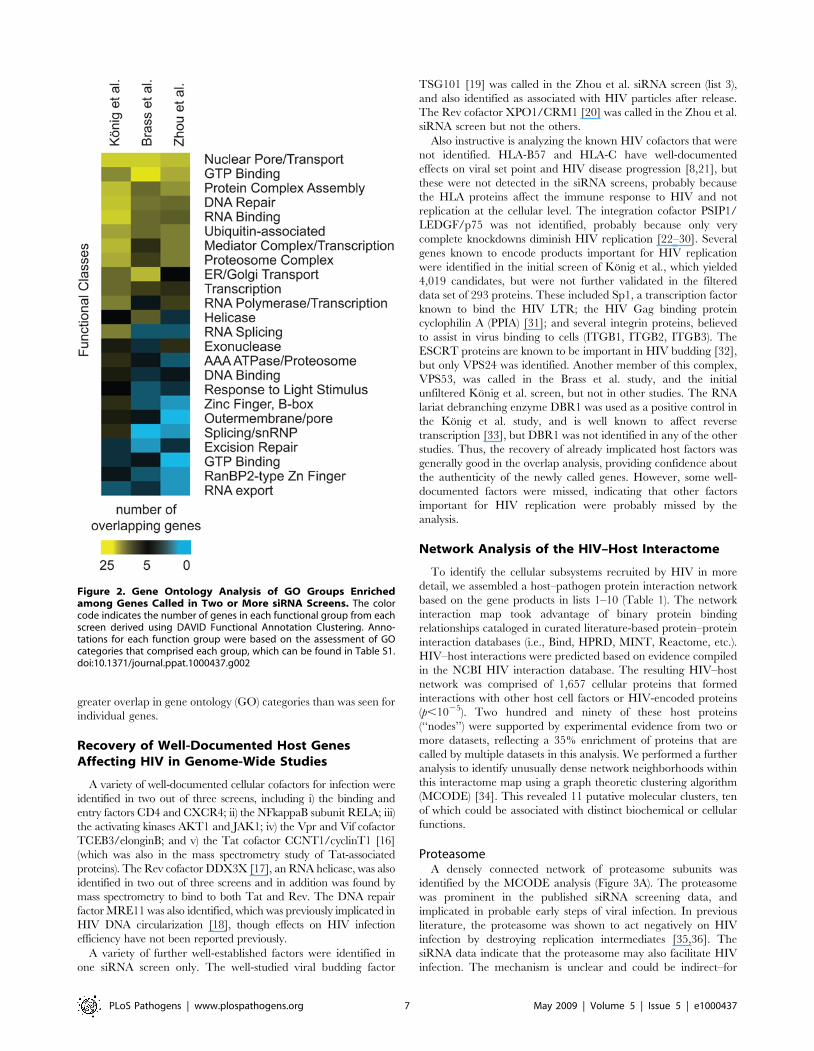
greater overlap in gene ontology (GO) categories than was seen for
individual genes.
Recovery of Well-Documented Host GenesAffecting HIV in Genome-Wide Studies
A variety of well-documented cellular cofactors for infection were
identified in two out of three screens, including i) the binding and
entry factors CD4 and CXCR4; ii) the NFkappaB subunit RELA; iii)
the activating kinases AKT1 and JAK1; iv) the Vpr and Vif cofactor
TCEB3/elonginB; and v) the Tat cofactor CCNT1/cyclinT1 [16]
(which was also in the mass spectrometry study of Tat-associated
proteins). The Rev cofactor DDX3X [17], an RNA helicase, was also
identified in two out of three screens and in addition was found by
mass spectrometry to bind to both Tat and Rev. The DNA repair
factor MRE11 was also identified, which was previously implicated in
HIV DNA circularization [18], though effects on HIV infection
efficiency have not been reported previously.
A variety of further well-established factors were identified in
one siRNA screen only. The well-studied viral budding factor
TSG101 [19] was called in the Zhou et al. siRNA screen (list 3),
and also identified as associated with HIV particles after release.
The Rev cofactor XPO1/CRM1 [20] was called in the Zhou et al.
siRNA screen but not the others.
Also instructive is analyzing the known HIV cofactors that were
not identified. HLA-B57 and HLA-C have well-documented
effects on viral set point and HIV disease progression [8,21], but
these were not detected in the siRNA screens, probably because
the HLA proteins affect the immune response to HIV and not
replication at the cellular level. The integration cofactor PSIP1/
LEDGF/p75 was not identified, probably because only very
complete knockdowns diminish HIV replication [22–30]. Several
genes known to encode products important for HIV replication
were identified in the initial screen of Konig et al., which yielded
4,019 candidates, but were not further validated in the filtered
data set of 293 proteins. These included Sp1, a transcription factor
known to bind the HIV LTR; the HIV Gag binding protein
cyclophilin A (PPIA) [31]; and several integrin proteins, believed
to assist in virus binding to cells (ITGB1, ITGB2, ITGB3). The
ESCRT proteins are known to be important in HIV budding [32],
but only VPS24 was identified. Another member of this complex,
VPS53, was called in the Brass et al. study, and the initial
unfiltered Konig et al. screen, but not in other studies. The RNA
lariat debranching enzyme DBR1 was used as a positive control in
the Konig et al. study, and is well known to affect reverse
transcription [33], but DBR1 was not identified in any of the other
studies. Thus, the recovery of already implicated host factors was
generally good in the overlap analysis, providing confidence about
the authenticity of the newly called genes. However, some well-
documented factors were missed, indicating that other factors
important for HIV replication were probably missed by the
analysis.
Network Analysis of the HIV–Host Interactome
To identify the cellular subsystems recruited by HIV in more
detail, we assembled a host–pathogen protein interaction network
based on the gene products in lists 1–10 (Table 1). The network
interaction map took advantage of binary protein binding
relationships cataloged in curated literature-based protein–protein
interaction databases (i.e., Bind, HPRD, MINT, Reactome, etc.).
HIV–host interactions were predicted based on evidence compiled
in the NCBI HIV interaction database. The resulting HIV–host
network was comprised of 1,657 cellular proteins that formed
interactions with other host cell factors or HIV-encoded proteins
(p,1025). Two hundred and ninety of these host proteins
(‘‘nodes’’) were supported by experimental evidence from two or
more datasets, reflecting a 35% enrichment of proteins that are
called by multiple datasets in this analysis. We performed a further
analysis to identify unusually dense network neighborhoods within
this interactome map using a graph theoretic clustering algorithm
(MCODE) [34]. This revealed 11 putative molecular clusters, ten
of which could be associated with distinct biochemical or cellular
functions.
ProteasomeA densely connected network of proteasome subunits was
identified by the MCODE analysis (Figure 3A). The proteasome
was prominent in the published siRNA screening data, and
implicated in probable early steps of viral infection. In previous
literature, the proteasome was shown to act negatively on HIV
infection by destroying replication intermediates [35,36]. The
siRNA data indicate that the proteasome may also facilitate HIV
infection. The mechanism is unclear and could be indirect–for
Figure 2. Gene Ontology Analysis of GO Groups Enrichedamong Genes Called in Two or More siRNA Screens. The colorcode indicates the number of genes in each functional group from eachscreen derived using DAVID Functional Annotation Clustering. Anno-tations for each function group were based on the assessment of GOcategories that comprised each group, which can be found in Table S1.doi:10.1371/journal.ppat.1000437.g002
PLoS Pathogens | www.plospathogens.org 7 May 2009 | Volume 5 | Issue 5 | e1000437
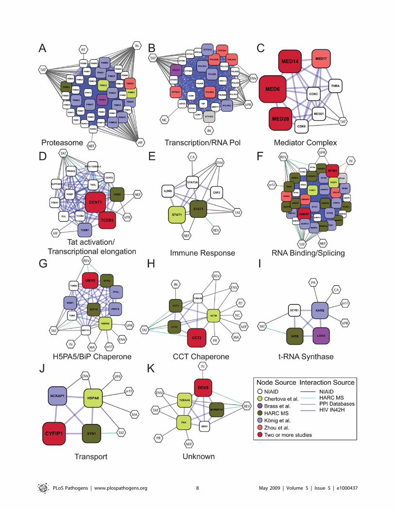
PLoS Pathogens | www.plospathogens.org 8 May 2009 | Volume 5 | Issue 5 | e1000437
example, reducing proteasome activity may alter cellular ubiquitin
levels, and so affect HIV replication by altering the free ubiquitin
pool.
Transcription/RNA PolymeraseGenes for subunits of RNA polymerase II and associated factors
were identified in several different screens, yielding a densely
connected network (Figure 3B). In some of the siRNA screens, the
knockdown of Pol II subunits was mapped to the step of Tat
transactivation. Because so many subunits were identified, the
simplest interpretation is that reduced dosage of the full complex is
responsible for the deficit in HIV replication.
Mediator ComplexMultiple subunits of the mediator complex were identified in
two or more siRNA screens (Table 3 and Figure 3C). The
mediator complex links transcriptional activator proteins to the
RNA polymerase II basal transcription apparatus, thereby
allowing transcriptional activation [37,38]. The observation that
so many subunits were identified suggests that activity of the
complex as a whole is the target of siRNA modulation. Viral
replication cycle mapping by Zhou et al. indicated that some of the
mediator proteins were needed to support Tat-activated tran-
scription, though studies in Konig et al. suggest a possible further
role in reverse transcription. The data can be accommodated in a
model where changes in dosage in the mediator complex are not
toxic to cells, but where Tat-activated transcription is extremely
sensitive to mediator dosage. Previously, mediator was shown to be
important for Sp1-driven transcription, and Sp1 is required for
transcription from the HIV LTR, suggesting possible involvement
of Sp1 as well.
Tat Activation/Transcriptional ElongationA dense network was formed containing the Tat cofactor cyclin
T1 (CCNT1) [16], which was identified in two out of three siRNA
screens and by mass spectrometry (Figure 3D). Together with its
binding partner CDK9, which was identified as a Tat binding
protein, cyclinT1 forms positive transcription elongation factor b
(P-TEFb). The MCODE analysis links the P-TEFb complex and
the elongin complex involved in transcriptional elongation.
Another factor, the RNA Pol II carboxyl-terminal domain
(CTD) phosphatase CTDP1, was also identified and was also
previously associated with Tat activation. In addition, two STAT
proteins, also involved in transcription and NFkappaB signaling
and implicated in lentiviral infection [39], were identified
(Figure 3E).
RNA Binding/SplicingA large cluster of RNA binding and splicing proteins was
identified in the MCODE analysis. Eleven of the cellular genes
encode protein components of hnRNP complexes (HNR factors)
that form on pre-mRNA and direct splicing and other activities.
HNRNPU contains both an RNA binding domain and a DNA
binding domain that mediates attachment to the nuclear scaffold,
potentially linking sites of mRNA synthesis to specific sub-nuclear
locations. Six further genes (SF3 factors) encode components of the
splicing factor 3 a/b complex, which is involved in activating the
U2 snRNP and promoting splicing. Three SNR proteins and two
SF proteins were also identified and are implicated in RNA
splicing and RNP formation. Several of these proteins were
implicated in the literature to modulate Tat or Rev function (e.g.,
[40–42]), and seven direct binding interactions to these viral
proteins were identified in the mass spectrometry data reported
here.
Several observations also suggest possible connections of
RNAP/splicing factors to the viral DNA integration step. Two
components of the splicing factor SF3 bound integrase in the yeast
two-hybrid data (list 9) [10]. The splicing protein SNW1/SKIIP1
was found by Konig et al. be selectively important at the
integration step [3]. The integrase-interacting protein PSIP1/
LEDGF/p75 appears to tether integrase to active transcription
units [23,29,43,44], and an alternatively spliced variant of this
protein (p52) is involved in RNA metabolism [45]. Though
indirect, these observations suggest a model in which splicing
factors may help recruit integrase to active transcription units,
which are favored for integration [46–48].
Another possible role of splicing factors is in maintaining the
proper balance between spliced and incompletely spliced HIV
RNAs. HIV replication requires multiply spliced messages
(encoding Tat, Rev, and Nef), singly spliced messages (encoding
Vif, Vpr, Env/Vpu, and a second form of Tat), and unspliced
messages (encoding Gag and Gag-Pol). The unspliced RNA also
serves as the genomic RNA. Alterations in dosage of splicing
factors by siRNA knockdown may well diminish HIV replication
by altering the ratios of the different HIV mRNA forms.
The BiP/GRP78/HSPA5 ChaperoneThe BiP/GRP78/HSPA5 protein chaperone was identified in
the network analysis (Figure 3G). BiP/GRP78/HSPA5 is a
member of the HSP70 family that is involved in the folding and
assembly of proteins in the endoplasmic reticulum. BiP has been
implicated in interacting with newly synthesized HIV gp160 SU/
TM precursor [49], and HSP70 family members have been
proposed to interact with Gag, Tat, Vpr, and MA. The MCODE
analysis connected BiP/GRP78/HSPA5 to a collection of nuclear
proteins involved in splicing (PRPF8, SFPQ, and SNW1), nuclear
matrix architecture (MATR3), and ubiquitylation (UBR5).
Determining how these cellular proteins modulate the interactions
of BiP/GRP78/HSPA5 with HIV proteins offers a potential route
to better understanding protein folding and sorting during HIV
replication.
The CCT ChaperoneThe MCODE analysis identified subunits of the chaperone
containing TCP1 (CCT) complex (Figure 3H). Subunits were
identified in siRNA screens, in HIV particles after budding, and
also as Tat binding proteins. This complex consists of two identical
stacked rings of eight subunits. Unfolded proteins are thought to
Figure 3. Gene Clusters, Generated Using PPI and MCODE Analysis, Derived from the Full Set of Genes Implicated in HIV Infection(Lists 1–10). The size of each node is proportional to the number of screens in which the host cell gene was called. Gene identifiers are in Table S1.Diamonds indicate genes from the NCBI HIV interactions database. Color code: red = Konig et al., green = Brass et al., blue = Zhou et al., cyan = Fellayet al., magenta = Frankel interaction screens (unpublished data), yellow = HIV particle associated, and grey = Studamire and Goff integraseinteractions. For genes that were called in multiple screens (larger symbols), a color was chosen arbitrarily from among the screens positive for thatgene. Default parameters were used, specifically— Degree Cutoff: 2. Node Score Cutoff: 0.0. Haircut: true. Fluff: false. K-Core: 2. Maximum Depth FromSeed: 100. (A) Proteasome; (B) Transcription/RNA Pol; (C) Mediator Complex; (D) Tat activation/Transcriptional elongation; (E) Immune response; (F)RNA Binding/Splicing; (G) H5PA5/BiP Chaperone; (H) CCT Chaperone; (I) t-RNA Synthase; (J) Transport; (K) Unknown.doi:10.1371/journal.ppat.1000437.g003
PLoS Pathogens | www.plospathogens.org 9 May 2009 | Volume 5 | Issue 5 | e1000437
pass through the central cavity, and become folded in an ATP-
dependent manner. The CCT chaperone has not previously been
associated with HIV replication, and represents a new candidate
for involvement in Tat activation and HIV budding.
Additional Densely Connected ClustersSeveral further functions were identified, including proteins
involved in t-RNA synthase function, transport, and one of
unknown function (Figure 3 I–3K). The t-RNA synthase and
transport complexes contained members that associated with Tat
according to the mass spectrometry study, and the unknown
complex contained a member binding to Rev, suggesting specific
links to HIV replication.
Other Newly Identified FunctionsSeveral sets of proteins were identified that were not called as
densely connected networks but appear to be functionally related.
The nuclear pore and associated factors were clustered in the
initial MCODE network but were not sufficiently densely to
emerge as a densely connected network. Proteins involved in
nuclear import identified in two out of three siRNA screens
included products of NUP153, RANBP2, TNPO3, and RGPD8.
NUP153 and TNPO3 have been associated with the trafficking of
HIV proteins previously [2,3,50–52]. RANBP2 is a giant gene
encoding a product that accumulates at nuclear pores and binds to
RAN, which is a small GTP-binding protein of the RAS
superfamily. RANBP2 also contains FG repeats, a cyclophilin-
related nucleoporin, and a domain that binds UBC9, the E2 for
SUMO1 transfer. RGPD8 is named for ‘‘RANBP2-like and GRIP
domain containing 8’’. It too accumulates at the nuclear pore and
is believed to assist in RNA and protein transport. The actions of
NUP153 and TNPO3 have been mapped to nuclear import of the
HIV preintegration complex in [2,3,51], and NUP153 has also
been proposed to be involved in export of HIV Rev [53].
Three genes were identified that affect the microtubule system.
MAP4 is a microtubule-associated protein that has not previously
been studied in detail. MID1IP1 is a regulator of microtubule
polymerization. CAV2 (caveolin 2) is involved in the formation of
plasma membrane invaginations involved in a variety of cellular
functions including signal transduction, cell growth, and apoptosis.
Caveoli have also been implicated as interacting with the
microtubule network [54]. Previous studies have suggested that
HIV particles may traffic along microtubules to reach the nucleus
[55]. Thus MAP4, MID1IP1, and possibly CAV2 are candidates
for cofactors in this process. Other proteins were also called in two
siRNA screens (ANAPC2, DMXL1, HMCN2, and IDH1) but are
of unknown function (Table 3).
Identifying New Drug Targets
One of the main reasons for carrying out the screens for host
factors is the hope of identifying new targets for HIV therapeutics.
Several studies have indentified potentially ‘‘druggable’’ human
proteins by cataloging families of InterPro domains where one
member is the target of one or more small molecule inhibitors with
drug-like properties. All members of the family are then proposed
as potential drug targets [56,57] (John Hogenesch, data available
at http://www1.qiagen.com/Products/GeneSilencing/LibrarySiRna/
SiRnaSets/HumanDruggableGenomesiRNASetV30.aspx?ShowInfo
=1). In Report S1 (pp. 72–120), we annotate our overlap study for
‘‘druggable’’ targets by these criteria. Focusing on an updated version
of the list from Hopkins and Groom [56], we found that eight of the
34 genes common to the two siRNA screens were called as potential
drug targets (Table 3, column labeled ‘‘druggable’’). Two of these
eight are in fact known to be the targets of small molecules with
activity against HIV. CXCR4 is the target of AMD3100 and related
molecules [58,59], and AKT1 is the target of miltefosine [60]. The
fact that two out of eight genes called as druggable are known
antiviral drug targets (at least in tissue culture) suggests that this
analysis is yielding viable new targets. Of the additional genes
encoding candidate drug targets, inhibitors have been reported for
the kinases ADRBK1/GRK2 and JAK1. These can be tested for
activity against HIV in cell culture. Annotation of the larger collection
of genes that were found on two or more lists (lists 1–9) yielded a
further 56 genes encoding potentially druggable cellular factors.
Summary
Analysis of genes called as important for HIV replication in
multiple genome-wide screens yielded a list rich in well-known
factors and also intriguing new candidates. Many important
factors were surely missed by this approach, but at least some of
the most promising new genes can be distilled from among the
9.5% of all human protein-coding genes now proposed to affect
HIV infection. Many of the new genes can be linked into clusters,
specifying cellular subsystems associated with HIV replication.
Promising drug targets could be discerned among the best-
documented new factors.
Methods
Overlap analysis and comparisons to random distributions were
carried out using R [61]. The p-values for overlaps between lists
were generated by comparison to results of random simulation and
by calculation based on the hypergeometric distribution as in [14].
No correction was applied for multiple comparisons. Networks
were generated using MCODE analysis on the binary interaction
file and plotted in cytoscape. The global interaction network was
judged to be statistically significant by comparison to random
simulation (p,1025). Subnetworks were selected that 1) contained
at least two proteins from different studies and 2) showed high
connectivity. The protein–protein interaction data for Nef, Tat,
and Rev from the HARC Center were derived using previously
described methods (LC MS-MS followed by database matching)
[62]. A more thorough analysis of these (early stage) data will be
reported elsewhere.
Updated versions of Report S1 documenting the overlap among
genome-wide screens can be found at http://www.hostpathogen.
org.
Supporting Information
Report S1 Gene List Comparison Report
Found at: doi:10.1371/journal.ppat.1000437.s001 (1.88 MB PDF)
Table S1 Gene IDs from Functional Clusters in Figure 3
Found at: doi:10.1371/journal.ppat.1000437.s002 (0.09 MB XLS)
Acknowledgments
We particularly thank Yingyao Zhou for help with the statistical analysis of
overlap among the siRNA screens. We thank Jenny Gu, Jiewei Xu, Marie
Fahey, and Melanie Brewer for technical assistance with mass spectrometry
and analyses.
PLoS Pathogens | www.plospathogens.org 10 May 2009 | Volume 5 | Issue 5 | e1000437
References
1. Rines DR, Tu B, Miraglia L, Welch GL, Zhang J, et al. (2006) High-content
screening of functional genomic libraries. Methods Enzymol 414: 530–565.
2. Brass AL, Dykxhoorn DM, Benita Y, Yan N, Engelman A, et al. (2008)Identification of host proteins required for HIV infection through a functional
genomic screen. Science 319(5865): 921–926. doi:10.1126/science.1152725.
3. Konig R, Zhou Y, Elleder D, Diamond TL, Bonamy GM, et al. (2008) Globalanalysis of host-pathogen interactions that regulate early-stage HIV-1 replica-
tion. Cell 135(1): 49–60.
4. Zhou H, Xu M, Huang Q, Gates AT, Zhang XD, et al. (2008) Genome-scaleRNAi screen for host factors required for HIV replication. Cell Host Microbe
4(5): 495–504. doi:10.1016/j.chom.2008.10.004.
5. Goff SP (2008) Knockdown screens to knockout HIV-1. Cell 135(3): 417–420.
6. Hutvagner G, Zamore PD (2002) RNAi: Nature abhors a double-strand. CurrOpin Genet Dev 12(2): 225–232.
7. Konig R, Chiang CY, Tu BP, Yan SF, DeJesus PD, et al. (2007) A probability-
based approach for the analysis of large-scale RNAi screens. Nat Methods 4(10):847–849. doi:10.1038/nmeth1089.
8. Fellay J, Shianna KV, Ge D, Colombo S, Ledergerber B, et al. (2007) A whole-
genome association study of major determinants for host control of HIV-1.
Science 317(5840): 944–947. doi:10.1126/science.1143767.9. Chertova E, Chertov O, Coren LV, Roser JD, Trubey CM, et al. (2006)
Proteomic and biochemical analysis of purified human immunodeficiency virus
type 1 produced from infected monocyte-derived macrophages. J Virol 80(18):9039–9052.
10. Studamire B, Goff SP (2008) Host proteins interacting with the moloney murine
leukemia virus integrase: Multiple transcriptional regulators and chromatinbinding factors. Retrovirology 5: 48. doi:10.1186/1742-4690-5-48.
11. Fu W, Sanders-Beer BE, Katz KS, Maglott DR, Pruitt KD, et al. (2009) Human
immunodeficiency virus type 1, human protein interaction database at NCBI.Nucleic Acids Res 37(Database issue): D417–D422. Epub 15 Oct 2008.
doi:10.1093/nar/gkn708.
12. Hao L, Sakurai A, Watanabe T, Sorensen E, Nidom CA, et al. (2008)Drosophila RNAi screen identifies host genes important for influenza virus
replication. Nature 454(7206): 890–893. doi:10.1038/nature07151.
13. Krishnan MN, Ng A, Sukumaran B, Gilfoy FD, Uchil PD, et al. (2008) RNAinterference screen for human genes associated with west nile virus infection.
Nature 455(7210): 242–245.
14. Fury W, Batliwalla F, Gregersen PK, Li W (2006) Overlapping probabilities oftop ranking gene lists, hypergeometric distribution, and stringency of gene
selection criterion. Conf Proc IEEE Eng Med Biol Soc 1: 5531–5534.
15. Dennis G, Sherman BT, Hosack DA, Yang J, Gao W, et al. (2003) Database for
annotation, visualization, and integrated discovery. Genome Bio 4: P3.16. Wei P, Garber ME, Fang SM, Fischer WH, Jones KA (1998) A novel CDK9-
associated C-type cyclin interacts directly with HIV-1 tat and mediates its high-
affinity, loop-specific binding to TAR RNA. Cell 92: 451–462.
17. Yedavalli VS, Neuveut C, Chi YH, Kleiman L, Jeang KT (2004) Requirementof DDX3 DEAD box RNA helicase for HIV-1 rev-RRE export function. Cell
119(3): 381–392.
18. Kilzer JM, Stracker TH, Beitzel B, Meek K, Weitzman MD, et al. (2003) Rolesof host cell factors in circularization of retroviral DNA. Virology 314: 460–467.
19. Garrus JE, von Schwedler UK, Pornillos OW, Morham SG, Zavitz KH, et al.
(2001) Tsg101 and the vacuolar protein sorting pathway are essential for hiv-1budding. Cell 107(1): 55–65.
20. Neville M, Stutz F, Lee L, Davis LI, Rosbash M (1997) The importin-beta family
member Crm1p bridges the interaction between rev and the nuclear porecomplex during nuclear export. Curr Biol 7(10): 767–775.
21. Matthews PC, Prendergast A, Leslie A, Crawford H, Payne R, et al. (2008)
Central role of reverting mutations in HLA associations with humanimmunodeficiency virus set point. J Virol 82(17): 8548–8559. doi:10.1128/
JVI.00580-08.
22. Llano M, Saenz DT, Meehan A, Wongthida P, Peretz M, et al. (2006) Anessential role for LEDGF/p75 in HIV integration. Science 314(5798): 461–464.
23. Marshall H, Ronen K, Berry C, Llano M, Sutherland H, et al. (2007) Role of
PSIP1/LEDGF/p75 in lentiviral infectivity and integration targeting. PLoSONE 2(12): e1340. doi:10.1371/journal.pone.0001340.
24. Ciuffi A, Diamond T, Hwang Y, Marshall H, Bushman FD (2006) Fusions of
LEDGF/p75 to lambda repressor promote HIV DNA integration near lambda
operators in vitro. Hum Gene Ther 17: 960–967.25. Llano M, Vanegas M, Fregoso O, Saenz D, Chung S, et al. (2004) LEDGF/p75
determines cellular trafficking of diverse lentiviral but not murine oncoretroviral
integrase proteins and is a component of functional lentiviral preintegrationcomplexes. J Virol 78(17): 9524–9537.
26. Cherepanov P (2007) LEDGF/p75 interacts with divergent lentiviral integrases
and modulates their enzymatic activity in vitro. Nucleic Acids Res 35(1):113–124.
27. Cherepanov P, Maertens G, Proost P, Devreese B, Van Beeumen J, et al. (2003)
HIV-1 integrase forms stable tetramers and associates with LEDGF/p75 proteinin human cells. J Biol Chem 278: 372–381.
28. Maertens G, Cherepanov P, Pluymers W, Busschots K, De Clercq E, et al.
(2003) LEDGF/p75 is essential for nuclear and chromosomal targeting of HIV-1integrase in human cells. J Biol Chem 278: 33528–33539.
29. Ciuffi A, Llano M, Poeschla E, Hoffmann C, Leipzig J, et al. (2005) A role forLEDGF/p75 in targeting HIV DNA integration. Nat Med 11(12): 1287–1289.
30. Turlure F, Devroe E, Silver PA, Engelman A (2004) Human cell proteins and
human immunodeficiency virus DNA integration. Front Biosci 9: 3187–3208.
31. Gamble TR, Vajdos FF, Yoo S, Worthylake DK, Houseweart M, et al. (1996)
Crystal structure of human cyclophillin A bound to the amino-terminal domainof HIV-1 capsid. Cell 87: 1285–1294.
32. von Schwedler UK, Stuchell M, Muller B, Ward DM, Chung HY, et al. (2003)
The protein network of HIV budding. Cell 114(6): 701–713.
33. Ye Y, De Leon J, Yokoyama N, Naidu Y, Camerini D (2005) DBR1 siRNAinhibition of HIV-1 replication. Retrovirology 2: 63. doi:10.1186/1742-4690-2-
63.
34. Bader GD, Hogue CW (2003) An automated method for finding molecularcomplexes in large protein interaction networks. BMC Bioinformatics 4: 2.
35. Schwartz O, V M, Friguet B, Arenzana-Seisdedos F, Heard J (1998) Antiviral
activity of the proteasome on incoming human immunodeficiency virus type 1.
J Virol 72: 3845–3850.
36. Butler SL, Johnson EP, Bushman FD (2002) HIV cDNA metabolism studied byfluorescence-monitored PCR: Notable stability of two-LTR circles. J Virol 76:
3739–3747.
37. Kuras L, Borggrefe T, Kornberg RD (2003) Association of the mediatorcomplex with enhancers of active genes. Proc Natl Acad Sci U S A 100(24):
13887–13891. doi:10.1073/pnas.2036346100.
38. Kornberg RD (2007) The molecular basis of eukaryotic transcription. Proc Natl
Acad Sci U S A 104(32): 12955–12961. doi:10.1073/pnas.0704138104.
39. Mohan M, Aye PP, Borda JT, Alvarez XJ, Lackner AA (2007) Gastrointestinaldisease in SIV-infected rhesus macaques is characterized by proinflammatory
dysregulation of the IL-6-JAK-STAT3 pathway. Am J Pathol 171(6):1952–1965.
40. Pongoski J, Asai K, Cochrane A (2002) Positive and negative modulation of
human immunodeficiency virus type 1 rev function by cis and trans regulators of
viral RNA splicing. J Virol 76(10): 5108–5120.
41. Roy BB, Hu J, Guo X, Russell RS, Guo F, et al. (2006) Association of RNAhelicase a with human immunodeficiency virus type 1 particles. J Biol Chem
281(18): 12625–12635. doi:10.1074/jbc.M510596200.
42. Li J, Tang H, Mullen TM, Westberg C, Reddy TR, et al. (1999) A role for RNAhelicase A in post-transcriptional regulation of HIV type 1. Proc Natl Acad
Sci U S A 96(2): 709–714.
43. Shun MC, Raghavendra NK, Vandegraaff N, Daigle JE, Hughes S, et al. (2007)LEDGF/p75 functions downstream from preintegration complex formation to
effect gene-specific HIV-1 integration. Genes Dev 21(14): 1767–1778.
44. Ciuffi A, Diamond TL, Hwang Y, Marshall HM, Bushman FD (2006)
Modulating target site selection during human immunodeficiency virus DNAintegration in vitro with an engineered tethering factor. Hum Gene Ther 17(9):
960–967.
45. Ge H, Si Y, Roeder RG (1998) Isolation of cDNAs encoding novel transcriptioncoactivators p52 and p75 reveals an alternate regulatory mechanism of
transcriptional activation. EMBO J 17: 6723–6729.
46. Schroder AR, Shinn P, Chen H, Berry C, Ecker JR, et al. (2002) HIV-1
integration in the human genome favors active genes and local hotspots. Cell110(4): 521–529.
47. Mitchell RS, Beitzel BF, Schroder AR, Shinn P, Chen H, et al. (2004) Retroviral
DNA integration: ASLV, HIV, and MLV show distinct target site preferences.PLoS Biol 2(8): e234. doi:10.1371/journal.pbio.0020234.
48. Wu X, Li Y, Crise B, Burgess SM (2003) Transcription start regions in the
human genome are favored targets for MLV integration. Science 300(5626):
1749–1751.
49. Earl PL, Moss B, Doms RW (1991) Folding, interaction with GRP78-BiP,assembly, and transport of the human immunodeficiency virus type 1 envelope
protein. J Virol 65(4): 2047–2055.
50. Luban J (2008) HIV-1 infection: Going nuclear with TNPO3/Transportin-SR2and integrase. Curr Biol 18(16): R710–R713.
51. Christ F, Thys W, De Rijck J, Gijsbers R, Albanese A, et al. (2008) Transportin-
SR2 imports HIV into the nucleus. Curr Biol 18(16): 1192–1202.
52. Varadarajan P, Mahalingam S, Liu P, Ng SB, Gandotra S, et al. (2005) Thefunctionally conserved nucleoporins Nup124p from fission yeast and the human
Nup153 mediate nuclear import and activity of the Tf1 retrotransposon and
HIV-1 vpr. Mol Biol Cell 16(4): 1823–1838. doi:10.1091/mbc.E04-07-0583.
53. Zolotukhin AS, Felber BK (1999) Nucleoporins nup98 and nup214 participatein nuclear export of human immunodeficiency virus type 1 rev. J Virol 73(1):
120–127.
54. Ito J, Kheirollah A, Nagayasu Y, Lu R, Kato K, et al. (2006) Apolipoprotein A-Iincreases association of cytosolic cholesterol and caveolin-1 with microtubule
cytoskeletons in rat astrocytes. J Neurochem 97(4): 1034–1043. doi:10.1111/
j.1471-4159.2006.03805.x.
55. McDonald D, Vodicka MA, Lucero G, Svitkina TM, Borisy GG, et al. (2002)Visualization of the intracellular behavior of HIV in living cells. J Cell Biol
159(3): 441–52.
56. Hopkins AL, Groom CR (2002) The druggable genome. Nat Rev Drug Discov1(9): 727–730.
PLoS Pathogens | www.plospathogens.org 11 May 2009 | Volume 5 | Issue 5 | e1000437
57. Orth AP, Batalov S, Perrone M, Chanda SK (2004) The promise of genomics to
identify novel therapeutic targets. Expert Opin Ther Targets 8(6): 587–596.58. Schols D, Este JA, Henson G, De Clercq E (1997) Bicyclams, a class of potent
anti-HIV agents, are targeted at the HIV coreceptor fusin/CXCR-4. Antiviral
Res 35(3): 147–156.59. Harrison JE, Lynch JB, Sierra LJ, Blackburn LA, Ray N, et al. (2008) Baseline
resistance of primary HIV-1 strains to the CXCR4 inhibitor AMD3100. J Virol82(23): 11695–11704. doi:10.1128/JVI.01303-08.
60. Chugh P, Bradel-Tretheway B, Monteiro-Filho CM, Planelles V, Maggirwar SB,
et al. (2008) Akt inhibitors as an HIV-1 infected macrophage-specific anti-viraltherapy. Retrovirology 5: 11. doi:10.1186/1742-4690-5-11.
61. R Development Core Team (2006) R: A language and environment for
statistical computing. R Foundation for Statistical Computing.62. Krogan NJ, Cagney G, Yu H, Zhong G, Guo X, et al. (2006) Global landscape
of protein complexes in the yeast saccharomyces cerevisiae. Nature 440(7084):637–643. doi:10.1038/nature04670.
PLoS Pathogens | www.plospathogens.org 12 May 2009 | Volume 5 | Issue 5 | e1000437