hospitals environment in Baghdad
10
Hussein et.al. Iraqi Journal of Science, 2013, Vol 54, No.4, pp:803-812 ___________________________________ *Email: [email protected] 803 Imipenem-Resistant Acinetobacter baumannii isolated from patients and hospitals environment in Baghdad Nadheema Hammood Hussein *1 , Harith Jabbar Fahad Al-Mathkhury 2 and Majeed Arsheed Sabbah 3 1 Department of Biology, College of Science, University of Al-Mustansiryah. 2 Department of Biology, College of Science, University of Baghdad. 3 Biotechnology Center, University of Al-Nahrain. Abstract During 2011, 1900 clinical specimens and 240 hospital environment specimens were collected from four hospitals in Baghdad. 128 isolates of Acinetobacter baumannii were obtained from clinical and environmental specimens in a percentage of 6.05% and 5.42%, respectively. The highest percentage of isolation, 83.62% was of sputum specimens and lower percentage of burns specimens 5.22%. The lowest incidence was of age range (71-80) years old group whereas the highest incidence was of age range (31-40) years old group. Also we found that the incidence was higher in males (66.96%) than that of females (33.04%) and the frequency of positive A. baumannii isolates was higher in intensive care units (ICUs). Results revealed eleven different resistotype patterns* designated arbitrarily from A-K and all our isolates showed multidrug resistance to those antibiotics. We found the highest percentage (85%) of imipenem resistant A. baumannii (IRAB) isolates were isolated from blood disease (leukemia) department followed by ICU, RCU, Burns, surgical and other departments, respectively. Keywords: Imipenem, Acinetobacter baumannii, nosocomial بكترياAcinetobacter baumannii بينيمملمقاومة ل ات بغدادلمعزولة من مرضى و بيئة مستشفيا اظيمة حمود حسين ن* 1 ر فهد المذخوري و حارث جبا2 و مجيد ارشيد س باح3 1 معة المستنصريةلجاعلوم، اية الحياة، كلوم ال قسم عل. 2 ، كليةلحياة قسم علوم امعة بغدادعلوم، جا ال. 3 هرينمعة النحيائية، جانة التقا مركز ا. صة الخ: ل عام خ2111 ، 1011 عينة سريرية و241 شفيات جمعها من مست تمشفياتة من بيئة المست عين فيحصول بغداد ،تم ال على121 لى بكتريازلة تعود ا عA. baumannii وبنسبة5.16 % ت السريريةلعينا من ا و%5.42 ت البيئيةلعينا من ا. ت الىنوع وصل لهذا الى نسبة عزل أعل31 . 25 % عليها منحصول تم اللقشع بينماذج ا نما اقل نسبة عزلت الىحروق ووصلذج ال نما عليها منحصول تم ال6.22 % من116 عزلة سريرية. كانت أقل نسبةعمارت بين مجموعة ا أصابا11 - 11 سنة بينما سجلت أعلى نسبة أ صابات بينعمار مجموعة ا31 - 41 سنةبة في الذكورصانت نسبة ا وكا( 55.05 )% أعلىناث في ا منها( 33.14 )% صابة ببكتريا ار ل و أعلى تكرA. baumannii كانلعنا في وحدات ا ية المركزة. لنتائج أظهرت ا11 لمقاومة سميت من نمطا لK-A عديد من المضلت مقاومة ل العز وكانت جميعلحياتيةدات ا ا ،فيما يخصت العز السريرية( 116 ) كانتلمقاومة متعددة ات العز جميع. تمت الى وصلى نسبة عزل أعل تسجيل( 16 )% لمقاومةت ا لعز لبنيممد ا لمضا اض الدمت امر من ردها المركزةلعناية وحدات اها ت، لعناية وحدات ا التنفسية ، ردهات احيةت الجرلردها الحروق، اخرىت الردها وا.
Transcript of hospitals environment in Baghdad

Hussein et.al. Iraqi Journal of Science, 2013, Vol 54, No.4, pp:803-812
___________________________________
*Email: [email protected]
803
Imipenem-Resistant Acinetobacter baumannii isolated from patients and
hospitals environment in Baghdad
Nadheema Hammood Hussein*1
, Harith Jabbar Fahad Al-Mathkhury2 and Majeed
Arsheed Sabbah3
1Department of Biology, College of Science, University of Al-Mustansiryah.
2Department of Biology, College of
Science, University of Baghdad. 3Biotechnology Center, University of Al-Nahrain.
Abstract During 2011, 1900 clinical specimens and 240 hospital environment specimens
were collected from four hospitals in Baghdad. 128 isolates of Acinetobacter
baumannii were obtained from clinical and environmental specimens in a percentage
of 6.05% and 5.42%, respectively. The highest percentage of isolation, 83.62% was
of sputum specimens and lower percentage of burns specimens 5.22%. The lowest
incidence was of age range (71-80) years old group whereas the highest incidence
was of age range (31-40) years old group. Also we found that the incidence was
higher in males (66.96%) than that of females (33.04%) and the frequency of
positive A. baumannii isolates was higher in intensive care units (ICUs). Results
revealed eleven different resistotype patterns* designated arbitrarily from A-K and
all our isolates showed multidrug resistance to those antibiotics. We found the
highest percentage (85%) of imipenem resistant A. baumannii (IRAB) isolates were
isolated from blood disease (leukemia) department followed by ICU, RCU, Burns,
surgical and other departments, respectively.
Keywords: Imipenem, Acinetobacter baumannii, nosocomial
المعزولة من مرضى و بيئة مستشفيات بغدادالمقاومة لألمبينيم Acinetobacter baumanniiبكتريا
3باحسو مجيد ارشيد 2و حارث جبار فهد المذخوري 1*نظيمة حمود حسين
.مركز التقانة االحيائية، جامعة النهرين3. العلوم، جامعة بغدادقسم علوم الحياة، كلية 2. قسم علوم الحياة، كلية العلوم، الجامعة المستنصرية1
:الخالصة في عينة من بيئة المستشفيات تم جمعها من مستشفيات 241عينة سريرية و 1011، 2111خالل عام
من العينات السريرية %5.16 وبنسبةA. baumannii عزلة تعود الى بكتريا 121على بغداد ،تم الحصولتم الحصول عليها من % 31.25 أعلى نسبة عزل لهذا النوع وصلت الى .من العينات البيئية 5.42%و
عزلة 116من % 6.22تم الحصول عليها من نماذج الحروق ووصلت الى اقل نسبة عزلنماذج القشع بينما صابات بين سنة بينما سجلت أعلى نسبة أ 11-11أصابات بين مجموعة األعماركانت أقل نسبة . سريرية
منها في اإلناث أعلى %(55.05) وكانت نسبة األصابة في الذكور سنة 41-31مجموعة األعمارأظهرت النتائج .المركزة يةفي وحدات العنا كان A. baumanniiو أعلى تكرار لألصابة ببكتريا %(33.14)
،فيما يخص ادات الحياتية وكانت جميع العزالت مقاومة للعديد من المضK-A نمطا للمقاومة سميت من 11 تسجيل أعلى نسبة عزل وصلت الى تم. جميع العزالت متعددة المقاومة كانت (116)السريرية العزالت
وحدات العناية ،تالها وحدات العناية المركزة من ردهات امراض الدملمضاد األمبنيم للعزالت المقاومة %(16) .والردهات األخرىالحروق، الردهات الجراحية ، ردهاتالتنفسية

Hussein et.al. Iraqi Journal of Science, 2013, Vol 54, No.4, pp:803-812
804
1. Introduction Acinetobacter baumannii is a glucose non-
fermentative Gram-negative coccobacillus
bacterium ]1,2[, nosocomial infections has
become an increasingly prevalent cause
especially in immunocompromised and in
Intensive Care Units (ICUs) patients in the last
few years ]3,4[. A. baumannii represents the
most clinically important and frequently
detected Acinetobacter species ]5[. During the
last two decades, A. baumannii has become a
pathogen of increased clinical importance due to
its remarkable ability to cause outbreaks and its
ability to acquire resistance to almost all
available antibiotics, including the carbapenems
(such as imipenem and meropenem)] 6,7[.
Carbapenems are the drugs of choice for the
treatment of serious nosocomial infections
caused by A. baumannii ]8,9[. Carbapenem-
resistant A. baumannii strains have been now
emerged around the world ]10-12[.
Furthermore, no study has been performed in
Iraq to investigate the distribution of Imipenem-
Resistant Acinetobacter baumannii in Iraqi
patients and hospital environment. Therefore,
this study was aimed to investigate the
distribution of imipenem-resistant A. baumannii
in patients and environment of four hospitals in
Baghdad and to determine the drug resistance
patterns of A. baumannii strains isolated from
inpatients and hospital environment.
2. Materials and Methods
Specimens collection
All samples were collected from January
toDecember 2011, it was about 1900 specimens
comprising; urine, wounds, burns, blood and
sputum, collected from residing in four hospitals
in Baghdad/ Medical city including: Baghdad
Teaching Hospital, The Martyr Gazi Al-Hariry
Hospital, Welfare Teaching Hospital and The
Burn Specialist Hospital.
Also 240 specimens were collected from
hospital environment specimens were collected
from (patients’ beds, tables, sinks, floors, air
samples and medical equipments).
Isolation and identification of Acinetobacter
baumannii
In the laboratory under aseptic conditions, the
collected specimens were streaked directly on
blood agar and MacConkey agar, incubated for
24 hrs at 37ºC. The non hemolytic opaque
creamy colonies on blood agar and non lactose
fermenting colonies on MacConkey agar were
subcultured on MacConkey agar and incubated
for another 24 hrs at 37ºC ]13[. All bacterial
isolates were examined for gram stainability and
conventional biochemical tests which include:
Oxidase test, Catalase test, Kligler iron agar
(KIA), Indole production test, Motility test,
Urease production test, Citrate utilization test,
Lactose fermentation test, Hemolysin
production, Growth at 44ºC according to ]13[
and discoloration of blood agar containing D-
glucose test]14[. Identification results were
confirmed by API 20E system.
Furthermore, species identification of
Acinetobacter baumannii isolates was
performed by using Polymerase chain reaction
(PCR) to detect blaOXA-51-Like genes ]15,16[.
DNA of each isolate was extracted using a
commercial purification system (Genomic DNA
Mini Kit (Geneaid, Thailand)). Forward and
reverse primer pair that detecting 353 bp
fragments of OXA-51-like genes, were chosen
according to the method described by ]16,17[,
and the sequences of primer pair used were
OXA-51-Like-F (5'-TAATGCTTTGATCG
GCCTTG-3') and OXA-51-Like-R (5'-
TGGATTGCAC TTCATCTTGG-3'). Primers
were purchased from (Alpha DNA, Canada) as
lyophilized form, dissolved in sterile deionized
distilled water to give a final concentration of
100 picomole/µl as recommended by provider
and stored in a deep freezer until use.
The extracted DNA, primers and PCR premix
(Accupower, Bionear (Korea)) that contains:
Taq DNA polymerase, MgCl2, deoxynucleotides
dNTPs, KCl, stabilizer and tracking dye and
Tris-HCl (pH 9.0), was thawed at 4°C, vortexed
and centrifuged briefly to bring the contents to
the bottom of the tubes. Optimization PCR was
accomplished after several trials, PCR mixture
was set up in a total volume of 50 µl included
5µl of PCR premix, 2 µl of each primer (10
picomole/ µl) and 4 µl of template DNA (100
ng/µl). The rest volume was completed with
sterile D.W. Negative control contained all
material except DNA, were D.W. was added
instead of template DNA. PCR reaction tubes
were vortexed and finally placed into
thermocycler PCR instrument
(Multigene(Gradient), USA).
The program that used in the thermocyler
PCR was carried out according to ]16,17[, which
include ; Initial denaturation at 94°C for 5min
followed by30 cycles of (Denaturation at94°C
for 25sec, Annealing at 53°C for 40sec and
Extension at 72°C for 50sec) then Final
extension at 72°C for 6min.

Hussein et.al. Iraqi Journal of Science, 2013, Vol 54, No.4, pp:803-812
805
Antibiotic susceptibility test
Kirby-Bauer method was followed as
described by ]18[ to carry out the antibiotics
susceptibility test for 20 different antibiotics:
amikacin (30µg), amoxicillin-clavulanicacid
(20/10µg), aztreonam (30µg), cefalothin (30µg),
cefepime (30µg), cefotaxime (30µg),
ceftazidime (30µg), ceftriaxon (30µg),
chloramphenicol (30µg), ciprofloxacin )5µg),
Colistin 01 ) µg), gentamicin (10µg), imipenem
(10µg), meropenem (10µg),piperacillin (100µg),
rifampin 5 ) µg), tetracycline (30µg), ticarcillin-
clavulanate(75/10µg), tobramycin (10µg) and
trimethoprime-sulphamethoxazole
(1.25/23.75µg). Inhibition zones developed
around the discs were measured by millimeter
(mm) using a metric ruler according to Clinical
Laboratories Standards Institute (CLSI) [19].
Escherichia coli (E. coli ATCC 25922) was
used as a quality control in susceptibility
determination.
Statistical analysis
Chi-square (χ2) test was employed for
comparison among groups, t-test was used to
analyse other data. P value < 0.05 was
considered statistically significant.
3. Results and Discussion
Isolation and identification of Acinetobacter
baumannii isolates
All isolates appeared as Gram-negative
coccobacilli and occasionally arranged in
diplococci. All isolates showed negative results
for oxidase test, motility test, indole production
test and urease production test, while the isolates
gave positive results to catalase test and citrate
utilization test. Kligler iron agar developed an
alkaline slant, no change bottom, H2S negative
without gas production. Also when A.
baumannii isolates were cultured on
MacConkey agar, they appeared as small, pale
and lactose non fermenter colonies, while on
blood agar they appeared as opaque, creamy and
non-hemolytic colonies. Growth at 44ºC was
positive for all A. baumannii isolates which
showed the ability to grow at this temperature
degree. This test was used to distinguished A.
baumannii (which wasable to grow at this
temperature degree) from other Acinetobacter
species which unable to grow at this temperature
degree ]20,21[. The results of biochemical tests
were listed in table 1.
Table 1- Biochemical test results for Acinetobacter
baumannii.
Result Biochemical test Id
+ Catalase production 1
+ Citrate utilization 2
+ Growth at 44ºC 3
_ (γ hemolysis)
Hemolysin
production 4
_ Indole production 5
_ Lactose
fermentation 6
_ Motility 7
_ Oxidase production 8
Alkaline slant /
No change
bottom, No gas ,
No H2S
Kliglar iron agar
(KIA) 9
_ Urease production 10
+; positive result, - ; negative result
Isolates of A. baumannii were also identified
by discoloration of blood agar containing D-
glucose, in which all isolates of A. baumannii
gave a positive result to this test by production
of a unique light-brown discoloration of the
surrounding blood agar (browning effect), while
another two isolates (Pseudomonas aeruginosa
and Moraxella catarrhalis) did not cause similar
discoloration (the browning effect was not
observed) as it depicted in figure 1.
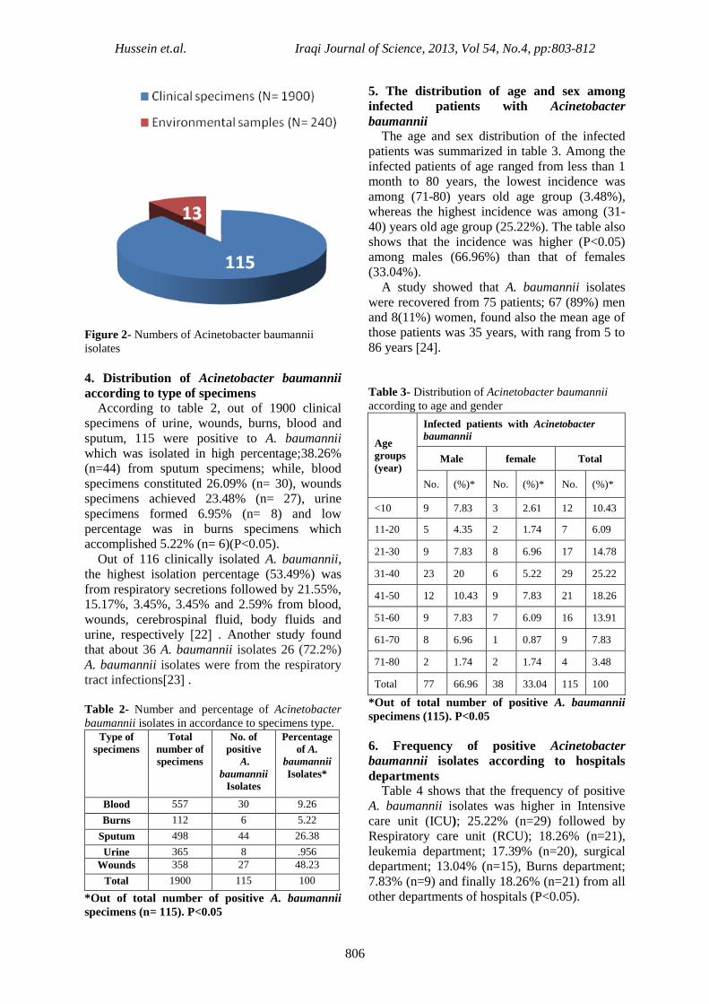
Figure 2 shows 1900 clinical specimens, 115
(6.05%) were identified as A. baumannii. The
environmental isolates of A. baumannii were
diagnosed with clinical isolates and out of 240
hospital environmental samples, 13 (5.42%)
were belonge to A. baumannii.
Figure 1- Blood agar containing D-glucose after 24
hours of incubation at 37ºC. (A) Pseudomonas
aeruginosa (B) Acinetobacter baumannii
(C) Moraxella catarrhalis.
Hussein et.al. Iraqi Journal of Science, 2013, Vol 54, No.4, pp:803-812
806
Figure 2- Numbers of Acinetobacter baumannii
isolates
4. Distribution of Acinetobacter baumannii
according to type of specimens
According to table 2, out of 1900 clinical
specimens of urine, wounds, burns, blood and
sputum, 115 were positive to A. baumannii
which was isolated in high percentage;38.26%
(n=44) from sputum specimens; while, blood
specimens constituted 26.09% (n= 30), wounds
specimens achieved 23.48% (n= 27), urine
specimens formed 6.95% (n= 8) and low
percentage was in burns specimens which
accomplished 5.22% (n= 6)(P<0.05).
Out of 116 clinically isolated A. baumannii,
the highest isolation percentage (53.49%) was
from respiratory secretions followed by 21.55%,
15.17%, 3.45%, 3.45% and 2.59% from blood,
wounds, cerebrospinal fluid, body fluids and
urine, respectively ]22 [ . Another study found
that about 36 A. baumannii isolates 26 (72.2%)
A. baumannii isolates were from the respiratory
tract infections]23 [ .
Table 2- Number and percentage of Acinetobacter
baumannii isolates in accordance to specimens type.
Type of
specimens
Total
number of
specimens
No. of
positive
A.
baumannii
Isolates
Percentage
of A.
baumannii
Isolates*
Blood 557 30 9.26
Burns 112 6 5.22
Sputum 498 44 26.38
Urine 365 8 .956
Wounds 358 27 48.23
Total 1900 115 100
*Out of total number of positive A. baumannii
specimens (n= 115). P<0.05
5. The distribution of age and sex among
infected patients with Acinetobacter
baumannii
The age and sex distribution of the infected
patients was summarized in table 3. Among the
infected patients of age ranged from less than 1
month to 80 years, the lowest incidence was
among (71-80) years old age group (3.48%),
whereas the highest incidence was among (31-
40) years old age group (25.22%). The table also
shows that the incidence was higher (P<0.05)
among males (66.96%) than that of females
(33.04%).
A study showed that A. baumannii isolates
were recovered from 75 patients; 67 (89%) men
and 8(11%) women, found also the mean age of
those patients was 35 years, with rang from 5 to
86 years ]24[.
Table 3- Distribution of Acinetobacter baumannii
according to age and gender
Infected patients with Acinetobacter
baumannii Age
groups
(year) Total female Male
(%)* No. (%)* No. (%)* No.
10.43 12 2.61 3 7.83 9 <10
6.09 7 1.74 2 4.35 5 11-20
14.78 17 6.96 8 7.83 9 21-30
25.22 29 5.22 6 20 23 31-40
18.26 21 7.83 9 10.43 12 41-50
13.91 16 6.09 7 7.83 9 51-60
7.83 9 0.87 1 6.96 8 61-70
3.48 4 1.74 2 1.74 2 71-80
100 115 33.04 38 66.96 77 Total
*Out of total number of positive A. baumannii
specimens (115). P<0.05
6. Frequency of positive Acinetobacter
baumannii isolates according to hospitals
departments
Table 4 shows that the frequency of positive
A. baumannii isolates was higher in Intensive
care unit (ICU); 25.22% (n=29) followed by
Respiratory care unit (RCU); 18.26% (n=21),
leukemia department; 17.39% (n=20), surgical
department; 13.04% (n=15), Burns department;
7.83% (n=9) and finally 18.26% (n=21) from all
other departments of hospitals (P<0.05).

Hussein et.al. Iraqi Journal of Science, 2013, Vol 54, No.4, pp:803-812
807
Table 4- Frequency of positive A. baumannii isolates
according to hospitals departments.
Percentage
of positive
isolates*
Number
of
positive
isolates
Hospitals
department
25.22 29 Intensive care
unit (ICU)
.2618 21 Respiratory
care unit
(RCU)
17.39 20 Blood disease
(Leukemia)
13.04 15 Surgical
7.83 9 Burns
18.26 21 Others
100 115 Total
*Out of total number of positive A. baumannii
specimens (n= 115).P<0.05
6. Identification of Acinetobacter baumannii
by Polymerase Chain Reaction (PCR)
blaOXA-51-like genes were found to be
present in all 128 (100%) A. baumannii clinical
and environmental studied isolates. The results
of the presence of blaOXA-51-like genes are
exemplified by the isolates shown in figure 3.
Figure 3- Detection ofblaOXA-51-Like byPCR.
Lane M, 100 bp DNA ladder; lanes 1-8,different
Acinetobacter baumannii isolates; lane C, Negative
control (had all PCR mixture including water instead
of DNA template). Detection was done on agarose
gel (1.5%) at 5 V/cm for 1.5 hour, stained with
ethidium bromide and visualized on a UV
transiluminator documentation system.
7. Antibiotics Susceptibility Data presented in figure 4 shows a high level
resistance of A. baumannii clinical isolates to
most of the antibiotics under test. The present
study revealed that all A. baumannii clinical
isolates had 100% resistance to amoxicillin-
clavulanic acid, cefepime, cefotaxime and
rifampin. This study also showed a highest
resistance to aztreonam (97.39%), ceftriaxone
(97.39%), ticarcillin-clavulanate (96.52%),
chloramphenicol (95.65%), piperacillin
(91.30%), cefalothin(91.03%), ceftazidime
(89.57%), gentamicin (87.83%), trimethoprime-
sulphamethoxazole (86.09%), ciprofloxacin
(83.48%), amikacin (72.17%) and colistin
(66.96%). Tobramycin and tetracycline recorded
moderate resistance; 46.09% and 47.83%,
respectively.
Given to notice that from 115 clinical isolates
of A. baumannii, 67 isolates (58.26%) were
resistant to both imipenem and meropenem and
8 (6.96%) of those isolates were intermediate to
both imipenem and meropenem, while 40
(34.78%) of those isolates were sensitive to both
imipenem and meropenem (figure 4).
Figure 4- Antibiotic resistance of 115 Acinetobacter
baumannii clinical isolates.
Similar study carried out in 2006 found that
the clinically identified A. baumannii isolates
were completely sensitive to imipenem while
they were 100% resistant to ciprofloxacin,
aztreonam and cephalothin. Also, they found
that the resistance percentage to tobramycin and
tetracycline were 82.35% and 100%,
respectively]25 [ . These differences may be

Hussein et.al. Iraqi Journal of Science, 2013, Vol 54, No.4, pp:803-812
808
attributed to the irrespective use of these two
antibiotics in our hospitals in the last few years.
Another study carried out in 2007 reported that
A. baumannii clinical isolates showed 100%
sensitivity to meropenem ]26[. Results of
another study carried out in 2010 found that A.
baumannii clinical isolates developed 100%
resistance to cefotaxime, ceftazidime,
ceftriaxone, 95.45% to cefepime,
chloramphenicol, aztronam and 40.90% to
imipenem ]27[. Upon these local studies, we can
notice interestingly the increase of resistance to
imipenem antibiotic in our hospitals.
Analysis of antibiotic resistance patterns
showed that all the 115 clinical isolates of A.
baumannii were multidrug resistant isolates.
Such resistance attributed to extensive use of
antimicrobial chemotherapy in clinical
environments ]28,29[.
Regarding colistin, our results showed a low
level of sensitivity reached to 31.30%. However,
many studies reported that the effective
antibiotic used to treat imipenem- resistant A.
baumannii isolate was colistin. For instance,
high sensitivity percentages to this antibiotic
(100%) were reported by ]30-32[.
Although the differences were insignificant
(P<0.05), the results presented in table 5
demonstrate the highest percentage (85%) of
imipenem resistant A. baumannii (IRAB)
isolates were isolated from blood disease
(leukemia) department followed by 75.86%,
66.67%, 60%, 44.44% and 42.86% from ICU,
RCU, surgical, Burns and other departments,
respectively (isolates showing intermediate
levels of susceptibility were considered as
resistant) [19,20].
Data presented in table 6 demonstrate the
highest percentage (77.27%) of imipenem
resistant A. baumannii (IRAB) isolates were
isolated from sputum specimens followed by
74.07%, 56.67%, 37.5% and 16.67% from
wounds, blood, urine and burns specimens,
respectively (isolates showing intermediate
levels of susceptibility were classified as
resistant) [19,20].
Table 5- Frequency of imipenem resistant
Acinetobacter baumannii isolates according to
hospitals departments.
Percentage
of imipenem
resistant
isolates
Number of
imipenem
resistant
isolates
Total
number of
A.
baumannii
isolates
Hospitals
departments
75.86 22 29 Intensive care
unit (ICU)
66.67 14 21 Respiratory
care unit
(RCU)
85 17 20 Blood disease
(Leukemia)
60 9 15 Surgical
44.44 4 9 Burns
42.86 9 21 Others
65.22 75 115 Total
Table 6- Frequency of imipenem resistant
Acinetobacter baumannii isolates according to type
of specimens.
Percentage
of
imipenem
resistant
isolates
Number
of
imipenem
resistant
isolates
Total
number of
A.
baumannii
isolates
Type
of
specimens
56.67 17 30 Blood
16.67 1 6 Burns
77.27 34 44 Sputum
37.5 3 8 Urine
74.07 20 27 Wounds
65.22 75 115 Total
On the other hand, the susceptibility of the
environment isolates toward different antibiotics
can be seen in figure -5, in which the highest
resistance percentages (100%) were found to
amoxicillin-clavulanic acid, cefepime,
cefotaxime, rifampin and ticarcillin-clavulanate.
Moreover, 84.62%, 84.62%, 84.62%,76.92%
,76.92% , 69.23%, 69.23%, 61.54%, 61.54%
and 53.85% of the environmental A. baumannii
isolates were resistant to ceftriaxone,
chloramphenicol, piperacillin,gentamicin,
trimethoprime-sulphamethoxazole, cefalothin,
ceftazidime, aztreonam, colistin and
ciprofloxacin, respectively. The lowest
resistance percentage (7.69%) was detected to
(imipenem and meropenem) then (15.38%) to
tetracycline followed by (23.08%) and (30.77%)
to tobramycin and amikacin, respectively.
The percentage of imipenem resistant A.
baumannii isolates among environmental
isolates (7.69%) was less than among A.
baumannii clinical isolates (58.26%).

Hussein et.al. Iraqi Journal of Science, 2013, Vol 54, No.4, pp:803-812
809
Figure 5- Antibiotic resistance of 13 Acinetobacter
baumannii environmental isolates.
Resistance of A. baumannii clinical and
environmental isolates to the tested antibiotics
revealed eleven different resistotype patterns
designated arbitrarily from A-K resistotypes
(isolates showing intermediate levels of
susceptibility were classified as resistant)
[19,20] as in table -7.
Pattern A accounted for 22 A. baumannii
isolates that were resistant to all antibiotics.
Pattern B included 15 A. baumannii isolates that
were resistant to most antibiotics but sensitive to
tobramycin only. Pattern C comprised 13 A.
baumannii isolates that were resistant to most
antibiotic but sensitive to colistin only. Fifteen
A. baumannii isolates showed Pattern D, being
resistant to all antibiotics but sensitive to
tobramycin and tetracycline only. Pattern E
involved 9 A. baumannii isolates that were
resistant to most antibiotics but sensitive to
tetracycline and colistin only. Pattern F covered
4 A. baumannii isolates that were resistant to
most antibiotics but sensitive to tetracycline,
tobramycin, amikacin and colistin. Eleven A.
baumannii isolates showed Pattern G, being
resistant to all antibiotics but sensitive to
imipenem, meropenem, amikacin and colistin.
Four A. baumannii isolates showed Pattern H,
being resistant to all antibiotics but sensitive to
tobramycin, imipenem, meropenem and
gentamicin. Twelve A. baumannii isolates
showed Pattern I, being resistant to all
antibiotics but sensitive to tobramycin,
tetracycline, imipenem and meropenem . Pattern
J accounted for 6 A. baumannii isolates that
were resistant to most antibiotics but sensitive to
tetracycline, piperacillin, ciprofloxacin,
amikacin, imipenem and meropenem. Finally,
17 A. baumannii isolates showed Pattern K,
being resistant to all antibiotics but sensitive to
tobramycin, tetracycline, imipenem,
meropenem, amikacin, ciprofloxacin,
ceftazidime, cefalothin and pipracillin.
Table 7- Resistotype patterns of Acinetobacer
baumannii isolates
Pattern Isolates codes Description
A
N20, N22, N26,
N28, N30, N31,
N33, N34, N43,
N63, N68, N95,
N101, N102,
N104, N114,
N116, N117,
N118, N119,
N122, N123
resistant to all
antibiotics
B
N29, N36, N64,
N70, N85, N86,
N89, N92, N96,
N98, N106,
N107, N110,
N111, N115
resistant to most
antibiotics but
sensitive to
tobramycin only
C
N4, N5, N6, N8,
N9, N35, N38,
N39, N40, N48,
N50, N51, N52
resistant to most
antibiotics but
sensitive to colistin
only
D
N32, N62, N65,
N69, N79, N83,
N84, N93, N97,
N99, N105,
N120, N121,
N124, N125
resistant to all
antibiotics but
sensitive to
tobramycin and
tetracycline only
E
N25, N27, N41,
N42, N47,N53,
N56, N71, N91
resistant to most
antibiotics but
sensitive to
tetracycline and
colistin
F N45, N54, N76,
N94
resistant to most
antibiotics but
sensitive to
tetracycline,
tobramycin, amikacin
and colistin
G
N1, N3, N10,
N17, N18, N19,
N21, N24, N37,
N44, N103
resistant to all
antibiotics but
sensitive to
imipenem,
meropenem, amikacin
and colistin

Hussein et.al. Iraqi Journal of Science, 2013, Vol 54, No.4, pp:803-812
810
H N11, N15, N59,
N126
resistant to all
antibiotics but
sensitive to
tobramycin,
imipenem,
meropenem and
gentamicin
I
N57, N66, N67,
N73, N74, N75,
N77, N80, N81,
N82, N87, N109
resistant to all
antibiotics but
sensitive to
tobramycin,
tetracycline,
imipenem and
meropenem
J
N46, N49,
N55,N58, N72,
N78
resistant to most
antibiotics but
sensitive to
tetracycline,piperacilli
n, ciprofloxacin,
amikacin,imipenem
and meropenem
K
N2, N7, N12,
N13, , N14, N16,
N23, N60, N61,
N88, N90, N100,
N108, N112,
N113, N127,
N128
resistant to all
antibioticsbut
sensitive to
tobramycin,
tetracycline,
imipenem,
meropenem,
amikacin,
ciprofloxacin,
ceftazidime,
cefalothin and
pipracillin
As a conclusion, the higher isolation rate of A.
baumannii was from ICU, more frequently from
sputum specimens. The infection with A.
baumannii was higher among males than
females, and the more infected age group was
31-40 years. The higher isolation percentage of
imipenem-resistant A. baumannii was from
blood disease (leukemia) department, in
particular from sputum specimens. All A.
baumannii clinical isolates showed multidrug
resistance; nevertheless, tobramycin and
tetracycline recorded moderate resistance. The
distribution of imipenem and other antibiotics
resistance among environmental isolates were
less than among A. baumannii clinical isolates.
The imipenem resistant A. baumannii increased
from 0% in 2006-2007 to 40.9% in 2010, and to
65.22% (resistant and intermediate) in 2011-
2012.
References
1. Kosmidis, C., Poulakou, G.,
Markogiannakis, A., and Daikos, G. L.
2012. Treatment Options for Infections
Caused by Carbapenem-resistant Gram-
negative Bacteria. European Infect. Dis,
6(1), pp: 28–34.
2. Park, S., Kim, H., Lee, K. M., Yoo, J. S.,
Yoo, J. I. I, Lee, Y. S. and Chung, G. T.
2013. Molecular and Epidemiological
Characterization of Carbapenem-Resistant
Acinetobacter baumanniiin Non-Tertiary
Korean Hospitals. Yonsei Med. J, 54(1),
pp:177-182.
3. Kabbaj, H., Seffar, M., Belefquih, B.,
Akka, D., Handor, N., Amor, M. and
Alaoui, A. E. 2012.Prevelanceofmetallo-B-
lactamases Producing Acinetobacter
baumannii in a Moroccan Hospital. ISRN
Infect. Dis,2013, pp:1-3.
4. Minandri, F., D’Arezzo,S., Antunes, L. C.
S., Pourcel, C. Principe, L., Petrosillo, N.
and Viscaa, P. 2012. Evidence of Diversity
among Epidemiologically Related
Carbapenemase-Producing Acinetobacter
baumannii Strains Belonging to
International Clonal Lineage II. J. Clinic.
Microbiol, 50(3), pp:590–597.
5. Endo, S.,Sasano, M., Yano, H., Inomata, S.,
Ishibashi, N., Aoyagi, T., Hatta, M., Gu, Y.,
Yamada, M., Tokuda , K., Kitagawa ,
M.,Kunishima, H., Hirakata, Y. andKaku,
M. 2012. IMP-1-producing carbapenem-
resistant Acinetobacte rursingii from Japan.
J. Antimicrob. Chemother, 67(10), pp:2533-
2538.
6. Boulanger, A., Nass, T., Fortineau, N.,
Figueiredo, S. and Nordmann, P. 2012.
NDM-1-Producing Acinetobacter
baumannii from Algeria. Antimicrob.
Agents Chemother, 56(4), pp:2214-2215.
7. Karah, N.,Sundsfjord, A., Towner, K. and
Samuelsen, O. 2012. Insights into the
global molecular epidemiology of
carbapenem non-susceptible clones of
Acinetobacter baumannii. Micro. Drug
Resist, 15(4), pp: 237-247.
8. Sohrabi, N., Farajnia , S., Akhi, M.
T., Nahaei , M. R., Naghili , B., Peymani ,
A., Amiri , Z., Rezaee, M. A. and Saeedi ,
N. 2012. Prevalence of OXA-type β-
lactamases among Acinetobacter
baumannii isolates from Northwest of Iran.
Microb. Drug Resist, 18(4), pp: 385-389.
9. Zhong, Q. M. D., Xu, W. M. D. , Wu, Y.M.
D., and Xu, H. M. D. 2012. Clonal Spread
of Carbapenem Non-susceptible
Acinetobacter baumannii in an Intensive
Care Unit ina Teaching Hospital in China.
Ann. Lab. Med, 32(6), pp: 413-419.

Hussein et.al. Iraqi Journal of Science, 2013, Vol 54, No.4, pp:803-812
811
10. Chang, K., Lin, M., Lin, N., Wu, W., Kuo,
H., Lin, T., Yang, T., Chen, Y. and Liou,
M. 2012. Clonal spread of multidrug-
resistant Acinetobacter baumannii in
eastern Taiwan. J. Microbiology,
Immunology and Infection, 45, pp: 37-42.
11. Kuo, S., Chang, S., Wang, H., Lai, H.,
Chen, P., Shiau, Y., Huang, W. and
Lauderdale, T. Y. 2012. Emergence of
extensively drug-resistant Acinetobacter
baumannii complex over 10 years:
Nationwide data from the Taiwan
Surveillance of Antimicrobial Resistance
(TSAR) program. BMC Infect. Dis12 و,
pp:200-215.
12. Sen, B., Lewandowski, K., Vaze, N.,
Emery, C., Hamilton, R., Brooks, A., and
Joshi, S. 2012. Characterization of
carbapenem-resistant MDR Acinetobacter
baumannii isolated from Philadelphia, PA
(United States). Infect. Dis. 827, pp:534-
540.
13. Forbes, B.A., Sahm, D.F. and weissfeld, A.
S.2007.Baily and scott's diagnostic
microbiology. 12thed. Mosby, Elsevire.
pp:334-339
14. .Siau, H., Yuen, K. , Ho, P., Luk, W.,
Wong, S . S .Y.,Woo, P. C. X., Lee,R .
A.andHui, W. 1998. Identification of
Acinetobacter on blood agar in presence of
D-glucose by inquebrowing effect. J. Clin. Microbiol, 36(5), pp:1404-1407.
15. Turton, J.F.,Woodford, N., Glover, J.,
Yarde, S., Kaufmann, M.E. and Pitt,
T.L.2006. Identification of Acinetobacter
baumanniiby detection of the blaOXA-51-
like carbapenemase gene intrinsic to this
species. J. Clin. Microbiol, 44(8), pp: 2974-
2976.
16. Morovat, T., Bahram, F., Mohammad, E.,
Setareh,S. and Mehdi,F. M. 2009.
Distribution of different carbapenem
resistant clones of Acinetobacter
baumanniiin Tehran Hospitals. New
Microbiologica, 32, pp: 265-271.
17. Woodford, N., Ellington, M. J., Coelho, J.
M., Turton, J. F., Ward, M. E., Brown, S.,
Amyes, S.G.B. and Livermore, D. M.
2006.Multiplex PCR for genes encoding
prevalent OXA carbapenemases in
Acinetobacter spp. International J. Antimicrob. Agents, 27, pp: 351-353.
18. WHO, (World Health Organization). 2003.
Basic laboratory procedures in clinical
bacteriology .2nd
ed. Geneva, Switzerland .
pp:103-121.
19. CLSI, (Clinical and Laboratory Standards
Institute). 2011. Performance standard for
antimicrobial susceptibility testing,
Twenty-First informational supplement.
M100-S21.31(1).
20. Feizabadi, M. M., atollahzadeh, B.,
Taherikalani, M., Rasooline-jad, M.,
Sadeghifard, N., Aligholi, M., Soroush, S.
and Mohammadi-Yegane, S. 2008.
Antimicrobial susceptibility patterns and
distribution of blaOXA genes among
Acinetobacter spp. Isolated from patients at
Tehran hospitals. Jpn. J. Infect. Dis, 61,
pp:274–278.
21. Peymani, A., Farajnia, S., Nahaei, M. R.,
Sohrabi, N., Abbasi, L., Ansarin,
K.andAzhari, F.2012.Prevalence of Class 1
Integron Among Multidrug-Resistant
Acinetobacter baumannii in Tabriz,
Northwest of Iran. Polish. J. Microbiol,
61(1), pp: 57–60.
22. Amudhan, S. M. ,Sekar, U ., Arunagiri, K.
and Sekar, B. 2011. OXA beta-lactamase-
mediated carbapenem resistance in
Acinetobacter baumannii. Indian Journal of
Medical Microbiol, 29(3), pp: 269-274.
23. Mammina, C., Palma, D. M., Bonura, C.,
Aleo, A., Fasciana, T., Sodano, C.,
Saporito, M. A., Verde, M. S., Calà, C.,
Cracchiolo, A. N. and Tetamo, R. 2012.
Epidemiology and clonality of carbapenem-
resistant Acinetobacter baumannii from an
intensive care unit in Palermo, Italy. BMC
Research Notes, 5, pp: 365-373.
24. Hujer, K. M., Hujer, A. M., Hulten, E. A.,
Bajakouzian, S., Adams, J. M., Donskey, C.
J., Ecker, D. J., Massire, C., Eshoo, M. W.,
Sampath, R., Thomson, J. M., Rather, P. N.,
Craft, D. W., Ewell, A. J., Jacobs, M. R.,
Paterson, D. L. and Bonomo, R. A. 2006.
Analysis of Antibiotic Resistance Genes in
Multidrug-Resistant Acinetobacter sp.
Isolates from Military and Civilian Patients
Treated at the Walter Reed Army Medical
Center. Antimicrob. Agents Chemother,
50(12), pp: 4114-4123.
25. Al- Khafaji, S. M. S. 2006. Study on
capsule ofAcinetobacter baumannii and its
effect on Immune Response. PH.D. Thesis.
Biology department.College of Sciences.
AL-Mustansiriyah University. Baghdad,
Iraq.

Hussein et.al. Iraqi Journal of Science, 2013, Vol 54, No.4, pp:803-812
812
26. Mosafer, H. K.2007. Effect of crude
fimbriae extract of Acinetobacter
baumanniion biotic and abiotic surfaces.
Msc. thesis. Biology department. College of
Science, AL-Mustansiriyah University.
Baghdad, Iraq.
27. Al-Mash’hadani, E. I. J. 2010. Study The
activity of Bacteriocin produced from
Lactobacillus plantarum on virulence
factors of Acinetobacter baumannii. Msc.
thesis. Biology department. College of
Science, AL-Mustansiriyah University.
28. Mak, K. J., Kim, M., Pham, J., Tapsall,
J.and White, P. A. 2009. Antibiotic
resistance determinants in nosocomial
strains of multidrug-resistant Acinetobacter
baumannii. J. Antimicrob. Chemother, 63,
pp: 47-54.
29. Liu, S., Wang, Y., Xu, J., Li, Y., Guo, J.,
Ke, Y., Yuan, X., Wang, L., Du, X., Wang,
Z., Huang, L., Zhang, N. and Chenb, Z.
2012. Genome Sequence of an OXA23-
Producing, Carbapenem-Resistant
Acinetobacte rbaumannii Strain of
Sequence Type ST75. J. Bacteriol, 194(21),
pp: 6000-6001.
30. Hello, S. L., Falcot, V., Lacassin, F.,
Mikulski, M. and Baumann, F. 2010. Risk
factors for carbapenem-resistant
Acinetobacter baumannii infections at a
tertiary care hospital in New Caledonia,
South Pacific. Scandinavian J. Infect. Dis,
42, pp: 821-826.
31. Pongpech, P., Amornnopparattana kul, S.,
Panapakdee, S., Fungwithaya, S., Nannha,
P., Dhiraputra, C. and Leelarasamee , A.
2010. Antibacterial Activity of
Carbapenem-Based Combinations Againts
Multidrug-Resistant Acinetobacter
baumannii. J. Med. Assoc. Thai, 93 (2),
pp:161-171.
32. Prakasam, G., Geethapriya, S.,
Jayakeerthana, K. H. and Ramesh, S. 2011.
Detection of Certain Virulence Attributes
and Antimicrobial Resistance Pattern
among Clinical Isolates of Acinetobacter
baumannii. International J. of Pharma&
Bio Science, 2(3), pp:501-507.