High-throughput discrimination of bacteria isolated from Astacus astacus and A. leptodactylus
12
Knowledge and Management of Aquatic Ecosystems (2014) 413, 04 http://www.kmae-journal.org c ONEMA, 2014 DOI: 10.1051/kmae/2014005 High-throughput discrimination of bacteria isolated from Astacus astacus and A. leptodactylus N. Topi ´ c Popovi ´ c (1) , R. Sauerborn Klobu ˇ car (1) , I. Maguire (2) , I. Strunjak-Perovi ´ c (1), , S. Kazazi ´ c (3) , J. Bariši ´ c (1) , M. Jadan (1) , G. Klobu ˇ car (2) , R. ˇ Což-Rakovac (1) Received October 3, 2013 Revised February 6, 2014 Accepted February 10, 2014 ABSTRACT Key-words: crayfish, bacteria, MALDI-TOF MS, API 20E Bacterial diseases and pathogens of crayfish are common, widespread, and occasionally causing serious mortalities. In order to take rapid mea- sures for correct treatment of crayfish diseases, the turnover time and accuracy in bacterial identification is an issue. Bacteria isolated from tis- sues of apparently healthy Astacus astacus and A. leptodactylus were identified by the commercial phenotypic tests (API 20E) and by the ma- trix assisted laser induced desorption ionization connected to the time of flight mass spectrometry (MALDI-TOF MS). For Gram-negative rods, API 20E resulted in fewer species identifications than MALDI-TOF MS (5.2% versus 52.61%). The most frequently identified genus from A. asta- cus and A. leptodactylus was Pseudomonas spp.: API 20E (47.82%) and MALDI-TOF MS (52.17%). Both systems identified 60.86% of total iso- lates identically to the genus. Hafnia alvei was the only isolate for which API 20E and MALDI-TOF MS had a concordant reading to the species. MALDI-TOF MS proved to be a powerful, low-cost, rapid tool in bacterial genus identification. This is the first report of a direct comparison between the two systems for the identification of bacteria in crayfish, and also the first report on using MALDI-TOF MS for discrimination of freshwater cray- fish bacterial isolates. RÉSUMÉ Discrimination haut-débit de bactéries isolées à partir d’Astacus astacus et A. leptodac- tylus Mots-clés : écrevisse, bactérie, MALDI-TOF MS, API 20E Les maladies bactériennes et les agents pathogènes des écrevisses sont com- muns, répandus, et de temps en temps entraînent des mortalités importantes. Afin de prendre des mesures rapides pour le traitement correct des maladies d’écre- visses, le temps nécessaire et l’exactitude dans l’identification bactérienne est une question. Des bactéries isolées dans des tissus d’Astacus astacus et d’A. lep- todactylus apparemment en bonne santé ont été identifiées par les tests phé- notypiques commerciaux (API 20E) et par spectromètre de masse couplant une source d’ionisation laser assistée par une matrice et un analyseur à temps de vol (MALDI-TOF MS). Pour les bâtonnets Gram-négatifs, API 20E a donné moins d’identifications d’espèces que MALDI-TOF MS (5,2 % contre 52,61 %). Le genre le plus souvent identifié à partir d’A. astacus et A. leptodactylus était Pseudomo- nas spp : API 20E (47,82 %) et MALDI- TOF MS (52,17 %). Les deux systèmes (1) Laboratory for Ichthyopathology-Biological Materials, Ru ¯ der Boškovi ´ c Institute, Zagreb, Croatia (2) Department of Zoology, Faculty of Science, University of Zagreb, Zagreb, Croatia (3) Laboratory for Chemical Kinetics and Atmospheric Chemistry, Ru ¯ der Boškovi ´ c Institute, Zagreb, Croatia Corresponding author: [email protected] Article published by EDP Sciences
-
Upload
independent -
Category
Documents
-
view
0 -
download
0
Transcript of High-throughput discrimination of bacteria isolated from Astacus astacus and A. leptodactylus

Knowledge and Management of Aquatic Ecosystems (2014) 413, 04 http://www.kmae-journal.orgc© ONEMA, 2014
DOI: 10.1051/kmae/2014005
High-throughput discrimination of bacteria isolatedfrom Astacus astacus and A. leptodactylus
N. Topic Popovic(1), R. Sauerborn Klobucar(1), I. Maguire(2),I. Strunjak-Perovic(1),�, S. Kazazic(3), J. Barišic(1), M. Jadan(1), G. Klobucar(2),R. Což-Rakovac(1)
Received October 3, 2013
Revised February 6, 2014
Accepted February 10, 2014
ABSTRACT
Key-words:crayfish,bacteria,MALDI-TOF MS,API 20E
Bacterial diseases and pathogens of crayfish are common, widespread,and occasionally causing serious mortalities. In order to take rapid mea-sures for correct treatment of crayfish diseases, the turnover time andaccuracy in bacterial identification is an issue. Bacteria isolated from tis-sues of apparently healthy Astacus astacus and A. leptodactylus wereidentified by the commercial phenotypic tests (API 20E) and by the ma-trix assisted laser induced desorption ionization connected to the timeof flight mass spectrometry (MALDI-TOF MS). For Gram-negative rods,API 20E resulted in fewer species identifications than MALDI-TOF MS(5.2% versus 52.61%). The most frequently identified genus from A. asta-cus and A. leptodactylus was Pseudomonas spp.: API 20E (47.82%) andMALDI-TOF MS (52.17%). Both systems identified 60.86% of total iso-lates identically to the genus. Hafnia alvei was the only isolate for whichAPI 20E and MALDI-TOF MS had a concordant reading to the species.MALDI-TOF MS proved to be a powerful, low-cost, rapid tool in bacterialgenus identification. This is the first report of a direct comparison betweenthe two systems for the identification of bacteria in crayfish, and also thefirst report on using MALDI-TOF MS for discrimination of freshwater cray-fish bacterial isolates.
RÉSUMÉ
Discrimination haut-débit de bactéries isolées à partir d’Astacus astacus et A. leptodac-tylus
Mots-clés :écrevisse,bactérie,MALDI-TOF MS,API 20E
Les maladies bactériennes et les agents pathogènes des écrevisses sont com-muns, répandus, et de temps en temps entraînent des mortalités importantes. Afinde prendre des mesures rapides pour le traitement correct des maladies d’écre-visses, le temps nécessaire et l’exactitude dans l’identification bactérienne estune question. Des bactéries isolées dans des tissus d’Astacus astacus et d’A. lep-todactylus apparemment en bonne santé ont été identifiées par les tests phé-notypiques commerciaux (API 20E) et par spectromètre de masse couplant unesource d’ionisation laser assistée par une matrice et un analyseur à temps devol (MALDI-TOF MS). Pour les bâtonnets Gram-négatifs, API 20E a donné moinsd’identifications d’espèces que MALDI-TOF MS (5,2 % contre 52,61 %). Le genrele plus souvent identifié à partir d’A. astacus et A. leptodactylus était Pseudomo-nas spp : API 20E (47,82 %) et MALDI- TOF MS (52,17 %). Les deux systèmes
(1) Laboratory for Ichthyopathology-Biological Materials, Ruder Boškovic Institute, Zagreb, Croatia(2) Department of Zoology, Faculty of Science, University of Zagreb, Zagreb, Croatia(3) Laboratory for Chemical Kinetics and Atmospheric Chemistry, Ruder Boškovic Institute, Zagreb, Croatia� Corresponding author: [email protected]
Article published by EDP Sciences

N. Topic Popovic et al.: Knowl. Managt. Aquatic Ecosyst. (2014) 413, 04
ont identifié 60,86 % des isolats totaux au même genre. Hafnia alvei était le seulisolat dont API 20E et MALDI-TOF MS ont une lecture concordante à l’espèce.MALDI-TOF MS s’est avéré être un outil rapide, puissant, à faible coût, pour l’iden-tification au niveau du genre bactérien. Ce travail est le premier d’une comparai-son directe entre les deux systèmes pour l’identification des bactéries chez lesécrevisses, et aussi le premier travail sur l’utilisation de MALDI-TOF MS pour ladiscrimination des isolats bactériens d’écrevisses d’eau douce.
INTRODUCTION
There are two genera and five species from the family Astacidae inhabiting the Eurasian con-tinent (Holdich et al., 2006). Astacus astacus (the noble crayfish) is nowadays distributed overthe eastern, central, and northern parts of Europe, while A. leptodactylus (the narrow-clawedor Turkish crayfish) inhabits Eastern Europe and Western Asia, and is spreading naturallywestwards through waterways (Holdich et al., 2006). In Croatia, A. astacus is distributed inthe continental region, forming both river and lake populations, while A. leptodactylus is foundin the rivers of eastern and central Croatia with tendency of spreading west- and southwards(Maguire and Gottstein-Matocec, 2004; Maguire et al., 2011). Populations of noble crayfishare considered rare; the species is designated as vulnerable and listed in the Bern Conven-tion, EU Habitat Directive and IUCN Red List of Threatened Species (Edsman et al., 2010).Noble crayfish is also treated as endangered in Croatia and is protected by Croatian law(Anonymous, 2005, 2008, 2009).Introduction of non-native American crayfish species into Europe has been responsible forthe transfer of the devastating disease crayfish plague caused by Aphanomyces astaci,which led to mass mortalities of native crayfish species (Diéguez-Uribeondo, 2006). Due toits dramatic impact onto native European crayfish species, A. astaci was extensively stud-ied from different aspects (description, characterization, diagnostics, genotypization, viru-lence, etc.) (Diéguez-Uribeondo, 2006; Makkonen, 2013). However, bacterial diseases andbacterial pathogens of crayfish have not been considered to such an extent, albeit commonand widespread. Typically, bacteria isolated from crayfish include both Gram-negative andGram-positive species, as representatives of the genera Acinetobacter, Aeromonas, Bacillus,Citrobacter, Corynebacterium, Flavobacterium, Micrococcus, Pseudomonas, Staphylococcusand Vibrio (Smith and Söderhäll, 1986; Vey, 1986; Alderman and Polglase, 1988; Edgertonet al., 2002; Romero and Jiménez, 2002; Quaglio et al., 2006a, 2006b; Jiravanichpaisal et al.,2009; Longshaw, 2011; Mickeniene and Šyvokiene, 2011). Bacterial infections leading to mor-talities have been documented in both farmed and wild crayfish, and were also reported inasymptomatic animals (Edgerton et al., 2002; Quaglio et al., 2006a, 2006b; Cooper et al.,2007; Jiravanichpaisal et al., 2009; Johnson and Paull, 2011; Longshaw, 2011; Longshawet al., 2012). Mostly, bacteria found in freshwater crayfish inhabit the ecosystem in which theylive, may be found in water and sediments, and they reside on the exoskeleton, gills or in thegut. Bacteriological investigations of crayfish have predominantly been performed on theirhaemolymph using standard microbiological methods, and also by histopathological exami-nations of tissues (Colwell et al., 1975; Johnson, 1976; Scott and Thune, 1986; Madetoja andJussila, 1996; Edgerton and Owens, 1999; Edgerton et al., 2002; Romero and Jiménez, 2002;Quaglio et al., 2006b; Jiravanichpaisal et al., 2009). When performing health status evalua-tions, considering correct identification and treatment of bacterial diseases and conditions,of (primarily) farmed crayfish, speed is always an issue. Rapid identification of environmen-tal bacteria via commercial phenotypic tests allows for a wide choice of tests selection, andAPI 20E (Biomerieux, Marcy l’Etoile, France), an identification system for Enterobacteriaceaeand other non-fastidious Gram-negative rods developed for clinical specimens, seems tobe increasingly used for the identification of aquatic pathogens (Topic Popovic et al., 2007;Sanjuán et al., 2009; Bastardo et al., 2012; Esteve et al., 2012; Soto et al., 2012). However,due to several shortcomings of this system, such as the wrong identifications and the need
04p2
N. Topic Popovic et al.: Knowl. Managt. Aquatic Ecosyst. (2014) 413, 04
of comparison with the diagnostic schemes based on reactions in conventional phenotypictests, more advanced methods for identification are sought after. Therefore, in addition tomorphological, biochemical microbiological testing analysis, along with the molecular iden-tification, the matrix assisted laser induced desorption ionization (MALDI) connected to thetime of flight (TOF) mass spectrometry (MS) channel, is now becoming a third diagnostic pil-lar with strong discriminating power (Risch et al., 2010). Its value is in a rapid screening ofthe organism and the accessible protein pattern for characterization and distinction (Petersenet al., 2009). MALDI-TOF MS can examine the pattern of proteins detected directly from in-tact bacteria, giving a reproducible spectra consisting of a series of peaks corresponding tomass-to-charge ratios of ions released from bacterial proteins during laser desorption (Dupontet al., 2010). MALDI-TOF MS is considered a tool with potential to replace phenotypic identifi-cation of bacteria in clinical microbiology laboratories (Bizzini et al., 2010; Ford and Burnham,2013; Jamal et al., 2013; Kok et al., 2013), especially due to its time-saving benefit where theextensive time needed with culture-based methods is reduced to a few minutes.In this study, we isolated bacteria from various tissues of apparently healthy Astacus astacusand A. leptodactylus and compared the performances of the API 20E panels to the BrukerBiotyper MALDI-TOF MS (Bruker Daltonics, Billerica, MA) for the identification of bacterialisolates. This is the first report of a direct comparison between the two systems for the iden-tification of bacteria in crayfish, and also the first report on using MALDI-TOF MS for thediscrimination of the freshwater crayfish bacterial isolates.
MATERIALS AND METHODS
> ANIMALS, SAMPLING, AND TISSUE PROCESSING
The study was carried out in spring 2013, on 10 specimens of Astacus astacus (mean weight43.63 g) and 10 specimens of A. leptodactylus (mean weight 55.62 g) of both sexes, all appar-ently healthy. All crayfish were cage-exposed in the gravel pit Jagodno in vicinity of Zagreb,Croatia. Specimens were randomly sampled, transported live to the laboratory and withinfew hours sacrificed by overdose of tricaine methane-sulfonate (MS-222, Sigma, St. Louis,Missouri, USA). Necropsy was performed immediately and tissues (gills, hepatopancreas, go-nads, gut) were fixed in 4% neutral buffered formalin, dehydrated through a graded ethanol-xylene series and embedded in paraplast. Sagital and transverse sections (3−5 µm) werestained with hematoxylin/eosin (H&E). Microphotographs were taken with a digital cameraDP 70 Olympus R© connected to an Olympus R© BX51 binocular microscope, and transferred toMicrosoftR© AnalySIS Soft Imaging System for interpretation.Samples of scrapings of exoskeleton, mouth region, gills, stomach, hepatopancreas, and in-testine were streaked onto Tryptone Soya Agar (TSA, CM0131 Oxoid Ltd, England, UK). Theplates were incubated at 22 ◦C for 48−72 h. Representative colonies were isolated and re-streaked on fresh medium until purity was attained. Growth of colonies was ascertained byvisual inspection. Pure colonies were Gram-stained and subjected to morphological, phys-iological and biochemical tests. The taxonomic position of the isolates was determined byAPI 20E panels and Bruker Biotyper MALDI-TOF MS.
> API 20E (BIOMERIEUX, MARCY L’ETOILE, FRANCE)
The API 20E tests were performed according to the manufacturer’s instructions with a fewalterations in order to adapt the system to the bacteria of freshwater crayfish: the incubationtime was increased to 48−72 h; the incubation temperature was lowered to 22 ◦C; only thefermentation of sugars was allowed by sealing the cups with sterile mineral oil in the carbo-hydrate tests. The API 20E uses 21 standardized and miniaturized biochemical tests and adatabase. It consists of 21 microtubes containing dehydrated substrates. These tubes wereinoculated with a bacterial suspension, which reconstituted the media. During incubation,
04p3

N. Topic Popovic et al.: Knowl. Managt. Aquatic Ecosyst. (2014) 413, 04
metabolism produced color changes that were either spontaneous or revealed by the additionof reagents. The reactions were read according to the table provided and the identificationwas obtained using the software provided by the manufacturer, the Apiweb. A seven-digitprofile was obtained for every tested isolate. API 20E ratings were based on three param-eters, including the likelihood of a match between the unknown organism’s profile and thecomputer profile, the relative value between the likelihood of the first and the likelihood of thesecond choices, and the number of tests against the first choice (Brown and Leff, 1996; TopicPopovic et al., 2007).
> BRUKER BIOTYPER MALDI-TOF (BRUKER DALTONICS, BILLERICA, MA)
Bacterial isolates (one loopful of each bacterial culture) were applied as a thin film to a 24-spotsteel plate (Bruker Daltonics) in two replicates and allowed to visibly dry at room temperature(referred to as the direct colony technique). Subsequently, 2 µL of MALDI matrix (a saturatedsolution of α-cyano-4-hydroxycinnamic acid in 50% acetonitrile and 2.5% trifluoroacetic acid)was applied to the colony and dried in a fume hood. The analysis was performed in a mannerthat ions generated with a 337-nm nitrogen laser were captured in the positive linear modein a mass range of 2 to 20 kDa. Positive ions were extracted with an accelerating voltageof 20 kV in linear mode. Each spectrum was the sum of the ions obtained from 200 lasershots performed in five different regions of the same well. Captured spectra were analyzedusing MALDI Biotyper automation control and Bruker Biotyper 2.0 software (Bruker Daltonics).The MALDI Biotyper database contained 3 740 spectra from 319 genera and 1 946 species.For each 24-spot plate, a standard (bacterial test standard; Bruker Daltonics) was includedto calibrate the instrument and validate the run. Identification criteria used were as follows: ascore of 2.300 to 3.000 indicated highly probable species level identification, a score of 2.000to 2.299 indicated secure genus identification with probable species identification, a score1.700 to 1.999 indicated probable identification to the genus level, and a score of <1.700 wasconsidered to be unreliable. The data obtained with the two replicates were added to minimizeany random effect. The presence or absence of peaks was considered as fingerprints for aparticular isolate. Identification of isolates corresponded to the species of the reference strainwith the best match in the database.
RESULTS
The external gross signs and necropsy findings from the crayfish did not indicate to anymanifest disease. Relevant bacteria were recovered from most tissues under examination.The majority of isolates were retrieved from gills (34.78%), stomach (21.74%), and mouth re-gion (17.39%), while less from other tissues: intestine (8.7%), hepatopancreas (8.7%), andexoskeleton (8.7%). Of 23 relevant isolates, only one matched completely in both API 20Eand MALDI-TOF MS readings (Hafnia alvei). Also, there were two unreliable identifications byMALDI-TOF MS for isolates which API 20E identified with “Good identification to the genus”as Pseudomonas aeruginosa. In comparison, API 20E assigned 7 isolates (30.43%) to “Un-acceptable”, “Doubtful”, or “Low discrimination” profiles, which was expected for 3 isolates,being Gram-positive rods and identified with MALDI-TOF MS as Bacillus cereus with “Securegenus identification with probable species identification” (Figure 1). For Gram-negative rods,the conventional method resulted in significantly fewer species identifications than MALDI-TOF MS (5.2% versus 52.61%). Detailed comparison of identification results between API20E and MALDI-TOF MS is presented in Table I. Overall, the most frequently identified genusfrom both A. astacus and A. leptodactylus was Pseudomonas spp.: with API 20E (47.82%)and with MALDI-TOF MS (52.17%), while both systems allocated Pseudomonas spp. identifi-cation for the respective isolates in 39.13% of total cases. Both systems identified 60.86% oftotal isolates identically to the genus. The mean time to identification with API 20E was 48 h,
04p4

N. Topic Popovic et al.: Knowl. Managt. Aquatic Ecosyst. (2014) 413, 04
Figure 1MALDI-TOF MS spectral profiles of bacterial isolates: Hafnia alvei isolated from hepatopancreas ofA. leptodactylus with “Highly probable species identification”; 3 isolates of Bacillus cereus from intestine,hepatopancreas and gills of A. leptodactylus, all within the category “Highly probable species identifica-tion”; Pseudomonas chlororaphis isolated from gills and mouth region of A. astacus, both isolates herepresented as the identical spectral profile with “Highly probable species identification”.
whereas MALDI-TOF MS needed less than 10 minutes per bacterial isolate. Histopathologi-cal findings of the hepatopancreas (Figure 2) indicated to its vacuolization as well as nodularformations in haemal sinus. Tissue sections of gill lamellae (Figure 3) showed epithelial walllifting and presence of dead cells.
DISCUSSION
Bacteria isolated from tissues of apparently healthy Astacus astacus and A. leptodactyluswere identified by the API 20E panels and the MALDI-TOF MS, and the two systems werecompared for their usefulness for identification of bacteria in crayfish. The most preva-lent genus identified by both API 20E and MALDI-TOF MS was Pseudomonas. Indeed,Pseudomonas spp. is one of the most frequently isolated Gram-negative bacteria fromcrayfish (Scott and Thune, 1986; Edgerton et al., 2002; Mickeniene and Šyvokiene, 2011).Neither A. astacus nor A. leptodactylus under this survey demonstrated any of the previ-ously described signs of Pseudomonas-related bacterial septicemia (Edgerton et al., 2002)
04p5

N. Topic Popovic et al.: Knowl. Managt. Aquatic Ecosyst. (2014) 413, 04
Tab
leI
Com
par
ison
ofid
entifi
catio
nre
sults
bet
wee
nA
PI2
0Ean
dM
ALD
I-TO
FM
Sfo
ris
olat
esfr
omA
stac
usas
tacu
san
dA
.lep
tod
acty
lus.
AP
I20E
(res
ult)
*M
ALD
I-T
OF
MS
(sco
re)*
*C
om
men
tA
stac
us
asta
cus
iso
late
sP
seu
dom
onas
flu
ores
cen
s/p
uti
da
Pse
udom
onas
kore
ensi
sB
acte
rialc
olon
ies
(cre
amin
colo
r)la
cked
fluor
esci
ngp
rop
ertie
s.(E
xcel
lent
iden
tifi
cati
on
toth
eg
enus
)(1
.913
)P
s.ko
reen
sis
isan
unlik
ely
isol
ate
ince
ntra
lCro
atia
.A
erom
onas
hyd
rop
hila
gro
up2
Aer
omon
aseu
cren
ophi
la(2
.165
)A
.euc
reno
phi
lais
foun
din
fres
hw
ater
s(V
ery
go
od
iden
tifi
cati
on
toth
eg
enus
)an
dha
sb
een
isol
ated
from
fish
and
cray
fish.
Pse
ud
omon
asae
rug
inos
aP
seud
omon
asko
reen
sis
Cre
am-c
olor
edb
acte
rialc
olon
ies
with
yello
wd
iffus
ing
pig
men
t.(V
ery
go
od
iden
tifi
cati
on
toth
eg
enus
)(2
.067
)P
s.ko
reen
sis
isan
unlik
ely
isol
ate
ince
ntra
lCro
atia
.P
seu
dom
onas
aeru
gin
osa
Pse
udom
onas
ced
rina
Mat
chin
gid
entifi
catio
nto
the
genu
s.(V
ery
go
od
iden
tifi
cati
on
toth
eg
enus
)(1
.914
)P
s.ce
drin
ab
elon
gsto
the
Ps.
fluor
esce
nsgr
oup
.P
seu
dom
onas
aeru
gin
osa
Not
relia
ble
iden
tifica
tion
Not
relia
ble
id.a
fter
mul
tiple
mea
sure
men
ts.
(Go
od
iden
tifi
cati
on
toth
eg
enus
)(1
.683
)P
seu
dom
onas
aeru
gin
osa
Not
relia
ble
iden
tifica
tion
As
abov
e.(G
oo
did
enti
fica
tio
nto
the
gen
us)
(1.6
83)
Pse
ud
omon
asae
rug
inos
aP
seud
omon
asch
loro
rap
his
Ora
nge-
colo
red
colo
nies
pos
sib
lyas
soci
ated
with
(Go
od
iden
tifi
cati
on)
(2.3
96)
Ps.
chlo
rora
phi
ssu
bsp
.aur
anth
iaca
orau
reof
acie
ns.
Pse
ud
omon
asae
rug
inos
a(G
oo
did
enti
fica
tio
n)P
seud
omon
asch
loro
rap
his
(2.3
96)
As
abov
e.S
hew
anel
lap
utr
efac
ien
sS
hew
anel
lab
altic
aS
h.p
utre
faci
ens
has
bee
nis
olat
edfr
omm
arin
een
viro
nmen
ts,
(Go
od
iden
tifi
cati
on)
(1.7
66)
Sh.
bal
tica
isfo
und
mai
nly
inw
ater
sof
the
Bal
ticS
ea.
Bru
cella
spp
.P
seud
omon
asth
iver
vale
nsis
Ps.
thiv
erva
lens
isis
aso
ilb
acte
rium
.(L
ow
dis
crim
inat
ion)
(2.0
45)
Och
rob
actr
um
anth
rop
iP
seud
omon
asfr
eder
iksb
erge
nsis
Sec
ure
genu
sid
entifi
catio
n(P
seud
omon
as)w
ithle
ssp
rob
able
(Lo
wd
iscr
imin
atio
n)(2
.272
)sp
ecie
sid
entifi
catio
n.P
anto
easp
p.
Art
hrob
acte
rau
resc
ens
Gra
m-p
ositi
vero
ds,
yello
w-c
olor
edco
loni
es.
(Lo
wd
iscr
imin
atio
n)(2
.180
)A
rthr
obac
teria
are
com
mon
lyfo
und
inso
il.A
erom
onas
hyd
rop
hila
gro
up1
Pse
udom
onas
pro
teol
ytic
aS
ecur
ege
nus
iden
tifica
tion
(Pse
udom
onas
).P
s.p
rote
olyt
ica
(Una
ccep
tab
lep
rofi
le)
(2.0
80)
isa
psy
chro
phi
licb
acte
rium
notl
ikel
yto
be
foun
din
cent
ralC
roat
ia.
04p6

N. Topic Popovic et al.: Knowl. Managt. Aquatic Ecosyst. (2014) 413, 04
Tab
leI
Con
tinue
d.
AP
I20E
(res
ult)
*M
ALD
I-T
OF
MS
(sco
re)*
*C
om
men
tA
stac
us
lep
tod
acty
lus
iso
late
sH
afn
iaal
vei
Haf
nia
alve
iP
revi
ousl
yis
olat
edfr
omfr
eshw
ater
cray
fish
(Lon
gsha
wet
al.,
2012
).(E
xcel
lent
iden
tifi
cati
on)
(2.0
53)
Op
por
tuni
stic
pat
hoge
nof
fres
hwat
erfis
h(A
ustin
and
Aus
tin,1
999)
.P
seu
dom
onas
aeru
gin
osa
Pse
udom
onas
kore
ensi
sM
atch
ing
iden
tifica
tion
toth
ege
nus.
(Exc
elle
ntid
enti
fica
tio
nto
the
gen
us)
(2.0
84)
Ps.
kore
ensi
sis
anun
likel
yis
olat
ein
cent
ralC
roat
ia.
Pse
ud
omon
asae
rug
inos
aP
seud
omon
asko
reen
sis
As
abov
e.P
seud
omon
asae
rugi
nosa
has
(Exc
elle
ntid
enti
fica
tio
nto
the
gen
us)
(1.9
67)
zoon
otic
pot
entia
l(A
ustin
and
Aus
tin,1
999)
.A
erom
onas
hyd
rop
hila
gro
up1
Aer
omon
asb
estia
rum
A.b
estia
rum
and
A.h
ydro
phi
last
and
inth
esa
me
phe
nogr
oup
,(V
ery
go
od
iden
tifi
cati
on
toth
eg
enus
)(2
.193
)d
escr
ibed
asth
eA
.hyd
rop
hila
com
ple
x(M
artin
oet
al.,
2011
).P
seu
dom
onas
flu
ores
cen
s/p
uti
da
Pse
udom
onas
kilo
nens
isM
atch
ing
iden
tifica
tion
toth
ege
nus.
(Go
od
iden
tifi
cati
on
toth
eg
enus
)(1
.915
)S
hew
anel
lap
utr
efac
ien
sS
hew
anel
lab
altic
aS
h.p
utre
faci
ens
has
bee
nis
olat
edfr
omm
arin
een
viro
nmen
ts,
(Go
od
iden
tifi
cati
on)
(1.9
47)
Sh.
bal
tica
isfo
und
mai
nly
inw
ater
sof
the
Bal
ticS
ea.
Pse
ud
omon
asae
rug
inos
aP
seud
omon
aski
lone
nsis
Mat
chin
gid
entifi
catio
nto
the
genu
s.(D
oub
tful
pro
file
)(1
.823
)B
urk
hol
der
iace
pac
iaB
acill
usce
reus
Gra
m-p
ositi
vero
ds.
(Lo
wd
iscr
imin
atio
n)(2
.131
)B
urk
hol
der
iace
pac
iaB
acill
usce
reus
Gra
m-p
ositi
vero
ds.
(Lo
wd
iscr
imin
atio
n)(2
.130
)B
urk
hol
der
iace
pac
iaB
acill
usce
reus
Gra
m-p
ositi
vero
ds.
(Lo
wd
iscr
imin
atio
n)(2
.200
)
*A
PI2
0Ere
sult:
exce
llent
iden
tifica
tion
(%id�
99.9
and
Tin
dex�
0.75
);ve
rygo
odid
entifi
catio
n(%
id�
99.0
and
Tin
dex�
0.50
);go
odid
entifi
catio
n(%
id�
99.9
and
Tin
dex�
0.25
);ac
cep
tab
leid
entifi
catio
n(%
id�
80.0
and
Tin
dex�
0).I
den
tifica
tion
toth
eta
xon:
one
sing
leta
xon
has
bee
nse
lect
ed;i
den
tifica
tion
toth
ege
nus
leve
l:2,
3or
4ta
xab
elon
ging
toth
esa
me
genu
sha
veb
een
sele
cted
;Low
dis
crim
inat
ion:
2,3
or4
taxa
bel
ongi
ngto
diff
eren
tge
nera
have
bee
nse
lect
ed;T
heid
entifi
catio
nis
“not
relia
ble
”if
the
sum
ofth
e%
idp
rop
osed
isle
ssth
an80
.0;T
hep
rofil
eis
dou
btf
ulif
ata
xon
havi
ngse
vera
ltes
tsag
ains
tth
eid
entifi
catio
nis
pre
sent
amon
gth
ose
pro
pos
ed;t
hep
rofil
eis
unac
cep
tab
leif
the
num
ber
ofch
oice
sp
rop
osed
is0,
allt
hegr
oss
freq
uenc
ies
bei
ngle
ssth
anth
eth
resh
old
valu
e.**
MA
LDI-
TOF
MS
scor
e:2.
300
to3.
000:
high
lyp
rob
able
spec
ies
iden
tifica
tion;
2.00
0to
2.29
9:se
cure
genu
sid
entifi
catio
nw
ithp
rob
able
spec
ies
iden
tifica
tion;
1.70
0to
1.99
9:p
rob
able
iden
tifica
tion
toth
ege
nus
leve
l;<
1.70
0:no
trel
iab
leid
entifi
catio
n.
04p7
N. Topic Popovic et al.: Knowl. Managt. Aquatic Ecosyst. (2014) 413, 04
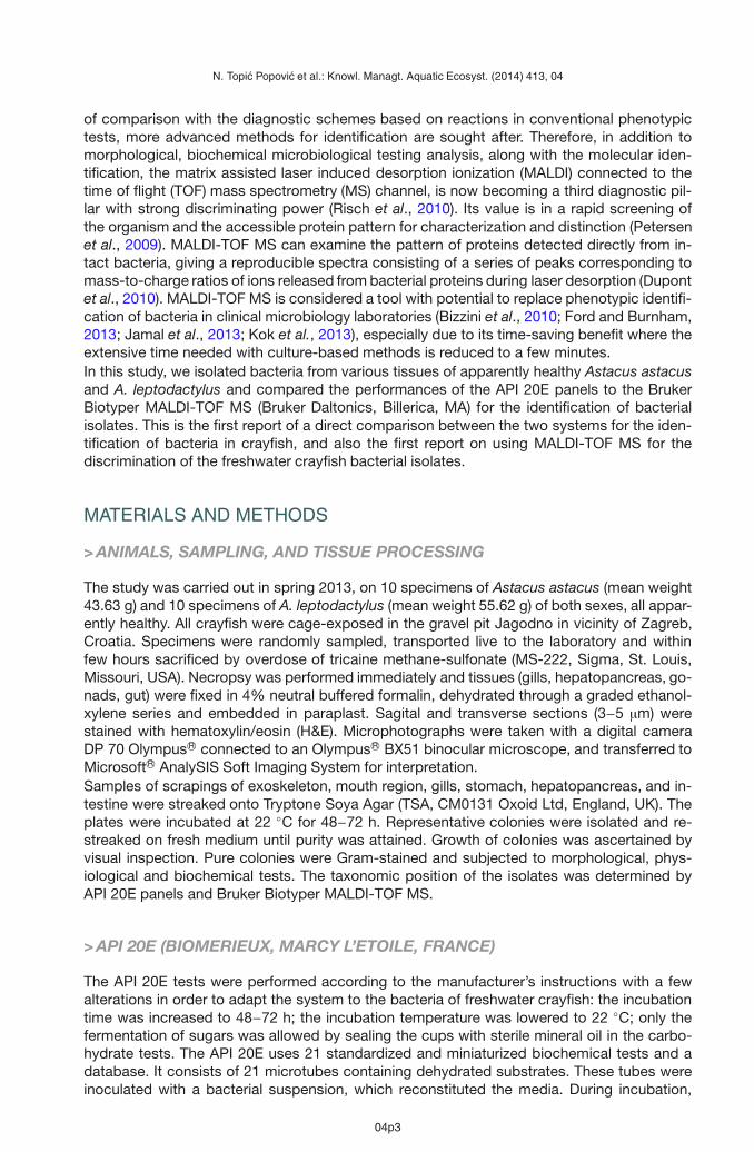
Figure 2Histopathological sections of A. astacus hepatopancreas showing nodular formations in haemal sinus,consisting of agglomerated phagocytic cells surrounding or ingesting bacterial cells (arrow). Also presentis thinning of the cell membrane leading to major vacuolization of hepatopancreas (asterisk). H&E, Scalebar 100 μm.
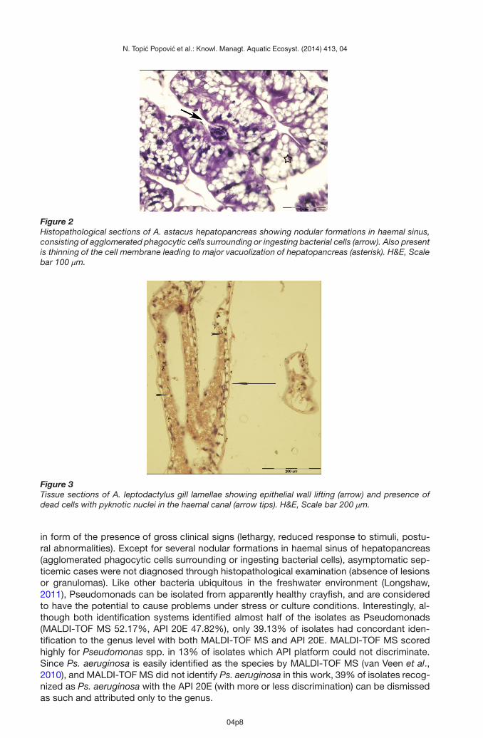
Figure 3Tissue sections of A. leptodactylus gill lamellae showing epithelial wall lifting (arrow) and presence ofdead cells with pyknotic nuclei in the haemal canal (arrow tips). H&E, Scale bar 200 μm.
in form of the presence of gross clinical signs (lethargy, reduced response to stimuli, postu-ral abnormalities). Except for several nodular formations in haemal sinus of hepatopancreas(agglomerated phagocytic cells surrounding or ingesting bacterial cells), asymptomatic sep-ticemic cases were not diagnosed through histopathological examination (absence of lesionsor granulomas). Like other bacteria ubiquitous in the freshwater environment (Longshaw,2011), Pseudomonads can be isolated from apparently healthy crayfish, and are consideredto have the potential to cause problems under stress or culture conditions. Interestingly, al-though both identification systems identified almost half of the isolates as Pseudomonads(MALDI-TOF MS 52.17%, API 20E 47.82%), only 39.13% of isolates had concordant iden-tification to the genus level with both MALDI-TOF MS and API 20E. MALDI-TOF MS scoredhighly for Pseudomonas spp. in 13% of isolates which API platform could not discriminate.Since Ps. aeruginosa is easily identified as the species by MALDI-TOF MS (van Veen et al.,2010), and MALDI-TOF MS did not identify Ps. aeruginosa in this work, 39% of isolates recog-nized as Ps. aeruginosa with the API 20E (with more or less discrimination) can be dismissedas such and attributed only to the genus.
04p8

N. Topic Popovic et al.: Knowl. Managt. Aquatic Ecosyst. (2014) 413, 04
It has been demonstrated that classical phenotypic methods can frequently misidentify non-fermenting bacteria (Pseudomonas included), and for this class of bacteria molecular toolssuch as 16S rRNA gene sequencing provide reliable results, but less accurate at the specieslevel (Campos Braga et al., 2013). Therefore, a reference database for MALDI-TOF MS basedon the identification of non-fermenters was established (Mellmann et al., 2009; Campos Bragaet al., 2013). MALDI-TOF MS was shown to identify correctly to the species level a numberof Pseudomonads, and outperform 16S rRNA sequencing at identifying members of bothPseudomonas and Bacillus genera (Böhme et al., 2013). In this work it identified B. cereus“Securely to the genus and probably to the species” with a high score.
MALDI-TOF MS exceeded API 20E in species identification of Gram-negative rods. In thiswork, Hafnia alvei was the only isolate for which both API 20E and MALDI-TOF MS hada concordant reading to the species level. Interestingly, although API 20E gave “Excellentidentification” for the profile 5305112 of the strip, that very profile was previously describedfor the reference culture of Yersinia ruckeri (Austin et al., 2003; Topic Popovic et al., 2007).MALDI-TOF MS however, identified it “Securely to the genus and probably to the species”and therefore confirmed the API result. H. alvei has previously been isolated from freshwatercrayfish (Longshaw et al., 2012), although its disease-causing properties in crayfish have notbeen described.
The disparity and problems in Aeromonas spp. identification (A. hydrophila group 1 (API 20E)versus A. bestiarum (MALDI-TOF MS)) can be attributed to close relatedness of the twospecies, which according to Martino et al. (2011) belong to the same phenogroup, describedas the A. hydrophila complex. The current taxonomic database of the MALDI-TOF MS Bio-typer system recognizes species that are currently of different taxonomic status and havenot been updated in the Apiweb system (Kierzkowska et al., 2013). The genus Aeromonascomprises 21 validly proposed species, and some of them are phenotypically very similar.MALDI-TOF MS can however provide their good separation at the genospecies level compa-rable with the phylogenetic tree obtained by gyrB gene sequencing; it categorized in clusterswell differentiated A. bestiarum and A. hydrophila (Benagli et al., 2012). Genus-level accu-racy of clinical and environmental Aeromonas isolates identified by MALDI-TOF MS in thework of Lamy et al. (2011) was 100%, while species-level accuracy reached 90.6%, mak-ing this system one of the most accurate and rapid methods for phenotypic identification ofAeromonads, albeit with the need of improvements in its database composition, taxonomyand discriminatory power (Lamy et al., 2011).
When comparing the performance of MALDI-TOF MS with conventional and API systems forclinical isolates of human material, the percentage of correct identifications is significantlyhigher than in this work, mainly due to the customized databases (Bizzini et al., 2010; Dupontet al., 2010; Risch et al., 2010; Martiny et al., 2011; Saffert et al., 2011; El-Bouri et al., 2012;Nagy et al., 2012; Campos Braga et al., 2013; Kierzkowska et al., 2013). For example, 97.2%of isolates had identical genus identification by both methods, while 79.9% yielded exactlythe same species identification in the work of van Veen et al. (2010), and conventional meth-ods also resulted in fewer species identification (83.1% versus 92% MALDI-TOF MS). Thedatabases of the both identification systems used in this work (MALDI-TOF MS and API 20E)are not comprehensive for environmental isolates, and therefore most discordant results weredue to the systematic database-related taxonomical differences. Obviously, the quality andreliability of the identification by MALDI-TOF MS depends on the quality and amount of refer-ence spectra present in the database (Seng et al., 2009; Calderaro et al., 2013).
The disparities on the species level between the two systems which identically identifiedthe isolates on the genus level are not necessarily of the major concern if one looks solelyinto rapid screening of crayfish bacterial flora with the purpose to get insight into the healthstatus of apparently healthy animals. However, diagnostics of bacterial diseases in cray-fish require a completely different approach, and necessitate precision. Generally, diagnos-tic methods based on phenotypic analysis are less frequently used, and molecular meth-ods dominate over traditional techniques, the golden standard being the 16S rRNA genesequencing. Nevertheless, the high cost of this assessment makes this technique impossible
04p9

N. Topic Popovic et al.: Knowl. Managt. Aquatic Ecosyst. (2014) 413, 04
to use in routine microbiology diagnostics (Kierzkowska et al., 2013) and the next step is themass spectrometry-assisted identification. MALDI-TOF MS has demonstrated to be a com-petent bacterial typing tool that extends phenotypic and genotypic approaches, allowing amore ample classification of bacterial strains (Böhme et al., 2013). It seems to be a power-ful, low-cost, rapid proteomic tool in bacterial genus (and frequently species) identificationfrom freshwater crayfish, however we do suggest combining it with classical microbiolog-ical methods, despite their drawbacks such as time-consuming reactions and sometimessubjective morphological observations requiring experience, at least until we benefit from theMALDI-TOF MS database extension. That done, rapid and accurate identification of crayfishpathogens with MALDI-TOF MS will significantly improve the bacterial disease recognition,immediate therapy approach, and enhance the outcomes of farmed crayfish populations, witha single direct colony testing.
ACKNOWLEDGEMENTS
The authors thank the IRES (Institute for Research and Development of Sustainable Ecosys-tems, Croatia) staff for their contribution to this work.
REFERENCES
Alderman D.J. and Polglase J.L., 1988. Pathogens, parasites and commensals. In: Holdich D.M. andLowery R.S. (eds.), Freshwater Crayfish: Biology. Management and Exploitation, Croom Helm,London, 167–212.
Anonymous, 2005. The Law of Nature Conservation. Zakon o zaštiti prirode. Narodne novine, NN 70/05(in Croatian).
Anonymous, 2008. Act on Amendments to the Law of Nature Conservation. Zakon o izmjenama i dop-unama Zakona o zaštiti prirode. Narodne novine, NN 139/08 (in Croatian).
Anonymous, 2009. Ordinance on the proclamation of protected and strictly protected wild taxa.Pravilnik o proglašavanju divljih svojti zašticenim i strogo zašticenim. Narodne novine, NN 99/09(in Croatian).
Austin B. and Austin D., 1999. Bacterial Fish Pathogens: Disease of Farmed and Wild Fish. Austin B.and Austin D. (eds), Springer-Praxis, Chichester, UK, p. 453.
Austin D.A., Robertson P.A.W. and Austin B., 2003. Recovery of a new biogroup of Yersinia ruckeri fromdiseased rainbow trout (Oncorhynchus mykiss, Walbaum). Syst. Appl. Microbiol., 26, 127–131.
Bastardo A., Ravelo C. and Romalde J.L., 2012. A polyphasic approach to study the intraspecific diver-sity of Yersinia ruckeri strains isolated from recent outbreaks in salmonid culture. Vet. Microbiol.,160, 176–182.
Benagli C., Demarta A., Caminada A.P., Ziegler D., Petrini O. and Tonolla M., 2012. A rapid MALDI-TOFMS identification database at genospecific level for clinical and environmental Aeromonas strains.PLoS One, 7, 10, 1–6.
Bizzini A., Durussel C., Bille J., Greub G. and Prod’hom G., 2010. Performance of matrix-assisted laserdesorption ionization-time of flight mass spectrometry for identification of bacterial strains rou-tinely isolated in a Clinical Microbiology Laboratory. J. Clin. Microbiol., 48, 5, 1549–1554.
Böhme K., Fernández-No I.C., Pazos M., Gallardo J.M., Barros-Velázquez J., Cañas B. and Calo-MataP., 2013. Identification and classification of seafood-borne pathogenic and spoilage bacteria: 16SrRNA sequencing versus MALDI-TOF MS fingerprinting. Electrophoresis, 34, 6, 877–887.
Brown B.J. and Leff L.G.,1996. Comparison of identification of aquatic bacteria using fatty acidmethylester analysis and API20E and NFT strips. Appl. Environ. Microbiol., 62, 2183–2185.
Calderaro A., Piccolo G., Montecchini S., Buttrini M., Gorrini C., Rossi S., Arcangeletti M.C., De ContoF., Medici M.C. and Chezzi C., 2013. MALDI-TOF MS analysis of human and animal Brachyspiraspecies and benefits of database extension. J. Proteomics, 78, 273–280.
Campos Braga P., Tata A., Goncalves dos Santos V., Barreiro J.R., Vilczaki Schwab N., Veiga dos SantosM., Nogueira Eberlin M. and Ramires Ferreira C., 2013. Bacterial identification: from the agar plateto the mass spectrometer. RSC Adv., 3, 994–1008.
04p10

N. Topic Popovic et al.: Knowl. Managt. Aquatic Ecosyst. (2014) 413, 04
Colwell R.R., Wicks T.C., Tubiash H.S., 1975. A comparative study of the bacterial flora of thehemolymph of Callinectes sapidus. Mar. Fish. Rev., 37, 29–33.
Cooper A., Layton R., Owens L., Ketheesan N. and Govan B., 2007. Evidence for the classification of acrayfish pathogen as a member of the genus Coxiella. Lett. Appl. Microbiol., 45, 558–563.
Diéguez-Uribeondo J., 2006. Pathogens, parasites ectocemmensals. In: Souty-Grosset C., HoldichD.M., Nöel P., Reynolds J.D. and Haffner P. (eds.), Atlas of crayfish in Europe, Museum Nationald’Histoire Naturelle, Paris, France, 133–149.
Dupont C., Sivadon-Tardy V., Bille E., Daupin B., Beretti L., Alvarez A.S., Degand N., Ferroni A.,Rottman M., Herrmann J.L., Nassif X., Ronco E. and Carbonnelle E., 2010. Identification of clin-ical coagulase-negative staphylococci, isolated in microbiology laboratories, by matrix-assistedlaser desorption/ionization-time of flight mass spectrometry and two automated systems. Clin.Microbiol. Infect., 16, 998–1004.
Edgerton B.F. and Owens L., 1999. Histopathological surveys of the redclaw freshwater crayfish, Cheraxquadricarinatus, in Australia. Aquaculture, 180, 23–40.
Edgerton B.F., Evans L.H. Stephens F.J. and Overstreet R.M., 2002. Synopsis of freshwater crayfishdiseases and commensal organisms. Aquaculture, 206, 57–135.
Edsman L., Füreder L., Gherardi F. and Souty-Grosset C. 2010. Astacus astacus. In: IUCN 2010.IUCN Red List of Threatened Species. Version 2010.4. www.iucnredlist.org. Downloaded on 19January 2011.
El-Bouri K., Johnston S., Rees E., Thomas I., Bome-Mannathoko N., Jones C., Reid M., Ben-Ismaeil B.,Davies A.P., Harris L.G. and Mack D., 2012. Comparison of bacterial identification by MALDI-TOFmass spectrometry and conventional diagnostic microbiology methods: Agreement, speed andcost implications. Br. J. Biomed. Sci., 69, 47–55.
Esteve C., Alcaide E., Blasco M.D., 2012. Aeromonas hydrophila subsp. dhakensis isolated from feces,water and fish in Mediterranean Spain. Microbes Environ., 27, 367–373.
Ford B.A. and Burnham C-A.D., 2013. Optimization of routine identification of clinically relevant gram-negative bacteria by use of matrix-assisted laser desorption-time of flight mass spectrometry andthe Bruker Biotyper. J. Clin. Microbiol., 51, 1412–1420.
Holdich D.M., Haffner P. and Nöel P., 2006. Species files. In: Souty-Grosset C., Holdich D.M., NöelP., Reynolds J.D. and Haffner P. (eds.), Atlas of crayfish in Europe, Museum National d’HistoireNaturelle, Paris, France, 49–131.
Jamal W.Y., Shanin M. and Rotimi V.O., 2013. Comparison of two matrix-assisted laserdesorption/ionization-time of flight (MALDI-TOF) mass spectrometry methods and API 20AN foridentification of clinically relevant anaerobic bacteria. J. Med. Microbiol., 62, 540–544.
Jiravanichpaisal P., Roos S., Edsman L., Liu H. and Söderhäll K., 2009. A highly virulent pathogen,Aeromonas hydrophila, from the freshwater crayfish Pacifastacus leniusculus. J. Invertebr. Pathol.,101, 56–66.
Johnson P.T., 1976. Bacterial infection in the blue crab, Callinectes sapidus: course of infection andhistopathology. J. Invertebr. Pathol., 28, 25–36.
Johnson P.T.J. and Paull S.H., 2011. The ecology and emergence of diseases in fresh waters. Freshw.Biol., 56, 638–657.
Kierzkowska M., Majwska A., Kuthan R.T., Sawicka-Grzelak A. and Mynarczyk M., 2013. A comparisonof Api 20A vs MALDI-TOF MS for routine identification of clinically significant anaerobic bacterialstrains to the species level. J. Microbiol. Meth., 92, 209–212.
Kok J., Chen S.C.A., Dwyer D.E. and Iredell J.R., 2013. Current status of matrix-assisted laser desorptionionization-time of flight mass spectrometry in the clinical microbiology laboratory. Pathology, 45,4–17.
Lamy B., Kodjo A. and Laurent F., 2011. Identification of Aeromonas isolates by matrix-assisted laserdesorption ionization-time of flight mass spectrometry. Diag. Microbiol. Infect. Dis., 71, 1–5.
Longshaw M., 2011. Diseases of crayfish: A review. J. Invertebr. Pathol., 106, 54–70.
Longshaw M., Bateman K.S., Stebbing P., Stentiford G.D. and Hockley F.A., 2012. Disease risks associ-ated with the importation and release of non-native crayfish species into mainland Britain. AquaticBiol., 16, 1–15.
Madetoja M. and Jussila J., 1996. Gram negative bacteria in the hemolymph of noble crayfish Astacusastacus, in an intensive crayfish culture system. Nord. J. Freshwater Res., 72, 88–90.
Maguire I. and Gottstein-Matocec S., 2004. The distribution pattern of freshwater crayfish in Croatia.Crustaceana, 77, 25–47.
04p11

N. Topic Popovic et al.: Knowl. Managt. Aquatic Ecosyst. (2014) 413, 04
Maguire I., Jelic M. and Klobucar G., 2011. Update on the distribution of freshwater crayfish in Croatia.Knowl. Managt. Aquatic Ecosyst. 401, Doi: 10.1051/kmae/2011051.
Makkonen J., 2013. The crayfish plague pathogen Aphanomyces astaci – Genetic diversity and adapta-tion to the host species. PhD thesis, University of Eastern Finland, p. 67.
Martino M.E., Fasolato L., Montemurro F., Rosteghin M., Manfrin A, Patarnello T., Novelli E. and CardazzB., 2011. Determination of microbial diversity of Aeromonas strains on the basis of multilocussequence typing, phenotype, and presence of putative virulence gene. Appl. Environ. Microbiol.,77, 4986–5000.
Martiny D., Dediste A., Debruyne L., Vlaes L., Haddou N.B., Vandamme P. and Vandenberg O., 2011.Accuracy of the API Campy system, the Vitek 2 Neisseria-Haemophilus card and matrix-assistedlaser desorption ionization-time of flight mass spectrometry for the identification of Campylobacterand related organisms. Clin. Microbiol. Infect., 17, 1001–1006.
Mellmann A., Bimet F., Bizet C., Borovskaya A.D., Drake R.R., Eigner U., Fahr A.M., He Y., Ilina E.N.,Kostrzewa M., Maier T., Mancinelli L., Moussaoui W., Prévost G., Putignani L., Seachord C.L.,Tang Y.W. and Harmsen D., 2009. High interlaboratory reproducibility of matrix-assisted laser des-orption ionization-time of flight mass spectrometry-based species identification of nonfermentingbacteria. J. Clin. Microbiol., 47, 3732–3734.
Mickeniene L. and Šyvokiene J., 2011. The study of bacteria on artificially incubated noble crayfisheggs. Inland Water Biol., 4, 137–142.
Nagy E., Becker S., Kostrzewa M., Barta N. and Urban E., 2012. The value of MALDI-TOF MS for theidentification of clinically relevant anaerobic bacteria in routine laboratories. J. Med. Microbiol., 61,1393–1400.
Petersen C.E., Valentine N.B. and Wahl K.L., 2009. Characterization of microorganisms by MALDI massspectrometry. Methods in molecular biology. Clifton, New Jersey, 492 p.
Quaglio F., Morolli C., Galuppi R., Tampieri M.P., Bonoli C., Marcer F., Rotundo G. and GerminaraG.S., 2006a. Sanitary-pathological examination of red swamp crayfish (Procambarus clarkii, Girard1852) in the Reno Valley. Freshwater Crayfish, 15, 1–10.
Quaglio F., Morolli C., Galuppi R., Bonoli C., Marcer F., Nobile L, DeLuise G. and Tampieri M.P.,2006b. Preliminary investigations of disease-causing organisms in the white-clawed crayfishAustropotamobius pallipes complex from streams of Northern Italy. Bull. Fr. Pêche Piscic.,380−381, 1271–1290.
Risch M., Radjenovic D., Nam Han J., Wydler M., Nydegger U. and Risch L., 2010. Comparison ofMALDI TOF with conventional identification of clinically relevant bacteria. Swiss Med. Wkly., 140,1–5.
Romero X. and Jiménez, R., 2002. Histopathological survey of diseases and pathogens present in red-claw crayfish, Cherax quadricarinatus (Von Martens), cultured in Ecuador. J. Fish Dis., 25, 653–667.
Saffert R.T., Cunningham S.A., Ihde S.M., Monon Jobe K.E., Mandrekar J. and Patel R., 2011.Comparison of Bruker Biotyper matrix-assisted laser desorption ionization-time of flight massspectrometry to BD Phoenix automated microbiology system for identification of Gram-negativebacilli. J. Clin. Microbiol., 49, 887–892.
Sanjuán E., Fouz B., Oliver J.D. and Amaro C., 2009. Evaluation of genotypic and phenotypic methodsto distinguish clinical from environmental Vibrio vulnificus strains. Appl. Environ. Microbiol., 75,1604–1613.
Scott J.R. and Thune R.L., 1986. Bacterial flora of hemolymph from red swamp crawfish, Procambarusclarkii (Girard), from commercial ponds. Aquaculture, 58, 161–165.
Seng P., Drancourt M., Gouriet F., La S.B., Founier P.E., Rolain J.M. et al., 2009. Ongoing revolution inbacteriology : routine identification of bacteria by matrix-assisted laser desorption ionization-timeof flight mass spectrometry. Clin. Infect. Dis., 49, 543–551.
Smith V.J. and Söderhäll K., 1986. Crayfish pathology: an overview. Freshwater Crayfish, 6, 199–211.Soto E., Griffin M., Arauz M., Riofrio A., Martinez A. and Cabrejoh M.E., 2012. Edwardsiella ictaluri as
the causative agent of mortality in cultured Nile tilapia. J. Aquat. Anim. Health, 24, 81–90.Topic Popovic N., Což-Rakovac R., Strunjak-Perovic I., 2007. Commercial phenotypic tests (API 20E) in
diagnosis of fish bacteria. Vet. Med. Czech, 52, 49–53.van Veen S.Q., Claas E.C.J. and Kuijper E.J., 2010. High-throughput identification of bacteria and yeast
by matrix-assisted laser desorption ionization-time of flight mass spectrometry in conventionalmedical microbiology laboratories. J. Clin. Microbiol., 48, 900–907.
Vey A., 1986. Disease problems during aquaculture of freshwater crayfish. Freshwater Crayfish, 6,212−222.
04p12